Regulation of Interleukin6 Expression in Porcine Immune Cells
Upload
independentCategory
view
1download
0
PLEASE SCROLL DOWN FOR ARTICLE
This article was downloaded by: [Pavelkic, Vesna M.]On: 13 November 2008Access details: Access Details: [subscription number 905462418]Publisher Informa HealthcareInforma Ltd Registered in England and Wales Registered Number: 1072954 Registered office: Mortimer House,37-41 Mortimer Street, London W1T 3JH, UK
Journal of Enzyme Inhibition and Medicinal ChemistryPublication details, including instructions for authors and subscription information:http://www.informaworld.com/smpp/title~content=t713642123
The influence of Al3+ ion on porcine pepsin activity in vitroVesna M. Pavelkic a; Kristina R. Gopcevic b; Danijela Z. Krstic b; Marija A. Ilic c
a Department of Physical Chemistry, Vinca-Institute of Nuclear Science, Belgrade, Serbia b School ofMedicine, University of Belgrade, Belgrade, Serbia c Institute of General and Physical Chemistry, Belgrade,Serbia
First Published:December2008
To cite this Article Pavelkic, Vesna M., Gopcevic, Kristina R., Krstic, Danijela Z. and Ilic, Marija A.(2008)'The influence of Al3+ ion onporcine pepsin activity in vitro',Journal of Enzyme Inhibition and Medicinal Chemistry,23:6,1002 — 1010
To link to this Article: DOI: 10.1080/14756360701841095
URL: http://dx.doi.org/10.1080/14756360701841095
Full terms and conditions of use: http://www.informaworld.com/terms-and-conditions-of-access.pdf
This article may be used for research, teaching and private study purposes. Any substantial orsystematic reproduction, re-distribution, re-selling, loan or sub-licensing, systematic supply ordistribution in any form to anyone is expressly forbidden.
The publisher does not give any warranty express or implied or make any representation that the contentswill be complete or accurate or up to date. The accuracy of any instructions, formulae and drug dosesshould be independently verified with primary sources. The publisher shall not be liable for any loss,actions, claims, proceedings, demand or costs or damages whatsoever or howsoever caused arising directlyor indirectly in connection with or arising out of the use of this material.
The influence of Al31 ion on porcine pepsin activity in vitro
VESNA M. PAVELKIC1, KRISTINA R. GOPCEVIC2, DANIJELA Z. KRSTIC2, &
MARIJA A. ILIC3
1Department of Physical Chemistry, Vinca-Institute of Nuclear Science, POB 522, 11001 Belgrade, Serbia, 2School of
Medicine, University of Belgrade, Visegradska 26, 11000 Belgrade, Serbia, and 3Institute of General and Physical Chemistry,
Studentski trg 12-16, 11000 Belgrade, Serbia
(Received 3 July 2007; accepted 5 November 2007)
AbstractThe in vitro effect of Al3þ ions in the concentration range 1.7·1026 M–8.7·1023 M on pepsin activity at pH 2, via kineticparameters and its electrophoretic mobility was evaluated. Kinetic study demonstrated the existence of an activation effect ofAl3þ at pH 2 on pepsin molecule. Kinetic analysis with respect to concentrations of haemoglobin showed that Al3þ ionsincrease the maximal velocity (Vmax) and kcat values rather than apparent affinity for substrate (KS) implying thenon-competitive nature of activation which indicated that aluminium was a non-essential activator of partial non-competitivetype. The values of the equilibrium constants KS and KmA for dissociation of corresponding complexes were evaluated as0.904 ^ 0.083 mM and 8.56 ^ 0.51mM, respectively. Dissociation constant KA, of activator from enzyme-activator complexcalculated via kinetic and direct measurement of Al3þ binding data, as well as activation constant A50, the activatorconcentration that gives a rate equal to half at a saturating concentration of activator, were found to be 8.82 ^ 0.90mM,8.39 ^ 0.76mM, and 8.05 ^ 0.48mM respectively. Native PAGE electrophoresis shows the decrease in electrophoreticmobility of pepsin and confirms modification of the electric charge and conformational changes of pepsin caused by boundAl3þ on the pepsin molecule. Al3þ induced conformational changes of pepsin were verified by UV-VIS and IR spectra.Moreover, the absence of conformational changes in the haemoglobin molecule in the presence of Al3þ ions confirms that theobtained activation is a consequence of conformational changes caused only in the pepsin molecule.
Keywords: Pepsin, aluminium, kinetics, activation, electrophoretic mobility
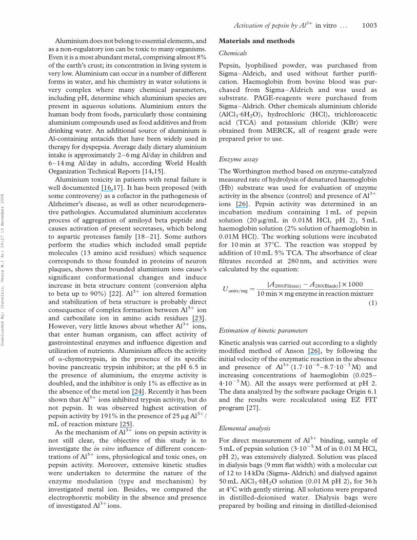
Introduction
Porcine pepsin A (EC 3.4.23.1), a prominent member
of the aspartic protease, is the principal proteolytic
enzyme of gastric juice and as the best understood of
this family of proteinases it was used as a model to
study related enzymes. As all aspartic proteinases
pepsin molecule consist of two homologous lobes
composed predominantly of b-sheets separated by a
hinged substrate-binding cleft. Two active site aspartic
residues occupy the cleft to which admittance is
restricted by a hinged flexible flap region [1–6]. The
pepsin consists of 326 residues with molecular weight
of 35000 Da and it derives from its zymogene
pepsinogen, by removal of 44 amino acids, from its
amino terminus, to give a single chain enzyme with a
low pI and three disulfide bridges. Each lobe of pepsin
contains a catalytic aspartic acid residue Asp32 and
Asp215 located at the centre of the binding cleft
between the domains. One of two Asp residues has to
be protonated and the other deprotonated, for the
protein to be active [1]. Due to the catalytic residues,
the active pH ranges from 1.0 to 5.0 [7–11]. Because
pepsin has been well structurally characterized, it
represents an appropriate model to study the effects of
metal ions on structure, function and kinetic
behaviour [12,13].
ISSN 1475-6366 print/ISSN 1475-6374 online q 2008 Informa UK Ltd.
DOI: 10.1080/14756360701841095
Correspondence: V. M. Pavelkic, Department of Physical Chemistry, Institute of Nuclear Sciences Vinca, POB 522, 11001 Belgrade, Serbia.Tel: 381 11 245 39 67. Fax: 381 11 244 72 07. E-mail: [email protected]
Journal of Enzyme Inhibition and Medicinal Chemistry, December 2008; 23(6): 1002–1010
Downloaded By: [Pavelkic, Vesna M.] At: 09:27 13 November 2008
Aluminium doesnot belong toessential elements, and
as a non-regulatory ion can be toxic to many organisms.
Even it is a most abundant metal, comprising almost 8%
of the earth’s crust; its concentration in living system is
very low. Aluminium can occur in a number of different
forms in water, and his chemistry in water solutions is
very complex where many chemical parameters,
including pH, determine which aluminium species are
present in aqueous solutions. Aluminium enters the
human body from foods, particularly those containing
aluminium compounds used as food additives and from
drinking water. An additional source of aluminium is
Al-containing antacids that have been widely used in
therapy for dyspepsia. Average daily dietary aluminium
intake is approximately 2–6 mg Al/day in children and
6–14 mg Al/day in adults, according World Health
Organization Technical Reports [14,15].
Aluminium toxicity in patients with renal failure is
well documented [16,17]. It has been proposed (with
some controversy) as a cofactor in the pathogenesis of
Alzheimer’s disease, as well as other neurodegenera-
tive pathologies. Accumulated aluminium accelerates
process of aggregation of amiloyd beta peptide and
causes activation of present secretases, which belong
to aspartic proteases family [18–21]. Some authors
perform the studies which included small peptide
molecules (13 amino acid residues) which sequence
corresponds to those founded in proteins of neuron
plaques, shows that bounded aluminium ions cause’s
significant conformational changes and induce
increase in beta structure content (conversion alpha
to beta up to 90%) [22]. Al3þ ion altered formation
and stabilization of beta structure is probably direct
consequence of complex formation between Al3þ ion
and carboxilate ion in amino acids residues [23].
However, very little knows about whether Al3þ ions,
that enter human organism, can affect activity of
gastrointestinal enzymes and influence digestion and
utilization of nutrients. Aluminium affects the activity
of a-chymotrypsin, in the presence of its specific
bovine pancreatic trypsin inhibitor; at the pH 6.5 in
the presence of aluminium, the enzyme activity is
doubled, and the inhibitor is only 1% as effective as in
the absence of the metal ion [24]. Recently it has been
shown that Al3þ ions inhibited trypsin activity, but do
not pepsin. It was observed highest activation of
pepsin activity by 191% in the presence of 25mg Al3þ /
mL of reaction mixture [25].
As the mechanism of Al3þ ions on pepsin activity is
not still clear, the objective of this study is to
investigate the in vitro influence of different concen-
trations of Al3þ ions, physiological and toxic ones, on
pepsin activity. Moreover, extensive kinetic studies
were undertaken to determine the nature of the
enzyme modulation (type and mechanism) by
investigated metal ion. Besides, we compared the
electrophoretic mobility in the absence and presence
of investigated Al3þ ions.
Materials and methods
Chemicals
Pepsin, lyophilised powder, was purchased from
Sigma–Aldrich, and used without further purifi-
cation. Haemoglobin from bovine blood was pur-
chased from Sigma–Aldrich and was used as
substrate. PAGE-reagents were purchased from
Sigma–Aldrich. Other chemicals aluminium chloride
(AlCl3·6H2O), hydrochloric (HCl), trichloroacetic
acid (TCA) and potassium chloride (KBr) were
obtained from MERCK, all of reagent grade were
prepared prior to use.
Enzyme assay
The Worthington method based on enzyme-catalyzed
measured rate of hydrolysis of denatured haemoglobin
(Hb) substrate was used for evaluation of enzyme
activity in the absence (control) and presence of Al3þ
ions [26]. Pepsin activity was determined in an
incubation medium containing 1 mL of pepsin
solution (20mg/mL in 0.01M HCl, pH 2), 5 mL
haemoglobin solution (2% solution of haemoglobin in
0.01M HCl). The working solutions were incubated
for 10 min at 378C. The reaction was stopped by
addition of 10 mL 5% TCA. The absorbance of clear
filtrates recorded at 280 nm, and activities were
calculated by the equation:
Uunits=mg ¼½A280ðFiltrateÞ 2 A280ðBlankÞ� £ 1000
10 min £ mg enzyme in reaction mixture
ð1Þ
Estimation of kinetic parameters
Kinetic analysis was carried out according to a slightly
modified method of Anson [26], by following the
initial velocity of the enzymatic reaction in the absence
and presence of Al3þ (1.7·1026–8.7·1023 M) and
increasing concentrations of haemoglobin (0.025–
4·1023 M). All the assays were performed at pH 2.
The data analyzed by the software package Origin 6.1
and the results were recalculated using EZ FIT
program [27].
Elemental analysis
For direct measurement of Al3þ binding, sample of
5 mL of pepsin solution (3·1025 M of in 0.01 M HCl,
pH 2), was extensively dialyzed. Solution was placed
in dialysis bags (9 mm flat width) with a molecular cut
of 12 to 14 kDa (Sigma- Aldrich) and dialysed against
50 mL AlCl3·6H2O solution (0.01 M pH 2), for 36 h
at 48C with gently stirring. All solutions were prepared
in distilled-deionised water. Dialysis bags were
prepared by boiling and rinsing in distilled-deionised
Activation of pepsin by Al3þ in vitro . . . 1003
Downloaded By: [Pavelkic, Vesna M.] At: 09:27 13 November 2008
water. The concentration of aluminium ions inside
and outside the dialysis bag was carried out by
inductively coupled plasma atomic emission spec-
troscopy (ICP-AES), (Spectroflame ICP, 2.5 kW,
27 MHz). ICP-AES analysis was performed by
measuring the intensity of radiation of the specific
wavelength emitted by aluminium at 396.152 nm and
with a sample flow rate of 1 mL min21. Integration
times were 1 per increment (i.e., analysis time of
1 minute per sample).
UVabsorbance measurements
UV absorbance measurements of pepsin and haemo-
globin samples, both in the absence and in the presence
of Al3þ (water solutions acidified with 0.01M HCl,
pH2), carried out on Beckman UV 5260 UV-VIS
spectrophotometer withan electro-thermal temperature
control cell unit. The temperature control performed
with DANA – Digital voltmeter model 4800 with
chromel-alumel thermocouples. A quartz cell with a
1 cm path length was used for all the absorbance studies.
All measurements were carried out at 378C.
IR studies
Pepsin-Al3þ and haemoglobin-Al3þ samples were
lyophilized and used for the further experiments.
The removal of any traces of water from the samples
was insured by drying samples over silica gel over night
and heating during 2 hours at 1008C. For infrared
spectroscopy, the samples were prepared in the form of
standard potassium bromide (KBr) pallet (the ratio
between pepsin and KBr and pepsin – Al3þ and KBr
were 1:100); the pallets of haemoglobin samples in KBr
were prepared in the same manner. Infrared spectra
between 4000 and 400 cm21 obtained on a Specord 75
IR, Carl Zeiss in double-beam operation vs. KBr as a
reference. IR spectra of both proteins in KBr matrices
recorded at room temperature (258C).
Polyacrylamide gel electrophoresis (PAGE)
Native electrophoresis of pepsin and haemoglobin on
10% polyacrylamide gel carried out at 48C during
90 min, according to the Laemmli procedure, at pH
8.3 [28]. Water solutions of all samples of enzyme
(pepsin dissolved in water to final concentration of
2 mg/mL) were titrated with HCl to pH 2 and
incubated at 378C, with addition of different
concentrations of Al3þ ion (1, 5 and 10 mM).
The samples of pepsin and haemoglobin were diluted
with sample buffer in ratio 1:1 (v/v) and applied on gel
in volume of 20mL. Visualization was performed with
Commassie Brilliant Blue G-250 dye. The gels
scanned and processed using Corel Draw 11.0
software package. Quantification of electrophoretic
mobility of the molecule is carried out via RS value,
where it is defined by:
RS ¼½distance of protein migration�=
½distance of tracing dye migration� ð2Þ
Statistical analysis
Graphs were plotted by using Microcal Origin program
(version 6.1). Values including KS, Vmax, KmS, KmA,
KA, and A50 and their standard errors are presented as
means ^ SEM (obtained by the linear regression
analysis). The statistical comparisons were performed
by Students t-test for paired observations. The means
of at least five observations was quoted in the text and
p , 0.01 was considered statistically significant.
Results
In vitro effects of Al3þ ions on porcine pepsin activity
The influence of Al3þ ions on porcine pepsin activity
was investigated in the concentration range
1.7·1026 M–8.7·1023 M at pH 2. We tested wide
range of Al3þ concentration that included physiologi-
cal as well as toxic doses of metal ion because of lack of
similar information in available literature. The effects
of chosen concentrations of Al3þ ions on in vitro
pepsin activity are presented in Figure 1.
It is obviously from Figure 1 that all investigated
concentration of Al3þ ions cause increase of pepsin
activity. The increasing concentrations of metal ions
induced increase of enzymatic activity. Values are
expressed as the percent of increased activity related to
the control, which considered as 100%. Aluminium
was found to stimulate the enzyme activity
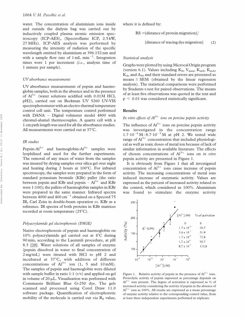
Figure 1. Relative activity of pepsin in the presence of Al3þ ions.
Proteolytic activity of pepsin expressed as percentage depends on
Al3þ ions present. The degree of activation is expressed as % of
increased activity considering the activity of pepsin in the absence of
Al3þ ions as 100%. All results are expressed as a mean percentage
of enzyme activity relative to the corresponding control value, from
at least three independent experiments performed in triplicate.
V. M. Pavelkic et al.1004
Downloaded By: [Pavelkic, Vesna M.] At: 09:27 13 November 2008
in dose-dependent manner and linear curve (the
correlation coefficient was 0.991) was obtained and
presented on Figure 1. The presence of 1.7·1026 M
Al3þ in incubation milieu causes increase in pepsin
activity for 30.7%. Increasing the amount of Al3þ led to
a more significant increase of proteolytic activity. So,
3.4·1025 M of Al3þ increases pepsin activity for 51.9%
(p , 0.01),1.7·1024 M for 72.8%,while in the presence
of 1.7·1023 M Al3þ in incubation milieu the activity of
pepsin was doubled (95.7%, p , 0.01). Maximal
investigated concentration of Al3þ ions, induce the
increase of pepsin activity for 135.8% (p , 0.01), in
comparison with corresponding control.
Kinetic analysis
In order to evaluate the nature of porcine pepsin
activation induced by Al3þ , kinetic parameters KS and
Vmax were determined by varying the concentration of
substrate–denatured haemoglobin. The kinetic prop-
erties of enzyme were determined in the presence of
desired concentration of metal ions (from 1.7·1026 to
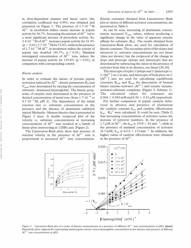
8.7·1023 M, pH 2). The dependence of the initial
reaction rate vs. substrate concentration in the
presence and the absence of aluminium exhibited
typical Michaelis–Menten kinetics that is presented in
Figure 2 inset. A double reciprocal plot of the
velocity vs. substrate concentrations at increasing
concentrations of Al3þ ions resulted in a family of
linear plots intersecting at 1/[Hb] axis (Figure 2).
The Lineweaver-Burk plots show that increase of
reaction velocity in the presence of Al3þ ions is
proportional to increased Al3þ ions concentration.
Kinetic constants obtained from Lineaweaver–Burk
plot at variety of different activator concentrations, are
presented in Table I.
As can be seen, increasing of aluminium concen-
tration increased Vmax values, without producing a
significant change in the value of apparent enzyme
affinity for substrate (KS). The results obtained from
Lineweaver-Burk plots, are used for calculation of
kinetic constants. The secondary plots of the slopes and
intersects vs. activator concentrations are not linear
(data not shown), but the reciprocal of the change in
slope and intercept (Dslope and Dintercept) that are
determined by subtracting the values in the presence of
activator from that in its absence, are linear [29,30].
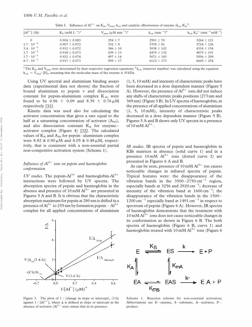
The intercepts of a plot 1/Dslope and 1/Dintercept vs.
1/ [Al3þ ] on 1/D axis, and intercepts of both plots on 1/
[Al3þ ] axis are used for calculating equilibrium
constants KmS and KmA for dissociation of formed
binary enzyme-activator (Al3þ ) and ternary enzyme-
activator-substrate complexes (Figure 3, Scheme 1).
The calculated values for constants are
0.904 ^ 0.083 mM and 8.56 ^ 0.51mM, respectively.
For further comparison of pepsin catalytic beha-
viour in absence and presence of aluminium
the catalytic constant kcat and catalytic effectiveness
kcat · KS21 were calculated. It could be seen (Table I)
that increasing concentrations of activator causes the
increase of turnover numbers. In the presence of
1.7mM of Al3þ , the kcat is 3378 ^ 81 min21, while in
the presence of maximal concentration of activator
(8.7 mM) kcat is 6112 ^ 173 min21. In addition, the
higher values of catalytic effectiveness were obtained
in the presence of Al3þ .
Figure 2. Lineweaver-Burk plot of a series of kinetics measurements in a presence of different Al3þ ions concentrations at pH2; (inset)
Hyperbolic plots (sigmoid fit) representing initial pepsin velocity versus haemoglobin concentration in the absence and presence of different
Al3þ ions concentrations at pH2.
Activation of pepsin by Al3þ in vitro . . . 1005
Downloaded By: [Pavelkic, Vesna M.] At: 09:27 13 November 2008
Using UV spectral and aluminium binding assays
data (experimental data not shown) the fraction of
bound aluminium to pepsin n and dissociation
constant for pepsin-aluminium complex K A, were
found to be 0.96 ^ 0.09 and 8.39 ^ 0.76mM
respectively [31].
Kinetic data was used also for calculating the
activator concentration that gives a rate equal to the
half at a saturating concentration of activator (A50),
and also dissociation constant KA for enzyme–
activator complex (Figure 4) [32]. The calculated
values of KA and A50 for pepsin–aluminium complex
were 8.82 6 0.90mM and 8.05 6 0.48mM, respect-
ively, that is consistent with a non-essential partial
non-competitive activation system (Scheme 1).
Influence of Al3þ ions on pepsin and haemoglobin
conformation
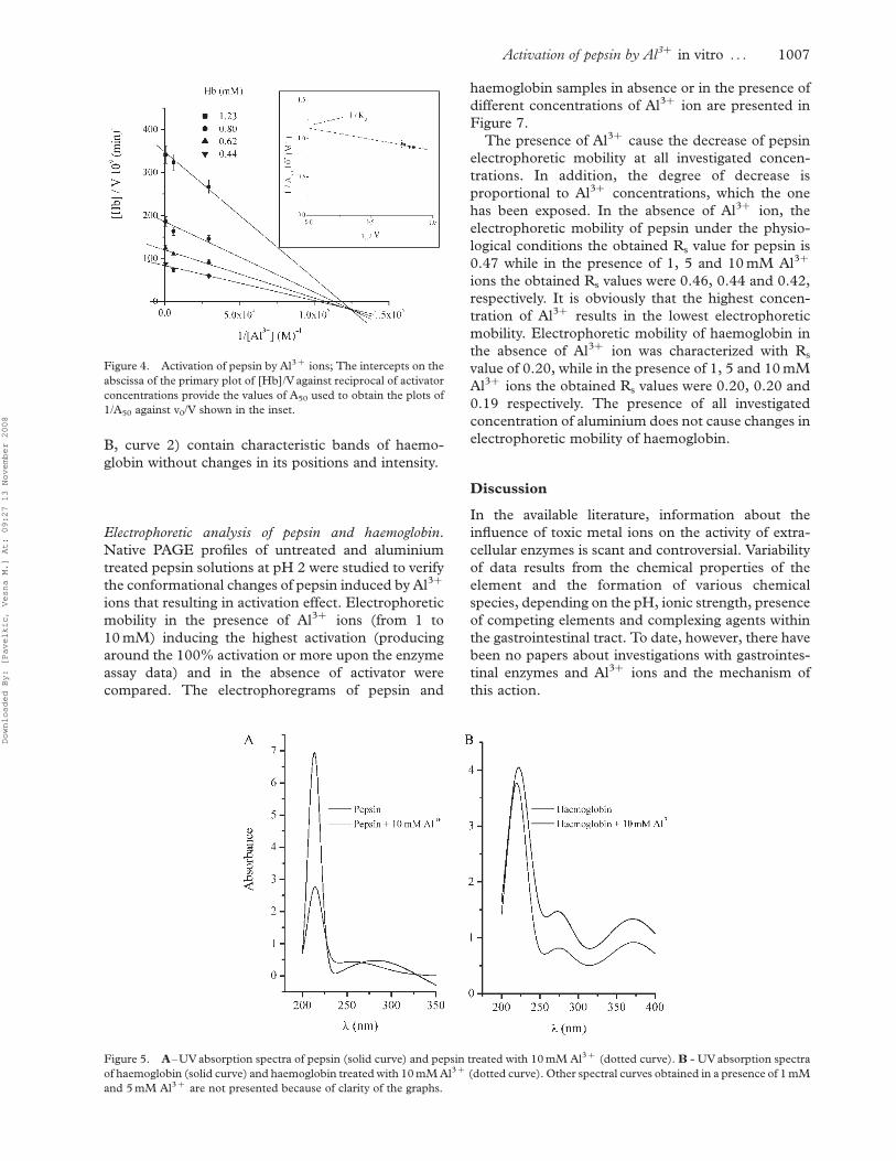
UV studies. The pepsin-Al3þ and haemoglobin-Al3þ
interactions were followed by UV spectra. The
absorption spectra of pepsin and haemoglobin in the
absence and presence of 10 mM Al3þ are presented in
Figures 5 A and B. It is obvious that the characteristic
absorption maximum for pepsin at 280 nm is shifted in a
presence of Al3þ to 255 nm by formation pepsin – Al3þ
complex for all applied concentrations of aluminium
(1, 5, 10 mM) and intensity of characteristic peaks have
been decreased in a dose dependent manner (Figure 5
A). However, the presence of Al3þ ions did not induce
any shifts of characteristic peaks positions (273 nm and
369 nm) (Figure 5 B). In UV spectra of haemoglobin, in
the presence of all applied concentrations of aluminium
(1, 5, 10 mM), intensity of characteristic peaks
decreased in a dose dependent manner (Figure 5 B).
Figures 5 A and B shows only UV spectra in a presence
of 10 mM Al3þ .
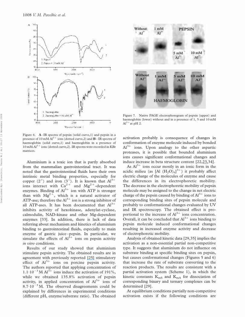
IR studies. IR spectra of pepsin and haemoglobin in
KBr matrices in absence (solid curve 1) and in a
presence 10 mM Al3þ ions (dotted curve 2) are
presented in Figures 6 A and B.
As can be seen, presence of 10 mM Al3þ ion causes
noticeable changes in infrared spectra of pepsin.
Typical features were: the disappearance of the
vibration bands in the 3500–2750 cm21 region,
especially bands at 3256 and 2920 cm21; decrease of
intensity of the vibration band at 1600 cm21; the
disappearance of the vibration bands in the 1500–
1200 cm21 especially band at 1491 cm21 in respect to
spectrum of pepsin (Figure 6 A). However, IR spectra
of haemoglobin demonstrate that the treatment with
10 mM Al3þ ions does not cause noticeable changes in
its conformation as shown in Figure 6 B. The both
spectra of haemoglobin (Figure 6 B, curve 1) and
haemoglobin treated with 10 mM Al3þ ions (Figure 6
Table I. Influence of Al3þ on Km, Vmax, kcat and catalytic effectiveness of enzyme (kcat Km21).
[Al3þ] (M) KS (mM L21)a Vmax (mM min21)a kcat (min21)b kcat KS21 (min21 mM21)
0 0.904 ^ 0.083 254 ^ 7 2591 ^ 70 3264 ^ 121
1.7 · 1026 0.907 ^ 0.072 332 ^ 8 3378 ^ 81 3724 ^ 126
3.4 · 1025 0.912 ^ 0.072 386 ^ 10 3938 ^ 102 4318 ^ 154
1.7 · 1024 0.918 ^ 0.073 439 ^ 13 4479 ^ 132 4879 ^ 191
1.7 · 1023 0.921 ^ 0.074 497 ^ 14 5071 ^ 143 5506 ^ 209
8.7 · 1023 0.917 ^ 0.073 599 ^ 17 6112 ^ 173 6665 ^ 254
a The Km and Vmax were determined by their respective regression equations; b kcat (turnover number) was calculated using the equation
kcat ¼ Vmax/ [E]t, assuming that the molecular mass of the enzyme is 35 kDa.
Scheme 1. Reaction scheme for non-essential activation;
Abbreviations are E—enzyme, S—substrate, A—activator, P—
product.
Figure 3. The plots of 1 / (change in slope or intercept), (1/D)
against 1 / [Al3þ ], where D is defined as slope or intercept in the
absence of activator (Al3þ ions) minus that in its presence.
V. M. Pavelkic et al.1006
Downloaded By: [Pavelkic, Vesna M.] At: 09:27 13 November 2008
B, curve 2) contain characteristic bands of haemo-
globin without changes in its positions and intensity.
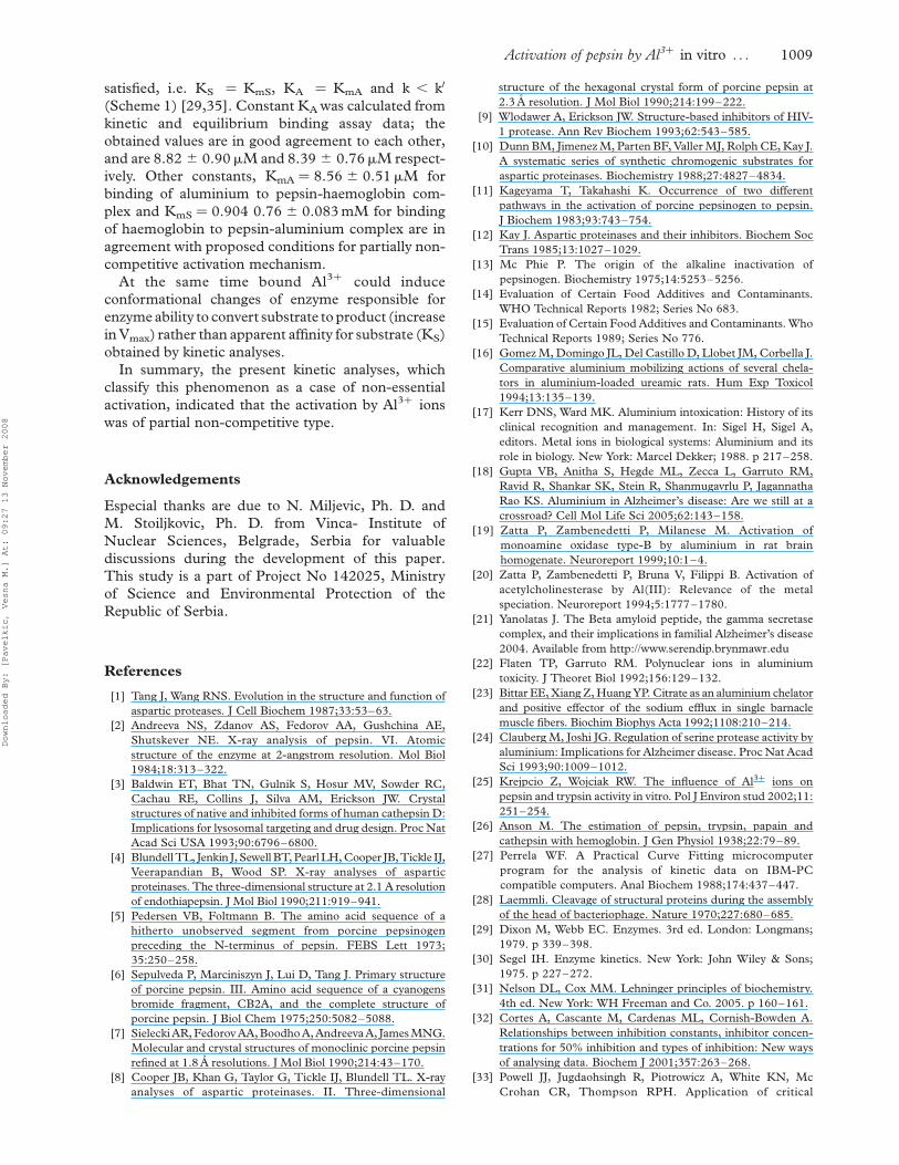
Electrophoretic analysis of pepsin and haemoglobin.
Native PAGE profiles of untreated and aluminium
treated pepsin solutions at pH 2 were studied to verify
the conformational changes of pepsin induced by Al3þ
ions that resulting in activation effect. Electrophoretic
mobility in the presence of Al3þ ions (from 1 to
10 mM) inducing the highest activation (producing
around the 100% activation or more upon the enzyme
assay data) and in the absence of activator were
compared. The electrophoregrams of pepsin and
haemoglobin samples in absence or in the presence of
different concentrations of Al3þ ion are presented in
Figure 7.
The presence of Al3þ cause the decrease of pepsin
electrophoretic mobility at all investigated concen-
trations. In addition, the degree of decrease is
proportional to Al3þ concentrations, which the one
has been exposed. In the absence of Al3þ ion, the
electrophoretic mobility of pepsin under the physio-
logical conditions the obtained Rs value for pepsin is
0.47 while in the presence of 1, 5 and 10 mM Al3þ
ions the obtained Rs values were 0.46, 0.44 and 0.42,
respectively. It is obviously that the highest concen-
tration of Al3þ results in the lowest electrophoretic
mobility. Electrophoretic mobility of haemoglobin in
the absence of Al3þ ion was characterized with Rs
value of 0.20, while in the presence of 1, 5 and 10 mM
Al3þ ions the obtained Rs values were 0.20, 0.20 and
0.19 respectively. The presence of all investigated
concentration of aluminium does not cause changes in
electrophoretic mobility of haemoglobin.
Discussion
In the available literature, information about the
influence of toxic metal ions on the activity of extra-
cellular enzymes is scant and controversial. Variability
of data results from the chemical properties of the
element and the formation of various chemical
species, depending on the pH, ionic strength, presence
of competing elements and complexing agents within
the gastrointestinal tract. To date, however, there have
been no papers about investigations with gastrointes-
tinal enzymes and Al3þ ions and the mechanism of
this action.
Figure 4. Activation of pepsin by Al3þ ions; The intercepts on the
abscissa of the primary plot of [Hb]/V against reciprocal of activator
concentrations provide the values of A50 used to obtain the plots of
1/A50 against v0/V shown in the inset.
Figure 5. A–UV absorption spectra of pepsin (solid curve) and pepsin treated with 10 mM Al3þ (dotted curve). B - UV absorption spectra
of haemoglobin (solid curve) and haemoglobin treated with 10 mM Al3þ (dotted curve). Other spectral curves obtained in a presence of 1 mM
and 5 mM Al3þ are not presented because of clarity of the graphs.
Activation of pepsin by Al3þ in vitro . . . 1007
Downloaded By: [Pavelkic, Vesna M.] At: 09:27 13 November 2008
Aluminium is a toxic ion that is partly absorbed
from the mammalian gastrointestinal tract. It was
noted that the gastrointestinal fluids have their own
intrinsic metal binding properties, especially for
copper (2þ) and iron (3þ). It is known that Al3þ
ions interact with Ca2 þ and Mg2þ -dependent
enzymes. Binding of Al3þ ion with ATP is stronger
than with Mg2þ , which is a natural activator of
ATP-ase; therefore the Al3þ ion is a strong inhibitor of
all ATP-ases. It has been documented that Al3þ
inhibits activity of hexokinase, adenylat-cyclase,
calmodulin, NAD-kinase and other Mg-dependent
enzymes [33]. In addition, there is lack of data
referring about mechanism and kinetics of aluminium
binding to gastrointestinal fluids, especially to main
enzyme of gastric juice–pepsin. In particular, we
simulate the effects of Al3þ ions on pepsin activity
in vitro conditions.
Results of our study showed that aluminium
stimulate pepsin activity. The obtained results are in
agreement with previously reported [25] stimulatory
effect of Al3þ ions on porcine pepsin activity.
The authors reported that applying concentration of
1.1·1023 M Al3þ ions induce the activation of 191%,
while we obtained 135.8% activation of pepsin
activity, in applied concentration of Al3þ ions of
8.7·1023 M. The observed disagreements could be
explained by differences in experimental conditions
(different pH, enzyme/substrate ratio). The obtained
activation probably is consequence of changes in
conformation of enzyme molecule induced by bonded
Al3þ ions. Upon analogy to the other aspartic
proteases, it is possible that bounded aluminium
ions causes significant conformational changes and
induce increase in beta structure content [22,23,34].
As Al3þ ions occur mostly in an ionic form in the
acidic milieu (as [Al (H2O)6]3þ ) it probably affect
electric charge of the molecules of enzyme and cause
the differences in its electrophoretic mobility.
The decrease in the electrophoretic mobility of pepsin
molecule may be assigned to the change in net electric
charge of the pepsin caused by binding of Al3þ ions to
corresponding binding sites of pepsin molecule and
probably to conformational changes evaluated by UV
and IR spectroscopy. The obtained effect is pro-
portional to the increase of Al3þ ions concentration.
Overall, it can be concluded that Al3þ ions binding to
pepsin molecule induced conformational changes
resulting in increased enzyme activity and decrease
of electrophoretic mobility.
Analysis of obtained kinetic data [29,35] implies the
activation as a non-essential partial non-competitive
type. It suggests that aluminium do not influence on
substrate binding at specific binding sites on pepsin,
but causes conformational changes (Figures 5 and 6)
that increase the rate of substrate converting to the
reaction products. The results are consistent with a
partial activation system (Scheme 1), in which the
kinetic constants KmS and KmA for dissociation of
corresponding binary and ternary complexes can be
determined [29].
At equilibrium conditions partially non-competitive
activation exists if the following conditions are
Figure 6. A–IR spectra of pepsin (solid curve,1) and pepsin in a
presence of 10 mM Al3þ ions (dotted curve,2) and B - IR spectra of
haemoglobin (solid curve,1) and haemoglobin in a presence of
10 mM Al3þ ions (dotted curve,2). IR spectra were recorded in KBr
matrices.
Figure 7. Native PAGE electrophoregram of pepsin (upper) and
haemoglobin (lower) without and in a presence of 1, 5 and 10 mM
Al3þat pH 2.
V. M. Pavelkic et al.1008
Downloaded By: [Pavelkic, Vesna M.] At: 09:27 13 November 2008
satisfied, i.e. KS ¼ KmS, KA ¼ KmA and k , k0
(Scheme 1) [29,35]. Constant KA was calculated from
kinetic and equilibrium binding assay data; the
obtained values are in good agreement to each other,
and are 8.82 ^ 0.90mM and 8.39 ^ 0.76mM respect-
ively. Other constants, KmA ¼ 8.56 ^ 0.51mM for
binding of aluminium to pepsin-haemoglobin com-
plex and KmS ¼ 0.904 0.76 ^ 0.083 mM for binding
of haemoglobin to pepsin-aluminium complex are in
agreement with proposed conditions for partially non-
competitive activation mechanism.
At the same time bound Al3þ could induce
conformational changes of enzyme responsible for
enzyme ability to convert substrate to product (increase
in Vmax) rather than apparent affinity for substrate (KS)
obtained by kinetic analyses.
In summary, the present kinetic analyses, which
classify this phenomenon as a case of non-essential
activation, indicated that the activation by Al3þ ions
was of partial non-competitive type.
Acknowledgements
Especial thanks are due to N. Miljevic, Ph. D. and
M. Stoiljkovic, Ph. D. from Vinca- Institute of
Nuclear Sciences, Belgrade, Serbia for valuable
discussions during the development of this paper.
This study is a part of Project No 142025, Ministry
of Science and Environmental Protection of the
Republic of Serbia.
References
[1] Tang J, Wang RNS. Evolution in the structure and function of
aspartic proteases. J Cell Biochem 1987;33:53–63.
[2] Andreeva NS, Zdanov AS, Fedorov AA, Gushchina AE,
Shutskever NE. X-ray analysis of pepsin. VI. Atomic
structure of the enzyme at 2-angstrom resolution. Mol Biol
1984;18:313–322.
[3] Baldwin ET, Bhat TN, Gulnik S, Hosur MV, Sowder RC,
Cachau RE, Collins J, Silva AM, Erickson JW. Crystal
structures of native and inhibited forms of human cathepsin D:
Implications for lysosomal targeting and drug design. Proc Nat
Acad Sci USA 1993;90:6796–6800.
[4] Blundell TL, Jenkin J, Sewell BT, Pearl LH, Cooper JB, Tickle IJ,
Veerapandian B, Wood SP. X-ray analyses of aspartic
proteinases. The three-dimensional structure at 2.1 A resolution
of endothiapepsin. J Mol Biol 1990;211:919–941.
[5] Pedersen VB, Foltmann B. The amino acid sequence of a
hitherto unobserved segment from porcine pepsinogen
preceding the N-terminus of pepsin. FEBS Lett 1973;
35:250–258.
[6] Sepulveda P, Marciniszyn J, Lui D, Tang J. Primary structure
of porcine pepsin. III. Amino acid sequence of a cyanogens
bromide fragment, CB2A, and the complete structure of
porcine pepsin. J Biol Chem 1975;250:5082–5088.
[7] Sielecki AR, Fedorov AA, Boodho A, Andreeva A, James MNG.
Molecular and crystal structures of monoclinic porcine pepsin
refined at 1.8 A resolutions. J Mol Biol 1990;214:43–170.
[8] Cooper JB, Khan G, Taylor G, Tickle IJ, Blundell TL. X-ray
analyses of aspartic proteinases. II. Three-dimensional
structure of the hexagonal crystal form of porcine pepsin at
2.3 A resolution. J Mol Biol 1990;214:199–222.
[9] Wlodawer A, Erickson JW. Structure-based inhibitors of HIV-
1 protease. Ann Rev Biochem 1993;62:543–585.
[10] Dunn BM, Jimenez M, Parten BF, Valler MJ, Rolph CE, Kay J.
A systematic series of synthetic chromogenic substrates for
aspartic proteinases. Biochemistry 1988;27:4827–4834.
[11] Kageyama T, Takahashi K. Occurrence of two different
pathways in the activation of porcine pepsinogen to pepsin.
J Biochem 1983;93:743–754.
[12] Kay J. Aspartic proteinases and their inhibitors. Biochem Soc
Trans 1985;13:1027–1029.
[13] Mc Phie P. The origin of the alkaline inactivation of
pepsinogen. Biochemistry 1975;14:5253–5256.
[14] Evaluation of Certain Food Additives and Contaminants.
WHO Technical Reports 1982; Series No 683.
[15] Evaluation of Certain Food Additives and Contaminants. Who
Technical Reports 1989; Series No 776.
[16] Gomez M, Domingo JL, Del Castillo D, Llobet JM, Corbella J.
Comparative aluminium mobilizing actions of several chela-
tors in aluminium-loaded ureamic rats. Hum Exp Toxicol
1994;13:135–139.
[17] Kerr DNS, Ward MK. Aluminium intoxication: History of its
clinical recognition and management. In: Sigel H, Sigel A,
editors. Metal ions in biological systems: Aluminium and its
role in biology. New York: Marcel Dekker; 1988. p 217–258.
[18] Gupta VB, Anitha S, Hegde ML, Zecca L, Garruto RM,
Ravid R, Shankar SK, Stein R, Shanmugavrlu P, Jagannatha
Rao KS. Aluminium in Alzheimer’s disease: Are we still at a
crossroad? Cell Mol Life Sci 2005;62:143–158.
[19] Zatta P, Zambenedetti P, Milanese M. Activation of
monoamine oxidase type-B by aluminium in rat brain
homogenate. Neuroreport 1999;10:1–4.
[20] Zatta P, Zambenedetti P, Bruna V, Filippi B. Activation of
acetylcholinesterase by Al(III): Relevance of the metal
speciation. Neuroreport 1994;5:1777–1780.
[21] Yanolatas J. The Beta amyloid peptide, the gamma secretase
complex, and their implications in familial Alzheimer’s disease
2004. Available from http://www.serendip.brynmawr.edu
[22] Flaten TP, Garruto RM. Polynuclear ions in aluminium
toxicity. J Theoret Biol 1992;156:129–132.
[23] Bittar EE, Xiang Z, Huang YP. Citrate as an aluminium chelator
and positive effector of the sodium efflux in single barnacle
muscle fibers. Biochim Biophys Acta 1992;1108:210–214.
[24] Clauberg M, Joshi JG. Regulation of serine protease activity by
aluminium: Implications for Alzheimer disease. Proc Nat Acad
Sci 1993;90:1009–1012.
[25] Krejpcio Z, Wojciak RW. The influence of Al3þ ions on
pepsin and trypsin activity in vitro. Pol J Environ stud 2002;11:
251–254.
[26] Anson M. The estimation of pepsin, trypsin, papain and
cathepsin with hemoglobin. J Gen Physiol 1938;22:79–89.
[27] Perrela WF. A Practical Curve Fitting microcomputer
program for the analysis of kinetic data on IBM-PC
compatible computers. Anal Biochem 1988;174:437–447.
[28] Laemmli. Cleavage of structural proteins during the assembly
of the head of bacteriophage. Nature 1970;227:680–685.
[29] Dixon M, Webb EC. Enzymes. 3rd ed. London: Longmans;
1979. p 339–398.
[30] Segel IH. Enzyme kinetics. New York: John Wiley & Sons;
1975. p 227–272.
[31] Nelson DL, Cox MM. Lehninger principles of biochemistry.
4th ed. New York: WH Freeman and Co. 2005. p 160–161.
[32] Cortes A, Cascante M, Cardenas ML, Cornish-Bowden A.
Relationships between inhibition constants, inhibitor concen-
trations for 50% inhibition and types of inhibition: New ways
of analysing data. Biochem J 2001;357:263–268.
[33] Powell JJ, Jugdaohsingh R, Piotrowicz A, White KN, Mc
Crohan CR, Thompson RPH. Application of critical
Activation of pepsin by Al3þ in vitro . . . 1009
Downloaded By: [Pavelkic, Vesna M.] At: 09:27 13 November 2008

precipitation assay to complex samples: Aluminium binding
capacity of human gastrointestinal fluids. Chem Spec
Bioavailability 2004;16:7–104.
[34] Cottrell JT, Harris LJ, Tanaka T, Yada RY. The sole lysine
residue in porcine pepsin works as a key residue for
catalysis and conformational flexibility. J Biol Chem 1997;
270:19974–19978.
[35] Fontes R, Ribeir JM, Sillero A. Inhibition and activation of
enyzmes. The effects on modifier on reaction rate and on
kinetic parameters. Acta Biochim Pol 2000;47:233–257.
V. M. Pavelkic et al.1010
Downloaded By: [Pavelkic, Vesna M.] At: 09:27 13 November 2008
Copyright © 2022 FDOKUMEN