
EFFECT OF Ca (II) ION ON THE IN VITRO AVAILABILITY AND PROTEIN BINDING OF AMLODIPINE BESYLATE
6
Original Article Effect of Ca (II) ion on the in vitro availability and protein binding of Amlodipine besylate Marzina Ajrin*, Newton Sen, Irfan Newaz Khan, Maria Islam Khan University of Science and Technology Chittagong (USTC), Foy’s Lake, Pahartali, Chittagong 4202, Bangladesh article info Article history: Received 17 July 2013 Accepted 14 August 2013 Available online 8 September 2013 Keywords: Amlodipine besylate Ca (II) ion Drugemetal interaction Protein binding abstract Background: The present study explicates the effect of metals ion (Ca 2þ ) on the in vitro availability of Amlodipine besylate owing to drugemetal interaction. Methods: Spectral studies were performed in an aqueous system at a fixed temperature (37 0.5) C and under different pH by UV spectrophotometric method at various con- centrations of drug and metal. A Job plot was used to determine the stoichiometry of a binding event and The Ardon’s method confirmed the complexation. An in vitro study of protein binding of Amlodipine besylate and their 1:1 mixture with Ca 2þ ion had been conducted by equilibrium dialysis method at (37 0.5) C and at pH 7.4 by using Bovine Serum Albumin (BSA). Results: Spectral studies detected the initial complexation. By Job’s plot it was found that the interaction of Amlodipine besylate with metal ion (Ca 2þ ) form one complex with metal at composition of 1:1. The Ardon’s spectrophotometric method confirmed the 1:1 complexation and the value of stability constant was higher at pH 7.4 (0.11). The percentage of protein binding of Amlodipine besylate with BSA was found to be 86% and 42% at high and low concentration range respectively. In presence of Ca 2þ the percentage of protein binding of drug increased 46% at lower concentration range and 94% at higher concen- tration zone. The results were statistically significant ( p < 0.05). The Scatchard plots showed that in class I binding sites, the value of affinity constant and number of binding sites of 1:1 complexes with Ca 2þ was 1.04 and 20.8 respectively. Conclusion: Drugemetal complex might, therefore, decrease the free drug in plasma and tissue systems. This may change the pharmacokinetic properties of the drug and may affect the pharmacological effects. It is thus inferred that care and monitoring must be taken during combination therapy of Amlodipine besylate and Ca 2þ . Copyright ª 2013, JPR Solutions; Published by Reed Elsevier India Pvt. Ltd. All rights reserved. 1. Introduction Study of interaction between drugs and metals is an active research area in bioinorganic chemistry. Interactions between drug and metal may inadvertently reduce or increase the drug effect. 1 Amlodipine besylate is a widely used anti- hypertensive drug. It selectively inhibits calcium influx across cell membranes in cardiac and vascular smooth * Corresponding author. Tel.: þ88 659070 1x124 (office), þ88 01719234022 (mobile). E-mail address: [email protected] (M. Ajrin). Available online at www.sciencedirect.com journal homepage: www.elsevier.com/locate/jopr journal of pharmacy research 7 (2013) 671 e676 0974-6943/$ e see front matter Copyright ª 2013, JPR Solutions; Published by Reed Elsevier India Pvt. Ltd. All rights reserved. http://dx.doi.org/10.1016/j.jopr.2013.08.017
Transcript of EFFECT OF Ca (II) ION ON THE IN VITRO AVAILABILITY AND PROTEIN BINDING OF AMLODIPINE BESYLATE
ww.sciencedirect.com
j o u r n a l o f p h a rm a c y r e s e a r c h 7 ( 2 0 1 3 ) 6 7 1e6 7 6
Available online at w
journal homepage: www.elsevier .com/locate/ jopr
Original Article
Effect of Ca (II) ion on the in vitro availability andprotein binding of Amlodipine besylate
Marzina Ajrin*, Newton Sen, Irfan Newaz Khan, Maria Islam Khan
University of Science and Technology Chittagong (USTC), Foy’s Lake, Pahartali, Chittagong 4202, Bangladesh
a r t i c l e i n f o
Article history:
Received 17 July 2013
Accepted 14 August 2013
Available online 8 September 2013
Keywords:
Amlodipine besylate
Ca (II) ion
Drugemetal interaction
Protein binding
* Corresponding author. Tel.: þ88 659070 1x1E-mail address: [email protected]
0974-6943/$ e see front matter Copyright ªhttp://dx.doi.org/10.1016/j.jopr.2013.08.017
a b s t r a c t
Background: The present study explicates the effect of metals ion (Ca2þ) on the in vitro
availability of Amlodipine besylate owing to drugemetal interaction.
Methods: Spectral studies were performed in an aqueous system at a fixed temperature
(37 � 0.5)�C and under different pH by UV spectrophotometric method at various con-
centrations of drug and metal. A Job plot was used to determine the stoichiometry of a
binding event and The Ardon’s method confirmed the complexation. An in vitro study of
protein binding of Amlodipine besylate and their 1:1 mixture with Ca2þ ion had been
conducted by equilibrium dialysis method at (37 � 0.5)�C and at pH 7.4 by using Bovine
Serum Albumin (BSA).
Results: Spectral studies detected the initial complexation. By Job’s plot it was found that
the interaction of Amlodipine besylate with metal ion (Ca2þ) form one complex with metal
at composition of 1:1. The Ardon’s spectrophotometric method confirmed the 1:1
complexation and the value of stability constant was higher at pH 7.4 (0.11). The percentage
of protein binding of Amlodipine besylate with BSA was found to be 86% and 42% at high
and low concentration range respectively. In presence of Ca2þ the percentage of protein
binding of drug increased 46% at lower concentration range and 94% at higher concen-
tration zone. The results were statistically significant ( p < 0.05).
The Scatchard plots showed that in class I binding sites, the value of affinity constant
and number of binding sites of 1:1 complexes with Ca2þ was 1.04 and 20.8 respectively.
Conclusion: Drugemetal complex might, therefore, decrease the free drug in plasma and
tissue systems. This may change the pharmacokinetic properties of the drug and may
affect the pharmacological effects. It is thus inferred that care and monitoring must be
taken during combination therapy of Amlodipine besylate and Ca2þ.
Copyright ª 2013, JPR Solutions; Published by Reed Elsevier India Pvt. Ltd. All rights
reserved.
1. Introduction drug andmetal may inadvertently reduce or increase the drug
Study of interaction between drugs and metals is an active
research area in bioinorganic chemistry. Interactions between
24 (office), þ88 017192340om (M. Ajrin).2013, JPR Solutions; Publi
effect.1 Amlodipine besylate is a widely used anti-
hypertensive drug. It selectively inhibits calcium influx
across cell membranes in cardiac and vascular smooth
22 (mobile).
shed by Reed Elsevier India Pvt. Ltd. All rights reserved.
j o u rn a l o f p h a rma c y r e s e a r c h 7 ( 2 0 1 3 ) 6 7 1e6 7 6672
muscle. It is a peripheral arteriolar vasodilator; thus it reduces
after load.2 It is useful for the treatment of angina, essential
hypertension, congestive heart failure and Reynaud’s dis-
ease.3 Calcium is the 5th most abundant element in the body
and the major fraction is in the bony structure. Calcium plays
important physiological roles in the maintenance of the
functional integrity of the nervous, muscular and skeletal
systems, cell membrane and capillary permeability.
Protein binding generally refers to the binding of a drug to
plasma proteins. The amount of drug bound to protein de-
termines how effective the drug is in the body. Serum albu-
min, the most abundant protein in the blood, plays a very
important role in the binding phenomenon and serves as a
depot and transport protein for numerous endogenous com-
pounds.4 Among the plasma protein, albumin is mostly
bounds to ligands or drug. Since number of protein binding
sites is limited, competition will exist between two drugs and
the drug with higher affinity will displace the other, causing
increased free drug concentration, which leads to higher
toxicity or short duration of action of the related drug.5 The
ability of one drug to inhibit the other is a function of their
relative concentration, binding affinities and specifically of
binding.6 BSA and HSA have structural similarity.7 In this
study BSA is in lieu of HAS, because of low cost and easy
availability.
This study was aimed to evaluate interaction of Amlodi-
pine besylate with (Ca2þ) present in multi-vitamins and foods
as well as influence of (Ca2þ) on protein binding of drug.
2. Methods
Amlodipine besylate were given by Square Pharmaceuticals
Ltd., Bangladesh, Bovine serum albumin (Fatty acid free,
fraction V, 96e98%, Sigma) and Dialysis Membrane (Medicell,
England) and Calcium chloride and all other reagents were
purchased from Merck, India. Following methods were used
for the commencement of the experiment:
2.1. Spectral studies
Initial detection of complexation of Amlodipine besylate and
Ca2þ had done from the nature of spectra of pure drug as well
as their 1:1mixtures in buffer solutions of pH 1.2, 2.2, 6.4, 7.4 at
a fixed concentration (0.1� 10�4) Mwere comparedwith those
of each interacting species.8 The various concentrations of the
samples were kept at very dilute levels in each case and
recorded between 300 and 400 nm using a UVeVIS automatic
recording instrument with a constant temperature cell
compartment and automatic recording unit.
2.2. Job’s spectrophotometric method of continuousvariation
This study was done by method of Vogel.9 A Job plot was used
to determine the stoichiometry of a binding event. In this
method, absorbance was measured at pH 1.2, 2.2, 6.4 and 7.4
at various concentrations (1 � 10�5 to 8 � 10�5 M) of Amlo-
dipine besylate with Ca2þ (2 � 10�5 to 9 � 10�5) at 365 nm. The
observed absorbance of the mixtures at various mole
fractions was subtracted from the sum of the values for free
drug and free metal. The absorbance differences (D) were
then plotted against the mole fractions of drugs in the
mixtures.
2.3. The Ardon’s spectrophotometric methods
This method was conducted according to Ardon.10 In this
method, concentrations of drugwere variedwhile keeping the
concentration of the metal fixed (2 � 10�5 M). All the experi-
ments were performed in buffer at pH 1.2, 2.2, 6.4 and pH 7.4.
The absorbance was measured at 365 nm by using
UVeVIS spectrophotometer. From Ardon’s plot, the value of
stability constants of the drugemetal complexwas calculated.
For calculation, the Ardon’s equation was used. This
equation is given below:
1ðD� ˛ACÞ ¼
1
KCð˛com � ˛AÞ½B�nþ 1Cð˛com � ˛AÞ
Here D ¼ Absorbance of the mixture; B ¼ Molar concentration
of the drug; C¼Molar concentration of themetal;˛com¼Molar
extinction co-efficient of the complex and ˛A ¼ Molar
extinction co-efficient of the drug.
2.4. Equilibrium dialysis
Equilibrium dialysis is one of the methods used for the
determination of the protein binding of any compound
developed by Singlass.11 Before conducting this method the
dialysis membrane are activated.12 The membrane pieces
were filled with BSA solution with different concentrations of
drug and their (1:1) drugemetal mixture, keeping the total
volume 4 ml. The membrane bags were immersed in 60 ml of
solution having pH 7.4 and were shaken gently at (37 � 0.5)�Cfor about 6 h in metabolic shaker. The absorbance of buffer
(outside the membrane bags) was measured at 365 nm using
the UVeVIS spectrophotometer and the concentrations of the
bound and unbound drugs were calculated using a standard
curve. The percentage of protein binding (F) was determined
by the formula:
F ¼ ½B� � ½A�½Total drug� � 100
where, A and B was the Molar concentration of free drug in
buffer compartment and Molar concentration of total drug in
protein compartment respectively.
2.5. Protein binding sites and the affinity constantscalculations
The Scatchard method13,14 was used for this purpose and a
curve was produced by plotting ‘r/[A]’ versus ‘r’ using the
equation:
r ¼ ½B� � ½A�½Protein�
where, r ¼ the ratio between the molar concentration of the
bound drug and the molar concentration of protein.
0
0.05
0.1
0.15
0.2
0.25
0.3
0.35
0.4
300 310 320 330 340 350 360 365 370 380 390 400
Abs
orba
nce
Wavelength (nm)
D
D + M
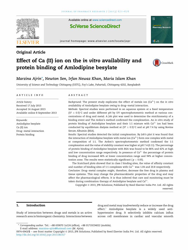
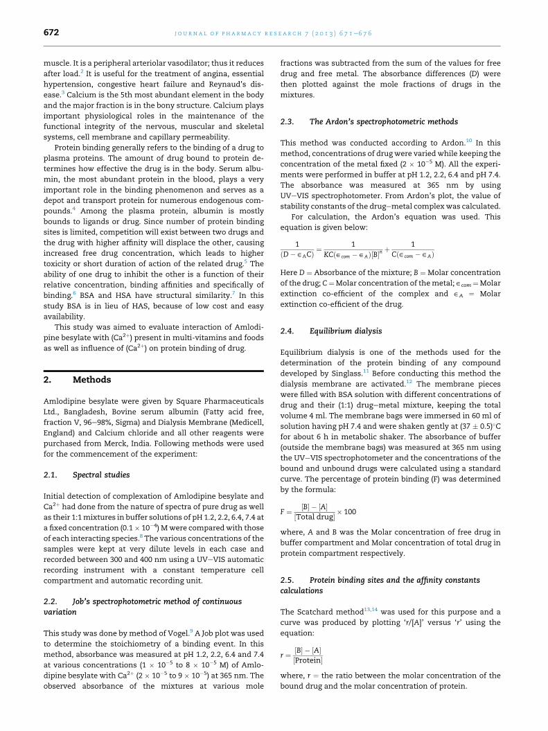
Fig. 1 e Comparison between the spectral plot of
Amlodipine besylate alone and with Ca2D at pH 1.2 & 2.2
respectively (The data are shown as mean ± SME).
0
0.05
0.1
0.15
0.2
0.25
0.3
0.35
0.4
0.45
300 310 320 330 340 350 360 365 370 380 390 400
Abs
orba
nce
Wavelength (nm)
D
D+M
Fig. 3 e Comparison between the spectral plot of
Amlodipine besylate alone and with Ca2D at pH 6.4 & 7.4
respectively (The data are shown as mean ± SME).
0.35
0.4D
j o u r n a l o f p h a rm a c y r e s e a r c h 7 ( 2 0 1 3 ) 6 7 1e6 7 6 673
2.6. Statistical analysis
The results were expressed as Mean � SEM values for each
experiment. Differences in mean values between experi-
mental groups were analyzed by unpaired t-test. A probability
values less than 0.05 ( p < 0.05) was defined to be significant.15
-0.05
0
0.05
0.1
0.15
0.2
0.25
0.3
300 310 320 330 340 350 360 365 370 380 390 400
Abs
orba
nce
Wavelength (nm)
D+M
Fig. 4 e Comparison between the spectral plot of
Amlodipine besylate alone and with Ca2D at pH 6.4 & 7.4
respectively (The data are shown as mean ± SME).
3. Results
3.1. Spectral study
It was seen that Amlodipine besylate gives a sharp peak at
365 nm. But when (Ca2þ) mixed with Amlodipine besylate in
1:1 ratio, the intensity of the peak of Amlodipine besylate
changes remarkably (absorbance decreases) i.e., absorption
characteristics are altered due to interaction but the position
of the compound do not shift (Figs. 1e4).
3.2. Study of job’s method
At experimental pH, Amlodipine besylate form strong 1:1
complexes with Ca2þ ion. Absorbance differences at pH 1.2,
0
0.05
0.1
0.15
0.2
0.25
0.3
0.35
300 310 320 330 340 350 360 365 370 380 390 400
Abs
orba
nce
Wavelength (nm)
D
D+ M
Fig. 2 e Comparison between the spectral plot of
Amlodipine besylate alone and with Ca2D at pH 1.2 & 2.2
respectively (The data are shown as mean ± SME).
Fig. 5 e Job’s Plot for Complexation of Amlodipine besylate
and Ca (II) ion at pH 1.2&2.2 (The data are shown as
mean ± SME).
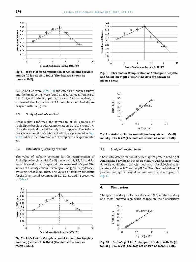
Fig. 6 e Job’s Plot for Complexation of Amlodipine besylate
and Ca (II) ion at pH 1.2&2.2 (The data are shown as
mean ± SME).
Fig. 8 e Job’s Plot for Complexation of Amlodipine besylate
and Ca (II) ion at pH 6.4&7.4 (The data are shown as
mean ± SME).
Fig. 9 e Ardon’s plot for Amlodipine besylate with Ca (II)
ion at pH 1.2 & 2.2 (The data are shown as mean ± SME).
j o u rn a l o f p h a rma c y r e s e a r c h 7 ( 2 0 1 3 ) 6 7 1e6 7 6674
2.2, 6.4 and 7.4 were (Figs. 5e8) indicated as “̂” shaped curves
and the break points were found at absorbance difference of
0.15, 0.16, 0.17 and 0.18 at pH 1.2, 2.2, 6.4 and 7.4 respectively. It
confirmed the formation of 1:1 complexes of Amlodipine
besylate with Ca (II) ion.
3.3. Study of Ardon’s method
Ardon’s plot confirmed the formation of 1:1 complex of
Amlodipine besylate with Ca (II) ion at pH 1.2, 2.2, 6.4 and 7.4,
since the method is valid for only 1:1 complexes. The Ardon’s
plots gave straight lines intercept which are presented in Figs.
9e12 indicate the formation of 1:1 complexes at experimental
pH.
3.4. Estimation of stability constant
The value of stability constant for the complexation of
Amlodipine besylate with Ca (II) ion at pH 1.2, 2.2, 6.4 and 7.4
were obtained from the spectral data using Ardon’s plot. The
values of stability constant were given as [(Intercept)/(slope)]
by using Ardon’s equation. The values of stability constants
for the drugemetal system at pH 1.2, 2.2, 6.4 and 7.4 presented
in Table 1
Fig. 7 e Job’s Plot for Complexation of Amlodipine besylate
and Ca (II) ion at pH 6.4&7.4 (The data are shown as
mean ± SME).
3.5. Study of protein binding
The in vitro determination of percentage of protein binding of
Amlodipine besylate and their 1:1 mixture with Ca (II) ion was
done by equilibrium dialysis method at physiological tem-
perature (37 � 0.5)�C and at pH 7.4. The observed values of
protein binding for drug alone and with metal are given in
Fig. 13.
4. Discussion
The spectra of drug molecules alone and (1:1) mixture of drug
and metal showed significant change in their absorption
Fig. 10 e Ardon’s plot for Amlodipine besylate with Ca (II)
ion at pH 1.2 & 2.2 (The data are shown as mean ± SME).
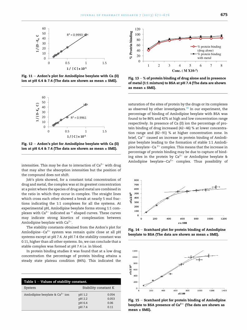
Fig. 11 e Ardon’s plot for Amlodipine besylate with Ca (II)
ion at pH 6.4 & 7.4 (The data are shown as mean ± SME).
Fig. 12 e Ardon’s plot for Amlodipine besylate with Ca (II)
ion at pH 6.4 & 7.4 (The data are shown as mean ± SME).
0
20
40
60
80
100
120
1 2 3 4 5 6 7 8
% P
rotr
in b
indi
ng
Conc. ( M X10-5)
% protein binding (drug alone)% protein binding with metal
Fig. 13 e % of protein binding of drug alone and in presence
of metal (1:1 mixture) to BSA at pH 7.4 (The data are shown
as mean ± SME).
Fig. 14 e Scatchard plot for protein binding of Amlodipine
besylate to BSA (The data are shown as mean ± SME).
j o u r n a l o f p h a rm a c y r e s e a r c h 7 ( 2 0 1 3 ) 6 7 1e6 7 6 675
intensities. This may be due to interaction of Ca2þ with drug
that may alter the absorption intensities but the position of
the compound does not shift.
Job’s plots showed, for a constant total concentration of
drug andmetal, the complex was at its greatest concentration
at a pointwhere the species of drug andmetal are combined in
the ratio in which they occur in complex. The straight lines
which cross each other showed a break at nearly 5 mol frac-
tions indicating the 1:1 complexes for all the systems. At
experimental pH, Amlodipine besylate forms strong 1:1 com-
plexes with Ca2þ indicated as ‘̂’ shaped curves. These curves
may indicate strong kinetics of complexation between
Amlodipine besylate with Ca2þ.
The stability constants obtained from the Ardon’s plot for
AmlodipineeCa2þ system was remain quite close at all pH
systems except at pH 7.4. At pH 7.4 the stability constant was
0.11, higher than all other systems. So, we can conclude that a
stable complex was formed at pH 7.4 i.e. in blood.
In protein binding studies it was found that at a low drug
concentration the percentage of protein binding attains a
steady state plateau condition (84%). This indicated the
Table 1 e Values of stability constant.
System Stability constant K
Amlodipine besylate & Ca2þ ion pH 1.2 0.054
pH 2.2 0.053
pH 6.4 0.06
pH 7.4 0.11
saturation of the sites of protein by the drugs or its complexes
as observed by other investigators.16 In our experiment, the
percentage of binding of Amlodipine besylate with BSA was
found to be 86% and 42% at high and low concentration range
respectively. In presence of Ca (II) ion the percentage of pro-
tein binding of drug increased (42e46) % at lower concentra-
tion range and (82e91) % at higher concentration zone. In
brief, Ca2þ caused an increase in protein binding of Amlodi-
pine besylate leading to the formation of stable 1:1 Amlodi-
pine besylateeCa 2þ complex. This means that the increase in
percentage of protein binding may be due to capture of bind-
ing sites in the protein by Ca2þ or Amlodipine besylate &
Amlodipine besylateeCa2þ complex. Thus possibility of
Fig. 15 e Scatchard plot for protein binding of Amlodipine
besylate to BSA presence of Ca2D (The data are shown as
mean ± SME).
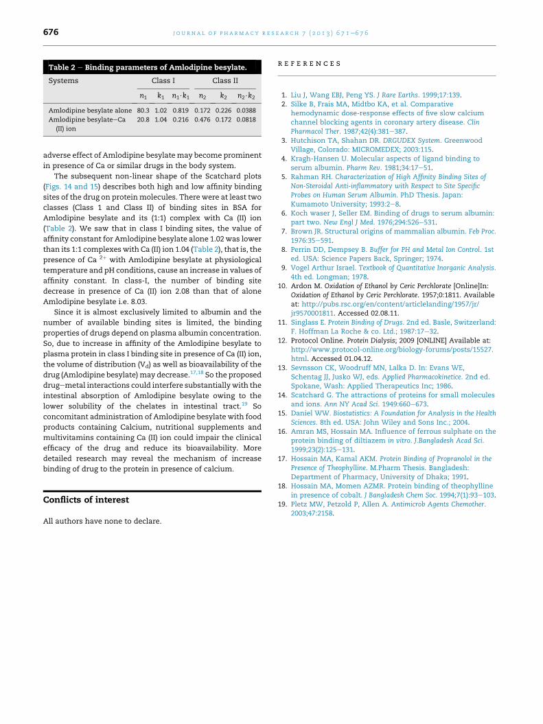
Table 2 e Binding parameters of Amlodipine besylate.
Systems Class I Class II
n1 k1 n1$k1 n2 k2 n2$k2
Amlodipine besylate alone 80.3 1.02 0.819 0.172 0.226 0.0388
Amlodipine besylateeCa
(II) ion
20.8 1.04 0.216 0.476 0.172 0.0818
j o u rn a l o f p h a rma c y r e s e a r c h 7 ( 2 0 1 3 ) 6 7 1e6 7 6676
adverse effect of Amlodipine besylatemay become prominent
in presence of Ca or similar drugs in the body system.
The subsequent non-linear shape of the Scatchard plots
(Figs. 14 and 15) describes both high and low affinity binding
sites of the drug on proteinmolecules. There were at least two
classes (Class 1 and Class II) of binding sites in BSA for
Amlodipine besylate and its (1:1) complex with Ca (II) ion
(Table 2). We saw that in class I binding sites, the value of
affinity constant for Amlodipine besylate alone 1.02 was lower
than its 1:1 complexeswith Ca (II) ion 1.04 (Table 2), that is, the
presence of Ca 2þ with Amlodipine besylate at physiological
temperature and pH conditions, cause an increase in values of
affinity constant. In class-I, the number of binding site
decrease in presence of Ca (II) ion 2.08 than that of alone
Amlodipine besylate i.e. 8.03.
Since it is almost exclusively limited to albumin and the
number of available binding sites is limited, the binding
properties of drugs depend on plasma albumin concentration.
So, due to increase in affinity of the Amlodipine besylate to
plasma protein in class I binding site in presence of Ca (II) ion,
the volume of distribution (Vd) as well as bioavailability of the
drug (Amlodipine besylate)may decrease.17,18 So the proposed
drugemetal interactions could interfere substantiallywith the
intestinal absorption of Amlodipine besylate owing to the
lower solubility of the chelates in intestinal tract.19 So
concomitant administration of Amlodipine besylate with food
products containing Calcium, nutritional supplements and
multivitamins containing Ca (II) ion could impair the clinical
efficacy of the drug and reduce its bioavailability. More
detailed research may reveal the mechanism of increase
binding of drug to the protein in presence of calcium.
Conflicts of interest
All authors have none to declare.
r e f e r e n c e s
1. Liu J, Wang EBJ, Peng YS. J Rare Earths. 1999;17:139.2. Silke B, Frais MA, Midtbo KA, et al. Comparative
hemodynamic dose-response effects of five slow calciumchannel blocking agents in coronary artery disease. ClinPharmacol Ther. 1987;42(4):381e387.
3. Hutchison TA, Shahan DR. DRGUDEX System. GreenwoodVillage, Colorado: MICROMEDEX; 2003:115.
4. Kragh-Hansen U. Molecular aspects of ligand binding toserum albumin. Pharm Rev. 1981;34:17e51.
5. Rahman RH. Characterization of High Affinity Binding Sites ofNon-Steroidal Anti-inflammatory with Respect to Site SpecificProbes on Human Serum Albumin. PhD Thesis. Japan:Kumamoto University; 1993:2e8.
6. Koch waser J, Seller EM. Binding of drugs to serum albumin:part two. New Engl J Med. 1976;294:526e531.
7. Brown JR. Structural origins of mammalian albumin. Feb Proc.1976:35e591.
8. Perrin DD, Dempsey B. Buffer for PH and Metal Ion Control. 1sted. USA: Science Papers Back, Springer; 1974.
9. Vogel Arthur Israel. Textbook of Quantitative Inorganic Analysis.4th ed. Longman; 1978.
10. Ardon M. Oxidation of Ethanol by Ceric Perchlorate [Online]In:Oxidation of Ethanol by Ceric Perchlorate. 1957;0:1811. Availableat: http://pubs.rsc.org/en/content/articlelanding/1957/jr/jr9570001811. Accessed 02.08.11.
11. Singlass E. Protein Binding of Drugs. 2nd ed. Basle, Switzerland:F. Hoffman La Roche & co. Ltd.; 1987:17e32.
12. Protocol Online. Protein Dialysis; 2009 [ONLINE] Available at:http://www.protocol-online.org/biology-forums/posts/15527.html. Accessed 01.04.12.
13. Sevnsson CK, Woodruff MN, Lalka D. In: Evans WE,Schentag JJ, Jusko WJ, eds. Applied Pharmacokinetice. 2nd ed.Spokane, Wash: Applied Therapeutics Inc; 1986.
14. Scatchard G. The attractions of proteins for small moleculesand ions. Ann NY Acad Sci. 1949:660e673.
15. Daniel WW. Biostatistics: A Foundation for Analysis in the HealthSciences. 8th ed. USA: John Wiley and Sons Inc.; 2004.
16. Amran MS, Hossain MA. Influence of ferrous sulphate on theprotein binding of diltiazem in vitro. J.Bangladesh Acad Sci.1999;23(2):125e131.
17. Hossain MA, Kamal AKM. Protein Binding of Propranolol in thePresence of Theophylline. M.Pharm Thesis. Bangladesh:Department of Pharmacy, University of Dhaka; 1991.
18. Hossain MA, Momen AZMR. Protein binding of theophyllinein presence of cobalt. J Bangladesh Chem Soc. 1994;7(1):93e103.
19. Pletz MW, Petzold P, Allen A. Antimicrob Agents Chemother.2003;47:2158.