The impact of elevated pulmonary artery pressure on exercise ...
266
School of Physiotherapy The impact of elevated pulmonary artery pressure on exercise responses Robin Fowler This thesis is presented for the Degree of Doctor of Philosophy of Curtin University October 2012
-
Upload
khangminh22 -
Category
Documents
-
view
2 -
download
0
Transcript of The impact of elevated pulmonary artery pressure on exercise ...

School of Physiotherapy
The impact of elevated pulmonary artery pressure on
exercise responses
Robin Fowler
This thesis is presented for the Degree of Doctor of Philosophy
of Curtin University
October 2012

DDEECCLLAARRAATTIIOONN
This thesis contains no material which has been accepted for the award of any other
degree or diploma in any university. To the best of my knowledge and belief this
thesis contains no material previously published by any other person except where
due acknowledgement has been made.
Signature:
Date:

iii
SSTTAATTEEMMEENNTT OOFF OORRIIGGIINNAALLIITTYY
This thesis is presented for the degree of Doctor of Philosophy at Curtin University,
Western Australia. Studies were undertaken between May 2006 and May 2012,
through the School of Physiotherapy at Curtin University, in association with the
Advanced Lung Disease Unit at Royal Perth Hospital and the Lung Institute of
Western Australia.
This research project was developed in association with my supervisors who have
been involved in editing both this thesis and all associated publications. All material
presented in this thesis is original.

iv
AABBSSTTRRAACCTT
Background and research questions
The four studies reported in this thesis investigated the implications of an elevated
pulmonary artery pressure (PAP) on the response to an exertional challenge. The
level of symptoms and exertion that healthcare professionals consider appropriate
for patients with pulmonary arterial hypertension (PAH) was explored in the first
study. In studies two, three, and four, exercise responses and exercise testing were
evaluated in individuals with an elevated PAP on exercise, but a normal PAP at rest
(exercise-induced pulmonary arterial hypertension, [EIPAH]). The following research
questions were addressed:
1. Is there consistency in the advice given by healthcare professionals in Australia
regarding physical exertion and symptoms, and in referral for exercise rehabilitation,
for patients with PAH?
2. What are the clinical implications of an elevated PAP in symptomatic individuals
with risk factors for PAH but who have a normal PAP at rest (EIPAH)?
3. Can the six-minute walk test (6MWT) identify reduced exercise capacity and
accurately estimate aerobic capacity in individuals with EIPAH?
4. Are the haemodynamic and symptomatic responses to maximal and submaximal
resistance exercise similar to the responses demonstrated during comparable
intensities of aerobic exercise in individuals with EIPAH?
Abstracts for the reported studies
This PhD program formed the basis for four publications in peer reviewed,
international, scientific journals. These four publications are summarised, in abstract
form, below. The full manuscripts of these publications constitute Chapters 4-7 of
this thesis.

v
Study 1: Australian perspective regarding recommendations
for physical activity and exercise rehabilitation in pulmonary
arterial hypertension.
Background: Limited data regarding the longer term consequences of physical
activity and exercise rehabilitation, or the origin and importance of exertional
symptoms associated with PAH, have made it difficult for clinicians to advise
patients with PAH regarding physical activity, the appropriate level of symptoms,
and exercise rehabilitation. The aim of this study was to determine the opinion of
healthcare professionals, within Australia, regarding acceptable levels of exertion
and symptoms, and referral for exercise rehabilitation in patients with PAH.
Methods: In 2010, 76 healthcare professionals at a specialist pulmonary
hypertension meeting, in Australia, were surveyed using a self-administered
questionnaire. The questionnaire included case studies of patients with PAH in
World Health Organisation functional classes II-IV. For each case study,
respondents were asked to report their opinion regarding the acceptable level of
exertion and symptoms during daily activities, and whether they would refer the
patient for exercise rehabilitation. Three additional questions asked about advice in
relation to four specific physical activities.
Results: The response rate was 70% (n=53). Overall, 58% of respondents
recommended patients undertake daily activities ‘as tolerated’. The other 42% of
respondents recommended either mild (14%) or moderate (17%) exertion, or did not
answer this question (11%). There was no consensus regarding acceptable levels of
breathlessness or fatigue, but the majority of respondents considered patients
should have no chest pain (73%) and no more than mild light-headedness (92%)
during daily activities. Overall, 63% of respondents would have referred patients for
exercise rehabilitation. There was little difference in opinion regarding the
acceptable level of exertion or symptoms, or referral for exercise rehabilitation,
according to functional class. However, the patients’ functional class did influence
the advice given regarding the specific physical activities.
Conclusions: In 2010, there were inconsistencies, between individual healthcare
professionals within Australia, regarding appropriate levels of physical exertion and
acceptable symptoms during daily activities. Almost two thirds of the respondents
reported they would refer patients for exercise rehabilitation.

vi
Study 2: Implications of exercise-induced pulmonary arterial
hypertension.
Background: Pulmonary arterial hypertension is associated with characteristic
exercise abnormalities and reduced quality of life (QoL). However, the implications
of an elevated PAP on exercise in individuals with a normal PAP at rest were
uncertain. The aim of this study was to characterise the haemodynamic and
ventilatory responses to exercise in a group of patients with unexplained dyspnoea,
increased risk for PAH, a normal mean pulmonary artery pressure (mPAP) at rest
and an elevated mPAP (>30mmHg) on exercise (exercise-induced pulmonary
arterial hypertension [EIPAH]).
Methods: Thirty-seven symptomatic patients (34 females), with risk factors for PAH,
and 20 healthy controls (19 females), underwent a symptom-limited
cardiopulmonary exercise test (CPET) and were assessed for QoL. Patient subjects
had a pulmonary artery catheter in situ during the exercise test.
Results: Seventeen subjects (15 females) had EIPAH, which we defined as mPAP
≤25mmHg at rest, and mPAP >30mmHg and pulmonary artery wedge pressure
<20mmHg on exercise. These subjects had reduced peak exercise cardiac output
(72±19% predicted). Further, compared with matched controls, subjects with EIPAH
had reduced peak oxygen consumption (1.2±0.4 vs 1.7±0.5 L/min, p<0.05), an
elevated ventilatory equivalent for carbon dioxide (41.0±7.3 vs 31.0±3.0, p<0.05)
and reduced end tidal carbon dioxide tension (32.6±3.6 vs 39.4±2.7mmHg, p<0.05)
at the anaerobic threshold. These exercise abnormalities were associated with
impaired QoL (p<0.05).
Conclusion: Elevated PAP on exercise can be associated with haemodynamic and
ventilatory abnormalities typical of PAH, along with impaired exercise capacity and
reduced QoL.
vii
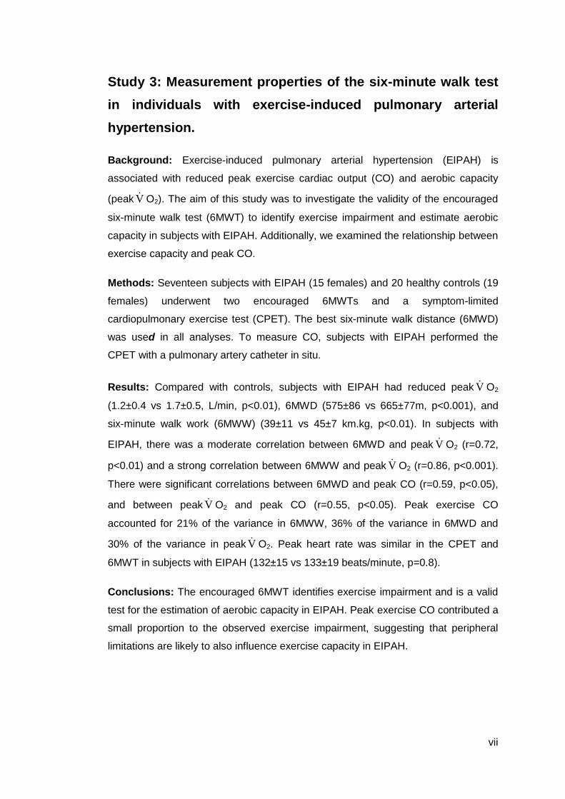
Study 3: Measurement properties of the six-minute walk test
in individuals with exercise-induced pulmonary arterial
hypertension.
Background: Exercise-induced pulmonary arterial hypertension (EIPAH) is
associated with reduced peak exercise cardiac output (CO) and aerobic capacity
(peak V O2). The aim of this study was to investigate the validity of the encouraged
six-minute walk test (6MWT) to identify exercise impairment and estimate aerobic
capacity in subjects with EIPAH. Additionally, we examined the relationship between
exercise capacity and peak CO.
Methods: Seventeen subjects with EIPAH (15 females) and 20 healthy controls (19
females) underwent two encouraged 6MWTs and a symptom-limited
cardiopulmonary exercise test (CPET). The best six-minute walk distance (6MWD)
was used in all analyses. To measure CO, subjects with EIPAH performed the
CPET with a pulmonary artery catheter in situ.
Results: Compared with controls, subjects with EIPAH had reduced peak V O2
(1.2±0.4 vs 1.7±0.5, L/min, p<0.01), 6MWD (575±86 vs 665±77m, p<0.001), and
six-minute walk work (6MWW) (39±11 vs 45±7 km.kg, p<0.01). In subjects with
EIPAH, there was a moderate correlation between 6MWD and peak V O2 (r=0.72,
p<0.01) and a strong correlation between 6MWW and peak V O2 (r=0.86, p<0.001).
There were significant correlations between 6MWD and peak CO (r=0.59, p<0.05),
and between peak V O2 and peak CO (r=0.55, p<0.05). Peak exercise CO
accounted for 21% of the variance in 6MWW, 36% of the variance in 6MWD and
30% of the variance in peak V O2. Peak heart rate was similar in the CPET and
6MWT in subjects with EIPAH (132±15 vs 133±19 beats/minute, p=0.8).
Conclusions: The encouraged 6MWT identifies exercise impairment and is a valid
test for the estimation of aerobic capacity in EIPAH. Peak exercise CO contributed a
small proportion to the observed exercise impairment, suggesting that peripheral
limitations are likely to also influence exercise capacity in EIPAH.

viii
Study 4: A comparison of the acute haemodynamic response
to aerobic and resistance exercise in subjects with exercise-
induced pulmonary arterial hypertension.
Background: Pulmonary arterial hypertension is associated with reduced muscle
strength. Exercise-induced pulmonary arterial hypertension is associated with
reduced exercise capacity and abnormal central haemodynamic responses to
maximal aerobic exercise, however muscle strength has not been evaluated in this
population. Aerobic and resistance exercise training are commonly employed to
treat reduced exercise capacity. However, the haemodynamic response to aerobic
and resistance exercise, at prescribed training intensities, in subjects with EIPAH
are unknown. The aims of this study were to i) evaluate muscle strength, and, ii) the
haemodynamic and symptomatic responses to maximal and submaximal and
aerobic resistance exercise in individuals with EIPAH.
Methods: Fourteen subjects (11 with scleroderma, 12 females) with EIPAH and 15
healthy controls (14 females) underwent a cardiopulmonary exercise test on a cycle
ergometer and a one repetition maximum (1RM) strength test on a bilateral leg
press. The EIPAH group was also evaluated during resistance and aerobic exercise
at 40% and 60% of maximum. The EIPAH subjects underwent all exercise tests with
a pulmonary artery catheter in situ. Lower limb extensor muscle strength,
determined by the 1RM weight, was compared between EIPAH and control groups.
The haemodynamic and symptomatic responses to maximal (1RM and at
peak V O2) and submaximal aerobic and resistance exercise were compared in the
EIPAH group.
Results: 1RM was lower in the EIPAH group compared with the control group (75
versus 87 kg, p<0.05). In the EIPAH group, there were no differences in
haemodynamic or symptomatic responses between the two modalities of
submaximal exercise. At maximal exercise, all haemodynamic and symptomatic
responses were lower during resistance compared with aerobic exercise (p<0.05).
Conclusions: The EIPAH group had reduced lower limb extensor muscle strength.
At the intensities studied, lower limb resistance exercise was well tolerated and was
mostly associated with similar or lower haemodynamic responses compared with
aerobic exercise, in subjects with EIPAH.

ix
TTAABBLLEE OOFF CCOONNTTEENNTTSS
Declaration ............................................................................................................... ii
Statement of originality ............................................................................................ iii
Abstract ................................................................................................................... iv
Table of contents ..................................................................................................... ix
List of tables ........................................................................................................... xv
List of figures ......................................................................................................... xvi
List of appendices ................................................................................................. xvii
Acknowledgements and funding ........................................................................... xviii
Publications arising from this thesis ........................................................................ xx
Awards ................................................................................................................ xxiv
List of abbreviations ............................................................................................. xxvi
Chapter 1 Introduction ............................................................................................. 1
1.1 Research questions ........................................................................................ 2
1.2 Question 1 ...................................................................................................... 3
1.2.1 Hypothesis ............................................................................................... 3
1.2.1.1 Background.................................................................................... 3
1.3 Question 2 ...................................................................................................... 4
1.3.1 Hypothesis ............................................................................................... 4
1.3.1.1 Background.................................................................................... 4
1.4 Question 3 ...................................................................................................... 5
1.4.1 Hypothesis ............................................................................................... 5
1.4.1.1 Background.................................................................................... 5
1.5 Question 4 ...................................................................................................... 6
1.5.1 Hypothesis ............................................................................................... 6
1.5.1.1 Background.................................................................................... 6
1.6 Novelty and significance of the research ........................................................ 7
Novelty ............................................................................................................. 7
Significance ...................................................................................................... 7
Chapter 2 Literature review ...................................................................................... 9
2.1 Introduction .................................................................................................... 9
2.2 Pulmonary arterial hypertension ..................................................................... 9
2.2.1 Pathogenesis ..........................................................................................10
2.2.2 Diagnosis ................................................................................................11
2.2.3 Haemodynamics .....................................................................................12

x
2.2.4 Pharmacologic therapy ...........................................................................13
2.2.5 Persistent abnormalities despite therapy .................................................14
2.2.6 Delays in diagnosis .................................................................................15
2.2.7 At risk populations ..................................................................................15
2.2.7.1 Screening programs ......................................................................16
2.2.8 Clinical presentation................................................................................16
2.2.8.1 Quality of life .................................................................................17
2.2.8.2 Impaired exercise capacity ............................................................18
2.2.8.3 Symptoms .....................................................................................18
2.2.9 Summary ................................................................................................19
2.3 Exercise abnormalities, exercise limitation and symptoms in PAH ................19
2.3.1 Haemodynamic responses during exercise in healthy individuals ...........19
2.3.2 Haemodynamic responses during exercise in PAH .................................21
2.3.2.1 Right ventricle/pulmonary haemodynamics and symptoms ...........22
2.3.2.2 J receptor activation ......................................................................23
2.3.3 Ventilatory responses during exercise ....................................................24
2.3.3.1 The relationship between ventilation and dyspnoea ......................24
2.3.3.2 Ventilatory response in healthy individuals ....................................24
2.3.3.3 Ventilatory response in PAH .........................................................25
2.3.4 Gas exchange and hypoxaemia ..............................................................27
2.3.4.1 Hypoxaemia and symptoms ..........................................................28
2.3.5 Peripheral airways function .....................................................................29
2.3.6 Fatigue, lightheadedness, chest pain, and palpitations in PAH ...............29
2.3.6.1 Fatigue ..........................................................................................29
2.3.6.2 Lightheadedness and syncope......................................................30
2.3.6.3 Cardiac ischaemia and chest pain ................................................31
2.3.6.4 Arrhythmias and palpitations .........................................................32
2.3.7 Systemic and peripheral abnormalities ...................................................33
2.3.7.1 Up-regulation of sympathetic nervous system activity ...................33
2.3.7.2 Chemoreceptor activation and the ergoreflex ................................34
2.3.7.3 Inflammation .................................................................................34
2.3.7.4 Systemic endothelial dysfunction ..................................................35
2.3.7.5 Skeletal and respiratory muscle myopathy ....................................36
2.3.8 Summary: Exercise limitation and symptoms in PAH ..............................39
2.4 Exercise testing in PAH .................................................................................40
2.4.1 General information ................................................................................40
2.4.2 Cardiopulmonary exercise test ................................................................41
xi
2.4.2.1 Exercise modality ..........................................................................42
2.4.2.2 Exercise protocol ..........................................................................43
2.4.2.3 Incremental CPET in LHF .............................................................43
2.4.2.4 Incremental CPET in COPD ..........................................................44
2.4.2.5 Incremental CPET in PAH .............................................................45
2.4.2.6 Safety of the incremental CPET ....................................................49
2.4.2.7 CPET for differential diagnosis ......................................................49
2.4.3 Exercise testing to assess central haemodynamics ................................53
2.4.3.1 Central haemodynamics in PAH ...................................................53
2.4.3.2 Sensitivity for change following intervention ..................................53
2.4.3.3 Simultaneous haemodynamic and gas exchange analysis ............53
2.4.3.4 Invasive exercise testing for differential diagnosis .........................53
2.4.3.5 Safety of invasive exercise testing ................................................54
2.4.4 Six-minute walk test (6MWT) ..................................................................55
2.4.4.1 Encouraged 6MWT .......................................................................56
2.4.4.2 Unencouraged 6MWT ...................................................................56
2.4.4.3 Six-minute walk test in LHF ...........................................................56
2.4.4.4 Six-minute walk test in COPD .......................................................57
2.4.4.5 Six-minute walk test in PAH ..........................................................57
2.4.5 Other tests of exercise response and exercise capacity ..........................60
2.4.5.1 Constant work load tests ...............................................................60
2.4.5.2 Incremental shuttle walk test .........................................................63
2.4.5.3 Treadmill tests in PAH ..................................................................64
2.4.5.4 Exercise echocardiography in PAH ...............................................65
2.4.6 Testing muscle strength ..........................................................................68
2.4.6.1 Measurement techniques used to determine muscle strength .......68
2.4.7 Summary: Exercise testing in PAH .........................................................70
2.5 Exercise-induced pulmonary arterial hypertension ........................................72
2.5.1 Exercise testing – potential role in early diagnosis of PAH ......................72
2.5.1.1 Exercise-induced pulmonary arterial hypertension ........................73
2.6 Final summary and gaps in knowledge ..........................................................75
Chapter 3 Methods .................................................................................................77
3.1 Introduction ...................................................................................................77
3.2 Methodology for study 1: Activity and exercise prescription in PAH ...............77
3.2.1 Questionnaire development ....................................................................78
3.2.2 Pilot study of the questionnaire ...............................................................78
3.2.3 Data collection for main study .................................................................79

xii
3.2.3.1 Study design .................................................................................79
3.2.3.2 Participants ...................................................................................79
3.2.3.3 Description of questionnaire ..........................................................79
3.2.3.4 Ethics approval .............................................................................80
3.2.3.5 Statistical analysis.........................................................................80
3.3 Methodology for studies 2-4: Exercise testing in symptomatic individuals with risk factors for PAH .............................................................................................81
3.3.1 Study design ...........................................................................................81
3.3.2 Subjects ..................................................................................................81
3.3.2.1 Recruitment of control subjects .....................................................82
3.3.3 Exercise testing protocols .......................................................................85
3.3.3.1 Six-minute walk test ......................................................................85
3.3.3.2 Cardiopulmonary exercise test (Figure 2) .....................................86
3.3.3.3 Resistance exercise testing (Figures 3 and 4) ...............................87
3.3.4 Haemodynamic measures ......................................................................90
3.3.4.1 Pulmonary haemodynamics ..........................................................90
3.3.4.2 Cardiac output ..............................................................................91
3.3.4.3 Mixed venous oxygen saturation ...................................................92
3.3.4.4 Systemic blood pressure ...............................................................92
3.3.4.5 Heart rate ......................................................................................93
3.3.5 Arterial oxygen saturation .......................................................................93
3.3.6 Symptomatic responses ..........................................................................93
3.3.7 Quality of life assessment .......................................................................93
3.3.8 Evaluation of usual physical activity ........................................................94
3.3.9 Equipment maintenance .........................................................................94
3.3.9.1 Vmax system maintenance and quality control .............................94
3.3.9.2 Calibration of the cycle ergometer .................................................95
3.3.10 Ethics approval .....................................................................................95
3.3.11 Data management and statistical analysis ............................................95
3.3.11.1 Statistical analyses .....................................................................96
3.3.11.2 Power calculations ......................................................................97
Chapter 4 Australian perspective regarding recommendations for physical activity and exercise rehabilitation in pulmonary arterial hypertension ................................99
4.1 Introduction ...................................................................................................99
4.2 Methods ...................................................................................................... 100
4.2.1 Participants ........................................................................................... 101
4.2.2 Data analysis ........................................................................................ 101

xiii
4.3 Results ........................................................................................................ 102
4.3.1 Demographics ....................................................................................... 102
4.3.2 Responses to questions relating to the case studies ............................. 103
4.3.2.1 Instructions for daily activity ........................................................ 103
4.3.2.2 Acceptable symptoms during daily activities ............................... 103
4.3.2.3 Referral for exercise rehabilitation ............................................... 104
4.3.2.4 Responses to questions regarding specific physical activities ..... 104
4.4 Discussion ................................................................................................... 106
4.5 Limitations ................................................................................................... 108
4.6 Conclusions ................................................................................................. 108
Chapter 5 Implications of exercise-induced pulmonary arterial hypertension ........ 109
5.1 Introduction ................................................................................................. 109
5.2 Methods ...................................................................................................... 110
5.2.1 Subjects ................................................................................................ 110
5.2.2 Study protocol ....................................................................................... 111
5.2.3 Statistical analysis ................................................................................ 112
5.3 Results ........................................................................................................ 113
5.3.1 Subject demographics .......................................................................... 113
5.3.2 Ventilatory comparisons between the EIPAH and control groups .......... 115
5.3.3 Quality of life comparisons between PAH, EIPAH, EILVDD and noPAH ...................................................................................................................... 115
5.3.4 Haemodynamic comparisons between EIPAH, PAH and noPAH .......... 119
5.3.5 Comparisons between the EIPAH and EILVDD groups ........................ 120
5.3.6 Influence of age and diagnosis of scleroderma ..................................... 120
5.4 Discussion ................................................................................................... 120
5.4.1 Limitations and general applicability of this study .................................. 124
5.5 Conclusions ................................................................................................. 125
Chapter 6 Measurement properties of the 6MWT in individuals with exercise-induced pulmonary arterial hypertension .............................................................. 126
6.1 Introduction ................................................................................................. 126
6.2 Methods ...................................................................................................... 127
6.2.1 Participants ........................................................................................... 127
6.2.2 Study design ......................................................................................... 128
6.2.2.1 Exercise protocols....................................................................... 129
6.2.2.2 Statistical analyses ..................................................................... 130
6.3 Results ........................................................................................................ 131
6.3.1 Exercise responses............................................................................... 132

xiv
6.3.1.1 Physiological and symptomatic responses, 6MWT and CPET .... 132
6.3.1.2 Exercise capacity ........................................................................ 133
6.3.1.3 Effect of test repetition on 6MWD ................................................ 133
6.3.1.4 Relationship between 6MWT results, peak V O2 and peak CO .... 134
6.4 Discussion ................................................................................................... 136
6.5 Limitations of the study ................................................................................ 138
6.6 Conclusions ................................................................................................. 138
Chapter 7 A comparison of the acute haemodynamic response to aerobic and resistance exercise in subjects with exercise-induced pulmonary arterial hypertension ......................................................................................................... 139
7.1 Introduction ................................................................................................. 139
7.2 Patients and methods .................................................................................. 141
7.2.1 Patient population ................................................................................. 141
7.2.2 Assessment of Central Haemodynamics ............................................... 141
7.2.3 Exercise protocols ................................................................................ 142
Cardiopulmonary exercise test (CPET) ................................................... 144
Resistance exercise ................................................................................ 144
7.2.4 Statistical analysis ................................................................................ 145
7.3 Results .................................................................................................... 146
7.3.1 Different modalities of submaximal exercise (EIPAH group) ................. 146
7.3.2 Maximal exercise responses (EIPAH group) ......................................... 146
7.4 Discussion ................................................................................................... 149
7.5 Limitations ................................................................................................... 151
7.6 Conclusions ................................................................................................. 151
Chapter 8 Summary, clinical implications and future research .............................. 153
8.1 Australian perspective regarding recommendations for physical activity and exercise rehabilitation in pulmonary arterial hypertension. ................................ 153
8.1.1 Areas that warrant further research ....................................................... 154
8.2 Implications of exercise-induced pulmonary arterial hypertension. .............. 155
8.2.1 Areas that warrant further research ................................................. 156
8.3 Measurement properties of the six-minute walk test in individuals with exercise-induced pulmonary arterial hypertension. ............................................ 157
8.3.1 Areas that warrant further research ................................................. 158
8.4 A comparison of the acute haemodynamic response to aerobic and resistance exercise in subjects with exercise-induced pulmonary arterial hypertension. ..... 158
8.4.1 Areas that warrant further research ................................................. 159
References ........................................................................................................... 161
Appendices 193

xv
LLIISSTT OOFF TTAABBLLEESS
Table 1. Summary of abnormalities identified on CPET in LHF, COPD and PAH....46
Table 2. CPET measures that differentiate PAH from other conditions ...................51
Table 3. Demographics of the questionnaire respondents .................................... 102
Table 4. Questionnaire responses ........................................................................ 105
Table 5. Baseline demographics, noPAH, PAH, EILVDD, EIPAH and controls ..... 114
Table 6. Exercise capacity, ventilatory and systemic hemodynamic responses and
limiting symptoms on the CPET .................................................................... 116
Table 7. Quality of Life (SF-36, Version 1) ............................................................ 117
Table 8. Haemodynamic variables measured at rest and during the CPET........... 118
Table 9. Subject characteristics, EIPAH and controls ........................................... 131
Table 10. Heart rate and symptomatic responses to the 6MWT and CPET........... 132
Table 11. 6MWT and CPET results ...................................................................... 133
Table 12. Demographics, resting haemodynamics and exercise capacity in subjects
with EIPAH (n=14) and controls (n=15) ......................................................... 147
Table 13. Physiologic and symptomatic responses to submaximal resistance and
aerobic exercise in subjects with EIPAH (n=14) ............................................ 148
Table 14. Physiologic and symptomatic responses to maximal resistance and
aerobic exercise in subjects with EIPAH (n=14) ............................................ 148
Table 15. Participant co-morbidities and medications, Studies 2-4………………203

xvi
LLIISSTT OOFF FFIIGGUURREESS
Figure 1. Subject recruitment, Studies 2-4. .............................................................84
Figure 2. Subject undergoing a CPET with simultaneous central haemodynamic
monitoring via RHC .........................................................................................88
Figure 3. (a) Starting and finishing position for the maximal and submaximal
resistance exercise protocols. (b) Position of legs during the extension phase of
the manoeuvre during the maximal and submaximal resistance exercise
protocols .........................................................................................................89
Figure 4. Subject in preparation for resistance exercise protocols, with right heart
catheter in situ for central haemodynamic monitoring ......................................89
Figure 5. Pressure versus flow during exercise. The relationship between mPAP and
CO during a symptom-limited CPET. ............................................................ 119
Figure 6. Bland-Altman plot showing agreement in 6MWD between two 6MWTs for
individuals with EIPAH .................................................................................. 134
Figure 7. Relationships between 6MWW and 6MWD with peak VO2, in EIPAH ... 135
Figure 8. Randomisation and exercise protocols…………………………………….143

xvii
LLIISSTT OOFF AAPPPPEENNDDIICCEESS
Appendix 1. Questionnaire used in the study together with the responses to each
question 193
Appendix 2. Participant co-morbidities and medications, Studies 2-4 203
Appendix 3. Article accepted for inclusion in Pulmonary Medicine, July 2012 204
Appendix 4. Copyright declaration 230
Appendix 5. License agreement for Chapter 5 231
Appendix 6. License agreement for Chapter 6 233
Appendix 7. License agreement for Chapter 7 239
xviii
AACCKKNNOOWWLLEEDDGGEEMMEENNTTSS AANNDD FFUUNNDDIINNGG
This research was supported by unconditional research grants from Bayer Schering
Pharmaceuticals Australia and Actelion Pharmaceuticals Australia.
Associate Professor Sue Jenkins and Dr Andrew Maiorana, my supervisors
throughout this PhD degree, for their time, expertise, endless patience, support and
encouragement. Thank you also to Andrew, for many, many hours of help with data
collection.
Professor Eli Gabbay for financial support for the project and my scholarship, and
for providing amazing opportunities to meet and talk to experts in the field. For being
a fantastic recruiter, and for his time and help during the early period of data
collection.
Dr Kevin Gain for his unwavering mentorship and support, especially in times when
it all seemed too hard. Thank you also for the many hours of lung function testing,
exercise test analysis, discussions about physiology and all you have taught me
about exercise testing.
Professor Gerry O’Driscoll who was instrumental in enabling this research to
happen, and who taught me the value of getting involved in every aspect of data
collection (despite the fear).
Dr Matthew Best for his help with right heart catheterisation and troubleshooting on
the days of data collection.
Min Ding who was a wonderful support on the days of data collection.
The participants of these studies. I greatly appreciate the time and effort you freely
contributed.
Professor Phil Thompson for his enthusiasm, and ongoing help and support.
Dr Martin Thomas for his many, many hours of exercise test supervision.
Dr Alex Higton for her ready and positive support whenever needed for medical
assessment of the controls and exercise test supervision.

xix
Chris Reed for his amazing IT support.
Alan Dick and Monty Brydon for their many hours of data collection and for technical
support and troubleshooting.
Peter McKinnon and Roger Collinson for patient, statistical support.
Bill Weightman for his support and help with statistics and submission hoops during
some hours of extreme frustration.
Mike Phillips and Sally Burrows for many hours of meetings regarding statistical
process.
The staff of the Royal Perth Hospital Cardiac Catheter Laboratory for their help with
right heart catheterisation.
The Heart Failure/Cardiac Transplant team for sharing their space, coffee machine
and printer.
The physio team at ECU, for their ongoing reminders that this was possible, and
their support through the latter and most frustrating part of this journey.
The physiotherapy department Royal Perth Hospital for releasing me to undertake
this PhD and their help and support.
Ineke Krom for her patience and help with formatting and presenting this thesis.
My friends who have remained loyal despite many years of neglect.
Neil, my children, and my parents, who have undertaken this journey with me.

xx
PPUUBBLLIICCAATTIIOONNSS AARRIISSIINNGG FFRROOMM TTHHIISS TTHHEESSIISS
Peer reviewed journals
Fowler R, Maiorana A, Jenkins S, Gain K, O’Driscoll G, Gabbay E. A comparison of
the acute haemodynamic response to aerobic and resistance exercise in subjects
with exercise-induced pulmonary arterial hypertension. Eur J Prev Cardiol. 2012;
DOI: 10.1177/2047487312445424
Fowler R, Jenkins S, Maiorana A, Gain K, O’Driscoll G, Gabbay E. Australian
perspective regarding recommendations for physical activity and exercise
rehabilitation in pulmonary arterial hypertension. J Multidisc Healthcare. 2011;
4:451-462
Fowler RM, Jenkins SC, Maiorana AJ, Gain KR, O’Driscoll G, Gabbay E.
Measurement properties of the 6-min walk test in individuals with exercise-induced
pulmonary arterial hypertension. Intern Med J 2011; 41:679-687
Fowler R, Maiorana M, Jenkins S, Gain K, O’Driscoll G, Gabbay E. Implications of
exercise-induced pulmonary arterial hypertension. Med Sci Sports Exerc 2011;
43:983-989. Associated commentary: Arena R. Detecting abnormal pulmonary
hemodynamics with cardiopulmonary exercise testing. Med Sci Sports Exerc 2011;
43:982
Supplementary publications during the period of enrolment,
to which a significant contribution was made
Fowler R, Gain K, Gabbay E. Exercise intolerance in pulmonary arterial
hypertension. Pulm Med. 2012; In Press (Accepted for inclusion in a Special Edition
on Pulmonary Hypertension, July 2012). See Appendix 3
Ganderton L, Jenkins S, Gain K, Fowler R, Winship P, Lunt D, Gabbay E. Short
term effects of exercise training on exercise capacity and quality of life in patients
with pulmonary arterial hypertension: protocol for a randomised controlled trial. BMC
Pulm Med 2011; 11:25-31

xxi
Ganderton L, Jenkins S, McKenna S, Gain K, Fowler R, Twiss J, Gabbay E.
Validation of the Cambridge Pulmonary Hypertension Outcome Review
(CAMPHOR) for the Australian and New Zealand population. Respirology 2011;
16:1235-1240
Strange G, Fowler R, Jary C, Dalton B, Stewart S, Gabbay E. Integrated care and
optimal management of pulmonary arterial hypertension. J Multidisc Healthcare.
2009; 2:67-78
Abstracts and presentations at scientific meetings related to
this PhD program of research
ATS International Conference, Denver, May 2011 (Poster).
Fowler R, Jenkins S, Maiorana A, O’Driscoll G, Gabbay E.
The six-minute walk test identifies exercise impairment and provides a valid
estimate of aerobic capacity in individuals with exercise-induced pulmonary arterial
hypertension.
ATS International Conference, Denver, May 2011 (Poster).
Fowler R, Maiorana A, Jenkins S, O’Driscoll G, Gabbay E.
Comparison of resistance and aerobic exercise in subjects with elevated pulmonary
artery pressure.
European Society of Cardiology Congress, Paris 2011 (Poster).
Maiorana A, Fowler R, Jenkins S, O’Driscoll G, Gabbay E.
A comparison of central haemodynamics during aerobic and resistance exercise in
pulmonary arterial hypertension.
Australian Health and Medical Research Congress. Melbourne, November
2010 (Invited speaker).
Fowler R, Maiorana A, Jenkins S, O’Driscoll G, Gabbay E.
Characteristics of patients with unexplained dyspnoea and risk factors for pulmonary
arterial hypertension.
xxii
Pulmonary Hypertension Perspectives meeting (Pfizer Australia). Sydney,
June 2010 (Invited speaker).
Fowler R.
Non-pharmaceutical therapies in pulmonary hypertension. Mainstream and
alternative options.
Annual Scientific Meeting of the American College of Sports Medicine. Seattle,
USA, May 2009 (Oral).
Maiorana A, Fowler R, Jenkins S, O’Driscoll G, Gabbay E.
Exercise-induced pulmonary arterial hypertension is associated with an attenuated
increase in cardiac output during exercise.
Young Investigator Awards, Royal Perth Hospital, July 2009 (Oral).
Fowler R, Maiorana A, Jenkins S, O’Driscoll G, Gabbay E.
Exercise induced pulmonary arterial hypertension is clinically important.
ATS International Conference, San Diego, May 2009 (Poster discussion).
Fowler R, Maiorana A, Jenkins S, O’Driscoll G, Gabbay E.
Exercise-induced pulmonary arterial hypertension is clinically important.
TSANZ Annual Scientific Meeting, Darwin, April 2009 (Oral).
Fowler R, Maiorana A, Jenkins S, O’Driscoll G, Gabbay E.
Exercise induced rises in pulmonary arterial pressure reflect the presence of a
pulmonary vasculopathy and are clinically important.
Lung Institute of Western Australia 10th Anniversary Research Symposium,
Perth, March 2009 (Invited speaker).
Fowler R, Maiorana A, Jenkins S, O’Driscoll G, Gabbay E.
Exercise induced pulmonary arterial hypertension is clinically important.
TSANZ Annual Scientific Meeting, WA Branch, Mandurah, October 2008 (Oral).
Fowler R, Maiorana A, Jenkins S, O’Driscoll G, Gabbay E.
Exercise induced pulmonary arterial hypertension is clinically important.
Annual conference of the Australian Cardiovascular Health and Rehabilitation
Association and the Chronic Disease Network, Alice Springs, August, 2008
(Oral).
Fowler R, Maiorana A, Jenkins S, O’Driscoll G, Gabbay E.
xxiii
Improving the diagnosis of pulmonary arterial hypertension using exercise testing;
enhancing access to early interventions and disease management.
World Congress of Cardiology, Buenos Aires, May 2008 (Oral).
Fowler R, Maiorana A, Jenkins S, Thomas M, Gabbay E, O’Driscoll G.
Invasive exercise testing improves the early diagnosis of pulmonary arterial
hypertension.
American College of Sports Medicine, Indianapolis, May 2008 (Oral).
Fowler R, Maiorana A, Jenkins S, Thomas M, Gabbay E, O’Driscoll G.
Early diagnosis of pulmonary arterial hypertension using exercise testing.
TSANZ Annual Scientific Meeting, Melbourne , March 2008 (Oral).
Fowler R, Maiorana A, Jenkins S, O’Driscoll G, Thomas M, Reed C, Gabbay E.
Simultaneous exercise testing and central haemodynamic monitoring increases
diagnostic sensitivity for pulmonary arterial hypertension.
International Society of Heart and Lung Transplantation, Annual General
Meeting and Scientific Sessions, Boston, April 2008 (Oral).
Fowler R, Maiorana A, Jenkins S, O'Driscoll G, Thomas M, Reed C, Gabbay E.
Exercise Induced Pulmonary Hypertension (EIPH) Is Clinically Important and
Precedes the Development of Pulmonary Hypertension (PH) at Rest.
TSANZ Annual Scientific Meeting, WA Branch, Swan Valley, October 2007
(Oral).
Fowler R, Maiorana A, Jenkins S, O’Driscoll G, Gabbay E.
Exercise induced rises in pulmonary artery pressure reflect the presence of a
pulmonary vasculopathy and are clinically important.
American Thoracic Society (ATS), Annual Scientific Congress San Francisco,
May 2007 (Poster).
Fowler R, Beckert L, Corris P, Jenkins S, Gabbay E.
Activity and exercise prescription for patients with pulmonary arterial hypertension.
TSANZ Annual Scientific Meeting, Auckland, March 2007 (Poster).
Fowler R, Beckert L, Corris P, Jenkins S, Gabbay E.
Activity and exercise prescription for patients with pulmonary arterial hypertension.

xxiv
AAWWAARRDDSS
2011
Fowler R, Maiorana M, Jenkins S, Gain K, O’Driscoll G, Gabbay E. Implications of
exercise-induced pulmonary arterial hypertension. Med Sci Sports Exerc 2011;
43:983-989.
Awarded Curtin University, School of Physiotherapy Elsevier Book Prize for
best article in a scientific journal in 2011.
2009
Young Investigator Awards, Royal Perth Hospital, July 2009 (Oral).
Fowler R, Maiorana A, Jenkins S, O’Driscoll G, Gabbay E.
Exercise induced pulmonary arterial hypertension is clinically important.
Awarded the encouragement prize.
TSANZ Annual Scientific Meeting, Darwin, April 2009 (Oral).
Fowler R, Maiorana A, Jenkins S, O’Driscoll G, Gabbay E.
Exercise induced rises in pulmonary arterial pressure reflect the presence of a
pulmonary vasculopathy and are clinically important.
Finalist in Ann Woolcock Young Investigator Session.
Awarded best physiotherapy research paper.
2008
TSANZ Annual Scientific Meeting, WA Branch, Mandurah, October 2008 (Oral).
Fowler R, Maiorana A, Jenkins S, O’Driscoll G, Gabbay E.
Exercise induced pulmonary arterial hypertension is clinically important.
Awarded the Travel Prize
Annual conference of the Australian Cardiovascular Health and Rehabilitation
Association and the Chronic Disease Network, Alice Springs, August, 2008
(Oral).
Fowler R, Maiorana A, Jenkins S, O’Driscoll G, Gabbay E.
Improving the diagnosis of pulmonary arterial hypertension using exercise testing;
enhancing access to early interventions and disease management.
Awarded Best New Research Prize.

xxv
TSANZ Annual Scientific Meeting, Melbourne , March 2008 (Oral).
Fowler R, Maiorana A, Jenkins S, O’Driscoll G, Thomas M, Reed C, Gabbay E.
Simultaneous exercise testing and central haemodynamic monitoring increases
diagnostic sensitivity for pulmonary arterial hypertension.
Awarded best presentation in the OLIV SIG.
Awarded best presentation in the physiotherapy SIG.

xxvi
LLIISSTT OOFF AABBBBRREEVVIIAATTIIOONNSS
1RM One repetition maximum 6MWD Six-minute walk distance 6MWT Six-minute walk test 6MWW Six-minute walk work AE Aerobic exercise AF Atrial fibrillation AT Anaerobic threshold BMI Body mass index BMPR2 Bone morphogenetic protein receptor type II gene bpm Beats per minute CAMPHOR Cambridge Pulmonary Hypertension Outcome Review CCO Continuous cardiac output CI Confidence Interval CO Cardiac output CO2 Carbon dioxide COPD Chronic obstructive pulmonary disease CPET Cardiopulmonary exercise test CWLT Constant work load test DLCO Diffusing capacity for carbon monoxide ECG Electrocardiograph EILVDD Exercise-induced left ventricular diastolic dysfunction EIPAH Exercise-induced pulmonary arterial hypertension FEV1 Forced expiratory volume in one second FVC Forced vital capacity HCO3
- Bicarbonate HFpEF Heart failure with preserved ejection fraction HFrEF Heart failure with reduced ejection fraction HR Heart rate HRQoL Health related quality of life IC Inspiratory capacity ILD Interstitial lung disease iPAH Idiopathic pulmonary arterial hypertension J receptors Juxtapulmonary receptors LHF Left-sided heart failure LV Left ventricle MET Metabolic equivalent MLHFQ Minnesota Living with Heart Failure Questionnaire mmHg Millimetres of mercury mPAP Mean pulmonary artery pressure MVC Maximum voluntary contraction MVV Maximum voluntary ventilation n Number of subjects NIH-PPH National Institute of Health – Primary Pulmonary Hypertension NO Nitric oxide noPAH No pulmonary arterial hypertension NT-proBNP N - terminal pro-B-type natriuretic peptide NYHA New York Heart Association O2 Oxygen O2 pulse Oxygen pulse

xxvii
PaCO2 Arterial carbon dioxide tension PAH Pulmonary arterial hypertension PaO2 Arterial oxygen tension PAP Pulmonary artery pressure PASP Pulmonary artery systolic pressure PAWP Pulmonary artery wedge pressure
peak V O2 Peak oxygen consumption
PetCO2 End tidal carbon dioxide tension PPH Primary pulmonary hypertension PVR Pulmonary vascular resistance QoL Quality of life RCT Randomised controlled trial RE Resistance exercise RER Respiratory exchange ratio RHC Right heart catheter RHF Right heart failure RPE Rate of perceived exertion RV Right ventricle SBP Systemic blood pressure SF-36 Medical Outcomes Study 36-Item Short Form Health Survey SpO2 Oxygen saturation SV Stroke volume SvO2 Mixed venous oxygen saturation TPG Transpulmonary gradient USA United States of America
V CO2 Carbon dioxide production
V E Minute ventilation
V E/ V CO2 Ventilatory equivalent for carbon dioxide
V Emax/MVV Minute ventilation/maximal voluntary ventilation
V Epeak Peak minute ventilation
V Max Maximum ventilation
V O2 Oxygen consumption
WHO World Health Organization

Chapter 1: Introduction
1
CCHHAAPPTTEERR 11
IINNTTRROODDUUCCTTIIOONN
Pulmonary arterial hypertension (PAH) is a progressive pulmonary vasculopathy.
Without treatment, and in a large proportion of individuals with PAH who receive
pharmaceutical therapy, the condition is associated with progressive pulmonary
vascular disease and right heart dysfunction, increasing exertional symptoms, and
worsening functional capacity and quality of life (QoL) (1). Early diagnosis and
treatment of PAH is associated with better outcomes following therapy with
improved survival, functional capacity and QoL (2, 3). However, early diagnosis is
difficult because of the non-specific nature of presentation (4) and diagnostic criteria
that can only define the condition once the pulmonary vasculopathy is advanced (5).
Delays in diagnosis have not reduced in over 20 years (4, 6).
Exertional symptoms, particularly dyspnoea and fatigue, are the presenting
complaint in up to 90% of individuals with PAH (7). Progression of the condition is
associated with worsening exercise tolerance and functional capacity. In individuals
with PAH, haemodynamic responses, measured during exercise, better reflect
functional capacity than haemodynamics measured at rest (8). Furthermore, gas
exchange and ventilatory responses measured during exercise reflect disease
severity and prognosis (9, 10), and are well described in PAH (11, 12). An
incremental cardiopulmonary exercise test (CPET), with continuous gas exchange
analysis, is well established as a non-invasive tool to aid differential diagnosis in
individuals with dyspnoea of unknown aetiology (13).
Despite improvements in haemodynamics and survival following the development of
pharmaceutical therapies that specifically address the pulmonary vascular
abnormalities associated with PAH, a large proportion of individuals with PAH report
persistent functional impairments (1). Historically, prior to the development of
effective therapies, individuals with PAH were encouraged not to undertake an
exercise program (14). However, interest has recently developed in the role of
exercise rehabilitation for individuals with PAH who have persistent functional
limitation. Prior to 2010, three studies had demonstrated that exercise rehabilitation
could be achieved without adverse events and resulted in improvements in exercise
capacity in individuals with PAH (15-17). However, there are few studies that report

Chapter 1: Introduction
2
the haemodynamic burden, in terms of the degree of elevation in pulmonary artery
pressure (PAP), pulmonary vascular resistance (PVR) and right ventricular
workload, associated with exercise in individuals with PAH. Furthermore, the
attitudes of healthcare professionals regarding the implications of exertional
symptoms, physical exertion and exercise rehabilitation were unknown.
This program of research involved four projects. The research was designed to
evaluate the consistency, amongst healthcare professionals within Australia,
regarding recommendations for patients with PAH pertaining to physical exertion,
symptoms and exercise rehabilitation. Further, the research explored the potential of
exercise testing to facilitate the early diagnosis of PAH, and evaluated the
implications of an elevated PAP, during exercise, in terms of the haemodynamic
response and clinical consequences, for symptomatic individuals with risk factors for
PAH.
The studies were designed to address the following research questions.
1.1 Research questions
1. Is there consistency in the advice given by healthcare professionals
regarding physical exertion and symptoms, and in referral for exercise
rehabilitation, for patients with PAH within Australia?
2. What are the clinical implications of an elevated PAP in symptomatic
individuals with risk factors for PAH but who have a normal PAP at rest (i.e.
exercise-induced pulmonary arterial hypertension, [EIPAH])?
3. Can the six-minute walk test (6MWT) identify reduced exercise capacity and
accurately estimate aerobic capacity in individuals with EIPAH?
4. Are the haemodynamic and symptomatic responses to maximal and
submaximal resistance exercise similar to the responses demonstrated
during comparable intensities of aerobic exercise in individuals with EIPAH?
This Chapter presents an overview of the literature pertaining to the development of
each research question. The hypothesis for each research question is described
and the significance of the research program is discussed.
Chapter 1: Introduction
3
1.2 Question 1
Is there consistency in the advice given by healthcare professionals regarding
physical exertion and symptoms, and in referral for exercise rehabilitation, for
patients with PAH within Australia?
1.2.1 Hypothesis
There will be no consistency in the advice given by healthcare professionals
regarding physical exertion and symptoms, or in referral for exercise rehabilitation,
for patients with PAH within Australia.
1.2.1.1 Background
Historical concerns regarding the response to physical exertion in individuals with
PAH, and reports of exertional syncope and sudden death (18-20), resulted in a
conservative approach to physical activity with recommendations for patients to
avoid exercise training and physical exertion beyond the performance of daily
activities (14). There has been limited discussion in the literature regarding the
origins and significance of the symptoms associated with PAH, specifically
symptoms of dyspnoea, fatigue and light-headedness. Furthermore, the first report
of exercise rehabilitation in PAH was published in 2005, in Japanese (17), and was
not accessible to most English speaking healthcare professionals. The first study of
exercise training in PAH, written in English, was published in 2006 and before 2010
only three reports in English (15, 16, 21), with a total of 51 subjects with PAH who
had undergone exercise rehabilitation, were published. In 2010 there were no longer
term data on patients with PAH who had undergone exercise training. Therefore, at
that time, there was very limited evidence to guide clinicians working within Australia
and managing patients with PAH, regarding acceptable levels of exertion and
symptoms, and the safety and efficacy of exercise rehabilitation for this population. It
was uncertain if there was any consistency in the advice clinicians were providing
for patients with PAH regarding physical activity and symptoms, and if patients were
being referred for exercise rehabilitation.

Chapter 1: Introduction
4
1.3 Question 2
What are the clinical implications of an elevated PAP in symptomatic individuals with
risk factors for PAH but who have a normal PAP at rest (i.e. EIPAH)?
1.3.1 Hypothesis
Symptomatic individuals with risk factors for PAH and a normal PAP at rest, but an
elevated PAP on exercise (i.e. EIPAH), will demonstrate reduced exercise capacity
and ventilatory abnormalities that are characteristic of PAH.
1.3.1.1 Background
Pulmonary arterial hypertension is a progressive condition with poor prognosis if
untreated (6). Pharmaceutical therapy improves survival, functional capacity and
quality of life (22-24) and is most effective if commenced before the development of
marked functional limitation (2, 3). Therefore early diagnosis of PAH is considered
important. However, despite the recent development of effective pharmaceutical
therapies, the timeliness in diagnosis of PAH has not improved in more than 20
years (4, 6, 25). The difficulties associated with early diagnosis of PAH include the
non-specific nature of symptoms and the lack of abnormalities on standard
assessment, until right heart failure (RHF) is present (4). A further, and important,
barrier to early diagnosis lies in the current diagnostic recommendations which only
support a diagnosis of PAH made on assessment of central haemodynamics
performed at rest (1). By the time diagnosis can be made at rest, up to 70% of the
small pulmonary arteries are diseased and the condition is advanced (5).
Pulmonary arterial hypertension is consistently associated with an elevated PAP
and PVR and reduced cardiac output (CO) at peak exercise (8, 26-28). Exertional
symptoms of dyspnoea and fatigue limit functional capacity. Aerobic capacity is
reduced, and characteristic ventilatory and gas exchange abnormalities are well
described (29). Exercise testing is well accepted as a non-invasive method for
identifying the physiological cause of exertional dyspnoea and a system for
identifying likely PAH based upon exercise responses is well established (13).
However, diagnosis of PAH cannot be made without accurate measurement of
central haemodynamics via right heart catheter (RHC) (1).

Chapter 1: Introduction
5
It is possible that measuring central haemodynamics during exercise could identify
individuals with elevated PAP and early PAH. However, a wide range of PAP is
demonstrated in healthy individuals during exercise (30) and therefore PAP during
exercise is difficult to interpret, in isolation. The identification of characteristic gas
exchange and ventilatory abnormalities in individuals with PAH (29) suggests that
simultaneous evaluation of central haemodynamic, gas exchange and ventilatory
responses during exercise has the potential to facilitate the early diagnosis of PAH
in symptomatic individuals with risk factors for PAH. This possibility formed the basis
for this research.
1.4 Question 3
Can the 6MWT identify reduced exercise capacity and accurately estimate aerobic
capacity in individuals with EIPAH?
1.4.1 Hypothesis
The 6MWT will identify reduced exercise capacity and will accurately estimate peak
oxygen consumption ( V O2) in individuals with EIPAH.
1.4.1.1 Background
Quantifying functional exercise capacity provides a means for identifying the
functional limitations associated with a condition, and can be used to objectively
measure deterioration related to disease progression or improvement with therapy.
The 6MWT is an extensively studied exercise test that is inexpensive, easy to
administer, and is widely used in clinical practice to measure exercise capacity (31).
Furthermore, the 6MWT is a valid test for quantifying functional exercise capacity
and accurately estimating peak V O2 in individuals with chronic obstructive
pulmonary disease (COPD) (31, 32) and PAH (33-35). Indeed, the main outcome of
the 6MWT, i.e. the six-minute walk distance (6MWD), has been the primary outcome
measure in the majority of clinical trials in PAH. However, the utility of the 6MWD in
accurately reflecting functional exercise capacity in individuals with well preserved
functional capacity has been challenged (36, 37). Individuals with EIPAH have a
mild to moderate reduction in functional capacity (27) and the capacity for the 6MWT
to identify impairments in exercise capacity and to estimate aerobic capacity in this

Chapter 1: Introduction
6
population has not previously been described. This study investigated the
measurement properties of the 6MWT in individuals with EIPAH.
1.5 Question 4
In individuals with EIPAH, are the haemodynamic and symptomatic responses to
maximal and submaximal resistance exercise similar to the responses demonstrated
during comparable intensities of aerobic exercise?
1.5.1 Hypothesis
There will be no difference in haemodynamic or symptomatic responses to
comparable intensities of lower limb aerobic and resistance exercise in individuals
with EIPAH.
1.5.1.1 Background
Pulmonary arterial hypertension is associated with skeletal muscle weakness (38,
39). Skeletal muscle strength is important in the performance of activities of daily
living and greater muscle strength and power are associated with reduced
haemodynamic stress at a given absolute muscle force (40). Resistance exercise
training is associated with improved muscle strength and is increasingly included in
exercise training programs for healthy individuals and patients with left-sided heart
failure (LHF) (40).
Historically, resistance exercise has been discouraged in individuals with PAH
because of concerns regarding the safety of this modality of exercise in this
population (41). Recent studies, involving small sample sizes, suggest that
resistance training can be achieved without adverse events and results in
improvements in muscle strength in PAH (16, 42). However, there are no data
describing the haemodynamic burden of resistance exercise, and few that quantify
the response to submaximal aerobic exercise in PAH (8, 28, 43). Evaluation of the
haemodynamic response to exercise assists in predicting the likely safety and
longer term consequences of exercise training in individuals with cardiovascular
conditions.
Individuals with EIPAH have haemodynamic abnormalities, at peak aerobic
exercise, similar to those identified in individuals with PAH (27). However, muscle

Chapter 1: Introduction
7
strength had not been evaluated, and the haemodynamic and symptomatic
responses to maximal and submaximal resistance and aerobic exercise, in
individuals with EIPAH, had not been determined. With increasing interest in aerobic
and resistance training in many clinical populations (44), including PAH (16, 42), the
need for and likely safety issues associated with resistance exercise training in
individuals with EIPAH warranted evaluation. This study quantified the
haemodynamic and symptomatic responses during an acute bout of maximal and
submaximal aerobic and resistance exercise in individuals with EIPAH.
1.6 Novelty and significance of the research
Novelty
The studies described in this thesis are the first to explore
1. the consistency in advice given to patients with PAH regarding appropriate
levels of physical exertion and acceptable symptoms during daily activities
and referral of patients with PAH for exercise rehabilitation, in Australia
2. the clinical implications of EIPAH in relation to exercise capacity and QoL
3. the measurement properties of the 6MWT in individuals with EIPAH
4. muscle strength, and the haemodynamic and symptomatic responses to
maximal and submaximal resistance exercise in individuals with EIPAH
Significance
1. Consistency in advice and referral for exercise rehabilitation is important in
promoting adherence, by patients with PAH, to recommendations provided
by healthcare professionals. The first study described in this thesis provides
information regarding the consistency of advice given by healthcare
professionals, within Australia in 2010, pertaining to physical activity and
acceptable symptoms during activity, and referral for exercise rehabilitation.
2. There is an increasing awareness of the difficulties in early diagnosis, but
also the need for early treatment, of PAH. Comprehensively exploring and
describing the exercise responses and the clinical implications of EIPAH
provides important baseline information regarding this condition. Should
EIPAH be found to progress to PAH, this study describes the role of exercise
testing in the early diagnosis of PAH. Also, regardless of possible disease

Chapter 1: Introduction
8
progression in EIPAH, the clinical consequences of EIPAH and a rationale
for treatment of this condition have been explored.
3. The 6MWT is an inexpensive, reliable, easy to administer test for clinical
practice, and for research. However, its validity for quantifying exercise
limitation in individuals with EIPAH, with well preserved functional capacity,
had not been determined. This study describes the measurement properties
of the 6MWT and describes its utility in EIPAH.
4. Reduced muscle strength impacts negatively on the performance of physical
activities, has been described in individuals with PAH. Prior to this research,
muscle strength in individuals with EIPAH had not been evaluated.
Increasing interest in aerobic and resistance exercise training in individuals
with PAH identifies a need to determine the haemodynamic burden and likely
safety of these therapies. Evaluating the response to intensities of
submaximal and maximal resistance and aerobic exercise that are used in
clinical practice in individuals with elevated PAP informs clinical practice, and
further research.
Chapter 2: Literature review
9
CCHHAAPPTTEERR 22
LLIITTEERRAATTUURREE RREEVVIIEEWW
2.1 Introduction
This chapter reviews the literature relating to the presentation of PAH, the likely
causes of functional abnormalities, and the role of exercise testing in this condition.
The chapter comprises four main sections. The first section (2.2) reviews the
pathophysiology, diagnosis and management of PAH. The second section (2.3)
discusses the factors that contribute to the exercise abnormalities, symptoms
associated with exercise, and exercise limitation in PAH. The third section (2.4)
reviews the literature relating to studies of exercise testing in individuals with PAH
and the fourth section (2.5) reviews the literature on EIPAH and exercise testing as
a potential means to facilitate the early diagnosis of PAH. Section 2.6 provides a
final summary of the literature review and identifies the gaps in the literature.
2.2 Pulmonary arterial hypertension
The following section defines and describes PAH and discusses diagnosis,
screening and treatment.
Pulmonary arterial hypertension is a condition defined by primary abnormalities in
the precapillary pulmonary arteries and arterioles. It forms Group 1 in the World
Health Organisation (WHO) classification of pulmonary hypertension (1). This
classification system identifies PAH as a unique entity, with a characteristic
pathophysiology, clinical presentation and response to therapy that separates it from
other forms of pulmonary hypertension.
Before a classification system for pulmonary hypertension was established,
idiopathic PAH (iPAH) was known as primary pulmonary hypertension (PPH) (6). In
this review, the terms iPAH and PPH will be used interchangeably, in accordance
with the terminology used in the original papers that have been cited.
Chapter 2: Literature review
10
2.2.1 Pathogenesis
A fundamental endothelial abnormality is thought to play a key role in the
pathogenesis of PAH (45-47). The endothelium lines the blood vessels of the
pulmonary and peripheral circulations and produces a number of compounds that
influence vascular tone, vessel wall structure, blood viscosity, blood and cell wall
interactions and thrombosis and fibrinolysis (48).
In the normal pulmonary circulation, the endothelium maintains vascular health and
facilitates appropriate adaptive responses to external stimuli, such as hypoxia or
acute changes in pulmonary blood flow (48). This is achieved through balance in a
number of opposing systems including the endothelin and angiotensin II systems
that promote vasoconstriction and proliferation of vascular wall cells, and the nitric
oxide (NO) and prostaglandin systems, that promote vasodilatation, apoptosis and
antithrombotic activity (48). When in balance, regeneration of cells is balanced by
cell apoptosis, regulation of vascular tone and blood flow is adapted to optimise gas
exchange, and vessels are maintained free of thrombus (48).
Current hypotheses regarding the pathogenesis of PAH include the proposal that
the abnormalities in the pulmonary vasculature develop through a two stage process
(49). Initially, following exposure to an exogenous stimulus such as a viral or
bacterial infection or toxin, transition of pulmonary artery smooth muscle cells and
pulmonary artery endothelial cells from a normal to abnormal phenotype occurs.
This is considered most likely due to multiple mutations of the bone morphogenetic
protein (BMP) receptor type II (BMPR2) and others genes such as serotonin
receptor and transporter, potassium and calcium channels and angiopoietin1 genes
(49). Disease progression is then thought to be sustained by cellular factors that
create a proliferative, anti-apoptotic, and vasoconstrictive state. In pulmonary artery
smooth muscle cells, in individuals with PAH, the anti-proliferative effect of BMP is
reversed and becomes proliferative and the pro-apoptotic effect of BMP is
attenuated. In pulmonary artery endothelial cells the anti-apoptotic or survival effect
of BMP changes and the system becomes pro-apoptotic (49). Elevated sensitivity of
contractile proteins to calcium, smooth muscle cell proliferation and endothelial
injury results in vasoconstriction and pulmonary vascular wall hypertrophy (49). The
subsequent pulmonary vascular endothelial dysfunction results in up-regulation of
the endothelin system, increased pulmonary vasoconstriction and cell proliferation.
In addition, the NO and prostaglandin systems are down-regulated, limiting

Chapter 2: Literature review
11
pulmonary vasodilatation and reducing the anti-proliferative and anti-thrombotic
effects of these compounds (47). Imbalance in the endothelin and NO/prostaglandin
systems is believed to be of primary importance in PAH (1), although this does not
occur in isolation. Recent studies have also identified a platelet derived increase in
serotonin and loss of voltage-gated potassium channels, both of which contribute to
smooth muscle vasoconstriction and cell proliferation. There is also evidence for up-
regulation in a number of growth factors such as vascular endothelial growth factor
(1).
Under normal circumstances, the pulmonary vasculature is in a state of active
dilatation (50) but imbalance in the production of vasodilators and vasoconstrictors
in PAH results in elevated pulmonary vascular tone. In addition, a state of pro-
proliferation results in smooth muscle hypertrophy and extension of smooth muscle
into the vessel walls in the periphery of the lung, and adventitial and intimal
proliferation. Reduced endothelial cell production of antithrombotic agents results in
intra-lumen thrombus and vessel obstruction. The ultimate outcome is a marked
reduction in the vasodilatory capacity, distensibility and patency of the pulmonary
circulation (1).
2.2.2 Diagnosis
Diagnosis of PAH requires exclusion of other conditions that may cause pulmonary
hypertension, including left-sided heart disease, pulmonary parenchymal or airway
disease, or pulmonary thromboembolic disease (1). Following exclusion of other
conditions, diagnosis is made by RHC assessment of PAP, pulmonary artery wedge
pressure (PAWP), CO and PVR. A resting mean PAP (mPAP) of >25mmHg, a
PAWP <15mmHg (which excludes pulmonary venous hypertension caused by left
heart disease) and a PVR of > 3 Woods units, are the criteria for diagnosis (1). Right
heart catheter evaluation is the ‘gold standard’ and a diagnosis of PAH cannot be
confirmed until RHC assessment has established that diagnostic criteria are met.
However, by the time PAP and PVR are elevated sufficiently to meet diagnostic
criteria up to 70% of the small pulmonary vessels are diseased (5). Before 2009,
guidelines for diagnosis of PAH included criteria at rest or on exercise (51).
However, due to a wide range of normal central haemodynamic responses during
exercise (30), the current diagnostic guidelines for PAH recommend that diagnosis
of PAH is based only on a RHC assessment performed at rest (1).

Chapter 2: Literature review
12
2.2.3 Haemodynamics
In PAH, the functional changes associated with a loss of vessel calibre and intra-
lumen obstruction, include a reduction in the size of the pulmonary vascular bed (1),
and a rise in PVR, PAP (43) and right ventricular afterload. The right ventricle (RV)
is a thin walled chamber, built for a low pressure circulation and, under normal
conditions, right ventricular afterload equates to 25% of left ventricular afterload (52,
53). In the presence of a sustained increase in right ventricular afterload a normal
heart undergoes adaptive hypertrophy of the myocardium. However, the capacity for
hypertrophic adaptation, and, in PAH, the development and progression of right
ventricular dysfunction varies among individuals (53). It has been proposed that the
development of RHF in PAH is related not only to elevated right ventricular
afterload, but also to intrinsic abnormalities in the wall of the RV (54). Although it has
not been a consistent finding (55), some individuals with PAH demonstrate an
insertion allele polymorphism in the angiotension-converting enzyme gene. This
insertion polymorphism is associated with a lower CO for the same PAP, compared
with individuals who have a deletion allele polymorphism in the same gene (56).
Having this insertion allele polymorphism is thought to contribute to the development
of right ventricular dysfunction in some individuals with PAH (57). Myocardial fibrosis
and reduced capillary density in the wall of the RV have been described in an animal
model of progressive PAH (54). It is proposed that ischaemia of the RV, due to an
imbalance between oxygen supply and demand, contributes to the development
RHF in PAH (54, 58-60), in the presence of elevated right ventricular afterload and
intrinsic abnormalities of the wall of the RV (54). In conditions such as scleroderma,
RV function can be further compromised by inflammatory changes related to
connective tissue disease (61). With an unrelenting increase in PVR associated with
disease progression in PAH, increasing contractile dysfunction occurs (52).
In PAH, increased right ventricular pressure results in flattening and leftward
movement of the interventricular septum during systole (62). Over time, the
interventricular septum becomes thickened and an abnormal interventricular
septal/posterior left ventricular wall ratio of >1 develops (63). At rest, left ventricular
ejection fraction has been shown to be normal in individuals with PAH (62, 64),
although left ventricular end-systolic and end-diastolic volume, and left ventricular
SV are reduced (64). This reduction in left ventricular volume and SV is thought to
be related to both reduced preload and altered septal geometry with subsequent
diastolic dysfunction (64). Reduced LV compliance has been demonstrated in PAH,

Chapter 2: Literature review
13
although the extent of this is limited and LV filling pressures are generally not
significantly elevated (64).
Initially, dilatation of the right atrium and RV results in a compensatory increase in
preload and maintenance of stroke volume (SV) (52), but as contractile dysfunction
worsens diastolic dysfunction develops, filling pressures rise and CO falls (52).
Cardiac output is determined by heart rate (HR) and SV. Right ventricular SV is
determined by contractility, end-diastolic volume and afterload. In healthy
individuals, contractility of the RV increases with exercise, resulting in a decrease in
right and left ventricular end-systolic volumes, stable right and left ventricular end-
diastolic volumes and an increase in SV (65). In contrast, in individuals with PAH,
right ventricular end-diastolic volume increases, left ventricular end-diastolic volume
decreases and SV does not change, consistent with an impaired capacity to
increase right ventricular contractility (65). In addition to reduced SV, PAH is
associated with chronotropic impairment, demonstrated by a failure to achieve a
normal HR response to exercise (26, 33, 66). Chronotropic impairment in PAH is
related to down-regulation of right ventricular myocardial beta-adrenoreceptor
activity (67), and reflects disease severity. The combined failure of SV and HR to
increase normally during exercise results in an attenuated rise in CO and systemic
blood pressure. Due to disease progression, in end stage PAH the RV fails to
maintain adequate CO at rest, and, in the majority of cases, death occurs from RHF
(5, 57).
2.2.4 Pharmacologic therapy
Until the advent of PAH specific pharmaceutical therapies that address the
pulmonary vascular abnormalities associated with PAH, management was
supportive and mostly comprised treatment for RHF, and anticoagulation. This
treatment included vasodilators, oxygen therapy, warfarin and digitalis (68). The
development of PAH specific therapies is relatively recent and the first
Pharmaceutical Benefit Scheme listed medication became available in Australia in
2004 (69). The availability of PAH specific therapies has seen an improvement in
haemodynamics and prognosis for individuals with PAH (1, 22). These PAH specific
therapies include prostacylin analogues, endothelin antagonists and
phosphodiesterase 5 inhibitors. Recent meta-analyses have shown a reduction in all
cause mortality following treatment with PAH specific pharmaceutical therapy,
compared with placebo, by 39% (23) and 43% (22) when data for all classes of PAH

Chapter 2: Literature review
14
specific medications were combined, and 51% for prostacyclin analogues alone
(24). Other important outcomes following PAH specific pharmaceutical therapy
include improvements in exercise capacity (22-24), dyspnoea scores (24),
haemodynamic status (22-24) and hospitalisation (22).
The functional consequences of PAH are commonly reported in terms of the
patients’ WHO functional class. This system for determining the functional capacity
of individuals with PAH was modified from the New York Heart Association (NYHA)
functional classification system and adopted by the WHO in 1998 (70). These
classification systems are often used interchangeably and both systems describe
four categories of physical function. Functional class I describes minimal limitation in
physical activity, functional class II describes mild limitation, functional class III
describes moderate limitation and functional class IV describes severe physical
limitation and symptoms at rest (71).
Commencement of PAH specific therapy prior to the development of marked
physical dysfunction (while patients are in WHO functional class I or II) is associated
with better outcomes than treatment once significant functional impairment (i.e.
WHO functional class III or IV) is evident (2, 3). Functional class, at diagnosis, has
been shown to be predictive of mortality (2, 51, 72) with patients in NYHA functional
class II at diagnosis having a markedly better survival than those in classes III and
IV (3).
2.2.5 Persistent abnormalities despite therapy
Evidence of better outcome if treatment is commenced earlier rather than later (2, 3,
24, 73, 74) suggests that PAH specific therapies delay disease progression (75).
However, while effective in slowing disease progression and improving
haemodynamics and function, current pharmaceutical therapy does not offer a cure
for the disease. Despite therapy, most patients with PAH have persistent vascular
and haemodynamic abnormalities, including an elevated PAP and PVR, and
reduced CO on exercise (1). While therapy improves outcome, these persistent
abnormalities contribute to an ongoing reduction in exercise capacity and
impairment in QoL. Furthermore, in the majority of individuals, progression of the
condition occurs despite therapy and life expectancy is reduced (1). Progression of
PAH is associated with worsening haemodynamics and deterioration in symptoms
and functional capacity (6).

Chapter 2: Literature review
15
2.2.6 Delays in diagnosis
The evidence for better outcomes with early treatment creates an imperative for
early diagnosis. However, there has been no improvement in the time from initial
symptom to diagnosis since a report was published, in 1987, from the National
Institutes of Health-Sponsored Patient Registry for the Characterisation of Primary
Pulmonary Hypertension in the United States of America (USA) (6). In this report the
mean time from onset of symptoms to diagnosis was 2.0 ± 4.9 years. Reports from
France in 2006 (25) and the USA in 2011 (4) describe mean delays in the diagnosis
of PAH of 2.2 years and 2.8 years, respectively. Furthermore, there has been no
improvement in NYHA functional class at presentation or diagnosis with
approximately three quarters of the patients in each of these three reports (4, 6, 25)
in NYHA functional class III or IV at presentation, or at the time of the diagnostic
RHC. That is, there has been no improvement in the timeliness of diagnosis over a
period of more than 20 years.
Delays in diagnosis are related to the non-specific nature of early symptoms, a delay
in presentation, and a failure to recognise the relevance of symptoms in younger
individuals, or in those with co-morbid conditions (4). Furthermore, early diagnosis is
not supported by the current guidelines which recommend that diagnosis is made at
rest (1), despite the knowledge that by the time mPAP is elevated to the diagnostic
threshold (>25mmHg at rest) the disease is already advanced (5).
2.2.7 At risk populations
Pulmonary arterial hypertension may be idiopathic, however certain individuals are
known to carry a greater risk for the development of PAH than the general
population. Those at greater risk include individuals with: a specific genetic
abnormality (BMPR2 mutation); connective tissue disease; human
immunodeficiency virus; exposure to certain medications and toxins including some
anorexigens, methamphetamine and cocaine; a congenital systemic to pulmonary
shunt; Eisenmenger’s syndrome; portal hypertension; some blood disorders
including sickle cell disease; or disease of the thyroid or spleen (1).

Chapter 2: Literature review
16
2.2.7.1 Screening programs
Because of the morbidity and mortality associated with untreated PAH, and the
improved outcomes associated with early therapy, screening programs for
populations at risk of PAH are recommended (1). The recommendations for
screening include yearly echocardiograms for individuals with the BMPR2 mutation,
scleroderma or sickle cell disease. For other at risk populations, echocardiogram is
recommended for individuals who report symptoms or have signs suggestive of
PAH. In individuals found to have a high pulmonary artery systolic pressure (PASP)
or right heart chamber enlargement on the screening echocardiogram, a RHC is
recommended (1).
Exercise tests for screening
Although diagnosis of PAH based upon elevated mPAP during exercise has been
removed from current diagnostic guidelines (1), there has been considerable recent
interest in the role of exercise testing in screening individuals with risk factors and
symptoms suggestive of PAH (2, 27, 76, 77). The research in this field is reported in
Section 2.4 and in Section 2.5. In particular, the research and literature around
invasive exercise haemodynamic and/or continuous gas exchange analysis in the
assessment of individuals with exertional dyspnoea and fatigue, risk factors for PAH
and an elevated mPAP (>30mmHg) during exercise but a mPAP ≤25mmHg at rest,
is described in Section 2.5 describing exercise-induced pulmonary arterial
hypertension (EIPAH) (27, 76, 77). This research identifies EIPAH as a clinical
condition associated with functional impairment and reduced QoL. It is considered
part of the spectrum of pulmonary vascular diseases (78) and has features that are
characteristic of PAH. However, it is not known whether EIPAH is an early or mild
form of PAH, or whether it is a different clinical condition. Longitudinal assessment
of individuals with EIPAH is required to determine the nature of this condition, over
time.
2.2.8 Clinical presentation
Pulmonary arterial hypertension is a progressive condition with poor prognosis (7).
Prior to the development of PAH specific pharmaceutical therapy, between 45 and
60% of patients died within 2 years of diagnosis (79). Despite advances in therapy,
current median survival remains poor at 5-6 years (80). Pulmonary arterial

Chapter 2: Literature review
17
hypertension is a relatively silent disease until the vascular pathology is extensive
and even in advanced disease the symptoms are often vague and non-specific.
Approximately 90% of individuals with PAH present with exercise intolerance as a
result of exertional dyspnoea (7). Other presenting symptoms include fatigue, light-
headedness, pre-syncope or syncope, chest pain and palpitations (7). On initial
investigation physical examination may be normal. However, in more advanced
disease, signs of RHF become evident with elevated jugular venous pressure, ankle
oedema, auscultatory and electrocardiographic signs of right ventricular dilatation
and hypertrophy. Enlargement of the main pulmonary arteries and paucity of
peripheral vessels may be seen on chest x-ray (81).
Even without treatment, there is a variable rate of pulmonary vascular disease
progression in PAH (7, 82). However, progression can be rapid (73) and there is
considerable mortality in patients who are in functional class II on presentation. In a
study of individuals in functional class II, with PAH associated with connective tissue
disease, 39% demonstrated disease progression over a mean period of 3.3 years
(2). Disease progression was defined as both a fall in 6MWD of 20% and an
increase in WHO functional class, or, a 20% increase in both mPAP and PVR on
follow-up RHC. Mortality in this study was 19.5% (2). Another study of 92 individuals
with PAH, in functional class II at baseline assessment, demonstrated symptomatic
progression of PAH, hospitalisation for PAH, or death, over a 6 month period, in
14% of subjects in the placebo arm of a clinical trial (73). These findings suggest
that diagnosis when a patient is in functional class II can be associated with
significant pulmonary vascular abnormalities and rapid disease progression.
2.2.8.1 Quality of life
Prior to 2004, little attention was paid to the assessment of QoL in individuals with
PAH (83). However, the availability of therapies that improve survival,
haemodynamics and functional capacity, and the difficulty in using survival as an
endpoint in clinical trials (1), has resulted in expansion of trial endpoints to include
measures of health-related QoL (HRQoL). It is also recognised that changes in
physiological measures may not always translate into a benefit, as perceived by the
patient (84). Since 2004 interest in the extent and nature of HRQoL impairments in
individuals with PAH, and in the change in HRQoL following treatment, has been
explored in a number of studies. A recent review by Chen et al (83) describes these
studies and their findings. These studies describe moderate to severe impairments
Chapter 2: Literature review
18
in all QoL domains, including physical, emotional and social functioning (85-88).
These impairments, in individuals with PAH, are similar in magnitude to those
reported by individuals with spinal cord injury (89), interstitial lung disease (90),
metastatic cancer (91), LHF with a reduced ejection fraction (HFrEF) (85, 92),
COPD (93), or end-stage renal failure (94). Studies that have evaluated HRQoL
prior to, and following, therapy have identified that, along with improvements in
functional capacity and haemodynamics, PAH specific pharmaceutical therapies
(85, 95-102), and exercise training (15), are associated with improvements in
HRQoL for individuals with PAH (103). However, despite these improvements
following therapy, many individuals report a persistent reduction in HRQoL (1).
2.2.8.2 Impaired exercise capacity
Exercise intolerance is a hallmark of PAH and exercise capacity is limited by
symptoms. Formal evaluation of exercise capacity in individuals with PAH uniformly
identifies reduced submaximal and maximal exercise capacity. Six minute walk
distance and peak V O2 are reduced (1, 9, 15, 29, 33-35, 41, 85, 104-109) and
impairments in exercise capacity correlate with prognosis (9, 110). A detailed
discussion of exercise testing and exercise capacity in individuals with PAH is
presented in Section 2.4 of this review.
2.2.8.3 Symptoms
In 1987, Rich and colleagues published prospective data from a USA National
Registry of PAH, detailing the clinical features of PAH (6). In this comprehensive
report, the mean time from onset of symptoms to diagnosis and Registry entry was 2
years. Dyspnoea was present in 60% of patients on initial presentation and 90% of
patients by the time of diagnosis and entry into the National Registry. Other
symptoms on presentation and Registry entry were; fatigue 19% and 47%
respectively, chest pain 7% and 47%, near syncope 5% and 41%, syncope 8% and
36%, leg oedema 3% and 37% and palpitations 5% and 33%.
A recent study confirmed the prevalence of these symptoms in individuals with PAH
(111). This study also reported the impact of symptoms on the pursuit of daily
activities, which was quantified as the level of interference. The highest level of
symptoms and symptoms that caused greatest interference in daily activities were
dyspnoea, symptoms associated with Raynaud’s Syndrome and fatigue (111).

Chapter 2: Literature review
19
Symptoms associated with PAH have a significant and detrimental influence on
functional capacity and QoL (111).
2.2.9 Summary
Pulmonary arterial hypertension is disease of the small pulmonary arteries and
arterioles that is associated with progressive central haemodynamic abnormalities,
right ventricular dysfunction and impairments in functional capacity and QoL.
Pharmaceutical therapy is effective in slowing progression of the disease, and in
improving prognosis, functional capacity and QoL, but does not offer a cure. In many
cases progression of the condition occurs and results in increasing symptoms,
functional impairment and premature mortality. Treatment before symptoms become
advanced is associated with better outcomes than treatment once the patient has
marked physical impairment, however, there has been no improvement in the
timeliness of diagnosis in more than 20 years.
2.3 Exercise abnormalities, exercise limitation and symptoms
in PAH
Before the development of RHF, the symptoms associated with PAH are only
evident on physical exertion. The following section reviews current literature relating
to exercise abnormalities associated with PAH, compared with responses in healthy
individuals, and discusses factors that contribute to exertional symptoms and
exercise limitation in PAH.
2.3.1 Haemodynamic responses during exercise in healthy individuals
At rest, in the upright position, a pressure gradient results in a gravity dependent
distribution of blood flow away from the apices, minimal vessel calibre and even
collapse of some of the small apical pulmonary vessels (112). The functional result
is a reservoir of quiescent vessels in the apices of the upright lung. At the onset of
exercise, increasing CO, pulmonary blood flow, PAP and a rise in left ventricular
filling pressure (reflected by an increase in PAWP) result in recruitment of the
quiescent pulmonary microcirculation (113). As exercise intensity increases, blood
flow and left ventricular end diastolic pressure continue to rise resulting in maximum
distension of the pulmonary vascular bed (114). Vascular recruitment and distension
Chapter 2: Literature review
20
enable large changes in blood volume to be accommodated with relatively small
increases in pressure (115). Pulmonary vascular resistance remains low (116, 117)
and commonly falls during exercise (118). Along with recruitment and passive
distension, there is evidence for active vasodilatation during exercise in the healthy
pulmonary circulation. Nitric oxide contributes to maintaining a low pulmonary
vascular tone at rest, blunts the vasoconstrictor influence of endothelin and actively
contributes to pulmonary vasodilatation during exercise (119). This response is
supported by parasympathetic mediated vasodilatation, which also contributes to a
reduction in pulmonary vascular tone in the normal pulmonary circulation during
exercise (119, 120).
In healthy, non-athletic adults, CO during maximal upright exercise increases 4 to 6
fold from the resting level. This is achieved by a 2 to 4 fold increase in HR and a 20-
50% increase in SV (121). In younger adults, the increase in SV is achieved through
greater left ventricular contractility and by peripheral vasodilation, and results in
reduced left ventricular end systolic volume (121). In some older individuals,
increases in left ventricular afterload due to arterial stiffening, increased aortic
impedance, impaired left ventricular diastolic function, and reduced contractility have
been reported to influence SV at peak exercise (122). However, a number of studies
demonstrate that age has little influence on maximum exercise SV response,
although the increase in SV in older individuals is generated by a greater
augmentation of preload, rather than by increased emptying of the left ventricle and
reduction of left ventricular end-systolic volume (121, 123, 124). Maximum HR
declines by approximately one beat per minute per year resulting in a fall in
maximum exercise CO (125).
Along with normal left ventricular and systemic vascular responses, increased CO
during exercise depends on a healthy pulmonary vasculature and RV. In the normal
pulmonary circulation the pulmonary capillary volume can double during exercise
(13). Through recruitment of the apical reservoir, distension of the small pulmonary
vessels and vasodilatation, the rise in PAP during exercise is, generally, less than
12mmHg (30) and the PVR remains low. As a consequence, even with exercise-
induced circulating blood volumes of 20 L/min, mPAP is normally lower than
30mmHg in non-athletic young subjects (116). The mPAP during exercise for middle
aged subjects (less than 50 years of age) is similar to the mPAP in young subjects
(126). However, some healthy individuals less than 50 years of age, and a
significant proportion of healthy individuals over 50 years of age, demonstrate

Chapter 2: Literature review
21
mPAP > 30mmHg during exercise. In a review of studies describing exercise
haemodynamics in healthy individuals (30), Kovacs et al reported that 21% of
individuals aged over 50 years demonstrate mPAP > 30mmHg during maximal
exercise. Furthermore, approximately 47% of individuals aged over 50 years
demonstrated mPAP > 30mmHg during ‘slight’ exercise. In this review by Kovacs,
limited data precluded statistical analysis of mPAP in individuals aged over 50 years
at maximal exercise. However previous reports have suggested that the values are
higher for healthy individuals aged over 60 years, with exercise mPAP reported to
be as high as 50mmHg (127).
As previously described, in older individuals, an increase in CO during exercise is
supported more by an increase in preload (123) and PAWP (30) than an increase in
cardiac contractility, compared with younger individuals. Other age-related changes
include reduced pulmonary vascular distensibility and higher PVR with increasing
age (114, 120, 128, 129). Furthermore, PVR falls during exercise in individuals less
than 50 years of age but remains unchanged in individuals older than 50 years of
age (128). However, despite a decrease in pulmonary vascular distensibility with
increasing age, the overall resistance to pulmonary blood flow remains low (123)
and most studies do not demonstrate a reduction in SV with increasing age (123,
124).
2.3.2 Haemodynamic responses during exercise in PAH
In PAH, elevated PAP overcomes the gravity driven pressure gradient in the lung,
and the reservoir of quiescent vessels in the apices is recruited at rest. This limits
the extent that the low pressure rise in PAWP, associated with the onset of exercise
(113), can increase the size of the pulmonary vascular bed. Furthermore, due to
impairment in the distensibility and vasodilatory capacity (119), and obstruction of
pulmonary vessels by thrombus, PVR is high during exercise (130) and the increase
in pulmonary blood flow during exercise is driven by a marked rise in PAP (43) and
right ventricular work.
When the vascular abnormalities associated with PAH become extensive and PVR
is very high, a limited capacity for the RV myocardium to generate sufficient force to
overcome the increased afterload results in reduced right ventricular output. The
resultant decrease in blood flow into the left atrium, in combination with left
ventricular compression from volume and pressure generated movement of the

Chapter 2: Literature review
22
interventricular septum towards the left (131), results in reduced left ventricular
preload (64) and a limited increase in SV (130, 132). This reduction in SV
contributes to an attenuated increase in left ventricular output and systemic oxygen
delivery. However, reduced CO during exercise in PAH is also related to a reduced
capacity to increase HR to maximum at peak exercise and this is considered to
represent chronotropic impairment (11, 26, 33, 133, 134). In advanced disease, the
attenuated CO response during exercise results in a failure of systemic blood
pressure to increase normally with exercise (9). Prognosis in PAH is known to be
closely associated with right ventricular function (52) and the systemic blood
pressure response during exercise (9).
2.3.2.1 Right ventricle/pulmonary haemodynamics and symptoms
There is increasing recognition that right ventricular dysfunction is the primary cause
of symptoms (135), functional impairment and mortality in PAH (58).
Mechanoreceptors and afferent sympathetic pathways situated in the right atrium
and RV relay details of right atrial and right ventricular pressure and volume and the
amount of work performed by the RV (136, 137) to the central nervous system, via
afferent sympathetic pathways. In PAH an increase in sympathetic activity (138)
occurs as a result of increased right atrial (139) or right ventricular systolic pressure
(140). In animal models of PAH, sympathetic pathways have been implicated in
mediating the association between right ventricular workload and ventilatory
response (141), with increased right ventricular pressure and stimulation of
mechanoreceptors in the right atrium, resulting in increased ventilation (141, 142).
A direct association exists between right ventricular function and exercise capacity
in PAH. Mean right atrial pressure has been demonstrated as the haemodynamic
variable which best correlates with exercise capacity (143), and SV and chronotropic
response have been shown to independently predict 6MWD in individuals with PAH
(133). Improvements in 6MWD, following pharmaceutical therapy, are positively
related to changes in SV, chronotropic response (133) and cardiac index (8), and
negatively related to changes in PVR and dyspnoea (133). Treatments that improve
haemodynamics by unloading the RV, and/or improving right ventricular contractility,
have also been shown to improve NYHA functional class (8).
Further insights into the role of the RV in the generation of symptoms and reduction
in exercise capacity can be drawn from studies in patients with LHF. Pulmonary
Chapter 2: Literature review
23
hypertension, due to elevated pulmonary venous pressure, is commonly associated
with LHF caused by HFrEF (due to ischaemia, cardiomyopathy or valve disease)
(144, 145), heart failure with preserved ejection fraction (HFpEF) (due to
cardiomyopathy or hypertension) (146), or heart failure due to reduced left atrial
compliance (144). While there is a poor correlation between exercise capacity and
left ventricular function in HFrEF, right ventricular function influences exercise
capacity, ventilatory response and prognosis in this condition (145, 147-152).
A high prevalence of pulmonary hypertension has also been reported in COPD (153,
154) and severe pulmonary fibrosis (155). Pulmonary hypertension in these
conditions is related to a combination of factors including hypoxic pulmonary
vasoconstriction, increased intra-thoracic pressure due to airflow obstruction, left
ventricular dysfunction and vascular remodelling (154, 155). In these pulmonary
conditions (154, 156, 157), and in HFrEF (150) and HFpEF (158), the ventilatory
response during exercise is more abnormal, exercise capacity is worse and levels of
dyspnoea and fatigue are greater in individuals with pulmonary hypertension than
those without. Furthermore, the increase in ventilatory response reflects the degree
of elevation of PAP in these individuals (159). These findings support a relationship
between right ventricular work, ventilatory response and symptoms in these
conditions.
2.3.2.2 J receptor activation
Juxtapulmonary capillary (J) receptors (also known as pulmonary C fibres) are
situated near the pulmonary capillaries within the lung parenchyma. They are
innervated by unmyelinated (80%) and myelinated (20%) vagal afferents (160). The
J receptors are normally dormant and are stimulated only with an increase in
pulmonary blood flow (161) and pulmonary capillary pressure, which occur in normal
subjects on exertion (162). Stimulation of the J receptors results in respiratory
sensations similar to those experienced during moderate exercise in healthy
individuals (163) and during exercise in patients with left ventricular dysfunction
(160). J receptor stimulation results in tachypnoea, and an elevation in ventilatory
response associated with PAH been as attributed, in part, to heightened J receptor
activity (164). It is possible that J receptor stimulation contributes to hyperventilation
and the sensation of dyspnoea, during exercise, in patients with PAH.

Chapter 2: Literature review
24
2.3.3 Ventilatory responses during exercise
2.3.3.1 The relationship between ventilation and dyspnoea
The relationship between ventilation and dyspnoea is well established from studies
of healthy individuals during exercise and in individuals with disease. Afferent neural
input relays details of ventilation from respiratory muscle spindles to the respiratory
centre in the medulla. Ventilation during rest and light exercise occurs with little or
no awareness of breathing (165). However, an increase in motor command to
ventilatory muscles is perceived as a sensation of respiratory work/effort, or
dyspnoea (166), and the increase in ventilation required to perform moderate or
intense exercise is accompanied by an increasing awareness of breathing to a point
where breathlessness is described, even in healthy individuals (165).
2.3.3.2 Ventilatory response in healthy individuals
In the healthy individual, at rest and during mild to moderate exercise, minute
ventilation ( V E) is driven by the arterial carbon dioxide tension (PaCO2), which
reflects carbon dioxide production ( V CO2), a product of aerobic metabolism. During
heavy exercise, in the absence of sufficient O2 and replenishment of ATP (anaerobic
metabolism), pyruvate breaks down to lactate and hydrogen ion (H+). Hydrogen ions
are buffered by HCO3-, producing CO2 and H2O, contributing to a further increase in
V CO2. Close to maximum exercise, when HCO3- sources are depleted and H+ is
not buffered, a fall in arterial pH stimulates central chemoreceptors, and further
stimulates V E (13). That is, during submaximal aerobic exercise as V CO2
increases V E increases in parallel, to maintain stable PaCO2 and pH. Past the
anaerobic threshold (AT), ventilation increases acutely to buffer the H+ produced
during anaerobic muscle work and the relationship between V E and V CO2 is no
longer closely associated. The relationship between V E and V CO2 can be
represented as the ventilatory equivalent for carbon dioxide ( V E/ V CO2). During
submaximal exercise, this relationship is linear and is commonly measured as the
slope of the relationship of V E versus V CO2 prior to the AT during an incremental
exercise test, or the V E/ V CO2 measured at the AT. The V E/ V CO2 at AT is more
reproducible than the slope of V E/ V CO2 (167). The V E/ V CO2 at AT increases

Chapter 2: Literature review
25
with increasing age such that V E/ V CO2 at AT is approximately 25 in healthy
individuals less than 20 years of age versus 29 in individuals over 60 years (13).
In an individual with normal lung function, end tidal carbon dioxide tension (PetCO2)
at rest reflects PaCO2 (13). The normal value for both PaCO2 and PetCO2 is
between 36 and 42mmHg (13), although at rest, PaCO2 is higher than PetCO2. In
the healthy individual PetCO2 rises from rest to the AT (12, 168) and becomes
higher than PaCO2, in response to increased V CO2. This occurs through increased
V E and elimination of carbon dioxide (CO2), in order to maintain a stable PaCO2.
Above the AT, PaCO2 and PetCO2 both fall (13). Following exercise cessation
PetCO2 continues to fall, reflecting a reduction in V CO2 (13).
2.3.3.3 Ventilatory response in PAH
Characteristic ventilatory and gas exchange abnormalities have been well defined in
PAH. Hyperventilation at rest, and on exercise, identified by an elevated V E/ V CO2
(measured at the AT or as the slope of the relationship between V E and V CO2
during an incremental exercise test prior to the AT) and reduced PaCO2 and PetCO2
(at rest and at the AT), are well recognised features of PAH (9, 11, 105, 169, 170).
The elevated V E/ V CO2 in PAH describes a dissociation between V CO2, PaCO2
and V E. An altered relationship between V E/ V CO2 and arterial oxygen tension
(PaO2), PaCO2 and arterial pH (169) suggests that elevated V E/ V CO2 during
submaximal exercise in PAH is not mediated by changes in arterial blood gases.
Initial reports of an elevated V E/ V CO2 suggested that increased ventilation in PAH
was due to ventilatory inefficiency caused by obstruction of the small pulmonary
vessels and subsequent ventilation/perfusion inequalities (171). However, this has
been challenged by ventilation/perfusion studies in patients with PAH which
demonstrate almost normal ventilation/perfusion relationships, at rest, and on
exercise (172, 173). This finding suggests that ventilation/perfusion inequalities are
not the predominant mechanism for the elevated V E/ V CO2 in PAH.
Furthermore, if ventilatory inefficiency was the primary cause of an elevated
V E/ V CO2, PaCO2 would be normal, reflecting increased V E as a physiological
mechanism to maintain a normal PaCO2 in the presence of increased dead space.
An increased ventilatory drive, rather than ventilatory inefficiency, is more likely to

Chapter 2: Literature review
26
be the cause of an elevated V E/ V CO2 in the presence of a reduced PaCO2 as in
PAH (12, 168, 174). There are no reports in the literature evaluating ventilatory drive
in PAH, and this is clearly an area that warrants investigation.
There is some evidence to suggest that the elevated ventilatory response
associated with PAH is related to central haemodynamic abnormalities.
The V E/ V CO2 at rest has been shown to correlate with PVR, and both V E/ V CO2
and PVR decrease in response to treatment with an intravenous prostacyclin
analogue (175). Furthermore, V E/ V CO2 correlates with mPAP (170). Arterial
carbon dioxide tension has been shown to correlate with cardiac index (174) and
changes in cardiac index associated with disease progression and increasing PVR
are reflected by changes in both V E/ V CO2 and PaCO2 in PAH (174). Finally,
elevated V E/ V CO2 is associated with poor prognosis (9), reflects disease severity
(11) and more advanced NYHA functional class (11) in PAH.
A distinct pattern of change in PetCO2 during exercise is evident in individuals with
PAH. In severe PAH, PetCO2 is low at rest and falls progressively throughout an
incremental exercise test (12), most likely reflecting a low and falling PaCO2 at rest
and on exercise, respectively (174). During recovery, PetCO2 rises (12), reflecting
slowed gas exchange kinetics and delayed recovery. In moderate PAH the rise in
PetCO2 from rest to the AT is minimal, or there is no rise at all, and in mild PAH the
rise in PetCO2 from rest to the AT is attenuated (12). This particular pattern of
PetCO2 distinguishes PAH from other conditions, including HFrEF and COPD (168).
Ventilatory response and dyspnoea in PAH
The relationship between ventilation and the sensation of dyspnoea in PAH is
complex. It is well established that individuals with PAH have a greater ventilatory
demand and V E throughout submaximal exercise. It is also evident that individuals
with PAH register an awareness of breathing during lower levels of exercise
compared with normal subjects (11). Increased ventilation in PAH reflects a
response to a number of stimuli as described in section 2.3.3.3, Page 25. There are
no data describing a direct association between V E and dyspnoea in PAH.
However it is likely that the abnormalities that result in elevated ventilation also
result in the sensation of exertional dyspnoea.

Chapter 2: Literature review
27
In both COPD and in interstitial lung disease (ILD), dynamic mechanical constraints
(176, 177), and increased central respiratory neural drive due to metabolic and
pulmonary gas exchange abnormalities (176), result in heightened afferent
proprioceptive feedback. It is thought that these inputs signal an inadequate
ventilatory response, relative to motor output, and contribute to dyspnoea (177). In
PAH, the cause of exertional dyspnoea has been less studied, however, evidence is
mounting for afferent input from the RV and/or atrium to the central nervous system
resulting in a heightened sensation of dyspnoea during exercise (135). It is plausible
that, along with metabolic (38, 39) and pulmonary gas exchange abnormalities (178,
179), inadequate CO, relative to motor output to the right ventricle, contributes to a
sensation of dyspnoea in individuals with PAH. That is, in COPD and ILD the
sensation of dyspnoea is associated with a heightened respiratory drive and an
imbalance in the work of breathing relative to capacity. According to a similar
rationale the sensation of dyspnoea in PAH may be associated with a heightened
respiratory drive and an imbalance in the work of the RV, relative to capacity.
2.3.4 Gas exchange and hypoxaemia
Reduced diffusing capacity for carbon monoxide (DLCO) is a common finding in
PAH (7, 25, 171, 178, 179). Reduced DLCO appears to be related to both impaired
pulmonary membrane perfusion capacity and, to a lesser extent, reduced pulmonary
capillary blood volume (171, 179). Reduced DLCO has been shown to correlate with
decreased peak V O2, peak O2 pulse, AT and a higher functional class in PAH (180).
Although the direct mechanism behind this association has not been established,
impaired gas exchange is most likely to contribute to reduced exercise capacity by
limiting O2 uptake in the lungs and reduced tissue O2 delivery, during exercise.
Oxygen desaturation occurs, during exercise, in individuals with PAH (9, 11). In
individuals with a patent foramen ovale, desaturation can be sudden and severe as
a shunt develops between the right and left circulations (13). In individuals with
moderate to severe PAH, without a patent foramen ovale, a progressive fall in O2
saturation (SpO2) occurs during exercise. It has been proposed that this is caused
by reduced venous O2 saturation secondary to reduced CO and tissue O2 delivery
(180). At rest, mixed venous O2 saturation has been shown to correlate with PaO2
(174). However, reduced O2 uptake in the lung secondary to rapid red cell transit
time (11), with blood flow at high speed, under high pressure due to a markedly

Chapter 2: Literature review
28
elevated mPAP, through narrowed vessels (171), diffusion impairment (179) and
ventilation/perfusion mismatch (172, 174, 181) also contribute to hypoxaemia in
PAH.
2.3.4.1 Hypoxaemia and symptoms
The influence of hypoxaemia on symptoms and exercise capacity in PAH is
complex. In humans, the symptomatic response to hypoxaemia is variable, however,
the degree of dyspnoea experienced by an individual appears to be closely related
to the increase in V E caused by reduced PaO2 (182). Hypoxaemia stimulates V E
through central chemoreceptors in the medulla and peripheral chemoreceptors in
the carotid and aortic bodies (182). The resulting increase in central motor output
and respiratory muscle activation contribute to dyspnoea. However, central
chemoreceptors are generally only stimulated when PaO2 is close to, or below,
50mmHg (183). While both hypoxaemia and an excessive ventilatory response have
been observed during exercise in individuals with PAH, except in the presence of a
patent foramen ovale or severe PAH, the levels of hypoxaemia are generally
insufficient to stimulate hypoxia sensitive central chemoreceptors (174, 184).
Furthermore, in HFrEF, hyperventilation occurs in the absence of hypoxaemia (185).
There are conflicting reports in the literature regarding the correlation between the
ventilatory response during exercise and PaO2 in individuals with PAH. Although
early studies demonstrated no correlation between V E/ V CO2 and PaO2 (169, 170),
a recent study identified a correlation between these measures at rest and at the AT
(174). Both elevated V E/ V CO2 and reduced PaO2 reflect disease severity in PAH
(9, 174) and therefore a correlation between these measures is not unexpected.
However, a direct link between the ventilatory response and hypoxemia in this
condition has not been established.
It is possible that hypoxaemia contributes to dyspnoea by its effects on skeletal
muscle chemoreceptors (186, 187) (see Section 2.3.7.2 Chemoreceptor activation
and the ergoreflex, Page 34). It is also possible that hypoxaemia contributes to
respiratory muscle fatigue by limiting oxygen dependent adenosine triphosphate
(ATP) regeneration. In healthy individuals undergoing prolonged exercise, fatigue
induced changes in the contractile properties of the respiratory muscle contributes to
a sensation of dyspnoea through imbalance in inspiratory muscle effort relative to
capacity (188). Respiratory muscle fatigue is associated with greater motor

Chapter 2: Literature review
29
activation and a greater sense of effort for a given muscle contraction in COPD
(189). The dyspnoea associated with the central nervous system’s perception of
inspiratory motor output, relative to capacity, is also influenced by a reduction in
respiratory muscle strength. Respiratory muscle weakness and fatigue contribute to
a sensation of dyspnoea in neuromuscular disorders and HFrEF (182, 190).
Preliminary investigations suggest that respiratory muscle weakness may be
present in PAH (191-193) and this may predispose the respiratory muscles to
fatigue, especially in the presence of hypoxaemia. Therefore, it is possible, that
hypoxaemia precipitates respiratory muscle fatigue and by this mechanism
contributes to the sensation of dyspnoea in PAH. This is an area that warrants
further investigation.
2.3.5 Peripheral airways function
In experimental models of PAH, structural changes in the airways result in increased
airway resistance (194, 195). Airflow limitation, especially in the peripheral airways
(196), and airway obstruction responsive to treatment with Salbutamol, has been
reported in humans with PAH (197). In addition, premature airway closure and
moderate lung hyperinflation at rest have also been demonstrated in PAH (196). In
individuals with PAH, mPAP greater than the group median has been associated
with a significantly higher residual volume/total lung capacity ratio than mPAP below
the median (196). In COPD (182) and asthma (198), airflow limitation and increased
end expiratory lung volume contribute to a sensation of dyspnoea. It is possible that
increased ventilation and reduced expiratory time during exercise result in expiratory
flow limitation and a degree of dynamic hyperinflation which exacerbates dyspnoea
in PAH.
2.3.6 Fatigue, lightheadedness, chest pain, and palpitations in PAH
2.3.6.1 Fatigue
Fatigue is commonly reported in HFrEF and PAH and is described as the limiting
factor during exercise testing in up to half of individuals with these conditions (11,
199). In HFrEF, reduced CO and leg blood flow contribute to muscle fatigue and
early termination of exercise (200). Slowed V O2 kinetics reflect an O2 deficit and
reduced ATP regeneration in HFrEF (201). In individuals with PPH, V O2 kinetics

Chapter 2: Literature review
30
during constant work load exercise and post-exercise recovery have been shown to
be slower than in healthy controls (43). Riley et al (43) suggested that slowed V O2
kinetics and an O2 deficit during exercise, due to inadequate replenishment of ATP
at moderate to high intensity exercise (13), in individuals with PAH is associated
with a similar depletion of high-energy compounds in the muscle in PAH, as
described in HFrEF (202).
Although depletion of high-energy compounds contributes to fatigue in HFrEF, CO
during an acute bout of exercise is not closely associated with measures of exercise
capacity in individuals with HFrEF (203). However, chronically reduced CO, resulting
in chronic muscle acidosis (204), is thought to contribute to exercise impairment
through a detrimental effect on systemic endothelial and skeletal muscle function
(205), chemoreceptor and ergoreflex up-regulation, increased ventilation, dyspnoea
and reduced exercise tolerance (206-208). It is likely that similar mechanisms
contribute to fatigue in PAH (see Section 2.3.7).
2.3.6.2 Lightheadedness and syncope
Exertional light-headedness, reported in advanced PAH, has been attributed to a
limited increase in CO, an insufficient rise in systemic blood pressure and reduced
cerebral blood flow during exercise (11). In some individuals, exercise is associated
with syncope (6). The development of syncope in PAH is often relatively slow, with
progression of symptoms following cessation of exercise (18), similar to responses
described in otherwise healthy subjects experiencing a vaso-vagal event, in whom
pre-syncopal symptoms such as nausea and dizziness can precede syncope by
minutes (209). In PAH, reported symptoms preceding syncope include light-
headedness, dizziness, epigastric fullness, choking or a sensation of tightening
about the heart (210). The similarity between syncope associated with PAH and
vaso-vagal episodes in otherwise healthy individuals (209) suggests the likelihood
that syncope associated with PAH, in some individuals, is a consequence of
impaired exercise CO, reduced venous return and a subsequent vaso-vagal reflex
response. A higher level of vagal activity has been reported in individuals with PPH
who had episodes of syncope, compared with those who did not, supporting the role
of a vagal response (140). In some individuals there is a long period between the
initial reports of syncope and death (210) suggesting that, in these individuals,
syncope is not a sign of terminal RHF.

Chapter 2: Literature review
31
In individuals with severe PAH, however, exertional syncope may be caused by
acute right ventricular failure, precipitated by a steep rise in PAP (18, 211), and is a
poor prognostic sign. Whether due to a vasovagal reflex, or acute right ventricular
dysfunction, the ultimate outcome that precedes the loss of consciousness in
individuals with PAH is a significant fall in CO, hypotension and bradycardia (18,
211).
It is plausible that the sensation described by some individuals with PAH as light-
headedness is caused, or exacerbated, by a low PaCO2 tension similar to the cause
of light-headedness associated with psychogenic hyperventilation (212). A reduction
in cerebral blood flow is associated with hypocapnia and can cause light-
headedness in normal subjects. Cerebral blood flow is linearly related to PaCO2,
above 22mmHg, and returns to within 90% of normal if hyperventilation is sustained
for more than 4 hours (212). In healthy individuals, light-headedness has been
associated with a mean PetCO2 level of 20mmHg (range 14 to 29mmHg) (213).
However, the impact of hypocapnia on cerebral blood flow and light-headedness,
during exercise, in PAH has not been studied.
2.3.6.3 Cardiac ischaemia and chest pain
In the National Perspective Study, chest pain was reported by 47% of patients with
iPAH, at the time of enrolment (6). Chest pain in PAH has been ascribed to either
right ventricular ischaemia or pulmonary artery dilatation causing compression of the
left main coronary artery. Limited CO, a failure of systemic blood pressure to rise
normally on exercise (9, 18), and elevated right ventricular pressure and
hypertrophy contribute to reduced coronary perfusion (214). In PAH, the
combination of reduced perfusion, elevated myocardial O2 demand and right
ventricular wall stress can lead to right ventricular ischaemia in the absence of
coronary atherosclerotic disease (215, 216). In severe PAH, compression of the left
main coronary artery by an enlarged pulmonary trunk is a recognised cause of left
ventricular ischemia and angina (217-220).
Other possible causes of chest pain
Elevated PAP, in some subjects with high altitude pulmonary oedema, has been
associated with chest pain, which subsides as the PAP returns to normal (221),
suggesting a relationship between PAP and chest pain in these individuals. J

Chapter 2: Literature review
32
receptor stimulation can induce chest pain and/or a sensation of pressure in the
substernal region (162). It is therefore possible, that chest pain reported by some
individuals with PAH is related to J receptor activation associated with elevated
PAP.
Hyperventilation has been described as a cause of atypical chest pain in
psychogenic disorders (212, 222). Hypocapnia causes coronary artery
vasoconstriction which can be associated with reduced coronary blood flow (223)
and myocardial hypoxia (212). In individuals with normal coronary arteries,
hyperventilation during exercise has been associated with electrocardiographic
changes resembling cardiac ischaemia (224). In light of the vulnerability of the RV to
ischaemia it is plausible that chest pain associated with PAH, in some individuals,
may be related to hyperventilation induced hypocapnia, coronary vasoconstriction
and myocardial hypoxia.
2.3.6.4 Arrhythmias and palpitations
There are few studies that report arrhythmias associated with PAH, and the cause of
palpitations in individuals with PAH has not been described. However, based upon
limited data, the most likely cause of palpitations in PAH is a tachyarrhythmia. Most
reported tachyarrhythmias have been benign in nature and there are no reports of
malignant arrhythmias in studies of patients with PAH, either at rest or on exertion.
In a study of patients with PPH, arrhythmias were recorded in 27 out of a population
of 101. Seventy percent of the arrhythmias recorded were sinus tachycardia, sinus
bradycardia or drug-induced first degree atrio-ventricular block (225). There were no
cases of ventricular arrhythmias or other life threatening arrhythmias in this study.
Although there was a greater incidence of arrhythmia in subjects who subsequently
died than those who survived, it was considered likely that this reflected the severity
of right ventricular dysfunction rather than arrhythmia as a cause of death. However,
sinus tachycardia was a poor prognostic sign. In another study of patients with PAH
(140), higher sympathetic tone and lower SpO2 was associated with a higher
incidence of arrhythmias. In this study, no life-threatening arrhythmias were
recorded over a period of 24 hours of continuous study.
In a recent report (226) 29% of the subjects with PAH who were studied were in
atrial fibrillation (AF). A causative relationship between PAH and AF has not been
established, however in this study individuals with AF and PAH demonstrated a
Chapter 2: Literature review
33
higher NYHA/WHO functional class, a shorter 6MWD, higher serum levels of the
cardiac enzyme NT-proBNP, and poorer renal function than individuals with PAH
who were in sinus rhythm (226). These findings suggest that AF is associated with
greater cardiac stress, and worse tissue perfusion and functional capacity, than
sinus rhythm, in individuals with PAH.
2.3.7 Systemic and peripheral abnormalities
Along with cardiac and pulmonary vascular dysfunction, a number of systemic and
peripheral abnormalities have been identified in individuals with PAH. The systemic
and peripheral factors that occur in association with PAH, and the likely contribution
of these abnormalities to reduced exercise capacity and symptoms, are discussed
below.
2.3.7.1 Up-regulation of sympathetic nervous system activity
Up-regulation of the sympathetic nervous system has been demonstrated in PAH
(138, 140). Afferent sympathetic pathways discharge at the upper physiological
ranges of right ventricular pressure (136) and sympathetic activity in PAH appears to
be directly related to the degree of elevation in right atrial pressure and reduction in
CO (139). In a study by Folino et al (140) the highest sympathetic activity in subjects
with PAH was associated with the highest right ventricular systolic pressure,
supporting an association between right ventricular work and sympathetic drive.
Folino et al (140) also found an association between reduced PaO2 and sympathetic
drive in PAH. It has been proposed that peripheral chemoreceptor activation,
initiated by hypoxaemia, contributes to activation of the sympathetic nervous system
in PAH (138). A role for hypoxaemia-induced peripheral chemoreceptor activity in
PAH is supported by observations that muscle sympathetic nerve activity and HR
are reduced in response to correction of hypoxaemia by the administration of 100%
oxygen (138). Velez-Roa et al (138) propose that the marked increase in
sympathetic nerve activity in PAH is, in the short term, partially mediated by
increased activation of peripheral chemoreceptors. Sympathetic pathways have
been implicated in mediating the association between right ventricular work load and
ventilatory response (141) and it is possible that increased sympathetic activity
influences the ventilatory response and sensation of dyspnoea in PAH.
Chapter 2: Literature review
34
Although the sympathetic system has little influence on pulmonary vascular tone at
rest it does have an appreciable effect when baseline activity is elevated and when
peripheral chemoreceptors are stimulated by hypoxia (227). In these conditions
sympathetic stimulation increases PVR (228). Up-regulated sympathetic activity in
PAH may be both a response to the functional consequences of PAH, including
hypoxaemia, and a contributor to elevated pulmonary vascular tone.
2.3.7.2 Chemoreceptor activation and the ergoreflex
Reduced muscle cell pH associated with anaerobic metabolism stimulates intra- and
extra-cellular chemoreceptors within the muscle (186, 229). In the absence of
hypoxaemia or reduced arterial pH, local muscle acidosis stimulates ventilation
(186, 187, 230, 231) via the ergoreflex. The ergoreflex system senses work
performed per unit muscle and metabolic by-products of exercise in skeletal muscle
which stimulate ventilation via the sympathetic nervous system (231, 232).
Increased ergoreflex activity contributes to a heightened ventilatory response (80,
232, 233).
To date, there have been no reported studies of ergoreflex responses in PAH.
However, increased chemoreceptor sensitivity and up-regulated ergoreflex activity is
implicated in the increase in ventilation and dyspnoea associated with exercise in
HFrEF (206, 227, 230, 233, 234). Increased chemoreceptor sensitivity to CO2
(predominantly central, medullary oversensitivity) or hypoxia (predominantly
peripheral, carotid oversensitivity) is present in up to 60% of individuals with HFrEF
(235). This ergoreflex up-regulation is thought to be secondary to chronic muscle
acidosis during exercise (204), although other factors including prostaglandins,
potassium and blood flow may also have a role (235). It is possible that, in the
longer term, chronic muscle acidosis also results in increased chemoreceptor
sensitivity, ergoreflex activity, ventilation and dyspnoea in individuals with PAH. This
is an area that warrants investigation.
2.3.7.3 Inflammation
It has recently been proposed that systemic inflammation plays a significant role in
the pathophysiology of PAH (236). Circulating markers of inflammation are elevated
in iPAH (236, 237) and it has been suggested that inflammation may contribute to
the pathogenesis of vascular remodelling in PAH (1). Systemic inflammation is more

Chapter 2: Literature review
35
pronounced in PAH associated with scleroderma (46, 237) and disease progression
and prognosis is worse in this condition than in iPAH (72). Mortality in the majority of
individuals with PAH associated with scleroderma is related to PAH progression and
RHF (238). While the literature on inflammation in PAH is very limited, these findings
suggest there may be an association between inflammation, pathogenesis and
disease severity in PAH.
2.3.7.4 Systemic endothelial dysfunction
Systemic endothelial dysfunction has been demonstrated in subjects with iPAH,
PAH associated with systemic sclerosis, and in first-order relatives of subjects with
PAH (239). Furthermore, Raynaud’s Syndrome is associated with marked
impairment in systemic endothelial function (240), is present in approximately 10%
of patients with PAH (6), and is a characteristic feature of systemic sclerosis, a
condition with a high prevalence of PAH (2, 241, 242).
The cause of systemic endothelial dysfunction in PAH is not known, however it has
been proposed that in COPD, HFrEF and post myocardial infarction, there is a direct
link between systemic inflammation and endothelial dysfunction (243-245).
Accordingly, it is plausible that systemic vascular dysfunction is, in part, mediated by
systemic inflammation in PAH.
Systemic endothelial function and skeletal blood flow
The systemic vascular endothelium plays an important regulatory role in the
maintenance and moderation of peripheral vasomotor tone (48). Exercise-induced
release of NO by the vascular endothelium promotes vasodilatation of the peripheral
blood vessels, within skeletal muscle, in healthy individuals (208). The endothelium
is pivotal in coordinating tissue perfusion, and, in HFrEF, due to its influence on
vascular tone and blood flow, endothelial dysfunction impacts negatively on O2
delivery to the periphery (246-248). Evidence of systemic endothelial dysfunction in
PAH (249) suggests that reduced peripheral blood flow may also be a source of
impaired O2 delivery to skeletal muscle during exercise in PAH (250).
Imbalance between circulating levels of vasodilators and vasoconstrictors has been
described in PAH. Reduced whole body production and/or increased metabolism of
the vasodilator NO (251), and elevated levels of the vasoconstrictor, endothelin,
have been identified (252, 253). Endothelin is important in the regulation of tissue

Chapter 2: Literature review
36
blood flow during exercise (254, 255) and may be particularly relevant in populations
where endogenous endothelin levels or sensitivity are elevated (255), such as in
PAH. It is plausible that down-regulation of the NO system and up-regulation of the
endothelin system compromise skeletal muscle blood flow and contribute to
symptoms and reduced exercise capacity in PAH (256).
Furthermore, it has been proposed that an improvement in exercise capacity, which
has been reported in the absence of clinically relevant improvements in central
haemodynamics following therapy with PAH specific pharmaceutical therapies, may
reflect enhanced endothelial function in the peripheral circulation in PAH (257). Also
supporting this theory is the finding that the endothelin antagonist, Bosentan,
improves peripheral vascular endothelial function in individuals with systemic
sclerosis (258).
2.3.7.5 Skeletal and respiratory muscle myopathy
Recent studies have identified muscle fibre changes and skeletal muscle weakness
in individuals with iPAH (38, 39). These muscle fibre changes include a lower than
normal portion of type I muscle fibres, a higher proportion of type II fibres, and an
enzyme profile compatible with a relatively higher potential for anaerobic than
aerobic energy metabolism.
Respiratory muscle weakness has also been demonstrated in PAH (192, 193).
Diaphragm muscle fibre changes in human subjects with pulmonary hypertension,
and hyperventilating rats in an experimental model of PAH, include severe atrophy
and reduced maximal force generating capacity in the muscle fibres (normalised for
fibre size) (191). In this study, the identified diaphragm muscle fibre changes were
not seen in the quadriceps muscle, suggesting that the changes were specific to the
diaphragm. Increased protein degradation, but no difference in protein synthesis,
was observed in the rat models of PAH suggesting that the muscle fibre atrophy was
related to proteolysis (191).
These findings demonstrate morphologic and functional changes in both the skeletal
muscles of the periphery and in the diaphragm of individuals with PAH. These
changes are likely to contribute to reduced exercise capacity in these individuals.
The cause of skeletal muscle dysfunction in PAH is uncertain. It has been proposed
that in HFrEF and COPD, skeletal muscle dysfunction, including reduced muscle
Chapter 2: Literature review
37
mass and contractility, muscle weakness and reduced endurance, is related, in part,
to systemic inflammation (229, 243, 259, 260). Furthermore, exercise training is
associated with a reduction in systemic inflammation (261-263), which is associated
with improvements in muscle function in HFrEF and COPD (264, 265). However,
this finding is not universal and, in COPD, there are contradictory results from
different studies (266). It is possible that systemic inflammation contributes to
muscle impairment in PAH, as described in HFrEF, although this hypothesis
requires investigation.
Skeletal and respiratory muscle dysfunction and symptoms
It has been proposed that altered muscle morphology contributes to an increased
ventilatory drive and dyspnoea associated with exercise intolerance in PAH, as in
other chronic cardiorespiratory conditions (230, 267, 268). Respiratory muscle
weakness (192, 193) and atrophy of type I and type II muscle fibres in the
diaphragm of humans with PAH (191), in association with elevated ventilation during
exercise (11), hypoxaemia and reduced CO is likely to contribute to respiratory
muscle fatigue and exertional dyspnoea in PAH.
Skeletal and respiratory muscle abnormalities resulting in early anaerobic
metabolism are believed to be important contributing factors to the sensation of
fatigue during exercise, and reduced exercise capacity, in HFrEF and COPD (268-
270). Similar skeletal muscle abnormalities are likely to contribute to the sensation
of fatigue in individuals with PAH.
Role of deconditioning
While the impact of reduced physical activity on muscle dysfunction and exercise
capacity in PAH has not specifically been defined, deconditioning may be a
contributing factor, as described in COPD (271) and HFrEF (206). While systemic
inflammation is likely to contribute to muscle atrophy and dysfunction in each of
these conditions, reduced physical activity is likely to perpetuate the muscle fibre
changes and dysfunction.

Chapter 2: Literature review
38
The influence of exercise rehabilitation on muscle function and exercise capacity in
PAH
Prior to the development of PAH specific pharmaceutical therapies, exercise training
was discouraged for individuals with PAH due concerns regarding the
haemodynamic consequences of physical exertion beyond the performance of usual
daily activities (14, 41). However, improvements in haemodynamics, disease
progression and survival have encouraged investigation into the role of exercise
rehabilitation for individuals with PAH, especially in light of persistent functional
limitations despite pharmaceutical therapy. The first exercise training study was
reported in Japanese in 2005 (17). This study explored the feasibility and “safety” of
exercise training in individuals with PAH who had recently commenced intravenous
epoprostenol. The results demonstrated that it was possible for patients with severe
PAH, on intravenous therapy, to undertake an exercise training program, and that
this could be achieved without adverse events. Although it is not possible to
determine the relative contributions of pharmaceutical therapy and exercise
rehabilitation, this study demonstrated positive outcomes in terms of increased
6MWD and lower limb strength, and improved NYHA class in individuals with PAH.
Following this initial report, there have been a number of small studies investigating
exercise rehabilitation in patients with PAH who are stable on PAH specific
pharmaceutical therapy (15, 16, 21, 42, 106). Only one of these studies was a
randomised controlled trial (15). The others were limited in methodology, being
intervention studies with small sample sizes (16, 42, 106), or a case report (21). The
total number of individuals who have undergone exercise rehabilitation in these
studies is 67. The exercise programs reported have differed in exercise modality
and intensity. Exercise modalities included aerobic exercise training using walking
and/or cycle ergometry (15, 16, 21, 42, 106), endurance muscle training with low
weights and high repetitions (16, 42), quadriceps strength training employing
weights up to 75% of the maximal weight achieved on a prior maximum strength test
(16, 42), and other modalities including yoga and mental training (15). Aerobic
exercise intensities ranged from 60 to 80% of maximal HR or workload, determined
from a cardiopulmonary test performed prior to exercise training. All studies
demonstrated improvements in exercise capacity and/or muscle strength following
exercise rehabilitation and two reported an improvement in functional class (15, 21).
One study and one case report also reported that exercise rehabilitation was
associated with an improvement in QoL (15, 21). All studies specifically reported
Chapter 2: Literature review
39
adverse events. None of the studies reported any serious adverse events or clinical
deterioration during the period of exercise training although minor adverse events,
mostly comprising self-limiting episodes of dizziness during or following exercise,
were reported in three of these studies (15, 16, 42).
One study has reviewed the longer term (24 ± 12 months) outcomes of 58
individuals who had undergone a short period (15 weeks) of exercise rehabilitation
(272). This study described good long term outcomes in these individuals, with 1
and 2 year survival rates of 100 and 95% respectively. In this study adverse events
were quantified and described, and no serious adverse events were reported. The
findings of this study suggest that exercise rehabilitation can be undertaken, by
individuals with PAH without longer term adverse outcomes.
Despite limitations in these preliminary studies, it appears that exercise training is
associated with improvements in exercise capacity and/or muscle function and can
be achieved without serious adverse events or clinical deterioration. However further
randomised controlled studies are required to confirm this suggestion.
2.3.8 Summary: Exercise limitation and symptoms in PAH
The two most common symptoms reported by patients on presentation (6) and the
symptoms that most commonly limit exercise capacity are dyspnoea and fatigue (9,
11). The cause of dyspnoea in PAH is multifactorial. Increased right ventricular
pressure and workload are likely to directly influence ventilation and dyspnoea in
PAH, via activation of the sympathetic nervous system. Reduced CO, a lower
systemic blood pressure, and peripheral endothelial dysfunction, in association with
hypoxaemia on exercise, contribute to reduced O2 delivery. Tissue and arterial
acidosis and possible up-regulation of chemoreceptors and ergoreflexes may further
contribute to elevated ventilation and dyspnoea. Muscle weakness, altered muscle
morphology and exercise-induced hypoxaemia increase the likelihood of respiratory
muscle fatigue and associated dyspnoea. It is possible that stimulation of J
receptors by increased pulmonary capillary pressure and stimulation of
mechanoreceptors within the lungs from expiratory airflow limitation may also
contribute to dyspnoea in PAH.
A limited increase in CO and early onset of anaerobic metabolism, reduced ATP
regeneration, and muscle fatigue are likely contributing factors to the sensation of

Chapter 2: Literature review
40
fatigue associated with PAH. Light-headedness on exertion in PAH is mostly likely
related to reduced CO and syncope may relate to a vaso-vagal event or, in severe
PAH, acute RHF. Chest pain has been attributed to myocardial ischaemia due to
either elevated RV work that exceeds coronary artery blood supply, compression of
the left main coronary artery by an enlarged pulmonary artery, or J receptor
activation. Palpitations are most likely caused by a benign tachyarrythmia.
It is feasible that hyperventilation contributes to the sensations of lightheadedness
and chest pain in some individuals with PAH, although this remains unstudied.
2.4 Exercise testing in PAH
This section describes the utility and efficacy of exercise testing in PAH. Most
reported studies are of investigations in adult individuals and, unless otherwise
specified, the findings reported in this literature review relate to individuals who are ≥
18 years of age.
The initial subsection presents details of the incremental CPET, including a section
on simultaneous invasive haemodynamic assessment. Further subsections include
discussion of the 6MWT and other tests with potential utility in PAH. These tests
include constant work load tests, the incremental and endurance shuttle walk tests,
treadmill tests using the Naughton-Balke protocol and derived metabolic
equivalents, and exercise echocardiography. Due to limited data in PAH, the
similarities between PAH, HFrEF and COPD in mechanisms and symptoms of
exercise intolerance, and the extensive evidence in exercise testing in HFrEF and
COPD, a brief discussion of the literature relating to exercise testing in these
conditions is also presented. Tests of muscle strength are briefly described and
discussed.
2.4.1 General information
Exercise tests provide a low risk assessment which in some cases is diagnostic
(e.g. exercise-induced bronchoconstriction or cardiac ischaemia), and if combined
with gas exchange analysis, can assist with differential diagnosis, and identification
of the major physiological factors associated with impaired exercise capacity in an
individual. Furthermore, exercise testing quantifies exercise capacity and functional

Chapter 2: Literature review
41
limitation, and the results assist in determining prognosis, the response to
therapeutic interventions and accurate exercise prescription.
2.4.2 Cardiopulmonary exercise test
The CPET involves the continuous measurement of respiratory gas exchange to
quantify V O2, V CO2 and V E during graded aerobic exercise. Gas exchange can
be measured using a mixing chamber, however, breath-by-breath gas exchange
analysis also allows for determination of the concentration of CO2 and O2 in end tidal
gases (13).
The results of a CPET are often expressed relative to the predicted value for an
individual. Age influences exercise capacity (measured both as peak V O2 and peak
workload), V O2 at the AT, and the ventilatory response, and therefore age-
appropriate predicted values are needed for interpreting individual results.
Furthermore, gender, race, height and body mass influence exercise responses and
these factors also need to be considered in the calculation of a predicted value (13).
Predicted values have been determined from the study of healthy individuals and, in
general, results within the mean ± the 95% confidence interval for a healthy
population provides a reasonable certainty that a response is normal (13).
If the aim of exercise testing is to assess maximal exercise responses, the test
needs to meet specific criteria. Maximal aerobic exercise is characterised by a lack
of increase in V O2 despite increasing workload. However, in many clinical
scenarios a maximal test cannot be achieved due to a physiological or symptom
limitation, in which case the term peak V O2 is used (13). This describes the highest
V O2, averaged over a 20-30 second period within the last 30 seconds of exercise,
prior to exercise termination. Prior to the AT, the respiratory exchange ratio (RER)
describes the ratio of V CO2 to V O2 and represents the predominant type of fuel
involved in muscle metabolism (273). Following the AT buffering of hydrogen ions by
bicarbonate also contributes to RER. An RER of >1.10 at test cessation is accepted
as an indication of a maximal effort (274).
Standard monitoring procedures during a CPET include continuous SpO2 via pulse
oximetry, 12 lead ECG for assessment of cardiac rhythm and rate, intermittent non-
invasive systemic blood pressure assessment (273), and ratings of dyspnoea and
Chapter 2: Literature review
42
perceived exertion. More advanced monitoring can be undertaken, as dictated by
the clinical situation, the available facilities and the experience of the staff. This
advanced monitoring can include arterial blood gas analysis via intermittent arterial
puncture or via an arterial line placed in the radial or brachial artery. An arterial line
allows repeated blood gas measurement, and accurate determination of dead space
ventilation via analysis of the difference between alveolar and arteriolar O2
concentrations. An arterial line also allows for continuous measurement of systemic
blood pressure. In specialised units, central haemodynamics can be assessed
during a CPET via an indwelling pulmonary artery catheter (275).
To determine the contribution of pulmonary parenchymal or airway abnormalities to
exercise limitation, lung function testing can be performed at rest and during
exercise (276). At rest, these tests typically include the forced expiratory volume in
one second (FEV1), forced vital capacity (FVC), maximum voluntary ventilation
(MVV), flow volume loops and inspiratory capacity (IC). During exercise, flow
volume loops and intermittent IC manoeuvres can be analysed to aid in the
assessment of changes in airflow and lung volumes during exercise.
The CPET allows the relative contribution of each component of aerobic metabolism
to be determined. Significant disease or dysfunction in any components of tissue
oxygen delivery, or utilisation, results in reduced aerobic and work capacities (159).
By systematic analysis of the variables measured during a CPET, the contribution to
effective gas transport and utilisation of each of these systems can been examined
(13).
2.4.2.1 Exercise modality
The most commonly used CPET protocol for assessment of exercise capacity,
prognosis and response to therapy involves performance of an incremental exercise
test to volitional exhaustion, or symptom limitation, on a cycle ergometer or a
treadmill. Measures are taken at baseline (rest), during unloaded exercise (in cycling
tests), during an incremental increase in work load, and during recovery.
An exercise test performed on a cycle ergometer has the advantage of providing a
quantifiable workload and less motion artefact than a treadmill test (273). However,
some disadvantages of cycle ergometry include the predominance of lower limb
work which results in underestimation of peak V O2 and the development of local

Chapter 2: Literature review
43
muscle fatigue as a limiting factor in some individuals. Dyspnoea may be greater on
a cycle ergometer CPET than a walking test (277, 278) although this finding is not
universal and may depend on the population being studied (279).
Although a walking test may be considered to better reflect daily activity than a
cycling test, walking on a treadmill is more complex than ground walking, and, in
some individuals with COPD, treadmill exercise elicits anxiety and increased
dyspnoea and therefore may not accurately reflect usual activity (280). In individuals
with HFrEF, treadmill exercise has been shown to result in a higher V E than cycling
(281). Differences in gas exchange and ventilatory response to walking and cycling
tests have also been described in PAH (279). These differences include higher V E,
V E/ V CO2, and a significantly lower PetCO2 at the AT during a walking test (a
modified shuttle walk test) compared with a cycling test. In all populations,
peak V O2 measured on a treadmill is typically 5-10% higher, compared with a
cycling test (273) and a walking test has better sensitivity for indentifying oxygen
desaturation (field or treadmill) than a cycling test (32).
The differences between treadmill and cycling tests demonstrate that these exercise
modalities are not interchangeable, and that standardisation of the test modality is
important for comparison of results within individuals, in comparison to predicted
values and between study groups.
2.4.2.2 Exercise protocol
In terms of the selected workload increments, there is no difference in peak V O2 ,
AT, peak V E, change in V O2 relative to the change in work rate or peak HR when
using ramp, 1 minute, 2 minute or 3 minute step protocols that use the same overall
average work rate increase (282). However, peak workload and exercise time are
lower in step protocols if the increments are large (283). This has relevance in
determining peak work capacity, relative to predicted values, and in the comparison
of results between tests in which different protocols have been utilised.
2.4.2.3 Incremental CPET in LHF
The role of the incremental CPET in HFrEF is well established (159, 284-286), and
indeed, it is considered the ‘gold standard’ for assessment of functional capacity,

Chapter 2: Literature review
44
disease severity and prognosis in this condition (274, 284). A threshold value for
peak V O2 associated with a greater likelihood of survival following cardiac
transplantation has been determined (286), and CPET has been incorporated into
recommendations for the pre-transplantation assessment of cardiac patients (287).
Peak V O2 (as an absolute value, indexed for body weight or as a percent
predicted), V O2 at the AT, exercise duration and workload, ventilatory response
(either as the slope of V E/ V CO2 during an incremental exercise test, or the ratio at
the AT), estimates of cardiac power (peak V O2 multiplied by systolic blood
pressure), chronotropic response and HR recovery, change in V O2/change in work
rate, and V O2 kinetics (during exercise and recovery) have all been investigated
and found to be useful measures to evaluate exercise capacity, prognosis and
response to therapy in individuals with HFrEF (284).
2.4.2.4 Incremental CPET in COPD
In COPD, the incremental CPET is also considered the ‘gold standard’ for
measurement of aerobic capacity (280). Useful dynamic measures of ventilatory and
airway function can be performed during a CPET. These include analysis of the
ventilatory reserve (1-peak V E/MVV), tidal flow-volume loop at different stages of
the exercise test relative to the maximal flow-volume loops obtained at rest, the tidal
volume and respiratory frequency, end expiratory lung volumes and the change in
IC. These measures provide detail of the likely degree of dynamic hyperinflation,
and the impact of flow limitation on exercise capacity (273). Other abnormalities in
COPD, that can be identified on a CPET, include the degree of ventilatory
‘inefficiency’, or increased dead space, by analysis of the difference between
alveolar and arterial O2, if arterial blood gas analysis is available, or by utilising a
proposed surrogate for dead space ventilation, the V E / V CO2 (13). However, care
needs to be taken in interpreting the cause of elevated V E/ V CO2 in COPD as it
may be an indication of hyperventilation related to pulmonary hypertension rather
than dead space ventilation caused by the primary parenchymal or airway disease
associated with COPD. This is particularly pertinent if the elevation in V E/ V CO2 is
out of proportion to the degree of airflow limitation (153). Arterial desaturation on
exercise in subjects with COPD may be evident during a CPET, especially in
individuals with a greater component of emphysema than chronic bronchitis, in

Chapter 2: Literature review
45
those with moderate to severe COPD, co-existent pulmonary hypertension and/or a
significant diffusion limitation (273, 288). A walking test (e.g. a treadmill test) is more
sensitive in detecting exercise desaturation and the need for, and response to,
supplemental O2 therapy than cycle ergometry in COPD and is therefore the test of
choice in this condition (280).
2.4.2.5 Incremental CPET in PAH
Identifying exercise and ventilatory abnormalities in PAH
In PAH, the incremental CPET has been shown to consistently identify reduced
peak V O2 and reduced V O2 at the AT (9, 11, 26, 34, 105). In this population, other
consistent abnormalities detected on a CPET include arterial O2 desaturation (105,
109, 279), reduced O2 pulse (9-11) and an elevated slope of V E/ V CO2 or ratio at
the AT (8, 11, 12, 26, 34, 105, 170). Recent studies have also identified an
abnormal reduction in PetCO2 at rest and at the AT, and an abnormal rise in PetCO2
during recovery (12, 168). The altered PetCO2 response during and following
exercise can differentiate PAH from other cardiopulmonary conditions (168) and
reflects disease severity (12). A lack of overlap between any key parameters of
aerobic function and ventilatory response between individuals with mild PPH and
healthy controls, suggests that a CPET may be a useful method of discriminating
individuals with and without PPH (11).
Ventilatory abnormalities identified during a CPET have been shown to correlate
with central haemodynamics and functional class in PAH. The slope of V E/ V CO2
correlates with mPAP in adults with pulmonary hypertension (170), and peak V O2
has been shown to correlate with mPAP and PVR in children (109). In adults with
PAH, correlations between NYHA functional class and a number of CPET
measures, including peak work rate, V O2 at AT, O2 pulse, slope of V E/ V CO2,
V E/ V CO2 ratio at AT, and peak V O2 have been described (11). Using stepwise
regression analysis, NYHA functional class could be estimated from both peak V O2
and the slope of V E/ V CO2 measured during an incremental CPET.

Chapter 2: Literature review
46
Summary
Specific, consistent abnormalities in exercise capacity, gas exchange and ventilatory
response are evident on a CPET in individuals with LHF, COPD and PAH. These
abnormalities are summarised in Table 1.
Table 1. Summary of abnormalities identified on CPET in LHF, COPD and PAH
Abnormalities identified on a CPET PAH LHF COPD
Reduced peak V O2 Y Y Y
Reduced AT Y Y Y
Elevated V E/ V CO2 Y Y Y
Reduced O2 pulse Y Y Y
O2 desaturation Y N Y
Rise in PetCO2 from rest to AT N Y Y
Reduced 1-peak V E/MVV N N Y
Reduced and falling IC during exercise N N Y
Abbreviations: CPET, cardiopulmonary exercise test; PAH, pulmonary arterial hypertension; LHF, left
heart failure; COPD, chronic obstructive pulmonary disease; V O2, peak oxygen consumption; AT,
anaerobic threshold; V E/ V CO2, ventilatory equivalent for carbon dioxide; O2, oxygen; PetCO2, end
tidal carbon dioxide tension; V E/MVV, the ratio of minute ventilation to maximal voluntary ventilation;
IC, inspiratory capacity; Y, yes; N, no.
Assessment of an intervention effect in PAH
The utility of an incremental CPET in evaluating the response to interventions in
PAH is uncertain, with varied results from different studies (29). In a study of heart-
lung transplant patients (169), and in single centre studies of pharmacologic therapy
for PAH (289), significant improvements in peak V O2 and V E/ V CO2 have been
identified following transplantation or pharmaceutical therapy. However multicentre
studies have demonstrated less conclusive results. Significant improvements in
Chapter 2: Literature review
47
6MWD and slower disease progression, demonstrating a beneficial effect of PAH
specific pharmaceutical therapy, in a multicentre trial, have been described in the
absence of an improvement in peak V O2 (290). In another multicentre trial,
improvements in 6MWD, functional class, cardiac index and PVR were seen
following therapy with all doses of an endothelin antagonist, Sitaxentan, whereas
only the highest dose of Sitaxentan was associated with an increase in peak V O2
(291). In these multicentre studies, a negative outcome would have been reported
had only the peak V O2 been studied and clinically relevant, therapeutic benefits of
these interventions would not have been identified. These findings suggest that
peak V O2 may be less sensitive in identifying a significant improvement in outcome
following pharmaceutical therapy in PAH than other study endpoints.
The Sitaxentan study demonstrated varied results from different centres (35).
Correlations between peak V O2 and 6MWD reported by 4 experienced centres
were significantly better than the correlations reported by 19 inexperienced centres.
At baseline assessment, experienced centres demonstrated a correlation of 0.657
between peak V O2 and 6MWD while inexperienced centres demonstrated a
correlation of only 0.338. In the study reporting these outcomes, the authors
suggested that the technical complexity of a CPET may influence the validity of the
results and reduce the reliability of data from inexperienced centres (35).
Furthermore, in inexperienced centres, it is recommended that caution is required in
using peak V O2 to guide treatment decisions or to study new therapies in PAH (29,
108).
Assessment of prognosis in PAH
Peak V O2 (9), the O2 pulse (10), V E/ V CO2 (9, 10, 109) and PetCO2 (9, 10) have
been identified as useful prognostic indicators in adult subjects with PPH (9) and/or
chronic thromboembolic pulmonary hypertension (10). In particular, peak V O2 >10.4
ml.kg-1.min-1 has been identified as the optimal cut-off for significantly better 1-year
survival (9). An increase in O2 pulse from rest to peak exercise of >3.3ml/beat is
associated with a significantly better 4 year survival than ≤3.3ml/beat (10). Other
CPET indices predictive of survival include peak exercise systolic and diastolic
blood pressure and peak HR. Exercise systolic blood pressure and peak V O2 have
been identified as better prognostic markers than haemodynamic parameters

Chapter 2: Literature review
48
measured at rest (9). Furthermore, peak exercise capacity of >75% of predicted has
been shown to identify individuals with PAH who are most likely to have a positive
response to acute vasodilator testing (143). While this finding is not in itself directly
prognostic, individuals who respond to acute vasodilators generally have much
better survival than those who demonstrate a poor response to a pulmonary
vasodilator. Stratifying individuals by their likely response to an acute vasodilator
may assist with selection of best medical therapy and with prognostication in PAH.
In a longitudinal study of children with PAH (109), standard prognostic statistics
were not employed, however, children who suffered an adverse event, defined as
death or commencement of prostacyclin therapy, had a lower peak V O2 and a
higher slope of V E/ V CO2 on a CPET, at the commencement of the study, than
subjects who did not have an adverse event during the follow up period.
Diagnosis of a patent foramen ovale in PAH
The CPET can be used to suggest the presence of a patent foramen ovale in PAH
(11, 13, 66). In individuals with a patent foramen ovale, the rise in right atrial
pressure generated by an exercise-induced increase in preload, promotes
movement of de-oxygenated blood from the right to the left sided circulation, sudden
hypoxaemia and change in ventilation. A sudden and sharp fall in PetCO2 and SpO2,
accompanied by a sudden and sharp rise in end tidal oxygen tension (PetO2), RER
and ventilatory equivalent for oxygen ( V E/ V O2) more than V E/ V CO2 (11) occurs
as a patent foramen ovale opens. These findings are considered diagnostic of this
condition (66). Because exercise results in pressure changes in the atria, a CPET is
likely to be more sensitive for detecting a patent foramen ovale than a resting test,
such as a resting echocardiogram (13).
Chronotropic response in PAH
A decreased HR response at peak exercise, commonly reported as an abnormal
chronotropic response, has been described in a number of studies of maximal
exercise, utilising a CPET, in PAH (11, 33, 43). It has been proposed that
chronotropic incompetence may be explained by abnormalities in the RV (133).
Down regulation of right ventricular myocardial beta-adrenoreceptors (67) may
contribute to chronotropic impairment, similar to altered myocardial beta-
adrenoreceptor activity in HFrEF (292). A prolonged HR recovery time following

Chapter 2: Literature review
49
exercise cessation is also a feature of both PAH and HFrEF and it has been
proposed that this may also represent chronotropic impairment (33).
2.4.2.6 Safety of the incremental CPET
The incidence of complications during a symptom-limited CPET, in populations with
known cardiac or pulmonary disorders, is between 2 and 5 per 100,000 tests (273).
A maximal symptom-limited CPET is considered a safe procedure (273) with an
acceptable risk/benefit ratio, even for populations with known cardiopulmonary
conditions.
Safety of incremental CPET in PAH
No adverse events have been reported during maximum incremental CPETs, in
adults with PAH (10-12, 33, 34, 105, 143, 168, 170, 279, 293, 294). Individuals with
severe PAH have been included in several studies reporting CPET outcomes,
including some individuals with symptoms preventing exercise beyond unloaded
cycling (143), and these tests have not been associated with adverse events. There
have been two reported studies with a specific focus on the safety of CPET in
paediatric patients (109, 295). These studies reported minor adverse events
described as desaturation to SpO2 ≤85%, ≥ 3mm ST-segment depression on an
ECG (109), or mild arrhythmia defined as isolated premature atrial or ventricular
contractions or ST segment depression (295). There were no serious adverse
events in either study.
However, in patients with acute right ventricular failure, recurrent syncope or a
history of life threatening cardiac arrhythmias, a maximal CPET is contraindicated
(273, 296). Furthermore, there is a subgroup of individuals with PAH who do not
tolerate a CPET due to severe symptoms on exertion (34, 143).
2.4.2.7 CPET for differential diagnosis
Exertional dyspnoea is a non-specific symptom associated with several conditions.
Algorithms developed by Wasserman and colleagues (13) provide a means for
identifying the most likely factor contributing to exercise limitation and exertional
dyspnoea in an individual. Differential diagnoses include coronary artery disease,
HFrEF, pulmonary parenchymal or airway disease, skeletal myopathy, peripheral
vascular disease and hyperventilation syndromes.

Chapter 2: Literature review
50
A likely diagnosis of PAH, based on an incremental CPET to volitional exhaustion, is
reflected by reduced peak V O2 and AT, a low O2 pulse, elevated V E/ V CO2, a
normal ventilatory reserve, and O2 desaturation (13). More recently determined
indices that support a diagnosis of PAH include a low PetCO2 at rest which fails to
rise normally at the AT and a rise rather than fall in PetCO2 during recovery (12,
168).
Although individuals with HFrEF, COPD and PAH present with similar exercise
abnormalities, each condition has features which differentiate it from the others (13).
Coronary artery disease (without HFrEF) is associated with a low peak V O2 and AT
but a normal V E/ V CO2 and SpO2. Left heart failure is associated with a low
peak V O2 and AT and in more severe cases with elevated V E/ V CO2 but normal
SpO2. Furthermore, while PetCO2 may be reduced at rest and at the AT, a rise in
PetCO2 from the commencement of exercise to the AT appears to discriminate
individuals with HFrEF from those with PAH (168). Individuals with pulmonary
parenchymal or airway disease demonstrate a low peak V O2 and AT, elevated
V E/ V CO2 and O2 desaturation, but have a reduced ventilatory reserve (defined as
<11L between peak exercise V E and MVV, or peak V E >85% of MVV), identifying
a ventilatory limit to exercise. Individuals with skeletal myopathies or peripheral
vascular disease demonstrate a low peak V O2 and AT, but normal O2 pulse and
V E/ V CO2 and PetCO2 responses, and normal SpO2. Individuals with
hyperventilation syndromes have a low peak V O2 and a low PetCO2 at rest but a
normal AT, a normal O2 pulse, normal SpO2 and a rise in PetCO2 between the
commencement of exercise and the AT (13). These differences in exercise
responses that differentiate PAH from other conditions are summarised in Table 2.
Summary
Specific abnormalities associated with PAH can be identified on a CPET. These
abnormalities can assist with differential diagnosis and help to stratify individuals
according to the likelihood of PAH.

Chapter 2: Literature review
51
Table 2. CPET measures that differentiate PAH from other conditions
Similar features Features suggestive of PAH
PAH vs CAD
(without HFrEF)
Reduced peak V O2
Reduced AT
Elevated V E/ V CO2
Reduced O2 pulse
O2 desaturation
Attenuated rise (or fall) in
PetCO2 from rest to AT
PAH vs HFrEF Reduced peak V O2
Reduced AT
Elevated V E/ V CO2
Reduced O2 pulse
O2 desaturation
Attenuated rise (or fall) in
PetCO2 from rest to AT
PAH vs pulmonary
parenchymal or
airway disease
Reduced peak V O2
Reduced AT
Elevated V E/ V CO2
O2 desaturation
Reduced O2 pulse
Attenuated rise (or fall) in
PetCO2 from rest to AT
Normal V E /MVV,
Normal IC
PAH vs
hyperventilation
syndromes
Reduced peak V O2
Reduced PetCO2 at rest
Attenuated rise (or fall) in
PetCO2 from rest to AT
Reduced AT
PAH vs muscle
myopathies or
peripheral vascular
disease
Reduced peak V O2
Reduced AT
Reduced O2 pulse
Elevated V E/ V CO2
Abbreviations: PAH, pulmonary arterial hypertension; CAD, coronary artery disease; HFrEF, heart
failure with reduced ejection fraction; V O2, peak oxygen consumption; AT, anaerobic threshold;
V E/ V CO2, ventilatory equivalent for carbon dioxide; O2, oxygen; PetCO2, end tidal carbon dioxide
tension; V E/MVV, the ratio of minute ventilation to maximal voluntary ventilation; IC, inspiratory
capacity.

Chapter 2: Literature review
52
Decision points in screening for pulmonary vascular disease
When using the algorithms developed by Wasserman et al (13), specific branch
point values, derived from measures taken during a CPET, can been used to
strengthen the likelihood of an accurate diagnosis of a pulmonary vascular limitation
to exercise (suggesting PAH). Specific branch point values that have been
recommended for PAH include a peak V O2 of <83% predicted, V O2 at AT of <40%
predicted, V E/ V CO2 at the AT of >37.5 and a breathing reserve of >11 L/min. This
system has 76% accuracy for the detection PAH, with a sensitivity of 79% and
specificity of 75% (164). However, modifications to this system have recently been
suggested. These modifications propose that optimum branch point values to
identify a pulmonary vasculopathy include peak V O2 <58% predicted, V O2 at AT <
38% predicted, V E/ V CO2 at the AT > 34 and a breathing reserve of > 8 L/min. The
modified system has an accuracy of 85% (79% sensitivity, 88% specificity) for
determining a pulmonary vascular limitation to exercise, defined as a PVR
>120dynes.sec/cm-5 (1.5 Wood units) and a maximum systemic O2 delivery ≤ 80%
predicted (164). It is possible that the inclusion of PetCO2 at AT and desaturation on
exercise would further improve the accuracy and specificity of this algorithm.
Assessment of individuals with systemic sclerosis
A recent study explored the ventilatory and gas exchange responses to exercise
during a CPET in individuals with systemic sclerosis (297). These investigators used
a diagnostic algorithm, which included PetCO2, to determine the likely cause of
exercise limitation in individuals with a peak V O2 and AT < 75% of predicted. They
identified 11 subjects with a pattern typical of a pulmonary vascular limit to exercise,
with a V E/ V CO2 ≥ 34 and a decrease in PetCO2 from rest to the AT. Five other
subjects were identified with likely left ventricular (LV) dysfunction, but without
severe HFrEF. These subjects also had a peak V O2 and AT < 75% of predicted, but
a normal V E/ V CO2 (<34) and no change, or a rise in PetCO2 from rest to the AT.
Although haemodynamic assessment was not undertaken to confirm the diagnosis
in this study, these findings suggest that gas exchange analysis during a CPET may
help discriminate between LV dysfunction and a pulmonary vasculopathy in subjects
with systemic sclerosis, similar to the findings previously reported in patients with
established PAH.

Chapter 2: Literature review
53
2.4.3 Exercise testing to assess central haemodynamics
2.4.3.1 Central haemodynamics in PAH
Several studies have investigated central haemodynamic responses during exercise
in subjects with PAH. These studies, regardless of methodology or exercise
intensity, have consistently identified an abnormal increase in PAP (28, 130, 298,
299), an attenuated rise in SV (130, 132), chronotropic impairment (34, 300),
reduced CO and a high PVR (27, 28, 76) during, or at peak exercise.
2.4.3.2 Sensitivity for change following intervention
Evaluating PAP, PVR, CO, SV and mixed venous O2 saturation during exercise
appears to have greater sensitivity for demonstrating benefits of pharmaceutical
therapy than measurement of these indices at rest (301).
2.4.3.3 Simultaneous haemodynamic and gas exchange analysis
A limited number of studies have utilised simultaneous haemodynamic monitoring
and gas exchange analysis during exercise in PAH. These studies have
demonstrated significant correlations between PAP and V E/ V O2, and PAP and
V E/ V CO2 (302), peak CO and peak V O2, and an inverse relationship between
PVR and peak V O2, and PAP and peak V O2 (303).
2.4.3.4 Invasive exercise testing for differential diagnosis
Individuals with LV dysfunction demonstrate reduced CO and exercise capacity, and
the exercise intolerance and symptoms reported by these individuals are similar to
those reported in PAH (203). It is difficult to differentiate these conditions without
advanced haemodynamic assessment. However, differentiation between LV
dysfunction and PAH is important, because there are fundamental differences in the
management of these conditions. Some PAH specific medications may be
detrimental for patients with LV dysfunction (1).
In individuals with LV systolic dysfunction, in contrast to PAH, LV emptying is
impaired and left atrial pressure and PAWP are elevated (304-306). Pulmonary
artery pressure increases due to back pressure arising from the pulmonary

Chapter 2: Literature review
54
circulation generated by elevated left atrial pressure. In individuals with LHF but
preserved systolic function, delayed or impaired relaxation of the LV results in an
elevated left atrial pressure, PAWP, and PAP. In both LV systolic and diastolic
dysfunction, PVR remains low (146). Therefore, differentiation between individuals
with LV dysfunction and PAH can be made by examining PAWP or left atrial
pressure, and PVR. This assessment has traditionally been performed at rest.
However, recently published studies have described the utility of invasive exercise
testing in identifying individuals who have a normal PAWP at rest but demonstrate
exercise-induced LV diastolic dysfunction. These individuals have an abnormal rise
in PAP on exercise related to an excessive increase in PAWP, demonstrating
delayed or impaired LV relaxation during exercise (76, 77). In contrast, individuals
with PAH demonstrate a small rise in PAWP during exercise, and the elevated PAP
associated PAH is related to impaired vascular dilatation, distensibility and patency,
within the pulmonary circulation (28, 302, 303). These differences on exercise in
individuals with PAH and LV dysfunction allow for accurate diagnosis of the cause
for the elevated PAP and can direct therapy (27, 76, 77).
2.4.3.5 Safety of invasive exercise testing
Right heart catheterisation is associated with a very low incidence of serious
complications. A review published in 2006 (307) reported that the combined
incidence of death or serious adverse events related to RHC, in patients with PAH,
was 1%. The adverse events reported included haematoma at the insertion site,
pneumothorax, self limiting vago-vagal events or cardiac arrhythmias, and
hypotension related to the administration of vasoactive agents. The mortality rate
was 0.05% with only 2 of the 4 deaths being clearly associated with RHC. In studies
of exercise testing with a RHC insitu in PAH, no adverse events have been reported
(27, 28, 76, 302, 303).
Summary
Limited evidence suggests that measuring central haemodynamics during exercise
is feasible and appears to be more sensitive to changes following therapy than
measuring haemodynamics at rest. Invasive exercise testing can differentiate
between PAH and exercise-induced LV dysfunction. Invasive exercise testing
carries no greater risk than the risk associated with the component parts of the

Chapter 2: Literature review
55
assessment. The risk of serious adverse events associated with RHC or CPET is
small.
2.4.4 Six-minute walk test (6MWT)
This section describes the literature relating to the encouraged and unencouraged
6MWT in individuals with PAH, and a brief discussion of its use in HFrEF and
COPD.
The 6MWT measures the distance an individual can walk during a period of 6
minutes (i.e. 6MWD). At rest, during the test, and following test completion, a
number of variables can be monitored to explore exercise responses, including
SpO2, HR, systemic blood pressure and symptom scores. With the development of
portable metabolic carts, it is now possible to also measure ventilatory and gas
exchange responses during a 6MWT. The additional data derived from metabolic
carts are mostly utilised for research purposes, and rarely available in clinical
practice. The minimum requirements for a 6MWT include a level, unobstructed,
indoor corridor of at least 30m, a set of markers to identify the turning point and a
timer (308). For clinical populations safety measures need to be taken into
consideration and access to a sphygmomanometer, pulse oximeter, supplemental
oxygen, and medical assistance should be available depending on the population
being tested. Test repetition has been shown to result in a clinically relevant
increase in 6MWD on a second test (309, 310). Therefore, in order to accurately
measure the effect of an intervention, two 6MWTs are required on initial
assessment.
Despite some limitations, including the effect of lower limb musculoskeletal
dysfunction on performance, the variation in test conduct, and the effort dependent
nature of the test, the 6MWT is believed to provide a better index of the capacity of
an individual to perform daily activities than an incremental CPET (31). Furthermore,
there is a closer correlation between 6MWD and formal measures of QoL than
between peak V O2 and QoL in individuals with COPD (311). Due to the ease of
administration, patient tolerance of the test, and better reflection of activities of daily
living, the 6MWT is thought to be the test of choice (over a two-minute walk test, a
12-minute walk test, or shuttle walk test) when using a functional walk test for
clinical or research purposes (31).

Chapter 2: Literature review
56
2.4.4.1 Encouraged 6MWT
Since 2002, the majority of 6MWTs described in studies of individuals with
pulmonary conditions have been performed according to the American Thoracic
Society guidelines (308). These guidelines recommend standardised instructions
prior to the test and standardised encouragement at intervals throughout the test, in
order to optimise and standardise test performance. In healthy individuals and those
with mild functional limitation, the encouraged 6MWT test is a submaximal test with
exercise intensity limited by the mechanical constraints associated with walking
(312, 313). However, similar HR and ventilatory responses during an encouraged
6MWT and a CPET have been described in individuals with PAH (76, 314), HFrEF
and COPD (315-317), suggesting the encouraged 6MWT is a high intensity exercise
test in these conditions. In individuals with a proven, or likely, cardiopulmonary
disorder continuous ECG monitoring and medical supervision are advocated for high
intensity exercise tests (318-320). This raises doubt about the safety of the
encouraged 6MWT in individuals with PAH and is an important issue that needs
further consideration.
2.4.4.2 Unencouraged 6MWT
The majority of clinical trials investigating pharmaceutical therapy in PAH have
employed an unencouraged 6MWT as a primary outcome measure. An
unencouraged test generally involves asking the patient to walk as far as they can in
a six minute period, without any form of encouragement during the test. The
rationale behind the use of an unencouraged, rather than encouraged, 6MWT as an
outcome measure in clinical trials in PAH has not been explained. However,
historically, individuals with PAH were considered at high risk of adverse outcomes
on exertion, especially in the period before effective pharmaceutical therapies were
available (41). Consequently, an unencouraged 6MWT may have been perceived as
potentially safer than an encouraged 6MWT in this population.
2.4.4.3 Six-minute walk test in LHF
Six-minute walk distance has been shown to correlate with NYHA functional class,
peak work rate, V E/ V O2 and V E/ V CO2 in HFrEF (33). The 6MWD also has
demonstrated utility for prognosis in advanced HFrEF (321-323). However, the
CPET has better predictive value for survival in HFrEF, especially in individuals with

Chapter 2: Literature review
57
well preserved functional capacity (284). A review of randomised controlled trials of
pharmaceutical therapy for HFrEF between 1988 and 2004, that utilised the 6MWD
as an outcome measure, reported that the 6MWT had little prognostic or
discriminative value in individuals without advanced cardiac disease (324).
Furthermore, serial monitoring of changes in 6MWD do not necessarily reflect
changes in peak V O2 or functional class in HFrEF (325). These findings suggest
that the 6MWT has limited utility in the assessment of individuals with HFrEF.
2.4.4.4 Six-minute walk test in COPD
The 6MWT has been extensively studied in COPD. It has demonstrated
reproducibility after one practice test (326, 327) and is responsive to change in
health status (31). Furthermore, in COPD, there is an association between 6MWD
and HRQoL, maximum exercise capacity and mortality (31, 32, 328, 329). In COPD,
the maximum V O2 during a 6MWT has been reported as equal to the peak V O2 on
a CPET, although the ventilatory equivalents and RER may be lower (316). The
6MWT is considered to elicit the maximum sustainable exercise in this population
(330). As previously reported, walking field tests in COPD, such as a 6MWT, are
sensitive to exercise O2 desaturation and 6MWD is used to evaluate the response to
interventions including pulmonary rehabilitation and the response to supplemental
oxygen (280).
2.4.4.5 Six-minute walk test in PAH
The 6MWD has been used as a primary endpoint in many pharmaceutical trials
(108, 331-333) and more recently as an endpoint in exercise training trials in PAH
(15, 16, 42, 106, 272, 334). As such, the 6MWT is one of the most frequently
reported, best established and most commonly utilised tests in randomised
controlled trials and the clinical management of patients with PAH.
Aerobic capacity
Six-minute walk distance has been shown to provide a valid estimate of peak V O2
in individuals with PAH (33-35) and is more sensitive to change with pharmaceutical
therapy than peak V O2 (35, 51).

Chapter 2: Literature review
58
Six minute walk distance and cardiac function in PAH
In PAH, the 6MWD has been shown to correlate with O2 pulse (r=0.57) (34). It has
been reported that 6MWD is positively related to CO, SV, mixed venous O2
saturation and chronotropic response (determined as peak HR-resting HR) and is
negatively related to PVR in both iPAH and nonidiopathic PAH (133). In this study
by Provencher et al (133), stepwise regression analysis demonstrated positive
associations between SV and 6MWD (r=0.52 and 0.65 in iPAH and nonidiopathic
PAH, respectively), and chronotropic response and 6MWD (r=0.56 and 0.49 in iPAH
and nonidiopathic PAH, respectively). Furthermore, changes in 6MWD after therapy
have been shown to be closely linked to changes in cardiac function (34, 35, 133).
Change in 6MWD following PAH specific pharmaceutical therapy has a positive
relationship with improvements in SV and chronotropic response, and a negative
relationship with changes in right atrial pressure (r2 ranging 0.13-0.15) (133). It has
been postulated that an increase in 6MWD in response to pharmaceutical therapy in
subjects with PAH, reflects unloading of the RV (335).
Six minute walk test and survival in PAH
In PAH, a direct association between 6MWD and survival has been demonstrated
(3, 34, 74, 97). Absolute and threshold values of 6MWD after treatment have been
shown to reflect long term outcomes in PAH (34, 110, 133). However, this has not
been a universal finding with some studies demonstrating statistically significant
increases in 6MWD which were not reflected in changes in survival or clinical
deterioration (108). Similar to the findings in COPD (336, 337), the 6MWT is
associated with greater O2 desaturation than a cycling test (314). Oxygen
desaturation during an encouraged 6MWT has been shown to be predictive of
mortality in PAH (184). A 26% increase in risk of death for each percent decrease in
SpO2 has been described in patients with PPH, in functional class II or III, who were
tested prior to the initiation of pharmaceutical therapy. This risk was not changed by
adjustment for right atrial pressure, mPAP, CO, PVR or functional class (184).
Six-minute walk work
Measures derived from the 6MWT include six-minute walk work (6MWW), the
product of 6MWD and body weight. In subjects with COPD, 6MWW has been shown
to correlate more strongly with disease severity and to have greater sensitivity and
specificity for low exercise capacity than 6MWD (338).

Chapter 2: Literature review
59
Similar to the findings in individuals with COPD (339), a better correlation between
6MWW and peak V O2 than 6MWD and peak V O2 has been described in individuals
with PAH (35, 314). This suggests that adjusting for body weight increases the utility
of the 6MWT in estimating peak V O2 and measuring aerobic capacity in PAH.
Utility
For patients with severe PAH, a 6MWT is likely to be the test of choice for
determining functional capacity, prognosis and response to therapy. The 6MWT was
tolerated in a population of patients who were unable to undergo a CPET due to
severe PAH, and 6MWD correlated with baseline CO, total pulmonary resistance
and mean right atrial pressure in this population (34).
As previously reported, the simplicity of the 6MWT, and the widely utilised
standardised testing procedure, may support its better utility, than a CPET, in
multicentre trials and in centres with limited experience in conducting a CPET (35).
However, there is some concern over the safety of an encouraged 6MWT in
individuals with PAH and in the utility of the 6MWT in individuals with well preserved
functional capacity (37).
Ceiling effect
A ceiling effect for 6MWD, limiting the utility of the 6MWT in subjects with preserved
functional capacity, has been described in studies in individuals with HFrEF (36) and
PAH (37). Because of the concern that a ceiling effect may influence the outcome in
clinical trials of pharmaceutical therapy for PAH, a 6MWD of >450m has been an
exclusion criterion in the majority of randomised controlled trials in PAH (340).
However, setting an absolute 6MWD value may exclude individuals with severe
haemodynamic impairment, especially in younger, taller subjects with a lower body
mass index (BMI) (340). Referencing the 6MWD to predicted values (which account
for age, gender and height) may be more appropriate than using the absolute value
(107), although care needs to be taken in selecting the reference equations to
ensure the predicted values are appropriate for the population being studied (313,
341, 342).
The role of percent predicted 6MWD in determining prognosis in PAH has recently
been explored (107). Percent predicted 6MWD was shown to be predictive of all-
cause mortality at baseline and following treatment, but there was no difference in

Chapter 2: Literature review
60
the predictive value between absolute and percent predicted 6MWD. These findings
suggest that there is no advantage in evaluating 6MWD as percent predicted for
determining prognosis in PAH.
Summary
The 6MWT has demonstrated utility for the estimation of aerobic capacity,
identification of reduced exercise capacity and for determining prognosis in PAH.
However, in individuals with PAH with well preserved functional capacity the use of
6MWD to identify reduced exercise capacity or response to therapy may be limited
by a ceiling effect.
Encouraged 6MWTs in PAH may remain in the field of research where
comprehensive monitoring is available during the test, whereas, in clinical practice
and in clinical trials, it may be more appropriate to employ the unencouraged 6MWT.
However, in individuals with better functional class, other exercise tests may be
more appropriate, especially to determine the outcome of an intervention.
2.4.5 Other tests of exercise response and exercise capacity
The following section describes other exercise tests that have been reported in PAH
and that may have utility for determining exercise capacity and response to therapy
in PAH, especially for individuals with well preserved functional capacity.
2.4.5.1 Constant work load tests
Constant work load tests (CWLT) are commonly performed on a treadmill or a cycle
ergometer. Continuous gas exchange analysis allows determination of change in
measures of ventilation and gas exchange, although, if the outcome to be analysed
is endurance time only, continuous gas exchange is not required. A set workload is
determined, and after a rapid transition from rest (or unloaded cycling in a cycle
ergometer test) to the exercise workload, a CWLT involves maintenance of a
constant workload until symptom-limitation, or for a predetermined time. The
workload can be set below or above the AT. That is, a CWLT may be performed as
a high intensity test above the AT, to symptom-limitation, or low-moderate intensity
test below the AT, with a predetermined time limit (typically 6-8 minutes) to assess
measures of physiological function, such as V O2, V E/ V CO2, IC and HR, at the
same workload pre- and post-intervention (343, 344). The intensity of the test
Chapter 2: Literature review
61
chosen will depend on the required outcomes and the patient population to be
studied. In order for endurance time on a high intensity CWLT to be informative, the
workload used must be set above the AT (276) to prevent steady state exercise. A
CWLT must be preceded by a maximal CPET in order to determine an appropriate
workload above, or below, the AT. Commonly used exercise intensities for a high
intensity CWLT are between 75 and 80% peak V O2 (276).
Primary outcome measures obtained during a CWLT include endurance time in high
intensity CWLTs and assessment of ventilatory and haemodynamic responses, gas
exchange kinetics and symptoms, during both high and low-moderate CWLTs. In
low-moderate intensity CWLTs, comparison of measures at the same time during
the initial test and a repeat test (isotime) is used to determine response to
interventions or disease progression (343, 345, 346). Constant work load tests have
demonstrated reliability and reproducibility (347, 348). Continuous 12 lead ECG and
intermittent blood pressure monitoring during the CWLT may reduce the risk of
adverse events.
Constant work load tests in COPD and cardiovascular disease
Constant work load tests have been utilised in the assessment of exercise capacity
and response to intervention in conditions such as COPD (343, 349) and
cardiovascular disease (350). It has been suggested that they may provide better
characterisation of exercise intolerance, have better prognostic power and better
demonstrate the effects of therapeutic interventions than a maximal CPET (276,
351) or 6MWT (343, 345, 352). In particular, measurements of endurance time,
symptoms and CPET variables such as V O2, V E/ V CO2, IC and HR at ‘isotime’
have been demonstrated as more sensitive than peak V O2 and 6MWD in the
evaluation of therapeutic interventions in COPD (343, 351). Large changes in time
to exhaustion on a CWLT have been demonstrated in the absence of a significant
increase in maximum exercise capacity on a CPET following pharmaceutical
therapy or exercise training in COPD (343, 352). However, the minimal clinically
important distance for this test has not been established (353). Furthermore, large
variability in the change in endurance time (354), and difficulties in the prediction of
the best work load for a post-intervention test, mean the high intensity CWLT does
not always accurately reflect the physiological changes associated with a particular
intervention in COPD (355).
Chapter 2: Literature review
62
Measuring V O2 kinetics during a low-intensity submaximal CWLT on a treadmill in
individuals referred for assessment for possible cardiac transplantation provides
complementary prognostic information to that determined on a CPET (346).
Additionally, in individuals with HFrEF, the slope of V E/ V CO2 determined during a
moderate-intensity CWLT has been shown to correlate significantly with the slope of
V E/ V CO2 determined on an incremental CPET (356). This finding suggests the
relationship between V E and V CO2 is consistent during an incremental and a
CWLT and is independent of the mode of exercise testing in this condition.
Constant work load tests in PAH
There are few studies reporting the CWLT in PAH. Riley et al (43) studied
individuals with PPH who underwent two CWLTs at a workload equivalent to the
V O2 at the AT plus 30% of the difference between the AT and the peak V O2.
These authors described abnormal V O2 kinetics in subjects with PPH, in relation to
healthy controls, both during exercise and recovery. In another study, a moderate
intensity CWLT was used to investigate dead space ventilation in individuals with
pulmonary vascular disease associated with systemic sclerosis, systemic lupus
erythmatosus or multiple small pulmonary emboli (299). Further, a series of
moderate intensity CWLTs, of increasing intensity, each for a duration of 7 minutes,
has been used to study the ventilatory response to exercise in subjects with PAH
secondary to Eisenmenger’s syndrome, or PPH, before and following heart-lung
transplantation, and compared with healthy controls. In this study the slope of
V E/ V CO2 during a moderate intensity CWLT was; (i) highly reproducible, (ii)
significantly higher in subjects with pulmonary hypertension than the healthy
controls, and, (iii) returned to normal at 4-6 weeks following heart-lung
transplantation (169).
Safety of constant work load tests
No adverse events have been reported in association with high or low-moderate
intensity CWLTs in the studies performed in subjects with COPD and HFrEF (343,
345, 346, 356) or the few in individuals with PAH (43, 169, 299).

Chapter 2: Literature review
63
Summary
There are insufficient data to reach any conclusions about the role of the CWLT in
the assessment of PAH. However, in light of the demonstrated utility of this test in
other conditions, this test warrants further investigation as a submaximal test that
has the potential to determine physiological changes over time and following
therapeutic intervention in individuals with PAH.
2.4.5.2 Incremental shuttle walk test
The incremental shuttle walk test (ISWT) is a graded, externally paced test which
involves walking around a 10m course at a pace dictated by a standardised
recording. The test is limited to 12 stages, of one minute per stage. At each minute
the walking speed is increased and the test ends when the subject is unable to
achieve the required pace, terminates the test due to intolerable symptoms, or the
12 stages of the test have been completed. The distance walked during the test is
the primary outcome measure (357). Monitoring of HR, SpO2, and symptom scores
can be performed prior to and during the test, and at test termination. Portable
metabolic carts allow continuous gas exchange analysis during the test, although
this is usually retained for research purposes and is not generally used in clinical
practice.
The incremental shuttle walk test in COPD
In individuals with COPD, the ISWT has been shown to predict survival (358), post-
operative morbidity (359), acute respiratory exacerbations (360) and response to
therapy (361, 362). In stable COPD, the distance covered on the ISWT has been
shown to correlate with peak V O2, peak V CO2 and peak V E during the ISWT, and
peak V O2 on a 6MWT (317, 363). In individuals recovering from an acute
exacerbation of COPD (364), and in individuals with stable, moderate to severe
COPD (317) there is no difference in HR, ventilatory response or symptoms at end
exercise between the ISWT and an encouraged 6MWT. In contrast, the ISWT has
been shown to elicit greater physiological responses than an unencouraged 6MWT
in individuals with stable COPD (363).
While responses are similar at test end, the ISWT and 6MWT have been
consistently shown to elicit different physiological response patterns during the test.

Chapter 2: Literature review
64
These differences in physiological response are similar to those described in the
comparisons between a CPET and 6MWT (317, 363). Incremental tests, such as the
ISWT and CPET, induce a linear increase in HR (317, 363), V O2 and V E (363), in
contrast to a 6MWT, where HR, V O2 and V E plateau from the third minute into the
test (363).
The incremental shuttle walk test in PAH
One study, reporting the ISWT, has been described in individuals with PAH (279). In
this study, the ISWT results were compared with results of a CPET performed on a
cycle ergometer. In this study, the ISWT was associated with a lower peak exercise
values of V O2, V CO2, O2 pulse, leg fatigue and SpO2 compared with the CPET
(279). These findings are in contrast to the results of studies in COPD in which
responses are generally similar on an ISWT and CPET (317, 363). The reasons for
the lower responses during the ISWT, compared with the CPET, in the study in
individuals with PAH, are uncertain. There were no reported adverse events
associated with the ISWT in the study of individuals with PAH (279). Due to limited
data, no conclusions can be made regarding the role of the ISWT in PAH.
2.4.5.3 Treadmill tests in PAH
The Naughton treadmill test has been used to measure the treatment effects of
Sildenafil on exercise capacity in PAH (100). In this study, exercise time improved
44% following six weeks of therapy. This improvement in exercise time occurred in
association with an improvement in cardiac index, PASP and QoL, demonstrating
that, in this study, the Naughton treadmill test was sensitive in detecting clinically
important changes in exercise capacity in PAH following therapy.
Evaluation of metabolic equivalents
The utility of the Naughton-Balke treadmill test, and derived metabolic equivalents
(MET), as an alternative to the 6MWT in assessing exercise capacity and outcomes
following intervention in individuals with PAH, with a 6MWD >400m, has recently
been explored (365). The findings of this study suggest that METs, derived from a
treadmill test, are more sensitive in detecting changes in exercise capacity following
PAH specific pharmaceutical therapy (over a one-year period) than 6MWD. In this
study, the Naughton-Balke treadmill test was found to be reliable and reproducible,

Chapter 2: Literature review
65
and to have efficacy in following clinical changes in individuals with PAH with well
preserved functional capacity.
The Naughton-Balke treadmill test, and derived METs, has also been shown to have
value in determining reduced exercise capacity, abnormal haemodynamics and
poorer outcome in individuals with PAH, over time. In a longitudinal study over 24
years, for each 1-MET decrease in exercise capacity there was a significant
increase in mortality (366). However, further work is required to determine the role of
treadmill test derived METs in PAH.
2.4.5.4 Exercise echocardiography in PAH
Echocardiography at rest
Echocardiography can be used to provide an estimate of PASP at rest (367) and to
evaluate systolic and diastolic right ventricular and left ventricular function (63).
There are important limitations, however, that prevent echocardiography from
providing a definitive diagnosis of PAH. These limitations include variations in the
methodologies used to estimate right atrial pressure, and inaccuracies in the
determination of PASP (71, 368, 369) with the magnitude of difference between
echocardiographic estimates and measured PASP as high as 38mmHg (370). In
one study, echocardiography underestimated PASP by >20mmHg in 31% of the
patients studied (371).
To determine the PASP, using resting Doppler ultrasound in the studies cited,
tricuspid regurgitation (TR) jet velocities were measured and estimated PASPs were
determined using the modified Bernoulli equation. That is, the peak velocity of the
TR jet is determined from the systolic pressure drop from right ventricle to right
atrium and the change in pressure = 4V2 (367, 370) . This measure was used, in
conjunction with echocardiographic estimation of right atrial pressure (RAP) based
on inferior vena cava (IVC) size and collapsibility to estimate the PASP using the
equation PASP = change in pressure + RAP (367). In the study by Rich et al (368),
the criteria for estimating RAP were: RAP = 0-5mmHg when the IVC diameter was
<17mm and collapsibility was >50%; 5-10mmHg when the IVC diameter was
<17mm and collapsibility was < 50%; 10-15mmHg when the IVC diameter was >17
mm and collapsibility was <50%; and 15-20mmHg when the IVC diameter was
>17mm with an absence of any collapsibility. In other studies RAP was determined

Chapter 2: Literature review
66
as a standardized value of 5mmHg (372), 10mmHg (373), or 14mmHg (371), or was
estimated based upon the vertical height of the jugular venous pulse (63, 71), or
was measured during right heart catheterisation (370).
Further limitations of echocardiography include difficulties in excluding pulmonary
venous hypertension or high CO states. Pulmonary venous hypertension can only
be excluded if accurate measures of left atrial pressure, or PAWP, can be made and
exclusion of high CO states requires accurate measurement of CO. Recent
advances in echocardiographic technology have enabled estimates of CO, PAWP
and PVR. However, measurement of CO on echocardiogram has poor accuracy
(367) and the correlation coefficients for PAWP and PVR are only moderate (0.65
and minus 0.6 respectively) (374).
Exercise echocardiography
Along with the limitations of echocardiography at rest, there are significant
limitations in the assessment of central haemodynamics, via echocardiography
during exercise. Age- and gender-related norms have not been fully established and
exercise measures of CO and PAWP have not been validated (367). Furthermore,
the definition of pulmonary hypertension on echocardiogram is variable. According
to WHO recommendations (372), a resting PASP between 40-50mmHg is indicative
of pulmonary hypertension. However PASP >35mmHg (373, 375) or >40mmHg
(376). One of the problems with exercise echocardiography has been the difficulty in
obtaining good quality images during exercise. In some studies, PASP has been
estimated immediately (within 60 seconds) following upright treadmill exercise (373,
375). Other studies have used echocardiography images obtained during exercise
using a supine cycle ergometer (376, 377). In the studies cited above, post-upright
exercise (373, 375) or Doppler images obtained during supine exercise (376, 377)
were used to determine TR velocity and PASP based upon the modified Bernoulli
equation, with an standardized estimate of RAP as 5mmHg (375, 376) or 10mmHg
(373).
Exercise echocardiography and screening for pulmonary hypertension
Despite the limitations of exercise echocardiography, there are a number of reports
in the literature which have utilised exercise echocardiography as a screening tool
for pulmonary hypertension in subjects with exertional dyspnoea (375, 378). Studies
of exercise echocardiography in the evaluation of individuals at risk of PAH have

Chapter 2: Literature review
67
been performed almost exclusively in subjects with scleroderma. In these studies, a
significant proportion of individuals with scleroderma with a normal PASP on resting
echocardiogram (PASP <30mmHg) have developed a marked increase in PASP
during exercise. Pulmonary hypertension, detected on exercise echocardiogram in
individuals with scleroderma, has been reported in 46% (375), 59% (373) or over
60% (379) of individuals, depending on the PASP criterion that was used.
Correlations between PASP and exercise capacity suggests a relationship between
exercise impairment and pulmonary hypertension in this population (375).
Furthermore, up to 81% of individuals with scleroderma, identified with pulmonary
hypertension on rest or exercise echocardiography, have been shown to have
evidence of PAH on RHC, at rest (378), or as previously defined, on exercise (51).
In one study of asymptomatic carriers of the abnormal gene mutation associated
with PAH (BMPR2), age 6-16 years, an abnormal rise in PASP (>40mmHg) on
exercise occurred in 14 out of 52 individuals (376). All 14 individuals with an
abnormal rise in PASP on exercise shared the BMPR2 genetic abnormality with the
four individuals who had PAH at rest. Only two out of 27 individuals with a normal
rise in PASP during exercise also shared this abnormality. These findings suggest
that exercise echocardiography may be a valuable screening tool which identifies an
abnormal pulmonary vascular response during exercise in at individuals with
increased risk for PAH, and warrants further investigation.
Safety of exercise echocardiography
Echocardiography laboratories around the world, known to perform stress
echocardiography, were surveyed between 1998 and 2004 (380). Reports were
available for 24,599 stress echocardiograms. Life threatening events (acute
myocardial infarction, n=1; sustained ventricular tachycardia, n=2; and cardiac
rupture n=1) occurred in 4 patients (event rate 1 in 6,574). From the results of this
study, it appears that exercise echocardiography is associated with a very low
incidence of serious adverse events in the general patient population. However,
there are no specific reports regarding the safety of exercise echocardiography in
PAH. No adverse events have been reported in the screening studies that included
exercise echocardiography.
Chapter 2: Literature review
68
2.4.6 Testing muscle strength
Specific studies of skeletal muscle strength in PAH have demonstrated weakness in
the forearm, quadriceps and respiratory muscles (16, 38, 39, 193). Adequate muscle
strength is required for effective performance of activities of daily living (381, 382)
and the evaluation of muscle strength is important in determining the extent to which
muscle weakness contributes to functional impairment. Tests of muscle strength are
also important for identifying an appropriate exercise training intensity, to assess the
response to exercise therapy and to determine any changes in muscle strength over
time, with increasing age or disease progression (383). This section describes the
use of muscle testing in the assessment of muscle strength in HFrEF, COPD and
PAH.
2.4.6.1 Measurement techniques used to determine muscle strength
Voluntary muscle strength can be measured using isometric, isoinertial or isokinetic
dynamometry (384, 385). Isometric dynamometry measures the amount of force that
can be exerted against an immovable object and is used to identify the maximal
voluntary contraction (MVC). Isokinetic dynamometry measures the amount of force
that can be generated while moving a limb around a joint at constant velocity and
uses specialised devices such as the Cybex and Kin-Com systems (385). Isoinertial
dynamometry measures the maximum weight that can be lifted over a range of
movement using a constant resistance to motion (385). The one repetition maximum
(1RM) exercise test is a form of isoinertial dynamometry. Dynamometry has been
considered the gold standard for evaluating muscle strength, however, specialised
equipment and personnel are needed to perform isometric and isokinetic
dynamometry and therefore these assessments are often not available in clinical
settings (386, 387). Furthermore, assessments employing isometric and isokinetic
dynamometry isolate single muscles or muscle groups. Isolated single limb muscle
testing does not necessarily represent the movement patterns used in everyday
activities or in typical exercise regimes (386). In contrast, the 1RM test assesses
muscle strength using movement patterns and co-ordinated muscle group
contractions that occur in everyday activities.

Chapter 2: Literature review
69
One repetition maximum test
In clinical practice, and in a number of exercise intervention studies in a variety of
populations, a 1RM test has been employed to determine baseline muscle strength
and changes induced by training (16, 38, 40, 388, 389). A 1RM test involves active
contraction of a muscle group, or muscle groups, through full range, lifting
increasingly heavy weights until the greatest weight that can be lifted, with good
technique, has been determined. The greatest weight lifted is the measure of
muscle strength. From this maximum weight, intensities for exercise training can be
derived. An advantage of employing the 1RM test, for determining muscle strength,
is that the same equipment is used for testing and training and this equipment is
readily available in most clinical settings. The 1RM test has demonstrated reliability
in evaluating maximal strength and is accepted as an accurate tool for assessing
muscle strength (386, 387).
Muscle strength testing in LHF and COPD
Muscle strength testing and resistance training have been widely utilised, for many
years, in the management of reduced muscle strength and functional impairment in
individuals with HFrEF. A comprehensive body of literature is available that
describes the safety and efficacy of resistance testing and exercise in this
population. Numerous studies describe the efficacy of the 1RM in the assessment of
muscle strength and change with resistance training, and a low rate of adverse
events during these studies. These studies have been summarised in a number of
comprehensive reviews (44, 390-392).
Studies of strength assessment and training in COPD also report reduced muscle
strength and improvements in strength with resistance exercise, and few adverse
events (393, 394).
Muscle strength testing in PAH
Concerns regarding the perceived increased risk of adverse events related to the
changes in HR, SBP and venous return associated with heavy resistance exercise
have resulted in limited investigation of this exercise modality in PAH (41). However,
recent studies have investigated upper limb muscle strength in subjects with PAH,
using a 1RM muscle test (38) or MVC and magnetic stimulation (39). Furthermore,
two studies investigating the efficacy of muscle strength training in PAH have

Chapter 2: Literature review
70
utilised the 1RM, or MVC, to determine the strength of lower limb muscle groups
and to set training intensity (16, 42). Preliminary evidence demonstrates that the
1RM test is sensitive in identifying reduced muscle strength (38) and is responsive
to improvements in muscle strength following resistance exercise training in PAH
(16, 42).
Safety of maximal resistance testing
A study of more than 26,000 maximal dynamic strength assessments, performed on
healthy adults, reported no cardiovascular events during testing (383). In individuals
with cardiac disease, who are considered at low risk for adverse events during
exercise, and in individuals with controlled hypertension, there have been no reports
of major adverse cardiovascular events during resistance testing or training (44).
The incidence of adverse events in cardiac patients considered to be at moderate to
high risk for an adverse outcome during exercise is low and the 1RM test is
condoned for this population by The American Heart Association Council on Clinical
Cardiology and Council on Nutrition, Physical Activity, and Metabolism (44),
provided good clinical judgement and close monitoring is employed during 1RM
testing. No adverse events associated with maximum strength testing have been
reported in studies in PAH (16, 38, 39, 42).
Summary: Strength testing in PAH
Both laboratory based dynamometry and 1RM tests can determine muscle strength.
The equipment required to perform a 1RM test is generally available in clinical
practice and can be used for both strength testing and for muscle training. In a small
number of study subjects with PAH, maximal muscle strength tests have been
performed without adverse events. However, further evaluation of muscle strength
testing in PAH is required before the safety and efficacy of strength testing can be
confirmed for this population.
2.4.7 Summary: Exercise testing in PAH
An incremental CPET performed on a cycle ergometer, or a treadmill, identifies and
quantifies reduced exercise capacity and exercise abnormalities in PAH.
Assessment of central haemodynamics during exercise identifies pulmonary
vascular abnormalities and right ventricular dysfunction associated with PAH.

Chapter 2: Literature review
71
Differential diagnosis between PAH and exercise-induced LV diastolic dysfunction
informs appropriate therapy.
The 6MWT identifies reduced exercise capacity and is responsive to change
following therapeutic intervention in individuals with PAH and reduced functional
capacity. However, a ceiling effect may limit the utility of the 6MWT in individuals
with well preserved functional capacity. The encouraged 6MWT is a high intensity
exercise test in individuals with PAH. The safety of an encouraged 6MWT in this
population has not been described. An unencouraged 6MWT detects impaired
exercise capacity and is responsive to change associated with therapeutic
intervention in PAH, and is the test of choice in clinical practice.
Both CPETs and 6MWTs are useful tests in determining prognosis in PAH.
However, for evaluating the response to therapeutic interventions, a CWLT may be
have greater sensitivity for determining a response to therapy in PAH, similar to
other populations. This exercise test requires further evaluation and there are too
few data to make any conclusions about the utility of this test in PAH.
A treadmill test, using the Naughton-Balke protocol and derived METs, appears to
have potential in measuring exercise capacity, response to therapy and prognosis,
especially in individuals with PAH who have well preserved functional capacity.
However, the rapid progression of workload used in the Naughton-Balke protocol is
likely to be inappropriate for patients with PAH and a more conservative ramp
exercise test protocol, with careful selection of the increment, is likely to be more
appropriate in this population.
There are limited data on the ISWT or ESWT in PAH. The safety of these tests
needs to be determined in PAH. The ISWT is a maximal, externally paced, exercise
test and continuous 12 lead ECG monitoring and intermittent systemic blood
pressure measurement are appropriate during such a test but are difficult, or
impossible, during an ISWT. An ESWT cannot be performed without a prior ISWT.
Exercise echocardiography is a useful tool for screening at risk populations but
should not be used for diagnosis of PAH.
The CPET, unencouraged 6MWT and exercise echocardiography appear to be safe
for patients with PAH, although a CPET is contraindicated for patients with very poor

Chapter 2: Literature review
72
functional limitation, exertional syncope or chest pain or a history of malignant
arrhythmias.
In clinical practice, and for research purposes, a 1RM test is effective in determining
muscle strength and change with therapeutic intervention. However, the safety and
utility of this test in PAH has not yet been determined.
2.5 Exercise-induced pulmonary arterial hypertension
This section describes the potential for exercise testing to identify early or mild
changes in the pulmonary circulation that are consistent with the vascular
abnormalities described in PAH.
2.5.1 Exercise testing – potential role in early diagnosis of PAH
There has recently been a great deal of interest in developing strategies to facilitate
the early diagnosis of PAH. This interest has been encouraged by the development
of pharmaceutical therapies that improve outcome for patients with PAH, especially
as these treatments have been shown to be more effective for individuals with better
functional capacity than for individuals with symptoms at rest (2, 3). There is an
active research interest in exercise testing as a potential means of facilitating the
early diagnosis of the PAH (1).
An early study of exercise testing in individuals with exertional dyspnoea and
possible PAH was published by James et al (395) in 2000. This study reported
exercise haemodynamics in 13 patients with exertional dyspnoea that could not be
explained by conventional pulmonary and cardiac investigations. Resting
haemodynamics were abnormal in six, and nine of the 13 subjects displayed a
marked elevation in PAP during exercise. In this study, differentiation of the cause of
increased PAP was not possible due to a lack of PAWP and PVR data. However,
the study demonstrated that invasive measurement of central haemodynamics
during exercise is feasible and may help to identify exercise abnormalities in
individuals with apparently normal haemodynamics at rest, and otherwise
unexplained dyspnoea.

Chapter 2: Literature review
73
2.5.1.1 Exercise-induced pulmonary arterial hypertension
More recently, Tolle et al (27) investigated central haemodynamics and
simultaneous gas exchange analysis in patients who were referred for investigation
of unexplained dyspnoea. This study demonstrated that, in selected patients in
whom there was a clinical suspicion of pulmonary hypertension and mPAP
≤25mmHg at rest, mPAP >30mmHg and PAWP <20mmHg on exercise was
associated with clinically important exercise abnormalities, including reduced CO at
peak exercise and reduced exercise capacity. These individuals were diagnosed
with a clinical condition called exercise induced PAH (EIPAH). Exercise-induced
pulmonary arterial hypertension is considered to represent part of the spectrum of
pulmonary vascular diseases (78).
In a study reported by Condliffe et al (2), which used invasive exercise testing to
identify EIPAH, follow up of the study subjects over time demonstrated that a
significant proportion of individuals with EIPAH progress to PAH, and/or die early
from RHF (2). This study by Condliffe et al reported disease progression to PAH in
19% (8/42), and mortality due to PAH and/or right heart failure in 10% (4/42) of
individuals with scleroderma and EIPAH at initial assessment over a period of 3
years. There are also preliminary data demonstrating that individuals with EIPAH
respond to PAH specific pharmaceutical therapy with improvements in central
haemodynamics and exercise capacity following therapy (77, 396).
Further evidence for the potential diagnostic capacity of invasive haemodynamics
during exercise in at risk individuals with normal haemodynamics at rest was
provided by Saggar et al (77). These authors determined four distinct entities when
they studied exercise responses, during incremental exercise to exhaustion or to
75% of maximum predicted HR, in 57 patients with systemic sclerosis. Following
assessment of haemodynamics via RHC during exercise, they stratified subjects
into one of four groups according to their haemodynamic response during exercise.
Along with mPAP and PAWP, this group evaluated the transpulmonary gradient
(TPG) which reflects the difference between mPAP and PAWP and assists with
identification of pulmonary vascular rather than LV dysfunction. The distinct entities
identified in this study included a group with normal haemodynamics at rest and on
exercise (mPAP ≤25mmHg, <30mmHg on exercise, n=15); a second group with
exercise-induced pulmonary venous hypertension (exercise mPAP >30mmHg,
PAWP >18mmHg, TPG <15mmHg; n=12); a third group with exercise-induced

Chapter 2: Literature review
74
pulmonary arterial hypertension (exercise mPAP >30mmHg, PAWP ≤18mmHg, TPG
≥15mmHg; n=21) and a fourth group with ‘out of proportion PH’ (exercise mPAP
>30mmHg, PAWP >18mmHg, TPG ≥15mmHg; n=9). ‘Out of proportion PH’
suggests dual pathology with likely pulmonary hypertension secondary to LV
dysfunction but with co-existent pulmonary vascular disease.
Recently, in a study of individuals <50 years of age, with symptoms suggestive of
pulmonary hypertension, a mPAP between 21 and 25mmHg at rest predicted an
increase in mPAP on exercise to >30mmHg (397). Individuals with mPAP
>30mmHg, during exercise, had a higher PVR and mPAP at rest than individuals
with a mPAP <30mmHg on exercise. The implications of these findings are not
certain, however, they suggest that, in individuals less than 50 years of age, mPAP
between 21 and 25mmHg and/or exercise mPAP >30mmHg may identify early
pulmonary vascular pathology. Long term follow up of these individuals is important
to determine whether these findings identify an early stage of PAH.
The studies in EIPAH suggest the possibility that invasive exercise testing may
assist with the early diagnosis of PAH. However, while a mPAP >30mmHg on
exercise was previously considered diagnostic of PAH (51) a study published in
2009 (30) demonstrated that many healthy older subjects, and some younger
subjects, develop a mPAP of over 30mmHg on exercise (30). Clearly as a
diagnostic criterion, mPAP >30 mmHg on exercise, in isolation, lacks specificity.
There is also some difficulty with the definition of a normal PAWP on exercise,
especially in older individuals. Although PAWP <20mmHg has been used as a
diagnostic criterion to exclude exercise-induced LV diastolic dysfunction in the
studies of EIPAH (2, 27, 76), studies of healthy individuals have shown that PAWP
rises to above 20mmHg, in response to exercise, in a large proportion of healthy
individuals (398, 399).
It is evident that identifying a threshold value for a single measure in isolation, such
as peak exercise mPAP or PAWP, has significant limitations in determining whether
the pulmonary circulation and LV function are normal. It is also evident that age
needs to be taken into consideration when interpreting results and in the
determination of diagnostic thresholds. However, after stratifying individuals based
upon symptoms, functional capacity, risk and clinical suspicion for the presence of
PAH, examining exercise haemodynamics (including CO, mPAP, PAWP and PVR)
along with gas exchange and ventilatory response during a CPET (including

Chapter 2: Literature review
75
peak V O2, AT, O2 pulse, V E/ V CO2, PetCO2, and SpO2) may have a role in
diagnosing the cause of exercise limitation, in a selected population.
Summary: Exercise testing – potential role in early diagnosis of PAH
Preliminary evidence suggests that haemodynamic monitoring during exercise has
the potential to assist with the early diagnosis of PAH. Gas exchange analysis may
assist with the interpretation of test results and support differential diagnosis.
However, further research exploring the role of exercise testing in individuals with
symptoms and risk factors for PAH is required. Serial assessment of individuals with
EIPAH is important to determine whether EIPAH progresses to PAH and, if so, in
what proportion of individuals this occurs.
For diagnosis of PAH prior to the development of advanced pulmonary vascular
pathology, the evidence suggests that a diagnostic composite should describe an
elevated PVR at rest and on exercise, reduced exercise SV and CO. Further
evidence for the condition would include reduced V O2 at peak exercise and at the
AT, elevated V E/ V CO2, and an attenuated rise in PetCO2 at the AT. Persistent
symptoms, functional impairment (WHO FC ≥ II), and impaired QoL would support
the clinical and functional importance of these findings. The threshold values for the
measures used to describe PAH, early in the course of the disease, need to be
established, but they should not be derived from the thresholds currently used to
identify PAH. The current thresholds for PAH have been established for individuals
with extensive pulmonary vascular disease and are likely to be insensitive in
detecting early disease.
2.6 Final summary and gaps in knowledge
This literature review has explored the pathophysiology and clinical presentation of
PAH. The central and systemic abnormalities associated with PAH and the influence
of these factors on exercise capacity, and the symptoms experienced by individuals
with PAH, are described and discussed. The literature around exercise testing and
the exercise responses associated with PAH is explored and the chapter concludes
with a discussion of the literature regarding exercise testing in the evaluation of
symptomatic individuals with risk factors for PAH.
Chapter 2: Literature review
76
There are limited data, and discussion within the literature, regarding the causes of
exertional symptoms and reduced exercise capacity in PAH. In particular the relative
contributions of central (RV function and pulmonary haemodynamics) and peripheral
factors (peripheral myopathy, endothelial dysfunction, peripheral chemoreceptor and
ergoreflex activity) towards exercise intolerance have not been determined.
Furthermore, the central haemodynamic response to exercise at training intensities
and long term outcomes of exercise training are not known and, therefore, the likely
longer term consequences of exercise rehabilitation for individuals with PAH are
uncertain.
The CPET and 6MWT have demonstrated utility in the evaluation of individuals with
PAH. Other exercise tests, such as the CWLT, have potential in this population,
especially for individuals with well preserved functional capacity.
The central haemodynamic response to exercise in individuals with PAH has been
evaluated in a limited number of studies. Extensive data have described the
ventilatory abnormalities associated with PAH. A small number of very recent
studies have explored the role of exercise testing in the evaluation of symptomatic
individuals with risk factors for PAH but have provided insufficient data to determine
the significance of an elevated PAP during exercise in individuals with a normal PAP
at rest. Limited haemodynamic, ventilatory and QoL data results in uncertainty
regarding the clinical consequences and likely significance of an elevated PAP in
these individuals.
Furthermore, the degree of functional impairment, in terms of reduced exercise
capacity and muscle dysfunction, in individuals with EIPAH is unknown. The utility
and likely safety of exercise tests that are commonly used in clinical practice, the
6MWT, CPET and 1RM, have not been investigated in this population.
These gaps in the literature formed the research questions and hypotheses that
have been the basis for this PhD program of study.
Chapter 3: Methods
77
CCHHAAPPTTEERR 33
MMEETTHHOODDSS
3.1 Introduction
This chapter describes the methodology utilised for the studies that comprised the
program of research for this PhD. Section 3.2 of this chapter details the
methodology for a qualitative study of attitudes and reported behaviours of
healthcare professionals, within Australia, who manage patients with PAH. This
section describes the methodology for the study reported in Chapter 4 of this thesis
and includes details of the study design, questionnaire development, pilot and field
testing, data collection and statistical analysis.
Section 3.3 describes the methodology used for the quantitative evaluation of
exercise responses in symptomatic individuals with risk factors for PAH. This section
describes the methodology for the studies reported in Chapters 5-7 of this thesis.
Included in this section is a description of the study designs, inclusion and exclusion
criteria and recruitment strategies, as well as the exercise testing protocols, details
of data collection and statistical analyses. General details regarding data
management and statistical analyses are also described.
Specific details of the methodology employed for each of the studies that comprise
the publications arising from this research are also included in Chapters 4-7. These
chapters contain the body of the manuscripts, as published, and include deliberate
repetition in reporting the methodology (along with background and discussion)
enabling the results of these studies to be read in context.
3.2 Methodology for study 1: Activity and exercise
prescription in PAH
The stages involved in this study were (1) development of a questionnaire; (2) pilot
study and field testing of the questionnaire; and (3) data collection.

Chapter 3: Methods
78
3.2.1 Questionnaire development
A questionnaire was developed, as part of this PhD program, in order to survey the
attitudes and behaviours of healthcare professionals who manage patients with
PAH, and who work within Australia. Management of patients with PAH is complex
and involves multidisciplinary healthcare. The questionnaire was designed for
presentation at a multidisciplinary pulmonary hypertension meeting to ensure that a
range of healthcare professionals involved in the management of patients with PAH
was surveyed. It was considered likely that the response rate would be optimised by
presenting the questionnaire, in hard copy, and it was designed for this format.
The initial stage of questionnaire development involved consultation with key
stakeholders. Seven clinical specialists in the fields of pulmonary hypertension
and/or exercise rehabilitation (medical officers, n=3; physiotherapists, n=3; exercise
physiologist, n=1) were consulted. This stage of the study involved the development
of case studies, typical of patients seen in clinical practice, and questions regarding
physical activity advice. A research fellow at Curtin University, with expertise in
qualitative research, was consulted and provided guidance regarding the design and
format of the questionnaire.
3.2.2 Pilot study of the questionnaire
Following formulation of the questionnaire, a pilot study was conducted. Seven
healthcare professionals (medical practitioners, n=3; nurses, n=2; physiotherapists,
n=2) from the pulmonary hypertension clinic at a tertiary hospital (Royal Perth
Hospital, Perth, Western Australia) were asked to complete the questionnaire and to
provide written and verbal feedback about its ease of use, applicability and length.
The individuals who undertook the pilot study were not involved in the development
of the questionnaire. No recommendations for change arose from this pilot study.
The questionnaire was then field tested at a pulmonary hypertension meeting in
Sydney, in June 2006. Eight-eight healthcare professionals who worked in
pulmonary hypertension clinics within Australia attended the Australian Pulmonary
Hypertension Experts meeting (Sydney, Australia; Actelion Pharmaceuticals
Australia, 24th and 25th June, 2006). Thirty-one participants (35%) of the meeting
returned the questionnaire. No changes were made to the original questionnaire
following this field test. However, additional questions were included following the

Chapter 3: Methods
79
field test in order to determine the type of advice given regarding selected physical
activities. These questions were included to determine the response to likely
questions patients would ask at clinic appointments.
3.2.3 Data collection for main study
3.2.3.1 Study design
The study used a cross sectional design with data collected via a self-administered
questionnaire. The questionnaire was distributed during the Pulmonary
Hypertension Perspectives Meeting in Sydney, Australia, 19th and 20th June, 2010.
The delay between the pilot study and the main study was primarily due to time
commitments related to data collection for the exercise testing components of this
PhD program (Chapters 5-7).
3.2.3.2 Participants
Eighty-nine healthcare professionals who worked in pulmonary hypertension clinics
within Australia attended the Pulmonary Hypertension Perspectives Meeting
(Sydney, Australia; Pfizer Pharmaceuticals Australia). Seventy-six of these
participants had a role in advising patients with PAH regarding physical activity and
exercise. The remaining participants were pharmacists (n=12) and a medical
student (n=1). Attendance was by invitation and the purpose of the meeting was to
provide a forum for experts within the field of pulmonary hypertension to discuss
best practice and future directions in the diagnosis and management of PAH. The
invitation to attend the meeting was sent to healthcare professionals in all pulmonary
hypertension clinics in Australia (personal communication, Pfizer Australia).
3.2.3.3 Description of questionnaire
The questionnaire is presented in Appendix 1 (Pages 193-202). Responses to the
questionnaire were anonymous. The questionnaire consisted of three sections. The
first section asked respondents to indicate the type of institution in which they
worked, their professional position, and the number of new PAH cases seen each
year. The second section comprised three scenarios reflecting routine clinical
practice, presented as two case studies. These case studies described adults with
PAH, in WHO functional classes II, III and IV. The first case represented two

Chapter 3: Methods
80
scenarios. This case initially represented a patient in functional class IV and,
following improvement with pharmaceutical therapy, represented a patient in
functional class II. The second case represented a patient in functional class III.
The data presented for each of the scenarios included the haemodynamic status
(i.e. PAP and pulmonary vascular resistance index) obtained from RHC
assessment, right ventricular function measured at rest on an echocardiogram,
6MWD and arterial blood gas data. For each scenario, participants were instructed
to respond to the same seven questions in accordance with their usual clinical
practice. The first question asked if they would perform any additional investigations.
The remaining questions were designed to obtain data pertaining to the
recommendations the respondent would provide regarding daily activity, acceptable
symptoms on exertion and whether they would refer the patient described in the
scenario for exercise rehabilitation.
The final section of the questionnaire consisted of three questions pertaining to four
specific physical activities, with respondents asked to indicate the advice they would
give to patients with PAH, in each of the functional classes II, III and IV. These
questions asked whether patients with PAH would be advised for or against lifting a
20kg weight, exercising in a non-hospital gymnasium, regularly using stairs and
slopes or adopting a sedentary lifestyle.
Respondents were also given the opportunity to provide further comment about
exercise/activity instructions for patients with PAH, or regarding the questionnaire, in
a dedicated section at the end of the questionnaire.
3.2.3.4 Ethics approval
Study 1 was approved by the Human Research Ethics Committee (HREC) of Curtin
University (approval number HR PHY0012006). Participant consent was implied by
return of the questionnaires.
3.2.3.5 Statistical analysis
Data analysis comprised descriptive statistics. Analysis was performed using SPSS
software (version 18, SPSS, Chicago, IL).

Chapter 3: Methods
81
3.3 Methodology for studies 2-4: Exercise testing in
symptomatic individuals with risk factors for PAH
3.3.1 Study design
The studies employed a prospective cross-sectional design. All data collection
occurred at Royal Perth Hospital, between 26th October 2006 and 14th November
2008. Subjects were required to attend for 2 sessions. Session 1 lasted
approximately 3 hours and involved informed consent, an ECG, two 6MWTs, QoL
assessment and evaluation of usual physical activity levels. For control subjects this
session also included a blood test and a medical assessment. Session two lasted
approximately 5 hours and occurred between 1 and 32 (mean of 5, median of 3)
days following session 1. This session involved right heart catheterisation (for
patient subjects only), an incremental CPET and resistance exercise testing.
3.3.2 Subjects
Consecutive adult patients, referred for investigation of possible PAH, were recruited
from the Pulmonary Hypertension Unit at Royal Perth Hospital according to clinical,
echocardiographic and lung function criteria. All subjects had been referred for
investigation of dyspnoea of unknown aetiology, were in WHO functional class II or
III, and were investigated for PAH as defined by the 2004 European Society of
Cardiology Guidelines (51), i.e. mean pulmonary artery pressure (mPAP) >25mmHg
and pulmonary artery wedge pressure <15mmHg at rest. Patients were carefully
screened and individuals with clinical or echocardiographic evidence of left heart
disease, a high probability of PAH at rest (PASP>45mmHg) (242), symptoms of
dyspnoea and fatigue at rest, a BMI >35kg/m2, anaemia (haemoglobin <110g/L), or
musculoskeletal impairment were excluded.
Inclusion criteria were exertional dyspnoea and risk factors for PAH, defined as
scleroderma with a haemoglobin corrected DLCO <70% predicted and normal lung
volumes, and/or a first degree relative with confirmed PAH and/or PASP 35-
45mmHg on echocardiogram. Forty-five patients, who met these criteria, underwent
further assessment to screen for pulmonary parenchymal or airway disease, using
high resolution computed tomography and bronchial provocation testing, and six
were subsequently excluded because of parenchymal lung or airway disease.

Chapter 3: Methods
82
Thirty-nine subjects underwent evaluation of resting and exercise haemodynamics,
exercise capacity, ventilatory response to exercise and QoL in Study 2. One subject
was withdrawn because of myocardial ischaemia on exercise and one due to an
incomplete assessment. Based upon historically accepted upper limits of normal
(400), subjects were identified as having EIPAH if mPAP was >30mmHg and PAWP
was <20mmHg, on exercise. One of these subjects was a current smoker but had
no evidence of smoking related respiratory, cardiac, or peripheral vascular disease.
Seventeen subjects were found to have EIPAH, six had PAH (mPAP >25mmHg,
pulmonary artery wedge pressure <15mmHg at rest) and 10 had noPAH (mPAP
≤25mmHg at rest and ≤30mmHg on exercise). Four subjects were found to have a
mPAP ≤25mmHg at rest, and a mPAP >30mmHg and a PAWP ≥20mmHg on
exercise, suggesting exercise-induced LV diastolic dysfunction.
The 17 subjects with EIPAH became participants in Study 3. Regarding subjects for
Study 4, one subject with EIPAH declined the invitation to participate and three
subjects did not perform the resistance exercise component of the study due to
logistical reasons. Data from the remaining subjects with EIPAH (n=14) were used
to compare the acute haemodynamic and symptomatic responses to the two modes
of exercise in Study 4.
3.3.2.1 Recruitment of control subjects
A population-based sample of 28 healthy control subjects was recruited from a
database of research volunteers established by the Lung Institute of Western
Australia (Perth, Western Australia) for genetic studies. Volunteers were included in
the database following postal requests mailed to randomly selected individuals
(using the program Marketing Pro, DtMS, Blackburn, Victoria, Australia) listed in the
telephone directory of Perth (population 1.3 million).
The volunteers on the database were screened for demographic details and those
who matched one of the EIPAH study subjects for gender, age (±5 years) and BMI
(±3kg.m-2, provided BMI was ≤35kg.m-2) were sent a letter of invitation for the study.
With the letter of invitation the volunteers were asked to return a form indicating their
availability for the study, and baseline information about weight and height, and
health issues. Forty-seven volunteers were sent a letter of invitation. Three did not
reply. Eleven declined the invitation due to cardiac or respiratory disease (n=5),

Chapter 3: Methods
83
other medical problems (n=4) or transport issues (n=2). Five did not provide a
reason for declining the invitation.
The remaining 28 volunteers expressed an interest in the study and all underwent a
medical examination and a series of screening tests to confirm their healthy status.
None of these subjects were current or recent (within previous 12 months) smokers.
The screening tests included a medical history, medical evaluation, an ECG and a
blood test to exclude haematological abnormalities and to determine levels of
haemoglobin and fasting blood sugar. A comprehensive echocardiogram was
performed with estimation of PASP and PVR. Evaluation of left and right atrial and
ventricular dimensions and function was performed to exclude cardiac dysfunction,
or likely pulmonary hypertension. Further evaluation included comprehensive lung
function tests to examine lung volumes, airflow, and DLCO. Lung function tests were
analysed against reference values to exclude pulmonary parenchymal or airway
abnormalities. A value within the 95% confidence intervals was accepted as a
healthy result.
Eight volunteers were withdrawn from the study due to ST segment depression
during CPET (n=1), elevated PASP (40mmHg) on echocardiogram and elevated
V E/ V CO2 on CPET (n=1), low haemoglobin (85 g.L-1) (n=1), reduced FEV1 (69%
predicted) (n=1), dilated left atrium on echocardiogram (n=2), reduced DLCO (70%
predicted) (n=1), or exertional chest pain and increased risk for ischaemic heart
disease (n=1). The remaining 20 volunteers made up the control group for Studies 2
and 3 and were evaluated for exercise capacity, ventilatory response to exercise
and QoL, but not central haemodynamics. Fifteen of the healthy controls, matched
to the EIPAH subjects in Study 4, were assessed for comparison of lower limb
muscle strength and exercise capacity with the EIPAH group, in Study 4. An
overview of subject recruitment is provided in Figure 1.

Chapter 3: Methods
84
Figure 1. Subject recruitment, Studies 2-4.
Abbreviations: PAH, pulmonary arterial hypertension; EIPAH, exercise-induced PAH; EILVDD,
exercise induced left ventricular diastolic dysfunction; 6MWT, six-minute walk test.
Healthy volunteers, Lung Institute of
Western Australia database, matched to
EIPAH subjects, n=47
Did not respond to the invitation or were
excluded, n=19
Available for main study protocols, n=39
Withdrawn, n=2
Data available for analysis, main study, n=37
For comparison with matched EIPAH
subjects, main study, n=20
Diagnosis
PAH EIPAH EILVDD noPAH
n=6 n=17 n=4 n=10
6MWT study, EIPAH group, n=17
Resistance versus aerobic exercise
study, EIPAH group, n=14
No data, resistance versus aerobic
exercise study (logistics), n=3
Resistance versus aerobic exercise
study, matched controls, n=15
6MWT study, Control group, n=20
EIPAH group, n=17
Patients, referred from the Pulmonary Hypertension Royal Perth Hospital, n=49
Excluded, n=10
Available for main study protocols, n=28
Withdrawn, n=8

Chapter 3: Methods
85
3.3.3 Exercise testing protocols
This section describes the details pertaining to assessment of 6MWD, maximal and
submaximal aerobic capacity and maximal and submaximal resistance exercise.
The exercise testing protocols included two 6MWTs, a single CPET on a cycle
ergometer, a 1RM test on a bilateral leg press, 20 repetitions at 40% of 1RM and 15
repetitions at 60% 1RM on the bilateral leg press. Subjects were asked to refrain
from vigorous exercise for within 24 hours and the single current smoker in the study
was asked to refrain from smoking for 24 hours prior to exercise testing. A pictorial
representation of test order and randomisation can be seen in Figure 8, Chapter 7,
Page 143. All 17 EIPAH subjects and 20 control subjects underwent the 6MWT
(Study 3) and submaximal and maximal aerobic exercise (Study 2). Fourteen EIPAH
subjects and 15 controls underwent submaximal and maximal resistance exercise
(Study 4). Demographic details of the subjects in each study are presented in
Chapters 5, 6 and 7 (Studies 2, 3 and 4 respectively).
The 6MWT was the first test performed by all subjects. The order of maximal
aerobic and resistance exercise was randomly allocated using a computer
generated random allocation list (Excel 2003, Microsoft, Seattle, USA). A 30 minute
recovery period was provided between the aerobic and resistance exercise
protocols, regardless of test order. All exercise tests were supervised by the primary
investigator (RF). The 6MWT was performed, in the morning, in a quiet hospital
corridor with no through traffic and during non-clinic periods. The CPET and
resistance exercise testing was performed in the morning on a second day of testing
in a dedicated exercise testing laboratory. Subjects were asked to refrain from
vigorous exercise, and the single current smoker in the study was asked to refrain
from smoking, for 24 hours prior to the exercise tests.
3.3.3.1 Six-minute walk test
Subjects were instructed to have a light breakfast on the day of the 6MWT. The
6MWT was performed according to the American Thoracic Society guidelines (308),
within an enclosed, unobstructed, flat, corridor. This corridor was accessed during
periods of relative inactivity i.e. during non clinic times. Subjects walked back and
forth along the corridor and turned in front of two cones placed 45m apart.
Standardised instructions were given prior to commencement of the test. Each

Chapter 3: Methods
86
minute during the test subjects were notified of the elapsed time and given standard
encouragement.
All subjects completed two 6MWTs, separated by a rest period of at least 30
minutes.
3.3.3.2 Cardiopulmonary exercise test (Figure 2)
Subjects were instructed to fast from midnight on the night before the CPET.
Subjects recruited from the Pulmonary Hypertension Unit had a pulmonary artery
catheter in situ during the CPET, for the measurement of central haemodynamics.
The CPET was performed using an electronically braked cycle ergometer attached
to a customised imaging table (Lode BV, Groningen, Netherlands) with the subject
in a semi-recumbent position (torso at 50 from the horizontal). An incremental
symptom-limited protocol was employed which involved a 3 minute baseline period
at rest followed by 15 Watt increments in workload every 3 minutes. The initial
workload was individualised based upon gender, age and 6MWD. Standardised
instructions and encouragement were provided to promote a maximum effort.
Breath-by-breath ventilation, O2 and CO2 concentrations and the derived V E, V O2
and V CO2 were determined using the Vmax metabolic analysis system (Vmax,
Sensormedics, CA, USA) (VMax Optima, Software Version 20-1A).
Anaerobic threshold
The AT was determined indirectly, using the V slope method (13), which identifies
the point during the CPET at which conversion of pyruvate to lactate and H+ is
required to supplement aerobic metabolism. At this point, buffering of H+ ions by
HCO3- contributes noticeably to V CO2 and V CO2 rises faster than V O2. While this
is an indirect measure of muscle metabolism, this method of determining the point at
which anaerobic exercise begins to contribute sufficient quantities of metabolic by-
products to alter the ventilatory response during exercise is robust and widely used
in research and clinical practice (13).
The AT was determined by two investigators of whom one was the primary
investigator (RF). The other investigator was an experienced respiratory scientist
and was blind to the subjects’ diagnosis. Each of these investigators independently
determined the AT and subsequently compared the results. In 3/39 (8%) of cases,

Chapter 3: Methods
87
there was disagreement of >0.1L. In each case, after discussion, agreement was
reached. There were no cases in which an AT could not be determined.
The V E/ V CO2 was determined as the value measured over 30 seconds, centred
on the AT.
Maximal aerobic exercise
The maximum workload was defined as the highest workload on the CPET that was
maintained for >30 seconds. Peak V O2 was defined as the 30 second average
measured at the maximum workload, within the last 30 seconds prior to test
termination. Minute ventilation was measured, and V O2, V CO2 and RER were
calculated. Predicted values for maximum V O2 were those determined by Jones et
al (401). Central haemodynamic responses to maximal aerobic exercise were
determined as the values averaged over 30 seconds within the last minute of
exercise.
Submaximal aerobic exercise
Haemodynamic responses to submaximal aerobic exercise were determined at 40
and 60% of peak V O2 averaged over a period of 30 seconds. The time at which 40
and 60% V O2 occurred was determined using a computer generated program with
proprietary software written in LabVIEW™ 7.0. The recorded data were then
reviewed and the haemodynamic responses were determined retrospectively. The
systemic blood pressure and symptomatic responses were those recorded closest
to this 30 second period of exercise.
3.3.3.3 Resistance exercise testing (Figures 3 and 4)
The order of submaximal resistance exercise protocols was randomly allocated
according to a computer generated random allocation list (Excel 2003, Microsoft,
Seattle, USA). A 10 minute recovery period was provided between each of the
resistance exercise protocols. A return to baseline haemodynamic and symptomatic
values was ensured before commencement of the subsequent exercise protocol.

Chapter 3: Methods
88
Figure 2. Subject undergoing a CPET with simultaneous central haemodynamic
monitoring via RHC
One repetition maximum test
Responses to maximal resistance exercise, and lower limb extensor muscle
strength, were assessed using a 1RM technique according to a standardised
protocol. A warm up, comprising 10 repetitions of a light weight (10-20 kg), was
followed by a rest period of two minutes prior to the 1RM manoeuvre. The initial
weight for 1RM assessment was calculated at 75% of the subjects’ body weight and
progressively greater weights were attempted until the maximum weight was
determined. A standardised rest period of 60 seconds was employed between each
1RM attempt. The 1RM was determined as the heaviest weight, to the nearest 5kg,
that could be lifted through a full range, with good technique and without breath-
holding. The maximum haemodynamic and symptomatic responses were
determined as those elicited during a technically correct lift performed using the
greatest weight. All lifting tasks were carefully supervised by the same investigator
(RF) and, to avoid breath holding, subjects were instructed to exhale during leg
extension. Prior to each lift, the subject was reminded to exhale during the
concentric muscle contraction and to inhale during the eccentric muscle contraction.
Standardised verbal encouragement was provided during each lift.

Chapter 3: Methods
89
Figure 3. (a) Starting and finishing position for the maximal and submaximal
resistance exercise protocols. (b) Position of legs during the extension phase of the
manoeuvre during the maximal and submaximal resistance exercise protocols
Figure 4. Subject in preparation for resistance exercise protocols, with right heart
catheter in situ for central haemodynamic monitoring

Chapter 3: Methods
90
Submaximal resistance exercise protocols
Submaximal resistance exercise was performed at 40 and 60% of 1RM. The
exercise protocols involved 20 repetitions at 40% 1RM over 60 seconds, and 15
repetitions at 60% 1RM over 45 seconds, with the cadence guided by a metronome.
For each set of submaximal resistance exercise, the central haemodynamic
response was taken as the average over the last 10 seconds of exercise.
3.3.4 Haemodynamic measures
3.3.4.1 Pulmonary haemodynamics
Pulmonary haemodynamics were measured using an 8F pulmonary artery catheter
(Edwards Lifesciences, Irvine, CA) inserted, under fluoroscopy, via the right internal
jugular vein. Insertion of the catheter was performed in a cardiac catheter laboratory.
Following catheterisation, subjects were transferred to the exercise laboratory.
Pulmonary artery pressure and right atrial pressure were recorded continuously
(Compact, Datex Engstrom, Finland) during rest and exercise periods. Prior to data
collection, and following any change of position, the transducer was recalibrated.
For calibration, the zero position was taken as the 4th intercostal space in the
midaxillary line.
In order to accurately determine resting and exercise values, all resting and exercise
periods, and events such as changes of position and PAWP measurement, were
electronically labelled. All data were subsequently examined, analysed and recorded
into a database by the primary investigator (RF).
Initial values of PAP, right atrial pressure, PAWP and CO were determined in the
supine position following a period of stabilisation (a minimum of 40 minutes following
catheter insertion). Stabilisation was defined as no change in patient position within
the previous 30 minutes and no change in HR (of >5 beats per minute) or mPAP (of
>5mmHg), within the previous 5 minutes. In six individuals a diagnosis of PAH was
made, based upon these measures.
Following this initial assessment, the subject stood and transferred to a customised
imaging table (Lode BV, Groningen, Netherlands) and was repositioned into a semi-
recumbent position (torso at 50 from horizontal). Following a second period of

Chapter 3: Methods
91
stabilisation, PAP, CO, PAWP and right atrial pressure were tagged as the resting
baseline values.
The subject then underwent assessment of exercise capacity, and haemodynamic
and symptomatic responses, to graded aerobic exercise (CPET) and maximal and
submaximal dynamic, lower limb extension resistance exercise.
Haemodynamic digital data were streamed from the Datex and Vigilance monitors to
a data acquisition system (Powerlab, AD Instruments, Australia). Storage of
analogue data (National Instruments, NI-DAQ 6008, 8 channel, 12-bit digital to
analogue converter using proprietary software written in LabVIEW™ 7.0) allowed
subsequent interrogation of the ECG and PAP wave form data and determination of
PAWP and PAP at end expiration. All measurements were corrected for phase
delay.
Prior to, and during the CPET, PAWP was recorded every 3 minutes, immediately
prior to an increase in workload, and in the final minute of the test. If a subject
performed less than 30 seconds of the final workload, the peak PAP, PAWP and
right atrial pressure responses were taken as the values measured within the last 30
seconds of the previous workload.
Pulmonary artery wedge pressure measurements during exercise were
subsequently utilised to differentiate subjects into groups, with and without an
elevated PAWP (≥20mmHg) on exercise. Subjects with a mPAP ≤25mmHg at rest,
and mPAP >30mmHg and PAWP <20mmHg during the CPET were grouped as
EIPAH (n=17). Subjects with a mPAP ≤25mmHg at rest, and mPAP >30mmHg and
PAWP ≥20mmHg on exercise, were grouped as exercise-induced LV diastolic
dysfunction (n=4), according to previously determined criteria (27).
3.3.4.2 Cardiac output
Continuous cardiac output (CCO) measurements were made using the 8F
pulmonary artery catheter (Edwards Lifesciences, Irvine, CA). Cardiac output was
monitored continuously until the catheter was removed following all test procedures.
Measurement of CCO is based upon the application of stochastic system
identification techniques of thermal dilution. The pulmonary artery catheter includes
a thermal filament which resides in the RV, once the catheter is in place. This
thermal filament transfers heat directly into the blood, according to a pseudorandom

Chapter 3: Methods
92
binary sequence. The resulting temperature change is detected in the pulmonary
artery and is cross-correlated with the input sequence to produce a thermodilution
washout curve. Cardiac output is computed from an equation that determines
conservation of heat using the area under the curve (402). Continuous cardiac
output measurements have been shown to have good accuracy when compared
with the bolus thermodilution (402-404) and an electromagnetic flowmeter device
(405). However, due to averaging of data and 60 second updates, this device was
not suitable to assess CO during short periods of exercise and CO was not
compared between maximal or submaximal aerobic versus resistance exercise.
Predicted peak CO was calculated assuming an arterial-venous O2 content
difference of ([haemoglobin] x 10) (13).
3.3.4.3 Mixed venous oxygen saturation
The pulmonary artery catheter utilised in this study, included an optical module. This
optical module includes an advanced fiberoptic reflectance oximetry system which
has previously been shown to provide an accurate continuous measure of mixed
venous O2 saturation (406, 407). In vitro calibration was performed prior to catheter
insertion, according to the manufacturer’s instructions (Edwards Lifesciences, Irvine,
CA). Mixed venous O2 saturation was measured continuously, at rest and during
exercise.
3.3.4.4 Systemic blood pressure
Systemic blood pressure was measured manually, at the brachial artery, at rest and
every 3 minutes during the CPET, within the last 60 seconds of each exercise stage
and within the last 60 seconds of the final workload. During the 1RM assessment,
the systemic blood pressure was measured over the period of the lift, with the cuff
inflated immediately prior to the lift and released as the lift was performed. During
submaximal exercise protocols, the systemic blood pressure was recorded over the
last 5 repetitions of the set of lifts. Cuff inflation occurred as the fifth from last
repetition was commenced and the cuff was deflated over the last two repetitions,
prior to test end.

Chapter 3: Methods
93
3.3.4.5 Heart rate
During the 6MWTs, HR was monitored continuously via a telemetry system (Polar
Electro Oy; Kempele, Finland). During the CPET and resistance exercise protocols
HR was measured continuously via a 12 lead ECG (Cardiosoft, Version 4.2, GE
Medical Systems, USA).
3.3.5 Arterial oxygen saturation
Oxyhaemoglobin saturation was determined continuously during the CPET and
resistance exercise protocols using a sensor connected securely to an ear lobe
(Oxypleth pulse oximeter and ear sensor, Novemetrics, CT, USA). An attempt to
monitor SpO2 during the 6MWT failed in the patient group due to difficulties in
obtaining accurate results. This was most likely related to the high proportion of
subjects with scleroderma related Raynaud’s Syndrome and changes in finger
perfusion during the walk test. There was no access, during this research, to a
portable device that included a forehead or ear probe.
3.3.6 Symptomatic responses
Symptomatic responses of dyspnoea, leg and general fatigue, and the rate of
perceived exertion (RPE) were assessed using the Borg Category Ratio Scale (408)
Symptomatic responses were recorded immediately following the 6MWT. During the
CPET, symptomatic responses were recorded within the last 60 seconds of each
exercise stage (every 3 minutes) and immediately following test end. During each of
the resistance exercise protocols, symptomatic responses were recorded
immediately following the test end.
3.3.7 Quality of life assessment
Quality of life was assessed using the Medical Outcomes Study 36-Item Short Form
Health Survey (SF-36) (Version 1). Subjects completed the SF36 during the rest
period between the two 6MWTs on day one of assessment. Following recoding of
scores, the scores for each domain and for the health transition items were summed
(409). The summed scores were then compared between the EIPAH and control
groups. Comparisons between EIPAH and control groups were analysed for each
domain and for health transition scores.

Chapter 3: Methods
94
3.3.8 Evaluation of usual physical activity
A subjective assessment of the control subjects’ participation in organised sport and
usual physical activity was performed during an initial telephone conversation
regarding the volunteers’ suitability for the study and to arrange the data collection
appointments. Volunteers who were engaged in organised sport or high levels of
daily activity were excluded from the study (n=6). All study participants completed
the Short Last 7 Days, Self-administered Format of the International Physical
Activity Questionnaire (IPAQ) (410) during the rest period between the 6MWTs on
the first day of assessment.
While there are recognised limitations in assessment tools that rely on self-reported
levels of activity, they provide a reasonable broad estimate of physical activity level
(411). The IPAQ has been validated for the estimation of activity levels (410, 412).
More quantitative analysis of activity levels, for example utilising accelerometry, was
deemed unnecessary for the broad matching of usual physical activity levels
required for this study.
3.3.9 Equipment maintenance
3.3.9.1 Vmax system maintenance and quality control
Prior to, and every 6 months during the period of the study, a validation procedure
was performed using two healthy hospital staff members (males=1) as biological
controls. In accordance with recommendations (319), this procedure consisted of
measuring ventilatory parameters at rest and during three workloads (60, 120 and
180 Watts) in each of the subjects. Exercise was performed at each workload for 6
minutes, to ensure steady state conditions. Results were then compared with
previous test results for each individual and values within 5% for V O2, 6% for
V CO2 and 5.5% for V E were taken to indicate acceptable validity (413)
Calibration of the Vmax system
Calibration of the Vmax system, according to published guidelines (319) and the
manufacturer’s instructions, was performed prior to each CPET. Current ambient
barometric pressure, temperature and relative humidity were entered prior to each
exercise test. Calibration of the mass flow sensor was performed before each test
Chapter 3: Methods
95
using a 3L syringe, according to the manufacturer’s instructions. Calibration was
accepted if the error was within ±3% of the known volume (273). Calibration gases
were taken from cylinders containing different gas concentrations (21% O2; O%
CO2; 79% N2 and 16% O2; 4% CO2; 80% N2). The calibration attempts failed and
were repeated if the difference between expected and actual O2 concentrations was
>2% or if the difference between expected and actual CO2 concentrations was
>0.25%. Both phase shift and response time calibrations were performed
automatically, according to the manufacturer’s procedure. Calibration was rejected if
the O2 response time was >0.15 seconds, or the CO2 response time was >0.15
seconds.
3.3.9.2 Calibration of the cycle ergometer
The cycle ergometer (Lode BV, Groningen, Netherlands) was purchased for the
study and calibrated following delivery and installation. Further calibrations were
performed, by the Bioengineering Department (Royal Perth Hospital), every 6
months during the period of data collection.
3.3.10 Ethics approval
This exercise study was approved by the HRECs of the Royal Perth Hospital (HREC
reference EC 2006/121) and Curtin University (approval number HR 108/2006).
Written, informed consent was obtained from all subjects prior to data collection.
3.3.11 Data management and statistical analysis
All data were collected, entered into a spreadsheet (Excel 2003, Microsoft, Seattle,
USA) and analysed by the principal investigator, with the exception of the data for
Chapter 5, ”Implications of exercise-induced pulmonary arterial hypertension”.
Assistance with this data analysis was provided by the Medical Research
Foundation, Royal Perth Hospital. These data were analysed using Stata® Version
11 (Stat Corp., College Station, TX) and SPSS® software version 18 (SPSS,
Chicago IL).
The assumption of normality was assessed by viewing frequency histograms,
assessing skewness and kurtosis and by the Shapiro Wilks statistic. Data that did
not conform to the assumption of normality were transformed prior to further
Chapter 3: Methods
96
statistical analysis. Transformation was performed to produce the natural log of the
data. Where data continued to deviate from a normal distribution following
transformation, and where group numbers were small, non-parametric analyses
were performed. Where data analysis included multiple comparisons, statistical
methods included regression analysis or adjustments using Holm’s Sequential
Bonferroni correction factor (see below).
3.3.11.1 Statistical analyses
Study 2. Implications of exercise-induced pulmonary arterial hypertension (Chapter
5)
A mixed model to allow for matching of control and EIPAH subjects was performed
for the analysis of the differences in baseline characteristics between control and
EIPAH groups, and a t-test was used to identify differences between these groups.
One-way ANOVA and post-hoc analysis with the Holm’s test was used to identify
the difference in baseline characteristics between noPAH, EIPAH and PAH groups.
Conditional logistic regression for matched subjects was performed to determine the
differences in haemodynamic, ventilatory and exercise capacity outcome measures,
and the Wilcoxon matched pairs signed-rank test was performed to determine the
difference in QoL measures, between the EIPAH and control groups. Multinomial
regression was used to determine the difference in outcome measures between the
noPAH, EIPAH and PAH groups. Statistical analyses were performed using Stata
Version 11 (Stata Corp, College Station, Tex, USA) or SPSS software version 18
(SPSS, Chicago, IL). A probability value (p) <0.05 was accepted as significant. Data
were expressed as number, percentage, mean ± standard deviation (SD) or median
(interquartile range), as stated.
Study 3. Measurement properties of the 6MWT in individuals with exercise-induced
pulmonary arterial hypertension (Chapter 6)
The effect of test repetition on 6MWD was examined using a general linear model
for repeated measures. For each group, the magnitude of change in 6MWD
between tests 1 and 2 was compared using the paired t-test. The greater 6MWD
was used in all subsequent analyses. The coefficient of repeatability was
determined as twice the SD of the difference in 6MWD measured on the two tests.
Comparisons of responses to the 6MWT and CPET for the EIPAH group and control

Chapter 3: Methods
97
group were performed for each group separately using paired t-tests (HR) and
Wilcoxon signed-rank tests (peak dyspnoea and RPE). Differences between the
EIPAH and control groups were analysed using unpaired t-tests (continuous
variables) and the Mann-Whitney U test (peak dyspnoea and RPE). Relationships
between 6MWD and 6MWW with peak values of V O2 and CO were assessed using
Pearson correlation coefficients and linear regression analysis. Data were analysed
using SPSS software Version 18 (SPSS, Chicago, IL, USA) A p-value of <0.05 was
considered to be statistically significant. Data were expressed as mean ± SD unless
otherwise stated.
Study 4. A comparison of the acute haemodynamic response to aerobic and
resistance exercise in subjects with exercise-induced pulmonary arterial
hypertension (Chapter 7)
Demographic data for the study and control groups were compared using the
independent t-test. Comparisons between responses to the aerobic and resistance
exercise were analysed using the Wilcoxon matched pairs signed-rank test. Holm’s
Sequential Bonferroni corrections were applied to adjust for multiple comparisons.
The statistical analyses were performed using SPSS software (Version 18, SPSS,
Chicago, IL). Unless Holm’s Sequential Bonferroni correction was applied, a p-value
<0.05 was considered significant. Data were expressed as median and interquartile
range, unless otherwise stated.
3.3.11.2 Power calculations
The initial power analysis, for Study 2 (Chapter 5), was based upon data in the
literature. Effect size was calculated based on an expected V E / V CO2 at AT of
29±4 in healthy individuals (11) and a V E / V CO2 above 34 as a discriminating
value for a pulmonary vascular limit to exercise (164) in the EIPAH group, in
comparison with the healthy control group. Assuming 80% power and a 5%
significance level, the sample size required to achieve a probability of 90% for
detecting a difference between the EIPAH and control group was 14 subjects in
each group.
Sample size for Study 4 (Chapter 7) was based on the power required to identify a
difference of 10mmHg in mPAP between resistance and aerobic exercise, assuming
a SD of 5.7mmHg. A SD of 5.7mmHg has previously been reported in the

Chapter 3: Methods
98
measurement of mPAP during exercise in a similar population (27). To achieve 80%
power, for a significance level of 5% (i.e. α=0.05), the sample size required to detect
a difference of 10mmHg between resistance and aerobic exercise was seven
subjects.

Chapter 4: Australian perspective regarding recommendations for physical activity
and exercise rehabilitation in pulmonary arterial hypertension
99
CCHHAAPPTTEERR 44
AAUUSSTTRRAALLIIAANN PPEERRSSPPEECCTTIIVVEE RREEGGAARRDDIINNGG
RREECCOOMMMMEENNDDAATTIIOONNSS FFOORR PPHHYYSSIICCAALL
AACCTTIIVVIITTYY AANNDD EEXXEERRCCIISSEE RREEHHAABBIILLIITTAATTIIOONN IINN
PPUULLMMOONNAARRYY AARRTTEERRIIAALL HHYYPPEERRTTEENNSSIIOONN
Fowler R, Jenkins S, Maiorana A, Gain K, O’Driscoll G, Gabbay E. Australian
perspective regarding recommendations for physical activity and exercise
rehabilitation in pulmonary arterial hypertension. J Multidisc Healthcare. 2011;
4:451-462
4.1 Introduction
Pulmonary arterial hypertension (PAH) is characterised by poor exercise tolerance
and impaired functional capacity. In patients with left heart failure (LHF) and chronic
obstructive pulmonary disease, similar impairments respond well to exercise
rehabilitation (267, 388, 414). Until recently, however, exercise rehabilitation was
discouraged for patients with PAH due to a perceived high risk of adverse outcomes
associated with physical exertion (41). Historically, symptoms were used to guide
physical activity and patients were advised to avoid exertion associated with light-
headedness, chest pain or severe dyspnoea (14). Otherwise, little consideration was
given regarding recommendations for physical activity or exercise rehabilitation in
individuals with PAH.
The recent introduction of PAH specific therapy has led to improved central
haemodynamics and prognosis for patients with PAH (1). Despite therapy, however,
many patients with PAH continue to experience significant impairment in physical
function and quality of life (85). The benefits seen following exercise rehabilitation in
patients with LHF and chronic obstructive pulmonary disease have been achieved in
the absence of adverse events or clinical deterioration and exercise rehabilitation
now constitutes an important adjunct to medical therapy in these populations (121,
415). These findings have stimulated an interest in the effects of exercise
Chapter 4: Australian perspective regarding recommendations for physical activity
and exercise rehabilitation in pulmonary arterial hypertension
100
rehabilitation in patients with PAH, but, prior to a feasibility study published in
Japanese in 2005 (17), there were no reports of exercise rehabilitation in this
population. Between 2006 and June 2010, a further three studies, with a total of 51
subjects with PAH who had undergone rehabilitation, were published (15, 16, 21).
Improvements in exercise capacity and quality of life were observed following
rehabilitation, without any adverse events or clinical deterioration (15-17, 21). Since
June 2010, two further studies of exercise rehabilitation in subjects with PAH (with a
total of 27 subjects) have been published (42, 106).
It is possible the paucity of data regarding the impact of physical activity and
exercise on outcomes in PAH has made it difficult for clinicians to provide consistent
advice regarding physical activity and to identify which patients are suitable for
exercise rehabilitation. The aim of this study was to determine the consistency of
approach by health care professionals who manage patients with PAH within
Australia, regarding appropriate levels of exertion and acceptable symptoms during
physical activity, and referral for exercise rehabilitation, for this patient population.
4.2 Methods
The study used a cross sectional design with data collected via a self-administered
questionnaire. The questionnaire was developed by the investigators specifically for
this study (see Appendix 1). Although responses were anonymous, respondents
were asked to indicate the type of institution that they worked in, their professional
position, and the number of new PAH cases seen each year.
Three scenarios reflective of clinical practice were presented as case studies. These
case studies described adults with PAH, one in each of the WHO functional classes
II, III and IV. The data presented for each case study included the haemodynamic
status (i.e. pulmonary artery pressure and pulmonary vascular resistance index)
obtained from right heart catheter assessment, right ventricular function measured
at rest on an echocardiogram, 6-minute walk distance, and arterial blood gas data.
For each case scenario, participants were instructed to respond to the same seven
questions in accordance with their usual clinical management. The first question
asked if they would perform any additional investigations. The remaining questions
were designed to obtain data pertaining to the recommendations the respondent
would provide regarding daily activity, acceptable symptoms on exertion, and
Chapter 4: Australian perspective regarding recommendations for physical activity
and exercise rehabilitation in pulmonary arterial hypertension
101
whether they would refer the patient described in the case study for exercise
rehabilitation.
Three additional questions, regarding four specific physical activities, were asked to
determine the advice the respondents would give to patients with PAH, in each of
the functional classes II-IV. These questions asked whether patients with PAH
would be advised for or against lifting a 20kg weight, exercising in a non-hospital
gym, regularly using stairs and slopes, or adopting a sedentary lifestyle.
Piloting of the questionnaire was performed in a sample of seven health care
professionals (medical practitioners, n=3; nurses, n=2; physiotherapists, n=2) from
the pulmonary hypertension clinic at a tertiary hospital (Royal Perth Hospital, Perth,
Western Australia). These individuals were instructed to complete the questionnaire,
and provide feedback about its ease of use, applicability and length. No
recommendations for change arose from this pilot study.
4.2.1 Participants
The survey was conducted at the Pulmonary Hypertension Perspectives Meeting
(Sydney, Australia; Pfizer Pharmaceuticals Australia) in June 2010. Eighty-nine
health care professionals who worked in pulmonary hypertension clinics within
Australia attended the meeting. Seventy-six of these participants had a role in
advising patients with PAH regarding physical activity and exercise. The remaining
participants were pharmacists (n=12) and a medical student (n=1). Attendance was
by invitation and the purpose of the meeting was to provide a forum for experts
within the field of pulmonary hypertension to discuss best practice and future
directions in the diagnosis and management of PAH.
The study was approved by the Human Research Ethics Committee of Curtin
University. Participant consent was implied by return of the questionnaires.
4.2.2 Data analysis
Data analysis comprised descriptive statistics. Analysis was performed using SPSS
software (version 18, SPSS, Chicago, IL).

Chapter 4: Australian perspective regarding recommendations for physical activity
and exercise rehabilitation in pulmonary arterial hypertension
102
4.3 Results
Fifty-three completed questionnaires were returned from participants who have a
role in providing advice to patients regarding daily activities, or in referring patients
for exercise rehabilitation. This represents a response rate of 70%. Some
respondents did not answer all questions and therefore not all percentages
presented in the results add to 100.
4.3.1 Demographics
Demographic data of the 53 respondents who completed the questionnaire are
presented in Table 3.
Responses to the questions reflecting usual clinical practice, in relation to the case
studies and four specific physical activities, are summarised in Table 4. Data
pertaining to the responses to all of the questions are presented in Appendix 1.
Table 3. Demographics of the questionnaire respondents
Type of institution Tertiary
Secondary
Private and public
Other
41(77)
8(15)
3(6)
1(2)
Profession Respiratory physician
Cardiologist
Rheumatologist
Nurse
Exercise physiologist
19(36)
8(15)
6(11)
19(36)
1(2)
Note: Data are number of responses with percentages given in parentheses.
Chapter 4: Australian perspective regarding recommendations for physical activity
and exercise rehabilitation in pulmonary arterial hypertension
103
4.3.2 Responses to questions relating to the case studies
4.3.2.1 Instructions for daily activity
The majority of respondents (67%) stated that they would have given the patients
verbal advice and 33% would have given written advice.
Overall, 58% of respondents stated that they would advise the patients to undertake
daily activities ‘as tolerated’, regardless of functional class (Table 4). The balance of
respondents moderated their instructions according to functional class. For example,
25% advised moderate activity for patients in functional class II, 9% advised
moderate activity for patients in functional class III and only 4% advised moderate
activity for patients in functional class IV (Table 4). Some respondents provided
details on how their patients would have been guided regarding daily activities.
These respondents reported that they would have: assessed the patients’ exercise
capacity, referred the patient for physiotherapy and/or pulmonary or cardiac
rehabilitation, advised the patient to use oxygen therapy during activity, or advised
the patient to undertake symptom-limited exertion (Appendix 1).
4.3.2.2 Acceptable symptoms during daily activities
Overall, the majority (92%) of respondents considered that either no light-
headedness (53%) or mild light-headedness (39%) was acceptable during daily
activities. A minority (7%) considered that moderate light-headedness was
acceptable. With regard to chest pain, 73% of respondents considered that no chest
pain was acceptable. A further 20% advised that mild chest pain was acceptable.
For both light-headedness and chest pain there was little variation according to
functional class (Table 4).
In terms of the breathlessness experienced during physical activities, overall, 20% of
respondents considered minimal, 44% considered moderate and 31% considered
breathlessness ‘as tolerated’ was acceptable during daily activities. However, more
respondents considered moderate rather than minimal breathlessness (51% versus
11%, respectively) was acceptable for the patient in functional class IV. When
considering fatigue levels during daily activities, overall 5% of respondents
considered that no fatigue was acceptable. A further 27% considered minimal

Chapter 4: Australian perspective regarding recommendations for physical activity
and exercise rehabilitation in pulmonary arterial hypertension
104
fatigue, 40% considered moderate fatigue and 21% considered that fatigue ‘as
tolerated’ was acceptable. With the exception of the different opinion pertaining to
acceptable levels of breathlessness for patients in functional class IV compared to
patients in classes II and III, there was little difference in the level of symptoms that
was considered acceptable across the functional classes (Table 4).
4.3.2.3 Referral for exercise rehabilitation
Overall, 63% of respondents stated they would have referred patients for exercise
rehabilitation and this did not vary depending on the patients’ functional class
(Table 4).
4.3.2.4 Responses to questions regarding specific physical activities
Respondents’ advice regarding the recommendations for the four specific physical
activities varied according to functional class. Fifty-seven percent of respondents
would have advised for, and 36% would have advised against, patients in functional
class II lifting a 20kg weight. The majority of respondents would have advised
patients in functional class II to use a non-hospital gym (70%) and to regularly use
stairs and slopes (79%). The majority of respondents would also have advised
patients in functional class II against adopting a sedentary lifestyle (91%). For
patients in functional class III, 83% of respondents would have advised against
lifting a 20kg weight and 72% would have advised against exercise in a non-hospital
gym. A similar number of respondents recommended that patients in functional
class III did and did not regularly use stairs and slopes (51% and 42%, respectively).
Eighty-five percent would have advised patients in functional class III against a
sedentary lifestyle. The majority of respondents stated that they would have advised
patients in functional class IV not to lift a 20kg weight (92%), not to use a non-
hospital gym (87%) and not to regularly use stairs or slopes (81%). The majority of
respondents would have advised patients in functional class IV to adopt a sedentary
lifestyle (75%).

Chapter 4: Physical activity in pulmonary arterial hypertension
105
Table 4. Questionnaire responses
Class II Class III Class IV
Method to guide daily activity Verbal 72 62 68 Written 38 25 36 Refer on for instruction 17 26 23
Refer for exercise rehabilitation Yes 62 60 66 No 38 32 34
Recommended level of exertion during daily activities Mild 6 13 23 Moderate 25 9 4 As tolerated 57 57 60
Acceptable breathlessness during daily activities Minimal 23 26 11 Moderate 44 38 51 As tolerated 30 28 34
Acceptable fatigue during daily activities None 6 8 2 Minimal 33 25 28 Moderate 37 34 50 As tolerated 22 25 17
Acceptable light-headedness during daily activities None 57 51 51 Mild 37 38 41 Moderate 4 4 6 To syncope 0 0 0
Acceptable chest pain during daily activities None 78 74 68 Mild 16 13 30 Moderate 2 4 0 As tolerated 4 2 2
Would you advise a patient to lift a 20kg weight Yes 36 9 0 No 57 83 92
Would you advise exercise in a non hospital gym Yes No
70 23
21 72
6 87
Would you advise regularly using stairs and slopes Yes No
79 13
51 42
11 81
Would you advise a sedentary lifestyle Yes No
2 91
8 85
17 75
Note: Data are percentage of responses. Some respondents did not answer all questions and therefore not all percentages add to 100. Raw data for the responses to all
questions are given in Appendix 1.

Chapter 4: Physical activity in pulmonary arterial hypertension
106
4.4 Discussion
The main findings of this survey of health care professionals attending a pulmonary
hypertension specific meeting in 2010 were: (1) there was inconsistency, between
individuals, regarding the acceptable level of exertion, breathlessness and fatigue
during daily activities for patients with PAH; (2) there was a consensus that, for
these patients, chest pain and anything more than mild light-headedness was not
acceptable during daily activities; (3) approximately two thirds of respondents stated
they would refer patients for exercise rehabilitation; (4) the level of acceptable
symptoms during daily activities and patterns of referral for exercise rehabilitation
did not vary greatly according to the patients’ functional class, but, (5) advice
regarding specific physical activities varied according to functional class.
The lack of evidence regarding the safety and impact of physical exertion on
haemodynamics in patients with PAH has made it difficult for clinicians to provide
recommendations regarding physical activity. Until recently, physical activity
guidelines for patients with PAH were only available from sources such as the
Pulmonary Hypertension Association (14) and were based upon studies in subjects
with LHF. Patients with PAH were advised to undertake light to moderate aerobic
exercise and to avoid exercise to the point of light-headedness, chest pain or severe
dyspnoea. Patients with severe functional limitation, and those with a history of
dizziness and/or fainting, were advised against undertaking an exercise program
(14).
The inconsistency in response in our study, regarding acceptable symptoms of
breathlessness and fatigue during physical activity, is likely to reflect uncertainty
about the significance of these symptoms. In contrast, the consensus regarding the
avoidance of light-headedness, a symptom suggestive of impaired cardiac output
(11), and the majority view recommending avoidance of chest pain, a symptom
suggestive of myocardial ischemia (416), is likely to reflect the understanding that
these symptoms can represent life threatening events. The inconsistencies in
opinion regarding the appropriate level of exertion and advice for common daily
activities, such as lifting a 20kg weight and the regular use of stairs and slopes,
suggests uncertainty about the impact of general physical activity on patient
outcome.

Chapter 4: Physical activity in pulmonary arterial hypertension
107
Recently, one randomised controlled trial (15), one non-randomised controlled trial
(106), three intervention trials (16, 17, 42) and one case report on exercise
rehabilitation (21) have been published. The subjects in these studies had idiopathic
PAH (n=83), chronic thrombo-embolic pulmonary hypertension (n=8), or PAH
associated with connective tissue disease (n=10) or congenital heart disease (n=1).
These publications reported positive outcomes associated with exercise
rehabilitation, achieved in the absence of adverse events. To the authors’
knowledge, only one published position statement on pulmonary rehabilitation (415)
and no position statements or guidelines on cardiac rehabilitation include specific
guidelines for exercise rehabilitation in PAH. In 2008, one publication reviewed
exercise responses in PAH and discussed exercise prescription for this population
(41).
In the current study, the finding that only two-thirds of respondents recommended
exercise rehabilitation for patients in functional classes II and III suggests that, in
2010, there was uncertainty regarding this intervention, despite publications
describing benefits of exercise training for this population (15-17, 21). This
uncertainty may be related to the low number of subjects in the published reports
and the publication of only one randomised controlled trial of exercise rehabilitation.
The only reported study of exercise rehabilitation that included a substantial number
of patients in functional class IV was published in Japanese (17). However, in our
study it was notable that the majority of respondents stated that they would refer
patients in functional class IV for exercise rehabilitation, despite the very limited
evidence for exercise rehabilitation in this subgroup. It is possible this finding relates
to a perception that exercise rehabilitation for patients in functional class IV will
improve functional capacity and minimise symptoms during activities of daily living. It
may also relate to the perceived need for patients with severe disease to be
educated about the risks of over exertion and to be supervised during exercise. The
advice for patients in functional class IV, regarding the specific physical activities
presented in this study, demonstrated a conservative approach towards physical
activity that was not supervised by a health care professional.
The point prevalence of idiopathic PAH in Australia is estimated at 40 per million
(417) and therefore, based upon Australian population statistics (418), the estimated
number of patients with idiopathic PAH in Australia is approximately 900. In 2010,
medical practitioners in 47 pulmonary hypertension clinics were licensed to

Chapter 4: Physical activity in pulmonary arterial hypertension
108
prescribe PAH specific pharmaceutical therapies (69). The rarity and complex
nature of PAH, and strict licensing conditions associated with the prescription of
PAH medications, means a limited number of health care professionals are involved
in the management of patients with PAH. The relatively small number of participants
in this study reflects the limited number of health care professionals, within Australia,
who work with patients who have PAH.
4.5 Limitations
Attendance at the meeting at which the survey was performed was by invitation,
resulting in likely selection bias. However, it also ensured the respondents were
directly involved in the management of patients with PAH.
It was not possible to identify any overlap between respondents, in terms of the
institutions that were represented, due to a deliberate decision to maintain the
respondents’ anonymity. However, the preservation of anonymity is likely to have
had a positive influence over the response rate and encouraged open responses.
4.6 Conclusions
In 2010, health care professionals within Australia were inconsistent in opinion
regarding appropriate levels of exertion and acceptable symptoms during daily
activities. There appeared to be some uncertainty regarding the role of exercise
rehabilitation in PAH. The findings of this study identify a need for further research to
support the development of guidelines on physical activity and exercise
rehabilitation for the PAH population in Australia.
Chapter 5: Implications of EIPAH
109
CCHHAAPPTTEERR 55
IIMMPPLLIICCAATTIIOONNSS OOFF EEXXEERRCCIISSEE--IINNDDUUCCEEDD
PPUULLMMOONNAARRYY AARRTTEERRIIAALL HHYYPPEERRTTEENNSSIIOONN
Fowler R, Maiorana M, Jenkins S, Gain K, O’Driscoll G, Gabbay E. Implications of
exercise-induced pulmonary arterial hypertension. Med Sci Sports Exerc 2011;
43:983-989.
5.1 Introduction
Pulmonary arterial hypertension (PAH) is a rapidly progressive condition associated
with marked functional limitation, impaired quality of life (QoL), and poor prognosis.
Patients typically present with exertional symptoms, but the diagnosis is based on
abnormalities in resting haemodynamics (1).
Elevated pulmonary vascular resistance, an attenuated increase in cardiac output
(CO) on exercise, and reduced peak oxygen consumption ( V O2) have been
reported in patients with a normal mean pulmonary artery pressure (mPAP) at rest
but an mPAP >30mmHg and pulmonary artery wedge pressure <20mmHg on
exercise (27). Historically, these pressures also defined PAH (400); however a
recent consensus statement on pulmonary hypertension excluded a diagnosis of
PAH based on responses to exercise (1). Uncertainty exists regarding the utility of a
specific threshold for mPAP during exercise to discriminate between patients with
pulmonary hypertension and healthy individuals (30). However, recent studies have
identified reduced exercise capacity associated with an exercise mPAP >28mmHg
(419) and progression from mPAP >30mmHg on exercise to PAH at rest in subjects
with connective tissue disease (2). These findings support the contention that mPAP
>30mmHg and pulmonary artery wedge pressure <20mmHg on exercise, in patients
with a clinical suspicion of PAH, may represent an early manifestation of the
pathology underlying PAH (27, 419).
Ventilatory and gas exchange abnormalities on exercise have been well described
in PAH (11-13, 168) and reflect the severity of the underlying functional impairment
(11, 12). Subjects with PAH have an elevated ventilatory equivalent for carbon

Chapter 5: Implications of EIPAH
110
dioxide ( V E / V CO2) and reduced end tidal carbon dioxide (PetCO2) at the
anaerobic threshold (AT), a fall in PetCO2 from rest to AT, and reduced oxygen
saturation at peak exercise. Therefore, concurrent evaluation of ventilatory and
central haemodynamic responses during exercise may help identify patients with
early pulmonary vascular disease and lead to earlier diagnosis of PAH. This is
important, given that pharmacotherapy is more effective in earlier stages of PAH (3,
74).
We studied a symptomatic cohort of patients, at risk of PAH, to determine whether
an exercise mPAP >30mmHg and pulmonary artery wedge pressure <20mmHg
(which we termed Exercise-Induced Pulmonary Arterial Hypertension [EIPAH] for
the purposes of this study) was associated with haemodynamic and ventilatory
abnormalities typical of PAH. We also sought to determine the effect of EIPAH on
exercise capacity and QoL.
5.2 Methods
5.2.1 Subjects
Consecutive adult patients referred for investigation of possible PAH were recruited
according to clinical, echocardiographic, and lung function criteria. Patients with
clinical or echocardiographic evidence of left heart disease, a high probability of
PAH at rest (pulmonary artery systolic pressure >45mmHg) (242), symptoms of
dyspnoea and fatigue at rest, a BMI >35kg.m-2, anaemia (haemoglobin <110g.L-1),
or musculoskeletal impairment were excluded.
Inclusion criteria were exertional dyspnoea and risk factors for PAH, defined as
scleroderma with a haemoglobin corrected diffusing capacity for carbon monoxide
<70% predicted and normal lung volumes, and/or a first degree relative with
confirmed PAH and/or pulmonary artery systolic pressure 35-45mmHg on
echocardiogram. Forty-five patients who met these criteria underwent further
assessment, including high resolution computed tomography and bronchial
provocation testing, and six were subsequently excluded because of parenchymal
lung or airway disease.
Thirty-nine subjects underwent evaluation of resting and exercise haemodynamics,
exercise capacity, ventilatory response to exercise, and QoL. One subject was
Chapter 5: Implications of EIPAH
111
withdrawn because of myocardial ischaemia on exercise and one due to an
incomplete assessment. Seventeen subjects were found to have EIPAH, 6 had PAH
(mPAP >25mmHg, pulmonary artery wedge pressure <15mmHg at rest) and 10 had
noPAH (mPAP ≤25mmHg at rest and ≤30mmHg on exercise). Four subjects were
found to have an mPAP ≤25mmHg at rest, and an mPAP >30mmHg and a
pulmonary artery wedge pressure ≥20mmHg on exercise, suggesting exercise-
induced left ventricular diastolic dysfunction [EILVDD]). Twenty healthy controls,
matched to the EIPAH group for age, gender and body mass index, were recruited
from the general population and evaluated for exercise capacity, ventilatory
response to exercise, and QoL but not central haemodynamics.
The study was approved by the Human Research Ethics Committees of the Royal
Perth Hospital and Curtin University. Written, informed consent was obtained from
all subjects.
5.2.2 Study protocol
Exercise capacity was assessed by 6-minute walk distance (6MWD) according to a
standardised protocol (308) and a cardiopulmonary exercise test. The
cardiopulmonary exercise test was performed using an electronically braked cycle
ergometer attached to a customised imaging table (Lode BV, Groningen, The
Netherlands) with the subject in a semi-recumbent position (torso at 50 from the
horizontal). An incremental symptom-limited protocol was used which comprised a 3
minute baseline period at rest followed by 15 Watt increments in workload every 3
minutes. The initial workload was individualized based on age, gender and 6MWD.
Standardised instructions and encouragement were provided to promote a
maximum effort.
Breath by breath ventilation, oxygen and carbon dioxide concentrations and the
derived minute ventilation, V O2 and carbon dioxide production were recorded
(Vmax Sensormedics, Yorba Linda, CA). The maximum workload was defined as
the highest workload sustained for >30 seconds. Peak V O2 was defined as the 30
second average centred on the highest V O2 measured at the maximum workload.
Minute ventilation, V O2, carbon dioxide production and RER were calculated in the
same manner. Predicted values for maximum V O2 were those determined by Jones
et al (401).

Chapter 5: Implications of EIPAH
112
Pulmonary artery pressure, pulmonary artery wedge pressure, and right atrial
pressure were measured in the semirecumbent position. For transducer calibration,
the zero position was taken as the fourth intercostal space in the midaxillary line.
Pulmonary artery pressure and right atrial pressure were recorded continuously
(Compact, Datex Engstrom, Helsinki, Finland) and CO was determined using
thermal filament thermodilution, updated every 60 seconds (Vigilance, Baxter, Irvine,
CA). Pulmonary artery wedge pressure was recorded at rest and every 3 minutes,
immediately before an increase in workload and in the final minute of the test. If a
subject performed < 30 seconds of the final workload, the peak pulmonary artery
pressure, pulmonary artery wedge pressure, right atrial pressure, and CO responses
were taken as the values measured within the last 60 seconds of the previous
workload.
Haemodynamic digital data were streamed from the Datex and Vigilance monitors to
a data acquisition system (Powerlab, AD Instruments, Sydney, Australia). Storage of
analogue data (National Instruments, NI-DAQ 6008, 8 channel, 12-bit digital to
analogue converter using proprietary software written in LabVIEW™ 7.0) allowed
subsequent interrogation of the ECG and pulmonary artery pressure wave form data
and determination of pulmonary artery wedge pressure and pulmonary artery
pressure at end expiration. All measurements were corrected for phase delay.
Predicted peak CO was calculated assuming an arterial-venous O2 content
difference of ([haemoglobin] x 10) (13). Systemic arterial pressure was measured
manually at the brachial artery.
Quality of life was assessed using the Medical Outcomes Study 36-Item Short Form
Health Survey (SF-36, Version 1) to determine current status and the transition from
12 months prior.
5.2.3 Statistical analysis
All data are presented as mean SD, unless otherwise stated. A mixed model to
allow for matching of control and EIPAH subjects was performed for the analysis of
the differences in baseline characteristics between control and EIPAH groups, and a
t-test was used to identify differences between these groups. One-way ANOVA and
post-hoc analysis with the Holm test was used to identify the difference in baseline
characteristics between noPAH, EIPAH and PAH groups. Conditional logistic
regression for matched subjects was performed to determine the differences in
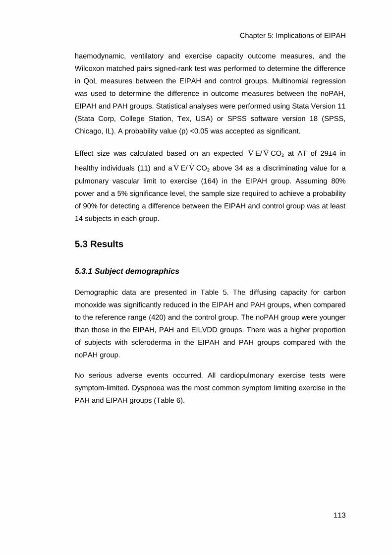
Chapter 5: Implications of EIPAH
113
haemodynamic, ventilatory and exercise capacity outcome measures, and the
Wilcoxon matched pairs signed-rank test was performed to determine the difference
in QoL measures between the EIPAH and control groups. Multinomial regression
was used to determine the difference in outcome measures between the noPAH,
EIPAH and PAH groups. Statistical analyses were performed using Stata Version 11
(Stata Corp, College Station, Tex, USA) or SPSS software version 18 (SPSS,
Chicago, IL). A probability value (p) <0.05 was accepted as significant.
Effect size was calculated based on an expected V E/ V CO2 at AT of 29±4 in
healthy individuals (11) and a V E/ V CO2 above 34 as a discriminating value for a
pulmonary vascular limit to exercise (164) in the EIPAH group. Assuming 80%
power and a 5% significance level, the sample size required to achieve a probability
of 90% for detecting a difference between the EIPAH and control group was at least
14 subjects in each group.
5.3 Results
5.3.1 Subject demographics
Demographic data are presented in Table 5. The diffusing capacity for carbon
monoxide was significantly reduced in the EIPAH and PAH groups, when compared
to the reference range (420) and the control group. The noPAH group were younger
than those in the EIPAH, PAH and EILVDD groups. There was a higher proportion
of subjects with scleroderma in the EIPAH and PAH groups compared with the
noPAH group.
No serious adverse events occurred. All cardiopulmonary exercise tests were
symptom-limited. Dyspnoea was the most common symptom limiting exercise in the
PAH and EIPAH groups (Table 6).

Chapter 5: Implications of EIPAH
114
Table 5. Baseline demographics, noPAH, PAH, EILVDD, EIPAH and controls
noPAH
(n=10)
PAH
(n=6)
EILVDD
(n=4)
EIPAH
(n=17)
Controls
(n=20)
P value
EIPAH vs control
Age, years 41±13 62±7* 64±3* 56±14* 57±13 0.8
Scleroderma, % 20 83* 50 71* 0
Female gender, % 90 100 100 88 95
Body mass index, kg/m2 27±4 25±2 25±4 25±5 25±4 0.9
WHO functional class, % II
III
0
0
50
50
100
0
65
35
N/A
N/A
FEV1, % predicted 104±18 99±7 86±8 93±11 112±11 <0.0001
FVC, % predicted 105±17 106±9 89±7 97±13 116±13 <0.0001
DLCO, % predicted 84±19 44±23* 82±15 74±18 87±13 0.03
Data are presented as mean ± SD, or percent. EIPAH compared with control using the independent t-test. * p<0.05, EIPAH, EILVDD and PAH compared with noPAH (Holm
post hoc test). Abbreviations: PAH, pulmonary arterial hypertension; EILVDD, exercise-induced left ventricular diastolic dysfunction; EIPAH, exercise-induced pulmonary arterial
hypertension; FEV1, forced expiratory volume in 1 second; FVC, forced vital capacity; DLCO, diffusing capacity for carbon monoxide.

Chapter 5: Implications of EIPAH
115
5.3.2 Ventilatory comparisons between the EIPAH and control groups
Compared with their matched control group, the EIPAH group demonstrated
significant ventilatory abnormalities and impairment in exercise capacity. Exercise-
induced PAH was associated with an elevated V E/ V CO2 and reduced PetCO2 at
AT and an attenuated rise in PetCO2 from rest to AT. Further, EIPAH was
associated with a significantly lower AT, peak V O2 and 6MWD (Table 6). These
abnormalities were similar to the characteristic responses demonstrated by the PAH
group, although less severe (Table 6).
There was an inverse correlation between V E/ V CO2 at AT and peak V O2
(r = -0.63, p<0.01) and between V E/ V CO2 at AT and 6MWD (r = -0.59, p<0.01)
when data from the EIPAH and PAH groups were combined.
The EIPAH group had a significant impairment in QoL, with the greatest impairment
in the physical domains (Table 7). Based upon responses to the health transition
item, a higher proportion of the EIPAH group than the control group reported that
their health was worse compared with the previous year (24% versus 5%
respectively, p<0.01).
5.3.3 Quality of life comparisons between PAH, EIPAH, EILVDD and
noPAH
There were no significant differences in QoL between the PAH, EIPAH, EILVVD and
noPAH groups, including the health transition responses (all p>0.05).

Chapter 5: Implications of EIPAH
116
Table 6. Exercise capacity, ventilatory and systemic hemodynamic responses and limiting symptoms on the CPET
noPAH
(n=10)
PAH
(n=6)
EILVDD
(n=4)
EIPAH
(n=17)
Controls
(n=20)
P value
EIPAH vs control
Peak V O2, L/min 1.7±0.5 0.8±0.3* 1.2±0.3 1.2±0.4 1.7±0.5 0.03
Peak V O2, % predicted 77±13 48±17* 69±16 64±18 90±19 0.03
Anaerobic threshold (AT), L/min 0.8±0.2 0.4±0.1*† 0.8±0.1 0.6±0.2 0.9±0.2 0.02
6MWD, metres 601±78 517±128 582±17 575±86 669±76 0.03
V E/ V CO2 at AT 34±4.2 51±13.9*‡ 32±0.8 41±7.3† 31±2.9 0.03
PetCO2 at AT, mmHg 36±2 28±6*‡ 39±0.6 33±4‡ 39±3 0.04
Change PetCO2, rest to AT 1.0±2.4 -0.2±2.1 2.2±2.6 0.8±3 3.7±2.3 0.04
SpO2 at peak exercise, % 96±2 90±5* 97±2 94±3 96±2 0.08
Limiting factor, % Dyspnoea
General fatigue
Leg Fatigue
30
50
20
67
17
0
50
0
50
41
24
35
5
24
67
0.046
Data are presented as mean ± SD. *p<0.05, EIPAH and PAH compared with noPAH, age adjusted (multinomial regression). †p <0.05, ‡p<0.01 PAH and EIPAH vs EILVDD group (multinomial regression). Abbreviations: PAH, pulmonary arterial hypertension; EILVDD, exercise-induced left ventricular diastolic dysfunction; EIPAH, exercise-induced
pulmonary arterial hypertension; Peak V O2, peak oxygen consumption; 6MWD, six-minute walk distance; V E/ V CO2 at AT, ventilatory equivalent for carbon dioxide; AT,
anaerobic threshold; PetCO2, end tidal carbon dioxide tension; SpO2, oxygen saturation.

Chapter 5: Implications of EIPAH
117
Table 7. Quality of Life (SF-36, Version 1)
noPAH
(n=10)
PAH
(n=6)
EILVDD
(n=4)
EIPAH
(n=17)
Controls
(n=20)
P value
EIPAH vs control
Physical functioning 52±18 47±16 63±19 54±16 93±9 0.0001
Role physical 33±35 21±40 63±32 49±45 100±0 0.0005
Bodily pain 63±25 71±29 64±24 56±27 77±26 0.03
General health 48±19 44±17 41±8 49±18 82±16 0.002
Vitality 39±19 51±20 50±11 50±13 76±13 0.0002
Social functioning 61±25 79±17 88±10 68±22 95±13 0.01
Role emotional 83±36 67±52 100±0 67±41 95±13 0.03
Mental health 69±16 72±19 48±20 66±21 85±16 0.002
Data are presented as mean ± SD. (Wilcoxon matched pairs signed-rank test). Abbreviations: PAH, pulmonary arterial hypertension; EILVDD, exercise-induced left ventricular
diastolic dysfunction; EIPAH, exercise-induced pulmonary arterial hypertension.

Chapter 5: Implications of EIPAH
118
Table 8. Haemodynamic variables measured at rest and during the CPET
noPAH
(n=10)
PAH
(n=6)
EILVDD
(n=4)
EIPAH
(n=17)
Control
(n=20)
P value
EIPAH vs noPAH
Resting mPAP, mmHg 13±2 30±9 18±4* 18±4 0.03
Peak mPAP, mmHg 23±2 51±15║ 37±6 37±5
Change in mPAP, mmHg 10±2 22±11* 19±7* 18±4 0.02
Resting CO at rest, L/min 5.7±0.7 5.4±1.1 6.1±1.2 5.4±1.4 0.6
Peak CO, L/min 12.4±3.1 8.6±2.6 10.4±0.9 10.4±2.8 0.5
Peak CO, % predicted 82±24 59±19 72±10 72±19 0.9
Resting PVR, Wood units 0.8±0.4 3.9±1.5* 1.5±0.3 2.2±1.2 0.04
Peak PVR, Wood units 1.3±0.5 3.9±1.3* 1.4±0.6 2.1±0.8 0.07
Resting RVSWI, gm.m/m2/beat 4.6±2.4 12.4±4.9§ 8.0±3.9 7.5±2.0 0.03
Peak RVSWI, gm.m/m2/beat 10.2±2.7 19.9±7.2§ 20.3±7.8* 17.6±3.6 0.03
Resting PAWP, mmHg 8.7±1.2 9±3.6 10.5±2.6 8.4±2.6 0.7
Peak PAWP, mmHg 10.0±3.0 14.5±9.3 24.0±3.6* 12.7±4.5 0.2
Resting RAP, mmHg 6±1.6 5.8±3.3 5.8±1.7 5.6±2.9 0.7
Peak RAP, mmHg
Resting systemic MAP, mmHg
Peak systemic MAP, mmHg
Resting HR, beats.min-1
Peak HR, beats.min-1
5.6±4.8
119±14
74±9
74±9
157±17
11.8±3.8
113±19
129±13
78±10
134±19*
5.5±7.8
107±19
128±16
80±16
131±28
7.8±2.5
109±16
125±12
74±10
133±15§
102±13
118±14
68±10
149±20
0.1
0.2
0.2
0.09
0.02
Data are presented as mean ± SD. *p<0.05, §p<0.01, ║p<0.001, EIPAH, EILVDD and PAH compared with noPAH, age adjusted (multinomial regression). Stratification by mPAP prevented statistical analysis of the difference in mPAP, or measures derived from mPAP, between PAH and noPAH groups at rest and between EIPAH and noPAH groups at peak exercise.
Abbreviations: PAH, pulmonary arterial hypertension; EILVDD, exercise-induced left ventricular diastolic dysfunction; EIPAH, exercise-induced pulmonary arterial hypertension;
mPAP, mean pulmonary artery pressure; CO, cardiac output; PVR, pulmonary vascular resistance; RVSWI, right ventricular stroke work index; PAWP, pulmonary artery wedge pressure; RAP, right atrial pressure; MAP, mean arterial pressure; HR, heart rate.
Chapter 5: Implications of EIPAH
119
5.3.4 Haemodynamic comparisons between EIPAH, PAH and noPAH
When adjusted for age, there was no significant difference in peak exercise CO
between groups. Peak exercise CO was lower than predicted in all groups (Table 8).
At rest, the EIPAH group demonstrated a higher mPAP, pulmonary vascular
resistance, and right ventricular stroke work index than the noPAH group. The
EIPAH and PAH groups had a significantly greater rise in mPAP (Table 8) and the
PAH group had a steeper slope in the relationship between mPAP and CO (Figure
5) from rest to peak exercise than the noPAH group.
Figure 5. Pressure versus flow during exercise. The relationship between mPAP and
CO during a symptom-limited CPET.
0
10
20
30
40
50
60
4 6 8 10 12 14
CO (L/min)
mP
AP
(m
mH
g)
PAH
EIPAH
noPAH
Slope 6.3*
Slope 1.2
Slope 2.8
*p = 0.009 PAH versus noPAH, pairwise comparison. Abbreviations: mPAP, mean pulmonary artery
pressure; CO, cardiac output; CPET, cardiopulmonary exercise test; PAH, pulmonary arterial
hypertension; EIPAH, exercise-induced pulmonary arterial hypertension; noPAH, no pulmonary arterial
hypertension
By definition, the PAH group had a higher mPAP at rest and the EIPAH group had a
higher mPAP at peak exercise than the noPAH group. Stratification by mPAP
prevented statistical analysis of the difference in mPAP, or measures derived from
mPAP, between PAH and noPAH groups at rest and between EIPAH and noPAH
groups at peak exercise.

Chapter 5: Implications of EIPAH
120
5.3.5 Comparisons between the EIPAH and EILVDD groups
The EIPAH group had a significantly higher V E/ V CO2 at AT (p=0.03) and a lower
PetCO2 at AT (p=0.008) compared with the EILVDD group. There were no
statistically significant differences in resting or exercise haemodynamic responses
between the EIPAH and EILVDD groups, although by definition, the group with
EILVVD had a higher pulmonary artery wedge pressure at peak exercise than the
EIPAH group. Similarly, there were no differences in AT, peak V O2 or 6MWD
between these groups.
5.3.6 Influence of age and diagnosis of scleroderma
Neither age (r = -0.05, 95% CI -0.26 to 0.15, p=0.6) nor the presence of scleroderma
(r=0.76, 95% CI -4.9 to 6.4, p=0.8) had an influence on peak exercise mPAP but
both were significant factors in determining peak exercise CO, with older age
(r= -0.08, 95% CI -0.14 to -0.03, p=0.005) and a diagnosis of scleroderma (r= -1.9,
95% CI -3.6 to -0.4, p=0.02) being associated with a lower peak exercise CO.
Subjects with scleroderma (n=20) were older than those without a clinical diagnosis
(mean age 58±12 versus 40±14 years, p<0.01). Scleroderma was associated with
impaired QoL (social functioning domain, p<0.05). Neither age nor a diagnosis of
scleroderma had a significant influence on any other outcome measure (all p>0.05).
5.4 Discussion
This study describes exercise abnormalities associated with EIPAH in symptomatic
patients, at risk of PAH. Exercise-induced PAH (mPAP >30mmHg and a pulmonary
artery wedge pressure <20mmHg on exercise) was associated with ventilatory
abnormalities and a clinically important reduction in exercise capacity, consistent
with a pulmonary vascular limitation to exercise and typical of PAH. These exercise
impairments were accompanied by a marked reduction in QoL.
Reduced exercise capacity and a low AT, in the presence of an elevated V E/ V CO2
and reduced oxygen saturation, identify a pulmonary vascular limitation to exercise,
according to a well accepted diagnostic algorithm for exercise interpretation (13).
Reduced PetCO2 at AT and a fall, rather than rise, in PetCO2 from rest to AT have
also been shown to be associated with PAH (12, 168). Our data confirm these

Chapter 5: Implications of EIPAH
121
findings in the group with PAH. Compared with healthy controls, similar but milder
ventilatory abnormalities were evident in the EIPAH group. These abnormalities in
the EIPAH group, in whom there was a clinical suspicion of PAH, are consistent with
a mild, or early, pulmonary vasculopathy, similar to that present in subjects with
PAH. The abnormal ventilatory responses demonstrated by the EIPAH group were
not seen in the group with EILVDD. This finding supports the suggestion that the
abnormalities demonstrated by the EILVDD group were more likely to be related to
left heart dysfunction than a pulmonary vasculopathy. Further, these results lend
weight to the suggestion that a cardiopulmonary exercise test may help differentiate
these groups. Our findings contrast with those of Walkey et al (421) who found no
difference in the V E/ V CO2 at AT between subjects with scleroderma and who had
an elevated pulmonary artery wedge pressure at peak exercise (EILVDD, n=4)
compared with those with a peak exercise pulmonary artery wedge pressure
<18mmHg and evidence of a pulmonary vascular limit to exercise (n=3). However,
according to our calculations, the Walkey study (23) was not powered to detect
differences between groups comprising numbers of three and four. There was no
overlap of V E/ V CO2 at AT between groups with a pulmonary vascular limit to
exercise or EILVDD, with all subjects in the pulmonary vascular group having a
higher V E/ V CO2 at AT than those with an elevated pulmonary artery wedge
pressure.
The EIPAH group demonstrated a significant impairment in exercise capacity, in
both peak V O2 and 6MWD. Although reduced peak V O2 has previously been
reported in EIPAH (27) this is the first study to demonstrate that 6MWD is
decreased. The 6MWD is a measure of submaximal exercise tolerance that is
considered to reflect the capacity to perform activities of daily living (31). The
inverse relationship between V E/ V CO2 at AT and both peak V O2 and 6MWD in
our study suggests an association between ventilatory abnormalities and impaired
maximal and submaximal exercise capacity in PAH and EIPAH.
When compared with healthy controls, the subjects with EIPAH had marked
reductions in QoL, with a greater magnitude of impairment in the physical health
domains than in the mental health domains. In PAH, exertional symptoms impact on
the ability to perform activities of daily living, which is reflected in the physical
domains of the SF-36 (85). Our study demonstrates similar results in the EIPAH
group, with exertional symptoms impacting negatively on QoL, particularly in the

Chapter 5: Implications of EIPAH
122
physical domains. The cause of the QoL impairment in the noPAH group is
uncertain. However, the finding of diminished QoL in this group is consistent with the
significant, persistent exertional symptoms reported by these subjects.
The finding of ventilatory abnormalities in the EIPAH group in the current study
contrasts with the observations made by Tolle et al (27) who identified
haemodynamic abnormalities and reduced aerobic capacity in EIPAH, but no
ventilatory differences between the EIPAH and control groups. This contrasting
finding is likely related to the characteristics of the control groups used in the two
studies. Subjects in the ‘normal’ group in the study by Tolle et al (27) were
symptomatic patients. In that study, the V E/ V CO2 at AT was not sensitive in
discriminating between subjects with and without EIPAH; however, the reported
mean V E/ V CO2 at AT of 36 for the ‘normal’ group (mean age 45.9 years) is
unlikely to represent a healthy response (164). Our inclusion of an asymptomatic
control group allowed evaluation of the ventilatory response of subjects with EIPAH
against responses in healthy age-, gender- and body mass index-matched control
subjects and identified indisputable ventilatory abnormalities associated with EIPAH.
In our study, there were significant haemodynamic differences between the EIPAH
and noPAH groups. However, the younger, symptomatic noPAH group
demonstrated impairments in peak exercise CO and exercise capacity, compared
with age appropriate reference values, confirming that they would not have been an
appropriate control group for this study.
Pulmonary arterial hypertension is known to be a progressive condition, although
the rate of progression is variable (7). The clinical course of subjects with elevated
mPAP on exercise is unknown. The finding that 24% of subjects with EIPAH in this
study reported worsening QoL during the previous year raises the possibility that
EIPAH may be associated with disease progression and represent an early phase of
PAH. This is consistent with the recent findings by Condliffe et al (2) who reported
that EIPAH associated with connective tissue disease progressed to PAH at rest in
19% of subjects and that death related to pulmonary hypertension and/or right heart
failure occurred in 10% of subjects during a mean period of 3.3 years.
The question whether EIPAH progresses to PAH remains unanswered. However,
regardless of progression, the current study identified marked functional
impairments associated with EIPAH, suggesting that this condition warrants
consideration for therapy. Clinical benefits, that include an improvement in 6MWD

Chapter 5: Implications of EIPAH
123
and functional class, have recently been described following pharmacologic therapy
for ‘exercise-uncovered’ PAH (defined as mPAP <25mmHg at rest but mPAP
>30mmHg and a pulmonary artery wedge pressure 15mmHg on exercise) in an
open-label trial (396). These findings demonstrate that, for selected patients with a
mPAP >30mmHg on exercise, PAH specific therapy is beneficial. Furthermore,
these findings would be consistent with EIPAH being part of a continuum in
pulmonary vascular disease.
We specifically chose a group with a high pretest probability of EIPAH to optimise
the diagnostic yield of EIPAH in our investigation. Patients with scleroderma are
known to be at increased risk of PAH (241). Scleroderma was present in 54% of the
patient subjects in our study, and 81% of these had PAH or EIPAH. Our results, and
those of Condliffe et al (2) suggest a high incidence of a pulmonary vascular
dysfunction and progression in subjects who have scleroderma, a reduced diffusing
capacity for carbon monoxide and exertional symptoms of dyspnoea and fatigue. In
our study, scleroderma was associated with a lower peak exercise CO and impaired
social functioning however there was no association between any of the other
identified functional limitations and a diagnosis of scleroderma. These impairments,
therefore, appear to reflect a pulmonary vasculopathy rather than other morbidities
associated with scleroderma. Kovacs et al (419) also reported an association
between increased exercise mPAP and impaired exercise capacity in subjects with
scleroderma and suggested a pulmonary vasculopathy as the likely cause for their
findings.
To accommodate the possibility that age would be a confounding factor in the
difference between the patient groups, we recruited an age-matched control group.
In comparison with this control group, clear and indisputable ventilatory, exercise
capacity, and QoL impairments were identified in the EIPAH group.
Previous studies reporting invasive central haemodynamics to describe the
development of pulmonary hypertension on exercise in symptomatic subjects with
an mPAP <25mmHg at rest (27, 302, 395, 422) have significant limitations. Early
studies neither measured pulmonary artery wedge pressure on exercise (302, 422)
nor excluded subjects with an abnormal pulmonary artery wedge pressure at rest or
on exercise (395) and therefore were unable to exclude post-capillary pulmonary
hypertension. The lack of a healthy control group in the study by Tolle et al (27)
Chapter 5: Implications of EIPAH
124
prevented an adequate description of the clinical sequelae of EIPAH. The current
study has addressed these limitations.
We have demonstrated that combining the assessment of central haemodynamic
and ventilatory responses during exercise can identify a likely pulmonary
vasculopathy that is not evident at rest. This analysis of pulmonary vascular function
under stress provides the potential to improve early diagnosis of PAH, if EIPAH is
found to be an early stage of PAH.
5.4.1 Limitations and general applicability of this study
No central haemodynamic data were collected for control subjects because this was
considered unethical due to the invasive nature of the procedure. This precluded
comparison of haemodynamic responses in the EIPAH group with those of healthy
controls.
We examined a carefully selected group of patients referred to a regional pulmonary
hypertension centre which services a population of 1.5 million people. We believe
that our findings cannot be applied to all patients presenting with unexplained
dyspnoea. Nonetheless, the group we studied represents a dilemma with which
clinicians are often faced, namely, a patient presenting with risk factors and a clinical
suspicion of PAH but who does not meet diagnostic criteria for PAH at rest.
It is likely that the significance of a rise in mPAP on exercise is influenced by age,
and the pre-test probability of a pulmonary vasculopathy as shown by others (2, 30).
Although the general applicability of our findings is uncertain, we believe the
abnormalities identified on exercise, in the subjects with EIPAH, are clinically
significant. They suggest that, in a selected group, a rise in mPAP with exercise to
>30mmHg should not be dismissed even if mPAP is <25mmHg at rest.
We have presented cross-sectional data only. Longitudinal follow up of the cohort
with EIPAH is being undertaken.
This study was not powered to detect a difference in outcome measures between
groups of four to ten subjects. Therefore, findings of no difference between the
noPAH, EILVDD and PAH with other groups should be interpreted with caution.

Chapter 5: Implications of EIPAH
125
The loss of independence of the control group, by individual matching of control and
EIPAH subjects, precluded statistical comparison of the noPAH, EILVDD and PAH
groups with the control group.
5.5 Conclusions
Haemodynamic findings on exercise, in isolation, may be difficult to interpret due to
a wide range of normal responses. However, the combination of haemodynamic and
ventilatory measures in the assessment of selected symptomatic patients can
identify abnormalities which are characteristic of PAH, may represent early
manifestations of a pulmonary vasculopathy, and may therefore facilitate earlier
diagnosis of PAH.
Addendum: Usual physical activity in EIPAH and control groups.
Method: An initial subjective assessment of the control subjects’ participation in
organised sport and usual physical activity was performed during the telephone
conversation made to confirm the volunteers’ suitability for the study and to arrange
the data collection appointments. Volunteers who were involved in organised sport
or high levels of usual physical activity were excluded from the study (n=6). Both the
control subjects and patients completed the Short Last 7 Days, Self-administered
Format of the International Physical Activity Questionnaire (IPAQ)(410) during the
rest period between the 6MWTs on the first day of assessment.
Results: Based upon the results obtained using the IPAQ (410) there was no
difference in the usual physical activity level between the EIPAH and control groups.
The total metabolic equivalent (MET) minutes per week was 4272±4859 in the
EIPAH group, versus 3304±2840 in the control group (p=0.7). The time spent sitting
on a week day, per week, was 1695±1125 min in the EIPAH group versus 1725±830
min in the control group (p=0.9).

Chapter 6: Measurement properties of the 6MWT in individuals with EIPAH
126
CCHHAAPPTTEERR 66
MMEEAASSUURREEMMEENNTT PPRROOPPEERRTTIIEESS OOFF TTHHEE 66MMWWTT
IINN IINNDDIIVVIIDDUUAALLSS WWIITTHH EEXXEERRCCIISSEE--IINNDDUUCCEEDD
PPUULLMMOONNAARRYY AARRTTEERRIIAALL HHYYPPEERRTTEENNSSIIOONN
Fowler RM, Jenkins SC, Maiorana AJ, Gain KR, O’Driscoll G, Gabbay E.
Measurement properties of the 6-min walk test in individuals with exercise-induced
pulmonary arterial hypertension. Intern Med J 2011; 41:679-687
6.1 Introduction
Pulmonary arterial hypertension (PAH) is a progressive condition characterised by
elevated pulmonary vascular resistance, reduced pulmonary blood flow and an
attenuated increase in cardiac output (CO) during exercise (27). The hallmark of
PAH is exertional intolerance with symptoms that include dyspnoea, fatigue and
light-headedness. The diagnosis of PAH is based on abnormal resting
haemodynamics (1). However, recent studies demonstrate that some individuals
have normal haemodynamics at rest but a higher than expected mean pulmonary
artery pressure (mPAP), in the presence of a normal pulmonary artery wedge
pressure and reduced cardiac output at peak exercise, in association with reduced
exercise capacity and symptoms typical of PAH (27, 76). This elevation in
pulmonary pressure on exercise has been called exercise-induced PAH (EIPAH)
and has been proposed to be part of the spectrum of pulmonary vascular diseases
(78).
The capacity of the cardiovascular, respiratory and skeletal muscle systems to
transport and utilize oxygen for aerobic metabolism is quantified by measuring peak
oxygen consumption (peak V O2) during a cardiopulmonary exercise test (CPET).
While peak V O2 reflects disease severity and prognosis in PAH (9), a CPET
requires specialist knowledge and equipment. In contrast, the six-minute walk test
(6MWT) requires minimal equipment, is easy to administer and is widely available.
Measures derived from the 6MWT include the six-minute walk distance (6MWD) and
six-minute walk work (6MWW), the product of 6MWD and body weight. Six-minute

Chapter 6: Measurement properties of the 6MWT in individuals with EIPAH
127
walk distance, which quantifies functional exercise impairment, has been shown to
provide a valid estimate of peak V O2 in individuals with PAH (34, 35) and is more
sensitive to change with pharmaceutical therapy in PAH than peak V O2 (35, 51).
Accordingly, 6MWD has been employed as the primary outcome measure for
clinical trials of pharmaceutical therapy in PAH (1). However, in subjects with
chronic obstructive pulmonary disease (COPD), 6MWW has been shown to
correlate more strongly with disease severity and to have greater sensitivity and
specificity for low exercise capacity than 6MWD (338).
The 6MWT can be administered with or without encouragement. Clinical trials in
PAH have mostly utilised a protocol devoid of encouragement to better reflect the
subjects’ ability to perform activities of daily living. However, the American Thoracic
Society recommends a 6MWT protocol with standardised encouragement, to
measure maximal walking capacity (308). Investigations reporting physiological
responses to a 6MWT in PAH have typically utilised an encouraged 6MWT (133,
365) given that it is more likely to identify a physiological limitation to exercise than
an unencouraged test. However, in individuals with mild-moderate impairment in
aerobic capacity, such as EIPAH (27), mechanical constraints that limit maximum
walking speed, rather than physiological dysfunction, may influence 6MWD. This
‘ceiling effect’ has been reported in patients with chronic heart failure (CHF) (36) and
PAH (37) with mild functional limitation (World Health Organization Functional Class
II, [WHO FC II]).
The aims of this study, in subjects with EIPAH, were to: (i) compare physiologic and
symptomatic responses to the 6MWT and CPET, and (ii) determine whether the
6MWT can identify functional exercise limitation and accurately estimate aerobic
capacity.
6.2 Methods
6.2.1 Participants
Forty-nine adults were recruited from the Pulmonary Hypertension Unit at Royal
Perth Hospital (Perth, Western Australia). All subjects had been referred for
investigation of dyspnoea of unknown aetiology, were in WHO FC II or III, and were
investigated for PAH as defined by the 2004 European Society of Cardiology
Chapter 6: Measurement properties of the 6MWT in individuals with EIPAH
128
Guidelines (51), that is, mean pulmonary artery pressure (mPAP) >25mmHg and
pulmonary artery wedge pressure ≤ 15mmHg at rest. Based upon historically
accepted upper limits of normal (400), subjects were identified as having exercise-
induced PAH (EIPAH) if mPAP was >30mmHg and pulmonary artery wedge
pressure was <20mmHg, on exercise. As determined prior to commencement of the
study, subjects were excluded or withdrawn from the study if they had a body mass
index (BMI) >35kg/m2 (n=4), ischaemic heart disease (n=1), a history or evidence of
pulmonary parenchymal or airway disease on high resolution computed tomography
or lung function testing (n=5), a musculoskeletal condition limiting exercise
performance (n=1), anaemia (n=1), PAH at rest (mPAP >25mmHg at rest, n=6),
normal mPAP at rest or on exercise (n=10), or a pulmonary artery wedge pressure
>15mmHg at rest or >20mmHg on exercise (n=4). Data from the remaining 17
subjects were used in the analyses.
A population-based sample of 20 healthy control subjects was recruited from a
database of research volunteers maintained by the Lung Institute of Western
Australia (Perth, Western Australia). Controls were matched to subjects with EIPAH
for gender, age (±5 years) and BMI (±3kg.m-2, provided BMI was ≤35kg.m-2). All
controls underwent medical examination and a series of screening tests (medical
history, echocardiogram, electrocardiogram, lung function tests and haematology) to
confirm their healthy status.
The study was approved by the Human Research Ethics Committees of Royal Perth
Hospital and Curtin University. Written informed consent was obtained from all
subjects prior to enrolment. This work was performed at Royal Perth Hospital, Perth,
Western Australia.
6.2.2 Study design
The study employed a prospective cross-sectional design. All subjects completed
two encouraged 6MWTs on one day, separated by at least 30 minutes rest. A
symptom-limited incremental CPET was performed between 1 and 32 (mean of 5)
days following the 6MWTs. Subjects recruited from the Pulmonary Hypertension
Unit had a pulmonary artery catheter in situ during the CPET, for the measurement
of central haemodynamics. All tests were supervised by the same investigator (RF).
Chapter 6: Measurement properties of the 6MWT in individuals with EIPAH
129
6.2.2.1 Exercise protocols
Six-minute walk test
The 6MWT was performed over a 45m course in an enclosed corridor according to
the American Thoracic Society guidelines (308). All subjects completed two 6MWTs,
separated by at least 30 minutes. Heart rate (Polar Electro Oy; Kempele, Finland)
was monitored throughout the test. Sensations of dyspnoea and leg fatigue were
assessed immediately following test cessation using the Borg Category Ratio Scale
(408).
Cardiopulmonary exercise test
The CPET was performed using an electronically braked cycle ergometer attached
to a customised imaging table (Lode BV, Groningen, Netherlands) with the subject
in a semi-recumbent position (torso at 50 from the horizontal). An incremental
symptom-limited protocol was employed which involved a 3 minute baseline period
followed by 15 Watt increments in workload every 3 minutes. The initial workload
was individualised based upon gender, age and 6MWD. Standardised instructions
and encouragement were provided to promote a maximum effort. Breath by breath
ventilation, oxygen and carbon dioxide concentrations and the derived minute
ventilation, V O2 and carbon dioxide output were determined using the Vmax
metabolic analysis system (Sensormedics, Yorba Linda, CA). Heart rate (12 lead
electrocardiogram; Cardiosoft, Version 4.2, GE Medical Systems, Milwaukee, WI)
and SpO2 (Oxypleth pulse oximeter and ear sensor, Novemetrics, Wallingford, CT)
were monitored continuously throughout the test.
The method of haemodynamic monitoring and data collection employed in the
assessment of the subjects with EIPAH has previously been reported (76). In brief,
pulmonary artery pressure was continuously monitored (Compact, Datex Engstrom,
Helsinki, Finland). Cardiac output was determined using thermal filament
thermodilution, updated every 60 seconds (Vigilance, Baxter, CA). Pulmonary artery
wedge pressure was recorded at rest and every 3 minutes, immediately prior to an
increase in workload, and in the final minute of the test. If a subject performed less
than 30 seconds of the final workload attempted, the mPAP, pulmonary artery
wedge pressure and CO responses were taken as the values measured within the
last 60 seconds of the previous workload. Systemic arterial pressure was measured

Chapter 6: Measurement properties of the 6MWT in individuals with EIPAH
130
manually, at the brachial artery, at rest and every 3 minutes, immediately prior to an
increase in workload and in the final minute of the test.
Ratings of dyspnoea and leg fatigue were recorded during the last 60 seconds of
each workload and immediately following test cessation. The maximum workload
was defined as the highest workload maintained for >30 seconds and peak V O2
was defined as the 30 second average measured at the maximum workload within
the last 30 seconds prior to test termination. Minute ventilation, V O2, carbon
dioxide output and respiratory exchange ratio were calculated.
The demographic, 6MWD and peak V O2 data for both EIPAH and control groups
have previously been reported.(76)
6.2.2.2 Statistical analyses
The effect of test repetition on 6MWD was examined using a general linear model
for repeated measures. For each group, the magnitude of change in 6MWD
between tests 1 and 2 was compared using the paired t-test. The greater 6MWD
was used in all subsequent analyses. The coefficient of repeatability was
determined as twice the SD of the difference in 6MWD measured on the two tests.
Comparisons of responses to the 6MWT and CPET for the EIPAH group and control
group were performed for each group separately using paired t-tests (heart rate
[HR]) and Wilcoxon signed rank tests (peak dyspnoea and rate of perceived
exertion). Differences between the EIPAH and control groups were analysed using
unpaired t-tests (continuous variables) and the Mann-Whitney U test (peak
dyspnoea and rate of rate of perceived exertion). Relationships between 6MWD and
6MWW with peak values of V O2 and CO were assessed using Pearson correlation
coefficients and linear regression analysis. Data were analysed using SPSS
software Version 18 (SPSS, Chicago, IL, USA) A p value of <0.05 was considered
to be statistically significant. Data are presented as mean ± SD unless otherwise
stated.

Chapter 6: Measurement properties of the 6MWT in individuals with EIPAH
131
6.3 Results
Subject characteristics are presented in Table 9. The subjects with EIPAH had a
lower forced expiratory volume in 1 second (FEV1) and forced vital capacity (FVC),
percent predicted, than the controls (p<0.001 for both). However, all lung function
test results for subjects in the EIPAH group were within the reference range (423)
and FEV1/FVC was the same in both groups indicating there was no evidence of
airflow obstruction in the EIPAH subjects. The diffusing capacity for carbon
monoxide was reduced in the EIPAH group when compared with the controls and
the reference range (420).
Table 9. Subject characteristics, EIPAH and controls
Characteristic EIPAH Controls
Females/males 15/2 19/1
Age (years) 56±14(35-81) 57±13(33-77)
Height (m) 1.63±0.8 1.65±0.7
Weight (kg) 67±12 68±9
BMI (kg/m2) 25±5 25±4
FEV1, % predicted 93±11** 112±11
FVC, % predicted 97±13** 116±13
FEV1/FVC % 78±7 78±7
DLCO, % predicted 74±18* 87±13
WHO Functional Class II 11
WHO Functional Class III 6
mPAP, rest (mmHg) 17.9±3.6
peak exercise 36.6±5.1
CO, rest (L/min) 5.4±1.4
peak exercise 10.4±2.8
PVR, rest (Wood units) 2.2±1.2
peak exercise 2.1±0.8
Note: Data are mean±SD (range) or number of subjects. * p<0.01, **p<0.001, EIPAH versus control.
Abbreviations: kg, kilograms; BMI, body mass index; FEV1, forced expiratory volume in 1 second; FVC,
forced vital capacity; DLCO, diffusing capacity for carbon monoxide; SpO2, oxygen saturation; WHO,
World Health Organisation; mPAP, mean pulmonary artery pressure; CO, cardiac output; PVR,
pulmonary vascular resistance.

Chapter 6: Measurement properties of the 6MWT in individuals with EIPAH
132
6.3.1 Exercise responses
There were no adverse events associated with the exercise tests. In the EIPAH
subjects, dyspnoea or leg fatigue was the most common symptom limiting
performance on both tests. In the controls, the most common factors limiting
performance on the 6MWT and CPET were an inability to walk faster and leg
fatigue, respectively (Table 10).
Table 10. Heart rate and symptomatic responses to the 6MWT and CPET
Note: Data are mean±SD, (percentage of subjects). †p<0.05, ‡p<0.01, §p<0.001 6MWT vs CPET.
*p<0.05, **p<0.01, EIPAH versus control. Abbreviations: 6MWT, six-minute walk test; CPET,
cardiopulmonary exercise test; bpm, beats per minute; RPE, rate of perceived exertion.
Mechanical constraints of walking limiting maximum distance.
6.3.1.1 Physiological and symptomatic responses, 6MWT and CPET
Peak HR was similar in the CPET and 6MWT in the EIPAH group (p=0.8), but
higher in the CPET than the 6MWT in the control group (p<0.05) (Table 10). Peak
HR was higher in the control group than the EIPAH group during the CPET (p<0.01)
(Table 10). The peak dyspnoea score was higher in the CPET than the 6MWT in
both groups (EIPAH p<0.01, control p<0.001) and higher in the EIPAH group than
the control group at the end of the 6MWT (p<0.05). There was no difference in the
peak dyspnoea score between groups at the end of the CPET (p=0.8).
EIPAH Control
6MWT CPET 6MWT CPET
Peak HR (bpm) 133±19 133±15** 139±17† 149±20
% predicted HR max 76±13 77±8** 80±11 86±9
Peak dyspnoea 3.4±1.5*‡ 4.8±1.2 2.2±1.2§ 5.0±1.8
Peak RPE 4.6±2* 6.0±2.4* 3.2±2 6.8±2.3
Limiting factor: Dyspnoea 8(47%) 7(41%) 0 1(5%)
Leg fatigue 8(47%) 6(35%) 5(24%) 14(67%)
Mechanics¶ 1(6%) 0 15(71%) 0
General fatigue 0 4(24%) 0 5(24%)
Other 0 0 1(4%) 1(4%)

Chapter 6: Measurement properties of the 6MWT in individuals with EIPAH
133
6.3.1.2 Exercise capacity
Six-minute walk distance, 6MWW and peak V O2 were all significantly reduced in the
EIPAH group, compared with the controls (p<0.01) (Table 11).
Table 11. 6MWT and CPET results
EIPAH Control
6MWT
6MWD, m (% predicted)45
575±86 (88±11)*** 669±76 (102±9)
6MWW, kg.km 39±11** 45±7
CPET
Peak V O2 , L/min, (% predicted) 1.2±0.4 (64±18)** 1.7±0.5 (90±19)
Peak V O2 , ml/min.kg-1
18±4** 25±7
Initial workload, Watts 18±13 (0-30) 43±21 (15-75)
Maximum workload, Watts 62±20 (30-90) 104±39 (60-195)
V E/ V CO2 at AT 41±7.3*** 31±2.9
Respiratory exchange ratio 1.05±0.06* 1.11±0.06
Notes: Data are mean ±SD (percent predicted, or range). *p<0.05, **p<0.01, ***p<0.001, EIPAH
versus control. Abbreviations: 6MWT, six-minute walk test; CPET, cardiopulmonary exercise test;
6MWD, six-minute walk distance; 6MWW, six-minute walk work; kg.km, kilogram times kilometre;
peak V O2 , peak oxygen consumption; V E/ V CO2 at AT, ventilatory equivalent for carbon dioxide at
the anaerobic threshold.
6.3.1.3 Effect of test repetition on 6MWD
Fifteen subjects (88%) in the EIPAH group and 16 (80%) subjects in the control
group increased their 6MWD on the second test. There was a significant difference
in 6MWD between test 1 and test 2 in both the EIPAH (556±86m versus 574±85,
p<0.001) and control (655±66m versus 668±77, p<0.01) groups. Test repetition was
associated with marked individual variation in the magnitude of change in 6MWD in
the EIPAH (+18±15m, range -16 to +39m, [3±3%]) and control (+9±11m, range -10
to +29m, [1±2%]) groups. There was no difference in the magnitude of change in
6MWD on the second test between the EIPAH and control groups (p=0.08). The
coefficient of repeatability for the EIPAH and control groups was 31m and 22m
respectively. The variability between 6MWTs 1 and 2 is demonstrated in Figure 6.

Chapter 6: Measurement properties of the 6MWT in individuals with EIPAH
134
Figure 6. Bland-Altman plot showing agreement in 6MWD between two 6MWTs for
individuals with EIPAH
-30
-20
-10
0
10
20
30
40
50
400 450 500 550 600 650 700
Mean 6MWD (m)
Diffe
re
nc
e b
etw
een
te
sts (m
)
The solid line indicates the mean difference in 6MWD between test 1 and test 2 (6MWT2 minus
6MWT1). The dashed lines indicate the mean of the difference ± 2SD. Abbreviations: 6MWD, six
minute walk distance; 6MWT, six minute walk test; EIPAH, exercise-induced pulmonary arterial
hypertension
6.3.1.4 Relationship between 6MWT results, peak V O2 and peak CO
Figure 7 illustrates the relationships between 6MWW and 6MWD with peak V O2 in
subjects with EIPAH. There was a strong correlation between 6MWW and
peak V O2 (L/min) for subjects with EIPAH (r=0.86, p<0.001) and a moderate
correlation for the control group (r=0.74, p<0.001). Peak V O2 accounted for 73%
and 55% of the variance in 6MWW in the EIPAH and control groups respectively.
There were moderate correlations between 6MWD and peak V O2 in the EIPAH
(r=0.72, p<0.01) and the control groups (r=0.61, p<0.01).

Chapter 6: Measurement properties of the 6MWT in individuals with EIPAH
135
Figure 7. Relationships between 6MWW and 6MWD with peak VO2, in EIPAH
Regression line, 95% confidence intervals (solid lines) and 95% prediction intervals (hatched lines) in
subjects with EIPAH demonstrating the relationship between (a) six-minute walk work (6MWW, kg.km)
and peak oxygen consumption (peak V O2, L/min) and (b) six-minute walk distance (6MWD, m) and
peak V O2, L/min.
a
b

Chapter 6: Measurement properties of the 6MWT in individuals with EIPAH
136
In the EIPAH group, there was a significant correlation between 6MWD and peak
exercise CO (r=0.59, p<0.05) but not between 6MWW and peak exercise CO
(r=0.46, p=0.07).
A significant correlation was evident between peak V O2 (L/min) and CO (r=0.55,
p<0.05). Peak exercise CO accounted for 21% of the variance in 6MWW, 36% of
the variance in 6MWD and 30% of the variance in V O2 (L/min).
6.4 Discussion
The principal findings from this study are that 6MWD and 6MWW, derived from an
encouraged 6MWT, identify reduced exercise capacity and correlate significantly
with peak V O2 in subjects with EIPAH. These findings suggest that the encouraged
6MWT is a valid test of exercise capacity in this cohort. While 6MWD, 6MWW and
peak V O2 all correlated with peak exercise CO, only a small proportion of the
impairment in exercise capacity was explained by peak exercise CO. This finding
suggests that factors other than cardiac output, during an acute bout of exercise,
contribute to exercise limitation in EIPAH.
The significant difference in 6MWD observed between the EIPAH and control
groups demonstrates the capacity of the encouraged 6MWT to detect reduced
functional exercise capacity in subjects with EIPAH who have mild-moderate
functional limitation (WHO FC II and III). Furthermore, the moderate (6MWD) to
strong (6MWW) correlations with peak V O2 suggests that the 6MWT is a valid
method of estimating aerobic capacity in the absence of a CPET in EIPAH, although
the wide prediction intervals highlight that the relationship was not strong in some
individuals, a finding that is consistent with other studies (424). This result
demonstrates that the 6MWT cannot be used to accurately predict peak V O2 in an
individual with EIPAH.
The similar peak HR elicited during the 6MWT and CPET in subjects with EIPAH
suggests that the encouraged 6MWT represents high intensity exercise for this
cohort. This is consistent with reports in other patient populations (315-317) in which
the encouraged 6MWT and CPET have been shown to elicit similar HR and
peak V O2 responses. This finding is in contrast to a report that found a significantly
higher peak HR during a CPET (employing cycle ergometry) compared with an

Chapter 6: Measurement properties of the 6MWT in individuals with EIPAH
137
unencouraged 6MWT in PAH (314). This disparity is likely to reflect the difference in
6MWT protocol between studies. An encouraged 6MWT has been shown to elicit a
greater physical effort than an unencouraged 6MWT in COPD and CHF (425). Our
findings suggest that an encouraged 6MWT, in subjects with EIPAH in WHO
functional classes II and III, is not subject to a ceiling effect and reflects
physiological limitation and aerobic capacity in this cohort.
In contrast to the subjects with EIPAH, the control subjects had lower peak HR and
dyspnoea scores during the 6MWT than the CPET, and, at best, moderate
correlations between peak V O2, 6MWD and 6MWW, suggesting that a ceiling effect
limited the capacity of the 6MWT to accurately estimate aerobic capacity in the
healthy individuals.
Subjects from both groups reported higher dyspnoea scores during the CPET than
the 6MWT. In the EIPAH group, in whom the maximum HR in each test were similar,
this may be due to the higher levels of local muscle acidosis (426), serum lactate
(427) and consequently V E (277, 314, 428) that occurs during a cycling test
compared with a walking test, contributing to increased ventilatory work and an
increased sensation of dyspnoea (199).
In previous studies of subjects with pulmonary vascular disease, reduced CO on
exercise has been associated with impaired oxygen uptake kinetics (43) and
reduced peak V O2 (27, 303). In our study, reduced CO during an acute bout of
exercise appeared to have a limited role in the exercise limitation identified in
subjects with EIPAH. There is increasing evidence that PAH is a systemic condition
(199) with features similar to CHF and COPD. Inflammation, increased sympathetic
nerve activity, a heightened ergoreflex response, skeletal muscle dysfunction and
systemic endothelial dysfunction contribute to exercise limitation in CHF (206, 248,
429, 430) and COPD (429, 431-433). These systemic abnormalities, with the
exception of ergoreflex up-regulation, have also been described in PAH (39, 138,
237, 249) and are potential contributors to exertional symptoms and exercise
limitation in EIPAH. This is an important area for future research.
Previous studies in cohorts with chronic lung conditions (309, 326, 434, 435),
cardiovascular disease (425) and healthy adults (312, 313, 436) have reported that
test repetition (familiarisation to the 6MWT protocol) results in a small but significant
increase in 6MWD on the second test. Our study extends this observation to

Chapter 6: Measurement properties of the 6MWT in individuals with EIPAH
138
subjects with EIPAH. While the mean increase in 6MWD with test repetition was
small there was a large variability among individuals, with the magnitude of increase
being as great as 39m. The coefficient of repeatability, of 31m, for 6MWD in our
EIPAH group indicates that an increase, or fall, in 6MWD of up to 31m in an
individual with EIPAH, may be due to measurement variability rather than to at
treatment effect, or worsening of the condition.
6.5 Limitations of the study
The sample size of this study was relatively small and further prospective studies
are required to confirm these findings. The high proportion of females in our study
reflects the higher proportion of females to males in PAH populations (437),
however, we cannot be certain that the results of this study translate to male
subjects with EIPAH. Measurement of gas exchange (peak V O2 and minute
ventilation) during the 6MWT would have strengthened this study and has previously
been shown to be feasible and useful in PAH (314). The respiratory exchange ratio
of 1.05±0.06 observed in the EIPAH group suggests that a maximum V O2 was not
achieved in some subjects, most likely because of early test termination due to
intolerable symptoms.
6.6 Conclusions
An encouraged 6MWT identified reduced exercise capacity and provided a valid
estimate of peak V O2 in subjects with EIPAH, although wide prediction intervals
demonstrated that peak V O2 cannot be accurately predicted from a 6MWT in an
individual with EIPAH. Peak exercise CO in EIPAH accounted for a small proportion
of the reduction in exercise capacity, suggesting that factors other than cardiac
output on an acute bout of exercise are likely to contribute significantly to exercise
limitation in this population.

Chapter 7: A comparison of the acute haemodynamic response to aerobic and
resistance exercise in subjects with EIPAH
139
CCHHAAPPTTEERR 77
AA CCOOMMPPAARRIISSOONN OOFF TTHHEE AACCUUTTEE
HHAAEEMMOODDYYNNAAMMIICC RREESSPPOONNSSEE TTOO AAEERROOBBIICC
AANNDD RREESSIISSTTAANNCCEE EEXXEERRCCIISSEE IINN SSUUBBJJEECCTTSS
WWIITTHH EEXXEERRCCIISSEE--IINNDDUUCCEEDD PPUULLMMOONNAARRYY
AARRTTEERRIIAALL HHYYPPEERRTTEENNSSIIOONN
Fowler R, Maiorana A, Jenkins S, Gain K, O’Driscoll G, Gabbay E. A comparison of
the acute haemodynamic response to aerobic and resistance exercise in subjects
with exercise-induced pulmonary arterial hypertension. Eur J Prev Cardiol. 2012;
DOI: 10.1177/2047487312445424
7.1 Introduction
Recent studies have identified that some individuals with unexplained exertional
symptoms and risk factors for pulmonary arterial hypertension (PAH) have a normal
pulmonary artery pressure (PAP) at rest but an elevated PAP during exercise (27,
76). Elevated mean PAP (mPAP) on exercise (>30mmHg) has been described in
healthy individuals (30), but in these individuals mPAP >30mmHg is associated with
a normal cardiac output and exercise capacity. In contrast, individuals with
unexplained exertional symptoms, risk factors for PAH, and mPAP >30mmHg during
exercise demonstrate reduced peak exercise cardiac output and aerobic capacity
(27, 76) and ventilatory abnormalities characteristic of PAH (76). These individuals
are believed to have a clinical condition which forms part of the spectrum of
pulmonary vascular diseases (78) and which has been described as exercise-
induced PAH (EIPAH). While it has not been determined whether EIPAH and PAH
represent the same condition, the similarities in presentation suggest that EIPAH
and PAH have several common features which contribute to impaired exercise
capacity.

Chapter 7: A comparison of the acute haemodynamic response to aerobic and
resistance exercise in subjects with EIPAH
140
Skeletal muscle weakness has recently been described in PAH (38, 39) and is a
recognised feature of conditions such as chronic obstructive pulmonary disease and
chronic heart failure (CHF) (270). Skeletal muscle dysfunction is believed to
contribute to reduced exercise capacity, and has been shown to improve with
exercise training in each of these conditions (16, 42, 270, 388, 393, 394, 438).
Historically, aerobic exercise (AE) has been the predominant mode of exercise
employed to treat reduced exercise capacity in individuals with cardiopulmonary
conditions. However, changes in muscle function following training are specific to
the applied stimulus (439). Therefore, if increasing muscle strength is a treatment
goal, resistance exercise (RE) training is the modality of choice to achieve this
particular outcome.
In CHF, RE has been shown to produce similar haemodynamic responses to
comparable intensities of AE (305, 440) and is associated with a low incidence of
adverse events (44). There is growing evidence that appropriately prescribed RE is
safe and efficacious in a variety of clinical populations and settings (44) and RE has
become an important component in exercise training programs.
Historically, concerns over the haemodynamic response elicited by exercise, and in
particular by RE, in individuals with PAH resulted in recommendations to avoid
exercise training and resistance activities (14). However, recent studies have
demonstrated that AE and combined AE and RE can be achieved without adverse
events or short term clinical deterioration, and result in improvements in exercise
capacity, muscle strength and quality of life in PAH (15-17, 42, 106). There are no
studies of the haemodynamic response to RE and few that quantify the response to
submaximal AE in PAH (8, 28, 43) and, although preliminary observations suggest
that RE can be achieved without adverse events in PAH, further investigation of RE
in individuals with an elevated PAP is warranted. Quantifying the haemodynamic
load and symptomatic responses to RE and AE, at exercise intensities utilised for
exercise testing and training, provides important clinical information on how
individuals with elevated PAP are likely to tolerate RE in the longer term.
We hypothesised that subjects with EIPAH would demonstrate (i) lower limb muscle
weakness, and (ii) no difference in haemodynamic or symptomatic responses to
comparable intensities of lower limb AE and RE.

Chapter 7: A comparison of the acute haemodynamic response to aerobic and
resistance exercise in subjects with EIPAH
141
7.2 Patients and methods
7.2.1 Patient population
Forty-nine adults were recruited from the Pulmonary Hypertension Unit at Royal
Perth Hospital (RPH, Perth, Western Australia). All subjects had been referred for
investigation of dyspnoea of unknown aetiology and were investigated for PAH as
defined by the 2004 European Society of Cardiology Guidelines (51). As previously
described (76), subjects were diagnosed with EIPAH if, on exercise, mPAP was
>30mmHg and pulmonary artery wedge pressure was <20mmHg, in accordance
with historically accepted upper limits of normal (400). Individuals were excluded if
they had mPAP >25mmHg at rest (n=5), mPAP <30mmHg on exercise (n=10),
pulmonary artery wedge pressure >20mmHg on exercise (n=4), body mass index
>35kg/m2 (n=4), ischaemic or left heart disease (n=1), history or evidence of
pulmonary parenchymal or airway disease on high resolution computed tomography
or lung function testing (n=5), musculoskeletal condition limiting exercise
performance (n=1) or anaemia (n=1). One subject declined the invitation to
participate and three subjects did not perform the RE component of the study due to
logistical reasons. Data from the subjects with EIPAH (n=14) were used to compare
the acute haemodynamic and symptomatic responses to the two modes of exercise.
Fifteen healthy subjects recruited from the local population, matched to the EIPAH
subjects for age, gender and body mass index, underwent identical assessment
(apart from central haemodynamics) for comparison of lower limb muscle strength
and exercise capacity. All control subjects underwent a medical examination and a
series of screening tests (medical history, echocardiogram, electrocardiogram, lung
function tests and haematology) to confirm their healthy status.
The study was approved by the Human Research Ethics Committees of Royal Perth
Hospital and Curtin University. Written, informed consent was obtained from all
subjects.
7.2.2 Assessment of Central Haemodynamics
Pulmonary haemodynamics were measured using an 8F pulmonary artery catheter
(Edwards Lifesciences, Irvine, CA) inserted, under fluoroscopy, via the right internal
jugular vein. Pulmonary artery pressure was recorded continuously at rest and

Chapter 7: A comparison of the acute haemodynamic response to aerobic and
resistance exercise in subjects with EIPAH
142
during all exercise protocols (Compact, Datex Engstrom, Finland). Cardiac output
was determined using thermal filament thermodilution, updated every 60 seconds, at
rest and during AE (Vigilance, Baxter, CA, USA). Due to the brief nature of the RE
protocols (≤60secs), cardiac output was not analysed during RE. The pulmonary
artery catheter contained a fibreoptic reflectance oximetry system providing
continuous measurement of mixed venous oxygen saturation. Pulmonary artery
wedge pressure was recorded at rest, every 3 minutes during, and in the final
minute of the AE protocol. Haemodynamic digital data were streamed from the
Datex and Vigilance monitors to a data acquisition system (Powerlab, AD
Instruments, Australia). Storage of analogue data (NI-DAQ 6008, 8 channel, 12-bit
digital to analogue converter using proprietary software written in LabVIEWTM 7.0)
allowed subsequent interrogation of the electrocardiograph and PAP wave form data
and determination of pulmonary artery wedge pressure and PAP at end expiration.
All measurements were corrected for phase delay.
7.2.3 Exercise protocols
Following catheterisation, subjects were transferred to an exercise laboratory for
assessment of haemodynamic and symptomatic responses to graded AE
(incremental cardiopulmonary exercise test, [CPET]) and maximal and submaximal
dynamic, lower limb extension RE. The order of maximal AE and RE, and
submaximal RE protocols was randomly allocated (see Figure 8). All exercise
protocols were performed with the subject seated on an imaging table (Lode BV,
Groningen, The Netherlands) in a semi-recumbent position (torso at 50 from
horizontal). The CPET was performed using an electronically-braked cycle
ergometer (Lode BV, Groningen, The Netherlands). The RE protocols were
performed on a customised bilateral leg press (RM Sporting Supplies, Perth,
Australia), secured to the imaging table. A 30 minute recovery period was provided
between the AE and RE protocols, regardless of test order, and a 10 minute
recovery period was provided between each of the RE protocols. Commencement of
the subsequent exercise protocol did not occur until haemodynamic and
symptomatic values had returned to baseline.
Heart rate (HR) and rhythm (12 lead electrocardiogram; Cardiosoft, Version 4.2, GE
Medical Systems, USA) and oxygen saturation (Oxypleth pulse oximeter and ear
sensor, Novemetrics, CT, USA) were monitored continuously throughout exercise

Chapter 7: A comparison of the acute haemodynamic response to aerobic and resistance exercise in subjects with EIPAH
143
Protocol 1 =
Protocol 2 =
Abbreviations: CPET, cardiopulmonary exercise test; 1RM, one repetition maximum test; 60% 1RM, 15 repetitions at 60% of 1RM weight; 40% 1RM, 20 repetitions at 40% of
1RM weight.
Randomisation
1
2
40% 1RM
60% 1RM
CPET
CPET 1RM
Randomisation
Randomisation
1RM
40% 1RM
40% 1RM 60% 1RM
60% 1RM
60% 1RM
40% 1RM
1
2
Figure 8. Randomisation and exercise protocols
Chapter 7: A comparison of the acute haemodynamic response to aerobic and
resistance exercise in subjects with EIPAH
144
testing. Systemic blood pressure (SBP) was measured manually at the brachial
artery. Symptomatic responses of dyspnoea and ratings of perceived exertion (RPE)
were assessed using the Borg Category Ratio Scale (408).
The responses to AE and RE were evaluated at peak and submaximal levels. Peak
oxygen consumption (peak V O2) and the one repetition maximum (1RM) were
determined to establish aerobic and resistance exercise capacity respectively. Two
submaximal workloads were derived from these values. These were calculated at 40
and 60% of peak V O2 and 1RM, to determine similar relative intensities of each
mode of exercise.
Cardiopulmonary exercise test (CPET)
An incremental symptom-limited CPET protocol was employed which involved a 3
minute baseline period followed by 15 Watt increments in workload every 3 minutes.
The initial workload was individualised based upon gender, age and six-minute walk
distance. Standardised instructions and encouragement were provided to promote a
maximum effort. Haemodynamic responses to submaximal AE were determined at
40 and 60% of peak V O2, averaged over a period of 30 seconds. The SBP and
symptomatic responses analysed were those recorded closest to this 30 second
period of exercise. The peak V O2 was defined as the 30 second average measured
in the final 30 seconds of the test, at maximum workload. Central haemodynamic
responses to maximal AE were determined as the values averaged over 30 seconds
within the last minute of exercise. If a subject performed less than 30 seconds of the
final workload, the peak V O2 and peak haemodynamic responses were taken as the
values measured over 30 seconds within the last 60 seconds of the previous
workload. The SBP was measured within the last 60 seconds of each exercise stage
and within the last 60 seconds of the final workload. Symptomatic responses were
recorded within the last 60 seconds of each exercise stage and immediately
following test end.
Resistance exercise
Responses to maximal RE, and lower limb extensor muscle strength, were
assessed using a 1RM technique according to a standardised protocol. A warm up,

Chapter 7: A comparison of the acute haemodynamic response to aerobic and
resistance exercise in subjects with EIPAH
145
comprising 10 repetitions of a light weight (10-20 kg), was followed by a rest period
of 2 minutes prior to the 1RM manoeuvre. The initial weight for 1RM assessment
was calculated at 75% of the subjects’ body weight and progressively greater
weights were attempted until the maximum weight was determined. A rest period of
60 seconds was employed between each 1RM attempt. The 1RM was determined
as the heaviest weight, to the nearest 5kg, that could be lifted through a full range,
with good technique and without breathholding. The maximum haemodynamic and
symptomatic responses were determined as those elicited during a technically
correct lift performed using the greatest weight. The SBP was measured over the
period of the lift, with the cuff inflated immediately prior to the lift and released as the
lift was performed. Symptomatic responses were recorded immediately following the
test end. All lifting tasks were carefully supervised by the same investigator and, to
avoid breath holding, subjects were instructed to exhale during leg extension.
Submaximal RE was performed at 40 and 60% of 1RM. The assessments involved
20 repetitions at 40% 1RM over 60 seconds, and 15 repetitions at 60% 1RM over 45
seconds, with the cadence guided by a metronome. For each set of submaximal RE,
the central haemodynamic response was taken as the average over the last 10
seconds of exercise. The SBP was recorded over the last five repetitions of the set
of lifts and symptomatic responses were recorded immediately following test
cessation.
7.2.4 Statistical analysis
Data are presented as median and interquartile range, unless otherwise stated.
Demographic data for the study and control groups were compared using the
independent t-test. Comparisons between AE and RE responses were analysed
using the Wilcoxon Matched Pairs Ranks test. Holm’s Sequential Bonferroni
corrections were applied to adjust for multiple comparisons. The statistical analyses
were performed using SPSS software (Version 18, SPSS, Chicago, IL). A p-value
<0.05 was accepted as significant.
Sample size was based on the power required to identify a difference of 10mmHg in
mPAP between RE and AE, assuming a standard deviation of 5.7mmHg. A standard
deviation of 5.7mmHg has previously been reported in the measurement of mPAP
during exercise in a similar population (27). To achieve 80% power, for a

Chapter 7: A comparison of the acute haemodynamic response to aerobic and
resistance exercise in subjects with EIPAH
146
significance level of 5% (i.e. α=0.05), the sample size required to detect a difference
of 10mmHg between RE and AE on exercise was at least seven subjects.
7.3 Results
No adverse events were recorded during the exercise assessments. Subject
demographics, resting haemodynamics (for the EIPAH group) and exercise capacity
data are presented in Table 12. The subjects with EIPAH had a lower forced
expiratory volume in 1 second and forced vital capacity, as percent predicted, than
the controls (p<0.001 for both). However, all lung function test results were within
the reference range (423) and there was no evidence of airflow obstruction in the
EIPAH subjects. There were no other differences between the EIPAH and control
groups in baseline demographics. The EIPAH group had a lower peak V O2
(p<0.001), six-minute walk distance (p<0.002) and 1RM weight (p=0.02) compared
with the control group (Table 12).
7.3.1 Different modalities of submaximal exercise (EIPAH group)
There were no significant differences in haemodynamics or symptoms between
exercise modalities at submaximal intensities (Table 13).
7.3.2 Maximal exercise responses (EIPAH group)
At peak exercise, the CPET was associated with significantly higher peak mPAP,
HR and systemic blood pressure, and more pronounced dyspnoea than the 1RM
exercise. Mixed venous oxygen saturation and oxygen saturation via pulse oximetry
were lower during the CPET than the 1RM test. There was no difference in RPE
(Table 14).

Chapter 7: A comparison of the acute haemodynamic response to aerobic and resistance exercise in subjects with EIPAH
147
Table 12. Demographics, resting haemodynamics and exercise capacity in subjects with EIPAH (n=14) and controls (n=15)
Note: Data are median (interquartile range) or number of subjects.
Abbreviations: EIPAH, exercise-induced pulmonary arterial hypertension; n, number of subjects; FEV1, forced expiratory volume in one second; FVC, forced vital capacity;
DLCO, diffusing capacity for carbon monoxide; mPAP, mean pulmonary artery pressure; PAWP, pulmonary artery wedge pressure; RAP, right atrial pressure; PVR, pulmonary
vascular resistance; HR, heart rate; SBP, systemic blood pressure.
EIPAH group Control group P Value EIPAH vs Control
Total number (n) 14 15
Scleroderma (n) 11 0 Female (n) 12 14 Age, years 56 (46-65) 57 (40-64) 0.97 Weight, kg 73 (67-80) 69 (60-75) 0.42 Body mass index, kg.m
-2 27 (22-29) 25 (22-28) 0.60
FEV1 (% predicted) 95 (87-105) 111 (106-119) 0.001 FVC (% predicted) 97 (92-104) 118 (103-129) 0.003 FEV1/FVC 78 (73-84) 80 (74-83) 0.7 DLCO (% predicted) 73 (62-87) 86 (80-89) 0.09 WHO Functional Class (n) Class II 9 0 Class III 5 0 Peak oxygen consumption, ml/kg.min
-1 18 (16-21) 25 (19-33) 0.001
Six minute walk distance, m 609 (553-634) 675 (651-720) 0.002 One resistance maximum weight, kg 75 (55-91) 90 (80-140) 0.02 Haemodynamics mPAP, mmHg 19 (16-20) CO, L/min 6.2 (4.8-7.0) PAWP , mmHg 8.0 (6.5-10.5) RAP, mmHg 6.0 (3.0-7.0) PVR, Wood units 1.9 (1.3-2.6) HR, beats/min 73 (69-83) Mean SBP, mmHg 100 (89-114)

Chapter 7: A comparison of the acute haemodynamic response to aerobic and
resistance exercise in subjects with EIPAH
148
Table 13. Physiologic and symptomatic responses to submaximal resistance and
aerobic exercise in subjects with EIPAH (n=14)
EIPAH group (n=14) P value
Resistance Aerobic
40% 1RM mPAP, mmHg 25 (22-33) 23 (22-28) 0.17 HR, beats/min 100 (91-106) 93 (83-102) 0.03* Mean SBP, mmHg 115 (105-124) 114 (107-121) 0.60 SVO2, % 54 (48-58) 58 (55-61) 0.12 Dyspnoea 2.8 (0.8-3.0) 1.0 (0.5-2.0) 0.02* RPE 3.7 (3.0-4.0) 3.0 (2.0-3.0) 0.11 60% 1RM mPAP, mmHg 25 (21-30) 26 (24-33) 0.05 HR, beats/min 101 (93-105) 103 (97-106) 0.16 Mean SBP, mmHg 113 (108-121) 116 (109-124) 0.40 SVO2, % 54 (52-60) 50 (46-54) 0.21 Dyspnoea 2.2 (1.7-3.8) 3.0 (2.0-3.0) 0.72 RPE 4.0 (3.0-5.0) 3.3 (3.0-4.0) 0.40 Note: Data are median (interquartile range). *not significant when Holm’s Sequential Bonferroni
corrections applied to adjust for multiple comparisons.
Abbreviations: EIPAH, exercise-induced pulmonary arterial hypertension; PAH, pulmonary arterial
hypertension; mPAP, mean pulmonary artery pressure; HR, heart rate; SBP, systemic blood pressure;
SVO2, mixed venous oxygen saturation; RPE, rate of perceived exertion.
Table 14. Physiologic and symptomatic responses to maximal resistance and aerobic
exercise in subjects with EIPAH (n=14)
EIPAH group (n=14) P value Resistance Aerobic
mPAP, mmHg 26 (24-33) 36 (33-40) 0.001 HR, beats/min 93 (83-99) 130 (121-150) 0.001 Mean SBP, mmHg 113 (105-121) 127 (123-141) 0.001 SVO2, % 61 (54-67) 42 (35-49) 0.001 SpO2, % 98 (97.7-98.3) 96 (92.7-97.0) 0.001 Dyspnoea 3.0 (1.5-3.3) 5.0 (4.0-6.3) 0.01 RPE 7.0 (4.8-9.0) 5.0 (4.0-9.3) 0.59
Note: Data are median (interquartile range).
Abbreviations: EIPAH, exercise-induced pulmonary arterial hypertension; PAH, pulmonary arterial
hypertension; mPAP, mean pulmonary artery pressure; HR, heart rate; SBP, systemic blood pressure;
SVO2, mixed venous oxygen saturation; SpO2, oxygen saturation; RPE, rate of perceived exertion.

Chapter 7: A comparison of the acute haemodynamic response to aerobic and
resistance exercise in subjects with EIPAH
149
7.4 Discussion
This study provides new insights into the haemodynamic responses elicited during
submaximal and maximal AE and RE in subjects with EIPAH and expands the
existing body of knowledge about the comparative response to submaximal and
maximal aerobic and resistance exercise. The main findings of this study were that:
(i) subjects with EIPAH have reduced lower limb extensor muscle strength, (ii)
dynamic, bilateral lower limb AE and RE performed at comparable submaximal
intensities were associated with similar haemodynamic and symptomatic responses,
and (iii) a maximal AE test was associated with significantly greater haemodynamic
and symptomatic responses than a maximal bilateral lower limb RE test.
In our study, the EIPAH group demonstrated reduced lower limb extensor muscle
strength compared with age-matched controls. Scleroderma can be associated with
muscle abnormalities (441, 442) and a large proportion of our EIPAH group had
scleroderma. However, reduced muscle strength has also previously been identified
in subjects with idiopathic PAH (38, 39), and improves with resistance training (16,
42). Lower limb extensor muscle strength is important in activities of daily living,
such as rising from a chair, climbing stairs, bending, and lifting. Furthermore,
improved muscle strength and power are associated with reduced hemodynamic
stress at a given absolute muscle force (40).
Recent studies of exercise training in PAH have demonstrated improvements in
aerobic capacity (15-17, 42, 106), quality of life (15) and muscle strength and
morphology (16, 42), without adverse events or clinical deterioration. However, little
is known about the haemodynamic impact and therefore longer term implications of
AE and RE in patients with elevated PAP and increased right ventricular work load.
Given the increasing application of exercise training in patients with PAH, it is timely
and prudent to determine the haemodynamic and symptomatic responses to
different, clinically relevant intensities and modes of exercise in individuals with
elevated PAP.
The intensities of RE studied in this research represent the upper limit of the
intensity recommended for the initial RE training period in patients with cardiac
disease (40%) (44) and an intensity that has the best effect size for increases in
muscle strength following training in previously untrained individuals (60%) (443).

Chapter 7: A comparison of the acute haemodynamic response to aerobic and
resistance exercise in subjects with EIPAH
150
Consistent with observations described in CHF patients (305, 440), the current study
demonstrates that in subjects with EIPAH, dynamic lower limb RE at these
intensities is associated with similar haemodynamic and symptomatic responses to
comparable intensities of AE. Although mPAP increased during the 40% and 60%
RM exercise sessions, the increase in mPAP was small (<10mmHg). While the
nature of RE investigated in this study, including short bouts of exertion interspersed
with rest periods, is likely to have influenced the observed haemodynamic burden of
RE, this mode of delivery reflects the prescription of RE in clinical practice. We
believe our findings support prospective, randomised, controlled trials, to quantify
the benefits and safety of low to moderate intensity AE and RE training in subjects
with EIPAH
The significantly higher haemodynamic and symptomatic responses at peak V O2
compared with the peak responses during the 1RM assessment, is most likely due
to the continuous nature of AE, the associated increase in cardiac output and
depletion of muscle energy stores. Maximal AE is associated with glycolysis and fat
oxidation, which rely upon increased cardiac output and oxygen delivery. Exercise
beyond the anaerobic threshold is associated with progressive depletion of energy
systems, accumulation of lactate and a fall in muscle pH (444). These changes
contribute to the sensation of dyspnoea towards the end of a maximal AE test (199).
In contrast, maximal RE relies upon the ATP-PC muscle stores which are in close
proximity to the contractile mechanism of the muscle and do not immediately
depend increased oxygen transport (444). The single repetitions of submaximal RE
(as performed during a 1RM test) allow time for muscle stores to be replenished,
and the by-products of muscle metabolism to be removed, between contractions.
Therefore, there is little stimulus for a sensation of dyspnoea in a 1RM test. The
difference in haemodynamic and symptomatic responses to maximal AE and RE
demonstrated in our study is consistent with these physiological differences between
exercise modalities. Maximal AE and RE tests are commonly used in research and
clinical practice to determine exercise capacity and to set training intensities. The
moderate haemodynamic response to a 1RM test in this study demonstrates that a
1RM test is well tolerated by patients with EIPAH.

Chapter 7: A comparison of the acute haemodynamic response to aerobic and
resistance exercise in subjects with EIPAH
151
7.5 Limitations
Due to the invasive nature of pulmonary artery catheterisation, no haemodynamic
data were available for the control subjects in this study. The presence of the
pulmonary artery catheter, inserted via the internal jugular vein, precluded upper
limb RE in this study. Our results cannot be extrapolated to upper limb RE.
Due to concerns regarding the haemodynamic consequences of RE in patients with
PAH, the intensities of exercise chosen for this study were conservative. It is not
possible to make any recommendations regarding the safety of exercise intensities
of greater than 60% of maximum AE or RE. However, having determined that the
haemodynamic burden of these intensities of AE and RE is modest in individuals
with EIPAH, it would now appear appropriate to investigate the response to higher
intensities of AE and RE in this population. The conclusions of this study relate only
to subjects with EIPAH and the results should not be used to predict the
haemodynamic or symptomatic responses to AE and RE in individuals with PAH.
Further studies are required in a broader cross section of individuals with elevated
PAP, including individuals with PAH.
The high proportion of females in our study reflects the higher proportion of females
to males with scleroderma associated PAH (437). We cannot be certain that the
results of this study translate to male subjects with EIPAH. Finally, a large proportion
of our study group had limited cutaneous scleroderma. This was most likely due to
an active pulmonary hypertension screening program run by the Rheumatology and
Pulmonary Hypertension Units at Royal Perth Hospital and should not be taken to
reflect the prevalence of scleroderma in EIPAH. In a previous study (76) we found
no association between scleroderma and reduced peak V O2 or six-minute walk
distance. However, we are unable to exclude the possibility that scleroderma related
muscle dysfunction contributed to reduced muscle strength in the subjects with
scleroderma in this study.
7.6 Conclusions
This study demonstrates that a 1RM test was associated with a lower
haemodynamic burden than a maximal AE test and identified lower limb muscle
weakness in subjects with EIPAH. In these individuals, submaximal lower limb RE

Chapter 7: A comparison of the acute haemodynamic response to aerobic and
resistance exercise in subjects with EIPAH
152
was well tolerated and resulted in similar haemodynamic and symptomatic
responses to comparable intensities of lower limb AE. This study provides
preliminary findings to support the safety of RE assessment and training in subjects
with EIPAH and forms a basis for further research. The haemodynamic and
symptomatic responses to submaximal and maximal RE in PAH remain undefined.
For these individuals, isometric, strenuous RE and competitive sports are
contraindicated, and exercise programs need to be supervised by appropriately
trained healthcare professionals with experience in PAH.

Chapter 8: Summary, clinical implications and future research
153
CCHHAAPPTTEERR 88
SSUUMMMMAARRYY,, CCLLIINNIICCAALL IIMMPPLLIICCAATTIIOONNSS AANNDD
FFUUTTUURREE RREESSEEAARRCCHH
This project comprised four main studies. The first study was designed to determine
the consistency of recommendations provided to patients with PAH, by healthcare
professionals within Australia, regarding physical exertion, acceptable symptoms on
physical activity and referral for exercise rehabilitation. The second study examined
the implications of EIPAH in relation to the clinical consequences of the condition
and the potential role of exercise testing in facilitating the early diagnosis of PAH.
The third study investigated the measurement properties and utility of the 6MWT in
identifying reduced exercise capacity and estimating aerobic capacity in subjects
with EIPAH. The fourth study evaluated muscle strength, and the haemodynamic
and symptomatic burden of maximal aerobic and resistance exercise and
comparable intensities of submaximal aerobic and resistance exercise, in individuals
with EIPAH. Each of these studies has been presented, as published in peer
reviewed journals, in this thesis (Chapters 4-7). This Chapter summarises the
findings, discusses the clinical implications of these studies, and identifies areas for
future research.
8.1 Australian perspective regarding recommendations for
physical activity and exercise rehabilitation in pulmonary
arterial hypertension.
Historic concerns about adverse outcomes associated with exercise (41), and
limited evidence regarding the haemodynamic consequences of physical activity in
PAH (8, 28, 43), have made it difficult for clinicians to guide patients with PAH
regarding appropriate levels of exertion and symptoms, and to determine which
patients are appropriate for exercise training.
In 2010, a survey of healthcare professionals involved in the management of
patients with PAH, within Australia, was undertaken. The aim of the study was to
determine the consistency of opinion regarding appropriate levels of exertion and
acceptable symptoms during physical activity, and referral for exercise rehabilitation,

Chapter 8: Summary, clinical implications and future research
154
for patients with PAH. The invitation to attend the meeting, at which the survey was
presented, was sent to healthcare professionals in all Australian pulmonary
hypertension clinics. The response rate for the survey was 70% (n=53). Therefore,
the respondents represented a significant proportion of individuals who have a
regular, clinical role in managing patients with PAH, within Australia.
This research identified a lack of consistency in response regarding appropriate
levels of physical exertion, and symptoms of dyspnoea and fatigue during daily
physical activity. There was a consensus regarding the avoidance of chest pain and
more than mild levels of light-headedness during daily activities for patients with
PAH.
With respect to the referral of patients with PAH for exercise rehabilitation, the
opinion of respondents was divided. Sixty-three percent of the survey respondents
would, and 35% would not, have referred patients with PAH for exercise
rehabilitation. The remaining 2% of respondents did not complete this question. The
patients’ functional class did not influence the recommendations for exercise
rehabilitation. These results demonstrate uncertainty, in 2010, regarding the role of
exercise rehabilitation for patients with PAH, regardless of functional class.
The findings of this study suggest that best practice in the management of patients
with PAH, regarding physical activity recommendations and exercise rehabilitation,
has not yet been determined. It remains difficult, under these circumstances, for
clinicians to know how to guide patients regarding physical activity and which
patients should be referred for exercise rehabilitation. Clinical guidelines for activity
prescription, that include recommendations for appropriate levels of exertional
dyspnoea and fatigue, are required. There is a body of literature that provides some
insight into the likely origins of exertional symptoms in PAH, and the outcomes
following exercise rehabilitation (see the Literature Review), however, further
research is required before definitive guidelines can be developed.
8.1.1 Areas that warrant further research
1. The origins and significance of exertional dyspnoea and fatigue in individuals
with PAH need to be determined.

Chapter 8: Summary, clinical implications and future research
155
2. The efficacy and safety of exercise rehabilitation in PAH need further
investigation to determine;
a. The influence of exercise rehabilitation on disease progression, clinical
worsening and long term functional outcomes.
b. Optimal exercise training intensity and modalities for patients with PAH.
c. The efficacy of exercise rehabilitation programs that are applicable to
usual clinical practice in Australia.
8.2 Implications of exercise-induced pulmonary arterial
hypertension.
Evidence supporting the benefits of pharmaceutical therapy for patients with PAH,
prior to the development of marked impairments in functional capacity, have created
an imperative for early diagnosis of this condition (1). However, delays in diagnosis
have not reduced in more than 20 years (4). Barriers to early diagnosis include the
non-specific nature of the symptoms, with 90% of individuals presenting with
exertional dyspnoea, but also to diagnostic criteria that can only detect PAH once
the pulmonary vasculopathy is advanced (1).
Study 2 investigated the potential of exercise testing to facilitate the early diagnosis
of PAH. This study was designed to evaluate the central haemodynamic responses
during a symptom-limited CPET in 37 individuals referred for investigation of
unexplained dyspnoea. Ventilatory responses during the CPET were subsequently
compared between 17 individuals who demonstrated a normal mPAP at rest but an
elevated mPAP and a normal PAWP on exercise (EIPAH) and 20 healthy controls.
The controls were matched for age, gender and BMI to individuals with EIPAH. The
control and EIPAH groups had similar levels of usual physical activity. Additional
evaluations included quantifying exercise capacity and QoL in the individuals with
EIPAH, in comparison with the matched controls.
The similarities in exercise responses demonstrated by individuals with EIPAH and
those described in PAH suggest that there are consistent abnormalities in the
physiological responses in these conditions, albeit of milder severity in the
individuals with EIPAH. Both conditions are associated with elevated resistance to
pulmonary blood flow, increased right ventricular workload and reduced exercise
cardiac output (76). Ventilatory responses in both conditions are consistent with a

Chapter 8: Summary, clinical implications and future research
156
pulmonary vasculopathy as defined by Wasserman et al (13) and Yasunobu et al
(12). Exercise capacity, QoL (especially in the physical domains), and the capacity
to undertake daily activities (functional class) are reduced in both PAH and EIPAH
(76). Furthermore, progression of EIPAH to PAH within three years in 19% (2) and
progression of QoL impairments in 24% of the individuals with EIPAH supports the
hypothesis that EIPAH is a progressive condition and an early stage of PAH.
However, these findings require confirmation, and further longitudinal studies are
necessary to be certain of the nature of EIPAH over time. In the interim, individuals
with EIPAH warrant regular, clinical follow-up to ensure if progression occurs to PAH
treatment is offered in a timely manner. Alternatively, if symptoms are marked, these
patients may warrant consideration for treatment with PAH specific therapy (77,
396).
The research reported in this thesis describes the most comprehensive evaluation
undertaken in individuals with EIPAH and the data derived from this study provides
important cross sectional information about this group of individuals, at baseline. A
follow up study of these individuals is currently being established. Longitudinal
evaluation of these individuals has the potential to determine whether EIPAH
progresses to PAH and, therefore, whether exercise testing can facilitate the early
diagnosis of PAH and optimise the timing of treatment for individuals with PAH.
Regardless of progression, the findings of this research demonstrate clinically
important abnormalities associated with reductions in exercise capacity and QoL in
individuals with EIPAH. Preliminary evidence suggests that individuals with EIPAH
respond positively to PAH specific therapy (77, 396). The clinical consequences of
EIPAH, identified in this study, suggest that individuals with this condition warrant
consideration for therapy.
8.2.1 Areas that warrant further research
1. Longitudinal follow up of individuals with EIPAH to determine if progression to
PAH occurs.
2. Clinical trials to determine the efficacy of PAH specific therapy in EIPAH.

Chapter 8: Summary, clinical implications and future research
157
8.3 Measurement properties of the six-minute walk test in
individuals with exercise-induced pulmonary arterial
hypertension.
The 6MWT is commonly used to measure exercise capacity in patients with PAH.
However, a ceiling effect for 6MWD has been reported in individuals with PAH who
have mild to moderate functional limitation (37). Exercise-induced PAH is associated
with mild-moderate impairment in aerobic capacity (27) and therefore the utility of
the 6MWT in quantifying exercise capacity was uncertain.
This study investigated the capacity of the 6MWT to identify reduced exercise
capacity and to estimate aerobic capacity in individuals with EIPAH. Individuals with
EIPAH (n=17) underwent two encouraged 6MWTs. Six-minute walk distance and
six-minute walk work results were compared between individuals with EIPAH and
healthy controls (n=20), matched for age, gender and body mass index. Both the
EIPAH and control groups also underwent a symptom-limited CPET on a cycle
ergometer, to determine the relationship between 6MWD and 6MWW and
peak V O2. Heart rate and symptom scores were measured during the 6MWT and
CPET, and compared, to determine relative exercise intensity of these tests in the
EIPAH group.
This study demonstrated that the encouraged 6MWT identifies reduced exercise
capacity and accurately estimates aerobic capacity in individuals with EIPAH. The
6MWW more accurately reflected aerobic capacity than the 6MWD in individuals
with EIPAH. The HR response was similar in the 6MWT and the CPET in the EIPAH
group, suggesting that the encouraged 6MWT is a high intensity exercise for these
individuals. Similar to other populations, test repetition resulted in a significant
increase in 6MWD, in individuals with EIPAH.
The results of this study suggest that the encouraged 6MWT can be used in clinical
practice to identify exercise limitation although, to allow for a learning effect, two
tests are required. However, this research raises doubt about the safety of the
encouraged 6MWT in individuals with EIPAH and is an important issue that needs
further consideration. In particular, consideration needs to be given regarding the
safety of performing this high intensity test in clinical practice, without medical
supervision, continuous 12 lead ECG and blood pressure monitoring during the test.

Chapter 8: Summary, clinical implications and future research
158
If the encouraged 6MWT is used for research or clinical practice in this population,
the 6MWW should be reported, as this better reflects aerobic capacity than 6MWD.
In the EIPAH group, the mean 6MWD achieved was 88% of predicted. The
responsiveness of the encouraged 6MWT, to positive changes following therapeutic
interventions, in individuals with EIPAH and a limited reduction in 6MWD remains
undetermined. Thus, the role of other outcome measures for the determination of
response to therapy in this group also needs investigation.
8.3.1 Areas that warrant further research
1. The safety of the encouraged 6MWT in EIPAH needs further investigation, with
a larger sample size.
2. The responsiveness of the 6MWT in determining beneficial outcomes following
therapy in EIPAH needs to be evaluated.
3. The role of other exercise tests that have the potential to demonstrate change
following therapy needs investigation in EIPAH.
8.4 A comparison of the acute haemodynamic response to
aerobic and resistance exercise in subjects with exercise-
induced pulmonary arterial hypertension.
Individuals with PAH have skeletal muscle weakness (38, 39). Muscle weakness
contributes to reduced physical function (270) but is amenable to exercise training.
Muscle and functional changes following exercise training are specific to the applied
stimulus (439). Therefore, to improve muscle strength, resistance training is the
treatment of choice.
Historical concerns regarding the impact of exercise, and in particular resistance
activities, on haemodynamics in patients with PAH resulted in recommendations that
patients avoid exercise and resistance activities (41). However, recent studies have
demonstrated that aerobic and resistance training, in patients with PAH, result in
short term benefits and can be achieved without adverse events (16, 42). Despite
this, the haemodynamic burden and, therefore, the potential longer term
consequences of aerobic and resistance exercise in patients with elevated PAP is
unknown.
Chapter 8: Summary, clinical implications and future research
159
This research evaluated lower limb extensor muscle strength and quantified the
haemodynamic and symptomatic responses to comparable intensities of
submaximal and maximal aerobic and resistance exercise in individuals with EIPAH
(n=14). In this study, individuals with EIPAH, and matched healthy controls (n=15),
underwent a 1RM exercise test on a custom built bilateral leg press to determine
muscle strength, and a symptom-limited CPET to determine maximal aerobic
capacity. Individuals with EIPAH also underwent submaximal resistance exercise
comprising 20 repetitions at 40% of 1RM and 15 repetitions at 60% 1RM. Maximal
aerobic exercise (peak V O2) was determined from the CPET and submaximal
intensities of aerobic exercise were evaluated at 40% and 60% peak V O2. The
EIPAH group underwent all resistance and aerobic exercise protocols with a
pulmonary artery catheter in situ, in order to measure central haemodynamics.
Individuals with EIPAH demonstrated reduced lower limb muscle strength,
compared with matched controls. Similar haemodynamic and symptomatic
responses to comparable intensities of submaximal lower limb resistance and
aerobic exercise were demonstrated in individuals with EIPAH. The 1RM muscle
strength test was associated with a modest haemodynamic response and this
response was significantly lower than during the maximal aerobic exercise test.
Resistance and aerobic exercise at intensities of 40% and 60% of maximum were
associated with an acceptable haemodynamic response and were well tolerated,
symptomatically, in individuals with EIPAH.
These findings demonstrate that a 1RM test identifies reduced muscle strength in
EIPAH. With careful application, the 1RM test is an appropriate test for further
research in individuals with a moderately elevated PAP. The findings also
demonstrate that exercise training programs utilising 40% 1RM and 60% 1RM are
likely to be well tolerated, and associated with acceptable longer term
haemodynamic outcomes, in individuals with EIPAH.
8.4.1 Areas that warrant further research
1. The safety and efficacy of moderate intensity lower limb resistance training in
individuals with EIPAH warrants investigation.
2. The haemodynamic and symptomatic responses to higher intensities of
resistance and aerobic exercise in individuals with EIPAH need to be
determined.

Chapter 8: Summary, clinical implications and future research
160
3. Evaluation of the haemodynamic and symptomatic responses to resistance and
aerobic exercise in individuals with PAH requires investigation. This is
especially relevant in light of the recent increase in the study and likely
implementation of exercise training programs for patients with PAH.

References
161
RREEFFEERREENNCCEESS
1. McLaughlin VV, Archer SL, Badesch DB, Barst RJ, Farber HW, Lindner JR, et al. ACCF/AHA 2009 expert consensus document on pulmonary hypertension. A report of the American College of Cardiology Foundation task force on expert consensus documents and the American Heart Association. Circulation. 2009;119:2250-94.
2. Condliffe R, Kiely DG, Peacock AJ, Corris PA, Gibbs SR, Vrapi F, et al. Connective tissue disease-associated pulmonary arterial hypertension in the modern treatment era. Am J Respir Crit Care Med. 2009;179:151-7.
3. Williams MH, Das C, Handler CE, Akram MR, Davar J, Denton CP, et al. Systemic sclerosis associated pulmonary hypertension: improved survival in the current era. Heart. 2006;92:926-32.
4. Brown LM, Chen H, Halpern S, Taichman D, McGoon MD, Farber HW, et al. Delay in recognition of pulmonary arterial hypertension: Factors identified from the REVEAL Registry. Chest. 2011;140:19-26.
5. Magliano M, Isenberg DA, Hillson J. Pulmonary hypertension in autoimmune rheumatic diseases. Where are we now? Arthritis Rheum. 2002;46:1997-2009.
6. Rich S, Dantzker DR, Ayres SM, Bergofsky EH, Brundage BH, Detre KM, et al. Primary pulmonary hypertension: a national prospective study. Ann Intern Med. 1987;107:216-23.
7. D'Alonzo GE, Barst RJ, Ayres SM, Bergofsky EH, Brundage BH, Detre KM, et al. Survival in patients with primary pulmonary hypertension. Results from a national prospective registry. Ann Intern Med. 1991;115:343-9.
8. Provencher S, Herve P, Sitbon O, Humbert M, Simonneau G, Chemla D. Changes in exercise haemodynamics during treatment in pulmonary arterial hypertension. Eur Respir J. 2008;32:393-8.
9. Wensel R, Opitz CF, Anker SD, Winkler J, Hoffken G, Kleber FX, et al. Assessment of survival in patients with primary pulmonary hypertension: Importance of cardiopulmonary exercise testing. Circulation. 2002;106:319-24.
10. Groepenhoff H, Vonk-Noordegraaf A, Boonstra A, Spreeuwenberg MD, Postmus PE, Bogaard HJ. Exercise testing to estimate survival in pulmonary hypertension. Med Sci Sports Exerc. 2008;40:1725-32.
11. Sun X-G, Hansen JE, Oudiz RJ, Wasserman K. Exercise pathophysiology in patients with primary pulmonary hypertension. Circulation. 2001;104:429-35.
12. Yasunobu Y, Oudiz RJ, Sun X-G, Hansen JE, Wasserman K. End-tidal PCO2 abnormality and exercise limitation in patients with primary pulmonary hypertension. Chest. 2005;127:1637-46.

References
162
13. Wasserman K, Hansen JE, Sue DY, Stringer WW, Whipp BJ. Principles of exercise testing and interpretation. 4th ed. Philadelphia: Lippincott Williams and Wilkins; 2005.
14. Pulmonary Hypertension Association. Consensus statements. Recommendations for exercise in patients with PAH. [Cited 17th February, 2007]; Available from: http://www.phassociation.org/Learn/Consensus-Statements. .
15. Mereles D, Ehlken N, Kreuscher S, Ghofrani S, Hoeper MM, Halank M, et al. Exercise and respiratory training improve exercise capacity and quality of life in patients with severe chronic pulmonary hypertension. Circulation. 2006;114:1482-9.
16. de Man FS, Handoko ML, Groepenhoff H, van 't Hul AJ, Abbink J, Koppers RJH, et al. Effects of exercise training in patients with idiopathic pulmonary arterial hypertension. Eur Respir J. 2009;34:669-75.
17. Uchi M, Saji T, Harada T. Feasibility of cardiopulmonary rehabilitation in patients with idiopathic pulmonary arterial hypertension treated with intravenous prostacyclin infusion therapy. J Cardiol. 2005;46:183-93.
18. Howarth S, Lowe JB. The mechanism of effort syncope in primary pulmonary hypertension and cyanotic congenital heart disease. Brit Heart J. 1953;15:47-54.
19. James TN. On the cause of syncope and sudden death in primary pulmonary hypertension. Ann Intern Med. 1961;56:252-64.
20. Srigley JA, MS P. Sudden death with clinically undiagnosed pulmonary hypertension. J Clin Forensic Med. 2005;5:264-7.
21. Shoemaker MJ, Wilt JL, Dasgupta R, Oudiz RJ. Exercise training in patients with pulmonary arterial hypertension: A case report. Cardiopulm Phys Ther J. 2009;20:12-8.
22. Galiè N, Manes A, Negro L, Palazzini M, Bacchi-Reggiani ML, Branzi A. A meta-analysis of randomized controlled trials in pulmonary arterial hypertension. Eur Heart J. 2009;30:394-403.
23. Macchia A, Marchioli R, Tognoni G, Scarano M, Marfisi R, Tavazzi L, et al. Systematic review of trials using vasodilators in pulmonary arterial hypertension: Why a new approach is needed. Am Heart J. 2010;159:245-57.
24. Ryerson CJ, Nayar S, Sin DD. Pharmacotherapy in pulmonary arterial hypertension: a systematic review and meta-analysis. Respir Res. 2010;11:1-10.
25. Humbert M, Sitbon O, Chaouat A, Bertocchi M, Habib G, Gressin V, et al. Pulmonary arterial hypertension in France. Results from a national registry. Am J Resp Crit Care Med. 2006;173:1023-30.
26. Riley MS, Porszasz J, Engelen MPKJ, Brundage BH, Wasserman K. Gas exchange responses to continuous incremental cycle ergometry exercise in primary pulmonary hypertension in humans. Eur J Appl Physiol. 2000;83:63-70.
27. Tolle JJ, Waxman AB, Van Horn TL, Pappagianopoulos PP, Systrom D. Exercise-induced pulmonary arterial hypertension. Circulation. 2008;118:2183-9.

References
163
28. Janicki JS, Weber KT, Likoff MJ, Fishman AP. The pressure-flow response of the pulmonary circulation in patients with heart failure and pulmonary vascular disease. Circulation. 1985;72:1270-8.
29. Arena R, Lavie CJ, Milani RV, Myers J, Guazzi M. Cardiopulmonary exercise testing in patients with pulmonary arterial hypertension: an evidence-based review. J Heart Lung Transplant. 2010;29:159-73.
30. Kovacs G, Berghold A, Scheidl S, Olschewski H. Pulmonary arterial pressure during rest and exercise in healthy subjects: a systematic review. Eur Respir J. 2009;34:888-94.
31. Solway S, Brooks D, Lacasse Y, Thomas S. A qualitative systematic overview of the measurement properties of functional walk tests used in the cardiorespiratory domain. Chest. 2001;119:256-70.
32. Jenkins S. 6-minute walk test in patients wth COPD: clinical applications in pulmonary rehabilitation. Physiotherapy. 2007;93:175-82.
33. Deboeck G, Niset G, Lamotte M, Vachiery J-L, Naeije R. Exercise testing in pulmonary arterial hypertension and in chronic heart failure. Eur Respir J. 2004;23:747-51.
34. Miyamoto S, Nagaya N, Satoh T, Kyotani S, Sakamaki F, Fujita M, et al. Clinical correlates and prognostic significance of six-minute walk test in patients with primary pulmonary hypertension. Am J Respir Crit Care Med. 2000;161:487-92.
35. Oudiz RJ, Barst R, Hansen J, Sun X-G, Garofano R, Wu X, et al. Cardiopulmonary exercise testing and six-minute walk correlations in pulmonary arterial hypertension. Am J Cardiol. 2006;997:123-6.
36. Lipkin DP, Scriven AJ, Crake T, Poole-Wilson PA. Six minute walking test for assessing exercise capacity in chronic heart failure. Br Med J. 1986;292:653-5.
37. Frost AE, Langleben D, Oudiz RJ, Hill N, Horn E, McLaughlin V, et al. The 6-minute walk test (6MWT) as an efficacy endpoint in pulmonary arterial hypertension clinical trials: Demonstration of a ceiling effect. Vasc Pharmacol. 2005;43:36-9.
38. Bauer R, Dehnert C, Schoene P, Filusch A, Bartsch P, Borst MM, et al. Skeletal muscle dysfunction in patients with idiopathic pulmonary arterial hypertension. Respir Med. 2007;101:2366-9.
39. Mainguy V, Maltais F, Saey D, Gagnon P, Martel S, Simon M, et al. Peripheral muscle dysfunction in idiopathic pulmonary arterial hypertension. Thorax. 2010;65:113-7.
40. Pollock ML, Franklin BA, Balady GJ, Chaitman BL, Fleg JL, Fletcher B, et al. Resistance exercise in individuals with and without cardiovascular disease: Benefits, rationale, safety, and prescription. An Advisory from the Committee on Exercise, Rehabilitation, and Prevention, Council on Clinical Cardiology, American Heart Association. Circulation. 2000;101:828-33.
41. Desai SA, Channick RN. Exercise in patients with pulmonary arterial hypertension. J Cardiopulm Rehab and Prev. 2008;28:12-6.

References
164
42. Mainguy V, Maltais F, Saey D, Gagnon P, Martel S, Simon M, et al. Effects of a rehabilitation program on skeletal muscle function in idiopathic pulmonary arterial hypertension. J Cardiopulm Rehab Prev. 2010;30:319-23.
43. Riley MS, Porszasz J, Engelen MPKJ, Shapiro SM, Brundage BH, Wasserman K. Responses to constant work rate bicycle ergometry exercise in primary pulmonary hypertension: the effect of inhaled nitric oxide. J Am Coll Cardiol. 2000;36:547-56.
44. Williams MA, Haskell WL, Ades PA, Amsterdam EA, Bittner V, Franklin BA, et al. Resistance exercise in individuals with and without cardiovascular disease: 2007 update. A scientific statement from the American Heart Association Council on Clinical Cardiology and Council on Nutrition, Physical Activity, and Metabolism. Circulation. 2007;116:572-84.
45. Humbert M, Morrell NW, Archer SL, Stenmark KR, MacLean MR, Lang IM, et al. Cellular and molecular pathobiology of pulmonary arterial hypertension. J Am Coll Cardiol. 2004;43:13S-24S.
46. Michelakis ED, Wilkins MR, Rabinovitch M. Emerging concepts and translational priorities in pulmonary arterial hypertension. Circulation. 2008;118:1486-95.
47. Budhiraja R, Tuder RM, Hassoun PM. Endothelial dysfunction in pulmonary hypertension. Circulation. 2004;109:159-65.
48. Rubanyi GM. Endothelium-derived relaxing and contracting factors. J Cell Biochem. 1991;46:27-36.
49. Yuan JX-J, Rubin LJ. Pathogenesis of pulmonary arterial hypertension. The need for multiple hits. Circulation. 2005;111:534-8.
50. Cooper CJ, Landzberg MJ, Anderson TJ, Charbonneau F, Creager MA, Ganz P, et al. Role of nitric oxide in the local regulation of pulmonary vascular resistance in humans. Circulation. 1996;93:266-71.
51. Galiè N, Torbicki A, Barst R, Dartevelle P, Haworth S, Higenbottam T, et al. Guidelines on diagnosis and treatment of pulmonary arterial hypertension. Eur Heart J. 2004;25:2243-78.
52. Voelkel NF, Quaife RA, Leinwand LA, Barst R, McGoon MD, Meldrum DR, et al. Right ventricular function and failure. Report of a National Heart, Lung, and Blood Institute working group on cellular and molecular mechanisms of right heart failure. Circulation. 2006;114:1883-91.
53. Bristow MR, Zisman LS, Lowes BD, Abraham WT, Badesch DB, Groves BM, et al. The pressure-overloaded right ventricle in pulmonary hypertension. Chest. 1998;114:101S-11S.
54. Bogaard HJ, Natarajan R, Henderson SC, Long CS, Kraskauskas D, Smithson L, et al. Chronic pulmonary artery pressure elevation is insufficient to explain right heart failure. Circulation. 2009;120:1951-60.

References
165
55. Hoeper MM, Tacacs A, Stellmacher U, Lichtinghagen R. Lack of association between angiotensin converting enzyme (ACE) genotype, serum ACE activity, and haemodynamics in patients with primary pulmonary hypertension. Heart. 2003;89:445-6.
56. Abraham WT, Raynolds MV, Badesch D, Wynne KM, GRoves BM, Roden RL, et al. Angiotensin-converting enzyme DD genotype in patients with primary pulmonary hypertension: increased frequency and association with preserved haemodynamics. J Renin Angiotensin Aldosterone Syst. 2003;4:27-30.
57. Chin KM, Kim N, Rubin LJ. The right ventricle in pulmonary hypertension. Coron Artery Dis. 2005;16:13-8.
58. Vonk Noordegraaf A, Galiè N. The role of the right ventricle in pulmonary
arterial hypertension. Eur Respir J. 2011;20:243-53.
59. Bogaard HJ, Abe K, Vonk Noordegraaf A, Voelkel NF. The right ventricle under pressure: cellular and molecular mechanisms of right-heart failure in pulmonary hypertension. Chest. 2009;135:794-804.
60. Partovian C, Adnot S, Eddahibi S, Teiger E, Levame M, Dreyfus P, et al. Heart and lung VEGF mRNA expression in rats with monocrotaline- or hypoxia-induced pulmonary hypertension. Am J Physiol Heart Circ Physiol. 1998;275:H1948-H56.
61. Champion HC. The heart in scleroderma. Rheum Dis Clin N Am. 2008;34:181-90.
62. Louie EK, Lin SS, Reynerston SI, Brundage BH, Levitsky S, Rich S. Pressure and volume loading of the right ventricle have opposite effects on left ventricular ejection fraction. Circulation. 1995;92:819-24.
63. Bossone E, Bodini BD, Mazza A, Allegra L. Pulmonary arterial hypertension. The key role of echocardiography. Chest. 2005;127:1836-43.
64. Nootens M, Wolfkiel CJ, Chomka EV, Rich S. Understanding right and left ventricular systolic function and interactions at rest and with exercise in primary pulmonary hypertension. Am J Cardiol. 1995;75:374-7.
65. Holverda S, Gan T-J, Marcus JT, Postmus PE, Boonstra A, Vonk-Noordegraaf A. Impaired stroke volume response to exercise in pulmonary arterial hypertension. J Am Coll Cardiol. 2006;47:1732-3.
66. Sun X-G, Hansen JE, Oudiz RJ, Wasserman K. Gas exchange detection of exercise-induced right-to-left shunt in patients with primary pulmonary hypertension. Circulation. 2002;105:54-60.
67. Bristow MR, Minobe W, Rasmussen R, Larrabee P, Skeri L, Klein JW, et al. B-adrenergic neuroeffector abnormalities in the failing human heart are produced by local rather than systemic mechanisms. J Clin Invest. 1992;89:803-15.
68. Badesch DB, Abman SH, Ahearn GS, Barst RJ, McCrory DC, Simonneau G, et al. Medical therapy for pulmonary arterial hypertension. ACCP evidence-based clinical practice guidelines. Chest. 2004;126:35S-62S.

References
166
69. Medicare Australia. Primary pulmonary and pulmonary arterial hypertension. 2010 [Cited 22nd August, 2010]; Available from: www.medicareaustralia.gov.au/provider/pbs/drugs2/hypertension.jsp.
70. Taichman DB, McGoon MD, Harhay MO, Archer-Chicko C, Sager JS, Murugappan M, et al. Wide variation in clinicians' assessment of New York Heart Association/World Health Organization functional class in patients with pulmonary arterial hypertension Mayo Clin Proc. 2009;84:586-92.
71. McGoon M, Gutterman D, Steen V, Barst R , McCrory DC, Fortin TA, et al. Screening, early detection, and diagnosis of pulmonary arterial hypertension. ACCP evidence-based clinical practice guidelines. Chest. 2004;126:14S-34S.
72. McLaughlin VV, Presberg KW, Doyle RL, Abam SJ, McCrory DC, Fortin T, et al. Prognosis of pulmonary arterial hypertension: ACCP evidence-based clinical practice guidelines. Chest. 2004;126(suppl 1):78S-92S.
73. Galiè N, Rubin LJ, Hoeper MM, Jansa P, Al-Hiti H, Meyer GMB, et al. Treatment of patients with mildly symptomatic pulmonary arterial hypertension with bosentan (EARLY study): a double-blind, randomised controlled trial. Lancet. 2008;371:2093-100.
74. McLaughlin VV, Sitbon O, Badesch DB, Barst RJ, Black C, Galie N, et al. Survival with first-line bosentan in patients with primary pulmonary hypertension. Eur Respir J. 2005;25:244-9.
75. Vachiery J-L, Coghlan G. Screening for pulmonary arterial hypertension in systemic sclerosis. Eur Respir J. 2009;18:162-9.
76. Fowler RM, Maiorana A, Jenkins S, Gain K, O'Driscoll G, Gabbay E. Implications of exercise-induced pulmonary arterial hypertension. Med Sci Sports Exerc. 2011;43:983-9.
77. Saggar R, Khanna D, Shapiro S, Furst DE, Clements PJ, Belperio JA, et al. Exercise induced pulmonary hypertension in systemic sclerosis and treatment with Ambrisentan: A prospective single center, open label, pilot study. Am J Resp Crit Care Med. 2011;183:A5885.
78. Oudiz RJ, Rubin LJ. Exercise-induced pulmonary arterial hypertension. A new addition to the spectrum of pulmonary vascular diseases. Circulation. 2008;118:2120-1.
79. Black C. Pulmonary arterial hypertension: are we doing enough to identify systemic sclerosis patients at high risk of this rare condition. Rheumatology. 2005;44:41-2.
80. Naeije R, van de Borne P. Clinical relevance of autonomic nervous system disturbances in pulmonary arterial hypertension. Eur Respir J. 2009;34:792-4.
81. Lehrman S, Romano P, Frishman W, Rashid A, Dobkin J, Reichel J. Primary pulmonary hypertension and cor pulmonale. Cardiol Rev. 2002;10:265-78.
82. Rich S, Levy PS. Characteristics of surviving and nonsurviving patients with primary pulmonary hypertension. Am J Med. 1984;76:573-8.

References
167
83. Chen H, Taichman DB, Doyle RL. Health-related quality of life and patient-reported outcomes in pulmonary arterial hypertension. Proc Am Thorac Soc. 2008;5:623-30.
84. De Vet H, Terwee C, Ostelo R, Beckerman H, Knol D, Bouter L. Minimal changes in health status questionnaires: distinction between minimally detectable change and minimally important change. Health Qual Life Outcomes. 2006;4:54.
85. Keogh AM, McNeil KD, Wlodarczyk J, Gabbay E, Williams TJ. Quality of life in pulmonary arterial hypertension: Improvement and maintenance with Bosentan. J Heart Lung Transplant. 2007;26:181-7.
86. Taichman DB, Shin J, Hud L, Archer-Chicko C, Kaplan S, Sager JS, et al. Health-related quality of life in patients with pulmonary arterial hypertension. Respir Res. 2005;6:92-102.
87. Cenedese E, Speich R, Dorschner L, Ulrich S, Maggiorini M, Jenni R, et al. Measurement of quality of life in pulmonary hypertension and its significance. Eur Respir J. 2006;28:808-15.
88. Shafazand S, Goldstein MK, Doyle RL, Hlatky MA, Gould MK. Health-related quality of life in patients with pulmonary arterial hypertension. Chest. 2004;126:1452-9.
89. Lucke KT, Coccia H, Goode JS, Lucke JF. Quality of life in spinal cord injured individuals and their caregivers during the initial 6 months following rehabilitiation. Qual Life Res. 2004;13:97-110.
90. Chang JA, Curtis JR, Patrick DL, Raghu G. Assessment of health-related quality of life in patients with interstitial lung disease. Chest. 1999;116:1175-82.
91. Albertsen PC, Aaronson NK, Muller MJ, Keller SD, Ware JEJ. Health-related quality of life among patients with metastatic prostate cancer. Urology. 1997;49:207-16.
92. Juenger J, Schellberg D, Kraemer S, Hanunstetter A, Zugck C, Herzog W, et al. Health related quality of life in patients with congestive heart failure: comparison with other chronic diseases and relation to functional variables. Heart. 2002;87:235-41.
93. Harper R, Brazier JE, Waterhouse JC, Walters SJ, Jones NMB, Howard P. Comparison of outcome measures for patients with chronic obstructive pulmonary disease (COPD) in an outpatient setting. Thorax. 1997;52:879-87.
94. Wight JP, Edwards L, Brazier J, Walters S, Payne JN, Brown CB. The SF-36 as an outcome measure of services for end-stage renal failure. Qual Health Care. 1998;7:209-21.
95. Galiè N, Brundage BH, Ghofrani HA, OUdiz RJ, Simonneau G, Safdar Z, et al. Tadalafil therapy for pulmonary arterial hypertension. Circulation. 2009;119:2894-903.

References
168
96. Pepke-Zaba J, Beardsworth A, Chan M, Angalakuditi M. Tadalafil therapy and health-related quality of life in pulmonary arterial hypertension. Curr Med Res Opin. 2009;25:2479-85.
97. Barst RJ, Rubin LJ, Long WA, McGoon MD, Rich S, Badesch DB, et al. A comparison of continuous intravenous epoprostenol (prostacyclin) with conventional therapy for primary pulmonary hypertension. N Engl J Med. 1996;334:296-301.
98. Simonneau G, Barst RJ, Galiè N, Naeije R, Rich S, Bourge RC, et al. Continuous subcutaneous infusion of treprostinil, a prostacyclin analogue, in patients with pulmonary arterial hypertension. A double-blind, randomized, placebo-controlled trial. Am J Resp Crit Care Med. 2002;165:800-4.
99. Wilkins MR, Paul GA, Strange JW, Tunariu N, Gin-Sing W, Banya WA, et al. Sildenafil versus endothelin receptor antagonist for pulmonary hypertension (SERAPH) study. Am J Resp Crit Care Med. 2005;171:1292-7.
100. Sastry BK, Narasimhan C, Reddy K, Raju S. Clinical efficacy of sildenafil in primary pulmonary hypertension: a randomized, placebo controlled, double blinded, cross-over study. J Am Coll Cardiol. 2004;43:1149-53.
101. Galiè N, Olschewski H, Oudiz R, Torres F, Frost A, Ghofrani HA, et al. Ambrisentan for the treatment of pulmonary arterial hypertension. Circulation. 2008;117:3010-9.
102. Olschewski H, Simonneau G, Galiè N, Higenbottam T, Naeije R, Rubin LJ, et al. Inhaled iloprost for severe pulmonary hypertension. New Engl J Med. 2002;347:322-9.
103. Rubenfire M, Lippo G, Bodini BD, Blasi F, Allegra L, Bossone E. Evaluating health-related quality of life, work ability, and disability in pulmonary arterial hypertension. An unmet need. Chest. 2009;136:597-603.
104. Brundage BH. Exercise testing in primary pulmonary hypertension: A valuable diagnostic tool. J Am Coll Cardiol. 1991;18:1745.
105. D'Alonzo GE, Gianotti LA, Pohil RL, Reagle RR, DuRee SL, Fuentes F, et al. Comparison of progressive exercise performance of normal subjects and patients with primary pulmonary hypertension. Chest. 1987;92:57-62.
106. Fox BD, Kassirer M, Weiss I, Raviv Y, Peled N, Shitrit D, et al. Ambulatory rehabilitation improves exercise capacity in patients with pulmonary hypertension. J Cardiac Fail. 2011;17:196-200.
107. Lee W-TN, Peacock AJ, Johnson MK. The role of per cent predicted 6-min walk distance in pulmonary arterial hypertension. Eur Respir J. 2010;36:1294-301.
108. Snow JL, Kawut SM. Surrogate end points in pulmonary arterial hypertension: Assessing the response to therapy. Clin Chest Med. 2007;28:75-89.
109. Yetman AT, Taylor AL, Doran A, Ivy DD. Utility of cardiopulmonary stress testing in assessing disease severity in children with pulmonary arterial hypertension. Am J Cardiol. 2005;95:697-9.

References
169
110. Sitbon O, Humbert M, Nunes H, Parent F, Garcia G, Herve P, et al. Long-term intravenous epoprostenol infusion in primary pulmonary hypertension: prognostic factors and survival. J Am Coll Cardiol. 2002;40:780-8.
111. Matura LA, McDonough A, Carroll DL. Cluster analysis of symptoms in pulmonary arterial hypertension: a pilot study. Eur J Cardiovasc Nurs. 2012;11:51-61.
112. Borst HG, McGregor M, Whittenberger JL, Berglund E. Influence of pulmonary arterial and left atrial pressures on pulmonary vascular resistance. Circulation Res. 1956;IV:393-9.
113. Reeves JT, Moon RE, Grover RF, Groves BM. Increased wedge pressure facilitates decreased lung vascular resistance during upright exercise. Chest. 1988;93 (Suppl 3):97S-9S.
114. Reeves JT, Dempsey JA, Grover RF. Pulmonary circulation during exercise. In: JT WKaR, editor. Pulmonary vascular physiology and pathophysiology New York: Dekker; 1989. p. 107-33.
115. Thadani U, Parker JO. Hemodynamics at rest and during supine and sitting bicycle exercise in normal subjects. Am J Cardiol 1978;41:52-9.
116. Ekelund LG, Holmgren A. Central hemodynamics during exercise. Circ Res 1967;20 (suppl.1):33-43.
117. Fowler NO. The normal pulmonary arterial pressure-flow relationships during exercise. Am J Med 1969;47:1-6.
118. Haverkamp HC, Miller JD, Romer LM, Eldridge MW, Dempsey JA. Physiological responses to exercise. In: Hamidi Q, Shannon J, Martin J, editors. The physiological basis of respiratory disease. Hamilton: BC Decker Inc; 2005. p. 525-40.
119. Merkus D, de Beer VJ, Houweling B, Duncker DJ. Control of pulmonary vascular tone during exercise in health and pulmonary hypertension. Pharm Ther. 2008;119:242-63.
120. Reeves JT, Linehan JH, Stenmark KR. Distensibility of the normal human lung circulation during exercise. Am J Physiol Lung Cell Mol Physiol. 2005;288:L419-L25.
121. Piña IL, Apstein CS, Balady GJ, Belardinelli R, Chaitman B, Duscha B, et al. Exercise and heart failure. A statement from the American Heart Association Committee on Exercise, Rehabilitation, and Prevention. Circulation. 2003;107:1210-25
122. Oxenham H, Sharpe N. Cardiovascular aging and heart failure. Eur J Heart Fail. 2003;5:427-34.
123. Fleg JL, O'Connor F, Gerstenblith G, Becker LC, Clulow J, Schulman SP, et al. Impact of age on the cardiovascular response to dynamic upright exercise in healthy men and women. J Appl Physiol. 1995;78:890-900.

References
170
124. Stratton JR, Levy WC, Cerqueira MD, Schwartz RS, Abrass IB. Cardiovascular responses to exercise. Effects of aging and exercise training in healthy men. Circulation. 1994;89:1648-55.
125. Kostis JB, Moreyra AE, Amendo MT, Pietro JD, Cosgrove N, Kuo PT. The effect of age on heart rate in subjects free of heart disease. Studies by ambulatory electrocardiography and maximal exercise stress test. Circulation. 1982;65:141-5.
126. Messin R, Degre S, Demaret B, Vandermoten P, Denolin H. The effect of age on pulmonary circulation in normal subjects. Prog Resp Res. 1969;5:385-94.
127. Granath A, Strandell T. Relationships between cardiac output, stroke volume and intracardiac pressures at rest and during exercise in supine position and some anthropometric data in healthy old men. Acta Medica Scand. 1964;176:447-66.
128. Kovacs G, Olschewski A, Berghold A, Olschewski H. Pulmonary vascular resistances during exercise in normal subjects: a systematic review. Eur Respir J. 2012;39:319-28.
129. Granath A, Jonsson B, Strandell T. Circulation in healthy old men, studied by right heart catheterization at rest and during exercise in supine and sitting position. Acta Medica Scand 1964;176:425-46.
130. Laskey WK, Ferrari VA, Palevsky HI, Kussmaul WG. Pulmonary artery hemodynamics in primary pulmonary hypertension. J Am Coll Cardiol. 1993;21:406-12.
131. van Wolferen SA, Marcus JT, Boonstra A, Marques KMJ, Bronzwaer JGF, Spreeuwenberg MD, et al. Prognostic value of right ventricular mass, volume, and function in idiopathic pulmonary arterial hypertension. Eur Heart J. 2007;28:1250-7.
132. Holverda S, Gan CT-J, Marcus T, Postmus PE, Boonstra A, Vonk-Noordegraaf A. Impaired stroke volume response to exercise in pulmonary arterial hypertension. J Am Coll Cardiol. 2006;47:1732-3.
133. Provencher S, Chemla D, Herve P, Sitbon O, Humbert M, Simonneau G. Heart rate responses during the 6-minute walk test in pulmonary arterial hypertension. Eur Respir J. 2006;27:114-20.
134. Wensel R, Jilek C, Dorr M, Francis DP, Stadler H, Lange T, et al. Impaired cardiac autonomic control relates to disease severity in pulmonary hypertension. Eur Respir J. 2009;34:895-901.
135. Naeije R, Huez S. Expert opinion on available options treating pulmonary arterial hypertension. Expert Opin Pharmacother. 2007;8(14):2247-65.
136. Kostreva DR, Zuperku EJ, Purtock RV, Coon RL, Kampine JP. Sympathetic afferent nerve activity of right heart origin. Am J Physiol. 1975;229:911-5.
137. Uchida Y. Afferent sympathetic nerve fibers with mechanoreceptors in the right heart. Am J Physiol. 1975;228:223-30.

References
171
138. Velez-Roa S, Ciarka A, Najem B, Vachiery J-L, Naeije R, van de Borne P. Increased sympathetic nerve activity in pulmonary artery hypertension. Circulation 2004;110:1308-12.
139. Ciarka A, Vachiery J-L, Houssiere A, Gujic M, Stoupel E, Velez-Roa S, et al. Atrial septostomy decreases sympathetic overactivity in pulmonary arterial hypertension. Chest. 2007;131:1831-7.
140. Folino AF, Bobbo F, Schiraldi C, Tona F, Romano S, Buja G, et al. Ventricular arrhythmias and autonomic profile in patients with primary pulmonary hypertension. Lung. 2003;181:321-8.
141. Jones PW, Huszczuk A, Wasserman K. Cardiac output as a controller of ventilation through changes in right ventricular load. J Appl Physiol. 1982;53:218-24.
142. Aguggini G, Clement MG, Widdicombe JG. Lung reflexes affecting the larynx in the pig, and the effect of pulmonary microembolism. Q J Exp Physiol. 1987;72:95-104.
143. Rhodes J, Barst RJ, Garofano RP, Thoele DG, Gersoy WM. Hemodynamic correlates of exercise function in patients with primary pulmonary hypertension. J Am Coll Cardiol. 1991;18:1738-44.
144. Oudiz RJ. Pulmonary hypertension associated with left-sided heart disease. Clin Chest Med. 2007;28:233-41.
145. Franciosa JA, Baker BJ, Seth L. Pulmonary versus systemic hemodynamics in determining exercise capacity of patients with chronic left ventricular failure. Am Heart J. 1985;110:807-13.
146. Shammas RL, Khan NUA, Nekkanti R, Movahed A. Diastolic heart failure and left ventricular diastolic dysfunction: What we know, and what we don't know! Int J Cardiol. 2007;115:284-92.
147. Di Salvo TG, Mathier M, Semigran MJ, Dec GW. Preserved right ventricular ejection fraction predicts exercise capacity and survival in advanced heart failure. J Am Coll Cardiol. 1995;25:1143-53.
148. Ukkonen H, Burwash IG, Dafoe W, de Kemp RA, Haddad H, Yoshinaga K, et al. Is ventilatory efficiency (VE/VCO2) slope associated with right ventricular
oxidative metabolism in patients with congestive heart failure. Eur J Heart Fail. 2008;10:1117-22.
149. Ukkonen H, Saraste M, Akkila J, Knuuti J, Karanko M, Iida H, et al. Myocardial efficiency during levosimendan infusion in congestive heart failure. Clin Pharmacol Ther. 2000;68:522-31.
150. Reindl I, Wernecke K-D, Opitz C, Wensel R, Konig D, Dengler T, et al. Impaired ventilatory efficiency in chronic heart failure: Possible role of pulmonary vasconstriction. Am Heart J. 1998;136:778-85.
151. Lewis GD, Shah RV, Pappagianopolas PP, Systrom DM, Semigran MJ. Determinants of ventilatory efficiency in heart failure: The role of right ventricular performance and pulmonary vascular tone. Circ Heart Fail. 2008;1:227-33.
References
172
152. Baker BJ, Wilen MM, Boyd CM, Dinh H, Franciosa JA. Relation of right ventricular ejection fraction to exercise capacity in chronic left ventricular failure. Am J Cardiol. 1984;54:596-9.
153. Herve M. Prevalence and diagnosis of severe pulmonary hypertension in patients with chronic obstructive pulmonary disease. Curr Opin Pulm Med. 2007;13:114-9.
154. Minai OA, Chaouat A, S A. Pulmonary hypertension in COPD: epidemiology, significance, and management. Chest. 2010;137 (Suppl):39S-51S.
155. Shor AF, Wainright JL, Cors CS, Lettieri CJ, Nathan SD. Pulmonary hypertension in patients with pulmonary fibrosis awaiting lung transplant. Eur Respir J. 2007;30:715-21.
156. Gläser S, Noga O, Koch B, Optiz CF, Schmidt B, Temmesfeld B, et al. Impact of pulmonary hypertension on gas exchange and exercise capacity in patients with pulmonary fibrosis. Respir Med. 2009;103:317-24.
157. Holverda S, Bogaard HJ, Groepenhoff H, Postmus PE, Boonstra A, Vonk-Noordegraaf A. Cardiopulmonary exercise test characteristics in patients with chronic obstructive pulmonary disease and associated pulmonary hypertension. Respiration. 2008;76:160-7.
158. Arena R, Owens DS, Arevalo J, Smith K, Mohiddin SA, Mcareavey D, et al. Ventilatory efficiency and resting hemodynamics in hypertrophic cardiomyopathy. Med Sci Sports Exerc. 2008;40:799-805.
159. Arena R, Sietsema KE. Cardiopulmonary exercise testing in the clinical evaluation of patients with heart and lung disease. Circulation. 2011;123:668-80.
160. Dehghani GA, Parvizi MR, Sharif-Kazemi MB, Raj H, Anand A, Paintal AS. Presence of lobeline-like sensations in exercising patients with left ventricular dysfunction. Respir Physiol & Neurobiol. 2004;143:9-20.
161. Anand A, Paintal AS. Reflex effects following selective stimulation of J receptors in the cat. J Physiol. 1980;299:553-72.
162. Paintal AS. Sensations from J receptors. Physiol. 1995;10:238-43.
163. Raj H, Singh VK, Anand A, Paintal AS. Sensory origin of lobeline-induced sensations: a correlative study in man and cat. J Physiol. 1995;482:235-46.
164. Markowitz DH, Systrom DM. Diagnosis of pulmonary vascular limit to exercise by cardiopulmonary exercise testing. J Heart Lung Transplant. 2004;23:88-95.
165. Killian KJ. Sense of effort and dyspnoea. Monaldi Arch Chest Dis. 1998;53:654-60.
166. Killian KJ, Jones NL. The use of exercise testing and other methods in the investigation of dyspnoea. Clin Chest Med. 1984;5:99-108.

References
173
167. Sun X-G, Hansen JE, Garatachea N, Storer TW, Wasserman K. Ventilatory efficiency during exercise in healthy subjects. Am J Respir Crit Care Med. 2002;166:1443-8.
168. Hansen JE, Ulubay G, Chow BF, Sun X-G, Wasserman K. Mixed-expired and end-tidal CO2 distinguish between ventilation and perfusion defects during exercise testing in patients with lung and heart diseases. Chest. 2007;132:977-83.
169. Theodore J, Robin ED, Morris AJR, Burke CM, Jamieson SW, Van Kessel A, et al. Augmented ventilatory response to exercise in pulmonary hypertension. Chest. 1986;89(1):39-44.
170. Reybrouck T, Mertens L, Schulze-Neick I, Austenat I, Eyskens B, Dumoulin M, et al. Ventilatory inefficiency for carbon dioxide during exercise in patients with pulmonary hypertension. Clinl Physiol. 1998;18:337-44.
171. Nadel JA, Gold WM, Burgess JH. Early diagnosis of chronic pulmonary vascular obstruction. Am J Med. 1968;44:16-25.
172. Dantzker DR, Bower JS. Mechanisms of gas exchange abnormality in patients with chronic obliterative pulmonary vascular disease. J Clin Invest. 1979;64:1050-5.
173. Mélot C, Naeije R, Mols P, Vandenbossche J-L, Denolin H. Effects of nifedipine on ventilation-perfusion matching in primary pulmonary hypertension. Chest. 1983;83:203-7.
174. Hoeper MM, Pletz MW, Golpon H, Welte T. Prognostic value of blood gas analyses in patients with idiopathic pulmonary arterial hypertension. Eur Respir J. 2007;29:944-50.
175. Ting H, Sun X-G, Chuang M-L, Lewis DA, Hansen JE, Wasserman K. A noninvasive assessment of pulmonary perfusion abnormality in patients with primary pulmonary hypertension. Chest. 2001;119:824-32.
176. O'Donnell DE, Ora J, Webb KA, Laveneziana P, Jensen D. Mechanisms of activity-related dyspnea in pulmonary diseases. Respir Physiol & Neurobiol. 2009;167:116-32.
177. Scano G, Innocenti-Bruni G, Stendardi L. Do obstructive and restrictive lung diseases share common underlying mechanisms of breathlessness? Respir Med. 2010;104:925-33.
178. Oppenheimer BW, Berger KI, Hadjiangelis NP, Norman RG, Rapoport DM, Goldring RM. Membrane diffusion in diseases of the pulmonary vasculature. Respir Med. 2006;100:1247-53.
179. Steenhuis LH, Groen HJM, Koeter GH, van der Mark Th W. Diffusion capacity and haemodynamics in primary and chronic thromboembolic pulmonary hypertension. Eur Respir J. 2000;16:276-81.
180. Dantzker DR, D'Alonzo GE, Bower JS, Popat K, Crevey BJ. Pulmonary gas exchange during exercise in patients with chronic obliterative pulmonary hypertension. Am Rev Respir Dis. 1984;130:412-6.

References
174
181. Bratel T, Lagerstrand L, Brodin L-A, Nowak J, Randmaa I. Ventilation-perfusion relationships in pulmonary arterial hypertension: effect of intravenous and inhaled prostacyclin treatment. Respir Physiol Neurobiol. 2007;158:59-69.
182. American Thoracic Society. Dyspnea. Mechanisms, Assessment, and Management: A consensus statement. Am J Respir Crit Care Med. 1999;159:321-40.
183. Lumb AB. Nunn's Applied Respiratory Physiology. 5th ed. Oxford: Butterworth Heinemann; 2000.
184. Paciocco G, Martinez FJ, Bossone E, Pielsticker E, Gillespie B, Rubenfire M. Oxygen desaturation on the six-minute walk test and mortality in untreated primary pulmonary hypertension. Eur Respir J. 2001;17:647-52.
185. Clark AL, Coats AJS. Usefulness of arterial blood gas estimations during exercise in patients with chronic heart failure. Br Heart F. 1994;71:528-30.
186. Oelberg DA, Evans AB, Hrovat MI, Pappagianopoulos PP, Patz S, Systrom DM. Skeletal muscle chemoreflex and pHi in exercise ventilatory control. J Appl Physiol. 1998;84:676-82.
187. Systrom D, Hrovat M, Oelberg D, Kazemi H. Skeletal muscle chemoreflex in exercise ventilatory conrol. In: Kazemi H, Chi-Sang P, editors. Frontiers in modelling and control of breathing integration at molecular, cellular, and systems levels. New York: Kluwer Acadeic/Pleum; 2001. p. 343-8.
188. Kearon MC, Summers E, Jones NL, Campbell EJM, Killian KJ. Breathing during prolonged exercise in humans. J Physiol 1991;442:477-87.
189. O'Donnell D, Banzett RB, Carrier-Kohlman V, Casaburi R, Davenport PW, Gandevia SC, et al. Pathophysiology of dyspnea in chronic obstructive pulmonary disease. A roundtable. Proc Am Thorac Soc. 2007;4:145-68.
190. Mancini DM, Henson D, LaManca J, Levine S. Respiratory muscle function and dyspnea in patients with chronic congestive heart failure. Circulation. 1992;86:909-18.
191. de Man FS, van Hees HWH, Handoko ML, Niessen HW, Schalij I, Humbert M, et al. Diaphragm muscle fibre weakness in pulmonary hypertension. Am J Resp Crit Care Med. 2011;183:1411-8.
192. Kabitz H-J, Schwoerer A, Bremer H-C, Soontag F, Walterspacher S, Walker D, et al. Impairment of respiratory muscle function in pulmonary hypertension. Clin Sci. 2008;114:165-71.
193. Meyer FJ, Lossnitzer D, Kristen AV, Schoene AM, Kubler W, Katus HA, et al. Respiratory muscle dysfunction in idiopathic pulmonary arterial hypertension. Eur Respir J. 2005;25:125-30.
194. Inscore SC, Stenmark KR, Orton C, Irvin CG. Neonatal calves develop airflow limitation due to chronic hypobaric hypoxia. J Appl Physiol. 1991;70:384-90.

References
175
195. Lai Y-L, Olson JW, Gillespie MN. Ventilatory dysfunction precedes pulmonary vascular changes in monocrotaline-treated rats. J Appl Physiol. 1991;70:561-6.
196. Meyer FJ, Ewert R, Hoeper MM, Olschewski H, Behr J, Winkler J, et al. Peripheral airway obstruction in primary pulmonary hypertension. Thorax. 2002;57:473-6.
197. Spiekerkoetter E, Hoeper MM, Fabel H. Acute hemodynamic effects of salbutamol aerosol in patients with primary pulmonary hypertension. Am J Resp Crit Care Med. 2001;163:A541.
198. Lougheed MD, Lam M, Forkert L, Webb KA, O'Donnell DE. Breathlessness during acute bronchoconstriction in asthma: pathophysiologic mechanisms. Am Rev Respir Dis. 1993;148:1452-9.
199. Jones NL, Killian KJ. Exercise limitation in health and disease. N Engl J Med. 2000;343:632-41.
200. Wilson JR, Martin JL, Schwartz D, Ferraro N. Exercise intolerance in patients with chronic heart failure: role of impaired nutritive flow to skeletal muscle. Circulation. 1984;69:1079-87.
201. Zhang YY, Wasserman K, Sietsema KE, Ben-Dov I, Barstow TJ, Mizumoto G, et al. O2 uptake kinetics in response to exercise. A measure of tissue anaerobiosis in heart failure. Chest. 1993;103:735-41.
202. Massie BM, Conway M, Rajagopalan B, Yonge R, Frostick S, Ledingham J, et al. Skeletal muscle metabolism during exercise under ischemic conditions in congestive heart failure. Evidence for abnormalities unrelated to blood flow. Circulation. 1988;78:320-6.
203. Selig SE, Levinger I, Williams AD, Smart N, Holland DJ, Maiorana A, et al. Exercise & Sports Science Australia position statement on exercise training and chronic heart failure. J Sci Med Sports. 2010;13:288-94.
204. Scott AC, Wensel R, Davos CH, Gerogiadou P, Kemp M, Hooper J, et al. Skeletal muscle reflex in heart failure patients. Role of hydrogen. Circulation. 2003;107:300-6.
205. Poole-Wilson PA, Buller NP, Lindsay DC. Blood flow and skeletal muscle in patients with heart failure. Chest. 1992;101:330S-2S.
206. Clark AL. Origin of symptoms in chronic heart failure. Heart. 2006;92:12-6.
207. Zelis R, Flaim SF. Alterations in vasomotor tone in congestive heart failure. Prog Cardiovasc Dis. 1982;24:437-59.
208. Hirooka Y, Imaizumi T, Tagawa T, Shiramoto M, Endo T, Ando S, et al. Effects of L-arginine on impaired acetylcholine-induced and ischemic vasodilation of the forearm in patients with heart failure. Circulation. 1994;90:658-68.

References
176
209. Furlan R, Piazza S, Dell'Orta S, Barbic F, Bianchi A, Mainardi L, et al. Cardiac autonomic patterns preceding occasional vasovagal reactions in healthy humans. Circulation. 1998;98:1756-61.
210. Dressler W. Effort syncope. Am J Med Sci. 1952;223:131-43.
211. Mikhail GW, Gibbs JSR, Yacoub MH. Pulmonary and systemic arterial pressure changes during syncope in primary pulmonary hypertension. Circulation. 2001;104:1326-7.
212. Gardner WN. The pathophysiology of hyperventilation disorders. Chest. 1996;109:516-34.
213. Rafferty GF, Saisch SG, Gardner WN. Relation of hypocapnic symptoms to rate of fall of end-tidal PCO2 in normal subjects. Respir Med. 1992;86:335-40.
214. Sleeper JC, Orgain ES, McIntosh HD. Primary pulmonary hypertension. Review of clinical features and pathologic physiology with a report of pulmonary hemodynamics derived from repeated catherization. Circulation. 1962;26:1358-69.
215. Torbicki A, Kurzyna M, Kuca P, Fijalkowska A, Sikora J, Florczyk M, et al. Detectable serum cardiac troponin T as a marker of poor prognosis among patients with chronic precapillary pulmonary hypertension. Circulation. 2003;108:844-8.
216. Gomez A, Bialostozky D, Zajarias A, Santos E, Palomar A, Martinez ML, et al. Right ventricular ischemia in patients with primary pulmonary hypertension. J Am Coll Cardiol. 2001;38:1137-42.
217. Mesquita SMF, Castro CRP, Ikari NM, Oliveira SA, Lopes AA. Likelihood of left main coronary artery compression based on pulmonary trunk diameter in patients with pulmonary hypertension. Am J Med. 2004;116:369-74.
218. Patrat J-F, Jondeau G, Dubourg O, Lacombe P, Rigaud M, Bourdarias J-P, et al. Left main coronary artery compression during primary pulmonary hypertension. Chest. 1997;112:842-3.
219. de Jesus Perez VA, Haddad F, Vagelos RH, Fearon W, Feinstein J, Zamanian RT. Angina associated with left main coronary artery compression in pulmonary hypertension. J Heart Lung Transplant. 2009;28:527-30.
220. Kawut SM, Silvestry FE, Ferrari VA, DeNofrio D, Axel L, Loh E, et al. Extrinsic compression of the left main coronary artery by the pulmonary artery in patients with long-standing pulmonary hypertension. Am J Cardiol. 1999;83:984-6.
221. Qadar Pasha MA, Newman JH. High-altitude disorders: Pulmonary hypertension. Pulmonary vascular disease: The global perspective. Chest. 2010;137:13S-9S.
222. Beck JG, Berisford A, Taegimeyer H. The effects of voluntary hyperventilation on patients with chest pain without coronary disease. Behav Res Ther. 1991;29:611-21.

References
177
223. Foex P, Ryder WA, MJ B. Carbon dioxide and coronary blood flow: direct effects or consequences of altered dynamics of the systemic circulation. Bull Eur Physiopathol Respir. 1980;16:185-94.
224. Gardin JM, Isner JM, Ronan JA, Fox SM 3rd. Pseudoischemic "false positive" S-T segment changes induced by hyperventilation in patients with mitral valve prolapse. Am J Cardiol. 1980;45:952-8.
225. Kanemoto N, Sasamoto H. Arrhythmias in primary pulmonary hypertension. Jap Heart J. 1979;20:765-75.
226. Rottlaender D, Motloch LJ, Schmidt D, Reda S, Larbig R, Wolny M, et al. Clinical impact of atrial fibrillation in patients with pulmonary hypertension. Plos One. 2012;7:e33902.
227. Piepoli MF, Guazzi M, Boriani G, Cicoira M, Corra U, Dalla Libera L, et al. Exercise intolerance in chronic heart failure: mechanisms and therapies. Part 1. Eur J Cardiovasc Prev Rehabil. 2010;17:637-42.
228. Barnes PJ, Liu SF. Regulation of pulmonary vascular tone. Pharmacol Rev 1995;47:87-131.
229. Ventura-Clapier R, De Sousa E, Veksler V. Metabolic myopathy in heart failure. News Physiol Sci. 2002;17:191-6.
230. Clark AL, Piepoli M, Coats AJS. Skeletal muscle and the control of ventilation on exercise: evidence for metabolic receptors. Eur J Clin Invest. 1995;25:299-305.
231. Piepoli M, Clark AL, Volterrani M, Adamopoulos S, Sleight P, Coats AJS. Contribution of muscle afferents to the hemodynamic, autonomic, and ventilatory responses to exercise in patients with chronic heart failure. Circulation. 1996;93:940-52.
232. Witte KK, Clark AL. Why does chronic heart failure cause breathlessness and fatigue. Prog Cardiovasc Dis. 2007;49:366-84.
233. Sinoway LI, Jianhua L. A perspective on the muscle reflex: implications for congestive heart failure. J Appl Physiol. 2005;99:5-22.
234. Ponikowski PP, Chua TP, Francis DP, Capucci A, Coats AJS, Piepoli MF. Muscle ergoreceptor overactivity reflects deterioration in clinical status and cardiorespiratory reflex control in chronic heart failure. Circulation. 2001;104:2324-30.
235. Gilligan DM, Panza JA, Kilcoyne CM, Waclawiw MA, Casino PR, Quyyumi AA. Contribution of endothelium-derived nitric oxide to exercise-induced vasodilation. Circulation. 1994;90:2853-8.
236. Hassoun PM. Role of inflammation in pulmonary arterial hypertension. PVRI review. 2010;2:2-3.
237. Dorfmuller P, Perros F, Balabanian K, Humbert M. Inflammation in pulmonary arterial hypertension. Eur Respir J. 2003;22:358-63.
References
178
238. Chung L, Liu J, Parsons L, Hassoun PM, McGoon M, Badesch DB, et al. Characterization of connective tissue disease-associated pulmonary arterial hypertension from REVEAL. Identifying systemic sclerosis as a unique phenotype. Chest. 2010;138:1383-94.
239. Hughes R, Tong J, Oates C, Lordan J, Corris P. Evidence for systemic endothelial dysfunction in patients and first-order relatives with pulmonary arterial hypertension. Chest. 2005;128(6):617S.
240. Bedarida G, Kim D, Blaschke TF, Hoffman BB. Venodilation in Raynaud's disease. The Lancet. 1993;342:1451-4.
241. Mukerjee D, St George D, Coleiro B, Knight C, Denton CP, Davar J, et al. Prevalence and outcome in systemic sclerosis associated pulmonary arterial hypertension: application of a registry approach. Ann Rheum Dis. 2003;62:1088-93.
242. Phung S, Strange G, Chung LP, Leong J, Dalton B, Roddy J, et al. Prevalence of pulmonary arterial hypertension in an Australian scleroderma population: screening allows for earlier diagnosis. Int Med J. 2009;39:682-91.
243. Yndestad A, Damas JK, Oie E, Ueland T, Gullestad L, Aukrust P. Systemic inflammation in heart failure - the whys and wherefores. Heart Fail Rev. 2006;11:83-92.
244. Rennard SI. Inflammation in COPD: a link to systemic comorbities. Eur Respir J. 2007;16:91-7.
245. Kovacs I, Toth J, Tarjan J, Koller A. Correlation of flow mediated dilatation with inflammatory markers in patients with impaired cardiac function. Beneficial effects of inhibitioin of ACE. Eur Heart J. 2006;8:451-9.
246. Kraemer MD, Kubo SH, Rector TS, Brunsvold N, Bank AJ. Pulmonary and peripheral vascular factors are important determinants of peak exercise oxygen uptake in patients with heart failure. J Am Coll Cardiol. 1993;21:641-8.
247. LeJemtel T, Maskin CS, Lucido D, Chadwick BJ. Failure to augment maximal limb blood flow in response to one-leg versus two-leg exercise in patients with severe heart failure. Circulation. 1986;74:245-51.
248. Kubo SH, Rector TS, Bank AJ, Williams RE, Heifetz SM. Endothelium-dependent vasodilatation is attenuated in patients with heart failure. Circulation. 1991;84:1589-96.
249. Peled N, Bendayan D, Shitrit D, Fox B, Yehoshua L, Kramer M. Peripheral endothelial dysfunction in patients with pulmonary arterial hypertension. Respir Med. 2008;102:1791-6.
250. Clifford PS, Hellsten Y. Vasodilatory mechanisms in contracting skeletal muscle. J Appl Physiol. 2004;97:393-403.
251. Demoncheaux EAG, Higenbottam T, Kiely DG, Wong J-M, Wharton S, Varcoe R, et al. Decreased whole body endogenous nitric oxide production in patients with primary pulmonary hypertension. J Vasc Res. 2005;42:133-6.

References
179
252. Cacoub P, Dorent R, Maistre G, et al. Endothelin-1 in primary pulmonary hypertension and the Eisenmenger syndrome. Am J Cardiol. 1993;71:448-50.
253. Stewart DJ, Levy RD, Cernacek P, Langleben D. Increased plasma endothelin-1 in pulmonary hypertension: Marker or mediator of disease? Ann Int Med. 1991;114:464-9.
254. Maeda S, Miyauchi T, Iemitsu M, Tanabe T, Irukayama-Tomobe Y, Goto K, et al. Involvement of endogenous endothelin-1 in exercise-induced redistribution of tissue blood flow. Circulation. 2002;106:2188-93.
255. Wray DW, Nishiyama SK, Donato AJ, Sander M, Wagner PD, Richardson RS. Endothelin-1-mediated vasconconstriction at rest and during dynamic exercise in healthy humans. Am J Physiol Heart Circ Physiol. 2007;293:H2550-H256.
256. Tolle J, Waxman A, Systrom D. Impaired systemic oxygen extraction at maximum exercise in pulmonary hypertension. Med Sci Sports Exerc. 2007:3-8.
257. Rich S. The effects of vasodilators in pulmonary hypertension. Pulmonary vascular or peripheral vascular? Circ Heart Fail. 2009;2:145-50.
258. Sfikakis PP, Papamichael C, StamatelopoulosKS, Tousoulis D, Fragiadaki KG, Katsichti P, et al. Improvement of vascular endothelial function using the oral endothelin receptor antagonist Bosentan in patients with systemic sclerosis. Arthritis Rheum. 2007;56:1985-93.
259. Adamopoulos S, Parissis J, Kroupis C, Georgiadis M, Karatzas D, Karavolias G, et al. Physical training reduces peripheral markers of inflammation in patients with chronic heart failure. Eur Heart J. 2001;22:791-7.
260. Duscha BD, Annex BH, Green HJ, Pippen AM, Kraus WE. Deconditioning fails to explain skeletal muscle alterations in men with chronic heart failure. J Am Coll Cardiol. 2002;39:1170-4.
261. Kasapis C, Thompson PD. The effects of physical activity on serum C-reactive protein and inflammatory markers. J Am Coll Cardiol. 2005;45:1563-9.
262. Lakka TA, Lakka H-M, Rankinen T, Leon AS, Rao DC, Skinner JS, et al. Effect of exercise training on plasma levels of C-reactive protein in healthy adults: the HERTIGAGE family study. Eur Heart J. 2005;26:2018-25.
263. Gleeson M, Bishop NC, Stensel DJ, Lindley MR, Mastana SS, Nimmo MA. The anti-inflammatory effects of exercise: mechanisms and implications for the prevention and treatment of disease. Nature Rev Immunol. 2011;11:607-15.
264. Gielen S, Adams V, Mobius-Winkler S, Linke A, Erbs S, Yu J, et al. Anti-inflammatory effects of exercise training in the skeletal muscle of patients with chronic heart failure. J Am Coll Cardiol. 2003;42:861-8.
265. van der Vlist J, Janssen TWJ. The potential anti-inflammatory effect of exercise in chronic obstructive pulmonary disease. Respiration. 2010;79:160-74.
266. Maltais F, Le Blanc P, Jobin J, Casaburi R. Peripheral muscle dysfunction in chronic obstructive pulmonary disease. Clin Chest Med. 2000;21.

References
180
267. Belardinelli R, Georgiou D, Cianci G, Purcaro A. Randomised controlled trial of long-term moderate exercise training in chronic heart failure. Circulation. 1999;99:1173-82.
268. American Thoracic Society, European Respiratory Society. ATS/ERS statement on skeletal muscle dysfunction in chronic obstructive pulmonary disease. Am J Respir Crit Care Med. 1999;159:S1-S40.
269. Drexler H, Riede U, Munzel T, Konig H, Funke E, Just H. Alterations in skeletal muscle in chronic heart failure. Circulation. 1992;85:1751-9.
270. Gosker HR, Wouters EFM, van der Vusse GJ, Schols AMWJ. Skeletal muscle dysfunction in chronic obstructive pulmonary disease and chronic heart failure: underlying mechanisms and therapy perspectives. Am J Clin Nutr. 2000;71:1033-47.
271. Casaburi R. Impacting patient-centred outcomes in COPD: deconditioning. Eur Respir J. 2006;99:42-6.
272. Grünig E, Ehlken N, Ghofrani A, Staehler G, Meyer FJ, Juenger J, et al. Effect of exercise and respiratory training on clinical progression and survival in patients with severe chronic pulmonary hypertension. Respiration. 2011;81:394-401.
273. American Thoracic Society/American College of Chest Physicians. ATS/ACCP statement on cardiopulmonary exercise testing. Am J Respir Crit Care Med. 2003;167:211-77.
274. Arena R, Myers J, Williams MA, Gulati M, Kligfield P, Balady GJ, et al. Assessment of functional capacity in clinical and research settings: a scientific statement from the American Heart Association committee on exercise, rehabilitation, and prevention of the Council on Clinical Cardiology and the Council on Cardiovascular Nursing. Circulation. 2007;116:329-43.
275. Oldham WM, Systrom D. Cardiopulmonary exercise testing in the evaluation of unexplained dyspnea. Adv Pulm Hypertens. 2010:101-6.
276. Ferrazza AM, Martolini D, Valli G, Palange P. Cardiopulmonary exercise testing in the functional and prognostic evaluation of patients with pulmonary diseases. Respiration. 2009;77:3-17.
277. Koyal SN, Whipp BJ, Huntsman D, Bray GA, Wasserman K. Ventilatory responses to the metabolic acidosis of treadmill and cycle ergometry. J Appl Physiol. 1976;40:864-7.
278. Mathur RS, Revill SM, Vara DD, Walton R, Morgan MDL. Comparison of peak oxygen consumption during cycle and treadmill exercise in severe chronic obstructive pulmonary disease. Thorax. 1995;50:829-83.
279. Valli G, Vizza DC, Onorati P, Badagliacca R, Ciuffa R, Poscia R, et al. Pathophysiological adaptations to walking and cycling in primary pulmonary hypertension. Eur J Appl Physiol. 2008;102:417-24.
280. Jenkins S. Which is the best exercise test to assess therapeutic intervention in COPD? Chronic Respir Dis. 2008;5:3-5.

References
181
281. Page E, Cohen-Solal A, Jondeau G, Douard H, Roul G, Kantelip JP, et al. Comparison of treadmill and bicycle exercise in patients with chronic heart failure. Chest. 1994;106:1002-6.
282. Zhang YY, Johnson MC, Chow N, Wasserman K. Effect of exercise testing protocol on parameters of aerobic function. Med Sci Sports Exerc. 1991;23:625-30.
283. Gläser S, Lodziewski S, Koch B, Opitz C, Volzke H, Ewert R. Influence of the incremental step size in work rate on exercise response and gas exchange in patients with pulmonary hypertension. BMC Pulmonary Med. 2008;8:doi:10.1186/471-2466-8-3.
284. Bittner V. Exercise testing in heart failure. Maximal, submaximal, or both? J Am Coll Cardiol. 2003;42:123-5.
285. Kleber FX, Vietzke G, Wernecke KD, Bauer U, Opitz C, Wensel R, et al. Impairment of ventilatory efficiency in heart failure. Prognostic impact. Circulation. 2000;101:2803-9.
286. Mancini DM, Eisen H, Kussmaul W, Mull R, Edmunds Jnr LH, Wilson JR. Value of peak exercise oxygen consumption for optimal timing of cardiac transplantation in ambulatory patients with heart failure. Circulation. 1991;83:778-86.
287. Hunt SA, Abraham WT, Chin MH, Feldman AM, Francis GS, Ganiats TG, et al. ACC/AHA 2005 guideline update for the diagnosis and management of chronic heart failure in the adult: a report of the American College of Cardiology/American Heart Association Task Force on Practice Guidelines (writing committee to update the 2001 guidelines for the evaluation and management of heart failure): developed in collaboration with the American College of Chest Physicians and the International Society for Heart and Lung Transplantation: endorsed by the Heart Rhythm Society Circulation. 2005;112:e154-e235.
288. Owen GR, Rogers RM, Pennock BE, Levin D. The diffusing capacity as a predictor of arterial oxygen desaturation during exercise in patients with chronic obstructive pulmonary disease. N Engl J Med. 1984;310:1218-21.
289. Wax D, Garafano R, Barst RJ. Effects of long-term infusion of prostacyclin on exercise performance in patients with primary pulmonary hypertension. Chest. 1999;116:914-20.
290. Barst RJ, McGoon M, McLaughlin V, Tapson V, Oudiz R, Shapiro S, et al. Beraprost therapy for pulmonary arterial hypertension. J Am Coll Cardiol. 2003;41:2119-25.
291. Barst RJ, Langleben D, Frost A, Horn EM, Oudiz RJ, Shapiro S, et al. Sitaxentan therapy for pulmonary arterial hypertension. Am J Resp Crit Care Med. 2004;169:441-7.
292. Colucci WS, Ribeiro JP, Rocco MB, Quigg RJ, Creager MA, Marsh JD, et al. Impaired chronotropic response to exercise in patients with congestive heart failure. Role of postsynaptic beta-adrenergic desensitisation. Circulation. 1989;80:314-23.

References
182
293. Hansen JE, Sun X, Yasunobu Y, Garafano RP, Gates G, Barst RJ, et al. Reproducibility of cardiopulmonary exercise measurements in patients with pulmonary arterial hypertension. Chest. 2004;126:816-24.
294. Wonisch M, Fruhwald FM, Maier R, Watzinger N, Hodl R, Kraxner W, et al. Continuous haemodynamic monitoring during exercise in patients with pulmonary hypertension. Int J Cardiol. 2005;101:415-20.
295. Smith G, Reyes JT, Russell JL, Humpl T. Safety of maximal cardiopulmonary exercise testing in pediatric patients with pulmonary hypertension. Chest. 2009;135:1209-14.
296. Rubin LJ. Primary pulmonary hypertension. Chest. 1993;104:236-50.
297. Dumitrescu D, Oudiz RJ, Karpouzas G, Hovanesyan A, Jayasinghe A, Hansen JE, et al. Developing pulmonary vasculopathy in systemic sclerosis, detected with non-invasive cardiopulmonary exercise testing. Plos One. 2010;5:e14293. doi: 10.1371/journal.pone.0014293.
298. Richards AM, Ikram H, Crozier IG, Nicholls MG, Jans S. Ambulatory pulmonary arterial pressure in primary pulmonary hypertension: variability, relation to systemic arterial pressure, and plasma catecholamines. Brit Heart J. 1990;63:103-8.
299. Mohsenifar Z, Tashkin DP, Levy SE, Bjerke RD, Clements PJ, Furst D. Lack of sensitivity of measurements of VD/VT at rest and during exercise in detection of hemodynamically significant pulmonary vascular abnormalities in collegen vascular disease. Am Rev Respir Dis. 1981;123:508-12.
300. Provencher S, Sitbon O, Humbert M, Cabrol S, Jais X, Simonneau G. Long-term outcome with first-line bosentan therapy idiopathic pulmonary arterial hypertension. Eur Heart J. 2006;27:589-95.
301. Blumberg FC, Riegger GAJ, Pfeifer M. Hemodynamic effects of aerosolized iloprost in pulmonary hypertension at rest and during exercise. Chest. 2002;121:1566-71.
302. Raeside DA, Smith A, Brown A, Patel KR, Madhok R, Cleland J, et al. Pulmonary artery pressure measurement during exercise testing in patients with suspected pulmonary hypertension. Eur Respir J. 2000;16:282-7.
303. Janicki JS, Weber KT, Likoff MJ, Fishman AP. Exercise testing to evaluate patients with pulmonary vascular disease. Am Rev Respir Dis. 1984;129 (Suppl):S93-S5.
304. Roul G, Moulichon M-E, Bariess P, Gries P, Koegler A, Sacrez J, et al. Prognostic factors of chronic heart failure in NYHA class II or III: value of invasive exercise haemodynamic data. Eur Heart J. 1995;16:1387-98.
305. Cheetham C, Green D, Collis J, Dembo L, O'Driscoll G. Effect of aerobic and resistance exercise on central hemodynamic responses in severe chronic heart failure. J Appl Physiol. 2002;93:175-80.

References
183
306. Lewis GD, Lachmann J, Camuso J, Lepore JJ, Shin J, Martinovic ME, et al. Sildenafil improves exercise hemodynamics and oxygen uptake in patients with systolic heart failure. Circulation. 2007;115:59-66.
307. Hoeper MM, Lee SM, Voswinckel R, Palazzini M, Jais X, Marinelli A, et al. Complications of right heart catheterization procedures in patients with pulmonary hypertension in experienced centers. J Am Coll Cardiol. 2006;48:2546-52.
308. American Thoracic Society. ATS statement. Guidelines for the six-minute walk test. Am J Respir Crit Care Med. 2002;166:111-7.
309. Jenkins S, Cecins NM. Six-minute walk test in pulmonary rehabilitation: Do all patients need a practice test? Respirology. 2010;15:1192-6.
310. Hernandes NA, Wouters EFM. Reproducibility of 6-minute walking test in patients with COPD. Eur Respir J. 2011;38:261-7.
311. Guyatt GH, Townsend M, Keller J, Singer J, Nogradi S. Measuring functional status in chronic lung disease: conclusions from a random control trial. Respir Med. 1991;85 (Suppl B):17-21.
312. Kervio G, Carre F, Ville NS. Reliability and intensity of the six-minute walk test in healthy elderly subjects. Med Sci Sports Exerc. 2003;35:169-74.
313. Jenkins S, Cecins N, Camarri B, Williams C, Thompson P, Eastwood P. Regression equations to predict 6-minute walk distance in middle-aged and elderly adults. Physiother Ther Prac. 2009;25(7):1-7.
314. Deboeck G, Niset G, Vachiery J-L, Moraine J-J, Naeije R. Physiological response to the six-minute walk test in pulmonary arterial hypertension. Eur Respir J. 2005;26:667-72.
315. Kervio G, Ville NS, Leclercq C, Daubert J-C, Carre F. Cardiorespiratory adaptations during the six-minute walk test in chronic heart failure patients. Eur J Cardiovasc Prev Rehabil. 2004;11:171-7.
316. Troosters T, Vilaro J, Rabinovich R, Casas A, Barbera JA, Rodriguez-Roisin R, et al. Physiological responses to the 6-min walk test in patients with chronic obstructive pulmonary disease. Eur Respir J. 2002;20:564-9.
317. Turner SE, Eastwood PR, Cecins NM, Hillman DR, Jenkins SC. Physiologic responses to incremental and self-paced exercise in COPD. Chest. 2004;126:766-73.
318. Fletcher GF, Balady GJ, Amsterdam EA, Chaitman B, Eckel R, Fleg J, et al. Exercise standards for testing and training: a statement for healthcare professionals from the American Heart Association. Circulation. 2001;104:1694-740.
319. Myers J, Arena R, Franklin B, Pina I, Jrays WE, McInnis K, et al. Recommendations for clinical exercise laboratories. A scientific statement from the American Heart Association. Circulation. 2009;119:3144-61.
320. Amercian College of Sports Medicine. ACSM's guidelines for exercise testing and prescription. 8th ed: Wolters Kluwer, Lippincott Williams & Wilkins; 2009.

References
184
321. Cahalin LP, Mathier MA, Semigran MJ, Dec GW, DiSalvo TG. The six-minute walk test predicts peak oxygen uptake and survival in patients with advanced heart failure. Chest. 1996;110:325-32.
322. Gitt AK, Wasserman K, Kilkowski C, Kleeman T, Kilkowski A, Bangert M, et al. Exercise anaerobic threshold and ventilatory efficiency identify heart failure patients for high risk of early death. Circulation. 2002;106:3079-84.
323. Roul G, Germain P, Bareiss P. Does the 6-minute walk test predict the prognosis in patients with NYHA class II or III heart failure? Am Heart J. 1998;136:449-57.
324. Olsson LG, Swedberg K, Clark AL, Witte KK, Cleland JG. Six minute corridor walk test as an outcome measure for the assessment of treatment in randomized, blinded intervention trials of chronic heart failure: a systematic review. Eur Heart J. 2005;26:778-93.
325. Cheetham C, Taylor R, Burke V, O'Driscoll G, Green DJ. The 6-minute walk test does not reliably detect changes in functional capacity of patients awaiting cardiac transplantation. J Heart and Lung Transplant. 2005;24:848-53.
326. Eiser N, Willsher D, Dore C. Reliability, repeatability and sensitivity to change of externally and self-paced walking tests in COPD patients. Respir Med. 2003;97:407-14.
327. Stevens D, Elpern E, Sharma K, Szidon P, Ankin M, Kesten S. Comparison of hallway and treadmill six-minute walk tests. Am J Resp Crit Care Med. 1999;160:1540-3.
328. Cotes CG, Pinto-Plata V, Kasprzyk K, Dordelly LJ, Celli BR. The 6-min walk distance, peak oxygen uptake, and mortality in COPD. Chest. 2007;132:1778-85.
329. Casanova C, Cote C, Marin JM, Pinto-Plata V, de Torres JP, Aguirre-Jaíme A, et al. Distance and oxygen desaturation during the 6-min walk test as predictors of long-term mortality in patients with COPD. Chest. 2008;134:746-52.
330. Casas A, Vilaro J, Rabinovich R, Mayer A, Barbera JA, Rodriguez-Roisin R, et al. Encouraged 6-min walking test indicates maximum sustainable exercise in COPD patients. Chest. 2005;128:55-61.
331. Torres F. Systematic review of randomised, double-blind clinical trials of oral agents conducted in patients with pulmonary arterial hypertension. Int J Clin Pract. 2007;61:1756-65.
332. Ventetuolo CE, Benza RL, Peacock AJ, Zamanian RT, Badesch DB, Kawut SM. Surrogate and combined end points in pulmonary arterial hypertension. Proc Am Thorac Soc. 2008;5:617-22.
333. Humbert M, Sitbon O, Simonneau G. Treatment of pulmonary arterial hypertension. N Engl J Med. 2004;351:1425-36.
334. Ganderton L, Jenkins S, Gain K, Fowler R, Winship P, Lunt D, et al. Short term effects of exercise training on exercise capacity and quality of life in patients

References
185
with pulmonary arterial hypertension: protocol for a randomised controlled trial. BMC Pulm Med. 2011;11:25.
335. Naeije R. The 6-min walk distance in pulmonary arterial hypertension. "Je t'aime, moi non plus". Chest. 2010;137:1258-9.
336. Palange P, Forte S, Onorati P, Manfredi F, Serra P, Carlone S. Ventilatory and metabolic adaptations to walking and cycling in patients with COPD. J Appl Physiol. 2000;88:1715-20.
337. Poulain M, Durand F, Palomba B, Ceugniet , Desplan J, Varray A, et al. 6-minute walk testing is more sensitive than maximal incremental cycle testing for detecting oxygen desaturation in patients with COPD. Chest. 2003;123:1401-7.
338. Carter R, Holiday DB, Nwasuruba C, Stocks J, Grothues C, Tiep B. 6-minute walk work for assessment of functional capacity in patients with COPD. Chest. 2003;123:1408-15.
339. Chuang ML, Lin IF, Wasserman K. The body weight-walking distance product as related to lung function, anaerobic threshold and peak VO2 in COPD patients. Respir Med. 2001;95:618-26.
340. Degano B, Sitbon O, Savale L, Garcia G, O'Callaghan DS, Jais X, et al. Characterization of pulmonary arterial hypertension patients walking more than 450 m in 6 min at diagnosis. Chest. 2010;137:1297-303.
341. Poh H, Eastwood PR, Cecins NM, Ho KT, Jenkins SC. Six-minute walk distance in healthy Singaporean adults cannot be predicted using reference equations derived from caucasian populations. . Respirology. 2006;11:211-6.
342. Casanova C, Celli BR, Barria P, Casas A, Cote C, de Torres JP, et al. The 6-min walk distance in healthy subjects: reference standards from seven countries. Eur Respir J. 2011;37:150-6.
343. Oga T, Nishimura K, Tsukino M, Hajiro T, Ikeda A, Izumi T. The effects of oxitropium bromide on exercise performance in patients with stable chronic obstructive pulmonary disease. A comparison of three different exercise tests. Am J Respir Crit Care Med. 2000;161:1897-901.
344. Palange P, Ward SA, Carlsen K-H, Casaburi R, Gallagher CG, Gosselink R, et al. Recommendations on the use of exercise testing in clinical practice. Eur Respir J. 2007;29:185-209.
345. Ortega F, Toral J, Cejudo P, Villagomez R, Sanchez H, Castillo J, et al. Comparison of effects of strength and endurance training in patients with chronic obstructive pulmonary disease. Am J Resp Crit Care Med. 2002;166:669-74.
346. Rickli H, Kiowski W, Brehm M, Weilenmann D, Schalcher C, Bernheim A, et al. Combining low-intensity and maximal exercise test results improves prognostic prediction in chronic heart failure. J Am Coll Cardiol. 2003;42:116-22.
347. van't Hul A, Gosselink R, Kwakkel G. Constant-load cycle endurance performance. J Cardiopulm Rehabil. 2003;23:143-50.

References
186
348. O'Donnell DE, Lam M, Webb KA. Measurement of symptoms, lung hyperinflation, and endurance during exercise in chronic obstructive pulmonary disease. Am J Resp Crit Care Med. 1998;158:1557-65.
349. Gardner WN, Meah MS, Bass C. Controlled study of respiratory responses during prolonged measurement in patients with chronic hyperventilation. Lancet 1986;2:826-30.
350. Koike A, Yajima T, Adachi H, Shimizu N, Kano H, Sugimoto K, et al. Evaluation of exercise capacity using submaximal exercise at a constant work rate in patients with cardiovascular disease. Circulation. 1995;91:1719-24.
351. Palange P, Ward SA, Carlsen K-H, Casaburi R, Gallagher CG, Gosselink R, et al. Recommendations on the use of exercise testing in clinical practice. Eur Respir J. 2007;29:185-209.
352. Ries AL, Kaplan S, Limberg TM, Prewitt LM. Effects of pulmonary rehabilitation on physiologic and psychosocial outcomes in patients with chronic obstructive pulmonary disease. Ann Intern Med. 1995;122:823-32.
353. Casaburi R. Factors determining constant work rate exercise tolerance in COPD and their role in dictating the minimal clinically important difference in response to interventions. COPD. 2005;2:131-6.
354. Vivodtzev I, Gagnon P, Pepin V, Saey D, Laviolette L, Brouillard C, et al. Physiological correlates of endurance time variability during constant-workrate cycling exercise in patients with COPD. Plos One. 2011;6:e17007.
355. Whipp BJ, Ward SA. Quantifying intervention-related improvements in exercise tolerance. Eur Respir J. 2009;33:1254-60.
356. Hoshimoto-Iwamoto M, Koike A, Nagayama O, Tajima A, Uejima T, Adachi H, et al. Determination of the VE/VCO2 slope from a constant work-rate exercise test in cardiac patients. J Physiol Sci. 2008;58:291-5.
357. Singh SJ, Morgan MDL, Scott S, Walters D, Hardman AE. Development of a shuttle walking test of disability in patients with chronic airways obstruction. Thorax. 1992;47:1019-24.
358. Ringbæk T, Martinez G, Brøndum E, Thøgersen J, Morgan M, Lange P. Shuttle walking test as predictor of survival in chronic obstructive pulmonary disease patients enrolled in a rehabilitation program. J Cardiopul Rehab Prev. 2010;30:409-14.
359. Geddes D, Davies M, Koyama H, Hansell D, Pastorino U, Pepper J, et al. Effect of lung-volume-reduction surgery in patients with severe emphysema. New Engl J Med. 2000;343:239-45.
360. Emtner MI, Arnardottir HR, Hallin R, Lindberg E, Janson C. Walking distance is a predictor of exacerbations in patients with chronic obstructive pulmonary disease. Respir Med. 2007;101:1037-40.

References
187
361. Evans RA, Singh SJ, Collier R, Williams JE, Morgan MDL. Pulmonary rehabilitation is successful for COPD irrespective of MRC dyspnoea grade. Respir Med. 2009;103:1070-5.
362. Hillerdal G, Lōfdahl C-G, Ström K, Skoogh B-E, Jorfeldt L, Nilsson F, et al. Comparison of lung volume reduction surgery and physical training on health status and physiologic outcomes: a randomized controlled clinical trial. Chest. 2005;128:3489-99.
363. Onorati P, Antonucci R, Valli G, Berton E, De Marco F, Serra P, et al. Non-invasive evaluation of gas exchange during a shuttle walking test vs. a 6-min walking test to assess exercise tolerance in COPD patients. Eur J Appl Physiol. 2003;89:331-6.
364. Vagaggini B, Taccola M, Severino S, Marcello M, Antonelli S, Brogi S, et al. Shuttle walking test and 6-minute walking test induce a similar cardiorespiratory performance in patients recovering from an acute exacerbation of chronic obstructive pulmonary disease. Respiration. 2003;72:579-84.
365. Gomberg-Maitland M, Huo D, Benza RL, McLaughlin V, Tapson VF, Barst R. Creation of a model comparing 6-minute walk test to metabolic equivalent in evaluating treatment effects in pulmonary arterial hypertension. J Heart Lung Transplant. 2007;26:732-8.
366. Shah SJ, Thenappan T, Rich S, Sur J, Archer SL, Gomberg-Maitland M. Value of exercise treadmill testing in the risk stratification of patients with pulmonary hypertension. Circ Heart Fail. 2009;2:278-86.
367. Celemajer DS, Marwick T. Echocardiographic and right heart catheterization techniques in patients with pulmonary arterial hypertension. Int J Cardiol. 2008;125:294-303.
368. Rich JD, Shah SJ, Swamy RS, Kamp A, Rich S. Inaccuracy of doppler echocardiographic estimates of pulmonary artery pressures in patients with pulmonary hypertension. Implications for clinical practice. Chest. 2011;139:988-93.
369. Bossone E, Citro R, Blasi F, Allegra L. Echocardiography in pulmonary arterial hypertension: an essential tool. Chest. 2007;131:339-41.
370. Brecker SJD, Gibbs JSR, Fox KM, Yacoub MH, Gibson DG. Comparison of doppler derived haemodynamic variables and simultaneous high fidelity pressure measurements in severe pulmonary hypertension. Brit Heart J. 1994;72:384-9.
371. Hinderliter AL, Willis PW, Barst RJ, Rich S, Rubin LJ, Badesch DB, et al. Effects of long-term infusion of prostacyclin (epoprostenol) on echocardiographic measures of right ventricular structure and function in primary pulmonary hypertension. Circulation. 1997;95:1479-86.
372. Chemla D, Castelain V, Herve P, Lecarpentier Y, Brimioulle S. Haemodynamic evaluation of pulmonary hypertension. Eur Respir J. 2002;20:1314-31.
373. Collins N, Bastian B, Jones C, Morgan R, Reeves G. Abnormal pulmonary vascular responses in patients registered with a systemic autoimmunity database:

References
188
Pulmonary hypertension assessment and screening evalutation using stress echocardiography (PHASE-1). Eur J Echocardiogr. 2006;7:439-46.
374. Ruan Q, Nagueh SF. Clinical application of tissue doppler imaging in patients with idiopathic pulmonary hypertension. Chest. 2007;131:395-401.
375. Alkotob ML, Soltani P, Sheatt MA, Katsetos MC, Rothfield N, Hager D, et al. Reduced exercise capacity and stress-induced pulmonary hypertension in patients with scleroderma. Chest. 2006;130:176-81.
376. Grünig E, Janssen B, Mereles D, Barth U, Borst MM, Vogt IR, et al. Abnormal pulmonary artery pressure response in asymptomatic carriers of primary pulmonary hypertension gene. Circulation. 2000;102:1145-50.
377. Grünig E, Mereles D, Hildebrandt W, Swenson ER, Kübler W, Kuecherer H, et al. Stress doppler echocardiography for identification of susceptibility to high altitude pulmonary edema. J Am Coll Cardiol. 2000;35:980-7.
378. Steen V, Chou M, Shanmugam V, Mathias M, Kuru T, Morrissey R. Exercise-induced pulmonary arterial hypertension in patients with systemic sclerosis. Chest. 2008;134:146-51.
379. Callejas-Rubio JL, Moreno-Escobar E, Martin de la Fuente P, Perez LL, Fernandez RR, Sanchez-Cano D, et al. Prevalence of exercise pulmonary arterial hypertension in scleroderma. J Rheumatol. 2008;35:9.
380. Varga A, Garcia MAR, Picano E, Registry ftISEC. Safety of stress echocardiography (from the International Stress Echo Complication Registry). Am J Cardiol. 2006;98:541-3.
381. Alexander NB, Schultze AB, Ashton-Miller JA, Gross MM, Giordani B. Muscle strength and rising from a chair in older adults. Muscle & Nerve. 1997;5 (suppl):S56-S9.
382. Jette AM, Jette DU. Functional and behavioural consequences of sarcopenia. Muscle & Nerve. 1997;5 (suppl):S39-S41.
383. Gordon NF, Kohl HW, Pollock ML, Vaandrager H, Gibbons LW, Blair SN. Cardiovascular safety of maximal strength testing in healthy adults. Am J Cardiol. 1995;76:851-2.
384. Selig SE, Carey MF, Menzies DG, Patterson J, Geerling RH, Williams AD, et al. Moderate-intensity resistance exercise training in patients with chronic heart failure improves strength, endurance, heart rate variability, and forearm blood flow. J Cardiac Fail. 2004;10:21-30.
385. Abernethy P, Wilson G, Logan P. Strength and power testing (dynamometry). Sports Med. 1995;19:401-17.
386. Levinger I, Goodman C, Hare DL, Jerums G, Toia D, Selig S. The reliability fo the 1RM strength test for untrained middle-aged individuals. J Sci Med Sports. 2009;12:310-6.

References
189
387. Verdijk LB, Van Loon L, Meijer K, Savelberg HHCM. One-repetition maximum strength test represents a valid means to assess leg strength in vivo in humans. J Sports Sci. 2009;27:59-68.
388. Maiorana A, O'Driscoll G, Cheetham C, Collis J, Goodman C, Rankin S, et al. Combined aerobic and resistance exercise training improves functional capacity and strength in CHF. J Appl Physiol. 2000;88:1565-70.
389. Maiorana A, O'Driscoll G, Dembo L, Goodman C, Taylor R, Green D. Exercise training, vascular function, and functional capacity in middle-aged subjects. Med Sci Sports Exerc. 2001;33:2022-8.
390. Working group on cardiac rehabilitation & exercise physiology and working group on heart failure of the European Society of Cardiology. Recommendations for exercise testing in chronic heart failure patients. Eur Heart J. 2001;22:37-45.
391. Werber-Zion G, Goldhammer E, Shaar A, Pollock ML. Left ventricular function during strength testing and resistance exercise in patients with left ventricular dysfunction. J Cardiopul Rehab. 2004;24:100-9.
392. Volakis KA, Tokmakidis SP. Resistance exercise training in patients with heart failure. Sports Med. 2005;35:1085-103.
393. Troosters T, Probst VS, Crul T, Pitta F, Gayan-Ramirez G, Decramer M, et al. Resistance training prevents deterioration in quadriceps muscle function during acute exacerbations of chronic obstructive pulmonary disease. Am J Resp Crit Care Med. 2010;181:1072-7.
394. Spruit MA, Gosselink R, Troosters T, De Paepe K, Decramer M. Resistance versus endurance training in patients with COPD and peripheral muscle weakness. Eur Respir J. 2002;19:1072-8.
395. James KB, Maurer J, Wolski K, Lutton SR, Haas G, Schilz R, et al. Exercise hemodynamic findings in patients with exertional dyspnea. Tex Heart Inst J. 2000;27:100-5.
396. Park MH, Ramani GV, Kop WJ, Uber PA, Mehra MR. Exercise-uncovered pulmonary arterial hypertension and pharmacologic therapy: Clinical benefits. J Heart Lung Transplant. 2009;29:228-9.
397. Whyte K, Hoette S, Herve P, Montani D, Jais X, Parent F, et al. The association between resting and mild-to-moderate exercise pulmonary artery pressure. Eur Respir J. 2012;39:313-8.
398. Groves BM, Reeves JT, Sutton JR, Wagner PD, Cymerman A, Malconian MK, et al. Operation everest II: elevated high-altitude pulmonary resistance unresponsive to oxygen. J Appl Physiol. 1987;63:521-30.
399. Wagner PD, Gale GE, Moon RE, Torre-Bueno JR, Stolp BW, Saltzman HA. Pulmonary gas exchange in humans exercising at sea level and simulated altitude. J Appl Physiol. 1986;61:260-70.
400. Hatano S, Strasser T. Primary pulmonary hypertension: report on a WHO meeting. Geneva, 15-17 Oct 1973. Albany, Ny: Q Corporation1975.

References
190
401. Jones NL, Makrides L, Hitchcock C, McCartney N. Normal standards for an incremental progressive cycle ergometer test. Am Rev Respir Dis. 1985;131:700-8.
402. Yelderman ML, Ramsay MA, Quinn MD, Paulsen AW, McKown RC, Gillman PH. Continuous thermodilution cardiac output measurement in intensive care unit patients. J Cardiothorac Vasc Anesth. 1992;6:270-4.
403. Mihm FG, Gettinger A, Hanson CW, Gilbert HC, Stover EP, Vender JS, et al. A multicenter evaluation of a new continuous cardiac output pulmonary artery catheter system. Crit Care Med. 1998;26:1346-50.
404. Boldt J, Menges T, Wollbruck M. Is continuous cardiac output measurement using thermodilution reliable in the critically ill patient? Crit Care Med. 1994;22:1913-8.
405. Hogue CW, Rosenbloom M, McCawley C, Lappas DG. Comparison of cardiac output measurement by continuous thermodilution with electromagnetometry in adult cardiac surgical patients. J Cardiothorac Vasc Anesth. 1994;8:631-5.
406. Divertie MB, McMicham JC. Continuous monitoring of mixed venous oxygen saturation. Chest. 1984;85:423-8.
407. Fahey PH, Harris K, Vanderwarf C. Clinical experience with continuous monitoring of mixed venous saturation in respiratory failure. Chest. 1984;86:748-52.
408. Borg GA. Psychophysical bases of exertion. Med Sci Sports Exerc. 1982;14:377-81.
409. Ware JE, Snow KK, Kosinski M, Gandek B. SF-36 Health Survey Manual and Interpretation Guide Boston, MA: The Health Institute, New England Medical Center; 1993.
410. Craig CL, Marshall AJ, Sjostrom M, Bauman AE, Booth ML, Ainsworth BE, et al. International Physical Activity Questionnaire: 12 country reliability and validity. Med Sci Sports Exerc. 2003;35:1381-95.
411. Sallis JF, Saelens BE. Assessment of physical activity by self-report: status, limitations, and future directions. Res Q Exerc Sport. 2000;71:1-14.
412. Kurtze N, Rangul V, Hustvedt B-E. Reliability and validity of the international physical activity questionnaire in the Nord-Trondelag health study (HUNT) population of men. BMC Med Res Method. 2008;8:63-71.
413. Wilmore JH, Stanforth PR, Turley KR, Gagnon J, Daw EW, Leon AS, et al. Reproducibility of cardiovascular, respiratory, and metabolic responses to submaximal exercise: The HERITAGE family study. Med Sci Sports Exerc. 1998;30:259-65.
414. Ries AL, Bauldoff GS, Carlin BW, Casaburi R, Emery CF, Mahler DA, et al. Pulmonary rehabilitation. Joint ACCP/AACVPR evidence-based clinical practice guidelines. Chest. 2007;131:4S-42S.

References
191
415. Nici L, Donner C, Wouters E, Zuwallack R, Ambrosino N, Bourbeau J, et al. American Thoracic Society/European Respiratory Society statement on pulmonary rehabilitation. Am J Resp Crit Care Med. 2006;173:1390-413.
416. Barst R. Evaluation and treatment for angina in pulmonary arterial hypertension. Am J Med. 2004;116:427-8.
417. Gabbay E, Reed A, Williams TJ. Assessment and treatment of pulmonary arterial hypertension: an Australian perspective in 2006. Int Med J. 2007;37:38-48.
418. Australian Bureau of Statistics. Population clock. [Cited 11th November, 2010]; Available from: http://www.abs.gov.au/ausstats/abs@nsf/mf/3101.0/.
419. Kovacs G, Maier R, Aberer E, Brodmann M, Scheidl S, Troster N, et al. Borderline pulmonary arterial pressure is associated with decreased exercise capacity in scleroderma. Am J Respir Crit Care Med. 2009;180:881-6.
420. Cotes JE, Chinn DJ, Quanjer Ph H, Roca J, Yernault Y-C. Standardization of the measurement of transfer factor (diffusing capacity). Eur Respir J. 1993;6, suppl 16:41-52.
421. Walkey AJ, Ieong M, Alikhan M, Farber HW. Cardiopulmonary exercise testing with right-heart catheterization in patients with systemic sclerosis. J Rheumatol. 2010;37:1871-7.
422. Raeside DA, Chalmers G, Clelland J, Madhok R, Peacock AJ. Pulmonary artery pressure variation in patients with connective tissue disease: 24 hour ambulatory pulmonary artery pressure monitoring. Thorax. 1998;53:857-62.
423. Cotes J. Lung function assessment and application in medicine. 4th ed. London: Blackwell Scientific Publications; 1975.
424. Ross RM, Murthy JN, Wollak ID, Jackson AS. The six minute walk test accurately estimates mean peak oxygen uptake. BMC Pulm Med. 2010;10:31-9.
425. Guyatt GH, Pugsley SO, Sullivan MJ, Thompson PJ, Berman LB, Jones NL, et al. Effect of encouragement on walking test performance. Thorax. 1984;39:818-22.
426. Le Mura LM, Von Duvillard SP. Clinical exercise physiology. Application and physiological principles. Philadelphia: Lippincott Williams and Wilkins; 2004.
427. Christensen CC, Ryg MS, Edvardsen A, Skjonsberg OH. Effect of exercise mode on oxygen uptake and blood gases in COPD patients. Respir Med. 2004;98:656-60.
428. Hsia D, Casburi R, Pradhan A, Torres E, Porszasz J. Physiologic responses to linear treadmill and cycle ergometer exercise in COPD. Eur Respir J. 2009.
429. Troosters T, Gosselink R, Decramer M. Chronic obstructive pulmonary disease and chronic heart failure. Two muscle diseases? J Cardiopulm Rehab 2004;24:137-45.

References
192
430. Mancia G. Sympathetic activation in congestive heart failure. Eur Heart J. 1990;11 (suppl A):3-11.
431. Andreas S, Anker SD, Scanlon PD, Somers VK. Neurohumoral activation as a link to systemic manifestations of chronic lung disease. Chest. 2005;128:3618-24.
432. Moro L, Pedone C, Scarlata S, Malafarina V, Fimognari F, Antonelli-Incalzi R. Endothelial dysfunction in chronic obstructive pulmonary disease. Angiology. 2008;59:357-64.
433. Heindl S, Lehnert M, Criee C-P, Hasenfuss G, Andreas S. Marked sympathetic activation in patients with chronic respiratory failure. Am J Respir Crit Care Med. 2001;164:597-601.
434. Spencer LM, Alison JA, McKeough ZJ. Six-minute walk test as an outcome measure. Am J Phys Med Rehabil. 2008;87224-228.
435. Sciurba F, Criner GJ, Lee SM, Mohsenifar Z, Shade D, Slivka W, et al. Six-minute walk distance in chronic obstructive pulmonary disease. Am J Resp Crit Care Med. 2003;167:1522-7.
436. Troosters T, Gosselink R DM. Six minute walking distance in healthy elderly subjects. Eur Respir J. 1999;14:270-4.
437. Manes A, Galie N, Branzi A. Women and pulmonary arterial hypertension. Eur Respir Mon. 2003;25:219-27.
438. Maiorana AJ, Naylor LH, Exterkate A, Swart A, Thijssen DHJ, Lam K, et al. The impact of exercise training on conduit artery wall thickness and remodelling in chronic heart failure patients. Hypertension. 2011;57:56-62.
439. American College of Sports Medicine. Progression models in resistance training for healthy adults. Med Sci Sports Exerc. 2009;41:687-708.
440. McKelvie RS, McCartney N, Tomlinson C, Bauer R, MacDougall JD. Comparison of hemodynamic responses to cycling and resistance exercise in congestive heart failure secondary to ischemic cardiomyopathy. Am J Cardiol. 1995;76:977-9.
441. Pope JE. Musculoskeletal involvement in scleroderma. Rheum Dis Clin N Am. 2003;29:391-408.
442. Olsen NJ, King LE, Park JH. Muscle abnormalities in scleroderma. Rheum Dis Clin N Am. 1996;22:783-96.
443. Rhea MA, Alvar BA, Burkett LN, Ball SD. A meta-analysis to determine the dose response for strength development. Med Sci Sports Exerc. 2003;35:456-64.
444. McArdle WD, Katch FI, Katch VL. Exercise physiology. Nutrition, energy and human performance. 7th ed. Baltimore: Lippincott Williams & Wilkins; 2010.
Every reasonable effort has been made to acknowledge the owners of copyright
material. I would be pleased to hear from any copyright owner who has been omitted
or incorrectly acknowledged.

193
Appendix 1: Questionnaire used in the study together with
the responses to each question (pages 194-202).
Data given in the responses are number of responses with percentages given in
parentheses.
Total number of respondents (n=53).

194
Activity and Exercise Guidelines for Patients with Pulmonary
Arterial Hypertension (PAH).
Background:
There is very little in the literature to guide exercise and physical activity
recommendations for patients with PAH. The Royal Perth Hospital Advanced Lung
Disease/Pulmonary Hypertension Research Group led by Associate Professor Eli
Gabbay, in conjunction with the Curtin University School of Physiotherapy, is
seeking to determine expert opinion on exercise and physical activity guidelines for
patients with PAH. We therefore request that you complete and return the following
questionnaire.
Participation is purely voluntary but we strongly encourage your contribution. There
is no individual identifying information on this questionnaire and therefore
anonymity will be protected. Information gained from this questionnaire may be used
in future presentations and publications.
Thank you for taking the time to complete this questionnaire.
For each of the following questions please circle your chosen answer.
Results
Type of Institution: n(%)
a) Tertiary/Major hospital 41(77)
b) Secondary/Regional hospital 8(15)
c) Private practice 3(6)
d) General practice 0
e) Other – please specify_____ 1(2)
Position:
a) Respiratory (Thoracic) Physician 19(36)
b) Cardiologist 8(15)
c) Rheumatologist 6(11)
d) Nurse specialist 19(36)
e) Exercise physiologist 1(2)
f) Other – please specify_______________________________________
Number of new PAH patients seen per year:
a) 0-10 18(34)
b) 11-50 24(45)
c) 51-100 4(8)
d) 101-150 4(8)
e) 151-200 0
f) 201-300 2(4)
g) >300 0

195
Two case studies are presented. Please circle the answer(s) that best
represents your usual response.
Case 1
44 year old female
WHO functional Class IV
Six minute walk distance 85 metres on room air.
ABGs on room air PaO2 51mmHg (6.8kPa), PaCO2 42mmHg (5.6kPa)
PAP 92/55, mean PAP 67mmHg on resting RHC study
PVRI 2291 dyne/sec/m2/cm5
Severely dilated right ventricle with severe global impairment of RV systolic
function
No evidence of thromboembolic disease
All usual tertiary hospital based assessment related to diagnosis and medical
management has been performed.
Results
In relation to exercise and activity prescription: n(%)
1. What further investigations would you perform:
a) None 28(53)
b) Exercise right heart catheter 8(15)
c) Exercise echo 5
d) Other – please explain_______________________________________
Results: Lung function tests (n=2), Cardiopulmonary exercise test (CPET) (n=1), CT
angiogram (n=1), overnight oximetry (n=1)
____________________________________________________________________
In the absence of these results:
2. What instructions for daily activities would you give:
a) None 0
b) Recommend only mild exertion 12(23)
c) Recommend moderate exertion 2(4)
d) Instruct the patient to continue activity as tolerated 32(60)
e) Other – please explain______________________________________
Results: Refer to physio/pulmonary rehab (n=6), oxygen therapy (n=4), symptom
limited exercise (n=2), overnight oximetry (n=1)
____________________________________________________________________

196
For this question, more than one answer may be selected
3. How would you guide the patient with respect to their daily activities. Would
you: Results
n(%)
a) Give verbal guidelines 36(68)
b) Give written guidelines 19(36)
c) Refer the patient for instruction 12(23)
d) Refer the patient for exercise rehabilitation 35(66)
e) Other – please explain_________________________________________
For this patient, which of the following symptoms would you consider to be
acceptable during daily activities:
4. Breathlessness
a) Minimal 6(11)
b) No more than moderate 27(51)
c) As much as the patient can tolerate 18(34)
d) Other – please explain_________________________________________
5. Light headedness
a) None 27(51)
b) Mild 22(41)
c) Moderate 3(6)
d) To the point of syncope 0
e) Other – please explain_________________________________________
6. Fatigue
a) None 1(2)
b) Minimal 15(28)
c) Moderate fatigue 26(50)
d) As much as the patient can tolerate 9(17)
e) Other – please explain_________________________________________
7. Chest pain
a) None 36(68)
b) Mild 16(30)
c) Moderate 0
d) As much as the patient can tolerate 1(2)
e) Other – please explain________________________________________

197
Following 6 months on a maximal dose of Epoprostenol this same patient has
the following results:
WHO functional class II
6MWD 405 metres on room air
Echo: mild RV dysfunction and RV dilatation
Mean PAP 45mmHg
PVRI 815dynes/s/m2/cm
5
Results
8. What further investigations would you perform: n(%)
a) None 36(68)
b) Exercise right heart catheter 5(9)
c) Exercise echo 3(6)
d) Other – please explain__________________________________________
Results: Six-minute walk test (6MWT) (n=1), CPET (n=2)
____________________________________________________________________
In the absence of any other results:
9. What instructions for daily activities would you give:
a) None 0
b) Recommend only mild exertion 3(6)
c) Recommend moderate exertion 13(25)
d) Instruct the patient to continue activity as tolerated 30(57)
e) Other – please explain________________________ 6(11)
Results: Assess exercise capacity/6MWT (n=3), review by physio/continue
pulmonary rehab (n=2), oxygen therapy (n=2), symptom limited exercise (n=3)
____________________________________________________________________
For this question, more than one answer may be selected
10. How would you guide the patient with respect to their daily activities.
Would you:
a) Give verbal guidelines 38(72)
b) Give written guidelines 20(38)
c) Refer the patient for instruction 9(17)
d) Refer the patient for exercise rehabilitation 33(62)
e) Other – please explain__________________________________________

198
For this patient, which of the following symptoms would you consider to be
acceptable during daily activities:
Results
11. Breathlessness n(%)
a) Minimal 12(23)
b) Moderate 23(44)
c) As much as the patient can tolerate 16(30)
d) Other – please explain________________________________________
12. Light headedness
a) None 30(57)
b) Mild 20(37)
c) Moderate 2(4)
d) To the point of syncope 0
e) Other – please explain________________________________________
13. Fatigue
a) None 3(6)
b) Minimal 18(33)
c) Moderate 20(37)
d) As much as the patient can tolerate 12(22)
e) Other – please explain________________________________________
14. Chest pain
a) None 41(78)
b) Mild 8(16)
c) Moderate 1(2)
d) As much as the patient can tolerate 2(4)
e) Other – please explain________________________________________

199
Case 2
36 year old female
WHO functional Class III
Six minute walk distance 305 metres on room air
ABGs on room air PaO2 88mmHg (11.7kPa), PaCO2 38mmHg (5.1kPa)
PAP 62/28, mean PAP 38mmHg on resting RHC study
PVRI 671 dyne/s/m2/cm5
Moderately dilated right ventricle with moderate global impairment of RV systolic
function
No evidence of thromboembolic disease
All usual tertiary hospital based assessment related to diagnosis and medical
management has been performed.
In relation to exercise and activity prescription: Results
15. What further investigations would you perform: n(%)
a) None 27(51)
b) Exercise right heart catheter 8(15)
c) Exercise echo 8(15)
d) Other – please explain________________________________________
Results: Lung function tests (n=1), CT pulmonary angiogram (n=1), 6MWT (n=1),
overnight oximetry (n=1)
____________________________________________________________________
In the absence of these results:
16. What instructions for daily activities would you give:
a) None 0
b) Recommend only mild exertion 7(13)
c) Recommend moderate exertion 5(9)
d) Instruct the patient to continue activity as tolerated 30(57)
e) Other – please explain____________________________ 7(13)
Results: Assess exercise capacity (n=1) /6MWT (n=2)/exercise right heart catheter
(n=1), cardiac or pulmonary rehab (n=2), oxygen therapy (n=1), symptom limited
exercise (n=3) / no strenuous exercise/Borg <6
__________________________________________________________________

200
For this question, more than one answer may be selected
17. How would you guide the patient with respect to their daily activities.
Would you:
Results
n(%)
a) Give verbal guidelines 33(62)
b) Give written guidelines 13(25)
c) Refer the patient for instruction 14(26)
d) Refer the patient for exercise rehabilitation 32(60)
e) Other – please explain_______________________________________________
For this patient, which of the following symptoms would you consider to be
acceptable during daily activities:
18. Breathlessness
a) Minimal 14(26)
b) Moderate 20(38)
c) As much as the patient can tolerate 15(28)
d) Other – please explain___________________________________________
19. Light headedness
a) None 27(51)
b) Mild 20(38)
c) Moderate 2(4)
d) To the point of syncope 0
e) Other – please explain___________________________________________
20. Fatigue
a) None 4(8)
b) Minimal 13(25)
c) Moderate 18(34)
d) As much as the patient can tolerate 13(25)
e) Other – please explain___________________________________________
21. Chest pain
a) None 39(74)
b) Mild 7(13)
c) Moderate 2(4)
d) As much as the patient can tolerate 1(2)
e) Other – please explain___________________________________________

201
Please circle either the yes or no response to each of the questions
below:
22. For a patient with established PAH in WHO functional class II would
you advise:
Results (n,%) (n,%)
a) Lifting 20kg in weight Yes (19,36) No (30,57)
b) Exercising in a non hospital gym Yes (37,70) No (12,23)
c) Regularly using stairs and slopes Yes (42,79) No (7,13)
d) A sedentary lifestyle Yes (1,2) No (48,91)
23. For a patient with established PAH in WHO functional class III would
you advise: a) Lifting 20kg in weight Yes (5,9) No (44,83)
b) Exercising in a non hospital gym Yes (11,21) No (38,72)
c) Regularly using stairs and slopes Yes (27,51) No (22,42)
d) A sedentary lifestyle Yes (4,8) No (45,85)
24. For a patient with established PAH in WHO functional class IV would
you advise:
a) Lifting 20kg in weight Yes (0) No (49,92)
b) Exercising in a non hospital gym Yes (3,6) No (46,87)
c) Regularly using stairs and slopes Yes (6,11) No (43,81)
d) A sedentary lifestyle Yes (9,17) No (40,75)
Further comments about exercise/activity instructions, or the
questionnaire:
Results: 1) Important all patients given an exercise prescription based on
professional, experienced assessment (eg physio); avoid heavy lifting and anaerobic
activity; avoid breath holding (eg during swimming)
2) Exercise to tolerance after supervised exercise in hospital gym for all class II/III
patients
3) As good as a drug. Should be used in all cases
4) Most patients in WHO II and III could do a rehab program but it would be PH
related and most would benefit from one-on-one supervised training with a specialist
5) For majority of patients exercise is advisable in consultation with treating
physician and physiotherapist/pulmonary rehab co-ordinator in a monitored
environment. Activity is always recommended however should be adjusted to
patients’ condition. Isometric exercises, gentle walking and use of oxygen should be
discussed individually with patients and carers.

202
Thank you again for your participation. A/Prof Sue Jenkins is a lecturer and Robin Fowler is a PhD student at Curtin
University.
If you wish to be informed of the collective results of this survey, please contact
Robin Fowler via email at the following address:
Postal Address:
c/ Advanced Lung Disease Unit
Royal Perth Hospital
Ainslie House, Murray Street
Box X2213 GPO
Perth 6847
Western Australia

Appendices
203
Appendix 2:
Table 15. Participant co-morbidities and medications, Studies 2-4
PAH (n=6)
EIPAH (n=17)
EILVDD (n=4)
noPAH (n=10)
Control (n=20)
Co-morbidities, n 2 2 0 4 1 0 0 2 0 0 1 0 2 0 0 0
6 4 1 7 0 6 0 5 1 2 1 1 2 2 0 1
2 1 0 1 1 1 0 0 0 0 0 0 0 0 1 0
2 1 0 2 1 1 0 0 1 1 0 0 0 1 0 0
3 1 0 1 0 1 1 2 1 0 1 1 2 0 0 0
Systemic hypertension Hypercholesterolaemia Type II diabetes GORD Mild ILD Hypothyroidism SVT Depression/Anxiety/Bipolar disorder Migraine Previous DVT Previous carcinoma Psoriasis, SLE, MCTD Osteopenia Iron deficiency anaemia Parkinson’s disease Type II diabetes
Medications, n 0 2 0 1 2 0 4 1 0 1 1 2 2 1 0 0
0 0 2 4 4 0 7 7 6 1 2 2 3 5 2 1
0 0 0 2 1 0 1 0 1 0 1 0 1 0 0 1
0 0 1 1 1 0 2 2 1 1 0 0 2 0 1 0
1 1 1 1 1 1 1 0 1 0 1 2 0 2 0 0
B blocker Calcium channel blocker ACE inhibitor Angiotenson II antagonist Statin Diuretic Proton pump inhibitor Adalat Thyroxine Warfarin Aspirin Calcium supplement Anti-inflammatory/anti-rheumatic Antidepressant/Antipsychotic Iron supplement Sinamet Dimerol

Appendices
204
Appendix 3: Article accepted for inclusion in Pulmonary Medicine, July 2012
Exercise intolerance in pulmonary arterial hypertension
Authors: Robin M Fowler, Kevin R Gain, Eli Gabbay
Abstract: Pulmonary arterial hypertension (PAH) is associated with symptoms of
dyspnea and fatigue, which contribute to exercise limitation. The origins and
significance of dyspnea and fatigue in PAH are not completely understood. This has
created uncertainly among healthcare professionals regarding acceptable levels of
these symptoms, on exertion, for patients with PAH. Dysfunction of the right
ventricle (RV) contributes to functional limitation and mortality in PAH, however, the
role of the RV in eliciting dyspnea and fatigue has not been thoroughly examined.
This review explores the contribution of the RV and systemic and peripheral
abnormalities to exercise limitation and symptoms in PAH. Further, it explores the
relationship between exercise abnormalities and symptoms, the utility of the
cardiopulmonary exercise test in identifying RV dysfunction, and offers suggestions
for further research.
1. Introduction
Pulmonary arterial hypertension is a condition defined by primary abnormalities in
the precapillary pulmonary arteries and arterioles. It forms Group 1 of the World
Health Organization classification of pulmonary hypertension (PH) [1]. This
classification system identifies PAH as a specific entity, with a characteristic
pathophysiology, clinical presentation and response to therapy that helps separate it
from other forms of pulmonary hypertension.
The most commonly reported symptoms on presentation in individuals with PAH are
dyspnea and fatigue. These symptoms limit physical function, and, by the time of
diagnosis, most individuals have marked functional limitation and are in New York
Heart Association (NYHA) Functional Class III or IV [2]. New York Heart Association
reflects disease severity and prognosis, and disease progression is associated with
worsening symptoms and functional capacity [1]. Recent development of
pharmaceutical therapies, which address the specific pulmonary vascular
abnormalities associated with PAH, has resulted in improved hemodynamics,
exercise capacity [3, 4] and prognosis [3] for individuals with PAH. However, despite

Appendices
205
therapy, many individuals continue to have exertional symptoms, functional
limitation and impaired quality of life (QoL) [5].
Exercise training has well established safety and efficacy for improving exercise
capacity and QoL in chronic obstructive pulmonary disease (COPD) [6] and left
heart failure (LHF) [7]. Although, historically, physical activity and exercise training
were discouraged for individuals with PAH, interest has recently developed in the
role of exercise training for individuals with PAH who have persistent functional
impairments, despite pharmaceutical therapy. Evidence from several small studies
suggests that well designed exercise training programs improve exercise capacity
and QoL, without major adverse events or clinical deterioration, in individuals who
are stable on PAH specific pharmaceutical therapy [8-12]. These studies reporting
exercise training have utilized moderate intensity exercise.
In a study using a monocrotaline rat model of PAH, which investigated moderate
intensity aerobic training [13], RV myocardial capillary density increased and
exercise capacity improved following exercise training in rats with stable PAH.
However, in rats in which progressive PAH had been induced with a higher dose of
monocrotaline, signs of RV inflammation and poorer survival occurred following
exercise training, in comparison with sedentary rats and rats with stable PAH which
had undergone exercise training [13].
The paucity of literature reporting exercise training in PAH has resulted in
uncertainty among healthcare professionals regarding appropriate levels of physical
exertion for individuals with PAH, and which patients are suitable for exercise
rehabilitation [14]. Furthermore, there is little in the literature regarding the causes
and significance of dyspnea and fatigue associated with PAH. Consequently,
healthcare professionals demonstrate inconsistency with respect to
recommendations for appropriate levels of dyspnea and fatigue during the
performance of daily activities in this population [14]. In light of the current interest in
exercise training in PAH, it is timely that consideration be given to the hemodynamic
consequences, and origins and significance of the symptoms, associated with
physical exertion in PAH. This review discusses the literature around exercise
physiology in PAH, the likely impact of RV dysfunction and systemic and peripheral
abnormalities on dyspnea, fatigue and exercise limitation.

Appendices
206
2. Central hemodynamics in PAH
A fundamental endothelial abnormality is thought to play a key role in the
pathogenesis and functional abnormalities associated with PAH. Imbalance in the
production of pulmonary vasodilators and vasoconstrictors, abnormal proliferation of
cells in the walls of the small pulmonary arteries and arterioles, and intra-lumenal
thrombus, result in a marked reduction in the vasodilatory capacity, distensibility and
patency of the pulmonary circulation [15, 16]. The clinical outcome is a rise in
pulmonary vascular resistance (PVR), pulmonary artery pressure (PAP) and RV
afterload [17].
In a normal heart, the RV response to a sustained increase in afterload is adaptive
myocardial hypertrophy. In PAH, with progressive vascular changes leading to an
unrelenting increase in PVR, there is a transition from RV wall hypertrophy to RV
dilatation [18]. The capacity for hypertrophic adaptation varies among individuals
[19], and it has been proposed that the development of right heart failure in PAH is
not only related to elevated RV afterload, but also to intrinsic abnormalities of the RV
wall [20], and may be related to myocardial inflammation [13]. Altered gene
expression is thought to contribute to the development of RV dysfunction in some
individuals [21]. In scleroderma, RV function can be further compromised by intrinsic
abnormalities of the myocardium, which may be secondary to chronic inflammation
[22]. However, the predominant cause of RV failure in PAH is believed to be RV
ischemia due to imbalance between oxygen supply and demand associated with
hypertrophy, increased RV workload and increased metabolic demand [23], without
a concomitant increase in capillarization [13, 18, 20, 24] and blood supply [25].
Initially, dilatation of the right atrium and RV in PAH results in a compensatory
increase in preload and maintenance of stroke volume (SV), but as contractile
dysfunction worsens diastolic dysfunction develops, filling pressures rise and RV
output falls [26]. The resultant decrease in left ventricular (LV) preload [27], and
pressure related movement of the interventricular septum to the left and LV
compression [28], lead to a fall in LV output and systemic oxygen delivery [29, 30].
3. Exercise abnormalities
Impairment in the distensibility and vasodilatory capacity, and reduction in the size
of the pulmonary vascular bed, mean that an increase in pulmonary blood flow with
exercise can only be achieved with a marked rise in PAP [31] and RV afterload [17].

Appendices
207
Reduced RV contractility results in a reduced capacity for SV to augment cardiac
output (CO) during exercise [30]. In addition to reduced SV, PAH is associated with
chronotropic impairment [32], demonstrated by a failure to achieve a normal
maximum heart rate at peak exercise [32-35]. Chronotropic impairment in PAH is
related to down regulation of RV myocardial beta-adrenoreceptor activity [36], and
reflects disease severity [32, 37]. The combined failure of SV and heart rate to
increase normally during exercise results in an attenuated rise in CO and systemic
blood pressure [38]. Prognosis in PAH is known to be closely associated with RV
function [26] and the systemic blood pressure response during exercise [38].
Ultimately the RV fails to function adequately at rest, and, in the majority of cases,
death occurs from RV failure [21].
4. The influence of right ventricular function on exercise capacity and
symptoms
There is increasing awareness that the primary cause of symptoms [39], functional
impairment and mortality in PAH is RV dysfunction [23]. Along with being strongly
associated with survival [40, 41], right atrial pressure has been identified as the
hemodynamic measure that has the strongest (negative) correlation with exercise
capacity in individuals with PAH [42]. Furthermore, indicators of RV function, SV and
chronotropic response, are strong and independent factors in determining the six-
minute walk distance (6MWD) [32]. Improvements in 6MWD are positively related to
changes in SV and chronotropic response [32] and cardiac index [17], and
negatively related to changes in PVR and the Borg scale rating of dyspnea following
PAH specific therapy [32]. Treatments that improve hemodynamics by unloading the
RV, and/or improving RV contractility, have also been shown to improve NYHA
functional class [17].
Further insights into the role of the RV in the generation of symptoms and reduction
in exercise capacity can be gained from studies in patients with left heart failure
(LHF). Pulmonary hypertension, due to elevated pulmonary venous pressure, is
commonly associated with LHF [43, 44]. While there is a poor correlation between
exercise capacity and left ventricular function in LHF [45], RV function influences
both exercise capacity and prognosis in this condition [46]. Resting PAP and PVR
correlate inversely, and right ventricular ejection fraction correlates positively with
peak oxygen consumption (VO2) [47, 48]. A high prevalence of PH has also been
reported in chronic obstructive pulmonary disease (COPD) [49, 50] and pulmonary

Appendices
208
fibrosis [51, 52]. In these conditions, and in LHF, exercise capacity is lower and
levels of dyspnea and fatigue are greater in individuals with pulmonary hypertension
than those without [50, 52-55].
Recently, a study of individuals with normal hemodynamics at rest, but a persistent
reduction in exercise capacity following successful pulmonary endarterectomy for
chronic thromboembolic disease, was undertaken to investigate the cause of
persistent exertional dyspnea and functional limitation [56]. This study identified
elevated PVR and reduced pulmonary arterial compliance during exercise, and
reduced exercise capacity in these individuals, in comparison with a control group.
The combination of PVR and pulmonary arterial compliance reflects the hydraulic
load imposed by the pulmonary circulation on the RV and the findings of this study
support the suggestion that elevated RV afterload negatively impacts on exercise
capacity and contributes to exertional dyspnea [56].
The RV most likely contributes to the sensation of dyspnea via mechanoreceptors
situated in the right atrium and RV. These receptors relay details of right atrial and
RV pressure and volume and the amount of work performed by the RV [57, 58], via
afferent sympathetic pathways, to the central nervous system. In PAH an increase in
sympathetic activity [59] appears directly related to the degree of elevation of right
atrial [60] or RV systolic pressure [61]. In animal models, sympathetic pathways
have been implicated in mediating the association between RV work load and
ventilatory response [62], with increased RV pressure, and stimulation of
mechanoreceptors in the right atrium, directly resulting in increased ventilation [62,
63].
5. Other abnormalities that contribute to reduced exercise capacity and
symptoms in PAH
5.1 Gas exchange and hypoxemia
Reduced diffusing capacity for carbon monoxide (DLCO) is a common finding in
PAH [41, 64-67]. Reduced DLCO appears to be related primarily to impaired
pulmonary membrane diffusing capacity and, to a lesser extent, reduced pulmonary
capillary blood volume [66, 67]. Reduced DLCO has been shown to correlate with
reduced exercise capacity and a higher functional class in PAH [68], likely reflecting
disease severity. However, reduced DLCO also indicates a limited capacity for
pulmonary gas exchange. In individuals with moderate to severe PAH, without a

Appendices
209
patent foramen ovale, a progressive fall in oxygen saturation occurs during exercise
[35, 38]. It has been proposed that this results from reduced venous oxygen
saturation secondary to reduced CO and tissue oxygen delivery [69]. At rest, mixed
venous oxygen saturation has been shown to correlate with arterial oxygen tension
(PaO2) [70, 71]. However, reduced oxygen uptake in the lung secondary to rapid red
cell transit time, diffusion impairment [66], and ventilation/perfusion mismatch [70,
72], also contributes to hypoxemia.
Hypoxemia stimulates ventilation through central chemoreceptors in the medulla
and peripheral chemoreceptors in the carotid and aortic bodies. However, central
chemoreceptors are generally only stimulated when PaO2 is close to, or below,
50mmHg [73]. There are conflicting data in the literature regarding a correlation
between the ventilatory response (represented by the ventilatory equivalent for
carbon dioxide [ V E/ V CO2]) during exercise and arterial oxygen tension (PaO2), in
individuals with PAH. Although early studies identified no correlation between V E/
V CO2 and PaO2 [74, 75], a recent study identified a correlation at rest and at the
anaerobic threshold [71]. Both elevated V E/ V CO2 and reduced PaO2 reflect
disease severity in PAH [38, 71] and a direct link between the ventilatory response
and hypoxemia in this condition has not been established. In LHF, hyperventilation
during exercise occurs in the absence of hypoxemia [76]. Except in the presence of
a patent foramen ovale or severe disease, the levels of hypoxemia in PAH are
insufficient to stimulate hypoxia sensitive central chemoreceptors and it is, therefore,
unlikely that hypoxemia makes a significant contribution to hyperventilation in the
majority of individuals with PAH.
Hypoxemia may, however, contribute to a sensation of dyspnea by predisposing the
respiratory muscles to fatigue. In healthy individuals undergoing prolonged exercise,
fatigue induced changes in the contractile properties of the respiratory muscles
contribute to a sensation of dyspnea through imbalance in inspiratory muscle effort
relative to capacity [77]. The dyspnea associated with central nervous system’s
perception of inspiratory motor output, relative to capacity, is also influenced by a
reduction in respiratory muscle strength [78]. Respiratory muscle weakness has
been demonstrated in PAH [79, 80] and there is evidence of atrophy of type I and
type II muscle fibres in the diaphragm of humans with PAH [81]. In the presence of
hypoxemia, along with elevated ventilation, respiratory muscle weakness, and

Appendices
210
reduced CO, the respiratory muscles are predisposed to fatigue, which may
contribute to the sensation of dyspnea during exercise in PAH.
5.2 Chemoreceptor activation
It is likely that reduced oxygen delivery contributes to increased ventilation and
dyspnea in PAH via activation of skeletal muscle chemoreceptors. Reduced muscle
cell pH associated with anaerobic metabolism stimulates intra- and extra-cellular
chemoreceptors within the muscle and, via the ergoreflex, results in increased
ventilation [82, 83]. In LHF, in the longer term, reduced CO during exercise, and
chronic muscle acidosis [84], result in increased ergoreflex sensitivity [85-87] and
increased ventilation and dyspnea [45]. It has been proposed that peripheral
chemoreceptor stimulation [59], and possibly increased ergoreflex sensitivity, also
contribute to increased ventilation and dyspnea in PAH, although there are no data
to confirm this possibility, to date.
5.3 Systemic endothelial dysfunction
Tissue oxygen delivery and aerobic metabolism depend upon adequate systemic
vascular function, along with CO and arterial oxygen content. Due to the influence of
the systemic endothelium on vascular tone and blood flow, endothelial dysfunction is
believed to negatively impact on oxygen delivery to the periphery in LHF [88-90].
Evidence of systemic endothelial dysfunction in PAH [91] suggests that reduced
peripheral blood flow may also be a source of impaired oxygen delivery, muscle
acidosis and elevated ventilation, during exercise, in PAH.
5.4 Skeletal muscle myopathy
Recent studies have identified muscle fibre changes and skeletal muscle weakness
in individuals with PAH [92, 93]. The muscle fibre changes include a lower portion of
type I muscle fibres, and an enzyme profile compatible with a relatively higher
potential for anaerobic than aerobic energy metabolism [93]. The cause of skeletal
muscle dysfunction in PAH is uncertain, although it is likely related to chronic muscle
acidosis, increased sympathetic activity [59, 61], systemic inflammation [94, 95] and
neurohormonal changes [18], similar to the causes of skeletal muscle dysfunction in
LHF [96]. Similarities in muscle dysfunction in LHF, COPD and PAH also suggest
that skeletal muscle atrophy and alterations in muscle morphology in PAH may
contribute to an elevated ventilatory drive and dyspnea, as described in LHF and
Appendices
211
COPD [45, 97, 98]. The improvement in muscle morphology and exercise capacity
following exercise training in PAH [10, 11] suggest that deconditioning also
contributes to exercise limitation in PAH.
6. Ventilatory response in PAH
Characteristic ventilatory abnormalities have been well defined in PAH.
Hyperventilation at rest, and on exercise, identified by an elevated V E/ CO2 and
reduced arterial carbon dioxide tension (PaCO2), is a well recognised feature of
PAH [35, 38, 74, 75, 99]. The elevated V E/ V CO2 in PAH describes a dissociation
between carbon dioxide production, PaCO2 and minute ventilation. The altered
relationship between V E/ V CO2, PaCO2 and arterial pH described in PAH [74]
suggests that elevated V E/ V CO2 during submaximal exercise in PAH is not
mediated by changes in arterial blood gases. Initial reports of an elevated V E/
V CO2 suggested that increased ventilation in PAH was due to ventilatory
inefficiency caused by obstruction of the small pulmonary vessels and subsequent
ventilation/perfusion inequalities [74, 75, 99]. However, this is unlikely to be the
predominant mechanism, as ventilation/perfusion studies in PAH do not
demonstrate marked ventilation/perfusion mismatch, at rest or on exercise [70, 100].
Furthermore, in PAH it is well established that PaCO2 is reduced at rest and on
exercise [40, 71]. If ventilatory inefficiency was the sole cause of an elevated V E/
V CO2, PaCO2 would be normal. An increased ventilatory drive, rather than
ventilatory inefficiency, is likely to be reflected in an elevated V E/ V CO2 in the
presence of a reduced PaCO2, as seen in PAH. This hypothesis warrants further
investigation.
There is evidence that the elevated ventilatory response associated with PAH is
related to central haemodynamic abnormalities. The V E/ V CO2 at rest has been
shown to correlate with PVR, and both V E/ V CO2 and PVR decrease in response
to treatment with an intravenous prostacyclin analogue [101]. The V E/ V CO2
correlates with PAP [75]. Arterial carbon dioxide tension has been shown to
correlate with cardiac index and changes in cardiac index associated with disease
progression and increasing PVR are reflected by changes in both V E/ V CO2 and
PaCO2 [71]. The V E/ V CO2 reflects disease severity and has been shown to
Appendices
212
correlate with NYHA functional class [35]. Furthermore, the V E/ V CO2 [38], and
PaCO2 are both prognostic markers in PAH [71].
In LHF, RV workload, indirectly determined by measurement of RV oxidative
metabolism [102, 103] and PVR [53, 104], correlates with V E/ V CO2. Furthermore,
in this condition, a significant negative relationship exists between RV ejection
fraction and V E/ V CO2 [104]. Changes in exercise PVR following treatment with
the phosphodiesterase inhibitor, Sildenafil, also correlate significantly with changes
in V E/ V CO2 [105] although there is no correlation between left ventricular function
at peak exercise and V E/ V CO2 [104]. Furthermore, the increase in V E/ V CO2
reflects the degree in elevation of PAP [106] supporting a relationship between RV
work, ventilatory response and symptoms in these conditions.
A distinct pattern of change in end tidal carbon dioxide tension (PetCO2) during
exercise is evident in individuals with PAH. In severe PAH, PetCO2 is low at rest and
falls progressively throughout an incremental exercise test [31, 107, 108], most likely
reflecting a low and falling PaCO2 at rest and on exercise, respectively. During
recovery PetCO2 rises, reflecting slowed gas exchange kinetics and delayed
recovery [31]. In moderate PAH the rise in PetCO2 from rest to the anaerobic
threshold (AT) is minimal, or absent, and in mild PAH the rise in PetCO2 from rest to
the AT is attenuated [108]. This particular pattern of PetCO2 response distinguishes
PAH from other conditions [107].
7. Evidence of RV dysfunction on a cardiopulmonary exercise test (CPET) in
individuals with PAH
In PAH, the incremental CPET consistently identifies reduced peak oxygen
consumption and reduced VO2 at the AT [31, 35, 38, 99, 109], reduced oxygen (O2)
pulse [35, 38, 110] and slowed VO2 kinetics [31]. The relationship between CO and
oxygen consumption is very strong in healthy individuals, such that VO2 is
considered a surrogate of CO and VO2/HR, or O2 pulse, has been used as a
surrogate of SV [111]. Reduced VO2 at peak exercise and AT, reduced O2 pulse,
and slowed VO2 kinetics during and following exercise reflect RV dysfunction,
reduced CO and an oxygen deficit during exercise [31, 111]. Oxygen desaturation
reflects reduced mixed venous oxygen saturation (along with reduced O2 uptake in
the lungs), further reflecting reduced CO and inadequate O2 delivery [69]. The well

Appendices
213
described elevation in V E/ V CO2 [17, 31, 35, 75, 99, 101, 108, 109] and the
relationship between V E/ V CO2 and cardiac function described in PAH suggests
that high values of V E/ V CO2 reflect high levels of RV pressure and workload [75,
101]. Low and falling PetCO2 at rest and during exercise reflect low levels of PaCO2
[71] associated with a ventilatory drive that is disconnected from carbon dioxide
production. Low PetCO2 is also suggestive of hyperventilation related to elevated
RV pressure and workload.
8. Exercise abnormalities and the functional consequences of exercise-
induced PAH
Invasive evaluation of central hemodynamics during exercise identifies individuals
who do not meet the diagnostic criteria for PAH but who have an elevated
pulmonary artery pressure and reduced CO at peak exercise (exercise-induced PAH
[EIPAH]) [112, 113]. These individuals demonstrate abnormalities during exercise
which are characteristic of the changes seen in PAH, albeit of a milder severity
[114]. In comparison to a healthy control group, individuals with EIPAH have
reduced peak VO2, reduced VO2 at AT [112, 113], reduced O2 pulse (Fowler et al.,
unpublished data) and a tendency towards arterial desaturation [113]. Individuals
with EIPAH also demonstrate elevated V E/ V CO2, reduced PetCO2 at the AT, and
an attenuated rise in PetCO2 from rest to the AT [113]. A higher proportion of these
individuals terminate exercise because of dyspnea, compared with matched healthy
controls (41% versus 5%, respectively) [113]. Furthermore, individuals with EIPAH
are in NYHA functional class II or III, and have reduced 6MWD [115], QoL [113] and
lower limb muscle strength compared with healthy individuals [116]. While it is
uncertain whether EIPAH is a progressive pulmonary vasculopathy similar to PAH, it
is apparent that exercise abnormalities identified during formal exercise testing
reflect a similar mechanism of exercise limitation, signs consistent with impaired RV
function during exercise, and possibly early systemic sequelae of a pulmonary
vasculopathy (including muscle dysfunction), as described in PAH.
9. The relationship between ventilation and dyspnoea
The relationship between ventilation and dyspnea is well established, from studies
of healthy individuals during exercise and in individuals with disease. Afferent neural
input relays details of ventilation from respiratory muscle spindles to the respiratory
centre in the medulla [117]. Ventilation during rest and light exercise occurs with

Appendices
214
little or no awareness of breathing [118]. However, an increase in motor command
to ventilatory muscles is perceived as a sensation of respiratory work/effort, or
dyspnea [78], and the increase in ventilation required to perform moderate or
intense exercise is accompanied by an increasing awareness of breathing to a point
where breathlessness is described, even in healthy subjects [118]. An individual with
PAH has a greater ventilatory demand and minute ventilation throughout
submaximal exercise and registers an awareness of breathing during lower levels of
exercise than a healthy individual [35]. This describes an association between
elevated ventilation and dyspnea in PAH.
10. Factors that contribute to fatigue in PAH
A sensation of fatigue is commonly reported in LHF, COPD and PAH and is
described as the limiting factor during exercise testing in up to half of individuals with
these conditions [35, 119]. In LHF, muscle fatigue and early termination of exercise
have been shown to be directly associated with reduced CO and leg blood flow, and
increased arterial lactate concentrations [120]. Through these mechanisms, reduced
CO is considered to influence the sensation of general fatigue in individuals with
LHF. It has been proposed that slowed VO2 kinetics and oxygen deficit in individuals
with PAH is associated with similar depletion of high-energy compounds in the
muscle as in LHF [31].
A change in muscle fibre proportion, with a reduction in type I and an increase in
type II muscle fibres [93], results in reduced aerobic capacity, early anaerobic
metabolism and an increased propensity for fatigue in the muscles in PAH. Similar
changes in muscle morphology and function in LHF and COPD are believed to be
important factors contributing to the sensation of fatigue during exercise, and
reduced exercise capacity, in these conditions [121]. The skeletal muscle
abnormalities identified in PAH [93] are also likely to contribute to the sensation of
fatigue associated with this condition.
11. Summary and conclusions
An acute increase in PAP and RV workload, in association with reduced oxygen
delivery during exercise, and the longer term systemic and peripheral sequelae of
PAH, contribute to increased ventilation during exercise in individuals with PAH. The
sensation of dyspnea reflects elevated ventilation during exercise and represents a
limited capacity for increasing CO to meet the elevated metabolic demands of

Appendices
215
physical activity. While longer term sequelae of reduced CO and tissue oxygenation
contribute to fatigue in PAH, in the short term, fatigue signifies inadequate tissue
oxygen delivery related to an attenuated rise in CO during exercise.
The symptoms of dyspnea and fatigue associated with PAH reflect both acute and
chronic RV dysfunction, influence functional class and, indirectly, predict survival.
The level of these symptoms on exertion is used by clinicians to grade disease
severity and prognosis in individuals with PAH. Clinicians are encouraged to also
use these symptoms to guide and monitor the response to physical activities and
exercise training in individuals with PAH. Severe dyspnea and fatigue are likely to
reflect high levels of RV work, which exceed RV capacity, and which potentially
contribute to RV ischemia, inflammation and progressive RV failure in individuals in
whom there is active disease progression.
A CPET identifies findings consistent with RV dysfunction during exercise in
individuals with PAH. A CPET also identifies a pulmonary vasculopathy and
impaired RV function during exercise in symptomatic individuals who do not meet
the diagnostic criteria for PAH. The CPET is encouraged as a tool to identify the
functional consequences of PAH, to stratify symptomatic individuals for invasive
evaluation, and for longitudinal follow up in individuals who do not have PAH on
initial assessment but who are at increased risk for developing PAH.
The evidence from exercise training studies, to date, suggests that, at least in the
short term, exercise training at moderate intensity is associated with improved
exercise capacity, without adverse outcomes, in individuals who are stable on PAH
specific therapy. For individuals with PAH who intend to undertake an exercise
training program, wherever possible, a prior CPET is encouraged. A CPET allows
the opportunity to screen individuals for risks associated with exercise (e.g. an
abnormal blood pressure or heart rate response) and allows accurate determination
of exercise intensity. The exercise intensity employed during training should be
prescribed according to the individuals’ CPET results, including the maximum heart
rate response (especially in light of chronotropic impairment in PAH) and
symptomatic responses at submaximal and maximal exercise. Clinicians are
strongly encouraged to utilize symptoms to monitor and guide exercise workload
and physical activity levels. Increasing or severe fatigue, and/or severe dyspnea
during exercise suggest a high level of RV work, which may have a detrimental
impact on RV function.

Appendices
216
12. Future research
While there are data which describe exercise limitation and provide insights into the
likely origin and symptoms associated with PAH, further research is required to
confirm and expand these findings. This research might include studies to clarify the
role of central hemodynamics and the RV in the origin of symptoms and exercise
limitation in this population. Invasive measures of RV function during exercise are
feasible, can be performed without adverse events and offer insights into the
hemodynamic responses associated with exercise. Evaluation of the role of the
central ventilatory drive, chronic muscle acidosis, the ergoreflex and muscle
dysfunction (including the role of deconditioning) would also be of value.
Complementary studies exploring the mechanisms by which exercise training
improves symptoms, exercise capacity and QoL are also required. Further studies
are needed to determine the optimal intensity for exercise training and the
appropriate level of symptoms during physical activity for individuals with PAH.
These studies should include randomised controlled trials directed at determining
the longer term outcomes of exercise training on central hemodynamics, RV
function, disease progression, exercise capacity and QoL. Trial endpoints might
include measures of RV function (ideally using magnetic resonance imaging,
invasive hemodynamics or echocardiography), the association between symptoms
and RV function, biomarkers such as brain naturetic peptide, QoL, longer term
changes in physical function and usual activity levels, peripheral endothelial
function, muscle strength, endurance and morphology (according to the exercise
modality studied), and the ventilatory response during submaximal and maximal
exercise testing.
Previous work in animal models of PAH suggests that exercise training trials in
animal models are feasible and useful. The findings suggest that studies of exercise
training in animal models may allow exploration of histological consequences of
training, and exploration of exercise intensities that are currently considered
potentially unsafe in human studies. Further exploration of the utility of ventilatory
response during exercise as a surrogate for RV function would also be of value in
animal, and human, studies.

Appendices
217
References
[1] V. V. McLaughlin, S. L. Archer, D. B. Badesch, et al., "ACCF/AHA 2009
expert consensus document on pulmonary hypertension. A report of the
American College of Cardiology Foundation task force on expert consensus
documents and the American Heart Association," Circulation, vol. 119, no.
16, pp. 2250-2294, 2009.
[2] L. M. Brown, H. Chen, S. Halpern, et al., "Delay in recognition of pulmonary
arterial hypertension: Factors identified from the REVEAL Registry," Chest,
vol. 140, no. 1, pp.19-26, 2011.
[3] N. Galiè, A. Manes, L. Negro, et al., "A meta-analysis of randomized
controlled trials in pulmonary arterial hypertension," European Heart Journal,
vol. 30, no. 4, pp. 394-403, 2009.
[4] M. Gomberg, C. Dufton, R. J. Oudiz, R. L. Benza, "Compelling evidence of
long-term outcomes in pulmonary arterial hypertension? A clinical
perspective," Journal of the American College of Cardiology, vol. 57, no. 9,
pp. 1053-1061, 2011.
[5] A. M. Keogh, K. D. McNeil, J. Wlodarczyk, E. Gabbay, and T. J. Williams,
"Quality of life in pulmonary arterial hypertension: Improvement and
maintenance with Bosentan," Journal of Heart and Lung Transplantation, vol.
26, no. 2, pp. 183-187, 2007.
[6] A. L. Ries, G. S. Bauldoff, B.W. Carlin, et al., "Pulmonary rehabilitation. Joint
ACC/AACVPR evidence-based clinical practice guidelines," Chest, vol. 131,
no. 5 Suppl., pp. 4S-42S, 2007.
[7] I. L. Piña, C. S. Apstein, G. J. Balady, et al., "Exercise and heart failure. A
statement from the American Heart Association Committee on Exercise,
Rehabilitation, and Prevention," Circulation, vol. 107, no. 8, pp. 1210-1225,
2003.
[8] D. Mereles, N. Ehlken, S. Kreuscher, et al., "Exercise and respiratory training
improve exercise capacity and quality of life in patients with severe chronic
pulmonary hypertension," Circulation, vol. 114, no. 14, pp.1482-1489, 2006.

Appendices
218
[9] B. D. Fox, M. Kassirer, I. Weiss, et al., "Ambulatory rehabilitation improves
exercise capacity in patients with pulmonary hypertension," Journal of
Cardiac Failure, vol. 17, no.3, pp. 196-200, 2011.
[10] F. S. de Man, M. L. Handoko, H. Groepenhoff, et al., "Effects of exercise
training in patients with idiopathic pulmonary arterial hypertension,"
European Respiratory Journal, vol. 34, no. 3, pp. 669-675, 2009.
[11] V. Mainguy, F. Maltais, D. Saey, et al., "Effects of a rehabilitation program on
skeletal muscle function in idiopathic pulmonary arterial hypertension,"
Journal of Cardiopulmonary Rehabilitation and Prevention, vol. 30, no. 5, pp.
319-323, 2010.
[12] E. Grünig, N. Ehlken, A. Ghofrani, et al., "Effect of exercise and respiratory
training on clinical progression and survival in patients with severe chronic
pulmonary hypertension," Respiration, vol. 81, no 5, pp. 394-401, 2011.
[13] M. L. Handoko, F. S. de Man, C. M. Happé, et al., "Opposite effects of
training in rats with stable and progressive pulmonary hypertension,"
Circulation, vol. 120, no. 1, pp. 42-49, 2009.
[14] R. Fowler, S. Jenkins, A. Maiorana, et al., "Australian perspective regarding
recommendations for physical activity and exercise rehabilitation in
pulmonary arterial hypertension," Journal of Multidisciplinary Healthcare, vol.
4, pp. 451-462, 2011.
[15] M. Humbert, N. W. Morrell, S. L. Archer, et al., "Cellular and molecular
pathobiology of pulmonary arterial hypertension," Journal of the American
College of Cardiology, vol. 43, no. 12, pp. 13S-24S, 2004.
[16] E. D. Michelakis, M. R. Wilkins, M. Rabinovitch, "Emerging concepts and
translational priorities in pulmonary arterial hypertension," Circulation, vol.
118, no. pp. 1486-1495, 2008.
[17] S. Provencher, P Hervé, O. Sitbon, et al., "Changes in exercise
haemodynamics during treatment in pulmonary arterial hypertension,"
European Respiratory Journal, vol. 32, no. 2, pp. 393-398, 2008.

Appendices
219
[18] H. J. Bogaard, K. Abe, A. Vonk Noordegraaf, et al., "The right ventricle under
pressure: cellular and molecular mechanisms of right-heart failure in
pulmonary hypertension," Chest, vol. 135, no. 3, pp. 794-804, 2009.
[19] M. R. Bristow, L. S. Zisman, B. D. Lowes, et al., "The pressure-overloaded
right ventricle in pulmonary hypertension," Chest, vol. 114, no. 3, 101S-
111S, 1998.
[20] H.J Bogaard, R. Natarajan, S. C. Henderson, et al., "Chronic pulmonary
artery pressure elevation is insufficient to explain right heart failure,"
Circulation vol. 120, no. 20, pp. 1951-1960, 2009.
[21] K. M. Chin, N. Kim, and L. J. Rubin, "The right ventricle in pulmonary
hypertension. Coronary Artery Disease vol. 16, no.1, pp. 13-18, 2005.
[22] H. C. Champion, "The heart in scleroderma," Rheumatic Diseases Clinics of
North America, vol. 34, no. pp. 181-19, 2008.
[23] A. Vonk Noordegraaf, and N. Galiè, "The role of the right ventricle in
pulmonary arterial hypertension," European Respiratory Journal, vol. 20, no.
122, pp. 243-253, 2011.
[24] C. Partovian, S. Adnot, S. Eddahibi S, et al., "Heart and lung VEGF mRNA
expression in rats with monocrotaline- or hypoxia-induced pulmonary
hypertension," American Journal of Physiology. Heart and Circulatory
Physiology, vol. 275, no. 44, pp. H1948-H1956,1998.
[25] M. Kajiya, M. Hirota, Y. Inai, et al., "Impaired NO-mediated vasodilation with
increased superoxide but robust EDHF function in right ventricular arterial
microvessels of pulmonary hypertensive rats," American Journal of
Physiology. Heart and Circulatory Physiology, vol. 292, no. 6, pp. H2737-
H2744, 2007.
[26] N. F. Voelkel, R. A. Quaife, L. A. Leinwand, et al., "Right ventricular function
and failure. Report of a National Heart, Lung, and Blood Institute working
group on cellular and molecular mechanisms of right heart failure,"
Circulation vol. 114, no. 17, pp. 1883-1891, 2006.

Appendices
220
[27] M. Nootens, C. J.Wolfkiel, E. V. Chomka, et al., "Understanding right and left
ventricular systolic function and interactions at rest and with exercise in
primary pulmonary hypertension," American Journal of Cardiology, vol. 75,
no. 5, pp. 374-377, 1995.
[28] S. A. van Wolferen, J. T. Marcus, A. Boonstra, et al., "Prognostic value of
right ventricular mass, volume, and function in idiopathic pulmonary arterial
hypertension," European Heart Journal, vol. 28, no. 10, pp. 1250-1257,
2007.
[29] W. K. Laskey, V. A. Ferrari, H. I. Palevsky, et al., "Pulmonary artery
hemodynamics in primary pulmonary hypertension," Journal of the American
College of Cardiology, vol. 21, no. 2, pp. 406-412, 1993.
[30] S. Holverda, C. T-J. Gan, T. Marcus, et al., "Impaired stroke volume
response to exercise in pulmonary arterial hypertension," Journal of the
American College of Cardiology, vol. 47, no. 8, pp. 1732-1733, 2006.
[31] M. S. Riley, J. Pórszász, M. P. K. J. Engelen, et al., "Responses to constant
work rate bicycle ergometry exercise in primary pulmonary hypertension: The
effect of inhaled nitric oxide," Journal of the American College of Cardiology
vol. 36, no. 2, pp. 547-556, 2000.
[32] S. Provencher, D. Chemla, P. Hervé, et al., "Heart rate responses during the
6-minute walk test in pulmonary arterial hypertension," European Respiratory
Journal, vol. 27, no.4, pp. 114-120, 2006.
[33] M. S. Riley, J. Porszasz, M. P. K. J. Engelen, B. H. Brundage, and K.
Wasserman, "Gas exchange responses to continuous incremental cycle
ergometry exercise in primary pulmonary hypertension in humans,"
European Journal of Applied Physiology, vol. 83, no. 1, pp. 63-70, 2000.
[34] G. Deboeck, G. Niset, M. Lamotte, et al., "Exercise testing in pulmonary
arterial hypertension and in chronic heart failure," European Respiratory
Journal, vol. 23, no. 5, pp. 747-751, 2004.
[35] X-G. Sun, J. E. Hansen, R. J. Oudiz, et al., "Exercise pathophysiology in
patients with primary pulmonary hypertension," Circulation, vol 104, no. 4,
pp. 429-435, 2001.

Appendices
221
[36] M. R. Bristow, W. Minobe, R. Rasmussen, et al., "B-adrenergic neuroeffector
abnormalities in the failing human heart are produced by local rather than
systemic mechanisms," Journal of Clinical Investigations, vol. 89, no. 3, pp.
803-815, 1992.
[37] R. Wensel, C. Jilek, M. Dorr, et al., "Impaired cardiac autonomic control
relates to disease severity in pulmonary hypertension," European
Respiratory Journal, vol. 34, no. 4, pp. 895-901, 2009.
[38] R. Wensel, C. F. Opitz, S. D. Anker, et al., "Assessment of survival in
patients with primary pulmonary hypertension: Importance of
cardiopulmonary exercise testing," Circulation, vol. 106, no. 3, pp. 319-324,
2002.
[39] R. Naeije R, and S. Huez, "Expert opinion on available options treating
pulmonary arterial hypertension," Expert Opinions in Pharmacotherapy, vol.
8, no. 14, pp. 2247-2265, 2007.
[40] S. Rich, D. R. Dantzker, S. M. Ayres, et al., "Primary pulmonary
hypertension: a national prospective study," Annals of Internal Medicine, vol.
107, no. 2, pp. 216-223, 1987.
[41] G. E. D'Alonzo, R. J. Barst, S. M. Ayres, et al., "Survival in patients with
primary pulmonary hypertension: results from a national propective registry,"
Annals of Internal Medicine, vol. 115, no. 5, pp. 343-349, 1991.
[42] J. Rhodes, R. J. Barst, R. P. Garofano, and D. G. Gersoy, "Hemodynamic
correlates of exercise function in patients with primary pulmonary
hypertension," Journal of the American College of Cardiology, vol. 18, no. 7,
pp. 1738-1744, 1991.
[43] R. J. Oudiz, "Pulmonary hypertension associated with left-sided heart
disease," Clinics in Chest Medicine, vol. 28, no. 1, pp. 233-241, 2007.
[44] A. P. Kalogeropoulos, J. D. Vega, A. L. Smith, et al., "Pulmonary
hypertension and right ventricular function in advanced heart failure,"
Congestive Heart Failure, vol. 17, no. 4, pp. 189-198, 2011.

Appendices
222
[45] A. L. Clark, "Origin of symptoms in chronic heart failure," Heart, vol. 92, no.
1, pp.12-16, 2006.
[46] T. G. Di Salvo, M. Mathier, M. J. Semigran, et al., "Preserved right ventricular
ejection fraction predicts exercise capacity and survival in advanced heart
failure," Journal of the American College of Cardiology, vol. 25, no. 5, pp.
1143-1153, 1995.
[47] J. B. Baker, M. M Wilen, C. M. Boyd, et al., "Relation of right ventricular
ejection fraction to exercise capacity in chronic left ventricular failure,"
American Journal of Cardiology, vol. 54, no. 6, pp. 596-599, 1984.
[48] J. A. Franciosa, J. B. Baker, and L. Seth, "Pulmonary versus systemic
hemodynamics in determining exercise capacity of patients with chronic left
ventricular failure," American Heart Journal, vol. 110, no. 4, pp. 807-813,
1985.
[49] O. A. Minai, A. Chaouat, and S. Adnot, Pulmonary hypertension in COPD:
epidemiology, significance, and management. Chest, vol. 137, no. 6, pp.
39S-51S, 2010.
[50] H. Mal, "Prevalence and diagnosis of severe pulmonary hypertension in
patients with chronic obstructive pulmonary disease," Current Opinion in
Pulmonary Medicine, vol. 13, no. 2, pp. 114-119, 2007.
[51] A. F. Shor, J.L. Wainright, C. S. Cors, et al., "Pulmonary hypertension in
patients with pulmonary fibrosis awaiting lung transplant," European
Respiratory Journal, vol. 30, no. 4, pp. 715-72, 2007.
[52] S. Gläser, O. Noga, B. Koch, et al., "Impact of pulmonary hypertension on
gas exchange and exercise capacity in patients with pulmonary fibrosis,"
Respiratory Medicine, vol. 103, no. 2, pp. 317-324, 2009.
[53] I. Reindl, K. D. Wernecke, C. Optiz, et al., "Impaired ventilatory efficiency in
chronic heart failure: possible role of pulmonary vasoconstriction," American
Heart Journal, vol. 136, no. 5, pp. 778-785, 1998.
[54] S. Holverda, H. J. Bogaard, H. Groepenhoff, et al., "Cardiopulmonary
exercise test characteristics in patients with chronic obstructive pulmonary

Appendices
223
disease and associated pulmonary hypertension," Respiration vol. 76, no. 2,
pp. 160-167, 2008.
[55] R. Arena, D. S. Owens, J. Arevalo, et al., "Ventilatory efficiency and resting
hemodynamics in hypertrophic cardiomyopathy," Medicine and Science in
Sports and Exercise, vol. 40, no. 5, pp. 799-805, 2008.
[56] D. Bonderman, A. M. Martischnig, K. Vonbank, et al., "Right ventricular load
at exercise is a cause of persistent exercise limitation in patients with normal
resting pulmonary vascular resistance after pulmonary endarterectomy,"
Chest, vol. 139, no. 1, pp. 122-127, 2011.
[57] D. R. Kostreva, E. J. Zuperku, R. V. Purtock, et al., "Sympathetic afferent
nerve activity of right heart origin," American Journal of Physiology, vol. 229,
no. 4, pp. 911-915, 1975.
[58] Y. Uchida, "Afferent sympathetic nerve fibers with mechanoreceptors in the
right heart," American Journal of Physiology, vol. 228, no. 1, pp. 223-230,
1975.
[59] S. Velez-Roa, A. Ciarka, B. Najem, et al., "Increased sympathetic nerve
activity in pulmonary artery hypertension," Circulation, vol. 110, no. 10, pp.
1308-1312, 2004.
[60] A. Ciarka, J-L. Vachiery, A. Houssiere, et al., "Atrial septostomy decreases
sympathetic overactivity in pulmonary arterial hypertension," Chest, vol. 131,
no. 6, pp. 1831-1837, 2007.
[61] A. F. Folino, F. Bobbo, C. Schiraldi, et al., "Ventricular arrhythmias and
autonomic profile in patients with primary pulmonary hypertension," Lung
vol. 181, no. 6, pp. 321-328, 2003.
[62] P. W. Jones, A. Huszczuk, and K. Wasserman, "Cardiac output as a
controller of ventilation through changes in right ventricular load," Journal of
Applied Physiology, vol. 53, no 1, pp. 218-224, 1982.
[63] G. Aguggini, M. G. Clement, J. G. Widdicombe, "Lung reflexes affecting the
larynx in the pig, and the effect of pulmonary microembolism," Quarterly
Journal of Experimental Physiol, vol. 72, no. 1, pp. 95-104, 1987.

Appendices
224
[64] M. Humbert, O. Sitbon, A. Chaouat, et al., "Pulmonary arterial hypertension
in France: results from a national registry," American Journal of Respiratory
and Critical Care Medicine, vol. 173, no. 9, pp. 1023-1030.
[65] J. A. Nadel, W. M. Gold, and J. H. Burgess, "Early diagnosis of chronic
pulmonary vascular obstruction," American Journal of Medicine, vol 44, no.
1, pp. 16-25, 1968.
[66] L. H. Steenhuis, H. J. M. Groen, G. H. Koëter, et al., "Diffusion capacity and
haemodynamics in primary and chronic thromboembolic pulmonary
hypertension," European Respiratory Journal, 16, no. 2, pp. 276-281, 2000.
[67] B. W. Oppenheimer, K.I. Berger, N.P. Hadjiangelis, et al., "Membrane
diffusion in diseases of the pulmonary vasculature," Respiratory Medicine,
vol. 100, no. 7, pp. 1247-1253, 2006.
[68] S. Chandra, S. J. Shah, T. Thenappan, et al., “Carbon monoxide diffusing
capacity and mortality in pulmonary arterial hypertension. Journal of Heart
and Lung Transplantation, vol. 29, no. 2, pp. 181-187, 2010.
[69] D. R. Dantzker, G. E. D'Alonzo, J. S. Bower, et al., "Pulmonary gas
exchange during exercise in patients with chronic obliterative pulmonary
hypertension," American Review of Respiratory Disease, vol. 130, no. 3, pp.
412-416, 1984.
[70] D. R. Dantzker, J. S. Bower, "Mechanisms of gas exchange abnormality in
patients with chronic obliterative pulmonary vascular disease," Journal of
Clinical Investigation, vol. 64, no. 4, pp. 1050-1055, 1979.
[71] M. M. Hoeper, M. W. Pletz, H. Golpon, et al., "Prognostic value of blood gas
analyses in patients with idiopathic pulmonary arterial hypertension,"
European Respiratory Journal, vol. 29, no. 5, pp. 944-950, 2007.
[72] T. Bratel, L. Lagerstrand, L-A. Brodin, "Ventilation-perfusion relationships in
pulmonary arterial hypertension: effect of intravenous and inhaled
prostacyclin treatment," Respiratory Physiology & Neurobiology, vol. 158, no.
1, pp. 59-69, 2007.
Appendices
225
[73] A. B. Lumb, Nunn's Applied Respiratory Physiology. 5th ed. Oxford:
Butterworth Heinemann, 2000.
[74] J. Theodore, E. D. Robin, A. J. R. Morris, et al., "Augmented ventilatory
response to exercise in pulmonary hypertension," Chest, 89, no. 1, pp. 39-
44, 1986.
[75] T. Reybrouck, L. Mertens, I. Schulze-Neick, et al., "Ventilatory inefficiency for
carbon dioxide during exercise in patients with pulmonary hypertension,"
Clinical Physiology, vol. 18, no. 4, pp. 337-344, 1998.
[76] A. L. Clark, and A. J. S. Coats, "Usefulness of arterial blood gas estimations
during exercise in patients with chronic heart failure," British Heart Journal,
vol. 71, no. 6, pp. 528-530, 1994.
[77] M. C. Kearon, E. Summers, N. L. Jones, et al., "Breathing during prolonged
exercise in humans," Journal of Physiology, vol. 442, pp. 477-487, 1991.
[78] K. J. Killian, and N. L. Jones, "The use of exercise testing and other methods
in the investigation of dyspnoea," Clinics in Chest Medicine, vol. 5, no. 1, pp.
99-108, 1984.
[79] H-J. Kabitz, A. Schwoerer, H-C. Bremer, et al., "Impairment of respiratory
muscle function in pulmonary hypertension," Clinical Science, vol. 114, pp.
165-171, 2008.
[80] Meyer FJ, Lossnitzer D, Kristen AV, et al. Respiratory muscle dysfunction in
idiopathic pulmonary arterial hypertension. European Respiratory Journal,
vol. 25, no. 1, pp. 125-130, 2005.
[81] F. S. de Man, H. W. H. van Hees, M. L. Handoko, et al., "Diaphragm muscle
fibre weakness in pulmonary hypertension," American Journal of Respiratory
and Critical Care Medicine, vol. 183, no. 10, pp. 1411-1418, 2011.
[82] D. A. Oelberg, A. B. Evans, M. I. Hrovat, et al., "Skeletal muscle chemoreflex
and pHi in exercise ventilatory control," Journal of Applied Physiology, vol.
84, no. 2, pp. 676-682, 1998.
[83] R. Ventura-Clapier, E. D. De Sousa, and V. Veksler, "Metabolic myopathy in
heart failure," News in Physiol Sciences, vol. 17, no. 5, pp. 191-196, 2002.

Appendices
226
[84] A. C. Scott, R. Wensel, C. H. Davos, et al., " Skeletal muscle reflex in heart
failure patients. Role of hydrogen," Circulation, vol. 107, no. 2, pp. 300-306,
2003.
[85] L. I. Sinoway, and L. Jianhua, "A perspective on the muscle reflex:
implications for congestive heart failure," Journal of Applied Physiology, vol.
99, no. 1, pp. 5-22, 2005.
[86] P. P. Ponikowski, T. P. Chua, D. P. Francis, et al., "Muscle ergoreceptor
overactivity reflects deterioration in clinical status and cardiorespiratory reflex
control in chronic heart failure," Circulation, vol. 104, no. 19, pp. 2324-2330,
2001.
[87] A. L. Clark AL, M. Piepoli, and A. J. S. Coats, "Skeletal muscle and the
control of ventilation on exercise: evidence for metabolic receptors,"
European Journal of Clinical Investigation, vol. 25, no. 5, pp. 299-305, 1995.
[88] M. D. Kraemer, S. H. Kubo, T. S. Rector, et al., "Pulmonary and peripheral
vascular factors are important determinants of peak exercise oxygen uptake
in patients with heart failure," Journal of the American College of Cardiology,
vol. 21, no. 3, pp. 641-648, 1993.
[89] T. LeJemtel, C. S. Maskin, D. Lucido, et al., "Failure to augment maximal
limb blood flow in response to one-leg versus two-leg exercise in patients
with severe heart failure," Circulation, vol. 74, no. 2, pp. 245-251, 1986.
[90] S. H. Kubo, T. S. Rector, A. J.Bank, et al., "Endothelium-dependent
vasodilatation is attenuated in patients with heart failure," Circulation, vol. 84,
no. 4, pp. 1589-1596, 1991.
[91] N. Peled, D. Bendayan, D. Shitrit, et al. "Peripheral endothelial dysfunction in
patients with pulmonary arterial hypertension," Respiratory Medicine, vol.
102, no. 12, pp. 1791-1796, 2008.
[92] R. Bauer, C. Dehnert, P. Schoene, et al., "Skeletal muscle dysfunction in
patients with idiopathic pulmonary arterial hypertension," Respiratory
Medicine, vol. 101, no. 11, pp. 2366-2369, 2007.

Appendices
227
[93] V. Mainguy, F. Maltais, D. Saey, et al., "Peripheral muscle dysfunction in
idiopathic pulmonary arterial hypertension," Thorax, vol. 65, no. 2, pp. 113-
117, 2010.
[94] P. Dorfmuller, F. Perros, K. Balabanian, et al., "Inflammation in pulmonary
arterial hypertension," European Respiratory Journal, 22:358-363, 2003.
[95] P. M. Hassoun, "Role of inflammation in pulmonary arterial hypertension,"
PVRI review, vol. 2, pp. 2-3, 2010.
[96] P. A. Poole-Wilson, N. P. Buller, D. C. Lindsay, "Blood flow and skeletal
muscle in patients with heart failure," Chest, vol. 101, no. 5, pp. 330S-332S,
1992.
[97] R. Belardinelli, D. Georgiou, Cianci G, et al. "Randomised controlled trial of
long-term moderate exercise training in chronic heart failure," Circulation,
vol. 99, no. 9, pp. 1173-1182, 1999.
[98] American Thoracic Society, European Respiratory Society. "ATS/ERS
statement on skeletal muscle dysfunction in chronic obstructive pulmonary
disease," American Journal of Respiratory and Critical Care Medicine, vol.
159, no. 4, Suppl. 1, pp. S1-S40, 1999.
[99] G. E. D'Alonzo, L. A. Gianotti, R. L. Pohil, et al., "Comparison of progressive
exercise performance of normal subjects and patients with primary
pulmonary hypertension," Chest, vol. 92, no. 1, pp. 57-62, 1987.
[100] C. Mélot, R. Naeije, P. Mols, et al., "Effects of nifedipine on ventilation-
perfusion matching in primary pulmonary hypertension," Chest, vol. 83, no.
2, pp. 203-207, 1983.
[101] H. Ting, Sun X-G, Chuang M-L, et al., "A noninvasive assessment of
pulmonary perfusion abnormality in patients with primary pulmonary
hypertension," Chest, vol. 119, no. 3, pp. 824-832, 2001.
[102] H. Ukkonen, I. G. Burwash, W. Dafoe, et al., "Is ventilatory efficiency
(VE/VCO2) slope associated with right ventricular oxidative metabolism in
patients with congestive heart failure," European Journal of Heart Failure,
vol. 10, no. 11, pp. 1117-1122, 2008.

Appendices
228
[103] H. Ukkonen, M. Saraste, J. Akkila, et al., "Myocardial efficiency during
levosimendan infusion in congestive heart failure," Clinical Pharmacology &
Therapeutics, vol. 68, no. 5, pp. 522-531, 2000.
[104] G. D. Lewis, R. V. Shah, P. P. Pappagianopolas, et al., "Determinants of
ventilatory efficiency in heart failure: The role of right ventricular performance
and pulmonary vascular tone," Circulation: Heart Failure, vol. 1, no. 4, pp.
227-233, 2008.
[105] G. D. Lewis, R. V. Shah, P. P. Pappagianopolas, et al., "Sildenafil improves
exercise hemodynamics and oxygen uptake in patients with systolic heart
failure," Circulation, vol. 115, no. 1, pp 59-66, 2007.
[106] R. Arena, K. E. Sietsema, "Cardiopulmonary exercise testing in the clinical
evaluation of patients with heart and lung disease," Circulation, vol. 123, no.
6, pp. 668-680, 2011.
[107] J. E. Hansen, G. Ulubay, B. F. Chow, et al., "Mixed-expired and end-tidal
CO2 distinguish between ventilation and perfusion defects during exercise
testing in patients with lung and heart diseases," Chest, vol. 132, no. 3, pp.
977-983, 2007.
[108] Y. Yasunobu, R. J. Oudiz, X-G. Sun, et al., "End-tidal PCO2 abnormality and
exercise limitation in patients with primary pulmonary hypertension," Chest,
vol. 127, no. 5, pp. 1637-1646, 2005.
[109] S. Miyamoto, N. Nagaya, T. Satoh, et al., "Clinical correlates and prognostic
significance of six-minute walk test in patients with primary pulmonary
hypertension”, American Journal of Respiratory and Critical Care Medicine,
vol. 161, no. 2, pp. 487-492, 2000.
[110] H. Groepenhoff, A. Vonk-Noordegraaf, A. Boonstra, et al., "Exercise testing
to estimate survival in pulmonary hypertension," Medicine and Science in
Sports and Exercise, vol. 40, no. 10, pp. 1725-1732, 2008.
[111] Wasserman K, Hansen JE, Sue DY, et al. Principles of exercise testing and
interpretation. 4th ed. Philadelphia: Lippincott Williams and Wilkins, 2005

Appendices
229
[112] J. J. Tolle, A.B. Waxman, T. L, Van Horn, et al., "Exercise-induced
pulmonary arterial hypertension," Circulation, vol. 118, no. 21, pp. 2183-
2189, 2008.
[113] R. M. Fowler, A. Maiorana, S. Jenkins, et al., "Implications of exercise-
induced pulmonary arterial hypertension," Medicine and Science in Sports
and Exercise, vol. 43, no. 6, pp. 983-989, 2011.
[114] R. Arena, C. J. Lavie, R. V. Milani RV, et al., "Cardiopulmonary exercise
testing in patients with pulmonary arterial hypertension: an evidence-based
review," Journal of Heart and Lung Transplantation, vol. 29, no. 2, pp. 159-
173, 2010.
[115] R.M. Fowler, S. C. Jenkins, A. J. Maiorana, et al., "Measurement properties
of the 6-min walk test in individuals with exercise-induced pulmonary arterial
hypertension," Internal Medicine Journal, vol. 41, no. 9, pp. 679-687, 2011.
[116] R. Fowler, A. J. Maiorana, S. Jenkins, et al., "A comparison of the acute
haemodynamic response to aerobic and resistance exercise in subjects with
exercise-induced pulmonary arterial hypertension," European Journal of
Cardiology Prevention, DOI:10.1177/2047487312445424, 2012.
[117] B. Caruana-Montaldo, K. Gleeson, C. W. Zwillich, "The control of breathing
in clinical practice," Chest, vol. 117, no. 1, pp. 205-225, 2000.
[118] K. J. Killian, "Sense of effort and dyspnoea," Monaldi Archives of Chest
Diseases, vol. 53, no. 6, pp. 654-660, 1998.
[119] N.L. Jones, and Killian KJ, "Exercise limitation in health and disease," The
New England Journal of Medicine, vol. 343, no. 9, pp. 632-641, 2000.
[120] J. R. Wilson, J. L. Martin, D. Schwartz, et al., "Exercise intolerance in
patients with chronic heart failure: role of impaired nutritive flow to skeletal
muscle," Circulation, vol. 69, no. 6, pp. 1079-1087, 1984.
[121] H. R. Gosker, E. F. M. Wouters, G. J. van der Vusse, et al., "Skeletal muscle
dysfunction in chronic obstructive pulmonary disease and chronic heart
failure: underlying mechanisms and therapy perspectives," American Journal
of Clinical Nutrition, vol. 71, pp. 1033-1047, 2000.
Appendices
230
Appendix 4: Copyright declaration
The ‘Journal of Multidisciplinary Healthcare’ and ‘Pulmonary Medicine’ are open
access journals without Copyright of the published manuscripts. Therefore, no
Copyright permission was required to include the manuscripts “Australian
perspective regarding recommendations for physical activity and exercise
rehabilitation in pulmonary arterial hypertension” (Chapter 4) and “Exercise
intolerance in pulmonary arterial hypertension” (Appendix 3) in this thesis.
Copyright permission for inclusion of the manuscripts “Implications of exercise-
induced pulmonary arterial hypertension” (Chapter 5), “Measurement
properties of the 6-min walk test in individuals with exercise-induced
pulmonary arterial hypertension” (Chapter 6) and “A comparison of the acute
haemodynamic response to aerobic and resistance exercise in subjects with
exercise-induced pulmonary arterial hypertension” (Chapter 7) are included as
Appendices 5-7.

Appendices
231
Appendix 5: License agreement for Chapter 5
Rightslink Printable License https://s100.copyright.com/AppDispatchServlet[20/04/2012 1:06:01 PM]
WOLTERS KLUWER HEALTH LICENSE
TERMS AND CONDITIONS
Apr 20, 2012
This is a License Agreement between Robin M Fowler ("You") and Wolters Kluwer Health ("Wolters Kluwer Health") provided by Copyright Clearance Center ("CCC"). The license consists of your order details, the terms and conditions provided by Wolters Kluwer Health, and the payment terms and conditions. All payments must be made in full to CCC. For payment instructions, please see
information listed at the bottom of this form. License Number 2892880218625 License date Apr 20, 2012 Licensed content publisher Wolters Kluwer Health Licensed content publication Medicine & Science in Sports & Exercise Licensed content title Implications of Exercise-Induced Pulmonary Arterial
Hypertension Licensed content author ROBIN FOWLER, ANDREW MAIORANA, SUE JENKINS, et al Licensed content date Jan 1, 2011 Volume Number 43 Issue Number 6 Type of Use Dissertation/Thesis
Requestor type Individual
Title of your thesis / Dissertation The impact of elevated pulmonary artery pressure on exercise responses Expected completion date May 2012 Estimated size(pages) 220 Billing Type Invoice
Billing address 183A Ewen Street Doubleview Perth, Western Australia 6018 Australia Customer reference info Total 0.00 USD Terms and Conditions
Terms and Conditions 1. A credit line will be prominently placed and include: for books - the author(s), title of book, editor, copyright holder, year of publication; For journals – the author(s), title of article, title of journal, volume number, issue number and inclusive pages. 2. The requestor warrants that the material shall not be used in any manner which may be considered derogatory to the title, content, or authors of the material, or to Wolters Kluwer. 3. Permission is granted for one time use only as specified in your correspondence. Rights herein do not apply to future reproductions, editions, revisions, or other derivative works. Once term has expired, permission to renew must be made in writing.

Appendices
232
4. Permission granted is non-exclusive, and is valid throughout the world in the English language and the languages specified in your original request. 5. Wolters Kluwer cannot supply the requestor with the original artwork or a "clean copy." 6. The requestor agrees to secure written permission from the author (for book material only). 7. Permission is valid if the borrowed material is original to a Wolters Kluwer imprint (Adis, Lippincott-Raven Publishers, Williams & Wilkins, Lea & Febiger, Harwal, Igaku-Shoin, Rapid Science, Little Brown & Company, Harper & Row Medical, American Journal of Nursing Co, and Urban & Schwarzenberg - English Language). 8. If you opt not to use the material requested above, please notify Rightslink within 90 days of the original invoice date. 9. Please note that articles in the ahead-of-print stage of publication can be cited and the content may be re-used by including the date of access and the unique DOI number. Any final changes in manuscripts will be made at the time of print publication and will be reflected in the final electronic version of the issue. Disclaimer: Articles appearing in the Published Ahead-of-Print section have been peer-reviewed and accepted for publication in the relevant journal and posted online before print publication. Articles appearing as publish ahead-of-print may contain statements, opinions, and information that have errors in facts, figures, or interpretation. Accordingly, Lippincott Williams & Wilkins, the editors and authors and their respective employees are not responsible or liable for the use of any such inaccurate or misleading data, opinion or information contained in the articles in this section. 10. Other Terms and Conditions: v1.1 If you would like to pay for this license now, please remit this license along with your payment made payable to "COPYRIGHT CLEARANCE CENTER" otherwise you will be invoiced within 48 hours of the license date. Payment should be in the form of a check or money order referencing your account number and this invoice number RLNK500764434.
Once you receive your invoice for this order, you may pay your invoice by credit card. Please follow instructions provided at that time. Make Payment To: Copyright Clearance Center
Dept 001
P.O. Box 843006 Boston, MA 02284-3006 For suggestions or comments regarding this order, contact RightsLink Customer Support: [email protected] or +1-877-622-5543 (toll free in the US) or +1- 978-646-2777. Gratis licenses (referencing $0 in the Total field) are free. Please retain this
printable license for your reference. No payment is required. Rightslink Printable License
https://s100.copyright.com/AppDispatchServlet[20/04/2012 1:06:01 PM]

Appendices
233
Appendix 6: License agreement for Chapter 6
Rightslink Printable License https://s100.copyright.com/AppDispatchServlet[20/04/2012 1:15:42 PM]
JOHN WILEY AND SONS LICENSE
TERMS AND CONDITIONS
Apr 20, 2012
This is a License Agreement between Robin M Fowler ("You") and John Wiley and Sons ("John Wiley and Sons") provided by Copyright Clearance Center ("CCC"). The license consists of your order details, the terms and conditions provided by John Wiley and Sons, and the payment terms and conditions. All payments must be made in full to CCC. For payment instructions, please see
information listed at the bottom of this form. License Number 2892881024075 License date Apr 20, 2012 Licensed content publisher John Wiley and Sons
Licensed content publication Internal Medicine Journal Licensed content title Measurement properties of the 6-min walk test in individuals with exercise-induced pulmonary arterial hypertension Licensed content author R. M. Fowler,S. C. Jenkins,A. J. Maiorana,K. R. Gain,G. O'Driscoll,E. Gabbay
Licensed content date Sep 8, 2011 Start page 679 End page 687 Type of use Dissertation/Thesis Requestor type Author of this Wiley article
Format Print and electronic Portion Full article
Will you be translating? No Order reference number Total 0.00 USD Terms and Conditions TERMS AND CONDITIONS
This copyrighted material is owned by or exclusively licensed to John Wiley & Sons, Inc. or one of its group companies (each a "Wiley Company") or a society for whom a Wiley Company has exclusive publishing rights in relation to a particular journal (collectively WILEY"). By clicking "accept" in connection with completing this licensing transaction, you agree that the following terms and conditions apply to this transaction (along with the billing and payment terms and conditions established by the Copyright Clearance Center Inc., ("CCC’s Billing and Payment terms and conditions"), at the time that you opened your Rightslink account (these are available at any time at http://myaccount.copyright.com) Terms and Conditions 1. The materials you have requested permission to reproduce (the "Materials") are protected by copyright.

Appendices
234
2. You are hereby granted a personal, non-exclusive, non-sublicensable, non-transferable, worldwide, limited license to reproduce the Materials for the purpose specified in the licensing process. This license is for a one-time use only with a maximum distribution equal to the number that you identified in the licensing process. Any form of republication granted by this licence must be completed within two years of the date of the grant of this licence (although copies prepared before may be distributed thereafter). The Materials shall not be used in any other manner or for any other purpose. Permission is granted subject to an appropriate acknowledgement given to the author, title of the material/book/journal and the publisher. You shall also duplicate the copyright notice that appears in the Wiley publication in your use of the Material. Permission is also granted on the understanding that nowhere in the text is a previously published source acknowledged for all or part of this Material. Any third party material is expressly excluded from this permission. 3. With respect to the Materials, all rights are reserved. Except as expressly granted by the terms of the license, no part of the Materials may be copied, modified, adapted (except for minor reformatting required by the new Publication), translated, reproduced, transferred or distributed, in any form or by any means, and no derivative works may be made based on the Materials without the prior permission of the respective copyright owner. You may not alter, remove or suppress in any manner any copyright, trademark or other notices displayed by the Materials. You may not license, rent, sell, loan, lease, pledge, offer as security, transfer or assign the Materials, or any of the rights granted to you hereunder to any other person. 4. The Materials and all of the intellectual property rights therein shall at all times remain the exclusive property of John Wiley & Sons Inc or one of its related companies (WILEY) or their respective licensors, and your interest therein is only that of having possession of and the right to reproduce the Materials pursuant to Section 2 herein during the continuance of this Agreement. You agree that you own no right, title or interest in or to the Materials or any of the intellectual property rights therein. You shall have no rights hereunder other than the license as provided for above in Section 2. No right, license or interest to any trademark, trade name, service mark or other branding ("Marks") of WILEY or its licensors is granted hereunder, and you agree that you shall not assert any such right, license or interest with respect thereto. 5. NEITHER WILEY NOR ITS LICENSORS MAKES ANY WARRANTY OR REPRESENTATION OF ANY KIND TO YOU OR ANY THIRD PARTY, EXPRESS, IMPLIED OR STATUTORY, WITH RESPECT TO THE MATERIALS OR THE ACCURACY OF ANY INFORMATION CONTAINED IN THE MATERIALS, INCLUDING, WITHOUT LIMITATION, ANY IMPLIED WARRANTY OF MERCHANTABILITY, ACCURACY, SATISFACTORY QUALITY, FITNESS FOR A

Appendices
235
PARTICULAR PURPOSE, USABILITY, INTEGRATION OR NON-INFRINGEMENT AND ALL SUCH WARRANTIES ARE HEREBY EXCLUDED BY WILEY AND ITS LICENSORS AND WAIVED BY YOU. 6. WILEY shall have the right to terminate this Agreement immediately upon breach of this Agreement by you. 7. You shall indemnify, defend and hold harmless WILEY, its Licensors and their respective directors, officers, agents and employees, from and against any actual or threatened claims, demands, causes of action or proceedings arising from any breach of this Agreement by you. 8. IN NO EVENT SHALL WILEY OR ITS LICENSORS BE LIABLE TO YOU OR ANY OTHER PARTY OR ANY OTHER PERSON OR ENTITY FOR ANY SPECIAL, CONSEQUENTIAL, INCIDENTAL, INDIRECT, EXEMPLARY OR PUNITIVE DAMAGES, HOWEVER CAUSED, ARISING OUT OF OR IN CONNECTION WITH THE DOWNLOADING, PROVISIONING, VIEWING OR USE OF THE MATERIALS REGARDLESS OF THE FORM OF ACTION, WHETHER FOR BREACH OF CONTRACT, BREACH OF WARRANTY, TORT, NEGLIGENCE, INFRINGEMENT OR OTHERWISE (INCLUDING, WITHOUT LIMITATION, DAMAGES BASED ON LOSS OF PROFITS, DATA, FILES, USE, BUSINESS OPPORTUNITY OR CLAIMS OF THIRD PARTIES), AND WHETHER OR NOT THE PARTY HAS BEEN ADVISED OF THE POSSIBILITY OF SUCH DAMAGES. THIS LIMITATION SHALL APPLY NOTWITHSTANDING ANY FAILURE OF ESSENTIAL PURPOSE OF ANY LIMITED REMEDY PROVIDED HEREIN. 9. Should any provision of this Agreement be held by a court of competent jurisdiction to be illegal, invalid, or unenforceable, that provision shall be deemed amended to achieve as nearly as possible the same economic effect as the original provision, and the legality, validity and enforceability of the remaining provisions of this Agreement shall not be affected or impaired thereby. 10. The failure of either party to enforce any term or condition of this Agreement shall not constitute a waiver of either party's right to enforce each and every term and condition of this Agreement. No breach under this agreement shall be deemed waived or excused by either party unless such waiver or consent is in writing signed by the party granting such waiver or consent. The waiver by or consent of a party to a breach of any provision of this Agreement shall not operate or be construed as a waiver of or consent to any other or subsequent breach by such other party. 11. This Agreement may not be assigned (including by operation of law or otherwise) by you without WILEY's prior written consent.

Appendices
236
12. Any fee required for this permission shall be non-refundable after thirty (30) days from receipt. 13. These terms and conditions together with CCC’s Billing and Payment terms and conditions (which are incorporated herein) form the entire agreement between you and WILEY concerning this licensing transaction and (in the absence of fraud) supersedes all prior agreements and representations of the parties, oral or written. This Agreement may not be amended except in writing signed by both parties. This Agreement shall be binding upon and inure to the benefit of the parties' successors, legal representatives, and authorized assigns. 14. In the event of any conflict between your obligations established by these terms and conditions and those established by CCC’s Billing and Payment terms and conditions, these terms and conditions shall prevail. 15. WILEY expressly reserves all rights not specifically granted in the combination of (i) the license details provided by you and accepted in the course of this licensing transaction, (ii) these terms and conditions and (iii) CCC’s Billing and Payment terms and conditions. 16. This Agreement will be void if the Type of Use, Format, Circulation, or Requestor Type was misrepresented during the licensing process. 17. This Agreement shall be governed by and construed in accordance with the laws of the State of New York, USA, without regards to such state’s conflict of law rules. Any legal action, suit or proceeding arising out of or relating to these Terms and Conditions or the breach thereof shall be instituted in a court of competent jurisdiction in New York County in the State of New York in the United States of America and each party hereby consents and submits to the personal jurisdiction of such court, waives any objection to venue in such court and consents to service of process by registered or certified mail, return receipt requested, at the last known address of such party. Wiley Open Access Terms and Conditions All research articles published in Wiley Open Access journals are fully open access: immediately freely available to read, download and share. Articles are published under the terms of the Creative Commons Attribution Non Commercial License. which permits use, distribution and reproduction in any medium, provided the original work is properly cited and is not used for commercial purposes. The license is subject to the Wiley Open Access terms and conditions: Wiley Open Access articles are protected by copyright and are posted to repositories and websites in accordance with the terms of the Creative Commons Attribution Non Commercial License. At the time of deposit, Wiley Open Access articles include all changes made during peer review,

Appendices
237
copyediting, and publishing. Repositories and websites that host the article are responsible for incorporating any publisher-supplied amendments or retractions issued subsequently. Wiley Open Access articles are also available without charge on Wiley's publishing platform, Wiley Online Library or any successor sites. Use by non-commercial users For non-commercial and non-promotional purposes individual users may access, download, copy, display and redistribute to colleagues Wiley Open Access articles, as well as adapt, translate, text- and data-mine the content subject to the following conditions:
The authors' moral rights are not compromised. These rights include the right of "paternity" (also known as "attribution" - the right for the author to be identified as such) and "integrity" (the right for the author not to have the work altered in such a way that the author's reputation or integrity may be impugned).
Where content in the article is identified as belonging to a third party, it is the obligation of the user to ensure that any reuse complies with the copyright policies of the owner of that content.
If article content is copied, downloaded or otherwise reused for non-commercial research and education purposes, a link to the appropriate bibliographic citation (authors, journal, article title, volume, issue, page numbers, DOI and the link to the definitive published version on Wiley Online Library) should be maintained.
Copyright notices and disclaimers must not be deleted.
Any translations, for which a prior translation agreement with Wiley has not been agreed, must prominently display the statement: "This is an unofficial translation of an article that appeared in a Wiley publication. The publisher has not endorsed this translation."
Use by commercial "for-profit" organisations Use of Wiley Open Access articles for commercial, promotional, or marketing purposes requires further explicit permission from Wiley and will be subject to a fee. Commercial purposes include:
Copying or downloading of articles, or linking to such articles for further redistribution, sale or licensing;
Copying, downloading or posting by a site or service that incorporates advertising with such content;
The inclusion or incorporation of article content in other works or services (other than normal quotations with an appropriate citation) that is then available for sale or licensing, for a fee (for example, a compilation produced for marketing purposes, inclusion in a sales pack)
Use of article content (other than normal quotations with appropriate citation) by for-profit organisations for promotional purposes
Linking to article content in e-mails redistributed for promotional, marketing or educational purposes;

Appendices
238
Use for the purposes of monetary reward by means of sale, resale, licence, loan, transfer or other form of commercial exploitation such as marketing products
Print reprints of Wiley Open Access articles can be purchased from: [email protected]
Other Terms and Conditions: BY CLICKING ON THE "I AGREE..." BOX, YOU ACKNOWLEDGE THAT YOU HAVE READ AND FULLY UNDERSTAND EACH OF THE SECTIONS OF AND PROVISIONS SET FORTH IN THIS AGREEMENT AND THAT YOU ARE IN AGREEMENT WITH AND ARE WILLING TO ACCEPT ALL OF YOUR OBLIGATIONS AS SET FORTH IN THIS AGREEMENT. v1.7 If you would like to pay for this license now, please remit this license along with your payment made payable to "COPYRIGHT CLEARANCE CENTER" otherwise you will be invoiced within 48 hours of the license date. Payment should be in the form of a check or money order referencing your account number and this invoice number RLNK500764436. Once you receive your invoice for this order, you may pay your invoice by credit
card. Please follow instructions provided at that time. Make Payment To: Copyright Clearance Center Dept 001 P.O. Box 843006
Boston, MA 02284-3006 For suggestions or comments regarding this order, contact RightsLink Customer Support: [email protected] or +1-877-622-5543 (toll free in the US) or +1- 978-646-2777. Gratis licenses (referencing $0 in the Total field) are free. Please retain this
printable license for your reference. No payment is required.
https://s100.copyright.com/AppDispatchServlet[20/04/2012 1:15:42 PM]
Rightslink Printable License

Appendices
239
Appendix 7: License agreement for Chapter 7
Rightslink® by Copyright Clearance Center https://s100.copyright.com/AppDispatchServlet[20/04/2012 1:23:53 PM]
Title: A comparison of the acute haemodynamic response to aerobic and
resistance exercise in subjects with exercise induced pulmonary arterial
hypertension Author: Robin M Fowler, Andrew J Maiorana, Sue C Jenkins, Kevin R Gain, Gerry
O'Driscoll, Eli Gabbay Publication: European Journal of Preventive Cardiology Publisher: Sage Publications Date: Apr 13, 2012 Copyright © 2012, European Society of Cardiology
Redirected Request If you are an Author inquiring about the re-use of your journal article, please note that after publication of the journal article, Authors may re-use their content in any later work written or edited by the Author or for the Author's classroom use, without seeking
permission from SAGE. For any other use of your work, please contact the publisher. For additional information see
www.sagepub.com/repository/binaries/journals/permissions/author_use.doc.