Heterogeneous Gene Expression and Functional Activity of Ryanodine Receptors in Resistance and...
18
Heterogeneous Gene Expression and Functional Activity of Ryanodine Receptors in Resistance and Conduit Pulmonary as well as Mesenteric Artery Smooth Muscle Cells Yun-Min Zheng, Qing-Song Wang, Qing-Hua Liu, Rakesh Rathore, Vishal Yadav, and Yong- Xiao Wang Center for Cardiovascular Sciences, Albany Medical College, Albany, N.Y., USA Abstract Background—Hypoxia causes heterogeneous contractile responses in resistance and conduit pulmonary as well as systemic (mesenteric) artery smooth muscle cells (RPASMCs, CPASMCs and MASMCs), but the underlying mechanisms are largely unknown. In this study, we aimed to investigate whether the gene expression and functional activity of ryanodine receptors (RyRs) would be different in these 3 cell types. Methods—RyR mRNA expression, Ca 2+ sparks and [Ca 2+ ] i were measured by real-time quantitative RT-PCR, laser scanning confocal microscopy and wide-field fluorescence microscopy, respectively. Results—All 3 RyR subtype (RyR1, RyR2 and RyR3) mRNAs are expressed in RPASMCs, CPASMCs and MASMCs, but their expression levels are different. Spontaneous Ca 2+ sparks (functional events of RyRs) show distinct frequency, amplitude, duration, size and kinetics in these 3 cell types. Similarly, activation of RyRs by caffeine, 4-chloro-m-cresol or high K + induces differential Ca 2+ release. Moreover, hypoxia-induced increase in [Ca 2+ ] i is largest in MASMCs relative to CPSAMCs and smallest in RPASMCs. Conclusion—This study provides comprehensive evidence that RyRs are heterogeneous in gene expression and functional activity in RPASMCs, CPASMCs and MASMCs, which may contribute to the diversity of excitation-contraction coupling and hypoxic Ca 2+ responses in different vascular smooth muscle cells. Keywords Ryanodine receptor; Calcium release; Hypoxia; Pulmonary artery; Mesenteric artery Introduction It is well known that hypoxia results in vasoconstriction in pulmonary arteries (hypoxic pulmonary vasoconstriction, HPV). This vasoconstriction can increase vascular resistance in poorly ventilated regions of the lung to ensure that blood flow is routed to well-aerated areas, which preserves the sufficient matching of regional alveolar ventilation and pulmonary perfusion, thereby allowing adequate gas exchange between the airways and pulmonary arteries to supply oxygenated blood to the rest of the body. During hypoxic stimulation, however, systemic arteries often dilate, which leads to a fall in arterial blood pressure to Dr. Yong-Xiao Wang, Albany Medical College, Center for Cardiovascular Sciences, 47 New Scotland Avenue, Albany, NY 12158 (USA), Tel. +1 518 262 9506, Fax +1 518 262 8101, E-Mail [email protected]. NIH Public Access Author Manuscript J Vasc Res. Author manuscript; available in PMC 2008 October 28. Published in final edited form as: J Vasc Res. 2008 ; 45(6): 469–479. doi:10.1159/000127438. NIH-PA Author Manuscript NIH-PA Author Manuscript NIH-PA Author Manuscript
-
Upload
independent -
Category
Documents
-
view
0 -
download
0
Transcript of Heterogeneous Gene Expression and Functional Activity of Ryanodine Receptors in Resistance and...

Heterogeneous Gene Expression and Functional Activity ofRyanodine Receptors in Resistance and Conduit Pulmonary aswell as Mesenteric Artery Smooth Muscle Cells
Yun-Min Zheng, Qing-Song Wang, Qing-Hua Liu, Rakesh Rathore, Vishal Yadav, and Yong-Xiao WangCenter for Cardiovascular Sciences, Albany Medical College, Albany, N.Y., USA
AbstractBackground—Hypoxia causes heterogeneous contractile responses in resistance and conduitpulmonary as well as systemic (mesenteric) artery smooth muscle cells (RPASMCs, CPASMCs andMASMCs), but the underlying mechanisms are largely unknown. In this study, we aimed toinvestigate whether the gene expression and functional activity of ryanodine receptors (RyRs) wouldbe different in these 3 cell types.
Methods—RyR mRNA expression, Ca2+ sparks and [Ca2+]i were measured by real-timequantitative RT-PCR, laser scanning confocal microscopy and wide-field fluorescence microscopy,respectively.
Results—All 3 RyR subtype (RyR1, RyR2 and RyR3) mRNAs are expressed in RPASMCs,CPASMCs and MASMCs, but their expression levels are different. Spontaneous Ca2+ sparks(functional events of RyRs) show distinct frequency, amplitude, duration, size and kinetics in these3 cell types. Similarly, activation of RyRs by caffeine, 4-chloro-m-cresol or high K+ inducesdifferential Ca2+ release. Moreover, hypoxia-induced increase in [Ca2+]i is largest in MASMCsrelative to CPSAMCs and smallest in RPASMCs.
Conclusion—This study provides comprehensive evidence that RyRs are heterogeneous in geneexpression and functional activity in RPASMCs, CPASMCs and MASMCs, which may contributeto the diversity of excitation-contraction coupling and hypoxic Ca2+ responses in different vascularsmooth muscle cells.
KeywordsRyanodine receptor; Calcium release; Hypoxia; Pulmonary artery; Mesenteric artery
IntroductionIt is well known that hypoxia results in vasoconstriction in pulmonary arteries (hypoxicpulmonary vasoconstriction, HPV). This vasoconstriction can increase vascular resistance inpoorly ventilated regions of the lung to ensure that blood flow is routed to well-aerated areas,which preserves the sufficient matching of regional alveolar ventilation and pulmonaryperfusion, thereby allowing adequate gas exchange between the airways and pulmonaryarteries to supply oxygenated blood to the rest of the body. During hypoxic stimulation,however, systemic arteries often dilate, which leads to a fall in arterial blood pressure to
Dr. Yong-Xiao Wang, Albany Medical College, Center for Cardiovascular Sciences, 47 New Scotland Avenue, Albany, NY 12158(USA), Tel. +1 518 262 9506, Fax +1 518 262 8101, E-Mail [email protected].
NIH Public AccessAuthor ManuscriptJ Vasc Res. Author manuscript; available in PMC 2008 October 28.
Published in final edited form as:J Vasc Res. 2008 ; 45(6): 469–479. doi:10.1159/000127438.
NIH
-PA Author Manuscript
NIH
-PA Author Manuscript
NIH
-PA Author Manuscript
increase vascular conductance; thus, blood flow remains more or less constant locally in organsor tissues. Furthermore, hypoxic vasoconstriction is much greater in resistance than conduitpulmonary arteries [1–4]. An increase in intracellular Ca2+ concentration, [Ca2+]i, inpulmonary artery smooth muscle cells (PASMCs) is a key element for HPV. We have recentlyfound that hypoxia induces a large increase in [Ca2+]i and contraction in PASMCs, but not inmesenteric artery smooth muscle cells (MASMCs) [5,6]. Similarly, Vadula et al. [7] havereported that hypoxia significantly increases [Ca2+]i in PASMCs, but not in cerebral arterysmooth muscle cells (SMCs). However, little is known about the cellular and molecularmechanisms for the heterogeneity of hypoxic responses in resistance and conduit PASMCs(RPASMCs and CPASMCs) as well as systemic (mesenteric) artery myocytes.
Using pharmacological blockers and gene deletion mice, we and other investigators havedemonstrated that ryanodine receptor (RyR) Ca2+ release channels play an important role inhypoxic increases in [Ca2+]i and the subsequent contraction in RPASMCs [5,7–12]. ThreeRyRs (RyR1, RyR2 and RyR3) are expressed in mammalian cells, each encoded by a distinctgene. Our recent study has revealed that RyR1, RyR2 and RyR3 mRNAs are expressed infreshly isolated rat RPASMCs [12]. In support of our findings, all 3 RyR mRNAs are detectedin rat intralobar pulmonary artery tissues [13]. Different studies using systemic vascular tissuesor cultured cells indicate RyR1, RyR2 and RyR3 mRNA expression [14–16], abundant RyR3,little RyR2 and no RyR1 mRNA expression [17,18], and only RyR1 mRNA expression [18].Nevertheless, there is no study to examine and compare the expression of RyR1, RyR2 andRyR3 in RPASMCs, CPASMCs and MASMCs.
Native RyR1 in skeletal muscle cells is physically coupled to plasmalemmal voltage-dependentCa2+ channels (VDCCs), by which a membrane depolarization causes a conformational changein VDCCs and then activates RyR1 without requiring Ca2+ influx, leading to massive Ca2+
release. In cardiac cells, RyR2 is tightly, but not physically linked to VDCCs; as a result,Ca2+ influx through VDCCs causes RyR2 activation and then further Ca2+ release, a processcalled Ca2+-induced Ca2+ release (CICR). RyR3 may not functionally couple to VDCCs inskeletal muscle cells, but it displays the activity of CICR when expressed in cell lines [19]. Inaddition, Ca2+ sensitivity is significantly lower in skeletal RyR1 than cardiac RyR2 and skeletalRyR3 [19]. Three RyRs can be diversely regulated by redox agents. It has been reported thatNADH activates skeletal RyR1, but inhibits cardiac RyR2 [20]. Moreover, RyR3 shows a loweraffinity but higher response to calmodulin than RyR1 in the presence of redox agents [21].Thus, RyR1, RyR2 and RyR3 may form a distinct Ca2+ release unit with plasmalemmal VDCCsand show a different sensitivity to Ca2+ and redox agents.
Hypoxia inhibits voltage-dependent K+ (KV) channels and subsequently causes membranedepolarization in PASMCs [22,23], which may perhaps result in VDCC opening and Ca2+
influx, thereby leading to RyR activation and further Ca2+ release. There is also evidence thatdifferent hypoxic responses in PASMCs and systemic artery SMCs are related to theintracellular generation of reactive oxygen species [6,24,25]. These results, together with theabove-mentioned, distinct characteristics of 3 RyRs in response to Ca2+ influx and redoxagents, led us to hypothesize that the gene expression and functional activity of RyRs may beheterogeneous in RPASMCs, CPASMCs and systemic artery SMCs. As an initial step to testthis intriguing hypothesis, we sought to examine and compare the expression levels of RyR1,RyR2 and RyR3 mRNAs in RPASMCs, CPASMCs and MASMCs, using real-timequantitative RT-PCR. In this set of experiments, freshly isolated cells were utilized to minimizethe potential problems with contamination of endothelial and other cells in vascular tissues[26] or changes in gene expression in cultured cells [18,27,28]. In order to provide functionalevidence for the heterogeneity of RyR expression, we next examined whether spontaneousCa2+ sparks (functional events of RyRs) as well as RyR agonist- and hypoxia-induced Ca2+
Zheng et al. Page 2
J Vasc Res. Author manuscript; available in PMC 2008 October 28.
NIH
-PA Author Manuscript
NIH
-PA Author Manuscript
NIH
-PA Author Manuscript
release were different in these 3 types of vascular SMCs using a laser scanning confocal andwide-field fluorescence microscope.
MethodsCell Preparation
Freshly isolated vascular SMCs were obtained from resistance (third-order and smallerbranches) and conduit (main trunk) pulmonary as well as resistance mesenteric arteries of adultmale Swiss Webster mice (Taconic, Germantown, N.Y., USA), as described previously [12].Briefly, animals were euthanized with intraperitoneal injections of sodium pentobarbital (150mg/kg) as approved by the Institutional Animal Use and Care Committee of Albany MedicalCollege. Resistance and conduit pulmonary as well as mesenteric arteries were removed andcleaned of the connective tissue and endothelium in normal physiological saline solution (PSS),and then cut into small pieces. The small arteries were incubated in low Ca2+ (0.1 mM) PSS(37°C) containing (in mg/ml) 1.5 papain, 0.25 dithioerythritol and 1 bovine serum albumin(BSA) for 20 min, and subsequently digested in low Ca2+ PSS containing (in mg/ml) 1 type IIcollagenase, 1 type H collagenase, 1 dithiothreitol and 1 BSA for 10–15 min. Single cells wereharvested by gentle trituration and kept on ice for use up to approximately 6 h. Normal PSScontained (in mM): NaCl 130, KCl 5.4, CaCl2 1.8, MgSO4 1, HEPES 10 and glucose 10 (pH7.4).
Immunofluorescence StainingExpression of smooth muscle-specific actin and myosin in freshly isolated cells from resistanceand conduit pulmonary as well as mesenteric arteries were examined usingimmunofluorescence staining, as described previously [12]. Briefly, cells were placed onfibronetin-coated glass coverslips for approximately 30 min at room temperature to allow thecells to well attach to the coverslips. Subsequently, cells were fixed in 4% paraformaldehydefor 15 min, permeabilized with 0.2% Triton X-100 in PSS for 30 min, and then incubated withanti-actin (smooth muscle) or anti-myosin (smooth) antibody (Sigma, St. Louis, Mo., USA) at4°C overnight, followed with Alexa Fluor 488-conjugated anti-mouse antibody (according tothe host species of primary antibody, 1:500 dilution) for 2 h. Immunofluorescence staining wasexamined using a Leica LSP2 laser scanning confocal microscope equipped with a 40× oilimmersion objective (numerical aperture 1.25). Alexa Fluor 488 was excited at 488 using akrypton-argon laser, and the emitted fluorescence at 510 nm was detected. Simultaneousexamination of fluorescence images of smooth muscle-specific actin staining and transmittedlight images of the same cells were performed using a Zeiss LSM510 laser scanning confocalmicroscope with a 40× oil immersion objective (numerical aperture 1.3). The argon laser at488 nm was used for excitation, and the emitted fluorescence at 510 nm as well as transmittedlight were detected by separate high-sensitivity photomultiplier tubes via fluorescent andtransmitted light detectors, respectively.
Real-Time Quantitative RT-PCRThe experimental protocol was similar to that described previously [12]. Total RNAs wereobtained from freshly isolated mouse vascular SMCs. Individual cells were collected using apatch clamp manipulator. In each single RT-PCR experiment, approximately 200 RPASMCs,CPASMCs and MASMCs collected from an individual mouse were used. The experiment wasrepeated 6 times. cDNAs were synthesized, and then amplified by specific target gene forwardand reverse primers with the iQ SYBR Green Supermix using an iCycler iQ Real-Time PCRDetection System (Bio-Rad, Hercules, Calif., USA). The forward and reverse primers were:5′-CCGGCGATGAATATGAACTT-3′ and 5′-TGATAGCCAGCAGAATGACG-3′ forRyR1 gene; 5′-CAT-GGACAGCTTCCCCTGAA-3′ and 5′-GTGTGACTGCCGTGC-TTGG-3′ for RyR2 gene; 5′-CTGGCCATCATTCAAGGTCT-3′ and 5′-
Zheng et al. Page 3
J Vasc Res. Author manuscript; available in PMC 2008 October 28.
NIH
-PA Author Manuscript
NIH
-PA Author Manuscript
NIH
-PA Author Manuscript
GTCTCCATGTCTTCCCGTA-3′ for RyR3 gene. The housekeeping gene glyceraldehyde-3-phosphate dehydrogenase (GAPDH) was used as endogenous control. To quantify mRNAlevels, the cloned DNAs for RyR1, RyR2, RyR3 and GAPDH genes were used for constructingstandard curves, which were made by plotting the cycle threshold versus the log of knownconcentrations. The cloned DNAs were sequenced for verification. Known standard DNAsand unknown sample cDNAs at series dilutions (1:10) were simultaneously amplified. Noreverse transcriptase and no template control experiments were performed to validate theproduct specificity. The amplification products were checked by electrophoresis. The mRNAlevels of RyR subtypes were normalized to the level of GAPDH.
Measurement of Ca2+ SparksCa2+ sparks were measured using a laser scanning confocal microscope (Zeiss LSM510; Zeiss,Göttingen, Germany), as reported previously [29]. Cells were loaded with fluo-4/AM (2.5μM) for 25 min at room temperature (approx. 22°C), and then perfused with bath solution for10 min to wash out extracellular dye and to allow the further conversion of intracellular dyeinto its nonester form. The dye was excited with 488 nm light emitted from a krypton/argonlaser. The emission fluorescence at 505 nm was detected. High-bandwidth temporal profilesof fluorescence intensity were obtained using the line-scanning mode. Each line image wastaken every 1.9 ms. The spatial resolutions for the x-y axis and z axis were 0.9 and 1.5 μm,respectively. The spatiotemporal characteristics of Ca2+ sparks (for example, frequency,amplitude, duration and lateral size) were analyzed using the Interactive Data Languagesoftware (Research Systems, Boulder, Colo., USA) [29]. The Ca2+ spark amplitude wasdetermined as the difference between the resting fluorescence (F0) and maximal fluorescenceof a spark, and expressed as Δ F/F0 (F/F0 – 1), where F was the fluorescence intensity of eachconfocal image; frequency was assessed as the number of Ca2+ sparks in each line scan imageper second and per micrometer of the line length; rise time was designated as the durationbetween the onset and peak of a spark; full duration at half maximum was measured at the timewhen a spark reached half the way to its peak; decay time was determined as the durationbetween the peak and end of a spark; full width at half maximum was calculated as the widthof the spark that exceeds half its amplitude; rise rate was defined as the average rate betweenthe onset and peak of a spark; decay rate was determined by the average rate between the sparkpeak and end.
Measurement of Whole-Cell [Ca2+]iMeasurements of [Ca2+]i were made using a wide-field fluorescence microscope (IonOptixCorp., Milton, Mass., USA) [12]. Vascular myocytes were loaded with fura-2/AM (2.5 μM)for 25 min at room temperature and then perfused with dye-free bath solution for 10 min. Fura-2was alternatively excited at wavelengths of 340 and 380 nm to acquire an image pair in 0.1 sand the emitted fluorescence was detected at 510 nm. [Ca2+]i was calculated by the followingequation:
Values of Rmax (maximum 340/380), Rmin (minimum 340/380) and β (ratio of 380 fluorescenceat Ca2+-free and saturating Ca2+ concentrations) were determined using 10 μM ionomycin and10 mM Ca2+ or 10 μM ionomycin and 10 mM EGTA for saturating and Ca2+-free conditions,respectively. KD (dissociation constant for Ca2+ binding to fura-2) of 386 nM was used [11].
Hypoxic ChallengeHypoxia was achieved by perfusing a bath solution equilibrated with 20% O2, 5% CO2 and75% N2 (normoxic solution) to a solution equilibrated with 1% O2, 5% CO2 and balance withN2 (hypoxic solution), as described previously [12]. The oxygen tension of the solution was
Zheng et al. Page 4
J Vasc Res. Author manuscript; available in PMC 2008 October 28.
NIH
-PA Author Manuscript
NIH
-PA Author Manuscript
NIH
-PA Author Manuscript
monitored by an oxygen electrode (OXEL-1; WPI, Sarasota, Fla., USA). To avoid atmosphericO2 reequilibration with the hypoxic bath solution, a glass coverslip was placed on top of aspecifically designed recording chamber that allowed input and output of bath solution to beequal. The contact surface between the coverslip and the chamber was sealed with high-vacuumgrease. Under these conditions, the bath pO2 in the normoxic and hypoxic solution wasapproximately 140 and 20 Torr, respectively.
ChemicalsAlexa Flour 488-conjugated goat anti-mouse antibody, fura-2/AM and fluo-4/AM werepurchased from Molecular Probes (Eugene, Oreg., USA); caffeine and 4-chloro-m-cresol(CMC) were obtained from Sigma.
Statistical AnalysisAll data were expressed as means ± SE. Statistical significance of differences betweenobservations was determined by one-way ANOVA. p < 0.05 was accepted as a statisticallysignificant level.
ResultsExpression of RyR Subtypes
The expression patterns and levels of RyR1, RyR2 and RyR3 in freshly isolated RPASMCs,CPASMCs as well as MASMCs were examined by using real-time quantitative RT-PCR. Inorder to verify that the freshly isolated cells from pulmonary and mesenteric arteries wereSMCs, we examined the expression of the smooth muscle-specific actin and myosin in thesecells using immunofluorescence staining. Similar to our previous findings in freshly isolatedcells from rat and mouse resistant pulmonary arteries [12,25], here we showed that the freshlyisolated cells from mouse resistance and conduit pulmonary as well as mesenteric arteries wereall stained by antibodies against smooth muscle-specific actin and myosin. To determine thepercentage of smooth muscle marker-positive cells, we simultaneously examined fluorescenceimages of smooth muscle actin staining and transmitted light images of the same cells using aZeiss LSM510 confocal microscope. As shown in figure 1a, nearly all isolated cells fromresistance pulmonary arteries were actin positive. Quantification analysis from 10 independentexperiments indicates that 99% cells were actin positive (415 of a total of 419 cells). A similarpercentage of actin-positive cells was observed for the cells isolated from conduit pulmonaryand mesenteric arteries. Therefore, the freshly isolated, fairly elongated cells from resistanceand conduit pulmonary as well as mesenteric arteries were SMCs, and were used as mRNAsources to examine RyR mRNA expression. As shown in figure 1b, gel electrophoresis revealedthat the PCR products for RyR1, RyR2 and RyR3 genes matched the predicted size of 81, 77and 83 bp, respectively, indicating that RyR1, RyR2 and RyR3 were all expressed in 3 typesof vascular SMCs, however, their expression levels were different in RPASMCs, CPASMCsand MASMCs (fig. 1c). The expression levels of RyR1, RyR2 and RyR3 were similar inRPASMCs; RyR1 ≈; RyR2 > RyR3 in CPASMCs and MASMCs. Additionally, RyR1expression level was similar in all 3 vascular SMCs, RyR2 level in RPASMCs was similar tothat in CPASMCs, but lower than that in MASMCs, and RyR3 level in RPASMCs was higherthan that in CPASMCs and MASMCs.
Spontaneous Ca2+ SparksSpontaneous Ca2+ sparks, as a measure of elementary and functional events of RyRs, havebeen observed in several types of vascular myocytes including PASMCs and MASMCs [5,30–32]. Here we examined whether spontaneous Ca2+ sparks could be different in freshlyisolated mouse RPASMCs, CPASMCs and MASMCs. As shown in figure 2a, we were able
Zheng et al. Page 5
J Vasc Res. Author manuscript; available in PMC 2008 October 28.
NIH
-PA Author Manuscript
NIH
-PA Author Manuscript
NIH
-PA Author Manuscript
to observe spontaneous Ca2+ sparks in all 3 types of cells. However, the spatiotemporalcharacteristics of Ca2+ sparks were different between RPASMCs, CPASMCs and MASMCs(fig. 2b). The mean frequency of Ca2+ sparks was RPASMCs ≈ CPASMCs < MASMCs (p <0.05); the mean amplitude was RPASMCs ≈ CPASMCs < MASMCs; the mean rise time wasRPASMCs ≈ MASMCs < CPASMCs; the mean duration (full duration at half maximum) wasRPASMCs < CPASMCs ≈ MASMCs; the mean decay time was RPASMCs ≈ CPASMCs <MASMCs; the mean spread size (full width at half maximum) was RPASMCs < CPASMCs< CPASMCs. A Ca2+ spark represents an elementary Ca2+ release event resulting from theopening of a cluster of several RyRs; as such, the rise rate is predominantly determined by theactivation characteristics of RyRs. Considering this, we also analyzed Ca2+ spark rise and decayrates, and found that the average rise rate of Ca2+ sparks was CPASMCs < RPASMCs <MASMCs, while the average decay rate was CPASMCs < RPASMCs ≈ MASMCs.
Caffeine-Induced Ca2+ ReleaseTo further provide functional evidence for the differential expression of RyRs in pulmonaryand systemic artery SMCs, we investigated whether caffeine (a classic RyR agonist) couldinduce differential Ca2+ release behaviors in freshly isolated mouse RPASMCs, CPASMCsand MASMCs. Application of caffeine (3 mM) induced a small increase in [Ca2+]i (Ca2+
release) in an RPASMC, a moderate increase in [Ca2+]i in a CPASMC and a large increase in[Ca2+]i in an MASMC. The mean increases in [Ca2+]i were 235 ± 62 nM in RPASMCs (n =13), 478 ± 52 nM in CPASMCs (n = 14) and 760 ± 101 nM in MASMCs (n = 8). At a higherconcentration (30 mM), caffeine induced a similar Ca2+ release in RPASMCs and CPASMCs,but a larger Ca2+ release in MASMCs (fig. 3).
CMC-Induced Ca2+ ReleaseSimilar to caffeine, the RyR agonist CMC also evoked divergent Ca2+ release in RPASMCs,CPASMCs and MASMCs. CMC (0.3 mM) induced a larger increase in [Ca2+]i in RPASMCsand CPASMCs than in MASMCs. The mean increases in [Ca2+]i were 519 ± 76 nM inRPASMCs (n = 18), 459 ± 53 nM in CPASMCs (n = 34) and 158 ± 16 nM in MASMCs (n =34; fig. 4b). Moreover, CMC at a higher concentration (3 mM) also induced a larger Ca2+
release in PASMCs than in MASMCs (fig. 4).
High K+-Induced Increase in [Ca2+]iIt is well known that high extracellular K+ can cause membrane depolarization and then activateVDCCs, resulting in the opening of RyRs and further Ca2+ release. Thus, we utilized highK+ stimulation as an alternate approach to examine whether or not RyRs showed differentfunctional activity in freshly isolated mouse RPASMCs, CPASMCs and MASMCs. HighK+-induced responses are summarized in figure 5b, in which high K+ (50 and 100 mM) induceda larger increase in [Ca2+]i in RPASMCs than in CPASMCs and MSAMCs.
Hypoxia-Induced Increases in [Ca2+]iHypoxia induces a large, sustained vasoconstriction in pulmonary arteries, but not in systemicarteries. Moreover, hypoxic vasoconstriction is much greater in resistance than conduitpulmonary arteries [1–4]. These data, together with the fact that RyR-mediated Ca2+ releasein SMCs plays a crucial role in the development of HPV [5,7–10,12,33,34], inspired us toexamine and compare hypoxia-induced increases [Ca2+]i in RPASMCs, CPASMCs andMASMCs. Typical examples of these experiments in RPASMCs and MASMCs are shown infigure 5a. The mean increase in [Ca2+]i following hypoxic challenge was 315 ± 17 nM inRPASMCs (n = 39), 96 ± 11 nM in CPASMCs (n = 11) and 52 ± 4 nM in MASMCs (n = 13;fig. 6b).
Zheng et al. Page 6
J Vasc Res. Author manuscript; available in PMC 2008 October 28.
NIH
-PA Author Manuscript
NIH
-PA Author Manuscript
NIH
-PA Author Manuscript
DiscussionIn this study, we have found that RyR1, RyR2 and RyR3 mRNAs are expressed in freshlyisolated mouse RPASMCs, CPASMCs and MASMCs using real-time quantitative RT-PCR(fig. 1). A couple of reports indicate that all 3 RyR mRNAs are expressed in rat resistancepulmonary artery SMCs and tissues [12,13]. Expression of RyR1, RyR2 and RyR3 mRNAs isoften found in systemic vascular SMCs [14–16,35], but previous studies have shown thepresence of RyR2 and RyR3, but not RyR1 mRNAs in aortic tissues [17], and only RyR3mRNA expression in cultured aortic SMCs [18]. These ambiguous data are likely to resultfrom the potential contamination of endothelial and other cells in vascular tissues [26] orchanges in RyR gene expression patterns and levels in cultured cells [18,27,28]. In support ofthis view, the RyR agonists caffeine and ryanodine are unable to induce Ca2+ release in culturedsystemic (aortic) and pulmonary vascular SMCs [12,18,27,28]. The findings from this studyusing freshly isolated vascular SMCs extend these previous reports. Moreover, our dataindicate that RyR1, RyR2 and RyR3 mRNA expression levels are significantly differentbetween RPASMCs, CPASMCs and MASMCs. RyR1 expression level is similar in all 3vascular SMCs, RyR2 level is RPASMCs ≈ CPASMCs < MASMCs and RyR3 level isRPASMCs > CPASMCs ≈ MASMCs. It may be interesting to perform additional studies inthe future to determine the functional consequences of the differential expression patterns andlevels of RyR1, RyR2 and RyR3 genes in RPASMCS, CPASMCs and MASMCs.
By means of a laser scanning confocal microscope (Zeiss LSM510), we have observedspontaneous Ca2+ sparks (functional events of RyRs) in freshly isolated mouse RPASMCs,CPASMCs and MASMCs ( fig. 2 ). The spatiotemporal characteristics of Ca2+ sparks,however, differ in these 3 types of cells. In general, Ca2+ sparks show a lower frequency, slowerrise rate, lower amplitude, shorter duration and smaller size in RPASMCs than in MASMCs,whereas Ca2+ sparks in CPASMCs show mixed characteristics of RPASMCs and MASMCs.It is known that native RyR1 in skeletal myocytes generates Ca2+ sparks at a lower frequency[36], while native RyR2 in cardiac cells produces Ca2+ sparks at a higher frequency [37]. Therole of native RyR3 in the generation of Ca2+ sparks in muscle cells is unclear, but previousstudies have shown that RyR3 gene deletion has no effect on the activity of Ca2+ sparks inmouse bladder and cerebral artery SMCs [16,38]. Presumably, the differential activity ofCa2+ sparks between the vascular SMCs (RPASMCs < MASMCs) is possibly due to diversitiesin functional expression and/or activity (per se) of RyR2, RyR1 or both. It is generally believedthat the number of RyRs in the cluster to form a functional Ca2+ release unit determines theCa2+ spark amplitude, and may also affect Ca2+ spark duration and spread size. This, togetherwith the failure of RyR3 gene deletion to affect Ca2+ sparks in vascular and other SMCs [16,38], leads us to speculate that smaller amplitude, shorter duration and smaller size of Ca2+
sparks in PASMCs relative to MASMCs are likely attributed, in part, to the lower number offunctional RyR2 and/or RyR1 in the former cells than in the latter.
Caffeine, a prototypical agonist of RyRs, induces heterogeneous Ca2+ release in RPASMCs,CPASMCs and MASMCs (fig. 3). Similarly, the RyR agonist CMC also triggers differentialCa2+ release in these 3 vascular SMCs (fig. 4). Moreover, high K+ exposure to open RyRs byactivating VDCCs produces a larger increase in [Ca2+]i in RPASMCs than in CPASMCs andMASMCs ( fig. 5 ). Taken together, these results further indicate that the functional expressionor activity of RyRs is dissimilar in 3 vascular cells. Intriguingly, we have also found thatcaffeine-induced Ca2+ release is correlated positively with RyR2 gene expression, butnegatively with RyR3 gene expression in PASMCs and MASMCs; furthermore, there is greaterCMC-induced increase in [Ca2+]i as well as higher RyR3 and lower RyR2 expression inPASMCs than in MASMCs. Although the biological significance of these phenomena isunclear, previous studies have shown that the sensitivity to caffeine is RyR2 > RyR1 [39] andRyR3 > RyR1 [40]; CMC has a lower sensitivity in RyR3 than in RyR1 and RyR2 [40,41].
Zheng et al. Page 7
J Vasc Res. Author manuscript; available in PMC 2008 October 28.
NIH
-PA Author Manuscript
NIH
-PA Author Manuscript
NIH
-PA Author Manuscript
Thus, we presume that the divergence in caffeine-induced Ca2+ release in PASMCs andMASMCs is likely to be associated with the differential expression and/or activity of RyR2,whereas the different CMC response is possibly related to the dissimilar expression and/oractivity of RyR3.
Numerous studies suggest that RyR-mediated Ca2+ release is a key factor for hypoxia-inducedincrease in [Ca2+]i and associated contraction in PASMCs [5,7–11,33,34]. Our recent workreveals that RyR3 gene deletion selectively inhibits hypoxia-induced increase in [Ca2+]i andcontraction in mouse RPASMCs [12]. In this study, we have found that RyR3 mRNAexpression level is much higher in PASMCs than in MASMCs (fig. 1). More importantly,hypoxia induces a large increase in [Ca2+]i in PASMCs, but not in MASMCs (fig. 6).Collectively, the observed differences in hypoxia-induced Ca2+ release and associatedvasoconstriction in PASMCs and MASMCs may perhaps be associated with the heterogeneityof functional RyR3 expression, activity or both. While further experiments in the future arenecessary to confirm this notion, the importance of RyR3 and also possibly other RyR subtypesin hypoxic Ca2+ and contractile responses in PASMCs is reinforced by the findings that hypoxicCa2+ release through RyRs may result in the opening of store-operated Ca2+ channels, whichcauses not only Ca2+ influx through the opening channels, but also membrane depolarizationand Ca2+ influx via VDCCs, providing a positive feedback mechanism to enhance hypoxicincrease in [Ca2+]i and contraction [42,43]. In support of these findings, an elegant study usinga genetic approach has demonstrated that acute HPV is completely abolished in isolated lungsfrom canonical transient receptor potential 6 (an important member of store-operated Ca2+
channels) gene-deleted mice [44].
Considering that hypoxia can cause KV channel inhibition and membrane depolarization inPASMCs [22,23], several research groups have started to examine the role of KV channels inthe heterogeneity of hypoxic Ca2+ and contractile responses in RPASMCs, CPASMCs andsystemic artery SMCs. Yuan et al. [23] have shown that hypoxia inhibits KV currents inPASMCs, but not in MASMCs. Additionally, hypoxic inhibition of KV currents is greater inRPASMCs than CPASMCs [1]. However, studies aimed at looking into the molecular identityof hypoxia-responsible KV channels have yielded largely inconsistent results [45,46]. It shouldalso be noted that hypoxic increases in [Ca2+]i and contraction in PASMCs are well preservedin the presence of KV channel blockers and high extracellular K+ [47–50], and Ca2+ releasefrom the sarcoplasmic reticulum may mediate the hypoxic inhibition of KV channels inPASMCs [51–53]. Nevertheless, it is worthy for additional experiments to verify the causalrelationship between KV channels and the diversity of hypoxic responses in PASMCs andsystemic artery SMCs.
A well-designed study by Leach et al. [54] has shown that hypoxia results in an initial transientand a subsequent sustained contraction in pulmonary arteries in the presence of intactendothelium, but only triggers an initial transient contraction without a second contractionfollowing mechanical removal of endothelium; however, the hypoxic response is absent inmesenteric arteries, and only manifests an initial phase following prostaglandin F2α-inducedpretone. These intriguing data raise another possibility that factors released from theendothelium may affect the contractility in SMCs, playing a role in the heterogeneity of hypoxicresponses in pulmonary and systemic circulation. It should also be noted that the importanceof inositol 1,4,5-triphosphate receptors (IP3Rs) is well established in vascular SMCs, but itsrole in hypoxia-induced responses is unclear. It has been proposed that a significant increasein [Ca2+]i may result in the activation of IP3 Rs and consequent Ca2+ release at a resting IP3level [55]. Thus, it is probable that during hypoxic stimulation, Ca2+ release through RyRscould open adjacent IP3Rs and subsequently induce further Ca2+ release in PASMCs; as such,this local CICR would contribute to hypoxic increase in [Ca2+]i. Because hypoxia causes, atbest, a minimal increase in [Ca2+]i in systemic (mesenteric) vascular SMCs, we assume that
Zheng et al. Page 8
J Vasc Res. Author manuscript; available in PMC 2008 October 28.
NIH
-PA Author Manuscript
NIH
-PA Author Manuscript
NIH
-PA Author Manuscript
the CICR process mediated by the local RyR/IP3R interaction might not happen in this celltype.
ConclusionIn the present study, we demonstrate that RyR1, RyR2 and RyR3 mRNAs are expressed infreshly isolated mouse RPASMCs, CPASMCs and MASMCs, and their expression levels aredifferent. Our functional studies reveal that the spontaneous opening probability of RyRs, aswell as agonist- and hypoxia-induced, RyR-mediated Ca2+ release are also different in these3 types of vascular SMCs. Therefore, RyRs are heterogeneous in gene expression andfunctional activity in RPSMCs, CPASMCs and MASMCs, which may contribute to thedivergence in excitation-contraction coupling and hypoxia-induced responses in these 3vascular SMCs. There is a fair amount of evidence that RyR1, RyR2 and RyR3 show distinctroles in the generation of Ca2+ sparks and produce different responses to caffeine and CMC;as such, we contemplate that the observed diversity in Ca2+ spark spatiotemporal characteristicsas well as agonist- and hypoxia-induced Ca2+ responses in different vascular SMCs mayperhaps be related to the dissimilar expression and/or function of 3 RyR subtypes.
AcknowledgementsThe authors are grateful to Ms. Jodi Heim for her technical assistance. This work was supported by the AmericanHeart Association and the National Institutes of Health (Y.-X.W.).
References1. Archer SL, Huang JM, Reeve HL, et al. Differential distribution of electrophysiologically distinct
myocytes in conduit and resistance arteries determines their response to nitric oxide and hypoxia. CircRes 1996;78:431–442. [PubMed: 8593702]
2. Madden JA, Dawson CA, Harder DR. Hypoxia-induced activation in small isolated pulmonary arteriesfrom the cat. J Appl Physiol 1985;59:113–118. [PubMed: 4030552]
3. Madden JA, Vadula MS, Kurup VP. Effects of hypoxia and other vasoactive agents on pulmonary andcerebral artery smooth muscle cells. Am J Physiol 1992;263:L384–L393. [PubMed: 1415563]
4. Bennie RE, Packer CS, Powell DR, Jin N, Rhoades RA. Biphasic contractile response of pulmonaryartery to hypoxia. Am J Physiol 1991;261:L156–L163. [PubMed: 1872410]
5. Wang YX, Zheng YM, Abdullaev II, Kotlikoff MI. Metabolic inhibition with cyanide inducesintracellular calcium release in pulmonary artery myocytes and Xenopus oocytes. Am J Physiol CellPhysiol 2003;284:C378–C388. [PubMed: 12388060]
6. Rathore R, Zheng YM, Li XQ, et al. Mitochondrial ROS-PKCε signaling axis is uniquely involved inhypoxic increase in [Ca2+]i in pulmonary artery smooth muscle cells. Biochem Biophys Res Commun2006;351:784–790. [PubMed: 17087917]
7. Vadula MS, Kleinman JG, Madden JA. Effect of hypoxia and norepinephrine on cytoplasmic freeCa2+ in pulmonary and cerebral arterial myocytes. Am J Physiol 1993;265:L591–L597. [PubMed:8279575]
8. Jabr RI, Toland H, Gelband CH, Wang XX, Hume JR. Prominent role of intracellular Ca2+ release inhypoxic vasoconstriction of canine pulmonary artery. Br J Pharmacol 1997;122:21–30. [PubMed:9298524]
9. Dipp M, Nye PC, Evans AM. Hypoxic release of calcium from the sarcoplasmic reticulum ofpulmonary artery smooth muscle. Am J Physiol Lung Cell Mol Physiol 2001;281:L318–L325.[PubMed: 11435205]
10. Salvaterra CG, Goldman WF. Acute hypoxia increases cytosolic calcium in cultured pulmonaryarterial myocytes. Am J Physiol 1993;264:L323–L328. [PubMed: 8384800]
11. Zheng YM, Mei QB, Wang QS, et al. Role of FKBP12.6 in hypoxia- and norepinephrine-inducedCa2+ release and contraction in pulmonary artery myocytes. Cell Calcium 2004;35:345–355.[PubMed: 15036951]
Zheng et al. Page 9
J Vasc Res. Author manuscript; available in PMC 2008 October 28.
NIH
-PA Author Manuscript
NIH
-PA Author Manuscript
NIH
-PA Author Manuscript
12. Zheng YM, Wang QS, Rathore R, et al. Type-3 ryanodine receptors mediate hypoxia-, but notneurotransmitter-induced calcium release and contraction in pulmonary artery smooth muscle cells.J Gen Physiol 2005;125:427–440. [PubMed: 15795312]
13. Yang XR, Lin MJ, Yip KP, et al. Multiple ryanodine receptor subtypes and heterogeneous ryanodinereceptor-gated Ca2+ stores in pulmonary arterial smooth muscle cells. Am J Physiol Lung Cell MolPhysiol 2005;289:L338–L348. [PubMed: 15863441]
14. Neylon CB, Richards SM, Larsen MA, Agrotis A, Bobik A. Multiple types of ryanodine receptor/Ca2+ release channels are expressed in vascular smooth muscle. Biochem Biophys Res Commun1995;215:814–821. [PubMed: 7488046]
15. Coussin F, Macrez N, Morel JL, Mironneau J. Requirement of ryanodine receptor subtypes 1 and 2for Ca2+-induced Ca2+ release in vascular myocytes. J Biol Chem 2000;275:9596–9603. [PubMed:10734110]
16. Lohn M, Jessner W, Furstenau M, et al. Regulation of calcium sparks and spontaneous transientoutward currents by RyR3 in arterial vascular smooth muscle cells. Circ Res 2001;89:1051–1057.[PubMed: 11717163]
17. Ledbetter MW, Preiner JK, Louis CF, Mickelson JR. Tissue distribution of ryanodine receptorisoforms and alleles determined by reverse transcription polymerase chain reaction. J Biol Chem1994;269:31544–31551. [PubMed: 7989322]
18. Vallot O, Combettes L, Jourdon P, et al. Intracellular Ca2+ handling in vascular smooth muscle cellsis affected by proliferation. Arterioscler Thromb Vasc Biol 2000;20:1225–1235. [PubMed:10807737]
19. Fill M, Copello JA. Ryanodine receptor calcium release channels. Physiol Rev 2002;82:893–922.[PubMed: 12270947]
20. Zima AV, Copello JA, Blatter LA. Differential modulation of cardiac and skeletal muscle ryanodinereceptors by NADH. FEBS Lett 2003;547:32–36. [PubMed: 12860382]
21. Yamaguchi N, Xu L, Pasek DA, Evans KE, Chen SR, Meissner G. Calmodulin regulation andidentification of calmodulin binding region of type-3 ryanodine receptor calcium release channel.Biochemistry 2005;44:15074–15081. [PubMed: 16274254]
22. Post JM, Hume JR, Archer SL, Weir EK. Direct role for potassium channel inhibition in hypoxicpulmonary vasoconstriction. Am J Physiol 1992;262:C882–C890. [PubMed: 1566816]
23. Yuan XJ, Goldman WF, Tod ML, Rubin LJ, Blaustein MP. Hypoxia reduces potassium currents incultured rat pulmonary but not mesenteric arterial myocytes. Am J Physiol 1993;264:L116–L123.[PubMed: 8447425]
24. Michelakis ED, Hampl V, Nsair A, et al. Diversity in mitochondrial function explains differences invascular oxygen sensing. Circ Res 2002;90:1307–1315. [PubMed: 12089069]
25. Wang QS, Zheng YM, Dong L, Ho YS, Guo Z, Wang YX. Role of mitochondrial reactive oxygenspecies in hypoxia-dependent increase in intracellular calcium in pulmonary artery myocytes. FreeRadic Biol Med 2007;42:642–653. [PubMed: 17291988]
26. Knot HJ. Calcium sparks unleashed in vascular smooth muscle: lessons from the RyR3 knockoutmouse. Circ Res 2001;89:941–943. [PubMed: 11717149]
27. Masuo M, Toyooka T, Shin WS, Sugimoto T. Growth-dependent alterations of intracellular Ca2+-handling mechanisms of vascular smooth muscle cells: PDGF negatively regulates functionalexpression of voltage-dependent, IP3-mediated, and Ca2+-induced Ca2+ release channels. Circ Res1991;69:1327–1339. [PubMed: 1657444]
28. Cortes SF, Lemos VS, Stoclet JC. Alterations in calcium stores in aortic myocytes from spontaneouslyhypertensive rats. Hypertension 1997;29:1322–1328. [PubMed: 9180636]
29. Liu QH, Zheng YM, Wang YX. Two distinct signaling pathways for regulation of spontaneous localCa2+ release by phospholipase C in airway smooth muscle cells. Pflugers Arch 2007;453:531–541.[PubMed: 17093969]
30. Bolton TB, Gordienko DV. Confocal imaging of calcium release events in single smooth musclecells. Acta Physiol Scand 1998;164:567–575. [PubMed: 9887979]
31. Janiak R, Wilson SM, Montague S, Hume JR. Heterogeneity of calcium stores and elementary releaseevents in canine pulmonary arterial smooth muscle cells. Am J Physiol Cell Physiol 2001;280:C22–C33. [PubMed: 11121373]
Zheng et al. Page 10
J Vasc Res. Author manuscript; available in PMC 2008 October 28.
NIH
-PA Author Manuscript
NIH
-PA Author Manuscript
NIH
-PA Author Manuscript
32. Remillard CV, Zhang WM, Shimoda LA, Sham JS. Physiological properties and functions of Ca2+
sparks in rat intrapulmonary arterial smooth muscle cells. Am J Physiol Lung Cell Mol Physiol2002;283:L433–L444. [PubMed: 12114206]
33. Wilson HL, Dipp M, Thomas JM, Lad C, Galione A, Evans AM. ADP-ribosyl cyclase and cyclicADP-ribose hydrolase act as a redox sensor a primary role for cyclic ADP-ribose in hypoxicpulmonary vasoconstriction. J Biol Chem 2001;276:11180–11188. [PubMed: 11116136]
34. Dipp M, Evans AM. Cyclic ADP-ribose is the primary triggered for hypoxic pulmonaryvasoconstriction in the rat lung in situ. Circ Res 2001;89:77–83. [PubMed: 11440981]
35. Mironneau J, Coussin F, Jeyakumar LH, Fleischer S, Mironneau C, Macrez N. Contribution ofryanodine receptor subtype 3 to Ca2+ responses in Ca2+-overloaded cultured rat portal vein myocytes.J Biol Chem 2001;276:11257–11264. [PubMed: 11150292]
36. Schneider MF, Ward CW. Initiation and termination of calcium sparks in skeletal muscle. Front Biosci2002;7:d1212–d1222. [PubMed: 11991854]
37. Cheng H, Lederer WJ, Cannell MB. Calcium sparks: elementary events underlying excitation-contraction coupling in heart muscle. Science 1993;262:740–744. [PubMed: 8235594]
38. Ji G, Feldman ME, Greene KS, Sorrentino V, Xin HB, Kotlikoff MI. RYR2 proteins contribute tothe formation of Ca2+ sparks in smooth muscle. J Gen Physiol 2004;123:377–386. [PubMed:15024040]
39. Copello JA, Barg S, Sonnleitner A, et al. Differential activation by Ca2+, ATP and caffeine of cardiacand skeletal muscle ryanodine receptors after block by Mg2+ J Membr Biol 2002;187:51–64.[PubMed: 12029377]
40. Fessenden JD, Wang Y, Moore RA, Chen SR, Allen PD, Pessah IN. Divergent functional propertiesof ryanodine receptor types 1 and 3 expressed in a myogenic cell line. Biophys J 2000;79:2509–2525.[PubMed: 11053126]
41. Fessenden JD, Perez CF, Goth S, Pessah IN, Allen PD. Identification of a key determinant of ryanodinereceptor type 1 required for activation by 4-chloro-m-cresol. J Biol Chem 2003;278:28727–28735.[PubMed: 12761215]
42. Ng LC, Wilson SM, Hume JR. Mobilization of sarcoplasmic reticulum stores by hypoxia leads toconsequent activation of capacitative Ca2+ entry in isolated canine pulmonary arterial smooth musclecells. J Physiol 2005;563:409–419. [PubMed: 15613369]
43. Ng LC, Wilson SM, McAllister CE, Hume JR. Role of InsP3 and ryanodine receptors in the activationof capacitative Ca2+ entry by store depletion or hypoxia in canine pulmonary arterial smooth musclecells. Br J Pharmacol 2007;152:101–111. [PubMed: 17592501]
44. Weissmann N, Dietrich A, Fuchs B, et al. Classical transient receptor potential channel 6 (TRPC6)is essential for hypoxic pulmonary vasoconstriction and alveolar gas exchange. Proc Natl Acad SciUSA 2006;103:19093–19098. [PubMed: 17142322]
45. Sylvester JT. Hypoxic pulmonary vasoconstriction: a radical view. Circ Res 2001;88:1228–1230.[PubMed: 11420297]
46. Sham JS. Hypoxic pulmonary vasoconstriction: ups and downs of reactive oxygen species. Circ Res2002;91:649–651. [PubMed: 12386138]
47. Hasunuma K, Rodman DM, McMurtry IF. Effects of K+ channel blockers on vascular tone in theperfused rat lung. Am Rev Respir Dis 1991;144:884–887. [PubMed: 1928966]
48. Sham JS, Crenshaw BR Jr, Deng LH, Shimoda LA, Sylvester JT. Effects of hypoxia in porcinepulmonary arterial myocytes: roles of KV channel and endothelin-1. Am J Physiol Lung Cell MolPhysiol 2000;279:L262–L272. [PubMed: 10926549]
49. Robertson TP, Hague D, Aaronson PI, Ward JP. Voltage-independent calcium entry in hypoxicpulmonary vasoconstriction of intrapulmonary arteries of the rat. J Physiol 2000;525:669–680.[PubMed: 10856120]
50. Kang TM, Park MK, Uhm DY. Characterization of hypoxia-induced [Ca2+]i rise in rabbit pulmonaryarterial smooth muscle cells. Life Sci 2002;70:2321–2333. [PubMed: 12005189]
51. Gelband CH, Gelband H. Ca2+ release from intracellular stores is an initial step in hypoxic pulmonaryvasoconstriction of rat pulmonary artery resistance vessels. Circulation 1997;96:3647–3654.[PubMed: 9396467]
Zheng et al. Page 11
J Vasc Res. Author manuscript; available in PMC 2008 October 28.
NIH
-PA Author Manuscript
NIH
-PA Author Manuscript
NIH
-PA Author Manuscript
52. Vandier C, Delpech M, Bonnet P. Spontaneous transient outward currents and delayed rectifier K+
current: effects of hypoxia. Am J Physiol 1998;275(1 Pt 1):L145–L154. [PubMed: 9688946]53. Post JM, Gelband CH, Hume JR. [Ca2+]i inhibition of K+ channels in canine pulmonary artery: novel
mechanism for hypoxia-induced membrane depolarization. Circ Res 1995;77:131–139. [PubMed:7788871]
54. Leach RM, Robertson TP, Twort CH, Ward JP. Hypoxic vasoconstriction in rat pulmonary andmesenteric arteries. Am J Physiol 1994;266:L223–L231. [PubMed: 8166292]
55. Ehrlich BE, Kaftan E, Bezprozvannaya S, Bezprozvanny I. The pharmacology of intracellular Ca2+-release channels. Trends Pharmacol Sci 1994;15:145–149. [PubMed: 7754532]
Zheng et al. Page 12
J Vasc Res. Author manuscript; available in PMC 2008 October 28.
NIH
-PA Author Manuscript
NIH
-PA Author Manuscript
NIH
-PA Author Manuscript
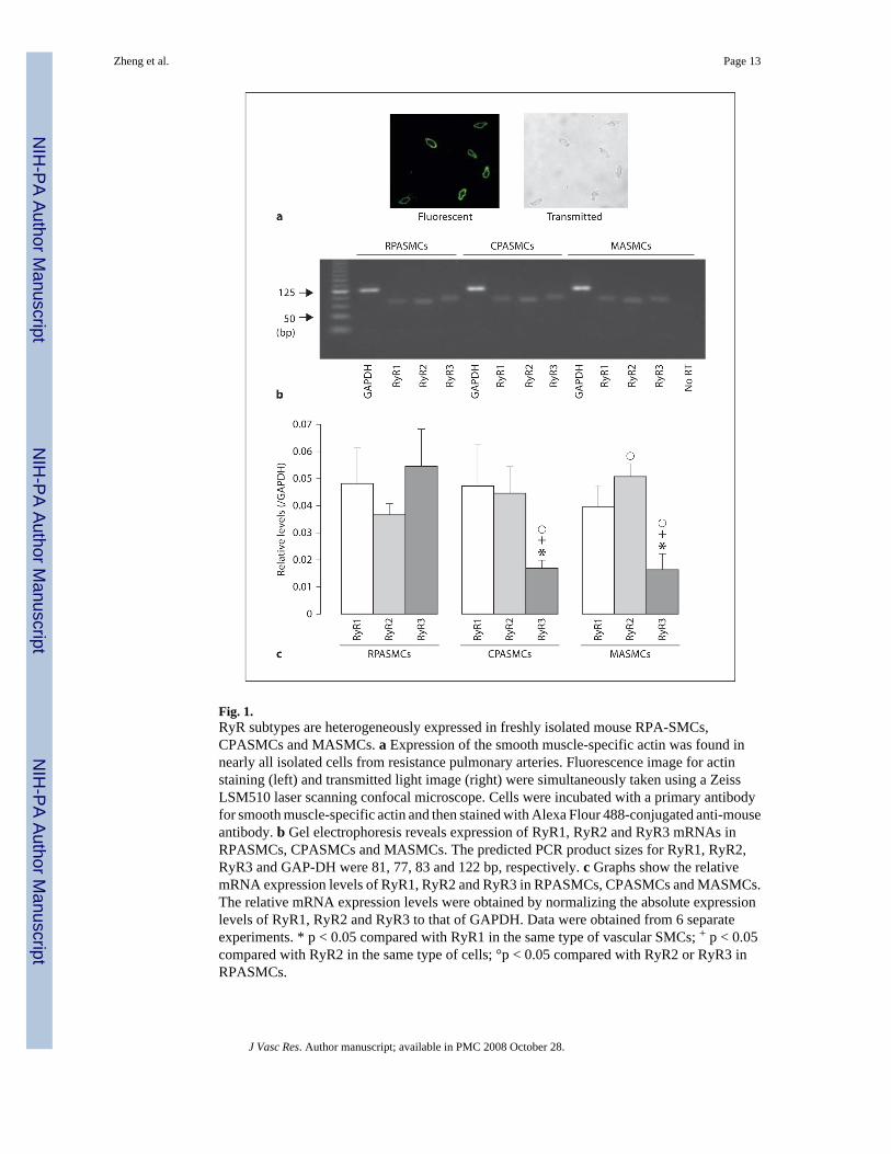
Fig. 1.RyR subtypes are heterogeneously expressed in freshly isolated mouse RPA-SMCs,CPASMCs and MASMCs. a Expression of the smooth muscle-specific actin was found innearly all isolated cells from resistance pulmonary arteries. Fluorescence image for actinstaining (left) and transmitted light image (right) were simultaneously taken using a ZeissLSM510 laser scanning confocal microscope. Cells were incubated with a primary antibodyfor smooth muscle-specific actin and then stained with Alexa Flour 488-conjugated anti-mouseantibody. b Gel electrophoresis reveals expression of RyR1, RyR2 and RyR3 mRNAs inRPASMCs, CPASMCs and MASMCs. The predicted PCR product sizes for RyR1, RyR2,RyR3 and GAP-DH were 81, 77, 83 and 122 bp, respectively. c Graphs show the relativemRNA expression levels of RyR1, RyR2 and RyR3 in RPASMCs, CPASMCs and MASMCs.The relative mRNA expression levels were obtained by normalizing the absolute expressionlevels of RyR1, RyR2 and RyR3 to that of GAPDH. Data were obtained from 6 separateexperiments. * p < 0.05 compared with RyR1 in the same type of vascular SMCs; + p < 0.05compared with RyR2 in the same type of cells; °p < 0.05 compared with RyR2 or RyR3 inRPASMCs.
Zheng et al. Page 13
J Vasc Res. Author manuscript; available in PMC 2008 October 28.
NIH
-PA Author Manuscript
NIH
-PA Author Manuscript
NIH
-PA Author Manuscript
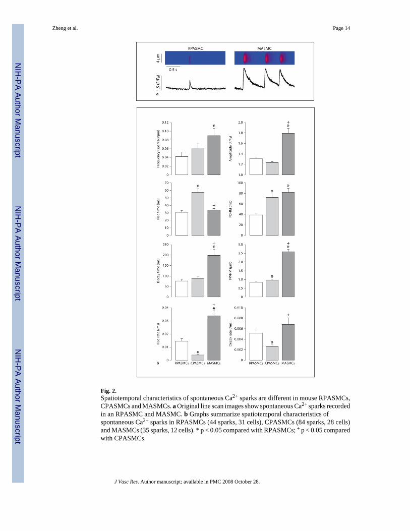
Fig. 2.Spatiotemporal characteristics of spontaneous Ca2+ sparks are different in mouse RPASMCs,CPASMCs and MASMCs. a Original line scan images show spontaneous Ca2+ sparks recordedin an RPASMC and MASMC. b Graphs summarize spatiotemporal characteristics ofspontaneous Ca2+ sparks in RPASMCs (44 sparks, 31 cells), CPASMCs (84 sparks, 28 cells)and MASMCs (35 sparks, 12 cells). * p < 0.05 compared with RPASMCs; + p < 0.05 comparedwith CPASMCs.
Zheng et al. Page 14
J Vasc Res. Author manuscript; available in PMC 2008 October 28.
NIH
-PA Author Manuscript
NIH
-PA Author Manuscript
NIH
-PA Author Manuscript
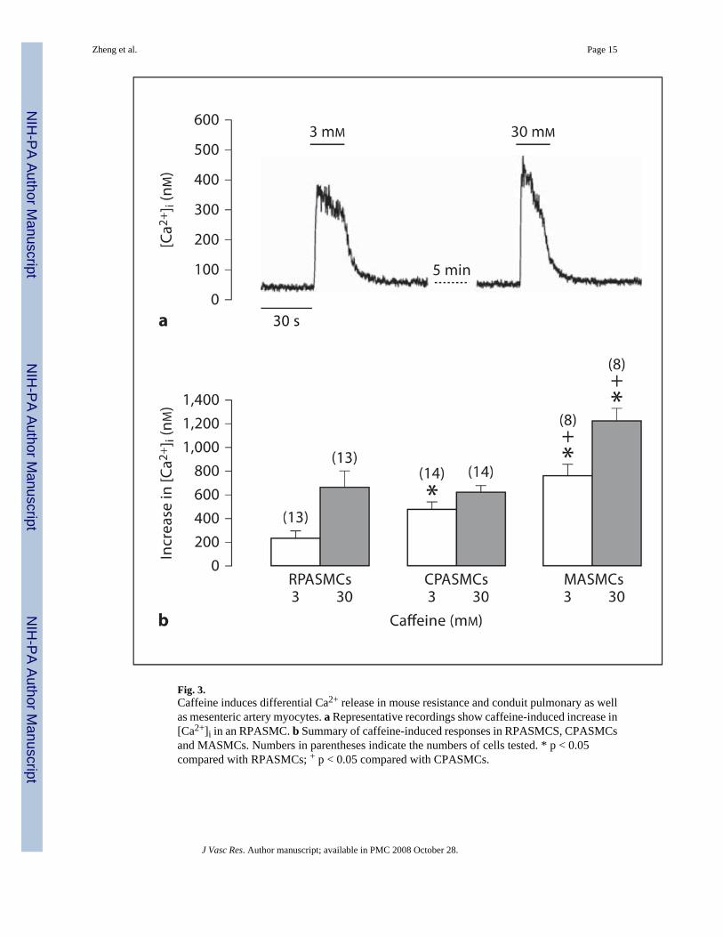
Fig. 3.Caffeine induces differential Ca2+ release in mouse resistance and conduit pulmonary as wellas mesenteric artery myocytes. a Representative recordings show caffeine-induced increase in[Ca2+]i in an RPASMC. b Summary of caffeine-induced responses in RPASMCS, CPASMCsand MASMCs. Numbers in parentheses indicate the numbers of cells tested. * p < 0.05compared with RPASMCs; + p < 0.05 compared with CPASMCs.
Zheng et al. Page 15
J Vasc Res. Author manuscript; available in PMC 2008 October 28.
NIH
-PA Author Manuscript
NIH
-PA Author Manuscript
NIH
-PA Author Manuscript
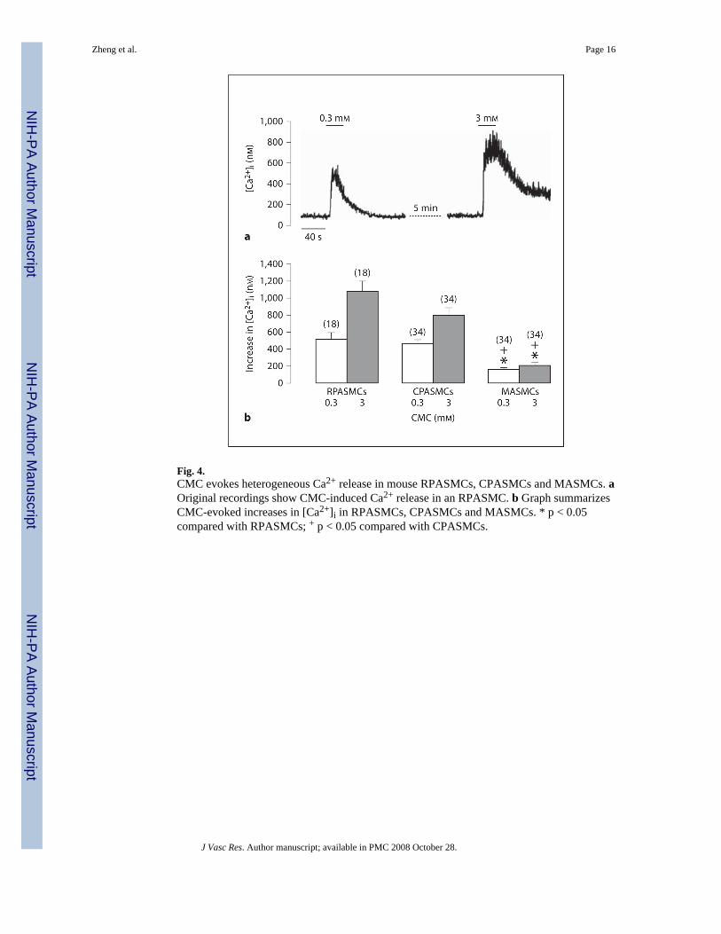
Fig. 4.CMC evokes heterogeneous Ca2+ release in mouse RPASMCs, CPASMCs and MASMCs. aOriginal recordings show CMC-induced Ca2+ release in an RPASMC. b Graph summarizesCMC-evoked increases in [Ca2+]i in RPASMCs, CPASMCs and MASMCs. * p < 0.05compared with RPASMCs; + p < 0.05 compared with CPASMCs.
Zheng et al. Page 16
J Vasc Res. Author manuscript; available in PMC 2008 October 28.
NIH
-PA Author Manuscript
NIH
-PA Author Manuscript
NIH
-PA Author Manuscript
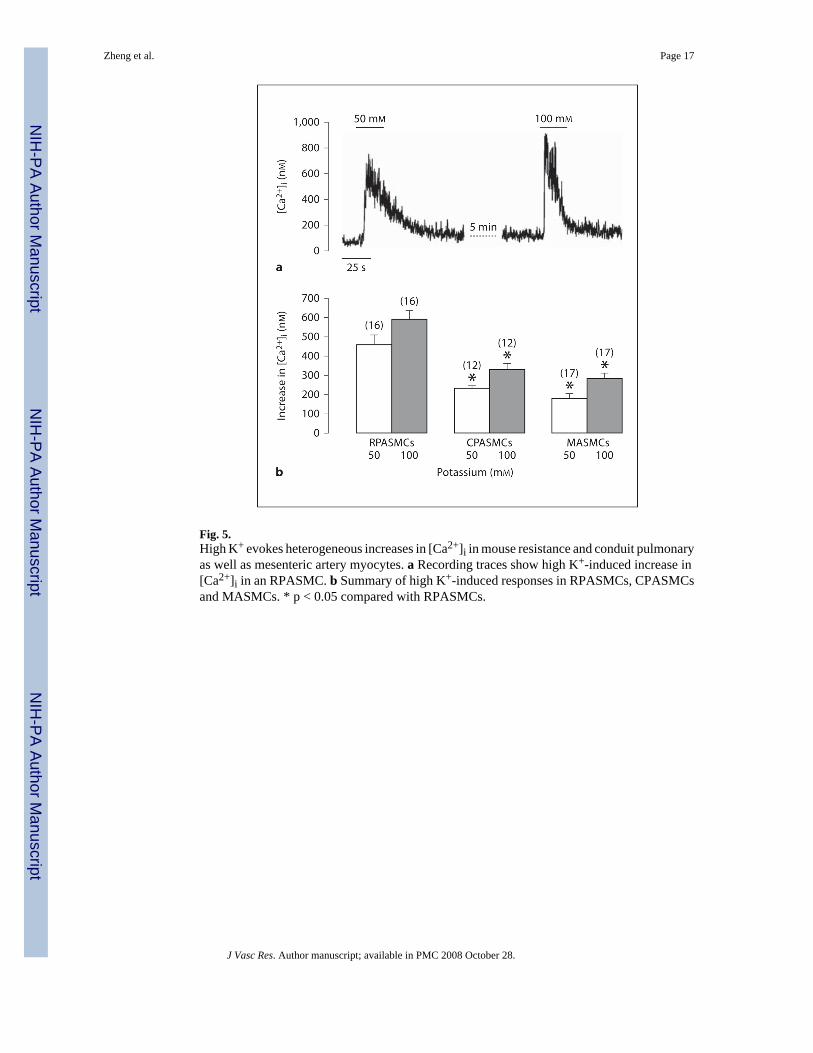
Fig. 5.High K+ evokes heterogeneous increases in [Ca2+]i in mouse resistance and conduit pulmonaryas well as mesenteric artery myocytes. a Recording traces show high K+-induced increase in[Ca2+]i in an RPASMC. b Summary of high K+-induced responses in RPASMCs, CPASMCsand MASMCs. * p < 0.05 compared with RPASMCs.
Zheng et al. Page 17
J Vasc Res. Author manuscript; available in PMC 2008 October 28.
NIH
-PA Author Manuscript
NIH
-PA Author Manuscript
NIH
-PA Author Manuscript

Fig. 6.Hypoxia-induced increases in [Ca2+]i are different in mouse RPASMCs, CPASMCs andMASMCs. a Representative traces show hypoxia-induced increase in [Ca2+]i in an RPASMCand MASMC. b Graph summarizes hypoxic Ca2+ responses in RPASMCs, CPASMCs andMASMCs. * p < 0.05 compared with RPASMCs; + p < 0.05 compared with CPASMCs.
Zheng et al. Page 18
J Vasc Res. Author manuscript; available in PMC 2008 October 28.
NIH
-PA Author Manuscript
NIH
-PA Author Manuscript
NIH
-PA Author Manuscript