Presenilin 1 interaction in the brain with a novel member of the Armadillo family
Brain (1999),122,1709–1719
The impact of different presenilin 1 andpresenilin 2 mutations on amyloid deposition,neurofibrillary changes and neuronal loss in thefamilial Alzheimer’s disease brainEvidence for other phenotype-modifying factors
Teresa Go´mez-Isla,1 Whitfield B. Growdon,1 Megan J. McNamara,1 David Nochlin,3 Thomas D. Bird,4
Juan Carlos Arango,5 Francisco Lopera,5 Kenneth S. Kosik,2 Peter L. Lantos,6 Nigel J. Cairns6 andBradley T. Hyman1
1Neurology Service, Massachusetts General Hospital, Correspondence to: Dr Teresa Go´mez-Isla, Department of2Neurology Service, Brigham and Women’s Hospital, Neurology, 420 Delaware St SE, University of Minnesota,Boston,3Department of Pathology (Neuropathology), Box 295 FUMC, Minneapolis, MN 55455, USAUniversity of Washington School of Medicine,4Neurology E-mail: [email protected], VA Medical Center and University of WashingtonSchool of Medicine, Seattle, USA,5Department ofPathology, University of Antioquı´a, Medellın, Colombiaand 6Department of Neuropathology, Institute ofPsychiatry, London, UK
SummaryTo assess the influence of the presenilin 1 (PS1) and 2(PS2) mutations on amyloid deposition, neurofibrillarytangle (NFT) formation and neuronal loss, we performedstereologically based counts in a high-order associationcortex, the superior temporal sulcus, of 30 familialAlzheimer’s disease cases carrying 10 different PS1 andPS2 mutations, 51 sporadic Alzheimer’s disease cases and33 non-demented control subjects. All the PS1 and PS2mutations assessed in this series led to enhanced depositionof total Aβ and Aβx-42/43 but not Aβx-40 senile plaques inthe superior temporal sulcus when compared with brainsfrom sporadic Alzheimer’s disease patients. Some of the
Keywords: Aβ plaques; neurofibrillary tangles; neuronal loss; presenilin; familial Alzheimer’s disease
Abbreviations: ANOVA 5 analysis of variance; APOE5 apolipoprotein E; APP5 amyloid-β protein precursor; NFT5neurofibrillary tangles; PS15 presenilin 1; PS25 presenilin 2
IntroductionAlthough the majority of cases of Alzheimer’s disease occurtypically after the age of 60–65 years, a smaller proportionof cases correspond to the early-onset (,60 years) autosomaldominant form of the disease. To date, defects in threegenes—the amyloid-β protein precursor (APP) gene onchromosome 21, the presenilin 1 (PS1) gene on chromosome14 and the presenilin 2 (PS2) gene on chromosome 1—have
© Oxford University Press 1999
PS1 mutations studied (M139V, I143F, G209V, R269H,E280A), but not others, were also associated with fasterrates of NFT formation and accelerated neuronal loss inthe majority of the patients who harboured them whencompared with sporadic Alzheimer’s disease patients. Inaddition, our analysis showed that dramatic quantitativedifferences in clinical and neuropathological features canexist even among family members with the identical PSmutation. This suggests that further individual or pedigreegenetic or epigenetic factors are likely to modulate PSphenotypes strongly.
been identified as the cause of a large proportion of early-onset familial Alzheimer’s disease (Goateet al., 1991; Levy-Lahadet al., 1995; Rogaevet al., 1995; Sherringtonet al.,1995). Six different mutations, all of them either within orclosely proximal to the Aβ domain of the gene, have beenfound in ~20 familial Alzheimer’s disease kindreds (Levyet al., 1990; Chartier-Harlinet al., 1991; Goateet al., 1991;
1710 T. Gomez-Islaet al.
Murrell et al., 1991; Hendrikset al., 1992; Mullanet al.,1992). However, Alzheimer’s disease families carrying APPmutations appear to account for fewer than 3% of allpublished cases of familial Alzheimer’s disease (Tanziet al.,1992). While it seems that mutations in the PS genesmay account for a large proportion of early-onset familialAlzheimer’s disease cases, the percentages that can beprecisely attributed to each gene remain unclear. Over 40different mutations have been described to date in the PS1gene that are associated with familial Alzheimer’s disease inmultiple kindreds of diverse ethnic origin, and two additionalmutations have been found in the PS2 gene, which mostlyaffect kindreds of Volga German ancestry (for review seeHardy, 1997).
The normal biological role of the APP and PS genes andthe mechanisms by which mutations in these genes lead toan Alzheimer’s disease phenotype remain unknown. It iswell established, however, that these causative gene defectsshare some phenotypic features: (i) they are all associatedwith early age of dementia onset, with an average age of 50years for APP mutations (range 45–60 years), 45 years forPS1 mutations (range 29–56 years) and 52 years for PS2mutations (range 40–85 years); (ii) the penetrance of thesemutations is nearly 100% (Rossoret al., 1996); and (iii) theyall alter, probably by different mechanisms, the normalprocessing of APP, leading to an increase in plasma andfibroblasts of a 42–43 amino acid form of Aβ (Aβx-42/43)(Younkin, 1995; Scheuneret al., 1996; Tomitaet al., 1997).Aβx-42/43 is known to be the earliest and most abundantspecies of Aβ in neuritic plaques (Mannet al., 1996), aswell as the most amyloidogenicin vitro (Jarrettet al., 1993).
Some neuropathological studies have shown that in APPand PS1 mutant brains there is a significant enhancement ofAβ deposition with a disproportionate increase of Aβx-42/43
species compared with sporadic Alzheimer’s disease brains(Iwatsuboet al., 1994; Tamaokaet al., 1994; Lemereet al.,1996; Mannet al., 1996; Go´mez-Isla et al., 1997b; Ishiiet al., 1997). To date, the only study in which amyloiddeposition in Alzheimer’s disease brains from individualscarrying PS2 mutations was addressed failed to show anysignificant increase of either total Aβ or Aβx-42/43 in thefrontal lobe of these brains compared with sporadicAlzheimer’s disease cases (Mannet al., 1997). The influencethat all these gene defects might have on the formation ofneurofibrillary tangles (NFT) and neuronal loss continues tobe unknown. Furthermore, the possibility that different APPand PS mutations might lead to unique neuropathologicalphenotypes remains unexplored.
To further address these issues we have performed detailedquantitative studies of Aβ deposits, rates of neurofibrillarytangle accumulation and neuronal loss in an associationcortex of brains from individuals harbouring 10 differentPS1 and PS2 mutations. The specific questions we addressedwere: (i) do PS1 and PS2 mutations lead to increased amountsof amyloid deposition, NFT formation or loss of neuronscompared with sporadic Alzheimer’s disease? and (ii) if
so, do all the PS mutations share the same pathologicalconsequences or can mutation-specific phenotypes beidentified?
The results of this study provide evidence of a significantincrease in total Aβ and Aβx-42/43 deposits in all the PS1 andPS2 mutant cases assessed and a unique effect of some ofthe PS1 mutations on the neurofibrillary pathology and ratesof neuronal loss in the Alzheimer’s disease brain.
Material and methodsWe had access to brains from 23 members of 11 familieswith early-onset familial Alzheimer’s disease carrying ninedifferent PS1 mutations, and seven members of six VolgaGerman families harbouring the same PS2 mutation (Table 1).Clinical and pathological descriptions of some of these familieshave been reported elsewhere (Birdet al., 1989; Lemereet al., 1996; Foxet al., 1997; Go´mez-Isla et al., 1997b;Loperaet al., 1997). Fifty-one sporadic Alzheimer’s diseasecases were used for comparison. Quantitative assessments of34 of the sporadic Alzheimer’s disease cases have beenreported previously (Go´mez-Islaet al., 1997a; McNamaraet al., 1998). All the cases of familial and sporadicAlzheimer’s disease studied met standard neuropathologicalcriteria for definite Alzheimer’s disease (Khachaturian, 1985;Mirra et al., 1991). The control group included 33 non-demented individuals. Data on 17 of them have been publishedpreviously and are included here for comparison (Go´mez-Isla et al., 1997a).
Neuropathological studiesFor detailed neuropathological studies, frozen sections 50µm thick were obtained from formalin-fixed blocks containingthe superior temporal sulcus at the level of the lateralgeniculate body. Quantitative analyses of neuronal numbers,amyloid deposits and neurofibrillary tangles were performedin adjacent sections containing the area of interest. Sectionswere stained using the Nissl procedure for neuronal counts,monoclonal antibodies against totalβ-amyloid deposits(10D5), end-specific monoclonal antibodies to label Aβx-40
(14C2) and Aβx-42/43 (21F12) amyloid species (McNamaraet al., 1998) (courtesy of Dr Dale Schenk, AthenaNeurosciences, South San Francisco, Calif., USA) andmonoclonal antibodies against paired helical filaments(PHF-1; courtesy of Dr Peter Davies, Bronx, New York) tocount NFT. Pretreatment of sections for immunostaining wasperformed with citrate buffer (pH5 6.0), heating the samplesat 100°C for 10 min, followed by 70% formic acid treatmentfor 10 min. Data were recorded using a Bioquant ImageAnalysis System (Nashville, Tenn., USA). The totalpercentage of cortex covered by senile plaques or amyloidburden in the superior temporal sulcus region and the ratiobetween the amounts of Aβx-40 and Aβx-42/43 deposits werecalculated. In each field, manual editing eliminated artefactsand staining associated with blood vessels. A detailed study
Quantitative neuropathology in presenilin brains 1711
Table 1 Demographics of familial Alzheimer’s disease (FAD), sporadic Alzheimer’s disease (SAD) and control cases
No. of patients PS mutation Age at death (years) Age at onset (years) Duration (years)
FAD PS12 E120D 56.56 7.8 43.56 13.4 136 5.71 E120K 54 42 122 M139V 59.56 24.7 526 24.04 7.56 0.71 I143F 57 51 65 G209V 486 5.5 426 4.7 6 6 3.12 A260V 57.56 3.5 39.56 2.1 186 1.41 R269H 56 47 91 E280A 50 45 55 E280A 54.46 8.4 46.86 7.1 7.66 2.12 E280A 636 1.4 55 86 1.41 Splice acceptor site 54 42 12
mutationTotal PS-1 Mean6 SD Mean6 SD Mean6 SD23 54.46 8.3 45.76 8.3 8.86 4.3
FAD PS22 N141I 76.56 2.1 66.56 2.1 101 N141I 74 56 181 N141I 59 49 101 N141I 69 62 71 N141I 72 54 181 N141I 80 58 22
Total PS2 Mean6 SD Mean6 SD Mean6 SD
7 72.46 6.9 58.96 6.6 13.66 5.7
SAD 51 80.16 9.3 71.86 12.8 8.36 5.4Controls 33 74.56 16.6 – –
of amyloid angiopathy in some of the familial Alzheimer’sdisease cases carrying PS2 mutations has been publishedelsewhere (Nochlinet al., 1998). Quantitative analyses ofneurons and NFT in the same region were carried out usingthe optical disector method as previously described (Go´mez-Isla et al., 1997a). In brief, the volume densities of neuronsand NFT were assessed in a reference volume measuring700µm along the pial surface by the entire depth of the greymatter located in the inferior bank of the superior temporalsulcus, ~1 cm medial to the crown of the middle temporalgyrus, by the thickness of the frozen sections (50µm). Thetotal numbers of neurons and NFT were obtained in eachbrain by multiplying the volume density obtained from thecounts by the volume of the superior temporal sulcusmeasured on each cross-section. The percentage of relativeneuronal loss in Alzheimer’s disease brains was calculatedby subtracting the number of neurons in every individualAlzheimer’s disease brain from the average number ofneurons in the control group.
Apolipoprotein E (APOE) genotype was available for themajority of cases of familial and sporadic Alzheimer’sdisease.
Statistical analysisMeasurements of total Aβ, Aβx-40 and Aβx-42/43 and theAβx-40/Aβx-42/43 ratio in PS1, PS2 and sporadic Alzheimer’sdisease cases were compared by analysis of variance
(ANOVA) at the 0.05 level of significance. Because ourprevious data suggested that the amount of NFT and theextent of neuronal loss in Alzheimer’s disease brains areclosely related to the duration of the illness (Go´mez-Islaet al., 1997a), we calculated rates of NFT formation andneuronal loss rates by dividing the absolute numbers obtainedfrom the counts by the duration of dementia symptoms inyears. The comparison of these rates among brains from PSand sporadic Alzheimer’s disease patients was done byANOVA at the 0.05 level of significance.
ResultsThe demographic features of this series are summarized inTable 1. A total of 30 familial Alzheimer’s disease casesfrom 17 families carrying 10 different PS1 or PS2 mutations,51 sporadic Alzheimer’s disease cases and 33 controls wereincluded in this study. The average age of onset of dementiawas significantly younger in PS1 and PS2 familialAlzheimer’s disease compared with sporadic Alzheimer’sdisease (45.76 8.3 years for PS1, 58.96 6.6 years for PS2versus 71.86 12.8 years for sporadic Alzheimer’s disease,P , 0.0001), and significantly lower in PS1 than in PS2familial Alzheimer’s disease cases (P 5 0.009). The averageduration of illness was significantly longer in PS2 familialAlzheimer’s disease than in PS1 familial and sporadicAlzheimer’s disease (13.66 5.7 years for PS2 versus 8.86
1712 T. Gomez-Islaet al.
Table 2 Total Aβ, Aβx-40 and Aβx-42/43 and Aβx-40/Aβx-42/43 ratio in familial (FAD) and sporadic (SAD) Alzheimer’s disease
Mutation Total Aβ (10D5)% Aβx-40 (14C2)% Aβx-42/43 (21F12)% Ratio Aβx-40/Aβx-42/43
FAD PS1 (n 5 23)2 E120D 10.56 2.3 1.26 0.8 8.96 1 0.136 0.071 E120K 12.9 5.6 11.1 0.512 M139V 14.16 5.6 7 6 1.9 12.66 7.4 0.626 0.211 I143F N/A 2 17.5 0.125 G209V 136 3.3 2.56 1.6 16.66 4.9 0.206 0.112 A260V 10.86 0.9 0.56 0.3 16.9 0.041 R269H 14.8 4 12.8 0.318 E280A 13.56 5.5 1.96 1.3 9.36 3.2 0.266 0.071 Splice acceptor site 22.3 3.6 21.4 0.17
mutationMean6 SD Mean6 SD Mean6 SD Mean6 SD13.3 6 4.4* 2.6 6 2.1 12.96 5* 0.26 6 0.18*
FAD PS2 (n 5 7) N141I 14.96 3.3* 1.3 6 1.4 11.66 5.2* 0.126 0.09*SAD (n 5 51) 8.26 3.1 3.36 2.2† 7.03 6 3† 0.46 6 0.24†
*P , 0.05 (total Aβ, Aβx-42/43 and Aβx-40/Aβx-42/43 ratio in PS1 and PS2 familial Alzheimer’s disease versus sporadic Alzheimer’sdisease).†(McNamaraet al., 1998).
Table 3 Total NFT and neuronal counts and rates of NFT accumulation and neuronal loss in familial (FAD) and sporadic(SAD) Alzheimer’s disease
Mutation NFT (103) Neurons (104) Rate of NFT formation Rate of neuron loss(103/year) (104/year)
FAD PS1 (n 5 23)2 E120D 13.36 2.2 6.366 1.70 1.086 0.30 0.236 0.031 E120K 10.4 5.15 0.81 0.332 M139V 13.96 3.6 7.886 2.96 1.896 0.66** 0.23 6 0.411 I143F 13.1 5.30 2.19** 0.695 G209V 10.36 2.5 4.696 0.91 2.876 2.66** 1.07 6 0.592 A260V 6.16 2.1 1.986 0.83 0.336 0.10 0.426 0.081 R269H 11.4 2.68 1.27** 0.758 E280A 7.66 3.7 4.956 1.81 1.536 1.50** 0.84 6 0.581 Splice acceptor site 7.8 5.63 0.65 0.32
mutationMean6 SD Mean6 SD Mean6 SD Mean6 SD9.6 6 3.6 4.986 1.98* 1.616 1.54** 0.67 6 0.51
FAD PS2 (n 5 7) N141I 7.36 4.2 4.456 1.07* 0.706 0.25 0.506 0.19SAD (n 5 51) 7.46 4.6 4.826 2.20* 0.896 0.43 0.916 0.13Controls (n 5 33) – 9.426 1.06 – –
P 5 0.06 (total NFT number in PS1 familial Alzheimer’s disease versus sporadic Alzheimer’s disease); *P , 0.05 (neurons in familialAlzheimer’s disease and sporadic Alzheimer’s disease versus controls); **P , 0.01 (rate of NFT/year in PS1 versus sporadicAlzheimer’s disease).
4.3 years for PS1 and 8.36 5.4 years for sporadic Alzheimer’sdisease,P , 0.05).
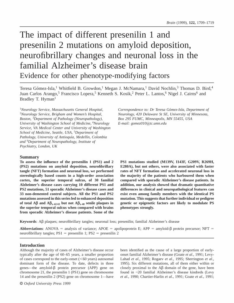
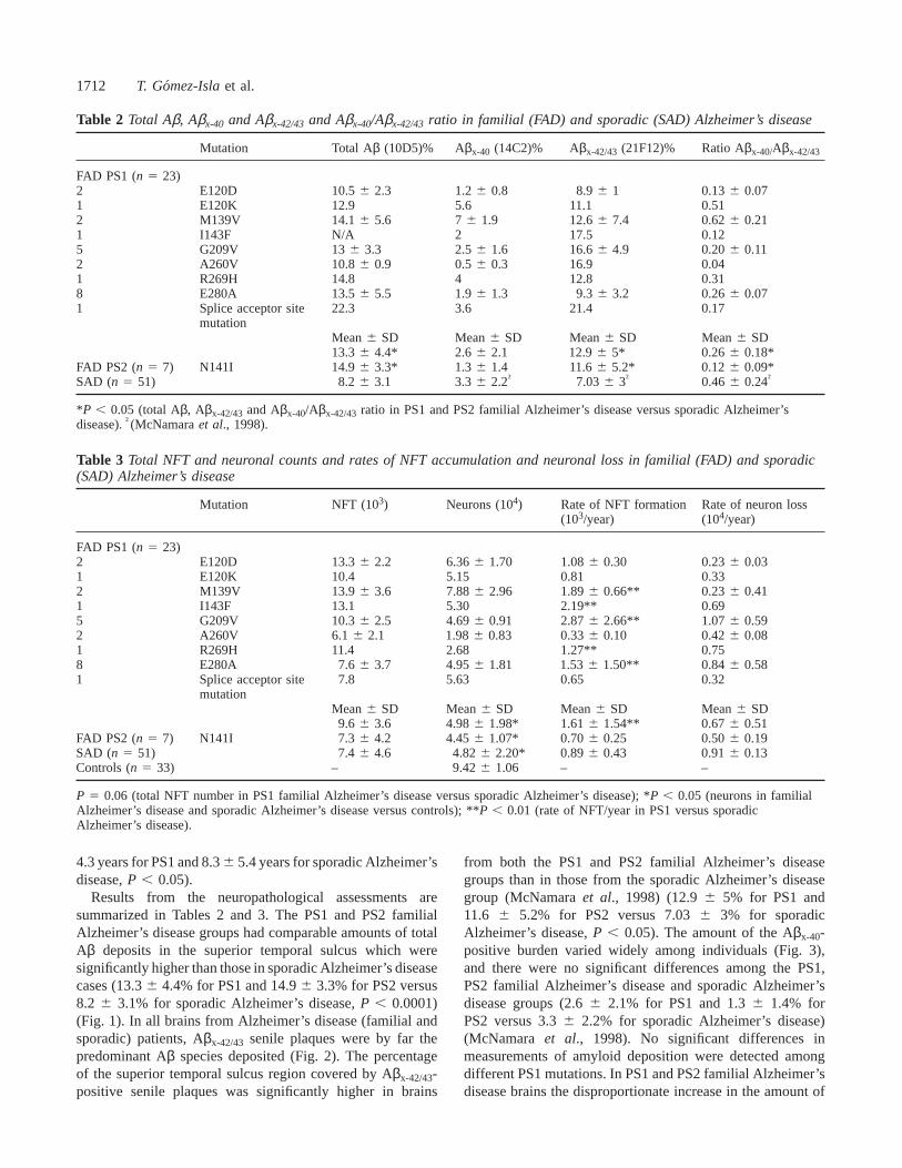
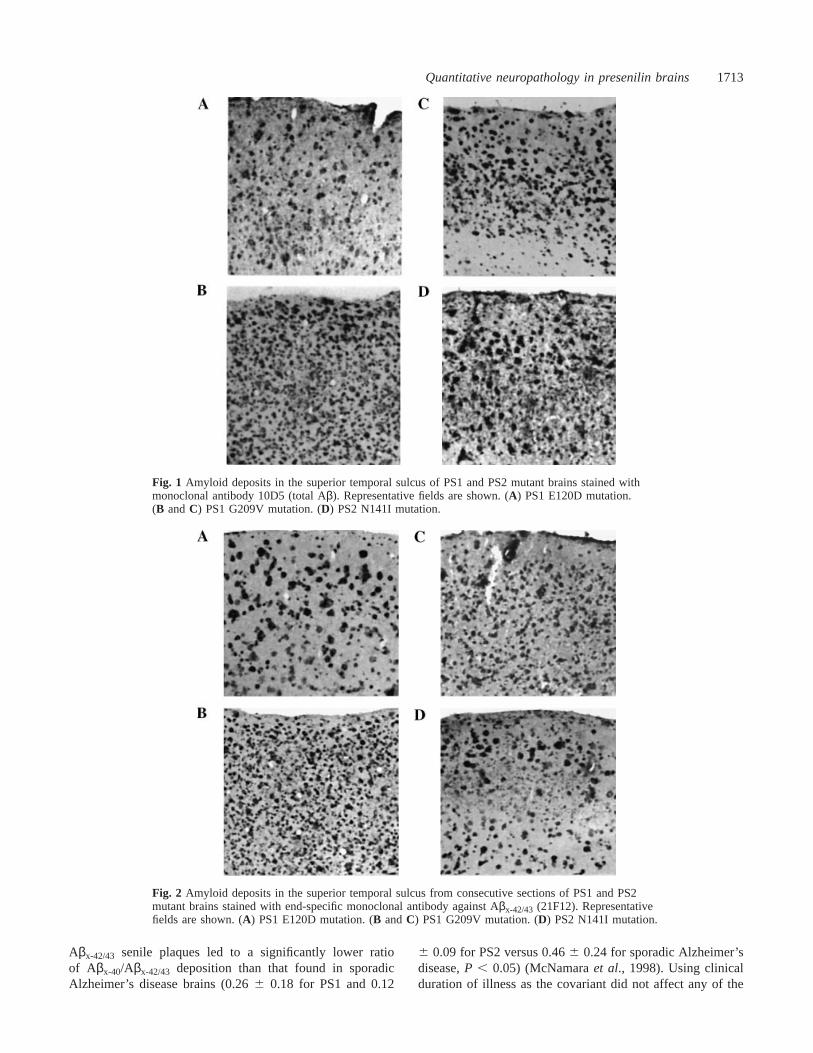
Results from the neuropathological assessments aresummarized in Tables 2 and 3. The PS1 and PS2 familialAlzheimer’s disease groups had comparable amounts of totalAβ deposits in the superior temporal sulcus which weresignificantly higher than those in sporadic Alzheimer’s diseasecases (13.36 4.4% for PS1 and 14.96 3.3% for PS2 versus8.2 6 3.1% for sporadic Alzheimer’s disease,P , 0.0001)(Fig. 1). In all brains from Alzheimer’s disease (familial andsporadic) patients, Aβx-42/43 senile plaques were by far thepredominant Aβ species deposited (Fig. 2). The percentageof the superior temporal sulcus region covered by Aβx-42/43-positive senile plaques was significantly higher in brains
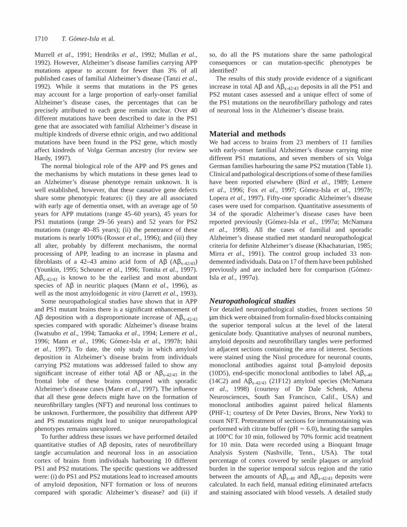
from both the PS1 and PS2 familial Alzheimer’s diseasegroups than in those from the sporadic Alzheimer’s diseasegroup (McNamaraet al., 1998) (12.96 5% for PS1 and11.6 6 5.2% for PS2 versus 7.036 3% for sporadicAlzheimer’s disease,P , 0.05). The amount of the Aβx-40-positive burden varied widely among individuals (Fig. 3),and there were no significant differences among the PS1,PS2 familial Alzheimer’s disease and sporadic Alzheimer’sdisease groups (2.66 2.1% for PS1 and 1.36 1.4% forPS2 versus 3.36 2.2% for sporadic Alzheimer’s disease)(McNamara et al., 1998). No significant differences inmeasurements of amyloid deposition were detected amongdifferent PS1 mutations. In PS1 and PS2 familial Alzheimer’sdisease brains the disproportionate increase in the amount of
Quantitative neuropathology in presenilin brains 1713
Fig. 1 Amyloid deposits in the superior temporal sulcus of PS1 and PS2 mutant brains stained withmonoclonal antibody 10D5 (total Aβ). Representative fields are shown. (A) PS1 E120D mutation.(B andC) PS1 G209V mutation. (D) PS2 N141I mutation.
Fig. 2 Amyloid deposits in the superior temporal sulcus from consecutive sections of PS1 and PS2mutant brains stained with end-specific monoclonal antibody against Aβx-42/43 (21F12). Representativefields are shown. (A) PS1 E120D mutation. (B andC) PS1 G209V mutation. (D) PS2 N141I mutation.
Aβx-42/43 senile plaques led to a significantly lower ratioof Aβx-40/Aβx-42/43 deposition than that found in sporadicAlzheimer’s disease brains (0.266 0.18 for PS1 and 0.12
6 0.09 for PS2 versus 0.466 0.24 for sporadic Alzheimer’sdisease,P , 0.05) (McNamaraet al., 1998). Using clinicalduration of illness as the covariant did not affect any of the
1714 T. Gomez-Islaet al.
Fig. 3 Amyloid deposits in the superior temporal sulcus from consecutive sections of PS1 and PS2mutant brains stained with end-specific monoclonal antibody against Aβx-40 (14C2). Representativefields are shown. (A) PS1 E120D mutation. (B andC) PS1 G209V mutation. (D) PS2 N141I mutation.
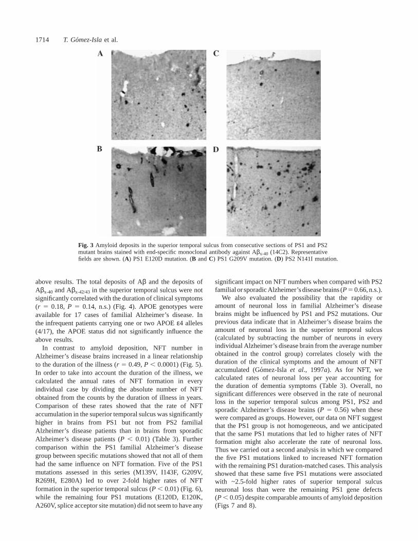
above results. The total deposits of Aβ and the deposits ofAβx-40 and Aβx-42/43 in the superior temporal sulcus were notsignificantly correlated with the duration of clinical symptoms(r 5 0.18, P 5 0.14, n.s.) (Fig. 4). APOE genotypes wereavailable for 17 cases of familial Alzheimer’s disease. Inthe infrequent patients carrying one or two APOEε4 alleles(4/17), the APOE status did not significantly influence theabove results.
In contrast to amyloid deposition, NFT number inAlzheimer’s disease brains increased in a linear relationshipto the duration of the illness (r 5 0.49,P , 0.0001) (Fig. 5).In order to take into account the duration of the illness, wecalculated the annual rates of NFT formation in everyindividual case by dividing the absolute number of NFTobtained from the counts by the duration of illness in years.Comparison of these rates showed that the rate of NFTaccumulation in the superior temporal sulcus was significantlyhigher in brains from PS1 but not from PS2 familialAlzheimer’s disease patients than in brains from sporadicAlzheimer’s disease patients (P , 0.01) (Table 3). Furthercomparison within the PS1 familial Alzheimer’s diseasegroup between specific mutations showed that not all of themhad the same influence on NFT formation. Five of the PS1mutations assessed in this series (M139V, I143F, G209V,R269H, E280A) led to over 2-fold higher rates of NFTformation in the superior temporal sulcus (P , 0.01) (Fig. 6),while the remaining four PS1 mutations (E120D, E120K,A260V, splice acceptor site mutation) did not seem to have any
significant impact on NFT numbers when compared with PS2familial or sporadic Alzheimer’s disease brains (P5 0.66, n.s.).
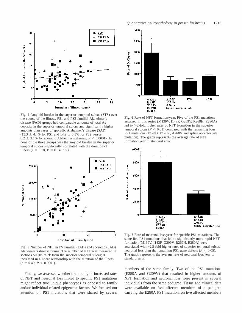
We also evaluated the possibility that the rapidity oramount of neuronal loss in familial Alzheimer’s diseasebrains might be influenced by PS1 and PS2 mutations. Ourprevious data indicate that in Alzheimer’s disease brains theamount of neuronal loss in the superior temporal sulcus(calculated by subtracting the number of neurons in everyindividual Alzheimer’s disease brain from the average numberobtained in the control group) correlates closely with theduration of the clinical symptoms and the amount of NFTaccumulated (Go´mez-Isla et al., 1997a). As for NFT, wecalculated rates of neuronal loss per year accounting forthe duration of dementia symptoms (Table 3). Overall, nosignificant differences were observed in the rate of neuronalloss in the superior temporal sulcus among PS1, PS2 andsporadic Alzheimer’s disease brains (P 5 0.56) when thesewere compared as groups. However, our data on NFT suggestthat the PS1 group is not homogeneous, and we anticipatedthat the same PS1 mutations that led to higher rates of NFTformation might also accelerate the rate of neuronal loss.Thus we carried out a second analysis in which we comparedthe five PS1 mutations linked to increased NFT formationwith the remaining PS1 duration-matched cases. This analysisshowed that these same five PS1 mutations were associatedwith ~2.5-fold higher rates of superior temporal sulcusneuronal loss than were the remaining PS1 gene defects(P , 0.05) despite comparable amounts of amyloid deposition(Figs 7 and 8).
Quantitative neuropathology in presenilin brains 1715
Fig. 4 Amyloid burden in the superior temporal sulcus (STS) overthe course of the illness. PS1 and PS2 familial Alzheimer’sdisease (FAD) groups had comparable amounts of total Aβdeposits in the superior temporal sulcus and significantly higheramounts than cases of sporadic Alzheimer’s disease (SAD)(13.3 6 4.4% for PS1 and 14.96 3.3% for PS2 versus8.2 6 3.1% for sporadic Alzheimer’s disease,P , 0.0001). Innone of the three groups was the amyloid burden in the superiortemporal sulcus significantly correlated with the duration ofillness (r 5 0.18,P 5 0.14, n.s.).
Fig. 5 Number of NFT in PS familial (FAD) and sporadic (SAD)Alzheimer’s disease brains. The number of NFT was measured insections 50µm thick from the superior temporal sulcus; itincreased in a linear relationship with the duration of the illness(r 5 0.49,P , 0.0001).
Finally, we assessed whether the finding of increased ratesof NFT and neuronal loss linked to specific PS1 mutationsmight reflect true unique phenotypes as opposed to familyand/or individual-related epigenetic factors. We focused ourattention on PS1 mutations that were shared by several
Fig. 6 Rate of NFT formation/year. Five of the PS1 mutationsassessed in this series (M139V, I143F, G209V, R269H, E280A)led to .2-fold higher rates of NFT formation in the superiortemporal sulcus (P , 0.01) compared with the remaining fourPS1 mutations (E120D, E120K, A260V and splice acceptor sitemutation). The graph represents the average rate of NFTformation/year6 standard error.
Fig. 7 Rate of neuronal loss/year for specific PS1 mutations. Thesame five PS1 mutations that led to significantly more rapid NFTformation (M139V, I143F, G209V, R269H, E280A) wereassociated with ~2.5-fold higher rates of superior temporal sulcusneuronal loss than the remaining PS1 gene defects (P , 0.05).The graph represents the average rate of neuronal loss/year6standard error.
members of the same family. Two of the PS1 mutations(E280A and G209V) that resulted in higher amounts ofNFT formation and neuronal loss were present in severalindividuals from the same pedigree. Tissue and clinical datawere available on five affected members of a pedigreecarrying the E280A PS1 mutation, on five affected members
1716 T. Gomez-Islaet al.
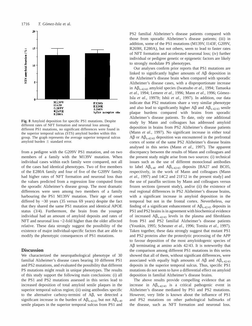
Fig. 8 Amyloid deposition for specific PS1 mutations. Despitedifferent rates of NFT formation and neuronal loss amongdifferent PS1 mutations, no significant differences were found inthe superior temporal sulcus (STS) amyloid burden within thisgroup. The graph represents the average superior temporal sulcusamyloid burden6 standard error.
from a pedigree with the G209V PS1 mutation, and on twomembers of a family with the M139V mutation. Whenindividual cases within each family were compared, not allof the cases had identical phenotypes. Two of five membersof the E280A family and four of five of the G209V familyhad higher rates of NFT formation and neuronal loss thanthe values predicted from a regression line computed fromthe sporadic Alzheimer’s disease group. The most dramaticdifferences were seen among two members of a familyharbouring the PS1 M139V mutation. Their age of onsetdiffered by ~30 years (35 versus 69 years) despite the factthat they shared the same PS1 mutation and identical APOEstatus (3/4). Furthermore, the brain from the youngerindividual had an amount of amyloid deposits and rates ofNFT and neuronal loss ~2-fold higher than the older affectedrelative. These data strongly suggest the possibility of theexistence of major individual-specific factors that are able tomodify the phenotypic consequences of PS1 mutations.
DiscussionWe characterized the neuropathological phenotype of 30familial Alzheimer’s disease cases bearing 10 different PS1and PS2 mutations, and evaluated the possibility that differentPS mutations might result in unique phenotypes. The resultsof this study support the following main conclusions: (i) allthe PS1 and PS2 mutations assessed in this series lead toincreased deposition of total amyloid senile plaques in thesuperior temporal sulcus region; (ii) using antibodies specificto the alternative carboxy-termini of Aβ, we detected asignificant increase in the burden of Aβx-42/43 but not Aβx-40
senile plaques in the superior temporal sulcus from PS1 and
PS2 familial Alzheimer’s disease patients compared withthose from sporadic Alzheimer’s disease patients; (iii) inaddition, some of the PS1 mutations (M139V, I143F, G209V,R269H, E280A), but not others, seem to lead to faster ratesof NFT formation and accelerated neuronal loss; (iv) furtherindividual or pedigree genetic or epigenetic factors are likelyto strongly modulate PS phenotypes.
Our analyses confirm prior reports that PS1 mutations arelinked to significantly higher amounts of Aβ deposition inthe Alzheimer’s disease brain when compared with sporadicAlzheimer’s disease cases, with a disproportionate increasein Aβx-42/43 amyloid species (Iwatsuboet al., 1994; Tamaokaet al., 1994; Lemereet al., 1996; Mannet al., 1996; Go´mez-Isla et al., 1997b; Ishii et al., 1997). In addition, our dataindicate that PS2 mutations share a very similar phenotypeand also lead to significantly higher Aβ and Aβx-42/43 senileplaque burdens compared with brains from sporadicAlzheimer’s disease patients. To date, only one additionalstudy by Mann and colleagues has addressed amyloiddeposition in brains from PS2 Alzheimer’s disease patients(Mann et al., 1997). No significant increase in either totalAβ or Aβx-42/43 deposition was encountered in the prefrontalcortex of some of the same PS2 Alzheimer’s disease brainsanalysed in this series (Mannet al., 1997). The apparentdiscrepancy between the results of Mann and colleagues andthe present study might arise from two sources: (i) technicalissues such as the use of different monoclonal antibodiesto label Aβx-40 and Aβx-42/43 deposits [BA27 and BC05,respectively, in the work of Mann and colleagues (Mannet al., 1997) and 14C2 and 21F12 in the present study] andthe use of paraffin sections by Mann and colleagues versusfrozen sections (present study), and/or (ii) the existence ofreal regional differences in PS2 Alzheimer’s disease brains,with a significant increase in amyloid deposition in thetemporal but not in the frontal cortex. Nevertheless, ourfinding of a significant enhancement of Aβx-42/43 deposits inPS1 and PS2 brains is in agreement with biochemical evidenceof increased Aβx-42/43 levels in the plasma and fibroblastsfrom PS1 and PS2 familial Alzheimer’s disease patients(Younkin, 1995; Scheuneret al., 1996; Tomitaet al., 1997).Taken together, these data strongly suggest that mutant PS1and PS2 proteins alter the proteolytic processing of the APPto favour deposition of the most amyloidogenic species ofAβ terminating at amino acids 42/43. It is noteworthy thatthe comparison among different PS1 mutations in this seriesshowed that all of them, without significant differences, wereassociated with equally high amounts of Aβ and Aβx-42/43
deposits in the superior temporal sulcus. Thus, specific PS1mutations do not seem to have a differential effect on amyloiddeposition in familial Alzheimer’s disease brains.
The above results provide compelling evidence that anincrease in Aβx-42/43 is a critical pathogenic event inAlzheimer’s disease mediated by PS1 and PS2 mutations.However, very little is known about the influence of PS1and PS2 mutations on other pathological hallmarks ofthe disease, such as NFT formation and neuronal loss.
Quantitative neuropathology in presenilin brains 1717
Some data indicate that PS mutations might also affect theselatter Alzheimer’s disease lesions. We observed previouslythat the R269H PS1 mutation was associated with increasedneurofibrillary pathology in a single case (Go´mez-Islaet al.,1997b). We had reasoned that if the changes observed in thatcase proved to be typical of PS mutations, then such PSmutations may affect both amyloid deposition andintraneuronal cytoskeletal changes in early-onset familialAlzheimer’s disease. Furthermore, recentin vitro studiesimplicate PS1 and PS2 genes in cell death. Wolozinet al. havereported that the overexpression of the familial Alzheimer’sdisease PS2 gene in PC12 cells increases their susceptibilityto apoptosis induced by trophic factor withdrawal or beta-amyloid (Wolozin et al., 1996). This finding has recentlybeen replicated by Guo and colleagues by expressing thePS1 L286V mutation in PC12 cells (Guoet al., 1997). Inaddition, an impairment in neurogenesis and massive neuronalloss in specific subregions have been found in PS1-deficientmice, further reinforcing the idea that PS1 is required fornormal neurogenesis and neuronal survival (Shenet al.,1997). If this is the case, PS mutant proteins might wellparticipate in or accelerate the loss of neurons in familialAlzheimer’s disease brains.
Our data, however, do not support a stereotypical influenceof PS1 or PS2 mutations on either NFT formation or neuronalloss. Some individuals, and indeed some PS1 mutations, areassociated with increased NFT formation and neuronal lossbut others are not. While these differences certainly contributeto the variability in clinical phenotype among differentmutations, this cannot be considered to be central to theeffect of mutant PS1 in causing Alzheimer’s disease.
Fox and colleagues compared the clinicopathologicalfeatures of 16 familial Alzheimer’s disease patients from twoapparently unrelated families in England bearing the M139Vmutation (Foxet al., 1997). The most important phenotypicdifference between the two families in the study of Fox andcolleagues was the significantly younger age of onset in oneof the families, which could not be accounted for by APOEstatus. The possible implication of a further pedigree-specificgenetic factor was raised by the authors. However, post-mortem examinations were conducted on only one affectedindividual from each family, limiting further conclusions onthe pathological phenotype of these pedigrees. Our currentneuropathological study also suggests the possibility thatindividual-specific genetic or epigenetic factors may havephenotype-modifying effects. Tissue was available from fiveaffected members of a family carrying the E280A PS1mutation and from five affected members of another familycarrying the G209V PS1 mutation. The age of onset in theE280A PS1 family was known in the five members, andranged from 39 to 54 years. The amount of amyloid in thesuperior temporal sulcus of these brains was quite similaramong individuals, but after covarying for the duration ofthe illness only two out of five had higher rates of NFTformation and neuronal loss than the values predicted froma regression line computed from the sporadic Alzheimer’s
disease group. Within the family carrying the G209V mutationthe age of onset ranged from 36 to 48 years. Four out of fivemembers of this family had higher rates of NFT accumulationand neuronal loss than the values predicted from duration-matched sporadic Alzheimer’s disease brains, but one casedid not differ from this latter group. These findings point tothe possibility of individual-specific modifying factors of aPS1 mutated gene. Perhaps the most illustrative case in thissense corresponds to two members of the same familyharbouring the PS1 M139V mutation. There was a dramaticdifference in the age of onset by.30 years despite the factthat they share not only the same PS1 mutation but also anidentical APOE status. Furthermore, in the brain of the casewith the younger age of onset we have found an increase of~2-fold in amyloid deposits and rates of NFT and neuronalloss compared with the older affected relative. Since the casewith younger onset fits more closely the average age of onsetdescribed in other members of families with this same PS1mutation, it is reasonable to suspect the presence of anunusual risk-modifying factor in the older individual.
In conclusion, all the familial Alzheimer’s disease PS1and PS2 mutations assessed in this series led to a significantincrease in the burden of Aβx-42/43, but not Aβx-40 senileplaques in the superior temporal sulcus compared with brainsfrom patients with sporadic Alzheimer’s disease. Five of thePS1 mutations studied were also associated with faster ratesof NFT formation and accelerated neuronal loss in themajority of the patients harbouring them compared withsporadic Alzheimer’s disease. This latter finding suggestseither that certain PS1 mutations have an impact on bothamyloid deposition and intraneuronal cytoskeletal changes,or that further pedigree- or individual-specific factorscontribute to these differences. Our analysis shows thatdramatic quantitative differences in clinical andneuropathological features can exist even among familymembers with the identical mutation, favouring the latterpossibility.
AcknowledgementsWe wish to thank Professor Martin N. Rossor (DementiaResearch Group, National Hospital, Queen Square, London),whose patients provided some of the material for this study.The work was supported by NIH grants AG08031, AG06786,AG05134 and AG08487 and Colciencias grant 1115–04–040–95.
ReferencesBird TD, Sumi SM, Nemens EJ, Nochlin D, Schellenberg G, LampeTH, et al. Phenotypic heterogeneity in familial Alzheimer’s disease:a study of 24 kindreds. Ann Neurol 1989; 25: 12–25.
Chartier-Harlin M, Crawford F, Houlden H, Warren A, Hughes D,Fidani L, et al. Early-onset Alzheimer’s disease caused by mutationsat codon 717 of theβ-amyloid precursor protein gene. Nature 1991;353: 844–6.

1718 T. Gomez-Islaet al.
Fox NC, Kennedy AM, Harvey RJ, Lantos PL, Roques PK,Collinge J, et al. Clinicopathological features of familial Alzheimer’sdisease associated with the M139V mutation in the presenilin 1gene. Pedigree but not mutation specific age at onset providesevidence for a further genetic factor. Brain 1997; 120: 491–501.
Goate A, Chartier-Harlin MC, Mullan M, Brown J, Crawford F,Fidani L, et al. Segregation of a missense mutation in the amyloidprecursor protein gene with familial Alzheimer’s disease [seecomments]. Nature 1991; 349: 704–9. Comment in: Nature 1991;349: 633–4, Comment in: Nature 1991; 350: 564.
Gomez-Isla T, Hollister R, West H, Mui S, Growdon JH, PetersenRC, et al. Neuronal loss correlates with but exceeds neurofibrillarytangles in Alzheimer’s disease. Ann Neurol 1997a; 41: 17–24.
Gomez-Isla T, Wasco W, Pettingell WP, Gurubhagavatula S, SchmidtSD, Jondro PD. et al. A novel presenilin-1 mutation: increased beta-amyloid and neurofibrillary changes. Ann Neurol 1997b; 41: 809–13.
Guo Q, Sopher BL, Furukawa K, Pham DG, Robinson N, MartinGM, et al. Alzheimer’s presenilin mutation sensitizes neural cellsto apoptosis induced by trophic factor withdrawal and amyloid beta-peptide: involvement of calcium and oxyradicals. J Neurosci 1997;17: 4212–22.
Hardy J. Amyloid, the presenilins and Alzheimer’s disease [seecomments]. [Review]. Trends Neurosci 1997; 20: 154–9. Commentin: Trends Neurosci 1997; 20: 558–9.
Hendriks L, van Duijn CM, Cras P, Cruts M, Van Hul W, vanHarskamp F, et al. Presenile dementia and cerebral haemorrhagelinked to a mutation at codon 692 of theβ-amyloid precursorprotein gene. Nat Genet 1992; 1: 218–21.
Ishii K, Ii K, Hasegawa T, Shoji S, Doi A, Mori H. Increased Aβ42(43)-plaque deposition in early-onset familial Alzheimer’s diseasebrains with the deletion of exon 9 and the missense point mutation(H163R) in the PS-1 gene. Neurosci Lett 1997; 228: 17–20.
Iwatsubo T, Odaka A, Suzuki N, Mizusawa H, Nukina N, Ihara Y.Visualization of Aβ42(43) and Aβ40 in senile plaques with end-specific Aβ monoclonals: evidence that an initially deposited speciesis Aβ 42(43). Neuron 1994; 13: 43–53.
Jarrett JT, Berger EP, Lansbury PT Jr. The carboxy terminus of thebeta amyloid protein is critical for the seeding of amyloid formation:implications for the pathogenesis of Alzheimer’s disease.Biochemistry 1993; 32: 4693–7.
Khachaturian ZS. Diagnosis of Alzheimer’s disease. Arch Neurol1985; 42: 1097–105.
Lemere CA, Lopera F, Kosik KS, Lendon CL, Ossa J, Saido TC,et al. The E280A presenilin 1 Alzheimer mutation produces increasedA beta 42 deposition and severe cerebellar pathology. Nat Med1996; 2: 1146–50.
Levy E, Carman MD, Fernandez-Madrid IJ, Power MD,Lieberburg I, van Duinen SG, et al. Mutation of the Alzheimer’sdisease amyloid gene in hereditary cerebral hemorrhage, Dutchtype. Science 1990; 248: 1124–6.
Levy-Lahad E, Wasco W, Poorkaj P, Romano DM, Oshima J,Pettingell WH, et al. Candidate gene for the chromosome 1 familialAlzheimer’s disease locus [see comments]. Science 1995; 269: 973–7. Comment in: Science 1995; 269: 917–8.
Lopera F, Ardilla A, Martinez A, Madrigal L, Arango-Viana JC,Lemere CA, et al. Clinical features of early-onset Alzheimer diseasein a large kindred with an E280A presenilin-1 mutation. JAMA1997; 277: 793–9.
Mann DM, Iwatsubo T, Cairns NJ, Lantos PL, Nochlin D, SumiSM, et al. Amyloid beta protein (Abeta) deposition in chromosome14-linked Alzheimer’s disease: predominance of Abeta42(43). AnnNeurol 1996; 40: 149–56.
Mann DM, Iwatsubo T, Nochlin D, Sumi SM, Levy-Lahad E,Bird TD. Amyloid (Abeta) deposition in chromosome 1-linkedAlzheimer’s disease: the Volga German families. Ann Neurol 1997;41: 52–7.
McNamara MJ, Go´mez-Isla T, Hyman BT. Apolipoprotein Egenotype and deposits of Aβ 40/42 and Aβ42 in Alzheimer’sdisease. Arch Neurol 1998; 55: 1001–4.
Mirra SS, Heyman A, McKeel D, Sumi SM, Crain BJ, BrownleeLM, et al. The Consortium to Establish a Registry for Alzheimer’sDisease (CERAD). Part II. Standardization of the neuropathologicassessment of Alzheimer’s disease. Neurology 1991; 41: 479–86.
Mullan M, Crawford F, Axelman K, Houlden H, Lilius L, Winblad B,et al. A pathogenic mutation for probable Alzheimer’s disease inthe APP gene at the N-terminus ofβ-amyloid. Nat Genet 1992; 1:345–7.
Murrell J, Farlow M, Ghetti B, Benson MD. A mutation in theamyloid precursor protein associated with hereditary Alzheimer’sdisease. Science 1991; 254: 97–9.
Nochlin D, Bird TD, Nemens EJ, Ball MJ, Sumi SM. Amyloidangiopathy in a Volga German family with Alzheimer’s disease anda presenilin-2 mutation (N141I). Ann Neurol 1998; 43: 131–5.
Rogaev EI, Sherrington R, Rogaeva EA, Levesque G, Ikeda M,Liang Y, et al. Familial Alzheimer’s disease in kindreds withmissense mutations in a gene on chromosome 1 related to theAlzheimer’s disease type 3 gene. Nature 1995; 376: 775–8.
Rossor MN, Fox NC, Beck J, Campbell TC, Collinge J. Incompletepenetrance of familial Alzheimer’s disease in a pedigree with anovel presenilin-1 gene mutation [letter]. Lancet 1996; 347: 1560.
Scheuner D, Eckman C, Jensen M, Song X, Citron M, Suzuki N,et al. Secreted amyloid beta-protein similar to that in the senileplaques of Alzheimer’s disease is increased in vivo by the presenilin1 and 2 and APP mutations linked to familial Alzheimer’s disease[see comments]. Nat Med 1996; 2: 864–70. Comment in: Nat Med1996; 2: 850–2.
Shen J, Bronson RT, Chen DF, Xia W, Selkoe DJ, Tonegawa S.Skeletal and CNS defects in presenilin-1-deficient mice. Cell 1997;89: 629–39.
Sherrington R, Rogaev EI, Liang Y, Rogaeva EA, Levesque G,Ikeda M, et al. Cloning of a gene bearing missense mutations inearly-onset familial Alzheimer’s disease [see comments]. Nature1995; 375: 754–60. Comment in: Nature 1995; 375: 734.
Tamaoka A, Odaka A, Ishibashi Y, Usami M, Sahara N, Suzuki N,et al. APP717 mis-sense mutation affects the ratio of amyloidβprotein species (A(1–42/43 and Aβ1–40) in familial Alzheimer’sdisease brain. J Biol Chem 1994; 269: 32721–4.
Quantitative neuropathology in presenilin brains 1719
Tanzi RE, Vaula G, Romano DM, Mortilla M, Huang TL, TuplerRG, et al. Assessment of amyloidβ protein precursor gene mutationsin a large set of familial and sporadic Alzheimer disease cases. AmJ Hum Genet 1992; 51: 273–82.
Tomita T, Maruyama K, Saido TC, Kume H, Shinozaki K,Tokuhiro S, et al. The presenilin 2 mutation (N141I) linked tofamilial Alzheimer disease (Volga German families) increases thesecretion of amyloid beta protein ending at the 42nd (or 43rd)residue [see comments]. Proc Natl Acad Sci USA 1997; 94: 2025–30. Comment in: Proc Natl Acad Sci USA 1997; 94: 2095–7.
Wolozin B, Iwasaki K, Vito P, Ganjei JK, Lacana E, Sunderland T,
et al. Participation of presenilin 2 in apoptosis: enhanced basalactivity conferred by an Alzheimer mutation. Science 1996; 274:1710–3.
Younkin SG. Evidence that A beta 42 is the real culprit inAlzheimer’s disease [editorial; comment]. Ann Neurol 1995; 37:287–8. Comment on: Ann Neurol; 37: 294–9.
Received December 15, 1998. Revised March 16, 1999.Accepted April 12, 1999
Copyright © 2022 FDOKUMEN