Antisecretory effects of three omeprazole regimens for maintenance treatment in duodenal ulcer
Upload
independentCategory
view
5download
0
International Journal of Pharmaceutics 277 (2004) 163–172
Note
The healing effect of TGF-� on gastric ulcer induced byacetylsalicylic acid in rats
G. Yetkina, N. Çelebia,∗, Ç. Özerb, B. Gönülb, C. Özogulca Department of Pharmaceutical Technology, Faculty of Pharmacy, Gazi University, 06330 Etiler, Ankara, Turkey
b Department of Physiology, Faculty of Medicine, Gazi University, 06510 Be¸s evler, Ankara, Turkeyc Department of Histology, Faculty of Medicine, Gazi University, 06510 Be¸s evler, Ankara, Turkey
Received 25 July 2003; received in revised form 10 August 2003; accepted 14 September 2003
Available online 13 April 2004
Abstract
The present study was designed to investigate the effects of microemulsion and aqueous solution containing transforminggrowth factor alpha (TGF-�) and/or aprotinin administered intragastrically (i.g.) on healing of acute gastric ulcers induced byacetylsalicylic acid (ASA). The microemulsion was prepared by modification of the microemulsion formulation described in ourprevious study. Acute gastric lesions were induced by the application of ASA (150 mg/kg in 1.5 ml of 0.2 N HCl i.g.). TGF-� insolution or microemulsion formulations were administered at a dose of 10�g/kg per 24 h i.g. for 2 days. The effects of TGF-�on the healing was evaluated with the measurement of ulcer score, basal gastric acid secretion, total protein content of gastricfluid, gastric mucus level and histological analysis. The results indicated that the highest decrease in ulcer area was observed ingroup treated with microemulsion containing TGF-� plus aprotinin (TA-ME). TGF-� in microemulsion formulation was moreeffective than TGF-� in solution formulation in the increase of gastric mucus secretion, in the decrease of gastric acid secretionsand ulcer scores. Histological evaluation of the gastric mucosa samples revealed that, best recovery was obtained in the TA-MEtreated group.© 2004 Elsevier B.V. All rights reserved.
Keywords:Gastric ulcer; Acetylsalicylic acid; Transforming growth factor alpha; Aprotinin; Microemulsion; Intragastric administration
1. Introduction
The exposure of the gastric mucosa to damag-ing agents such as non-steroidal anti-inflammatorydrugs (NSAIDs), ethanol, bile salts and hyperosmo-lar NaCl or stress may cause extensive injury to thesurface epithelium, with loss of the continuity of thislayer (Konturek, 1988; ¸Sener-Muratoglu et al., 2001).Acetylsalicylic acid (ASA) one of the most widelyused non-steroidal anti-inflammatory drugs, damages
∗ Corresponding author. Tel.:+90-312-215-0545;fax: +90-312-212-7958.
E-mail address:[email protected] (N. Çelebi).
gastrointestinal mucosa by irritant action, causingalterations of mucosal permeability and/or suppres-sion of prostaglandin synthesis (Bennett and Schultz,1993).
Wound healing is a complex biologic process thatis well characterized at the microscopic level, butits regulation is poorly understood at the molecularlevel (Schultz et al., 1991). Understanding of this bi-ological process of wound healing at general phasesof inflammation, wound cell migration, mitosis andremodeling is based on the synthesis and release ofseveral specific peptide growth factors at the siteof injury (Konturek et al., 1992). Epidermal growthfactor (EGF) and transforming growth factor alpha
0378-5173/$ – see front matter © 2004 Elsevier B.V. All rights reserved.doi:10.1016/j.ijpharm.2003.09.051

164 G. Yetkin et al. / International Journal of Pharmaceutics 277 (2004) 163–172
(TGF-�) play important roles in the natural mech-anism of wound healing. These growth factors arehomologous peptides produced in the gastrointestinaltract and show a similar spectrum of biologic activities(Konturek et al., 1995). TGF-� promotes the prolifer-ation of gastric epithelial cells and inhibits acute gas-tric mucosal lesions induced by chemicals in normaland injured gastric mucosa. It has been shown thatparenteral treatment with EGF or TGF-�, decreaseddose-dependently acute mucosal damage induced byvarious strong irritants (Bagwe et al., 2001). TGF-�,like most other protein/peptide drugs, is clinically ad-ministered parenterally. Although various routes havebeen examined, the oral route seems a suitable alterna-tive. The use of microemulsions is recently suggestedas a drug carrier system for peptides. Microemulsionsare drug delivery systems of peptides which can beadministered orally, because of their improved drugsolubilization, long shelf life, and ease of preparationand administration (Ogle et al., 1985). In our previousstudies, the effects of intragastric (i.g.) administrationof microemulsion formulation of EGF on the healingof acute gastric ulcers, induced by cold restraint stressin rats were investigated (Türkyılmaz et al., 1998;Çelebi et al., 2002). The present study was designed:(a) to investigate the effects of microemulsion andaqueous solution, containing TGF-� administered i.g.on healing of gastric ulcers induced by ASA, (b) toexamine incorporation of aprotinin, which is an en-zyme inhibitor, to microemulsion formulation and (c)to observe the positive effect on TGF-�’s efficacy.
2. Materials and methods
2.1. Materials
Mouse TGF-� was purchased from Sigma (USA).Aprotinin (Trasylol 6128 KIU/mg) and ASA weredonated by Bayer Türk Drug Manufacturer. LabrafilM 1944 CS (unsaturated polyglycolysed glycerides)was provided by Gattefossé® (France). Arlacel 186(glycerolmonooleate–propylene glycol) and Brij 35(polyoxyethylene lauryl ether) were produced byICI Pharmaceuticals® (UK). Absolute alcohol wassupplied by Riedel-de Haen (Germany). All otherreagents for histological evaluation were of the bestquality available.
2.2. Methods
2.2.1. Preparation and physical characteristics ofthe microemulsion
The w/o microemulsion was prepared by modify-ing the microemulsion formulation, which was de-scribed in our previous study (Türkyılmaz et al., 1998).Like the previous study, surfactant:co-surfactant ratiowas chosen as 2.5, but the amount of surfactant andco-surfactant was used as low as possible in this study.Labrafil M 1944 CS, Arlacel 186: Brij 35 (5:1), ab-solute alcohol and distilled water were used as theoil phase, surfactant, co-surfactant and aqueous phase,respectively. TGF-� (10�g/kg) was added into thewater phase, aprotinin (3000 KIU/ml) was incorpo-rated as an enzyme inhibitor in the microemulsionformulation.
The droplet size, viscosity, density, turbidity, re-fractive index, phase separation and pH measurementswere performed to characterize the microemulsion.All physical characteristics of the microemulsion weremeasured when TGF-� and aprotinin were not presentin the system. The physical stability of the microemul-sion was determined under different storage conditions(4, 25 and 40◦C) during 12 months.
2.2.2. In vivo studies2.2.2.1. Study design.Male albino Wistar ratsweighing 200± 20 g were used throughout the study.They were divided into eight major groups, including5–10 rats. Rats were fed with a standard diet and wa-ter ad lib. Acute gastric lesions were induced by i.g.administration of acidified ASA (150 mg/kg dissolvedin 0.2 N HCl) to rats, which were fasted for 24 h be-fore the experiments, but had free access to water. Thefollowing experiments were approved by the EthicsCommitee of Gazi University Medical Faculty, forthe care and use of laboratory animals. The design ofthe experimental animal groups is shown inTable 1.
2.2.2.2. Determination of basal gastric acid secretion.In order to test the basal gastric acidity, the rats wasanaesthetized at 90 min after ASA application, the py-lorus was ligated and the stomach was cannulated.Firstly, the chymus pH was assessed (first pH) andthen 1 ml of physiologic saline solution (PSS) was in-jected into the stomach. After 30 min, gastric contentwas collected, pH was assessed again (second pH) and
G. Yetkin et al. / International Journal of Pharmaceutics 277 (2004) 163–172 165
Table 1Design of experimental animal groups
Code Application
C Healthy rats, control (N = 7)ASA Acetylsalicylic acid (150 mg/kg) suspended in 1.5 ml of 0.2 N HCl was administered intragastrically (N = 5)ASA + UT Untreated rats with ASA ulcer (N = 5)SF Physiologic saline solution without TGF-� was administered intragastrically for 2 days (N = 10)T-SF PSS containing TGF-� was administered at a dose of 10�g/(kg day) intragastrically for 2 days (N = 10)ME Microemulsion without TGF-� was administered intragastrically for 2 days (N = 10)T-ME Microemulsion containing TGF-� was administered at a dose of 10�g/(kg day) intragastrically for 2 days (N = 10)A-ME Microemulsion containing aprotinin (3000 KIU/ml) was administered intragastrically for 2 days (N = 10)TA-ME Microemulsion containing TGF-� (10�g/(kg day)) and aprotinin (3000 KIU/ml) was administered
intragastrically for 2 days (N = 10)
centrifuged at 1000× g for 10 min to remove residualdebris. Net volume of gastric fluid was measured andtotal acidity was determined by titration against 0.01 NNaOH to pH 7.0, and expressed as microequivalentH+ per 30 min (�eq. H+/30 min).
2.2.2.3. Determination of total protein content of gas-tric fluid. The total protein content of gastric fluidwas determined by the method ofLowry et al. (1951).The centrifuged gastric fluid was mixed with sodiumcarbonate, sodium hydroxide and sodium tartrate, andfinally hydrated with copper(II) sulphate solution. Thereaction was terminated by adding a phenol reagent,and the absorbance was measured with a spectropho-tometer (Schimadzu UV-1208) at 750 nm against itsreagent blank.
2.2.2.4. Determination of ulcer score.The rats werekilled under anesthesia (thiopental, 50 mg/kg), thestomach was removed and opened along the greatercurvature. Lesion area was determined by measuringeach lesion along its greatest diameter under a dissec-tion microscope. Five such lesions were taken as theequivalent of a 1 mm2 ulcer lesion (Ogle et al., 1985).The ulcer score was expressed as square millimeters.
2.2.2.5. Determination of gastric mucus.The stom-ach was opened along the lesser curvature, washedwith saline, and weighted. The measurement of gas-tric mucus levels bound to the epithelial surface wasperformed as reported previously, using a spectropho-tometer (Blandizzi et al., 1997). The amount of Alcianblue extracted per gram of wet glandular stomach wascalculated from the standard curves.
2.2.2.6. Histological studies.Histological evalua-tion was carried out to determine the effects of i.g.TGF-� on damaged gastric mucosa. Tissue sampleswere prepared for transmission electron microscopy(TEM). The pieces were placed in 2.5% glutaralde-hyde and then with phosphate buffer saline post fixingwith 1% osmium tetraoxide. The specimens were firstembedded in araldite–dodeceyl succinic anhydride(DDSA-CY 212; 1:1, v/v) overnight at room temper-ature, then at 24 h at 40◦C and 48 h at 60◦C. Thinsections were stained with lead citrate and uranylacetate and were photographed using Carl Zeiss EM900 electron microscope.
2.2.2.7. Statistical analysis.Results are given asmean±S.E.M. Groups of data were compared with ananalysis of variance (ANOVA) followed by Tukey’smultiple comparison tests. Values ofP < 0.05 wereregarded as significant. ‘n’ indicates the number ofexperiment.
3. Results and discussion
Preliminary reports pointed out that, studies arerequired to determine whether alterations in TGF andEGF levels in gastric mucosa under various phys-iopathologic conditions correlate with the degree ofmucosal damage (Konturek et al., 1992; Kontureket al., 1997). It was reported that, the gastric ulcerhealing effect of a single subcutaneous (s.c.) ad-ministration of rhEGF microspheres was increased1.44-fold compared with s.c. administration of rhEGFloaded microspheres twice a day. It may be due tooptimized osmotic pressure, high encapsulation ef-
166 G. Yetkin et al. / International Journal of Pharmaceutics 277 (2004) 163–172
ficiency and sustained release pattern (Han et al.,2001). It was also reported that, the EGF formula-tion in microemulsion was more effective than thesolution form in gastric ulcer healing induced bycold strain stress model (Çelebi et al., 2002; Akbulutet al., 2002).
In the present study microemulsion and aqueoussolution of TGF-� and/or aprotinin were used for thetreatment of gastric ulcers induced by i.g. acidifiedASA application, because TGF-� can be affected bythe acid pepsin of the gastric juice, causing two- tofive-fold loss of biological activity (Marchbank et al.,2002).
3.1. Physical characteristics of the microemulsion
The mean droplet size of the microemulsion was4.9 ± 1.9 nm. The results indicate that the physicalcharacteristics, such as viscosity, density, turbidity, re-fractive index, phase separation, pH and droplet sizeof the modified microemulsion formulation did notchange under different storage temperatures (4, 25 and40◦C) during 12 months.
The functional mechanisms of chronic gastricmucosal protection includes increased mucus secre-tion, cell proliferation, mucosal barrier stabilizationand decreased acid secretion (Szabo and German,1987).Thus, we examined the effects of TGF-� for-mulation by ulcer score, basal gastric acid secretion,
0
20
40
60
80
100
120
ASA ASA+UT SF T-SF ME A-ME T-ME TA-ME C
Bas
al g
astr
ic a
cid
sec
reti
on
(m
ol H
+ /30
min
.)
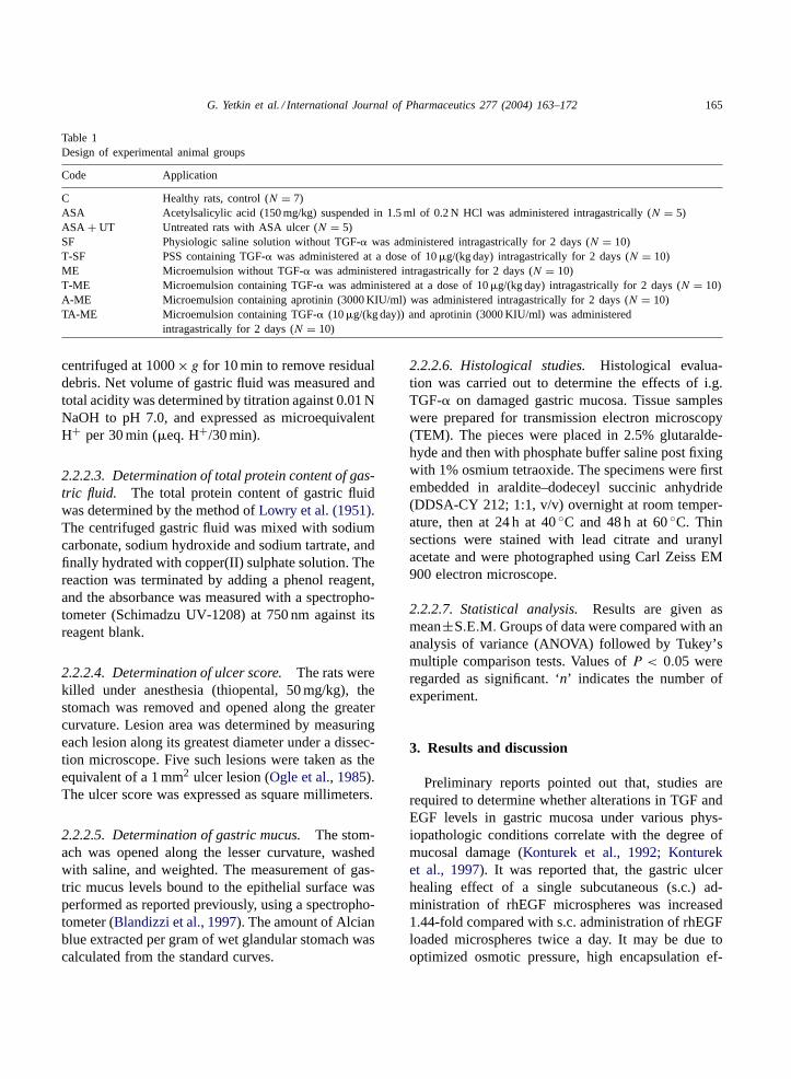
Fig. 1. Basal gastric acid secretion in different experimental groups. Data shown are mean± S.E.M. of 5–10 rats.P < 0.05 forT-SF/SF, A-ME/C;P < 0.01 for T-ME/T-SF, T-SF/C, A-ME/ME;P < 0.001 for TA-ME/ASA, TA-ME/ASA-UT, TA-ME/ME, T-ME/ASA,T-ME/ME, T-SF/ASA, A-ME/ASA, A-ME/TA-ME.
total protein content of gastric fluid, gastric mucuslevel and histological evaluation.
3.2. Effects of TGF-α on basal gastric acidsecretion
Konturek et al. (1992)reported that TGF and EGFadministered i.g., was found failed to affect gastricacid secretion or to prevent the mucosal damage, sug-gesting that these peptides are unable to influence themucosal integrity. In the present study, the formula-tions of TGF-� showed different effects on gastric acidsecretions.
The effects of i.g. administration of TGF-� solu-tion or microemulsion formulations on basal gastricacid secretion of gastric ulcer induced by ASA in ratsare shown inFig. 1. The gastric acid secretions ofgroups which were treated by microemulsions con-taining aprotinin (A-ME) or TGF-� (T-ME) or TGF-�plus aprotinin (TA-ME), were significantly lower thanthose of the microemulsion treated group (ME). Theresults of those are 38.1±4.6�eq. H+/30 min, 30.0±1.7�eq. H+/30 min, 20.8 ± 1.3�eq. H+/30 min and54.4 ± 3.5�eq. H+/30 min, respectively (P < 0.01,<0.001,<0.001). The gastric acid secretion was sig-nificantly reduced after intragastric administration ofTGF-� solution (T-SF) compared to its control group(SF), at 42.9±2.6�eq. H+/30 min and 57.2±3.9�eq.H+/30 min, respectively (P < 0.05).

G. Yetkin et al. / International Journal of Pharmaceutics 277 (2004) 163–172 167
The basal gastric acid secretion obtained by i.g.administration of TGF-� microemulsion was foundlower than that of TGF-� solution (P < 0.01). Thus,the microemulsion formulation can be consideredmore effective than solution of TGF-�.
On the other hand, the group which was treated withthe microemulsion containing TGF-� plus aprotinin(TA-ME) had the lowest basal gastric acid secretionlevel (20.8 ± 1.3�eq. H+/30 min). Only this group’sbasal gastric acid level decreased when compared tothe untreated (ASA+ UT) group’s level (P < 0.001).
Based on the evaluation of the basal gastric secre-tion, the gastric pH measured (fasting pH) in the col-lected gastric juice, increased significantly in TAME(pH 3.65± 0.15) and TME groups (pH 3.55± 0.20),compared to the control group (pH 2.57± 0.28) (P <
0.01, <0.05).
3.3. Effects of TGF-α on total protein content ofgastric fluid
The effects of TGF-� in solution or in microemul-sion formulations, on total protein content of the gas-tric fluid with gastric ulcer induced by ASA in rats, areshown inFig. 2. The total protein content of gastricfluid in fasted T-ME and TA-ME groups decreasedremarkably, when compared with the ME group, at2.12± 0.18 mg protein/ml, 1.82± 0.18 mg protein/mland 3.87 ± 0.47 mg protein/ml, respectively (P <
0
1
2
3
4
5
ASA ASA+UT SF T-SF ME A-ME T-ME TA-ME C
Th
e to
tal p
rote
in c
on
ten
t o
f g
astr
ic f
luid
(mg
pro
tein
/mL
)
Fig. 2. The total protein content of gastric fluid in different experimental groups. Data shown are mean± S.E.M. of 5–10 rats.P < 0.05for TA-ME/ASA-UT; P < 0.001 for TA-ME/ME, T-ME/ME.
0.001). In contrast, there was no significant reductionin total protein content of gastric fluid in the T-SFand A-ME groups, compared to SF and ME groupsat 2.83 ± 0.14 mg protein/ml, 2.75 ± 0.26 mg pro-tein/ml, 3.52±0.31 mg protein/ml and 3.87±0.47 mgprotein/ml, respectively (P > 0.05). In addition, thetotal protein content of gastric fluid of TA-ME grouphad a remarkable reduction, compared with ASA-UTgroup (P < 0.05). On the other hand, the differencebetween total protein content of gastric fluid mea-sured at T-ME and T-SF groups was not statisticallysignificant (P > 0.05).
Marchbank et al. (2002)reported that TGF-� (1–50)is cleaved to TGF-� (1–43) by acid pepsin and thisis the predominant form in normal gastric juice. Neu-tralization by i.g. or taking acid suppressants, causedthe predominant form to be TGF-� (1–50). TGF-�(1–43) reduced gastric ulcer area in rat, induced withindomethacin by 19% whereas, TGF-� (1–50) reducedarea by 62%.
In the present study reduced total protein contentand ulcer area in TA-ME group may have relevanceto the actions of new formulation.
3.4. Effects of TGF-α on ulcer score
The gastropathy associated with the ingestion ofnon-steroidal anti-inflammatory drugs such as as-pirin, is a common side effect of this class of drugs
168 G. Yetkin et al. / International Journal of Pharmaceutics 277 (2004) 163–172
0
20
40
60
80
100
ASA ASA+UT SF T-SF ME A-ME T-ME TA-ME C
Ulc
er s
core
s (m
m2 )
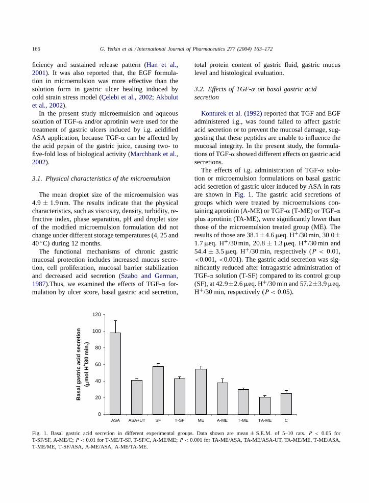
Fig. 3. Ulcer scores in different experimental groups. Data shown are mean±S.E.M. of 5–10 rats.P < 0.01 for T-ME/ASA-UT, T-ME/T-SF,A-ME/ME; P < 0.001 for TA-ME/ASA, TA-ME/ASA-UT, TA-ME/ME, T-ME/ASA, T-ME/ME, T-SF/ASA, T-SF/SF, A-ME/ASA,A-ME/T-ME, A-ME/TA-ME.
(Konturek et al., 1994). Animal studies then con-firmed the mechanisms of induction of the NSAIDulcer. These models have helped to answer key ques-tions, which lead to our current knowledge of NSAIDdamage (Lee, 2000).
The effects of TGF-� in solution or microemulsionformulations on ulcer score in rats are shown inFig. 3.The i.g. administration of ASA (150 mg/kg) produced
0
20
40
60
80
100
ASA ASA+UT SF T-SF ME A-ME T-ME TA-ME C
Gas
tric
mu
cus
leve
ls
(g
/g t
issu
e)
Fig. 4. Gastric mucus levels in different experimental groups. Data shown are mean±S.E.M. of 5–10 rats.P < 0.05 for T-SF/SF;P < 0.01for T-ME/T-SF, T-SF/ASA, A-ME/T-ME P < 0.001 for TA-ME/ASA, TA-ME/ASA-UT, TA-ME/ME, T-ME/ASA, T-ME/ASA-UT,T-ME/ME, A-ME/TA-ME.
gastric ulcers in all tested animals with an average ini-tial area of 89.0±3.8 mm2. The ulcer scores of groupstreated with TGF-� and/or aprotinin in microemul-sion or solution (A-ME, T-ME, TA-ME and T-SF)were calculated as 45.3 ± 2.7 mm2, 21.7 ± 3.0 mm2,12.2 ± 1.2 mm2 and 34.7 ± 1.5 mm2, respectively.The ulcer scores of the untreated groups (ASA-UT)or treated groups with vehicles (SF and ME) were

G. Yetkin et al. / International Journal of Pharmaceutics 277 (2004) 163–172 169
determined as 39.0 ± 3.7 mm2, 61.6 ± 2.7 mm2 and62.8±5.6 mm2. As shown in results, gastric ulcer wassignificantly decreased in 4 days after the ulcer induc-tion. The mean ulcer areas in all treated groups withTGF-� (T-ME, TA-ME and T-SF) reduced signifi-cantly, compared to their control groups (ME and SF)(P < 0.001). The results indicate that, the ulcer scoredecreased significantly in T-ME and TA-ME groups(P < 0.001), but there was no significant decreasein T-SF and A-ME groups (P > 0.05) compared tountreated group (ASA+ UT). Furthermore, the i.g.administration of TGF-� in microemulsion was moreeffective than TGF-� aqueous solution in the healingof gastric ulcer (P < 0.01). On the other hand, thehighest decrease in ulcer area was observed in group
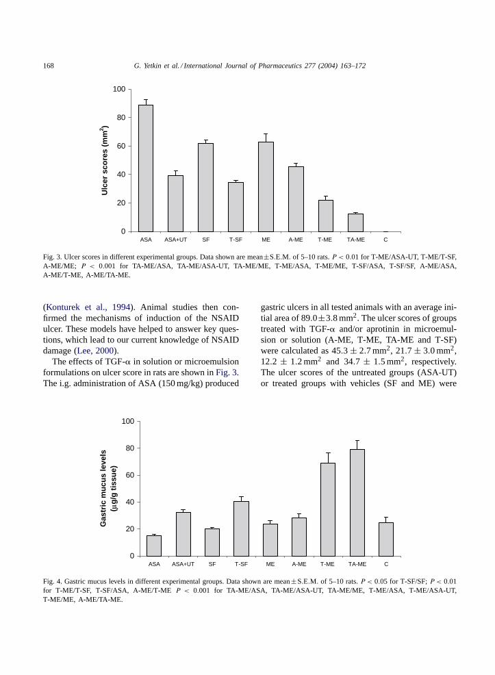
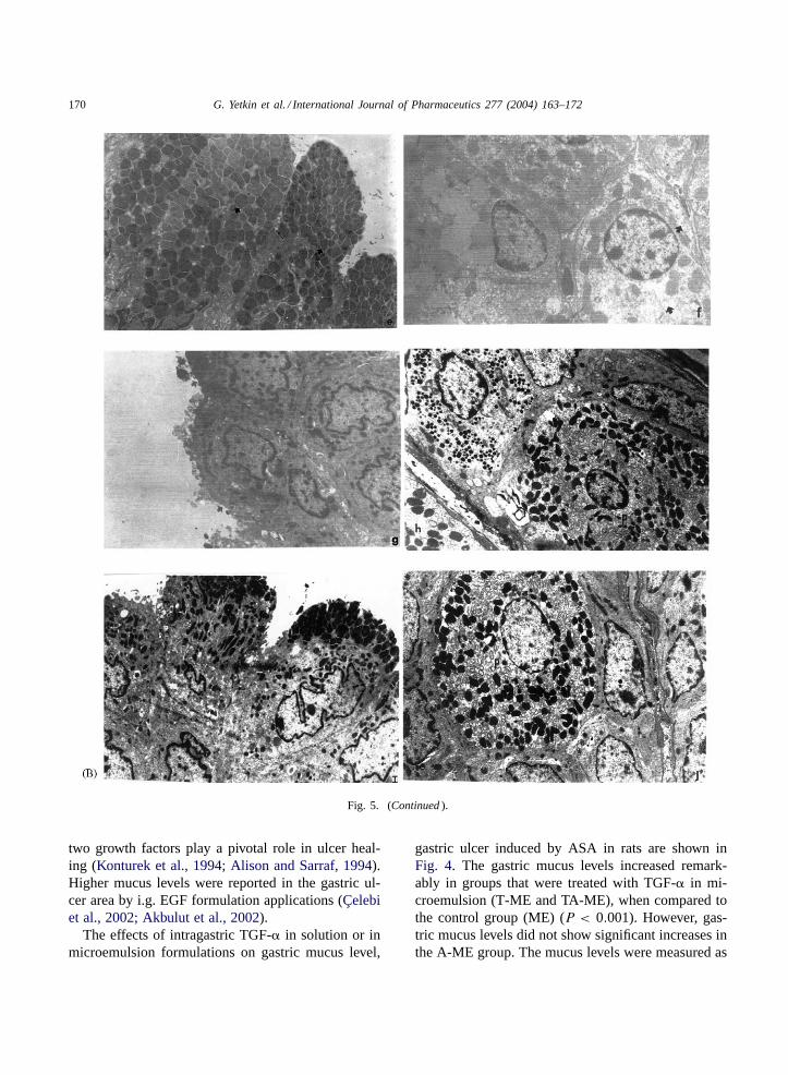
Fig. 5. (A) Transmission electron microscopic appearance of gastric mucosa in control and ASA groups (uranyl acetate–lead citrate, 9000×).Control group: (a) mucus granules (thick aurous); (b) normal appearance of parietal cells. ASA group: (c) mucus granules (thick aurous)broken intracytoplasmic junction (thick aurous); (d) active appearance of parietal cells with lots of intracytoplasmic canalicules (thickaurous). (B) Transmission electron microscopic appearance of gastric mucosa in AME, T-ME and TA-ME groups (uranyl acetate–leadcitrate, 9000×). AME group: (e) mucus granules (thick aurous) broken intracytoplasmic granules (the aurous); (f) degenerative areas incytoplasm (thick aurous). TME group: (g) normal appearance of mucus cells (thick aurous); (h) normal appearance of parietal cells (P).TAME group: (i) normal mucus cells (thick aurous); (j) normal parietal cells (P).
treated with the microemulsion containing TGF-� plusaprotinin.
3.5. Effects of TGF-α on gastric mucus level
Growth factors in ulcer healing has some effectson gastric secretion as decreases in acid secretion andincreases in mucus levels (Szabo and Vincze, 2000).Both EGF and TGF-� exert their effects by interac-tion with a specific cell membrane receptor (Milaniand Calabro, 2001). TGF-� appears to be the primaryphysiological ligand for the EGF receptor (EGFR)(Alison and Sarraf, 1994). Since both EGF and TGF-�promote cell proliferation, stimulate cell migration andinhibit gastric acid secretion, it is likely that these
170 G. Yetkin et al. / International Journal of Pharmaceutics 277 (2004) 163–172
Fig. 5. (Continued).
two growth factors play a pivotal role in ulcer heal-ing (Konturek et al., 1994; Alison and Sarraf, 1994).Higher mucus levels were reported in the gastric ul-cer area by i.g. EGF formulation applications (Çelebiet al., 2002; Akbulut et al., 2002).
The effects of intragastric TGF-� in solution or inmicroemulsion formulations on gastric mucus level,
gastric ulcer induced by ASA in rats are shown inFig. 4. The gastric mucus levels increased remark-ably in groups that were treated with TGF-� in mi-croemulsion (T-ME and TA-ME), when compared tothe control group (ME) (P < 0.001). However, gas-tric mucus levels did not show significant increases inthe A-ME group. The mucus levels were measured as

G. Yetkin et al. / International Journal of Pharmaceutics 277 (2004) 163–172 171
79.1± 6.6�g/g, 68.9± 7.8�g/g, 28.4± 3.2�g/g and23.6 ± 3.0�g/g tissue in TA-ME, T-ME, A-ME andME groups, respectively. The effect of TSF group ongastric mucus secretion was significantly higher thanits control group (SF) (P < 0.05) when compared toASA + UT group. There was no difference betweenthese mucus levels (P > 0.05). In contrary, the gas-tric mucus levels of TA-ME and T-ME treated groupswere found to be significantly higher than ASA+ UTgroup (P < 0.001).
Just like the other results in the decrease of gastricacid secretions and ulcer scores, TGF-� in microemul-sion formulation was more effective than TGF-� insolution formulation in the increase of gastric mucussecretion.
3.6. Evaluation of histological studies
At the end of histological studies, surface mu-cous cells and parietal cells were evaluated (Fig. 5Aand B). The results of TEM indicated that, exceptionof TME, TAME and control groups, others showedvariety of degenerative changes in surface mucous ep-ithelia which are the deformations of connected units,separation in surface cells membranes, electron palemucous granules or their decreasing in number. Be-sides, enlargement in connected units of degranulationis observed. On the other hand, parietal cells showedsome degenerative changes too. They had activatedappearance and their intracytoplasmic canalicules areincreased. Moreover, opening between the inner andouter nucleus membrane was clear. They had enlargeddegenerative cytoplasmic areas and crystolysrs in themitochandrions were also observed. As shown inFig. 5Bin TME and specially TAME groups, the cellswere absolutely in normal structure as the controlgroups (Fig. 5B).
As a conclusion of histological experiments showedthat, the microemulsion containing TGF-� stomachulcer wounds were increasingly healed. According tothe results of biochemical analysis and histologicalevaluation of TA-ME formulation is found to be moreeffective then the other formulations.
These results suggest that protease inhibitor apro-tinin and ME might usefully improve the absorptionand the extension of half-life of TGF-� as previouslymentioned by other research groups (Trenktrog et al.,1995; Tozaki et al., 1998; Anderle et al., 2002).
4. Conclusion
These findings suggest that TGF-� incorporatedwith aprotinin into microemulsion formulation wasthe most effective one on acidified ASA inducedgastric ulcer treatment.
Acknowledgements
This study was supported by grants from TurkishScientific and Technical Research Council (Tübitak,SBAG 2164). We also thank to Gattéfosse andEczacıba¸sı Pharmaceutical Inc. for kindly providingLabrafil M 1944 CS and TGF-�.
References
Akbulut, K.G., Gönül, B., Türkyılmaz, A., Çelebi, N., 2002. Therole of epidermal growth factor formulation on stress ulcerhealing of gastric mucosa. Surg. Today 32, 880–883.
Alison, M.R., Sarraf, C.E., 1994. The role of growth ractors ingastrointestinal cell proliferation. Cell Biol. Int. 18, 1–10.
Anderle, P., Langguth, P., Rubas, W., Merkle, H.P., 2002. In vitroassessment of IGF-I stability. J. Pharm. Sci. 91, 290–300.
Bagwe, R.P., Kanicky, J.R., Palla, B.J., Patanjali, P.K., Shah, D.O.,2001. Improved drug delivery using microemulsions: rationale,recent progress, and new horizons. Crit. Rev. Ther. Drug CarrierSyst. 18, 77–140.
Bennett, N.T., Schultz, G.S., 1993. Growth factors and woundhealing: Part II. Role in normal and chronic wound healing.Am. J. Surg. 166, 74–81.
Blandizzi, C., Geherardi, G., Marveggio, C., Lazzeri, G., Natale,G., Carignani, D., Colucci, R., Del Tacca, M., 1997. Suraminenhances ethanol-induced injury to gastric mucosa in rats. Dig.Dis. Sci. 42, 1233–1241.
Çelebi, N., Türkyılmaz, A., Gönül, B., Özogul, C., 2002. Effectsof epidermal growth factor microemulsion formulation on thehealing of stress-induced gastric ulcers in rats. J. ControlledRelease 83, 197–210.
Han, K., Lee, K.D., Gao, Z.G., Park, J.S., 2001. Preperationand evaluation of poly(l-lactic acid) microspheres containingrhEGF for chronic gastric ulcer healing. J. Controlled Release75, 259–269.
Konturek, J.W., Dembinski, A., Stoll, R., Domschke, W.S.J.,1994. Mucosal adaptation to aspirin induced gastric damage inhumans. Studies on blood flow, gastric mucocal growth, andneutrophil activation. Gut 35, 1197–1204.
Konturek, P.C., Ernst, H., Konturek, S.J., Bobrzynski, A.J., Faller,G., Klingler, C., Hang, E.G., 1997. Mucosal expression andluminal release of epidermal and transforming growth factorin patients with duodenal ulcer before and after eradication ofHelicobacter pylori. Gut 40, 463–469.

172 G. Yetkin et al. / International Journal of Pharmaceutics 277 (2004) 163–172
Konturek, P.C., Konturek, S.J., Brzozowski, T., Ernst, H., 1995.Epidermal growth factor and transforming growth factor-�:role in protection and healing of gastric mucosal lesions. Eur.J. Gastroenterol. 7, 933–938.
Konturek, S.J., Brzozowski, T., Majka, J., Dembinski, A., Slo-miany, A., Slomiany, B.L., 1992. Transforming growth factoralpha and epidermal growth factor in protection and healing ofgastric mucosal injury. Scand. J. Gastroenterol. 27, 649–655.
Konturek, S.J., 1988. Role of epidermal growth factor in gastropro-tection and ulcer healing. Scand. J. Gastroenterol. 23, 129–133.
Lee, A., 2000. Animal models of gastroduodenal ulcer disease.Bailliere’s Clin. Gastroenterol. 14, 75–96.
Lowry, O.H., Rosebrough, N.J., Farr, A.L., Randall, R.J., 1951.Protein measurement with the folin phenol reagent. J. Biol.Chem. 193, 265–275.
Marchbank, T., Boulton, R., Hansel, H., Playford, R.J., 2002.Human transforming growth factor a (TGF-alpha) is digestedto a smaller (1–43), less biologically active, form in acidicjuice. Gut 51, 787–792.
Milani, S., Calabro, A., 2001. Role of growth factors and theirreceptors in gastric ulcer healing. Microsc. Res. Tech. 53, 360–371.
Ogle, C.W., Cho, C.H., Tong, M.C., Koo, M.W.L., 1985. Theinfluence of verapamil on the gastric effects of stress in rats.Eur. J. Pharmacol. 112, 399–404.
Schultz, G.S., Rotatori, D.S., Clark, W., 1991. EGF and TGF-�
in wound healing and repair. J. Cell. Biochem. 45, 346–352.
Sener-Muratoglu, G., Paskaloglu, K., Arbak, S., Hürdag, C.,Ayanoglu-Dülger, G., 2001. Protective effect of famotidine,omeprazole, and melatonin against acetylsalicylic acid inducedgastric damage in rats. Dig. Dis. Sci. 46, 318–330.
Szabo, S., German, P., 1987. Mechanisms of gastric cytoprotection.J. Clin. Gastroenterol. 9, 8–13.
Szabo, S., Vincze, A., 2000. Growth factor in ulcer healing: lessonsfrom recent studies. J. Physiol. (Paris) 94, 77–81.
Tozaki, H., Odoriba, T., Iseki, T., Taniguchi, T., Fujita, T.,Murakami, M., Muranishi, S., Yamamato, A., 1998. Use ofprotease inhibitors to improve calcitonin absorption from thesmall and large intestine in rats. J. Pharm. Pharmacol. 50,913–920.
Trenktrog, T., Muller, B.W., Seıfert, J., 1995. In vitro-investigationinto the enhancement of intestinal peptide absorption byemulsion systems. Eur. J. Pharm. Biopharm. 41, 284–290.
Türkyılmaz, A., Çelebi, N., Gönül, B., Alkan-Önyüksel, H., 1998,Physical characterization and stability of a microemulsion forpotential oral administration of a peptide. In: Hıncal, A.A.,Kas, H.S. (Eds.), Biomedical Science and Technology, RecentDevelopments in the Pharmaceutical and Medical Sciences.Plenum Press, New York, pp. 65–72.
Copyright © 2022 FDOKUMEN