Suppression of Latent Transforming Growth Factor (TGF)- 1 Restores Growth Inhibitory TGF- ...
15
Suppression of Latent Transforming Growth Factor (TGF)-1 Restores Growth Inhibitory TGF- Signaling through microRNAs □ S Received for publication, December 3, 2010, and in revised form, March 9, 2011 Published, JBC Papers in Press, March 14, 2011, DOI 10.1074/jbc.M110.208652 Afzal M. Dogar, Harry Towbin, and Jonathan Hall 1 From the Department of Chemistry and Applied Biosciences, Institute of Pharmaceutical Sciences, ETH Zurich, 8093 Zurich, Switzerland Cancer cells secreting excess latent TGF- are often resistant to TGF- induced growth inhibition. We observed that RNAi against TGF-1 led to apoptotic death in such cell lines with features that were, paradoxically, reminiscent of TGF- signal- ing activity and that included transiently enhanced SMAD2 and AKT phosphorylation. A comprehensive search in Hela cells for potential microRNA drivers of this mechanism revealed that RNAi against TGF-1 led to induction of pro-apoptotic miR-34a and to a globally decreased oncomir expression. The reduced levels of the oncomirs miR-18a and miR-24 accounted for the observed derepression of two TGF-1 processing factors, thrombospondin-1, and furin, respectively. Our data suggest a novel mechanism in which latent TGF-1, thrombospondin 1, and furin form a microRNA-mediated regulatory feedback loop. For cells with high levels of latent TGF-, this provides a poten- tially widespread mechanism of escape from TGF--mediated growth arrest at the earliest point in the signaling pathway, TGF- processing. Transforming growth factor- (TGF-) 2 polypeptides are cytokines from a large family of ligands and receptors, which regulate epithelial, neuronal, and immune cells by controlling proliferation, differentiation, and survival processes with an intricate complexity. Cells synthesize the three isoforms of TGF- in excess and their rate-limiting, complex mechanisms of activation are regulated in a cell type and stimulation-specific fashion (1). TGF-1 is expressed as a pro-peptide precursor comprising the mature form and a latency-associated peptide (LAP). The pro-peptide dimerizes and is nicked before secre- tion by furin-like proteases but remains self-associated. Secre- tion is promoted after conjugation of a latent TGF--binding protein (LTBP) to the LAP forming a large latent complex (LLC), which associates with the extracellular matrix. Mature TGF- can be released from the LLC in vitro by heating or acid but in vivo release requires cell surface furin proteases as well as extracellular matrix proteins such as thrombospondin-1 (THBS1; TSP1) and integrins that induce conformational changes in the complex to promote dissociation of mature ligand. The mechanisms of activation, and also the type of intracellular signaling, may depend on the TGF--containing complex bound to the cell surface. Consequently, cells may respond differently to TGF- from autocrine or exogenous ori- gin by activating, for example, alternative growth inhibitory pathways (2). By maintaining a source of latent TGF- close to its site of action, cells can initiate rapid signaling without the need for new protein synthesis (3). Once activated, TGF- binds a membrane-bound serine/threonine receptor complex (TRI/TRII), which phosphorylates various substrates. These include transcription factors SMAD2 and SMAD3, which accu- mulate in nuclear complexes with co-activators and co-repres- sors, or molecules from numerous non-Smad pathways (4). The cellular response to TGF- is thus a balanced activation of Smad and/or non-Smad signaling pathways determined by cel- lular “context” (5, 6), as well as signaling thresholds and signal- ing duration (7, 8). TGF- signaling via SMADs causes growth inhibition of epi- thelial cells by transcriptional induction of cyclin-dependent kinase inhibitors P21 and P15, and the repression of transcrip- tion factors MYC, ID1, and ID2 (9, 5). TGF- is often found up-regulated in tumors and tumor cell lines (10, 11) but resis- tance to growth inhibitory TGF- signaling is an important and common event in tumorigenesis (12). Whereas some tumors acquire somatic changes in TGF- signaling components e.g. mutations in SMADs or TGF- receptors, others become resistant to the antiproliferative response while maintaining the ability to signal. Here, TGF- becomes an oncogenic factor inducing proliferation, angiogenesis, and metastasis. Several mechanisms of resistance have been documented, mostly involving downstream pathway components (13). In particular, the PI3K-AKT survival pathway plays a prominent role in can- cers which are resistant to cytostatic TGF- signaling (14, 15), for example by blocking induction of P21 in glioblastoma cells (16). Thus, inhibition of PI3K-AKT signaling restored TGF- growth arrest in glioblastoma (16) and in colon carcinoma cell lines (15). MicroRNAs (miRNAs) are single-stranded RNAs of 22 nucleotides which regulate the expression of a large fraction of genes. MiRNAs bind with partial sequence complementarity to sites, usually in the 3-UTRs of mRNAs, and inhibit protein translation or induce mRNA degradation. An increasingly rec- ognized feature of miRNA function is the parallel targeting of regulatory pathway members by multiple miRNAs (17). There- fore, even relatively modest changes in the expression levels of individual miRNAs and their protein targets often have signif- □ S The on-line version of this article (available at http://www.jbc.org) contains supplemental Tables S1 and S2. Author’s Choice—Final version full access. 1 To whom correspondence should be addressed: Institute of Pharmaceutical Sciences, ETH Zurich, HCI H437, Wolfgang-Pauli-Str. 10, CH-8093 Zurich, Switzerland. E-mail: [email protected]. 2 The abbreviations used are: TGF, transforming growth factor; LAP, latency- associated peptide; LLC, large latent complex; miRNA, microRNA. THE JOURNAL OF BIOLOGICAL CHEMISTRY VOL. 286, NO. 18, pp. 16447–16458, May 6, 2011 Author’s Choice © 2011 by The American Society for Biochemistry and Molecular Biology, Inc. Printed in the U.S.A. MAY 6, 2011 • VOLUME 286 • NUMBER 18 JOURNAL OF BIOLOGICAL CHEMISTRY 16447 by guest on July 13, 2016 http://www.jbc.org/ Downloaded from by guest on July 13, 2016 http://www.jbc.org/ Downloaded from by guest on July 13, 2016 http://www.jbc.org/ Downloaded from by guest on July 13, 2016 http://www.jbc.org/ Downloaded from
Transcript of Suppression of Latent Transforming Growth Factor (TGF)- 1 Restores Growth Inhibitory TGF- ...
Suppression of Latent Transforming Growth Factor (TGF)-�1Restores Growth Inhibitory TGF-� Signaling throughmicroRNAs□S
Received for publication, December 3, 2010, and in revised form, March 9, 2011 Published, JBC Papers in Press, March 14, 2011, DOI 10.1074/jbc.M110.208652
Afzal M. Dogar, Harry Towbin, and Jonathan Hall1
From the Department of Chemistry and Applied Biosciences, Institute of Pharmaceutical Sciences, ETH Zurich, 8093 Zurich, Switzerland
Cancer cells secreting excess latent TGF-� are often resistantto TGF-� induced growth inhibition. We observed that RNAiagainst TGF-�1 led to apoptotic death in such cell lines withfeatures that were, paradoxically, reminiscent of TGF-� signal-ing activity and that included transiently enhanced SMAD2and AKT phosphorylation. A comprehensive search in Helacells for potentialmicroRNAdrivers of thismechanism revealedthat RNAi against TGF-�1 led to induction of pro-apoptoticmiR-34a and to a globally decreased oncomir expression. Thereduced levels of the oncomirs miR-18a and miR-24 accountedfor theobservedderepressionof twoTGF-�1processing factors,thrombospondin-1, and furin, respectively. Our data suggest anovel mechanism in which latent TGF-�1, thrombospondin 1,and furin formamicroRNA-mediated regulatory feedback loop.For cells with high levels of latent TGF-�, this provides a poten-tially widespread mechanism of escape from TGF-�-mediatedgrowth arrest at the earliest point in the signaling pathway,TGF-� processing.
Transforming growth factor-� (TGF-�)2 polypeptides arecytokines from a large family of ligands and receptors, whichregulate epithelial, neuronal, and immune cells by controllingproliferation, differentiation, and survival processes with anintricate complexity. Cells synthesize the three isoforms ofTGF-� in excess and their rate-limiting, complex mechanismsof activation are regulated in a cell type and stimulation-specificfashion (1). TGF-�1 is expressed as a pro-peptide precursorcomprising the mature form and a latency-associated peptide(LAP). The pro-peptide dimerizes and is nicked before secre-tion by furin-like proteases but remains self-associated. Secre-tion is promoted after conjugation of a latent TGF-�-bindingprotein (LTBP) to the LAP forming a large latent complex(LLC), which associates with the extracellular matrix. MatureTGF-� can be released from the LLC in vitro by heating or acidbut in vivo release requires cell surface furin proteases as wellas extracellular matrix proteins such as thrombospondin-1(THBS1; TSP1) and integrins that induce conformationalchanges in the complex to promote dissociation of mature
ligand. The mechanisms of activation, and also the type ofintracellular signaling, may depend on the TGF-�-containingcomplex bound to the cell surface. Consequently, cells mayrespond differently to TGF-� from autocrine or exogenous ori-gin by activating, for example, alternative growth inhibitorypathways (2). By maintaining a source of latent TGF-� close toits site of action, cells can initiate rapid signaling without theneed for new protein synthesis (3). Once activated, TGF-�binds a membrane-bound serine/threonine receptor complex(T�RI/T�RII), which phosphorylates various substrates. Theseinclude transcription factors SMAD2 and SMAD3,which accu-mulate in nuclear complexes with co-activators and co-repres-sors, ormolecules fromnumerous non-Smadpathways (4). Thecellular response to TGF-� is thus a balanced activation ofSmad and/or non-Smad signaling pathways determined by cel-lular “context” (5, 6), as well as signaling thresholds and signal-ing duration (7, 8).TGF-� signaling via SMADs causes growth inhibition of epi-
thelial cells by transcriptional induction of cyclin-dependentkinase inhibitors P21 and P15, and the repression of transcrip-tion factors MYC, ID1, and ID2 (9, 5). TGF-� is often foundup-regulated in tumors and tumor cell lines (10, 11) but resis-tance to growth inhibitory TGF-� signaling is an important andcommon event in tumorigenesis (12). Whereas some tumorsacquire somatic changes in TGF-� signaling components e.g.mutations in SMADs or TGF-� receptors, others becomeresistant to the antiproliferative responsewhilemaintaining theability to signal. Here, TGF-� becomes an oncogenic factorinducing proliferation, angiogenesis, and metastasis. Severalmechanisms of resistance have been documented, mostlyinvolving downstream pathway components (13). In particular,the PI3K-AKT survival pathway plays a prominent role in can-cers which are resistant to cytostatic TGF-� signaling (14, 15),for example by blocking induction of P21 in glioblastoma cells(16). Thus, inhibition of PI3K-AKT signaling restored TGF-�growth arrest in glioblastoma (16) and in colon carcinoma celllines (15).MicroRNAs (miRNAs) are single-stranded RNAs of �22
nucleotides which regulate the expression of a large fraction ofgenes.MiRNAs bind with partial sequence complementarity tosites, usually in the 3�-UTRs of mRNAs, and inhibit proteintranslation or induce mRNA degradation. An increasingly rec-ognized feature of miRNA function is the parallel targeting ofregulatory pathwaymembers bymultiple miRNAs (17). There-fore, even relatively modest changes in the expression levels ofindividual miRNAs and their protein targets often have signif-
□S The on-line version of this article (available at http://www.jbc.org) containssupplemental Tables S1 and S2.Author’s Choice—Final version full access.
1 To whom correspondence should be addressed: Institute of PharmaceuticalSciences, ETH Zurich, HCI H437, Wolfgang-Pauli-Str. 10, CH-8093 Zurich,Switzerland. E-mail: [email protected].
2 The abbreviations used are: TGF, transforming growth factor; LAP, latency-associated peptide; LLC, large latent complex; miRNA, microRNA.
THE JOURNAL OF BIOLOGICAL CHEMISTRY VOL. 286, NO. 18, pp. 16447–16458, May 6, 2011Author’s Choice © 2011 by The American Society for Biochemistry and Molecular Biology, Inc. Printed in the U.S.A.
MAY 6, 2011 • VOLUME 286 • NUMBER 18 JOURNAL OF BIOLOGICAL CHEMISTRY 16447
by guest on July 13, 2016http://w
ww
.jbc.org/D
ownloaded from
by guest on July 13, 2016
http://ww
w.jbc.org/
Dow
nloaded from
by guest on July 13, 2016http://w
ww
.jbc.org/D
ownloaded from
by guest on July 13, 2016
http://ww
w.jbc.org/
Dow
nloaded from

icant phenotypic consequences. Signaling pathways usemiRNAs to switch the gene expression programs of cells (18,19) and not surprisingly, the TGF-� pathway has many docu-mented examples ofmiRNA activity including:miR-15/16 (20),miR-224 (21),miR-106b-25 (22),miR-200 family (23–26),miR-155 (27), miR-181b/d (28), miR-21 (29), miR-17�92 (30, 31),andmiR-24 (32–35).In a search for miRNAs which play central roles in disease-
associated mechanisms we selected TGF-� signaling for inves-tigation.We conducted RNAi against TGF-�1 in several cancercell lines and observed apoptosis after 3 days. TGF-�1 depletionin HeLa and LN-18 cells was associated with induction ofTGF-� processing factors and transient TGF-� signaling priorto apoptosis. In HeLa cells apoptosis was preceded by repres-sion ofmiR-18a andmiR-24, which target THBS1 and FURIN,respectively. The data suggest that in some cell lines latentTGF-�1 inhibits cytostatic TGF-� signaling through post-tran-scriptional repression of its own processing factors. For tumorcells which secrete high levels of latent TGF-� this representspotentially an important mechanism of escape from cytostaticTGF-� signaling at the earliest point in the pathway. Further-more, it implies that in these instances pharmacological target-ing of TGF-�1mRNAmay be of therapeutic value in patholog-ical mechanisms caused by loss of the cytostatic response toTGF-� signaling.
EXPERIMENTAL PROCEDURES
Cell Culture and Transfections—Hela (ATCC, CCL-2) andLN-18 (ATCC, CRL-2610) cells from LGC (Molsheim, FR)were maintained in Dulbecco’s modified Eagle’s medium(Invitrogen) supplemented with 10% fetal bovine serum(FBS; Sigma). SiTGF�1 targeting human TGF-�1 (NM_000660) is CCAACUAUUGCUUCAGCUC (1712–1730);siTGF�1(s) is CGUGGAGCUGUACCAGAAA (1368–1386).SiRNAs,miRNAmimics and inhibitors were fromDharmacon,siCon was from Ambion (AM4640). RNAs were transfectedusing Oligofectamine (12252-011, Invitrogen) according tomanufacturer’s instructions. Recombinant human matureTGF-�1 (100-B), latent TGF-�1 (299-LT/CF), and TGF-� neu-tralizing antibody (MAB1835; Clone 1D11) were from R&DSystems. SB431542 was from Santa Cruz Biotechnology(SC-204265).RNA and miRNA Q-PCR—Total RNA was extracted using
mirVanaTM miRNA Isolation kit (AM1560; Ambion). FormRNA analysis, 1 �g of total RNA was reverse transcribedusing the M-MLV reverse transcriptase kit (28025-013, Invit-rogen) according to manufacturer’s instructions. Expressionlevels were assayed using Power SYBR�Green PCRMasterMix(4367659; Applied Biosystems). PCR cycling conditions were95 °C/10 min and 40 cycles of (95 °C/15 s; 60 °C/1 min). Valueswere normalized using GAPDH or average of all measuredmRNAs. RT-PCR primers sequences are shown in supplemen-tal Table S1.For miRNA analyses, the TaqMan� Human MicroRNA
Assay set v1.0 (4383443; Applied Biosystems) was used. TotalRNAwas reverse transcribed with miRNA-specific RT primersand amplified with TaqMan miRNA-specific primers. cDNAwas synthesized from 10–20 ng of total RNA in a 15 �l volume
using TaqManMicroRNA Reverse Transcription kit (4366597;Applied Biosystems), according tomanufacturer’s instructions.All samples were measured in triplicates; the threshold cycle(CT) values were transformed to relative quantities.ELISA—TGF-�1 in cultured cells was quantifiedwith human
TGF-�1 DuoSet ELISA (DY240; R&D Systems), according tothe manufacturer’s instructions. Cells were grown in 96-wellplates in 1% FBS-containing media and transfected withsiRNAs. Supernatants were acidified with 1 MHCl and neutral-ized with 1.2 M NaOH/0.5 M HEPES prior to assay for totalTGF-�1. The concentrations of active TGF-�1 were analyzedon non-acidified samples. TGF-�1 in the FBS was initially 180pg/ml for the 1% FBS concentration used in these experiments.Protein Analysis—Cells were lysed with RIPA lysis buffer (R
0278; Sigma). Protein concentrations were determined using aBCA assay (Thermo Fisher Scientific 23225), 10–20 �g of pro-tein wasmixed with equal quantities of SDS loading buffer (100mM Tris-HCl, 4% SDS, 20% glycerol, 0.2% bromphenol blue).Samples were heated at 99 °C for 5 min, separated on SDS gelsand transferred to polyvinylidene difluoride membranes. Non-specific membrane binding was blocked for 1 h at room tem-perature with 5% BSA (or milk) in phosphate-buffered salinecontaining 0.05% Tween 20. Membranes were incubated over-night at 4 °Cwith primary antibodies fromCell Signaling (TGF-�1: 3711; P-SMAD2: 3108; SMAD2: 3122; P-AKT: 4060; AKT9272) and from Santa Cruz Biotechnology (p53: SC-126; P21:SC-71811; TSP1: SC-81755; FURIN: SC-20801). After washingmembranes were incubated with horseradish peroxidase-con-jugated secondary antibodies for 1–2 h at room temperature inblocking buffer. Signals generated by the chemiluminescentsubstrate (ECL(�); AmershamBiosciences) were captured by acooled CCD camera (Bio-Rad). Protein bands were quantifiedby densitometry using the analysis software ImageJ.Plasmid DNA Transfections and Luciferase Assays—Firefly
luciferase reporter genes containing full-length human3�-UTRs from THBS1 (S211182) and FURIN (S209837) werefrom SwitchGear Genomics (Menlo Park, CA). cDNA ofTGF-�1 (SC119746) was from OriGene (Rockville, MD).cDNA of THBS1 (pbla-htsp1) was from InvivoGen (San Diego,CA). SBE-Luc reporter construct was a gift from Dr. B. Vogel-stein and contains four SMAD2/3 binding elements. For lucif-erase assays, Hela cells were grown in white 96-well plates, 20ng of plasmid DNAs were transfected per well using jetPEI(101-10; Polyplus) according to manufacturer’s protocol.SiRNAs were transfected after 24 h with indicated doses. After24 h, supernatants were removed and 30�l of Bright-Glo Lucif-erase substrate (E2610; Promega)was added to eachwell. Lumi-nescence signals were measured on a microtiter plate reader(Mithras LB940, Berthold Technologies).Apoptosis Assay—Caspase-3/7 activity was measured in
supernatants and lysates of transfected cells using a chemilumi-nescent substrate (Caspase-Glo 3/7 substrate, G8090, Pro-mega). Cells were grown in 96-well plates, and transfected withsiRNAs. For time-course experiments, 5 �l of supernatantswere transferred from the same wells at 24, 48 and 72 h timepoints to white 384-well plates and mixed with equal volumesof substrate. Chemiluminescencewasmeasured in sealed platesafter 30 min at room temperature in a plate reader. For mea-
microRNA Control of TGF-� Processing
16448 JOURNAL OF BIOLOGICAL CHEMISTRY VOLUME 286 • NUMBER 18 • MAY 6, 2011
by guest on July 13, 2016http://w
ww
.jbc.org/D
ownloaded from

surements of cell-associated caspase 3/7 activity cells werelysed in PBS containing 1% Triton X-100 and 5 �l of lysateswere mixed with equal volumes of substrate and otherwiseassayed as above.
RESULTS
Inhibition of TGF-�1 mRNA Causes Apoptosis in CervicalCarcinomaCells—Likemost cervical carcinoma cell linesHeLaexpress high levels of TGF-�1 (36). They respond to exoge-nously-delivered,mature recombinant TGF-�1 but they do notundergo apoptosis (data not shown). Independent treatment ofHeLa cells with two siRNAs (siTGF�1 and siTGF�1(s)) specificto the TGF-�1 sequence led to a dose-dependent down-regu-lation of TGF-�1 mRNA by greater than 80% at 15 nM by bothsiRNAs (Fig. 1A), though siTGF�1 was more potent in mostassays. Loss of TGF-�1 mRNA caused a rapid (24 h) reductionof intracellular un-nicked latent TGF-�1 protein, which wasdetected by a pan anti-TGF-� antibody (Fig. 1B). Reduction intotal TGF-�1 protein in cell supernatants was quantified usingan ELISA specific for the TGF-�1 isoform after treatment withacid (Fig. 1C), which converts the majority of latent nickedTGF-�1 to the mature form (37). Cells undergoing TGF-�1RNAi showed signs of increasing caspase 3/7 activity on day 2post-transfection, which rose dramatically on day 3 (Fig. 1D)and was associated withmassive cell death. Similar results wereobtained after the transfection of siTGF�1 into Caski and Sihacell lines (Fig. 1E). The results suggest that high levels ofTGF-�1 are essential for the survival of cervical carcinomacells. In order to ascertain the mechanism of cell death, RNAfrom treated cells was analyzed for the expression of cell deathgenes using real-time PCR (Q-PCR). Several p53 responsegenes including CDKN1A (P21), BAX, TNFR10B, FAS, andBTG2 were strongly induced at 48h (Fig. 2A). Accordingly,analysis of protein samples from treated cells showed dose-de-pendent up-regulation of p53 and P21 proteins (Fig. 2B). HeLacells express wild-type p53 and RB proteins which are inacti-vated byHPVE6 andE7, respectively. Inhibition of viral proteinexpression reactivates these tumor suppressor proteins (38)and indeed,HPVE6mRNAwas repressed as a result of TGF-�1RNAi (Fig. 2A).miRNAs Contribute to TGF-�1-RNAi-mediated Apoptosis in
HeLa Cells—Most miRNA-mRNA interactions are fine-tuningcontributions to the robustness of systems (17). In a small num-ber of cases, however, a single miRNA-mRNA interaction iscapable of switching a gene expression program in cells. Thedysregulation of such interactions may contribute to disease-causing mechanisms and therefore represent potential drugtargets. In a first step to identify miRNAs which influenceTGF-�1 RNAi-mediated apoptosis, we measured the expres-sion of 448 miRNAs in cells treated with a single dose ofsiTGF�1 or oligofectamine control using stem-loop PCR. Fromthe screen we selected 48miRNAs for a repeat study using bothTGF-�1 siRNAs at three doses. We set stringent standards forthe analysis ofmiRNAdata because, comparedwith treatment-induced changes in mRNA expression, fold changes in indivi-dual miRNAs are typically lower. We called “absent” anymiRNAs with cycle threshold (CT) values higher than 29. OnlymiRNAs which were regulated in the same direction by both
siRNAswere considered of interest. A large fraction ofmiRNAswhich passed these filters has been previously associated withcancer and/or elements of TGF-� signaling (Fig. 2C). For exam-ple, the large induction ofmiR-34,which is capable of inducingapoptosis alone by targeting genes involved in cell cycle andapoptosis, was consistent with the induction of p53 (39, 40). Onthe other hand, the modest down-regulation of oncomirsmiR-18a,miR-20b,miR-93,miR-24,miR-181b/d, andmiR-155 wasconsistent with a network of antiproliferative miRNA activity.Furthermore, many of these miRNAs have been previouslyreported to be repressed during TGF-� signaling includingmiR-24, miR-181b/d, miR-224, miR-155, and miR-17�92cluster.To identify miRNAs whose regulation contributed to the
TGF-�1 RNAi-induced apoptosis, we measured caspase 3/7activity after transfection of siTGF�1 in combinationwith dou-ble-strandedRNA reagents. In experiments designed to controlfor unspecific effects of siRNA against TGF-�1, delivery ofincreasing doses of a plasmid expressing (latent) TGF-�1mRNA rescued the cells from siTGF�1-mediated caspase 3/7induction (Fig. 2D). Interestingly, a clear reproducible reduc-tion in background caspase 3/7 activity was also apparent at thehighest dose of plasmid treatment alone, and correlated withthe highest levels of TGF-�1 mRNA and protein (data notshown). A siRNA toTP53 (siP53) (41) also partially reversed theapoptosis and served as an additional positive control (Fig. 2E).The negative control siCon showed a small effect whereasmiR-34 showed no effect in combination with siTGF�1 (Fig.2F). The anti-miRNA oligonucleotide of miR-34a (AMO-34a)inhibited siTGF�1-induced apoptosis almost to backgroundlevels (Fig. 2E). Mimics of miR-18, miR-24, and miR-181 wereable to reduce partially caspase 3/7 induction, consistent with ashared role in the apoptotic phenotype.Repression of Latent TGF-�1 Leads to Increased TGF-�
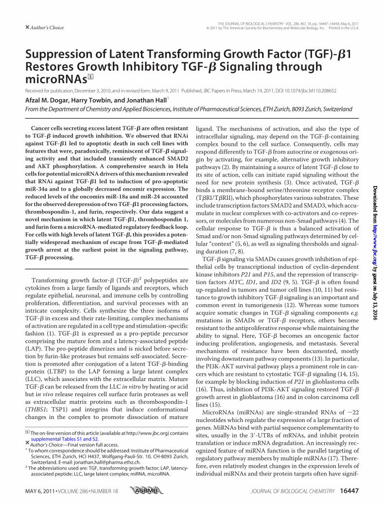
Processing—The relationship between AKT cell survival andp53 cell death pathways has been described as a balance (4, 42).Analysis of protein from siTGF�1-treated cells showed a dose-dependent rise in phospho-AKT (P-AKT) on day 1, followed bydose-dependent decreases on days 2 and 3. This rise appearedto precede p53 induction, and the onset of apoptosis (Fig. 3A). Atransient induction of AKT activity prior to apoptosis was alsodescribed during an investigation of TGF-� processing/signal-ing in mink lung epithelial cells (43). Taking together this aswell as other literature reports (15, 44) with our observationsled us to suspect that paradoxically we were observing a resto-ration of growth-inhibitory TGF-� signaling activity duringTGF-�1 RNAi. It therefore also implied that the high levels oflatent TGF-� produced by cells contributed to their resistanceto TGF-� induced growth inhibition, possibly through elevatedAKT signaling (15). Mindful of the difficulties of measuringTGF-� activity in cells undergoing TGF-�1 RNAi, we assayedfor phospho-SMAD2 (P-SMAD2) activity after TGF-�1 RNAiat 7, 24, and 48 h (Fig. 3B). After 24 and 48 h levels of P-SMAD2decreased, as expected for a potent, time-dependent down-reg-ulation of TGF-�1 ligand. However, a transient induction ofP-SMAD2was observed at 7 h post-siRNA transfection in com-parison to slightly elevated levels of total SMAD2. At this pointTGF-�1 mRNA had been reduced by �50% (Fig. 3C). In sup-
microRNA Control of TGF-� Processing
MAY 6, 2011 • VOLUME 286 • NUMBER 18 JOURNAL OF BIOLOGICAL CHEMISTRY 16449
by guest on July 13, 2016http://w
ww
.jbc.org/D
ownloaded from

port of this data, cells treated with siTGF�1 and a luciferasereporter gene construct bearing four SMAD binding elements(SBE) in its promoter yielded an induction of luciferase activityat both 8 and 24 h (Fig. 3C).The majority of TGF-� secreted by most cells is in a latent
nicked form. Given themulti-step nature of TGF-� processing,it seemed plausible that an increased TGF-� signaling activity
might arise from increased processing of extracellular latentTGF-�, despite declining levels of intracellular TGF-�1 due toRNAi. We therefore examined siTGF�1-treated HeLa cells forindications of elevated TGF-� processing. Concentrating cellsupernatants facilitates detection of trace quantities of totalextracellular TGF-� by Western blot (45). However, our anti-TGF� antibody does not distinguish between the three mature
FIGURE 1. Down-regulation of TGF-�1 by RNAi leads to apoptosis in cervical carcinoma cells. A, TGF-�1 mRNA after transfection of Hela cells with siRNAstargeting TGF-�1 and a control siRNA (siCon). Total RNA was isolated 48 h post-transfection. Relative expression of TGF-�1 mRNA is displayed (mean of PCRtriplicates; single RNA samples � S.D.). B, cell-associated latent un-nicked TGF-�1 after siRNA treatment. Proteins from Hela cells 24 h post-transfection wereanalyzed using a pan anti-TGF-� antibody (left panel) and were quantified by densitometry (right panel). C, total secreted TGF-�1 after siRNA transfection.Supernatants from Hela cells grown in 1% FBS media were assayed by ELISA after acidification 72 h post-transfection. Total TGF-�1 protein is displayed (meanof triplicate transfections � S.D.). D, caspase 3/7 activity in Hela cells was measured 48 h and 72 h post-transfection (mean of triplicate transfections � S.D.).E, Caski and Siha cells were treated with increasing doses of siTGF�1. Caspase 3/7 activity was measured 72 h post-transfection (mean of triplicate transfec-tions � S.D.).
microRNA Control of TGF-� Processing
16450 JOURNAL OF BIOLOGICAL CHEMISTRY VOLUME 286 • NUMBER 18 • MAY 6, 2011
by guest on July 13, 2016http://w
ww
.jbc.org/D
ownloaded from

FIGURE 2. Changes in expression of selected mRNAs, miRNAs, and proteins upon TGF-�1 RNAi in Hela cells. A, selected mRNAs after transfection withTGF-�1 siRNAs. Total RNA was subjected to Q-PCR analysis 48 h post-transfection. Data were normalized to average of CT values of all assayed mRNAs. Relativeexpressions of mRNAs are displayed (mean of PCR triplicates; single RNA samples � S.D.). B, p53 and P21 proteins after siRNA treatment. Cell lysates wereanalyzed with antibodies 48 h post-transfection. C, miRNA expression after siRNA treatment. Total RNA was isolated 48 h post-transfection and subjected tomiRNA stem-loop Q-PCR. Data were normalized to hsa-miR-30c. Relative miRNA expression levels are displayed. Numerical data including p values are shownin supplemental Table S2 (mean of PCR triplicates; single RNA samples � S.E.). D, attenuation of siTGF�1-mediated caspase 3/7 induction after co-transfectionwith plasmid expressing TGF-�1 cDNA. Cells were first treated with increasing doses of TGF-�1 expressing plasmid, 24 h later transfected with an apoptosis-inducing dose of siTGF�1 (30 nM). Caspase 3/7 activity was measured 72 h post-transfection (mean of triplicate transfections � S.D.). E, attenuation ofsiTGF�1-mediated caspase 3/7 induction after co-transfection with selected miRNAs or AMOs. Cells were treated simultaneously with an apoptosis-inducingdose of siTGF�1 (15 nM) and increasing doses of indicated reagents. Caspase 3/7 activity was measured 72 h post-transfection (mean of triplicatetransfections �S.D.). F, HeLa cells were treated with increasing doses of hsa-mir-34c alone or in combination with an apoptosis-inducing 15 nM dose ofsiTGF�1. Caspase 3/7 activity was measured 72 h post-transfection (mean of triplicate transfections � S.D.).
microRNA Control of TGF-� Processing
MAY 6, 2011 • VOLUME 286 • NUMBER 18 JOURNAL OF BIOLOGICAL CHEMISTRY 16451
by guest on July 13, 2016http://w
ww
.jbc.org/D
ownloaded from
microRNA Control of TGF-� Processing
16452 JOURNAL OF BIOLOGICAL CHEMISTRY VOLUME 286 • NUMBER 18 • MAY 6, 2011
by guest on July 13, 2016http://w
ww
.jbc.org/D
ownloaded from

TGF-� isoforms, nor small amounts of TGF-� present from theoutset in the serum needed for cell proliferation (see “Experi-mental Procedures”). We examined latent and total TGF-�protein in cellular lysates and concentrated cell media, respec-tively with increasing doses of siTGF�1 (Fig. 3D). As RNAilowered levels of the un-nicked precursor latent TGF-�1 inlysates, so levels of total TGF-� cytokine appeared to increase,though we could not be certain of the source nor the isoform.We therefore turned to the TGF-�1 ELISA, which measurestotal TGF-�1 protein with excellent sensitivity after acid treat-ment of supernatants converts nicked latent TGF-�1 to themature (12 kDa) form. We transfected cells with siTGF�1 andthen measured mature TGF-�1 after 72 h, with and withoutacidic work-up. ELISA after 1 M HCl acid treatment revealed astrong dose-dependent reduction of total TGF-�1 (Fig. 1C).Forgoing acid treatment, however, returned only backgroundvalues, indicating undetectable amounts of mature TGF-�1 inthe supernatants. As an alternative, we therefore modified theprotocol in effort to measure changes in processing activity. Toachieve levels of measurable mature TGF-�1 without acidifica-tion, recombinant nicked latent TGF-�1 protein was added tocultures of siCon- and siTGF�1-treated cells prior to ELISA at72 h. SiTGF�1, but not siCon treatment now increased matureTGF-�1 up to 3-fold (Fig. 3E). We concluded that TGF-�1RNAi mediates secretion of factors that are capable of process-ing exogenously-added latent TGF-�1 to mature TGF-�1.Extrapolating, we surmised that these same factors would alsobe capable of converting endogenously-derived latent TGF-�to mature TGF-�, and that this was likely the source of tran-sient signaling through SMAD2 prior to apoptosis.To determine whether increased extracellular TGF-�-proc-
essing activity contributed to apoptosis during TGF-� RNAi,we used un-treated “indicator” recipient cells, as previouslydescribed (43). Supernatants fromHeLa cells treated with esca-lating doses of siTGF�1 or siCon were transferred after 24 h tofour cervical carcinoma cell cultures (HeLa, Caski, Siha, C33-A)and caspase 3/7 activity was assayed after a further 72 h (Fig.3F). The transferred supernatants contained no detectablecaspase 3/7 activity prior to transfer to indicator cultures (seeFig. 1D). Dose-dependent induction of caspase 3/7 activity wasobserved in HeLa, Caski, and Siha indicator cells. RNA isolatedfrom theHela indicator culture at 24 h post-transfer showed nodown-regulation of TGF-�1mRNA indicating that the apopto-sis in these cells did not result from RNAi caused by transfer ofany siTGF�1 from the primary transfection. C33-A is a non-
HPV infected cervical carcinoma cell line which does notexpress T�RII. No caspase 3/7 activation was observed aftermedia transfer to C33-A recipient cultures, suggesting thatapoptosis in HeLa, Caski, and Siha indicator cells and also, pre-sumably, in the primary transfected HeLa cells required func-tional T�RII. We transfected Hela cells with a fixed dose ofsiTGF�1 in combination with increasing doses of either theblocking anti-TGF-� antibody, or a selective inhibitor of T�RI(SB431542). Inductions of caspase activity were partially atten-uated (Fig. 3G). However, a greater attenuationwas obtained bytreating the indicator HeLa cells with either the anti-TGF-�1antibody or SB431542 upon supernatant transfer from thesiTGF�1-treated HeLa cells (Fig. 3H). All taken together, theresults implied that apoptosis occurred, at least partially,through restoration of cytostatic TGF-� signaling via T�RIand/or T�RII.TGF-�1 RNAiUp-regulates TSP1 and FURIN—We searched
next for the source of elevated TGF-� processing activity.TGF-� stimulates transcription of many of its own activatorsincluding FURIN andTHBS1, a member of the secreted throm-bospondin family (46). Soluble TSP1 binds to and activateslatent TGF-� in cell supernatants as well as in a cell-free system(47). FURIN is a protease, which nicks latent TGF-� primarilyin the cell but there are also reports of its secretion (48). ThemRNA levels of THBS1 and FURIN were slightly elevated afterTGF-�1 RNAi (Fig. 2A), however both proteins showed astrong induction on siTGF�1 treatment more in accordancewith a post-transcriptional activation (Fig. 3D). Transfection ofcells with a cDNA expressing THBS1 (Fig. 4A) was associatedwith a 3-fold increase in caspase 3/7 activity 2–3 days post-transfection, and a 5–6 fold increase in recipient HeLa cells onmedia transfer (Fig. 4B). Recently, THBS1was shown to be tar-geted by miRNAs of themiR-17�92 cluster (49), which is itselftranscriptionally repressed by p53 (50). In one account miR-18awas unveiled as amajor regulator of tumor angiogenesis viaits interaction with THBS1 (18). Recently, miR-18a was shownto regulate SMAD2 (31). The relatively long 3�-UTR of FURINshows conserved predicted binding sites formiR-17�92,miR-137, andmiR-24 (www.targetscan.org: V5.1), all of which wererepressed by TGF-�1 RNAi (Fig. 2C).MiR-24 is one of themosthighly expressed miRNAs in HeLa cells and is reportedly sup-pressed by Smad signaling (35). Furthermore, expression ofmiR-24was reported to be altered by TGF-� in hepatocellular-carcinoma cells (51), although functions of miR-24 in TGF-�signaling are likely cell-type specific (32). To determine
FIGURE 3. Activation of TGF-�1 signaling upon down-regulation of latent TGF-�1 in Hela cells. A, time course of AKT-phosphorylation and p53 inductionafter treatment with siTGF�1. Proteins were analyzed by Western blot. Bar graphs show densitometric evaluations. B, time course of SMAD2 phosphorylation.C, SMAD binding element luciferase reporter assay. Cells were transfected with SBE4 plasmid and were treated with siRNAs. Luciferase activity was measured8 h (left panel) and 24 h (center panel) post-siRNA transfection. Relative luciferase activity is displayed (mean of triplicate transfections � S.D.). Hela cells treatedwith siTGF�1. Total RNA was collected after 7 h and TGF-�1 mRNA levels were measured (right panel). D, induction of TGF-� processing factors upon TGF-�1RNAi. Cells cultured in media containing 1% FBS were treated with siTGF�1. Lysates and concentrated supernatants were analyzed by Western blotting 24 hpost-transfection. E, induction of processing activity by siTGF�1 treatment. Cells grown in 1% FBS media were treated with siRNAs: 20 ng/ml recombinantlatent TGF-�1 was added 0, 24, and 48 h post-transfection. TGF-�1 in supernatants was measured by ELISA without acidification 72 h post-transfection (meanof triplicate transfections � S.D.). F, caspase 3/7 induction in indicator cell lines by conditioned media of siTGF�1-treated Hela cells. Cells were treated withsiRNAs for 24 h. The conditioned media were transferred to recipient cells and caspase 3/7 activity was measured 72 h later (mean of triplicate transfections �S.D.). G, suppression of caspase 3/7 activation in transfected Hela cells by TGF-� inhibitors. Hela cells were treated with 15 nM siRNAs in combination withincreasing doses of either the blocking anti-TGF-� antibody, or SB431542. Caspase 3/7 activity was measured 72 h post-transfection (mean of triplicatetransfections � S.D.). H, suppression of media transfer-induced caspase 3/7 activation in indicator cells by TGF-� inhibitors. Hela cells were treated with 15 nM
siRNAs and after 24 h the conditioned media were transferred to non-transfected recipient Hela cells. TGF-� blocking antibody or SB431542 were added to therecipient cells and caspase 3/7 activation was measured after 72 h (mean of triplicate transfections � S.D.).
microRNA Control of TGF-� Processing
MAY 6, 2011 • VOLUME 286 • NUMBER 18 JOURNAL OF BIOLOGICAL CHEMISTRY 16453
by guest on July 13, 2016http://w
ww
.jbc.org/D
ownloaded from

FIGURE 4. Increased maturation of TGF-� upon down-regulation of latent TGF-�1 in HeLa cells involves miRNAs. A, HeLa cells transfected with THBS1plasmid. Total RNA was isolated 24 h post-transfection and subjected to Q-PCR using THBS1 specific primers. B, HeLa cells transfected with THBS1 plasmid.Caspase 3/7 activity was measured from supernatants 24 h post-transfection (left panel). Caspase 3/7 activity was measured from supernatants of recipientHeLa cells 48 h post-transfer (right panel) (mean of triplicate transfections �S.D.). C and D, cells transfected with THBS1 and FURIN 3�-UTR reporter plasmids weretreated after 24 h with siRNAs or miRNAs. Luciferase activity was measured 48 h after plasmid transfections. Relative luciferase activity is displayed (mean oftriplicate transfections � S.D.). E and F, cells grown in media containing 1% FBS were treated with miRNAs. Western blots of proteins from lysates, andsupernatants are displayed. G and H, HeLa cells were transfected with miR-18a and miR-24. Total RNA was isolated 72 h post-transfection and Q-PCR analysiswas performed. Relative expressions of TGF-�1 and FURIN mRNAs are displayed (mean of PCR triplicates; single RNA samples � S.D.). I, HeLa cells weresimultaneously transfected with 15 nM siTGF�1 and increasing doses of mir-18a, mir-24, siCon, and miR-181b. Western blot analyses were performed 24 hpost-transfection.
microRNA Control of TGF-� Processing
16454 JOURNAL OF BIOLOGICAL CHEMISTRY VOLUME 286 • NUMBER 18 • MAY 6, 2011
by guest on July 13, 2016http://w
ww
.jbc.org/D
ownloaded from

whether THBS1 or FURIN were post-transcriptionally up-reg-ulated by siTGF�1 we used luciferase reporter constructs bear-ing their full-length 3�-UTRs. In contrast to siCon, increasingdoses of siTGF�1 elevated luciferase activity suggesting that theinduction of these factors during TGF-� RNAi derived to asignificant degree fromderepression of their UTR (Fig. 4,C andD). To establish whether miR-18a and miR-24 contributed tothe regulation of TSP1 and FURIN during TGF-�1 RNAi, weco-transfected miRNA mimics and their respective reporterconstructs into cells. MiR-18a and miR-24 dose-dependentlyinhibited luciferase-THBS1 and luciferase-FURIN by upto 50%(Fig. 4,C andD). To confirm thatmiR-18a andmiR-24 are ableto regulate endogenous TSP1 and FURIN, respectively, we iso-lated protein from cells treated independently with miRNAmimics. A strong reduction in TSP1 was observed 24 h aftertreatment with miR-18a (Fig. 4E), whereas no inhibition wasobtained from miR-24 (Fig. 4F). In contrast, FURIN was verystrongly repressed by miR-24, at both mRNA (Fig. 4H) andprotein levels (Fig. 4F). Moreover, basal levels of latent un-nicked TGF-�1 in cell lysates rose as levels of extracellularTSP1 and cellular FURIN dropped on addition ofmiR-18a andmiR-24, respectively, but not on treatment withmiR-20b (datanot shown). In the case ofmiR-18amimic, this resulted at leastpartly from increased transcription of TGF-�1 (Fig. 4G),whereas for miR-24, it was likely due to intracellular accumu-lation of the un-nicked latent TGF-�1 as FURINwas repressed.To confirm the functional importance of the repression ofmiR-18a and miR-24 during the siTGF-�1-mediated processing oflatent TGF-�, we again co-transfected cells with each mimic incombination with a 15 nM dose of siTGF-�1. Similar to theTGF-�1 overexpression vector, increasing amounts of eithermiR-18a ormiR-24 countered the effects of siTGF-�1: levels oflatent TGF-�1 protein were raised (Fig. 4I) and caspase 3/7activity was attenuated (Fig. 2, D and E). No such effects wereobtained from siCon and only a minor effect was observed atthe highest dose from addition ofmiR-181b (Fig. 4I).
In summary, our results demonstrate that TGF-�1 RNAiactivates TGF-�-processing factors TSP1 and FURIN in partby attenuating their post-transcriptional repression by miR-18aandmiR-24, respectively. Furthermore, asmiR-18a andmiR-24accumulate latent un-nicked TGF-�1 the data also suggeststhat latent TGF-�1, miR-18a, miR-24, TSP1, and FURIN aremembers of a regulatory feedback loop controlling, at least inpart, the cytostatic response to TGF-� in HeLa cells. Extrapo-lating, the experiments suggest a newmechanism inwhich can-cer cells inhibit processing of TGF-� to its active mature formto achieve resistance to growth inhibitory signaling. As recom-binant mature TGF-� artificially added to cells is also notgrowth inhibitory, an alternative mechanism may be responsi-ble here, consistent with accounts of clear differences in thepathways by which exogenously added mature TGF-� andautocrine TGF-� inhibit cell growth in some cell lines (2).TGF-�1RNAiActivates TGF-�Processing in LN-18Glioblas-
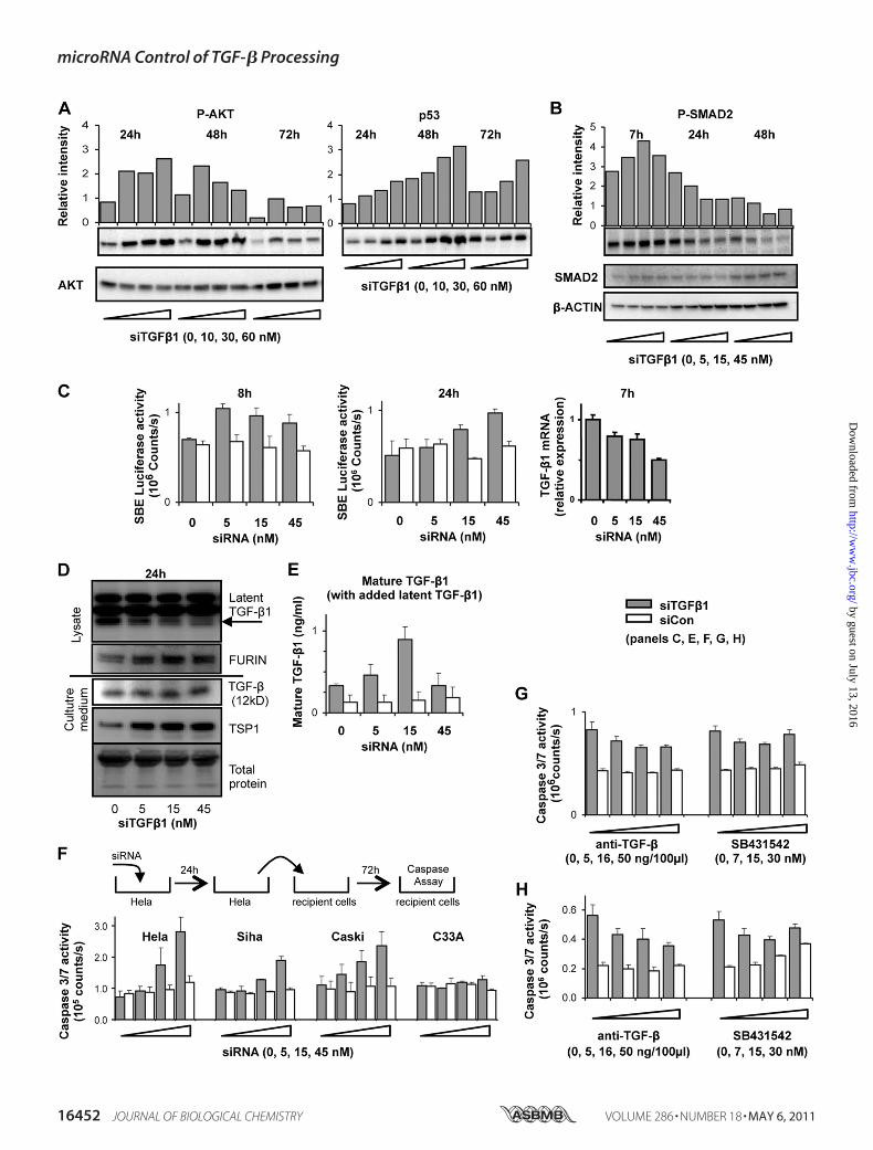
toma Cells—We next investigated whether TGF-�1 RNAi acti-vates TGF-� processing in cell types other than HeLa. TGF-�plays an important role in malignant glioblastoma. LN-18 cells,derived from a malignant glioma (52), carry a non-functional(heterozygous) TP53 gene (53) and express high levels of
TGF-�1 (45). We confirmed that LN-18 cells do not undergocaspase 3/7 induction upon treatment with human recombi-nant TGF-�1. LN-18 cells transfected with increasing concen-trations of siTGF�1 yielded a dose-dependent down-regulationof TGF-�1 mRNA (Fig. 5A) and a reduction of total TGF-�1protein present in supernatants (Fig. 5B). Interestingly, levels ofTHBS1mRNA increased by 2-fold (Fig. 5A), indicating perhapsa stronger transcriptional regulation of the gene in these cellscompared with in HeLa. The mRNAs of FURIN, SMAD2,CDKN1A, and TP53 remained constant. After 3 days, LN-18cells underwent apoptosis as shown by induction of caspase 3/7activity (Fig. 5C).Western blots from cell lysates showed reduc-tion of latent TGF-�1 and a corresponding dose-dependentincrease in P-SMAD2 and p53, but no major change in FURIN(Fig. 5D). Isolation of protein from concentratedmedia enabledprobing for the regulation of TSP1 and total TGF-�. The for-mer showed a strong dose-dependent up-regulation (Fig. 5E),however only traces of TGF-� could be observed. We turnedtherefore to the ELISA protocols with non-acid work-up. Incontrast to HeLa cells, it was not necessary to add recombinantlatent TGF-�1 to treated LN-18 cells to assay for changesin TGF-�1 processing. Increasing doses of siTGF�1 led toincreased amounts of mature TGF-�1 in comparison to siContreated cells (Fig. 5F). We examined levels of selected miRNAsin treated cells. SiTGF�1 reduced miR-18a levels by �30%,consistent with the induction of TSP1 protein (Fig. 5G). As inHeLa, transfection of LN-18 with miR-18 mimic alone led toaccumulation of latent TGF-�1 (Fig. 5H).
The results in LN-18 cells partially replicate the results fromHeLa cells. Inhibition of latent TGF-�1 by RNAi leads toincreasedTSP1 (but not FURIN), increased processing of latentTGF-�1, activation of P-SMAD2 and caspase 3/7 induction.Elevated levels of TSP1 likely derive from both transcriptionaland post-transcriptional regulation withmiR-18a contributingto the latter. The induction of caspase 3/7 activity in this celllinewas therefore also consistentwith restoration of theTGF-�cytostatic effect. However, the apoptotic mechanism appearsnot to involve transcriptional activity of p53 or P21, in accord-ance with P53 status and previous literature reports of unusualmechanisms of apoptosis in certain glioma cell lines (54).
DISCUSSION
Dysregulation of TGF-� signaling is at the heart of a varietyof important diseases. Inmany cancers only the tumor suppres-sor function of TGF-� is inactivated and therefore TGF-� sig-naling not only fails to protect cells against uncontrolled prolif-eration, but it also drives invasion and metastasis. A detailedunderstanding of the mechanisms by which tumor cells loseonly the TGF-� growth inhibitory response might lead to safe,new, and efficacious therapeutic strategies. Several mecha-nisms have been described but are mostly concentrated on thedownstream effectors of cytostatic TGF-� signaling (13). Theyinclude two accounts concerning TGF-�-associated oncomirs.In one, the repression of SMAD5 by miR-155 was associatedwith the development of leukemia/lymphoma in transgenicmice (55). In the second, overexpression of the miR-106b�25cluster attenuated TGF-�1 growth inhibition in gastric cancercells by blocking the synthesis of P21 and BIM (22). Despite a
microRNA Control of TGF-� Processing
MAY 6, 2011 • VOLUME 286 • NUMBER 18 JOURNAL OF BIOLOGICAL CHEMISTRY 16455
by guest on July 13, 2016http://w
ww
.jbc.org/D
ownloaded from
vast literature on TGF-�, aspects of its maturation in the con-text of cancer are rarely reported.Nevertheless, it iswell-knownthat cells synthesize excess TGF-� precursors, that the process-ing and activation of the ligand precursors are rate-limitingsteps in their bioavailability and that high levels of TGF-�detected by ELISA are commonly found circulating in cancerpatients. In general, also, cancer cell lines which have lost thecytostatic response to TGF-�1 often express high levels oflatent TGF-� and in cervical carcinoma cell lines specifically,levels of latent TGF-� correlate inversely with cytostaticresponse (36).To our knowledge there are no reports which clarify a causal
mechanistic link between high levels of latent TGF-� and theloss of the TGF-� cytostatic response; however, the failure toactivate secreted latent TGF-� was proposed as a possiblesource of resistance long ago (56). Here we show that latentTGF-�1 inhibits the TGF-� cytostatic response in some tumor
cell lines through a regulatory feedback loop involving miRNAcontrol of latent TGF-� processing factors. We demonstratedthat inhibition of TGF-�1 by RNAi in TP53-positive HeLa andTP53-mutated LN-18 cell lines induces caspase 3/7 activity andcell death. Data from a variety of experiments in these two celllines pointed to restoration of cytostatic TGF-� signaling as thesource of the apoptosis. We discovered an increased latentTGF-� processing activity in HeLa and LN-18 supernatantsundergoing TGF-�1 RNAi derived from elevated levels ofsecreted TSP1 and leading, in turn, to induction of P-SMAD2.In HeLa cells this increased extracellular TGF-�1 processingactivity was reinforced by a strong up-regulation of a secondTGF-� processing factor, intracellular FURIN. The results of alarge miRNA expression profile after TGF-�1 RNAi indicatedpotentially important post-transcriptional contributions to thismechanism. First, the relatively modest suppression of severaloncomirs and the strong induction ofmiR-34awere suggestive
FIGURE 5. Reactivation of TGF-� signaling upon down-regulation of latent TGF-�1 by siTGF�1 in LN-18 cells. A, cells were transfected with siTGF�1, andQ-PCR analysis was performed 48 h post-transfection. Relative expression of mRNA is displayed (mean of PCR triplicates; single RNA samples � S.D.). B, cellsgrown in 5% FBS media were treated with siRNAs. Supernatants were collected 72 h post-transfection, and total TGF-�1 was measured by ELISA afteracidification. Relative expression of TGF-�1 protein is displayed (mean of triplicate transfections �S.D.). C, cells grown in 5% FBS media were treated withsiRNAs. Caspase 3/7 activity was measured from lysates 72 h post-transfection (mean of triplicate transfections � S.D.). D and E, cells grown in media containing1% FBS were treated with siTGF�1. Protein was isolated from lysates, and concentrated media after 24 h. Western blots of latent TGF-�1, P-SMAD2, FURIN, p53from lysates, and TSP1 from concentrated media are displayed. F, cells grown in 5% FBS media were treated with siRNAs. Cell supernatants were collected 72 hpost-transfection and mature TGF-�1 measured by ELISA without acidification. Mature TGF-�1 protein is displayed (mean of triplicate transfections �S.D.).G, Q-PCR was performed on total RNA isolated after 48 h from cells transfected as in F. Relative miRNA expression is displayed (mean of PCR triplicates; singleRNA samples �S.E.). H, cells grown in media containing 1% FBS were treated with mir-18a. Western blot analysis of latent TGF-�1 was performed on lysates after24 h.
microRNA Control of TGF-� Processing
16456 JOURNAL OF BIOLOGICAL CHEMISTRY VOLUME 286 • NUMBER 18 • MAY 6, 2011
by guest on July 13, 2016http://w
ww
.jbc.org/D
ownloaded from

of a coordinated antiproliferativemiRNAnetwork in operation,somewhat akin to accounts of wide-scale miRNA reprogram-ming byVEGF signaling (18) orMYC-driven transcription (57).Second, rescue experiments usingmimics ofmiR-18a andmiR-24, which target TGF-� processing factors THBS1 and FURIN,respectively, increased levels of intracellular un-nicked latentTGF-�1, by distinct mechanisms and partially abrogated theapoptosis in amanner similar to a TGF-�1 overexpression con-struct. The data demonstrate important roles for latent TGF-�1, its processing factorsTHBS1 and FURIN as well asmiR-18aandmiR-24 in a feedback loop that regulates at least in part thematuration and cytostatic activity of TGF-� (Fig. 6).We did notconduct a full transcriptomic or proteomic analysis and there-fore we cannot exclude that other mediators may play signifi-cant roles in themechanism. The feedback loop in HeLa cells isinterrupted by TGF-�1 RNAi which, in turn, lowers levels oflatent TGF-�1, miR-18a, and miR-24, thereby de-repressingTHBS1, FURIN, and latent TGF-� processing. Two reportsoffer plausible explanations as to how high homeostatic levelsof latent TGF-�1 in HeLa maintain indirectly the expression ofoncomirs miR-18a and miR-24. In one, p53 was shown torepress transcriptionally the miR-17–92 cluster containingmiR-18a (50); in the second,miR-24was shown to be inhibitedvia SMAD signaling sites in its promoter (35).Our data suggest an additional mechanism for cancer cells
which secrete high levels of (latent) TGF-�1 to develop resis-tance to TGF-�-mediated growth inhibition. This mechanismstands apart from others because it takes place at the earliestpoint ofTGF-� signaling, the processing stage. The relevance ofthese findings for cancer are potentially important because: 1) ahigh proportion of patient tumors which secrete large amountsof this cytokine are reported to be refractory to TGF-� growthinhibition and 2) restoration of TGF-� cytostatic response maybe achievable pharmacologically through the use of antisenseor double-stranded oligoribonucleotides directed to TGF-�1mRNA. Our findings add a new layer of complexity to the biol-ogy of TGF-�. They emphasize the importance of consideringlatent andmature TGF-� as distinct entities and the processingof latent TGF-� as an integral part of growth inhibitory TGF-�signaling.
Acknowledgments—We thank M. Labow, W. Filipowicz, and A. Ger-ber for useful discussions, and B. Vogelstein for the SBE reporterplasmid.
REFERENCES1. Annes, J. P., Munger, J. S., and Rifkin, D. B. (2003) J. Cell Sci. 116, 217–2242. Wang, J., Sergina, N., Ko, T. C., Gong, J., and Brattain, M. G. (2004) J. Biol.
Chem. 279, 40237–402443. tenDijke, P., andArthur, H.M. (2007)Nat. Rev.Mol. Cell Biol. 8, 857–8694. Derynck, R., and Zhang, Y. E. (2003) Nature 425, 577–5845. Massague, J. (2008) Cell 134, 215–2306. Zhang, Y. E. (2009) Cell Res. 19, 128–1397. Dumont, N., Bakin, A. V., and Arteaga, C. L. (2003) J. Biol. Chem. 278,
3275–32858. Ye, S. C., Foster, J. M., Li, W., Liang, J., Zborowska, E., Venkateswarlu, S.,
Gong, J., Brattain, M. G., and Willson, J. K. (1999) Cancer Res. 59,4725–4731
9. Alexandrow, M. G., and Moses, H. L. (1995) Cancer Res. 55, 1452–145710. Derynck, R., Goeddel, D. V., Ullrich, A., Gutterman, J. U., Williams, R. D.,
Bringman, T. S., and Berger, W. H. (1987) Cancer Res. 47, 707–71211. Teicher, B. A. (2007) Clin. Cancer Res. 13, 6247–625112. Pardali, K., andMoustakas, A. (2007) Biochim. Biophys. Acta 1775, 21–6213. Seoane, J. (2006) Carcinogenesis 27, 2148–215614. Bakin, A. V., Tomlinson, A. K., Bhowmick, N. A., Moses, H. L., and
Arteaga, C. L. (2000) J. Biol. Chem. 275, 36803–3681015. Wang, J. W., Yang, L., Yang, J., Kuropatwinski, K., Wang, W., Liu, X. Q.,
Hauser, J., and Brattain, M. G. (2008) Cancer Res. 68, 3152–316016. Seoane, J., Le, H. V., Shen, L., Anderson, S. A., andMassague, J. (2004)Cell
117, 211–22317. Bushati, N., and Cohen, S. M. (2007) Annu. Rev. Cell Dev. Biol. 23,
175–20518. Suarez, Y., Fernandez-Hernando, C., Yu, J., Gerber, S. A., Harrison, K. D.,
Pober, J. S., Iruela-Arispe, M. L., Merkenschlager, M., and Sessa, W. C.(2008) Proc. Natl. Acad. Sci. U.S.A. 105, 14082–14087
19. Li, X., and Carthew, R. W. (2005) Cell 123, 1267–127720. Martello,G., Zacchigna, L., Inui,M.,Montagner,M., Adorno,M.,Mamidi,
A., Morsut, L., Soligo, S., Tran, U., Dupont, S., Cordenonsi, M., Wessely,O., and Piccolo, S. (2007) Nature 449, 183–188
21. Yao, G., Yin, M., Lian, J., Tian, H., Liu, L., Li, X., and Sun, F. (2010) Mol.Endocrinol. 24, 540–551
22. Petrocca, F., Visone, R., Onelli, M. R., Shah, M. H., Nicoloso, M. S., deMartino, I., Iliopoulos, D., Pilozzi, E., Liu, C. G., Negrini, M., Cavazzini, L.,Volinia, S., Alder, H., Ruco, L. P., Baldassarre, G., Croce, C. M., and Vec-chione, A. (2008) Cancer Cell 13, 272–286
23. Gregory, P. A., Bert, A. G., Paterson, E. L., Barry, S. C., Tsykin, A., Farshid,G., Vadas, M. A., Khew-Goodall, Y., and Goodall, G. J. (2008) Nat. CellBiol. 10, 593–601
24. Park, S. M., Gaur, A. B., Lengyel, E., and Peter, M. E. (2008)Genes Dev. 22,894–907
25. Burk, U., Schubert, J., Wellner, U., Schmalhofer, O., Vincan, E., Spaderna,S., and Brabletz, T. (2008) EMBO Reports 9, 582–589
26. Korpal, M., Lee, E. S., Hu, G., and Kang, Y. (2008) J. Biol. Chem. 283,14910–14914
27. Kong, W., Yang, H. H., He, L., Zhao, J. J., Coppola, D., Dalton, W. S., andCheng, J. Q. (2008)Mol. Cell. Biol. 28, 6773–6784
28. Wang, B., Hsu, S. H.,Majumder, S., Kutay, H., Huang,W., Jacob, S. T., andGhoshal, K. (2010) Oncogene 29, 1787–1797
29. Davis, B. N., Hilyard, A. C., Lagna, G., and Hata, A. (2008) Nature 454,56–61
30. Dews, M., Fox, J. L., Hultine, S., Sundaram, P., Wang,W., Yingqui, Y., Liu,Y. Y., Furth, E., Enders, G. H., El-Deiry, W., Schelter, J. M., Cleary, M. A.,and Thomas-Tikhonenko, A. (2010) Cancer Res. 70, 8233–8246
31. Mestdagh, P., Bostrom, A. K., Impens, F., Fredlund, E., Van Peer, G., DeAntonellis, P., von Steding, K., Ghesquiere, B., Schulte, S., Dews, M.,Thomas-Tikhonenko, A., Schulte, J., Zollo, M., Schramm, A., Gevaert, K.,Axelson, H., Speleman, F., and Vandesompele, J. (2010) Mol. Cell 40,
FIGURE 6. Latent TGF-�1, TSP1, FURIN, miR-18a, and miR-24 are part of aregulatory feedback loop. High levels of latent TGF-�1 indirectly maintainexpression of miR-18a and miR-24 in HeLa cells, resulting in repression of TSP1and FURIN, reduced processing of latent TGF-� precursors, increased TGF-�1transcription and accumulation of un-nicked latent TGF-�1. Upon TGF-�1RNAi miR-18a and miR-24 are inhibited, TSP1 and FURIN are induced, therebyincreasing latent TGF-� processing and leading to apoptosis. Dotted arrowsrepresent indirect regulations, filled arrows represent direct interactions.
microRNA Control of TGF-� Processing
MAY 6, 2011 • VOLUME 286 • NUMBER 18 JOURNAL OF BIOLOGICAL CHEMISTRY 16457
by guest on July 13, 2016http://w
ww
.jbc.org/D
ownloaded from

762–77332. Chan, M. C., Hilyard, A. C., Wu, C., Davis, B. N., Hill, N. S., Lal, A.,
Lieberman, J., Lagna, G., and Hata, A. (2010) EMBO J. 29, 559–57333. Luna, C., Li, G., Qiu, J., Epstein, D.L., and Gonzalez, P. (2011) J. Cell. Phys.
226, 1407–141434. Wang, Q., Huang, Z., Xue, H., Jin, C., Ju, X., Han, J. J., and Chen, Y. (2008)
Blood 111, 588–59535. Sun,Q., Zhang, Y., Yang,G., Chen, X., Zhang, Y., Cao,G.,Wang, J., Sun, Y.,
Zhang, P., Fan, M., Shao, N., and Yang, X. (2008) Nucleic Acids Res. 36,2690–2699
36. Kloth, J. N., Fleuren, G. J., Oosting, J., de Menezes, R. X., Eilers, P. H.,Kenter, G. G., and Gorter, A. (2005) Carcinogenesis 26, 1493–1502
37. Lyons, R. M., Keski-Oja, J., and Moses, H. L. (1988) J. Cell Biol. 106,1659–1665
38. Goodwin, E. C., and DiMaio, D. (2000) Proc. Natl. Acad. Sci. U.S.A. 97,12513–12518
39. He, X., He, L., and Hannon, G. J. (2007) Cancer Res. 67, 11099–1110140. Wang, X., Wang, H. K., McCoy, J. P., Banerjee, N. S., Rader, J. S., Broker,
T. R., Meyers, C., Chow, L. T., and Zheng, Z. M. (2009) RNA 15, 637–64741. Park, S. Y., Lee, J. H., Ha,M., Nam, J.W., andKim, V.N. (2009)Nat. Struct.
Mol. Biol. 16, 23–2942. Oren, M., Damalas, A., Gottlieb, T., Michael, D., Taplick, J., Leal, J. F.,
Maya, R., Moas, M., Seger, R., Taya, Y., and Ben-Ze’ev, A. (2002) Biochem.Pharmacol. 64, 865–871
43. Solovyan, V. T., and Keski-Oja, J. (2006) J. Cell. Physiol. 207, 445–45344. Gal, A., Sjoblom, T., Fedorova, L., Imreh, S., Beug, H., and Moustakas, A.
(2008) Oncogene 27, 1218–123045. Tritschler, I., Gramatzki, D., Capper, D.,Mittelbronn,M.,Meyermann, R.,
Saharinen, J., Wick, W., Keski-Oja, J., andWeller, M. (2009) Int. J. Cancer125, 530–540
46. Jenkins, G. (2008) Int. J. Biochem. Cell Biol. 40, 1068–107847. Schultz-Cherry, S., Ribeiro, S., Gentry, L., andMurphy-Ullrich, J. E. (1994)
J. Biol. Chem. 269, 26775–2678248. Blakytny, R., Ludlow, A., Martin, G. E., Ireland, G., Lund, L. R., Ferguson,
M. W., and Brunner, G. (2004) J. Cell. Physiol. 199, 67–7649. Dews, M., Homayouni, A., Yu, D., Murphy, D., Sevignani, C., Wentzel, E.,
Furth, E. E., Lee, W. M., Enders, G. H., Mendell, J. T., and Thomas-Tikhonenko, A. (2006) Nat. Genet. 38, 1060–1065
50. Yan, H. L., Xue, G., Mei, Q., Wang, Y. Z., Ding, F. X., Liu, M. F., Lu, M. H.,Tang, Y., Yu, H. Y., and Sun, S. H. (2009) EMBO J. 28, 2719–2732
51. Huang, S., He, X., Ding, J., Liang, L., Zhao, Y., Zhang, Z., Yao, X., Pan, Z.,Zhang, P., Li, J., Wan, D., and Gu, J. (2008) Int. J. Cancer 123, 972–978
52. Diserens, A. C., de Tribolet, N., Martin-Achard, A., Gaide, A. C., Schnegg,J. F., and Carrel, S. (1981) Acta Neuropathol. 53, 21–28
53. VanMeir, E. G., Kikuchi, T., Tada,M., Li, H., Diserens, A. C.,Wojcik, B. E.,Huang, H. J., Friedmann, T., de Tribolet, N., and Cavenee, W. K. (1994)Cancer Res. 54, 649–652
54. Wischhusen, J., Naumann, U., Ohgaki, H., Rastinejad, F., and Weller, M.(2003) Oncogene 22, 8233–8245
55. Rai, D., Kim, S. W., McKeller, M. R., Dahia, P. L., and Aguiar, R. C. (2010)Proc. Natl. Acad. Sci. U.S.A. 107, 3111–3116
56. Jennings, M. T., Maciunas, R. J., Carver, R., Bascom, C. C., Juneau, P.,Misulis, K., and Moses, H. L. (1991) Int. J. Cancer 49, 129–139
57. Chang, T. C., Yu, D., Lee, Y. S., Wentzel, E. A., Arking, D. E., West, K. M.,Dang, C. V., Thomas-Tikhonenko, A., and Mendell, J. T. (2008) Nat.Genet. 40, 43–50
microRNA Control of TGF-� Processing
16458 JOURNAL OF BIOLOGICAL CHEMISTRY VOLUME 286 • NUMBER 18 • MAY 6, 2011
by guest on July 13, 2016http://w
ww
.jbc.org/D
ownloaded from

SUPPLEMENTAL INFORMATION
Suppression of latent TGF-beta1 restores growth inhibitory TGF-beta
signaling through microRNAs
Afzal M. Dogar, Harry Towbin and Jonathan Hall
Ref_seq Name Sequence of primerNM_000660.3 TGFB1_F GCAGGGATAACACACTGCAA
TGFB1_R GGCCATGAGAAGCAGGAA
NM_003238.2 TGFB2_F ACAAGAGCAGAAGGCGAATG
TGFB2_R TGCAGCAGGGACAGTGTAAG
NM_005901.4 SMAD2_F GGAATTTGCTGCTCTTCTGG
SMAD2_R TCTGCCTTCGGTATTCTGCT
X04354.1 18E6/7_F ATGCTGCATGCCATAAATGT
18E6/7_R CCCAGTGTTAGTTAGTTTTTCCAA
NM_000546 TP53_F GCGCACAGAGGAAGAGAATC
TP53_R GGAGAGGAGCTGGTGTTGTT
NM_078467 CDKN1A_F GGAGACTCTCAGGGTCGAAA
CDKN1A_R CTTCCTGTGGGCGGATTAG
NM_138763 BAX_F TCTGACGGCAACTTCAACTG
BAX_R CAGCCCATGATGGTTCTGAT
NM_006763 BTG2_F AGCGAGCAGAGGCTTAAGGT
BTG2_R TCTTGTGGTTGATGCGAATG
NM_000043 FAS_F TGCAGAAAGCACAGAAAGGA
FAS_R TGACTCCAGCAATAGTGGTGAT
NM_147187 TNFR10B_F TTGTGGCTGTGTTTGTTTGC
TNFR10B_R CAGGTCGTTGTGAGCTTCTG
NM_003246 THBS1_F AGCATGGTCCTGGAACTCAG
THBS1_R CAGCTCATTGGCCAACTCTT
NM_002569.2 FURIN_F GGCATTGTGGTCTCCATTCT
FURIN_R GGGTCCTGGTCATTGACATC
NM_002046.3 GAPDH_F GAAGGTGAAGGTCGGAGTCA
GAPDH_R AATGAAGGGGTCATTGATGG
Supplemental Table S1. Sequences of forward (F) and reverse (R) PCR primers used to analyze
mRNA expression after treatment with siTGF1 and siTGF1(s).

siTGFβ1 Normalized to untreated (0 nM) siTGFβ1 (s) Normalized to untreated (0 nM)averages TTESTs SEM averages TTESTs SEM
0.05 0.05miRNAs 0 nM 5 nM 15 nM 45 nM 5 nM 15 nM 45 nM 0 nM 5 nM 15 nM 45 nM miRNAs 0 nM 5 nM 15 nM 45 nM 5 nM 15 nM 45 nM 0 nM 5 nM 15 nM 45 nMhsa-miR-10a 1.0 0.52 0.40 0.45 0.07 0.07 0.07 0.14 0.14 0.07 0.09 hsa-miR-10a 1.0 1.59 1.18 0.93 0.08 0.45 0.70 0.21 0.21 0.14 0.08hsa-miR-17-5p 1.0 0.23 0.47 0.22 0.02 0.10 0.02 0.37 0.05 0.03 0.07 hsa-miR-17-5p 1.0 0.80 0.84 1.08 0.31 0.57 0.59 0.16 0.14 0.23 0.12hsa-miR-21 1.0 0.34 0.74 0.60 0.02 0.26 0.15 0.20 0.09 0.12 0.18 hsa-miR-21 1.0 0.55 1.09 0.71 0.05 0.71 0.12 0.19 0.10 0.18 0.07hsa-miR-24 1.0 0.34 0.40 0.26 0.05 0.03 0.01 0.23 0.12 0.08 0.08 hsa-miR-24 1.0 0.59 0.49 0.37 0.09 0.09 0.01 0.21 0.11 0.14 0.06hsa-miR-34a 1.0 0.59 5.21 2.46 0.10 0.00 0.03 0.21 0.13 0.46 0.67 hsa-miR-34a 1.0 1.38 2.14 6.47 0.06 0.01 0.00 0.14 0.19 0.36 0.50hsa-miR-92 1.0 0.47 1.23 0.59 0.01 0.38 0.01 0.15 0.07 0.22 0.11 hsa-miR-92 1.0 0.85 0.87 0.52 0.23 0.22 0.01 0.14 0.10 0.09 0.07hsa-miR-93 1.0 0.47 0.61 0.27 0.03 0.20 0.00 0.21 0.10 0.16 0.05 hsa-miR-93 1.0 0.78 0.73 0.35 0.17 0.04 0.00 0.16 0.10 0.07 0.03hsa-miR-155 1.0 0.26 0.41 0.30 0.01 0.00 0.00 0.11 0.08 0.01 0.07 hsa-miR-155 1.0 0.65 0.58 0.32 0.04 0.03 0.00 0.15 0.09 0.09 0.06hsa-miR-301 1.0 0.30 0.35 0.17 0.00 0.00 0.00 0.17 0.05 0.01 0.04 hsa-miR-301 1.0 0.51 0.89 1.24 0.01 0.33 0.14 0.14 0.07 0.11 0.15hsa-miR-20a 1.0 0.63 0.52 0.46 0.10 0.04 0.02 0.21 0.12 0.07 0.10 hsa-miR-20a 1.0 1.03 0.98 0.76 0.76 0.89 0.04 0.14 0.10 0.16 0.06hsa-miR-137 1.0 0.38 0.32 0.23 0.08 0.02 0.02 0.29 0.13 0.05 0.08 hsa-miR-137 1.0 0.85 0.49 0.34 0.61 0.07 0.02 0.29 0.13 0.08 0.06hsa-miR-146b 1.0 0.42 0.61 0.42 0.07 0.16 0.20 0.20 0.14 0.14 0.24 hsa-miR-146b 1.0 1.14 1.63 1.00 0.52 0.05 1.00 0.17 0.19 0.26 0.22hsa-miR-181b 1.0 0.47 0.51 0.36 0.09 0.14 0.06 0.32 0.09 0.10 0.11 hsa-miR-181b 1.0 0.82 0.44 0.18 0.08 0.02 0.00 0.14 0.08 0.09 0.02hsa-miR-181d 1.0 0.44 0.42 0.32 0.04 0.03 0.02 0.18 0.12 0.10 0.10 hsa-miR-181d 1.0 0.81 0.46 0.17 0.25 0.01 0.00 0.19 0.07 0.06 0.03hsa-miR-20b 1.0 0.46 0.27 0.19 0.05 0.01 0.01 0.25 0.09 0.05 0.06 hsa-miR-20b 1.0 0.83 0.57 0.27 0.23 0.03 0.00 0.18 0.08 0.09 0.05hsa-miR-152 1.0 0.50 0.40 0.27 0.10 0.02 0.04 0.23 0.14 0.06 0.12 hsa-miR-152 1.0 0.88 0.52 0.36 0.13 0.00 0.00 0.13 0.09 0.07 0.07hsa-miR-30c 1.0 1.00 1.00 1.00 1.00 1.00 1.00 0.16 0.18 0.02 0.26 hsa-miR-30c 1.0 1.00 1.00 1.00 1.00 1.00 1.00 0.17 0.12 0.12 0.10hsa-miR-224 1.0 0.61 0.53 0.34 0.30 0.15 0.07 0.32 0.20 0.10 0.12 hsa-miR-224 1.0 0.66 0.30 0.14 0.17 0.02 0.00 0.20 0.14 0.08 0.02hsa-miR-28 1.0 0.56 0.66 0.64 0.17 0.27 0.26 0.31 0.13 0.11 0.17 hsa-miR-28 1.0 0.85 0.45 0.41 0.49 0.05 0.04 0.22 0.12 0.10 0.10hsa-miR-190 1.0 0.61 0.51 0.36 0.08 0.02 0.01 0.22 0.10 0.02 0.08 hsa-miR-190 1.0 0.79 0.34 0.38 0.10 0.00 0.00 0.14 0.10 0.03 0.05hsa-miR-193a 1.0 0.52 0.44 0.46 0.01 0.00 0.03 0.13 0.09 0.05 0.14 hsa-miR-193a 1.0 0.81 0.64 0.66 0.44 0.32 0.15 0.27 0.08 0.21 0.05hsa-miR-196a 1.0 0.50 0.35 0.29 0.04 0.01 0.02 0.20 0.11 0.03 0.09 hsa-miR-196a 1.0 0.86 0.58 0.43 0.36 0.02 0.00 0.14 0.13 0.09 0.04hsa-miR-374 1.0 0.55 0.46 0.37 0.01 0.01 0.02 0.15 0.08 0.07 0.11 hsa-miR-374 1.0 0.69 0.44 0.31 0.09 0.00 0.00 0.15 0.11 0.06 0.03hsa-miR-106b 1.0 0.50 0.50 0.40 0.03 0.04 0.01 0.19 0.09 0.09 0.10 hsa-miR-106b 1.0 1.02 0.96 0.91 0.94 0.88 0.72 0.25 0.10 0.15 0.12hsa-miR-130b 1.0 0.55 0.31 0.31 0.05 0.00 0.00 0.12 0.14 0.02 0.07 hsa-miR-130b 1.0 0.88 0.57 0.56 0.06 0.01 0.00 0.13 0.07 0.07 0.04hsa-miR-29a 1.0 0.79 1.02 1.03 0.37 0.94 0.91 0.25 0.12 0.10 0.22 hsa-miR-29a 1.0 1.29 0.83 0.76 0.01 0.17 0.16 0.13 0.11 0.11 0.13hsa-miR-29c 1.0 0.78 0.72 0.89 0.35 0.27 0.66 0.26 0.10 0.06 0.19 hsa-miR-29c 1.0 1.13 1.46 5.71 0.08 0.00 0.00 0.13 0.09 0.13 0.52hsa-miR-181a 1.0 0.97 0.74 0.83 0.95 0.56 0.68 0.41 0.15 0.19 0.19 hsa-miR-181a 1.0 1.03 0.75 1.01 0.90 0.37 0.96 0.26 0.12 0.15 0.11hsa-miR-106a 1.0 0.69 0.66 0.82 0.26 0.24 0.49 0.28 0.13 0.11 0.16 hsa-miR-106a 1.0 0.25 0.99 1.13 0.41 0.96 0.44 0.19 0.38 0.13 0.08hsa-miR-18a 1.0 0.90 0.67 0.55 0.08 0.00 0.00 0.09 0.03 0.05 0.09 hsa-miR-18a 1.0 0.84 0.62 0.65 0.03 0.00 0.00 0.05 0.04 0.02 0.02
Supplemental Table S2. MiRNA expression analysis after treatment of HeLa cells with siTGF1
and siTGF1(s). Total RNA was isolated 48h post-siRNA-transfection and subjected to miRNA
stem-loop Q-PCR. Data were normalized to expression levels of hsa-miR-30c, which remained
constant throughout treatment with both siRNAs. MiRNA expression levels relative to the 0nM
siRNA treatment are displayed. P-values below 0.05 are indicated as shaded boxes and were
calculated using t-test.

Afzal M. Dogar, Harry Towbin and Jonathan Hall Signaling through microRNAsβInhibitory TGF-
1 Restores GrowthβSuppression of Latent Transforming Growth Factor (TGF)-
doi: 10.1074/jbc.M110.208652 originally published online March 14, 20112011, 286:16447-16458.J. Biol. Chem.
10.1074/jbc.M110.208652Access the most updated version of this article at doi:
Alerts:
When a correction for this article is posted•
When this article is cited•
to choose from all of JBC's e-mail alertsClick here
Supplemental material:
http://www.jbc.org/content/suppl/2011/03/14/M110.208652.DC1.html
http://www.jbc.org/content/286/18/16447.full.html#ref-list-1
This article cites 57 references, 25 of which can be accessed free at
by guest on July 13, 2016http://w
ww
.jbc.org/D
ownloaded from