The bacteriology of chronic venous leg ulcer examined by culture-independent molecular methods
12
The bacteriology of chronic venous leg ulcer examined by culture-independent molecular methods Trine R. Thomsen, PhD 1,2 ; Martin S. Aasholm, MSc 1 ; Vibeke B. Rudkjbing, BSc 1 ; Aaron M. Saunders, PhD 1,2 ; Thomas Bjarnsholt, PhD 3 ; Michael Givskov, Dr. Tech 3 ; Klaus Kirketerp-Mller, MD 4 ; Per H. Nielsen, PhD 1 1. Department of Biotechnology, Chemistry and Environmental Engineering, Aalborg University, Aalborg, Denmark, 2. The Danish Technological Institute, Chemistry and Water Technology, A ˚ rhus, Denmark, 3. Department of International Health, Immunology and Microbiology, University of Copenhagen, Copenhagen, Denmark, and 4. Copenhagen Wound Healing Center, Bispebjerg Hospital, Bispebjerg, Denmark Reprint requests: Trine R. Thomsen, Department of Biotechnology, Chemistry and Environmental Engineering, Aalborg University, Sohngaardsholmsvej 49, DK-9000 Aalborg, Denmark. Tel: 45 7220 1828; Fax: 45 9814 1808; Email: [email protected] Manuscript received: October 10, 2008 Accepted in final form: November 1, 2009 DOI:10.1111/j.1524-475X.2009.00561.x ABSTRACT The bacterial microbiota plays an important role in the prolonged healing of chronic venous leg ulcers. The present study compared the bacterial diversity within ulcer material from 14 skin graft operations of chronic venous leg ulcers using culture-based methods and molecular biological methods, such as 16S rRNA gene sequencing, fingerprinting, quantitative polymerase chain reaction, and fluorescence in situ hybridization. Each wound contained an average of 5.4 species but the actual species varied between wounds. The diversity determined by culture-based methods and the molecular biological methods was different. All the wounds contained Staphylococcus aureus, whereas Pseudomonas aerugi- nosa was in six out of 14 wounds. Molecular methods detected anaerobic patho- gens in four ulcers that were not detected with anaerobic culture methods. Quantitative polymerase chain reaction was used to compare the abundance of S. aureus and P. aeruginosa at different locations in the ulcers and their numbers varied greatly between samples taken at different locations in the same ulcer. This should be considered when ulcers are investigated in routine clinical care. The differences between the results obtained with culture-based and molecular-based approaches demonstrate that the use of one approach alone is not able to identify all of the bacteria present in the wounds. Chronic venous leg ulcers (CVLU) are a debilitating and often painful disease that affects approximately 1% of the world’s population. 1,2 Apart from the human conse- quences, the treatment of wounds is expensive; in Denmark alone, wound treatment has been estimated to cost approximately two billion Danish kroner per year ( US$360 million), 2 and in the United Kingdom, France, and Germany an estimated 1.5–2% of the annual healthcare budget. 3,4 The conditions leading to a CVLU are not fully under- stood; however, the primary cause is most likely insuffi- cient valvular function of the veins in the legs causing increased hydrostatic pressure leading to edema of the subcutaneous tissue, which predispose to ulceration. This is linked to old age, obesity, height of the person, he- reditary increased risk, number of births (more births lead to increased risk) and occupations in which the person is mainly standing. By removing edema with com- pression therapy, most CVLU will heal, but a number of ulcers will not despite effective treatment. In these cases, a well-documented and effective treatment is surgical debridement and split skin transplant. 2 Other treatments like topical negative pressure therapy have been found useful. Maggot debridement therapy have also proved promising, which involves having larvae from the fly Lucilia sericata removing necrotic tissue and bacteria from the wound, and in this way aiding the wound healing process. 5 One of the factors affecting the effectiveness of wound healing therapies is the specific microorganisms that colo- nize the CVLU. 6 For example, the presence of Pseudomo- nas aeruginosa can retard the healing of wounds due to their ability to form biofilms. 6 Many studies describe bio- film as an important factor for the chronic behavior of chronic wounds, 6–10 and the spatial organization of these biofilms in a wound might be complex due to, for example, variations in environmental conditions and population composition. 11 Initial experiments by Bjarnsholt et al. 6 showed that P. aeruginosa in CVLU were assembled in microcolony-based structures unevenly distributed across the wound surface, and this uneven distribution might lead to insufficient sampling and wrong diagnosis. 6 Until recently, the bacteria associated with CVLU have only been examined by culture-dependent methods by tak- ing a swab or biopsy from the wound and using it as inoc- ulate for various bacterial cultures. The emergence of molecular biology methods has illustrated that culture- dependent methods often underestimate the bacteria pres- ent, and especially ulcers with slow growing, fastidious, or anaerobic microbes. 9,12–14 Davies et al. 15 found that 40% of the organisms identified from CVLU by molecular bio- logical methods could not be identified by culture-depen- dent methods, although most were species that are normally considered culturable. The purpose of this study was to investigate the micro- bial diversity of chronic ulcers with molecular biological Wound Rep Reg (2010) 18 38–49 c 2010 by the Wound Healing Society 38 Wound Repair and Regeneration
Transcript of The bacteriology of chronic venous leg ulcer examined by culture-independent molecular methods
The bacteriology of chronic venous leg ulcer examinedby culture-independent molecular methods
Trine R. Thomsen, PhD1,2; Martin S. Aasholm, MSc1; Vibeke B. Rudkj�bing, BSc1; Aaron M. Saunders, PhD1,2;Thomas Bjarnsholt, PhD3; Michael Givskov, Dr. Tech3; Klaus Kirketerp-M�ller, MD4; Per H. Nielsen, PhD1
1. Department of Biotechnology, Chemistry and Environmental Engineering, Aalborg University, Aalborg, Denmark,
2. The Danish Technological Institute, Chemistry and Water Technology, Arhus, Denmark,
3. Department of International Health, Immunology and Microbiology, University of Copenhagen, Copenhagen, Denmark, and
4. Copenhagen Wound Healing Center, Bispebjerg Hospital, Bispebjerg, Denmark
Reprint requests:Trine R. Thomsen, Department of
Biotechnology, Chemistry and
Environmental Engineering, Aalborg
University, Sohngaardsholmsvej 49,
DK-9000 Aalborg, Denmark.
Tel: 45 7220 1828;
Fax: 45 9814 1808;
Email: [email protected]
Manuscript received: October 10, 2008
Accepted in final form: November 1, 2009
DOI:10.1111/j.1524-475X.2009.00561.x
ABSTRACT
The bacterial microbiota plays an important role in the prolonged healing ofchronic venous leg ulcers. The present study compared the bacterial diversitywithin ulcer material from 14 skin graft operations of chronic venous leg ulcersusing culture-based methods and molecular biological methods, such as 16SrRNA gene sequencing, fingerprinting, quantitative polymerase chain reaction,and fluorescence in situ hybridization. Each wound contained an average of 5.4species but the actual species varied between wounds. The diversity determinedby culture-based methods and the molecular biological methods was different.All the wounds contained Staphylococcus aureus, whereas Pseudomonas aerugi-nosa was in six out of 14 wounds. Molecular methods detected anaerobic patho-gens in four ulcers that were not detected with anaerobic culture methods.Quantitative polymerase chain reaction was used to compare the abundance ofS. aureus and P. aeruginosa at different locations in the ulcers and their numbersvaried greatly between samples taken at different locations in the same ulcer. Thisshould be considered when ulcers are investigated in routine clinical care. Thedifferences between the results obtained with culture-based and molecular-basedapproaches demonstrate that the use of one approach alone is not able to identifyall of the bacteria present in the wounds.
Chronic venous leg ulcers (CVLU) are a debilitating andoften painful disease that affects approximately 1% of theworld’s population.1,2 Apart from the human conse-quences, the treatment of wounds is expensive; inDenmark alone, wound treatment has been estimatedto cost approximately two billion Danish kroner peryear (�US$360 million),2 and in the United Kingdom,France, and Germany an estimated 1.5–2% of the annualhealthcare budget.3,4
The conditions leading to a CVLU are not fully under-stood; however, the primary cause is most likely insuffi-cient valvular function of the veins in the legs causingincreased hydrostatic pressure leading to edema ofthe subcutaneous tissue, which predispose to ulceration.This is linked to old age, obesity, height of the person, he-reditary increased risk, number of births (more birthslead to increased risk) and occupations in which theperson is mainly standing. By removing edema with com-pression therapy, most CVLU will heal, but a number ofulcers will not despite effective treatment. In these cases, awell-documented and effective treatment is surgicaldebridement and split skin transplant.2 Other treatmentslike topical negative pressure therapy have been founduseful. Maggot debridement therapy have also provedpromising, which involves having larvae from the flyLucilia sericata removing necrotic tissue and bacteriafrom the wound, and in this way aiding the wound healingprocess.5
One of the factors affecting the effectiveness of woundhealing therapies is the specific microorganisms that colo-nize the CVLU.6 For example, the presence of Pseudomo-nas aeruginosa can retard the healing of wounds due totheir ability to form biofilms.6 Many studies describe bio-film as an important factor for the chronic behavior ofchronic wounds,6–10 and the spatial organization of thesebiofilms in a wound might be complex due to, for example,variations in environmental conditions and populationcomposition.11 Initial experiments by Bjarnsholt et al.6
showed that P. aeruginosa in CVLU were assembled inmicrocolony-based structures unevenly distributed acrossthe wound surface, and this uneven distribution might leadto insufficient sampling and wrong diagnosis.6
Until recently, the bacteria associated with CVLU haveonly been examined by culture-dependent methods by tak-ing a swab or biopsy from the wound and using it as inoc-ulate for various bacterial cultures. The emergence ofmolecular biology methods has illustrated that culture-dependent methods often underestimate the bacteria pres-ent, and especially ulcers with slow growing, fastidious, oranaerobic microbes.9,12–14 Davies et al.15 found that 40%of the organisms identified from CVLU by molecular bio-logical methods could not be identified by culture-depen-dent methods, although most were species that arenormally considered culturable.
The purpose of this study was to investigate the micro-bial diversity of chronic ulcers with molecular biological
Wound Rep Reg (2010) 18 38–49 c� 2010 by the Wound Healing Society38
Wound Repair and Regeneration
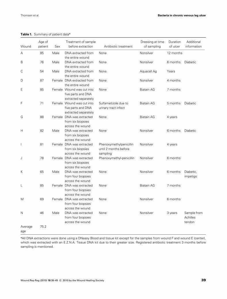
Table 1. Summary of patient datan
Wound
Age of
patient Sex
Treatment of sample
before extraction Antibiotic treatment
Dressing at time
of sampling
Duration
of ulcer
Additional
information
A 85 Male DNA extracted from
the entire wound
None Nonsilver 12 months
B 76 Male DNA extracted from
the entire wound
None Nonsilver 6 months Diabetic
C 54 Male DNA extracted from
the entire wound
None Aquacell Ag Years
D 87 Female DNA extracted from
the entire wound
None Nonsilver 4 months
E 85 Female Wound was cut into
five parts and DNA
extracted separately
None Biatain AG 7 months
F 71 Female Wound was cut into
five parts and DNA
extracted separately
Sulfametizole due to
urinary tract infect
Biatain AG 5 months Diabetic
G 88 Female DNA was extracted
from six biopsies
across the wound
None Biatain AG 4 years
H 82 Male DNA was extracted
from six biopsies
across the wound
None Nonsilver 6 months Diabetic
I 81 Female DNA was extracted
from six biopsies
across the wound
Phenoxymethylpenicillin
until 2 months before
sampling
Nonsilver 4 years
J 78 Female DNA was extracted
from six biopsies
across the wound
Phenoxymethyl-penicillin Nonsilver 6 months
K 65 Male DNA was extracted
from four biopsies
across the wound
None Nonsilver 6 months Diabetic,
impetigo
L 85 Female DNA was extracted
from four biopsies
across the wound
None Biatain AG 7 months
M 69 Female DNA was extracted
from four biopsies
across the wound
None Nonsilver 6 months
N 46 Male DNA was extracted
from four biopsies
across the wound
None Nonsilver 3 years Sample from
Achilles
tendon
Average
age
75.2
nAll DNA extractions were done using a DNeasy Blood and tissue kit except for the samples from wound F and wound E (center),
which was extracted with an E.Z.N.A. Tissue DNA kit due to their greater size. Registered antibiotic treatment 3 months before
sampling is mentioned.
Wound Rep Reg (2010) 18 38–49 c� 2010 by the Wound Healing Society 39
Bacteria in chronic venous leg ulcerThomsen et al.
methods and to compare these results with the conven-tional culture-dependent techniques. Furthermore, thespatial organization of bacteria in CVLU was examined.
MATERIALS AND METHODS
Patient population, sampling, and DNA extraction
The excision of biopsies and swabs of the wounds forculture-dependent and -independent experiments was per-formed by Copenhagen Wound Healing Center, Bispeb-jerg Hospital (Copenhagen, Denmark). Samples wereobtained from patients diagnosed with chronic venous legwounds just before surgical debridement and split skintransplant. In total, chronic wounds from 14 patients wereinvestigated (named as wound A–N). The patients’ age,sex, antibiotic treatment, dressings at the time of sampling,and additional information are described in Table 1. Pa-tients with wound B, F, H, and Kwere also diagnosed withdiabetes mellitus.
All ulcers were chronic and nonhealing despite optimalwound care and compression therapy. The duration of theulcers are shown in Table 1. The patients were not receiv-ing antibiotic treatment during the three months beforesampling with three exceptions: Patient F was receivingsulfonametizole at the time of sampling, and patients J andI had received phenoxymethylpenicillin up until 3 days and2 months before sampling, respectively. Five of the pa-tients’ wounds had been dressed with a silver-releasingdressing in the period before sampling (patient C, E, F, G,and L). The samples were collected with the patients’ ac-ceptance and in accordance with the biomedical projectprotocol (KA-20051011) approved by the Danish scientificethical board.
On the day of surgery, the area surrounding the ulcerwas swabbed with chlorhexidine in 70% alcohol but thesurface of the ulcer was not disturbed. The excised woundmaterial from the patient was transferred to a sterileGreiner tube and stored at �20 1C until DNA extraction.
Before DNA extraction, the frozen wounds werethawed and cut to smaller pieces using sterile disposablescalpels. The total DNA content of wound F and E wasextracted using an E.Z.N.A Tissue DNA kit (Omega Bio-Tek, VWR, Herlev, Denmark). Other wounds were cut tosmaller pieces and were extracted using a DNeasy Bloodand Tissue kit from Qiagen (Hilden, Germany). Both kitsare based on proteinase K digestion for 2–4 hours.
Culture analysis
Identification of bacteria from the wounds by culturingwas performed by the Department of Clinical Microbiol-ogy, Hvidovre Hospital, according to their standard pro-tocols. Tissue samples were transported in sterilecontainers and swabs were transported in Stuart medium.Anaerobe culturing was performed on anaerobe plates(Statens Serum Institute [SSI], Copenhagen, Denmark) ina CO2 atmosphere at 37 1C for 2 and 4 days. Aerobe cul-turing was performed on horse-blood agar (SSI) and Blueplates (SSI) for 1 and 2 days, respectively.
16S rRNA gene amplification
The 16S rRNA genes were amplified by polymerase chainreaction (PCR) using Taq DNA polymerase with primerstargeting conserved domains. The primers were 8F16 and1390R17 and the samples were amplified according toThomsen et al.18 Negative controls including water andPCR mix were included for every five samples and werealways negative indicating that there was no contaminationof the reagents. Stringent procedures were generally used toavoid contamination, e.g., by using a PCR cabinet withUVlight and all DNA handling was carried out with aerosolfilter pipette tips to avoid cross contamination.
Cloning, sequencing, and phylogenetic studies
The amplified 16S rRNA gene products were purified witha Qiaquick PCR purification kit (Qiagen), according to themanufacturer’s instructions. Cloning was performed usinga TOPO TA Clonings kit (Invitrogen, Taastrup, Den-mark) for sequencing. Plasmids were purified using aFastplasmid mini kit (Eppendorf, Horsholm, Denmark)and purified plasmids were amplified usingM13 primers totest for inserts with the correct length. The plasmids weresequenced by Macrogen Inc. (Seoul, South Korea) usingthe M13F primer. The closest relative of the clones wereidentified by performing a BLAST search of the sequencesat http://www.ncbi.nlm.nih.gov/blast. At least one repre-sentative clone from each species was additionally se-quenced using the M13R primer, in order to obtainconsensus sequences covering the entire length of theDNA fragments. Checks for chimeric sequences were con-ducted using the program BELLEROPHON.19
The ARB software20 was used for the alignment ofimported sequences with the FastAligner tool, and align-ments were subsequently refined manually and phyloge-netic analysis was performed. Only unambiguouslyaligned sequences were used for the calculation of treesusing distance matrix, parsimony, and maximum likeli-hood approaches using default settings in the ARBsoftware. The Bacteria sequence conservation filter of thessu_jan04_corr_opt ARB database [available at http://www.arb-home.de]) in addition was applied. Phylogenetictrees were initially constructed using the consensus se-quences representing the different groups of bacteria. Sub-sequently, partial sequences were added to the existingconsensus trees by the ‘‘add species to existing tree’’ func-tion in the ARB software. Priorly, a filter was carried outto define which positions to be used in adding the partialsequences (data not shown). Generally, the results ob-tained by the NCBI Blast Search corresponded well to thephylogenetic identifications. The coverage ratio (C) foreach of the clone libraries were calculated with C ¼ ð1�ðn1 �N�1ÞÞ � 100% where n1 is the number of operationaltaxonomic units (OTUs) containing only one sequenceand N is the total number of clones analyzed.21
Denaturant gradient gel electrophoresis (DGGE)fingerprinting
Amplification of samples for DGGE was performed usingprimers 341F-GC 22 and 907R.17 The PCR products were
Wound Rep Reg (2010) 18 38–49 c� 2010 by the Wound Healing Society40
Bacteria in chronic venous leg ulcer Thomsen et al.
run on 8% polyacrylamide gels containing denaturant gra-dients of 30–70%, in 1�TAE buffer at 100V overnight us-ing the D-GENEt gel system (Biorad) and stained withSYBR Gold (Invitrogen). The most intensive DGGEbands were excised and prepared for sequencing. Theexcised bands were reamplified with PCR, and the PCRproducts were thereafter purified using a NucleoSpinExtract II Machery Nagel and sequenced commerciallyby Macrogen Inc.
Quantitative PCR (qPCR)
Pure culture DNA was extracted using a FastDNAs SpinKit for Soil (MP Biomedicals, LLC, Illkirch, France), ac-cording to the manufacturer’s instructions. qPCR targetingthe nuc gene23 and oprL gene24 was used to measure theamount of Staphylococcus aureus and P. aeruginosa, re-spectively. For each determination, triplicates of 20mL re-actions were run with each containing: 12.5mLBrilliants IISYBRs Green qPCR Master mix (Stratagene, AH Diag-nostics, Aarhus, Denmark), 25mg BSA, 10mM of eachprimer, and 0.75mM reference dye and 5mL of template orstandard. Reactions were run on an Mx3005P (Stratagene)for 10 minutes at 95 1C, 40 cycles of 30 seconds at 95 1C, 30seconds at 62 1C (nuc)/ 62 1C (oprL), 60 seconds at 72 1Cand 15 seconds, and SYBR data capture at 80 1C (nuc)/82 1C (oprL). For S. aureus, the specific product was sepa-rated at 79 1C and forP. aeruginosa at 90 1C. The specificityof the PCR reactions performed for each run was con-firmed by the melting curve analysis and gel electrophore-sis. Standard curves were prepared from serial dilutions ofS. aureus (DSM 6148) and P. aeruginosa (DSM 1253) ge-nomic DNA (5�106–5�101) in AE buffer (Qiagen). Thelimit of detection was 100 gene copies per PCR.
Fluorescence in situ hybridization (FISH)
After removal from the patient, the tissue sample wastransferred to 4% neutral formaldehyde buffer and em-bedded in paraffin wax, cut into 4-mm–thick slides, andstored at room temperature. Before the hybridization, theparaffin was removed by xylene. The slides were treatedusing a Histology FISH Accessory Kit from DAKO cyto-mation according to the protocol. Hybridization was per-formed by covering the slide with 20 mL of hybridizationbuffer containing 0.9M NaCl, 0.02M Tris/HCl, 0.01%SDS, and formamide, depending on the requirement of theprobes and probe mix (5 ng/mL). The probes used were anEUB mix (EUB-338,25 EUB II-338,26 and EUB III-33826)targeting most Bacteria; BET42a with GAM42a competi-tor27 targeting most Betaproteobacteria; a mix ofLGC354b, LGC354A, and LGC354C28 targeting theFirmicutes, and probe Sau29 targeting S. aureus. For moreinformation about the probes, consult probeBase.30
Lastly, the slides where treated with Vectashield hardsetmounting mediumwith DAPI (40,6-diamidino-2-phenylin-dole). Unspecific binding was examined by applying Non-EUB probes on a slide as described above. This revealedsporadic nonspecific binding but only with little signalintensity, and hence it was possible to use probes toexamine CVLU. PNA FISH was performed as describedpreviously.10
Nucleotide accession numbers
GenBank accession numbers for 16S rRNA gene consen-sus sequences determined in this study are EU931393-EU931450.
RESULTS
Culture analysis
Culture analysis of the 14 wounds (A–N) showed the pres-ence of more than one species in all but one of the wounds(Tables 2 and 3). Although a diversity of other bacteriawere isolated, S. aureus was detected in 13 wounds, P.aeruginosa in six, Klebsiella oxytoca in three, and Enter-ococcus sp. in three wounds. No obligate anaerobic specieswere detected in any of the wounds.
DGGE fingerprinting
The results of DGGE fingerprinting are shown in Tables 2and 3, indicated by an ‘‘S.’’ DGGE detected S. aureus in allof the wounds except wound C, despite S. aureus being de-tected by the culture methods. Wound E and F showed thepresence of additional uncultured bacteria. DGGEshowed that the wounds also contained a variety of anaer-obic bacteria with multiple findings of species such as Fine-goldia magna, Anaerococcus vaginalis, Peptoniphilusasaccharolyticus, Peptoniphlus harei, and Peptostreptococ-cus anaerobius, often with several of these species in thesame wound. P. aeruginosa was detected in only onewound with DGGE fingerprinting despite its detection insix wounds using the culture methods. An average of 3.2species per wound were detected using DGGE fingerprint-ing and 3.0 species per wound were detected using culturemethods. In combination, DGGE and culture identified5.4 species per wound.
Clone library and sequence analysis
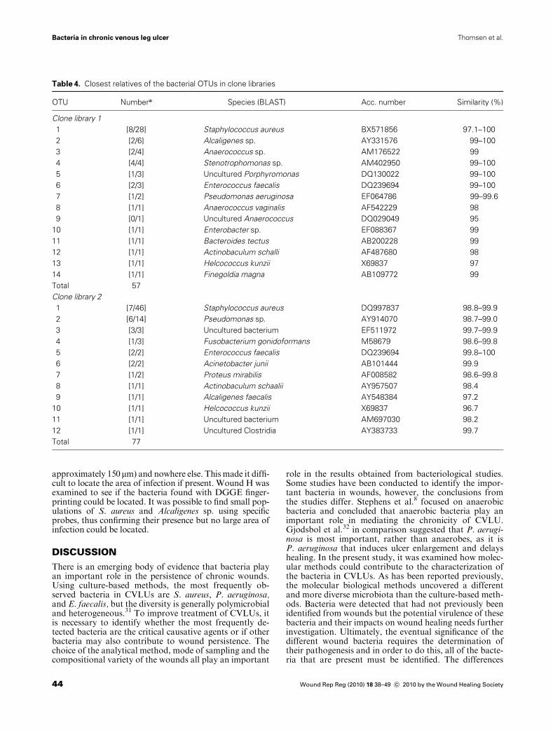
To elucidate the bacterial diversity in the samples, clonelibraries were constructed where the amplified 16S rRNAgenes were inserted into cloning vectors, thereby a separa-tion of the different fragments and its subsequent sequenc-ing were possible. The sequences from the two clonelibraries (clone library 1 from wounds A–F and clone li-brary 2 from wounds G–N) were divided into OTUs usinga similarity level of > 97%. A total of 60 clones weresequenced for clone library 1 and 94 clones for library 2.Table 4 shows the name and accession number of the clos-est relative for each OTU as identified by the phylogeneticanalysis.
Clone library 1 showed many S. aureus and some Alcali-genes sp., Anaerococcus sp., Stenotrophomonas sp., Enter-ococcus faecalis, and P. aeruginosa. Clone library 2 showeda large amount of S. aureus and P. aeruginosa. Almost allOTUs have a similarity of > 97% with their closest rela-tives. Only OTU 9 (uncultured Anaerococcus) in clone li-brary 1 and OTU 10 Helcococcus kunzii in clone library 2had a smaller similarity than 97% indicating that theseOTUs had a lower phylogenetic resolution. The coverageratio for the clone library 1 was 87.7% and for clone li-brary 2 was 93.5%.
Wound Rep Reg (2010) 18 38–49 c� 2010 by the Wound Healing Society 41
Bacteria in chronic venous leg ulcerThomsen et al.
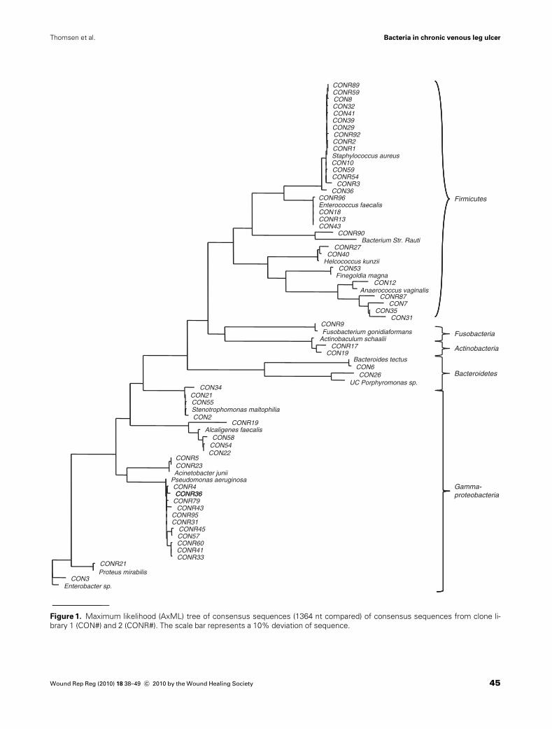
The consensus sequences in clone library 1 and 2 wereused to produce phylogenetic trees to determine thedetailed phylogenetic relationship of the 16S rRNA geneof the clones. A neighbor joining tree, a maximumparsimony tree, and a maximum likelihood tree all showedcongruent phylogenetic relationships, and the maximumlikelihood tree is shown in Figure 1. The locations onthe tree confirm the BLAST identification of the se-quences. The sequences are distributed into five phyla:Proteobacteria, Firmicutes, Bacteroidetes, Fusobacteria,and Actinobacteria. Similar bacteria were identified inthe two clone libraries, although clone library 1 did notdetect any bacteria from the phylum Fusobacteriaand clone library 2 did not detect any Bacteriodetes. Theclone libraries were dominated by sequences related toS. aureus and P. aeruginosa, but also contained many se-quences from E. faecalis, Alcaligenes faecalis, andStenotrophomonas maltophilia.
All 110 partial 16S rRNA gene sequences obtained fromDGGE were added to the consensus maximum likelihoodtree (data not shown) to confirm the result of the BLASTsearch. While the BLAST result was confirmed for most ofthe sequences, the phylogenetic analysis showed that it was
not possible to distinguish the sequences identified asdifferent Alcaligenes and Ahcromobacter species and noPeptoniphilus could be differentiated to more than the ge-nus level. It also showed that the DGGE fingerprinting se-quences most related to Fusobacterium equinum accordingto the BLAST were located closer to Finegoldia gonidia-formans on the tree. F. gonidiaformans was also found inclone library 2.
Quantitative PCR
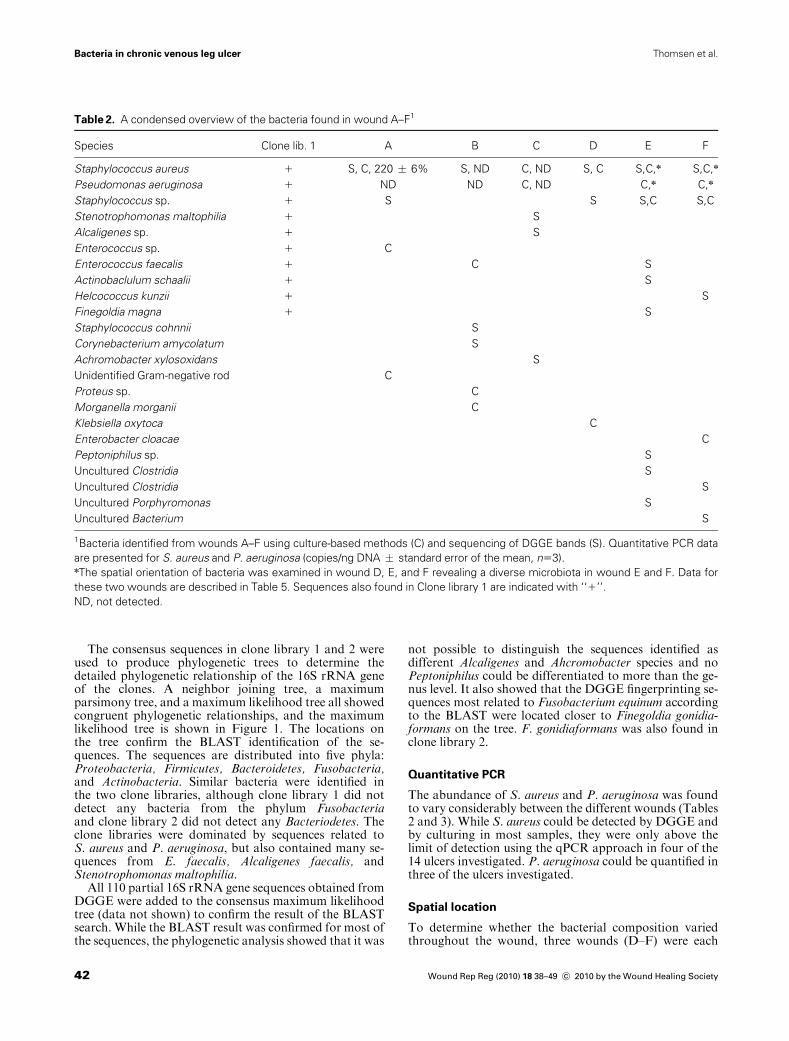
The abundance of S. aureus and P. aeruginosa was foundto vary considerably between the different wounds (Tables2 and 3). While S. aureus could be detected by DGGE andby culturing in most samples, they were only above thelimit of detection using the qPCR approach in four of the14 ulcers investigated. P. aeruginosa could be quantified inthree of the ulcers investigated.
Spatial location
To determine whether the bacterial composition variedthroughout the wound, three wounds (D–F) were each
Table 2. A condensed overview of the bacteria found in wound A–F1
Species Clone lib. 1 A B C D E F
Staphylococcus aureus 1 S, C, 220 � 6% S, ND C, ND S, C S,C,n S,C,n
Pseudomonas aeruginosa 1 ND ND C, ND C,n C,n
Staphylococcus sp. 1 S S S,C S,C
Stenotrophomonas maltophilia 1 S
Alcaligenes sp. 1 S
Enterococcus sp. 1 C
Enterococcus faecalis 1 C S
Actinobaclulum schaalii 1 S
Helcococcus kunzii 1 S
Finegoldia magna 1 S
Staphylococcus cohnnii S
Corynebacterium amycolatum S
Achromobacter xylosoxidans S
Unidentified Gram-negative rod C
Proteus sp. C
Morganella morganii C
Klebsiella oxytoca C
Enterobacter cloacae C
Peptoniphilus sp. S
Uncultured Clostridia S
Uncultured Clostridia S
Uncultured Porphyromonas S
Uncultured Bacterium S
1Bacteria identified from wounds A–F using culture-based methods (C) and sequencing of DGGE bands (S). Quantitative PCR data
are presented for S. aureus and P. aeruginosa (copies/ng DNA � standard error of the mean, n53).nThe spatial orientation of bacteria was examined in wound D, E, and F revealing a diverse microbiota in wound E and F. Data for
these two wounds are described in Table 5. Sequences also found in Clone library 1 are indicated with ‘‘1’’.
ND, not detected.
Wound Rep Reg (2010) 18 38–49 c� 2010 by the Wound Healing Society42
Bacteria in chronic venous leg ulcer Thomsen et al.
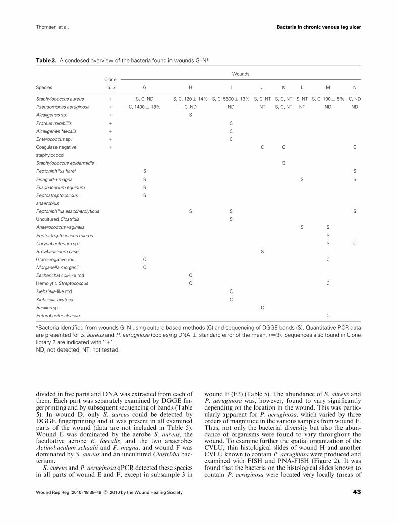
divided in five parts and DNA was extracted from each ofthem. Each part was separately examined by DGGE fin-gerprinting and by subsequent sequencing of bands (Table5). In wound D, only S. aureus could be detected byDGGE fingerprinting and it was present in all examinedparts of the wound (data are not included in Table 5).Wound E was dominated by the aerobe S. aureus, thefacultative aerobe E. faecalis, and the two anaerobesActinobaculum schaalii and F. magna, and wound F wasdominated by S. aureus and an uncultured Clostridia bac-terium.
S. aureus and P. aeruginosa qPCR detected these speciesin all parts of wound E and F, except in subsample 3 in
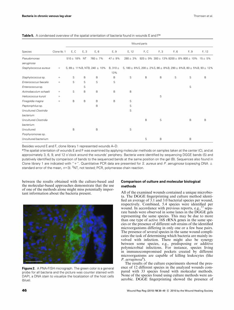
wound E (E3) (Table 5). The abundance of S. aureus andP. aeruginosa was, however, found to vary significantlydepending on the location in the wound. This was partic-ularly apparent for P. aeruginosa, which varied by threeorders of magnitude in the various samples from wound F.Thus, not only the bacterial diversity but also the abun-dance of organisms were found to vary throughout thewound. To examine further the spatial organization of theCVLU, thin histological slides of wound H and anotherCVLU known to contain P. aeruginosa were produced andexamined with FISH and PNA-FISH (Figure 2). It wasfound that the bacteria on the histological slides known tocontain P. aeruginosa were located very locally (areas of
Table 3. A condesed overview of the bacteria found in wounds G–Nn
Species
Clone
lib. 2
Wounds
G H I J K L M N
Staphylococcus aureus 1 S, C, ND S, C, 120� 14% S, C, 5600� 13% S, C, NT S, C, NT S, NT S, C, 100� 5% C, ND
Pseudomonas aeruginosa 1 C, 1400� 18% C, ND ND NT S, C, NT NT ND ND
Alcaligenes sp. 1 S
Proteus mirabillis 1 C
Alcaligenes faecalis 1 C
Enterococcus sp. 1 C
Coagulase negative
staphylococci
1 C C C
Staphylococcus epidermidis S
Peptoniphilus harei S S
Finegoldia magna S S S
Fusobacerium equinum S
Peptostreptococcus
anaerobius
S
Peptoniphilus asaccharolyticus S S S
Uncultured Clostridia S
Anaerococcus vaginalis S S
Peptostreptococcus micros S
Corynebacterium sp. S C
Brevibacterium casei S
Gram-negative rod C C
Morganella morganii C
Escherichia coli-like rod C
Hemolytic Streptococcus C C
Klebsiella-like rod C
Klebsiella oxytoca C
Bacillus sp. C
Enterobacter cloacae C
nBacteria identified from wounds G–N using culture-based methods (C) and sequencing of DGGE bands (S). Quantitative PCR data
are presented for S. aureus and P. aeruginosa (copies/ng DNA � standard error of the mean, n53). Sequences also found in Clone
library 2 are indicated with ‘‘1’’.
ND, not detected, NT, not tested.
Wound Rep Reg (2010) 18 38–49 c� 2010 by the Wound Healing Society 43
Bacteria in chronic venous leg ulcerThomsen et al.
approximately 150mm) and nowhere else. This made it diffi-cult to locate the area of infection if present. Wound H wasexamined to see if the bacteria found with DGGE finger-printing could be located. It was possible to find small pop-ulations of S. aureus and Alcaligenes sp. using specificprobes, thus confirming their presence but no large area ofinfection could be located.
DISCUSSION
There is an emerging body of evidence that bacteria playan important role in the persistence of chronic wounds.Using culture-based methods, the most frequently ob-served bacteria in CVLUs are S. aureus, P. aeruginosa,and E. faecalis, but the diversity is generally polymicrobialand heterogeneous.31 To improve treatment of CVLUs, itis necessary to identify whether the most frequently de-tected bacteria are the critical causative agents or if otherbacteria may also contribute to wound persistence. Thechoice of the analytical method, mode of sampling and thecompositional variety of the wounds all play an important
role in the results obtained from bacteriological studies.Some studies have been conducted to identify the impor-tant bacteria in wounds, however, the conclusions fromthe studies differ. Stephens et al.8 focused on anaerobicbacteria and concluded that anaerobic bacteria play animportant role in mediating the chronicity of CVLU.Gjodsbol et al.32 in comparison suggested that P. aerugi-nosa is most important, rather than anaerobes, as it isP. aeruginosa that induces ulcer enlargement and delayshealing. In the present study, it was examined how molec-ular methods could contribute to the characterization ofthe bacteria in CVLUs. As has been reported previously,the molecular biological methods uncovered a differentand more diverse microbiota than the culture-based meth-ods. Bacteria were detected that had not previously beenidentified from wounds but the potential virulence of thesebacteria and their impacts on wound healing needs furtherinvestigation. Ultimately, the eventual significance of thedifferent wound bacteria requires the determination oftheir pathogenesis and in order to do this, all of the bacte-ria that are present must be identified. The differences
Table 4. Closest relatives of the bacterial OTUs in clone libraries
OTU Numbern Species (BLAST) Acc. number Similarity (%)
Clone library 1
1 [8/28] Staphylococcus aureus BX571856 97.1–100
2 [2/6] Alcaligenes sp. AY331576 99–100
3 [2/4] Anaerococcus sp. AM176522 99
4 [4/4] Stenotrophomonas sp. AM402950 99–100
5 [1/3] Uncultured Porphyromonas DQ130022 99–100
6 [2/3] Enterococcus faecalis DQ239694 99–100
7 [1/2] Pseudomonas aeruginosa EF064786 99–99.6
8 [1/1] Anaerococcus vaginalis AF542229 98
9 [0/1] Uncultured Anaerococcus DQ029049 95
10 [1/1] Enterobacter sp. EF088367 99
11 [1/1] Bacteroides tectus AB200228 99
12 [1/1] Actinobaculum schalli AF487680 98
13 [1/1] Helcococcus kunzii X69837 97
14 [1/1] Finegoldia magna AB109772 99
Total 57
Clone library 2
1 [7/46] Staphylococcus aureus DQ997837 98.8–99.9
2 [6/14] Pseudomonas sp. AY914070 98.7–99.0
3 [3/3] Uncultured bacterium EF511972 99.7–99.9
4 [1/3] Fusobacterium gonidoformans M58679 98.6–99.8
5 [2/2] Enterococcus faecalis DQ239694 99.8–100
6 [2/2] Acinetobacter junii AB101444 99.9
7 [1/2] Proteus mirabilis AF008582 98.6–99.8
8 [1/1] Actinobaculum schaalii AY957507 98.4
9 [1/1] Alcaligenes faecalis AY548384 97.2
10 [1/1] Helcococcus kunzii X69837 96.7
11 [1/1] Uncultured bacterium AM697030 98.2
12 [1/1] Uncultured Clostridia AY383733 99.7
Total 77
Wound Rep Reg (2010) 18 38–49 c� 2010 by the Wound Healing Society44
Bacteria in chronic venous leg ulcer Thomsen et al.
CONR89CONR59CON8CON32CON41CON39CON29CONR92CONR2CONR1Staphylococcus aureusCON10CON59CONR54
CONR3CON36
CONR96Enterococcus faecalisCON18CONR13CON43
CONR90Bacterium Str. Rauti
CONR27CON40
Firmicutes
Helcococcus kunziiCON53
Finegoldia magnaCON12
Anaerococcus vaginalisCONR87
CON7CON35
CON31CONR9Fusobacterium gonidiaformans
Actinobaculum schaaliiFusobacteria
CONR17CON19
Bacteroides tectusCON6CON26
UC Porphyromonas sp.CON34
CON21CON55Stenotrophomonas maltophilia
Bacteroidetes
Actinobacteria
CON2CONR19
Alcaligenes faecalisCON58
CON54CON22
CONR5CONR23Acinetobacter junii
Pseudomonas aeruginosaCONR4CONR36
Gamma-proteobacteriaCONR36
CONR79CONR43
CONR95CONR31
CONR45CON57CONR60CONR41CONR33
CONR21Proteus mirabilis
CON3Enterobacter sp.
Figure 1. Maximum likelihood (AxML) tree of consensus sequences (1364 nt compared) of consensus sequences from clone li-
brary 1 (CON#) and 2 (CONR#). The scale bar represents a 10% deviation of sequence.
Wound Rep Reg (2010) 18 38–49 c� 2010 by the Wound Healing Society 45
Bacteria in chronic venous leg ulcerThomsen et al.
between the results obtained with the culture-based andthe molecular-based approaches demonstrate that the useof one of the methods alone might miss potentially impor-tant information about the bacteria present.
Comparison of culture and molecular biological
methods
All of the examined wounds contained a unique microbio-ta. The DGGE fingerprinting and culture method identi-fied an average of 3.1 and 3.0 bacterial species per wound,respectively. Combined, 5.4 species were identified perwound. In accordance with previous reports, e.g.,13 sepa-rate bands were observed in some lanes in the DGGE gelsrepresenting the same species. This may be due to morethan one type of active 16S rRNA genes in the same spe-cies or the presence of different sub strains of the identifiedmicroorganisms differing in only one or a few base pairs.The presence of several species in the same wound compli-cates the task of determining which bacteria are mainly in-volved with infection. There might also be synergybetween some species, e.g., predisposing or additivepolymicrobial infections. For instance, species livingin immunocompromised pockets created by differentmicroorganisms are capable of killing leukocytes (likeP. aeruginosa6).
The results of the culture experiments showed the pres-ence of 12 different species in the analyzed wounds com-pared with 33 species found with molecular methods.None of the species found using culture methods were an-aerobic. DGGE fingerprinting showed the presence of
Table 5. A condensed overview of the spatial orientation of bacteria found in wounds E and Fn
Species Clone lib. 1
Wound parts
E, C E, 3 E, 6 E, 9 E, 12 F, C F, 3 F, 6 F, 9 F, 12
Pseudomonas
aeruginosa
510� 18% NT 760� 7% 47� 9% 280� 3% 920� 9% 300� 13% 8200� 8% 800� 10% 15� 5%
Staphylococcus aureus 1 S, 89� 11%B, NTB, 240 � 10% B, 310�13%
S, 180� 8%S, 200� 2%S, 86� 8%B, 290� 8%B, 80� 5%B, 93� 12%
Staphylococcus sp. 1 S B B B S B B S S B
Enterococcus faecalis 1 S S S S
Enterococcus sp. S S
Actinobaculum schaalii 1 S B B B B
Helicococcus kunzii 1 S
Finegoldia magna 1 B B B B S
Peptoniphilus sp. B S
Uncultured Clostridia
bacterium
S
Uncultured Clostridia
bacterium
B S B B
Uncultured
Porphyromonas sp.
B B S
Uncultured bacterium S B B
Besides wound E and F, clone library 1 represented wounds A–D.nThe spatial orientation of wounds E and F was examined by applying molecular methods on samples taken at the center (C), and at
approximately 3, 6, 9, and 12 o’clock around the wounds’ periphery. Bacteria were identified by sequencing DGGE bands (S) and
putatively identified by comparison of bands to the sequenced bands at the same position on the gel (B). Sequences also found in
Clone library 1 are indicated with ‘‘1’’. Quantitative PCR data are presented for S. aureus and P. aeruginosa (copies/ng DNA �standard error of the mean, n53). []NT, not tested; PCR, polymerase chain reaction.
Figure 2. A PNA-FISH micrograph. The green color is a general
probe for all bacteria and the picture was counter stained with
DAPI, a DNA stain to visualize the localization of the host cells
(blue).
Wound Rep Reg (2010) 18 38–49 c� 2010 by the Wound Healing Society46
Bacteria in chronic venous leg ulcer Thomsen et al.

anaerobic bacteria in wound G, H, I, M, and N. The an-aerobic species are often overlooked by culture methodsbecause they require longer culture times and previouslylacked a valid identification scheme.8 Many of the bacteriaidentified by DGGE fingerprinting have close relativesidentified previously by culture experiments, and are there-fore likely themselves to be culturable to some degree.
Some of the observed differences between the resultsobtained by the culture and the molecular methods couldbe due to the inability to differentiate species on the cultureplates or that some specimens were collected as a biopsyand others using a swab. The differences may also be at-tributable to a fraction of the bacteria being dead or in aviable but unculturable state. This can be caused by the useof antibiotics15; however, only wound F was receiving an-tibiotics and indeed this wound showed the presence ofonly one species. The other two wounds, which had beentreated with antibiotics until a short time before the study,both showed a diverse microbiota detected by culturemethods. Based on these findings, there is no evidence oflarge amounts of residual genetic material from organismsno longer colonizing the ulcer bed.
Cultivation techniques have some limitations, but themolecular biological methods also have biases. These in-clude amplification of naked DNA, unknown DNA recov-ery yields from extraction, differential amplification due toPCR primer bias, 16S rRNA copy number, and heteroge-neity and co-migration of bands on DGGE fingerprinting.Some of the biases associated with, e.g., the DGGE ap-proach were compensated by using the cloning approach, inwhich different primers were used with different specificities.
Diversity of CVLU bacteria
The clone library and DGGE analysis revealed a large di-versity of bacteria of which some have not been associatedpreviously with wounds: Brevibacterium casei, Corynebac-terium simulans, Corynebacterium amycolatum. A. schaalii,P. harei, F. gonidiaformans, Bacteroides tectus, Achromo-bacter xylosoxidans, A. faecalis, and some uncultured bac-teria. B. casei has been identified as an opportunisticpathogen in immunocompromised patients. The case re-ports by Reinert et al.33 and Brazzola et al.34 are examples,describing that B. casei needs a host with reduced immunesystem in order to initiate infection. Two other bacteriafrom phylum Actinobacteria (C. simulans and C. amy-colatum) were also identified. The Corynebacteria areknown as an aerobe and ubiquitous on human skin andare all opportunistic pathogens. C. amycolatum is fre-quently isolated from clinical specimens and infectedwounds and it is resistant to most antibiotics35 whereasC. simulans is a rare species found previously in blood andbile samples.36 A. schaalii is a Gram-positive bacterium re-sembling normal skin flora and it is often overlooked byculture methods due to its slow growth in ambient air. Re-cently A. schaalii has been found as a pathogen in 10 casesof urinary infection.37 P. harei belongs to the anaerobicGram-positive family Peptostreptococcaceae, which is aheterogeneous family of opportunistic pathogens coloniz-ing the skin and the mucosal surfaces of humans.35H. kun-zii has been isolated previously from human skin and fromdiabetic foot wounds. It is mainly identified as a part of apolymicrobial community38 but it has also been seen as the
sole pathogen in a foot abscess.39 The Fusobacteria areGram-negative anaerobes found in the human gastroin-testinal tract. Here, they are a part of the polymicrobialflora but they are also involved in a variety of differentdiseases.40 The phylum Fusobacterium is often associatedwith chronic wounds.41 F. gonidiaformans is a rare type ofFusobacterium species isolated previously from infecteddog bites42 and from skin infections.43 In both surveys, theF. gonidiaformans constituted a very small percentage ofthe isolated bacteria. A. xylosoxidans and A. faecalis areboth aerobe Gram-negative Betaproteobacteria from theAlcaligenaceae family. They are ubiquitous in the environ-ment but rarely involved with human disease. They havebeen isolated from blood cultures of various immunosup-pressed patients44 and also appeared in a recent study ofchronic wounds by Dowd et al.12 The unculturedPorphyromonas (DQ130022) was identified previouslyfrom the forearm of a healthy human45 and the unculturedbacterium (AY958901) was identified from the vaginal ep-ithelium of a healthy woman.46
Phylogenetic analysis showed that the 33 different speciesbelonged to six phyla. Both in terms of the number of differ-ent species and the number of identified clones, the Proteo-bacteria and the Firmicutes (Clostridia) were the dominatingphyla. Gao et al.45 examined the skin flora of healthy fore-arms in a large molecular biological study. They found thatthe dominating phylumwas theActinobacteria, although theFirmicutes and Proteobacteria were also present in highnumbers. Healthy skin seems to be the only human environ-ment where Actinobacteria are dominating.45 In compari-son, the inner mucosal surfaces of humans (e.g., colon andoral cavity) are dominated by Firmicutes and Proteobacte-ria.45 This difference is probably due to environmentalchanges such as humidity and changes in pH value.
Eleven of the species were confirmed with both thecloning approach and DGGE fingerprinting. Therewas not a complete overlap between the findings of thetwo molecular methods and a reason for this might be thatthe DNA from the wounds was pooled before cloning onbasis of the intensities of the bands on a gel. Another ex-planation might be that the primers used in the two meth-ods had different affinity. Differences between the findingsof the applied methods were also seen by Dowd et al.12
This study also indicated the presence of a varied anaer-obic flora dominated by F. magna and P. asaccharolyticus,which were found in three wounds each. Table 3 (repre-senting wound G,M, and N) also shows that the anaerobicspecies were often located in the same wound. This sug-gests that anaerobic pockets were present in the woundand that there is a possible synergistic effect between them.Stephens et al.8 tested the effects of P. vaginalis, F. magna,and P. asaccharolyticus on cellular wound healing re-sponses and found that they caused delayed reepitheliaza-tion and defective extracellular matrix reorganization andangiogenesis in vitro. These are all important steps inwound healing. They also compared this with the effect ofP. aeruginosa and found that this had less detrimentaleffect compared with the anaerobes.
Spatial orientation of bacteria in CVLU
The results from the DGGE approach investigating thespatial orientation of the bacteria in three wounds
Wound Rep Reg (2010) 18 38–49 c� 2010 by the Wound Healing Society 47
Bacteria in chronic venous leg ulcerThomsen et al.

illustrated that if only one biopsy from a wound was ana-lyzed it would most likely not represent the bacterial com-position of the entire wound. The qPCR resultsdemonstrated that the abundance of S. aureus and P.aeruginosa also varied depending on the different locationsin the wound. The technique is rapid and has recently beenused to determinate Pseudomonas in a chronic woundwithin few hours, enabling fast decisions on treatment.47
In addition, multiple biopsies from the same wound canalso indicate which species of bacteria are most importantfor the infection as these are probably present in largenumbers all over the wound. Furthermore, it supports theclaim that the bacteria found in wounds are located inniches, which covers their needs. Using FISH, we detectedbacteria in microcolonies also known as biofilms (Figure2), which might explain how the bacteria survive inside thewound bed. This correlates with the finding that in someCVLU, P. aeruginosa live in large biofilms underneath thewound surface.6 Antibacterial dressings, e.g., silver con-taining dressings are likely to influence the bacterial floraon the surface of the wounds. However, as the PNA-FISHpictures show that the bacteria reside deep in the tissue, itis not likely that bacteria will be influenced by the dress-ings. Furthermore, all swabs were taken after thoroughlysurgical revision far away from local antimicrobialdressings. This indicates that the diversity was probablynot influenced by the dressing, but by other factors suchas antibiotics and difference in skin flora. The FISHtechnology increases the understanding of the pathologyof bacteria in chronic wounds and how it might impacttherapies.
This study compared the bacterial flora of differenttypes of wound material from 14 skin graft operations ofCVLU. Results from the culture methods were comparedwith the results from the molecular biological methods,which showed that the flora of the wounds varied, as didthe number of S. aureus and P. aeruginosa investigatedby qPCR. Each wound contained multiple species butapart from that the methods detected rather differentfloras. An average of 5.4 species were found in eachwound by the methods combined. All of the woundscontained S. aureus but P. aeruginosa was also frequent.The molecular biological methods detected a variedanaerobic flora in four of the wounds and species notfound previously in CVLU were identified. All of thesewere known pathogens. No anaerobes or new specieswere detected with culture methods. It was also foundthat the wound flora was different and that the numberof the pathogens S. aureus and P. aeruginosa varied,depending on which location and depth of the woundwas examined. Three wounds were examined and theyshowed that some species were present all over whilesome were only present in parts of the wounds. Thisemphasizes the need for multiple samplings when exam-ining wounds, and swabs and biopsies each have specificadvantages as sampling technologies.
qPCR is a promising fast method for fast characteriza-tion of the bacteria present in ulcers, and importantlythe running cost is comparable with the cultivationtechniques. The next important step is to elucidate the bac-teria that contribute to the pathogenicity of these chronicwounds. This information could be used to develop theoptimal sampling, identification, and treatment regimes.
ACKNOWLEDGMENTS
The Danish Technical Research Council supported thisstudy under the innovation consortia ‘‘BIOMED.’’ Wethank Jane Ildal, Aalborg University, for valuable techni-cal assistance and Bo J�rgensen, Copenhagen WoundHealing Center, for collecting samples. The authors wouldalso like to thank AdvanDx Inc., Woburn, MA, USA forsupplying the PNA probes for the FISH experiments. TBreceived financial support from The Carlsberg Foundationand Lundbeck Foundation (the role of biofilms in chronicinfections).
REFERENCES
1. Trent J, Falabella A, Eaglstein W, Kirsner R. Venous ulcers:pathophysiology and treatment options. Ostomy WoundManage 2005; 51: 38–54.
2. Gottrup F. A specialized wound-healing center concept: im-portance of a multidisciplinary department structure and sur-gical treatment facilities in the treatment of chronic wounds.Am J Surg 2004; 187 (5A): 38S–43S.
3. Laing W. Chronic venous diseases of the leg. London: Officeof Health Economics, 1992.
4. Ruckley C. Socioeconomic impact of chronic venous insuffi-ciency and leg ulcers. Angiology 1997; 48: 67–9.
5. Church JCT, CourtenayM.Maggot debridement therapy forchronic wounds. Lower Extremity Wounds 2002; 1: 129–34.
6. Bjarnsholt T, Kirketerp-M�ller K, Jensen PØ, Madsen KG,Rasmussen TB, Krogfelt K, H�iby N, Givskov M. Whychronic wounds won’t heal: a novel hypothesis. Wound Re-pair Regen 2008; 16: 2–10.
7. Edwards R, Harding KG. Bacteria and wound healing. CurOp Infect Dis 2004; 17: 91–6.
8. Stephens P, Wall I, Wilson M, Hill K, Davies C, Hill C, Har-ding K, Thomas D. Anaerobic cocci populating the deep tis-sues of chronic wounds impair cellular wound healingresponses in vitro. Br J Derm 2003; 148: 456–466.
9. James G, Swogger E, Wolcott R, Pulcin iE, Secor P, SestrichJ, Costerton JW, Stewart PS. Biofilms in chronic wounds.Wound Repair Regen 2008; 16: 37–44.
10. Kirketerp-Moller K, Jensen P, Fazli M, Madsen K, PedersenJ, Moser C, Tolker-Nielsen T, Hoiby N, Givskov M, Bjarn-sholt T. The distribution, organization and ecology of bacte-ria in chronic wounds. J Clin Microbiol 2008; 46: 2717–22.
11. Tolker-Nielsen T, Molin S. Spatial organization of microbialbiofilm communities. Microb Ecol 2000; 40: 75–84.
12. Dowd S, Sun Y, Secor P, Rhoads D, Wolcott B, James G,Wolcott R. Survey of bacterial diversity in chronic woundsusing Pyrosequencing, DGGE, and full ribosome shotgunsequencing. BMC Microbiol 2008; 8: 43.
13. Andersen A, Hill K, Stephens P, Thomas D, Jorgensen B,Krogfelt K. Bacterial profiling using skin grafting, standardculture and molecular bacteriological methods. J WoundCare 2007; 16: 171–5.
14. Hill KE, Davies CE, Wilson MJ, Stephens P, Harding KG,Thomas DW.Molecular analysis of the microflora in chronicvenous leg ulceration. J Med Microbiol 2003; 52: 365–9.
15. Davies CE, Hill KE, Wilson MJ, Stephens P, Hill CM, Har-ding KG, Thomas DW. Use of 16S ribosomal DNA PCRand denaturing gradient gel electrophoresis for analysis of
Wound Rep Reg (2010) 18 38–49 c� 2010 by the Wound Healing Society48
Bacteria in chronic venous leg ulcer Thomsen et al.
the microfloras of healing and nonhealing chronic venous legulcers. J Clin Microbiol 2004; 42: 3549–3557.
16. Juretschko S, Timmermann G, Schmid M, Schleifer K-H,Pommerening-Roser A, Koops HP, Wagner M. Combinedmolecular and conventional analysis of nitrifying bacteriumdiversity in activated sludge: Nitrosococcus mobilis and Nit-rosospira-like bacteria as dominant populations. Appl Envi-ron Microbiol 1998; 64: 3042–3051.
17. Lane DJ 16/23S rRNA sequencing. In:Nucleic acid techniquesin bacterial systematics, Stackebrandt E, Goodfellow M, ed-itors. Chichester: Wiley, 1991: 113–175.
18. Thomsen TR, Ramsing NB, Finster K. Biogeochemical andmolecular signatures of anaerobic methane oxidation in amarine sediment. Appl Environ Microbiol 2001; 67: 1646–56.
19. Hugenholtz P, Huber T. Chimeric 16S rDNA sequences ofdiverse origin are accumulating in the public databases. Int JSyst Evol Microbiol 2003; 53: 289–293.
20. Ludwig W, Strunk O, Westram R, Richter L, Meier H,Yadhukumar, Buchner A, Lai T, Steppi S, Jobb G, ForsterW, Brettske I, Gerber S, Ginhart AW, Gross O, Grumann S,Hermann S, Jost R, Konig A, Liss T, Lussmann R, May M,Nonhoff B, Reichel B, Strehlow R, Stamatakis A, Stuckm-ann N, Vilbig A, Lenke M, Ludwig T, Bode A, Schleifer K-H. ARB: a software environment for sequence data. NucleicAcids Res 2004; 32: 1363–71.
21. Juretschko S, Loy A, Lehner A, Wagner M. The microbialcommunity composition of a nitrifying-dentrifying activatedsludge from an industrial sewage treatment plant analyzed bythe full-cycle rRNA approach. J Syst Appl Microbiol 2002;25: 84–99.
22. Muyzer G, De Waal EC, Uitterlinden AG. Profiling of com-plex microbial populations by denaturing gradient gel elec-trophoresis analysis of polymerase chain reaction-amplifiedgenes coding for 16S rRNA.Appl EnvironMicrobiol 1993; 59:695–700.
23. Brakstad O, Aasbakk K, Maeland J. Detection of Staphylo-coccus aureus by polymerase chain reaction amplification ofamplification of the nuc gene. J Clin Microbiol 1992; 30:1654–60.
24. Jaffe R, Lane J, Bates C. Real-time identification of Pseudo-monas aeruginosa direct from clinical samples. J Clin LabAnal 2001; 15: 131–7.
25. Amann R, Ludwig W, Schleifer KH. Phylogenetic identifica-tion and in situ detection of individual microbial cells withoutcultivation.Microbiol Rev 1995; 59: 143–69.
26. Daims H, Bruhl A, Amann R, Schleifer K-H, Wagner M.The domain-specific probe EUB338 is insufficient for the de-tection of all Bacteria: development and evaluation of a morecomprehensive probe set. Syst Appl Microbiol 1999; 22: 434–44.
27. Manz W, Amann R, Ludwig W, Wagner M, Schleifer K-H.Phylogenetic oligodeoxynucleotide probes for the major sub-classes of proteobacteria: problems and solutions. Syst ApplMicrobiol 1992; 15: 593–600.
28. Meier H, Amann R, Ludwig W, Schleifer K-H. Specificoligonucleotide probes for in situ detection of a major groupof gram-positive bacteria with low DNA G1C content. SystAppl Microbiol 1999; 22: 186–96.
29. Kempf V, Trebesius K, Autenrieth I. Fluorescent In situ hy-bridization allows rapid identification of microorganisms inblood cultures. J Clin Microbiol 2000; 38: 830–8.
30. Loy A, Horn M, Wagner M. probeBase—an online resourcefor rRNA-targeted oligonucleotide probes.Nucleic Acids Res2003; 31: 514–6.
31. Davies C, Wilson M, Hill K, Stephens P, Hill C, Harding K,Thomas D. Use of molecular techniques to study microbialdiversity in the skin: chronic wounds reevaluated.Wound Re-pair Regen 2001; 9: 332–340.
32. Gjodsbol K, Christensen J, Karlsmark T, Jorgensen B,Klein B, Krogfelt K. Multiple bacterial species reside inchronic wounds: a longitudinal study. Int Wound J 2006; 3:225–31.
33. Reinert RR, Schnitzler N, Haase G, Lutticken R, Fabry U,Schaal KP, Funke G. Recurrent Bacteremia due to Brevibac-terium casei in an immunocompromised patient. Eur J ClinMicrobiol Infect Dis 1995; 14: 1082–6.
34. Brazzola P, Zbinen R, Rudin C, Schaad UB, Heininger U.Brevibacterium casei sepsis in an 18-year-old female withAIDS. J Clin Microbiol 2000; 38: 3513–4.
35. Murray MAP, Rosenthal KS, Pfaller MA. Med Microbiol.5th ed. Philadelphia, PA: Elsevier Mosby, 2005.
36. Bernard KA, Munro C, Wiebe D, Ongsansoy E. Character-istics of rare or recently described Corynebacterium speciesrecovered from human clinical material in Canada. J ClinMicrobiol 2002; 40: 4375–81.
37. Reinhard M, Prag J, Kemp M, Andreasen K, KlemmensenB, H�jlyng N, S�rensen SH, Christensen JJ. Ten cases ofActinobaculum schalii infection: clinical relevance, bacterialidentification, and antiobiotic susceptibility. J Clin Microbiol2005; 43: 5306–8.
38. Haas J, Jernick L, Scardina R, Teruya J, Caliendo A, RuoffK. Colonization of skin by Helcococcus kunzii. J Clin Micro-biol 1997; 35: 2759–61.
39. Riegel P, Lepargneur J. Isolation of Helcococcus kunzii froma post-surgical foot abscess. Int J Med Microbiol 2003; 293:437–9.
40. Johnson C, Wexler H, Becker S, Garcia M, Finegold S. Cell-wall-defective variants of Fusobacterium. AntimicrobialAgents Chem 1988; 33: 369–72.
41. Hernandez R. The use of systemic antibiotics in the treatmentof chronic wounds. Dermatologic Therapy 2006; 19: 326–37.
42. Talan D, Citron J, Abrahamian F, Moran G, Goldstein E.Bacteriologic analysis of infected dog and cat bites. N Eng JMed 1999; 340: 85–92.
43. Goldstein EJC, Citron DM, Merriam CV, Warren Y, TyrrellKL, Gesser RM. General microbiology and in vitro suscep-tibility of anaerobes isolated from complicated skin and kin-structure infections in patients enrolled in a comparative trialof ertapenem versus piperacillin-tazobactam. Clin Infec Dis2002; 35 (Suppl.): S119–25.
44. Aisenberg G, Rolston K, Safdar A. Bacteremia caused byAchromobacter and Alcaligenes species in 46 patients withcancer (1989–2003). Cancer 2004; 101: 2134–40.
45. Gao Z, Tseng C, Pei Z, Blaser MJ. Molecular analysis of hu-man forearm superficial skin bacterial biota. Proc Natl AcadSoc 2007; 104: 2927–2932.
46. Hyman RW, Fukushima M, Diamond L, Kumm J, GiudiceLC, Davies RW. Microbes on the human vaginal epithelium.Proc Natl Acad Soc 2005; 102: 7952–7.
47. Wolcott R, Dowd S. A rapid molecular method for charac-terizing bacterial bioburden in chronic wounds. J WoundCare 2009; 17: 513–6.
Wound Rep Reg (2010) 18 38–49 c� 2010 by the Wound Healing Society 49
Bacteria in chronic venous leg ulcerThomsen et al.