Chronic cerebrospinal venous insufficiency: masked multimodal imaging assessment
Upload
khangminh22Category
view
3download
0
Zivadinov and Chung BMC Medicine 2013, 11:260http://www.biomedcentral.com/1741-7015/11/260
DEBATE Open Access
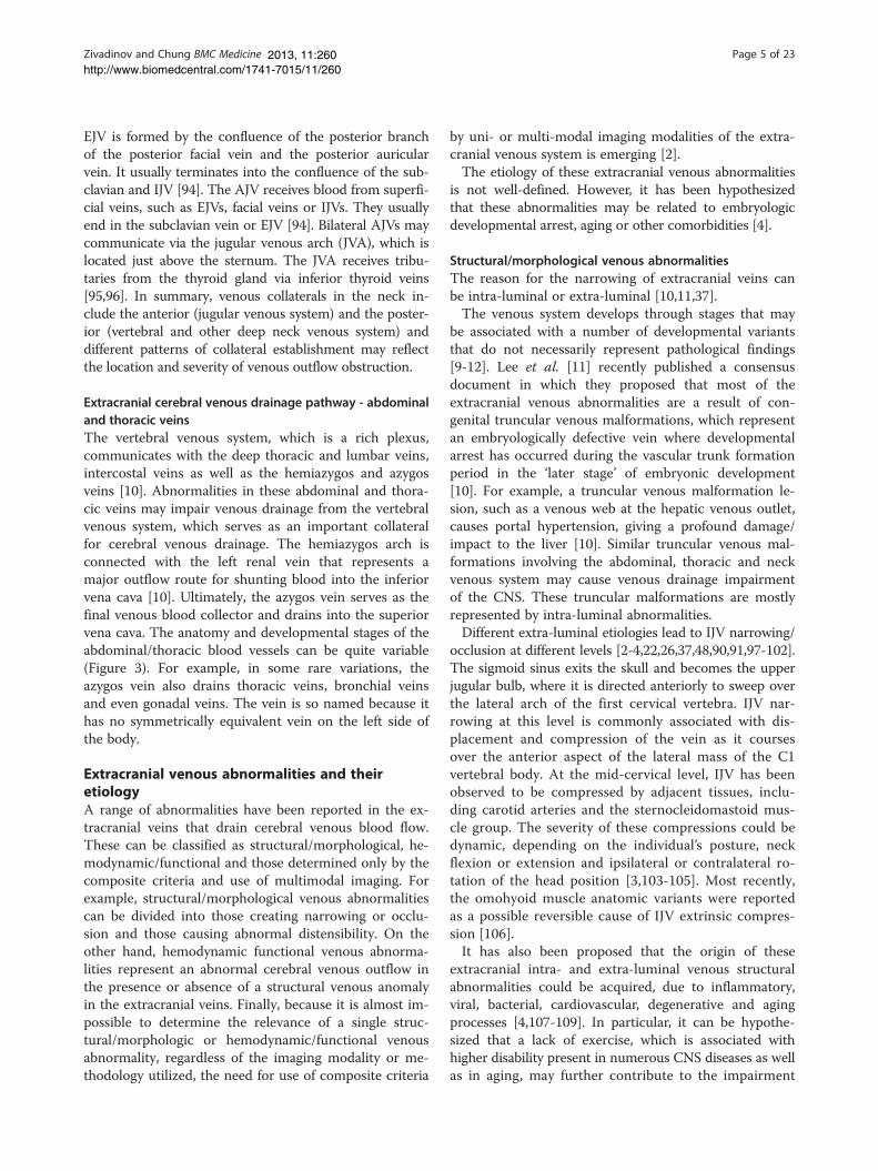
Potential involvement of the extracranial venoussystem in central nervous system disordersand agingRobert Zivadinov1,2* and Chih-Ping Chung3,4
Abstract
Background: The role of the extracranial venous system in the pathology of central nervous system (CNS) disordersand aging is largely unknown. It is acknowledged that the development of the venous system is subject to manyvariations and that these variations do not necessarily represent pathological findings. The idea has been changingwith regards to the extracranial venous system.
Discussion: A range of extracranial venous abnormalities have recently been reported, which could be classified asstructural/morphological, hemodynamic/functional and those determined only by the composite criteria and use ofmultimodal imaging. The presence of these abnormalities usually disrupts normal blood flow and is associated withthe development of prominent collateral circulation. The etiology of these abnormalities may be related toembryologic developmental arrest, aging or other comorbidities. Several CNS disorders have been linked to thepresence and severity of jugular venous reflux. Another composite criteria-based vascular condition named chroniccerebrospinal venous insufficiency (CCSVI) was recently introduced. CCSVI is characterized by abnormalities of themain extracranial cerebrospinal venous outflow routes that may interfere with normal venous outflow.
Summary: Additional research is needed to better define the role of the extracranial venous system in relation toCNS disorders and aging. The use of endovascular treatment for the correction of these extracranial venousabnormalities should be discouraged, until potential benefit is demonstrated in properly-designed, blinded,randomized and controlled clinical trials.
Please see related editorial: http://www.biomedcentral.com/1741-7015/11/259.
Keywords: Jugular vein reflux, CCSVI, Anatomy, Etiology, Pathophysiology, Classification, Diagnosis, CNS disorders,Aging, Multiple sclerosis, Compensatory mechanism
BackgroundMounting evidence suggests that a number of inflam-matory and neurodegenerative central nervous system(CNS) disorders may be related to vascular factors [1].While the role of arterial supply abnormalities in relationto the pathology of CNS disorders is well-defined, the roleof venous drainage impairment, for example, extracranial
* Correspondence: [email protected] Neuroimaging Analysis Center, Department of Neurology, School ofMedicine and Biomedical Sciences, University at Buffalo, State University ofNew York, Buffalo, NY, USA2MR Imaging Clinical Translational Research Center, School of Medicine andBiomedical Sciences, University at Buffalo, State University of New York, 100High St., Buffalo, NY 14203, USAFull list of author information is available at the end of the article
© Zivadinov and Chung; licensee BioMeCreative Commons Attribution License (http:/distribution, and reproduction in any medium
2013
venous abnormalities, is largely unknown [2-7]. The com-plexity, inter-individual variability and frequent asym-metry of the extracranial venous system, compared to theperipheral venous and arterial systems make explorationof the link between intracranial and extracranial pathologyextremely difficult [2,8]. Moreover, additional factors, in-cluding postural change, cardiac function, respiration,frequent change in lumen diameter, hydration status,hypovolemia and the presence of nearby structures,may influence correct assessments of the veins inregards to the presence of structural or hemodynamicextracranial venous abnormalities [2-7].Compared with the arterial system, the development
of the extracranial venous system is subject to many
d Central Ltd. This is an open access article distributed under the terms of the/creativecommons.org/licenses/by/2.0), which permits unrestricted use,, provided the original work is properly cited.

Zivadinov and Chung BMC Medicine Page 2 of 232013, 11:260http://www.biomedcentral.com/1741-7015/11/260
variations. Therefore, in the past, these variations wereacknowledged as non-pathological findings [9-12]. Avariety of congenital extracranial venous abnormalities/developmental variants has been described [10,11]. How-ever, investigations aimed to define the nature of thesevenous abnormalities/developmental variants and theirclinical significances are lacking [13,14].Several CNS disorders, such as transient global am-
nesia, transient monocular blindness, cough headacheand primary exertional headache, have been linked tothe presence and severity of uni- or bi-lateral jugularvenous reflux (JVR) in the last two decades [15-20].More recently, an intense interest in better understan-ding the role of the extracranial venous system in thepathophysiology of CNS disorders has been generated bythe introduction of a composite criteria-based vascularcondition named chronic cerebrospinal venous insuffi-ciency (CCSVI). CCSVI is characterized by abnormalitiesof the main extracranial cerebrospinal venous outflowroutes that interfere with normal venous drainage, asevidenced by Doppler sonography (DS) [21-23]. It wasoriginally hypothesized that CCSVI implies a patholo-gical condition that leads to abnormal venous drainageof the brain parenchyma and increased susceptibility tomultiple sclerosis (MS) [22]. While, the condition wasoriginally described in MS patients, it became immedi-ately clear from the independent results of the first con-trolled studies that patients with other CNS disordersand healthy individuals may also show a high prevalenceof this condition [2,24-27]. However, because healthy in-dividuals do not suffer from CNS disorders, its clinicalrelevance as a nosological entity was immediately ques-tioned [26,28]. Indeed, as more research studies havebecome available, the very concept of CCSVI, its diagnos-tic utility and clinical impact for MS have all been ques-tioned, as no causal relationship between CCSVI and MShas been confirmed [24-27,29-49]. In addition, the contro-versy and debate around CCSVI has been fueled by thepostulated therapeutic effect of venous insufficiency cor-rection using endovascular procedures [21], without firstdetermining a real need for the procedure itself and test-ing its safety and efficacy in properly designed random-ized, controlled and blinded trials [28,50,51].Given that substantial resources by various govern-
ments and funding agencies have been devoted to study-ing the concept of CCSVI, it was recently proposed thatfunding of CCSVI research should be immediately aban-doned because it is a waste of valuable time, money andintellectual energy [52-56]. Nevertheless, the concept ofCCSVI has triggered an intense and rapid accumulationof knowledge over the past four years and has stimulatedthe need for further research to better understand thefunction and potential role of the extracranial venoussystem in CNS disorders and aging [57].
This review article highlights the need for better classifi-cation of extracranial venous abnormalities/developmentalvariants that is independent of any single diagnostic im-aging modality. It also examines the anatomy, etiology andpathophysiology associated with venous abnormalities, aswell as the clinical correlates in relation to various CNSdisorders and aging.
Anatomy of the extracranial venous systemIn order to understand the potential role of the extracra-nial venous system in diseases of the CNS and aging, itis important to first appreciate the structure and func-tion of the cerebral venous drainage system. Because thissystem is complex and poorly understood, in this sec-tion, a brief overview of the relevant anatomy is pre-sented to assist the reader.Cerebral circulation encompasses both the arterial and
venous systems. The venous system contains appro-ximately 70% of the blood volume, with approximatelythree-quarters of it within small veins and venules [58-64].It is a system that is often asymmetric and conside-rably represents a more variable pattern than the ar-terial system [5].
Cerebral venous system; superficial and deep veinsThe venous drainage from cerebral hemispheres consistsof two systems; the superficial and the deep venoussystem (Figure 1) [60-64]. The superficial system drainsblood from the cortex and superficial white matter (WM)by cortical veins, collected by dural sinuses. There are twoimportant dural sinuses: the superior sagittal sinus (SSS)draining dorso-laterally and the cavernous sinus draininganteroventrally. The transverse sinus then drains the SSSequally on both sides in only 20% of cases and asymme-trically in more than 50% of cases, depending on the con-figuration of torcular Herophili [60,63]. In 20% of cases,one transverse sinus drains the SSS in total (most often onthe right side) and the other one drains the straight sinus,which collects blood from the deep venous system [63].The cavernous sinus extends from the superior orbitalfissure to the petrous apex, which receives orbital venousand middle cranial fossa drainage. From the cavernoussinus, blood drains posterolaterally along the superior pe-trosal sinus into the transverse sinus and inferior-laterallyalong the inferior petrosal sinus into the sigmoid sinus.The deep cerebral venous system drains the deep WM
and the regions surrounding the lateral and third ventri-cles or the basal cistern [60-62,65]. Three veins unitejust behind the interventricular foramen of the Monroto form the internal cerebral vein(s). These include thechoroid vein, septal vein and thalamostriate vein. Thevein of Galen is a short (1 to 2 cm long), thick vein thatpasses posterosuperiorly behind the splenium of corpuscallosum in the quadrigeminal cistern. The vein of Galen

Figure 1 Intracranial venous system anatomy of dural sinuses, cortical veins, deep intracerebral veins and cavernous sinus. The figurewas reproduced with permission from the Radiology Assistant website: (http://www.radiologyassistant.nl/en/p4befacb3e4691/cerebral-venous-thrombosis.html).
Zivadinov and Chung BMC Medicine Page 3 of 232013, 11:260http://www.biomedcentral.com/1741-7015/11/260
receives the internal cerebral vein, the basal veins ofRosenthal and the posterior fossa veins and then drainsto the anterior end of the straight sinus where this uniteswith the inferior sagittal sinus. The main collecting veinfor the deep venous system is the straight sinus, whichreceives the venous blood from the vein of Galen andflows into the transverse sinus (most often into the leftside). The basal vein of Rosenthal is an important colla-teral pathway for the internal cerebral veins and the veinof Galen. By connecting with the superficial Sylvian veinvia the deep Sylvian vein, venous blood flow can bypassthe straight sinus.Venous drainage of the posterior fossa mainly depends
on the galenic system and the petrosal system and to alesser extent, the tentorial veins and the transverse sinuses[60-63]. Therefore, factors influencing galenic systemdrainage would lead to venous congestion in both posteriorfossa and brain regions drained by the deep venous system.
Extracranial cerebral venous drainage pathway - neckveinsMost of the cerebral venous drainage is via neck veins;mainly the internal jugular vein (IJV), vertebral venous sys-tem and deep cervical veins (veins in cervical soft tissue)(Figure 2) [66-70]. Consequently, there is a good reason tobelieve that impaired extracranial venous drainage func-tions or structures might cause cerebral venous drain-age insufficiency and consequent neurological deficits.
The IJVs are the largest veins in the neck and are gen-erally considered to be the most important cerebral ven-ous outflow pathways. Venous drainage of the superficialand deep cerebral venous system is via the transverse si-nuses to the sigmoid sinuses, which then drain into theIJV. The inferior petrosal sinus, a major drainage routecollecting blood from the cavernous sinus, communi-cates with the basilar plexus, anterior and lateral con-dylar veins, anterior condylar confluence and vertebralvenous plexus before draining into the IJVs [68,71,72].The IJVs then join with the subclavian veins to form thebrachiocephalic vein (BV). The confluence of the bi-lateral BV is the superior vena cava, which ultimatelydrains venous blood into the heart. Several tributaries inthe neck also drain into the IJVs [73-75]. These bilateralIJV branches will interconnect with each other at themidline to form anastomosing plexi that can serve ascollateral channels to maintain adequate venous drai-nage when the principal pathways are obstructed [73,74].The vertebral venous system consists of two compo-
nents; one is the vertebral venous plexus and the otheris the vertebral vein (VV) [8,68,76,77]. The vertebral ve-nous plexus can be subdivided as internal (posterior andanterior internal vertebral plexus) and external (posteriorand anterior external vertebral plexus) [8,68,76,77].Complex connections of cerebral venous outflow with
the vertebral venous system over the craniocervical junc-tion have been displayed by several human cadavers and

Figure 2 Illustration depicting the predominant veins and sinuses involved in the craniocervical venous outflow. Venous narrowing isdepicted at locations of interest in chronic cerebrospinal venous insufficiency. The figure was reproduced with permission from Lazzaro MA,Zaidat OO, Mueller-Kronast N, Taqi MA, Woo D. Endovascular therapy for chronic cerebrospinal venous insufficiency in multiple sclerosis.Front Neurol 2011, 2:44.
Zivadinov and Chung BMC Medicine Page 4 of 232013, 11:260http://www.biomedcentral.com/1741-7015/11/260
angiographic studies [66,68,71,78-80]. The IJVs can alsoexhibit anastomosis with the other extracranial venousdrainage system within the craniocervical junction re-gion, which includes the anterior condylar confluent(ACC) and its tributes. Numerous anastomoses of theACC make it a crossroad between the cavernous sinus,dural sinuses of the posterior fossa, IJVs and posteriorcervical outflow tract (vertebral venous system and deepcervical veins).
IJV valvesThe IJV valves make IJV a buffer zone between largecentral veins and the cerebral venous system. Althoughthere are anatomical variations, the valves are generallylocated about 0.5 cm above the union of the subclavianvein and IJVs at the lower limit of the jugular bulb[81-85], which are shown in 96.8% of the general popu-lation [82,84]. The IJV valves are generally thought toprevent the backflow of venous blood and backward ve-nous pressure into the cerebral venous system duringconditions where the central venous pressure or intra-thoracic pressure is increased, such as chest compressionduring external cardiopulmonary resuscitation, severe or
repetitive cough and straining [81,83-86]. The pressuregradient across competent IJV valves can be as high as100 mmHg [86]. Without competent IJV valves, a sus-tained or prolonged retrograde-transmitted venous pres-sure via IJVs might impair cerebral venous drainage andlead to neurological deficits. For example, IJV valve in-competence has been associated with encephalopathyafter cardiopulmonary resuscitation [81,83-85].
Other neck veins serving as collaterals for cerebralvenous drainageCollateral veins probably represent physiological varia-tions of the venous system that may play a compen-satory role when there is narrowing of the principalpathways of the extracranial venous system [2,5]. Theextra-jugular cerebral venous drainage system for cerebralvenous drainage mainly consists of the vertebral venoussystem and deep cervical veins [22,36,66-70,87-91]. Theexternal jugular vein (EJV) and anterior jugular vein(AJV), compared with the IJV, are located superficiallyin the neck. They serve as collaterals and become pro-minent (enlarged lumen) when the main cerebral venousdrainage pathways (IJV and VV) are compromised [92,93].
Zivadinov and Chung BMC Medicine Page 5 of 232013, 11:260http://www.biomedcentral.com/1741-7015/11/260
EJV is formed by the confluence of the posterior branchof the posterior facial vein and the posterior auricularvein. It usually terminates into the confluence of the sub-clavian and IJV [94]. The AJV receives blood from superfi-cial veins, such as EJVs, facial veins or IJVs. They usuallyend in the subclavian vein or EJV [94]. Bilateral AJVs maycommunicate via the jugular venous arch (JVA), which islocated just above the sternum. The JVA receives tribu-taries from the thyroid gland via inferior thyroid veins[95,96]. In summary, venous collaterals in the neck in-clude the anterior (jugular venous system) and the poster-ior (vertebral and other deep neck venous system) anddifferent patterns of collateral establishment may reflectthe location and severity of venous outflow obstruction.
Extracranial cerebral venous drainage pathway - abdominaland thoracic veinsThe vertebral venous system, which is a rich plexus,communicates with the deep thoracic and lumbar veins,intercostal veins as well as the hemiazygos and azygosveins [10]. Abnormalities in these abdominal and thora-cic veins may impair venous drainage from the vertebralvenous system, which serves as an important collateralfor cerebral venous drainage. The hemiazygos arch isconnected with the left renal vein that represents amajor outflow route for shunting blood into the inferiorvena cava [10]. Ultimately, the azygos vein serves as thefinal venous blood collector and drains into the superiorvena cava. The anatomy and developmental stages of theabdominal/thoracic blood vessels can be quite variable(Figure 3). For example, in some rare variations, theazygos vein also drains thoracic veins, bronchial veinsand even gonadal veins. The vein is so named because ithas no symmetrically equivalent vein on the left side ofthe body.
Extracranial venous abnormalities and theiretiologyA range of abnormalities have been reported in the ex-tracranial veins that drain cerebral venous blood flow.These can be classified as structural/morphological, he-modynamic/functional and those determined only by thecomposite criteria and use of multimodal imaging. Forexample, structural/morphological venous abnormalitiescan be divided into those creating narrowing or occlu-sion and those causing abnormal distensibility. On theother hand, hemodynamic functional venous abnorma-lities represent an abnormal cerebral venous outflow inthe presence or absence of a structural venous anomalyin the extracranial veins. Finally, because it is almost im-possible to determine the relevance of a single struc-tural/morphologic or hemodynamic/functional venousabnormality, regardless of the imaging modality or me-thodology utilized, the need for use of composite criteria
by uni- or multi-modal imaging modalities of the extra-cranial venous system is emerging [2].The etiology of these extracranial venous abnormalities
is not well-defined. However, it has been hypothesizedthat these abnormalities may be related to embryologicdevelopmental arrest, aging or other comorbidities [4].
Structural/morphological venous abnormalitiesThe reason for the narrowing of extracranial veins canbe intra-luminal or extra-luminal [10,11,37].The venous system develops through stages that may
be associated with a number of developmental variantsthat do not necessarily represent pathological findings[9-12]. Lee et al. [11] recently published a consensusdocument in which they proposed that most of theextracranial venous abnormalities are a result of con-genital truncular venous malformations, which representan embryologically defective vein where developmentalarrest has occurred during the vascular trunk formationperiod in the ‘later stage’ of embryonic development[10]. For example, a truncular venous malformation le-sion, such as a venous web at the hepatic venous outlet,causes portal hypertension, giving a profound damage/impact to the liver [10]. Similar truncular venous mal-formations involving the abdominal, thoracic and neckvenous system may cause venous drainage impairmentof the CNS. These truncular malformations are mostlyrepresented by intra-luminal abnormalities.Different extra-luminal etiologies lead to IJV narrowing/
occlusion at different levels [2-4,22,26,37,48,90,91,97-102].The sigmoid sinus exits the skull and becomes the upperjugular bulb, where it is directed anteriorly to sweep overthe lateral arch of the first cervical vertebra. IJV nar-rowing at this level is commonly associated with dis-placement and compression of the vein as it coursesover the anterior aspect of the lateral mass of the C1vertebral body. At the mid-cervical level, IJV has beenobserved to be compressed by adjacent tissues, inclu-ding carotid arteries and the sternocleidomastoid mus-cle group. The severity of these compressions could bedynamic, depending on the individual’s posture, neckflexion or extension and ipsilateral or contralateral ro-tation of the head position [3,103-105]. Most recently,the omohyoid muscle anatomic variants were reportedas a possible reversible cause of IJV extrinsic compres-sion [106].It has also been proposed that the origin of these
extracranial intra- and extra-luminal venous structuralabnormalities could be acquired, due to inflammatory,viral, bacterial, cardiovascular, degenerative and agingprocesses [4,107-109]. In particular, it can be hypothe-sized that a lack of exercise, which is associated withhigher disability present in numerous CNS diseases as wellas in aging, may further contribute to the impairment

Figure 3 Paired anterior cardinal veins form common cardinal veins with paired posterior cardinal veins, draining centrally into thesinus venosus (sinus horns) as depicted (top). Paired anterior cardinals soon form an anastomosis between them; the connection grows fromthe left to the right anterior cardinal vein to form the left brachiocephalic (innominate) vein (bottom). The left anterior cardinal vein distal (cranial)to the anastomosis becomes the ‘left internal jugular vein,’ while the left anterior cardinal vein proximal to the brachiocephalic anastomosisregresses/atrophies to become the base of the ‘coronary sinus’ of the heart as displayed. The right anterior cardinal (precardinal) vein proximal tothe right brachiocephalic vein forms the superior vena cava (SVC) with the common cardinal, and terminal/proximal segment of the posteriorcardinal (postcardinal) vein. The figure was reproduced with permission from Lee BB: Venous embryology: the key to understandinganomalous venous conditions. Phlebolymphology 2012, 4:170–181.
Zivadinov and Chung BMC Medicine Page 6 of 232013, 11:260http://www.biomedcentral.com/1741-7015/11/260
of structural/morphological extracranial venous drainagepathways.Pathological studies aimed to define the nature of these
venous abnormalities or developmental variants are lack-ing [13,14]. Most recently, Diaconu et al. examined
the IJVs, the BV and the azygos vein from 20 cadavers(10 controls and 10 MS patients) and concluded that theanatomy of the extracranial venous system has significantvariability, including a differing number of valves in differ-ent regions and variable characteristics of the valves [14].

Zivadinov and Chung BMC Medicine Page 7 of 232013, 11:260http://www.biomedcentral.com/1741-7015/11/260
Coen et al. examined specimens from the IJVs of MS pa-tients who underwent surgical reconstruction of the IJV,specimens of the great saphenous vein used for surgicalreconstruction and specimens from patients without MS[13]. Focal thickenings of the wall associated with a higherexpression of type III collagen in the adventitia was de-tected in specimens of MS patients. It could be hypo-thesized that this focal thickening of the venous wall isassociated with the vein wall not reacting to a givenchange in transmural pressure. This phenomenon can bedetected with various imaging modalities, as reduced dis-tensibility/pulsatility/paradox.
Narrowing or occlusion of the venous drainage pathwaysRestriction of the extracranial venous lumen may lead toabnormal narrowing, which represents a stenosis or evencomplete occlusion. The definition of “significant nar-rowing leading to stenosis of the major extracranialveins” is still arbitrary as no consensus guidelines areavailable at this time [2]. The lumen of the extracranialveins is not constant and may exhibit considerable vari-ability, depending on anatomical location. Usually, thepresence of significant narrowing or stenosis is definedas venous lumen reduction ≥50% respect to the proximaladjacent vein segment, on magnetic resonance venogra-phy (MRV), catheter venography (CV) and intravascularultrasound (IVUS) [2,4,22,27,37,90,101,110-113]. How-ever, the concept of a significant obstruction being whenthe vessel has been reduced to 50% of its diameter(which corresponds to a 75% reduction in cross-sec-tional area (CSA)) is derived mainly from observationsin the arterial system [2]. Therefore, these criteria maynot be applicable to the venous system as there are somefundamental differences between the two. In addition,the diameter of the veins varies with the anatomical levelof the vein, particularly in the IJVs. Therefore, moresophisticated qualitative and quantitative criteria areneeded to adequately assess the significant narrowing ofthe extracranial veins. Finally, further research is neededto determine whether the concept of significant nar-rowing corresponds to the hemodynamic consequencesfor the intra-cranial venous drainage, as recently re-ported [27,98,114]. For example, Traboloulsee et al. [27]recently proposed that a hemodynamically significantnarrowing of the extracranial vein on CV is present, if atleast one of the following criteria is recorded: 1) reflux(persistent retrograde flow of most of the contrast bolusafter injection is completed); 2) stasis (contrast present4 s after the injection); or 3) abnormal collaterals (oneor more vessels >50% the size of the adjacent primaryvessel or two or more collateral vessels present at <50%the size of the adjacent primary vessel).Narrowing or occlusion of the extracranial veins can be
observed at any level and the presence of multiple stenotic
lesions is frequently observed [22,26,37,48,90,91,97-102].By far, the most frequently identified site of IJV venousstructural/morphological abnormalities is at the region ofthe jugular valve just cephalad to the internal jugular con-fluence with the BV [3,22,26,37,48,90,91,97-102]. In theazygos vein, the most common location of narrowing is atthe level of the azygos arch [22,110].Extracranial cerebral venous drainage pathway nar-
rowing or occlusion is most frequently detected by sin-gle imaging modalities, including DS, MRV, CV or IVUS[2,4,97,113,115,116], although other non-invasive diag-nostic techniques such as computed tomography ve-nography and plethysmoghy are emerging as usefultools to study these abnormalities in a research setting[2,117-119].
Intra-luminal abnormalities A intra-luminal struc-tural/morphological abnormality is defined on DS as anechogenic structure extending from the endothelial lin-ing of the vein wall with or without associated hemo-dynamic changes (reflux, decreased/no flow and so on)(Table 1) [5,22,37]. These include abnormal valves, web,multiple septa and/or flaps located in a cluster. Flapscan be defined as thin linear echogenic structures ex-tending from the endothelial lining of a vein wall, whileseptum is a thin linear echogenic structure extendingfrom the endothelial lining of a vein wall and attached toit at both ends. The septum may extend across a vein toattach on opposing sides or attach on the same side andthe membrane shows as membranous structure almostoccluding the entire diameter of the vein [37]. Websrepresent multiple septae and/or flaps located in a clus-ter. In addition, Karmon et al. [110] described these onIVUS as intra-luminal hyperechoic filling defects anddouble parallel lumen of the veins. Various subtypes ofmalformed IJV valves have been reported, including fusedleaflets, transverse leaflets, long leaflet, ectopic leaflet, ac-cessory leaflet, inverted valves, sigmoid valves and doublevalves [14,27,37,102].Intra-luminal venous abnormalities are found at pro-
ximal IJV just cephalad to the junction with BV by Bmode of DS and IVUS [3,22,26,37,48,90,91,97,99-102],while IVUS emerges as the most useful technique todetect intra-luminal abnormalities in the azygos vein[110-112,121]. There are no consensus guidelines withrespect to the usefulness of CV for the detection ofintra-luminal abnormalities. The recent position state-ment of The International Society for NeurovascularDisease (ISNVD) on the use of CV did not provide clearguidelines on this issue [115]. However, it has been re-ported that the use of diluted angiographic contrast mayallow a better visualization of these intra-luminal struc-tures (valve leaflets, webs and so on), while the non-diluted contrast allows a better opacification of epidural

Table 1 Classification of the venous drainage pathways due to the extracranial structural/morphological, venousabnormalities
Types [10,11,22,27,37,110,111,115,120] Definition
Intra-luminal: • Web: multiple septae and/or flaps located in a cluster.
This is an echogenic structure detected by DS or by IVUS extending fromthe endothelial lining of the vein wall with/without the presence offunctional abnormality. Use of a diluted angiographic contrast may helpidentification of these abnormalities on CV. These abnormalities includeweb, flap, septum, membrane, hyperechoic filling defect, double parallellumen and malformed valve.
• Flap: thin linear echogenic structure extending from the endotheliallining of a vein wall.
• Septum: thin linear echogenic structure extending from the endotheliallining of a vein wall and attached to it at both ends. The septum mayextend across a vein to attach on opposing sides or attach on thesame side.
• Membrane: membranous structure almost occluding the entirediameter of the vein.
• Hyperechoic filling defect: an eccentric hyperechoic crescent with adistinct sonographic signal, reminiscent of chronic organized thrombus.
• Double parallel lumen: multiple small channels in the venous wall.
• Malformed valve: dysdynamic or fibrous valve.
Extra-luminal: • Narrowing: presence of significant narrowing (defined as venous lumenreduction ≥50% respect to the proximal adjacent vein segment on CVor CSA measurement of proximal IJV ≤0.3 cm2 on DS.
This is a restriction of the venous wall or narrowing detected on DS, CV,IVUS or MRV. These abnormalities include narrowing and annulus.
• Annulus: circumferential thickened vein wall that is restricting the veinfrom fully expanding with respiratory or positional changes.
• Vein wall not reacting to a given change in transmural pressure on CV,IVUS or DS; non-compliant.
Abnormal IJV distensibility/pulsatility/paradox:
Legend: CSA, cross sectional area; CV, catheter venography; DS, Doppler sonography; IJV, internal jugular vein; IVUS, intravascular ultrasound; MRV, magneticresonance venography.
Zivadinov and Chung BMC Medicine Page 8 of 232013, 11:260http://www.biomedcentral.com/1741-7015/11/260
and other collaterals, as well as a better estimation ofoverall features of the veins.In addition, it is very difficult to estimate the real contri-
bution of intra-luminal abnormalities to significant narrow-ing, as they can be easily displaced by the catheter or by aninflated balloon and upon deflation, fall right back in to theiroriginal position and continue to obstruct flow. It is also un-known at this time what the variations of these abnormal-ities are with respiratory, positional and activity changes.The role of intra-luminal abnormalities in venous drai-
nage impairment has to be defined according to thetemporal evidence of altered brain drainage due to theseabnormalities. For example, Dolic et al. reported thatthe presence and number of intra-luminal IJV malforma-tions were related to a higher number of collateral veinsand functional abnormalities [37]. Of all intra-luminalabnormalities examined, the malformed valve (impairedmobility or thickened fibrotic valve), the septum and flapoccurred most frequently in MS patients, as well as inhealthy subjects [37].The prevalence of intra-luminal abnormalities is not
firmly established in the general population. Dolic et al.reported that a substantial number of MS patients (68%)and healthy subjects (49.2%) presented with at least oneintra-luminal venous abnormality in their IJVs, as evidencedby DS [37]. In the Prospective Randomized Endovasculartherapy in Multiple Sclerosis (PREMiSe) study, Karmonet al. found that intra-luminal abnormalities can be even
more frequent in the azygos vein of MS patients (85%), asevidenced by IVUS. Further invasive studies are, therefore,required to investigate the prevalence of intra-luminal ab-normalities in a variety of CNS diseases and the generalpopulation as well as their impact on the hemodynamicconsequences of intra-cranial venous drainage.
Extra-luminal abnormalities The extra-luminal struc-tural/morphological abnormalities include narrowingand annulus (Table 1) [5,22,37]. As previously stated,the significant extra-luminal narrowing is considered avessel that has been reduced to 50% of its diameter andthat corresponds to a ≤0.3 cm2 of CSA proximal IJVmeasurement on DS in the supine position [22,37].Annulus, a circumferential thickened vein wall that isrestricting the vein from fully expanding with respira-tory or positional changes, is another extra-luminaltype of narrowing [22,37,102].The prevalence of extra-luminal abnormalities has
only been anecdotally investigated. Dolic et al. reportedthat 22% of MS patients and 11.1% of healthy subjectspresented with narrowing ≤0.3 cm2 of CSA proximal IJVon DS in the supine position [37]. In another recent in-vasive study, Traboulsee et al. performed a CV in 79 MSpatients and 98 healthy controls in which they investiga-ted >50% narrowing of the IJVs (valvular or non-valvularvein segment) in comparison with a normal referencesegment (widest vein segment below the mandible) in

Zivadinov and Chung BMC Medicine Page 9 of 232013, 11:260http://www.biomedcentral.com/1741-7015/11/260
the supine position, and >50% narrowing of the azygosvein relative to the largest normal segment in the supineposition [27]. Therefore, the >50% narrowing on the CVwas not assessed respect to the proximal adjacent veinsegment measurement. Using these criteria, they foundthat 74% of MS patients, 70% of healthy controls and66% of unaffected siblings of MS patients had >50% nar-rowing on the CV in at least one of these three extracra-nial veins. In addition, they reported that 51%, 54% and45% of these narrowing, respectively, created hemody-namically abnormal flow, as defined by the CV criteria[27]. Although this high rate of narrowing was describedfor the first time in healthy controls and while the au-thors concluded that venous narrowing is a commonanatomical variant in healthy subjects, these data have tobe interpreted with caution because of the narrowingcriteria definition applied. Only longitudinal studies willbe able to discern the real prevalence of extra-luminalabnormalities based on the demographic characteristicsin different populations.
Abnormal IJV distensibility/pulsatility/paradoxVessel compliance describes the extent to which volumechanges in response to a given change in transmuralpressure [122,123]. A venous wall not reacting to a givenchange in transmural pressure on CV, IVUS or DS isconsidered to be non-compliant (Table 1). Venous com-pliance was studied in vitro and in vivo by plethysmogra-phy [124], DS [26,37,125-130] and IVUS [110-112,116].Those studies showed that large veins, compared witharteries, have a greater volume increment in responseto increased transmural pressure, for example, a grea-ter distensibility, within a wide-range of physiologicpressures.Chung [120] used DS to measure the change in the
vessel-lumen area of IJV during different grades ofValsalva maneuver (VM), which increases transmuralpressure in IJV [131] in patients with migraine and inhealthy individuals. The venodilatation of IJV in re-sponse to each level of VM pressure in patients withmigraine was significantly less than that in healthy indi-viduals. The reproducibility of this method appears ac-ceptable [120]. Dolic et al. measured frequency and thenumber of paradox (vein wall not reacting to respiratoryphase, non-compliant) using DS between healthy in-dividuals and MS patients and found a relatively lowprevalence (<1%) of these venous abnormalities in bothgroups [37].Karmon et al. [110] used IVUS to examine reduced
respiratory pulsatility or normal pulsatility (presenceor absence of expansion movements of the vein wallaccording to respiratory frequency (10 to 20/minuteduring deep inspiration and during VM)) to confirmthe pathologic versus the physiologic nature of the vein
narrowing. They found reduced pulsatility in 35% ofright IJVs, 55% of left IJVs and 35% of the azygos veinin MS patients.
Hemodynamic/functional venous abnormalitiesThe hemodynamic/functional abnormalities include ven-ous reflux/bidirectional flow, abnormal flow, no flowand abnormal posture control of IJV flow (Table 2).The etiologies of continuous JVR include central ve-
nous obstruction, such as mediastinal goiter, medias-tinal masses, aortic aneurysm or venous thrombosis (SVCsyndrome) [133-136] and one special anatomic factor oc-curring on the left side. Left BV has a more obtuse angleand a longer length before joining the superior vena cavathan the right BV. Additionally, the left BV goes throughthe narrow space between the sternum and the thoracicoutlet arteries before entering the superior vena cava. Itmay be that this narrow space can compress the left BV,causing narrowing of the lumen or even occlusion, result-ing in left spontaneous JVR [137-139]. A higher frequencyof JVR in the elderly may be due to the more-frequentengorged thoracic outlet arteries in this population [138].VM-induced JVR, for example, IJV valve incompe-
tence, is frequently seen in situations which have an ele-vated central venous pressure, such as congestive heartdisease, tricuspid valve regurgitation, primary pulmonaryhypertension and chronic obstructive pulmonary disease[86,140,141]. These conditions with chronic elevatedvenous pressure may damage the IJV valve graduallyand make them incompetent. As with spontaneous JVR,VM-induced JVR is found more frequently at an olderage [85,142].
Venous reflux/bidirectional flowVenous reflux has been observed in the IJV, JV branches,VV, the azygos vein and in the intracerebral veins (basalveins of Rosenthal, superior and inferior petrosal sinus,and cavernous sinus, superior ophthalmic vein) by useof DS [19,20,24,26,33,40,64,97,143,144].
Valsalva maneuver induced jugular venous reflux Ve-nous reflux in IJV (JVR) is the most commonly foundvenous hemodynamic abnormality which has been as-sociated with certain CNS disorders. The pressure gradi-ent determines the direction of flow in the veins [60];therefore, JVR indicates an abnormal (reversed) pressuregradient resulting from increased venous pressure pro-ximally [64]. When JVR results from elevated venouspressure proximal to the IJV valve, it is also known asIJV valve incompetence [86]. In physiological situations,the most frequently encountered reversed pressure gra-dient is due to VM-like activities which increase in-trathoracic pressure. These activities include coughing,defecating, sexual intercourse and heavy lifting, and so

Table 2 Classification for the extracranial hemodynamic/functional venous abnormalities
Types [22,24,25,27,29,40,47,48,64,90,91,98,101,110-112,114,116,118,119] Definition
Venous reflux/bidirectional flow:
Valsalva maneuver induced jugular venous reflux: • Valsalva maneuver-like activities which increase intrathoracicpressure may lead to IJV incompetence, known as jugular veinreflux and measured on DS or IVUS.
*Spontaneous venous reflux: • Present on DS examination in the IJV and vertebral veins and formore than 0.88 seconds with the head at 90° and 0°; delayedemptying time on CV.
*Venous reflux in the intracerebral veins: • Reflux/bidirectional flow on DS in the deep cerebral veins isdefined as reverse flow for a duration of 0.5 s in one of theintra-cranial veins.
Abnormal venous flow distribution in extracranial veins: • Measurement of blood flow, blood volume and blood velocity byusing DS, MR phase contrast imaging, CV or IVUS.
No flow in extracranial veins: • No flow on DS or IVUS or contrast noted in the vein on CV andMRV, despite deep breaths.
Abnormal posture control of IJV flow: • A negative ΔCSA on DS represents the loss of the normal posturalcontrol; altered estimation of changes in venous capacitance andvenous resistance by posture change on plethysmography.
Legend: CSA. cross sectional area; CV, catheter venography; DS, Doppler sonography; IJV, internal jugular vein; IVUS, intravascular ultrasound; MRV, magneticresonance venography.*The controversy regarding the methodological validity of these quantitative definitions for spontaneous and intracerebral venous reflux included recent positionstatements from the ISNVD [97], the European Society of Neurosonology and Cerebral Hemodynamics (ESNCH) [132] and review studies [7] that expressedconsiderable concerns regarding the accuracy of the proposed criterion.
Zivadinov and Chung BMC Medicine Page 10 of 232013, 11:260http://www.biomedcentral.com/1741-7015/11/260
on. During these activities, JVR will happen if the IJVvalve is incompetent. This kind of JVR could be detectedby DS and IVUS during VM [64,110]. Generally, VM-induced JVR is found more in the right IJV than in theleft one [85,120]. In a large IJV hemodynamic registrywith a wide age range from a healthy population, themean prevalence of VM-induced JVR is 26% and 12% inthe right and left IJV, respectively [120]. There is a higherfrequency of VM-induced JVR in the elderly [85,142]. Inpeople younger than 40 and older than 70 years old, theprevalence of VM-induced JVR is 18% and 30%, respec-tively, in the right IJV, and 6% and 26%, respectively, inthe left IJV [120]. Patients with a chronic elevated centralvenous pressure, such as congestive heart disease [86,140],tricuspid valve regurgitation [86,139], primary pulmo-nary hypertension [140] and chronic obstructive pul-monary disease [141], also have higher frequency ofVM-induced JVR.
Spontaneous venous reflux Besides VM-induced JVR,there is another kind of JVR, spontaneous JVR, which isdetected spontaneously at rest. Central venous obstruc-tion and dural arterio-venous fistula (AVF) should beconsidered in individuals with a continuous JVR. Thecauses of central venous obstruction producing con-tinuous JVR include goiter, mediastinal masses, aorticaneurysm and venous thrombosis (superior vena cavasyndrome) [133-136]. Furthermore, continuous JVR ismostly reported on the left side because of the anatomiccharacteristics of the left BV that drains the left IJV[137,138,143]. This phenomenon is reported in normal
individuals with a frequency of 0.2 to 0.4% [137,139].Left JVR caused by this anatomic factor could refluxinto the cerebral venous system as high as the level ofbasilar plexus via sigmoid sinus, transverse sinus andinferior petrosal sinus [143]. If there is another etiologyfor spontaneous, continuous JVR and for spontaneousintermittent JVR, it would need further evaluation.Recently, Zamboni et al. introduced a quantitative def-
inition of spontaneous venous reflux/bidirectional flowin the IJVs and/or in the VVs in sitting and in supine po-sitions, as flow directed towards the brain for a durationof >0.88 s and incorporated it as one of the five venoushemodynamic (VH) criteria for the diagnosis of CCSVI.Using these criteria, Zamboni et al. investigated 65 MSpatients and 235 controls composed, respectively, of heal-thy subjects, healthy subjects older than MS patients, pa-tients affected by other neurological diseases and oldercontrols not affected by neurological diseases but sche-duled for CV by means of DS. They reported that 77% ofMS patients and 0% of healthy controls (odds ratio 1,123)presented with spontaneous venous reflux/bidirectio-nal flow in the IJVs [22]. Using the same DS criteria,Zivadinov et al. reported that out of 289 MS patientsand 163 healthy controls, 45% of MS patients and 20.2%of healthy controls presented with spontaneous venous re-flux/bidirectional flow in the IJVs [26]. However, Doeepet al., using the same DS criteria in a study involving 56MS patients and 20 healthy controls, found that nobodypresented with spontaneous venous reflux/bidirectio-nal flow in the IJVs. The controversy regarding themethodological validity of the quantitative definition of

Zivadinov and Chung BMC Medicine Page 11 of 232013, 11:260http://www.biomedcentral.com/1741-7015/11/260
spontaneous venous reflux has resulted in position state-ments from the ISNVD [97], the European Society ofNeurosonology and Cerebral Hemodynamics (ESNCH)[132] and review studies [7]; all of which expressed con-siderable concerns regarding the accuracy of the proposedcriterion. Zamboni et al. argued that the value of >0.88 sallows operators to differentiate between a physiologicand pathologic reflux, adopting this threshold value froma study that examined IJV valve insufficiency during a VM[145]. Valdueza et al. [7] questioned the validity of this ap-proach because the reference values gained during a VMdo likely not apply to situations where the flow measure-ments take place in resting conditions. Nevertheless, thiscriterion has been widely-applied in recent studies aimedat determining the prevalence of CCSVI in patients withMS (Table 2) [24-27,30-36,40-45,100,146].One of the important limits of DS for the detection of
venous hemodynamic functional abnormalities is thatthe azygos vein cannot be directly imaged. While thespecificity for detecting VV reflux on DS is high, thesensitivity is relatively low [36]. In our opinion, there arecurrently no available noninvasive imaging methods thatcan depict venous reflux in the azygos vein. Therefore,further development of imaging techniques is needed inrelation to the accurate detection of venous reflux in theazygos vein [2].In addition, using CV, Trabolusee et al. showed
that >50% of MS patients and healthy controls showedhemodynamically abnormal flow in their IJVs and azygosvein, although they did not specify what was the exactprevalence of spontaneous reflux [27]. Based on thisconflicting information from invasive and non-invasivestudies, there is a need to further investigate the realprevalence of spontaneous reflux according to the demo-graphic characteristics in different populations, using bothinvasive and non-invasive imaging methods.
Venous reflux in the intracerebral veins Zamboniet al. defined reflux/bidirectional flow in the deep intra-cerebral veins as reverse flow for a duration of 0.5 s inone of the veins and reported a prevalence of 54% inMS patients and 0% in healthy controls (Table 2) [22].Zivadinov et al. reported a prevalence of 46.8% in MS pa-tients and 12.7% in healthy controls [26], while Doeppet al. showed that no healthy controls and only one of 56MS patients presented with this DS criterion.The assessment of this criterion is particularly contro-
versial because the quantification and direction of theblood flow in veins connecting the cortical veins withdeep veins may vary considerably as a consequence ofthe physiologic inter-individual variation of the cerebralvenous anatomy and methodological issues related tothe use of DS [7,25,36,97,132]. To avoid this issue, moresophisticated imaging techniques like fusion imaging
technology [147] and quality Doppler profiles (QDP)were recently proposed; however, validation and applic-ability of those approaches remain unclear at this time.
Abnormal venous flow distribution in the extracranial veinsThe measurement of blood flow, as well as velocity andblood volume, could be potentially more reliable in as-sessing the degree of venous outflow obstruction in theextracranial venous system.IJV drains most of the cerebral venous blood flow dur-
ing supine position [8,60,67,69]. A DS study showed thata total jugular flow volume of more than two-thirds ofthe global cerebral arterial inflow volume is present in72% of healthy individuals and that less than one-thirdof the global cerebral arterial inflow volume is found inonly 6% of healthy individuals [70]. Mancini et al. usedcontrast-enhanced DS to assess cerebral circulation times(CCT) in MS patients and healthy subjects which showedthat MS patients had a significantly prolonged CCT andmore frequent retrograde flow in IJVs [40]. Doepp et al.[25] reported that the decrease of total jugular blood vol-ume flow on switching to the upright position was sig-nificantly less pronounced in MS patients, leading tosignificantly higher blood volume flow in the latter posi-tion. The meaning of these findings needs to be furtherexplored but they were interpreted as an important signof cerebral venous abnormality [148].Another way to determine abnormal flow in the extra-
cranial veins is to use phase-contrast MR angiography(PC-MRI) in order to measure blood flow and velocity[98,114,149]. Haacke et al. reported an abnormal flowdistribution of IJV in patients with MS [98]. A total ju-gular flow volume of less than two-thirds of the globalcerebral arterial inflow (arterial/venous flow mismatch)was found more frequently than in the healthy indi-viduals. Furthermore, in these MS patients, the arterial/venous flow mismatch in the IJV stenotic group was sig-nificantly greater than the nonstenotic group. Therefore,this phenomenon of arterial/venous flow mismatch couldbe indicative of structural abnormalities in the main extra-cranial venous drainage pathway.Karmon et al. used CV to estimate emptying time in
MS patients [110]. They found prolonged emptying timein MS patients with stenotic IJVs.
No flow in the extracranial veinsThe absence of flow in the IJV or/and VV in both thesupine and sitting positions is mostly demonstrated byDS studies [26,97,99,100]. For example, Zamboni et al.reported that 63% of examined MS patients and 3% ofhealthy controls fulfilled this criterion on DS [22], whileZivadinov et al. by using the same methodology foundthat only 10.4% of MS patients and 7.4% showed abnor-mal flow in the IJVs. A similar prevalence was found by

Zivadinov and Chung BMC Medicine Page 12 of 232013, 11:260http://www.biomedcentral.com/1741-7015/11/260
Doepp et al., who reported 8.9% of abnormal flow in MSpatients and 5% in healthy controls [25]. MRV, IVUSand CV also have played an increasingly important rolein diagnosing a lack of flow in the IJVs, VVs and azygosvein [21,30,35-37,47,48,90,91,101,102,110,113,114,150].
Abnormal posture control of IJV flowExtracranial venous drainage is position-dependent[8,60,67,69]. Extra-jugular venous pathways are responsiblefor cerebral venous outflow in the upright position whenan IJV is collapsed due to both increased external pressureand decreased IJV venous pressure when upright [60,151].A negative ΔCSA represents the loss of the normal pos-tural control, denoting a positive finding. Zamboni et al.proposed an assessment of reverted postural control ofthe main cerebral venous outflow pathway by measuringthe difference in the CSA of the IJVs in the supine and up-right positions and reported a prevalence of 51% in MSpatients and 11% in healthy controls [22]. A number ofother studies showed a substantially lower prevalence ofthis phenomenon in MS patients and healthy controls[22,24-26,31,43,44]. Other techniques, like plethysmogra-phy have been proposed as methods for the assessment ofvenous obstruction based on an estimation of changesin venous capacitance and venous resistance by pos-ture change [118,119].
Venous abnormalities determined by composite criteriaand multimodal imaging modalitiesThe venous system is a complex, low-pressure, freelycommunicating network of vessels that is often asym-metric and represents significantly more variability thanextracranial arterial anatomy. Because of this, it is almostimpossible to determine the relevance of any single re-ported finding or imaging modality criteria, when con-sidered in isolation, regardless of the imaging modalityor methodology utilized. Therefore, the use of compositecriteria using uni-modal and multi-modal imaging mo-dalities are emerging as potentially useful tools to iden-tify and evaluate possible pathologies of the extracranialvenous system (Table 3) [2,121].
Chronic cerebrospinal venous insufficiencyIn 2009, Zamboni et al. coined the term CCSVI introdu-cing four extracranial and one intracranial VH criteria[21-23]. The VH DS criteria include: (1) reflux presentin an outflow pathway (IJV and/or VV) with the head at0° and 90°; (2) reflux in the intracranial veins/deep ce-rebral veins; (3) high resolution B-mode evidence ofproximal IJV narrowing and/or other B-mode anomalies;(4) flow not detectable in the IJVs and/or VVs despitenumerous deep inspirations; and (5) abnormal posturecontrol of IJV flow. CCSVI was described as a vascular con-dition characterized by anomalies of the main extracranial
veins, mainly in IJVs and azygos veins that interfere withnormal venous outflow from the brain to the periphery,being specifically associated with MS [21-23].CCSVI implies a pathological condition or disorder
which is diagnosed using color DS of the extracranial(neck) - and intracerebral (deep cerebral) veins. A cutofffor CCSVI diagnosis classification consists of two ormore abnormal DS VH criteria [22,23]. The construct ofthe CCSVI cut-off is based on an arbitrary decisionbiased toward characteristics of the originally studiedpopulation and on the obtained results without furthertesting and validation of the datasets [22,23]. The cate-gorical variable construct of the CCSVI diagnosis maycontribute to explaining major inconsistencies in theprevalence of findings of CCSVI between different studies[22-26,29-34,40-42,45,49,100,146,153]. Zamboni et al. ori-ginally reported that of 109 MS patients studied, 100%presented with DS diagnosis of CCSVI, while of 177healthy controls, 0% met the CCSVI DS criteria [23].Zivadinov et al. used the same DS criteria and showedthat 56.1% of MS patients and 22.7% of healthy controlsmet DS criteria for a diagnosis of CCSVI [26], whileDoepp et al. found no MS patients and healthy controlsfulfilled these criteria [25]. Most recently, Comi et al. per-formed a multicenter CoSMo study that involved 35 cen-ters in Italy and evaluated 1,767 subjects, including 1,165MS patients, 226 patients with other neurologic diseasesand 376 healthy controls [153]. The prevalence of centralCCSVI reading by three DS experts was 3.26% in MS pa-tients, 3.1% in other neurological diseases and 2.13% inhealthy controls. The overall CCSVI prevalence in thelocal readings was significantly higher, as compared to thefirst centralized reading (14.9% versus 3.2%; P<0.001) butthere was no difference in the prevalence among the threestudy groups. Therefore, it can be concluded from theseand other DS CCSVI studies [2] that given that multipleVH criteria are acquired, the reproducibility of the cate-gorical CCSVI diagnosis depends on the training level,skills of the operator and reading criteria. Also to note, itis not easy to be blinded and standardized in either aresearch or clinical setting [36,153,154]. Because of this,usefulness and applicability of these criteria in clinical re-search and practice is limited.While the CCSVI diagnosis construct is based only on
the DS criteria, Zamboni et al. performed CV in theiroriginal study and confirmed their DS findings in 65 MSpatients and 48 healthy controls [22]. They created thefour patterns of venous obstruction, highly indicative ofCCSVI, including narrowing of the proximal azygos veinand complete occlusion of one IJV (type A), narrowingof both IJVs and the proximal azygos vein (type B), bi-lateral narrowing IJVs only (type C) and azygos vein nar-rowing (type D). By using these CV patterns indicativeof CCSVI, they were able to classify all MS patients into

Table 3 Classification for the extracranial venous abnormalities determined by composite criteria or use of multimodalimaging with relative compensatory mechanisms
Types [2,27,36,37,48,91,98,110,152] Definition
Venous abnormalities determined by composite criteriaand multimodal imaging modalities:
CCSVI: • A cutoff for CCSVI diagnosis classification consists of two or more abnormal DS VH criteria.
VHISS: • VHISS is based on the sum of extracranial venous abnormality VH criteria based parametersmeasured for each of the five CCSVI criteria examined and is ranging from 0 to 16.
Multimodal imaging application for detection ofextracranial venous abnormalities
• Use of multimodal imaging criteria on DS, MRV, CV and IVUS to determine a significantnarrowing of extracranial venous system with hemodynamic consequences for theintracranial venous drainage.
Compensatory mechanisms for venous abnormalities:
Collateral veins: • The presence of two or more extracranial collateral veins and of epidural collateral veinsmay serve as an indirect sign of impaired venous outflow.
Legend: CCSVI, chronic cerebrospinal venous insufficiency; CV, catheter venography; DS, Doppler sonography; IVUS, intravascular ultrasound; MRV, magneticresonance venography; VH, venous hemodynamic criteria; VHISS, venous hemodynamic insufficiency score.
Zivadinov and Chung BMC Medicine Page 13 of 232013, 11:260http://www.biomedcentral.com/1741-7015/11/260
the particular CV patterns and none of the healthy con-trols [22]. Most recently, Traboulsee et al. performed astudy that investigated the same CV patterns in 79 MSpatients and 98 healthy controls and found that only 2%of MS patients, 2% of unaffected siblings and 3% of un-related healthy controls presented with these CV CCSVIpatterns [27].Based on this and other evidence [2], the DS compo-
site criteria-based diagnosis of CCSVI should be usedwith caution and cannot imply a pathological conditionthat requires an endovascular intervention. Screeningand monitoring of the extracranial venous abnormalitiesusing a combined non-invasive and invasive imaging ap-proach should help establish the actual incidences andprevalence of this condition in various populations.
Venous hemodynamic insufficiency severity scoreTo create a more comprehensive quantitative measureindicative of the severity of extracranial venous systemdrainage impairment that is not biased by categoricalconstruct, Zamboni et al. introduced the venous hemo-dynamic insufficiency severity score (VHISS). VHISS isbased on the sum of extracranial structural and hemo-dynamic venous abnormality VH criteria based parametersmeasured for each of the five CCSVI DS criteria examined[152]. VHISS ranges from 0 to 16. In a number of recentstudies, VHISS showed a better relationship with otherclinical and MRI outcomes, than did the diagnosis ofCCSVI [152,155-159]. For example, Weinstock-Guttmanet al. showed that a CCSVI DS diagnosis was not associatedwith disability, as measured by the Expanded DisabilityStatus Scale (EDSS) in MS patients, while the VHISS wasrelated to the EDSS subscores [155]. Therefore, quantitativecomposite criteria which reflect the total amount ofextracranial venous abnormalities may be more usefulin predicting clinical and other imaging outcomes inCNS disorders and aging than the categorical ones.
Multimodal imaging application for detection of venousabnormalitiesThe discrepancy in the prevalence of extracranial venousabnormalities between different studies using non-in-vasive and invasive imaging techniques [22-26,29-34,40-42,45,49,100,146] emphasizes the urgent need forthe use of a multimodal imaging approach for betterunderstanding of these venous abnormalities and de-velopmental variants [2]. The prevalence of venous ab-normalities of the extracranial venous system is evenhigher, when investigated with sophisticated invasiveimaging techniques [27,110-112,116]. A multi-modalimaging approach is recommended to determine therange of venous abnormalities and anatomic variantsand to what extent they are present in various healthyand disease groups as well as disease conditions [2].Creation of multimodal imaging quantitative criteria thatwill incorporate structural and hemodynamic findings todescribe extracranial abnormalities is the most importantstep toward understanding what is physiological and whatis pathological.
Compensatory mechanisms for venous abnormalitiesFrom a biomechanical point of view, the presence of col-lateral flows is the strongest evidence for constrictedprincipal venous pathways and venous hypertension.This is because increased up-stream blood pressure isrequired to open up (inflate) the collateral veins, byovercoming the elastic forces in the endothelia whichwould normally mean that the lumen of these vesselsremains narrow. In subjects with IJV narrowing, prom-inent extra-jugular veins serving as collaterals havebeen demonstrated in many studies [22,27,37,87-91,121].While healthy individuals regularly present with extracra-nial venous collateral circulation, the presence of two ormore collateral neck veins most likely represents acompensatory mechanism for impaired venous outflow

Zivadinov and Chung BMC Medicine Page 14 of 232013, 11:260http://www.biomedcentral.com/1741-7015/11/260
because it bypasses blocked veins and thereby reducesresistance to drainage [27,36,37]. The use of CV andMRV represents an excellent way for the assessment ofthe possible prominence or collateralization of the ex-tracranial neck veins [2].Thoracic epidural collateralization was observed in MS
patients with a narrowing (detected by IVUS or CV)[22,110,121]. The existence of collaterals in cases withno observed azygos vein narrowing may stem from thepresence of intra-luminal abnormalities that are evidenton IVUS but not on CV [110,121]. The presence of ve-nous abnormalities may disrupt anterograde flow longenough that collaterals are recruited to compensate.Moreover, the presence of these extensive epidural col-laterals may reflect venous hypertension in the cervicaland thoracic spinal cord, a hallmark of the CCSVI hy-pothesis [110,121].
Pathophysiology of extracranial venousabnormalities (theories and current evidence)Studies and observations of diseases with inadequate ce-rebral arterial supply are extensive compared with thoserelated to cerebral venous drainage disorders. The poorunderstanding of the pathophysiology may consequentlyunderestimate the impact of cerebral venous drainageabnormalities in a variety of CNS disorders [7,60,156].Consequently, there is a need for more basic science andclinical studies to increase our knowledge and under-standing of the clinical association and pathophysio-logies of cerebral venous drainage abnormalities. Herebelow, we report some of the presumed theories andcurrent available evidence regarding the pathophysiol-ogies of extracranial venous abnormalities.
Decreased cerebral perfusion by increased cerebralvenous pressureAn obstruction of the extracranial venous drainage path-ways may reduce the supply of brain nutrients and po-tentially result in hypoxia. A hypoxia-like condition hasbeen evidenced in patients with many neurodegenerativediseases, including MS. Therefore, local blood conges-tion and secondary hyperemia of the brain parenchymamay be related to extracranial venous hemodynamicabnormalities that result in increased cerebral venouspressure [157]. Nevertheless, it is not clear at this timewhether reduced perfusion of the brain parenchyma inMS patients is a sign of vascular pathology, decreasedmetabolic demand [158] or precipitated hemodynamicchanges in the extracranial venous pathways [159,160].
Jugular venous refluxRetrograde flow detected in IJV, for example, JVR, mightcause cerebral venous drainage impairment. Without acompetent IJV valve or with venous pressure higher than
IJV valve’s competence, JVR will occur [64,157]. The ele-vated venous pressure would cause retrograde transmis-sion through IJVs into the cerebral venous system, whichmay increase cerebral venous pressure and then decreasecerebral perfusion pressure and cerebral blood flow(CBF), leading to cerebral venous ischemia [38,64,86,157,161,162]. The exact magnitude of increased cerebral ven-ous pressure that would lead to altered CBF is unknownat this time. For example, Meyer-Schwickerath et al. in-vestigated intracranial venous pressure by using ophthal-modynamometry in 29 MS patients, 28 healthy subjectsand 19 cases with elevated intracranial pressure and foundno evidence of increased intracranial pressure in MS pa-tients or healthy controls [163]. On the other hand, Beggset al. reported that rapid discharging of the contents ofthe cortical veins might lead to a transient increase inpressure in the SSS of patients with MS [118]. More re-search is needed to elucidate whether extracranial venousabnormalities may lead to increased venous pressure inthe SSS.After several clinical observations concerning JVR,
Chung and Hu [17,18,20,64,120,142-144,162,164,165] havemade efforts to provide more evidence supporting the the-ory that retrograde transmission of venous pressure byJVR has an impact on cerebral circulation. They studiedhealthy individuals and found that subjects with VM-induced JVR have wider retinal venular diameters andhigher CBF decrement during VM compared to subjectswithout JVR [164,165]. These results imply that retrogradetransmission of venous pressure by JVR could reach thecerebral venous system and decrease CBF respectively.They have also established an animal model of JVR to elu-cidate a more detailed pathophysiology of JVR [166].There is other evidence supporting the theory that
JVR can cause harm to cerebral structures, especially tothe WM [18,167-169]. Clinical reports of unilateral duralAVF with venous reflux from sigmoid sinus could pro-duce bilateral diffuse cerebral WM abnormalities onMRI and hypoperfusion in these WM abnormalities onsingle-photon emission computed tomography [167-169].Another clinical study of aged people also showed that theseverity of age-related WM abnormalities (leukoraiosis) isassociated with the severity of JVR which is not caused byAVF [18].Even in dural AVF, an additional precipitating factor,
such as contralateral venous outflow obstruction, wouldbe needed to exacerbate the severity of cerebral venouscongestion and neurological deficits [170-172]. For exam-ple, JVR needs other precipitating factors, which wouldcause cerebral vascular abnormalities, to be able to cor-relate with the severity of age-related WM abnormalities[18]. The association between the presence of JVR andcough syncope is strengthened when there is an ele-vated level of circulatory endothelin 1, on which a strong

Zivadinov and Chung BMC Medicine Page 15 of 232013, 11:260http://www.biomedcentral.com/1741-7015/11/260
vasoconstrictor may synergistically act on cerebral vesselsand perfusion [16].
Extracranial venous drainage obstructionThere are only a few clinical studies to evaluate the im-pact of extracranial venous drainage obstruction oncerebral circulation. Bilateral occlusion of IJV in infantshas shown a decrease of extracranial artery inflow, mostlikely due to increased cerebral venous pressure and de-creased perfusion pressure [171]. Rat models with bila-teral jugular vein occlusion showed a reversible decreaseof CBF and no histopathological changes in the brain;however, this study only observed the effects within oneweek [172]. A recent study used SJL mice with bilateraljugular vein ligation and the mice were observed for upto six months after ligation [170]. Sham-operated miceand mice induced with experimental autoimmune en-cephalomyelitis were used as negative and positive con-trols, respectively. The authors did not identify changesin the brain–blood barrier (BBB) permeability, neuroin-flammation, demyelination or clinical signs in the jugularvein ligation group compared to the sham group. Whe-ther or not it does and how cerebral extracranial venousdrainage pathway obstructions, such as narrowing/occlu-sion, influent cerebral circulation and structures contri-bute to the problem need more study.Since prominent venous collaterals appear after occlu-
sion of the principal venous drainage pathways in hu-man and animal studies [22,27,37,69,76,77,87-91,98], itis reasonable to postulate that the capacity for the estab-lishment of collaterals might play an important role indetermining the impacts of extracranial venous drainageobstruction on cerebral circulation and structures.As in JVR, additional precipitating factors may be
needed in addition to extracranial venous drainage ob-struction, in order for pathological effects to occur. Forexample, IJV compression by the lateral arch of C1 ver-tebra would cause cerebellar venous congestion and he-morrhage only under a long-term posture (head rotationto contralateral side with neck extension) for unilateralsupratentorial craniotomy [103].
Cerebral microvascular damage by cerebral venoushypertensionCerebral venous hypertension would cause microvas-cular abnormalities, such as impaired arteriolar autore-gulation and endothelial function, BBB damage, venularwall thickening, hyalinosis and possibly iron deposition[169,173-179]. To demonstrate whether extracranial ve-nous drainage obstruction may elevate cerebral venoushypertension and lead to these microvascular abnorma-lities would need further studies. However, Beggs [157]and Dake et al. [3] postulated that extracranial venousdrainage abnormalities may increase cerebral venous
pressure and consequently cause microvascular endothe-lial activation as well as BBB damage, which might favorautoimmune leukocyte accumulation in cerebral vascu-latures and invasion into the brain. This presumptionwould support that extracranial venous abnormalitiesmay play a potential role in the pathophysiology of CNSdisorders.
Altered cerebrospinal fluid flow dynamics, asconsequence of impaired extracranial venous drainageNormal cerebrospinal fluid (CSF) circulation, in whichhomeostasis is maintained between the ultra-filtration ofCSF (in the veins of the lateral ventricles) and clearanceinto the venous system at the level of the dural sinuses,depends on efficient extracranial venous drainage. Anyocclusion of the extracranial venous drainage pathwaysis likely to induce hypertension in the venous sinuses[40]. Increased pressure in the SSS can inhibit the absorp-tion of CSF through the arachnoid villi, decrease CSFbrain parenchyma drainage and induce hypoxic stress inthe endothelia [180]. Moreover, after reopening of the ex-tracranial veins drainage pathways by means of venousangioplasty in MS patients, significant improvement inthe CSF flow were detected [181].A recent hydrodynamic analysis by Beggs summarizes
the relationship between extracranial venous abnorma-lities and increased CSF pulsatility dynamics and de-creased CBF changes intracranially, which are commonlyobserved in conditions like leukoraiosis, normal-pressurehydrocephalus (NPH) and MS [157]. Given that NPH isassociated with venous hypertension in the dural sinuses[182], it may be that impaired cerebral venous outflow al-ters the dynamics of the intracranial CSF system, irre-spective of any pathology. In order to evaluate whether ornot CCSVI is associated with changes in the dynamicsof the intracranial CSF system, Beggs et al. undertooka study involving 51 age-matched healthy individuals(32 CCSVI negative and 19 CCSVI positive subjects)with no family history of MS [183]. They found thatnet positive CSF flow was 32% greater in the CCSVIpositive group compared with the CCSVI negative groupindicating that CSF dynamics are altered in CCSVI posi-tive healthy individuals, as demonstrated by increased pul-satility. This finding was accompanied by enlargement ofthe CSF spaces, suggesting that structural changes may beoccurring in the brain parenchyma of CCSVI positivehealthy individuals.A recent article reported that natural sleep or anes-
thesia is associated with an increased flushing of thetoxic material from the CNS, suggesting a new biologicalpurpose for sleep [184]. The authors found a 60% in-crease in the interstitial space during sleep, resultingin a striking increase in convective exchange of CSFwith interstitial fluid. Alzheimer’s disease (AD), the

Zivadinov and Chung BMC Medicine Page 16 of 232013, 11:260http://www.biomedcentral.com/1741-7015/11/260
most common form of dementia in the elderly, isthought to be caused by an imbalance between amyloid-β(Aβ) production and clearance leading to Aβ accumula-tion in the CNS, which then causes neuronal damage anddeath manifesting as progressive clinical dementia [185].Patients with AD have a 30% slower clearance of Aβ [18].One of the possible etiologies of decreased Aβ clearancemay be related to decreased CSF flow due to narrowing ofthe extracranial venous system pathways, as recently sug-gested [183,186].Because the venous drainage of the CNS is mostly
driven by the IJVs in the supine position, the relationshipbetween CSF flow clearance and the presence of extra-cranial venous abnormalities should be further exploredin aging and neurodegenerative disorders.
The role of precipitating risk factors for the extracranialvenous abnormalitiesExploring the role of precipitating risk factors for extra-cranial venous abnormalities may help elucidate theirpathophysiology [4,108,109]. Dolic et al. studied 240healthy individuals and found that the presence of heartdisease, especially heart murmurs, obesity and cigarettesmoking were associated with an increased prevalence ofextracranial venous abnormalities. In another study, in-cluding 252 healthy individuals, they reported that ahistory of infectious mononucleosis and irritable bowelsyndrome was associated with a diagnosis of CCSVI[108]. While, these results may imply that acquired car-diac valvular disease-related hemodynamic changes andinflammation (autoimmune or infection) may be in-volved in the pathophysiology of venous structural andhemodynamic venous abnormalities; no causality can beestablished without conducting prospective longitudinalobservational studies [4].Evidence is mounting that the prevalence of extra-
cranial venous abnormalities increases with aging [2,4].However, at this time, it is not clear whether an inci-dence of these abnormalities may differ over the lifetimeor in relation to the disease states. For example, Dolicet al. used DS and MRV to study extracranial venousabnormalities in the IJVs of 150 MS patients and 63healthy individuals. They reported that different struc-tural and hemodynamic venous abnormalities were ob-served at different stages of MS disease [37]. Based onthese findings, they proposed a chronological develop-ment of venous abnormalities in which intra-luminalstructure abnormalities develop first, followed by hemo-dynamic functional abnormalities and the developmentof venous compensatory response mechanisms (collateralsestablishment). When this compensatory ability is over-come, extra-luminal abnormalities begin to form [37].This theory is supported by a number of recent studieswhich found that extra-luminal venous abnormalities are
very rare at MS disease onset but become more frequentin subjects with a longer MS duration [24,26,31,35,36,47].However, longitudinal observational studies will need tobe conducted in order to prove or disprove the dynamicof extracranial venous system changes over time.
Decreased IJV distensibility in migraineLarge veins have a great distensibility in response to in-creased transmural pressure, which helps keep venouspressure within a normal physiologic pressure [125-130].When IJV loses this compensation ability, it becomesprone to IJV venous hypertension, which might impaircerebral venous drainage or retrogradely transmit venoushypertension into cerebral circulation.A decreased IJV distensibility in subjects with migraine
was found compared to healthy individuals [120]. Triggerfactors, such as stress, sleep deprivation and menstrualcycle, are frequently found in patients with migraine[187-189]. Certain triggering factors of migraine would in-crease the sympathetic tone which could increase the ven-ous tone and pressure [190-192]. It has been postulatedthat less compliant IJVs in subjects with migraine have lessability to compensate and alleviate increased IJV pressureby these triggering factors and, therefore, increased IJVpressure might transmit into cerebral venous structuresand lead to a headache attack [120].
Associated central nervous system disordersand agingA link between the presence and severity of extracranialvenous abnormalities and several CNS disorders as wellas aging are emerging. The described associations aremainly reported with JVR, CCSVI and abnormal disten-sibility vein conditions.The central issue to be determined is whether struc-
tural/functional abnormalities and their developmentalvariations may play a potential role, as precipitating fac-tors, in increased susceptibility for a number of CNSdiseases.
Associations with jugular venous refluxStudies finding clinical associations between JVR andneurological disorders are emerging [64].
Inducible central nervous system disordersCNS disorders induced by VM-like activities (for example,cough, straining and certain physical exercises, and so on)are found to be associated with VM-induced JVR (for ex-ample, IJV valve incompetence). These CNS disorders in-clude transient global amnesia [17,143,193-196], transientmonocular blindness [20], cough, headache [15], exertio-nal headache [19] and cough syncope [16,197]. JVR duringVM-like activities causes retrograde transmission of pres-sure into cerebral venous circulation and causes transient

Zivadinov and Chung BMC Medicine Page 17 of 232013, 11:260http://www.biomedcentral.com/1741-7015/11/260
cerebral venous hypertension and decreased CBF in cer-tain brain regions and relevant neurological deficits.
Age-related central nervous system disordersCompared with inducible JVR, sustained JVR may causesustained, elevated cerebral venous pressure and CBFdecrement. Besides chronic hypoperfusion, chronic ven-ous hypertension would cause venular wall thickeningand activate inflammation in venular walls and perive-nular tissues [178,198]. In image and autopsy studies ofchronic cerebral venous hypertension, diffuse WM chan-ges, BBB damage and perivenular demyelinating werenoted [165-169,199-201].Recently, it has been found that the severity of age-
related WM changes (leukoraiosis) is related to the se-verity of JVR, especially lesions in caudal brain regions(the occipital, basal ganglia and infratentorial regions)[18]. As mentioned above, the frequencies of both spon-taneous and VM-induced JVR does increase with age[85,138,142]. JVR with a sustained (in spontaneous JVR)or long-term repetitive (in VM-induced JVR) retrograde-transmitted venous pressure into cerebral venous systemwould cause harm to cerebral vasculatures and tissues,which may accumulate with aging and lead to age-related chronic cerebral hypoperfusion and consequentlyWM abnormalities [162,164,165]. Most recently, Chunget al. investigated whether JVR is associated with cere-bral WM changes in 12 individuals with AD, 24 withmild cognitive impairment (MCI) and in 17 elderly age-and sex-matched controls [186]. The results of this studysuggested that there may be an association between JVRand WM in AD patients, implying that cerebral venousoutflow impairment may play a role in the dynamics ofWM changes/formation in AD patients, particularly inthe periventricular regions. Whether or not JVR plays arole in other neurological diseases associated with age-related cerebral circulatory insufficiency, is a question tobe answered in future longitudinal studies.
Associations with chronic cerebrospinal venousinsufficiency (CCSVI)CCSVI was initially described in the context of MS[22,23]. It gained quick popularity among MS patientsbecause of the postulated possibility of venous insuffi-ciency correction using endovascular procedures [119].However, it became clear with the first controlled studiesthat CCSVI is not the cause of MS and can be presentin healthy individuals and patients with other neurologicdiseases [24,25,33,41,42,159]. The major amount of know-ledge regarding MS points toward immune etiopatho-genesis [202]. A number of recent studies examined acause-and-effect relationship between MS and CCSVI byapplying the so-called Bradford Hill criteria [4,52,203].The Bradford Hill criteria examines the strength of the
association, the consistency, the specificity, the temporal-ity, the biological gradient and plausibility, the coherence,the experiment and the analogy between the two phenom-ena [204]. In the case of MS and CCSVI, all of these cri-teria are partially or not fulfilled [4,52,203]. However, theprecipitating role of extracranial venous abnormalities infacilitating immune attack, mediated by host-viruses ingenetically predetermined individuals, cannot be excluded,in our opinion at this time completely, and should be fur-ther investigated. However, it could also be that reducedperfusion in MS patients may exert a precipitating role ininducing structural/functional changes of the extracranialvenous system.The CCSVI hypothesis has provoked great controversy
and debate in the MS research community since it wasfirst presented [28,50,52-57]. Whether CCSVI is a syn-drome or condition that is primarily characterized bysymptoms, such as headache, fatigue, sleep disturbances,autonomic dysfunctions and so on, that can be improvedusing endovascular treatment and possibly independentfrom the other underlying disease process is unclear atthis time [51].Many MS patients have undergone endovascular treat-
ment for CCSVI procedures in either an open-label orprivate care setting [51]. The most important driver ofthis momentum has been a tremendous patient advocacy-based response in support of the widespread availability ofvenous angioplasty. Many patients with a desire to achievea cure for this chronic, severely disabling malady havetraveled far and wide to receive treatments from surgeonsspecializing in the venous angioplasty procedure. As withmany yet unproven therapies, safety and efficacy concernshave been raised [54-56,205-214] without properly de-signed clinical trials [51,213,214]. An unknown number ofMS patients have reportedly suffered serious adverseevents, including stroke and death. For example, Ghezziet al. reported in a retrospective study severe adverseevents after endovascular treatment in 15 of 462 subjects(3.3%) at a variable interval after the procedure [211].These included jugular thrombosis in seven patients, andtetraventricular hydrocephalus, stroke, paroxysmal atrialfibrillation, status epilepticus, aspiration pneumonia, hy-pertension with tachycardia or bleeding of bedsores in theremaining seven cases. One patient died because of myo-cardial infarction 10 weeks after the procedure. Therefore,the risk of severe adverse events related to endovasculartreatment for CCSVI must be carefully considered.A number of uncontrolled endovascular studies re-
ported subjective physical and quality of life improve-ments in MS patients after endovascular treatment forCCSVI [213-216]. However, no objective evidence of im-provement is available at this time [56]. It is our viewthat the association between CCSVI and MS can only bestudied in blinded, randomized, controlled clinical trials

Zivadinov and Chung BMC Medicine Page 18 of 232013, 11:260http://www.biomedcentral.com/1741-7015/11/260
that will assess the benefits of endovascular interven-tions according to established clinical (annualized re-lapse rate, sustained disability progression), MRI (lesionactivity and brain atrophy) and quality-of-life treatmentoutcomes. However, only safe and ethical approachesshould be encouraged in designing new clinical trials.
Associations with abnormal extracranial vein distensibilityChung and Hu [120] found that patients with commonmigraine have decreased venodilatation of IJV in res-ponse to each level of VM pressure compared withhealthy individuals, which may play a role in the patho-physiology of migraine [120].
Conclusions and perspectivesThe classification of the presence and severity of extra-cranial venous abnormalities/developmental variants byimaging and pathology findings should be the first stepin the determination of their role in the pathology ofCNS disorders and aging. The extracranial venous abnor-malities could be classified as structural/morphological,hemodynamic/functional and those determined only bythe composite criteria and use of multimodal imaging.One of the central issues to be further investigated is
the definition of significant narrowing leading to stenosisof the major extracranial veins. The current definition(narrowing of >50%) respect to the proximal adjacentvein segment is mainly derived from observations in thearterial system. Even more important is to establish whatconstitutes a significant narrowing of extracranial venoussystem with hemodynamic consequences for the intra-cranial venous drainage. More sophisticated and valida-ted quantitative single or composite multimodal imagingcriteria are needed to adequately assess the clinical im-pact of significant narrowing with hemodynamic conse-quences of the extracranial veins for the CNS pathology.Because disruption of normal flow is associated withprominent collateral circulation as the main compensa-tory mechanism, this has to be taken into account whendetermining the impact of significant narrowing.The etiology and pathophysiology of extracranial ven-
ous abnormalities in relation to aging or the develop-ment of other CNS comorbidities should be furtherinvestigated. Pathological and imaging approaches needto investigate the origin of extracranial venous abnor-malities. It is necessary to determine the incidence andprevalence of extracranial venous abnormalities in rela-tion to embryologic/developmental arrest factors, demo-graphic factors (such as age, sex, race), cardiovascularrisk factors (smoking, obesity, hypertension, diabetes,hyperlipidemia), inflammatory comorbidities and otherpossible precipitating risk factors, such as one’s level ofexercise and diet. Only properly designed, safe and
ethical studies should be encouraged in collecting thislongitudinal observational information.While some CNS disorders have been linked to the
presence and severity of JVR and CCSVI, the ultimatecause-consequence relationship has not been firmly estab-lished. CCSVI triggered great interest and debate, high-lighting the need for a better understanding of the role ofextracranial venous abnormalities but many questions re-main unanswered at this time. The use of endovasculartreatment for the correction of these extracranial venousabnormalities should be discouraged until the potentialbenefit is demonstrated in properly-designed blinded, ran-domized and controlled clinical trials.
AbbreviationsACC: Anterior condylar confluent; AD: Alzheimer’s disease; AJV: Anteriorjugular vein; AVF: Arterio-venous fistula; BBB: Brain–blood barrier;BV: Brachiocephalic vein; CCSVI: Chronic cerebrospinal venous insufficiency;CNS: Central nervous system; CSA: Cross-sectional area; CSF: Cerebrospinalfluid; CV: Catheter venography; DS: Doppler sonography; EDSS: Expandeddisability status scale; EJV: External jugular vein; ESNCH: European Society ofNeurosonology and Cerebral Hemodynamics; IJV: Internal jugular vein;ISNVD: International Society for Neurovascular Diseases; IVUS: Intravascularultrasound; JVA: Jugular venous arch; JVR: Jugular venous reflux;MRV: Magnetic resonance venography; MS: Multiple sclerosis; NPH:Normal-pressure hydrocephalus; PC-MRI: Phase-contrast MR angiography;QDP: Quality Doppler profiles; SSS: Superior sagittal sinus; VH: Venoushemodynamic; VHISS: Venous hemodynamic insufficiency severity score;VM: Valsalva maneuver; VV: Vertebral vein; WM: White matter.
Competing interestRobert Zivadinov received personal compensation from Teva Neuroscience,Biogen Idec, EMD Serono, Bayer, Genzyme-Sanofi, Novartis, Claret andGeneral Electric for speaking and consultant fees. He received financialsupport for research activities from Biogen Idec, Teva Neuroscience,Genzyme-Sanofi, Novartis and EMD Serono.Chih-Ping Chung has no conflict of interest to report.
Authors’ contributionsRZ and C-PC conceptualized and designed the study, performed literatureresearch, drafted the manuscript and revised it critically. They approved thefinal version of the manuscript and both serve as guarantors of the study.
AcknowledgmentsWe thank Clive B. Beggs and Kresimir Dolic for their critical review of themanuscript and Kelly Denz for technical assistance in the manuscriptpreparation.
Author details1Buffalo Neuroimaging Analysis Center, Department of Neurology, School ofMedicine and Biomedical Sciences, University at Buffalo, State University ofNew York, Buffalo, NY, USA. 2MR Imaging Clinical Translational ResearchCenter, School of Medicine and Biomedical Sciences, University at Buffalo,State University of New York, 100 High St., Buffalo, NY 14203, USA.3Department of Neurology, Taipei Veterans General Hospital, Taipei, Taiwan.4Department of Neurology, National Yang Ming University of Medicine,Taipei, Taiwan.
Received: 5 July 2013 Accepted: 22 November 2013Published:
References1. Zlokovic BV: The blood–brain barrier in health and chronic
neurodegenerative disorders. Neuron 2008, 57:178–201.2. Dolic K, Siddiqui A, Karmon Y, Marr K, Zivadinov R: The role of
noninvasive and invasive diagnostic imaging techniques for detection of
17 Dec 2013
Zivadinov and Chung BMC Medicine Page 19 of 232013, 11:260http://www.biomedcentral.com/1741-7015/11/260
extra-cranial venous system anomalies and their development variants.BMC Med 2013, 11:155.
3. Dake MD, Zivadinov R, Haacke EM: Chronic cerebrospinal venousinsufficiency in multiple sclerosis: a historical perspective. Funct Neurol2011, 26:181–195.
4. Zivadinov R, Ramanathan M, Dolic K, Marr K, Karmon Y, Siddiqui AH,Benedict RH, Weinstock-Guttman B: Chronic cerebrospinal venousinsufficiency in multiple sclerosis: diagnostic, pathogenetic, clinical andtreatment perspectives. Expert Rev Neurother 2011, 11:1277–1294.
5. Zamboni P, Galeotti R: The chronic cerebrospinal venous insufficiencysyndrome. Phlebology 2010, 25:269–279.
6. Haacke EM: Chronic cerebral spinal venous insufficiency in multiplesclerosis. Expert Rev Neurother 2011, 11:5–9.
7. Valdueza JM, Doepp F, Schreiber SJ, van Oosten BW, Schmierer K, Paul F,Wattjes MP: What went wrong? The flawed concept of cerebrospinalvenous insufficiency. J Cereb Blood Flow Metab 2013, 33:657–668.
8. Epstein HM, Linde HW, Crampton AR, Ciric IS, Eckenhoff JE: The vertebralvenous plexus as a major cerebral venous outflow tract. Anesthesiology1970, 32:332–337.
9. Biceroglu H, Albayram S, Ogullar S, Hasiloglu ZI, Selcuk H, Yuksel O,Karaaslan B, Yildiz C, Kiris A: Direct venous spinal reabsorption ofcerebrospinal fluid: a new concept with serial magnetic resonancecisternography in rabbits. J Neurosurg Spine 2012, 16:394–401.
10. Lee AB, Laredo J, Neville R: Embryological background of truncularvenous malformation in the extracranial venous pathways as thecause of chronic cerebro spinal venous insufficiency. Int Angiol 2010,29:95–108.
11. Lee BB, Bergan J, Gloviczki P, Laredo J, Loose DA, Mattassi R, Parsi K,Villavicencio JL, Zamboni P: Diagnosis and treatment of venousmalformations Consensus Document of the International Union ofPhlebology (IUP)-2009. Int Angiol 2009, 28:434–451.
12. Pascual-Castroviejo I: The association of extracranial and intracranialvascular malformations in children. Can J Neurol Sci 1985, 12:139–148.
13. Coen M, Menegatti E, Salvi F, Mascoli F, Zamboni P, Gabbiani G,Bochaton-Piallat ML: Altered collagen expression in jugular veins inmultiple sclerosis. Cardiovasc Pathol 2013, 22:33–38.
14. Diaconu CI, Staugaitis SM, Fox RJ, Rae-Grant A, Schwanger C, McBride JM: Atechnical approach to dissecting and assessing cadaveric veins pertinentto chronic cerebrospinal venous insufficiency in multiple sclerosis. NeurolRes 2012, 34:810–818.
15. Chuang YM, Hu HH: Cough headache and thoracic inlet valvularcompetence in uremia. Eur Neurol 2005, 53:78–80.
16. Chung CP, Cheng CY, Zivadinov R, Chen WC, Sheng WY, Lee YC, Hu HH,Hsu HY, Yang KY: Jugular venous reflux and plasma endothelin-1 areassociated with cough syncope: a case control pilot study. BMC Neurol2013, 13:9.
17. Chung CP, Hsu HY, Chao AC, Sheng WY, Soong BW, Hu HH: Transientglobal amnesia: cerebral venous outflow impairment-insight from theabnormal flow patterns of the internal jugular vein. Ultrasound Med Biol2007, 33:1727–1735.
18. Chung CP, Wang PN, Wu YH, Tsao YC, Sheng WY, Lin KN, Lin SJ, Hu HH:More severe white matter changes in the elderly with jugular venousreflux. Ann Neurol 2011, 69:553–559.
19. Doepp F, Valdueza JM, Schreiber SJ: Incompetence of internal jugularvalve in patients with primary exertional headache: a risk factor?Cephalalgia 2008, 28:182–185.
20. Hsu HY, Chao AC, Chen YY, Yang FY, Chung CP, Sheng WY, Yen MY, Hu HH:Reflux of jugular and retrobulbar venous flow in transient monocularblindness. Ann Neurol 2008, 63:247–253.
21. Zamboni P, Galeotti R, Menegatti E, Malagoni AM, Gianesini S, Bartolomei I,Mascoli F, Salvi F: A prospective open-label study of endovasculartreatment of chronic cerebrospinal venous insufficiency. J Vasc Surg 2009,50:1348–1358. e1-3. Erratum in: J Vasc Surg 2010, 51:1079.
22. Zamboni P, Galeotti R, Menegatti E, Malagoni AM, Tacconi G, Dall’Ara S,Bartolomei I, Salvi F: Chronic cerebrospinal venous insufficiency inpatients with multiple sclerosis. J Neurol Neurosurg Psychiatry 2009,80:392–399.
23. Zamboni P, Menegatti E, Galeotti R, Malagoni AM, Tacconi G, Dall’Ara S,Bartolomei I, Salvi F: The value of cerebral Doppler venoushaemodynamics in the assessment of multiple sclerosis. J Neurol Sci 2009,282:21–27.
24. Baracchini C, Perini P, Calabrese M, Causin F, Rinaldi F, Gallo P: No evidenceof chronic cerebrospinal venous insufficiency at multiple sclerosis onset.Ann Neurol 2011, 69:90–99.
25. Doepp F, Paul F, Valdueza JM, Schmierer K, Schreiber SJ: No cerebrocervicalvenous congestion in patients with multiple sclerosis. Ann Neurol 2010,68:173–183.
26. Zivadinov R, Marr K, Cutter G, Ramanathan M, Benedict RH, Kennedy C,Elfadil M, Yeh AE, Reuther J, Brooks C, Hunt K, Andrews M, Carl E,Dwyer MG, Hojnacki D, Weinstock-Guttman B: Prevalence, sensitivity, andspecificity of chronic cerebrospinal venous insufficiency in MS. Neurology2011, 77:138–144.
27. Traboulsee A, Knox K, Machan L, Zhao Y, Yee I, Rauscher A, Klass D, Szkup P,Robert O, Kopriva D, Lala S, Li DK, Sadovnick D: Prevalence of extracranialvenous narrowing on catheter venography in people with multiplesclerosis, their siblings, and unrelated healthy controls: a blinded,case–control study. Lancet 2013. Epub ahead of print.
28. Khan O, Filippi M, Freedman MS, Barkhof F, Dore-Duffy P, Lassmann H,Trapp B, Bar-Or A, Zak I, Siegel MJ, Lisak R: Chronic cerebrospinal venousinsufficiency and multiple sclerosis. Ann Neurol 2010, 67:286–290.
29. Al-Omari MH, Rousan LA: Internal jugular vein morphology andhemodynamics in patients with multiple sclerosis. Int Angiol 2010,29:115–120.
30. Baracchini C, Perini P, Causin F, Calabrese M, Rinaldi F, Gallo P: Progressivemultiple sclerosis is not associated with chronic cerebrospinal venousinsufficiency. Neurology 2011, 77:844–850.
31. Bastianello S, Romani A, Viselner G, Tibaldi EC, Giugni E, Altieri M, Cecconi P,Mendozzi L, Farina M, Mariani D, Bresciamorra V, Lagace A, McDonald S,Bono G, Bergamaschi R: Chronic cerebrospinal venous insufficiency inmultiple sclerosis: clinical correlates from a multicentre study. BMCNeurol 2011, 11:132.
32. Blinkenberg M, Akeson P, Sillesen H, Lovgaard S, Sellebjerg F, Paulson OB,Siebner HR, Sorensen PS: Chronic cerebrospinal venous insufficiencyand venous stenoses in multiple sclerosis. Acta Neurol Scand 2012,126:421–427.
33. Centonze D, Floris R, Stefanini M, Rossi S, Fabiano S, Castelli M, Marziali S,Spinelli A, Motta C, Garaci FG, Bernardi G, Simonetti G: Proposed chroniccerebrospinal venous insufficiency criteria do not predict multiplesclerosis risk or severity. Ann Neurol 2011, 70:51–58.
34. Chambers B, Chambers J, Cameron H, Macdonell R: Chronic cerebrospinalvenous insufficiency is not more prevalent in patients with mild multiplesclerosis: a sonographer-blinded, case–control ultrasound study. MultScler 2013, 19:749–756.
35. Doepp F, Wurfel JT, Pfueller CF, Valdueza JM, Petersen D, Paul F,Schreiber SJ: Venous drainage in multiple sclerosis: a combined MRI andultrasound study. Neurology 2011, 77:1745–1751.
36. Dolic K, Marr K, Valnarov V, Dwyer MG, Carl E, Hagemeier J, Kennedy C,Brooks C, Kilanowski C, Hunt K, Hojnacki D, Weinstock-Guttman B,Zivadinov R: Sensitivity and specificity for screening of chroniccerebrospinal venous insufficiency using a multimodal non-invasiveimaging approach in patients with multiple sclerosis. Funct Neurol2011, 26:205–214.
37. Dolic K, Marr K, Valnarov V, Dwyer MG, Carl E, Karmon Y, Kennedy C, Brooks C,Kilanowski C, Hunt K, Siddiqui AH, Hojnacki D, Weinstock-Guttman B,Zivadinov R: Intra- and extraluminal structural and functional venousanomalies in multiple sclerosis, as evidenced by 2 noninvasive imagingtechniques. AJNR Am J Neuroradiol 2012, 33:16–23.
38. Garaci FG, Marziali S, Meschini A, Fornari M, Rossi S, Melis M, Fabiano S,Stefanini M, Simonetti G, Centonze D, Floris R: Brain hemodynamicchanges associated with chronic cerebrospinal venous insufficiency arenot specific to multiple sclerosis and do not increase its severity.Radiology 2012, 265:233–239.
39. Krogias C, Schroder A, Wiendl H, Hohlfeld R, Gold R: “Chronic cerebrospinalvenous insufficiency” and multiple sclerosis: critical analysis and firstobservation in an unselected cohort of MS patients. Nervenarzt 2010,81:740–746.
40. Mancini M, Morra VB, Di Donato O, Maglio V, Lanzillo R, Liuzzi R, Salvatore E,Brunetti A, Iaccarino V, Salvatore M: Multiple sclerosis: cerebral circulationtime. Radiology 2012, 262:947–955.
41. Marder E, Gupta P, Greenberg BM, Frohman EM, Awad AM, Bagert B,Stuve O: No cerebral or cervical venous insufficiency in US veterans withmultiple sclerosis. Arch Neurol 2011, 68:1521–1525.

Zivadinov and Chung BMC Medicine Page 20 of 232013, 11:260http://www.biomedcentral.com/1741-7015/11/260
42. Mayer CA, Pfeilschifter W, Lorenz MW, Nedelmann M, Bechmann I,Steinmetz H, Ziemann U: The perfect crime? CCSVI not leaving a trace inMS. J Neurol Neurosurg Psychiatry 2011, 82:436–440.
43. Monti L, Menci E, Ulivelli M, Cerase A, Bartalini S, Piu P, Marotti N, Leonini S,Galluzzi P, Romano DG, Casasco AE, Venturi C: QuantitativeColourDopplerSonography evaluation of cerebral venous outflow: acomparative study between patients with multiple sclerosis andcontrols. PLoS One 2011, 6:e25012.
44. Patti F, Nicoletti A, Leone C, Messina S, D’Amico E, Lo Fermo S, Paradisi V,Bruno E, Quattrocchi G, Veroux P, Di Pino L, Costanzo L, Zappia M: Multiplesclerosis and CCSVI: a population-based case control study. PLoS One2012, 7:e41227.
45. Radak D, Kolar J, Tanaskovic S, Sagic D, Antonic Z, Mitrasinovic A, Babic S,Nenezic D, Ilijevski N: Morphological and haemodynamic abnormalities inthe jugular veins of patients with multiple sclerosis. Phlebology 2012,27:168–172.
46. Simka M, Ludyga T, Latacz P, Kazibudzki M: Diagnostic accuracy of currentsonographic criteria for the detection of outflow abnormalities in theinternal jugular veins. Phlebology 2013, 28:285–292.
47. Yamout B, Herlopian A, Issa Z, Habib RH, Fawaz A, Salame J, Wadih A,Awdeh H, Muallem N, Raad R, Al-Kutoubi A: Extracranial venous stenosis isan unlikely cause of multiple sclerosis. Mult Scler 2010, 16:1341–1348.
48. Zaharchuk G, Fischbein NJ, Rosenberg J, Herfkens RJ, Dake MD: Comparisonof MR and contrast venography of the cervical venous system inmultiple sclerosis. AJNR Am J Neuroradiol 2011, 32:1482–1489.
49. Zaniewski M, Kostecki J, Kuczmik W, Ziaja D, Opala G, Swiat M, Korzeniowski T,Majewski E, Urbanek T, Pawlicki K: Neck duplex Doppler ultrasoundevaluation for assessing chronic cerebrospinal venous insufficiency inmultiple sclerosis patients. Phlebology 2013, 28:24–31.
50. Baracchini C, Atzori M, Gallo P: CCSVI and MS: no meaning, no fact. NeurolSci 2013, 34:269–279.
51. Zivadinov R, Weinstock-Guttman B: Role of venoplasty for treatment ofmultiple sclerosis: value of open-label studies and surrogate treatmentoutcomes. J Vasc Interv Radiol 2012, 23:1308–1310.
52. Ghezzi A: Funding CCSVI research is/was a waste of valuable time,money and intellectual energy: yes. Mult Scler 2013, 19:855–857.
53. Hutchinson M: Funding CCSVI research is/was a waste of valuabletime, money and intellectual energy: commentary. Mult Scler 2013,19:861–862.
54. Pullman D, Zarzeczny A, Picard A: Media, politics and science policy: MSand evidence from the CCSVI trenches. BMC Med Ethics 2013, 14:6.
55. Paul F, Wattjes MP: Chronic cerebrospinal venous insufficiency in multiplesclerosis: the final curtain. Lancet 2013. Epub ahead of print.
56. Barkhof F, Wattjes MP: Multiple sclerosis: CCSVI deconstructed anddiscarded. Nat Rev Neurol 2013. Epub ahead of print.
57. Zivadinov R, Weinstock-Guttman B: Funding CCSVI research is/was a wasteof valuable time, money and intellectual energy: no. Mult Scler 2013,19:858–860.
58. Pang CC: Autonomic control of the venous system in health and disease:effects of drugs. Pharmacol Ther 2001, 90:179–230.
59. Marmarou A, Shulman K, LaMorgese J: Compartmental analysis ofcompliance and outflow resistance of the cerebrospinal fluid system.J Neurosurg 1975, 43:523–534.
60. Schaller B: Physiology of cerebral venous blood flow: from experimentaldata in animals to normal function in humans. Brain Res Brain Res Rev2004, 46:243–260.
61. Meder JF, Chiras J, Roland J, Guinet P, Bracard S, Bargy F: Venous territoriesof the brain. J Neuroradiol 1994, 21:118–133.
62. Schmidek HH, Auer LM, Kapp JP: The cerebral venous system. Neurosurgery1985, 17:663–678.
63. Suzuki Y, Ikeda H, Shimadu M, Ikeda Y, Matsumoto K: Variations of thebasal vein: identification using three-dimensional CT angiography. AJNRAm J Neuroradiol 2001, 22:670–676.
64. Chung C, Hu H: Jugular venous reflux. J Med Ultrasound 2008, 16:210–222.65. Ono M, Rhoton AL Jr, Peace D, Rodriguez RJ: Microsurgical anatomy of the
deep venous system of the brain. Neurosurgery 1984, 15:621–657.66. Caruso RD, Rosenbaum AE, Chang JK, Joy SE: Craniocervical junction
venous anatomy on enhanced MR images: the suboccipital cavernoussinus. AJNR Am J Neuroradiol 1999, 20:1127–1131.
67. Valdueza JM, von Munster T, Hoffman O, Schreiber S, Einhaupl KM: Posturaldependency of the cerebral venous outflow. Lancet 2000, 355:200–201.
68. San Millan Ruiz D, Gailloud P, Rufenacht DA, Delavelle J, Henry F, Fasel JH:The craniocervical venous system in relation to cerebral venousdrainage. AJNR Am J Neuroradiol 2002, 23:1500–1508.
69. Schreiber SJ, Lurtzing F, Gotze R, Doepp F, Klingebiel R, Valdueza JM:Extrajugular pathways of human cerebral venous blood drainageassessed by duplex ultrasound. J Appl Physiol 2003, 94:1802–1805.
70. Doepp F, Schreiber SJ, von Munster T, Rademacher J, Klingebiel R,Valdueza JM: How does the blood leave the brain? A systematicultrasound analysis of cerebral venous drainage patterns. Neuroradiology2004, 46:565–570.
71. Tanoue S, Kiyosue H, Sagara Y, Hori Y, Okahara M, Kashiwagi J, Mori H:Venous structures at the craniocervical junction: anatomical variationsevaluated by multidetector row CT. Br J Radiol 2010, 83:831–840.
72. Zhang W, Ye Y, Chen J, Wang Y, Chen R, Xiong K, Li X, Zhang S: Study oninferior petrosal sinus and its confluence pattern with relevant veins byMSCT. Surg Radiol Anat 2010, 32:563–572.
73. Cummings C: Otolaryngology - Head and Neck Surgery. Philadelphia, PA:Mosby Books; 1935.
74. Doppman JL, Melson GL, Evens RG, Hammond WG: Selective superior andinferior thyroid vein catheterization. Invest Radiol 1969, 4:97–99.
75. Shima H, von Luedinghausen M, Ohno K, Michi K: Anatomy of microvascularanastomosis in the neck. Plast Reconstr Surg 1998, 101:33–41.
76. Andeweg J: The anatomy of collateral venous flow from the brain and itsvalue in aetiological interpretation of intracranial pathology.Neuroradiology 1996, 38:621–628.
77. Eckenhoff JE: The physiologic significance of the vertebral venous plexus.Surg Gynecol Obstet 1970, 131:72–78.
78. Arnautovic KI, al-Mefty O, Pait TG, Krisht AF, Husain MM: The suboccipitalcavernous sinus. J Neurosurg 1997, 86:252–262.
79. Ginsberg LE: The posterior condylar canal. AJNR Am J Neuroradiol 1994,15:969–972.
80. Weissman JL: Condylar canal vein: unfamiliar normal structure as seen atCT and MR imaging. Radiology 1994, 190:81–84.
81. Harvey W: Cardiac Classics. St. Louis, MO: CV Mosby; 1941.82. Pucheu A, Evans J, Thomas D, Scheuble C, Pucheu M: Doppler
ultrasonography of normal neck veins. J Clin Ultrasound 1994, 22:367–373.83. Silva MA, Deen KI, Fernando DJ, Sheriffdeen AH: The internal jugular vein
valve may have a significant role in the prevention of venous reflux:evidence from live and cadaveric human subjects. Clin Physiol FunctImaging 2002, 22:202–205.
84. Brownlow RL Jr, McKinney WM: Ultrasonic evaluation of jugular venousvalve competence. J Ultrasound Med 1985, 4:169–172.
85. Akkawi NM, Agosti C, Borroni B, Rozzini L, Magoni M, Vignolo LA,Padovani A: Jugular valve incompetence: a study using air contrastultrasonography on a general population. J Ultrasound Med 2002,21:747–751.
86. Fisher J, Vaghaiwalla F, Tsitlik J, Levin H, Brinker J, Weisfeldt M, Yin F:Determinants and clinical significance of jugular venous valvecompetence. Circulation 1982, 65:188–196.
87. Gius JA, Grier DH: Venous adaptation following bilateral radical neckdissection with excision of the jugular veins. Surgery 1950, 28:305–321.
88. Sugarbaker ED, Wiley HM: Intracranial-pressure studies incident toresection of the internal jugular veins. Cancer 1951, 4:242–250.
89. Doepp F, Hoffmann O, Schreiber S, Lammert I, Einhaupl KM, Valdueza JM:Venous collateral blood flow assessed by Doppler ultrasound afterunilateral radical neck dissection. Ann Otol Rhinol Laryngol 2001,110:1055–1058.
90. Zivadinov R, Lopez-Soriano A, Weinstock-Guttman B, Schirda CV, Magnano CR,Dolic K, Kennedy CL, Brooks CL, Reuther JA, Hunt K, Andrews M, Dwyer MG,Hojnacki DW: Use of MR venography for characterization of the extracranialvenous system in patients with multiple sclerosis and healthy controlsubjects. Radiology 2011, 258:562–570.
91. McTaggart RA, Fischbein NJ, Elkins CJ, Hsiao A, Cutalo MJ, Rosenberg J,Dake MD, Zaharchuk G: Extracranial venous drainage patterns in patientswith multiple sclerosis and healthy controls. AJNR Am J Neuroradiol 2012,33:1615–1620.
92. Escott EJ, Branstetter BF: It’s not a cervical lymph node, it’s a vein: CT andMR imaging findings in the veins of the head and neck. Radiographics2006, 26:1501–1515.
93. Stickle BR, McFarlane H: Prediction of a small internal jugular vein byexternal jugular vein diameter. Anaesthesia 1997, 52:220–222.

Zivadinov and Chung BMC Medicine Page 21 of 232013, 11:260http://www.biomedcentral.com/1741-7015/11/260
94. Deslaugiers B, Vaysse P, Combes JM, Guitard J, Moscovici J, Visentin M,Vardon D, Becue J: Contribution to the study of the tributaries andthe termination of the external jugular vein. Surg Radiol Anat 1994,16:173–177.
95. Schummer W, Schummer C, Bredle D, Frober R: The anterior jugularvenous system: variability and clinical impact. Anesth Analg 2004,99:1625–1629.
96. Chasen MH, Charnsangavej C: Venous chest anatomy: clinical implications.Eur J Radiol 1998, 27:2–14.
97. Nicolaides AN, Morovic S, Menegatti E, Viselner G, Zamboni P: Screening forchronic cerebrospinal venous insufficiency (CCSVI) using ultrasound:recommendations for a protocol. Funct Neurol 2011, 26:229–248.
98. Haacke EM, Feng W, Utriainen D, Trifan G, Wu Z, Latif Z, Katkuri Y, Hewett J,Hubbard D: Patients with multiple sclerosis with structural venousabnormalities on MR imaging exhibit an abnormal flow distribution ofthe internal jugular veins. J Vasc Interv Radiol 2012, 23:60–68. e1-3.
99. Kantarci F, Albayram S, Mihmanli I, Siva A: Internal jugular veinmorphology and hemodynamics in patients with multiple sclerosis.Int Angiol 2010, 29:471. author reply 472.
100. Simka M, Kostecki J, Zaniewski M, Majewski E, Hartel M: ExtracranialDoppler sonographic criteria of chronic cerebrospinal venousinsufficiency in the patients with multiple sclerosis. Int Angiol 2010,29:109–114.
101. Zivadinov R, Galeotti R, Hojnacki D, Menegatti E, Dwyer MG, Schirda C,Malagoni AM, Marr K, Kennedy C, Bartolomei I, Magnano C, Salvi F,Weinstock-Guttman B, Zamboni P: Value of MR venography fordetection of internal jugular vein anomalies in multiple sclerosis: a pilotlongitudinal study. AJNR Am J Neuroradiol 2011, 32:938–946.
102. Al-Omari MH, Al-Bashir A: Internal jugular vein valve morphology in thepatients with chronic cerebrospinal venous insufficiency (CCSVI);angiographic findings and schematic demonstrations. Rev Recent ClinTrials 2012, 7:83–87.
103. Seoane E, Rhoton AL Jr: Compression of the internal jugular vein by thetransverse process of the atlas as the cause of cerebellar hemorrhageafter supratentorial craniotomy. Surg Neurol 1999, 51:500–505.
104. Caplan L, Ringlestein E: Color-Coded-Duplex Ultrasonography of the CerebralVessels. New York: Schattauer; 1999.
105. Turba UC, Uflacker R, Hannegan C, Selby JB: Anatomic relationship of theinternal jugular vein and the common carotid artery applied topercutaneous transjugular procedures. Cardiovasc Intervent Radiol 2005,28:303–306.
106. Gianesini S, Menegatti E, Mascoli F, Salvi F, Bastianello S, Zamboni P: Theomohyoid muscle entrapment of the internal jugular vein. A still unclearpathogenetic mechanism. Phlebology 2013. Epub ahead of print.
107. Thibault PK: Multiple sclerosis: a chronic infective cerebrospinal venulitis?Phlebology 2012, 27:207–218.
108. Dolic K, Weinstock-Guttman B, Marr K, Valnarov V, Carl E, Hagemeier J,Brooks C, Kilanowski C, Hojnacki D, Ramanathan M, Zivadinov R: Risk factorsfor chronic cerebrospinal venous insufficiency (CCSVI) in a large cohortof volunteers. PLoS One 2011, 6:e28062.
109. Dolic K, Weinstock-Guttman B, Marr K, Valnarov V, Carl E, Hagemeier J,Kennedy C, Kilanowski C, Hojnacki D, Ramanathan M, Zivadinov R: Heartdisease, overweight, and cigarette smoking are associated withincreased prevalence of extra-cranial venous abnormalities. Neurol Res2012, 34:819–827.
110. Karmon Y, Zivadinov R, Weinstock-Guttman B, Marr K, Valnarov V, Dolic K,Kennedy CL, Hojnacki D, Carl EM, Hagemeier J, Hopkins LN, Levy EI,Siddiqui AH: Comparison of intravascular ultrasound with conventionalvenography for detection of extracranial venous abnormalities indicativeof chronic cerebrospinal venous insufficiency. J Vasc Interv Radiol 2013,24:1487–1498. e1.
111. Scalise F, Farina M, Manfredi M, Auguadro C, Novelli E: Assessment ofjugular endovascular malformations in chronic cerebrospinal venousinsufficiency: colour-Doppler scanning and catheter venographycompared with intravascular ultrasound. Phlebology 2013, 28:409–417.
112. Lugli M, Morelli M, Guerzoni S, Maleti O: The hypothesis ofpatho-physiological correlation between chronic cerebrospinalvenous insufficiency and multiple sclerosis: rationale of treatment.Phlebology 2012, 27:178–186.
113. Utriainen D, Feng W, Elias S, Latif Z, Hubbard D, Haacke EM: Usingmagnetic resonance imaging as a means to study chronic cerebral
spinal venous insufficiency in multiple sclerosis patients. Tech Vasc IntervRadiol 2012, 15:101–112.
114. Utriainen D, Trifan G, Sethi S, Elias S, Hewett J, Feng W, Haacke EM:Magnetic resonance imaging signatures of vascular pathology inmultiple sclerosis. Neurol Res 2012, 34:780–792.
115. Simka M, Hubbard D, Siddiqui AH, Dake MD, Sclafani SJ, Al-Omari M, Eisele CG,Haskal ZJ, Ludyga T, Miloševič ZV, Sievert H, Stehling MK, Zapf S, Zorc M,International Society for Neurovascular Disease: Catheter venography for theassessment of internal jugular veins and azygous vein: position statementby expert panel of the International Society for Neurovascular Disease. Vasa2013, 42:168–176.
116. Sclafani SJ: Intravascular ultrasound in the diagnosis and treatment ofchronic cerebrospinal venous insufficiency. Tech Vasc Interv Radiol 2012,15:131–143.
117. Wetzel SG, Kirsch E, Stock KW, Kolbe M, Kaim A, Radue EW: Cerebral veins:comparative study of CT venography with intraarterial digitalsubtraction angiography. AJNR Am J Neuroradiol 1999, 20:249–255.
118. Beggs C, Shepherd S, Zamboni P: Cerebral venous outflow resistance andinterpretation of cervical plethysmography data with respect to thediagnosis of chronic cerebrospinal venous insufficiency. Phlebology 2012.Epub ahead of print.
119. Zamboni P, Menegatti E, Conforti P, Shepherd S, Tessari M, Beggs C:Assessment of cerebral venous return by a novel plethysmographymethod. J Vasc Surg 2012, 56:677–685. e1.
120. Chung CP, Chao AC, Hsu HY, Lin SJ, Hu HH: Decreased jugular venousdistensibility in migraine. Ultrasound Med Biol 2010, 36:11–16.
121. Zivadinov R, Karmon Y, Dolic K, Hagemeier J, Marr K, Valnarov V,Kennedy CL, Hojnacki D, Carl EM, Hopkins LN, Levy EI, Weinstock-Guttman B,Siddiqui AH: Multimodal noninvasive and invasive imaging of extracranialvenous abnormalities indicative of CCSVI: results of the PREMiSe pilotstudy. BMC Neurol 2013, 13:151.
122. Hirakawa S, Rothe C, Shoukas A, Tyberg J: Veins: Their Functional Role in theCirculation. Tokyo: Springer-Verlag; 1993.
123. Klabunde R: Cardiovascular Physiology Concepts. Philadelphia: LippincottWilliams and Wilkins; 2004.
124. Neglen P, Raju S: The pressure/volume relationship of the calf: ameasurement of vein compliance? J Cardiovasc Surg (Torino) 1995,36:219–224.
125. Armstrong PJ, Sutherland R, Scott DH: The effect of position and differentmanoeuvres on internal jugular vein diameter size. Acta AnaesthesiolScand 1994, 38:229–231.
126. Berczi V, Greene AS, Dornyei G, Csengody J, Hodi G, Kadar A, Monos E:Venous myogenic tone: studies in human and canine vessels. Am JPhysiol 1992, 263:H315–H320.
127. Berczi V, Molnar AA, Apor A, Kovacs V, Ruzics C, Varallyay C, Huttl K, MonosE, Nadasy GL: Non-invasive assessment of human large vein diameter,capacity, distensibility and ellipticity in situ: dependence on anatomicallocation, age, body position and pressure. Eur J Appl Physiol 2005,95:283–289.
128. Botero M, White SE, Younginer JG, Lobato EB: Effects of trendelenburgposition and positive intrathoracic pressure on internal jugular veincross-sectional area in anesthetized children. J Clin Anesth 2001, 13:90–93.
129. Duvekot JJ, Cheriex EC, Tan WD, Heidendal GA, Peeters LL: Measurement ofanterior-posterior diameter of inferior vena cava by ultrasonography: anew non-invasive method to assess acute changes in vascular fillingstate. Cardiovasc Res 1994, 28:1269–1272.
130. Lobato EB, Florete OG Jr, Paige GB, Morey TE: Cross-sectional area andintravascular pressure of the right internal jugular vein duringanesthesia: effects of Trendelenburg position, positive intrathoracicpressure, and hepatic compression. J Clin Anesth 1998, 10:1–5.
131. Attubato MJ, Katz ES, Feit F, Bernstein N, Schwartzman D, Kronzon I: Venouschanges occurring during the Valsalva maneuver: evaluation byintravascular ultrasound. Am J Cardiol 1994, 74:408–410.
132. Baracchini C, Valdueza JM, Del Sette M, Baltgaile G, Bartels E, Bornstein NM,Klingelhoefer J, Molina C, Niederkorn K, Siebler M, Sturzenegger M,Ringelstein BE, Russell D, Csiba L: CCSVI and MS: a statement from theEuropean Society of neurosonology and cerebral hemodynamics.J Neurol 2012, 259:2585–2589.
133. Silverstein GE, Burke G, Goldberg D, Halko A: Superior vena caval systemobstruction caused by benign endothoracic goiter. Dis Chest 1969,56:519–523.

Zivadinov and Chung BMC Medicine Page 22 of 232013, 11:260http://www.biomedcentral.com/1741-7015/11/260
134. Peart RA, Driedger AA: Effect of obstructed mediastinal venous return ondynamic brain blood flow studies: Case report. J Nucl Med 1975, 16:622–625.
135. Fred HL, Wukasch DC, Petrany Z: Transient compression of the leftinnominate vein. Circulation 1964, 29:758–761.
136. Miyamae T: Interpretation of 99 m Tc superior vena cavograms andresults of studies in 92 patients. Radiology 1973, 108:339–352.
137. Steinbach JJ, Mattar AG, Mahin DT: Alteration of the cerebral bloodflowstudy due to reflux in internal jugular veins. J Nucl Med 1976, 17:61–64.
138. Tanaka T, Uemura K, Takahashi M, Takehara S, Fukaya T, Tokuyama T, SatohA, Ryu H: Compression of the left brachiocephalic vein: cause of highsignal intensity of the left sigmoid sinus and internal jugular vein on MRimages. Radiology 1993, 188:355–361.
139. Lamoureux J: Cervical venous reflux: a normal variant of radionuclidecerebral blood flow study in nuclear medicine. Am J Roentgenol RadiumTher Nucl Med 1975, 124:276–280.
140. Dresser LP, McKinney WM: Anatomic and pathophysiologic studies of thehuman internal jugular valve. Am J Surg 1987, 154:220–224.
141. Doepp F, Bahr D, John M, Hoernig S, Valdueza JM, Schreiber SJ: Internaljugular vein valve incompetence in COPD and primary pulmonaryhypertension. J Clin Ultrasound 2008, 36:480–484.
142. Chung CP, Lin YJ, Chao AC, Lin SJ, Chen YY, Wang YJ, Hu HH: Jugularvenous hemodynamic changes with aging. Ultrasound Med Biol 2010,36:1776–1782.
143. Chung CP, Hsu HY, Chao AC, Chang FC, Sheng WY, Hu HH: Detection ofintracranial venous reflux in patients of transient global amnesia.Neurology 2006, 66:1873–1877.
144. Chung CP, Hsu HY, Chao AC, Wong WJ, Sheng WY, Hu HH: Flow volume inthe jugular vein and related hemodynamics in the branches of thejugular vein. Ultrasound Med Biol 2007, 33:500–505.
145. Nedelmann M, Eicke BM, Dieterich M: Functional and morphologicalcriteria of internal jugular valve insufficiency as assessed by ultrasound.J Neuroimaging 2005, 15:70–75.
146. Van den Berg PJ, Van den Berg GB, Westerhuis LW, Visser LH: Occurrence ofCCSVI in patients with MS and its relationship with iron metabolism andvaricose veins. Eur J Neurol 2013, 20:519–526.
147. Zamboni P, Menegatti E, Viselner G, Morovic S, Bastianello S: Fusionimaging technology of the intracranial veins. Phlebology 2012,27:360–367.
148. Beggs C: Multiple sclerosis appears to be associated with cerebralvenous abnormalities. Ann Neurol 2010, 68:560–561. author reply 561–562.
149. Sundstrom P, Wahlin A, Ambarki K, Birgander R, Eklund A, Malm J: Venousand cerebrospinal fluid flow in multiple sclerosis: a case–control study.Ann Neurol 2010, 68:255–259.
150. Mandato K, Englander M, Keating L, Vachon J, Siskin GP: Cathetervenography and endovascular treatment of chronic cerebrospinalvenous insufficiency. Tech Vasc Interv Radiol 2012, 15:121–130.
151. Tankisi A, Rolighed Larsen J, Rasmussen M, Dahl B, Cold GE: The effects of10 degrees reverse trendelenburg position on ICP and CPP in pronepositioned patients subjected to craniotomy for occipital or cerebellartumours. Acta Neurochir (Wien) 2002, 144:665–670.
152. Zamboni P, Menegatti E, Weinstock-Guttman B, Schirda C, Cox JL,Malagoni AM, Hojanacki D, Kennedy C, Carl E, Dwyer MG, Bergsland N,Galeotti R, Hussein S, Bartolomei I, Salvi F, Zivadinov R: The severity ofchronic cerebrospinal venous insufficiency in patients with multiplesclerosis is related to altered cerebrospinal fluid dynamics. Funct Neurol2009, 24:133–138.
153. Comi G, Battaglia M, Bertolotto A, Sette MD, Ghezzi A, Malferrari G,Salvetti M, Sormani M, Tesio L, Stolz E, Zaratin P, Mancardi G, CoSMoCollaborative Study Group: Observational case–control study of theprevalence of chronic cerebrospinal venous insufficiency in multiplesclerosis: results from the CoSMo study. Mult Scler 2013, 19:1508–1517.
154. Menegatti E, Genova V, Tessari M, Malagoni AM, Bartolomei I, Zuolo M,Galeotti R, Salvi F, Zamboni P: The reproducibility of colour Doppler inchronic cerebrospinal venous insufficiency associated with multiplesclerosis. Int Angiol 2010, 29:121–126.
155. Weinstock-Guttman B, Ramanathan M, Marr K, Hojnack D, Benedict RH,Morgan C, Yeh EA, Carl E, Kennedy C, Reuther J, Brooks C, Hunt K, Elfadil M,Andrews M, Zivadinov R: Clinical correlates of chronic cerebrospinalvenous insufficiency in multiple sclerosis. BMC Neurol 2012, 12:26.
156. D’Haeseleer M, Cambron M, Vanopdenbosch L, De Keyser J: Vascularaspects of multiple sclerosis. Lancet Neurol 2011, 10:657–666.
157. Beggs CB: Venous hemodynamics in neurological disorders: an analyticalreview with hydrodynamic analysis. BMC Med 2013, 11:142.
158. Wuerfel J, Paul F, Zipp F: Cerebral blood perfusion changes in multiplesclerosis. J Neurol Sci 2007, 259:16–20.
159. Zivadinov R, Poloni GU, Marr K, Schirda CV, Magnano CR, Carl E, BergslandN, Hojnacki D, Kennedy C, Beggs CB, Dwyer MG, Weinstock-Guttman B:Decreased brain venous vasculature visibility on susceptibility-weightedimaging venography in patients with multiple sclerosis is related tochronic cerebrospinal venous insufficiency. BMC Neurol 2011, 11:128.
160. Sinnecker T, Bozin I, Dorr J, Pfueller CF, Harms L, Niendorf T, Brandt AU, PaulF, Wuerfel J: Periventricular venous density in multiple sclerosis isinversely associated with T2 lesion count: a 7 Tesla MRI study. Mult Scler2013, 19:316–325.
161. Zamboni P, Menegatti E, Weinstock-Guttman B, Dwyer MG, Schirda CV,Malagoni AM, Hojnacki D, Kennedy C, Carl E, Bergsland N, Magnano C,Bartolomei I, Salvi F, Zivadinov R: Hypoperfusion of brain parenchyma isassociated with the severity of chronic cerebrospinal venous insufficiencyin patients with multiple sclerosis: a cross-sectional preliminary report. BMCMed 2011, 9:22.
162. Chung CP, Hu HH: Pathogenesis of leukoaraiosis: role of jugular venousreflux. Med Hypotheses 2010, 75:85–90.
163. Meyer-Schwickerath R, Haug C, Hacker A, Fink F, Seidel D, Hartung HP,Haupts MR: Intracranial venous pressure is normal in patients withmultiple sclerosis. Mult Scler 2011, 17:637–638.
164. Chung CP, Hsu HY, Chao AC, Cheng CY, Lin SJ, Hu HH: Jugular venousreflux affects ocular venous system in transient monocular blindness.Cerebrovasc Dis 2010, 29:122–129.
165. Wu IH, Sheng WY, Hu HH, Chung CP: Jugular venous reflux couldinfluence cerebral blood flow: a transcranial Doppler study. Acta NeurolTaiwan 2011, 20:15–21.
166. Shieh CC, Hu HH, Yang ST, Lee TH, Hsu JC, Wai YY, Wong HF, Chung CP:Validation of the jugular venous reflux animal model by three-dimensional time-of-flight MRA with a clinical scanner. Cerebrovasc Dis2010, 30:167–171.
167. Waragai M, Takeuchi H, Fukushima T, Haisa T, Yonemitsu T: MRI and SPECTstudies of dural arteriovenous fistulas presenting as pure progressivedementia with leukoencephalopathy: a cause of treatable dementia.Eur J Neurol 2006, 13:754–759.
168. Yamakami I, Kobayashi E, Yamaura A: Diffuse white matter changescaused by dural arteriovenous fistula. J Clin Neurosci 2001, 8:471–475.
169. Hurst RW, Bagley LJ, Galetta S, Glosser G, Lieberman AP, Trojanowski J,Sinson G, Stecker M, Zager E, Raps EC, Flamm ES: Dementia resulting fromdural arteriovenous fistulas: the pathologic findings of venoushypertensive encephalopathy. AJNR Am J Neuroradiol 1998, 19:1267–1273.
170. Atkinson W, Forghani R, Wojtkiewicz GR, Pulli B, Iwamoto Y, Ueno T,Waterman P, Truelove J, Oklu R, Chen JW: Ligation of the jugular veinsdoes not result in brain inflammation or demyelination in mice. PLoSOne 2012, 7:e33671.
171. Cowan F, Thoresen M: Ultrasound study of the cranial venous system inthe human new-born infant and the adult. Acta Physiol Scand 1983,117:131–137.
172. Sakata K, Endo Y, Kimura F, Yamamoto I: Effects of bilateral jugular veinligation on local cerebral blood flow. Skull Base Surg 1999, 9:207–210.
173. Nakase H, Kempski OS, Heimann A, Takeshima T, Tintera J: Microcirculationafter cerebral venous occlusions as assessed by laser Doppler scanning.J Neurosurg 1997, 87:307–314.
174. Nakase H, Nagata K, Otsuka H, Sakaki T, Kempski O: Local cerebral bloodflow autoregulation following “asymptomatic” cerebral venous occlusionin the rat. J Neurosurg 1998, 89:118–124.
175. Morgan MK, Johnston I, Besser M, Baines D: Cerebral arteriovenousmalformations, steal, and the hypertensive breakthrough threshold. Anexperimental study in rats. J Neurosurg 1987, 66:563–567.
176. Schaller B, Graf R: Cerebral venous infarction: the pathophysiologicalconcept. Cerebrovasc Dis 2004, 18:179–188.
177. Garcia J, Budka H, Mckeever PE, Sarnat HB, Sima AAF: Neuropathology: theDiagnostic Approach. Philadelphia, PA: Mosby; 1997.
178. Shintaku M, Yasui N: Chronic superior sagittal sinus thrombosis withphlebosclerotic changes of the subarachnoid and intracerebral veins.Neuropathology 2006, 26:323–328.
179. Zivadinov R, Schirda C, Dwyer MG, Haacke ME, Weinstock-Guttman B,Menegatti E, Heininen-Brown M, Magnano C, Malagoni AM, Wack DS,
Zivadinov and Chung BMC Medicine Page 23 of 232013, 11:260http://www.biomedcentral.com/1741-7015/11/260
Hojnacki D, Kennedy C, Carl E, Bergsland N, Hussein S, Poloni G, Bartolomei I,Salvi F, Zamboni P: Chronic cerebrospinal venous insufficiency and irondeposition on susceptibility-weighted imaging in patients with multiplesclerosis: a pilot case–control study. Int Angiol 2010, 29:158–175.
180. Magnano C, Schirda C, Weinstock-Guttman B, Wack DS, Lindzen E,Hojnacki D, Bergsland N, Kennedy C, Belov P, Dwyer MG, Poloni GU,Beggs CB, Zivadinov R: Cine cerebrospinal fluid imaging in multiplesclerosis. J Magn Reson Imaging 2012, 36:825–834.
181. Zivadinov R, Magnano C, Galeotti R, Schirda C, Menegatti E, Weinstock-Guttman B, Marr K, Bartolomei I, Hagemeier J, Malagoni AM, Hojnacki D,Kennedy C, Carl E, Beggs C, Salvi F, Zamboni P: Changes of cinecerebrospinal fluid dynamics in patients with multiple sclerosis treatedwith percutaneous transluminal angioplasty: a case–control study. J VascInterv Radiol 2013, 24:829–838.
182. Bateman G: Vascular compliance in normal pressure hydrocephalus. Am JNeuroradiol 2000, 21:1574–1585.
183. Beggs CB, Magnano C, Shepherd SJ, Marr K, Valnarov V, Hojnacki D,Bergsland N, Belov P, Grisafi S, Dwyer MG, Carl E, Weinstock-Guttman B,Zivadinov R: Aqueductal cerebrospinal fluid pulsatility in healthyindividuals is affected by impaired cerebral venous outflow. J MagnReson Imaging 2013. Epub ahead of print.
184. Xie L, Kang H, Xu Q, Chen MJ, Liao Y, Thiyagarajan M, O’Donnell J,Christensen DJ, Nicholson C, Iliff JJ, Takano T, Deane R, Nedergaard M:Sleep drives metabolite clearance from the adult brain. Science 2013,342:373–377.
185. Mawuenyega KG, Sigurdson W, Ovod V, Munsell L, Kasten T, Morris JC,Yarasheski KE, Bateman RJ: Decreased clearance of CNS beta-amyloid inAlzheimer’s disease. Science 2010, 330:1774.
186. Chung CP, Beggs C, Wang PN, Bergsland N, Shepherd S, Cheng CY,Ramasamy DP, Dwyer MG, Hu HH, Zivadinov R: Jugular venous reflux andwhite matter abnormalities in Alzheimer’s disease: a pilot study.J Alzheimers Dis 2013. Epub ahead of print.
187. Rasmussen BK: Migraine and tension-type headache in a generalpopulation: precipitating factors, female hormones, sleep pattern andrelation to lifestyle. Pain 1993, 53:65–72.
188. Spierings EL, Ranke AH, Honkoop PC: Precipitating and aggravatingfactors of migraine versus tension-type headache. Headache 2001,41:554–558.
189. Wöber C, Holzhammer J, Zeitlhofer J, Wessely P, Wöber-Bingöl C: Triggerfactors of migraine and tension-type headache: experience andknowledge of the patients. J Headache Pain 2006, 7:188–195.
190. Minson CT, Halliwill JR, Young TM, Joyner MJ: Influence of the menstrualcycle on sympathetic activity, baroreflex sensitivity, and vasculartransduction in young women. Circulation 2000, 101:862–868.
191. Rozanski A, Blumenthal JA, Kaplan J: Impact of psychological factors onthe pathogenesis of cardiovascular disease and implications for therapy.Circulation 1999, 99:2192–2217.
192. Zhong X, Hilton HJ, Gates GJ, Jelic S, Stern Y, Bartels MN, Demeersman RE,Basner RC: Increased sympathetic and decreased parasympatheticcardiovascular modulation in normal humans with acute sleepdeprivation. J Appl Physiol 2005, 98:2024–2032.
193. Lewis SL: Aetiology of transient global amnesia. Lancet 1998, 352:397–399.194. Sander D, Winbeck K, Etgen T, Knapp R, Klingelhofer J, Conrad B:
Disturbance of venous flow patterns in patients with transient globalamnesia. Lancet 2000, 356:1982–1984.
195. Maalikjy Akkawi N, Agosti C, Anzola GP, Borroni B, Magoni M, Pezzini A,Rozzini L, Vignolo LA, Padovani A: Transient global amnesia: a clinical andsonographic study. Eur Neurol 2003, 49:67–71.
196. Schreiber SJ, Doepp F, Klingebiel R, Valdueza JM: Internal jugular veinvalve incompetence and intracranial venous anatomy in transient globalamnesia. J Neurol Neurosurg Psychiatry 2005, 76:509–513.
197. Styczynski G, Dobosiewicz A, Abramczyk P, Styczynska M: Internaljugular vein valve insufficiency in cough syncope. Neurology 2008,70:812–813.
198. Schmid-Schonbein G: Molecular basis of venous insufficiency. In The VeinBook. 2nd edition. Edited by Bergan J, Bunke N. Oxford, UK: OxfordUniversity Press; 2013:67–78.
199. Lasjaunias P, Chiu M, ter Brugge K, Tolia A, Hurth M, Bernstein M:Neurological manifestations of intracranial dural arteriovenousmalformations. J Neurosurg 1986, 64:724–730.
200. Bederson JB, Wiestler OD, Brustle O, Roth P, Frick R, Yasargil MG: Intracranialvenous hypertension and the effects of venous outflow obstruction in arat model of arteriovenous fistula. Neurosurgery 1991, 29:341–350.
201. Nornes H, Grip A: Hemodynamic aspects of cerebral arteriovenousmalformations. J Neurosurg 1980, 53:456–464.
202. Frohman EM, Racke MK, Raine CS: Multiple sclerosis–the plaque and itspathogenesis. N Engl J Med 2006, 354:942–955.
203. Bagert BA, Marder E, Stuve O: Chronic cerebrospinal venous insufficiencyand multiple sclerosis. Arch Neurol 2011, 68:1379–1384.
204. Hill A: The environment and disease: association or causation. Proc R SocMed 1965, 58:295–300.
205. Burton JM, Alikhani K, Goyal M, Costello F, White C, Patry D, Bell R, Hill MD:Complications in MS patients after CCSVI procedures abroad (Calgary,AB). Can J Neurol Sci 2011, 38:741–746.
206. Dolezal O, Horakova D, Gdovinova Z, Szilasiova J: Serious complication ofpercutaneous angioplasty with stent implantation in so called “chroniccerebrospinal venous insufficiency” in multiple sclerosis patient. PragueMed Rep 2012, 113:289–293.
207. Ludyga T, Kazibudzki M, Simka M, Hartel M, Swierad M, Piegza J, Latacz P,Sedlak L, Tochowicz M: Endovascular treatment for chronic cerebrospinalvenous insufficiency: is the procedure safe? Phlebology 2010, 25:286–295.
208. Mandato KD, Hegener PF, Siskin GP, Haskal ZJ, Englander MJ, Garla S,Mitchell N, Reutzel L, Doti C: Safety of endovascular treatment of chroniccerebrospinal venous insufficiency: a report of 240 patients withmultiple sclerosis. J Vasc Interv Radiol 2012, 23:55–59.
209. Petrov I, Grozdinski L, Kaninski G, Iliev N, Iloska M, Radev A: Safety profile ofendovascular treatment for chronic cerebrospinal venous insufficiency inpatients with multiple sclerosis. J Endovasc Ther 2011, 18:314–323.
210. Alroughani R, Lamdhade S, Thussu A: Endovascular treatment of chroniccerebrospinal venous insufficiency in multiple sclerosis: a retrospectivestudy. Int J Neurosci 2013, 123:324–328.
211. Ghezzi A, Annovazzi P, Amato M, Capello E, Cavalla P, Cocco E, Falcini M,Gallo A, Patti F, Perini P, Rodegher ME, Rovaris M, Rottoli MR, Comi G, MSStudy Group-Italian Society of Neurology: Adverse events after endovasculartreatment of chronic cerebro-spinal venous insufficiency (CCSVI) in patientswith multiple sclerosis. Mult Scler 2013, 19:961–963.
212. Ghezzi A, Annovazzi P, Cocco E, Coarelli G, Lugaresi A, Rovaris M, Patti F,Capello E, Rodegher ME, Moiola L, Malucchi S, Salemi G, De Rossi N,Provinciali L, Perini P, Bergamaschi R, Scarpini E, Lus G, Gallo A, Tola MR,Amato MP, Rottoli MR, Bianchi A, Comi G: MS Study Group-Italian Societyof Neurology: Endovascular treatment of CCSVI in patients with multiplesclerosis: clinical outcome of 462 cases. Neurol Sci 2013, 34:1633–1637.
213. Hubbard D, Ponec D, Gooding J, Saxon R, Sauder H, Haacke M: Clinicalimprovement after extracranial venoplasty in multiple sclerosis. J VascInterv Radiol 2012, 23:1302–1308.
214. Denislic M, Milosevic Z, Zorc M, Ravnik IZ, Mendiz O: Disability caused bymultiple sclerosis is associated with the number of extra cranial venousstenoses: possible improvement by venous angioplasty. Results of aprospective study. Phlebology 2013, 28:353–360.
215. Malagoni AM, Galeotti R, Menegatti E, Manfredini F, Basaglia N, Salvi F,Zamboni P: Is chronic fatigue the symptom of venous insufficiencyassociated with multiple sclerosis? A longitudinal pilot study. Int Angiol2010, 29:176–182.
216. Radak D, Kolar J, Sagic D, Ilijevski N, Tanaskovic S, Aleksic N, Marinkovic J,Mitrasinovic A, Radak S, Babic S, Matic P, Vlajinac H: Percutaneousangioplasty of internal jugular and azygous veins in patients withchronic cerebrospinal venous insufficiency and multiple sclerosis: earlyand mid-term results. Phlebology 2013. Epub ahead of print.
Cite this article as: Zivadinov and Chung: Potential involvement of theextracranial venous system in central nervous system disordersand aging. BMC Medicine
10.1186/1741-7015-11-260
2013, 11:260
Copyright © 2022 FDOKUMEN