Imatinib relaxes the pulmonary venous bed of guinea pigs
13
RESEARCH Open Access Imatinib relaxes the pulmonary venous bed of guinea pigs Nina A. Maihöfer 1 , Said Suleiman 1 , Daniela Dreymüller 1 , Paul W. Manley 2 , Rolf Rossaint 3 , Stefan Uhlig 1 , Christian Martin 1† and Annette D. Rieg 1,3*† Abstract Background: Recently, the IMPRES study revealed that systemic imatinib improves exercise capacity in patients with advanced pulmonary arterial hypertension. Imatinib blocks the tyrosine kinase activity of the platelet-derived growth factor (PDGF)-receptor (PDGFR), acts antiproliferative and relaxes pulmonary arteries. However so far, the relaxant effects of imatinib on pulmonary veins (PVs) and on the postcapillary resistance are unknown, although pulmonary hypertension (PH) due to left heart disease (LHD) is most common and primarily affects PVs. Next, it is unknown whether activation of PDGFR alters the pulmonary venous tone. Due to the reported adverse effects of systemic imatinib, we evaluated the effects of nebulized imatinib on the postcapillary resistance. Methods: Precision-cut lung slices (PCLS) were prepared from guinea pigs. PVs were pre-constricted with Endothelin-1 (ET-1) and the imatinib-induced relaxation was studied by videomicroscopy; PDGF-BB-related vascular properties were evaluated as well. The effects of perfused/nebulized imatinib on the postcapillary resistance were studied in cavine isolated perfused lungs (IPL). Intracellular cAMP/cGMP was measured by ELISA in PVs. Results: In PCLS, imatinib (100 μM) relaxed pre-constricted PVs (126%). In PVs, imatinib increased cAMP, but not cGMP and inhibition of adenyl cyclase or protein kinase A reduced the imatinib-induced relaxation. Further, inhibition of K ATP -channels, BK Ca 2þ -channels or K v -channels diminished the imatinib-induced relaxation, whereas inhibition of NO-signaling was without effect. In the IPL, perfusion or nebulization of imatinib reduced the ET-1-induced increase of the postcapillary resistance. In PCLS, PDGF-BB contracted PVs, which was blocked by imatinib and by the PDGFR-β kinase inhibitor SU6668, whereas inhibition of PDGFR-α (ponatinib) had no significant effect. Conversely, PDGFR-β kinase inhibitors (SU6668/DMPQ) relaxed PVs pre-constricted with ET-1 comparable to imatinib, whereas the PDGFR-α kinase inhibitor ponatinib did not. Conclusions: Imatinib-induced relaxation depends on cAMP and on the activation of K + -channels. Perfused or nebulized imatinib significantly reduces the postcapillary resistance in the pre-constricted (ET-1) pulmonary venous bed. Hence, nebulization of imatinib is feasible and might reduce systemic side effects. Conversely, PDGF-BB contracts PVs by activation of PDGFR-β suggesting that imatinib-induced relaxation depends on PDGFR-β-antagonism. Imatinib combines short-term relaxant and long-term antiproliferative effects. Thus, imatinib might be a promising therapy for PH due to LHD. Keywords: Pulmonary veins, Postcapillary resistance, Pulmonary hypertension due to left heart disease, Imatinib, Tyrosine kinase inhibitors, PDGFR * Correspondence: [email protected] † Equal contributors 1 Institute of Pharmacology and Toxicology, Medical Faculty Aachen, RWTH-Aachen, Aachen, Germany 3 Department of Anesthesiology, Medical Faculty Aachen, RWTH-Aachen, Aachen, Germany Full list of author information is available at the end of the article © The Author(s). 2017 Open Access This article is distributed under the terms of the Creative Commons Attribution 4.0 International License (http://creativecommons.org/licenses/by/4.0/), which permits unrestricted use, distribution, and reproduction in any medium, provided you give appropriate credit to the original author(s) and the source, provide a link to the Creative Commons license, and indicate if changes were made. The Creative Commons Public Domain Dedication waiver (http://creativecommons.org/publicdomain/zero/1.0/) applies to the data made available in this article, unless otherwise stated. Maihöfer et al. Respiratory Research (2017) 18:32 DOI 10.1186/s12931-017-0514-0
-
Upload
khangminh22 -
Category
Documents
-
view
0 -
download
0
Transcript of Imatinib relaxes the pulmonary venous bed of guinea pigs

RESEARCH Open Access
Imatinib relaxes the pulmonary venous bedof guinea pigsNina A. Maihöfer1, Said Suleiman1, Daniela Dreymüller1, Paul W. Manley2, Rolf Rossaint3, Stefan Uhlig1,Christian Martin1† and Annette D. Rieg1,3*†
Abstract
Background: Recently, the IMPRES study revealed that systemic imatinib improves exercise capacity in patientswith advanced pulmonary arterial hypertension. Imatinib blocks the tyrosine kinase activity of the platelet-derivedgrowth factor (PDGF)-receptor (PDGFR), acts antiproliferative and relaxes pulmonary arteries. However so far, therelaxant effects of imatinib on pulmonary veins (PVs) and on the postcapillary resistance are unknown, althoughpulmonary hypertension (PH) due to left heart disease (LHD) is most common and primarily affects PVs. Next, it isunknown whether activation of PDGFR alters the pulmonary venous tone. Due to the reported adverse effects ofsystemic imatinib, we evaluated the effects of nebulized imatinib on the postcapillary resistance.
Methods: Precision-cut lung slices (PCLS) were prepared from guinea pigs. PVs were pre-constricted with Endothelin-1(ET-1) and the imatinib-induced relaxation was studied by videomicroscopy; PDGF-BB-related vascular properties wereevaluated as well. The effects of perfused/nebulized imatinib on the postcapillary resistance were studied in cavineisolated perfused lungs (IPL). Intracellular cAMP/cGMP was measured by ELISA in PVs.
Results: In PCLS, imatinib (100 μM) relaxed pre-constricted PVs (126%). In PVs, imatinib increased cAMP, but not cGMPand inhibition of adenyl cyclase or protein kinase A reduced the imatinib-induced relaxation. Further, inhibitionof KATP-channels, BKCa2þ -channels or Kv-channels diminished the imatinib-induced relaxation, whereas inhibitionof NO-signaling was without effect. In the IPL, perfusion or nebulization of imatinib reduced the ET-1-inducedincrease of the postcapillary resistance. In PCLS, PDGF-BB contracted PVs, which was blocked by imatinib and bythe PDGFR-β kinase inhibitor SU6668, whereas inhibition of PDGFR-α (ponatinib) had no significant effect.Conversely, PDGFR-β kinase inhibitors (SU6668/DMPQ) relaxed PVs pre-constricted with ET-1 comparable toimatinib, whereas the PDGFR-α kinase inhibitor ponatinib did not.
Conclusions: Imatinib-induced relaxation depends on cAMP and on the activation of K+-channels. Perfused ornebulized imatinib significantly reduces the postcapillary resistance in the pre-constricted (ET-1) pulmonary venous bed.Hence, nebulization of imatinib is feasible and might reduce systemic side effects. Conversely, PDGF-BB contracts PVsby activation of PDGFR-β suggesting that imatinib-induced relaxation depends on PDGFR-β-antagonism. Imatinibcombines short-term relaxant and long-term antiproliferative effects. Thus, imatinib might be a promising therapy forPH due to LHD.
Keywords: Pulmonary veins, Postcapillary resistance, Pulmonary hypertension due to left heart disease, Imatinib,Tyrosine kinase inhibitors, PDGFR
* Correspondence: [email protected]†Equal contributors1Institute of Pharmacology and Toxicology, Medical Faculty Aachen,RWTH-Aachen, Aachen, Germany3Department of Anesthesiology, Medical Faculty Aachen, RWTH-Aachen,Aachen, GermanyFull list of author information is available at the end of the article
© The Author(s). 2017 Open Access This article is distributed under the terms of the Creative Commons Attribution 4.0International License (http://creativecommons.org/licenses/by/4.0/), which permits unrestricted use, distribution, andreproduction in any medium, provided you give appropriate credit to the original author(s) and the source, provide a link tothe Creative Commons license, and indicate if changes were made. The Creative Commons Public Domain Dedication waiver(http://creativecommons.org/publicdomain/zero/1.0/) applies to the data made available in this article, unless otherwise stated.
Maihöfer et al. Respiratory Research (2017) 18:32 DOI 10.1186/s12931-017-0514-0

BackgroundPulmonary hypertension (PH) due to left heart disease(LHD) is the most common cause of PH [1–5]. In re-sponse to elevated left ventricular (LV) or atrial fillingpressure, PH due to LHD is associated with increasedpulmonary capillary wedge pressure (Ppcw) >15 mmHgleading by backwards transmission to elevated pulmon-ary arterial pressure (PPA) ≥25 mmHg, even though thetranspulmonary gradient (TPG) often remains ≤12 mmHg[6]. Though, some patients develop severe PH character-ized by a TPG ≥12, by proliferative pulmonary vasculardisease and elevated pulmonary vascular resistance (PVR)[6]. PH due to LHD primarily affects the pulmonary veins(PVs) [7], thus it is also called postcapillary PH or pulmon-ary venous hypertension (PVH) [1, 4, 8]. This feature ispivotal for the therapy of PH due to LHD, as vasodilatorsacting primarily in the pulmonary arterial bed may in-crease pulmonary perfusion leading to elevated hydrostaticpressure, pulmonary edema and elevated PVR [8]. Untilnow, specific agents to treat PH due to LHD are missing,although some agents, e.g. the PDE-III inhibitor milrinoneexert relaxant effects in human PVs [9]. Thus, currentguidelines recommend the management of the underlyingcondition, e.g. the therapy of LHD, the repair of valvularheart disease or LV-assist device implantation [3, 6].However, cardiac surgery is at high risk. Therefore, newtherapeutic approaches are warranted.The tyrosine kinase inhibitor (TKI) imatinib represents
such a novel approach which demonstrated alreadypromise in experimental models [10] and clinical trials[11–13]. Moreover, two studies in rats show that ima-tinib may act also as a vasodilator in rat pulmonary ar-teries (PAs) and aortas [14, 15]. Recently, the IMPRESstudy showed that oral imatinib improves exercisecapacity and hemodynamics in patients with advancedpulmonary arterial hypertension (PAH) [16]. So far, al-though relevant, the effects of imatinib have beennever studied in the pulmonary venous bed or in PHdue to LHD. Of note, the IMPRES study uncoveredalso severe imatinib-related adverse effects such assubdural hematoma or QTc prolongation [16, 17].Thus, a local application, e.g. by nebulization could beadvantageous to improve the safety of imatinib [18].Imatinib resembles an intriguing approach in PH, as it
targets the vascular remodeling, a major pathophysiologiccondition of PH. It inhibits the receptor tyrosine kinase(RTK) platelet derived growth factor (PDGF)-receptor(PDGFR) and counteracts thereby PDGF [19, 20]. BothPDGF and its receptor are overexpressed in pulmonaryarterial smooth muscle cells (SMCs) and to lesser ex-tent in endothelial cells of patients with idiopathic PAH[21]. Further, PDGFR-β immunoreactivity is increasedin small arterioles and venules, as well as in capillariesof patients with pulmonary venous occlusive disease
and systemic sclerosis associated PAH [22]. PDGFRconsists of two subunits (αα, αβ, ββ) among whichPDGFR-β mediates proliferation [23]. At present it isnot known whether PDGF alters the pulmonary vascu-lar tone. In systemic vessels, PDGF acts as a vasodilatorin mesenteric arteries [24, 25] and as a vasoconstrictorin aortas [26]. However, these discrepant findings donot allow to predict the action of PDGF in the lungs, inparticular, because the pulmonary and the systemic vas-culature are remarkably dissimilar [27].With regard to PH due to LHD, there are several
unsolved issues concerning the effects of imatinib andPDGF on the pulmonary venous tone. 1) Does imatinibrelax PVs? 2) Which are the possible underlying mecha-nisms? 3) Does imatinib affect the postcapillary resist-ance (Rpost)? 4) Does imatinib exert pulmonary relaxanteffects when applied by nebulization? 5) Does PDGF actas a pulmonary vasoconstrictor? — A finding that wouldin part explain the relaxant effects of imatinib. In thepresent study, we address these questions to evaluate thevalue of imatinib within the context of PH due LHD.
MethodsAnimalsFemale Dunkin Hartley GPs (350 ± 50 g) were pur-chased from Charles River (Sulzfeld, Germany). Animalstudies were approved by the Landesamt für Natur,Umwelt und Verbraucherschutz Nordrhein-Westfalen(ID: 8.87-51.05.20.10.245) and performed following theDirective 2010/63/EU of the European Parliament.
PCLSPCLS were prepared as described [28, 29]. Briefly, intra-peritoneal anesthesia was performed with 95 mg kg−1
pentobarbital (Narcoren; Garbsen, Germany) and veri-fied by missing reflexes. The GP was bled, the tracheacannulated and the diaphragm opened. The lungs werefilled via the trachea with 1.5% low melting agarose. Toharden the lungs, they were cooled with ice. Then, tissuecores (diameter 11 mm) were prepared and cut into about300 μM thick slices with a Krumdieck tissue slicer(Alabama Research & Development, Munford, AL, USA).PCLS were incubated at 37 °C and repeated mediumchanges were performed to wash out the agarose.
Cyclic AMP and cGMP enzyme immunoassayTo analyze cAMP/cGMP, PVs were isolated out of tissuecores guided by their anatomical landmarks, e.g. the PAsaccompany the airways, whereas PV lies aside. PVs wereincubated in medium, flushed with imatinib (100 μM)and after 30 min frozen by liquid nitrogen. Cyclic AMP/cGMP was quantified with ELISA-kits following themanufacturer’s protocol. For stabilization, all samplesor standards were acetylated. To measure cAMP, all
Maihöfer et al. Respiratory Research (2017) 18:32 Page 2 of 13
samples were diluted 1:2 with 0.1 M HCL. The ELISAwas evaluated at 405 nM (GENIOS, Tecan, Switzerland).
Measurements, pharmacological interventions andvideomicroscopyTo study the relaxant effects of imatinib, PVs were in-cubated with 1 nM ET-1 (Fig. 1b) to induce a stablecontraction after 1 h. If a signaling pathway was evalu-ated, PCLS were additionally pre-treated for 1 h withone of the following inhibitors at concentrations about10–100 fold above the IC50 value of the target: Adenylcyclase: 100 μM SQ22536 (IC50: 1.4 – 200 μM) [30];protein kinase A (PKA): 1 μM KT5720 (IC50: 60 nM); NO-synthase: 100 μM L-NAME (IC50: 25 μM); protein kinaseG (PKG): 2 μM KT5823 (IC50: 0.23 μM); KATP-channel:10 μM glibenclamide (IC50: 20 – 200 nM); BKCa
2+-channel:100 nM iberiotoxin (IC50: 10 nM); Kv-channel: 5 mM 4-aminopyridine (IC50: 0.3 – 1.1 mM) [31]; PDGFR-α: 100nM ponatinib (IC50: 1.1 nM) [32–34]; PDGFR-β: 5 μMSU6668 (IC50: 0.008 – 0.1 μM) [35–37] or 5 μM DMPQ(IC50: 0.08 μM). Subsequently, concentration-responsecurves with imatinib, ponatinib, SU6668 or DMPQ were
performed. To study the contractile effect of PDGF-BB,PVs were pretreated with 100 μM imatinib (PDGFR); 5 μMSU6668 (PDGFR-β) or 100 nM ponatinib (PDGFR-α). InPCLS, all changes of the initial vessel area (IVA) werequantified in % and are reported as “Change [% of IVA]”.Hence, a vessel area <100% indicates contraction and a ves-sel area >100% indicates relaxation. To compare relaxationof pre-treated vessels, the vessel area was defined after pre-treatment again as 100%. Concentration-response curvesof the vasodilators were performed and the effects reportedas “Change [% of IVA]”. In the graphs, all pre-treatmentswere indicated. The luminal area of PVs was monitoredwith a digital video camera (Leica Viscam 1280, Leica DFC280). The images were analyzed with Optimas 6.5 (MediaCybernetics, Bothell, WA).
Isolated perfused lungs of the GPCavine lungs were prepared as described [9, 38]. Briefly,intraperitoneal anesthesia was performed (pentobarbital:95 mg kg−1) and verified by missing reflexes. The GPwas bled, the trachea cannulated and the lung ventilatedwith positive pressure (70 breaths/min). The left ventricle’s
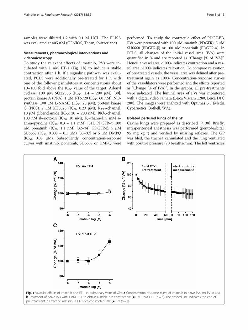
Fig. 1 Vascular effects of imatinib and ET-1 in pulmonary veins of GPs. a Concentration-response curve of imatinib in naïve PVs: (□) PV (n = 5).b Treatment of naïve PVs with 1 nM ET-1 to obtain a stable pre-constriction: (■) PV 1 nM ET-1 (n = 6). The dashed line indicates the end ofpre-treatment. c Effect of imatinib in ET-1-pre-constricted PVs: (■) PV (n = 9)
Maihöfer et al. Respiratory Research (2017) 18:32 Page 3 of 13

apex was cut and cannulas were placed in the pulmonaryartery (perfusion inflow) and in the left atrium (perfusionoutflow). The lung was perfused at constant flow(12,5 mL/min) with Krebs-Henseleit buffer, containing 2%bovine serum albumin, 0.1% glucose, 0.3% HEPES and 50nM salbutamol to prevent bronchoconstriction [39]. Thetemperature of the perfusate was maintained at 37 °C witha water bath and the pH was maintained between 7.35and 7.45 by gassing with carbon dioxide. Heart and lungswere removed and transferred into a negative-pressurechamber. Tidal volume, compliance, resistance, pulmonalarterial pressure (PPA), left atrial pressure (PLA), and theflow were continuously monitored. As soon as the respira-tory and hemodynamic variables remained stable over10 min, ET-1 (20 nM) was added to the recirculating per-fusion buffer (total volume 200 mL) to enhance Rpost [40].Ten minutes after the application of ET-1, imatinib(10 μM) was perfused. At a buffer volume of 200 mL, thiscorresponds to total amount of 1.18 mg imatinib or to3.5 mg/kg body weight imatinib, respectively. Thereafter,changes of the capillary pressure (Pcap) were measuredevery 10 min by the double occlusion method [38],Rpost and the precapillary resistance (Rpre) were calcu-
lated by the following equations: Rpost ¼ Pcap−PLAFlow and
Rpre ¼ PPA−PcapFlow .
In order to examine the effects of nebulized imatinibon Rpost, isolated perfused GP lungs were perfused asdescribed before. After the increase of Rpost by ET-1(20 nM), 3 mL of imatinib (10 mM), corresponding toa total amount of 17,691 mg imatinib were nebulizedover a period of 130 min. Assuming a lung flow of 0,21 L/min (70 breaths à 3 mL) and a pressure of 1.5 bar, the totalamount of inhaled imatinib corresponds to less than 4% ofthe nebulized amount of imatinib [41], namely 0,71 mg,corresponding to 2 mg/kg body weight imatinib, respect-ively. The contractile effect of ET-1 agonists strongly var-ies. Therefore, the maximal ET-1-induced increase of Rpost
was normalized to 100% and all submaximal increases ofRpost were indicated in % of the maximal increase of Rpost.
ChemicalsImatinib was provided by Novartis (Basel, Switzerland);nebulized imatinib was solved in aqua at a concentrationof 10 mM. SQ22536, KT5720, KT5823, glibenclamide,iberiotoxin, 4-aminopyridine and DMPQ were purchasedfrom Tocris Bioscience (Ellisville, Missouri, USA). ET-1was acquired from BIOTRENDS (Wangen, Switzerland)and SU6668 and ponatinib were acquired from Biomol(Hamburg, Germany). L-Name or standard laboratorychemicals were obtained from Sigma-Aldrich (Steinheim,Germany). The ELISA-kits were acquired from Enzo(Lörrach, Germany). Human PDGF-BB was delivered byPeprotech (Hamburg, Germany).
Statistical analysisStatistics were conducted using SAS software 9.3 (SASInstitute, Cary, North Carolina, USA) and GraphPad Prism5.01 (GraphPad, La Jolla, USA). The data in Figs. 4, 5 and6a,b,e,f were analyzed using a linear mixed model analysis(LMM) with the covariance structures VC or AR(1); EC50
values (Figs. 2b, c, e, f, 3b and 6c, d) were calculated bythe standard 4-parameter logistic non-linear regressionmodel (GraphPad, La Jolla, USA). The AIC-criterion wasused to select the most parsimonious model, i.e. a com-mon top, bottom, slope and EC50 value in the regressionmodel or the covariance matrix with the least number ofparameters. Non-parametric analysis (Figs. 2a, d and 3a, c)was performed by the Mann–Whitney U test. All p-valueswere adjusted for multiple comparisons by the false discov-ery rate and are presented as mean ± SEM; n indicates thenumbers of animals. P <0.05 was considered as significant.
ResultsWe studied the relaxant effects of imatinib in naïve (notpre-constricted) and in pre-constricted PVs.
ET-1-induced pre-constriction and imatinib-inducedrelaxationImatinib did not relax naïve PVs from GPs (Fig. 1a). Toobtain a stable and comparable contraction PVs were pre-constricted with ET-1 (1 nM). After 1 h, ET-1 (1 nM)contracted PVs to 69% of IVA (Fig. 1b), and imatinib(100 μM) relaxed PVs to 126% of IVA (Fig. 1c).
Involvement of the cAMP/PKA-pathway to the vasorelaxanteffect of imatinibIn PVs, imatinib increased intracellular cAMP (Fig. 2a).The functional role of the cAMP/PKA-pathway wasaddressed by pre-treatments with the adenyl cyclase-inhibitor SQ22536 (100 μM) and the PKA-inhibitorKT5720 (1 μM). Both inhibitors alone do not alter thecontractile effect of ET-1 [29], but here they decreasedthe imatinib-induced relaxation (Fig. 2b, c). To studythe contribution of NO or NO-downstream products(cGMP/PKG) to the imatinib-induced relaxation, PVs werepre-treated with the NO-synthase-inhibitor L-NAME(100 μM) or the PKG-inhibitor KT5823 (2 μM). Bothinhibitors do not affect the ET-1-induced contraction[29]. Here, L-NAME and KT5823 did also not alter therelaxant effect of imatinib (Fig. 2e, f). Accordingly, imatinibdid not increase intracellular cGMP-levels in PVs (Fig. 2d).
Involvement of K+-channels to the vasorelaxant effect ofimatinibWe further evaluated the impact of K+-channel-activationin the imatinib-induced relaxation of cavine PVs.KATP -channel-inhibition by glibenclamide (10 μM),
Maihöfer et al. Respiratory Research (2017) 18:32 Page 4 of 13

BKCa2þ -channel-inhibition by iberiotoxin (100 nM)
and Kv -channel-inhibition by 5 mM 4-aminopyridine(4-AP) do not affect ET-1-induced contraction [29].However, inhibition of KATP -channels, BKCa
2þ -channelsor Kv-channels significantly reduced the relaxant effect ofimatinib (Fig. 3a-c); inhibition of KATP - or Kv -channelswas most effective.
Imatinib lowers the Rpost in the IPLTo elucidate whether and how imatinib affects the pul-monary venous bed in an intact organ, we perfused andnebulized imatinib in the IPL after raising Rpost by ET-1. In the IPL, perfusion of 20 nM ET-1 increased PPAabout 600% of baseline from 1.5 cmH2O to 9.5 cmH2O(p <0.0001), whereas control lungs had no increase ofPPA and remained stable (Fig. 4a). ET-1 did not affectPLA (Fig. 4b). Further, the experiments with the doubleocclusion method revealed that ET-1 significantly en-hanced Rpre up to 312% and Rpost up to 169% of base-line (Fig. 4c, d). Next we studied if perfused or nebulizedimatinib significantly affects Rpost. Perfusion of imatinib
significantly lowered the ET-1-induced increase of Rpost
(Fig. 5a), even though Rpost was still increased (p <0.05)compared to control lungs (Fig. 5a). Nebulization of ima-tinib (calculated dosage: 2 mg/kg body weight) reducedthe ET-1-induced increase of Rpost from the time points110 – 150 min, but Rpost was still significantly increased(p <0.001) compared to control lungs (Fig. 5b).
The role of PDGFR-β and interaction of ET-1 with PDGFRThe antiproliferative properties of imatinib are likelyexplained by PDGFR-inhibition. Thus, we hypothesizedthat the imatinib-induced relaxation may depend on in-hibition of PDGFR kinase activity and further that PDGF-BB may possibly exert contractile properties in pulmonaryvessels. To test this hypothesis and to identify, whichPDGFR-subunit might be responsible for a possible con-tractile effect, we pre-treated PVs with 100 μM imatinib(PDGFR), 5 μM SU6668 (PDGFR-β) or 100 nM ponatinib(PDGFR-α) prior to the treatment with 10 or 100 nMPDGF-BB. Control PVs treated with 10 or 100 nM PDGF-BB contracted up to 70% of IVA. In contrast, pre-treatment
Fig. 2 Role of cAMP/cGMP in imatinib-induced relaxation of GP pulmonary veins. a Effect of imatinib on intracellular cAMP. b Effect of inhibitionof adenyl cyclase (SQ22536) on imatinib-induced relaxation in ET-1 (1 nM) pre-constricted PVs: (■) imatinib (n = 9); ( ) SQ22536 (100 μM), imatinib(n = 7); (□) SQ22526 (100 μM) (n = 6). c Effect of inhibition of PKA (KT5720) on imatinib-induced relaxation in ET-1 (1 nM) pre-constricted PVs: (■)imatinib (n = 9); ( ) KT5720 (1 μM), imatinib (n = 7); (□) KT5720 (1 μM) (n = 6). d Effect of imatinib on cGMP. e Effect of inhibition of NO-synthase(L-NAME) on imatinib-induced relaxation in ET-1 (1 nM) pre-constricted PVs: (■) imatinib (n = 9); ( ) L-NAME (100 μM), imatinib (n = 8); (□)L-NAME (100 μM) (n = 5). f Effect of inhibition of PKG (KT5823) on imatinib-induced relaxation in ET-1 (1 nM) pre-constricted PVs: (■) imatinib (n = 9);( ) KT5823 (2 μM), imatinib (n = 11); (□) KT5823 (2 μM) (n = 6). A/D) Statistics was performed by the Mann–Whitney U test. b/c/e/f) Asterisks indicatedifferent EC50 values. P <0.05 are considered as significant: * p <0.05, ** p <0.01 and *** p <0.001
Maihöfer et al. Respiratory Research (2017) 18:32 Page 5 of 13

with imatinib and SU6668 abolished the contractile effectof PDGF-BB, whereas pre-treatment with ponatinib onlyinsignificantly reduced the contractile effect of PDGF-BB(Fig. 6a, b). Next, we analyzed the effects of the PDGFR-αinhibitor ponatinib and the PDGFR-β inhibitors SU6668and DMPQ on PVs pre-constricted with ET-1. Treatmentwith SU6668 and DMPQ relaxed PVs pre-constricted withET-1 up to 140% of IVA (Fig. 6c), whereas treatment withthe PDGFR-α inhibitor ponatinib had no significantrelaxant effect (Fig. 6c). Further, concomitant pre-treatmentwith 5 μM SU6668 and 1 nM ET-1 or 5 μM DMPQ and 1nM ET-1 (Fig. 6d) abolished imatinib-induced relaxation,exceptional if it was applied at 100 μM. Next, we studiedwhether stimulation of ET-1 receptors interacts any-how with PDGFR or conversely, if activation ofPDGFR contributes to the contractile effect of ET-1.In order to inhibit PDGFR, we pre-treated PVs 60 minwith imatinib (10 or 100 μM), 5 μM SU6668 or 100nM ponatinib prior to the treatment of 1 nM ET-1(Fig. 6e, f ). We found, irrespective of inhibition ofPDGFR-αβ, ET-1 contracted PVs without significantdifference (Fig. 6e, f ).
DiscussionThe tyrosine kinase inhibitor imatinib represents anintriguing approach to treat PH. Initially, research fo-cused on the antiproliferative effects of imatinib, inline with the reversal of vascular remodeling in experi-mental studies [42]. Interestingly, in clinical trials andcase reports, imatinib also ameliorated hemodynamics[11, 12, 16] and restored heart function [13] suggestingthat it may also improve vascular tone. Meanwhile,corroborating studies in PAs from healthy or hyperten-sive rats demonstrated the relaxant effects of imatinib[14, 15]. Clinically, however, effects on PVs may bemore relevant since the most common cause of PH isLHD, a condition that primarily affects PVs [8, 43].Therefore, in the present study we focused on the relaxanteffects of imatinib in the pulmonary venous bed. We se-lected GPs, as the access to human lung tissue is limitedand the pulmonary smooth muscle pharmacology of GPsresembles human tissue much better than rats or mice[28, 44]. Most studies were performed using PCLS, amethod [28, 29] that allows to study PVs within their nat-ural tissue anatomy [45]. To study the effects of imatinib
Fig. 3 Contribution of K+-channels to imatinib-induced relaxation of GP pulmonary veins. a Effect of inhibition of KATP-channels (glibenclamide)on imatinib-induced relaxation in ET-1 (1 nM) pre-constricted PVs: (■) imatinib (n = 9); ( ) glibenclamide (10 μM), imatinib (n = 8); (□) glibenclamide(10 μM) (n= 8); b Effect of inhibition of BKCa2þ-channels (iberiotoxin) on imatinib-induced relaxation in ET-1 (1 nM) pre-constricted PVs: (■)imatinib (n = 9); ( ) iberiotoxin (100 nM), imatinib (n = 8); (□) iberiotoxin (100 nM) (n = 8); c Effect of inhibition of Kv-channels (5-aminopyridine)on imatinib-induced relaxation in ET-1 (1 nM) pre-constricted PVs: (■) imatinib (n = 9); ( ) 4-AP (5 mM), imatinib (n = 10); (□) 4-AP (5 mM) (n = 10).a/c Corresponding concentrations of (■) and ( ) were compared by the Mann–Whitney U test. b Asterisks indicate different EC50 values. P <0.05 areconsidered as significant: * p <0.05 and ** p <0.01
Maihöfer et al. Respiratory Research (2017) 18:32 Page 6 of 13

Fig. 4 Influence of ET-1 on different segments of the pulmonary circulation in the IPL. a Influence of 20 nM ET-1 on the pulmonary arterial pressure(PPA): (○) control (n = 6); (●) ET-1 20 nM (n = 7); b Influence of 20 nM ET-1 on the left atrial pressure (PLA): (○) control (n = 6); (●) ET-1 20 nM(n = 7); c Influence of 20 nM ET-1 on the precapillary resistance (Rpre): (○) control (n = 6); (●) ET-1 20 nM (n = 7); d Influence of 20 nM ET-1 onthe postcapillary resistance (Rpost): (○) control (n = 6); (●) ET-1 20 nM (n = 7). a-d) Statistics was performed by a LMM. P <0.05 are consideredas significant: * p <0.05, ** p <0.01 and *** p <0.001
Fig. 5 Influence of perfused and nebulized imatinib on the ET-1-induced increase of Rpost. a Influence of perfused imatinib on the ET-1-inducedincrease of the postcapillary resistance (Rpost): (○) control (n = 6); (●) ET-1 20 nM (n = 7); ( ) ET-1 20 nM / imatinib 20 mM (n = 6). b Influence ofnebulized imatinib on the ET-1-induced increase of the postcapillary resistance (Rpost): (○) control (n = 6); (●) ET-1 20 nM (n = 7); ( ) ET-1 20 nM/imatinib 20 mM (n = 7). a-b Statistics was performed by a LMM. P <0.05 are considered as significant: * p <0.05 and *** p <0.001
Maihöfer et al. Respiratory Research (2017) 18:32 Page 7 of 13

on the Rpost, we used the double occlusion method [38] inisolated perfused lungs (IPL) of GPs. To mimic a patho-physiological aspect of PH, namely the overexpression ofET-1 and its receptor [46, 47], we pre-constricted PVswith ET-1.
In the present study, imatinib relaxed pre-constrictedPVs with an EC50 value of 32 μM in line with a previousstudy in PAs [14]. Imatinib-induced relaxation wasdependent on the generation of cAMP and on the acti-vation of K+-channels. In the IPL, perfused or nebulized
Fig. 6 The role of PDGFR-β and interaction of ET-1 with PDGFR. a Effect of inhibition of PDGFR (imatinib) on the contractile effect of 10 nMPDGF-BB: (■) PV: PDGF-BB (10 nM) (n = 5); (□) PV: imatinib (100 μM), PDGF-BB (n = 5). b Effect of inhibition of PDGFR-α (ponatinib) and PDGFR-β(SU6668) on the contractile effect of PDGF-BB: (■) PV: PDGF-BB (100 nM) (n = 7); (□) PV: SU6668 (5 μM), PDGF-BB (100 nM) (n = 6); ( ) PV: Ponatinib(100 nM), PDGF-BB (100 nM) (n = 7). c The relaxant effects of the unselective TKI imatinib and the PDGFR-β inhibitors SU6668 or DMPQ in ET-1pre-constricted PVs: (■) PV 1 nM ET-1/imatinib (n = 5); ( ) PV: 1 nM ET-1/SU6668 (n = 5); (□) PV: 1 nM ET-1/DMPQ (n = 5); (●) PV: 1 nM ET-1/ponatinib(n = 5); d The relaxant effect of the unselective TKI imatinib after inhibition of PDGFR-β by SU6668 or DMPQ: (■) PV: 5 μM SU6668/1 nM ET-1/imatinib;(□) PV: 5 μM DMPQ/1 nM ET-1/imatinib; e/f Effect of inhibition of PDGFR (imatinib), PDGFR-α (ponatinib) and PDGFR-β (SU6668) on ET-1 inducedcontraction: (■) PV: ET-1 (1 nM) (n = 5); ( ) PV: 100 μM imatinib/1 nM ET-1 (n = 5); ( ) PV: 1 μM imatinib/1 nM ET-1 (n = 5); ( ) PV: 5 μM SU6668/1 nMET-1 (n = 5); ( ) PV: 100 nM ponatinib/1 nM ET-1 (n = 5). Statistics was performed by LMM (Fig. 6a, b, e, f). Asterics indicate different EC50 values(Fig. 6c, d). P <0.05 are considered as significant: *** < 0.001
Maihöfer et al. Respiratory Research (2017) 18:32 Page 8 of 13

imatinib reduced the ET-1-induced increase of Rpost.Notably, nebulized imatinib might prevent perfusion/ventilation mismatches and imatinib-related systemicside effects such as subdural hematoma or hypotension.Further, PDGF-BB contracts PVs, a finding which mightexplain in part why imatinib exerts relaxation. Our re-sults suggest the use of imatinib in the therapy of PHdue to LHD.
Mechanisms contributing to imatinib-induced relaxationThe tone of vascular SMCs is regulated by various sig-naling cascades, which ultimately modulate intracellularCa2+-levels or/and Ca2+-sensitivity of the myofilaments.In this context, the nucleotides cAMP and cGMP play aleading part. Cyclic AMP — via activation of PKA —causes relaxation by K+-channel-stimulation. Further,cAMP acts in a Ca2+-desensitizing manner: 1) cAMPblocks myosin light chain kinase (MLCK); 2) cAMP acti-vates myosin light chain phosphatase (MLCP) [48]. Ithas been reported that Ca2+-desensitization plays arole in imatinib-induced relaxation [14]. Here we showthat imatinib leads to increased intracellular cAMPlevels in cavine PVs. The functional relevance of thiscAMP-increase was shown by inhibiting adenyl cy-clase (SQ22536) or PKA (KT5720); both inhibitorsattenuated the relaxant effect of imatinib. The gener-ation of cAMP has not yet been reported within thecontext of imatinib- or TKI-induced vasorelaxation,even though they are supported by own unpublishedresults from human pulmonary vessels and by Kim et al.[49] who proved in murine interstitial cell of Cajals thatthe imatinib-induced inhibition of pacemaker potentialsdepends on the generation of cAMP [49]. These resultsraise the question, how to explain the imatinib-induced in-crease of cAMP or more precisely how PDGFR-inhibitionis linked to the activation of Gαs, the inhibition of Gαi orthe inhibition of PDE-III: 1) There is evidence that RTKsand GPCR interact together, e.g. via transactivation [50],as it was already shown for EGF and Gαi [51]. 2) Recentlyit was reported that PDGF inhibits regulators of G proteinsignaling (RGS) [52]. In particular, it was shown thatPDGF suppresses the expression of RGS-5 in vascularSMC; since downregulation of RGS-5 activates Gαi, adenylcyclase is blocked and the generation of intracellularcAMP reduced.Cyclic AMP/PKA-signaling appears critical for the
imatinib-induced relaxation, although inhibition of adenylcyclase or PKA did not completely prevent it. This sug-gests the involvement of other signaling cascades such asNO/cGMP/PKG. PKG promotes Ca2+-desensitizationvia MLCP-activation and stimulates K+-channels [48].The NO/cGMP/PKG-signaling highly depends on anintact endothelium releasing aside NO [53] also prosta-cyclin [54, 55]. In PCLS, PVs dispose of an intact
endothelium [56], even though NO/cGMP/PKG-signalingdid not contribute to imatinib-induced relaxation, as 1)imatinib failed to increase cGMP; 2) inhibition of NO-synthesis (L-NAME) or PKG (KT5823) did not attenuateimatinib-induced relaxation. Despite these results, wecannot conclude if imatinib-induced relaxation is endo-thelium independent or dependent, as it is highly basedon cAMP/PKA-signaling and within this context, theendothelial release of prostacyclin might be of impact.Our results corroborate in part those of Abe et al. [14],who showed that imatinib relaxes PAs from hypertensiverats, despite NO-inhibition. Aside that, they [14] proved inendothelial denuded PAs (rats) that the relaxant effect ofimatinib does not depend on the endothelium or theendothelial prostacyclin release, as well. In contrast, inSMCs from human corpus cavernosum [57] and in pros-tatic SMCs [58], imatinib-induced relaxation appears todepend on NO-signaling. Thus, the role of NO/PKG/cGMP in the imatinib-induced relaxation appears to bespecies and organ-specific.To study further mechanisms, we evaluated the im-
pact of KATP-, BKCa2þ - and Kv-channels, all of which
are expressed in pulmonary vessels [59, 60]. Activationof K+-channels leads to cell membrane hyperpolariza-tion resulting in reduced cytosolic Ca2+-influx and inrelaxation of vascular SMCs. In GP PVs, inhibition ofall three K+-channels attenuated imatinib-induced re-laxation, with KATP- and Kv-channel inhibitors beingmore effective. The minor role of BKCa
2þ -channelsmight be explained by the fact that BKCa
2þ -channelsdominate in conduit PAs [59], although in rat PAsK+-channel-activation could not explain the imatinib-induced relaxation [15]. In line with our results, a dominantrole of KATP- versus BKCa
2þ -channels was demonstratedfor the imatinib-induced relaxation in human prostaticSMCs [58]. Conversely, PDGF has been shown to decreaseKv-channel currents [61]. Moreover, the imatinib-inducedactivation of K+-channels does not only mediate relaxation,but also contributes to its antiproliferative effects, as hyper-polarization of the cell membrane lowers the intracellu-lar Ca2+-influx/content and counteracts the vascularremodeling [59]. A similar relationship has also beenshown for the Ca2+-sensitizer levosimendan that de-velops pulmonary vasorelaxant effects [29] and anti-proliferative properties via K+-channel-activation [62].Finally, imatinib-induced relaxation is based on cAMP/PKA dependent mechanisms and on K+-channel activa-tion. Both signaling pathways may interact to someextent in an additive manner [48, 63].
Pulmonary vasorelaxant effects of perfused andnebulized Imatinib in the IPLThe resistance of the pulmonary venous bed, preciselythe postcapillary resistance (Rpost) is increased in PH due
Maihöfer et al. Respiratory Research (2017) 18:32 Page 9 of 13

to LHD and mainly responsible for the generation ofhydrostatic edema [6]. Vice versa, reduction of Rpost isone of the major therapeutic goals in this disease. To ad-dress the vasorelaxant effects of imatinib on Rpost in situ,we perfused imatinib in isolated lungs. This set-up en-ables: 1) to address the pulmonary arterial and venoussegment independent from each other; 2) to monitorPPA, PLA and the capillary pressure (Pcap) and 3) to cal-culate Rpost from PLA and Pcap. In order to access the re-sistance of the pulmonary venous bed, the calculation ofRpost is highly relevant, because it includes the smallPVs. Further, it is superior to the Ppcw, which rather re-flects the pressure in large PVs [64] or PLA [65].Perfusion of 20 nM ET-1 significantly increased PPA,
Rpre and Rpost in the IPL. In contrast, PLA did notchange, as it is expected due to the reservoir function ofthe left atrium in this model. In the present study, perfu-sion of 10 μM imatinib significantly lowered Rpost. Thisfinding is in line with the results of the IMPRES studythat showed improved hemodynamics after the applica-tion of imatinib, expressed as a decrease in PVR and anincrease in the 6 min walk distance [16]; of note, theIMPRES study only included patients with PAH [16].Our results are further supported by Shah et al. [13],who found that imatinib improves right but also leftheart function. However, the IMPRES study [16], as wellas the long-term extension of the IMPRES study [17]further revealed undesirable imatinib-related side effectssuch as vomiting, nausea or peripheral edema that de-crease patient compliance; and even more critical werethe occurrence of subdural hematoma, QTc prolonga-tion, cardiac failure and syncope, the latter two mostprobably being caused by systemic hypotension [16, 17].Unfortunately, however in PH, systemic hypotension isquite problematic, as it decreases the coronary perfusionpressure and consequently worsens left and right ven-tricular function [66]. To find a way to prevent systemicside effects, we evaluated the possibility and efficacy toapply imatinib locally via the airways [18], as it is alreadyusual in PH for NO-donors, prostanoids or PDE-inhibitors[67, 68]. In the IPL, we could show that nebulized imatinibreduces the ET-1-induced increase of Rpost. The resultsfrom the IPL (perfusion/nebulization) correspond to ourdata from PCLS, even though nebulization of imatinib wasless effective in reducing Rpost than perfusion. As men-tioned above (methods), these differences are probablyexplained by the maximal imatinib up-take: the ima-tinib up-take via nebulization is 0.71 mg correspondingto 2 mg/kg body weight imatinib and via perfusion1.18 mg corresponding to 3.5 mg/kg body weight ima-tinib (the ratio is 0.6). Other effects may also play a rolesuch as an insufficient imatinib-uptake through thealveo-capillary membrane or the degradation of imatinibby intraalveolar macrophages. Here, we performed an
IPL model; thus the extent to which nebulized imatinibprovokes systemic adverse effects remains to be shown.In summary, we focused on central PVs in PCLS andwe addressed Rpost (IPL), resembling the peripheral partof the pulmonary venous bed [69, 70]. In both models,we demonstrated that imatinib relaxes the pulmonaryvenous bed of GPs.
The role of PDGFR-β and interaction of ET-1 with PDGFRThe antiproliferative effects of imatinib are attributedto inhibition of the tyrosine kinase activity of PDGFR [20].Beyond that, PDGFR activation is known to enhance thetone of various vessel types from different species bythe increase of intracellular Ca2+ [26, 71]. So far it isunknown, if imatinib-induced relaxation is linked toPDGFR-inhibition, and further if PDGF also contractspulmonary vessels. Here, we showed that PDGF-BBcontracts PVs and that this contraction is completelyprevented by the unselective PDGFR-αβ-inhibitor ima-tinib. PDGFR-β-inhibition (SU6668) abolished PDGF-BB-induced contraction, whereas PDGFR-α-inhibition(ponatinib) reduced it only mildly and non-significantly.In general, PDGF-BB binds to PDGFR-αα, αβ or ββ [23].Hence, our results suggest 1) a leading role of PDGFR-ββor/and PDGFR-αβ, as inhibition of the β-subunit com-pletely prevented PDGF-BB-induced contraction and 2) aminor role of PDGFR-αα, as inhibition of the α-subunitdid not prevent contraction. SU6668 has multiple targetssuch as Aurora kinases, TBK1 and the RTK VEGFR-2,FGFR-1, EGFR or FLK-1/KDR [35]. SU6668 (5 μM)blocks VEGFR-2 (IC50 0.34–2.43 μM) and FGFR-1(IC50 1.2–10 μM) only moderately, whereas ponatinib(100 nM) potently blocks VEGFR-2 (IC50 1.5 nM) andFGFR-1 (IC50 2.2 nM). Hence, if the inhibition of VEGFR-2 or FGFR-2 would prevent the PDGF-BB-induced con-traction, ponatinib should be more effective than SU6668.In addition, the complete inhibition of EGFR (IC50 >100nM) or FLK-1/KDR (IC50 2.1 μM) would require SU6668concentrations above 5 μM. These considerations supportthe idea that the contractile effect of PDGF-BB is pre-vented by inhibition of PDGFR-β [32–37, 72–75]. Con-versely, the PDGFR-β inhibitors SU6668 and DMPQrelaxed pre-constricted PVs, whereas the PDGFR-α inhibi-tor ponatinib had no effect. Further, after inhibition ofPDGFR-β by SU6668 or DMPQ (both 5 μM), imatinibonly relaxed pre-constricted PVs at 100 μM. These datasuggests that imatinib-induced relaxation depends onPDGFR-β inhibition. Thus, our findings suggest thatPDGFR-β antagonism does not only attenuate the es-tablishment of PH [10, 76], but also mediates vasorelax-ation. Our results are supported by Shiba et al. [77]who proved in cerebral arteries that imatinib counter-acts PDGFR-signaling and prevents vasospasm. In an
Maihöfer et al. Respiratory Research (2017) 18:32 Page 10 of 13

effort to identify a link between ET-1- and PDGFR sig-naling, we studied if activated ET-1 receptors interactanyhow with PDGFR. To this end, we inhibited PDGFRprior to the treatment with ET-1. Neither inhibition ofPDGFR (imatinib), PDGFR-β (SU6668) nor PDGFR-α(ponatinib) did reduce ET-1-induced contraction. Hence,it appears that stimulation of ET-1 receptors does not acti-vate PDGFR or it takes place, but without being sufficientto mediate contraction, possibly because ligand-binding ismissing. Recently, Harada and coworkers [78] could showin rat L6 myoblasts that stimulation of ET-1 receptors ac-tivates PDGFR downstream signaling by an unknownmechanism. However, they did not study ET-1-inducedphosphorylation of PDGFR [78].Irrespective from a possible interaction of ET-1 and
PDGFR, the relaxant effect of imatinib is not limited toET-1, as it relaxed PAs pre-constricted with U46619 [14,15], serotonin [14] or L-NAME [15]. Further, imatinib ex-erts relaxant effects in different tissues, i.e. systemic vessels[15], prostatic tissue [58], PAs [14, 15], corpus cavernosum[57], myometrium [79] and stomach [80] from humans,rabbits, GPs, rats or sheep. Thus, imatinib-induced relax-ation appears to be a widespread phenomenon. Beyondthat, other TKIs also exert pulmonary vascular relaxanteffects (nilotinib/sorafenib) [14] or prevent experimentalpulmonary vascular remodeling by PDGFR-inhibition(nilotinib/dasatinib). Within this context, dasatinib plays aparticular role, as it also inhibits Src kinases playing a piv-otal role in pulmonary arterial remodeling [81, 82]. That isprobably why; dasatinib reverses experimental PH evenmore potently than imatinib [81]. However in patients withCML, dasatinib also provoke drug-induced PAH by anunknown mechanism [83, 84]. Fortunately, this dasatinib-associated adverse effect appears to be reversible, rareand limited to dasatinib [85–88].
ConclusionsImatinib relaxes PVs by activation of cAMP/PKA andpotassium channels (KATP-, BKCa
2þ-, Kv-channels), mecha-nisms that are related in an unknown way to the inhibitionof PDGFR-β. The clinical importance of imatinib-inducedrelaxation is supported by reports that imatinib improvedthe clinical condition of a patient with pulmonary venousocclusive disease within 24 h [12] and ameliorated PH dueto congenital hernia [11]. The prompt improvement in bothcases favors relaxant effects of imatinib in contrast to thereversal of vascular remodeling which lasts longer [10].However, until now studies with human pulmonary vesselsare still missing and quite difficult to obtain. Our datasuggest a beneficial role of imatinib in PH due to LHD.Since imatinib combines short-term relaxant, even ifinhaled and long-term antiproliferative effects, it mayrepresent a promising approach to treat PH due to LHD.
AbbreviationsGPs: Guinea pigs; IPL: Isolated perfused lungs; LHD: Left heart disease;LMM: Linear mixed model analysis; MLCK: Myosin light chain kinase;MLCP: Myosin light chain phosphatase; PAH: Pulmonary arterial hypertension;PAs: Pulmonary arteries; Pcap: Capillary pressure; PCLS: Precision-cut lungslices; PDGF: Platelet derived growth factor; PDGFR: PDGF-receptor;PH: Pulmonary hypertension; PLA: Left atrial pressure; PPA: Pulmonary arterialpressure; Ppcw: Pulmonary capillary wedge pressure; PVH: Pulmonary venoushypertension; PVR: Pulmonary vascular resistance; PVs: Pulmonary veins;RGS: Regulators of G protein signaling; Rpost: Postcapillary resistance;Rpre: Precapillary resistance; RTK: Receptor tyrosine kinase; SMCs: Smoothmuscle cells; TKI: Tyrosine kinase inhibitor; TPG: Transpulmonary gradient (TPG)
AcknowledgementsThis project was supported by the START programme of the RWTH Aachen. Wefurther gratefully acknowledge Hanna Czajkowska and Tanja Woopen for excellenttechnical assistance, as well as Novartis, Switzerland for providing imatinib.
FundingThis work was funded by the START program (grant 109/14) of the RWTH-Aachen. The funders had no influence of the study design, data collectionand analysis, decision to publish or preparation of the manuscript.
Availability of data and materialsThe datasets generated and analysed during the current study are availablefrom the corresponding author on reasonable request.
Authors’ contributionsNAM performed the experiments, analysed the data, interpreted the dataand wrote the manuscript. SS performed the experiments, analysed thedata and interpreted the data. DD performed the experiments, interpretedthe data and critically reviewed the manuscript. PWM analysed the data,interpreted the data and critically reviewed the manuscript. RR analysedthe data, interpreted the data and critically reviewed the manuscript.SU analysed the data, interpreted the data and critically reviewed themanuscript. CM designed the study, analysed the data, interpreted thedata and critically reviewed the manuscript. ADR designed the study,performed the experiments, analysed the data, interpreted the data andwrote the manuscript. All authors read and approved the final manuscript.
Competing interestsThe authors declare that they have no competing interests.
Consent for publicationNot applicable.
Ethics approval and consent to participateFemale Dunkin Hartley GPs (350 ± 50 g) were purchased from Charles River(Sulzfeld, Germany). Animal studies were approved by the Landesamt für Natur,Umwelt und Verbraucherschutz Nordrhein-Westfalen (ID: 8.87-51.05.20.10.245)and performed following the Directive 2010/63/EU of the European Parliament.Animal experiments were performed after deep anesthesia with intraperitonealpentobarbital (95 mg kg−1; Narcoren: Garbsen, Germany) which was verified bymissing reflexes. Then the GP was bled, the trachea cannulated and thediaphragm opened.
Author details1Institute of Pharmacology and Toxicology, Medical Faculty Aachen,RWTH-Aachen, Aachen, Germany. 2Novartis Pharma, AG, Basel, Switzerland.3Department of Anesthesiology, Medical Faculty Aachen, RWTH-Aachen,Aachen, Germany.
Received: 30 September 2016 Accepted: 19 January 2017
References1. Breitling S, Ravindran K, Goldenberg NM, Kuebler WM. The pathophysiology
of pulmonary hypertension in left heart disease. Am J Physiol Lung Cell Mol Physiol.2015;309:L924–41.
2. Galie N, Humbert M, Vachiery JL, Gibbs S, Lang I, Torbicki A, et al.2015 ESC/ERS guidelines for the diagnosis and treatment of pulmonary
Maihöfer et al. Respiratory Research (2017) 18:32 Page 11 of 13

hypertension: the joint task force for the diagnosis and treatment ofpulmonary hypertension of the european society of cardiology (ESC)and the european respiratory society (ERS): endorsed by: associationfor european paediatric and congenital cardiology (AEPC), internationalsociety for heart and lung transplantation (ISHLT. Eur Respir J.2015;46:903–75.
3. Galie N, Humbert M, Vachiery JL, Gibbs S, Lang I, Torbicki A, et al. 2015 ESC/ERS guidelines for the diagnosis and treatment of pulmonary hypertension:the joint task force for the diagnosis and treatment of pulmonaryhypertension of the european society of cardiology (ESC) and the europeanrespiratory society (ERS): endorsed by: association for european paediatricand congenital cardiology (AEPC), international society for heart and lungtransplantation (ISHLT). Eur Heart J. 2016;37:67–119.
4. Kulik TJ. Pulmonary hypertension caused by pulmonary venoushypertension. Pulm Circ. 2014;4:581–95.
5. Simonneau G, Gatzoulis MA, Adatia I, Celermajer D, Denton C, Ghofrani A, etal. Updated clinical classification of pulmonary hypertension. J Am CollCardiol. 2013;62:D34–41.
6. Adir Y, Offer A. Pulmonary hypertension associated with left heart disease.Eur Respir Monogr. 2012;57:119–37.
7. McLaughlin VV, Archer SL, Badesch DB, Barst RJ, Farber HW, Lindner JR, et al.ACCF/AHA 2009 expert consensus document on pulmonary hypertension areport of the american college of cardiology foundation task force onexpert consensus documents and the american heart associationdeveloped in collaboration with the american college of chest physicians;american thoracic society, Inc.; and the pulmonary hypertension association.J Am Coll Cardiol. 2009;53:1573–619.
8. Dadfarmay S, Berkowitz R, Kim B, Manchikalapudi RB. Differentiatingpulmonary arterial and pulmonary venous hypertension and theimplications for therapy. Congest Heart Fail. 2010;16:287–91.
9. Rieg AD, Suleiman S, Perez-Bouza A, Braunschweig T, Spillner JW, SchröderT, Verjans E, Schälte G, Rossaint R, Uhlig S, Martin C. Milrinone relaxespulmonary veins in guinea pigs and humans. PLoS One. 2014;9(1):e87685.
10. Schermuly RT, Dony E, Ghofrani HA, Pullamsetti S, Savai R, Roth M, et al.Reversal of experimental pulmonary hypertension by PDGF inhibition. J ClinInvest. 2005;115:2811–21.
11. Frenckner B, Broome M, Lindstrom M, Radell P. Platelet-derived growthfactor inhibition—a new treatment of pulmonary hypertension incongenital diaphragmatic hernia? J Pediatr Surg. 2008;43:1928–31.
12. Overbeek MJ, van Nieuw Amerongen GP, Boonstra A, Smit EF, Vonk-Noordegraaf A. Possible role of imatinib in clinical pulmonary veno-occlusive disease. Eur Respir J. 2008;32:232–5.
13. Shah AM, Campbell P, Rocha GQ, Peacock A, Barst RJ, Quinn D, et al.Effect of imatinib as add-on therapy on echocardiographic measures ofright ventricular function in patients with significant pulmonary arterialhypertension. Eur Heart J. 2015;36:623–32.
14. Abe K, Toba M, Alzoubi A, Koubsky K, Ito M, Ota H, et al. Tyrosine kinaseinhibitors are potent acute pulmonary vasodilators in rats. Am J Respir CellMol Biol. 2011;45:804–8.
15. Pankey EA, Thammasibon S, Lasker GF, Baber S, Lasky JA, Kadowitz PJ.Imatinib attenuates monocrotaline pulmonary hypertension and has potentvasodilator activity in the pulmonary and systemic vascular beds of the rat.Am J Physiol Heart Circ Physiol. 2013;305(9):H1288–96.
16. Hoeper MM, Barst RJ, Bourge RC, Feldman J, Frost AE, Galie N, et al. Imatinibmesylate as add-on therapy for pulmonary arterial hypertension: results ofthe randomized IMPRES study. Circulation. 2013;127:1128–38.
17. Frost AE, Barst RJ, Hoeper MM, Chang HJ, Frantz RP, Fukumoto Y, et al.Long-term safety and efficacy of imatinib in pulmonary arterialhypertension. J Heart Lung Transplant. 2015;34:1366–75.
18. Pitsiou G, Zarogoulidis P, Petridis D, Kioumis I, Lampaki S, Organtzis J, et al.Inhaled tyrosine kinase inhibitors for pulmonary hypertension: a possiblefuture treatment. Drug Des Devel Ther. 2014;8:1753–63.
19. Berghausen E, Ten FH, Rosenkranz S. Targeting of platelet-derived growthfactor signaling in pulmonary arterial hypertension. Handb Exp Pharmacol.2013;218:381–408.
20. Zhang P, Gao WY, Turner S, Ducatman BS. Gleevec (STI-571) inhibits lungcancer cell growth (A549) and potentiates the cisplatin effect in vitro.Mol Cancer. 2003;2:1.
21. Perros F, Montani D, Dorfmuller P, Durand-Gasselin I, Tcherakian C, Le PJ,et al. Platelet-derived growth factor expression and function in idiopathicpulmonary arterial hypertension. Am J Respir Crit Care Med. 2008;178:81–8.
22. Overbeek MJ, Boonstra A, Voskuyl AE, Vonk MC, Vonk-Noordegraaf A, vanBerkel MP, et al. Platelet-derived growth factor receptor-beta and epidermalgrowth factor receptor in pulmonary vasculature of systemic sclerosis-associated pulmonary arterial hypertension versus idiopathic pulmonaryarterial hypertension and pulmonary veno-occlusive disease: a case–controlstudy. Arthritis Res Ther. 2011;13:R61.
23. Andrae J, Gallini R, Betsholtz C. Role of platelet-derived growth factors inphysiology and medicine. Genes Dev. 2008;22:1276–312.
24. Takase H, Oemar BS, Pech M, Luscher TF. Platelet-derived growth factor-inducedvasodilatation in mesenteric resistance arteries by nitric oxide: blunted responsein spontaneous hypertension. J Cardiovasc Pharmacol. 1999;33:223–8.
25. Yamawaki H, Sato K, Hori M, Ozaki H, Karaki H. Platelet-derived growthfactor causes endothelium-independent relaxation of rabbit mesentericartery via the release of a prostanoid. Br J Pharmacol. 2000;131:1546–52.
26. Berk BC, Alexander RW, Brock TA, Gimbrone Jr MA, Webb RC.Vasoconstriction: a new activity for platelet-derived growth factor. Science.1986;232:87–90.
27. Kuebler WM, Yang Y, Samapati R, Uhlig S. Vascular barrier regulation by PAF,ceramide, caveolae, and NO - an intricate signaling network with discrepanteffects in the pulmonary and systemic vasculature. Cell Physiol Biochem.2010;26:29–40.
28. Ressmeyer A, Larsson A, Vollmer E, Dahlen S, Uhlig S, Martin C.Characterisation of guinea pig precision-cut lung slices: comparison withhuman tissues. Eur Respir J. 2006;28:603–11.
29. Rieg AD, Rossaint R, Verjans E, Maihöfer NA, Uhlig S, Martin C.Levosimendan relaxes pulmonary arteries and veins in precision-cut lungslices – the role of KATP-channels, cAMP and cGMP. PLoS One. 2013;86:e66195. doi:10.1371/journalpone0066195.
30. Hourani SM, Boon K, Fooks HM, Prentice DJ. Role of cyclic nucleotides invasodilations of the rat thoracic aorta induced by adenosine analogues.Br J Pharmacol. 2001;133:833–40.
31. Ko EA, Han J, Jung ID, Park WS. Physiological roles of K+ channels invascular smooth muscle cells. J Smooth Muscle Res. 2008;44:65–81.
32. Gozgit JM, Wong MJ, Wardwell S, Tyner JW, Loriaux MM, Mohemmad QK, etal. Potent activity of ponatinib (AP24534) in models of FLT3-driven acutemyeloid leukemia and other hematologic malignancies. Mol Cancer Ther.2011;10:1028–35.
33. Huang WS, Metcalf CA, Sundaramoorthi R, Wang Y, Zou D, Thomas RM, etal. Discovery of 3-[2-(imidazo[1,2-b]pyridazin-3-yl)ethynyl]-4-methyl-N-{4-[(4-methylpiperazin-1-y l)methyl]-3-(trifluoromethyl)phenyl}benzamide(AP24534), a potent, orally active pan-inhibitor of breakpoint cluster region-abelson (BCR-ABL) kinase including the T315I gatekeeper mutant. J Med Chem.2010;53:4701–19.
34. O’Hare T, Shakespeare WC, Zhu X, Eide CA, Rivera VM, Wang F, et al.AP24534, a pan-BCR-ABL inhibitor for chronic myeloid leukemia, potentlyinhibits the T315I mutant and overcomes mutation-based resistance. CancerCell. 2009;16:401–12.
35. Godl K, Gruss OJ, Eickhoff J, Wissing J, Blencke S, Weber M, et al. Proteomiccharacterization of the angiogenesis inhibitor SU6668 reveals multipleimpacts on cellular kinase signaling. Cancer Res. 2005;65:6919–26.
36. Laird AD, Vajkoczy P, Shawver LK, Thurnher A, Liang C, Mohammadi M, et al.SU6668 is a potent antiangiogenic and antitumor agent that inducesregression of established tumors. Cancer Res. 2000;60:4152–60.
37. Laird AD, Christensen JG, Li G, Carver J, Smith K, Xin X, et al. SU6668 inhibitsFlk-1/KDR and PDGFRbeta in vivo, resulting in rapid apoptosis of tumorvasculature and tumor regression in mice. FASEB J. 2002;16:681–90.
38. Uhlig S, Wollin L. An improved setup for the isolated perfused rat lung.J Pharmacol Toxicol Methods. 1994;31:85–94.
39. Atzori L, Bannenberg G, Corriga AM, Moldeus P, Ryrfeldt A. Sulfur dioxide-induced bronchoconstriction in the isolated perfused and ventilatedguinea-pig lung. Respiration. 1992;59:16–21.
40. Horgan MJ, Pinheiro JM, Malik AB. Mechanism of endothelin-1-inducedpulmonary vasoconstriction. Circ Res. 1991;69:157–64.
41. Hugo Sachs Elektronik Harvard Apparatus GmbH, inventor; OperatingInstructions for PARI Jet-Nebulizer No. 73–1963. 2008.
42. Ghofrani HA, Seeger W, Grimminger F. Imatinib for the treatment ofpulmonary arterial hypertension. N Engl J Med. 2005;353:1412–3.
43. Guazzi M, Galie N. Pulmonary hypertension in left heart disease. Eur Respir Rev.2012;21:338–46.
44. Schlepütz M, Rieg AD, Seehase S, Spillner J, Perez-Bouza A, Braunschweig T,Schroeder T, Bernau M, Lambermont V, Schlumbohm C, Sewald K,
Maihöfer et al. Respiratory Research (2017) 18:32 Page 12 of 13

Autschbach R, Braun A, Kramer BW, Uhlig S, Martin C. Neurally MediatedAirway Constriction in Human and Other Species: A Comparative StudyUsing Precision-Cut Lung Slices (PCLS). PLoS One. 2012;7(10):e47344.
45. Sanderson MJ. Exploring lung physiology in health and disease with lungslices. Pulm Pharmacol Ther. 2011;24:452–65.
46. Giaid A, Yanagisawa M, Langleben D, Michel RP, Levy R, Shennib H, et al.Expression of endothelin-1 in the lungs of patients with pulmonaryhypertension. N Engl J Med. 1993;328:1732–9.
47. Schneider MP, Boesen EI, Pollock DM. Contrasting actions of endothelinET(A) and ET(B) receptors in cardiovascular disease. Annu Rev Pharmacol Toxicol.2007;47:731–59.
48. Morgado M, Cairrao E, Santos-Silva AJ, Verde I. Cyclic nucleotide-dependent relaxation pathways in vascular smooth muscle. Cell MolLife Sci. 2012;69:247–66.
49. Kim BJ, Chae H, Kwon YK, Choi S, Jun JY, Jeon JH, et al. Effects of imatinibmesylate in interstitial cells of Cajal from murine small intestine. Biol PharmBull. 2010;33:993–7.
50. Dhanasekaran DN. Transducing the signals: a G protein takes a new identity.Sci STKE. 2006;2006:e31.
51. Bhandari D, Lopez-Sanchez I, To A, Lo IC, Aznar N, Leyme A, et al. Cyclin-dependent kinase 5 activates guanine nucleotide exchange factor GIV/Girdinto orchestrate migration-proliferation dichotomy. Proc Natl Acad Sci U S A.2015;112:E4874–83.
52. Gunaje JJ, Bahrami AJ, Schwartz SM, Daum G, Mahoney Jr WM. PDGF-dependent regulation of regulator of G protein signaling-5 expressionand vascular smooth muscle cell functionality. Am J Physiol Cell Physiol.2011;301(2):C478–89.
53. Vanhoutte PM, Zhao Y, Xu A, Leung SW. Thirty years of saying NO: sources,fate, actions, and misfortunes of the endothelium-derived vasodilatormediator. Circ Res. 2016;119:375–96.
54. Kang KT. Endothelium-derived relaxing factors of small resistance arteries inhypertension. Toxicol Res. 2014;30:141–8.
55. Nava E, Llorens S. The paracrine control of vascular motion. A historicalperspective. Pharmacol Res. 2016;113:125–45.
56. Rieg AD, Rossaint R, Uhlig S, Martin C. Cardiovascular agents affect thetone of pulmonary arteries and veins in precision-cut lung slices. PLoSOne. 2011;6(12):e29698.
57. Gur S, Sikka SC, Abdel-Mageed AB, Elmageed ZY, Rezk B, Pankey E, et al.Imatinib Mesylate (Gleevec) Induces Human Corpus Cavernosum Relaxationby Inhibiting Receptor Tyrosine Kinases (RTKs): Identification of New RTKTargets. Urology. 2013;82(3):745.e11–6.
58. Ozgur-Akdemir A, Demirturk K, Karabakan M, Volkan-Oztekin C, AbdulkadirNA, Cetinkaya M, et al. Imatinib mesylate (Gleevec) as protein-tyrosinekinase inhibitor elicits smooth muscle relaxation in isolated human prostatictissue. Urology. 2011;78:968–6.
59. Bonnet S, Archer SL. Potassium channel diversity in the pulmonary arteriesand pulmonary veins: implications for regulation of the pulmonaryvasculature in health and during pulmonary hypertension. Pharmacol Ther.2007;115:56–69.
60. Michelakis ED, Weir EK, Wu X, Nsair A, Waite R, Hashimoto K, et al.Potassium channels regulate tone in rat pulmonary veins. Am J PhysiolLung Cell Mol Physiol. 2001;280:L1138–47.
61. Timpe LC, Fantl WJ. Modulation of a voltage-activated potassium channelby peptide growth factor receptors. J Neurosci. 1994;14:1195–201.
62. Revermann M, Schloss M, Mieth A, Babelova A, Schroder K, Neofitidou S,et al. Levosimendan attenuates pulmonary vascular remodeling. IntensiveCare Med. 2011;37:1368–77.
63. Pinterova M, Behuliak M, Kunes J, Zicha J. Involvement of BKCa and KVpotassium channels in cAMP-induced vasodilatation: their insufficientfunction in genetic hypertension. Physiol Res. 2014;63:275–85.
64. Montani D, Price L, Dorfmuller P, Achouh L, Jais X, Yaici A, et al. Pulmonaryveno-occlusive disease. Eur Respir J. 2009;33:189–200.
65. Chaliki HP, Hurrell DG, Nishimura RA, Reinke RA, Appleton CP. Pulmonaryvenous pressure: relationship to pulmonary artery, pulmonary wedge, andleft atrial pressure in normal, lightly sedated dogs. Catheter CardiovascInterv. 2002;56:432–8.
66. Gille J, Seyfarth HJ, Gerlach S, Malcharek M, Czeslick E, Sablotzki A. Perioperativeanesthesiological management of patients with pulmonary hypertension.Anesthesiol Res Pract. 2012;2012:356982.
67. Hill NS, Preston IR, Roberts KE. Inhaled therapies for pulmonaryhypertension. Respir Care. 2015;60:794–802.
68. Thunberg CA, Morozowich ST, Ramakrishna H. Inhaled therapy for themanagement of perioperative pulmonary hypertension. Ann Card Anaesth.2015;18:394–402.
69. Gao Y, Raj JU. Role of veins in regulation of pulmonary circulation.Am J Physiol Lung Cell Mol Physiol. 2005;288:L213–26.
70. Raj JU, Ramanathan R, Chen P, Anderson J. Effect of hematocrit onmicrovascular pressures in 3- to 5-weeks-old rabbit lungs. Am J Physiol.1989;256:H766–71.
71. Wijetunge S, Hughes AD. Effect of platelet-derived growth factor onvoltage-operated calcium channels in rabbit isolated ear artery cells.Br J Pharmacol. 1995;115(3):534–8.
72. Buchdunger E, Zimmermann J, Mett H, Meyer T, Muller M, Regenass U, et al.Selective inhibition of the platelet-derived growth factor signal transductionpathway by a protein-tyrosine kinase inhibitor of the 2-phenylaminopyrimidine class. Proc Natl Acad Sci U S A. 1995;92:2558–62.
73. Deininger M, Buchdunger E, Druker BJ. The development of imatinib as atherapeutic agent for chronic myeloid leukemia. Blood. 2005;105:2640–53.
74. Heinrich MC, Griffith DJ, Druker BJ, Wait CL, Ott KA, Zigler AJ. Inhibition ofc-kit receptor tyrosine kinase activity by STI 571, a selective tyrosine kinaseinhibitor. Blood. 2000;96:925–32.
75. Manley PW, Stiefl N, Cowan-Jacob SW, Kaufman S, Mestan J, Wartmann M,et al. Structural resemblances and comparisons of the relativepharmacological properties of imatinib and nilotinib. Bioorg Med Chem.2010;18:6977–86.
76. Balasubramaniam V, Le Cras TD, Ivy DD, Grover TR, Kinsella JP, Abman SH.Role of platelet-derived growth factor in vascular remodeling during pulmonaryhypertension in the ovine fetus. Am J Physiol Lung Cell Mol Physiol. 2003;284:L826–33.
77. Shiba M, Suzuki H, Fujimoto M, Shimojo N, Imanaka-Yoshida K, Yoshida T, et al.Imatinib mesylate prevents cerebral vasospasm after subarachnoid hemorrhagevia inhibiting tenascin-C expression in rats. Neurobiol Dis. 2012;46:172–9.
78. Harada T, Horinouchi T, Higa T, Hoshi A, Higashi T, Terada K, et al.Endothelin-1 activates extracellular signal-regulated kinases 1/2 viatransactivation of platelet-derived growth factor receptor in rat L6myoblasts. Life Sci. 2014;104:24–31.
79. Hutchings G, Deprest J, Nilius B, Roskams T, De RD. The effect of imatinibmesylate on the contractility of isolated rabbit myometrial strips.Gynecol Obstet Invest. 2006;62:79–83.
80. Hashitani H, Hayase M, Suzuki H. Effects of imatinib mesylate onspontaneous electrical and mechanical activity in smooth muscle of theguinea-pig stomach. Br J Pharmacol. 2008;154:451–9.
81. Pullamsetti SS, Berghausen EM, Dabral S, Tretyn A, Butrous E, Savai R, et al.Role of Src tyrosine kinases in experimental pulmonary hypertension.Arterioscler Thromb Vasc Biol. 2012;32:1354–65.
82. Rix U, Hantschel O, Durnberger G, Remsing Rix LL, Planyavsky M, Fernbach NV,et al. Chemical proteomic profiles of the BCR-ABL inhibitors imatinib, nilotinib,and dasatinib reveal novel kinase and nonkinase targets. Blood. 2007;110:4055–63.
83. Bauer S, Buchanan S, Ryan I. Tyrosine kinase inhibitors for the treatment ofchronic-phase chronic myeloid leukemia: long-term patient care andmanagement. J Adv Pract Oncol. 2016;7:42–54.
84. Montani D, Bergot E, Gunther S, Savale L, Bergeron A, Bourdin A, et al.Pulmonary arterial hypertension in patients treated by dasatinib.Circulation. 2012;125:2128–37.
85. Buchelli Ramirez HL, Alvarez Alvarez CM, Rodriguez Reguero JJ, GarciaClemente MM, Casan CP. Reversible pre-capillary pulmonary hypertensiondue to dasatinib. Respir Care. 2014;59:e77–80.
86. Hennigs JK, Keller G, Baumann HJ, Honecker F, Kluge S, Bokemeyer C, et al.Multi tyrosine kinase inhibitor dasatinib as novel cause of severe pre-capillary pulmonary hypertension? BMC Pulm Med. 2011;11:30.
87. Mattei D, Feola M, Orzan F, Mordini N, Rapezzi D, Gallamini A. Reversibledasatinib-induced pulmonary arterial hypertension and right ventricle failure ina previously allografted CML patient. Bone Marrow Transplant. 2009;43:967–8.
88. Rasheed W, Flaim B, Seymour JF. Reversible severe pulmonary hypertensionsecondary to dasatinib in a patient with chronic myeloid leukemia.Leuk Res. 2009;33:861–4.
Maihöfer et al. Respiratory Research (2017) 18:32 Page 13 of 13