Pigs taking wing with transposons and recombinases
16
Genome Biology 2007, 8(Suppl 1):S13 Review Pigs taking wing with transposons and recombinases Karl J Clark* †‡¤ , Daniel F Carlson* †¤ and Scott C Fahrenkrug* †‡ *Department of Animal Science at the University of Minnesota, Fitch Ave, St. Paul, MN 55108, USA. † The Arnold and Mabel Beckman Center for Transposon Research at the University of Minnesota, Church St, Minneapolis, MN 55455, USA. ‡ The Animal Biotechnology Center at the University of Minnesota, Fitch Ave, St. Paul, MN 55108, USA. ¤ These authors contributed equally to this work. Correspondence: Scott Fahrenkrug. E-mail: [email protected] Abstract Swine production has been an important part of our lives since the late Mesolithic or early Neolithic periods, and ranks number one in world meat production. Pig production also contributes to high-value-added medical markets in the form of pharmaceuticals, heart valves, and surgical materials. Genetic engineering, including the addition of exogenous genetic material or manipulation of the endogenous genome, holds great promise for changing pig phenotypes for agricultural and medical applications. Although the first transgenic pigs were described in 1985, poor survival of manipulated embryos; inefficiencies in the integration, transmission, and expression of transgenes; and expensive husbandry costs have impeded the widespread application of pig genetic engineering. Sequencing of the pig genome and advances in reproductive technologies have rejuvenated efforts to apply transgenesis to swine. Pigs provide a compelling new resource for the directed production of pharmaceutical proteins and the provision of cells, vascular grafts, and organs for xenotransplantation. Additionally, given remarkable similarities in the physiology and size of people and pigs, swine will increasingly provide large animal models of human disease where rodent models are insufficient. We review the challenges facing pig transgenesis and discuss the utility of transposases and recombinases for enhancing the success and sophistication of pig genetic engineering. ‘The paradise of my fancy is one where pigs have wings.’ (GK Chesterton). Published: 31 October 2007 Genome Biology 2007, 8(Suppl 1):S13 (doi:10.1186/gb-2007-8-S1-S13) The electronic version of this article is the complete one and can be found online at http://genomebiology.com/2007/8/S1/S13 © 2007 BioMed Central Ltd Introduction Pigs are ungulates native to Eurasia collectively grouped under the genus Sus within the Suidae family. Phylo- geographic analysis reveals that pigs were domesticated independently at least seven times around the globe, first at least 9,000 years ago [1,2]. Our longstanding affinity for pigs stems from their omnivorous ability to convert even our scraps into healthy and nutritious pork. Since their domestication, pigs have also captured our cultural imagination. Their intelligence and almost human behavior finds pigs intertwined with us in mythology, language, and art. The meat pig represents a significant commodity worldwide, in 2004 producing more than 89 million tons of meat [3] and contributing more than $50 billion to the US economy alone [4]. Co-products from hogs play a vital although less visible role in maintaining and improving the quality of human life, being the primary source of more than 20 drugs and pharmaceuticals [5]. Pig insulin, which differs from the human protein by a single amino acid, had saved the lives of innumerable type 1 diabetic patients before the development of recombinant human insulin. Pig heart valves are used to repair damaged or diseased human hearts, and pig skin is used to treat severe burn victims and to produce collagen scaffolds, gels, and other surgical materials. The anthropomorphism of pigs in our culture seems almost prescient, given what we now know to be extensive simi- larities between human and pig molecular, cellular, and systems physiology [6,7]. Pigs were Galen’s preferred models in his quest for truth about human anatomy during an era
Transcript of Pigs taking wing with transposons and recombinases

Genome Biology 2007, 8(Suppl 1):S13
ReviewPigs taking wing with transposons and recombinasesKarl J Clark*†‡¤, Daniel F Carlson*†¤ and Scott C Fahrenkrug*†‡
*Department of Animal Science at the University of Minnesota, Fitch Ave, St. Paul, MN 55108, USA. †The Arnold and Mabel Beckman Centerfor Transposon Research at the University of Minnesota, Church St, Minneapolis, MN 55455, USA. ‡The Animal Biotechnology Center at theUniversity of Minnesota, Fitch Ave, St. Paul, MN 55108, USA. ¤These authors contributed equally to this work.
Correspondence: Scott Fahrenkrug. E-mail: [email protected]
Abstract
Swine production has been an important part of our lives since the late Mesolithic or earlyNeolithic periods, and ranks number one in world meat production. Pig production alsocontributes to high-value-added medical markets in the form of pharmaceuticals, heart valves, andsurgical materials. Genetic engineering, including the addition of exogenous genetic material ormanipulation of the endogenous genome, holds great promise for changing pig phenotypes foragricultural and medical applications. Although the first transgenic pigs were described in 1985,poor survival of manipulated embryos; inefficiencies in the integration, transmission, andexpression of transgenes; and expensive husbandry costs have impeded the widespreadapplication of pig genetic engineering. Sequencing of the pig genome and advances in reproductivetechnologies have rejuvenated efforts to apply transgenesis to swine. Pigs provide a compellingnew resource for the directed production of pharmaceutical proteins and the provision of cells,vascular grafts, and organs for xenotransplantation. Additionally, given remarkable similarities inthe physiology and size of people and pigs, swine will increasingly provide large animal models ofhuman disease where rodent models are insufficient. We review the challenges facing pigtransgenesis and discuss the utility of transposases and recombinases for enhancing the successand sophistication of pig genetic engineering. ‘The paradise of my fancy is one where pigs havewings.’ (GK Chesterton).
Published: 31 October 2007
Genome Biology 2007, 8(Suppl 1):S13 (doi:10.1186/gb-2007-8-S1-S13)
The electronic version of this article is the complete one and can befound online at http://genomebiology.com/2007/8/S1/S13
© 2007 BioMed Central Ltd
IntroductionPigs are ungulates native to Eurasia collectively grouped
under the genus Sus within the Suidae family. Phylo-
geographic analysis reveals that pigs were domesticated
independently at least seven times around the globe, first at
least 9,000 years ago [1,2]. Our longstanding affinity for pigs
stems from their omnivorous ability to convert even our
scraps into healthy and nutritious pork. Since their
domestication, pigs have also captured our cultural
imagination. Their intelligence and almost human behavior
finds pigs intertwined with us in mythology, language, and
art. The meat pig represents a significant commodity
worldwide, in 2004 producing more than 89 million tons of
meat [3] and contributing more than $50 billion to the US
economy alone [4]. Co-products from hogs play a vital
although less visible role in maintaining and improving the
quality of human life, being the primary source of more than
20 drugs and pharmaceuticals [5]. Pig insulin, which differs
from the human protein by a single amino acid, had saved
the lives of innumerable type 1 diabetic patients before the
development of recombinant human insulin. Pig heart valves
are used to repair damaged or diseased human hearts, and
pig skin is used to treat severe burn victims and to produce
collagen scaffolds, gels, and other surgical materials.
The anthropomorphism of pigs in our culture seems almost
prescient, given what we now know to be extensive simi-
larities between human and pig molecular, cellular, and
systems physiology [6,7]. Pigs were Galen’s preferred models
in his quest for truth about human anatomy during an era

that forbade human dissection [8]. Christian Barnard, who
performed the worlds first heart transplant in 1967, once
remarked that, ‘Strange as it may seem, in several anatomic
aspects the pig is closer to the human being than any other
animal’ [9], a view that motivated the development of the
Minnesota minipig at the Hormel Institute in Austin,
Minnesota [9,10]. Improvements in our ability to
manipulate the pig genome will increase the importance of
pigs in biomedicine, both as models of human disease and as
donors of cells, tissues, and organs for xenotransplantation.
Goals and applications of pig genomemodificationSince their domestication, producers have striven to improve
the performance of pigs by the selection and improvement of
pig genetics, and by engineering of systems for their
production. Significant contemporary efforts are focused on
genetic improvement using genetic marker assisted selection
[11,12] and genetical genomics [13,14]. With the emergence of
technologies for animal transgenesis and genetic engineering,
scientists have also sought to improve the performance or
change the phenotype of pigs based on directed genetic
modification. Agricultural objectives include enhancing
growth and nutrient partitioning [15-17], changing pork
composition [18,19], supplementing milk composition for
piglet consumption [20,21], improving pig resistance to
pathogens [22], and even reducing the environmental impact
of pig waste [23]. Efforts to expand the utility of pigs as
bioreactors for pharmaceutical production have targeted the
expression of therapeutic proteins in their milk [24-26],
blood [27,28], urine [29], and potentially semen [30,31].
A survey of the US National Institutes of Health CRISP
(Computer Retrieval of Information on Scientific Projects)
database reveals that pigs are currently the subjects of more
than 450 active research projects. Among these, a handful
aim to alter pigs genetically and so develop large animal
models of human disease. Nearly a decade ago, a pig model
of retinitis pigmentosa was created by germline transgenesis
with a dominant mutant rhodopsin gene (Pro347Leu) [32].
This model provided important data regarding the earliest
stages in photoreceptor degeneration in this condition.
Contemporary targets include models of arteriosclerosis and
cystic fibrosis [33,34], diseases in which animal size and
physiology diminish the utility of mouse models.
Xenotransplantation - the transplantation of cells, tissue, and
organs from one species to another - may be the most
important application of pig genetic engineering. According
to the United Network for Organ Sharing, nearly 94,000
people are currently on the waiting list for organ transplants
in the USA alone, with only 20% likely to receive this life
saving procedure (Table 1) because of a shortage of suitable
organs or tissues. Targets for the genetic modification of pigs
for xenotransplantation have thus far emphasized reducing
the immunogenicity of pig cells and tissues, and preventing
the hyperacute rejection (HAR) and acute vascular rejection
responses that are observed within minutes and days,
respectively, after transplantation of pig organs to non-
human primates (NHPs).
HAR of porcine organs by old world primate recipients is
mediated through preformed antibodies against galactosyl-
α-1,3-galactose epitopes expressed on the surface of pig
cells. Antigen recognition leads to complement activation
and assembly of membrane attack complexes on the surface
of donor tissue endothelium, causing cell lysis, hemorrhage,
and clotting that occludes the donor tissue blood supply.
Transgenic pigs have been developed that express
regulators of the complement cascade, including CD55
(decay accelerating factor), CD59, and CD46 (membrane
co-factor protein), which are intended to suppress the
assembly of membrane attack complexes on donor tissues
[35-37]. Xenogenic transplants of organs from these pigs
into NHPs have indeed exhibited significant improvement
in terms of controlling HAR. A complementary approach
has focused on eliminating the galactosyl-α-1,3-galactose
antigen from the surface of donor cells. Several groups
achieved this feat by generating pigs without the gene
encoding α-1,3-galactosyltransferase, which is the enzyme
that is required for this sugar modification [38]. This was
accomplished by the serial ‘knockout’ of the gene in
cultured pig fibroblasts, followed by somatic cell nuclear
transfer (SCNT) to generate pigs. This revolutionary
accomplishment marks the beginning of a new era in pig
genetic engineering, providing a path to the generation of
pigs based on both gene supplementation and ablation.
Pig cells are also a promising resource to counter the limited
supply of human tissues for cell-based therapy, particularly
http://genomebiology.com/2007/8/S1/S13 Genome Biology 2007, Volume 8, Suppl 1, Article S13 Clarke et al. S13.2
Genome Biology 2007, 8(Suppl 1):S13
Table 1
United Nations Organ Sharing Network US transplantationdata on 22 October 2006 (11:00 hours)
Organ Waiting patients
Kidney 68,343
Pancreas 1,734
Kidney/pancreas 2,415
Liver 17,098
Intestine 238
Heart 2,842
Lung 2,888
Heart/lung 146
Total 95,708
neurologic disorders and diabetes. Recent clinical and pre-
clinical trials of islet cell transplantation and xenotrans-
plantation, respectively, suggest that xenogeneic cellular
therapy may indeed provide a viable option for the treatment
of diabetes. Serendipitously, adult pig islets do not express
the galactosyl-α-1,3-galactose epitope. Instead, rejection
[39] of xenogeneic islets in NHPs results from direct or
indirect activation of T cells by donor pig xenopeptides.
Targeted prevention of T cell co-stimulation has led to great
strides in pig islet xenotransplantation to NHPs [40,41].
However, maintenance of immunosuppression puts patients at
risk for opportunistic infections, and can cause significant
cardiovascular, renal, hematologic, gastrointestinal, and (in
female patients) reproductive toxicity [42,43]. Pig transgenesis
could provide an alternative approach to systemic T-cell co-
stimulation blockade, instead relying on the local provision of
immunotherapeutic proteins by the xenograft [44,45].
Prevention of zoonotic transmission of pathogens from
donor pigs to patients is also crucial for clinical application
of porcine xenotransplantation. Although husbandry in a
biosecure environment can eliminate most risk, endogenous
agents such as porcine endogenous retroviruses (PERVs)
require special attention. Indeed, upon co-cultivation of pig
and human cells, PERVs inefficiently traverse the species
barrier [46-48]. Although no evidence of pig to human
transfer has ever been observed in vivo [49-51], it is prudent
to develop pigs with a reduced genetic potential for PERV
transmission [52-56].
Casting pearls unto swine (porcine transgenesis)Generation of transgenic pigs, like that of other mammals,
has traditionally relied on the introduction of exogenous
DNA expression constructs into the pig genome by
pronuclear injection (PNI) [57]. Although PNI remains the
primary method of mouse transgenesis, low rates of
germline transmission and expensive husbandry costs have
interfered with the widespread application of this technology
to livestock. SCNT has emerged as an excellent alternative to
PNI, boasting transgenesis rates of 100% depending on
selection of donor nuclei. Despite the success of both PNI
and SCNT in pig transgenesis, both methods suffer from an
extremely low transgenesis rate per embryo/ova processed.
Recent successes in the application of lentiviral transduction
to pig transgenesis have demonstrated it to be quite efficient,
and improvements in embryo survival result in transgenesis
efficiencies of about 80% of live-born animals with a
concomitant increase in the rate of transgenesis per embryo
processed [58,59]. Recent results in mice suggest that
coupling PNI with transposon systems also provides a viable
alternative to transgenic pig production. We discuss the
strengths and weaknesses of each of these approaches, and
present an analysis of the value of transposons as tools for
mammalian transgenesis, with an emphasis on pigs (also see
Table 2).
A poke in a pig (pronuclear injection)PNI was the first method used to produce transgenic pigs
[57]. Generally, this involves surgical harvest of pronuclear
staged embryos from the oviduct of donor animals, injection
of a DNA solution into the male pronuclei, and then transfer
of injected embryos into the oviduct of a recipient female at a
similar stage of estrus. Significant challenges in coordinating
the reproductive cycles of donors and recipients have been
countered with the development of excellent methods for
estrous synchronization and superovulation [60]. Pronuclear
microinjection is further complicated by the presence of a
lipid-laden cytoplasm that obfuscates visualization of the
pronucleus. However, brief centrifugation stratifies the cyto-
plasm, revealing the pronucleus in 66% to 85% of embryos
[61]. Tail-docks, ear-clips, or blood of live-born piglets is
usually screened by either polymerase chain reaction or
Southern blotting to identify transgenic founders and to
eliminate nontransgenic animals from further husbandry.
There are two primary bottlenecks that limit the efficiency of
this approach: embryo survival and the efficiency of trans-
gene integration. In vitro culture and manipulation severely
reduce the survival of injected embryos. Unfortunately,
simply transferring embryos from one pig oviduct to another
results in live-birth rates of only 35% to 40% of transferred
embryos [62]. Microinjection results in only 10% to 15% of
transferred embryos surviving to term [63,64], with
increased losses probably due to physical perturbation of the
cell and toxicity of DNA and associated impurities [65,66].
Transgenesis frequencies per injected embryo have ranged
between 0.24% and 2.6% [67,68], although a transgenesis
rate as high as 4.2% following optimization of DNA
concentration was recently reported [66]. A compromise
between embryo survival and transgenesis is required to
obtain the greatest overall efficiency of transgenic offspring
per injected embryo, because increasing the concentration of
injected DNA enhances transgenesis but reduces the number
of animals born [66]. As discussed below, enzymatic delivery
of transgenes to the genome by transposons may permit the
use of low DNA concentrations, thereby maximizing live-
birth rate without compromising rates of transgenesis.
A notable limitation of PNI is an inability to create allelic
substitution (so-called knock-out or knock-in) by homologous
recombination (HR). Therefore, alternative methods are
required to generate hypomorphic, loss-of-function, or null
pigs depleted of specific gene products. One approach
successfully used in pigs relied on PNI-mediated transgenesis
with a dominant negative transgene [32]. As mentioned above,
Petters and coworkers [32] developed an informative swine
model of retinitis pigmentosa based on directed expression of a
dominant negative allele of the human rhodopsin gene.
However, dominant negative alleles will not be available for
every target and so are likely to be limiting. RNA interference
(RNAi), on the other hand, provides a seemingly universal
method for depleting gene function in swine (for review [69]).
http://genomebiology.com/2007/8/S1/S13 Genome Biology 2007, Volume 8, Suppl 1, Article S13 Clarke et al. S13.3
Genome Biology 2007, 8(Suppl 1):S13
RNAi is an evolutionarily conserved surveillance mechanism
that responds to double-stranded RNA by sequence-specific
silencing of gene expression. Stable expression of short
hairpin RNA in eukaryotic cells using H1, U6, and 7S K
polymerase III promoters [70,71,] as well as polymerase II
promoters [72], has proven effective in eliminating mRNA
transcribed from targeted genes. Peng and coworkers [73]
recently observed RNAi-mediated mouse phenotypes after
PNI transgenesis without toxicity. Indeed, we were able to
generate gastrointestinal phenocopies of cystic fibrosis in
mice by PNI transgenesis with transposons expressing short
hairpin RNA directed against the cystic fibrosis trans-
membrane regulator (Carlson and co-workers, unpublished
data). These observations, coupled with the demonstrated
efficacy of RNAi in pig cells [33], suggest that RNAi
represents an efficient, dominant, and specific approach to
developing transgenic pigs by PNI or SCNT.
Turning a sow’s ear into a silk purse (somatic cellnuclear transfer)SCNT, or cloning, involves the transfer of a somatic cell
nucleus from a donor cell into an enucleated oocyte, fusion
and activation of the reconstructed embryo, and subsequent
transfer to surrogate females to establish pregnancy. Since
its introduction, SCNT has become a popular alternative to
PNI for the addition of transgenes to the pig genome for
several reasons (Table 2). Two of the more notable advan-
tages of SCNT in producing transgenic offspring by gene
addition are the rate of transgenesis among live-born
offspring and the possibility of screening nuclear donor cells
for transgenesis and gene expression before embryo recon-
struction. Depending on the method of donor cell trans-
fection and selection, the transgenesis rate in SCNT piglets
can be 100%. However, considering that only 0.05% to 1.2%
[74,75] of reconstructed embryos will produce live offspring,
the overall rate of transgenesis per reconstructed embryo is
similar to that for PNI. Another advantage of SCNT is the
ready commercial availability of oocytes, which can be
matured in vitro and then enucleated before receiving nuclei
from donor cells.
Although the ability to screen for transgene expression in
donor cells before cloning provides some advantage, given
their restricted lineage, transgene expression in porcine fetal
fibroblasts (PFFs) is frequently not expected to be indicative
of expression in animals derived from them. The most
striking advantage of SCNT is the ability to achieve HR in
cultured donor cells [74,76-79], demonstrated by several
groups focused on eliminating the α-(1,3)-galactosyltrans-
ferase locus. This has important implications for the
knockout or allelic replacement of target genes, although
other loci may be more challenging, given that loci vary in
the efficiency with which they can be targeted [80,81].
Additionally, unlike murine embryonic stem cells, the
http://genomebiology.com/2007/8/S1/S13 Genome Biology 2007, Volume 8, Suppl 1, Article S13 Clarke et al. S13.4
Genome Biology 2007, 8(Suppl 1):S13
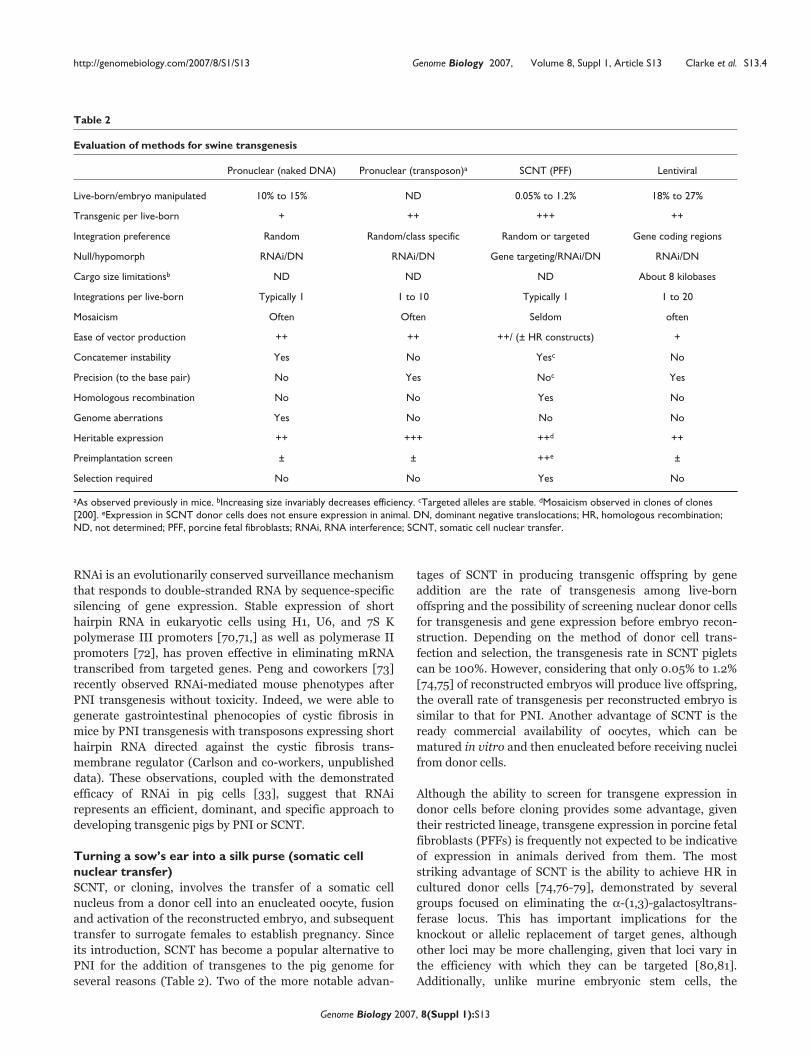
Table 2
Evaluation of methods for swine transgenesis
Pronuclear (naked DNA) Pronuclear (transposon)a SCNT (PFF) Lentiviral
Live-born/embryo manipulated 10% to 15% ND 0.05% to 1.2% 18% to 27%
Transgenic per live-born + ++ +++ ++
Integration preference Random Random/class specific Random or targeted Gene coding regions
Null/hypomorph RNAi/DN RNAi/DN Gene targeting/RNAi/DN RNAi/DN
Cargo size limitationsb ND ND ND About 8 kilobases
Integrations per live-born Typically 1 1 to 10 Typically 1 1 to 20
Mosaicism Often Often Seldom often
Ease of vector production ++ ++ ++/ (± HR constructs) +
Concatemer instability Yes No Yesc No
Precision (to the base pair) No Yes Noc Yes
Homologous recombination No No Yes No
Genome aberrations Yes No No No
Heritable expression ++ +++ ++d ++
Preimplantation screen ± ± ++e ±
Selection required No No Yes No
aAs observed previously in mice. bIncreasing size invariably decreases efficiency. cTargeted alleles are stable. dMosaicism observed in clones of clones[200]. eExpression in SCNT donor cells does not ensure expression in animal. DN, dominant negative translocations; HR, homologous recombination;ND, not determined; PFF, porcine fetal fibroblasts; RNAi, RNA interference; SCNT, somatic cell nuclear transfer.
window of opportunity for isolating recombined cellular
clones, and thus the complexity of manipulations possible, is
limited by PFF cellular senescence. The limited lifespan of
PFFs has prohibited serial transgenesis, genetic manipula-
tion, or selection cassette recycling in vitro. Although serial
genetic manipulations in pig could be achieved by standard
breeding, this is slow and implies excessive husbandry costs
(>10 months from impregnation to sexual maturity).
Instead, researchers have used an iterative cloning approach,
in which each round of genetic modification requires isola-
tion of fetal fibroblasts, genetic manipulation, re-cloning, re-
implantation, and fetal development [74,79,82]. Despite this
clever solution, inefficiencies in nuclear reprogramming and
SCNT render this approach to creating pigs with complex
genetic manipulations or multiple transgenes difficult and
time consuming. A porcine cellular resource more amenable
to genetic manipulation, less susceptible to cellular senescence,
and more effectively reprogrammed would dramatically
improve the efficiency of complex genetic manipulation in
vitro before SCNT.
Given their potential in terms of long-term culture and their
superiority as nuclear donors [83,84], embryonic stem cells
are a highly desirable resource for pig transgenesis and
cloning. Indeed, successful derivation of germline competent
embryonic stem cells from livestock species has been an
actively pursued goal for many years [85]. Although many
groups have reported isolation of embryonic stem-like cells,
far fewer have produced cells demonstrated to contribute to
chimeric piglets when injected into an early blastocyst
[86,87], and to date no evidence of germline chimerism from
porcine embryonic stem cells has been reported. However,
the recent isolation of multipotent cells from pigs by several
groups may provide alternative cellular resources with many
of the desirable features of embryonic stem cells [88-94],
with the potential to increase the efficiency and complexity
of genetic manipulations by SCNT.
The naked truth about DNA integrationStable integration and expression of a transgene in the pig
genome requires that several conserved, fundamental
barriers be overcome. The initial barrier is entry of the trans-
gene into a cell, embryo, or ova. This has been accomplished
by either direct microinjection of DNA into cells or ova, by
transfection of cells with DNA complexed with cationic
lipids, polycations, or other conjugating substances, or by
electroporation. Subsequent trafficking of DNA into the
nucleus is not understood, but it may require dissolution of
the nuclear membrane when a cell divides (for review [95]).
Once within the nucleus, the transgene must rely on cellular
machinery to serendipitously insert the transgene into host
chromosomes.
Linearized DNA integrates with an efficiency fivefold greater
than that of supercoiled DNA [96], and so it is preferred for
the generation of transgenic cells and animals. This
observation makes sense, considering that the DNA double
strand break (DSB) repair machinery is responsible for
transgene integration, with nonhomologous end joining
(NHEJ) being the most prominent mechanism [97]. As the
name implies, NHEJ responds to DNA DSBs in cells by
nonhomologous ligation of available DSBs. The introduction
of 104 copies of a transgene into a cell (in the case of PNI)
provides a great deal of substrate for NHEJ, giving rise to
head to tail, multicopy gene arrays (concatemers) of
extrachromosomal DNA before or simultaneous with
integration into chromosomes. NHEJ acts very rapidly in
mouse embryos, with concatemers observed in 100% of
embryos only 5 to 10 min after DNA injection [98]. These
concatemers are either degraded or find their way into the
genome, presumably at a DSB [98], resulting in transgenic
mice carrying a transgene concatemer at one or more loci in
the genome [98-101].
Although use of naked DNA has provided an effective
method for producing transgenic cells and animals, signifi-
cant complications associated with un-facilitated integration
have been described. Concatemerized transgenes are prone
to silencing by the host for several reasons. Flanking GC-rich
bacterial sequences may accompany the transgene cassette,
causing hypermethylation and resulting in transgene
silencing [102,103]. Additionally, the nature of a concatemer
itself (multiple tandem copies of a transgene at a single
locus) can stimulate transgene silencing [104,105] - a
phenomenon that is partially ameliorated by the use of viral
and transposon systems that deliver precise single copies of
transgenes to the genome.
Genetic lesions and instability have also been encountered
with un-facilitated integration of DNA, resulting in deletions
adjacent to the insertion site, chromosomal translocations,
and insertion of additional genomic sequence within a
transgene concatemer [106-110]. These types of genomic
alterations may not be overtly detected, but they could
certainly affect the health of animals produced by PNI or
from genetically modified cells by SCNT. Furthermore,
valuable transgenic animal lines may suffer from transgene
instability, giving rise to rearrangements at the transgene
locus that can result in loss of transgene concatemers
(possibly including flanking DNA), lower than expected
transmission to offspring, somatic mosaicism of F1 progeny,
or increased morbidity [111-113]. In contrast, the precise
integration of transgenes by viral and transpositional
transgenesis provides for reduced concatemer-associated
transgene instability.
Viral transgenesisRecent publications [58,59] reported a highly efficient
method for transgenic swine production using pseudotyped
lentiviruses. Like PNI, current methods for lentiviral trans-
genesis rely on surgical procurement of early embryos and
implantation into the reproductive tract of a synchronized
http://genomebiology.com/2007/8/S1/S13 Genome Biology 2007, Volume 8, Suppl 1, Article S13 Clarke et al. S13.5
Genome Biology 2007, 8(Suppl 1):S13
recipient after treatment. Then, concentrated pseudotyped
lentivirus is microinjected into the peri-vitelline space of the
early embryo, whereupon the viral machinery mediates
transport of the transgene to the nucleus and integration of
provirus into the pig genome. Peri-vitelline injection is
minimally invasive to the embryo, probably accounting for
enhanced embryo survival (18% to 27%) compared with PNI
and SCNT (Table 2) [58]. In addition, reported rates of live-
born pig transgenesis of 70% [59] and 92% [58] rival those
observed with SCNT, providing an overall transgenesis
efficiency of 13% and 25%, respectively, of transferred embryos
resulting in transgenic piglets. These overall transgenesis rates
are about tenfold better than those with PNI or SCNT on a per
embryo basis. Furthermore, most transgenic F0 animals have
multiple copies of the proviral insert (up to 20 in the report by
Hofmann and coworkers [59]).
Inserting this many transgenes is both good and bad. The
good news is that, with patience, there are many chances to
identify a transgene with an appropriate expression domain.
The bad news is that if anything other than ubiquitous
expression is desired, then identification of a transgene with
an appropriate expression pattern requires breeding to
segregate away other transgene loci. A further complication
is the tendency of lentiviruses to insert into or near
transcriptional units [114,115], increasing the likelihood of
insertional mutagenesis or position effects from nearby
endogenous enhancer elements. Several studies have also
noted an increased likelihood of transgene silencing in the
context of the retroviral genome [116]. In agreement with
this tendency, Hofmann and coworkers [117] observed loss
of transgene expression in one-third of outbred F1 animals
attributed to transgene methylation. However, transgene
expression was consistent between sibling animals carrying
the same insertion, suggesting that expression was fixed for
a specific insertion before germline transmission. Con-
straints on lentiviral cargo capacity (Table 2), the potential
use of cryptic splice signals in the gene expression cassette
during reverse transcription of the viral genome, and a
requirement for viral titers of 109 to 1010 particles per
milliliter all complicate the construction and preparation of
lentiviral transgene vectors. Nevertheless, the efficiency of
transgenesis using this technique is the greatest thus far
reported; it is therefore likely to remain a valuable
implement in the pig genetic engineering toolbox.
Transposons in vertebratesTransposable elements, especially DNA transposons, have
been used extensively for germline transformation of
invertebrates and plants. Efficient integration of DNA into
the genome is one of the reasons why transposon-based
insertional mutagenesis is an essential component of large-
scale functional genomic efforts in many species, including
bacteria, yeast, insects, and plants [118-123]. The application
of transposons to vertebrate biology began in 1997 with the
‘reawakening’ of the Sleeping Beauty transposon [124]. Ivics
and coworkers [124] reconstructed the SB10 transposase
based on the consensus sequence of inactive transposons
littered throughout several salmonid genomes. The refur-
bished SB10 transposase facilitated efficient gene transfer in
cultured cells from many vertebrate species [125]. Since the
restoration of Sleeping Beauty, other transposon systems
including Tol2 [126,127], piggyBac [128,129], Frog Prince
[130], Minos [131], Himar1 [132], and Passport [133] (Clark
and coworkers, unpublished data) have been used to
transpose DNA into vertebrate cells. DNA ‘cut and paste’
transposons are capable of enzymatically moving a gene
expression cassette from a delivery vector into a host
genome. The transposase binds to the inverted terminal
repeats of the transposon, excises it from its original
location, and integrates it into the genome. Domestication of
transposon systems generally finds them operating as a
binary system: the transposon vector containing the
transgene expression cassette flanked by terminal repeats of
the transposon; and the transposase enzyme, which can be
provided by a second gene expression cassette on the same
(cis) or separate vector (trans), as mRNA [134-136] or
potentially as recombinant protein.
The Tc1/mariner family of transposons [137], whose
members include Sleeping Beauty, Frog Prince, Minos,
Himar1, and Passport, randomly integrate into TA dinucleo-
tides distributed around the genome. Upon integration, the
TA dinucleotide is duplicated at each exterior end of the
inverted terminal repeats. The piggyBac transposon, the
founding member of the piggyBac family of transposons
[138], integrates into a TTAA tetranucleotide, which is
duplicated at each end of the transposon. Tol2, a member of
the hAT family of transposons [139], does not integrate into
a specific target sequence, instead relying on local DNA
deformation [140]; it nonetheless also creates a target site
duplication of eight base pairs at the junction between
transposon and genome. Transposons mobilized by
transposase result in a DSB at the excision site that is
repaired by cellular machinery. The major repair pathway
for Sleeping Beauty is NHEJ, which most often results in
conversion of the original TA dinucleotide to a TACA/TGTA,
although other repair sequences have been observed,
including small insertions and deletions [141,142]. This
canonical footprint results in a five-nucleotide insertion
that would disrupt the coding sequence of an interrupted
open reading frame. Tol2 repair also relies on NHEJ
without a predominant repair sequence because of variance
in target-site sequences. Insertions and deletions have also
been observed after Tol2 excision [143,144]. Mobilization of
the piggyBac transposon, on the other hand, generally
results in restoration of the duplicated TTAA back to a
single TTAA, leaving no disruption at the excision site
[128]. The clean repair of excised transposons, as well as
piggyBac’s proclivity for landing in genes [145,146] (see
below), suggest that it will be valuable as a reagent for
functional genomics.
http://genomebiology.com/2007/8/S1/S13 Genome Biology 2007, Volume 8, Suppl 1, Article S13 Clarke et al. S13.6
Genome Biology 2007, 8(Suppl 1):S13

Sleeping Beauty, Tol2, and piggyBac systems have all been
used to produce transgenic animals, including fish, frogs,
mice, and rats [135,136,145,147,148] (Guerts and co-
workers, unpublished data) by pronuclear or cytoplasmic
DNA microinjection. Transposons have also been
remobilized in vivo from chromosomal locations, often
leading to their vacating the original locus and taking up
residence at a new one. For example, expression of Sleeping
Beauty transposase in the germline of mice has been used to
mobilize transposons previously introduced into the mouse
genome [149-152]. Gene and enhancer trap vectors have
been developed and used for germline mutagenesis in fish
and mice for functional genomic applications [152-158].
Similarly, Sleeping Beauty vectors have been used to
identify genes that are involved in cancer genesis by causing
activation of proto-oncogenes or interruption of tumor
suppressor genes by remobilization of transposons in
somatic tissues of mice [159,160].
Despite the benefits of transposition, there are perhaps some
limitations. There have been several reports indicating a
decrease in transposition efficiency with increasing trans-
poson size [125,161,162]. However, in all of these cases the
influence of plasmid size on transfection was not accounted
for, despite the fact that even small differences in plasmid
size can alter transfection efficiency [163]. Where trans-
position can be observed without being confounded by
transfection, for example in PNI or upon mobilization from a
genomic context, large transposons appear to mobilize with
nearly the same efficiency as do smaller ones [145,158]. As
mentioned above, some transposons prefer to integrate into
transcription units. This can be either a benefit or a
disadvantage, depending on whether the goal is to mutate
genes or to safely deliver a transgene. In this case, having
multiple transposon systems available may permit selection
based on the application and the temperament of a
particular transposon. For instance, at first glance piggyBac
appears to integrate preferentially into or very near
transcription units, landing in them as much as 67% of the
time [145,146]. By contrast, Sleeping Beauty does not
integrate into transcription units at a rate much higher than
what would be expected by random integration [164].
Genetic engineering with site-specific recombinasesSite-specific recombinases, such as the P1 bacteriophage
cyclization recombinase enzyme (Cre) and flippase (Flp)
from Saccharomyces cerevisiae, have revolutionized genetic
engineering by allowing efficient and accurate manipulation
of the genome by site-directed deletion, inversion, insertion,
or chromosomal exchange (for review [165]). The use of
recombinases and their recognition sites in trans has
allowed the development of ‘genetic switches’ for the
conditional activation or inactivation of gene expression.
Specific and complex control of transgene expression can be
achieved in a manner that is dependent on the spatio-
temporal expression domain of the recombinase(s). The
ability to express the recombinase from tightly regulated
spatially or temporally restricted promoters has allowed
investigation of gene function beyond their initial develop-
mental role, potentially lethal as a null, and to examine the
role played by a gene product in specific tissues in late-stage
embryos or adults.
Application of transposons and recombinasesfor genetic engineering of pigsTransposons and recombinases for mobilizingtransgenes in pig cellsWe recently reported, for the first time, transpositional
transgenesis in pig cells using Sleeping Beauty, Passport,
Tol2, and piggyBac transposon systems [166]. Initial assess-
ment of these transposons relied primarily on a porcine
endometrial glandular epithelial (PEGE) cell line [167],
which is one of very few immortalized cellular resources
available for pigs. In PEGE cells, transposons increased
cellular transgenesis from 5-fold to 28-fold above back-
ground, depending on which transposon system was used. In
addition to the baseline enhancement of transgenesis
measured by clone formation, transposons differed in their
robustness of integration, as indicated by the number of
integrations per clone, which ranged from 1 to 15. Southern
analysis of cellular clones revealed that the vast majority of
transgene insertions resulted from transposition, a fact
borne out by analysis of the junctions between transgene
and the genome for each class of transposon. Without
optimization in PEGE cells, piggyBac and Tol2 transposon
systems were more active than Sleeping Beauty, which was
more active than Passport. The Passport transposon system
relies on wild-type sequences isolated from the Pleuronectes
plattesa genome, representing the only vertebrate Tc1-type
transposon thus far found to be active in its native form
(Clark and coworkers, unpublished data). Its activity could
probably be improved by engineering of its inverted terminal
repeats or transposase, analogous to improvements made to
the Sleeping Beauty system [168]. Additional hyperactive
mutants of Sleeping Beauty might also be more active in pig
cells [169-171]. However, although it may be possible to
further improve transposon systems for application to PEGE
cells, transposon efficiency varies depending on the cell type
[125,129,130]. Therefore, the relative activity of any
transposon system in different pig cells, including pig
embryos, requires further investigation. It is likely that,
depending on the application, there will be distinct and
overlapping roles for a variety of transposon systems in
swine genetics (Figure 1). It is therefore quite promising that
four unique transposon systems result in enhanced trans-
genesis as well as precise integration of expression cassettes
into one or more genomic locus in swine.
In addition to characterizing the activity of four vertebrate
transposons in porcine cells, Clark and coworkers [166] also
demonstrated for the first time the ability of Cre and Flp
http://genomebiology.com/2007/8/S1/S13 Genome Biology 2007, Volume 8, Suppl 1, Article S13 Clarke et al. S13.7
Genome Biology 2007, 8(Suppl 1):S13

recombinases to mediate site-specific recombination of the
pig genome. Both Cre and Flp recombinase were functional
in pig cells, as indicated by their ability to remove a positive-
negative selection cassette from episomal and numerous
genomic locations. In addition, a Cre-dependent genetic
switch was demonstrated to be effective in mediating
conditional gene expression from episomal and genome-
resident transposons. This study provides the basis for
developing transposon and recombinase based tools for
genetic engineering of the swine genome.
Transposition and recombination for porcine somaticcell nuclear transferThe first step in creating transgenic pigs by SCNT involves
the transgenesis of cells that will serve as nuclear donors.
This generally involves transfecting or electroporating PFFs
or another suitable cell type with DNA expression
constructs. Most if not all transgenesis by SCNT involves the
co-delivery (in cis or trans) of a selectable marker for
enrichment of transgenic cells destined to serve as nuclear
donors. Certainly, this in not the limiting step in producing
transgenic pigs by SCNT. However, the routine use of
transposons would increase the efficiency of cellular trans-
genesis while avoiding concatemerization and integration of
CpG-rich vector sequences. Since the production of
transgenic swine can be quite expensive, any advantage with
regard to stable transgene expression should be exploited. In
addition, the introduction of multiple, unlinked transgenes
by transposition could increase the value of founder pigs,
although breeding would be required to segregate these loci.
The most compelling application of recombinases in porcine
SCNT relates to selection cassette recycling. Elimination of
selectable marker genes from prospective donor cells simpli-
fies genotype-phenotype correlations and eliminates the
potential for selection cassette interference [172] on trans-
gene expression. It would of course be important to minimize
the presence of extraneous DNA (especially antibiotic
resistance genes) from genetically modified pigs were they
ever to be considered for entry into the food chain. The use
of a positive/negative selectable transgene such as PuroΔTK
or HygroCodA [173,174] flanked with recombinase
recognition site (RRS) provides a facile substrate for marker
removal using site-specific recombinases before SCNT
(Figure 2). Unfortunately, because their tendency toward
senescence, the most commonly used cellular resource for
SCNT (PFFs) are not amenable to the extended culture
required for multiple rounds of drug selection. Recently
developed mesenchymal and multipotent stem cells from
pigs may provide a solution to this dilemma, because they
appear to be amenable to extended culture [89-91,175,176],
genetic manipulation [88,92,177], and use as nuclear donors
for SCNT [92-94]. Elimination of RRS flanked selection
cassettes could also await breeding of cloned transgenic pigs
to a line of pigs that express Cre or Flp recombinase in their
germline (Figure 2).
Similar strategies for selection cassette recycling can be used
after homologous recombination if gene targeting vectors
are designed with RRS flanking positive/negative selectable
markers (Figure 3). A simple case of selection cassette
recycling requires flanking a positive/negative selectable
marker, such as PuroΔTK or HygroCodA [173,174], with RRS
sites (Figure 3b). In addition to RRS sites, the gene targeting
vector must contain a unique negative marker (not part of
the positive/negative marker) for counter-selection against
random integration; this could be CodA, TK, or diphtheria
toxin [178]. After selection of homologous recombinants, the
cells can be transfected with Cre and selected for loss of the
positive/negative marker (gancyclovir for PuroΔTK). Cells
that lose the selection cassette will grow and can be used for
nuclear donors before SCNT. Alternatively, the selection
cassette can be removed after SCNT by crossing transgene
carriers to pigs expressing Cre recombinase or by delivering
recombinase transiently in carrier embryos by micro-
injection of Cre mRNA or protein.
In addition to selection cassette recycling, recombinases can
be used to create conditional nulls following gene targeting
http://genomebiology.com/2007/8/S1/S13 Genome Biology 2007, Volume 8, Suppl 1, Article S13 Clarke et al. S13.8
Genome Biology 2007, 8(Suppl 1):S13
Figure 1Applications of transposition to porcine transgenesis. Presented is flowdiagram of the primary steps involved in the production of transgenic pigsby somatic cell nuclear transfer (SCNT), pronuclear injection (PNI), andlentiviral transduction (LVT). Each procedure requires the surgicalisolation of oocytes or embryos. SCNT requires the production oftransgenic donor cells, which can be augmented by transposon-mediatedtransgenesis (TnT). The donor cells are injected into enucleated oocytes,which are then fused and activated before embryo transfer into arecipient. PNI involves the injection of DNA into the male pronucleibefore nuclei fusion. PNI can be augmented by TnT. LVT occurs byinjection into the peri-vitelline space of staged embryos. In all casesmanipulated embryos are surgically implanted into a synchronizedrecipient sow. A portion of the recipient sows will maintain pregnancyuntil parturition. The piglets can then be screened for the presence of thetransgene by polymerase chain reaction, Southern hybridization, ordetection of marker gene expression.

(Figure 3c). In this case two sets of RRSs are used. The first
set flanks the positive/negative selectable marker for
selection cassette recycling to ensure that there is no
impairment of gene function at the locus. Ideally, the
selection cassette would be removed in vitro before SCNT,
but this could also be done in pig, as described above. The
second set of RRS flanks a critical element of the locus, for
example an exon within the coding region. The conditional
knockdown of the locus can then be achieved by crossing the
conditional null carrier to pigs expressing Cre in a desired
manner.
Transposition and recombination for porcinepronuclear injectionThe combination of transposons and recombinases may also
greatly increase the efficiency and complexity of transgenic
pig production by PNI. Sleeping Beauty, Tol2, and piggyBac
transposons have all been used for germline transformation
of multiple species by PNI and cytoplasmic microinjection,
at a rate far superior to unfacilitated DNA injection. In
particular Sleeping Beauty and piggyBac transposons have
been used for the generation of transgenic mice by PNI.
Dupuy and coworkers [136] saw the rate of transgenic live-
born pups increase from 29% up to 45% using the Sleeping
Beauty transposon system. Ding and colleagues [145] saw
increases in mouse embryo transgenesis rates from 10% to
35%, from 18% to 66%, and from 5% to 46% after PNI with
three transposons when piggyBac transposase was included.
In addition, with reports of increased transposition using
methylated Sleeping Beauty transposons that were
methylated in vitro before transfection [179], Geurts and co-
workers (unpublished data) tested the influence of this
treatment on the efficiency of mouse transgenesis by PNI.
This preliminary experiment yielded an unprecedented live-
born transgenesis rate of 90%, with integrations that were
later transmitted to F1 mice and shown to express in a locus-
dependent manner. The fact that four transposon systems
were recently demonstrated to be active in pig cells bodes
well for their application to porcine transgenesis by PNI. A
modest improvement in the rate of swine embryo
transgenesis using transposons could have a significant
impact on the efficiency of swine engineering for agricultural
and medical applications. The observation of multiple
transposed integrations in pig cells (1 to 15) and in
transgenic mouse embryos and pups (1 to 10) also suggests
that it will be possible to create pigs with multiple stable,
unlinked, and reliably expressed transgenes using one or
more transposon system [145] (Clark and coworkers,
unpublished data).
There are a number of reasons to include RRS in transposons
to be used for PNI. Selection cassette recycling is mentioned
above. In addition, to circumvent unsuspected deleterious
effects of ubiquitously expressed transgenes, it may be
desirable to include conditional (recombinase-activated) gene
expression cassettes (Figure 4). For example, a transgene
http://genomebiology.com/2007/8/S1/S13 Genome Biology 2007, Volume 8, Suppl 1, Article S13 Clarke et al. S13.9
Genome Biology 2007, 8(Suppl 1):S13
Figure 2Selection cassette recycling of selectable marker in pigs. A transposon containing two genes, a transgene of interest (‘gene’, red) and a selectable marker(‘marker’, green) can be used to construct a transgenic pig. The promoters can differ for the two genes. Generally, the marker will be driven by aubiquitous promoter (Ub), allowing selection for expressing donor cells (somatic cell nuclear transfer [SCNT]) or piglets (pronuclear injection [PNI] orlentiviral transduction [LVT]), whereas the promoter for the transgene could be a ubiquitous promoter or tissue-specific promoter (TSP). In the diagramthe ‘gene’ is driven by a TSP. Expression in an F0 animal is depicted by the pig on the left with the marker being expressed ubiquitously and the transgenebeing expressed in a tissue-specific manner, shown here as pancreatic expression. Crossing this pig to a pig that ubiquitously expresses Cre recombinase(blue) would result in F1 progeny that lost expression of the ubiquitous marker and retained expression of the transgene in the pancreas. Ubiquitous Creexpression would occur in the F1 (50% or 100%, depending on whether the Cre pig was heterozygous or homozygous), but this would be irrelevant toanalysis of the F1 phenotype. RRS, recombinase recognition site.

encoding a visibly or systemically detectable protein (for
example, green fluorescent protein or secreted alkaline
phosphatase) could be expressed in the default state (either
ubiquitously or in specific tissues), potentially facilitating the
identification of transgenic piglets. Conditional juxtaposition
of the downstream transgene could be activated in later
generations by crossing to a pig that expresses Cre
recombinase ubiquitously or in a tissue-specific manner.
A role for transposons in somatic cell therapiesPorcine models of gene therapyIn addition to the germline transformation, transposon
systems can increase the stable integration of transgenes
into somatic cells. In fact, the Sleeping Beauty transposon
system is actively being developed for several gene therapy
applications. Currently, much of this work is being done in
rodent models with successful long-term expression of
therapeutic transgenes [180-188]. However, the methodo-
logy of gene delivery, clinical dosage, and efficacy of
treatments in the mouse may not be directly applicable to
treatment of human patients. It is therefore likely that large
animal models will be important in advancing clinically
relevant gene therapy protocols. Pigs have been used to
improve surgical techniques for years because of their
similarity in size and physiology to humans, as well as their
widespread availability as an accepted part of the human
food chain. It is therefore quite reasonable to test gene
therapy protocols in pigs. For example, hydrodynamic
delivery of DNA by the injection of a large volume of DNA
solution into the tail-vein of mice results in significant DNA
uptake into the liver [180]; however, this technique is
unlikely to be directly scalable to large animals or humans.
DNA has successfully been delivered by local hydrodynamic
injection into pig arterial vessels [189] and muscle [190],
although - as expected for naked DNA - the expression was
short lived. Perhaps similar local hydrodynamic delivery
coupled with transposons could allow selective uptake and
maintained expression by these tissues or other targets, such
as liver, without the need for systemic injection of large
volumes of fluid. Pigs may also provide an ideal large animal
model for testing the efficacy of reagents being developed for
systemic delivery of therapeutic genes to specific tissues or
organs.
The potential tractability of pigs for development of large
animal models of human disease makes them an attractive
system not only for developing gene delivery protocols but
also for testing the efficacy of these regimens in curing
disease. For example, the National Swine Research and
Resource Center is currently developing pig models of cystic
fibrosis based on gene knockout and transposon-based RNAi
[33]. These pigs not only may provide the first animal model
of the cystic fibrosis pulmonary phenotype, but they may
also be ideal for the development of gene therapy protocols
to treat this devastating disease.
http://genomebiology.com/2007/8/S1/S13 Genome Biology 2007, Volume 8, Suppl 1, Article S13 Clarke et al. S13.10
Genome Biology 2007, 8(Suppl 1):S13
Figure 3Selection cassette recycling after homologous recombination. (a) Anillustration of a typical homologous recombination event utilizing apositive/negative selection scheme. The gene sequence is shown withexons 2, 3, and 4. The targeting vector replaces exon 3 with a positiveselectable marker, such as the PGK driven neomycin resistance cassette(phosphoglycerate kinase [PGK]-neoR), and utilizes a negative selectablemarker, such as the herpes simplex virus promoter driven thymidinekinase gene (HSV-TK), to counter-select against random integration of thetargeting vector. Homologous recombination results in replacement ofexon 3 with the PGK-neoR cassette. (b) The use of site-specificrecombinases such as Cre or Flp allows removal of a selection cassettebefore or after the production of an animal by SCNT. In order toaccomplish this, positive marker is flanked by recombinase recognitionsites like loxP. After homologous recombination (HR) the selectioncassette can be removed by Cre recombinase in culture or in vivo. In orderto select efficiently for removal of the selection cassette in vitro, apositive/negative selectable marker such as PuroΔTK or hygroCodA, withthe negative selection marker outside the homology arms (for example,CodA, TK, or diphtheria toxin). (c) Schematic for generating conditionalknockout alleles using site-specific recombinases Cre and Flp. A targetingconstruct is generated that leaves each exon intact, but includes loxP sitesflanking an exon critical for gene function, in this case exon 3 (wild-type oran alternative allele). The selectable marker, flanked by frt sites, can beremoved from the targeted allele either in vitro or in vivo to avoid selectioncassette interference of the modified allele. Animals carrying this targetedmodification can be crossed to animals that express Cre ubiquitously or ina specific tissue, resulting in progeny with a deletion of exon 3 in thewhole animal or in a specific tissue.
DNA vaccinationThe evolutionary speed of viruses and bacteria challenges our
ability to develop efficacious protein-based vaccines. Molecular
biology, on the other hand, provides a rapid approach to the
cloning and expression of potential antigens. The promise of
DNA as a pharmaceutical has been actively pursued since the
observation that naked DNA injection into muscle can direct
the production of protein [191]. Applications in gene therapy
and vaccination have been extensively explored, stimulated
by the fact that DNA can be prepared in large quantities in
compliance with cGMP standards, and in a lyophilized form
independent of the traditional cold chain. Although both
humoral and cellular immune responses can be mobilized
with DNA vaccines, problems with DNA delivery and the
intercellular trafficking of antigen have limited their success
[192]. To date, only two DNA vaccines have been licensed for
use in animals; a DNA vaccine to protect farmed salmon and
trout from infectious hematopoietic necrosis virus, and one to
protect horses from West Nile virus [193,194]. Recent
findings suggest that transposons may provide for more
efficient and longer lasting cellular transgenesis to increase
the expression and intercellular trafficking of antigens.
Indeed, in the context of developing transposon-based
reagents for gene therapy, a robust immune response to the
expression of genes from Sleeping Beauty transposons
encoding either clotting factor VIII [186] or iduronidase [195]
have been observed in mice. Given that transposons are
active in pig cells, swine could serve as excellent preclinical
models for human vaccine development, in addition to their
obvious importance in the development of vaccines targeted
against pathogens important to swine production.
High on the hog (conclusions and horizons)The relevance of pigs to agriculture and medicine makes
them unique among large animal models. With the complete
sequence of their genome soon to be delivered, pigs are
likely to play an increasing role in defining gene function in
human disease using reverse genetic approaches. The use of
enzymatic approaches such as transposition and recombina-
tion should expand the ease and complexity of genetic
modifications available with which to engineer the pig to
model human disease and to produce agricultural and
biomedical products.
In addition, pigs may also be amenable to forward genetic
screens because of reproductive fecundity (about ten piglets
per litter) that rivals that of mice. With appropriate planning
and coordination, and the use of clever molecular reagents,
conducting a mutagenesis screen in pigs could provide
important information about gene function in large animals.
Some cancers and age-related disease etiologies, as well as
therapies for treating them, might be better studied in pigs,
which commonly live to be ten years old and, in rare
exceptions, into their second decade.
Transposons are ideal for use as insertional mutagens,
particularly piggyBac, which tends to land in transcription
http://genomebiology.com/2007/8/S1/S13 Genome Biology 2007, Volume 8, Suppl 1, Article S13 Clarke et al. S13.11
Genome Biology 2007, 8(Suppl 1):S13
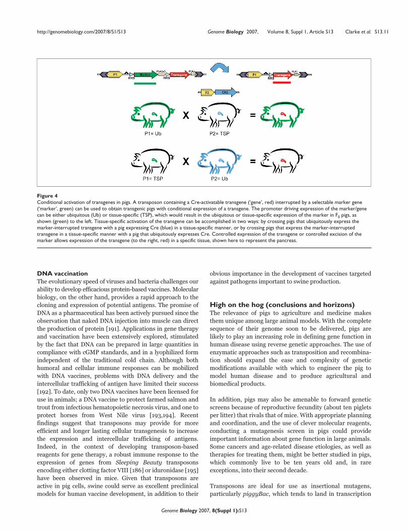
Figure 4Conditional activation of transgenes in pigs. A transposon containing a Cre-activatable transgene (‘gene’, red) interrupted by a selectable marker gene(‘marker’, green) can be used to obtain transgenic pigs with conditional expression of a transgene. The promoter driving expression of the marker/genecan be either ubiquitous (Ub) or tissue-specific (TSP), which would result in the ubiquitous or tissue-specific expression of the marker in F0 pigs, asshown (green) to the left. Tissue-specific activation of the transgene can be accomplished in two ways: by crossing pigs that ubiquitously express themarker-interrupted transgene with a pig expressing Cre (blue) in a tissue-specific manner, or by crossing pigs that express the marker-interruptedtransgene in a tissue-specific manner with a pig that ubiquitously expresses Cre. Controlled expression of the transgene or controlled excision of themarker allows expression of the transgene (to the right, red) in a specific tissue, shown here to represent the pancreas.

units and can later be excised for reversion analysis.
Specialized ‘trapping’ vectors based on transposons are able
to cause mutations efficiently upon insertion into a trans-
cription unit, and make identification of the interrupted
gene straightforward [152,153,156,158,196,197]. Transposon-
based mutagenesis screens in mice have generally relied first
on the generation of two mouse strains: one transgenic for a
mutagenic transposon vector (usually in the form of a
concatemer) and another strain transgenic for the corres-
ponding transposase expression construct [149,150,158].
Breeding these lines together provides doubly transgenic
‘seed’ mice, in which germline mobilization of the trans-
poson provides for the recovery of mutated loci in an out-
crossed generation. However, with a 4-month gestation
period and 6 months to sexual maturity, mutagenesis in pigs
using this strategy would require a minimum of 4 years
before mutations could be bred to homozygosity and a
screen initiated.
More immediate would be to use a strategy recently applied
in zebrafish [157,197], which treats the injected generation
as seed stock by supplying both transposon and transposase.
Given a reasonable rate of transgenesis by transposon-based
PNI of pig embryos, mutant alleles could be bred to
homozygosity and a screen initiated within 2 years. Each F0
could be a source of 1 to 15 transposon insertions, with about
12% to 25% of the integrations ‘trapping’ a transcription unit
[145,153,164]. The direct injection method will provide proof
of principle in the shortest amount of time. However, the
longer initial investment required for the production of
double-transgenic ‘seed’ boars would be rewarded by a
nearly constant supply of novel gene traps due to re-
mobilization of transposons in the male germline. Addition-
ally, improvement in the efficiency of cloning and the
availability of porcine stem cells allows another attractive
approach. Development of a library of ‘trapped’, character-
ized, and catalogued pig stem cell clones could provide an
on-demand resource for the generation of pigs by SCNT,
analogous to strategies used for generating mice from
‘trapped’ embryonic stem cell clones [198]. Using this
approach, transposon-trapped alleles could be bred to
homozygosity and a phenotypic analysis begun in pigs in less
than 1 year. In the woven words of Charlotte the spider, the
unique contributions of such pigs would surely reveal dear
Wilbur to represent ‘Some Pig’ [199].
Competing interestsKJC and SCF are both inventors on University of Minnesota
Patents involving Sleeping Beauty Transposon technology.
AcknowledgementThis article has been published as part of Genome Biology Volume 8, Supplement 1, 2007: Transposons in vertebrate functional genomics. The full contents of the supplement are available online at http://genomebiology.com/supplements/8/S1.
References1. Epstein J, Bichard M: Pigs. In Evolution of Domesticated Animals. Edited
by Mason IL. New York: Longman; 1984:145-162.2. Larson G, Dobney K, Albarella U, Fang M, Matisoo-Smith E, Robins J,
Lowden S, Finlayson H, Brand T, Willerslev E, et al.: Worldwidephylogeography of wild boar reveals multiple centers of pigdomestication. Science 2005, 307:1618-1621.
3. Global Livestock Health and Production Atlas [http://www.fao.org/ag/aga/glipha/index.jsp]
4. Otto D, Lawrence J: The United States pork industry 2003:patterns and economic importance [http://www.mnpork.com/producer/presentations/other/uslawrence2003.pdf].
5. Pork facts - everything but the oink: pharmaceutical co-prod-ucts [http://www.pork.org/newsandinformation/quickfacts/PorkFacts16.aspx]
6. McCrea MR, Tribe DE: The baby pig as a laboratory animal.J Physiol 1954, 124:52P.
7. Bustad LK, McClellan RO: Miniature swine: development, man-agement, and utilization. Lab Anim Care 1968, Suppl:280-287.
8. Patel G, Duffin J: History of Medicine: A Scandalously Short Introduction.Toronto, Canada: University of Toronto Press; 2000.
9. Watson L: The Whole Hog: Exploring the Extraordinary Potential of Pigs.Washington DC: Smithsonian Books; 2004.
10. England DC, Winters LM, Carpenter LE: The development of abreed of miniature swine: a preliminary report. Growth 1954,18:207-214.
11. Rothschild MF: From a sow’s ear to a silk purse: real progressin porcine genomics. Cytogenet Genome Res 2003, 102:95-99.
12. Rothschild MF: Porcine genomics delivers new tools andresults: this little piggy did more than just go to market.Genet Res 2004, 83:1-6.
13. Swine Protein-Annotated Oligonucleotide Microarray[www.pigoligoarray.org]
14. Haley C, de Koning DJ: Genetical genomics in livestock: poten-tials and pitfalls. Anim Genet 2006, Suppl 1:10-12.
15. Vize PD, Michalska AE, Ashman R, Lloyd B, Stone BA, Quinn P,Wells JR, Seamark RF: Introduction of a porcine growthhormone fusion gene into transgenic pigs promotes growth.J Cell Sci 1988, 90:295-300.
16. Wieghart M, Hoover JL, McGrane MM, Hanson RW, Rottman FM,Holtzman SH, Wagner TE, Pinkert CA: Production of transgenicpigs harbouring a rat phosphoenolpyruvate carboxykinase-bovine growth hormone fusion gene. J Reprod Fertil Suppl 1990,41:89-96.
17. Pursel V, Wall RJ, Mitchell AD, Elsasser TH, Solomon MB, ColemanME, DeMayo F, Schwartz RJ: Expression of Insulin-like Growth Factor I inSkeletal Muscle of Transgenic Swine. Wallingford, UK: CAB Interna-tional; 1999.
18. Lai L, Kang JX, Li R, Wang J, Witt WT, Yong HY, Hao Y, Wax DM,Murphy CN, Rieke A et al: Generation of cloned transgenic pigsrich in omega-3 fatty acids. Nat Biotechnol 2006, 24:435-436.
19. Saeki K, Matsumoto K, Kinoshita M, Suzuki I, Tasaka Y, Kano K,Taguchi Y, Mikami K, Hirabayashi M, Kashiwazaki N, et al.: Func-tional expression of a Delta12 fatty acid desaturase genefrom spinach in transgenic pigs. Proc Natl Acad Sci USA 2004,101:6361-6366.
20. Noble MS, Rodriguez-Zas S, Cook JB, Bleck GT, Hurley WL,Wheeler MB: Lactational performance of first-parity trans-genic gilts expressing bovine alpha-lactalbumin in their milk.J Anim Sci 2002, 80:1090-1096.
21. Monaco MH, Gronlund DE, Bleck GT, Hurley WL, Wheeler MB,Donovan SM: Mammary specific transgenic over-expressionof insulin-like growth factor-I (IGF-I) increases pig milk IGF-I and IGF binding proteins, with no effect on milk composi-tion or yield. Transgenic Res 2005, 14:761-773.
22. Muller M, Brenig B, Winnacker EL, Brem G: Transgenic pigs car-rying cDNA copies encoding the murine Mx1 protein whichconfers resistance to influenza virus infection. Gene 1992,121:263-270.
23. Golovan SP, Meidinger RG, Ajakaiye A, Cottrill M, Wiederkehr MZ,Barney DJ, Plante C, Pollard JW, Fan MZ, Hayes MA, et al.: Pigsexpressing salivary phytase produce low-phosphorusmanure. Nat Biotechnol 2001, 19:741-745.
24. Park JK, Lee YK, Lee P, Chung HJ, Kim S, Lee HG, Seo MK, Han JH,Park CG, Kim HT, et al.: Recombinant human erythropoietin pro-duced in milk of transgenic pigs. J Biotechnol 2006, 122:362-371.
http://genomebiology.com/2007/8/S1/S13 Genome Biology 2007, Volume 8, Suppl 1, Article S13 Clarke et al. S13.12
Genome Biology 2007, 8(Suppl 1):S13

25. Paleyanda RK, Velander WH, Lee TK, Scandella DH, GwazdauskasFC, Knight JW, Hoyer LW, Drohan WN, Lubon H: Transgenicpigs produce functional human factor VIII in milk. Nat Biotech-nol 1997, 15:971-975.
26. Velander WH, Johnson JL, Page RL, Russell CG, Subramanian A,Wilkins TD, Gwazdauskas FC, Pittius C, Drohan WN: High-levelexpression of a heterologous protein in the milk of trans-genic swine using the cDNA encoding human protein C. ProcNatl Acad Sci USA 1992, 89:12003-12007.
27. Sharma A, Martin MJ, Okabe JF, Truglio RA, Dhanjal NK, Logan JS,Kumar R: An isologous porcine promoter permits high levelexpression of human hemoglobin in transgenic swine.Biotechnology (NY) 1994, 12:55-59.
28. Swanson ME, Martin MJ, O’Donnell JK, Hoover K, Lago W, HuntressV, Parsons CT, Pinkert CA, Pilder S, Logan JS: Production of func-tional human hemoglobin in transgenic swine. Biotechnology(NY) 1992, 10:557-559.
29. Lee GS, Kim HS, Hyun SH, Lee SH, Jeon HY, Nam DH, Jeong YW,Kim S, Kim JH, Han JY, et al.: Production of transgenic clonedpiglets from genetically transformed fetal fibroblasts selectedby green fluorescent protein. Theriogenology 2005, 63:973-991.
30. Dyck MK, Gagne D, Ouellet M, Senechal JF, Belanger E, Lacroix D,Sirard MA, Pothier F: Seminal vesicle production and secretionof growth hormone into seminal fluid. Nat Biotechnol 1999, 17:1087-1090.
31. Dyck MK, Lacroix D, Pothier F, Sirard MA: Making recombinantproteins in animals—different systems, different applica-tions. Trends Biotechnol 2003, 21:394-399.
32. Petters RM, Alexander CA, Wells KD, Collins EB, Sommer JR,Blanton MR, Rojas G, Hao Y, Flowers WL, Banin E, et al.: Geneti-cally engineered large animal model for studying conephotoreceptor survival and degeneration in retinitis pig-mentosa. Nat Biotechnol 1997, 15:965-970.
33. Palmer ML, Lee SY, Carlson D, Fahrenkrug S, O’Grady SM: Stableknockdown of CFTR establishes a role for the channel inP2Y receptor-stimulated anion secretion. J Cell Physiol 2006,206:759-770.
34. Bruscia E, Sangiuolo F, Sinibaldi P, Goncz KK, Novelli G, GruenertDC: Isolation of CF cell lines corrected at DeltaF508-CFTRlocus by SFHR-mediated targeting. Gene Ther 2002, 9:683-685.
35. Bucher P, Morel P, Buhler LH: Xenotransplantation: an updateon recent progress and future perspectives. Transpl Int 2005,18:894-901.
36. Cox A, Zhong R: Current advances in xenotransplantation.Hepatobiliary Pancreat Dis Int 2005, 4:490-494.
37. Houdebine LM: Use of transgenic animals to improve humanhealth and animal production. Reprod Domest Anim 2005, 40:269-281.
38. Zhong R: Gal knockout and beyond. Am J Transplant 2007, 7:5-11.39. Rayat GR, Rajotte RV, Hering BJ, Binette TM, Korbutt GS: In vitro
and in vivo expression of Galalpha-(1,3)Gal on porcine isletcells is age dependent. J Endocrinol 2003, 177:127-135.
40. Cardona K, Korbutt GS, Milas Z, Lyon J, Cano J, Jiang W, Bello-Laborn H, Hacquoil B, Strobert E, Gangappa S, et al.: Long-termsurvival of neonatal porcine islets in nonhuman primates bytargeting costimulation pathways. Nat Med 2006, 12:304-306.
41. Hering BJ, Wijkstrom M, Graham ML, Hardstedt M, Aasheim TC, JieT, Ansite JD, Nakano M, Cheng J, Li W, et al.: Prolonged diabetesreversal after intraportal xenotransplantation of wild-typeporcine islets in immunosuppressed nonhuman primates.Nat Med 2006, 12:301-303.
42. Rother KI, Harlan DM: Challenges facing islet transplantationfor the treatment of type 1 diabetes mellitus. J Clin Invest2004, 114:877-883.
43. Cure P, Pileggi A, Froud T, Norris PM, Baidal DA, Cornejo A, HafizMM, Ponte G, Poggioli R, Yu J, et al.: Alterations of the femalereproductive system in recipients of islet grafts. Transplanta-tion 2004, 78:1576-1581.
44. Martin C, Plat M, Nerriere-Daguin V, Coulon F, Uzbekova S, VenturiE, Conde F, Hermel JM, Hantraye P, Tesson L, et al.: Transgenicexpression of CTLA4-Ig by fetal pig neurons for xenotrans-plantation. Transgenic Res 2005, 14:373-384.
45. Sutherland RM, Brady JL, Georgiou HM, Thomas HE, Lew AM: Pro-tective effect of CTLA4Ig secreted by transgenic fetal pan-creas allografts. Transplantation 2000, 69:1806-1812.
46. Martin U, Winkler ME, Id M, Radeke H, Arseniev L, Takeuchi Y,Simon AR, Patience C, Haverich A, Steinhoff G: Productive
infection of primary human endothelial cells by pig endoge-nous retrovirus (PERV). Xenotransplantation 2000, 7:138-142.
47. Blusch JH, Patience C, Takeuchi Y, Templin C, Roos C, Von DerHelm K, Steinhoff G, Martin U: Infection of nonhuman primatecells by pig endogenous retrovirus. J Virol 2000, 74:7687-7690.
48. Patience C, Takeuchi Y, Weiss RA: Infection of human cells byan endogenous retrovirus of pigs. Nat Med 1997, 3:282-286.
49. Winkler ME, Winkler M, Burian R, Hecker J, Loss M, Przemeck M,Lorenz R, Patience C, Karlas A, Sommer S, et al.: Analysis of pig-to-human porcine endogenous retrovirus transmission in atriple-species kidney xenotransplantation model. Transpl Int2005, 17:848-858.
50. Fishman JA, Patience C: Xenotransplantation: infectious riskrevisited. Am J Transplant 2004, 4:1383-1390.
51. Patience C, Patton GS, Takeuchi Y, Weiss RA, McClure MO,Rydberg L, Breimer ME: No evidence of pig DNA or retroviralinfection in patients with short-term extracorporeal con-nection to pig kidneys. Lancet 1998, 352:699-701.
52. Bartosch B, Stefanidis D, Myers R, Weiss R, Patience C, Takeuchi Y:Evidence and consequence of porcine endogenous retro-virus recombination. J Virol 2004, 78:13880-13890.
53. Scobie L, Taylor S, Wood JC, Suling KM, Quinn G, Meikle S, PatienceC, Schuurman HJ, Onions DE: Absence of replication-compe-tent human-tropic porcine endogenous retroviruses in thegerm line DNA of inbred miniature swine. J Virol 2004, 78:2502-2509.
54. Miyagawa S, Nakatsu S, Nakagawa T, Kondo A, Matsunami K,Hazama K, Yamada J, Tomonaga K, Miyazawa T, Shirakura R: Pre-vention of PERV infections in pig to human xenotransplan-tation by the RNA interference silences gene. J Biochem(Tokyo) 2005, 137:503-508.
55. Karlas A, Kurth R, Denner J: Inhibition of porcine endogenousretroviruses by RNA interference: increasing the safety ofxenotransplantation. Virology 2004, 325:18-23.
56. Jonsson SR, Hache G, Stenglein MD, Fahrenkrug SC, Andresdottir V,Harris RS: Evolutionarily conserved and non-conserved retro-virus restriction activities of artiodactyl APOBEC3F pro-teins. Nucleic Acids Res 2006, 34:5683-5694.
57. Hammer RE, Pursel VG, Rexroad CE Jr, Wall RJ, Bolt DJ, Ebert KM,Palmiter RD, Brinster RL: Production of transgenic rabbits,sheep and pigs by microinjection. Nature 1985, 315:680-683.
58. Whitelaw CB, Radcliffe PA, Ritchie WA, Carlisle A, Ellard FM, PenaRN, Rowe J, Clark AJ, King TJ, Mitrophanous KA: Efficient genera-tion of transgenic pigs using equine infectious anaemia virus(EIAV) derived vector. FEBS Lett 2004, 571:233-236.
59. Hofmann A, Kessler B, Ewerling S, Weppert M, Vogg B, Ludwig H,Stojkovic M, Boelhauve M, Brem G, Wolf E, et al.: Efficient trans-genesis in farm animals by lentiviral vectors. EMBO Rep 2003,4:1054-1060.
60. Martin MJ, Pinkert CA: Production of transgenic swine by DNAmicroinjection. In Transgenic Animal Technology: A Laboratory Hand-book, 2nd ed. Edited by Pinkert CA. Amsterdam, Boston: AcademicPress; 2002:307-336.
61. Wall RJ, Pursel VG, Hammer RE, Brinster RL: Development ofporcine ova that were centrifuged to permit visualization ofpronuclei and nuclei. Biol Reprod 1985, 32:645-651.
62. Youngs CR: Factors influencing the success of embryo trans-fer in the pig. Theriogenology 2001, 56:1311-1320.
63. Springmann K, Brem G: Embryo transfer in swine in relation toa gene transfer program [in German]. Tierarztl Prax Suppl 1989,4:21-25.
64. Niemann H, Rath D: Progress in reproductive biotechnology inswine. Theriogenology 2001, 56:1291-1304.
65. Wall RJ, Paleyanda RK, Foster JA, Powell A, Rexroad C, Lubon H:DNA preparation method can influence outcome of trans-genic animal experiments. Anim Biotechnol 2000, 11:19-32.
66. Nottle MB, Haskard KA, Verma PJ, Du ZT, Grupen CG, McIlfatrickSM, Ashman RJ, Harrison SJ, Barlow H, Wigley PL, et al.: Effect ofDNA concentration on transgenesis rates in mice and pigs.Transgenic Res 2001, 10:523-531.
67. Pinkert CA, Kooyman DL, Dyer TJ: Enhanced growth perfor-mance in transgenic swine. Biotechnology 1991, 16:251-258.
68. Kues WA, Schwinzer R, Wirth D, Verhoeyen E, Lemme E, Her-rmann D, Barg-Kues B, Hauser H, Wonigeit K, Niemann H: Epige-netic silencing and tissue independent expression of a noveltetracycline inducible system in double-transgenic pigs.FASEB J 2006, 20:1200-1202.
http://genomebiology.com/2007/8/S1/S13 Genome Biology 2007, Volume 8, Suppl 1, Article S13 Clarke et al. S13.13
Genome Biology 2007, 8(Suppl 1):S13

69. Palmer ML, Fahrenkrug SC, O’Grady SM: RNA interference andion channel physiology. Cell Biochem Biophys 2006, 46:175-192.
70. Brummelkamp TR, Bernards R, Agami R: A system for stableexpression of short interfering RNAs in mammalian cells.Science 2002, 296:550-553.
71. Koper-Emde D, Herrmann L, Sandrock B, Benecke BJ: RNA inter-ference by small hairpin RNAs synthesised under control ofthe human 7S K RNA promoter. Biol Chem 2004, 385:791-794.).
72. Denti MA, Rosa A, Sthandier O, De Angelis FG, Bozzoni I: A newvector, based on the PolII promoter of the U1 snRNA gene,for the expression of siRNAs in mammalian cells. Mol Ther2004, 10:191-199.
73. Peng S, York JP, Zhang P: A transgenic approach for RNA inter-ference-based genetic screening in mice. Proc Natl Acad Sci USA2006, 103:2252-2256.
74. Takahagi Y, Fujimura T, Miyagawa S, Nagashima H, Shigehisa T, Shi-rakura R, Murakami H: Production of alpha 1,3-galactosyltrans-ferase gene knockout pigs expressing both humandecay-accelerating factor and N-acetylglucosaminyltrans-ferase III. Mol Reprod Dev 2005, 71:331-338.
75. Watanabe S, Iwamoto M, Suzuki S, Fuchimoto D, Honma D, Nagai T,Hashimoto M, Yazaki S, Sato M, Onishi A: A novel method forthe production of transgenic cloned pigs: electroporation-mediated gene transfer to non-cultured cells and subse-quent selection with puromycin. Biol Reprod 2005, 72:309-315.
76. Harrison SJ, Guidolin A, Faast R, Crocker LA, Giannakis C, D’ApiceAJ, Nottle MB, Lyons I: Efficient generation of alpha(1,3) galac-tosyltransferase knockout porcine fetal fibroblasts fornuclear transfer. Transgenic Res 2002, 11:143-150.
77. Jin DI, Lee SH, Choi JH, Lee JS, Lee JE, Park KW, Seo JS: Targetingefficiency of a-1,3-galactosyl transferase gene in pig fetalfibroblast cells. Exp Mol Med 2003, 35:572-577.
78. Lai L, Kolber-Simonds D, Park KW, Cheong HT, Greenstein JL, ImGS, Samuel M, Bonk A, Rieke A, Day BN, et al.: Production ofalpha-1,3-galactosyltransferase knockout pigs by nucleartransfer cloning. Science 2002, 295:1089-1092.
79. Ramsoondar JJ, Machaty Z, Costa C, Williams BL, Fodor WL,Bondioli KR: Production of alpha 1,3-galactosyltransferase-knockout cloned pigs expressing human alpha 1,2-fuco-sylosyltransferase. Biol Reprod 2003, 69:437-445.
80. Hasty P, Crist M, Grompe M, Bradley A: Efficiency of insertionversus replacement vector targeting varies at differentchromosomal loci. Mol Cell Biol 1994, 14:8385-8390.
81. Schultes NP, Szostak JW: A poly(dA.dT) tract is a componentof the recombination initiation site at the ARG4 locus inSaccharomyces cerevisiae. Mol Cell Biol 1991, 11:322-328.
82. Phelps CJ, Koike C, Vaught TD, Boone J, Wells KD, Chen SH, Ball S,Specht SM, Polejaeva IA, Monahan JA, et al.: Production of alpha1,3-galactosyltransferase-deficient pigs. Science 2003, 299:411-414.
83. Rideout WM III, Wakayama T, Wutz A, Eggan K, Jackson-Grusby L,Dausman J, Yanagimachi R, Jaenisch R: Generation of mice fromwild-type and targeted ES cells by nuclear cloning. Nat Genet2000, 24:109-110.
84. Eggan K, Akutsu H, Loring J, Jackson-Grusby L, Klemm M, RideoutWM, 3rd, Yanagimachi R, Jaenisch R: Hybrid vigor, fetal over-growth, and viability of mice derived by nuclear cloning andtetraploid embryo complementation. Proc Natl Acad Sci USA2001, 98:6209-6214.
85. Wheeler MB, Walters EM: Transgenic technology and applica-tions in swine. Theriogenology 2001, 56:1345-1369.
86. Chen LR, Shiue YL, Bertolini L, Medrano JF, BonDurant RH, Ander-son GB: Establishment of pluripotent cell lines from porcinepreimplantation embryos. Theriogenology 1999, 52:195-212.
87. Wheeler MB: Development and validation of swine embry-onic stem cells: a review. Reprod Fertil Dev 1994, 6:563-568.
88. Bosch P, Pratt SL, Stice SL: Isolation, characterization, genemodification, and nuclear reprogramming of porcine mes-enchymal stem cells. Biol Reprod 2006, 74:46-57.
89. Dyce PW, Zhu H, Craig J, Li J: Stem cells with multilineagepotential derived from porcine skin. Biochem Biophys ResCommun 2004, 316:651-658.
90. Ringe J, Kaps C, Schmitt B, Buscher K, Bartel J, Smolian H, Schultz O,Burmester GR, Haupl T, Sittinger M: Porcine mesenchymal stemcells. Induction of distinct mesenchymal cell lineages. CellTissue Res 2002, 307:321-327.
91. Kues WA, Petersen B, Mysegades W, Carnwath JW, Niemann H:Isolation of murine and porcine fetal stem cells fromsomatic tissue. Biol Reprod 2005, 72:1020-1028.
92. Colleoni S, Donofrio G, Lagutina I, Duchi R, Galli C, Lazzari G:Establishment, differentiation, electroporation, viral trans-duction, and nuclear transfer of bovine and porcine mes-enchymal stem cells. Cloning Stem Cells 2005, 7:154-166.
93. Faast R, Harrison SJ, Beebe LF, McIlfatrick SM, Ashman RJ, NottleMB: Use of adult mesenchymal stem cells isolated frombone marrow and blood for somatic cell nuclear transfer inpigs. Cloning Stem Cells 2006, 8:166-173.
94. Tomii R, Kurome M, Ochiai T, Wako N, Ueda H, Hirakawa K, KanoK, Nagashima H: Production of cloned pigs by nuclear transferof preadipocytes established from adult mature adipocytes.Cloning Stem Cells 2005, 7:279-288.
95. Lechardeur D, Lukacs GL: Nucleocytoplasmic transport ofplasmid DNA: a perilous journey from the cytoplasm to thenucleus. Hum Gene Ther 2006, 17:882-889.
96. Brinster RL, Chen HY, Trumbauer ME, Yagle MK, Palmiter RD:Factors affecting the efficiency of introducing foreign DNAinto mice by microinjecting eggs. Proc Natl Acad Sci USA 1985,82:4438-4442.
97. Bishop JO, Smith P: Mechanism of chromosomal integration ofmicroinjected DNA. Mol Biol Med 1989, 6:283-298.
98. Burdon TG, Wall RJ: Fate of microinjected genes in pre-implantation mouse embryos. Mol Reprod Dev 1992, 33:436-442.
99. Gordon JW, Ruddle FH: DNA-mediated genetic transforma-tion of mouse embryos and bone marrow: a review. Gene1985, 33:121-136.
100. Palmiter RD, Brinster RL: Germ-line transformation of mice.Annu Rev Genet 1986, 20:465-499.
101. Pawlik KM, Sun CW, Higgins NP, Townes TM: End joining ofgenomic DNA and transgene DNA in fertilized mouse eggs.Gene 1995, 165:173-181.
102. Townes TM, Chen HY, Lingrel JB, Palmiter RD, Brinster RL:Expression of human beta-globin genes in transgenic mice:effects of a flanking metallothionein-human growthhormone fusion gene. Mol Cell Biol 1985, 5:1977-1983.
103. Dalle B, Rubin JE, Alkan O, Sukonnik T, Pasceri P, Yao S, Pawliuk R,Leboulch P, Ellis J: eGFP reporter genes silence LCRbeta-globin transgene expression via CpG dinucleotides. Mol Ther2005, 11:591-599.
104. Garrick D, Fiering S, Martin DI, Whitelaw E: Repeat-induced genesilencing in mammals. Nat Genet 1998, 18:56-59.
105. Dorer DR, Henikoff S: Transgene repeat arrays interact withdistant heterochromatin and cause silencing in cis andtrans. Genetics 1997, 147:1181-1190.
106. Chen CM, Choo KB, Cheng WT: Frequent deletions andsequence aberrations at the transgene junctions of trans-genic mice carrying the papillomavirus regulatory and theSV40 TAg gene sequences. Transgenic Res 1995, 4:52-59.
107. Pravtcheva DD, Wise TL: A postimplantation lethal mutationinduced by transgene insertion on mouse chromosome 8.Genomics 1995, 30:529-544.
108. Nakanishi T, Kuroiwa A, Yamada S, Isotani A, Yamashita A, TairakaA, Hayashi T, Takagi T, Ikawa M, Matsuda Y, et al.: FISH analysis of142 EGFP transgene integration sites into the mousegenome. Genomics 2002, 80:564-574.
109. Bishop JO: Chromosomal Insertion of Foreign DNA. In Trans-genic Animals : Generation and Use. Edited by Houdebine L-M. Amster-dam, the Netherlands: Harwood Academic Publishers; 1997:219-223.
110. Covarrubias L, Nishida Y, Mintz B: Early postimplantationembryo lethality due to DNA rearrangements in a trans-genic mouse strain. Proc Natl Acad Sci USA 1986, 83:6020-6024.
111. Pravtcheva DD, Wise TL: Transgene instability in mice injectedwith an in vitro methylated Igf2 gene. Mutat Res 2003, 529:35-50.
112. Scrable H, Stambrook PJ: A genetic program for deletion offoreign DNA from the mammalian genome. Mutat Res 1999,429:225-237.
113. Wilkie TM, Palmiter RD: Analysis of the integrant in MyK-103transgenic mice in which males fail to transmit the inte-grant. Mol Cell Biol 1987, 7:1646-1655.
114. Schroder AR, Shinn P, Chen H, Berry C, Ecker JR, Bushman F: HIV-1 integration in the human genome favors active genes andlocal hotspots. Cell 2002, 110:521-529.
http://genomebiology.com/2007/8/S1/S13 Genome Biology 2007, Volume 8, Suppl 1, Article S13 Clarke et al. S13.14
Genome Biology 2007, 8(Suppl 1):S13

115. Wu X, Li Y, Crise B, Burgess SM: Transcription start regions inthe human genome are favored targets for MLV integra-tion. Science 2003, 300:1749-1751.
116. Pannell D, Ellis J: Silencing of gene expression: implications fordesign of retrovirus vectors. Rev Med Virol 2001, 11:205-217.
117. Hofmann A, Kessler B, Ewerling S, Kabermann A, Brem G, Wolf E,Pfeifer A: Epigenetic regulation of lentiviral transgene vectorsin a large animal model. Mol Ther 2006, 13:59-66.
118. Brutnell TP: Transposon tagging in maize. Funct Integr Genomics2002, 2:4-12.
119. Hamer L, DeZwaan TM, Montenegro-Chamorro MV, Frank SA,Hamer JE: Recent advances in large-scale transposon mutage-nesis. Curr Opin Chem Biol 2001, 5:67-73.
120. Parinov S, Sundaresan V: Functional genomics in Arabidopsis:large-scale insertional mutagenesis complements thegenome sequencing project. Curr Opin Biotechnol 2000, 11:157-161.
121. Spradling AC, Stern DM, Kiss I, Roote J, Laverty T, Rubin GM: Genedisruptions using P transposable elements: an integral com-ponent of the Drosophila genome project. Proc Natl Acad SciUSA 1995, 92:10824-10830.
122. Vidan S, Snyder M: Large-scale mutagenesis: yeast genetics inthe genome era. Curr Opin Biotechnol 2001, 12:28-34.
123. Voelker LL, Dybvig K: Transposon mutagenesis. Methods Mol Biol1998, 104:235-238.
124. Ivics Z, Hackett PB, Plasterk RH, Izsvak Z: Molecular reconstruc-tion of Sleeping Beauty, a Tc1-like transposon from fish, andits transposition in human cells. Cell 1997, 91:501-510.
125. Izsvak Z, Ivics Z, Plasterk RH: Sleeping Beauty, a wide host-range transposon vector for genetic transformation in ver-tebrates. J Mol Biol 2000, 302:93-102.
126. Koga A, Iida A, Kamiya M, Hayashi R, Hori H, Ishikawa Y, TachibanaA: The medaka fish Tol2 transposable element can undergoexcision in human and mouse cells. J Hum Genet 2003, 48:231-235.
127. Koga A, Suzuki M, Inagaki H, Bessho Y, Hori H: Transposableelement in fish. Nature 1996, 383:30.
128. Fraser MJ, Ciszczon T, Elick T, Bauser C: Precise excision ofTTAA-specific lepidopteran transposons piggyBac (IFP2)and tagalong (TFP3) from the baculovirus genome in celllines from two species of Lepidoptera. Insect Mol Biol 1996, 5:141-151.
129. Wu SC, Meir YJ, Coates CJ, Handler AM, Pelczar P, Moisyadi S,Kaminski JM: piggyBac is a flexible and highly active transpo-son as compared to Sleeping Beauty, Tol2, and Mos1 inmammalian cells. Proc Natl Acad Sci USA 2006, 103:15008-15013.
130. Miskey C, Izsvak Z, Plasterk RH, Ivics Z: The Frog Prince: areconstructed transposon from Rana pipiens with hightranspositional activity in vertebrate cells. Nucleic Acids Res2003, 31:6873-6881.
131. Klinakis AG, Zagoraiou L, Vassilatis DK, Savakis C: Genome-wideinsertional mutagenesis in human cells by the Drosophilamobile element Minos. EMBO Rep 2000, 1:416-421.
132. Keravala A, Liu D, Lechman ER, Wolfe D, Nash JA, Lampe DJ,Robbins PD: Hyperactive Himar1 transposase mediatestransposition in cell culture and enhances gene expressionin vivo. Hum Gene Ther 2006, 17:1006-1018.
133. Leaver MJ: A family of Tc1-like transposons from thegenomes of fishes and frogs: evidence for horizontal trans-mission. Gene 2001, 271:203-214.
134. Wilber A, Frandsen JL, Geurts JL, Largaespada DA, Hackett PB,McIvor RS: RNA as a source of transposase for sleepingbeauty-mediated gene insertion and expression in somaticcells and tissues. Mol Ther 2006, 13:625-630.
135. Davidson AE, Balciunas D, Mohn D, Shaffer J, Hermanson S, Siva-subbu S, Cliff MP, Hackett PB, Ekker SC: Efficient gene deliveryand gene expression in zebrafish using the Sleeping Beautytransposon. Dev Biol 2003, 263:191-202.
136. Dupuy AJ, Clark K, Carlson CM, Fritz S, Davidson AE, Markley KM,Finley K, Fletcher CF, Ekker SC, Hackett PB, et al.: Mammaliangerm-line transgenesis by transposition. Proc Natl Acad Sci USA2002, 99:4495-4499.
137. Plasterk RH, Izsvak Z, Ivics Z: Resident aliens: the Tc1/marinersuperfamily of transposable elements. Trends Genet 1999, 15:326-332.
138. Sarkar A, Sim C, Hong YS, Hogan JR, Fraser MJ, Robertson HM,Collins FH: Molecular evolutionary analysis of the widespread
piggyBac transposon family and related ‘domesticated’sequences. Mol Genet Genomics 2003, 270:173-180.
139. Kempken F, Windhofer F: The hAT family: a versatile transpo-son group common to plants, fungi, animals, and man. Chro-mosoma 2001, 110:1-9.
140. Hackett CS, Geurts AM, Wangensteen KJ, Balciunas D, Ekker SC,Hackett PB: Predicting transposon chromosomal insertionsites: implications for functional genomics and genetherapy. Genome Biol 2007, 8(Suppl 1):S12.
141. Izsvak Z, Stuwe EE, Fiedler D, Katzer A, Jeggo PA, Ivics Z: Healingthe wounds inflicted by sleeping beauty transposition bydouble-strand break repair in mammalian somatic cells. MolCell 2004, 13:279-290.
142. Liu G, Aronovich EL, Cui Z, Whitley CB, Hackett PB: Excision ofSleeping Beauty transposons: parameters and applicationsto gene therapy. J Gene Med 2004, 6:574-583.
143. Kawakami K, Imanaka K, Itoh M, Taira M: Excision of the Tol2transposable element of the medaka fish Oryzias latipes inXenopus laevis and Xenopus tropicalis. Gene 2004, 338:93-98.
144. Kawakami K, Koga A, Hori H, Shima A: Excision of the tol2transposable element of the medaka fish, Oryzias latipes, inzebrafish, Danio rerio. Gene 1998, 225:17-22.
145. Ding S, Wu X, Li G, Han M, Zhuang Y, Xu T: Efficient transposi-tion of the piggyBac (PB) transposon in mammalian cellsand mice. Cell 2005, 122:473-483.
146. Wilson MH, Coates CJ, George AL Jr: PiggyBac transposon-mediated gene transfer in human cells. Mol Ther 2007, 15:139-145.
147. Hamlet MR, Yergeau DA, Kuliyev E, Takeda M, Taira M, KawakamiK, Mead PE: Tol2 transposon-mediated transgenesis inXenopus tropicalis. Genesis 2006, 44:438-445.
148. Kawakami K, Shima A, Kawakami N: Identification of a functionaltransposase of the Tol2 element, an Ac-like element fromthe Japanese medaka fish, and its transposition in thezebrafish germ lineage. Proc Natl Acad Sci USA 2000, 97:11403-11408.
149. Dupuy AJ, Fritz S, Largaespada DA: Transposition and gene dis-ruption in the male germline of the mouse. Genesis 2001, 30:82-88.
150. Fischer SE, Wienholds E, Plasterk RH: Regulated transposition ofa fish transposon in the mouse germ line. Proc Natl Acad SciUSA 2001, 98:6759-6764.
151. Horie K, Kuroiwa A, Ikawa M, Okabe M, Kondoh G, Matsuda Y,Takeda J: Efficient chromosomal transposition of a Tc1/mariner-like transposon Sleeping Beauty in mice. Proc NatlAcad Sci USA 2001, 98:9191-9196.
152 Geurts AM, Wilber A, Carlson CM, Lobitz PD, Clark KJ, Hackett PB,McIvor RS, Largaespada DA: Conditional gene expression in themouse using a Sleeping Beauty gene-trap transposon. BMCBiotechnol 2006, 6:30.
153. Clark KJ, Geurts AM, Bell JB, Hackett PB: Transposon vectors forgene-trap insertional mutagenesis in vertebrates. Genesis2004, 39:225-233.
154. Parinov S, Kondrichin I, Korzh V, Emelyanov A: Tol2 transposon-mediated enhancer trap to identify developmentally regu-lated zebrafish genes in vivo. Dev Dyn 2004, 231:449-459.
155. Grabher C, Henrich T, Sasado T, Arenz A, Wittbrodt J, Furutani-Seiki M: Transposon-mediated enhancer trapping in medaka.Gene 2003, 322:57-66.
156. Kawakami K, Takeda H, Kawakami N, Kobayashi M, Matsuda N,Mishina M: A transposon-mediated gene trap approach iden-tifies developmentally regulated genes in zebrafish. Dev Cell2004, 7:133-144.
157. Balciunas D, Davidson AE, Sivasubbu S, Hermanson SB, Welle Z,Ekker SC: Enhancer trapping in zebrafish using the SleepingBeauty transposon. BMC Genomics 2004, 5:62.
158. Horie K, Yusa K, Yae K, Odajima J, Fischer SE, Keng VW, HayakawaT, Mizuno S, Kondoh G, Ijiri T, et al.: Characterization of Sleep-ing Beauty transposition and its application to geneticscreening in mice. Mol Cell Biol 2003, 23:9189-9207.
159. Collier LS, Carlson CM, Ravimohan S, Dupuy AJ, Largaespada DA:Cancer gene discovery in solid tumours using transposon-based somatic mutagenesis in the mouse. Nature 2005, 436:272-276.
160. Dupuy AJ, Akagi K, Largaespada DA, Copeland NG, Jenkins NA:Mammalian mutagenesis using a highly mobile somaticSleeping Beauty transposon system. Nature 2005, 436:221-226.
http://genomebiology.com/2007/8/S1/S13 Genome Biology 2007, Volume 8, Suppl 1, Article S13 Clarke et al. S13.15
Genome Biology 2007, 8(Suppl 1):S13

161. Geurts AM, Yang Y, Clark KJ, Liu G, Cui Z, Dupuy AJ, Bell JB,Largaespada DA, Hackett PB: Gene transfer into genomes ofhuman cells by the sleeping beauty transposon system. MolTher 2003, 8:108-117.
162. Karsi A, Moav B, Hackett P, Liu Z: Effects of insert size on trans-position efficiency of the sleeping beauty transposon inmouse cells. Mar Biotechnol (NY) 2001, 3:241-245.
163. Yin W, Xiang P, Li Q: Investigations of the effect of DNA sizein transient transfection assay using dual luciferase system.Anal Biochem 2005, 346:289-294.
164. Yant SR, Wu X, Huang Y, Garrison B, Burgess SM, Kay MA: High-resolution genome-wide mapping of transposon integrationin mammals. Mol Cell Biol 2005, 25:2085-2094.
165. Branda CS, Dymecki SM: Talking about a revolution: theimpact of site-specific recombinases on genetic analyses inmice. Dev Cell 2004, 6:7-28.
166. Clark KJ, Carlson DF, Foster LK, Kong BW, Foster DN: Enzymaticengineering of the porcine genome with transposons andrecombinases. BMC Biotechnol 2007, 7:42.
167. Deachapunya C, Palmer-Densmore M, O’Grady SM: Insulin stimu-lates transepithelial sodium transport by activation of aprotein phosphatase that increases Na-K ATPase activity inendometrial epithelial cells. J Gen Physiol 1999, 114:561-574.
168. Cui Z, Geurts AM, Liu G, Kaufman CD, Hackett PB: Structure-function analysis of the inverted terminal repeats of thesleeping beauty transposon. J Mol Biol 2002, 318:1221-1235.
169. Baus J, Liu L, Heggestad AD, Sanz S, Fletcher BS: Hyperactivetransposase mutants of the Sleeping Beauty transposon. MolTher 2005, 12:1148-1156.
170. Yant SR, Park J, Huang Y, Mikkelsen JG, Kay MA: Mutational analy-sis of the N-terminal DNA-binding domain of sleepingbeauty transposase: critical residues for DNA binding andhyperactivity in mammalian cells. Mol Cell Biol 2004, 24:9239-9247.
171. Zayed H, Izsvak Z, Walisko O, Ivics Z: Development of hyperac-tive sleeping beauty transposon vectors by mutationalanalysis. Mol Ther 2004, 9:292-304.
172. Abuin A, Bradley A: Recycling selectable markers in mouseembryonic stem cells. Mol Cell Biol 1996, 16:1851-1856.
173. Karreman C: A new set of positive/negative selectablemarkers for mammalian cells. Gene 1998, 218:57-61.
174. Chen YT, Bradley A: A new positive/negative selectablemarker, puDeltatk, for use in embryonic stem cells. Genesis2000, 28:31-35.
175. Zeng L, Rahrmann E, Hu Q, Lund T, Sandquist L, Felten M, O’Brien TD, Zhang J, Verfaillie C: Multi-potent adult progenitor cellsfrom swine bone marrow. Stem Cells 2006, 24:2355-2366.
176. Price EM, Prather RS, Foley CM: Multipotent adult progenitorcell lines originating from the peripheral blood of green flu-orescent protein transgenic Swine. Stem Cells Dev 2006, 15:507-522.
177. Bosch P, Fouletier-Dilling C, Olmsted-Davis EA, Davis AR, Stice SL:Efficient adenoviral-mediated gene delivery into porcinemesenchymal stem cells. Mol Reprod Dev 2006, 73:1393-1403.
178. Yagi T, Nada S, Watanabe N, Tamemoto H, Kohmura N, Ikawa Y,Aizawa S: A novel negative selection for homologous recom-binants using diphtheria toxin A fragment gene. Anal Biochem1993, 214:77-86.
179. Yusa K, Takeda J, Horie K: Enhancement of Sleeping Beautytransposition by CpG methylation: possible role of hete-rochromatin formation. Mol Cell Biol 2004, 24:4004-4018.
189. Yant SR, Meuse L, Chiu W, Ivics Z, Izsvak Z, Kay MA: Somaticintegration and long-term transgene expression in normaland haemophilic mice using a DNA transposon system. NatGenet 2000, 25:35-41.
181. Montini E, Held PK, Noll M, Morcinek N, Al-Dhalimy M, Finegold M,Yant SR, Kay MA, Grompe M: In vivo correction of murinetyrosinemia type I by DNA-mediated transposition. Mol Ther2002, 6:759-769.
182. Yant SR, Ehrhardt A, Mikkelsen JG, Meuse L, Pham T, Kay MA:Transposition from a gutless adeno-transposon vector stabi-lizes transgene expression in vivo. Nat Biotechnol 2002, 20:999-1005.
183. Belur LR, Frandsen JL, Dupuy AJ, Ingbar DH, Largaespada DA,Hackett PB, Scott McIvor R: Gene insertion and long-termexpression in lung mediated by the Sleeping Beauty trans-poson system. Mol Ther 2003, 8:501-507.
184. He CX, Shi D, Wu WJ, Ding YF, Feng DM, Lu B, Chen HM, Yao JH,Shen Q, Lu DR, et al.: Insulin expression in livers of diabeticmice mediated by hydrodynamics-based administration.World J Gastroenterol 2004, 10:567-572.
185. Liu L, Sanz S, Heggestad AD, Antharam V, Notterpek L, Fletcher BS:Endothelial targeting of the Sleeping Beauty transposonwithin lung. Mol Ther 2004, 10:97-105.
186. Ohlfest JR, Frandsen JL, Fritz S, Lobitz PD, Perkinson SG, Clark KJ,Nelsestuen G, Key NS, McIvor RS, Hackett PB, et al.: Phenotypiccorrection and long-term expression of factor VIII in hemo-philic mice by immunotolerization and nonviral gene trans-fer using the Sleeping Beauty transposon system. Blood 2005,105:2691-2698.
187. Liu L, Liu H, Visner G, Fletcher BS: Sleeping Beauty-mediatedeNOS gene therapy attenuates monocrotaline-induced pul-monary hypertension in rats. FASEB J 2006, 20:2594-2596.
188. Liu L, Mah C, Fletcher BS: Sustained FVIII expression and phe-notypic correction of hemophilia A in neonatal mice usingan endothelial-targeted sleeping beauty transposon. Mol Ther2006, 13:1006-1015.
189. Nabel EG, Yang ZY, Plautz G, Forough R, Zhan X, Haudenschild CC,Maciag T, Nabel GJ: Recombinant fibroblast growth factor-1promotes intimal hyperplasia and angiogenesis in arteries invivo. Nature 1993, 362:844-846.
190. Danialou G, Comtois AS, Matecki S, Nalbantoglu J, Karpati G,Gilbert R, Geoffroy P, Gilligan S, Tanguay JF, Petrof BJ: Optimiza-tion of regional intraarterial naked DNA-mediated trans-gene delivery to skeletal muscles in a large animal model.Mol Ther 2005, 11:257-266.
191. Wolff JA, Malone RW, Williams P, Chong W, Acsadi G, Jani A,Felgner PL: Direct gene transfer into mouse muscle in vivo.Science 1990, 247:1465-1468.
192. Ulmer JB, Wahren B, Liu MA: Gene-based vaccines: recenttechnical and clinical advances. Trends Mol Med 2006, 12:216-222.
193. Davis BS, Chang GJ, Cropp B, Roehrig JT, Martin DA, Mitchell CJ,Bowen R, Bunning ML: West Nile virus recombinant DNAvaccine protects mouse and horse from virus challenge andexpresses in vitro a noninfectious recombinant antigen thatcan be used in enzyme-linked immunosorbent assays. J Virol2001, 75:4040-4047.
194. Anderson ED, Mourich DV, Fahrenkrug SC, LaPatra S, Shepherd J,Leong JA: Genetic immunization of rainbow trout(Oncorhynchus mykiss) against infectious hematopoieticnecrosis virus. Mol Mar Biol Biotechnol 1996, 5:114-122.
195. Aronovich EL, Bell JB, Belur LR, Gunther R, Koniar B, Erickson DC,Schachern PA, Matise I, McIvor RS, Whitley CB, Hackett PB: Pro-longed expression of a lysosomal enzyme in mouse liverafter Sleeping Beauty transposon-mediated gene delivery:implications for non-viral gene therapy of mucopolysaccha-ridoses. J Gene Med 2007, 9:403-415.
196. Bonin CP, Mann RS: A piggyBac transposon gene trap for theanalysis of gene expression and function in Drosophila. Genet-ics 2004, 167:1801-1811.
197. Sivasubbu S, Balciunas D, Davidson AE, Pickart MA, Hermanson SB,Wangensteen KJ, Wolbrink DC, Ekker SC: Gene-breaking trans-poson mutagenesis reveals an essential role for histoneH2afza in zebrafish larval development. Mech Dev 2006, 123:513-529.
198. Stryke D, Kawamoto M, Huang CC, Johns SJ, King LA, Harper CA,Meng EC, Lee RE, Yee A, L’Italien L, et al.: BayGenomics: aresource of insertional mutations in mouse embryonic stemcells. Nucleic Acids Res 2003, 31:278-281.
199. White EB: Charlotte’s Web. New York, NY: Harper Collins; 1952.200. Park KW, Lai L, Cheong HT, Cabot R, Sun QY, Wu G, Rucker EB,
Durtschi D, Bonk A, Samuel M, et al.: Mosaic gene expression innuclear transfer-derived embryos and the production ofcloned transgenic pigs from ear-derived fibroblasts. BiolReprod 2002, 66:1001-1005.
http://genomebiology.com/2007/8/S1/S13 Genome Biology 2007, Volume 8, Suppl 1, Article S13 Clarke et al. S13.16
Genome Biology 2007, 8(Suppl 1):S13