Developing a pressure ulcer risk factor minimum data set and risk assessment framework
Upload
khangminh22Category
view
0download
0
ERIGUNMAM
____________________________________________________________________ 2
A CLINICAL STUDY ON
ERIGUNMAM (PEPTIC ULCER)
WITH THE EVALUATION OF SIDDHA DRUG
PIRANDAI VADAGAM
The dissertation submitted by
Dr. G. ANITHA THERESE (Reg. No. 321411102)
Under the Guidance of
Dr. R. MENAKA M.D.(S)
Submitted to
THE TAMIL NADU DR. M.G.R. MEDICAL UNIVERSITY
In partial fulfillment of the requirements For the award of the degree of
SIDDHA MARUTHUVA PERARIGNAR
DOCTOR OF MEDICINE (SIDDHA)
BRANCH I – MARUTHUVAM
POST GRADUATE DEPARTMENT OF MARUTHUVAM
THE GOVERNMENT SIDDHA MEDICAL COLLEGE
CHENNAI – 106
OCTOBER - 2017

ERIGUNMAM
____________________________________________________________________ 1
CERTIFICATE
This is to certify that the dissertation entitled ―A CLINICAL STUDY ON
ERIGUNMAM” is a bonafide work done by Dr. G.ANITHA THERESE,
Government Siddha Medical College, Chennai – 600 106 in partial fulfillment
of the University rules and regulations for award of SIDDHA MARUTHUVA
PERARIGNAR under my guidance and supervision during the academic year
2014– 2017.
Name & Signature of the Guide
Name & Signature of the Head of the Department
Name & Signature of the Dean/ Principal

ERIGUNMAM
____________________________________________________________________ 3
ACKNOWLEDGEMENT
I first of all express my elegance to Almighty God.
I am extremely grateful to the siddhars for their blessings to me to complete
this dissertation work successfully.
I am grateful to thank Prof.Dr.P.Parthibhan M.D.(S), joint director of Indian
medicine and homeopathy Chennai – 106, for his encouragement given during the
course of this study.
At this outset, I would like to extend my heartful and sincere gratitude to my
Principal Prof. Dr. K. Kanakavalli, M.D.(S) of Govt. Siddha Medical College,
Chennai – 106, for her useful support and constant encouragement during the course
of this study.
I extend my cordial thanks to Prof. Dr.N.Anbu M.D.(S), Head of the
Department, Department of Maruthuvam, Govt. Siddha Medical College, Chennai –
106, for his valuable guidance, useful support and kind opinions throughout this study.
I wish to express my heartful and sincere gratitude to my Guide Dr.
R.Menaka, M.D.(S) Lecturer of Govt. Siddha Medical College, Chennai- 106, for her
very valuable inputs into this study right from stage of its formation.
I also extend my thanks to Dr.U.Chithra, M.D.(S), for her kind opinions in
this dissertation work.
I am very glad to thank Dr. S.M.Chitra, M.D.(S), for her kind opinions in this
dissertation work.
I am very glad to thank Dr. R.Sasirekha, M.D.(S), for her kind opinions in
this dissertation work
I wish to thank Dr.Vidhya M.B.B.S., D.M.R.D., Sonologist, Arignar Anna
Govt. Hospital of Indian Medicine, Chennai-106.
I also convey mythanks to Dr.D.Sivaraman M.Pharm, Sathyabama
University, chennai for doing my Toxicological and Pharmacological studies of my
trial medicine.
I also convey my sincere thanks to R.Shakila, Research officer(chemistry),
C.C.R.S., Chennai-106 for doing my physico chemical analysis for my trial
medicine.

ERIGUNMAM
____________________________________________________________________ 4
I like to thank, Prof. S. Selvaraj, M.Sc, M.Phil, HOD, Department of
Biochemistry, Government Siddha Medical College, Arumbakkam – 106 for my
biochemical analysis.
I deeply convey my gratitude to Dr. Sathiya Rajeswaran, M.D(S),
Director.(i/c), C.C.R.S., Chennai-106 for his moral and timely support during my
work.
I also convey my special thanks to Dr. Manivasagam, B.S.M.S,
M.Sc.Biostatistics and epidemiology, for the part in Bio-statistical analysis of my
results.
I thank Librarian Mr.V.Dhandapani, M.Com, M.Lis, Dr.Ambedkar Library,
GSMC, Chennai-106.
I would like to thank all the teaching staffs of PG department, Govt. Siddha
Medical College, Chennai – 106 for their timely suggestion and encouragement.
Last and most importantly, I am indebited to all my patients for willingly
accepting themselves for this study.
Also I wish to express my thanks to my husband Mr.M.Anand MCA for his
kind co-operation.
Also I wish to express my thanks to my parents Mr.P.J.Gokulan and
Mrs.G.Alice, and all my well wishers for their kind co-operation.
Also I wish to express my thanks to my sisters G.Kiruba Jasmine, M.Phil
Microbiology and G.Amala Roseline MBA for their kind co-operation.
ERIGUNMAM
____________________________________________________________________ 5
CONTENTS

ERIGUNMAM
____________________________________________________________________ 6
CONTENTS S.No TITLE PAGE. No
1 INTRODUCTION 1
2 AIM AND OBJECTIVES 4
3 REVIEW OF LITERATURE
SIDDHA ASPECT 5
MODERN ASPECT 30
TRIAL DRUG 58
4 MATERIALS AND METHODS 62
5 RESULTS AND OBSERVATION 64
6 DISCUSSION 91
7 SUMMARY 99
8 CONCLUSION 100
9 ANNEXURES
RESEARCH METHODOLOGY
CERTIFICATE
101
DRUG AUTHENTICATION 102
TOXICOLOGICAL STUDY 103
PHARMACOLOGICAL STUDY 131
PHYSICO CHEMICAL ANALYSIS 140
BIO CHEMICAL ANALYSIS 144
INTITUTIONAL ETHICS COMMITTEE
CERTIFICATE
149
BIO STATISTICAL ANALYSIS 151
CONSENT FORM 152
CASE SHEET PROFORMA 155
10 BIBILIOGRAPHY 162

ERIGUNMAM
____________________________________________________________________ 7
INTRODUCTION

ERIGUNMAM
____________________________________________________________________ 8
INTRODUCTION
Siddha system of medicine is formerly sponsored and developed by the
Siddhars in Tamil Land. The word ‗Siddha‘ comes from the word ‗Siddhi‘ which
means perfection or great Supernatural powers. Those who won the supernatural
powers are known as Siddhars.
Siddha system of medicine is one of the most ancient traditional systems of
medicine. It has been developed by the Siddhars, who engaged in pursuit of
knowledge on physical,chemical and biological phenomena of the Universe.
Generally the Siddhars are considered to be super-human beings who defined
life and out laws of nature. The Siddhars are the spiritual scientists of Tamil Nadu.
Unlike other systems of medicine, the Siddha science comprises all kinds of sciences
such as Alchemy, Yoga, Philosophy, Astrology, Astronomy, Metaphysics, Chemistry
etc.. They are expert in their experiments, observations and revolutions which
sometimes go beyond the assessments of modern laboratory parameters.
―
”1
From the above mentioned poem, even minor changes in the macrcosum , the
universe will immediately affect the microcosm ie the human bein Siddhars found that
the basis of all the matters in the Universe is based on PanchaBoothas. Every matter in
the Universe is found by mixing of the basic elements of undetectable portion. The
existence of Pancha Bootham is identified by the characteristics of particular matter.
Likewise the body is also a manifestation of this Pancha Bhoothas.
Siddhars evolved the Mukkutra Theory namely Vatham, Pitham and Kapham.
They selected drugs to treat the diseases by knowing the taste of the drugs and
combination of elements and knowing the vitiated ‗Thathu‘ in such a way that it not
only subside the Pathological signs and symptoms but also rectify the root cause like
deranged Thathus and tryto maintain the equilibrium of the Mukkutram in the body.
The term ―PEPTIC ULCER‖ refers to an ulcer in the lower oesophagus,
stomach or duodenum, in the jejunum or rarely in the ileum. Ulcers in the stomach or
duodenum may be acute or chronic; both penetrate the muscularis mucosae but the
ERIGUNMAM
____________________________________________________________________ 9
acute ulcer shows no evidence of fibrosis. It is the most common ulcer of an area of
gastrointestinal tract that is usually acidic and thus extremely painful. H.pylori is one
of the main causes, drugs such as Aspirin, Ibuprufen, NSAIDs, Irregular food habits,
eating spicy &junk foods, smoking, stress also cause Peptic Ulcer.
Gunmam as explained by great Siddhars has clinical symptoms as like that of
Acid Peptic Disorders [APD]. The recurrence of APD/ Pepsin which may primarily
resulted from stress and anxiety but also related with the coinciding existence of
Helicobacter pylori in Peptic Ulcer cases.
Drugs that reduce gastric acid secretion effectively promote healing.
Minimizing the consumption of NSAIDs, Alcohol and Tobacco are important
adjuncts to drug therapies in both Peptic Ulcer and APD.
The Etiology, Clinical Features and Treatment aspects of Eri Gunmam
mentioned in Yugi Vaidya Chinthamani-800, Thirumoolar Thirumandhiram is taken
for my study.
ORIGIN OF PEPTIC ULCER
Gastric and duodenal ulcers usually cannot be differentiated based on history
alone, although some findings may be suggestive. Epigastric pain is the most common
symptom of both gastric and duodenal ulcers. It is characterized by a gnawing or
burning sensation and occurs after meals—classically, shortly after meals with gastric
ulcer and 2-3 hours afterward with duodenal ulcer. Food or antacids relieve the pain of
duodenal ulcers but provide minimal relief of gastric ulcer pain.
Population Surveys and the multicentric study conducted by the Indian Council
of Medical Research, on the prevalence of peptic ulcer, the lifetime prevalence of the
peptic ulcer was 0-61% in Delhi, 0.69% in Chandigarh, and 0.75% in Chennai.The
Point prevalence of peptic ulcer in India was 4.72% and the lifetime prevalence was
11-22%.2
The prevalence of peptic ulcer increased with age, with a peak prevalence of
28.8% in the 5th
decade of life. Peptic ulcer was not only related to socio-economic
status. Peptic ulcer (Gunmam) is a disease have seen commonly among white-collar,
coolie‘s, farmer, labour, poor & rich.
Duodenal ulcer pain often awakens the patient at night. About 50-80% of
patients with duodenal ulcers experience nightly pain, as opposed to only 30-40% of
patients with gastric ulcers and 20-40% of patients with nonulcer dyspepsia (NUD).

ERIGUNMAM
____________________________________________________________________ 10
Pain typically follows a daily pattern specific to the patient. Pain with radiation to the
back is suggestive of a posterior penetrating gastric ulcer complicated by pancreatitis.
Patients who develop gastric outlet obstruction as a result of a chronic,
untreatedduodenal ulcer usually report a history of fullness and bloating associated
with nausea and emesis that occurs several hours after food intake. A common
misconception is that adults with gastric outlet obstruction present with nausea and
emesis immediately after a meal.
Pirandai Vadagam is my trial drug for my dissertation work since the literally
evidence support me very well to study the effect of the drug in Eri Gunmam.
PIRANDAI VADAGAM a herbo formulation drug,consisting of six drugs
which are fully herbs is mentioned in THERAIYAR THARU, a Siddha Text. The
ingredients of Pirandai Vadagai possess ANTI-ULCER ACTIVITY, which are
purified as given in the Siddha Text SIGICHCHA RATHNA DEEBAM.
During my Under-Graduate studies, I came across many patients suffering
from Nausea, dyspepsia, epigastric pain, borborgyms, vomiting, regurgitation and
mental depression. With this experience I have chosen the disease Eri Gunmam
which correlates with PEPTIC ULCER for the clinical study of dissertation work on
the basis of Siddha concepts.

ERIGUNMAM
____________________________________________________________________ 11
AIM AND OBJECTIVE
ERIGUNMAM
____________________________________________________________________ 12
AIM
The purpose of this study is to evaluate the safety and efficacy of Siddha
herbal formulation of ―PIRANDAI VADAGAM‖ in the treatment of ERI GUNMAM.
OBJECTIVES
Collections of various Siddha literatures of the study.
Herbal Identification and authentication of the trial drug.
To prepare the trial drug ―PIRANDAI VADAGAM‖ as per Standard
Operative Procedures drug preparation.
To study the evaluation of Siddha trial drug ―PIRANDAI VADAGAM‖ for
ERI GUNMAM.
To evaluate the biochemical , Anti-Ulcer & physio -chemical analysis of
the trial drug.
To evaluate the safety profile like acute toxicity, sub acute toxicity of the
trial drug in animal models as per OECD guidelines.
To evaluate the pharmacological analysis of ANTI-ULCER ACTIVITY for
my trial drug.
To correlate the Siddha aspects of ERI GUNMAN to PEPTIC ULCER of
Modern Medicine with aspect of aetiology, classification, pathology,
prognosis and clinical features.
To gather the Siddha diagnostic parameters by Mukkutram,
Udalthathukkal, Uyirthathukkal , and Envagai thervugal.
To use modern parameters to confirm the diagnosis and prognosis of the
disease .
To make a clinical observation about the disease in relation of age, sex,
occupation, social economic status, diet and family history.
The haematological analysis, urine analysis, Endoscopy studies will be
done.
To study the subjects through investigation method before and after
treatment in all patients.
To find out the statistical analysis and efficacy of the trial drug through
clinical study.
ERIGUNMAM
____________________________________________________________________ 13
REVIEW
OF
LITERATURE
ERIGUNMAM
____________________________________________________________________ 14
REVIEW OF LITERATURE
SIDDHA ASPECT
GUNMAM
SYNONYM: GULMAM
DEFINITION
Gunmam is a clinical entity which depress both body and mind since it is
called as Gunmam. Gunmam is a generic name for gastro intestinal disorders usually
associated with abdominal pain before or after food with abdominal signs like
epigastric pain and burning with relation to food, nausea, vomiting, anorexia, bloating
and fullness of stomach, diarrhoea, indigestion.
ETIOLOGY AND PATHOLOGY
Siddhars have recorded the following causes for the manifestation of Eri
Gunmam they are
“
3”
Description in agasthiyar guru naadi sasthiram
“

ERIGUNMAM
____________________________________________________________________ 15
16’’
Dietic factor and habits :
A. Irregular food habit.
B. The frequent intake of hot foods.
C. Prolonged starvation ,hardly digestable foods.
D. Tubers which will produce flatulence.
E. Unbridled sexual indulgence are considered to be predisposing factors.
Siddhars believed that over unbridled sexual indulgence is a predominant
cause for all diseases ,which decreases the resistance of individuals increasing the
susceptibility to diseases.
PSYCHOSOMATIC CAUSES
Yugi munivar attributes one‘s own angry and grief as the causes for gunmam.
“ ச ”
In Agasthiyar kanma kaandam ,Agasthiyar has cited the psychospirtual
reasons for gunmam as follows
“
ட
“
Sin pertaining to those who have deprived the dwellings of others, humiliating
elders and polite neighbours and taking food in the presence of starved people
are the predominant reasons for the sprouting of the aweful diseases by their
actions and covetous mind in the past.

ERIGUNMAM
____________________________________________________________________ 16
The great world poet our Thiruvalluvar in his Thirukkural.
“
”
States that anger is a ‗self killer‘ and says the way for conquering death is
being force from anger ,fear and worry.
In Thirumoolar karukkadai vaidhyam Thirumoolar says that
TYPES OF CLASSIFICATION
According to siddha there are eight types of Gunmam. They are named on the
clinching or cardinal symptoms and signs, whereas the peptic ulcer is classified into
two types only accordingly to the organic lesions and situations.
YUGI MUNI‟ S CLASSIFICATION
Yugi Munivar in his siddha clinical medicine has classified Gunma noi into
eight types. They are
1. Vayu Gunmam (or) Payuru Gunmam
2. Vatha Gunmam
3. Pitha Gunmam
4. Sethma Gunmam
5. Eri Gunmam
6. Vali Gunmam
7. Sathi Gunmam
8. Sanni Gunmam
1. VATHA GUNMAM – Signs and symptoms
“

ERIGUNMAM
____________________________________________________________________ 17
4’’
Dryness of the tongue , anorexia , constipation, headache, pain all over the
body, power diminished in the upper and lower extremities , inability to walk,
heaviness of the body, general debility, restlessness, fainting, etc.,
2.PITHA GUNMAM- Signs and symptoms
“
5”
Yellowish discolouration of the face, nausea, vomiting, fainting, accumulation
of mucous secretion in the lungs, dyspnoea, guiddiness increases when exposure to
sunlight, reddish discolouration of urine, increased thirst, constipation, etc.,
Dr.Kuppusamy Mudaliar adds that the vitiated pitha in the inflamed stomach
will cause indigestion and vomoiting of the semi digested food with blood, headache,
burning eyes and the yellowish discolouration of the body also.
Later , there will be intense pain with short intervals, bloating, anorexia, dryness
of mouth, hearts burn, gastric eructations, sleeplessness, etc.,
3.SETHMA GUNMAM-Signs and symptoms
“

ERIGUNMAM
____________________________________________________________________ 18
6”
Excessive salivation, emasiation, bronochospasm, lowered vitality, loss
of appetite, fainting, pallor, cough, sudden rigor of the body, heaviness of the head,
etc.,
4. VAYU GUNMAM- Signs and symptoms
“ 7”
Anorexia, indigestion, bloating of the abdomen with increased peristalsis and
rigidity in the lower abdomen with sweating, general debility, drowsiness, etc.,
5. SANNI GUNMAM- Signs and symptoms
மசன
8”

ERIGUNMAM
____________________________________________________________________ 19
Fainting, coma, chillness of the body, loss of appetite,borborygmus in the
lower abdomen, excessive salivaton, diarrhoea, saltish taste, cough, dyspnoea, etc.,
6. ERI GUNMAM- Signs and symptoms
―
9”
Stomach burn, borborygmus, excessive salivation, headache, bloating,
eructation, sweating, diarrhea, anorexia, etc.
7. VANTHI GUNMAM- Signs and symptoms
“
10”
Burning in the hypochondric region, fainting, coma, dull pain accompanied by
vomiting, constipation, increased appetite, diastite, prominence of the veins,
numbness, etc.

ERIGUNMAM
____________________________________________________________________ 20
8. VALI GUNMAM- Signs and symptoms
“
யசன 11”
Bloating of the abdomen, wrinkles in the skin, dryness, confusion, disturbed
sleep, borborygmus, piercing pain, loss of appetite, pain the hypochondrium, pain the
back, pain the hip, pain all over the body, etc..,
Thirukanda Munivar
Classified GUNMAM into eight types but he differs from Yugi Munivar.
They are
1.Vadha Gunmam
2.Pitha Gunmam
3.Kapha Gunmam
4.Vatha Pitha Gunmam
5.Vatha Kapha Gunmam
6.Pitha Kapha Gunmam
7.Thrithoda Gunmam
8.Rattha Gunmam
The Rattha Gunmam classified into Rattha Gunmam and Rattha Pitha gunmam.
Thirumoolar‟s view
Thirumoolar classified gunmam into four types as follows,
1.Vadha Gunmam
2.Pitha Gunmam
3.Iya Gunmam
4.MegaGunmam

ERIGUNMAM
____________________________________________________________________ 21
1.VATHA GUNMAM
Combination of disturbed vatha and disturbed vayu results in vatha Gunmam.
The signs and symptoms are spasmodic pain in the stomach, piercing pain in the
intestine which also Spasmodic in nature, etc.,
“
12”
2. PITHA GUNMAM
Combination of disturbed pitha and disturbed vayu in the pitha Gunmam.
Signs and symptoms are pain in the abdomen after completion of digestion [Hunger
Pain] excessive salivation, vomiting, etc.,
“
13”
3. IYA GUNMAM
Combination of disturbed Kapha and disturbed vayu results in Iya Gunmam.
Intolerance of food , fermentation of the swallowed food, accompanied by spasmodic
pain the abdomen and pain all over the body.
“
14”
4. MEGA GUNMAM
Combination of mega with disturbed vayu results in Mega Gunmam.
Spasmodic pain the abdomen, constipation, etc.,

ERIGUNMAM
____________________________________________________________________ 22
“ 15”
32”
As per the above poem Agasthiyar in the work ‗Guru Naadi Sasthiram‘ stated
the causes and clinical features of ‗Gunma Noi‘.
CLINICAL FEATURES
17”
In the above poem, the following clinical features of Gunmam as per
Agasthiyar in his work. Agasthiyar Vaidya Kaviyam-1500 are described
Stones in food, paddy with pointed edge in food and worms. Clinical features are
flatulence, abdominal pain and indigestion

ERIGUNMAM
____________________________________________________________________ 23
VITIATION OF MUKKUTRAMS (THRIDHOSAS)
As Therayar used to say – there is no Gunmam without the vitiation is due to
irregular food habits , Psychic factors and activities, etc.,
As a resulted of vitiated Vatha the three important Vayus Uthanan, Apanan and
Samanan are also vitiated. The vitiation of the above phenomena results indigestion ,
pain in the abdomen, distention, increased peristalsis, diarrhea, vomiting, etc., which
are the signs and symptoms of Gunmam. The persistence of the above condition
results in debilitation of Saram, Senneer, Oon, Kozhuppu and other thathus.
MUKKUTRA THEORY
The siddha concept is that , whatever be the course, attribute to the occurrence
of ―GUNMAM‖ or any other diseases. The manifestations of the disease is the result
of disturbed ―Dhoshas‖ i.e vadha, pitha, kapha.
“
” -
Tastes of the foods have great influence over the physiological activity of three
dhoshas, because the tastes and thiri dhoshas are firmed by the different combination
of five elements i.e. pancha Bhoothas. The combination of five elements in Thiri
Dhoshas are as follows,
1. Vadha - --- vali ( ) + vinn ( )
2. Pitha - ---Thee ( )
3. kabam - ---Neer ( ) +Mann ( )
The elemental combination of tastes are as follows,
1. Sweet ( = + )
2. Sour ( = + )
3. Salt ( = + )

ERIGUNMAM
____________________________________________________________________ 24
4. Bitter ( = + )
5. Pungency ( = + )
6. Astringent ( = + )
For example the taste sweet is the combination of mann and Neer . The Dhosha
kapha possesses the same combination, so it is clear that the excess of sweet will
initiate kapha and it can be balanced by administering the tastes which consists of the
other three boothas.
Similarly administration of sour taste things in pithaa diseases will produce
exacerbations of the ailment. Adversely the disease will be alleviated by administering
things which consists of opponent element.
ERI-GUNMAM
Burning pain in the stomach, borborygmus, excessive salivation, headache,
bloating, guiddiness, eructation, sweating and diarrhea are the common symptoms in
Eri Gunmam.
9
THINAI
Geographically, living country has been divided into five distinct physical
regions, namely:
1. Kurinchi
2. Mullai
3. Marutham
4. Neithal
5. Paalai
ERIGUNMAM
____________________________________________________________________ 25
Each region has got its own characteristic features which influence the
inhabitants, mental, physical, economic, occupational and cultural activities. In each
regions on the basis of its peculiar physical and climatic features some ailments are
endemic. The preventive and curvative measures for these ailments are stated in the
medical literature.
KALAM [Seasons]
With reference to the position of the sun in the orbit, the year divided into six
seasons. They are,
1.Kaar kalam- Avani and Purattasi [August & September]
2.Koothir Kalam- Iyppasi and Karthigai [Oct & Nov]
3.Munpani Kalam-Margazhi and Thai [Dec & Jan]
4.Pinpani Kalam-Masi and Panguni[Feb & March]
5.Elavenil Kalam-Chithirai and Vaigasi[April & May]
6.Mudivenir Kalam-Aani and Aadi [June& July]
In every season there will be changes in the land, water, plants, animals and
human beings, which will modify the physiology and making them susceptible to
certain specific disease which are common in these seasons. The siddhars have been
anticipated those changes and advised certain measures in the form of diet, purgative
exercises, etc., to avoid the onset of such ailment,
UYIR THATHU
Knowledge of three Uyir thathus and seven Udal Kattugal will be helpful to
do detailed study on the disease.
Vatham
It is the life manifestation of Vayu and Ahaya boothas. It is mathirai alavu is 1
Location of Vatham
Vatham located in the abanan, faeces, idakalai, spermatic cord, Pelvic bone,
skin, nerves, joints, hairs and muscles.

ERIGUNMAM
____________________________________________________________________ 26
FUNCTIONS OF VATHAM
TYPES OF VATHAM: It has 10 types
1. Pranan ( Uyir Kaal )
It is responsible for respiration and digestion. But in Eri Gunmam some of
patients affected indigestion.
2. Abanan ( Keezhnokku Kaal )
It lies below the umbilicus responsible for the downward expulsion of stools,
urine and constriction of anal sphinchters. But in Eri Gunmam some of patient‟ s
affected diarrhoea and some patients have constipation.
3. Viyanan ( Paravu Kaal )
It is responsible for the absorption and distribution of food. In but in Eri
Gunmam some patients affected malabsorption
4. Uthanan ( Melnokku Kaal )
It is responsible for the absorption and distribution of food. But in Eri Gunmam
some of patients affected malabsorption, nausea, vomiting.
5. Samanan ( Nadu Kaal )
It is responsible for the balancing of the vayus: absorption of nutrient‘s and
balances of the body. But in Eri Gunmam some of patients affected indigestion and
malabsorption
6. Nagan
It is responsible for the movement for eyelids.
7. Koorman
It is responsible for the sight, closing of eyelids, yawning and closure of
mouth.
8. Kirukaran
It is responsible for the secretion of mouth and nose, appetite, sneezing, cough.
In Eri Gunmam some of patients had loss of appetite.
9. Devathathan
It is responsible for aggravating of the emotional disturbances anger, etc. some
of patients affected stress and strain.
10. Thanajayan
It escapes from the head on the third day after death.

ERIGUNMAM
____________________________________________________________________ 27
PITHAM
It is the life manifestation of the thee bootham. It‟ s mathirai is ½.
Location of Pitham in the body:
Pitham is located in Pirana Vayu, blood, moolakini, heart, umbilical region,
abdomen, sweating, saliva, eyes and skin.
Functions of Pitham:
Pitham controls digestion, temperature, vision, appetite, thirst, taste and
strength of the body. It is responsible for the formation of red or yellow colour in the
body and heat especially during digestion. It is also responsible for giddiness, increase
of blood, discolouration of stools, urine, anger, memory and bitter and sour taste.
1. Analagam
Its action is characteristic of thee. This is responsible for digestion of food. In
Eri Gunmam some of patients affected like indigestion.
2. Ranjagam
It is responsible for the colour and contents of blood. In Eri Gunmam some
patients affected inability to do work properly.
3. Saathagam
It lies in the heart. It is responsible for the action after thinking. In Eri
Gunmam is affected inability to do work properly.
4. Prasagam
It is responsible for the complexion of skin.
5. Aalosagam
It is responsible for the vision.Some patients affected defective vision.
KABAM
It is the life manifestation of mann and neer. It is mathirai is ¼.
Location of Kabam
Kabam is located in samana vayu, sperm, head, tongue, uvula, fat, bone
marrow, blood, nose, chest, nerve, bone, brain, eyes, and joint and it provides the
material for the structure of every cell of the body.

ERIGUNMAM
____________________________________________________________________ 28
Functions of Kabam
Generally it acts as a destructive factor in the body. When Kabam is in normal
condition, it maintains heart function, taste, coolness of eyes, lubricates and aids free
movements of the joints.
1. Avalambagam
It causes diseases of the respiratory system when it is affected thereby
indirectly affecting the other Iyyams.
2. Kilethagam
Appetite and digestion may not be normal when it is affected. In Eri Gunmam
some patients affected indigestion and loss of appetite.
3. Pothagam
It is present in the tongue and gives and taste. Some patients affected and
causes anorexia.
4. Tharpagam
Present in the head and is responsible for coolness of the eyes, sometimes may
be reffered to csf, which is normal in Erigunmam.
5. Santhigam
It is present in the joints and helps free movements. Necessary for lubrication
and free movement of joints. It is not affected in Erigunmam.
1. VATHAM
Increased Vatham
Emaciation, desire to hot food, shivering, abdominal bloating, constipation,
fatigue, sleeplessness, giddiness and lazyness.
Decreased Vatham
Pain all over the body, low voice, loss of attentiveness, unconsciousness and
other disease of increased kabam.
2. PITHAM
Increased Pitham
Yellowishness of eye, stools, urine and skin, excessive thirst and appetite,
burning sensation of the body and sleeplessness.

ERIGUNMAM
____________________________________________________________________ 29
Decreased Pitham
Hypothermia, loss of skin complexion and also causes derangement of kabam.
3. KABAM
Increased Kabam :
Increased salaivation, inactiveness, heaviness of the body, impaired joint
movement, dyspnoea, cough and increased sleep.
Decreased Kabam :
Giddiness, flattening of chest, increased sweating and palpitation.
As per the disturbed proportion of Thiridosha the Uthana vayu, Samanavayu
and Apana vayu which control the secretary and motility function of the digestive
tract, consequently the prasaka pitham which is responsible for the acid nature of the
gastric juice and the kilathaka kapha which is responsible for mucus secretions of the
Amarvasayam (stomach), disturbed unfavourably. Moreover the Apana vayu is
responsible for the flatulence of the alimentary tract and passing motion normally. As
the total disturbance of the above phenomena manifest inflammation of the gastric
mucosa, indigestion, pain, vomiting, gastric eructation, heartburn, constipation etc.
PINIYARI MURAIMAI
The method adopted to find out a disease in Siddha is known as PINIYARI
MURAIMAI. It is based on the following principles.
―Pori ― is the five organs of perception namely Nose, Eyes, Tongue, Ears, and Skin. ―
Pulan ― is the five objects of senses smell. Taste, vision auditory and respectly
corresponding to ―Pori ―. Poriyalarithal and Pulanal Therthal go hand in hand with the
concept to examining the patients ― Pori ― and ― Pulan ― with that of the ― Patients ―.
Pori and Physician Pulan‖.
―Vinathal ― is a method of inquiring the detail of either the patients problem that
made him to approach the physician from his own or his / her attendents who
accompany them.
Along with, above mentioned principles is also carried out inspection in
modern medicine. Besides, Thottuparthal (palpation) and Thattiparthal (percussion)
are also used to diagnose a patient.
The primi method adopted to diagnose the disease is by means of ―Envagai
Thervugal ―(Eight types of investigation), Envagai Thervugal of Physician instruments
and can be understood by the following versus.

ERIGUNMAM
____________________________________________________________________ 30
“
19”
―In Agasthiyar Vaidhiya Vallathi 600, Envagai Thervugal has been mentioned
as ―Attavitha paritchai‖.
“
. 20”
ENVAGAI THERVUGAL:
1. Naa
2. Niram
3. Mozhi
4. Vizhi
5. Sparism
6. Malam
7. Moothiram
8. Naadi
1. Naa (Tongue)
The colour character and condition of the tongue change according to the
changes of Mukkutram. In Eri Gunmam, some patients have pallor tongue and some
patients.
2. Niram (Colour)
Signs of Vatha, Pitha, Kapha, colours, mixed colour cyanosis, pallor, flushing
or yellowish discolouration can be studies by means of Niram. In Eri Gunmam some
patients have pallor skin due to anaemia.
3. Mozhi (Speech)
Constitues high or low pitched voice, slurring and incoherent speech, nasal or
crying, hoarseness of voice etc.
4. Vizhi (Eye)
Along with sight, anatomical lesions are noted. Burning of the eyes,
lacrimation, irritation, colour change of the eyes also noted.

ERIGUNMAM
____________________________________________________________________ 31
5. Sparisam (Skin)
By palpation and inspection, the following informtions were elicited.
Temperature of the skin, whether uniformly hot or cold, thickness, fissures / hard
swelling, wrinkles, pigmentation of hairs etc.
6. Malam (Stools)
Vatha type: Hard, rough, dry, scanty and black.
Pitha type: Loose stools with yellow colour, moderate in quantity.
Kapha type: Gray or white coloured stools, huge in quantity with slimy, mucus
and frothy bubbles.
In Eri Gunmam some of them have diarrhea.
7. Moothiram (Urine)
The examination of urine is classified under 2 headings.
a. Neerkuri – (Niram, Edai, Manam, Nurai, Enjal)
b. Neikuri – (Vadha neer, Pitha neer, Kapha neer and Thontha neer)
a. Neerkuri
1. Niram indicates the colour of the urine.
2. Edai indicates specific gravity of the urine.
3. Manam indicates odour of the urine.
4. Nurai indicates frothy nature of the urine
5. Enjal indicates the quantity of urine (increased or decreased) and deposites
of urine voided.
In addition to that the frequency of micturition, taste and sediments also noted.
Neerkuri
d;
21
―

ERIGUNMAM
____________________________________________________________________ 32
PROCEDURE
Neerkuri
Prior to the day of the urine examination for Neerkuri and Neikuri the patient
is advised to take a balanced diet and good sleep.
After waking up in the morning the first urine is collected in a glass container and is
subjected to analyse with in one and half an hours.
Neikuri
A drop of gingely oil is added to the side of the vitreas without disturbing the
vessel and the neikuri should be noticed in direct sunlight.
The character of Vatha neer
― 22
―
The character of Pitha neer
― 23‖
The character of Kapha neer
― 24
―
The character of Thontha neer
Njhd;wpy; 25
The character of Mukkutra neer
When the drop of oil drowns in to the urine, it indicates Mukkutra neer.
8. Naadi
Naadi is responsible for the exercise of life can be felt one inch below the wrist
on the radical side by means of palpation with the tips of the index, middle and ring
finger, corresponding to vatham, pitham, kabam.
Three humors vatham, pitham, kabam exists in the rations 1:1/2:1/4 normally.
Dearrangement in these rations leads to various disease entities.

ERIGUNMAM
____________________________________________________________________ 33
In Gunmam the following naadi can be felt.
1.‖ ”
― ―
― ―
2 .‖
26‖
3.―
"
”
5."
”
6."
……………………………………………………………
……………………………………………………………
27
‖
7.
28
‖
The facts regarding Envagai thervugal suggest that it is mostly used diagnostic
role in Siddha system of medicines and more concentration should be emphasized to
earn proficient knowledge.
Beside Envagai thervugal a disease can also be diagnosed by means of other
methods namely Kanmendiriam, Ganaendriam, Uyirthathukkal, 7 Udalkattukkal,
Paruva kaalam, Thinai. Hence a complete through knowledge about the disease can be
studied out systemically and properly in Siddha system of medicine.

ERIGUNMAM
____________________________________________________________________ 34
SEVEN UDAL KATTUGAL
There are seven primary body tissues which constitute the entire human body
and all the organs of the various system.
1. Saaram:
It is the end product of digestive process. It gives strength to the body and
mind. It is affected in all patients. They have indigestion.
2. Seneer:
The saram after absorption is converted into seneer. It is responsible for
knowledge, strength and health complexion. In Eri Gunmam all patients have
malabsorption.
3. Oon:
It gives figure and shape to the body. It is responsible for the movement of the
body. In Eri Gunmam some patients have loss of weight.
4. Kozhuppu:
It lubricates the organ and thus facilitates their function.
5. Enbu:
Gives shape to the body helps locomotion and protects vital organs.
6. Moolai (Machai)
Present in the bone and it gives strength, maintains the normal condition of the
bone.
7. Sukkilam ( Suronitham )
Responsible for reproduction.
DIFFERENTIAL DIAGNOSIS
Gunmam should be differentiated from the following chronic disease of the
Gastro intestinal tract which resembles Gunmam.

ERIGUNMAM
____________________________________________________________________ 35
GUNMA SOOLAI
அசன
29
Constipation, retention of wine, bloating of the abdomen, borborygymes
accompanied by vomiting, stabling pain in the abdomen excessive salivation, gastric
evacuation, general emaciation, lower fever, dryness of the body
(அ) :
30
Indigestion intake of impure water intake of food which are excessive in sour.
Bitter and sweet tastes and frequent starvation the seetham in the stomach is vitiated.
The vitiated seetham causes dullness in the secretary and motility functions in the
stomach. The vitalized Vatha disturbed the physiological functions of samanavayu as a
result of which manifest the pain in the abdomen and hypochondrium. The pain is
pricking in character.

ERIGUNMAM
____________________________________________________________________ 36
FINAL DIAGNOSIS
After the confirmation of diagnosis of Gunmam, the type of the Gunmam is
confirmed by comparing the identities and differences of the signs and symptoms and
the results obtained by Envagai Thervugal, Nadi and Mukkuttram.
PROGNOSIS
According to Noi nadal – Noi mudal Nadal part 1 Vayu Gunmam, Vali
Gunmam, Eri Gunmam, Sathi Gunmam and Pitha Gunmam are curable.
Vatha Gunmam, Sanni gunmam and Iya Gunmam are the varieties which are
hardly possible to cure.
According to Sathaga naadi and Kannusamiyam Gunmam associated with
hiccup, dysnoea, diarrhoea, unconciosness are the signs of bad prognosis and leads to
death.
MANAGEMENT ( )
The word noi neekkam is based on
1. To bring back altered three doshas in normal condition.
2. Treatment of the disease
3. Pathiyam (Diet restrictions)
The derangement of the doshas can brought back to normal condition by
thefollowing line of treatment.
1
Vatha dosham can be brought down by Viresanam,
Pitha dosham can be brought down by Vamanam,
Kapha dosha can be brought down by Anjanam.
― ”31
Hence Vadha Dosham is the main cause for Gunmam. So it can be set right by
giving viresanam.

ERIGUNMAM
____________________________________________________________________ 37
For Viresanam strong purgatives like Nervalam content are usually avoided
and mild laxatives can be given for this study. Any one of the following purgatives
may also give.
a. Vellai Ennai: 15-30 ml early in the morning with hot water
b. Merugulli Ennai: 10-15 ml early in the morning with hot water
According to the patients‘ body weight and vigorous of the disease the
selection of the purgative drugs and dosage may be altered.
TREATMENT OF ERIGUNMAM
After the Thiridhoshas are brought down to its equilibrium state, the signs and
symptoms of disease should be treated properly.
For this study
Pirandai vadagam – 1gm vadagam 2 times / day with chewable with water after
food.
DIET & DIET RESTRICTIONS
“Prevention is better than cure” is the basic aim of all medical system.
Siddhars had followed a rational and scientific way for prevention of illness.
Thiruvalluvar had mentioned in his “MARUNTHU ATHIKARAM‖ a 10 Kurals
explains about the prevention of disease
―
‖
―
‖44
―
‖
―
‖
During the course of treatment all the patients were given uniform hospital
diet. The patients were also advised to avoid spicy food, sour, purgent food, fast food,
non-veg diet and they advised to take timely food. There were advised to take easily

ERIGUNMAM
____________________________________________________________________ 38
digestible diet like steam cooked food, tender vegetables, cereals, butter milk, Greens,
fruits and fruit juices.
As irregular diet is the main etiological factor for Gunmam all the patients
were chiefly advised to their food in times.
They are advised to have well cooked cereals, green leafy vegetables pulse
and rice.
They are advised to get rid of spicy, tubers, food roughage diet, semi
cooked and unhygienic diet.
Patients were advised to avoid non vegetarian diet.
DITETIC FACTORS WHICH AGGREVATE „„GUNMA NOI‟‟
Tubers which will produce flatulence.
Prolonged starvation.
Hardly digestible foods.
The frequent intake of hot foods.
Untimely food.
Unbridled sexual indulgence is considered to be predisposing factors.
Medical advice related with habits:
Patients were advised to get rid of smoking, alcohol etc.
Advised to have timely diet.

ERIGUNMAM
____________________________________________________________________ 39
MODERN ASPECTS
ANATOMY OF GASTRO INTESTINAL TRACT
ERIGUNMAM
____________________________________________________________________ 40
ASPECTS OF MODERN MEDICINE
STOMACH
Anatomy
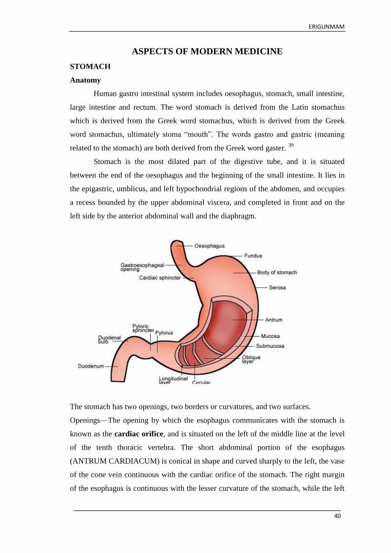
Human gastro intestinal system includes oesophagus, stomach, small intestine,
large intestine and rectum. The word stomach is derived from the Latin stomachus
which is derived from the Greek word stomachus, which is derived from the Greek
word stomachus, ultimately stoma ―mouth‖. The words gastro and gastric (meaning
related to the stomach) are both derived from the Greek word gaster. 39
Stomach is the most dilated part of the digestive tube, and it is situated
between the end of the oesophagus and the beginning of the small intestine. It lies in
the epigastric, umblicus, and left hypochondrial regions of the abdomen, and occupies
a recess bounded by the upper abdominal viscera, and completed in front and on the
left side by the anterior abdominal wall and the diaphragm.
The stomach has two openings, two borders or curvatures, and two surfaces.
Openings—The opening by which the esophagus communicates with the stomach is
known as the cardiac orifice, and is situated on the left of the middle line at the level
of the tenth thoracic vertebra. The short abdominal portion of the esophagus
(ANTRUM CARDIACUM) is conical in shape and curved sharply to the left, the vase
of the cone vein continuous with the cardiac orifice of the stomach. The right margin
of the esophagus is continuous with the lesser curvature of the stomach, while the left

ERIGUNMAM
____________________________________________________________________ 41
margin of the esophagus is continuous with the lesser curvature of the stomach, while
the left margin joins the greater curvature at an acute angle, termed the incisura
cardiaca.
The pyloric orifice communicates with the duodenum, and its position is
usually indicated on the surface of the stomach by a circular groove, the
duodenopyloric constriction. This orifice lies to the right of the middle line at the level
of the upper border of the first lumbar vertebra.
Curvature
The lesser curvature (curvatura ventriculi minor), extending between the
cardiac and pyloric orifices, forms the right or posterior border of the stomach. It
descends as a continuation of the right margin of the esophagus in front of the fibers of
the right crus of the diaphragm, and then, turning to the right, it crosses the first
lumbar vertebra and ends at the pylorus. Nearer its pyloric than its cardiac end is a
well-marked notch, the incisura angularis, which varies somewhat in position with the
state of distension of the viscus it serves to separate the stomach into a right and a left
portion. The lesser curvature gives attachment to the two layers of the heplatogastric
ligament, and between these two layers are the left gastric artery and the right gastric
branch of the hepatic artery.
The greater curvature (curvature ventriculi major) is directed mainly forward,
and is four or five times as long as the lesser curvature. Starting from the cardiac
orifice at the incisura cardiaca, it forms an arch backward, upward, and to the leftthe
highest point of the convexity is on a level with the sixth left costal cartilage. Form
this level it may be followed downward and forward, with aslight convexity to the left
as low as the cartilage of the ninth rib it then turns to the right, to the end of the
pylorus. Directly opposite the incisura anglaris of the lesser curvature the greater
curvature presents a dilatation, which is the left extremity of the pyloric part this
dilatation is limited on the right by as light groove, the sulcus intermedius, which is
about 2. 5 cm, from the duodenopyloric constriction. The portion between the sulcus
intermedius and the duodenopyloric constriction is termed the pyloric antrum. At its
commencement the greater curvature is covered by peritoneum continuous with that
covering the front of the organ. The left part of the curvature gives attachment to the
gastrolienal ligament, while to its anterior portion are appaced the two layers of the
greater omentum, separated from each other by the gastroepilploic vessels.
ERIGUNMAM
____________________________________________________________________ 42
Surfaces
When the stomach is in the contracted condition, its surfaces are directed
upward and downward respectively, but when the viscus is distended they are directed
forward, and backward. They may therefore be described as anterosuperior and
postero-inferior.
Antero-superior Surface
The left half of this surface is in contact with the diaphragm, which separates it
from the base of the left lung, the pericardium, and the seventh, eighth, and ninth ribs,
and intercostals spaces of the left side. The right half is in relation with the left and
quadrate lobes of the liver ad with the anterior abdominal wall. When the stomach is
empty, the transverse colon may lie on the front part of this surface. The whole surface
is covered by peritoneum.
Postero-inferior Surface
It is in relation with the diaphragm, the spleen, the left suprarenal gland, the
upper part of the front of the left kidney, the anterior surface of the pancreas, the left
cokic flexure, and the upper lauer of the transverse mesocolon. These structures form a
shallow bed, the stomach bed, on which the viscus rests. The transverse mesocolon
separates the stomach from the duodenojejunal flexure and small intestine. The
postero-inferior surface is covered by peritoneum, except over a small area close to the
cardiac orifice this area is limited by the lines of attachment of the gastrophrenic
ligament, and lies in apposition with the diaphragm, and frequently with the upper
portion of the left suprarenal gland.
Component Parts of the Stomach
A plane passing through the incisura agularis on the lesser curvature and the
left limit of the opposed dilatation on the greater curvature divides the stomach into a
left portion or body and a right or pyloric portion. The left portion of the body is
known as the fundus, and is marked off from the remainder of the body by plane
passing horizontally through the cardiac orifice. The pyloric portion is divided by a
plane through the sulcus intermedius at right angles to the long axis of this portion the
part to the right of this plane is the pyloric antrum.

ERIGUNMAM
____________________________________________________________________ 43
Position of the Stomach
The position of the stomach varies with the posture, with the amount of the
stomach contents and with the condition of the intestines on which it rests. In the erect
posture the empty stomach is somewhat J-shaped the part above the cardiac orifice is
usually distended with gasthe pylorus descends to the level of the second lumbar
vertebra and the most dependent part of the stomach is at the level of the umbilicus.
Variation in the amount of its contents affects mainly the cardiac portion, the pyloric
portion remaining in a more or less contracted condition during the process of
digestion. As the stomach fills it tends to expand forward and downward in the
direction of least resistance, but when this is interfered with by a distended condition
of the colon or intestines the fundus presses upward on the liver and diaphragm and
gives rise to the feelings of oppression and palpitation complained of in such cases.
The position of the full stomach depends, as already indicated, on the state of
the intestines, when these are empty the fundus expand vertically and also forward, the
pylorus is displaced toward the right and the whole organ assumes an oblique position,
so that it‘s surfaces are directed more forward and backward. The lowest part of the
stomach is at the pyloric vestibule, which reaches to the region of the umbilicus.
Where the intestines interfere with the downward expansion of the fundus the stomach
retains the horizontal position which is the characteristics of the contracted viscus.
Examination of the stomach during kife by x-rays as confirmed this findings,
and as demonstrated that, in the erec posture, the full stomach usually presents a hook
–like appearance the long axis of the clinical fundus being directed down word, medial
word, and forward toward the umbilicus, while the pyloric portion curves upward to
the duodenopyloric junction
Interior of the stomach
A common form is that shown in if the viscus be laid open by a section through
the plane of its two curvatures, it is seem to consist of segments(a) a large globular
position on the left and (b) a narrow tubular part on the right. This correspond to the
clinical subdivision of fundus and pyloric portion already described, and are separated
by a constriction which indents the body and greater curvature, but does not invalve
the lesser curvature. To the left of the cardiac orifice is the incisura cardiac.
The projection of the notch into the cavity of the stomach increases the organ
distends, and has been supposed to act as a valve preventing regurgitation into the

ERIGUNMAM
____________________________________________________________________ 44
esophagus. In pyloric portion are seen(a) the elevation corresponding to the incisura
angularis, and (b)the circular projection from the duodeno pyloric contriction which
forms the pyloric valve. The separation of the pyloric antrum from the rest of the
ppyloric part is scarcely indicated.
The pyloric valve (valvula pylori) is formed by a reduplication of the mucus
membrane of the stomach, covering a muscular ring composed of a thickened portion
of the circular layer of the muscular coat. Some of the deeper longitudinal fibers turn
in and interlace with the circular fibers of the valve.
Structures-The wall of the stomach consists of four coats serous, muscular, areolar,
and mucous, together with vessels and nerves.
The serous coat (tunica serosa) is derived from the peritoneum, and covers the
entire surface of the organ, excepting along the greater and lesser curvatures at the
points of attachment of the greater and lesser omenta here the two layers of
peritoneum leave a small triangular space, along which the nutrient vesssels and
nerves pass. On the posterior surface of the stomach, close to the cardiac orifice. there
is also a small area uncovered by peritoneum, where the organ is in contact with the
under surface of the diaphragm.
The muscular coat (tunica muscularis) is situated immediately beneath the
serous covering, with which it is closely connected. It consists of three sets of smooth
muscle fibers- longitudinal, circular and oblique.
The longitudinal fibers (stratum longitudinal)are the most superficial, and are
arranged in two sets. The first set consist of fibers continues with the longitudinal
fibers of the esophagous they radiate in a stellate manner from the cardiac orifice and
are practically all last before the pyloric portion is reached. The second set commences
on the body of the stomach and passes to the right, its fibers becoming more pickly
distributed as they approach the pylorus. Some of the more superficial fibers of this
set pass on to the duodenum, but the deeper fibers dip inward and interlace with the
circular fibers of the pyloric valve.
The circular fibers (stratum circulare) form a uniform layer over the whole
extent of the stomach beneath the longitudinal fibers. At the pylorus they most
abundant and are aggregated into a circular ring, which projects into the lumen, and
forms, with the fold of mucus membrane covering a surface, the pyloric valve. They

ERIGUNMAM
____________________________________________________________________ 45
are continuous with the circular fibers of the esophagus, but are sharply marked off
from the circular fibers of the duodenum.
The oblique fibers (fibrae oblique) internal to the circular layer, are limited
chiefly to the cardiac end stomach, where they are disposed as a thick uniform layer,
covering both surfaces, some passing obliquely from left to right, others from right to
left, around the cardiac end.
The areolar or sub mucous coat (tela sub mucosa) consist of a loose, areolar
tissue, connecting the mucous and muscular layers.
The mucous membrane(tunica mucosa) is thickened its surface is smooth, soft,
and velvety. In the fresh state it is of a pinkish tinge at the pyloric end, and of a red or
reddish-brown colour over the rest of its surface. In infancy it is of a brighter hue, the
vascular redness being more marked. It is thin at the cardiac extremity, but thicker
toward the pylorus. During the contracted state of the organ it is thrown into
numerous plaits or rugae, which for the most part, have a longitudinal direction, and
are most marked toward the pyloric end of the stomach, and along the greater
curvature. These folds are entirely obliterated when the organ becomes distended.
Structures of the mucous membrane
When examined with a lens, the inner surface of the mucous membrane
presents a peculiar honeycomb appearance from being covered with small shallow
depresens or alveoli, of a polygonal are exagonal form, which vary from 0. 12 to 0. 25
mm in diameter. These are the ducts of the gastric glands, and at the bottom of each
may be seen one or more minute orifices, the openings of the gland tubes. The surface
of the mucous membrane is covered by a single layer of columnar epithelium with
occational globlet cells. This epithelium commences very abruptly at the cardiac
orifice, where there is a sudden transition from the stratified epithelium of the
esophagus. The epithelial lining of the gland ducts is the same character and is
continuous with the general epithelial lining of the stomach.
The Gastric Glands
The gastric glands are of three kinds(a)pyloric, (b)cardiac, and (c ) fundus or
oxyntic glands. They are tubular in character, and are formed of a delicate basement
membrane, consisting of flattened transparent endothelial cells lined by epithelium.
The pyloric glands are found in the pyloric portion of the stomach. They consist of two
or three short closed tubes opening into a common duct or mouth. These tubes are

ERIGUNMAM
____________________________________________________________________ 46
wavy, and are about one-half the length of the duct. The duct is lined by columnar
cells, continuous with the epithelium lining the surface of the mucous membrane of
the stomach, the tubes by shorter and more cubical cell which are finely granular. The
cardiac glands few in number, occur close to the cardiac orifice. They are of two kinds
(1)simple tubular glands resembling those of the pyloric end of the stomach, but with
short ducts (2)compound racemose gland resembling the duodenal glands. The fundus
glands are found in the body and fundus of the stomach, they are simple tubes two or
more of which open into a single duct. The duct, however in these glands is shorter
than in the pyloric variety, sometimes not amounting to more than one-sixth of the
whole length of the glandit is lined throughout by columnar epithelium. The gland
tubes are straight and parelel to each other. At the point where they open into the duct,
which is termed the neck, the epithelium alters, and consists of short coloumnar or
polyhedral, granular cells, which all most fill the tube, so that the lumen becomes
suddenly constricted and is continued down as a very fine channel. They are known as
the chief or central cells of the gland. Between these cells and the basement
membrane, larger oval cells, which is stain deeply with eosin, are found these cells are
studded throughout the tube intervals, giving it a beaded or various appearance. These
are known as the parietal or oxyntic cell, and they are connected with the lumen by
fine channel which run into their substance. Between the glands the mucous
membrane consists of a connective tissue framework, with lymphoid tissue. In places,
this later tissue, especially early life, is collected into little masses, which to a certain
extent resemble the solitary nodules of the intestine, and are termed the lenticular
glands of the stomach. They are not, however, so distinctly circumscribed as the
solitary nodules. Beneath the mucous membrane, and between it and the sub mucous
coat, is a thin stratum of involuntary muscular fibre(muscularis mucosae), which in
some parts consists only of a single longitudinal layersin others soft two layers, an
inner circular and an outer longitudinal.
Vessels and Nerves - the artery supply the stomach are
1) The left gastric
2) The right gastric
3) Right gastro epiploic branches of the hepatic
4) Left gastro epiploic branches of the hepatic
5) Short gastric branches of the lineal

ERIGUNMAM
____________________________________________________________________ 47
They supply the muscular coat, ramify in the submucous coat, and are finally
distributed to the mucous membrane. The arrangement of the vessel in the mucous
membrane is somewhat peculiar. The arteries breakup at the base of the gastric tubules
into a plexus of fine capularis which run upward between the tubues, anastomosing
with each other, and ending in a plexus of large cappularis, which surround mouths of
the tubes, and also form exagonal meshes around the ducts. From these the veins arise,
and pursue a straight course downward, between the tubules, to the submucous tissue
HISTOLOGY
Anatomy of the duodenum
Location of the duodenum
The duodenum lies in the upper abdomen, mainly in the epigastric region and
extending into the umbilical quadrant of the abdomen. It starts at around the level of
the L1 vertebra(superior part), runs downwards to the right of the L1 to L3 vertebrae
(descending part), crosses the body of the L3 vertebra(inferior part)and the courses
upwards on the left of L3 and L2 vertebrae. Here it terminates at the duodenojejunal
flexure which is about 2 to 3 cm to the left of the L2 vertebrae. 40

ERIGUNMAM
____________________________________________________________________ 48
The duodenum is the first of the three parts of the small intestines and continues
from the pylorus of the stomach. It is the shortest part of the small intestine, measuring
approximately 25 cms in length and is also most important site of digestion as the
pancreatic enzymes and bile empty inti the duodenum.
The duodenum runs a C-shaped course cupping the head of the pancreas and
terminates at the duodenojejunal junction (flexure) where the jejunum (second part of
the small intestine) arises. It can be divided into 4 parts
Superior part – approximately 5 centimeters long
Decending part -7 to 10 centimeters long
Inferior part - 6 to 8 centimeters long
Ascending part – approximately 5 centimeters long
Most of the duodenum is fixed in its position unlike the other parts of the small
intestine that are fairly mobile. The duodenal papilla which is the opening of the
hepatopancreatic ducts (bile + pancreatic ducts ) is located in the descending part of
the duodenum.
Blood supply to the duodenum
Arterial blood supply is via the celiac trunk and superior mesenteric artery. The
gastroduodenal artery arising from the celiac trunk and its superior
pancreaticoduodenal branch supply the superior part of the duodenum and portion of
the descending part proximal to the duodenal papilla. The inferior

ERIGUNMAM
____________________________________________________________________ 49
pancreaticoduodenal artery arising from the superior mesenteric artery supplies the
portion of the duodenum distal to the duodenal papilla.
Venous drainage is via veins that correspond to the arteries and empty directly
into the hepatic portal vein or indirectly via the spleenic and superior mesenteric veins.
Nerve Supply to the Duouenum
Innervation lf the duodenum is vis the celiac and superior mesenteric plexuses
with nerves derived from the vagus and greater and greater and lesser splanchnic
nerves.
Lymphatic Drainage of the Duodenum
Anterior lymphatic vessels drain into the pancreativoduodenal and pyloric
lymphnodes, while the posterior lymphatic vessels drain into the superior mesenteric
lymph nodes. Further drainage is into the celiac lymph nodes.
Applied Anatomy
The gastric ulcers are common in the lesser curvature but much in the pyloric
region.
a) Vesssels to the pyloric end of the stomach carry less blood compared to their
size, since they are branches of the hepatic than to the left gastric and they branch to
the stomach from the splemic. The difference in the blood supply as regarded as one of
the factors responsible for the occurrence of larger percentage of gastric of ulcers
towards the pyloric end of the mucosa.
b) There is no sub mucosal plexus. The occurrence of this type i. e. direct from
the sub-serous vessels apparently increase from cardiac to the pyloric regions in man.
Increased vagal tone can prodce marked constriction of the mcosl vessel of this region
causing ischemia and necrosis.
c) Further musculature of the pyoric end is thicker and more powerful.
d)Anterior-venous anastomoses occurs in the gastro-duodena mucosa and
disfanction is thee might lead to loca ischemia and ulcer formation.
RADIOLOGICAL ANATOMY
The alimentary tract can be demonstrated raiologically by giving barium meal
a watery suspension of barium sulphate and taking X-ray pictures aata regular
intervals. The fundus of the stomach, the lesser and greater curvature and the angularis
can be easily made out.

ERIGUNMAM
____________________________________________________________________ 50
The barium meal passes into the first part of the duodenum and forms a
homogenous triangular shadow called the duodena‘ cap, its base being directed
towards the pylorus. The duodenal cap shadow is smooth due to the absence of
mucous fold. Persistent deformity of the duodenal cap is characteristic of duodenal
ulcer.
The pylorus protrudes in to the proximal half of the first part of duodenum
which is kept patent and the barium fills it and this part casts the duodemal cap
shadow. The reaining parts of the duodenum show a fifty it and this part casts the
duodenal cap shadow. The remaining parts of the duodenam show a fifty shodow. But
retrosperitoneal remains coapsed the duodenal ulcer are more common in the first part
of the duodenum. 41
Physiology of the Alimentary Tract
The alimentay trac is a co-ordinated structure with the function of ingesting
and absorbing nutriens and excreting unabsorbed waste products. It should not be
regarded as aseries of separate organs. Since the role of each component isclosely
related to that of other parts of the tract. Its operation may be considered under the
following heading.
1. Controlling andCo-ordinating Mechanisms
The autonomic nervous system and harmones, includes gastrin, secretin and
cholecystokinin (Pancreozgmin)controls and co-ordinaes and seretion.
2. Motility
The carefully controlled moltility o the trct is responsible for the orderly
progression of nutrients through the system so that the stage of digestion and
absorption is appropriate to a given region of the tract.
3. Secretion
The secretion of enzymesand detergents enables protein, carbohydrate nd fat to
be digested before absorption. The secretion of eetrolytes provides the correct PH for
each stage of digestion.
4. Absorption
The absorptive system consists of specialized cells, together with the portal
venous system and lymphatics.

ERIGUNMAM
____________________________________________________________________ 51
5. Defence Mechanisms
These are necessary to protect the mucosa fro its own digestive enzymes and
from the bacterial population to which it is exposed. These mechanisms include a
rapid turnover of the epithelial cells, the produvtion of mucous and a specialized
imunoogical system.
6. Motility
Apart from the striated muscle in the upper oesophages, smooth musce is
responsible for the motility of the gastrointestinal tract. The smooth muscle produces
―slow waves‖ hich are conducted over long distances. These do not result in
contraction but they enable contractions in different areas to be co-ordinated.
Stomach
The normal tonic contraction of the stomach is inhibited by the arrival of food
probably by means of a centrally mediated vagal reflex. This termed receptive
relaxtion so that a large increase in volume in accompanied by only small rise in
pressure with the lumen. The gastric slow wave contros the equency and direction of
antral peristalsis which is responsible for the through mixing of the gastric contents
and their progressive emptying into the duodenum.
Several mechamisms exists to prevent the duodenum receiving more nutrient
than it can deal with. Chemrecetors for fat and acid an osmoreceptors in the duodenal
mucosa control gastric emptying by means of local reflexes and the release of secretin,
choleccystokinin and other enteric hormones. Approximately half of a semi-solid meal
levels the stomach in about 30 minutes.
Small Intestine
Here the co-ordination is due to the slow wave in the longitudinal muscle
fibres. It is he pacemaker which dictates the times at which any given segment of the
gut can contrac. The frequency of the slow wave in the duodenum is greater than in
the ilium, thus enabling the prominal bowe to override more distal areas.
Immunological system.
The lamina propria of the stomach and the intestine contains many lymphocytes
and plasma cells. Some of these cells sysnthesise secretary Ig A which is resistant to
digestion by intestinal enzyme and has a role in protecting mucosal surface from

ERIGUNMAM
____________________________________________________________________ 52
bacterial invasion. It is thus of particular importance in the small intestine where
bacterial colonization is deleterious.
THE SYMPTOMS OF ALIMENTARY DISEASE
Pain is often the most important sysptom of gastrointestinal disease. It must be
analysed in relation to its main site, radiation character, severity, duration, frequency,
time of occurrence, aggravating and relieving factors and any associated phenomena.
The characteristics of abdominal of pain are often diagnostic for example in peptic
ulceration and acute appendicitis.
Loss of appetite (anorexia) may be a local cause such as carcinoma of the stomach,
but may also be a feature of any debilitating disease or due to psychological
disturbance. Water brash is the sudden filling of the mouth with Saliva which is
produced as a reflex response to a variety of symptoms from the upper gastrointestinal
tract, e. g. peptic ulcer pain.
Vomiting may occur in diseases of the stomach or intestine. Vomiting of large
quantities of food and secretions late in the day or night indicates gastric outlet
obstruction. Vomiting which relieves pain is often due to a peptic ulcer.
Heartburn is a burning retrosternal sensation due to reflex esophagitis.
Regurgitation is the appearance of previously swallowed food in the mouth without
vomiting. It usually has an acid or bitter taste because of the presence of gastric juice
or bile but not in patients with obstruction in the eesophagus.
Dysphagia difficulty in swallowing.
Flatulence is often due to excessive swallowing of air (aerophagy) which in turn may
be due to anxiety under normal circumstance a small anount air may be expelled as a
belch. The remainder passes into the intestine. Some will be absorbed but most,
particularly the nitrogen, will be expelled per rectum.
Constipation and Diarrhoea are sometimes difficult to define.
Loss of weight may be due to areduced intake of food because of anorexia nausea or
vomiting to malabsorption of nutsients or to the loss of protein from a diseased bowel
as in ulcerative colitis carcinoma is the most important alimentary cause of loss of
weight. Anaemia Usually occur in massive hemorrhage or iin a non-observed passage
of tarry stools.

ERIGUNMAM
____________________________________________________________________ 53
PEPTIC ULCER
Definition
The term‘Peptic ulcer‘ refers to an ulcer in the lower oesophagus, stomach or
duodenum, in the jejunum after surgical anastomosis to the stomach. Or rarely in the
ileum adjacent to a Meckel‘s diverticulum, ulcers in the sstomach or deuodenum may
be evidence of fibrosis. Erosions do not penetrate the muscularis mucosae. 42
Other School of Thought
Chronic peptic ulcer is by definition an ulcer that occurs in those portions of
the alimentary tract which come into contact with gastric juice. Peptic ulcer could
occur in the lower portions of the oesophagus, the stomach, the duodenum, the
jejunum and after gastroenterostomy in the lower duodenum and jejumum in the
patients who are not operated but is a victim of the Zolligner Ellison syndrome and in
Meckel‘s diverticulum containing gastric mucosa. While ―peptic ulcer‘ embrance all
of these, we would suggest that in clinical usage designation for each lesion can be
made according to its anatomic location, such as ‗Gastric‘, ‗duodenal‘, on ‗jejunal‘
There is no single etiologic factors responsible for this lesion and each factor
that influences the final outcome acts only in a contributory capacity.
The incidence of peptic ulcer is decreasing in many western communities, it
still affects approximately 10% of all adult males. The male to female ratio for
duodenal Ulcer various from 41 to 21 in different communities while that for gastric
ulcer is 21 or less. 43

ERIGUNMAM
____________________________________________________________________ 54
There is growing evidence that cigarette smoking prevents healing of gastric
and duodenal ulcers and it may be a factor contributing to their development.
AETIOLOGY
Heredity
Patients with peptic ulcer often have a family history of the disease. This is
particularly the case with duodenal ulcers which develops below the age of 20years.
Gastric and duodenal ulcers are inherited as separate disorders thus the relatives of
gastric ulcer patients have three times the expected number of gastric ulcers but
duodenal ulcer occurs with the same frequency amongst relatives as in the general
population. 44
Acid-pepsin Versus mucosal resistance
The immediate cause of peptic ulceration is digestion of the mucosa by acid
and pepsin up the gastric juice, but the sequence events leading to this is unknow.
Digestion by acid and pepsin cannot be the only factor invalved, because the normal
stomach is obviously capable of resisting digestion by its own secretions. The concept
of ulcer aetiology may b written as acid plus pepsin versus mucosal resistance. Some
factors which affect this balance can be identified.
Gastric Hypersecretion
Ulcers occurs only in the presence of acid and pepsin they are never found in
achlorohydric patients such as those with pernicious anemia. On the other handsevere
intractable peptic ulceration nearly always occurs in patients with the zollinger-
Ellisson syndrome which is characterized by very high acid secretion. Acid secretion
is more important in the actiology of duodenal than gastric ulcer, because patients with
duodenal ulcer, as a group, secrete more hydrocholoric acid normal individuals.
Factors Reducing Mucosal Resistance
Several drugs particularly those used in Rheumatoid arthritis will disrupt the
gastric mucosal barrier when aspirin is in solution at a PH below 3. 5, it is
undissociated a and fat-soluble, so that it is absorbed through the lipoprotein
membrane of the surface epithelial cells during absorption damages the membrane and
the tight junctions. It also inhibits prostaglandin synthesis thus reducing bicarbonate
secretion by the surface epithelial cells. Aspirin has beeb shown to be ab important

ERIGUNMAM
____________________________________________________________________ 55
actiological factors in gastric ulcer. There is also a relationship between aspirin
ingestion and acute bleeding from the upper gastrointestinal tract.
Reflux of bile and intestinal secretions into the stomachoccurs more frequently
in patients with gastric ulcers than in normal individuals or patients with duodenal
ulcer, due presumably to a poorly functioning pyloric sptineter. Bile damages the
gastric mucosal arrier, predisposing the mucosa to ulceration chronic gastritis is more
common in patients with gastric ulcer and it may be caused by damage from
regurgitated bile and intestinal secretions.
Occupational Factors
The occupational survey carried out by hussian from Hyderbad reported that
60%duodenal ulcer cases were in farmers. It may be trace that peptic ulcer is
commonamong south Indian agriculturists. It is also common is executives, doctors
and industrialists.
Diet
Peptic ulcer is associated with high consumption of refined as compared with
unrefined cereal carbohydrate. The lack of protein deficient diet and untimely meals in
these refined food resulting in a failure to butter gastric acid ingestion of refined
cereals is the prominent factors in the increased incidence of duodenal ulcer.
Smoking, Alcohol and Drugs
Incidence of peptic ulcer is high among smokers than among non-smokers.
Gastric ulcer tend to heal more rapidlyin patients who step smoking than in those who
do not smoking decreases the therapy. All these facts suggest that it is an actiological
factor in the development of peptic ulcer. Gastric ulcer commonly occurs in
association with alcoholic cirrhosis. There is much suggestive evidence that treatment
with aspirin. Phenylbuto yona etc. may aggravate peptic ulcer incidence.

ERIGUNMAM
____________________________________________________________________ 56
Association with Anxiety and personality
People who are highly nervous and emotional and who worry, fear and feel
anxiety are particularly suscesptible. These emotional and nervous factors in turn may
lead to hyper secretion and hyper mobility of the stomach the nervous control of the
vascular system in the gastric on duodenal walls may be so disturbed that there is
diminision in the blook supply to the mucosa of the stomach and duodenum making its
susceptible to acid secretion.
Association with other Diseases
Peptic ulcers in association with almost all diseases, the incidents is noted
inpatients with Achlohydia namely pernicious Anaemis and Atrophy Gastritis Gastric
Carcinoma, Diaphargmatic, Herinia, Duodenal stases, emphasema, corpulsmonale and
Rheumatoid disease, Cirrosis of liver, Tuberculosis.
Pathology
Chronic gastric ulcer is usually single 90%are situated on the lesser curve
within the antrum or at the junction between body and and antral mucosa, chcer is
usually in the first part of thechronic duodenal ulcer is usually in the first part of the
duodenum just distal to the junction of pyloric and duodenal mucosa 50%are on the
anterior wall, more than one peptic ulcer is found in 10-15% of patients acute ulcers
on erosions are frequently multiple, and are more widely distributed. 44
Clinical Features
A duodenal ulcer follows a chronic course for upto 20years and while the
treatment with histamine H2-receptor antagonist drugs may effect prompt healing,
there is no evidence that the natural history of the ulcer is affected. The course of
gastric ulcer is probably less chronic. While there are good grounds gor believing that
gastric and duodenal ulcers are different deceases it is convenient to describe the
general features of ―Peptic Ulcers‖ as inclusive of both, there is no difference in their
occurrence.
Peptic ulcer may be present in different w. however, the ulcer may come
toaways. The commonest is chronic, episodic pain extending over months over on
months or years. However, the ulcer may come toa attention as an acute episode with
bleeding or perforation, with little or no previoushistory, occasionally the patient
ERIGUNMAM
____________________________________________________________________ 57
presents with the symptoms of gastric outlet obstruction, having negligible trouble
previously.
Pain is the characteristic symptoms of peptic ulcer, and it has three notable
features, localization to the epigastrium, relationship to food and periodicity.
Ulcer pain is typically referred to the epigastrium it is localized uaually in the
acid line or to the right. So that the patient can indicate the site with one finger, ‘the
pointing sign‘. Occasionallly ulcer pain is not clearly localized, it may e referred
diffusely in the epigastrium, the lower chest or to the back in the interscapular region
in the fifth to eighth thoracic segments.
Pain referred to the inter scapular area suggests duodenal or post bulbar
ulceration. The description of the pain is not eapecially helpful, although patients
commonly describe it as gnawing or burning.
Most patients recognized a relationship of the pair to the food, although the
relationship varies between patients, and in the same patients, and in the same patient
from time to time. Duodenal ulcer pain tends to occur between meal times, so that the
patient may describe it as ‗hujger Pain, which is characteristically relieved by fook. A
notable feature of duodenal ulcer is pain awakening the patienr from sleep 2 to 3 hours
after refising. The pain of gastric ulcer occurs less regularly it frequently occurs within
an hour of eating, is less often relieved by food and it rarely occurs at night. Besides
the characteristic relief obtained after eating, ulcer pain is almost invariably relieved
by antacids or by vomiting.
Ulcers pain is characteristically episodic occurring regularly then disappearing
to recover ulcers or months later. Between attacks, the patient feels perfectly well, and
may eat and drink with impunity. bouts of pain may at first last only a day also at a
time, and occur only once or twice a year. As the natural history evalves, however
episodes begin to last longer and occur more frequently, so that in severe cases
remissions of pain may be short lived and pain or discomfort becomes more or less
persistent. The cause for these relapses is difficult to be established seasonal factors
may be operative, sometimes psychological stress may be blamed, sometimes, dietary
indiscretion and sometimes alcohol in excess. Most commonly no reason can be found
for the relapse.
Pain is sometimes absent or so slight as to be described by the patient. Such
epigastrium or a poorly defined sense of unease after eating. Other complaints include

ERIGUNMAM
____________________________________________________________________ 58
episodic nausea and sometimes anorexia, as well as heartkburn or water brash
vomiting in clear patients almost always relieves pain and when it is persistent may
result in weight loss. This helps to distinguish it from vomiting of psychological
origin, in which weight is usually maintained. Persistent vomiting in an ulcer subject
usually indicates some degree of gastric narrowing. In such patients, vomiting is
usually copious, so that the patient is ―surprised‖ at the volume, the patient often
recognizes food ate twelve on more hours previously. Although there is no constant
change in bowel rhythm during an ulcer relapse, some patient are aware of
constipation or diarrhea when dyspepsia reappears.
Physical signs
The only physical sign that may be present is ‗the pointing sign‘ which, when
accompanied by localized tenderness, is practically diagnostic of an ulcer. However,
tenderness may be completely absent, in patients with gastric outlet obstruction, the
stomach may be visibly distended, a succession splash may be present and gastric
peristalsis may be seen.
Effect of Diet, Drugs, Tobacco and Alcohol
There is no evidence that dietary manipulation affects the symptoms on ulcer
healing and therefore strict diets should be avoided. It is particularily important that
aspirin and other anti inflammatory drugs are not used in peptic ulceration. Their is no
evidence that shopping smoking accelerates the healing of gastric ulcers and it is likely
to be applied to duodenal ulcers patients should therefore be adviced to give up
smoking exacerbrations of ulcer disease because it aggravates their symptoms. It
seems reasonable to encourage moderation in drinking habits in all patients with peptic
ulceration. .
Complication
Complications of peptic ulcer are hemorrhage perforation and gastric out let
obstruction and ulcer cancer.
Gastric duodenal Hemorrhage
Gastro duodenal hemorrhage is recognized by haematenesis(vomiting of
blood)and or melaena (passage of blood in the stools) and usually there are symptoms
of hypovolaemia. Upper gastro intestinal haemorrhage carries a mortality that may

ERIGUNMAM
____________________________________________________________________ 59
reach 30%in elderly and shocked patients. A history of significant blood loss wihin the
previous 48 hours should lead to immediate admission to hospital.
Aetiology
The common causes of bleeding are chronic gastric and duodenal ulcers (50%)
erosions (15-30%) oesophageal varices (10%)and mucosal lacarations at the cardia
due to vomiting (Malloryweise syndrome-7%). Less frequent cause are cancer of the
stomach and other tumours such as leiomyoma, Oesophagitis, stress ulcers and
bleeding disorders.
Erosions are usually caused by the ingestion of aspirin either alone or in
combination with alchohol or non-steroidal anti-inflammatory drugs. In some patients
the stomach shows petechiae. Multiple erosions and areas of confluent muosal
bleeding, this appearance is called acute haemorrhagec gastritis. The usual
presentation of strees ulcer, caused by burns or head injury, is with heamatemesis and
melaena. 45
CLINICAL FEATURES
In severe bleeding from whatever cause, the patient complaints of weakness,
faintness, nausea and sweating, these symptoms are followed by haematemeses or
melaena with a sudden large bleed whereas melaena alone indicates that bleedg is
slower and less in amount. It blood remains in the stomach it becomes partially
digested and appears brown and granular in the vomit or gastric aspirate, like, coffee
grounds blood passing through the intestinal canal is also altered in appearance, so that
the faces become black and sticky, a ‗tarry‘ stool. But is in severe bleeding transit may
be so rapid that the blood in the rectum is bright red. On examination, the patient ma
be shocked on restless and disorientated because of cerebral anoxia. These signs may
be absent in the young patient in whom compensatory mechanism are more effective.
Acute Perforation of a Peptic Ulcer
When free perforation occurs, the contents of the stomach escape into the
peritomeal cavity. It perforation occurs without loss of contents as in the accidental
perforation of the empty stomach at gastroscopy, few symptoms are produced and the
accident may even pass unnoticed. It follows that the symptoms of perforation are
those of peritomitis, and they are in proportion to the extent of peritoneal soiling.
Occasionally the symptoms of perforation appears and rapidly subside, pre sumably

ERIGUNMAM
____________________________________________________________________ 60
the perforation has then closed spontaneously, or more commonly the ulcer has
perforated locally into an area confined by adhesions to adhesions to adjacent
structures. Perforation occurs more commonly in duodenal than in gastric ulcers ad
usually in ulcers on the anterior wall. About one quarter of all perforations occur in
acute ulcers.
Acute perforation carries a mortality of about 5%. The outlook is poorest in
elderly patients. When a large perforation results in extensive peritonitis on when
operation is delayed.
Gastric Outlet obstructions
An ulcer in the region of the pylorus may result in gastric outlet obstruction.
This may be due to fibrous structure or to oedema or spasm produced by the ulcer,
frequently it is a combination of all three, Long-standing obstruction may lead to
severe. ‘Retention gastritis‘ on even the secondary gastric ulcer.
In addition to chronic duodenal ulcer, or benign gastric ulcer at or near the
pylorus, gastric outlet obstruction may be caused by carcinoma of the antrum and by a
rare condition kown as adult hypertrophic pyloric stenosis.
The syndrome of gastric outlet obstruction is loosely described as ‗pyloric
stenosis‘. Even whe the cause is chronec duodenal ulcer, and the stenosis is distal to
the pylorus thus in ―Pyloric‖obstruction due to duodenal stenosis, the pylorus itself
may be seen radiologically to be greatly dilated.
Clinical Features
Symptoms of obstruction are usually preceded by a long history of duodenal
ulceration. Without such symptoms, a patient with gastric outlet obstruction is likely to
have a pyloric carcinoma when there has been an ulcer, the symptoms change, so that
vomiting produces such striking relief that a patient may start to eat immediately after
the stomach has been emptied. If the obstruction progress, the stomach dilates so that,
eventually, surprisingly largely amounts of gastric content may be vomited. Particles
of food which have been eaten 24 hours or more previously may be recognized in the
vomit.
An earlier symptom is the the blood urea may be raised because of sense of
repletion soon after eating relatively small amount of food. The loss of gastric contents
results in water and electrolyte depletion. The blood urea may be raised because of

ERIGUNMAM
____________________________________________________________________ 61
dehydration alkalosis develops if large amounts of hydrochloric acid are lost, as
occurs particularly in obstruction due to duodenal ulcer.
Zollinger-Ellison Syndrome
This is a rare disorder in which severe peptic ulceration occurs due usually to
an adenoma or hyperplasia of the islets of the pancreas secreting large amount of
gastrin which stimulates the parietal cells of the stomach excessively. The acid output
may be so great that the ‗acid tide‘may reach the upper small intestine, reducing the
luminal PH to 2 og less at the pH pancreatic lipase is inactivated and bile acids may be
precipitated causing diarrhea and steaforrhoea. Excessive gastric secretion results in
large volumes on aspiration under ‗basal‘ conditions. Pentagastrin does not increase
the secretary rate much above ‗basal‘, values, since the stomach is already continosly
secreting at or near nominal rates.
Clinical Features
The ulcers are often multiple and severe and may occur in unusual sites such as
the hehunan or the oesophagus. The history is usually short and bleeding and
perforation are common. The syndrome may present form of severe recrurrent
ulceration following a standard operation for peptic ulcer, the underlying cause not
having been recognized.
The diagnosis should be suspected in all patients with unusual or severe peptic
ulceration especially coarse barium meal examinarion shows abnormally coarse
gastricmucosal tolds. It may be confirmed by finding very high levels of gastrin in the
circulation.
Complication following Gastric Surgery
Although most operations carried out for the relief of peptic ulcer are
successful, 10%of patients will develop complications months of years afterwards.
Some of these such as anaemea and nutritional impairment, develop insidiously such
patients should be reviewed atleast once in a year.
Recurrent ulcer, after surgery for duodenal ulcer, it is usually due to
insufficient reduction of the secretary capacity of the stomach because of incomplete
vagaotomy or inadequate gastrectomy. A jejuna ulcer develops just distal to the
jejunogastric anastomosis, becarse the jejuna mucosa is more susceptible to acid-
pepsin digestion than gastric or duodenal mucosa. About 15% of selective vagotmy

ERIGUNMAM
____________________________________________________________________ 62
but the operation has the virtue of being free from the side effects associated with
resection truncal vagotomy or drainage procedures.
Anaemia is a common seqel to operation on the stomach, particularly partial
gastrectomy, due to inadequate absoption of iron, or to recurrent minor blood loss
from gastritis or oesophagitis.
Nutritional impairment and osteomalacia. In a small propotion of patients there
is some nutritional impairment following gastric surgery. 46
DIFFERENTIAL DIAGNOSIS
1. Chronic intestinal Amoebiases
History of recurret dysentery, caecum and pelvic color aare tender and cord
like, liver may be palpable and tender, stool may show cysts of entamoeba histolytica.
2. Chronic cholecystitis
There may be history of biliary colic and jaundice in the past, murphy‘s sign is
positive. Rarely gull bladder may be palpable. Chlocytography settles the diagnosis by
showing dysfunction of the gall bladder with or without stone.
3. Chronic Appendicitis
There may be history of acute appendicitis in the past, Mcburney‘s point is
tender, F. T. M. and barium meal x-ray of appendix may show irregularity or no
filling.
4. . Chronic Gastritis
There is anorexia, discomfort in the upper andomen without any definite
tenderness, F. T. M. shows low acid but excess of mucous in all samples, barium meal
x-ray shows coarse or tine gastric rugae.
5. Chronic Pancreatitis
There may be history of acute pancreatitis in the past, pain radiating to the
back may be present without definite relation with food. Steatorrhoea and diabetes
mellitus may be present, X-ray of the abdomen may reveal pancreatic calcification. 47

ERIGUNMAM
____________________________________________________________________ 63
SPECIAL INVESTIGATIONS
1. Endoscopy
In recent years endoscopic photography, both still and motion, has become
possible and gives excellent pictures. The flexible fibroscope now enables one to
examine the oesophagus, stomach and duodenum and at the same time obtain biopsies
and material for cytological examination.
It is used I diagnosis purpose for the oesophagitis, oesophageal ulcer, gastric
ulcer, duodenal ulcer, duodenitis malignant cancer, biopsy can also be obtained to find
out in gastric ulcer is benign or malignant.
2. Fractional Test Meal
The patient who was on starvation during the previous night is asked to swllow
the ryles tube at 5a. m. and the entire stomach contents a fasting juice are aspirated
with a donel, record syringe. The patient is them given a pint of warm gruel to drink
the gruel is prepared by boiling two table spoonfuls of the oatiomeal in two pints of
water until the quantity is reduced to on pint. Every 15ml of gastric contents is now
aspirated until 21/2hours have elapsed or until such time as 15ml can no longer be
aspirated. These samples are examined for total acidity, free Hcl, bile, blood, mucous,
and starch and the results recoded on a chart in a gastric ulcer, the cures of tree hel,
and total acidity are high, normal or just above the normal limit. Blood may be present
in some of the specimen. The climbing curve is due to pylorospasm which prevents

ERIGUNMAM
____________________________________________________________________ 64
regurgitation of bile or allows the acidity to rise continuously. Besides carcinoma
achlorhydria is found in pernicious anaencia, gastritis, chronic appendicitis, etc, but
association or blood in all the specimen is strongly suggestive of a carcinoma.
sometimes cancer cells can be demonstrated into washing after gastric lavage.
This test is no more needed to make correct diagnosis of peptic ulcer except to
exclude the role of vagatomy during surgical management.
3. Examination of stool
Black and farry stool melana is well known in a peptic ulce when the
haemorrhage is large. Small hemorrhage need special chemical test for detection.
4. Radiological features of peptic ulcer(Barium Meal series)
Peptic ulceration only occurs in those parts of the alimentary canal which are
bathed in the acid and pepsin secretions. The radiological features of peptic ulcer
varies from a mild erosion to a malignant ulcer.
Although in clinical experience duodenal ulcer are for more frequently than
gastric ulcers in the ratio of 10 or 20 to 1 they are approximately equal.
ROENTGEN SIGNS OF ULCERATION
The presence of a ‗fleck‘ or crater. This sign represents the presence of barium
and is regaraded as essential for the diagnosis.
Changes in the Neighbouring Rugae
These are cedema, irregularity end the cart wheel appearance in which the
rugae radiate from the fleck or crater.
Functional changes such spasm, increase in peristalsis or irritability are
common.
Characteristics Associated with the site of Ulceration
Ulcers in the body of the stomach are more prevalent along the lesser curvature
ulcers of the greater curvature are rare.
Mucosal Relief with small amount of Barium shocos
1. Barium spot or fleck.
2. Edematous mucosa at base
3. Radiating rugae
4. Coarse rugae often there
5. When capped by air miniasn in correct position suspect penetration.

ERIGUNMAM
____________________________________________________________________ 65
6. When seen in profile it is an out pouching 10th
a broad kbase. Most often on
lesser curvature. But requires flourscopy in every degree of obliquity for
demonstation.
Radiological Features of Malignant Gastric Ulcer
Irregularity in mucosa adjoining ulcer niche.
1. No peristalsis here
2. The Niche does not entered eyond the line of stomach.
3. Associated duodenal ulcer usually indicated the gastric ulcer is bebingn.
4. Ulceration of greates curvature is usually malignant.
A less common site for ulcers is the pyloric but even here it tends to occur along the
lesser curvature. This ulcers produces a gastric stasis.
Duodenal ulcers
The common site for duodenal ulcer is in the duodenal cap and they may occur
on either the anterior or posterior walls less frequently post bulbar area.
Radiological Features are
a. Acute penetrating as erosive stage
1. Ulcer Niche
2. Edematous Mucosal Hallow
3. Thick pyloric Rugae
4. Spastic
b. Beginning scar formation
1. Ulcer Niche
2. Thickened surrounding mucosa
3. Rugae converting like cart wheel spokes
4. Pseudo diverticulum formation
5. Bulb may appear fragnated on compression.
c. Late scaring stage
1. Niche or pscudo divertiulum
2. Contracted deformed fibrotic bulb rigid walls
3. Thick pyloric rugae
Post bulbar ulcers shows deformed bulb48

ERIGUNMAM
____________________________________________________________________ 66
MATERIALS
AND METHODS

ERIGUNMAM
____________________________________________________________________ 67
MATERIALS AND METHODS
Study Designs
An open clinical trial on Eri Gunmam was carried out in the Post Graduate
Department of maruthuvam in Arignar Anna Hospital of Indian Medicine attached
with Govt.Siddha Medical College Chennai – 106 during the period of 2015 – 2017.
The study was approved by Institutional Ethics Committee (IEC) and the
approval number is IEC No : GSMC – CH – ME – 4 / 2015 / 003. It was registered
in Clinical Trials Registry – India ( CTRI ) and the reference number is
REF/ 2016 / 12 / 012978.
POPULATION AND SAMPLE
The population consists of all patients satisfying the inclusion and exclusion
criteria mentioned below. Sample consists of Eri Gunmam patients attending the
OPD of Arignar Anna Hospital, Arumbakkam, Chennai-106.
SAMPLE SIZE
20 patients
SELECTION OF PATIENTS
Inclusion criteria
Cases were selected on the basis of the following signs and symptoms
Age:18-60 years
Sex :Both.
Epigastric pain and burning with relation to food
Nausea
Vomiting
Anorexia
Bloating and fullness of stomach
Diarrhea
Weight loss.

ERIGUNMAM
____________________________________________________________________ 68
Exclusion criteria
Known case of pyloric stenosis
Known case of cancer in the stomach
Known case of acute abdominal colics
Known case of cholelithiasis
Known case of ulcer perforation.
INVESTIGATIONS
Routine laboratory investigations done in the out-patient department of
Government Arignar Anna Hospital of Indian Medicine, Chennai-106.
Special Investigations like Endoscopic examination was made on the disease
Eri Gunmam ( Peptic Ulcer ) for all patients.
Based on the signs and symptoms, investigations, diagnostic methods based on
Siddha aspect like Envagai thervugal, mukkuttra nilaigal, seven udal kattugal, Thinai
and Kalam, the diagnosis was made and the treatment was given.
SELECTION OF MEDICINE AND SHEDULE
All the patients were tried with Pirandai Vadagam ( 1gm ), twice a day with
water, after food for 48 days. Ref: THERAIYAR THARU.(page no:42,43)49
The medicine was selected to stabilize the deranged uyir thathus and
strengthening seven udal kattugal. The trial medicine Pirandai Vadagam is purified
properly and prepared according to the literature. The drug PIRANDAI VADAGAM
is given for the entire course of treatment.
It is studied properly for Phytochemical analysis and Pharmacological analysis.
The above parameters were done at Pharmacology Department of Sathyabama
University, Chennai.

ERIGUNMAM
____________________________________________________________________ 69
TRAIL DRUG
PIRANDAI VADAGAM
REFERENCE: THERAIYAR THARU49
Page.No:42,43.
INGREDIENTS:
Pirandai
Thalisabathiri
Inji
Kadukai
Nellikai
Dhandrikai
All the above ingredients are taken in equal proportion STANDARD OPERATIVE PROCEDURE
I got authentication for the above drugs and they are purified as given in SIGICHCHA RATHTHA DHEEBAM .Then the above mentioned PIRANDAI, INJI & NELLIKAI should be made into fine paste. Other ingredients are made into fine powder and mixed with the above paste. Then the mixed content is grinded in the kalvam and made into pills of 1gm. Dried in the shadow and packed up. DOSE 1gm vadagam, Twice a day, chewable ADJUVENT: Water DURATION: 48 Days (1 MANDALAM) REFERENCE: THERAIYAR THARU Page.No:42,43.
PROPERTIES OF TRAIL DRUG
PIRANDAI
Botanical Name: Cissus quadrangularis
Actions: Alterative, Stomachic
33.
THALISABATHIRI Botanical Name: Taxus baccata
Actions:

ERIGUNMAM
____________________________________________________________________ 70
Stomachic, Carminative, Tonic.34
INJI Botanical Name: Zingiber officinalis
Actions: Stomachic, Carminative, Digestive.
35
KADUKAI Botanical Name: Terminalia chebula Actions:
Astringent, Alterative.36
NELLIKAI Botanical Name: phyllanthus emblica
Actions: Refridgerant, Laxative.37
DHANDRIKAI
Botanical Name: Terminalia belleric Actions:
Astringent, Laxative, Tonic.38

ERIGUNMAM
____________________________________________________________________ 71
PIRADAI THALISABATHIRI
INJI NELLIKAI
KADUKAI DHANDRIKAI

ERIGUNMAM
____________________________________________________________________ 72
PIRADAI VADAGAM

ERIGUNMAM
____________________________________________________________________ 73
RESULTS AND
OBSERVATION
ERIGUNMAM
____________________________________________________________________ 74
RESULTS AND OBSERVATION
The study on Erigunmam was carried out in 20 patients in the out patient
department, Pothu Maruthuvam, Aringnar Anna Hospital of Indian medicine attached
with Govt Siddha Medical college chennai during 2015-2017 analysed.
The observations were made and tabulated with following criteria:
Sex distribution
Age distribution
Socio-economic status
Duration of illness
Food habits
Habits
Religion
Paruvakkalam
Thinai
Mukkutram
o Vathakutram
o Pittha kutram
o Kabha kutram
Udal Thaathukal
Envagai thervugal
Naadi
Blood grouping
Clinical features
Gradation of result
PIRANDAI VADAGAM 1vadagam twice a day, chewable, with water, after food.
Among the 20 cases epigastric pain, burning sensation with relation to food,
nausea, vomiting, anorexia, bloating and fullness of the stomach, diarrhoea, weight
loss and other symptoms were relieved after the administration of the medicines.
Digestion with good appetite and no chronic effects were observed.
1. GENDER DISTRIBUTION

ERIGUNMAM
____________________________________________________________________ 75
S.No GENDER NUMBER OF CASES PERCENTAGE (%)
1 Male 14 70%
2 Female 6 30%
INFERENCE
About 70% were males and 30% were females
According to this males are more prone to peptic ulcer.
Male70%
Female30%
GENDER DISTRIBUTION

ERIGUNMAM
____________________________________________________________________ 76
2. AGE DISTRIBUTION
S.No AGE IN YEARS NUMBER OF
CASES
PERCENTAGE (%)
1 21 - 30 yrs 7 35%
2 31 - 40 yrs 5 25%
3 41 - 50 yrs 5 25%
4 51 - 60 yrs 3 15%
INFERENCE
Majority of the case, that is, 35% were in the 3rd
decade, 25% were in the 4th
and 5th
decade, 15% were in the 6th
decade.
35%
25% 25%
15%
21 - 30 yrs 31 - 40 yrs 41 - 50 yrs 51 - 60 yrs
AGE DISTRIBUTION

ERIGUNMAM
____________________________________________________________________ 77
3. SOCIO – ECONOMIC STATUS
S.No SOCIO – ECONOMIC
STATUS
NUMBER OF
CASES
PERCENTAGE
(%)
1 Low Income
( below 2 lakhs per annum)
12 60%
2 Moderate Income
( 2 to 5 lakhs per annum)
5 25%
3 High Income
( Above 5 lakhs per annum)
3 15%
INFERENCE:
Among 20 cases 60% comes under low economic status, 25% of them under
moderate status and 15% of them under high income status.
Low Income 60%
Moderate Income 25%
High Income15%
SOCIO – ECONOMIC STATUS
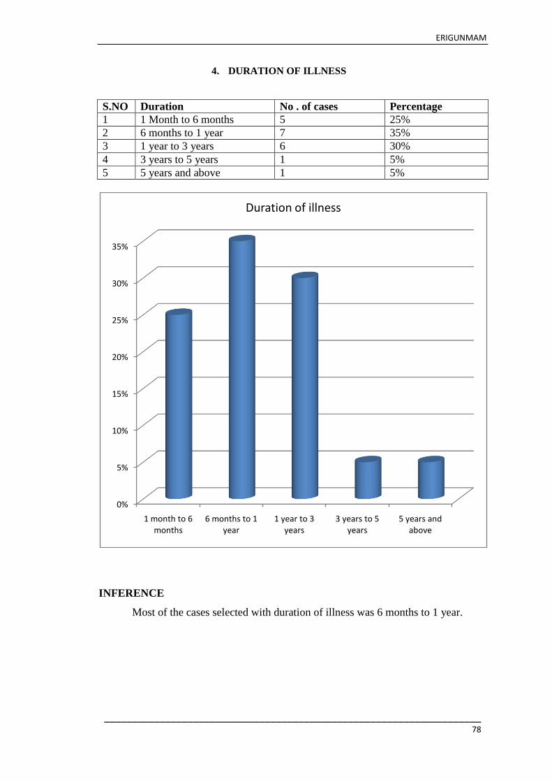
ERIGUNMAM
____________________________________________________________________ 78
4. DURATION OF ILLNESS
S.NO Duration No . of cases Percentage
1 1 Month to 6 months 5 25%
2 6 months to 1 year 7 35%
3 1 year to 3 years 6 30%
4 3 years to 5 years 1 5%
5 5 years and above 1 5%
INFERENCE
Most of the cases selected with duration of illness was 6 months to 1 year.
0%
5%
10%
15%
20%
25%
30%
35%
1 month to 6 months
6 months to 1 year
1 year to 3 years
3 years to 5 years
5 years and above
Duration of illness

ERIGUNMAM
____________________________________________________________________ 79
5. DIETARY HABITS
S.No DIET NUMBER OF
CASES
PERCENTAGE (%)
1 Vegetarian 5 25%
2 Mixed diet 15 75%
INFERENCE
Among 20 patients, 5 patients (25%) were taking vegetarian food and 15
patients (75%) were taking mixed diet.
25%
75%
DIETARY HABITS
VEGETARIAN MIXED DIET

ERIGUNMAM
____________________________________________________________________ 80
6. HABITS
S.NO HABITS NO.OF
PATIENTS
PERCENTAGE(%)
1 Alcohol 5 25%
2 Smoker 4 20%
3 BOTH(alcoholic and
smokers)
5 25%
4 No Other Habits 6 30%
INFERENCE
Among 20 patients 5 patients were taking alcohol, 4 patients were smokers and
5 patients are taken both (Alcohol and smoking), 6 patients were having no other
habits
7.SEASONAL OCCURRENCE
ALCOHOL SMOKER BOTH No Other Habits
25%
20%
25%
30%
HABITS

ERIGUNMAM
____________________________________________________________________ 81
S.No KAALAM (Season) NUMBER OF CASES PERCENTAGE
(%)
1 Kaar Kaalam
( Mid Aug – Mid Oct)
1 5%
2 koothir Kaalam
(Mid Oct – Mid Dec)
1 5%
3 Munpani Kaalam
(Mid Dec – Mid Feb)
15 75%
4 Pinpani Kaalam
(Mid Feb – Mid Apr)
3 15%
5 Elavenir Kaalam
(Mid Apr – Mid Jun)
0 0%
6 Muthuvenir Kaalam
(Mid Jun – Mid Aug)
0 0%
INFERENCE
According to paruvakaalam highest incident of 15 cases (75%) were noted in
munpani kaalam , 3 cases (15%) were noted in pinpani kaalam, 1case (5%) was noted
in koothir kaalam, nil cases (0%) were noted in elavenir kaalam, 1 case (5%) was
noted in karkaalam And nil case (0%) were noted in muthuvenir kaalam.
Kaar Kaalam koothir Kaalam
Munpani Kaalam
Pinpani Kaalam
Elavenir Kaalam
Muthuvenir Kaalam
5% 5%
75%
15%
0 0
SEASONAL OCCURRENCE
PERCENTAGE (%)
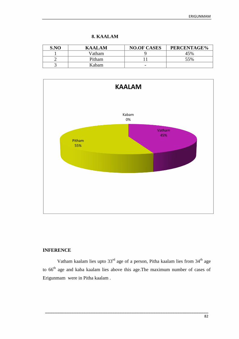
ERIGUNMAM
____________________________________________________________________ 82
8. KAALAM
S.NO KAALAM NO.OF CASES PERCENTAGE%
1 Vatham 9 45%
2 Pitham 11 55%
3 Kabam -
INFERENCE
Vatham kaalam lies upto 33rd
age of a person, Pitha kaalam lies from 34th
age
to 66th
age and kaba kaalam lies above this age.The maximum number of cases of
Erigunmam were in Pitha kaalam .
Vatham45%
Pitham55%
Kabam0%
KAALAM

ERIGUNMAM
____________________________________________________________________ 83
9. DISTRIBUTION OF THINAI
S.No THINAI NUMBER OF CASES PERCENTAGE (%)
1 Kurinji 0 0%
2 Mullai 0 0%
3 Marutham 2 10%
4 Neithal 18 90%
5 Paalai 0 0%
INFERENCE
According to thinai, the highest distribution of 90% was noted in neithal, 10%
in marutham.
Kurinji Mullai Marutham Neithal Paalai
0.00% 0.00%
10%
90%
0%
THINAI
PERCENTAGE (%)

ERIGUNMAM
____________________________________________________________________ 84
10. DISTRIBUTION OF MUKKUTRAM – VATHAM
S.No VATHAM NUMBER OF CASES PERCENTAGE (%)
1 Pranan 0 0%
2 Abanan 4 20%
3 Viyanan 20 100%
4 Uthanan 20 100%
5 Samanan 0 100%
6 Nagan 0 0%
7 Koorman 2 10%
8 Kirukaran 9 45%
9 Devathathan 4 20%
10 Thananjeyan 0 0%
INFERENCE
Out of 20 patients Abanan was affected in 4 patients (20%), Viyanan was
affected in 20 pateints (100%), Uthanan was affected in 20 patients (100%), Samanan
was affected in 20 patients (100%), Koorman was affected in 2 patients(10%),
Kirukaran was affected in 9 patients (45%) and Devathathan was affected in 4 patients
(20%)
0%
20%
100% 100% 100%
0%
10%
45%
20%
0%0%
20%
40%
60%
80%
100%
120%
VATHAM
VATHAM

ERIGUNMAM
____________________________________________________________________ 85
11. DISTRIBUTION OF MUKKUTRAM – PITHAM
S.No PITHAM NUMBER OF
CASES
PERCENTAGE
(%)
1 Analagam 20 100%
2 Ranjagam 5 25%
3 Saathagam 20 100%
4 Alosagam 3 15%
5 Pirasagam 0 0%
INFERENCE
Out of 20 patients, Analagam was affected in 20 patients (100%), Ranjagam
was affected in 5 patients (25%), Alosagam was affected in 3 patients, Sathagam was
affected in 20 patients (100%).
Analagam Ranjagam Saathagam Alosagam Pirasagam
100%
25%
100%
15%
0%
PITHAM

ERIGUNMAM
____________________________________________________________________ 86
12. DISTRIBUTION OF MUKKUTRAM – KABHAM
S.No KABHAM NUMBER OF
CASES
PERCENTAGE (%)
1 Avalambagam 0 0%
2 Klethagam 20 100%
3 Pothagam 11 55%
4 Tharpagam 0 0%
5 Santhigam 3 15%
INFERENCE
Out of 20 patients, Kiledhagam was affected in 20 patients (100%), Pothagam
was affected in 11 patients(55%), Santhigam was affected in 3 patients (15%).
Avalambagam Klethagam Pothagam Tharpagam Santhigam
0%
100%
55%
0%
15%
KABHAM
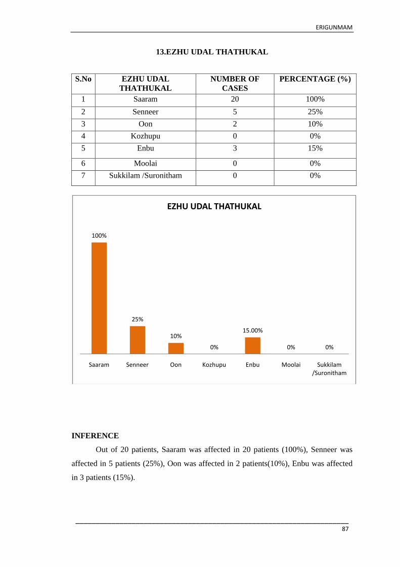
ERIGUNMAM
____________________________________________________________________ 87
13.EZHU UDAL THATHUKAL
S.No EZHU UDAL
THATHUKAL
NUMBER OF
CASES
PERCENTAGE (%)
1 Saaram 20 100%
2 Senneer 5 25%
3 Oon 2 10%
4 Kozhupu 0 0%
5 Enbu 3 15%
6 Moolai 0 0%
7 Sukkilam /Suronitham 0 0%
INFERENCE
Out of 20 patients, Saaram was affected in 20 patients (100%), Senneer was
affected in 5 patients (25%), Oon was affected in 2 patients(10%), Enbu was affected
in 3 patients (15%).
100%
25%
10%
0%
15.00%
0% 0%
Saaram Senneer Oon Kozhupu Enbu Moolai Sukkilam /Suronitham
EZHU UDAL THATHUKAL

ERIGUNMAM
____________________________________________________________________ 88
14.EN VAGAI THERVUGAL
S.No EN VAGAI
THERVUGAL
NUMBER OF
CASES
PERCENTAGE (%)
1 Naa 10 50%
2 Niram 5 25%
3 Mozhi 12 60%
4 Vizhi 2 10%
5 Sparisam 8 40%
6 Malam 4 20%
7 Moothiram 0 0%
8 Naadi 20 100%
INFERENCE
In Envagai thervu, Naa was affected in 10 patients(50%), Niram was affected
in 5 patients (25%), Mozhi was affected in 12 patients, Vizhi was affected in 2
patients, Sparism was affected in 8 patients, Malam was affected in 4 patients (20%)
and Naadi was affected in 20 patients (100%).
50%
25%
60%
10%
40%
20%
0%
100%
Naa Niram Mozhi Vizhi Sparisam Malam Moothiram Naadi
EN VAGAI THERVUGAL
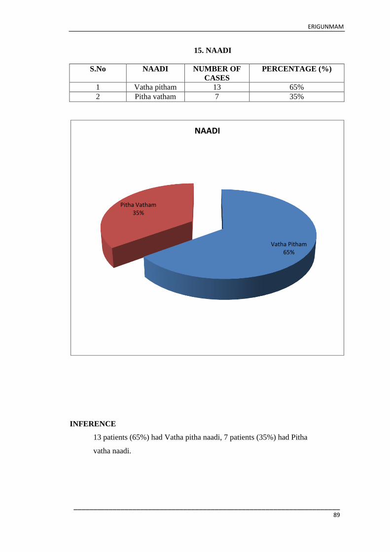
ERIGUNMAM
____________________________________________________________________ 89
15. NAADI
S.No NAADI NUMBER OF
CASES
PERCENTAGE (%)
1 Vatha pitham 13 65%
2 Pitha vatham 7 35%
INFERENCE
13 patients (65%) had Vatha pitha naadi, 7 patients (35%) had Pitha
vatha naadi.
Vatha Pitham65%
Pitha Vatham35%
NAADI

ERIGUNMAM
____________________________________________________________________ 90
16.CLINICAL MANIFESTATION
S.No CLINICAL
MANIFESTATION
NO.OF CASES PERCENTAGE
(%)
1 Epigastric pain 20 100%
2 Heart burn 8 40%
3 Abdominal
discomfort
15 75%
4 Pain related to food 17 85%
5 Regurgitation 8 40%
6 Tenderness in
epigastric region
16 80%
7 Loss of weight 2 10%
8 Diarrhoea 4 20%
INFERENCE
Epigastric pain was present in 20cases (100%),
Heart burn was seen in 8cases (40%),
Abdominal discomfort was present in 15 cases (75%),
Tenderness in epigastric region was seen in 16 cases (80%),
Diarrhoea was seen in 4 patients (20%) ,
Regurgitation was present in 8cases (40%),
Loss of appetite was seen in 2 cases (10%) and
100%
40%
75%85%
40%
80%
10%20%
CLINICAL MANIFESTATION
PERCENTAGE

ERIGUNMAM
____________________________________________________________________ 91
Pain related to food was present in 17 cases (85%)
17. CLINICAL PROGNOSIS
S.
No
SIGNS&
SYMPTOMS
BEFORE
TREATMENT
AFTER TREATMENT
NO.OF
CASE
S
PERCENTAG
E (%)
NO.OF
CASES
PERCENTA
GE (%)
1 Epigastric pain 20 100% 4 20%
2 Heart burn 8 40% 1 5%
3 Abdominal
discomfort
15 75% 2 10%
4 Pain related to food 17 85% 2 10%
5 Regurgitation 8 40% 1 5%
6 Tenderness in
epigastric region
16 80% 2 10%
7 Loss of weight 2 10% 0 0%
8 Diarrhoea 4 20% 0 0%
CLINICAL PROGNOSIS
INFERENCE
Among 20 patients 16 patients improved from Epigastric pain
Among 8 patients 7 patients improved from Heart burn
Among 15 patients 13 patients improved from Abdominal discomfort
Among 17 patients 15 patients improved from pain related to food
Among 8 patients 7 patients improved from regurgitation
Among 16 patients 14 patients relief from tenderness in abdominal region.
Among 2 patients , all got relief from loss of weight
Among 4 patients, all got relief from Diarrhoea
100%
40%
75%85%
40%
80%
10%20%20%
5% 10% 10% 5% 10%0% 0%
BEFORE TREATMENT AFTER TREATMENT

ERIGUNMAM
____________________________________________________________________ 92
18.GRADING OF RESULTS
S.No RESULTS NUMBER OF CASES PERCENTAGE (%)
1 Good 15 75%
2 Fair 3 15%
3 Poor 2 10%
INFERENCE
Results obtained were
75% of cases showed good results.
15% of cases showed fair results.
10% of cases showed poor results.
The Results were based on clinical improvement.
Good75%
Fair15%
Poor10%
GRADING OF RESULTS

ERIGUNMAM
____________________________________________________________________ 83
LABORATORY INVESTIGATION
S.
No
Op No Age/Se
x
Before treatment After Treatment ESR (mm) Hb (gms%) Urine analysis
TC
(cu/mm
DC TC
(cu/mm
DC BT AT BT AT
P% L% E% P% L% E
%
½ hr 1 hr ½
hr
1 hr BT AT Alb Sug Dep Alb Sug Dep
1 8370 47/M 9800 56 38 6 9700 56 38 6 3 8 3 7 9.8 11.5 N N FE N N N
2 5318 60/F 9600 54 42 4 9800 54 48 3 5 16 5 12 13.8 13.8 N N N N N N
3 4949 23/M 9400 59 32 7 9600 62 32 6 4 8 4 6 9.6 12 N N N N N N
4 4724 25/F 9600 55 39 6 9700 55 39 6 25 50 20 40 9.6 11.7 N N FE N N FE
5 7433 47/M 10400 60 34 5 10250 58 35 4 8 16 10 20 14 14 N N FE N N FE
6 8474 27/M 8000 61 35 4 8000 61 39 3 5 12 08 20 12.8 13 N N N N N N
7 8764 27/F 8700 54 39 2 8700 53 37 4 2 6 3 6 11 12.2 N N FE N N FE
8 9003 51/M 9700 62 35 3 9400 60 38 3 10 25 8 18 12 12.5 N N FE N N FE
9 9346 22/F 8400 56 36 4 8600 58 40 2 13 22 10 18 9.8 10 N N N N N N
10 9477 33/F 7600 51 45 4 7600 60 33 4 2 3 2 3 13.6 13.6 N N FE N N FE
11 9842 43/M 9100 58 34 6 9350 56 36 4 4 9 3 8 9.6 11.5 N N N N N N
12 358 43/M 8500 55 41 4 9400 59 33 4 3 7 3 6 12.4 13.2 N N FE N N N
13 3731 40/M 7500 56 40 4 7500 52 35 4 20 23 16 18 10.8 11 N N N N N FE
14 5080 32/M 8900 51 46 3 8900 51 40 3 2 5 4 6 12 12.3 N N FE N N N
15 5086 36/M 8300 52 43 5 8300 51 33 3 8 20 9 20 11.6 12 N N FE N N N
16 5778 29/M 7750 55 35 6 7750 54 31 5 10 18 12 26 16.8 16.8 N N FE N N N
17 9771 53/M 8200 59 41 5 8400 60 41 4 15 35 16 37 11 11.5 N N FE N N N
18 3722 22/M 6800 55 42 3 6800 46 42 10 4 12 18 20 8.6 11.5 N N FE N N N
19 3674 35/F 9700 55 39 6 9700 55 39 6 6 12 7 12 9.6 11 N N FE N N N
20 113 23/M 9200 60 38 5 9 400 54 28 6 16 36 13 25 9.8 9.8 N N FE N N FE

ERIGUNMAM
84
PROGNOSIS CHART
S.
No
Name of
the
patient
Age/Sex
Duration
of
medicine
taken
Investigation
Results Before treatment
After
treatment
1 8370 47 /M 48 Days
Pain in epigastric,
heart burn, Pain
related to food,
Regurgitation,
Diarrhoea
Cured and
improved Good
2 5318 60 /F 48 Days
Pain in epigastric
region, heart burn,
Pain related to food,
Regurgitation
Cured and
improved Good
3 4949 23 /M 48 Days
Pain in epigastric,
Abdominal
discomfort, Pain
related to food,
Tenderness in
epigastric region,
Diarrhoea
Cured and
improved Good
4 4724 25 /F 48 Days
Pain in epigastric,
Abdominal
discomfort, Pain
related to food,
Tenderness in
epigastric region
Symptoms
relieved Fair
5 7433 47 /M 48 Days
Pain in epigastric,
Abdominal
discomfort,
Regurgitation Pain
related to food,
Tenderness in
epigastric region,
Loss of weight
Cured and
improved Good
6 8474 43 /M 48 Days
Pain in epigastric ,
heart burn, Pain
related to food,
Regurgitation
Symptoms
relieved Fair
7 8764 27 /F 48 Days
Pain in epigastric,
Abdominal
discomfort, Pain
related to food,
Tenderness in
Cured and
improved
Good

ERIGUNMAM
85
epigastric region,
8 9003 51 /M 48 Days
Pain in epigastric,
heart burn,
Regurgitation
Tenderness in
epigastric region,
Cured and
improved Good
9 9346 22 /F 48 Days
Epigastric pain,
Abdominal
discomfort, Pain
related to food,
Tenderness in
epigastric, Loss of
weight
Cured and
improved Good
10 9477 33 /F 48 Days
Pain in epigastric,
heartburn,diarrhoea
Regurgitation,pain
related to food,
Tenderness in
epigastric region,
Symptoms
relieved Fair
11 9842 43 /M 48 Days
Pain in epigastric,
Abdominal
discomfort, Pain
related to food,
Tenderness in
epigastric region
Cured and
improved Good
12 358 43 /M 48 Days
Pain in epigastric,
Abdominal
discomfort,
Regurgitation,Pain
related to food,
Tenderness in
epigastric region
Cured and
improved Good
13 3731 40 /M 48 Days
Pain in epigastric,
heart burn,
Abdominal
discomfort, pain
related to food,
Diarrhoea
Cured and
improved Good
14 5080 32 /M 48 Days
Pain in epigastric,
Abdominal
discomfort, Pain
related to food,
Tenderness in
Cured and
improved Good
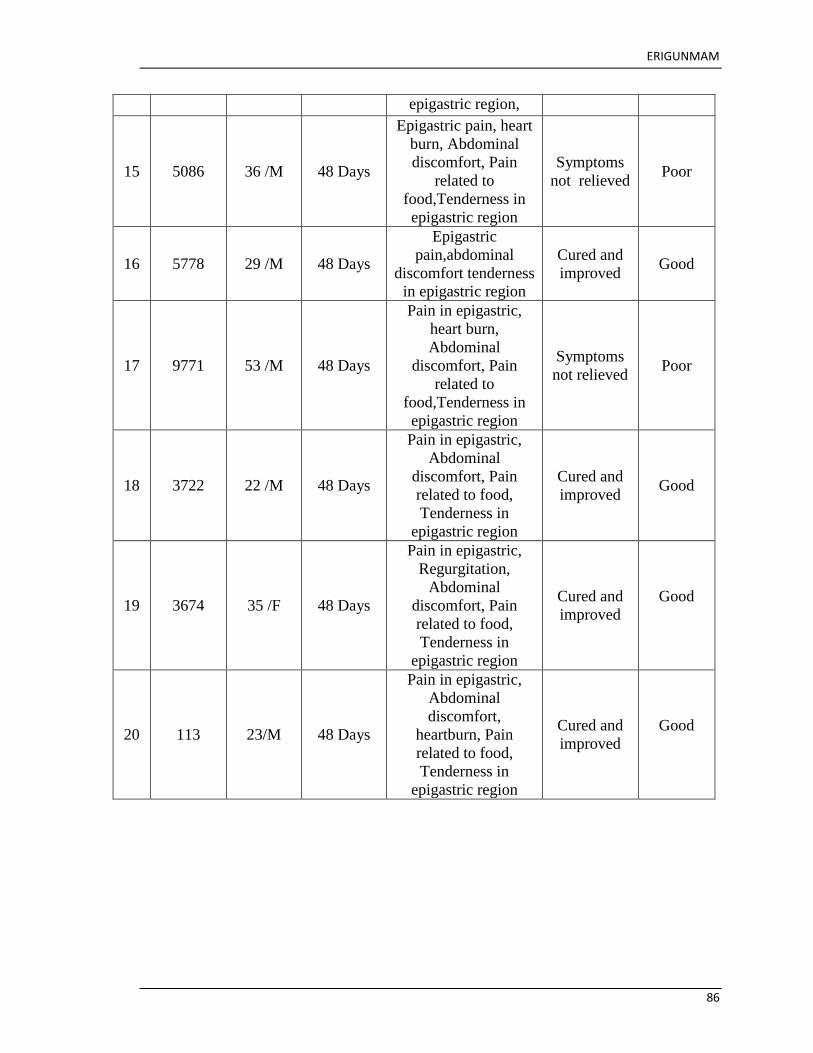
ERIGUNMAM
86
epigastric region,
15 5086 36 /M 48 Days
Epigastric pain, heart
burn, Abdominal
discomfort, Pain
related to
food,Tenderness in
epigastric region
Symptoms
not relieved Poor
16 5778 29 /M 48 Days
Epigastric
pain,abdominal
discomfort tenderness
in epigastric region
Cured and
improved Good
17 9771 53 /M 48 Days
Pain in epigastric,
heart burn,
Abdominal
discomfort, Pain
related to
food,Tenderness in
epigastric region
Symptoms
not relieved Poor
18 3722 22 /M 48 Days
Pain in epigastric,
Abdominal
discomfort, Pain
related to food,
Tenderness in
epigastric region
Cured and
improved Good
19 3674 35 /F 48 Days
Pain in epigastric,
Regurgitation,
Abdominal
discomfort, Pain
related to food,
Tenderness in
epigastric region
Cured and
improved
Good
20 113 23/M 48 Days
Pain in epigastric,
Abdominal
discomfort,
heartburn, Pain
related to food,
Tenderness in
epigastric region
Cured and
improved
Good

ERIGUNMAM
87
PATIENT REPORT
Before Treatment
Hospital: Anna Peripheral Hospital, Anna Nagar East, Chennai 102.
Patient OP.No.8370
Date: 20.7.2016
Age: 47
Sex: Male
Address:
No.197, Vinayagapuram,
Avadi,
Chennai.
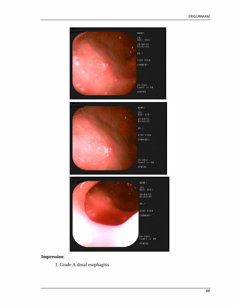
ERIGUNMAM
88
Impression:
1. Grade A distal esophagitis

ERIGUNMAM
89
2. Multiple tiny superficial clear based ulcer in anterior wall to first part of
duodenum.
PATIENT REPORT
AFTER TREATMENT
Hospital: Anna Peripheral Hospital, Anna Nagar East, Chennai 102.
Patient OP.No.8370
Date: 11-10-2016
Age: 47
Sex: Male
Address:
No.197,Vinayagapuram,
Avadi,
Chennai.

ERIGUNMAM
90
IMPRESSION:
Oesophagus, fundus, body and pylorus part of the stomach, Ist and IInd part of
duodenum is normal.

ERIGUNMAM
91
DISCUSSION

ERIGUNMAM
92
DISCUSSION In Siddha literature Gunmam are 8 in number. Eri Gunmam is one of the 8
types of Gunmam mentioned in Yugi Vaithiya Cinthamani 800.The term Gunmam is a
clinical entity which depresses both body and mind, since it is called as Gunmam. The
term ―PEPTIC ULCER‖ refers to an ulcer in the lower oesophagus, stomach or
duodenum, in the jejunum or rarely in the ileum. Ulcers in the stomach or duodenum may
be acute or chronic; both penetrate the muscularis mucosae but the acute ulcer shows no
evidence of fibrosis.
It is the most common ulcer of an area of gastrointestinal tract that is usually
acidic and thus extremely painful. H.pylori is one of the main causes, drugs such as
Aspirin, Ibuprufen, NSAIDs, Irregular food habits, eating spicy and junk foods, smoking,
stress also cause Peptic Ulcer.
The patients were examined based on Siddha and as well as modern aspects.
All the necessary investigations were made during the study. The results obtained from
their studies were discussed below for better conclusion.
Various Siddha Literatures had been studied and discussed for choosing the trial medicine
for treating Eri Gunmam and finally chosen Pirandai Vadagam which is mentioned in
THERAIYAR THARU.
Authentication is a critical step for successful and reliable clinical applications
and for further experimental studies on Siddha Drugs.
DRUG AUTHETICATION
Fresh specimens of Cissus quadrangularis, Zingiber officinalis were collected from a
farm and Taxus baccata, Terminalia chebula , Phyllanthus emblica, Terminalia bellerica
were collected from raw drugs shop and I have got authentication from the Botanist Dr. S.
SANKARANARAYANAN M.Sc., M.Phil., Ph.D., Dept, of Medicinal Botany, Govt.
Siddha Medical College, Arumbakkam, Chennai – 106.
PHSIOCHEMICAL ANALYSIS
The trial drug Pirandai vadagam had contains total ash -4.19, loss on
drying (at 105 oc )-8.53, water soluble ash – 2.48, acid insoluble ash – 0.07, water

ERIGUNMAM
93
soluble extractive – 38.15, alcohol soluble extractive – 33.2 and pH value (10%)- 3.5.
TLC /HPTLC was enclosed in Annexures.
TOXICITY STUDY
ACUTE TOXICITY
Acute and sub acute toxicity studies were conducted on experimental rats at
Sathyabama University, Chennai, Tamilnadu. Acute toxicity study of the drug Pirandai
Vadagam was carried out as the OECD guideline - 423 (Organisation to Econamic Co-
operation and Development).
The acute toxicity study of Pirandai Vadagam was studied and the drug was
proved safer for long term administration, as it did not exhibit any significant toxicity.
IAEC Approval NoSU/CLATR/IAEC/IV/019/2016.
SUB ACUTE TOXICITY
Sub acute toxicity study as per the guideline of – 407. Under the dosage of
Pirandai Vadagam 200mg / kg (Low dose), 400mg / kg (High dose), it did not exhibit any
significant. IAEC Approval No SU/CLATR/IAEC/IV/019/2016.
HISTO PATHOLOGY
At the end of toxicity studies the animal were sacrified and histopathology of
vital organs like Liver, Kidney, Spleen, Lungs were carried out. The studies haven‘t
shown the evidence of remarkable pathological lesions in the tissues.
PHARMACOLOGICAL ACTIVITY
The pharmacology studies of Pirandai Vadagam was carried out at
Sathyabama University, Chennai,Tamilnadu.
The trial drug Pirandai Vadagam had shown a potent Anti-Ulcer activity during the
studies. IAEC Approval NoSU/CLATR/IAEC/VII/046/2016.
BIOCHEMICAL ANALYSIS
Pirandai Vadagam contains Acid radicals such as Chloride, Phosphate and
Basic radicals such as Iron, Calcium and Potassium and Alkaloids.
ERIGUNMAM
94
IEC AND CTRI
The study was approved by Institutional Ethics Committee ( IEC ) and the
approval number is GSMC – CH – ME – 4 / 2015 / 003. It was registered in Clinical
Trials Registry – India ( CTRI ) and the Reference number is REF/ 2016 / 12 / 012978.
An open clinical study on ERI GUNMAM ( PEPTIC ULCER ) was carried out in the Post
Graduate Department of Pothu Maruthuvam in Govt.Siddha Medical College attached to
Arignar Anna Hospital of Indian Medicine, Chennai – 106 during the period of 2015 to
2017.
Population and sample
The population consists of all patients satisfying the inclusion and exclusion
criteria mentioned below. Sample consists of 20 ERI GUNMAM patients, who attended
the OPD of Arignar Anna Hospital, Arumbakkam, Chennai – 106.
CLINICAL STUDY
Clinical studies were conducted followed by CTRI registration with the sample
size of 20 patients. In my study,20 patients with Eri Gunmam were selected in the
Department of Maruthuvam, Government Siddha Medical College, attached to Arignar
Anna Govt. Hospital for Indian Medicine, Arumbakkam, Chennai – 106.
All the necessary investigations were carried out to all patients and trial drug
was given. Weekly once follow up was done. Total duration of treatment ranges from 48
days. All the patients were strictly advised to follow diet restriction and peaceful life style
to normalize the immune mechanism.
GENDER DISTRIBUTION
From selected 20 cases of study 70% (14) of cases were male and 30% (6)
were female.
70% of men were affected, so Peptic Ulcer is most commonly affected in
male.
AGE DISTRIBUTION
Out of 20 cases 7 patients (35%) were between 21 – 30 years, 5 patients
(25%) were between 31 – 40 years, 5 patients (25%) were between 41 – 50 years, 3
patients (15%) were between 51 – 60 years, High incidences of Peptic Ulcer were noted
in age ranging of 21 – 30 years during the studies.

ERIGUNMAM
95
SEASONAL INCIDENCE
According to Paruva kaalam highest incidence of 75% were noted in Munpani
kaalam and 15% cases were noted in Pinpani kaalam and 5% comes under Kaar kaalam
and Koothir kaalam.
When clinical trial of 20 cases were enquired about the seasonal link, most of
the cases were in Munpani Kaalam due to seasonal variation.
OCCUPATIONAL STATUS
From selected 20 cases, 9 patients (45%) were coolies, 6 patients (30%) were
office goers, 5 patients (25%) were homemakers. Stress and Strain were more common
among office goers, irregular food habits and intake of spicy junk food were common
among coolies and office goers which were the main causes of Eri Gunmam.
SOCIO ECONOMIC STATUS
Recording Socio Economic Status 12 patients (60) were low income and 5
cases (25%) from middle income and 3 cases (15%) from high income.
The people living in poor Socio Economic Status were more affected because
of life style and environmental factors.
DIET REFERENCE
Out of 20 cases, most of the cases 15 (75%) were taken mixed diet, and 5 cases
(25%) had vegetarian diet only.
Eri Gunmam is more prone to mixed diet intakers, because the mixed diet
contains of more spicy and fried foods.
THINAI DISTRIBUTION
According to the study, nearly 18 cases (90%) were from Neithal thinai and 2
cases (10%) from Marutham.

ERIGUNMAM
96
MUKKUTRAM
DISTRIBUTION OF VATHAM
According to classification of Vatham, derangement of Viyanan , Samanan
and Uthanan, 4 patients (20%) was affected with Abanan, 9 patients (45%) was affected
with Kirukaran, 4 patients (20%) was affected with devathatthan, 2 patients (10%) was
affected with Koorman and none affected with Pranan, Naagan and Thananjeyan.
Samanan and Uthanan are the main constituents in the digestion of food, affected
Samanan and Uthanan produce regurgitation of food
Affected Abanan produced diarrhoea.
Affected Koorman produced impairment of eyesight.
Affected Kirukaran produced loss of taste.
DISTRIBUTION OF PITHAM
According to Pitham 20 cases (100%) were affected derangement of Saathgam
and Analagam, 5cases (25%) was affected Ranjagam, and 2cases (15%) was affected with
Alosagam.
Affected analagam produced loss of appetite.
Affected Saathagam produced inability of doing regular works properly.
Affected Ranjagam produced pallor of skin, eye and reduced hemoglobin.
Affected Alosagam produced impairment of eye sight.
DISTTRIBUTION OF KABAM
According to the study, all cases (20) (100%) affected by Klethagam, 11 cases
(55%) affected by Pothagam and 3 cases (15%) affected by Santhigam.2 cases(10%) were
affected by Tharpagam.
Affected Klethagam produced loss of appetite.
Affected Pothagam produced loss of taste.
Affected Tharpagam caused impairment of eye sight
EZHU UDAL KATTUGAL
According to this, Saaram was affected in all the patients (100%), Oon was
affected in 2 cases (10%), Seneer were affected in 5 patients (25%). None affected with
Kolupu, Moolai and Sukkilam / Suronitham.

ERIGUNMAM
97
Affected Saaram results in Loss of appetite and tiredness.
Affected Seneer produced pallor of skin, eye and reduced hemoglobin.
Affected Oon produced pain and tenderness in epigastric region.
ENVAGAI THERVUGAL
According to Envagai thervugal, Naa was affected in 10 patients (50%),
Niram was affected in 5 patients (25%), Mozhi was affected in 12 patients (60%), Vizhi
was affected in 2 patients (10%), Sparisam was affected in 8 patients (40%), Malam was
affected in 4 patients (20%), Naadi was affected in for all the 20 patients(100%).
Naa was affected due to anaemia (pale colour).
Niram was affected due to anaemia (pale colour).
Vizhi was affected due to dullness of vision.
Mozhi was affected due to low pitched sound.
Sparisam was affected due to tenderness pain, mild temperature in
epigastric region.
Malam was affected due to constipation.
Naadi was affected in all patients.
NAADI
Out of 20 patients , 13 patients (65%) had Vatha Pitham. and 7 patients (35%) had
Pitha Vadham
NEIKURI REFERENCE
Out of 20 patients, 13(65%) patients had Vatha Neer, 7(35%) patients had
Pitha Neer.
ERI GUNMAM is a Vatha reflected disease. So most of the cases had Vatha neer,when a
drop of gingely oil was dropped into the early morning urine sample, it spreads like snake.
DURATION OF ILLNESS PRIOR TO TREATMENT
Out of 20 patients, 7 patients (35%) belongs to below 6 months to 1 year, 6
patients (30%) belongs to below 1 year to 3 year, 5 patients (25%) belongs to below 1
months to 6 months, 1 patients (55%) belongs to below 3 years to 5 years, and 1 patients
(55%) belongs to below 5 years and above.

ERIGUNMAM
98
CLINICAL MANIFESTATION
In respect of the patients with Peptic Ulcer, the clinical manifestation of
epigastric pain, pain related to food , abdominal discomfort, tenderness in epigastric
region were present in most of the cases.
Epigastric pain was present in 20cases (100%),
Heart burn was seen in 8cases (40%),
Abdominal discomfort was present in 15 cases (75%),
Tenderness in epigastric region was seen in 16 cases (80%),
Diarrhoea was seen in 4 patients (20%) ,
Regurgitation was present in 8cases (40%),
Loss of appetite was seen in 2 cases (10%) and
Pain related to food was present in 17 cases (85%)
CLINICAL PROGNOSIS
The clinical signs and symptoms were improved after treatment,
16 cases (80%) was cured from Epigastric pain,
7(35%)cases were improved from Heartburn,
13cases (65%) were cured from Abdominal discomfort,
15(75%) cases were cured from Pain related to food,
7(35%) cases were cured from Regurgitation,
14(70%) cases were cured from Tenderness in Epigastric region, loss of
weight and
diarrhoea were cured completely .
IMPROVEMENT
Among the total 20 patients all were improved. Clinical symptoms before and
after treatment were noted. To obtain prognosis of each clinical symptom, the following
formulae was used

ERIGUNMAM
99
Thus the clinical trial study showed significant clinical improvement in certain clinical
manifestation of ERI GUNMAM such as Epigastric pain, Abdominal discomfort,
Tenderness in Epigastric region, Pain related to food, Heart burn and Regurgitation . Loss
of appetite and Diarrhoea were cured completely.
INVESTIGATION
In Blood tests, TC, DC, ESR, Hb% serum creatinine, blood urea were
investigated.
URINE
Albumin, Sugar, Deposit were investigated.
SPECIAL INVESTICATION
Endoscopic Examination – Gastritis, Oesophagitis, and Duodenitis were
investigated.
BIO STATISTICAL STUDY
Since the p value is significant in all clinical manifestations , there is significant
reducing of clinical manifestations among the patients for the treatment of ERI
GUNMAM. Hence it is concluded that the treatment was effective and significant.
GRADING OF RESULTS
Out of the 20 cases, 15cases (75%) had Good result and 3(15%) had Fair result, 2
cases (10%) shown Poor result at the end of the treatment.
ERIGUNMAM
100
SUMMARY

ERIGUNMAM
101
SUMMARY
The clinical study on ERI GUNMAM was carried out in Post Graduate
Department of Pothu Maruthuvam, Government Siddha Medical College, Aringar Anna
Hospital, Chennai – 106 during the period of 2015-2017.
A total of 20 patients were treated in the Outpatient department. The clinical and
pathological assessment was carried out on the basis of Siddha and Modern aspects.
All the patients were treated with Pirandai Vadagam, 1gm, chewable, b.d daily,
after food for duration of 48days.
Males were mostly affected (70%).
Most of the patients were in the age group between 21-30 years (35%)
Most of the patients were from Neithal Thinai (90%).
The disease is more common in coolies (45%), so high incidence occurs in men.
Most of the patients were affected in Munpanikaalam (75%).
The diseases was more common in Pithavatham yakkai cases (65%).
In Vali, Abanan (20%), Viyanan (100%), Samanan (100%), Uthanan(100%)
Koorman (10%), Kirukaran(45%) and Devadhathan (20%) were affected.
In Azhal, Analagam (100%) Ranjagam (25%), Saathagam (100%), Alosagam
(10%), were affected.
In Iyyam, Kilethegam (100%), Pothagam(55%),Tharpagam(10%) and
Santhigam(15%) were affected.
In Ezhu udal kattugal, Saaram (100%), Seneer (25%), Oon (10%), and Enbu
(15%) were affected.
Regarding naadi, Vathapitha naadi (65%) was the most common naadi observed.
The Toxicological studies of the trial medicine reveal no toxicity.
The Pharmacological studies reveal that, the trial drug has Anti-Ulcer activity.
Bio- statistical analysis of the clinical trial reveals significant p value < 0.05 and ˂
0.01 and concluded that the treatment is effective and significant.
Regarding grading of the result, 15 patients (75%) have shown good improvement,
3 patients (15%) have shown fair improvement, 2 patients (10%) have shown poor
improvement.

ERIGUNMAM
102
CONCLUSION

ERIGUNMAM
103
CONCLUSION
ERI GUNMAM (Peptic Ulcer) is mainly due to the derangement of Pitha kutram.
The trial medicine, Pirandai Vadagam has predominant astringent taste, neutralizes
the increased Pitham, thereby it acts on ETHURURAI MARUTHUVAM.
The Pirandai Vadagam reveals no toxicity in the preclinical studies and hence
proved to be safe for human administration.
From the pre clinical pharmacological studies it is evident that, the trial medicine
has significant Anti-Ulcer activity and analgesic activity.
No contra indication was reported during the course of the treatment.
The trial medicine gave maximam relief from the symptoms of ERI GUNMAM.
Therefore the author concluded that, the trial medicine PIRANDAI
VADAGAM is an effective drug for ERI GUNMAM (Peptic Ulcer).

ERIGUNMAM
104
ANNEXURES

ERIGUNMAM
105

ERIGUNMAM
106
IAEC - TOXICOLOGICAL STUDY

ERIGUNMAM
107
Project Report on Toxicity Profiling of Pirandai Vadagam
ACUTE TOXICITY STUDY
Acute toxicity study of the study drug Pirandai Vadagam was carried out as per
OECD guideline (Organization for Economic Co-operation and Development)
Guideline-423.
Animal
Healthy adult Wistar albino rat weighing between 170-200 g were used for the
study. The animals were housed in poly propylene cages and were kept in well ventilated
with 100% fresh air by air handling unit (AHU). A 12 light / dark cycle were maintained
.Room temperature was maintained between 22 + 20
Cand relative humidity 50–65%.
They were provided with food (Sai feeds, Bangalore, India) and water ad libitum. All the
animals were acclimatized to the laboratory for 7 days prior to the start of the study.
The experimental protocol was approved by The Institutional Animal Ethics
Committee of Sathyabama University, Chennai, Tamil Nadu, India.
Acute toxicity Study
Acute toxicity study will be carried out in accordance with OECD guideline 423 1.
The animals were fasted overnight with free access to water. The study was conducted
with single oral dose administration of Pirandai Vadagam.
IAEC SU/CLATR/IAEC/IV/019/2016
Animal Grouping
One group consist of 6 female rats were used for this study. The dose utilized for
evaluation of acute toxicity study is about 2000 mg/kg higher than that of the therapeutic
dose.
Name Dr. G. Anitha Therese
IAEC SU/CLATR/IAEC/IV/019/2016
Name of the
Formulation Pirandai Vadagam
Abbreviation PV

ERIGUNMAM
108
Animal Grouping
GROUP I : Animals received Test drug 2000 mg/kg (p.o)
The animals were fasted overnight (12- 16 hrs) with free access to water. The
study was conducted with single oral administration of study drug Pirandai Vadagam
2000mg/kg (p.o). The animals were observed continuously for first 72 h and then 14 days
for emerging signs of behavioral changes, body weight changes and for mortality.
Occurrence of toxicity in animals were observed continuously for the first 4 to 24
h and observed periodically for the next 14 days. Observation includes the change in
skin, fur, eyes and mucus membrane. Appearance of C.N.S,C.V.S and A.N.S related
toxicity such as tremors, convulsions, sedation, steric behavior, respiratory distress,
cardiovascular collapse, response to sensory stimuli, salivation, diarrhea, lethargy, sleep,
coma and mortality were observed with special attention.
Body weight was recorded periodically. At the end of the experiment all animals
were subjected for gross necropsy and observed for pathological changes.
SUB-ACUTE TOXICITY STUDY
Sub-acute toxicity study was carried out as per OECD guidelines Guideline-407 2.
Animals
Healthy adult Wistar albino rat weighing between 170-200 g were used for the
study. The animals were housed in poly propylene cages and were kept in well ventilated
with 100% fresh air by air handling unit (AHU). A 12 light / dark cycle were maintained
.Room temperature was maintained between 22 + 2o
Cand relative humidity 50–65%.
They were provided with food (Sai feeds, Bangalore, India) and water ad libitum. All the
animals were acclimatized to the laboratory for 7 days prior to the start of the study.
The experimental protocol was approved by The Institutional Animal Ethics
Committee of Sathyabama University, Chennai, Tamil Nadu, India.
ERIGUNMAM
109
IAEC SU/CLATR/IAEC/IV/019/2016
Animal Grouping
Animals were divided into three groups of 06 animals each consist of 3 male and 3
female rats.
GROUP I : Animals received saline 5 ml/kg b.w (p.o)
GROUP II :Animals received low dose of test drug 200 mg/kg (p.o)
GROUP III :Animals received high dose of test drug 400 mg/kg (p.o)
The animals were randomly divided into control group and drug treated groups for
two different doses viz. low dose (200 mg/kg b.w) and high dose (400 mg/kg b.w).
The animals were administrated with the study drug once daily for 28 days. The
animals in group I (control group) received normal saline 5 ml/kg b.w. The animals in
group II received low dose of Pirandai Vadagam 200 mg/kg b.w (p.o) and group III
received high dose of Pirandai Vadagam 400 mg/kg b.w (p.o).
The rats were weighed periodically and observed for signs of toxicity pertains to
C.N.S, C.V.S, A.N.S including behavioral changes, food - water intake and morphological
changes. At the end of 28th
day, the animals were fasted for overnight with free access to
water. On 29th
day the animals were sacrificed with excess anesthesia. Blood samples
were collected from aorta and stored in EDTA (ethylenediamine –tetra actate) for
Hematological analysis and for serum generation for biochemical analysis.
The vital organs including heart, brain, lungs, spleen, kidneys, liver, stomach,
testes, and ovary were harvested and carefully examined for gross lesions. The organs
were preserved in 10% formalin for histopathological assessment and interpretation.
Hematological analysis
Blood samples were analyzed using established procedures and automated Bayer
Hematology analyzer. Parameters evaluated include Packed Cell Volume (PCV), Red
Blood Cells (RBC) count, White blood cell count (WBC), Platelet Count, Hemoglobin
(Hb), Mean cell Haemoglobin Concentration (MCHC), Mean Red Cell Volume (MCV),
Mean Cell Hemoglobin (MCH), Mean platelet volume (MPV), Neutrophils, Eosinophil‘s,
Basophils, Lymphocytes and Monocytes.
Biochemical analysis3

ERIGUNMAM
110
Serum samples were analyzed for High Density Lipoprotein (HDL), Low density
Lipoprotein (LDL) , Very low density Lipoprotein (VLDL) , Triglycerides (TGL), Total
Cholesterol , Blood urea nitrogen (BUN), Creatinine, Albumin, Total Protein, Glucose,
Uric acid, Aspartate Transaminase (AST), Alanine amino Transaminase (ALT) and
Alkaline Phosphatase (ALP) using Mind ray auto analyzer model BS 120.
Histopathological evaluation4
Organs included of heart, brain, lungs, spleen, kidneys, liver, stomach, testes and
ovary. Histological slides of organs were made and observed under the microscope. The
pathological observations of cross section of these organs were performed on gross and
microscopic bases. Histological examinations were performed on the preserved tissues
with particular emphasis on those which showed gross pathological changes.
Statistical analysis
The statistical analysis was carried by one way ANOVA (GRAPH PAD PRISM 5
computer program). Results were expressed as mean ± standard error .A statistical
comparison was carried out using the Dunnet‘s test for the control and treatment group.
Fecal Pellet Analysis
Methodology
Rats of control and treatment group were allowed to explore to open field on clean and
sterile cage with blotting paper. The collected pellets were analyzed for consistency,
color, Shape, Presence of blood cells etc
Acute Toxicity Study
Analysis Group I

ERIGUNMAM
111
Consistency Soft
Shape Oblong
Colour Dark Green
Mucous Shedding Absence
Blood Cells Absent
Signs of Infection None Observed
Sub-Acute Toxicity Study
Analysis Group I Group II Group III
Consistency Soft Soft Soft
Shape Oblong Round headed Round headed
Colour Brownish green Pale green Pale green
Mucous Shedding Absence Absence Absence
Blood Cells Absent Absent Absent
Signs of Infection None Observed None Observed None Observed
Muscle Grip Strength Analysis
Methodology
The grip strength test is a simple non-invasive method designed to evaluate rat muscle
force in vivo. Rats of control and drug treated group was allowed to hold the pull bar with
both the hind limbs firmly then the animal was gently pulled back with the tail until the
animal lost the grip toward the bar. The procedure was repeated to get the average value.
Muscle gripness of the drug treated group was compared to that of the control rat to
ensure the change in coordination.
Metabolic Cage for Urine Collection
Rat of control and treatment group was placed individually in metabolic cage with free
access to feed and water. Urine dropping from the animal was collected using specialized
wire mesh system fixed at the base of the cage having provision to trap the fecal pellet
mixed with urine sample. The collected urine sample was subjected to analysis with
respect to colour, pH, glucose, ketone bodies, pus and blood cells.
RESULTS Assessment of clinical signs in rats treated with Pirandai Vadagam on Acute toxicity
study
Parameter Group I
Clinical Signs Parameters for the
duration of 14 days Test Drug 2000mg/ Kg
Number of animals observed 6 Female

ERIGUNMAM
112
Lacrimation Absence
Salivation Absence
Animal appearance Normal
Tonic Movement Absence
Clonic Movement Absence
Laxative action Mild
Touch Response Normal
Response to Sound Normal Response
Response to Light Normal Response
Mobility Normal Response
Respiratory Distress Nil
Skin Color Normal
Stereotype behavior Absence
Piloerection Absence
Limb Paralysis Absence
Posture Normal
Open field behavior Normal
Gait Balancing Normal
Freezing Behaviour Absent
Sings of Stress and Anxiety None Observed
Muscular coordination Normal
Muscle grip Normal
Sedation Absence
Social Behavior Normal
Urine Analysis No Abnormality
Urine Colour Yellowish
Urine Ph 7
Urine -Glucose Absence
Urine -Ketones Absence
Urine- Bilirubin Absence
Urine-Blood Cells Negative
Urine - Pus cells Negative
Mortality Nil
Quantitative data on the body weight of rats treated with Pirandai Vadagam in

ERIGUNMAM
113
Acute toxicity study
Group I
Before Treatment
Weight in Gms
After Treatment
Weight in Gms
Mean 184.5 191
Std. Deviation 7.609 6.229
Std. Error 3.106 2.543
Values are mean ± S.D (n = 6 per group). Control and treatment group were compared
statistically using one way ANOVA followed by Dunnett‘s test.
Assessment of clinical signs in rats treated with Pirandai Vadagam on Sub-Acute
toxicity study
Parameter Group I Group II Group III
Clinical Signs Parameters
for the duration of 28
days Control
Test Drug
200mg/ Kg
Test Drug
400mg/ Kg
Number of animals
observed
3 Male and 3
Female
3 Male and
3 Female
3 Male and
3 Female
Lacrimation Absence Absence Absence
Salivation Absence Absence Absence
Animal appearance Normal Normal Normal
Tonic Movement Absence Absence Absence
Clonic Movement Absence Absence Absence
Laxative action Absence Absence Absence
Touch Response Normal Normal Normal
Response to Sound
Normal
Response Normal
Response
Normal
Response
Response to Light
Normal
Response Normal
Response
Normal
Response
Mobility Normal Normal Normal
Respiratory Distress Nil Nil Nil
Skin Color Normal Normal Normal
Stereotype behavior Absence Absence Absence
Piloerection Absence Absence Absence
Limb Paralysis Absence Absence Absence
Posture Normal Normal Normal
Open field behavior Normal Normal Normal

ERIGUNMAM
114
Gait Balancing Normal Normal Normal
Freezing Behaviour Absent Absent Absent
Sings of Stress and
Anxiety
None
Observed None
Observed
None
Observed
Muscular coordination Normal Normal Normal
Muscle grip Normal Normal Normal
Sedation Absence Absence Absence
Social Behavior Normal Normal Normal
Urine Analysis
No
Abnormality No
Abnormality
No
Abnormality
Urine Colour Yellowish Yellowish Yellowish
Urine pH 7 7 6
Urine -
Glucose Absence Absence Absence
Urine -
Ketones Absence Absence Absence
Urine-
Bilirubin Absence Absence Absence
Urine-Blood Cells Negative Negative Negative
Urine - Pus cells Negative Negative Negative
Mortality Nil Nil Nil
Effect of Pirandai Vadagam on Body weight of Rats in Sub-acute toxicity study
Group I
Before Treatment Weight
in Gms
After Treatment Weight
in Gms
Mean 187.3 198.7
Std. Deviation 5.645 6.408
Std. Error 2.305 2.616
Group II
Before Treatment Weight
in Gms
After Treatment Weight
in Gms
Mean 181.2 191
Std. Deviation 7.083 6.229
Std. Error 2.892 2.543
Group III Before Treatment
After Treatment Weight
in Gms
Mean 179.8 189.8
Std. Deviation 7.25 6.676
Std. Error 2.96 2.725
Values are mean ± S.D (n = 6 per group of which 3 males and 3 females). Control and
treatment groups were compared statistically using one way ANOVA followed by
Dunnett‘s test.

ERIGUNMAM
115
Quantitative data on the food and water intake of rats treated with Pirandai
Vadagam for 28 days in Sub-acute toxicity study
GROUP I Food intake Water intake
Mean 19.25 41.25
Std. Deviation 3.072 1.371
Std. Error 1.536 0.6855
GROUP II Food intake Water intake
Mean 16 35.58
Std. Deviation 3.485 2.713
Std. Error 1.743 1.357
GROUP III Food intake Water intake
Mean 17.75 31.58
Std. Deviation 1.664 2.201
Std. Error 0.8319 1.1
Values are mean ± S.D (n = 6 per group of which 3 males and 3 females). Control and
treatment groups were compared statistically using one way ANOVA followed by
Dunnett‘s test.
Effect of Pirandai Vadagam on Haematology profile of rats in sub-acute toxicity
study
GROUP I
WBC
count
(×103
µl)
RBC
(×10 6
µl)
PLT
(×10 3
µl)
MCV
(fl)
MCH
(pg)
MCHC
(g/dl)
HGB
(g/dl)
Mean 13.5 7.217 684.3 63.22 19.37 31.47 11.88
Std.
Deviation 1.536 1.559 90.86 4.269 3.838 2.083 2.109
Std. Error 0.6272 0.6364 37.09 1.743 1.567 0.8504 0.8612
GROUP II
WBC
count
(×103
µl)
RBC
(×10 6
µl)
PLT
(×10 3
µl)
MCV
(fl)
MCH
(pg)
MCHC
(g/dl)
HGB
(g/dl)
Mean 10.65 6.717 791.8 66.7 21.15 33.02 11.63

ERIGUNMAM
116
Std.
Deviation 2.07 1.141 229.6 3.286 1.733 1.699 1.398
Std. Error 0.8449 0.4658 93.73 1.342 0.7075 0.6935 0.5708
GROUP
III
WBC
count
(×103
µl)
RBC
(×10 6
µl)
PLT
(×10 3
µl)
MCV
(fl)
MCH
(pg)
MCHC
(g/dl)
HGB
(g/dl)
Mean 13.68 6.367 900.3 64.98 21.05 32.8 12.95
Std.
Deviation 1.938 0.709 90.77 2.498 2.382 1.595 1.314
Std. Error 0.7914 0.2894 37.06 1.02 0.9725 0.6512 0.5365
Values are mean ± S.D (n = 6 per group of which 3 males and 3 females). Control and
treatment groups were compared statistically using one way ANOVA followed by
Dunnett‘s test.
Effect of Pirandai Vadagam on Haematology profile of rats in sub-acute toxicity
study.
GROUP
I
Lymph
(%)
Mon
(%)
Neutrophils
(X
103/mm
3) Eosinophils
(%)
Basophils
(%)
MPV
(fl)
Mean 70.74 2.1 2.533 1.3 0.5 6.267
Std.
Deviation 6.62 0.7874 1.001 0.2828 0.5477 1.283
Std. Error 2.702 0.3215 0.4088 0.1155 0.2236 0.5239
GROUP
II
Lymph
(%)
Mon
(%)
Neutrophils
(X
103/mm
3) Eosinophils
(%)
Basophils
(%)
MPV
(fl)
Mean 74.28 3.083 1.75 1.417 0.1667 5.65
Std.
Deviation 5.849 1.32 0.7662 0.3971 0.4082 1.065
Std. Error 2.388 0.5388 0.3128 0.1621 0.1667 0.4349
GROUP
III
Lymph
(%)
Mon
(%)
Neutrophils
(X
103/mm
3) Eosinophils
(%)
Basophils
(%)
MPV
(fl)
Mean 81.35 4.233 2.017 1.5 0.5 6.067
Std.
Deviation 5.833 1.692 0.4622 0.3521 0.5477 1.16
Std. Error 2.381 0.6907 0.1887 0.1438 0.2236 0.4738
Values are mean ± S.D (n = 6 per group of which 3 males and 3 females). Control and
treatment groups were compared statistically using one way ANOVA followed by
Dunnett‘s test.

ERIGUNMAM
117
Effect of Pirandai Vadagam on Serum Bio-chemistry profile of rats in sub-acute
toxicity study
GROU
P I
Blood
sugar ®
(mg/dl)
BUN
(mg/dl)
Serum
creatini
ne
(mg/dl)
Serum
total
choleste
rol
(mg/dl)
Serum
triglyce
rides
level
(mg/dl)
Serum
HDL
choleste
rol
(mg/dl)
Serum
LDL
choleste
rol
(mg/dl)
Serum
VLDL
choleste
rol
(mg/dl)
Mean 86.17 11.17 0.6167 117.2 80.67 68.67 29.67 16.82
Std.
Deviatio
n 8.931 3.251 0.2994 16.98 11.93 12.45 17.75 1.707
Std.
Error 3.646 1.327 0.1222 6.93 4.869 5.084 7.246 0.6969
GROU
P II
Blood
sugar ®
(mg/dl)
BUN
(mg/dl)
Serum
creatini
ne
(mg/dl)
Serum
total
choleste
rol
(mg/dl)
Serum
triglyce
rides
level
(mg/dl)
Serum
HDL
choleste
rol
(mg/dl)
Serum
LDL
choleste
rol
(mg/dl)
Serum
VLDL
choleste
rol
(mg/dl)
Mean 89.83 19 0.8167 102.2 86.33 58.33 37.67 15.22
Std.
Deviatio
n 10.38 2.757 0.2858 14.51 15.97 13.53 10.65 2.106
Std.
Error 4.238 1.125 0.1167 5.924 6.52 5.524 4.349 0.8596
GROU
P III
Blood
sugar ®
(mg/dl)
BUN
(mg/dl)
Serum
creatini
ne
(mg/dl)
Serum
total
choleste
rol
(mg/dl)
Serum
triglyce
rides
level
(mg/dl)
Serum
HDL
choleste
rol
(mg/dl)
Serum
LDL
choleste
rol
(mg/dl)
Serum
VLDL
choleste
rol
(mg/dl)
Mean 71.5 16.67 0.6833 122.3 95.17 56 30.5 14.67
Std.
Deviatio
n 11.47 6.218 0.2714 9.953 7.026 11.88 7.609 2.149
Std.
Error 4.682 2.539 0.1108 4.063 2.868 4.851 3.106 0.8774
Values are mean ± S.D (n = 6 per group of which 3 males and 3 females). Control and
treatment groups were compared statistically using one way ANOVA followed by
Dunnett‘s test.
Effect of Pirandai Vadagam on Serum Bio-chemistry profile of rats in sub-acute
toxicity study
GROUP I
Serum
total
protein
(g/dl)
Serum
albumin
(g/dl) (AST)
(IU/ml)
(ALT)
(IU/L)
(ALP)
(IU/L)
Mean 4.917 3.717 99.5 32.83 247
Std.
Deviation 2.043 1.08 22.51 10.57 20.91
Std. Error 0.834 0.4408 9.19 4.316 8.536

ERIGUNMAM
118
GROUP II
Serum
total
protein
(g/dl)
Serum
albumin
(g/dl) (AST)
(IU/ml)
(ALT)
(IU/L)
(ALP)
(IU/L)
Mean 4.117 2.883 97.33 39.67 217.3
Std.
Deviation 0.5636 0.6242 24.67 5.61 14.72
Std. Error 0.2301 0.2548 10.07 2.29 6.009
GROUP III
Serum
total
protein
(g/dl)
Serum
albumin
(g/dl) (AST)
(IU/ml)
(ALT)
(IU/L)
(ALP)
(IU/L)
Mean 5.183 3.333 129.8 22.33 199.2
Std.
Deviation 1.028 0.9585 10.21 4.179 43.43
Std. Error 0.4199 0.3913 4.167 1.706 17.73
Values are mean ± S.D (n = 6 per group of which 3 males and 3 females). Control and
treatment groups were compared statistically using one way ANOVA followed by
Dunnett‘s test.
Organ Gross Observation of rats treated with Pirandai Vadagam for 28 days in Sub-
acute toxicity study.
Treatment Female

ERIGUNMAM
119
Histopathology of Brain (Female Rat) in Sub-acute toxicity Study
Low Power Magnification 10X
GROUP I GROUP II GROUP III
High Power Magnification 40X
GROUP I GROUP II GROUP III
Histopathology of Kidney (Female Rat) in Sub-acute toxicity Study
Low Power Magnification 10X
GROUP I GROUP II GROUP III
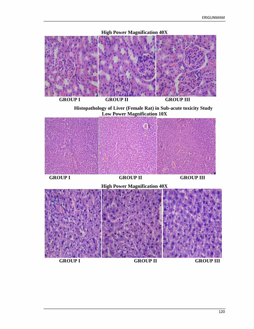
ERIGUNMAM
120
High Power Magnification 40X
GROUP I GROUP II GROUP III
Histopathology of Liver (Female Rat) in Sub-acute toxicity Study
Low Power Magnification 10X
GROUP I GROUP II GROUP III
High Power Magnification 40X
GROUP I GROUP II GROUP III

ERIGUNMAM
121
Histopathology of Lung (Female Rat) in Sub-acute toxicity Study
Low Power Magnification 10X
GROUP I GROUP II GROUP III
High Power Magnification 40X
GROUP I GROUP II GROUP III
Histopathology of Spleen (Female Rat) in Sub-acute toxicity Study
Low Power Magnification 10X
GROUP I GROUP II GROUP III

ERIGUNMAM
122
High Power Magnification 40X
GROUP I GROUP II GROUP III
Histopathology of Stomach (Female Rat) in Sub-acute toxicity Study
Low Power Magnification 10X
GROUP I GROUP II GROUP III

ERIGUNMAM
123
High Power Magnification 40X
GROUP I GROUP II GROUP III
Histopathology of Uterus (Female Rat) in Sub-acute toxicity Study
Low Power Magnification 10X
GROUP I GROUP II GROUP III
High Power Magnification 40X
GROUP I GROUP II GROUP III
Histopathology of Ovary (Female Rat) in Sub-acute toxicity Study
Low Power Magnification 10X

ERIGUNMAM
124
GROUP I GROUP II GROUP III
High Power Magnification 40X
GROUP I GROUP II GROUP III
Treatment Male
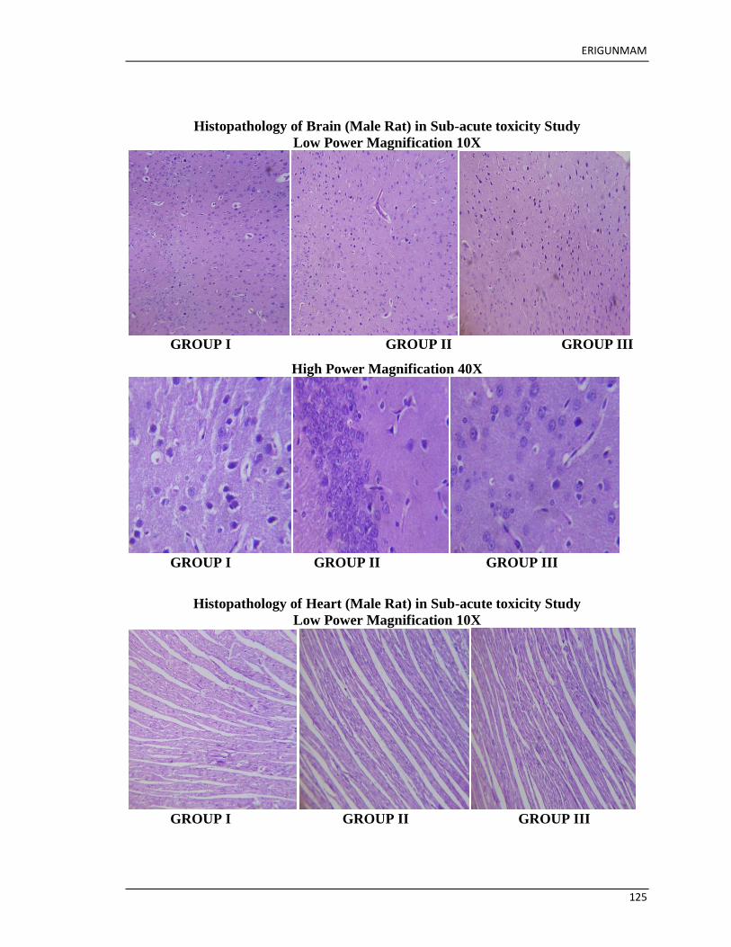
ERIGUNMAM
125
Histopathology of Brain (Male Rat) in Sub-acute toxicity Study
Low Power Magnification 10X
GROUP I GROUP II GROUP III
High Power Magnification 40X
GROUP I GROUP II GROUP III
Histopathology of Heart (Male Rat) in Sub-acute toxicity Study
Low Power Magnification 10X
GROUP I GROUP II GROUP III

ERIGUNMAM
126
High Power Magnification 40X
GROUP I GROUP II GROUP III
Histopathology of Kidney (Male Rat) in Sub-acute toxicity Study
Low Power Magnification 10X
GROUP I GROUP II GROUP III
High Power Magnification 40X
GROUP I GROUP II GROUP III

ERIGUNMAM
127
Histopathology of Liver (Male Rat) in Sub-acute toxicity Study
Low Power Magnification 10X
GROUP I GROUP II GROUP III
High Power Magnification 40X
GROUP I GROUP II GROUP III
Histopathology of Lung (Male Rat) in Sub-acute toxicity Study
Low Power Magnification 10X

ERIGUNMAM
128
GROUP I GROUP II GROUP III
High Power Magnification 40X
GROUP I GROUP II GROUP III
Histopathology of Stomach (Male Rat) in Sub-acute toxicity Study
Low Power Magnification 10X
GROUP I GROUP II GROUP III

ERIGUNMAM
129
High Power Magnification 40X
GROUP I GROUP II GROUP III
Histopathology of Spleen (Male Rat) in Sub-acute toxicity Study
Low Power Magnification 10X
GROUP I GROUP II GROUP III

ERIGUNMAM
130
High Power Magnification 40X
GROUP I GROUP II GROUP III
Histopathology of Testes (Male Rat) in Sub-acute toxicity Study
Low Power Magnification 10X
GROUP I GROUP II GROUP III

ERIGUNMAM
131
High Power Magnification 40X
GROUP I GROUP II GROUP III
Quantitative data on absolute organ weight of rats treated with Pirandai Vadagam
for 28 days in Sub-acute toxicity study.
GROU
P I
HEAR
T
(gms)
LIVE
R
(gms)
KIDN
EYS
(gms)
SPLE
EN
(gms)
BRAI
N
(gms)
LUNG
(gms)
STOM
ACH
(gms)
TEST
ES
(gms)
UTER
US &
OVAR
Y
(gms)
Mean 0.5417 6.62 1.507 0.4 1.667 1.633 1.183 2.533 1.133
Std.
Deviati
on
0.0526
9 0.7811 0.1987 0.1265 0.1751 0.1633 0.2639 0.9074 0.4041
Std.
Error
0.0215
1 0.3189 0.0811
0.0516
4
0.0714
9
0.0666
7 0.1078 0.5239 0.2333
GROU
P II
HEAR
T
(gms)
LIVE
R
(gms)
KIDN
EYS
(gms)
SPLE
EN
(gms)
BRAI
N
(gms)
LUNG
(gms)
STOM
ACH
(gms)
TEST
ES
(gms)
UTER
US &
OVAR
Y
(gms)
Mean 0.73 6.765 1.598 0.6167 1.683 1.567 1.233 3.467 1.467
Std.
Deviati
on 0.1982 0.7213 0.2169 0.2787 0.1602 0.2582 0.2503 0.7638
0.0577
4
Std.
Error
0.0809
1 0.2945
0.0885
6 0.1138 0.0654 0.1054 0.1022 0.441
0.0333
3
GROU
P III
HEAR
T
(gms)
LIVE
R
(gms)
KIDN
EYS
(gms)
SPLE
EN
(gms)
BRAI
N
(gms)
LUNG
(gms)
STOM
ACH
(gms)
TEST
ES
(gms)
UTER
US &
OVAR
Y
(gms)
Mean 0.6717 6.313 1.493 0.6 1.7 1.617 1.283 2.833 1.333
Std.
Deviati
on
0.0793
5 1.016 0.2999 0.1673 0.1897 0.1169 0.3656 0.5508 0.1528

ERIGUNMAM
132
Std.
Error 0.0324 0.415 0.1224
0.0683
1
0.0774
6
0.0477
3 0.1493 0.318
0.0881
9
Values are mean ± S.D (n = 6 per group of which 3 males and 3 females) for Heart, Liver,
Kidney, Brain, Spleen, Lung, Stomach. Values are mean ± S.D (n = 3 per group per sex )
for testes , ovary and uterus for Control and treatment groups were compared statistically
using one way ANOVA followed by Dunnett‘s test.
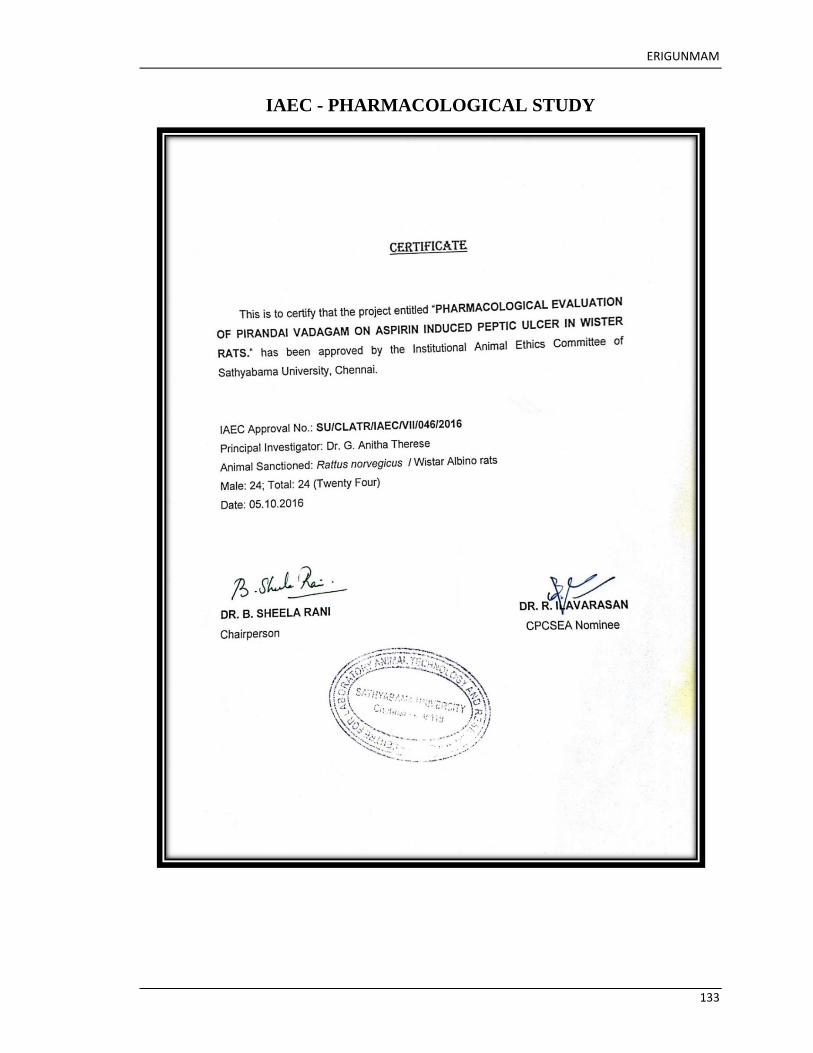
ERIGUNMAM
133
IAEC - PHARMACOLOGICAL STUDY

ERIGUNMAM
134
Pharmacological Evaluation of Pirandai Vadagam on Aspirin induced gastric
ulcer in wistar rats
Name: Dr. G. ANITHA THERESE
IAEC: SU/CLATR/IEAC/VII/046/2016
Animals
Healthy adult Wistar albino male rats weighing between 200-220 g were used for
the study. The animals were housed in poly propylene cages and were kept in well
ventilated with 100% fresh air by air handling unit . A 12 light / dark cycle were
maintained .Room temperature was maintained between 22 + 2o
Cand relative humidity
50–65%. They were provided with food (Sai feeds, Bangalore, India) and water ad
libitum. All the animals were acclimatized to the laboratory for 7 days prior to the start of
the study.The experimental protocol was approved by The Institutional Animal Ethics
Committee of Sathyabama University, Chennai, Tamil Nadu, India.
IAEC: SU/CLATR/IEAC/VII/046/2016
Experimental Methodology
The animals were grouped into four groups of 6 animals each. Group I (Control
group) -received normal saline, Group II – Ulcer control rats received 200mg/kg of
Aspirin ,p.o for the period of 7 days (Day 1 to 7). Group III (Low dose treated group):
Aspirin ulcerated rats was treated with 200mg/kg of PirandaiVadagam,p.o for the period
of 07 days 1 hr prior to the administration of aspirin.Group IV (High dose treated group):
Aspirin ulcerated rats was treated with 400mg/kg of PirandaiVadagam,p.ofor the period
of 07 days 1 hr prior to the administration of aspirin.
Sample Collection
At the end of the study, before sacrifice, the animals were fasted for overnight with free
access to water. Animals were sacrificed with excess anesthesia.The stomach was
removed and opened along the greater curvature. The stomach was gently rinsed with
water to remove the gastric contents and blood clots. The inner surface of free stomach
was examined for gastric lesions. The number of ulcers was counted. Ulcer scoring was
carried out according to the method by as given below 32
.

ERIGUNMAM
135
The scores were:
0 = no ulcer
1 = superficial ulcer
2 = deep ulcer
3 = perforation
Ulcer score
Ulcer index was measured by using the following formula
UI = UN + US + UP × 10−1
.
Where UI is the ulcer index; UN is the average number of ulcers per animal; US is the
average number of severity score and UP is the percentage of animals with ulcers.
Percentage inhibition of ulceration
Percentage inhibition of ulceration was calculated as follows:
× 100
There was a low percentage of ulcer in the study drug treated animals.
Reference
1. SantinJR.Antiulcer effects of Achyroclinesatureoides (Lam.) DC (Asteraceae)
(Marcela), a folk medicine plant, in different experimental models‖, Journal of
Ethnopharmacology.2010; 130:334-341.
Effect of PirandaiVadagam on Ulcer severity score of Aspirin ulcerated rats
GROUP I Ulcer Severity Score
Mean 0
Std. Deviation 0
Std. Error 0
GROUP II Ulcer Severity Score

ERIGUNMAM
136
Mean 2.667
Std. Deviation 0.5164
Std. Error 0.2108
GROUP III Ulcer Severity Score
Mean 1.167
Std. Deviation 0.9832
Std. Error 0.4014
GROUP IV Ulcer Severity Score
Mean 1
Std. Deviation 1.265
Std. Error 0.5164
Effect of PirandaiVadagam on Ulcer Index of Aspirin ulcerated rats
Group Treatment and Dose
Aspirin Induced Ulcer
Ulcer Index
Percentage of
Ulcer
Protection
I Normal Saline - 100
II 200mg/kg of Aspirin 11.15 ± 0.08 -
III
Aspirin + 200mg/kg of
PirandaiVadagam 4.53 ± 0.41 58.47
IV
Aspirin + 400mg/kg of
PirandaiVadagam 3.38 ± 0.42 66.63
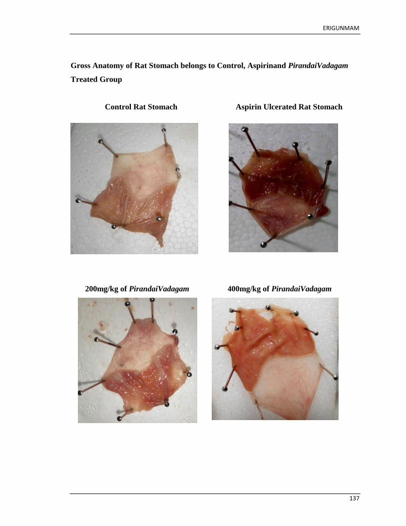
ERIGUNMAM
137
Gross Anatomy of Rat Stomach belongs to Control, Aspirinand PirandaiVadagam
Treated Group
Control Rat Stomach Aspirin Ulcerated Rat Stomach
200mg/kg of PirandaiVadagam 400mg/kg of PirandaiVadagam

ERIGUNMAM
138
Effect of Pirandai vadagam and standard drug on Aspirin induced Peptic ulcer in
rats
Data
are
expr
esse
d as
mea
n ±
SE
M
(n=6). Statistical analysis by One way ANOVA followed by Dunnett‘s multiple
comparison test * P<0.05, ** P<0.01, *** P<0.001 compared to control.
Treatment Dose Aspirin Induced
Ulcer Index
Aspirin % of
Ulcer Protection
Control 2ml/kg 4.61±0.31 -
Standard
(Rantidine)
27 mg/kg 2.31±0.35 *** 89.56 *
Pirandai
vadagam
200mg/kg 1.78±1.27 *** 29.38 ***
Pirandai
vadagam
400mg/kg 2.66±1.56 ** 38.17 ***

ERIGUNMAM
139
Histology of Rat Stomach (H&E Staining)
Low Power Magnification 10 X
Control Group Aspirin Ulcerated group
200mg/kg of PirandaiVadagam 400mg/kg of PirandaiVadagam

ERIGUNMAM
140
Histology of Rat Stomach (H&E Staining)
High Power Magnification 40 X
Control Group Aspirin Ulcerated group
200mg/kg of PirandaiVadagam 400mg/kg of PirandaiVadagam
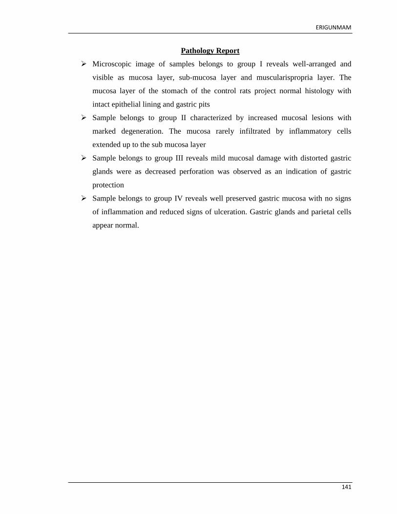
ERIGUNMAM
141
Pathology Report
Microscopic image of samples belongs to group I reveals well-arranged and
visible as mucosa layer, sub-mucosa layer and muscularispropria layer. The
mucosa layer of the stomach of the control rats project normal histology with
intact epithelial lining and gastric pits
Sample belongs to group II characterized by increased mucosal lesions with
marked degeneration. The mucosa rarely infiltrated by inflammatory cells
extended up to the sub mucosa layer
Sample belongs to group III reveals mild mucosal damage with distorted gastric
glands were as decreased perforation was observed as an indication of gastric
protection
Sample belongs to group IV reveals well preserved gastric mucosa with no signs
of inflammation and reduced signs of ulceration. Gastric glands and parietal cells
appear normal.

ERIGUNMAM
142
PHYSICO CHEMICAL ANALYSIS

ERIGUNMAM
143
Sample Name: Pirandai Vadagam
Stationary Phase - Silica Gel 60 F254
Mobile Phase - Toulene : Ethyl Acetate : (5:1 v/v)
= 254 nm
= 366 nm
= 575 nm
(Derivatized)
Color Rf value(s) Color Rf value(s) Color Rf value(s)
Green 0.04 Maroon 0.05 Ash 0.02
Green 0.11 Red 0.09 Ash 0.06
Green 0.14 Maroon 0.15 Ash 0.09
Green 0.21 Blue 0.22 Light
Yellow
0.13
Green 0.34 Maroon 0.33 Light Blue 0.20
Green 0.54 Maroon 0.40 Light Blue 0.32
Green 0.61 Red 0.50 Light Blue 0.42
Green 0.66 Red 0.55 Light Blue 0.54
Sky Blue 0.58 Ash 0.66
Maroon 0.62 Ash 0.83
Red 0.66 Blue 0.97
Red 0.69
Blue 0.84

ERIGUNMAM
144
HPTLC Chromatogram @ 254 nm:
Peak Table @ 254 nm:
3D Chromatogram @ 254 nm:

ERIGUNMAM
145

ERIGUNMAM
146
BIO-CHEMICAL ANALYSIS OF TRIAL MEDICINE Preparation of Sodium Carbonate extract
2 gm of the sample drug is mixed 5 gm of Sodium carbonate and taken in a 100 ml
beaker and 20 ml of distilled water is added. The solution is boiled for 10 minutes, cooled
and then filtered. The filtrate is called sodium carbonate extract.
S.No EXPERIMENT OBSERVATION INFERENCE
I TEST FOR ACID RADICALS
1a Test for Sulphate
2 ml of the above prepared
extractis taken in a test tube. To
this add2ml of 4% Ammonium
oxalatesolution.
Absence of
White Precipitate
Absent
B 2ml of extract is added with
2mlof dilute hydrochloric acid
untilthe effervescence ceases
off. Then2ml barium chloride
solution isadded.
Absence of
White Precipitate
Absent
2 Test for Chloride:
2ml of extract is added with
dilutenitric acid till the
effervescenceceases. Then 2ml
of silver nitratesolution is
added.
white precipitate is
Obtained
Present
3 Test for Phosphate
2ml of the extract is treated
with 2 ml of Ammonium
molybdate solution and 2ml of
concentrated nitric acid.
Presence of Yellow
precipitate.
Present
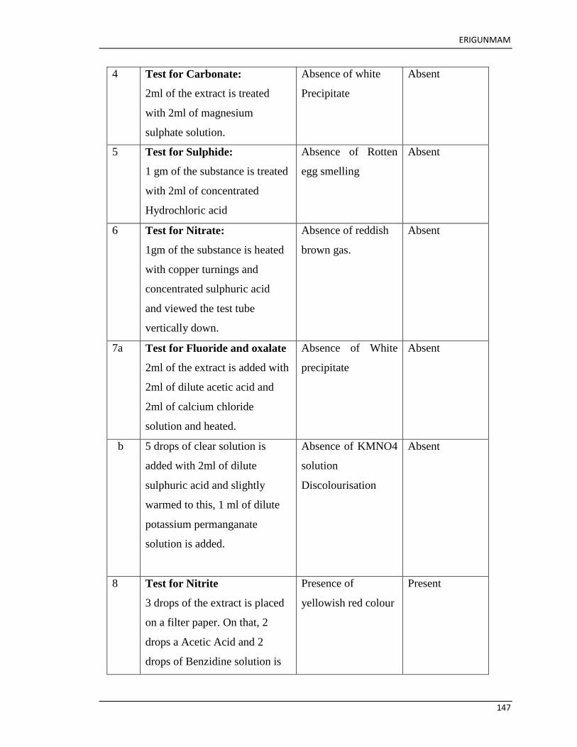
ERIGUNMAM
147
4 Test for Carbonate:
2ml of the extract is treated
with 2ml of magnesium
sulphate solution.
Absence of white
Precipitate
Absent
5 Test for Sulphide:
1 gm of the substance is treated
with 2ml of concentrated
Hydrochloric acid
Absence of Rotten
egg smelling
Absent
6 Test for Nitrate:
1gm of the substance is heated
with copper turnings and
concentrated sulphuric acid
and viewed the test tube
vertically down.
Absence of reddish
brown gas.
Absent
7a Test for Fluoride and oxalate
2ml of the extract is added with
2ml of dilute acetic acid and
2ml of calcium chloride
solution and heated.
Absence of White
precipitate
Absent
b 5 drops of clear solution is
added with 2ml of dilute
sulphuric acid and slightly
warmed to this, 1 ml of dilute
potassium permanganate
solution is added.
Absence of KMNO4
solution
Discolourisation
Absent
8 Test for Nitrite
3 drops of the extract is placed
on a filter paper. On that, 2
drops a Acetic Acid and 2
drops of Benzidine solution is
Presence of
yellowish red colour
Present

ERIGUNMAM
148
placed.
9 Test for Borate
2 pinches of the substance is
made into paste by using
Sulphuric acid and Alcohol
(95%) and introduced into the
blue flame.
Absence of Green
tinged flame
Absent
II TEST FOR BASIC RADICALS
10 Test for lead
2 ml of the extract is added
with 2 ml of Potassium iodide
solution.
Absence of Yellow
precipitate
Absent
11a Test for Copper
One pinch of substance is
made into paste with
concentrated Hydrochloric acid
in a watch glass and introduced
into the non luminous part of
the flame.
Absence of Bluish
green coloured
flame.
Absent
B 2ml of the extract is added with
excess of Ammonia solution
Absence of deep
blue
Absent
12 Test for Aluminium
To the 2 ml of extract. Sodium
Hydroxide solution is added in
drops to excess.
Absence of White
Precipitate.
Absent
13a Test for Iron
To the 2 ml of extract, 2 ml of
Ammonium Thiocyanate
Solution is added.
Blood red colour Present
B To the 2 ml of extract, 2 ml of
Ammonium Thiocyanate
Blood red colour
obtained
Present
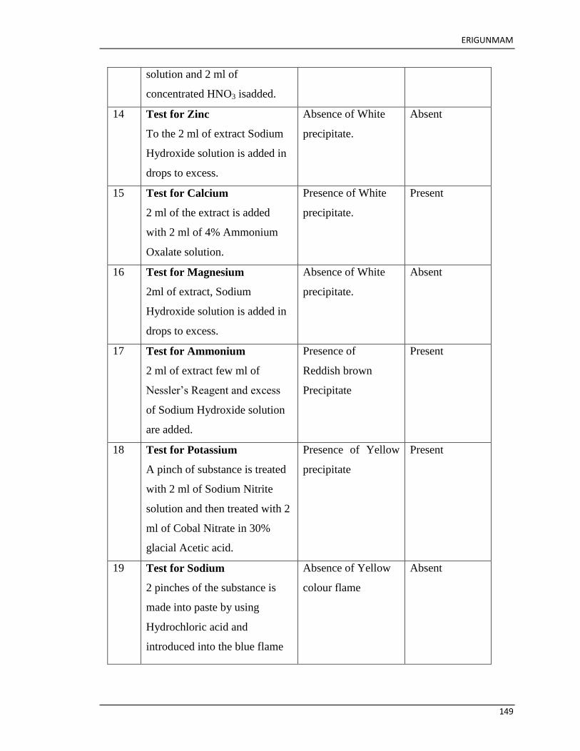
ERIGUNMAM
149
solution and 2 ml of
concentrated HNO3 isadded.
14 Test for Zinc
To the 2 ml of extract Sodium
Hydroxide solution is added in
drops to excess.
Absence of White
precipitate.
Absent
15 Test for Calcium
2 ml of the extract is added
with 2 ml of 4% Ammonium
Oxalate solution.
Presence of White
precipitate.
Present
16 Test for Magnesium
2ml of extract, Sodium
Hydroxide solution is added in
drops to excess.
Absence of White
precipitate.
Absent
17 Test for Ammonium
2 ml of extract few ml of
Nessler‘s Reagent and excess
of Sodium Hydroxide solution
are added.
Presence of
Reddish brown
Precipitate
Present
18 Test for Potassium
A pinch of substance is treated
with 2 ml of Sodium Nitrite
solution and then treated with 2
ml of Cobal Nitrate in 30%
glacial Acetic acid.
Presence of Yellow
precipitate
Present
19 Test for Sodium
2 pinches of the substance is
made into paste by using
Hydrochloric acid and
introduced into the blue flame
Absence of Yellow
colour flame
Absent

ERIGUNMAM
150
20 Test for Mercury
2 ml of the extract is treated
with 2 ml of Sodium
Hydroxide solution.
Absence of yellow
Precipitate
Absent
21 Test for Arsenic
2 ml of extract is treated with 2
ml of silver Nitrate solution
Absence of
Yellow precipitate
Absent
22 Test for Starch
2ml of extract is treated with
weak iodine solution
Absence of
Blue colour
Absent
23 Test of reducing Sugar
5ml of Benedicts qualitative
solution is taken in a test tube
and allowed to boil for 2
minutes and added 10 drops of
the extract and again boiled for
2 minutes. The colour changes
are noted.
Absence of Green
colour
Absent
24 Test of the alkaloids
2ml of the extract is treated
with 2ml of potassium lodide
solution.
Presence of
Red colour
Present
25 Test of the proteins
2ml of the extract is treated
with 2ml of 5% NaOH ,mix
well and add 2 drops of copper
sulphate solution.
Absence of
Violet colour
Absent
RESULTS:

ERIGUNMAM
151
Thus the given sample Pirandai vadagam contains
Chloride, Phosphate, Iron, Calcium, Potassium and Alkaloids.

ERIGUNMAM
152
INTITUTIONAL ETHICS COMMITTEE CERTIFICATE

ERIGUNMAM
153
INTITUTIONAL ETHICS COMMITTEE CERTIFICATE
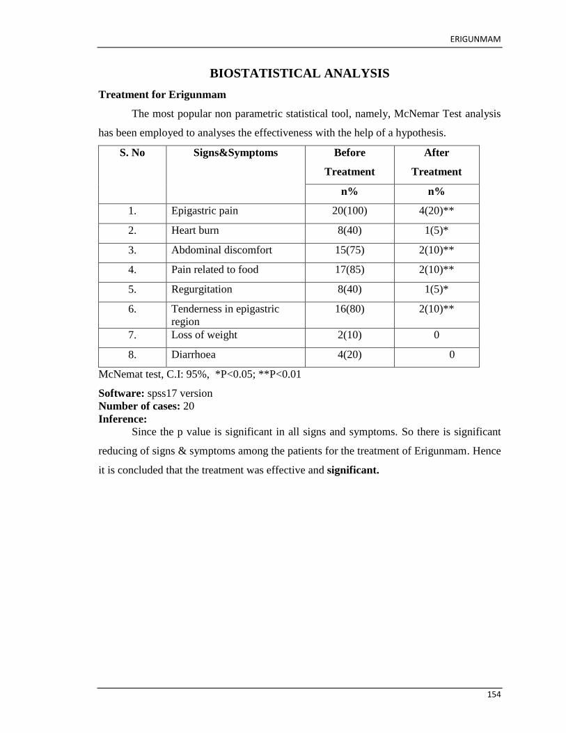
ERIGUNMAM
154
BIOSTATISTICAL ANALYSIS
Treatment for Erigunmam
The most popular non parametric statistical tool, namely, McNemar Test analysis
has been employed to analyses the effectiveness with the help of a hypothesis.
S. No Signs&Symptoms Before
Treatment
After
Treatment
n% n%
1. Epigastric pain 20(100) 4(20)**
2. Heart burn 8(40) 1(5)*
3. Abdominal discomfort 15(75) 2(10)**
4. Pain related to food 17(85) 2(10)**
5. Regurgitation 8(40) 1(5)*
6. Tenderness in epigastric
region
16(80) 2(10)**
7. Loss of weight 2(10) 0
8. Diarrhoea 4(20) 0
McNemat test, C.I: 95%, *P<0.05; **P<0.01
Software: spss17 version
Number of cases: 20
Inference:
Since the p value is significant in all signs and symptoms. So there is significant
reducing of signs & symptoms among the patients for the treatment of Erigunmam. Hence
it is concluded that the treatment was effective and significant.
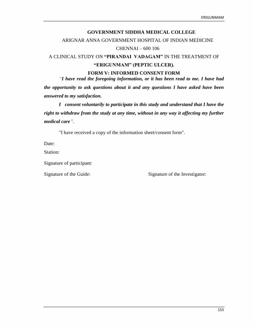
ERIGUNMAM
155
GOVERNMENT SIDDHA MEDICAL COLLEGE
ARIGNAR ANNA GOVERNMENT HOSPITAL OF INDIAN MEDICINE
CHENNAI – 600 106
A CLINICAL STUDY ON “PIRANDAI VADAGAM” IN THE TREATMENT OF
“ERIGUNMAM” (PEPTIC ULCER).
FORM V: INFORMED CONSENT FORM
“I have read the foregoing information, or it has been read to me. I have had
the opportunity to ask questions about it and any questions I have asked have been
answered to my satisfaction.
I consent voluntarily to participate in this study and understand that I have the
right to withdraw from the study at any time, without in any way it affecting my further
medical care”.
"I have received a copy of the information sheet/consent form".
Date:
Station:
Signature of participant:
Signature of the Guide: Signature of the Investigator:

ERIGUNMAM
156
,
( )

ERIGUNMAM
157
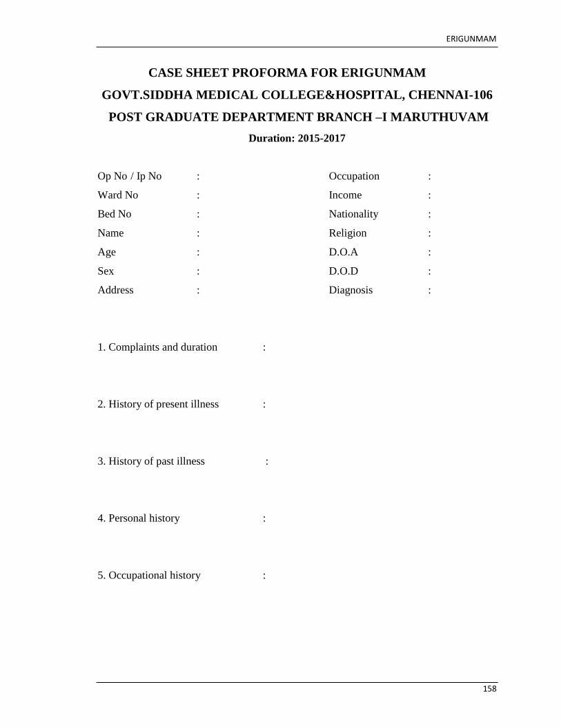
ERIGUNMAM
158
CASE SHEET PROFORMA FOR ERIGUNMAM
GOVT.SIDDHA MEDICAL COLLEGE&HOSPITAL, CHENNAI-106
POST GRADUATE DEPARTMENT BRANCH –I MARUTHUVAM
Duration: 2015-2017
Op No / Ip No : Occupation :
Ward No : Income :
Bed No : Nationality :
Name : Religion :
Age : D.O.A :
Sex : D.O.D :
Address : Diagnosis :
1. Complaints and duration :
2. History of present illness :
3. History of past illness :
4. Personal history :
5. Occupational history :

ERIGUNMAM
159
6. Personal Habits :Veg/nonveg/smoker/Alcoholic/Tobacco chewer
7. Family History :
8.Obstetric History :
GENERAL EXAMINATION
Patient consciousness :
Body Built :
Nourishment :
Anaemia :
Jaundice :
Cyanosis :
Clubbing :
JVP :
Tracheal deviation :
Pedal oedema :
Lymph adenopathy :
VITAL SIGNS
Body Temp :
Pulse :
Respiratory rate :
Blood Pressure :
Weight :
SIDDHA ASPECT
NILAM
Kurinchi :

ERIGUNMAM
160
Mullai :
Marutham :
Neithal :
Palai :
PARUVA KALAM
Kaar :
Koothir :
Munpani :
Pinpani :
Elavenil :
Muduvenil :
YAAKKAI(Udal)
Vaatham :
Pitham :
Kabam :
Kalappu :
GUNAM
Satthuvam :
Rajotham :
Thamasam :
PORI/PULANGAL (SENSORY
ORGANS)
Mei –Sensation :
Vaai – Taste :
Kan – Vision :
Mooku - Smell :
Sevi – Hearing :

ERIGUNMAM
KANMENTHRIYAM/KANNMA VIDAYAM [MOTOR ORGANS]
Kai- Dhaanam :
Kaal-Kamanam :
Vaai-Vasanam :
Eruvaai- Visarkkam :
Karuvaai-Aanantham :
UYIR THATHUKKAL
A.VATHAM
Piranan :
Abanan :
Viyanan :
Udanan :
Samanan :
Nagan :
Koorman :
Kirukaran :
Devathathan :
Thananjeyan :
B.PITHAM
Anar pitham :
Ranjaga pitham :
Saathaga pitham :
Pirrasaga pitham :
Alosaga pitham :
C.KAPAM
Avalambagam :
Kilethagam :
Pothagam :
Tharpagam :
Santhigam :

ERIGUNMAM
UDALTHAATHUKKAL
Saaram :
Senner :
Oon :
Kozhuppu :
Enbu :
Moolai :
Sukkilam/Suronitham :
ENVAGAI THERVUGAL
1.Naa :
2.Niram :
3.Mozhi :
4.Vizhi :
5.Sparisam :
6.Malam :
7.Moothiram
Neer Kuri :
Niram :
Manam :
Edai :
Nurai :
Enjal :
Nei Kuri :
8.Naadi :
MODERN ASPECT
Sytemic Examination
Inspection :
Palpation :
Percussion :

ERIGUNMAM
Auscultation :
Others Systems
Cardio Vascular System :
Respiratory system :
Central nervous system :
Genito urinary system :
Endocrine system :
CLINICAL SIGN AND SYMPTOMS OF ERIGUNMAM
Symptoms Before
Treatment
After
treatment
1.Epigastric pain and
burning with related to
food
2. Loss of appetite
3. Nausea
4.Vomiting
5. Anorexia
6.Bloating and fullness of
stomach
7.Weight loss
INVESTIGATION
1. BLOOD
TC, DC, ESR, Hb
Bleeding time, Clotting time
Blood sugar
Blood urea
Serum cholesterol

ERIGUNMAM
2. URINE
Albumin
Sugar
Deposits
3.SPECIAL INVESTIGATION:
ENDOSCOPY
CASE SUMMARY
DIAGNOSIS
TRIAL DRUG : PIRANDAI VADAGAM
Dose : 1 gm vadagam; Twice a day
Anubanam : Chewable
Duration of Treatment : 48 days.
Pathiam (Do‘s and Don‘ts)
Prognosis at the end of the Treatment.
Medical Officer Signature:
Guide:
HOD:

ERIGUNMAM
BIBLOGRAPHY

ERIGUNMAM
BIBLOGRAPHY
1. Siddha Maruthuvanga Churukkam By Dr.C.S.Uthamarayan, Hpim.Department Of
Indian Medicine, Governmentnof Tamil Nadu, 1983. Page 36.
2..MALHOTRA SL PEPTIC ULCER IN INDIA AND ITS AETIOLOGY .gut .1964.
Page no. 412-416
3.Yugi Vaidhiya Chinthamani 800, Dr.K.Anbarasu, B.S.M.S, Printer
S.P.Ramachandren, Thamarai Publication, Chennai-26.Page-65.
4.Yugi Vaidhiya Chinthamani 800, Dr.K.Anbarasu, B.S.M.S, Printers
S.P.Ramachandren, Thamarai Publication, Chennai-26.Page-66.
5.Yugi Vaidhiya Chinthamani 800, Dr.K.Anbarasu, B.S.M.S, Printers
S.P.Ramachandren, Thamarai Publication, Chennai-26.Page-66.
6.Yugi Vaidhiya Chinthamani 800, Dr.K.Anbarasu, B.S.M.S, Printers
S.P.Ramachandren, Thamarai Publication, Chennai-26.Page-67.
7.Yugi Vaidhiya Chinthamani 800, Dr.K.Anbarasu, B.S.M.S, Printers
S.P.Ramachandren, Thamarai Publication, Chennai-26.Page-67.
8.Yugi Vaidhiya Chinthamani 800, Dr.K.Anbarasu, B.S.M.S, Printers
S.P.Ramachandren, Thamarai Publication, Chennai-26.Page-67.
9.Yugi Vaidhiya Chinthamani 800, Dr.K.Anbarasu, B.S.M.S, Printers
S.P.Ramachandren, Thamarai Publication, Chennai-26.Page-67
10..Yugi Vaidhiya Chinthamani 800, Dr.K.Anbarasu, B.S.M.S, Printers
S.P.Ramachandren, Thamarai Publication, Chennai-26.Page-68.
11.Yugi Vaidhiya Chinthamani 800, Dr.K.Anbarasu, B.S.M.S, Printers
S.P.Ramachandren, Thamarai Publication, Chennai-26.Page-68.
12.Thirumoolar Karukkadai Vaithiyam 600,Page 44, Thamarai Publication,
S.P.Ramachandren
13.Thirumoolar Karukkadai Vaithiyam 600,Page 43, Thamarai Publication,
S.P.Ramachandren.

ERIGUNMAM
14.Thirumoolar Karukkadai Vaithiyam 600,Page 44, Thamarai Publication,
S.P.Ramachandren.
15.Thirumoolar Karukkadai Vaithiyam 600,Page 44, Thamarai Publication,
S.P.Ramachandren
16.Agathiya Maamunivar Guru Naadi Saathiram235, Page 21. B.Rathina Nayakar and
Sons,Chennai- 79 page no-21
17.Agathiyar Vaidhiya Kaviyam 1500, Page 29, Thamarai Publication,
S.P.Ramachandren. Thamarai Publication, Chennai-26. Page-68
18.Yugi Vaidhiya Chinthamani 800, Dr.K.Anbarasu, B.S.M.S, Printer
S.P.Ramachandren, Thamarai Publication, Chennai-26. Page-68
19. Dr. M. Shanmugavelu, H.B.I.M, Noi Nadal Noi Mudhal Nadal Thirattu, Indian
Medicine and Homeopathy, Fourth Edition 2006, Pg -108
20.Agathiyar vaithiya Vallathi 600, Page 18, Thamarai Publication, S.P.Ramachandren
21. Dr. M. Shanmugavelu, H.B.I.M, Noi Nadal Noi Mudhal Nadal Thirattu, Indian
Medicine and Homeopathy, Fourth Edition 2006, Pg -270
22.Dr. M. Shanmugavelu, H.B.I.M, Noi Nadal Noi Mudhal Nadal Thirattu, Indian
Medicine and Homeopathy, Fourth Edition 2006, Pg -270
23.Dr. M. Shanmugavelu, H.B.I.M, Noi Nadal Noi Mudhal Nadal Thirattu, Indian
Medicine and Homeopathy, Fourth Edition 2006, Pg -282
24.Dr. M. Shanmugavelu, H.B.I.M, Noi Nadal Noi Mudhal Nadal Thirattu, Indian
Medicine and Homeopathy, Fourth Edition 2006, Pg -298
25.Dr. M. Shanmugavelu, H.B.I.M, Noi Nadal Noi Mudhal Nadal Thirattu, Indian
Medicine and Homeopathy, Fourth Edition 2006, Pg -299
26.Dr. M. Shanmugavelu, H.B.I.M, Noi Nadal Noi Mudhal Nadal Thirattu, Indian
Medicine and Homeopathy, Fourth Edition 2006, Pg -299
27.Dr. M. Shanmugavelu, H.B.I.M, Noi Nadal Noi Mudhal Nadal Thirattu, Indian
Medicine and Homeopathy, Fourth Edition 2006, Pg -299
ERIGUNMAM
28.Dr. M. Shanmugavelu, H.B.I.M, Noi Nadal Noi Mudhal Nadal Thirattu, Indian
Medicine and Homeopathy, Fourth Edition 2006, Pg -27
29.Yugi Vaidhiya Chinthamani 800, Dr.K.Anbarasu, B.S.M.S, Printers
S.P.Ramachandren, Thamarai Publication, Chennai-26. Page-68
30.Yugi Vaidhiya Chinthamani 800, Dr.K.Anbarasu, B.S.M.S, Printers
32S.P.Ramachandren, Thamarai Publication, Chennai-26. Page-68
31. Dr. M. Shanmugavelu, H.B.I.M, Noi Nadal Noi Mudhal Nadal Thirattu, Indian
Medicine and Homeopathy, Fourth Edition 2006. Pg -363
32.Agathiya Munivar Aruliya Agathiyar Kanma Kandam 800,Muthar Pathippu
1901,Page-16
33. Gunapadam Part 1, Mooligai Vaguppu,Vaithiya Rathinam, K.S.Murugesha
Mudhaliyar,(Directorate Of Indian Medicine And Homeopathy).Page 675.
34. Gunapadam Part 1, Mooligai Vaguppu,Vaithiya Rathinam, K.S.Murugesha
Mudhaliyar,(Directorate Of Indian Medicine And Homeopathy).Page 510.
35.Gunapadam Part 1, Mooligai Vaguppu,Vaithiya Rathinam, K.S.Murugesha
Mudhaliyar,(Directorate Of Indian Medicine And Homeopathy).Page 99.
36.Gunapadam Part 1, Mooligai Vaguppu,Vaithiya Rathinam, K.S.Murugesha
Mudhaliyar,(Directorate Of Indian Medicine And Homeopathy).Page 201.
37.Gunapadam Part 1, Mooligai Vaguppu,Vaithiya Rathinam, K.S.Murugesha
Mudhaliyar,(Directorate Of Indian Medicine And Homeopathy).Page 620.
38.Gunapadam Part 1, Mooligai Vaguppu,Vaithiya Rathinam, K.S.Murugesha
Mudhaliyar,(Directorate Of Indian Medicine And Homeopathy).Page 512.
39.Gray‟ s Anatomy, Henery Gray, Thirty Eighth Edition , page no 1758-1764.
40.Davidson‟ s Principles & Practice Of Medicine, By John A.A. Hunter, 20th
Edition, P. 885-888.
41.Human Physiology C.C. Chatterjee Nov Edition 1997, page 509,510.
42.Harrison‟ s Principles Of Internal Medicine , Vol.2, P. 1596,1 th Edition.
43.Robbinson‟ s And Cotran Pathological Basis Of Disease Seventh Edition 2006.

ERIGUNMAM
( page 773-775,774,776.)
44. Hutchison‟ s Clinical Methods, Michael Swash, 20th Edition, P. 75-77.
45. Ghosh. M.N; Fundamentales Of Experimental Pharmacology, Kolkatta, Scientific
Book Agency,2 Edition 5 & 155 (1984).
46.Glossary Of Indian Medicinal Plants By R.N.Chopra, S.C. Nayar And I.C.
ChopraReprinted 1986.
47.National Institute Of Science Communication, New Delhi, P.246.
48.Indian Medicinal Plants, Vol. 2. Kritikar And Basu, Illustrated By K.S.Maharkar,
E. Blatter,J.E.Cavis, P.1178-1179.
49.Theraiyar Tharu, by Thiyagarajan, published by Ananda Kumar, P.42,43.
TOXICOLOGICAL STUDIES
1.OECD Guideline for testing of Chemicals (2001) Guideline 423: Acute Oral
Toxicity-Acute Toxic Class Method.
2.OECD Guide lines 407 for testing of chemicals. Repeated dose 28-Day Oral
Toxicity Study in Rodents. 2008: pp 2- 8.
3.Jain N, Sharma P, Sharma N, Joshi S C. Haemato-biochemical profile following sub
acute toxicity of malathion in male albino rats. Pharmacologyonline. 2009;2:500–506.
4.Suvarna, S.K., C.Layton and J.D. Bancroft. 2013. Bancroft‘s theory and practice of
histological techniques. 7th edn, Churchill Livingstone, London
Copyright © 2022 FDOKUMEN