fact or fallacy? The problem of pressure ulcer prevention
9
80 Clinical REVIEW Wounds UK, 2009, Vol 5, No 4 Dressings can prevent pressure ulcers: fact or fallacy? The problem of pressure ulcer prevention Martyn Butcher, Geoffrey Thompson Martyn Butcher is an Independent Non-medical Prescriber, Independent Tissue Viability and Wound Care Consultant; Geoffrey Thompson is an Infection Prevention/Control Audit and Research Nurse, Worcestershire Acute Hospitals Trust NHS UK, Independent Tissue Viability Nurse, Trustee member, Wound Care Alliance UK In part one of this two-part article, the authors discuss the aetiology of pressure ulcers, the means of identifying those patients at risk, the range of clinical intervention strategies implemented to try and prevent their formation and the problems faced by clinicians in developing cost-effective solutions to pressure ulcer prevention. Part two will set out the scientific evidence to support the use of dressing materials to prevent pressure damage, discuss the clinical realities faced by clinicians and explore if the use of wound dressing materials has any part in a modern pressure ulcer prevention strategy. T he development of pressure ulcers in vulnerable, at-risk individuals is a significant burden on healthcare resources and it has been stated that their development can be viewed as an indicator of poor quality care (Department of Health, 1993; Olshansky, 2005). Despite position papers indicating some pressure ulcers may be unavoidable (Wound, Ostomy and Continence Nurses Society [WOCNS], 2009), there is still a stigma surrounding their formation and a drive to affect improved preventative strategies. Many different approaches to care have been adopted to prevent their development and yet pressure ulceration remains one of the most significant issues in health care today. One approach which has KEY WORDS Pressure Shear Pressure damage Guidelines Evidence-based practice Figure 1. Pressure ulcers: A. Moisture damage to the sacrum; B. Presumed full-thickness damage under eschar; C. Blanching hyperaemia to elbows; D. Mixed levels of tissue damage, grades 1, 2, 3 and possibly 4. A B D C been largely overlooked is the potential benefit of using wound care materials not to treat damage, but to help prevent it in the first instance. Pressure ulcers as an issue Pressure ulcers are an all too common problem that occur in both hospital and community environments (Weir, 2007; Stotts and Wu, 2009) and are reported worldwide by numerous authors and agencies (European Pressure Ulcer Advisory Panel [EPUAP], 2003; Clark et al, 2004; National Institute for Health and Clinical Excellence [NICE], 2005). US estimates of pressure ulcer
-
Upload
khangminh22 -
Category
Documents
-
view
4 -
download
0
Transcript of fact or fallacy? The problem of pressure ulcer prevention
80
Clinical REVIEW
Wounds uk, 2009, Vol 5, No 4
Dressings can prevent pressure ulcers: fact or fallacy? The problem of pressure ulcer prevention
Martyn Butcher, Geoffrey Thompson
Martyn Butcher is an Independent Non-medical Prescriber, Independent Tissue Viability and Wound Care Consultant; Geoffrey Thompson is an Infection Prevention/Control Audit and Research Nurse, Worcestershire Acute Hospitals Trust NHS UK, Independent Tissue Viability Nurse, Trustee member, Wound Care Alliance UK
In part one of this two-part article, the authors discuss the aetiology of pressure ulcers, the means of identifying those patients at risk, the range of clinical intervention strategies implemented to try and prevent their formation and the problems faced by clinicians in developing cost-effective solutions to pressure ulcer prevention. Part two will set out the scientific evidence to support the use of dressing materials to prevent pressure damage, discuss the clinical realities faced by clinicians and explore if the use of wound dressing materials has any part in a modern pressure ulcer prevention strategy.
The development of pressure ulcers in vulnerable, at-risk individuals is a significant burden
on healthcare resources and it has been stated that their development can be viewed as an indicator of poor quality care (Department of Health, 1993; Olshansky, 2005). Despite position papers indicating some pressure ulcers may be unavoidable (Wound, Ostomy and Continence Nurses Society [WOCNS], 2009), there is still a stigma surrounding their formation and a drive to affect improved preventative strategies. Many different approaches to care have been adopted to prevent their development and yet pressure ulceration remains one of the most significant issues in health care today. One approach which has
KEY WORDSPressure ShearPressure damage GuidelinesEvidence-based practice
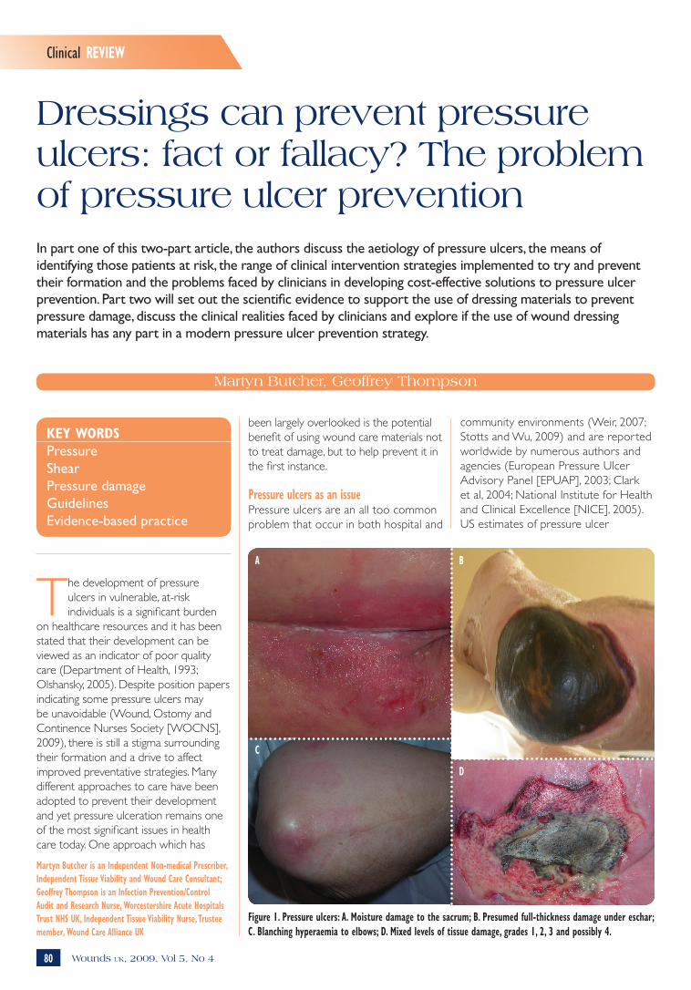
Figure 1. Pressure ulcers: A. Moisture damage to the sacrum; B. Presumed full-thickness damage under eschar; C. Blanching hyperaemia to elbows; D. Mixed levels of tissue damage, grades 1, 2, 3 and possibly 4.
A B
D
C
been largely overlooked is the potential benefit of using wound care materials not to treat damage, but to help prevent it in the first instance.
Pressure ulcers as an issuePressure ulcers are an all too common problem that occur in both hospital and
community environments (Weir, 2007; Stotts and Wu, 2009) and are reported worldwide by numerous authors and agencies (European Pressure Ulcer Advisory Panel [EPUAP], 2003; Clark et al, 2004; National Institute for Health and Clinical Excellence [NICE], 2005). US estimates of pressure ulcer
Butcher CS4 final.indd 80 28/10/2009 16:52
Clinical REVIEW
81Wounds uk, 2009, Vol 5, No 4
incidence vary. In 1994 Bergstom et al reported that at least one million people developed pressure ulcers. Subsequently, the Institute for Health Improvement estimated that 2.5 million users of US healthcare institutions develop pressure ulcer each year (Bales and Padwojski, 2009). Ultimately, if not treated appropriately, they can develop into severe and complex wounds with potentially devastating consequences for the patient that may require surgical intervention to bring about healing (Brown et al, 2007).
AetiologyPressure ulcers are caused by prolonged and/or repeated ischaemic insults without adequate time for total tissue recovery, resulting in tissue necrosis (Hagisawa et al, 2004). These are manifested as localised areas of tissue breakdown involving the skin and/or deeper tissues (EPUAP, 2003), and generally occur as a result of unrelieved pressure to any part of the body, especially portions over bony or cartilaginous areas (Weir, 2007), such as the sacrum, elbows, knees, heels and ankles (Figure 1).
When looking at the aetiology of pressure ulcers, Braden et al (2000) developed a conceptual frame to help understand the various risk factors leading to ulcer formation, dividing the causes into two groups; ‘extrinsic’ and ‘intrinsic’.
Extrinsic factors are physical mechanisms, events or circumstances that are external to the patient who develops pressure ulcers. Intrinsic patient-specific factors are unique to the individual, such as: 8Age8Nutrition8General health status8Innate level of activity and mobility 8Morbidities such as diabetes.
While Bergstrom (2005) refers to more than 100 factors associated with pressure ulcer risk, such as previous medical history, comorbidities, fractured hip, spinal cord injury, cardiovascular disease, space in this paper does not permit a detailed listing and discussion
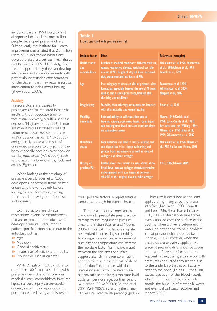
Table 1Factors associated with pressure ulcer risk
Intrinsic factor Effect References (examples)
Health status and comorbidities
Number of medical conditions: diabetes mellitus, cancer, respiratory disease, peripheral vascular disease (PVD), length of stay all show increased risk, prevalence and incidence of PUs
Makleburst et al, 1994; Papantonio et al, 1994; Allman et al, 1995; Lewicki et al, 1997
Age Increasing age = increased risk of pressure ulcer formation, especially beyond the age of 70 from cardiac and neurological issues, lowered skin elasticity and resilience
Papantonio et al, 1994; Whittington et al, 2000; Margolis et al, 2002
Drug history Steroids, chemotherapy, anticoagulants interfere with skin integrity and wound healing
Nixon et al, 2001
Mobility/ immobility
Reduced ability to self-reposition due to trauma, surgery, post anaesthesia. Spinal injury can prolong unrelieved pressure exposure times on vulnerable tissues
Munro, 1940; Kosiak et al, 1958; Exton-Smith et al, 1961; Berlowitz and van Wilking, 1989; Allman et al, 1995; Bliss et al, 1999; Schoonhoven et al, 2002
Nutritional status
Poor nutrition can lead to muscle wasting and soft tissue loss + less tissue cushioning and greater bony prominences, as well as reduced collagen and tissue strength
Makleburst et al, 1994; Allman et al, 1995; Collier and Moore, 2006
History of previous PUs
Healed, ulcer sites remain an area of risk of re-breakdown because collagen structure remains mal-organised with scar tissue at between 40–80% of the original tissue tensile strength
NICE, 2005; Ichioka, 2005
on all possible factors. A representative sample can though be seen in Table 1.
Three main extrinsic mechanisms are known to precipitate pressure ulcer damage to the integument: pressure, shear and friction (Collier and Moore, 2006). Other extrinsic factors may also be involved in increasing vulnerability to damage; for example, environmental humidity and temperature can increase the moisture factor (or micro-climate) between the skin and the surface support, alter skin friction co-efficient and therefore increase the risk of shear and friction. This interacts with the unique intrinsic factors relative to each patient, such as the body’s moisture level, body temperature, age, continence and medication (EPUAP, 2003; Bouton et al, 2005; Weir, 2007), increasing the chance of pressure ulcer development (Figure 2).
Pressure is described as the load applied at right angles to the tissue interface (Krouskop, 1983; Bennett and Lee, 1986; Shear Force Initiative [SFI], 2006). External pressure forces evenly applied over the surface of the body, as when a diver is submerged in water, do not appear to be a problem in that pressure ulcers do not form (Sprigle, 2000). However, when the pressures are unevenly applied, with gradient pressure differences between the point of pressure focus and the adjacent tissues, damage can occur with pressures conducted through the skin to the underlying tissues particularly close to the bone (Le et al, 1984). This causes occlusion of the blood vessels which, if unrelieved, leads to cellular anoxia, the build-up of metabolic waste and eventual cell death (Collier and Moore, 2006).
Butcher CS4 final.indd 81 25/10/2009 19:06

82
Clinical REVIEW
Wounds uk, 2009, Vol 5, No 4
The amount of pressure required to precipitate cell damage is dependent on the intensity of pressure, the duration of exposure (Kosiak, 1961), and to the individual’s ability to cope with pressure loading (Daniel et al, 1981).
Controversy reigns over what pressure is required to induce capillary closure (Russell, 1998), but what is widely accepted is that even low pressures may cause tissue damage if exposure is prolonged (Read, 2001). This may be due to the way in which the pressure gradient is transmitted through tissues, a phenomenon known as the McClemont ‘cone of pressure’ (McClemont, 1984). An interface pressure such as 50mmHg between the skin and the support surface is
transmitted through the different underlying tissues; skin, subcutaneous fat, muscle and finally bone, with a cone-shaped increase in pressure of three to five times that at the interface so that pressures as high as 200mmHg might be experienced at the bony prominence (Collier and Moore, 2006).
It is commonly quoted that a safe level of pressure is 32mmHg, with 32mmHg being the arteriolar closing pressure and 12mmHg the venous limb side of the capillary loop (Landis, 1930). However, this early experimental work was undertaken on nail-bed pressures in healthy volunteers and so is now widely regarded as a guide rather than a definitive measure. Many experts believe that there is no direct link between
the internal pressures generated in the tissues under compression and the external pressure at the interface between the support surface and the skin under compression. As the average interface pressure is usually much greater than 32mmHg, it is assumed that the internal pressure will be high, although this cannot be measured in the clinical setting (Bader and Oomens, 2006).
Normal physiological response to pressure stressing includes the development of blanching erythema. This occurs as an adaptive response to short-term ischaemia in which previously stressed blood vessels dilate causing a temporary red ‘flush’ in the tissues (Dealey, 1994). This flush fades on light finger pressure and normally fades shortly after blood flow is restored.
Non-blanching erythema arises from either prolonged exposure to low-level pressure or short exposure to high pressure (the specific level of pressure varying between individuals), indicating that tissue damage has occurred. In this case the erythema is not due to a temporary flush of blood rushing into the area, but to local capillary disruption and leakage of blood into the surrounding tissues. Normal skin colour is not restored. This is considered to be the beginning of a pressure ulcer or grade 1 damage in some ulcer classification systems (Bethell, 2003) (Figure 3).
In darker pigmented individuals this ‘blanching’ may not be apparent. Thus, it is important to contrast the differences between the pressure points and the surrounding skin, as early damage, although not visible, may feel hotter, colder, harder or look shinier than the healthy skin (Bethell, 2003). With this in mind, healthcare staff should be familiar with the normal skin colour and tone of their patients.
The degree of vulnerability to pressure varies from person to person due to:8Tissue tolerance variations between
individuals through the combination of extrinsic and intrinsic factors unique to the individual (Bridel, 1993)
Pre
ssur
e ul
cer
deve
lopm
ent
Figure 2. Flowchart for the prediction and prevention of pressure ulcers. Taken from the Australian Wound Management Association (AWMA) Clinical Practice Guidelines (AWMA, 2001).
Butcher CS4 final.indd 82 25/10/2009 19:06

84 Wounds uk, 2009, Vol 5, No 4
Clinical REVIEW
8Pressure duration over the pressure points which can result in damage from high pressure for short intense periods, which can be as damaging as low pressure for prolonged periods (Bell, 2005)
8Collagen function protecting the microcirculation helps to maintain the pressures inside and outside the cells preventing cell bursting. Collagen levels vary from person to person with lessening protective qualities with aging (Russell, 1998)
8Autoregulatory processes initiated when external pressure is sensed, leading to increased internal capillary pressure, reduced blood flow and reactive hyperaemia to counteract the pressure loading.
These mechanisms can fail when the external pressure exceeds the person’s diastolic pressure rather than the 32mmHg often quoted (Nixon, 2001).
The response of tissue to external forces varies greatly, being dependent on a large number of factors. It is therefore not possible to establish a ‘safe level of pressure’. In addition, tolerance to pressure can vary greatly from individual to individual due to the interplay of external factors listed in Table 1. Given the highly variable nature of pressure transmission, capillary closure and the individual’s normal and adaptive responses to pressure stress, the production of time/pressure curves (mathematical models for predicting the time likely to cause tissue damage when tissue is exposed to specific levels of pressure [Reswick and Rogers, 1976]) may be of little practical benefit to the clinician in everyday practice (Sharp and McLaws, 2005; Grefen, 2009).
Shear is a mechanical stress applied parallel to the skin. The SFI describes it as: ‘An action or stress resulting from applied forces which causes or tends to cause two contiguous internal parts of the body to deform in the transverse plane (i.e. “shear strain”)’ (SFI, 2006). This sliding or twisting force occurs continuously within soft tissues even when perpendicular pressure is applied, but increases greatly when combined with lateral movement, as seen when
the body is adapting to the inclination of the bed or when sitting in a chair. If the skin adheres to the surface support (which is more likely if the skin is moist or wet from environmental factors or intrinsically from incontinence or sweating) (Weir, 2007; Beldon, 2008), the tissues attached to the gradually moving skeletal frame become distorted which, in turn, distort the blood vessels leading to their collapse or rupture.
Shear forces are generated as a result of the interplay of friction and pressure (Collier and Moore, 2006). When applied, shear increases the effects of pressure resulting in vascular occlusion at only half the pressure of non-stressed tissues (Bennett and Lee, 1986). Shear forces may also have a significant role in the development of deep tissue damage, although this is difficult to measure in the clinical setting (Russell, 1998). Potentially, shearing is the most serious extrinsic risk factor due to the rapidity with which it can result in tissue damage (Sharp and McLaws, 2005). This is more likely to occur in the elderly as a result of loose, fragile skin and the ease with which the different tissue types can be sheared off their respective attachments (Allman et al, 1995).
The edges of ulcers caused by shear forces appear to be ragged with more uneven wound margins, often with surrounding epidermal scuffing. Bruising may also be a feature (Figure 4).
The mechanisms of shear damage have important consequences for the planning and delivery of preventative care interventions, even though there are few clinical methods to estimate shearing forces or their resultant effects on tissues (Verluysen, 1985). It is hoped that the work of the SFI will add to this body of knowledge.
Friction is a complex phenomenon which depends on complex physical science and engineering concepts. In simplistic terms, within the context of friction-induced tissue damage, we are referring to kinetic friction. Kinetic (or dynamic) friction occurs when two objects are moving relative to each
other and rub together. Bergman-Evans et al (1994) define it as the resistance to lateral movement. Kinetic friction is dependent on mass, force applied and the friction co-efficients of the surfaces involved. Clinically, the effect of friction between the skin and a support surface has important dynamics that can initiate pressure ulcer formation:8It can cause excessive wear to the
cornified layers of the skin with resultant exposure of the underlying structures (Read, 2001)
8It can cause the formation of blisters as separation occurs between the layers of the epidermis leading to
Figure 3. Sacral area showing clinical features of blanching hyperaemia.
Figure 4. Shear pressure damage occurring in a young woman during childbirth.
Butcher CS4 final.indd 84 25/10/2009 19:06

Wound care SCIENCEClinical REVIEW
86 Wounds uk, 2009, Vol 5, No 4
dermatitis, moisture-induced damage and superficial pressure ulceration. The EPUAP have suggested that moisture-induced damage should be categorised separately from pressure ulcers. In practice, this differentiation can be difficult to interpret clinically. Defloor and Schoonhoven (2004) and Defloor et al (2005) identified that reliability of the EPUAP tool was low when used to differentiate moisture lesions and superficial pressure ulcers from photographic evidence. Indeed, writers such as Houwing et al (2007) argue that such a distinction should not be made as it distracts clinicians from the need to implement appropriate pressure ulcer prevention strategies, and, as McDonagh (2008) points out, these two phenomena can co-exist within a client at a given point in time.
Aetiological pathwaysControversy exists as to the aetiological route by which pressure ulcers form and progress. It is acknowledged that pressure ulcers are primarily caused by sustained mechanical loading, however, prevention of ulcer formation by reducing the degree of loading alone remains difficult to achieve. This is mainly due to poor understanding of the underlying pathways whereby mechanical loading leads to tissue breakdown (Bouten et al, 2005).
Three theories have been postulated to explain this process:
exposure of the underlying dermis (Butcher, 1999)
8The deformation of skin can lead to further deformation in deeper tissues (shear damage).
The amount of damage caused depends on tissue resistance and the interplay of friction and pressure. Pressure and friction together cause more damage than friction alone and will induce greater shear forces (Figure 5).
MoistureAlthough not directly indicated as a mechanism of pressure damage, the role of moisture is pivotal in the development of friction damage and so is a secondary factor in shear forces (Beldon, 2008) (Figure 6). Moisture levels within the cells of the epidermis have a direct bearing on the friction co-efficient of this tissue. Even at relatively low levels, moisture causes a rise in friction co-efficient making skin ‘stick’ to surfaces (Nacht et al, 1981). In addition, when exposed to moisture for prolonged periods, the keratinised cells of the epidermis swell and become waterlogged. This reduces their ability to withstand friction and can result in epidermal stripping.
These features have new relevance since the re-classification of moisture lesions (Bethell, 2003; Butcher, 2005; Beldon, 2008). There is a close association between incontinence
1. Pressure ulcers form via the top-to-bottom model
2. Pressure ulcers form via the bottom-to-top model
3. Pressure ulcers form via a middle approach model.
Theory 1Pressure and shear induce local ischaemia, and impaired drainage impairs the transport of oxygen and nutrients to and metabolic waste products away from the cells within the affected tissues. Eventually this leads to cell necrosis and the formation of an ulcer. There are sound arguments for damage to muscle tissue as it is metabolically more active than skin.
Theory 2This model states that when pressure is relieved from the compressed tissues by patient repositioning or the use of an alternating pressure-alleviating mattress (APAM), it is the restoration of blood flow after the load-removal rather than impaired blood flow during pressure loading that is the mechanism of tissue necrosis. It is claimed that it is an over-abundant release of oxygen-free radicals during pressure off-loading that causes the damage.
Theory 3In the third model, tissue damage may start anywhere between the skin and the underlying bone, but can include the skin surface and bone interface, concurrently or haphazardly, to produce a pressure ulcer.
Prevalence and incidence of pressure ulcersUnless correctly identified and treated, pressure ulcers can have a significant effect upon the patient’s quality of life
Figure 6. Sacral region showing clinical features of moisture damage combined with shear and pressure. Figure 5. Shear and friction damage to stump from badly fitting prosthesis.
Butcher CS4 final.indd 86 25/10/2009 19:06

Clinical REVIEW
88 Wounds uk, 2009, Vol 5, No 4
and may, under certain circumstances, prove fatal. The deaths of thousands of patients are attributed to pressure ulcers and their complications every year (Agam and Gefen, 2007). Data relating to incidence (a statistical measurement of the number of individuals developing a condition) and pressure ulcers varies considerably. A recent literature review investigated pressure ulcer prevalence and incidence in intensive care patients. The analysis of data from published papers highlighted these variations with pressure ulcer prevalence (the number of individuals with pressure ulcers as a percentage of the total defined population at one point in time) in intensive care settings, ranging from 4% in Denmark to 49% in Germany, while incidence ranged from 38% to 124% (Shahin et al, 2008). In a Canadian study in 2004 the national prevalence figure across all care settings was estimated at 26% (Woodbury and Houghton, 2004). More specifically, a recent study has shown that the prevalence of pressure ulceration within the population receiving health care in Bradford, UK was 0.74 people with a pressure ulcer per 1000 population (95%, CI 0.6–0.8) (Vowden and Vowden, 2009).
Cost of pressure ulcers to health carePatients with pressure ulcers place a burden on health care as they require a significant amount of medical resources to treat. A recent survey evaluated the impact of wound care in Bradford and Airedale NHS Primary Care Trust in the UK (Vowden et al, 2009), and showed that the prevalence of patients with a wound was 3.55 per 1000 population. The estimated cost to the US hospital sector is $11 billion per annum (Bales and Padwojski, 2009). This has been considered unsustainable and unacceptable. In an effort to control costs and raise quality standards, the Centers for Medicare and Medicaid Services (CMS) has determined it will no longer reimburse hospitals for treating a range of hospital-acquired conditions including pressure ulcers (Bergquist-Beringer et al, 2009). This is having a serious impact on US healthcare management and service provision and has lessons for the UK
healthcare sector. The majority of wounds were surgical/trauma (48%), leg/foot (28%) and pressure ulcers (21%). Prevalence of wounds among hospital inpatients was 30.7%. Of these, 11.6% were pressure ulcers, of which 66% were hospital-acquired. Further cases have received attention; over $3 million was awarded by a Florida court in 2008 (Legal Eagle, 2008), while the Supreme Court of Mississippi approved a $1 million award against a nursing home (Legal Eagle, 2007).
In a study undertaken on patients developing a pressure ulcer to estimate the annual cost of treating pressure
hospital-acquired pressure ulcers were a significant component and focus for potential cost reductions.
Legal issuesThe direct costs of patient treatment are not the only area of expense. Increasingly, the spectre of the threat of legal action is taking a greater place in pressure ulcer management. In a US study, hospital stays for the treatment of pressure ulcers have been estimated to be in the region of $37,800 (Weir, 2007). It has been shown that these patients require 50% more nursing time, remain hospitalised for significantly longer periods, and incur higher hospital charges (Bradon and Endowed, 2008). Pressure ulcers are the leading iatrogenic causes of death reported in developed countries, second only to adverse drug reactions (Barczak et al, 1997).
In November 2000 the State of Hawaii convicted an individual of manslaughter in the death of a patient at a nursing home for permitting the progression of decubitus ulcers without seeking medical help, and for not bringing the patient back to a doctor for treatment of the ulcers (Di Maio and Di Maio, 2002). A number of authors have highlighted the increase in litigation associated with malpractice related to pressure ulcers not only in the US (Bennet et al, 2000; Levine et al, 2008; Meehan and Hill, 2009), but also in Europe (Cherry, 2006). It therefore makes clinical and economic sense to takes measures to minimise pressure ulcer risk by taking preventative actions (Meehan and Hill, 2009).
Standard preventative interventionsPossibly due to the emphasis of scientific research on the role of pressure within pressure ulcer aetiology, most effort appears to have gone into strategies to reduce or attempt to eliminate pressure in the clinical setting. Over the past thirty years many manufacturers have developed a wide variety of support surfaces, principally mattresses, aimed at this particular endpoint. With such a wide range of products there can be confusion over product selection for a given pressure
The direct costs of patient treatment are not the only area of expense. Increasingly, the spectre of the threat of legal action is taking a greater place in pressure ulcer management.
ulcers in the UK, the actual costs were derived from a bottom-up methodology, based on the daily resources required to deliver protocols of care reflecting good clinical practice. The results showed that at this time the cost of treating a pressure ulcer varied from £1,064 (grade/stage 1) to £10,551 (grade/stage 4). Costs increase with ulcer grade/stage because the time to heal is longer and because the incidence of complications is higher in more severe cases. At the time of writing, the total cost in the UK was estimated at £1.4–£2.1 billion annually (4% of total NHS expenditure). The study also showed that most of the associated cost was related to nurse time (Bennet et al, 2004). Vowden et al (2009) also concluded that the most important components are the costs of wound-related hospitalisation and the opportunity cost of nurse time (the indirect cost incurred to the healthcare provider by the nurse undertaking care for this individual which would otherwise be utilised caring for other patients). In total, 32% of patients treated in hospital accounted for 63% of total costs, of which the development of
Butcher CS4 final.indd 88 25/10/2009 19:06

Clinical REVIEW
90 Wounds uk, 2009, Vol 5, No 4
ulcer risk (Rithalia, 1996), and there is a need for an understanding of the difference between the mattress and cushion classes (Finucane, 2006). Standard interventions to prevent pressure ulcer formation have included the use of specific redistributive surfaces as either pressure-reducing appliances or pressure-relieving mattresses or cushions.
Pressure-reducing support surfaces vary from relatively simple foam and slashed foam constructions to gel, fluid, and air-filled systems. There are also more complex dynamic pressure-reducing low airloss systems and dynamic foam (Thompson, 2006; Gray et al, 2008), or forms of ‘air-float’ (Thompson et al, 2008) in which pressure at the interface between the dependent skin and the support surface is reduced through the use of the conforming support surface, thereby spreading load and reducing pressure per square centimetre.
In addition, the materials that used to cover such devices have become more technically advanced with non-stretch PVC covers giving way to two- and three-way stretch which encourages greater conformity between the body and the mattress/cushion. Improved vapour permeability with PU materials also reduces the risk of moisture build-up at the interface, with the aim of reducing the friction/shear co-efficient (Jay, 1995).
It stands to reason that if one of the major components of pressure ulcer formation is the application of unrelieved pressure, then the reduction of this pressure to sub-morbid levels is a key factor in pressure damage prevention. Pressure redistribution through offloading provides tissues with the time needed for cellular repair and the restoration of normal cellular activity. In its basic form, this is achieved by offloading tissues through either manual repositioning or the use of splints, wedges and other repositioning devices (Guttmann, 1955, 1976).
Cyclical offloading teamed with the use of a conforming interface is one
approach to this problem. This approach is adopted by those using APAMs where load is supported by alternating, conforming air cells. These cells periodically change their pressure profile in a pre-set cycle, thereby altering the area of tissue exposed to compression stresses. However, some clinicians prefer constant low pressure support surfaces, such as those found in air fluidised and low air loss systems. Unfortunately, there is little data to indicate which approach is preferable.
Reduction of friction and shearWhile friction and shear are cited as the other mechanisms of pressure damage, due to technical and ethical issues little research has been undertaken in this area (Ohura et al, 2005). For this reason, the reduction of these components in clinical practice has generally been undertaken based on anecdotal evidence. Due to the risk of increasing shear forces, previous practices such as massage of high risk tissues have been indicated as dangerous (Dyson, 1978; Pritchard and Mallett, 1993; Buss et al, 1997; Shahin et al, 2009), and so have been largely abandoned. Clinicians have been advised to use care in positioning patients to minimise shear force, (Maklebust, 1987; AWMA, 2001) and to use low-friction turning/repositioning aids to minimise skin and soft tissue damage (Butcher, 2005). Some writers have also indicated that the use of dressings and skin sealants may help in reducing friction and therefore reduce the risks of friction damage and shear forces (AWMA, 2001; Black, 2004; Butcher, 2005).
The practice of using simple adhesive dressings to minimise friction is accepted by many authorities as commonplace among healthcare workers and the general public. How many of us have used adhesive tape or wound plasters on our heels to prevent new footwear from rubbing and producing painful blisters? (Is this any different from the concept of using dressings to prevent pressure ulcers?) The effects of ‘rubbing’ are to produce friction which is, by definition, one of the primary mechanisms of pressure
ulcer formation. However, some wound care practitioners continue to warn that dressings do not prevent pressure damage and, as such, their use is neither scientifically validated nor cost-effective.
This is a contentious issue which demands fur ther inspection. Its relevance cannot be overstated when one considers that while the clinical community is aware of the mechanisms of pressure damage and enormous amounts of money have been invested in pressure redistributive surfaces, particularly the dynamic devices, pressure ulcers remain such a common occurrence (Vangilder et al, 2008).
In the second part of this paper in a subsequent issue of Wounds UK, the authors will look at the evidence available to support the use of dressings to prevent pressure ulcer formation, and what properties such products might need to make them an effective tool in clinical use.
This work has been made possible through an educational grant from Mölnlycke Health Care Ltd.
ReferencesAgam L, Gefen A (2007) Pressure ulcers and deep tissue injury: a bioengineering perspective. J Wound Care 16(8): 336–42
Allman RM, Goode PS, Patrick MM, et al (1995) Pressure ulcer risk factors among hospitalized patients with activity limitation. JAMA 273(11): 865–70
Australian Wound Management Association (2001) Mechanical Loading and Support Surfaces. In: AWMA Clinical Practice Guidelines for the prediction and prevention of pressure ulcers. Cambridge Publishing, Western Australia
Bader D, Oomens C (2006) Recent advances in pressure ulcer research. In: Romanelli M, Clark M, Cherry G, Colin D, Defloor T, eds. Science and Practice of Pressure Ulcer Management. Springer-Verlag, London
Bales I, Padwojski A (2009) Reaching for the moon: achieving zero pressure ulcer prevalence. J Wound Care 18(4): 137–44
Barczak CA, Barnett RI, Childs EJ, Bosley LM (1997) Fourth national pressure ulcer prevalence survey. Adv Wound Care 10(4): 18–26
Wuk
Butcher CS4 final.indd 90 25/10/2009 19:06

Clinical REVIEW
92 Wounds uk, 2009, Vol 5, No 4
Beldon P (2008) Problems encountered managing pressure ulceration of the sacrum. J Comm Nursing, Wound Care Supplement 13(12): s6–s12
Bell J (2005) The role of pressure-redistributing equipment in the prevention and management of pressure ulcers. J Wound Care 14(4): 185–8
Bennett G, Dealey C, Posnett J (2004) The cost of pressure ulcers in the UK. Age Ageing. 33: 230–5
Bennett L, Lee B (1986) Shear versus pressure as causative factors in skin blood flow occlusions. Arch Phys Med Rehab 60: 309–14
Bennett RG, O’Sullivan J, DeVito EM, Remsburg R (2000) The increasing medical malpractice risk related to pressure ulcers in the United States. J Am Geriatr Soc 48(1): 73–81
Bergman-Evans B, Cuddigan J, Bergstrom N (1994) Clinical Practice Guidelines: prediction and prevention of pressure ulcers. J Gerontol Nurs 20(9): 19–26
Bergquist-Beringer S, Davidson J, Agosto C, et al (2009) Evaluation of the National Database of Nursing Quality Indicators (NDNQI) Training Program on Pressure Ulcers. J Contin Educ Nurs 40(6): 252–8
Bergstrom N, Allman RM, Alvarez OM, et al (1994) Pressure ulcer treatment. Clinical Practice Guideline No 15 (AHCPR Publication no. 95-0652) Rockville, MD. US Dept of Health and Human Services, Public Health Services, Agency for Health Care Policy and Research
Bergstrom N (2005) Patients at risk for pressure ulcers and evidence-based care for pressure ulcer prevention. In: Bader D, et al Pressure Ulcer Research: Current and Future Perspectives. Springer, Berlin
Berlowitz DR, van B Wilking (1989) Risk factors for pressure sores: a comparison of cross-sectional and cohort-derived data. J Am Geriatr Soc 37: 1043–50
Bethell E (2003) Controversies in classifying and assigning grade 1 pressure ulcers. J Wound Care 12(1): 33–6
Black J (2004) Preventing heel pressure ulcers. Nurs 34(11): 17
Bliss M, Simini B (1999) When are the seeds of postoperative pressure sores sown? Often during surgery. Br J Med 319(7214): 863–4
Bouten C, Oomens C, Colin D, et al (2005) The aetiolology of pressure ulcers: a hierarchical approach. In: Bader D, Bouten C, Colin D, et al, eds. Pressure Ulcer Research. New York. Springer
Bradon BJ, Bergstom N, Baggerly J, et al (2000) A conceptual schema for the study of the etiology of pressure sores. Rehab Nurs 25: 105–10
Bradon JW, Endowed LW (2008) Pressure Ulcers, Surgical Treatment and Principles.
Available online at: http://emedicine.medscape.com/article/1293724-overview
Bridel J (1993) The aetiology of pressure sores. J Wound Care 2(4): 330–8
Brown DL, Kasten J, Smith DJ, et al (2007) Surgical management of pressure ulcers. In: Krasner D, Rodeheaver G, Sibbald RG, eds. Chronic Wound Care: A Clinical Source for Healthcare Professionals. 4th edn. HMP Communications
Buss IC, Halfens RJ, Abu-Saad HH (1997) The effectiveness of massage in preventing pressure sores: a literature review. Rehabil Nurs 22(5): 229–34
Butcher M (1999) Identifying and combating the risk of pressure. Nurse Stand 14(3): 58–63
Butcher M (2005) Prevention and management of superficial pressure ulcers. J Comm Nursing. Wound Care Supplement. S16–20
Cherry G (2006) The European Pressure Ulcer Advisory Panel: a means of identifying and dealing with a major health problem with a European initiative. In: Romanelli M, Clark M, Cherry G, Colin D, Defloor T, eds. Science and Practice of Pressure Ulcer Management. Springer-Verlag, London
Clark M, Defloor T, Bours G (2004) A Pilot study of the prevalence of pressure ulcers in European hospitals. In: Clark M, ed. Pressure Ulcers: Recent advances in tissue viability. Quay Books. London
Collier M, Moore Z (2006) Etiology and risk factors. In: Romanelli M, Clark M, Cherry G, Colin D, Defloor T eds. Science and Practice of Pressure Ulcer Management. Springer-Verlag, London: 27–36
Daniel RK, Priest DL, Wheatley DC (1981) Aetiologic factors in pressure sores: an experimental model. Arch Phys Med Rehab 62: 492–8
Dealey C (1994) The Care of Wounds. Blackwell Scientific Publications. London
Defloor D, Schoonhoven L (2004) Inter-rater reliability of the EPUAP pressure ulcer classification system. J Clin Nurs 13: 952–6
Defloor D, Schoonhoven L, Vanderwee K, et al (2006) Reliability of the European Pressure Ulcer Advisory Panel classification system. J Adv Nurs 54(2): 189–98
Defloor T, Schonhoven L, Fletcher J, et al (2005) EPUAP Statement — Pressure Ulcer Classification differentiation between pressure ulcers and moisture lesions. Available online at: www.epuap.org/review6_3/page6.html
Department of Health (1993) Pressure sores: a key quality indicator. DoH, London
Di Maio VJ, Di Maio TG (2002) Homicide by decubitus ulcers. Am J Forensic Med Pathol 23(1): 1–4
Dyson R (1978) Bedsores — the injuries hospital staff inflict on their patients. Nurs Mirror 146(24): 30–2
European Pressure Ulcer Advisory Panel (2003) European Pressure Ulcer Advisory Panel. Report from the guideline development group. EPUAP Rev 5(3): 80–2
Exton-Smith AN, Sherwin RW (1961) The prevention of pressure sores. Significance of spontaneous bodily movements. Lancet 2(7212): 1124–6
Finucane C (2006) A guide to dynamic and static pressure-distributing products. Int J Ther Rehab 13(6): 283–6
Gray D, Cooper P, Bertram M, et al (2008) A clinical audit of the Softform Premier Active™ mattress in two acute care of the elderly wards. Wounds UK 4(4): 124–8
Grefen A (2009) Reswick and Rogers pressure-time curve for pressure ulcer risk. Part 1. Nurs Standard 23(45): 64–74
Guttmann L (1955) The problem of treatment of pressure sores in spinal paraplegics. Br J Plast Surg 8: 196–213
Guttmann L (1976) The prevention and treatment of pressure sores. In: Kenedi RM, Cowden JM, Scales JT, eds. Bedsore Biomechanics. The MacMillan Press Ltd, London
Hagisawa S, Shimada T, Arao H, Asada Y (2004) Morphological architecture and distribution of blood capillaries and elastic fibres in the human skin. In: Clarke M,
Key points
8 Pressure ulcers have a significant impact on health-care budgets.
8 The development of pressure ulcers has been cited as being an indicator of poor quality care.
8 Pressure, shear and friction are considered to be the main mechanisms of damage.
8 Pressure ulcer incidence remains at a worryingly high level.
8 Alternative strategies to pressure ulcer prevention need to be considered.
Butcher CS4 final.indd 92 25/10/2009 19:06

Clinical REVIEW
93Wounds uk, 2009, Vol 5, No 4
McDonagh D (2008) Moisture lesion or pressure ulcer? A review of the literature. J Wound Care 17(11): 461–6
Munro D (1940) Care of the back following spinal-cord injuries: A consideration of bed sores. N Engl J Med 223(11): 331–98
Nacht S, Close J, Yeung D (1981) Skin friction co-efficient changes induced by skin hydration and emollient application and correlation with perceived skin feel. J Soc Cosmet Chem 32: 55–65
National Institute for Health and Clinical Excellence (2005) The management of pressure ulcers in primary and secondary care: a clinical practice guideline (CG29). NICE, London. Available online at: www.nice.org.uk/nicemedia/pdf/CG029fullguideline.pdf
Nixon J (2001) The pathophysiology and aetiology of pressure ulcers. In: Morison J, Van Rijswijk L, eds. The Prevention and Treatment of Pressure Ulcers. Mosby
Olshansky K (2005) Pressure ulcers — a national embarrassment. Ostomy Wound Management 51(5): 88
Ohura N, Ichioka S, Nakatsuka T, Shibata M (2005) Evaluating dressing materials for the prevention of shear force in the treatment of pressure ulcers. J Wound Care 14(9): 401–4
Papantonio CJ, Wallop JM, Kolodner KB (1994) Sacral ulcers following cardiac surgery: incidence and risk factors. Adv Wound Care 7(2): 24–36
Pritchard AR, Mallett J (1993) Wound management. In: Pritchard AR, Mallett J, eds. The Royal Marsden Hospital Manual of Clinical Nursing Procedures. Blackwell Science, Oxford
Read S (2001) Treatment of a heel blister caused by pressure and friction. Br J Nurs 10(1): 10–19
Reswick J Rogers JE (1976) Experience at Rancho Los Amigos Hospital with devices and techniques to prevent pressure ulcers. In: Kenedi RM, Cowan JM, Scales JT, eds. Bedsore Biomechanics. Macmillan Press, London 301–10
Rithalia S (1996) Pressure sores: which foam mattress and why? J Tissue Viability 6(3): 115–19
Russell L (1998) Physiology of the skin and prevention of pressure sores. Br J Nurs 7(18): 1084–96
Schoonhoven L, Defloor T, Grypdonck MH (2002) Incidence of pressure ulcers due to surgery. J Clin Nurs 11(4): 479–87
Shahin ES, Dassen T, Halfens RJ (2009) Pressure ulcer prevention in intensive care patients: guidelines and practice. J Eval Clin Pract 15(2): 370–4
Shahin ES, Dassen T, Halfens RJ (2008) Pressure ulcer prevalence and incidence in
intensive care patients: a literature review. Nurs Crit Care 13(2): 71–9
Sharp CA, McLaws M-L (2005) A discourse on pressure ulcer physiology: the implications of repositioning and staging. World Wide Wounds. Available online at: www.worldwidewounds.com/2005/october/sharp/discourse-on-pressure-ulcer-physiology.htm
Shear Force Initiative (2006) Story of SFI — history and sponsorship. Available online at: www.shearforceinitiative.com/Pages/Sponsorship.php
Sprigle S (2000) Effects of forces and the selection of support surfaces. Top Geriatr Rehab 16(2): 47–62
Stotts H, Wu (2009) Hospital recovery is facilitated by prevention of pressure ulcers in older adults. Crit Care Nurs Clin N Am 19(3): 269–75
Thompson G (2006) Softform Premier Active Mattress: a novel step-up/step-down approach. Br J Nurs 15(18): 988–93
Thompson G, Bevan J, White R (2008) Examining the Carital Optima air-float mattress through patient experience and pressure mapping. Wounds UK 4(3): 72–82
Vangilder C, Macfarlane GD, Meyer S (2008) Results of nine international pressure ulcer surveys: 1989 to 2005. Ostomy Wound Management 54(2): 40–54
Versluysen M (1985) Pressure sores in elderly patients. The epidemiology related to hip operations. J Bone Joint Surg Br 67(1): 10–3
Vowden KR, Vowden P (2009) A survey of wound care provision within one English health care district. J Tissue Viability 18(1): 2–6
Vowden K, Vowden P, Posnett J (2009) The resource costs of wound care in Bradford and Airedale primary care trust in the UK. J Wound Care 18(3): 93–4, 96–8, 100 passim
Weir (2007) Pressure ulcers: assessment, classification and management. In: Krasner D, Rodeheaver GT, Sibbald RG, eds. Chronic Wound Care: a clinical source book for healthcare professionals. 4th edn. HMP Communications, Malvern
Whittington K, Patrick M, Roberts J (2000) A national study of pressure ulcer prevalence, cost and risk assessment in acute care hospitals. J Wound Care Nurs 24(4): 209–15
Woodbury MG, Houghton PE (2004) Prevalence of pressure ulcers in Canadian healthcare settings. Ostomy Wound Management 50(10): 22–38
Wound, Ostomy and Continence Nurses Society (2009) Position paper: Avoidable versus unavoidable pressure ulcers. WOCN, Mt Laurel, New Jersey, US. Available online at: www.wocn.org/About_Us/News/37
ed. Pressure ulcers: Recent advances in tissue viability. Quay books Division, MA Healthcare Ltd, London: 31–8
Houwing RH, Arends JW, Canninga-van Dijk MR, et al (2007) Is the distinction between superficial pressure ulcers and moisture lesions justifiable? A clinical-pathological study. Skinmed 6(3): 113–17
Ichioka S, Ohura N, Nakatsuka T (2005) Benefits of surgical reconstruction in pressure ulcers with a nonadvancing edge and scar formation. J Wound Care 14(7): 201–305
Jay E (1995) How different constant low pressure support surfaces address pressure and shear forces. J Tissue Viability 5(4): 118–23
Kosiak M (1961 ) Etiology of decubitus ulcers. Arch Phys Med Rehab 42: 129–31
Kosiak M, Kubucuk WG, Olson M, et al (1958) Evaluation of pressure as a factor in the production of ischial ulcers. Arch Phys Med Rehabil 40(2): 62–9
Krouskop TA (1983) A synthesis of the factors that contribute to pressure sore formation. Med Hypothesis 11: 255–67
Landis K (1930) Studies of capillary blood pressure in human skin. Heart 15: 209
Le KM, Madsen BL, Barth PW, et al (1984) An in-depth look at pressure sores using monolithic silicon pressure sensors. Plast Reconstr Surg 74(6): 745–56
Leagle Eagle (2007) Editorial: Skin breakdown; facility hit with substantial judgement for poor nursing care, documentation. Legal Eagle Eye Newsletter for the Nursing Profession 15(10): 8
Leagle Eagle (2008) Editorial: Decubitus ulcers: home health nurse’s care ruled negligent. Legal Eagle Eye Newsletter for the Nursing Profession 16(4): 7
Levine JM, Savino F, Peterson M, Wolf CR (2008) Risk management for pressure ulcers: when the family shows up with a camera. J Am Med Dir Assoc 9(5): 360–3
Lewicki LJ, Mion L, Splane KG, et al (1997) Patient risk factors for pressure ulcers during cardiac surgery. AORN J 65(5): 933–42
Maklebust J (1987) Pressure ulcers: etiology and prevention. Nurs Clin N Am 22: 359–77
Makleburst JA, Magnan MA (1994) Risk factors associated with having a pressure ulcer: a secondary data analysis. Adv Wound Care 7(6): 25–42
Margolis DJ, Bilker W, Knauss J, et al (2002) The incidence and prevalence of pressure ulcers among elderly patients in general medical practice. Ann Epidimiol 12: 321–5
Meehan M, Hill M (2009) Pressure ulcers in nursing homes: does negligence litigation exceed available evidence? Ostomy Wound Management 48: 3
McClemont E (1984) Pressure sores. Nursing 2(21) Supplement: 1–3
Butcher CS4 final.indd 93 25/10/2009 19:06