The frequency, complications and aetiology of ADHD in new onset paediatric epilepsy
[Mishu et al., Vol.3 (9): September, 2014] ISSN: 2277-9655
Scientific Journal Impact Factor: 3.449
(ISRA), Impact Factor: 1.852
http: // www.ijesrt.com (C)International Journal of Engineering Sciences & Research Technology
[419]
IJESRT INTERNATIONAL JOURNAL OF ENGINEERING SCIENCES & RESEARCH
TECHNOLOGY A Review on Pressure Ulcer: Aetiology, Cost, Detection and Prevention Systems
Mr. Mahbub C Mishu*1 , Dr. Venketesh N Dubey2, Prof. Tamas Hickish3, Prof. Jonathan Cole4 *1PhD Researcher, Faculty of Science and Technology, Bournemouth University, UK
2 Associate Professor, Faculty of Science and Technology, Bournemouth University,UK 3 Visiting Professor, Faculty of Science and Technology ,Bournemouth University, UK 4 Visiting Professor, Faculty of Science and Technology, Bournemouth University, UK
Abstract Pressure ulcer (also known as pressure sore, bedsore, ischemia, decubitus ulcer) is a global challenge for
today’s healthcare society. Found in several locations in the human body such as the sacrum, heel, back of the head,
shoulder, knee caps, it occurs when soft tissues are under continuous loading and a subject’s mobility is restricted
(bedbound/chair bound). Blood flow in soft tissues becomes insufficient leading to tissue necrosis (cell death) and
pressure ulcer. The subject’s physiological parameters (age, body mass index) and types of body support surface
materials (mattress) are also factors in the formation of pressure ulcer. The economic impacts of these are huge, and the
subject’s quality of life is reduced in many ways. There are several methods of detecting and preventing ulceration in
human body. Detection depends on assessing local pressure on tissue and prevention on scales of risk used to assess a
subject prior to admission. There are also various types of mattresses (air cushioned/liquid filled/foam) available to
prevent ulceration. But, despite this work, pressure ulcers remain common.This article reviews the aetiology, cost,
detection and prevention of these ulcers.
Keywords: Pressure ulcer, Tissue necrosis, Cost, Risk Assessment, Detection, Prevention, Body Mass Index
Introduction The modern healthcare industry is facing a major
challenge to prevent pressure ulcers (PU) in the human
body. In UK, approximately 412,000 people develop
pressure ulcers each yearin hospitals while lying on beds
or sitting on chair for longer periods. This costs the UK
hospitals approximately £1.4-£2.1 billion a year (nearly
4% of NHS budget) and has been identified as one of the
most serious problems in UK’s healthcare industry[1][2].
People with mobility impairments, spinal cord injury,
head trauma or multiple scleroses (MS) are most at risk
of pressure ulcers [3][4], but elderly people are more
prone to develop pressure ulcer as well, and their
numbers large. PU occur where soft tissues are subject to
continuous loading and, as a result, blood circulation in
soft tissues becomes low, oxygenation falls, leading to
tissue necrosis and, in turn, pressure ulcer (see Figure
1).The subject’s physiological parameters (age, body
mass index) along with the support surface material
(mattress) have significant roles in the genesis and risks
of pressure ulcer formation. Immobility leads to a
pressure at the interface of the skin and support surface
material, so called interface pressure without the usual
relief from movement. The tissue underneath the skin has
reduced blood flow and oxygenation, leading to tissue
necrosis (cell death). So it is very important to relieve the
interface pressure in a timely way.
Figure 1: A subject with heel pressure ulcer [5]
Ulcers can form at a number of areas on the body
according to the pressures on them with recumbency and
their resilience, depending on skin thickness, blood flow,
underlying bone etc.
[Mishu et al., Vol.3 (9): September, 2014] ISSN: 2277-9655
Scientific Journal Impact Factor: 3.449
(ISRA), Impact Factor: 1.852
http: // www.ijesrt.com (C)International Journal of Engineering Sciences & Research Technology
[420]
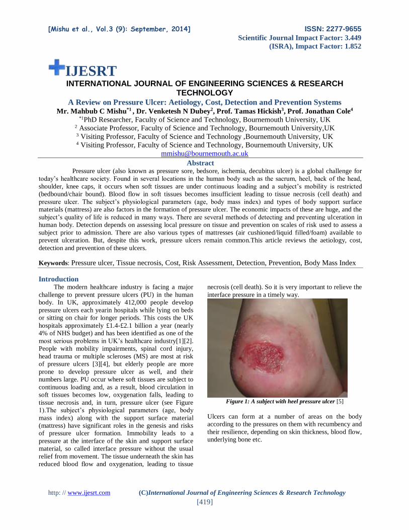
Figure 2: common pressure points in human body [6]
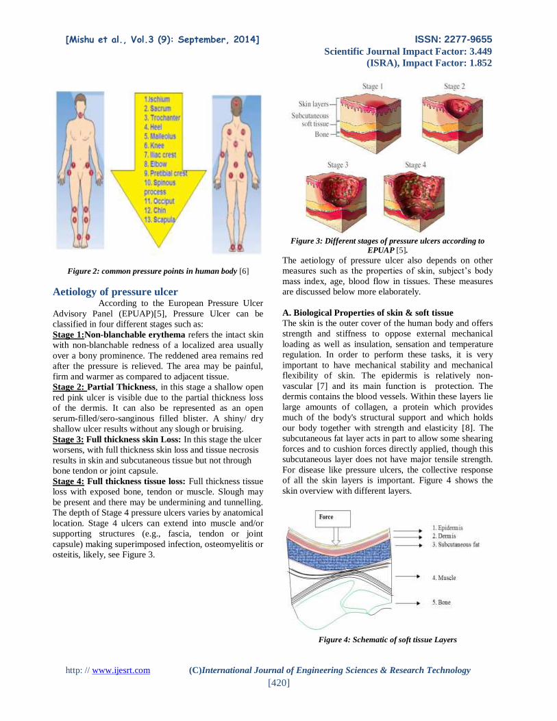
Aetiology of pressure ulcer
According to the European Pressure Ulcer
Advisory Panel (EPUAP)[5], Pressure Ulcer can be
classified in four different stages such as:
Stage 1:Non-blanchable erythema refers the intact skin
with non-blanchable redness of a localized area usually
over a bony prominence. The reddened area remains red
after the pressure is relieved. The area may be painful,
firm and warmer as compared to adjacent tissue.
Stage 2: Partial Thickness, in this stage a shallow open
red pink ulcer is visible due to the partial thickness loss
of the dermis. It can also be represented as an open
serum-filled/sero-sanginous filled blister. A shiny/ dry
shallow ulcer results without any slough or bruising.
Stage 3: Full thickness skin Loss: In this stage the ulcer
worsens, with full thickness skin loss and tissue necrosis
results in skin and subcutaneous tissue but not through
bone tendon or joint capsule.
Stage 4: Full thickness tissue loss: Full thickness tissue
loss with exposed bone, tendon or muscle. Slough may
be present and there may be undermining and tunnelling.
The depth of Stage 4 pressure ulcers varies by anatomical
location. Stage 4 ulcers can extend into muscle and/or
supporting structures (e.g., fascia, tendon or joint
capsule) making superimposed infection, osteomyelitis or
osteitis, likely, see Figure 3.
Figure 3: Different stages of pressure ulcers according to
EPUAP [5].
The aetiology of pressure ulcer also depends on other
measures such as the properties of skin, subject’s body
mass index, age, blood flow in tissues. These measures
are discussed below more elaborately.
A. Biological Properties of skin & soft tissue
The skin is the outer cover of the human body and offers
strength and stiffness to oppose external mechanical
loading as well as insulation, sensation and temperature
regulation. In order to perform these tasks, it is very
important to have mechanical stability and mechanical
flexibility of skin. The epidermis is relatively non-
vascular [7] and its main function is protection. The
dermis contains the blood vessels. Within these layers lie
large amounts of collagen, a protein which provides
much of the body's structural support and which holds
our body together with strength and elasticity [8]. The
subcutaneous fat layer acts in part to allow some shearing
forces and to cushion forces directly applied, though this
subcutaneous layer does not have major tensile strength.
For disease like pressure ulcers, the collective response
of all the skin layers is important. Figure 4 shows the
skin overview with different layers.
Figure 4: Schematic of soft tissue Layers

[Mishu et al., Vol.3 (9): September, 2014] ISSN: 2277-9655
Scientific Journal Impact Factor: 3.449
(ISRA), Impact Factor: 1.852
http: // www.ijesrt.com (C)International Journal of Engineering Sciences & Research Technology
[421]
The pressure distributing properties of muscle are
good[9] but the subcutaneous tissue and particularly
muscle is more susceptible to pressure induced injury
than the epidermis[9], [10]. External forces have
differing effects on the different tissue layers, which in
turn have differing resilience. And of course some body
parts are more susceptible because of the forces they
meet and because of the relation between skin,
subcutaneous tissue and bone. In additionas we age the
collagen content of the dermis is decreased and elasticity
is loss, leading to less resilience to pressure. [11].
B. Subject’s body mass index and age
Body mass index has relevance in the development of
ulcers, since these are more likely to occur in areas where
there is little tissue and fat between the bone and skin. If
a person is malnourished, there is also less cushioning
between the bony surface and the skin [12].Another
factor is age, since with this; Also the skin gets dry.
Patients aged over 65 are more susceptible to develop
pressure ulcers and it has a great deal of correlation with
the skin [13].The changes in skin function and structure
mentioned above, along with risks that occur in overall
health and functional capability can put elderly patient at
a high risk for developing pressure ulcer.
C. Blood flow
Blood flow is a major factor in the formation of pressure
ulcers. When it is reduced, oxygenation to the tissue falls.
Blood flow, in turn, relates to the patient’s systemic
blood pressure since once local pressure in a tissue
exceeds arteriolar pressure, blood flow to that particular
region stops[14][15]. This is known as “localized
ischemia” [16]. An important factor in the skin is the rate
of blood flow in different areas of the body. For example,
sacral blood flow is higher than over the gluteus
maximus[17][16]. This is important because when blood
flow is decreased from an increase in external pressure
there is more damage to the sacral; thus correlating to
more incidences of pressure ulcers in the sacral region
than the gluteus maximus[16].Tissue below the skin
breaks down due to anoxia (the lack of oxygen) and lack
of blood flow, see Figure 5.
Figure 5: Pressure ulcer development over time.
The Economic impact of pressure ulcer
A report by Peter J Franks[18]showed that a
hospital would spend €901,000 to €1,614,000 per year. If
the prevention strategy in the care of patients is included
then the cost will be increased at €3,794,000 [18][19].
The standard cost per person for the different stages of
pressure ulcer has been estimated at €1,489 for stage I,
€6,162 for stage II, €10,238 for stage III and €14,771 for
stage IV. In UK, the number of people who develop PU
annually has been estimated as 140,000 for stage I,
170,000 for stage II, 50,000 for stage III and 50,000 for
stage IV. In European Union (EU) the total annual cost
of pressure ulcers was estimated yearly at €214 million
(stage I), €1,047 million (stage II), €544 million (stage
III) and €670 million (stage IV). In Australia it is
established by a research that a subject with pressure
ulcer requires extra 4.31 days in hospital compared to
other patients and the cost of this extra days were
estimated as AU$28 million (€170.7 million)
yearly[20]. In USA pressure ulcer cost the healthcare
industry US$11 billion yearly with the average cost for
each subjectUS$43,000. Also the length of stay in
hospital is 3 times higher for the subject with pressure
ulcer. Also around 14.8% of total population in USA
develop pressure ulcer whereas around 20% of people
develop pressure ulcer in Europe[21][22]. Figure 6 shows
the population affected by pressure ulcer globally per
year. Apart from the increased morbidity and having
patients at risk of hospital based infections from extended
stays in wards, PU add a huge economic cost to society.
[Mishu et al., Vol.3 (9): September, 2014] ISSN: 2277-9655
Scientific Journal Impact Factor: 3.449
(ISRA), Impact Factor: 1.852
http: // www.ijesrt.com (C)International Journal of Engineering Sciences & Research Technology
[422]
Figure 6: Worldwide population affected by pressure ulcer
[22]
Pressure ulcer Detection Systems
Given these clinical and economic
imperatives, detection and prevention of PUs are of
major importance. Prevention methods have focussed on
risk factors and predictive models, whilst early detection
has involved systems to measure pressures on patients’
skin in vulnerable areas. These systems are either
capacitive or piezoresistive and they measure external
force only. Also load cell sensors, Carbon Nano coil
(CNC), Metal strain gages are available for detection
purposes. These technologies have both advantages and
disadvantages, e.g. capacitive pressure sensors are
susceptible to electrical interference due to its high
impedance,metal strain gauges needs supplementary
configuration to identify force[23].
In (Yip, M. et al., 2009)capacitive sensors are used to
measure the external forceon the human body. The
change of capacitance occurs over a small distance of
place due to a separation of two conductive plates [24].
An example of capacitive pressure sensing established by
Yip, M. is shown in figure 7.
Figure 7: Parallel plate capacitor model with variable
capacitance due to modulation of the dielectric thickness by
the applied pressure [24]
In figure 8, a schematic of capacitive sensor array is
shown (described by [24]. The capacitors are arranged in
11 by 9 arrays. Each unit capacitance C x y in row x and
column y depends on the pressure applied there. The
array is scanned in every 81ms at a sampling frequency
of 12-HZ. A 16-bit analogue-to-digital (A to D)
converter is used to obtain results. The columns are
multiplexed by using one 2:1 multiplexer per column. An
array of capacitive pressure sensors is located under the
patient’s bed. The sensing is done by an analogue device.
A low power microcontroller controls the measurement
sequence. The digitized data is then transmitted to a
computer via a USB interface using a chip. A Graphic
User Interface written in visual basic is used to plot the
data in real time and post processing is done in Matlab.
In order to interface the electronics with the sensor sheet,
a USB-powered PCB was designed and used.
Figure 8: Capacitive sensor array [24]
Though the hysteresis of the capacitive system is
recorded >10% but the limitation of this type of design
includes sensor to sensor variations and drifting. Periodic
re-calibration is required for individual sensor to
overcome the drifting. Moreover, the wearing is complex
and the power consumption is high in this type of design.
Also this research does not show individual pressure
induced in tissue and support surface.
Abraham et al, have designed a low cost, disposable
mattress for non-invasive sleep and movement
monitoring, see Figure 9. cPaper which is a nonwoven
material is used to design the pressure sensing array
using capacitive principles [25].The conductivity of a
single ply cPaper area can be controlled by loading
[Mishu et al., Vol.3 (9): September, 2014] ISSN: 2277-9655
Scientific Journal Impact Factor: 3.449
(ISRA), Impact Factor: 1.852
http: // www.ijesrt.com (C)International Journal of Engineering Sciences & Research Technology
[423]
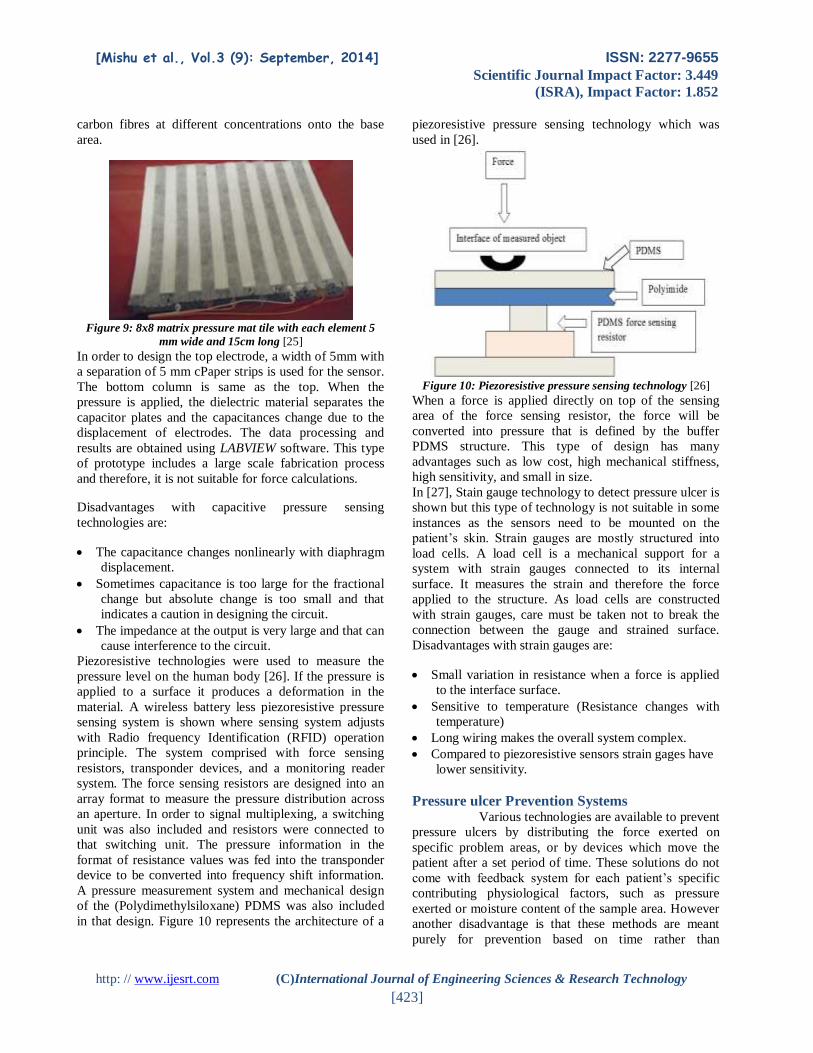
carbon fibres at different concentrations onto the base
area.
Figure 9: 8x8 matrix pressure mat tile with each element 5
mm wide and 15cm long [25]
In order to design the top electrode, a width of 5mm with
a separation of 5 mm cPaper strips is used for the sensor.
The bottom column is same as the top. When the
pressure is applied, the dielectric material separates the
capacitor plates and the capacitances change due to the
displacement of electrodes. The data processing and
results are obtained using LABVIEW software. This type
of prototype includes a large scale fabrication process
and therefore, it is not suitable for force calculations.
Disadvantages with capacitive pressure sensing
technologies are:
The capacitance changes nonlinearly with diaphragm
displacement.
Sometimes capacitance is too large for the fractional
change but absolute change is too small and that
indicates a caution in designing the circuit.
The impedance at the output is very large and that can
cause interference to the circuit.
Piezoresistive technologies were used to measure the
pressure level on the human body [26]. If the pressure is
applied to a surface it produces a deformation in the
material. A wireless battery less piezoresistive pressure
sensing system is shown where sensing system adjusts
with Radio frequency Identification (RFID) operation
principle. The system comprised with force sensing
resistors, transponder devices, and a monitoring reader
system. The force sensing resistors are designed into an
array format to measure the pressure distribution across
an aperture. In order to signal multiplexing, a switching
unit was also included and resistors were connected to
that switching unit. The pressure information in the
format of resistance values was fed into the transponder
device to be converted into frequency shift information.
A pressure measurement system and mechanical design
of the (Polydimethylsiloxane) PDMS was also included
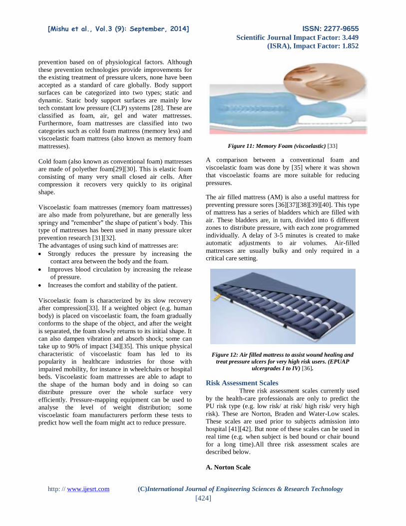
in that design. Figure 10 represents the architecture of a
piezoresistive pressure sensing technology which was
used in [26].
Figure 10: Piezoresistive pressure sensing technology [26]
When a force is applied directly on top of the sensing
area of the force sensing resistor, the force will be
converted into pressure that is defined by the buffer
PDMS structure. This type of design has many
advantages such as low cost, high mechanical stiffness,
high sensitivity, and small in size.
In [27], Stain gauge technology to detect pressure ulcer is
shown but this type of technology is not suitable in some
instances as the sensors need to be mounted on the
patient’s skin. Strain gauges are mostly structured into
load cells. A load cell is a mechanical support for a
system with strain gauges connected to its internal
surface. It measures the strain and therefore the force
applied to the structure. As load cells are constructed
with strain gauges, care must be taken not to break the
connection between the gauge and strained surface.
Disadvantages with strain gauges are:
Small variation in resistance when a force is applied
to the interface surface.
Sensitive to temperature (Resistance changes with
temperature)
Long wiring makes the overall system complex.
Compared to piezoresistive sensors strain gages have
lower sensitivity.
Pressure ulcer Prevention Systems
Various technologies are available to prevent
pressure ulcers by distributing the force exerted on
specific problem areas, or by devices which move the
patient after a set period of time. These solutions do not
come with feedback system for each patient’s specific
contributing physiological factors, such as pressure
exerted or moisture content of the sample area. However
another disadvantage is that these methods are meant
purely for prevention based on time rather than
[Mishu et al., Vol.3 (9): September, 2014] ISSN: 2277-9655
Scientific Journal Impact Factor: 3.449
(ISRA), Impact Factor: 1.852
http: // www.ijesrt.com (C)International Journal of Engineering Sciences & Research Technology
[424]
prevention based on of physiological factors. Although
these prevention technologies provide improvements for
the existing treatment of pressure ulcers, none have been
accepted as a standard of care globally. Body support
surfaces can be categorized into two types; static and
dynamic. Static body support surfaces are mainly low
tech constant low pressure (CLP) systems [28]. These are
classified as foam, air, gel and water mattresses.
Furthermore, foam mattresses are classified into two
categories such as cold foam mattress (memory less) and
viscoelastic foam mattress (also known as memory foam
mattresses).
Cold foam (also known as conventional foam) mattresses
are made of polyether foam[29][30]. This is elastic foam
consisting of many very small closed air cells. After
compression it recovers very quickly to its original
shape.
Viscoelastic foam mattresses (memory foam mattresses)
are also made from polyurethane, but are generally less
springy and "remember" the shape of patient’s body. This
type of mattresses has been used in many pressure ulcer
prevention research [31][32].
The advantages of using such kind of mattresses are:
Strongly reduces the pressure by increasing the
contact area between the body and the foam.
Improves blood circulation by increasing the release
of pressure.
Increases the comfort and stability of the patient.
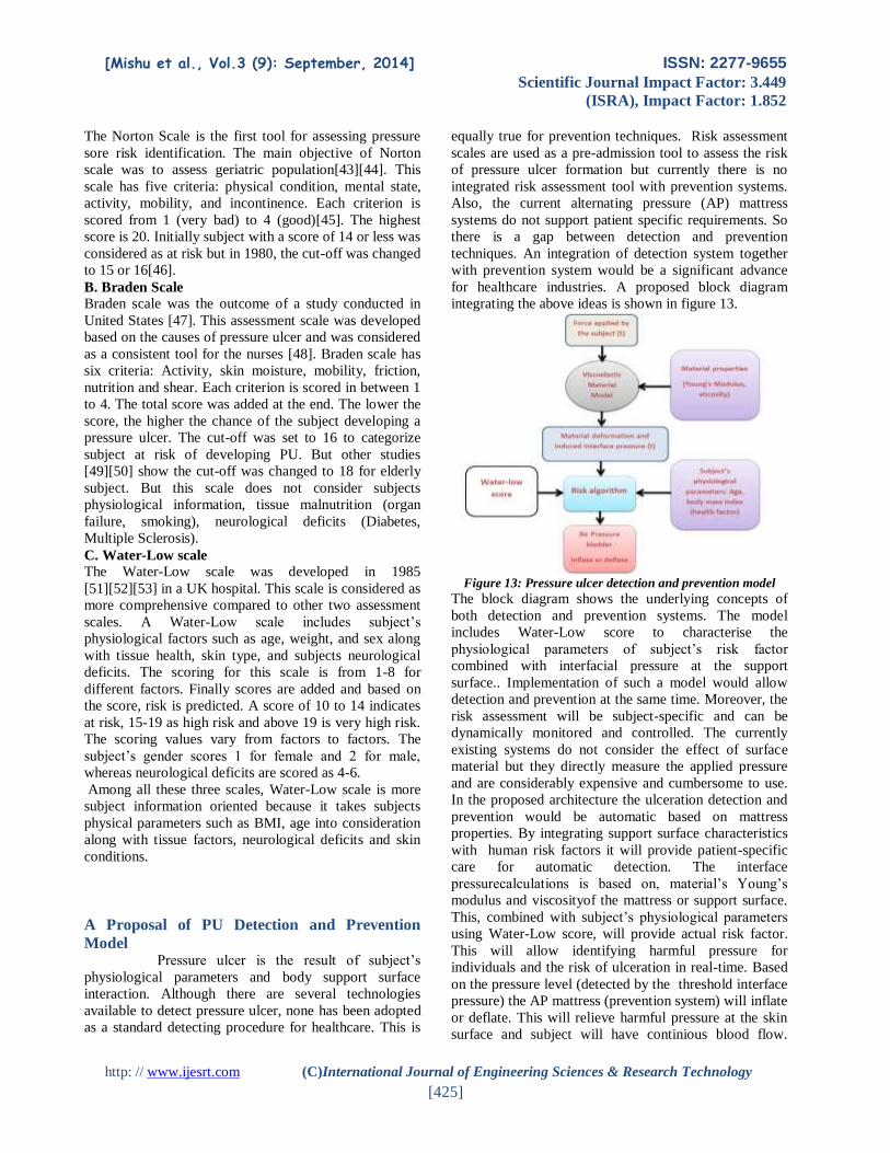
Viscoelastic foam is characterized by its slow recovery
after compression[33]. If a weighted object (e.g. human
body) is placed on viscoelastic foam, the foam gradually
conforms to the shape of the object, and after the weight
is separated, the foam slowly returns to its initial shape. It
can also dampen vibration and absorb shock; some can
take up to 90% of impact [34][35]. This unique physical
characteristic of viscoelastic foam has led to its
popularity in healthcare industries for those with
impaired mobility, for instance in wheelchairs or hospital
beds. Viscoelastic foam mattresses are able to adapt to
the shape of the human body and in doing so can
distribute pressure over the whole surface very
efficiently. Pressure-mapping equipment can be used to
analyse the level of weight distribution; some
viscoelastic foam manufacturers perform these tests to
predict how well the foam might act to reduce pressure.
Figure 11: Memory Foam (viscoelastic) [33]
A comparison between a conventional foam and
viscoelastic foam was done by [35] where it was shown
that viscoelastic foams are more suitable for reducing
pressures.
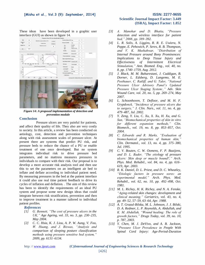
The air filled mattress (AM) is also a useful mattress for
preventing pressure sores [36][37][38][39][40]. This type
of mattress has a series of bladders which are filled with
air. These bladders are, in turn, divided into 6 different
zones to distribute pressure, with each zone programmed
individually. A delay of 3-5 minutes is created to make
automatic adjustments to air volumes. Air-filled
mattresses are usually bulky and only required in a
critical care setting.
Figure 12: Air filled mattress to assist wound healing and
treat pressure ulcers for very high risk users. (EPUAP
ulcergrades I to IV) [36].
Risk Assessment Scales Three risk assessment scales currently used
by the health-care professionals are only to predict the
PU risk type (e.g. low risk/ at risk/ high risk/ very high
risk). These are Norton, Braden and Water-Low scales.
These scales are used prior to subjects admission into
hospital [41][42]. But none of these scales can be used in
real time (e.g. when subject is bed bound or chair bound
for a long time).All three risk assessment scales are
described below.
A. Norton Scale
[Mishu et al., Vol.3 (9): September, 2014] ISSN: 2277-9655
Scientific Journal Impact Factor: 3.449
(ISRA), Impact Factor: 1.852
http: // www.ijesrt.com (C)International Journal of Engineering Sciences & Research Technology
[425]
The Norton Scale is the first tool for assessing pressure
sore risk identification. The main objective of Norton
scale was to assess geriatric population[43][44]. This
scale has five criteria: physical condition, mental state,
activity, mobility, and incontinence. Each criterion is
scored from 1 (very bad) to 4 (good)[45]. The highest
score is 20. Initially subject with a score of 14 or less was
considered as at risk but in 1980, the cut-off was changed
to 15 or 16[46].
B. Braden Scale
Braden scale was the outcome of a study conducted in
United States [47]. This assessment scale was developed
based on the causes of pressure ulcer and was considered
as a consistent tool for the nurses [48]. Braden scale has
six criteria: Activity, skin moisture, mobility, friction,
nutrition and shear. Each criterion is scored in between 1
to 4. The total score was added at the end. The lower the
score, the higher the chance of the subject developing a
pressure ulcer. The cut-off was set to 16 to categorize
subject at risk of developing PU. But other studies
[49][50] show the cut-off was changed to 18 for elderly
subject. But this scale does not consider subjects
physiological information, tissue malnutrition (organ
failure, smoking), neurological deficits (Diabetes,
Multiple Sclerosis).
C. Water-Low scale
The Water-Low scale was developed in 1985
[51][52][53] in a UK hospital. This scale is considered as
more comprehensive compared to other two assessment
scales. A Water-Low scale includes subject’s
physiological factors such as age, weight, and sex along
with tissue health, skin type, and subjects neurological
deficits. The scoring for this scale is from 1-8 for
different factors. Finally scores are added and based on
the score, risk is predicted. A score of 10 to 14 indicates
at risk, 15-19 as high risk and above 19 is very high risk.
The scoring values vary from factors to factors. The
subject’s gender scores 1 for female and 2 for male,
whereas neurological deficits are scored as 4-6.
Among all these three scales, Water-Low scale is more
subject information oriented because it takes subjects
physical parameters such as BMI, age into consideration
along with tissue factors, neurological deficits and skin
conditions.
A Proposal of PU Detection and Prevention
Model Pressure ulcer is the result of subject’s
physiological parameters and body support surface
interaction. Although there are several technologies
available to detect pressure ulcer, none has been adopted
as a standard detecting procedure for healthcare. This is
equally true for prevention techniques. Risk assessment
scales are used as a pre-admission tool to assess the risk
of pressure ulcer formation but currently there is no
integrated risk assessment tool with prevention systems.
Also, the current alternating pressure (AP) mattress
systems do not support patient specific requirements. So
there is a gap between detection and prevention
techniques. An integration of detection system together
with prevention system would be a significant advance
for healthcare industries. A proposed block diagram
integrating the above ideas is shown in figure 13.
Figure 13: Pressure ulcer detection and prevention model
The block diagram shows the underlying concepts of
both detection and prevention systems. The model
includes Water-Low score to characterise the
physiological parameters of subject’s risk factor
combined with interfacial pressure at the support
surface.. Implementation of such a model would allow
detection and prevention at the same time. Moreover, the
risk assessment will be subject-specific and can be
dynamically monitored and controlled. The currently
existing systems do not consider the effect of surface
material but they directly measure the applied pressure
and are considerably expensive and cumbersome to use.
In the proposed architecture the ulceration detection and
prevention would be automatic based on mattress
properties. By integrating support surface characteristics
with human risk factors it will provide patient-specific
care for automatic detection. The interface
pressurecalculations is based on, material’s Young’s
modulus and viscosityof the mattress or support surface.
This, combined with subject’s physiological parameters
using Water-Low score, will provide actual risk factor.
This will allow identifying harmful pressure for
individuals and the risk of ulceration in real-time. Based
on the pressure level (detected by the threshold interface
pressure) the AP mattress (prevention system) will inflate
or deflate. This will relieve harmful pressure at the skin
surface and subject will have continious blood flow.
[Mishu et al., Vol.3 (9): September, 2014] ISSN: 2277-9655
Scientific Journal Impact Factor: 3.449
(ISRA), Impact Factor: 1.852
http: // www.ijesrt.com (C)International Journal of Engineering Sciences & Research Technology
[426]
These ideas have been developed in a graphic user
interface (GUI) as shown in figure 14.
Figure 14: A proposed implemantation of detection and
prevention module
Conclusion
Pressure ulcers are very painful for patients,
and affect their quality of life. They also are very costly
to society. In this article, a review has been conducted on
aetiology, cost, detection and prevention techniques
along with risk assessment scales of pressure ulcer. At
present there are systems that predict PU risk, and
pressure beds to reduce the chance of a PU or enable
treatment of one once developed. But no system
integrates individual risk to drive pressure bed
parameters, and no mattress measures pressures in
individuals to compare with their risk. Our proposal is to
develop a more accurate risk analysis tool and then use
this to set the parameters on an intelligent air bed to
inflate and deflate according to individual patient need.
By measuring pressures in the bed at the patient interface
it could also use real time patient feedback to drive its
cycles of inflation and deflation. . The aim of this review
has been to identify the requirements of an ideal PU
system and propose some new design ideas that could
integrate between risk stratification, prevention and then
to improve treatment in a manner tailored to individual
patient profiles.
References [1] G. Bennett, “The cost of pressure ulcers in the
UK,” Age Ageing, vol. 33, no. 3, pp. 230–235,
May 2004.
[2] C. C. Hsia, K. J. Liou, A. P. W. Aung, V. Foo,
W. Huang, and J. Biswas, “Analysis and
comparison of sleeping posture classification
methods using pressure sensitive bed system,”
2009, pp. 6131–6134.
[3] A. Manohar and D. Bhatia, “Pressure
detection and wireless interface for patient
bed,” 2008, pp. 389–392.
[4] L. R. Solis, A. Liggins, R. R. E. Uwiera, N.
Poppe, E. Pehowich, P. Seres, R. B. Thompson,
and V. K. Mushahwar, “Distribution of
Internal Pressure around Bony Prominences:
Implications to Deep Tissue Injury and
Effectiveness of Intermittent Electrical
Stimulation,” Ann. Biomed. Eng., vol. 40, no.
8, pp. 1740–1759, Aug. 2012.
[5] J. Black, M. M. Baharestani, J. Cuddigan, B.
Dorner, L. Edsberg, D. Langemo, M. E.
Posthauer, C. Ratliff, and G. Taler, “National
Pressure Ulcer Advisory Panel’s Updated
Pressure Ulcer Staging System:,” Adv. Skin
Wound Care, vol. 20, no. 5, pp. 269–274, May
2007.
[6] L. Schoonhoven, T. Defloor, and M. H. F.
Grypdonck, “Incidence of pressure ulcers due
to surgery,” J. Clin. Nurs., vol. 11, no. 4, pp.
479–487, Jul. 2002.
[7] Y. Zeng, Y. Liu, C. Xu, X. Xu, H. Xu, and G.
Sun, “Biomechanical properties of skin in vitro
for different expansion methods,” Clin.
Biomech., vol. 19, no. 8, pp. 853–857, Oct.
2004.
[8] C. Edwards and R. Marks, “Evaluation of
biomechanical properties of human skin,”
Clin. Dermatol., vol. 13, no. 4, pp. 375–380,
Jul. 1995.
[9] C. V. Bouten, C. W. Oomens, F. P. Baaijens,
and D. L. Bader, “The etiology of pressure
ulcers: Skin deep or muscle bound?,” Arch.
Phys. Med. Rehabil., vol. 84, no. 4, pp. 616–
619, Apr. 2003.
[10] R. K. Daniel, D. L. Priest, and D. C. Wheatley,
“Etiologic factors in pressure sores: an
experimental model,” Arch. Phys. Med.
Rehabil., vol. 62, no. 10, pp. 492–498, Oct.
1981.
[11] M. L. Richey, H. K. Richey, and N. A. Fenske,
“Aging-related skin changes: development and
clinical meaning,” Geriatrics, vol. 43, no. 4,
pp. 49–52, 57–59, 63–64, Apr. 1988.
[12] A. T. Grazul-Bilska, M. L. Johnson, J. J. Bilski,
D. A. Redmer, L. P. Reynolds, A. Abdullah, and
K. M. Abdullah, “Wound healing: The role of
growth factors,” Drugs Today, vol. 39, no. 10,
p. 787, 2003.
[13] Y. Chen, M. J. DeVivo, and A. B. Jackson,
“Pressure Ulcer Prevalence in People With
Spinal Cord Injury: Age-Period-Duration
[Mishu et al., Vol.3 (9): September, 2014] ISSN: 2277-9655
Scientific Journal Impact Factor: 3.449
(ISRA), Impact Factor: 1.852
http: // www.ijesrt.com (C)International Journal of Engineering Sciences & Research Technology
[427]
Effects,” Arch. Phys. Med. Rehabil., vol. 86,
no. 6, pp. 1208–1213, Jun. 2005.
[14] A. C. Ek, G. Gustavsson, and D. H. Lewis,
“The local skin blood flow in areas at risk for
pressure sores treated with massage,” Scand.
J. Rehabil. Med., vol. 17, no. 2, pp. 81–86,
1985.
[15] R. J. van Marum, J. H. Meijer, and M. W.
Ribbe, “The relationship between pressure
ulcers and skin blood flow response after a
local cold provocation,” Arch. Phys. Med.
Rehabil., vol. 83, no. 1, pp. 40–43, Jan. 2002.
[16] D. R. Thomas, “Does Pressure Cause Pressure
Ulcers? An Inquiry Into the Etiology of
Pressure Ulcers,” J. Am. Med. Dir. Assoc., vol.
11, no. 6, pp. 397–405, Jul. 2010.
[17] R. Frantz, G. C. Xakellis, and M. Arteaga,
“The effects of prolonged pressure on skin
blood flow in elderly patients at risk for
pressure ulcers,” Decubitus, vol. 6, no. 6, pp.
16–20, Nov. 1993.
[18] J. Posnett and P. J. Franks, “The burden of
chronic wounds in the UK,” Nurs. Times, vol.
104, no. 3, pp. 44–45, Jan. 2008.
[19] N. Graves, F. Birrell, and M. Whitby, “Effect
of Pressure Ulcers on Length of Hospital Stay
•,” Infect. Control Hosp. Epidemiol., vol. 26,
no. 3, pp. 293–297, Mar. 2005.
[20] N. Graves, F. A. Birrell, and M. Whitby,
“Modeling the economic losses from pressure
ulcers among hospitalized patients in
Australia,” Wound Repair Regen., vol. 13, no.
5, pp. 462–467, Sep. 2005.
[21] C. H. Lyder, R. Shannon, O. Empleo-Frazier,
D. McGeHee, and C. White, “A comprehensive
program to prevent pressure ulcers in long-
term care: exploring costs and outcomes,”
Ostomy. Wound Manage., vol. 48, no. 4, pp.
52–62, Apr. 2002.
[22] K. Spilsbury, A. Nelson, N. Cullum, C. Iglesias,
J. Nixon, and S. Mason, “Pressure ulcers and
their treatment and effects on quality of life:
hospital inpatient perspectives,” J. Adv. Nurs.,
vol. 57, no. 5, pp. 494–504, Mar. 2007.
[23] J. Elfehri, F. Boussu, V. Koncar, and C.
Vasseur, “Novel approach of ulcer prevention
based on pressure distribution control
algorithm,” 2011, pp. 265–270.
[24] M. Yip, D. D. He, E. Winokur, A. G.
Balderrama, R. Sheridan, and Hongshen Ma,
“A flexible pressure monitoring system for
pressure ulcer prevention,” 2009, pp. 1212–
1215.
[25] J. K. Abraham, S. Sullivan, and S.
Ranganathan, “Low-cost and disposable
pressure sensor mat for non-invasive sleep and
movement monitoring applications,” 2011, pp.
4745–4748.
[26] L.-C. Hsu, W.-D. Huang, S.-J. Tang, and J. C.
Chiao, “<title>A wireless batteryless
piezoresistive pressure sensing
system</title>,” 2008, p. 72681D–
72681D–10.
[27] Y. Zhu, L. Shen, and J. Song, “Body pressure
distribution research and zone design of pocket
spring mattresses,” 2011, pp. 1656–1659.
[28] C. Bansal, R. Scott, D. Stewart, and C. J.
Cockerell, “Decubitus ulcers: A review of the
literature,” Int. J. Dermatol., vol. 44, no. 10,
pp. 805–810, Oct. 2005.
[29] M. van Leen, S. Hovius, J. Neyens, R. Halfens,
and J. Schols, “Pressure relief, cold foam or
static air? A single center, prospective,
controlled randomized clinical trial in a Dutch
nursing home,” J. Tissue Viability, vol. 20, no.
1, pp. 30–34, Feb. 2011.
[30] M. Malbrain, B. Hendriks, P. Wijnands, D.
Denie, A. Jans, J. Vanpellicom, and B. De
Keulenaer, “A pilot randomised controlled
trial comparing reactive air and active
alternating pressure mattresses in the
prevention and treatment of pressure ulcers
among medical ICU patients,” J. Tissue
Viability, vol. 19, no. 1, pp. 7–15, Feb. 2010.
[31] L. Gunningberg, “Are patients with or at risk
of pressure ulcers allocated appropriate
prevention measures?,” Int. J. Nurs. Pract.,
vol. 11, no. 2, pp. 58–67, Apr. 2005.
[32] T. Defloor, D. D. Bacquer, and M. H. F.
Grypdonck, “The effect of various
combinations of turning and pressure reducing
devices on the incidence of pressure ulcers,”
Int. J. Nurs. Stud., vol. 42, no. 1, pp. 37–46,
Jan. 2005.
[33] J. Feuchtinger, R. Bie, T. Dassen, and R.
Halfens, “A 4-cm thermoactive viscoelastic
foam pad on the operating room table to
prevent pressure ulcer during cardiac
surgery,” J. Clin. Nurs., vol. 15, no. 2, pp.
162–167, Feb. 2006.
[34] J. Nixon, D. McElvenny, S. Mason, J. Brown,
and S. Bond, “A sequential randomised
controlled trial comparing a dry visco-elastic
polymer pad and standard operating table
mattress in the prevention of post-operative
pressure sores,” Int. J. Nurs. Stud., vol. 35, no.
4, pp. 193–203, Aug. 1998.
[Mishu et al., Vol.3 (9): September, 2014] ISSN: 2277-9655
Scientific Journal Impact Factor: 3.449
(ISRA), Impact Factor: 1.852
http: // www.ijesrt.com (C)International Journal of Engineering Sciences & Research Technology
[428]
[35] T. Defloor and J. D. S. De Schuijmer,
“Preventing pressure ulcers: An evaluation of
four operating-table mattresses,” Appl. Nurs.
Res., vol. 13, no. 3, pp. 134–141, Aug. 2000.
[36] D. Colin, J.-M. Rochet, P. Ribinik, B. Barrois,
Y. Passadori, and J.-M. Michel, “What is the
best support surface in prevention and
treatment, as of 2012, for a patient at risk
and/or suffering from pressure ulcer sore?
Developing French guidelines for clinical
practice,” Ann. Phys. Rehabil. Med., vol. 55,
no. 7, pp. 466–481, Oct. 2012.
[37] R. Fontaine, “A quantitative analysis of
pressure and shear in the effectiveness of
support surfaces*1,” J. WOCN, vol. 25, no. 5,
pp. 227–232, Sep. 1998.
[38] E. Sving, L. Gunningberg, M. Högman, and A.-
G. Mamhidir, “Registered nurses’ attention to
and perceptions of pressure ulcer prevention in
hospital settings: Registered nurses’ attention
to pressure ulcer,” J. Clin. Nurs., vol. 21, no.
9–10, pp. 1293–1303, May 2012.
[39] J. W. DeVocht, D. G. Wilder, E. R. Bandstra,
and K. F. Spratt, “Biomechanical evaluation of
four different mattresses,” Appl. Ergon., vol.
37, no. 3, pp. 297–304, May 2006.
[40] T. Defloor, “The effect of position and mattress
on interface pressure,” Appl. Nurs. Res., vol.
13, no. 1, pp. 2–11, Feb. 2000.
[41] P. Papanikolaou, P. Lyne, and D. Anthony,
“Risk assessment scales for pressure ulcers: A
methodological review,” Int. J. Nurs. Stud.,
vol. 44, no. 2, pp. 285–296, Feb. 2007.
[42] L. Bolton, “Which Pressure Ulcer Risk
Assessment Scales are Valid for Use in the
Clinical Setting?:,” J. Wound. Ostomy
Continence Nurs., vol. 34, no. 4, pp. 368–381,
Jul. 2007.
[43] B. Berglund and G. Nordström, “The Use of
the Modified Norton Scale in Nursing-Home
Patients,” Scand. J. Caring Sci., vol. 9, no. 3,
pp. 165–169, Sep. 1995.
[44] J. Cox, “Predictors of Pressure Ulcers in Adult
Critical Care Patients,” Am. J. Crit. Care, vol.
20, no. 5, pp. 364–375, Sep. 2011.
[45] R. Lincoln, R. Roberts, A. Maddox, S. Levine,
and C. Patterson, “Use of the Norton Pressure
Sore Risk Assessment Scoring System with
elderly patients in acute care,” J. Enterostomal
Ther., vol. 13, no. 4, pp. 132–138, Aug. 1986.
[46] F. Hamilton, “An analysis of the literature
pertaining to pressure sore risk-assessment
scales,” J. Clin. Nurs., vol. 1, no. 4, pp. 185–
193, Jul. 1992.
[47] N. Bergstrom, P. J. Demuth, and B. J. Braden,
“A clinical trial of the Braden Scale for
Predicting Pressure Sore Risk,” Nurs. Clin.
North Am., vol. 22, no. 2, pp. 417–428, Jun.
1987.
[48] G. D. Salvadalena, M. L. Snyder, and K. E.
Brogdon, “Clinical trial of the Braden Scale
on an acute care medical unit,” J. Nurs. Off.
Publ. Int. Assoc. Enteros. Ther., vol. 19, no. 5,
pp. 160–165, Oct. 1992.
[49] C. H. Lyder, C. Yu, J. Emerling, R. Mangat, D.
Stevenson, O. Empleo-Frazier, and J. McKay,
“The braden scale for pressure ulcer risk:
Evaluating the predictive validity in black and
latino/hispanic elders,” Appl. Nurs. Res., vol.
12, no. 2, pp. 60–68, May 1999.
[50] T. VandenBosch, C. Montoye, M. Satwicz, K.
Durkee-Leonard, and B. Boylan-Lewis,
“Predictive validity of the braden scale and
nurse perception in identifying pressure ulcer
risk,” Appl. Nurs. Res., vol. 9, no. 2, pp. 80–86,
May 1996.
[51] J. Waterlow, “Pressure sores: a risk
assessment card,” Nurs. Times, vol. 81, no. 48,
pp. 49–55, Dec. 1985.
[52] C. Wai-Han, C. Kit-Wai, P. French, L. Yim-
Sheung, and T. Lai-Kwan, “Which pressure
sore risk calculator? A study of the
effectiveness of the Norton scale in Hong
Kong,” Int. J. Nurs. Stud., vol. 34, no. 2, pp.
165–169, Apr. 1997.
[53] K. Balzer, C. Pohl, T. Dassen, and R. Halfens,
“The Norton, Waterlow, Braden, and Care
Dependency ScalesL Comparing Their Validity
When Identifying Patientsʼ Pressure Sore
Risk:,” J. Wound. Ostomy Continence Nurs.,
vol. 34, no. 4, pp. 389–398, Jul. 2007.
Copyright © 2022 FDOKUMEN