Cell fate decisions within the mouse organizer are governed by graded Nodal signals

The Drosophila Jun-N-terminal kinase is required for cell morphogenesis but not for DJun-dependent cell fate specification in the eye Juan R. Riesgo-Escovar, Mario Jenni, Andreas Fritz, 1 and Ernst Hafen 2
Zoologisches Institut, Universit~it Zi~rich, Winterthurerstrasse 190, CH-8057 Z~irich, Switzerland
We cloned and characterized the Drosophila homolog of mammalian Jun-N-terminal kinases (DJNK). We show that DJNK is encoded by basket (bsk). Like hemipterous (hep), which encodes the Drosophila JNK kinase, bsk is required in the embryo for dorsal closure, a process involving coordinate cell shape changes of ectodermal cells. Dorsal closure can also be blocked by dominant negative Drosophila cdc42, which has been shown to act upstream of JNKK in vertebrates. Therefore it appears that the JNK pathway is conserved and that it is involved in controlling cell morphogenesis in Drosophila. Although DJNK efficiently phosphorylates DJun in vitro, bsk function is not required for the specification of cell fate in the developing eye, a process that requires MAP kinase and DJun function.
[Key Words: Jun-N-terminal kinase; dorsal closure; basket; Drosophila; MAPK]
Received July 8, 1996; revised version accepted September 17, 1996.
Mitogen-activated protein kinase (MAPK) cascades transduce a variety of signals in eukaryotic cells in re- sponse to multiple extracellular stimuli. Depending on cell type, duration of stimulus, and pathway, they medi- ate a range of responses including proliferation, differen- tiation, and the regulation of metabolic pathways in dif- ferentiated cells (Marshall 1995). In yeast, for example, three different MAPK pathways have been identified, each one regulating different intracellular responses (Herskowitz 1995).
In vertebrate cells, much has been learned from bio- chemical studies of the extracellular-signal-regulated protein kinases (ERK) pathways and their involvement in growth control and cellular differentiation (for review, see Marshall 1994, 1995; Nishida and Gotoh 1993). In invertebrates, the corresponding MAPK pathway has been unraveled through the use of genetics in Drosophila (for review, see Dickson and Hafen 1993; Zipursky and Rubin 1994; Wassarman et al. 1995), Caenorhabditis el- egans (for review, see Kayne and Sternberg 1995; Kenyon 1995), and yeast (Herskowitz 1995). In Drosophila, this MAPK pathway (R1/MAPK) is required for many pro- cesses during development, such as in the specification of terminal structures of the larva in response to the Torso receptor, in the formation of wing veins in re- sponse to the Drosophila EGF receptor homolog, and in
~Present address: Institute of Neuroscience, University of Oregon, Eu- gene, Oregon 97403 USA. 2Corresponding author.
the differentiation of photoreceptor cells in the com- pound eye (for review, see Dominguez and Hafen 1996).
By contrast, the role of the Jun-N-terminal kinase (JNK) pathway, another MAPK pathway, is less well un- derstood. In particular, there is a dearth of genetic stud- ies of the JNK pathway in a multicellular organism, al- though recently a Drosophila JNK kinase has been re- ported (Glise et al. 1995). From studies in vertebrate cell culture systems the JNK pathway has been implicated in the response of cells to stress, growth factors, and Ras activation (Karin and Hunter 1995). In vitro, mammalian JNKs efficiently phosphorylate c-Jun on two serine resi- dues (Ser63 and Ser73) in the amino-terminal domain of the protein. This phosphorylation correlates well with c-Jun activation. In contrast, ERKs seem to phosphory- late c-Jun at another site in the carboxy-terminal domain that correlates with inhibition of c-Jun DNA binding (Karin and Hunter 1995). Ras can activate both path- ways, but to different potencies in several cell lines (Karin and Hunter 1995). Recent reports have suggested other interactions between the JNK and ERK pathways. For example, in rat PC-12 cells, the JNK pathway plays a role in apoptosis, whereas the ERK pathway promotes cell survival (Xia et al. 1995). In contrast, in anergic T cells both pathways need to be activated to mediate the response of T cells to antigen presentation (Suet al. 1994; Fields et al. 1996; Li et al. 1996). Because all these studies have been carried out in cultured cells, little is known about the role of the JNK pathway during the development of multicellular organisms and its interac-
GENES & DEVELOPMENT 10:2759-2768 �9 1996 by Cold Spring Harbor Laboratory Press ISSN 0890-9369/96 $5.00 2759
Cold Spring Harbor Laboratory Press on October 14, 2016 - Published by genesdev.cshlp.orgDownloaded from
Riesgo-Escovar et al.
tion with other parallel MAP kinase pathways such as the ERK pathway in vivo.
To characterize the JNK pathway in a genetically ame- nable muhicel lular organism, we have cloned and char- acterized DJNK, the Drosophila homolog of mammalian JNKs. Here we show that DJNK is encoded by the basket (bsk) gene and that mutations in bsk, like mutations in the DJNKK gene hemipterous (hep) (Glise et al. 1995), disrupt dorsal closure, a process during embryonic devel- opment that involves cell shape changes and cytoskele- tal reorganization (Young et al. 1993). We present evi- dence that the JNK and R1/MAPK pathways in Droso- phila are largely independent in the embryo and the eye. Our results also suggest that DJNK function is not re- quired for Jun function in the eye.
Results
Drosophila INK is a protein kinase
From a staged embryonic cDNA library we isolated cDNAs whose deduced amino acid sequence exhibits high similarity to mammal ian JNKs. Because of this, we named the encoded protein DJNK (Drosophila Jun-N- terminal kinase). Northern blot analysis probed with the DJNK cDNA revealed a single band of 1.5 kb, the size of which corresponds to the longest cDNA (1507 bp)(data not shown).
The degree of amino acid identity between the Droso- phila sequence and its mammalian counterparts is >70% (Fig. 1A). In the kinase domain, DJNK contains all the conserved residues of protein kinases, and the TPY motif between kinase subdomains VII and VIII that is phosphorylated on the threonine and tyrosine residues to activate JNKs. The amino acid sequence between kinase subdomains IX and X varies between the INKs, and de- termines the efficiency of binding to c-Jun (Kallunki et al. 1994). In this region, DJNK is most homologous to JNK2. In contrast, DJNK shares only 50% amino acid identity with the Drosophila Rolled (R1) MAPK. This suggests that R1/ERK MAPKs and JNKs diverged in ev- olution before the separation of vertebrates and inverte- brates.
Next, we characterized DJNK biochemically. To do so, we performed in vitro kinase assays with Jun and myelin basic protein (MBP) as substrates. Western blot analysis of Drosophila extracts of embryos, larvae, pupae, and adults probed with anti-JNK1 antibody revealed a single band with an apparent molecular weight of 49 kD, the predicted weight of DJNK (Fig. 2A). This antibody spe- cifically recognizes DJNK and does not cross-react with the similarly sized R1/MAP kinase on Western blots (Fig. 2B). To test for DJNK protein kinase activity we immu- noprecipitated DJNK and R1/MAP kinase from larval protein extracts and performed in vitro kinase assays on the immunoprecipitates using DJun as a substrate (Fig. 2B). Like their vertebrate homologs, both DJNK and R1/ MAP kinase phosphorylated DJun.
Finally, we wanted to test whether DJNK can be in- duced by stress, as it has been shown for mammalian
A 1 5 0
huJNKl . . . . . . . . . . . . . . . . . . . . . . . . . . . . . . . . . . . . . MSR SKRDNNFYSV
huJNK3 MSLHFLYYCS EPTLDVKIAF CQGFDKQVDS YIAKHYNMSK SKVDNQFYSV
huJNK2 ..................................... MSD SKCDSQFYSV
DJNK ....................................... M TTAQHQHYTV
Consensus .................................. MS- SK-DNQFYSV
51 - I II i00
huJNKl EIGDSTFTVL KRYQNLKPIG SGAQGIVCAA YDAILERNVA IKKLSRPFQN
huJNK3 EVGDSTF~JL KRYQNLKPIG SGAQGIVCAA YDAVLDRNVA IKKLSRPFQN
huJNK2 QVADSTFTVL KRYQQLKPIG SGAQGIVCAA FDTVLGINVA VKKLSRPFQN
DJNK EVGDTNFTIH SRYINLRPIG $GAQGIVCAA YDTITQQNVA IKKLSRPFQN
Consensus EVGDSTFTVL KRYQNLKPIG SGAQ~I~CAA YD--L-RNVAII~<I~RPFQN
101- III IV - v 150
huJNKl QTHAKRAYRE LVLMKCVNHK NIIGLLNVFT PQKSLEEFQD VYIVMELMDA
huJNK3 QTHAKRAYRE LVLMKCVNHK NIISLLNVFT PQKTLEEFQD VYLVMELMDA
huJNK2 QTHAKRAYRE LVLLKCVNHK NIISLLNVFT PQKTLEEFQD VYLVIMELMDA
DJNK VTHAKRAYRE FKLMKLVNHK NIIGLLNAFT PQRNLEEFQD VYLVMELMDA
Consensus QTHAKRAYP~ LVLMKCVNHK N~I LLNVFT PQKTLEEFQD VYLVMELMDA
151 - Vl 200
huJNKI NLCQVIQMEL DHERMSYLLY QMLCGIKHLH SAGIIHRDLK PSNIVVKSDC
huJNK3 NLCQVIQMEL DHERMSYLLY QMLCGIKHLH SAGIIHRDLK PSNIWKSDC
huJNK2 NLCQVIHMEL DHERMSYLLY QMLCGIKHLH SAGIIHRDLK PSNIWKSDC
DJNK NLCQVIQMDL DHDRFISYLLY QMLCGIKHLH SAGIIHRDLK PSNIWKADC
Consensus NLCQVIQMEL DHERMSYLLY QMLC~IKHLH SAGIId~d~K PSNIWKSDC
201 VII - VIII - IX 250
huJNKl TLKILDFGLA RTAGTSFMMT~_pJ[VVTRYYRA PEVILGMGYK ENVDLWSVGC
huJNK3 TLKILDFGLA RTAGTSFMM~TRYYRA PEVILGMGYK ENVDIWSVGC
huJNK2 TLKILDFGLA RTACTNFMMT~_p~,~WTRYYRA PEVILGMGYK ENVDIWSVGC
DJNK TLKILDFGLA RTAGTTFMM~YVVTRYYRA PEVILGMGYT ENrVDIWSVOC
Consensus TLK~LDFGLA RTAGTSFMMT~..2_IrVV~RY~d~P,~EVILGMGYK ENVd~IWSVGC
251 X 3 0 0
huJNKl IMGEMVCHKI LFp~RDYIDQ WNKVIEQLGT PCPEFMKKLQ PTVRTYVENR
huJNK3 IMGEMVRHKI LFPGRDYIDQ WNKVIEQGGT PCPEFMKKLQ PTVRNYVENR
huJNK2 IMGELVKgqV IFQgTDHIDQ WNKVIEQLGT PSAEFMKKLQ PTVRNYVENR
DJNK IMGEMIRGGV LFPGTDHIDQ WNKIIEQLGT PSPSFMQRLQ PTVRNY~'ENR
Consensus IMGEMVR-K LFPG-D-IDQ WNKVIEQLGT P-PEFMKKLQ PTVRNYVE~R
301 XI 350
huJNKl PKYAGYSFEK LFPDVLFPAD SEHN.KLKAS QARDLLSKML VIDASKRISV
huJNK3 PKYAGLTFPK LFPDSLFPAD SEHN.KLKAS QARDLLSKML VIDPAKRISV
huJNK2 PKYPGIKFEE LFPDWIFPSE SERD.KIKTS QARDLLSKML VIDPDKRISV
DJNK PRYTGYSFDR LFPDGLFPND NNQNSRRKAS DARNLLSKML VIDPEQRISV
Consensus PKYAGYSFEKLFPD-LFPAD SEHN-KLKAS QARDLLSKML VIDP-KRISV
351 400
huJNKI DEALQHPYIN'VWYDPSEAEA PPPKIPDKQL DEREHTIEEW KELIYKEVMD
huJNK3 DDALQHPYIN VWYDPAEVEA PPPQIYDKQL DEREHTIEEW KELIYKEVMN
huJNK2 DEALRHPYIT VWYDPAEAEA PPPQIYDAQL EEREHAIEEW .ELIYKEVMD
DJNK DEALKHEYIN VWYDAEEVDR P .......... LRSHHITAW ..........
Consensus DEALQHPYIN %~YDPAE-EA PPPQIYDKQL DEREHTIEEW KELIYKEVMD
huJNKI
huJNK3
huJNK2
DJNK
Consensus
401 450
LEERTKNGVI RGQPSPLAQV QQX . . . . . . . . . . . . . . . . . . . . . . . . . . .
S E E K T K N G W K G Q P S P S A Q V Q Q . . . . . . . . . . . . . . . . . . . . . . . . . . . .
WEERSKNGGV KDQPPDAAVS $NATPSQSSS INDISSMSTE QTVASDTDSS
.... TKGNTL WSSGRS ..................................
-EERTKNGVV GQPSP AQV QQ ...................
451 462
huJNKI . . . . . . . . . . . .
huJNK3 . . . . . . . . . . . .
huJNK2 LDASTGPLEG CR DJNK ............
Consensus ....
B 3 fs(2)4 r 5 / bsk
_ T (jun k inase) ME31B dror
DISTAL PROXIMAL
H ~ B ~1 H S EESIH E T S B E I I I I I I I , - - I ? I 0 1 2 3 4 5 6 7 8 9 10 11 12 13 14 15 16
B= Bam H I kb E= EcoRI I I pWX rescue
const ruct . = Hind III _..( ~ Df (2L) f lp ,47 E S= Spe I
Figure l. Molecular characterizat ion of DJNK. (A) Sequence alignment of DJNK with mammalian INKs. The conserved TPY motif is marked with bold underlines, the kinase subdomains are marked with roman numerals above the sequences, the con- served residues within the kinase domain are underlined in the consensus sequence, and the "specificity-determining region" domain is underlined in the four listed sequences. {B) Molecular map of the 31B region where the D]NK is located. The DINK open reading frame encodes 354 amino acids and is split by five introns. It lies proximal to fs(2)4 and ME31B, and distal to the receptor tyrosine kinase gene dror. Below the genomic restric- tion map are diagrammed the extent of the pWX rescue con- struct and the extent of Df(2L)flp147E.
2760 GENES & DEVELOPMENT
Cold Spring Harbor Laboratory Press on October 14, 2016 - Published by genesdev.cshlp.orgDownloaded from

Figure 2. Biochemical characterization of DJNK. (A) Western blot of protein extracts of the following developmental stages probed with the JNK1 antibody; embryos (lane 1), first instar larvae (lane 2), second instar larvae (lane 3), early third instar larvae (lane 4), late third instar larvae (lane 5), pupae (lane 6), male flies (lane 7), and female flies (lane 8). For A the molecular weight markers are 52 kD, 35 kD, 32 kD; for D, 84 kD, 62 kD, and 52 kD. (B) Kinase assay of larval lysates with DJun as a substrate. The lysate was split in two: One-half was precipitated with anti-JNK1 antibody (lane I), and the other half with anti- Rolled antibody (lane 2). The kinase assay blot was then probed with JNK1 antibody. The upper panel represents the anti-JNK1 antibody Western, showing that anti-JNK1 antibody does not recognize Rolled, and the lower panel the corresponding kinase autoradiogram. The arrowhead marks the position of the DJNK band. (C) Normalized kinase assays with either 5-rain UV- treated larvae, or unirradiated controls. Irradiated and unirradi- ated lysates were split in two, and treated as in B. Normaliza- tion is to unirradiated controls, first column of each pair (n = 5). (D) Western blot with JNK1 antibody of embryonic lysates from the following mutants: bskl/bsk 1 (lane I); bskl/+ (lane 2); bsk2/bsk 2 (lane 3); bsk2/ + (lane 4); Df(2L)flp147E/Df(2L)flp147E (lane 5); Df(2L)flp147E/+ (lane 6). The big arrowhead marks DJNK. As a control, the blot was also developed with anti-hsp83 antibodies (small arrowhead). (E) Normalized DJNK kinase ac- tivity in embryonic lysates of bsk 1, bsk 2, and Df(2L)flpI47E homozygotes normalized to their balancer siblings (first col- umns of each pair), n = 4 for bsk z and Df(2L)flpI47E, and n = 3 for bsk 2. (F) Normalized DJNK kinase activity in male/female wild-type larvae compared with male hep ~/sibling wild-type fe- males larvae, n = 5.
Drosophila JNK and cell morphogenesis
DJNK is expressed dynamical ly during embryonic developm en t
To examine the pattern of DJNK expression during em- bryogenesis we hybridized whole-mount embryos with an antisense RNA probe synthesized from a DJNK cDNA template. DJNK is present homogeneously, albeit at low levels, in early syncit ial stage embryos (Fig. 3A). Later, during gastrulation, high levels of DJNK m R N A are detected in groups of cells that wil l undergo shape changes during morphogenetic movements : the cephalic furrow, the anterior and posterior transverse folds, and the leading edge of the ventrolateral epidermis (Fig. 3C- F). Beginning at stage 8-9 of embryogenesis expression is also detected in the peripheral and central nervous sys- tem (CNS), and this expression is main ta ined in the CNS and imaginal disks during larval stages (Fig. 3B--F; data not shown). The dynamic and specific expression pattern of DJNK contrasts wi th the homogeneous expression pattern of other kinases, including Hep and R1. In verte- brates, JNK3 expression is also specific: It is expressed in subsets of pyramidal cells in the CNS (Mohit et al. 1995).
Mutations in basket disrupt DJNK function
The DJNK gene is located in the 31B region on the poly- tene chromosomes (data not shown). Molecular charac- terization of the region showed that DJNK is flanked by two transcription units corresponding to the fs(2)4 and the ME31B loci on the distal side, and the receptor tyrosine kinase gene dror (Wilson et al. 1993; A. Fritz unpubl.) on the proximal side (Fig. 1B). Among other mutat ions known to map to this region is basket (bsk) (Nfisslein-Volhard et al. 1984). Homozygous bsk mutant embryos die at the end of embryogenesis wi th a dorsal hole in their cuticle (Nfisslein-Volhard et al. 1984; Linds- ley and Z i m m 1992). Because mutat ions in hep, which encodes Drosophila JNKK, have a s imilar embryonic
JNKs (Hibi et al. 1993; D6rijard et al. 1994; Engelberg et al. 1994; Kyriakis et al. 1994; Minden et al. 1994; Pombo et al. 1994). Third instar larvae were UV irradiated for 5 min, and after a period of recovery, used for kinase as- says. Under these conditions DJNK activity was en- hanced, whereas Rolled activity was not (Fig. 2C). These results show that DJNK is a protein kinase most akin to JNKs, and dist inct from Rolled/MAPKs. Like its verte- brate homolog it phosphorylates DJun in vitro and is inducible by UV treatment.
Figure 3. Localization of DJNK mRNA in whole mount em- bryos. In all panels anterior is left, and in A,B, and C dorsal is up. D and E are ventral views, and F is a dorsal view. Arrows in C,E, and F mark the leading edge of the ventrolateral epidermis. (A) Stage 5 embryo; (B,D) stage 7-8; (C,E,F) stage 12. For nomen- clature, see Campos-Ortega and Hartenstein (1985).
GENES & DEVELOPMENT 2761
Cold Spring Harbor Laboratory Press on October 14, 2016 - Published by genesdev.cshlp.orgDownloaded from
Riesgo-Escovar et ai.
phenotype, we wanted to test whether bsk encodes DJNK. To this end we performed transgenic rescue ex- periments in bsk mutant backgrounds with a 5-kb geno- mic construct (pWX; Fig. 1B) that only includes the DJNK transcript. A single copy of the pWX construct was able to rescue to viable, fertile adults the lethality of heteroallelic combinations of bsk mutants and deficien- cies uncovering bsk. Because pWX does not include dror, fs(2)4, or ME31B sequences, we conclude that the bsk gene codes for DJNK.
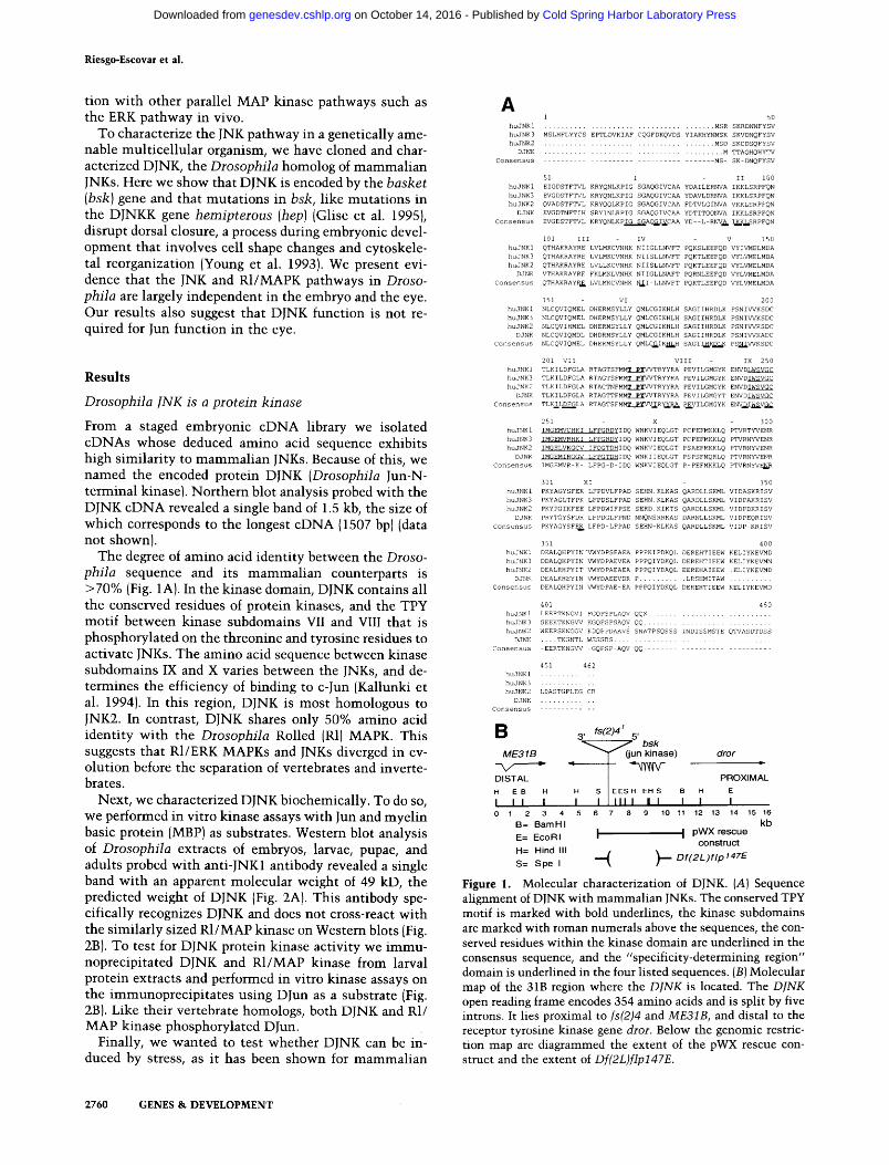
The bsk alleles and deficiencies can be ordered in an allelic series with increasing strength of the cuticular phenotypes: Df(2L)J27 < bsk 1 < bsk 2 < Df(2L)flp147E (Fig. 4B--E). Df(2L)flp147E is a small, 3-kb deletion gen- erated by an imprecise excision event of the P-element insertion in fs(2)4 (K. Beckingham, pers. comm.). We found that Df(2L)flp147E breaks proximal to the bsk gene, deleting most of the bsk coding sequence including the kinase domain, and part of the fs(2)4 transcript. Therefore Df(2L)flp147E is a null allele for bsk. In addi- tion, we found that bsk ~ is temperature-sensitive. At 18~ egg lays show weaker phenotypes (Fig. 4F).
Next we examined the expression and activity of DJNK by Western blot analysis and in vitro kinase assays on extracts of bsk mutants, bsk ~ mutant embryos still have wild-type levels of DJNK, whereas both bsk 2 and Df(2L)flp147E show strong reductions in DJNK com- pared with their respective heterozygous siblings (Fig. 2D). Kinase activity from bsk ~ and bsk 2 mutant embryos
Figure 4. Allelic series of bsk/DJNK mutant phenotypes. Cu- ticular preparation of embryos of the following genotypes: (A) Wild-type; (B) bskl/bskl; (C) bsk2/bsk2; (D) Df(2L)flpI47E/ Df(2L)flp147E; (E,I) bskl /Df(2L)flp147E derived from bskl /bsk ~ germ-line clones; (F) bskl/bsk 1 reared at 18~ (G)hepl; and (H) UAS-Dcdc42N17/+; B69-Gal 4/+. I was photographed before digestion with Hoyer's to show the embryonic tissues. In all panels anterior is left, and dorsal is up. (I) Ventrolateral view.
was significantly reduced (60% and 40% of heterozygous siblings, respectively), whereas Df(2L)flp147E mutant embryos had only 13% kinase activity of heterozygous siblings (Fig. 2E). The residual DJNK activity observed in the Df(2L)flp147E homozygous embryos could be due to maternal contribution of Bsk product in the egg.
basket shows maternal contribution and disrupts initiation of dorsal closure
We examined the effect of removal of the maternal con- tribution by making germ-line clones using mitotic recombination. Embryos lacking both maternal and zygotic bsk activity show an extreme dorsal open phen- otype: Cuticle is formed only in the ventral and ventro- lateral parts of the mutant embryo (Fig. 4E and I). This phenotype is stronger than phenotypes observed by re- moving only the zygotic bsk activity. We conclude that bsk has a maternal contribution. However, bsk 1 em- bryos derived from bsk ~ germ-line clones are rescued by zygotic bsk + function to fertile adults. This indicates that the zygotic bsk expression can compensate for the lack of maternal product.
Dorsal closure occurs during mid-embryogenesis and involves cell shape changes but not cell division (Cam- pos-Ortega and Hartenstein 1985). The cells of the lead- ing edge of the ventrolateral epidermis elongate and stretch in the dorsoventral axis (followed by rows of epi- dermal cells underneath) until the two edges meet in the dorsal midline and close the embryo, effectively inter- nalizing the cells of the amnioserosa. The epidermal cells stretch over the surface of the amnioserosa cells. Apical accumulation of actin and myosin in the leading edge cells initiates the final steps of dorsal closure (Young et al. 1993). The latter process is disrupted in strong zipper mutants, zipper codes for a nonmuscle my- osin heavy chain (Young et al. 1993).
We stained bsk mutant embryos with antibodies against Spectrin and Coracle (Pesacreta et al. 1989; Fe- hon et al. 1994). Coracle is the Drosophila homolog of the vertebrate band 4.1 cytoskeletal protein (Fehon et al. 1994). Both anti Spectrin and Coracle antibodies mark the profiles of the cells, bsk 1 embryos show the initial features of dorsal closure: The leading edge cells change shape and elongate, but the process fails to come to com- pletion, because the first rows underneath the leading edge cells either show only a partial change in shape, or fail to change cell shape completely (Fig. 5B). In wild- type embryos, the epidermal cells underneath the lead- ing edge change shape and elongate after the leading edge cells do (Fig. 5A). Df(2L)flp147E embryos show a more extreme phenotype: The leading edge cells elongate even less than in bsk ~ mutants (Fig. 5C). Embryos derived from bsk ~ germ-line clones with no paternal contribu- tion show no change in cell shape and dorsal closure never initiates (Fig. 5D). This result shows that bsk is required for the initiation of dorsal closure.
In bsk mutants, the spreading defect of the cells is more pronounced in anterior cells. In the posterior part of the embryo in weak mutants, some stretching epider-
2762 GENES & DEVELOPMENT
Cold Spring Harbor Laboratory Press on October 14, 2016 - Published by genesdev.cshlp.orgDownloaded from

Drosophila JNK and cell morphogenesis
(Harden et al. 1995). We tested whether interfering with Dcdc42 function would also give a dorsal closure pheno- type. We used a transgene that encodes a dominant neg- ative form of Dcdc42 (UAS-Dcdc42 N~7) (Eaton et al. 1995) and expressed it under the control of Gal4 in the Ga14-69B line (Brand and Perrimon 1993). This line drives Gal4 expression in epidermal cells during embryo- genesis (Brand and Perrimon 1993). Embryos with one copy of UAS-Dcdc42 N17 and 69B showed a dorsal open phenotype (Fig. 4H). This result suggests that Dcdc42 like DracA acts in dorsal closure regulation. Therefore it is possible that Dcdc42 and DracA function in the DJNK pathway like their vertebrate homologs.
Figure S. Cell shape changes during dorsal closure in bsk mu- tant embryos. (A) Wild-type larvae showing the ventrolateral epithelia during dorsal closure over the amnioserosa cells. (B) bsk 1 embryo, where the leading edge cells (arrows in B, C, and D) and some underlying epithelial cells have begun elongation, but the process stopped midway because the epithelium is partially detached from the underlying amnioserosa cells (top, at left). (C) Df(2L)flp147E embryo showing only partial elongation of the leading edge cells. (D) bsk 1 germ-line clone where the leading edge cells have not undergone cell elongation (cf. D with B). A and D were stained with anti coracle antibody, and B, C, and D with anti spectrin antibody.
mal cells meet at the dorsal midline and effect closure, but not in the anterior part. As a consequence, the ante- rior portion remains open and the forming head struc- tures and mouthparts are thrusted forward, together with parts of the gut and the CNS. Ventral furrow for- mation and other morphogenetic movements not di- rectly affected by dorsal closure occur normally in the bsk mutants, showing that the defect is specific to dorsal closure. The graded series of bsk mutant phenotypes de- scribed here argues for a sustained requirement of bsk throughout the process of dorsal closure: Reductions in the amount of protein are reflected in a concomitant reduction in the extent of cell elongation. A similar con- clusion was drawn from transgene rescue experiments of hep (Glise et al. 1995).
Dominant negative Dcdc42 also shows a dorsal open phenotype
The small GTPases Rac and Cdc42 have been implicated together with JNKK and JNK in the JNK pathway in vertebrates (Coso et al. 1995; Hill et al. 1995; Minden et al. 1995). In Drosophila, the homologs have been cloned (Dcdc42 and Drac), but no mutations have been isolated (Eaton et al. 1995). However, expression of a dominant negative form of DracA under the control of the heat shock promoter resulted in dorsal closure phenotypes
bsk regulates puckered expression
puckered (puc) mutations disrupt the formation of a su- ture between the two sides of the embryo in the dorsal midline, puc is expressed in the leading edge cells during dorsal closure, as revealed by lacZ expression of an en- hancer trap insertion in puc (pug E69) (Ring and Martinez- Arias 1993). In hep 1 mutants, puc-lacZ is not expressed in the dorsal rim cells during dorsal closure, suggesting that expression of puc-lacZ is dependent on hep func- tion (Glise et al. 1995). To test whetherpuc expression is also dependent on bsk function we stained embryos from a cross of Df(2L)flp147E/+; puc-lacZ/+ and bskl/+ flies. From this cross half of the progeny carry the puc- lacZ transgene and should show staining of the dorsal rim cells if bsk function were not required for puc ex- pression. We found, however, that of 324 embryos at the stage of dorsal closure, only 92 showed detectable puc- lacZ staining. This number deviates significantly from the expected half that was observed in control crosses not involving bsk alleles. In fact, these numbers fit a distribution of one-quarter staining and three-quarters nonstaining embryos (X 2 = 1.99) and suggest that not only bskl/Df(2L)flp147E embryos but also the embryos that are heterozygous for the bsk null allele [Df(2L)flp147E] lack detectable levels of lacZ staining. These results sug- gest that puc-lacZ expression in the dorsal rim cells is dependent on hep and on bsk function. It is interesting to note that it has been shown recently that puc encodes a member of the CL100 family of dual specificity MAP kinase phosphatases (A. Gampel, E. Martin-Blanco, and A. Martinez-Arias, pers. comm.). In vertebrates it has been shown that CLIO0 is transcriptionally induced by the activation of the MAP kinase pathway and stress (Keyse and Emslie 1992; Sun et al. 1993). Because puc expression is similarly regulated by the DJNK pathway, it appears that yet another element of this pathway has been conserved.
We have also noted that in older bsk mutant embryos (scored by the bsk cuticular phenotype) heterozygous for puc-lacZ, there is an ectopic expression of puc-lacZ, including cells of the amnioserosa (data not shown). This indicates that bsk function might be required later to represses puc-lacZ expression. In this regard, it is inter- esting to note that DJNK activity is increased in extracts
GENES & DEVELOPMENT 2763
Cold Spring Harbor Laboratory Press on October 14, 2016 - Published by genesdev.cshlp.orgDownloaded from

Riesgo-Escovar et ai.
of hep ~ mutant larvae (Fig. 2F). This result suggests that DJNK activity is not properly regulated in hep ~ mutants.
bsk function is not required for cell fate specification in the eye
We have shown that DJNK phosphorylates DJun in vitro. Because DJun has been implicated in the specification of the R7 photoreceptor cells in the developing eye (Boh- mann et al. 1994; Treier et al. 1995), we wanted to in- vestigate a possible role of bsk in photoreceptor cell specification. In situ hybridization experiments with a bsk RNA probe showed expression in the eye antennal disk (data not shown). We generated clones of homozy- gous bsk 1 cells in a heterozygous bsk ~ background in the eye by mitotic recombination, bsk ~ mutant cells prolif- erate normally because the clone size is similar to that of control clones. Furthermore, within the bsk ~ mutant clones of young flies ommatidia had developed normally. Only occasionally mutant ommatidia with altered num- ber of photoreceptor cells are seen (Fig. 6). The specifi- cation of the R7 photoreceptor cell, which is most sen- sitive to the expression of a dominant negative DJun protein (Treier et al. 1995), is not affected in bsk mutant clones. Because bsk 1 is not a complete loss-of-function allele of bsk, it is possible that in the bsk ~ cells there is still sufficient JNK activity for normal Jun activation. We therefore tested also clones of Df(2L)flp147E. Cells homozygous for Df(2L)flp147E lack not only bsk func- tion but also fs(2)4 function, which affects cell size (J. Riesgo-Escovar and E. Hafen, in prep.). Despite their re- duced size, homozygous Df(2L)flp147E cells were able to form normal ommatidial units with eight photoreceptor cells (data not shown). These results indicate that in ab- sence of bsk function specification of photoreceptor cells is normal.
D i s c u s s i o n
We have isolated and characterized a novel Jun kinase in
Figure 6. Analysis of bsk function in the developing eye. Tan- gential section, at the level of the R7 photoreceptor cell, through the eye of a bskl/+ fly carrying a clone of homozygous bsk 1 cells (marked by the absence of pigment granules). Note that most ommatidia in the clone are wild-type; typically only a few have abnormal numbers of photoreceptors (arrowhead).
Drosophila. Sequence analysis reveals high homology to its vertebrate counterparts, especially to JNK2. As is the case for its mammalian homologs, DJun is a good sub- strate for DJNK in vitro. We show that DJNK is encoded by the bsk gene. The embryonic lethality associated with bsk loss-of-function alleles is fully rescued by a genomic rescue fragment encompassing only the DJNK transcription unit. DJNK protein levels and kinase activ- ity are severely reduced in embryos homozygous for ei- ther the strong bsk 2 allele or the Df(2L)flp147E. Muta- tions in bsk and in hep (Glise et al. 1995) affect the pro- cess of dorsal closure, suggesting that both of these kinases are required together in this process.
The role of Bsk in dorsal closure
A large number of embryonic lethal mutations disrupt the process of dorsal closure during embryogenesis and display a common "dorsal open" phenotype (Jfirgens et al. 1984; Nfisslein-Volhard et al. 1984; Wieschaus et al. 1984). Cloned genes in this group can be grouped into two main classes: genes involved in the regulation of dorsal closure, and genes coding for structural proteins required for dorsal closure.
The first class is represented by members of the JNK pathway: bsk and hep (Glise et al. 1995) and possibly DracA (Harden et al. 1995) and Dcdc42. Cleared cuticles of mutant embryos in this class show only dorsal closure phenotypes, whereas the body plan seems unaffected. Mutations in decapentaplegic (dpp), which encodes a TGF-~ homolog, or in the genes coding for its receptors, thick veins and punt, or schnurri, which encodes a tran- scription factor acting downstream of the Dpp receptors, also show dorsal open phenotypes (Jfirgens et al. 1984; Nfisslein-Volhard et al. 1984; Affolter et al. 1994; Ru- perte et al. 1995). It is unclear at present how the Dpp and the JNK pathway cooperate in this process.
The second class of mutants identifies genes coding for structural elements, mostly associated with the cyto- skeleton, that are needed in dorsal closure. This group includes zipper, which encodes a nonmuscle myosin (Young et al. 1993); inflated, coding for an ot-integrin subunit (Wilcox et al. 1989); lethal(1)myospheroid, cod- ing for a J3-integrin subunit (Wieschaus et al. 1984); and coracle, which encodes a band 4.1 homolog (Fehon et al. 1994).
When is Bsk required during dorsal closure? We ob- serve a wide range of dorsal open phenotypes depending on the strength of the mutant allele and the presence or absence of maternally derived Bsk. The strongest pheno- type is observed when both maternal and zygotic Bsk are reduced. The resulting embryos fail to initiate the closing process. When only the zygotic component is removed the process initiates normally but is not completed. This argues strongly for a sustained require- ment of bsk during dorsal closure, which normally lasts
2hr. Little is known about the exact role that hep and bsk
play in the process of dorsal closure. Specifically, it is not clear what the target of jNK phosphorylation is in dorsal
2764 GENES & DEVELOPMENT
Cold Spring Harbor Laboratory Press on October 14, 2016 - Published by genesdev.cshlp.orgDownloaded from

Drosophila JNK and cell morphogenesis
closure. JNK could directly phosphorylate and modify cytoskeletal components involved in dorsal closure such as zipper, coracle, inflated, and l(1)myospheroid. Alter- natively, JNK could modify the activity of transcription factors that are known to be involved in dorsal closure. Mutat ions in genes coding for several transcription fac- tors have dorsal open phenotypes, like pannier (Ramain et al. 1993; Heitzler et al. i996) and serpent (Abel et al. 1993; Frank and Rushlow 1996), two GATA transcrip- tion factors; and anterior open (Ntisslein-Volhard et al. 1984), an ETS domain protein (Rogge et al. 1995). The fact that both hep and bsk mutants affect the expression of puc as monitored by the puc- lacZ staining suggests that JNK acts by regulating transcription factors rather than by directly modifying cytoskeletal components in- volved in the actual process of cell shape change. Ge- netic and biochemical experiments are needed to iden- tify the putative transcription factors whose activity is modulated by the JNK pathway as well as further target genes whose expression is modified by the pathway.
Different developmental processes are controlled by the JNK pathway and the MAP kinase pathway in Drosophila
With the identif ication and cloning of bsk and hep (Glise et al. 1995), the JNK pathway is f i rmly established in Drosophila. Dominan t negative versions of DracA {Harden et al. 1995) and Dcdc42 give also dorsal closure phenotypes. It is thus possible that they form part of the pathway, as in the vertebrate JNK pathway. A more de- finit ive p lacement of Drac and Dcdc42 in the DJNK pathway requires the availabil i ty of loss-of-function mu- tants of these genes and gain of function mutat ions in hep and bsk. In the absence of such mutants we have tested whether bsk mutants act as dominant suppressors of the gain of funct ion phenotype obtained by expressing activated Dcdc42 (UAS-Dcdc v12) in ectodermal cells in the embryo. Prel iminary results suggest that removal of one copy of bsk suppresses partially the dominant phe- notype of activated Dcdc42 (data not shown). These re- sults are consistent wi th a model in which Dcdc42 acts via DJNK during embryogenesis.
The phenotypes of mutat ions in genes of the JNK path- way are different from those of muta t ions in the R1/MAP kinase pathway in both the embryo and the adult. In the embryo, bsk is involved in dorsal closure and r / i s re- quired for signaling in the Torso pathway (Brunner et al. 1994b). In larvae, in contrast to r/, bsk appears not to be required for cell proliferation and for cell fate specifica- tion in the eye. Therefore, it appears that during Droso- phila development the JNK and MAP kinase pathway are largely independent.
In vertebrate cell culture systems, the main substrate for JNK is the transcription factor Jun. We have shown that DJNK can also phosphorylate DJun efficiently in vitro. Whether DJun is also a major in vivo target of DJNK, however, is questionable. Mutations in DJun have not been identified yet, but through the use of dom- inant negative and consti tut ively active Jun proteins ex-
pressed selectively in the developing eye it has been shown that DJun may funct ion in the specification of neural fate in the R7 equivalence group in response to the Sevenless signaling pathway (Treier et al. 1995). If indeed bsk were the major kinase involved in the acti- vation of DJun, we would expect impa i rment of photo- receptor cell specification in clones of homozygous bsk cells. However, bsk mutan t cells differentiate largely into normal-looking ommatidia . Therefore it appears that DJNK is not involved in photoreceptor cell specifi- cation and is not required for Jun activation in the eye. This is consistent wi th recent biochemical evidence sug- gesting that DJun phosphorylation is dependent on the activity of the roi led/MAP kinase in Drosophila (Peve- rali et al. 1996).
Materia ls and m e t h o d s
Genetics
Df(2L)flp147E was a gift of K. Beckingham (Rice University, Houston, TX). bsk 1 and bsk 2 were obtained from the Tiibingen stock center, hep 1, hep rTs, and puc E69 from S. Noselli (Centre National de Recherche Scientifique, Toulouse, France); the Ga14-69B line from K. Basler (University of Z/Jrich, Switzer- land), and Df(2L)J2 and Df(2L)J27 from the Indiana Stock Cen- ter. The UAS-Dcdc42 N17 and UAS-Dcdc42 v12 transgenic flies were from L. Luo (University of California, San Francisco).
Besides Df(2L)flp147E, Df (2L)J2 and Df(2L)J27, with approx- imately the same breakpoints (31B-32A), had been reported to uncover bsk (N6sslein-Volhard et al. 1984; Clegg et al. 1993). We performed complementation tests between the deficiencies and the extant bsk alleles in all pairwise combinations. Failure of complementation was observed in all cases except with Df(2L)J27. Viable transheterozygous with the bsk alleles and Df(2L)]27 were recovered. Egg lays derived from these crosses and from the balanced Df(2L)J27 stock, though, gave rise to a fraction of dead embryos with dorsal open phenotypes, indistin- guishable from weak to intermediate bsk phenotypes. We con- clude that Df(2L)J27 shows partial complementation with bsk behaving as a weak bsk allele. It is possible that Df(2L)J27 breaks distally near bsk, and disrupts regulatory sequences. The bsk 2 allele has a stronger dorsal open phenotype than bsk ~ but could not be used for the clonal analysis because it contains at least two additional closely linked lethal mutations on the same chromosome.
For rescue experiments we used three different insertions of pWX: one insertion on the second chromosome, and two on the third chromosome. The pWX line with the insertion on the second was recombined onto the Df(2L)flp147E chromosome and then homozygosed. For the third chromosomal lines, flies heterozygous for a bsk allele or Df(2L)J2/CyO; pWX/+ were crossed to Df(2L)flp147E/CyO and the progeny scored for viable transheterozygotes. We rescued the following mutant combina- tions: Df(2L)flp147E/bsk 1 and Df(2L)flp147E/Df(2L)J2 for one insertion, and Df(2L)flp147E/bsk 2 for the other.
From the crosses with pWX insertions, rescued Df(2L)flp147E/ Df(2L) J2; pWX/ + and pWX, Df(2L)flp147E also uncover fs(2)4 mutations and homozygous females were sterile, as expected. We also used a longer rescue construct (10 kb) in the X chro- mosome, pWAX. pWAX contains the fs(2)4 transcript and bsk, pWAX is sufficient to rescue both bsk and fs(2)4 alleles (J.R. Riesgo-Escovar and E. Hafen, in prep.).
A chi-square test was used to assess the distribution of stage
GENES & DEVELOPMENT 2765
Cold Spring Harbor Laboratory Press on October 14, 2016 - Published by genesdev.cshlp.orgDownloaded from

Riesgo-Escovar et al.
8-15 embryos stained for lacZ expression from the cross Df(2L)flp147E/ + ; pucE69/ + males to bskl /CyO females.
Clonal analysis
Germ-line clones of bsk ~ were generated by use of the "domi- nant female sterile" technique (Hou et al. 1995) with the FLP- FRT recombinase system (Xu and Rubin 1993). For bsk 2 and Df(2L)flpl 47E, the same procedure was followed, but eggs were not recovered. Df(2L)flp147E also partially deletes a female ster- ile gene {J.R. Riesgo-Escovar and E. Hafen, in prep.), bsk 2 has additional mutations on the chromosome (J.R. Riesgo-Escovar and E. Hafen, unpubl.); this could account for the female steril- ity observed. We also generated bsk 1 and Df(2L)flp147E mutant clones marked by the absence of the white gene marker using the FLP-FRT recombinase system of (Xu and Rubin 1993).
Germ-line transformations
A 10-kb genomic fragment {positions 1.2-11.5 in Fig. 1B) cloned in the pW8 transformation vector {Klemenz et al. 1987) was digested partially with SpeI and the appropriate fragment reli- gated to generate pWX. Transgenic flies were generated accord- ing to Basler et al. ( 1991).
Cuticle preparations
Embryos were dechorionated in 50% chlorox, devitellinized, washed, and mounted in Hoyer's medium. Cuticle preparations were photographed on a Zeiss Axiophot under bright and dark fields.
Histology
For antibody stainings, embryos were treated as described {Young et al. 1991). Anti Spectrin antibodies were used at a 1:500 dilution, anti-Coracle at 1:250. Fluorescently labeled sec- ondary antibodies {Jackson, Inc.) were used at a 1:100 dilution. Coracle protein distribution in bsk mutant embryos is normal when both maternal and zygotic bsk function is removed. Mounted embryos were viewed on a Multiprobe laser confocal microscope {Molecular Dynamics, Inc.). Eye sections were done according to Brunner et al. (1994b). Images were assembled us- ing Adobe Photoshop 3.0 software.
In situ hybridizations were performed essentially as described (Tautz and Pfeifle 1989) for embryos and larval brains using an antisense RNA probe encompassing the entire DJNK eDNA. Sense probes were used in parallel as controls.
Molecular techniques
Standard molecular techniques were used (Sambrook et al. 19891. cDNAs and the corresponding genomic region were iso- lated, mapped, and sequenced on both strands using Sequenase according to the manufacturer's instructions with the use of several sequencing primers. Data was assembled and analyzed using the GCG software {Wisconsin).
Kinase assays
Anti-JNK1 antibody was from Santa Cruz Biotechnology, Inc. MBP immune complex kinase assays were done essentially as described (Brunner et al. 1994a), except that for DJNK assays, 5 ~1 of JNK1 antibody was used per sample, and protein A-Seph- arose beads, antibody, and lysates were added at the same time. Larval, pupal or embryonic extracts were homogenized in a ho-
mogenizer adapted with a plastic Eppendorf tube pestle by six steps in RIPA buffer [phosphate buffered saline (PBS), 1% Noni- det P40, 0.5% sodium deoxycholate, and 0.1% SDS, to which was added every time fresh 10 mg/ml phenyl methyl sulfonyl fluoride (PMSF) in propanol { 10 ~1), aprotinin (30 ~l/ml) and 100 mM sodium orthovanadate]. For Westerns, we used a 1:1000 dilution of the JNK1 antibody.
DJNK immunoprecipitated activity is concentration-depen- dent, and saturates between 20 and 30 min of incubation (data not shown). For UV treatment experiments, larvae were co l lected in empty glass vials with moist filter discs, and subjected to UV light from a transilluminator for 5 rain at a 1-cm distance. The vials were then removed, and larvae allowed to recover (45 min) before processing. Harsher UV treatments (15 rain) inhibit DJNK by 50%, and enhance Rolled activity (100%) In = 3 as- says) (data not shown). For kinase assays in a hep I background, we took advantage of the fact that hep I hypomorphs are semile- thai. We made a stock of hep 1 and an attached X chromosome balancer, such that males are always hep 1, and females are al- ways wild-type. We separated male and female third instar lar- vae from the same cultures for kinase assays.
A c k n o w l e d g m e n t s
We thank K. Beckingham for providing the Df(2L)flp147E stock and for sharing unpublished results. We thank C. Wilson, S. Noselli, K. Mathews, and the Tfibingen Stock center for fly stocks. We thank R. Fehon for the Coracle antibody; K. Basler, B. Dickson, M. Dominguez, K. D~cker, S. Leevers, and I. Rod- riguez for critical reading of the manuscript; and members of the Hafen lab {including S. Leevers and L. MacDougall) for dis- cussions and constructive criticisms. This work was supported by grants from the Human Frontiers Science Organization to J.R.R. and a grant from the Swiss National Science Foundation to E.H.
The publication costs of this article were defrayed in part by payment of page charges. This article must therefore be hereby marked "advertisement" in accordance with 18 USC section 1734 solely to indicate this fact.
N o t e
The DINK cDNA sequence has been submitted to GenBank under accession no. U73196.
R e f e r e n c e s
Abel, T., A.M. Michelson and T. Maniatis. 1993. A Drosophila GATA family member that binds to Adh regulatory se- quences is expressed in the developing fat body. Develop- ment 119: 623-633.
Affolter, M., D. Nellen, U. Nussbaumer, and K. Basler. 1994. Multiple requirements for the receptor serine/threonine ki- nase thick veins reveal novel functions of TGF[3 homologs during Drosophila embryogenesis. Development 120:3105- 3117.
Basler, K., B. Christen, and E. Hafen. 1991. Ligand-independent activation of the sevenless receptor tyrosine kinase changes the fate of cells in the developing Drosophila eye. Cell 64: 1069-1082.
Bohmann, D., M.C. Ellis, L.M. Staszewski, and M. Mlodzik. 1994. Drosophila Jun mediates gas-dependent photoreceptor determination. Cell 78: 973-986.
Brand, A.H. and N. Perrimon. 1993. Targeted gene expression as a means of altering cell fates and generating dominant phe- notypes. Development 118:401-415.
2766 GENES & DEVELOPMENT
Cold Spring Harbor Laboratory Press on October 14, 2016 - Published by genesdev.cshlp.orgDownloaded from

Drosophila INK and cell morphogenesis
Brunner, D., K. DOcker, N. Oellers, E. Hafen, H. Scholz, and C. Kliimbt. 1994a. The ETS domain protein Pointed-P2 is a tar- get of MAP kinase in the Sevenless signal transduction path- way. Nature 370: 386-389.
Brunner, D., N. Oellers, J. Szabad, W.H. Biggs III, S.L. Zipursky, and E. Hafen. 1994b. A gain of function mutation in Droso- phila MAP kinase activates multiple receptor tyrosine ki- nase signalling pathways. Cell 76: 875-888.
Campos-Ortega, J. and V. Hartenstein. 1985. The embryonic development of Drosophila melanogaster. Springer-Verlag, Berlin, Germany.
Clegg, N.J., I.P. Whitehead, J.K. Brock, D.A. Sinclair, R. Mottus, G. Stromotich, M.J. Harrington, and T.A. Grigliatti. 1993. A cytogenetic analysis of chromosomal region 31 of Droso- phila melanogaster. Genetics 134: 221-230.
Coso, O.A., M. Chiariello, J.-C. Yu, H. Teramoto, P. Crespo, N. Xu, T. Miki, and J.S. Gutkind. 1995. The small GTP-binding proteins Racl and Cdc42 regulate the activity of the JNK/ SAPK signalling pathway. Cell 81:1137-1146.
DOrijard, B., M. Hibi, I.H. Wu, T. Barrett, B. Su, T. Deng, M. Karin, and R.J. Davis. 1994. JNKI: A protein kinase stimu- lated by UV light and Ha-Ras that binds and phosphorylates the c-Jun activation domain. Cell 76: 1025-1037.
Dickson, B. and E. Hafen. 1993. Genetic dissection of eye de- velopment in Drosophila. In The development of Drosophila melanogaster (ed. A. Martinez-Arias and M. Bate), pp. 1327- 1362. Cold Spring Harbor Laboratory Press, Cold Spring Har- bor, NY.
Dominguez, M. and E. Hafen. 1996. Genetic dissection of cell fate specification in the developing eye of Drosophila. Semin. Cell Dev. Biol. 7: 219-226.
Eaton, S., P. Auvinen, L.Q. Luo, Y.N. Jan, and K. Simons. 1995. CDC42 and Racl control different actin-dependent pro- cesses in the Drosophila wing disc epithelium. J. Cell Biol. 131: 151-164.
Engelberg, D., C. Klein, H. Martinetto, K. Struhl, and M. Karin. 1994. The UV response involving the Ras signaling pathway and AP-1 transcription factors is conserved between yeast and mammals. Cell 77" 381-390.
Fehon, R.G., I.A. Dawson, and T.S. Artavanis. 1994. A Droso- phila homologue of membrane-skeleton protein 4.1 is asso- ciated with septate junctions and is encoded by the coracle gene. Development 120: 545-557.
Fields, P.E., T.F. Gajewski, and F.W. Fitch. 1996. Blocked Ras activation in anergic CD4+ T cells. Science 271: 1276- 1278.
Frank, L.H. and C. Rushlow. 1996. A group of genes required for the maintenance of the amnioserosa tissue in Drosophila. Development 122: 1343-1352.
Glise, B., H. Bourbon, and S. Noselli. 1995. hemipterous en- codes a novel Drosophila MAP kinase kinase, required for epithelial cell sheet movement. Cell 83:451-462 .
Harden, N., H.Y. Loh, W. Chia, and L. Lim. 1995. A dominant inhibitory version of the small GTP-binding protein Rac dis- rupts cytoskeletal structures and inhibits developmental cell shape changes in Drosophila. Development 121: 903- 914.
Heitzler, P., M. Haenlin, P. Ramain, M. Calleja, and P. Simpson. 1996. A genetic analysis of pannier, a gene necessary for viability of dorsal tissues and bristle positioning in Droso- phila. Genetics 143: 1271-1286.
Herskowitz, I. 1995. MAP kinase pathways in yeast: For mating and more. Cell 80: 187-197.
Hibi, M., A. Lin, T. Smeal, A. Minden, and M. Karin. 1993. Identification of an oncoprotein- and UV-responsive protein kinase that binds and potentiates the c-Jun activation do-
main. Genes & Dev. 7: 2135-2148. Hill, C.S., J. Wynne, and R. Treisman. 1995. The Rho family
GTPases RhoA, Racl, and Cdc42Hs regulate transcriptional activation by SRF. Cell 81:1159 -1170 .
Hou, X.S., T.-B. Chou, M.B. Melnick, and N. Perrimon. 1995. The torso receptor tyrosine kinase can activate Raf in a Ras- independent pathway. Cell 81: 63-71.
JOrgens, G., C. Ni~sslein-Volhard, and H. Kluding. 1984. Muta- tions affecting the pattern of the larval cuticle in Drosophila melanogaster II. Zygotic loci on the third chromosome. Roux's Arch. Dev. Biol. 193: 183-295.
Kallunki, T., B. Su, I. Tsigelny, H.K. Sluss, B. DOrijard, G. Moore, R. Davis, and M. Karin. 1994. JNK2 contains a spec- ificity-determining region responsible for efficient c-Jun binding and phosphorylation. Genes & Dev. 8: 2996--3007.
Karin, M. and T. Hunter. 1995. Transcriptional control by pro- tein phosphorylation: Signal transmission from the cell sur- face to the nucleus. Curr. Biol. 5: 747-757.
Kayne, P.S. and P.W. Sternberg. 1995. Ras pathways in Cae- norhabditis elegans. Curr. Opin. Genet. Dev. 5" 38--43.
Kenyon, C. 1995. A perfect vulva every time: Gradients and signaling cascades in C. elegans. Cell 82: 171-174.
Keyse, S.M. and E.A. Emslie. 1992. Oxidative stress and heat shock induce a human gene encoding a protein-tyrosine phosphatase. Nature 359: 644--647.
Klemenz, R., U. Weber, and W.J. Gehring. 1987. The white gene as a marker in a new P-element vector for gene transfer in Drosophila. Nucleic Acids Res. 15: 3947-3959.
Kyriakis, J.M., P. Banerjee, E. Nikolakaki, T. Dai, E.A. Rubie, M.F. Ahmad, J. Avruch, and J.R. Woodgett. 1994. The stress- activated protein kinase subfamily of c-Jun kinases. Nature 369: 156--160.
Li, W., C.D. Whaley, A. Mondino, and D.L. Mueller. 1996. Blocked signal transduction to the ERK and JNK protein ki- nases in anergic CD4+ T cells. Science 271: 1272-1276.
Lindsley, D.L. and G.G. Zimm. 1992. The genome of Drosophila melanogaster. Academic Press, San Diego, CA.
Marshall, C.]. 1994. MAP kinase kinase kinase, MAP kinase kinase and MAP kinase. Curr. Opin. Genet. Dev. 4: 82-89.
1995. Specificity of receptor tyrosine kinase signaling: Transient versus sustained extracellular signal-regulated ki- nase activation. Cell 80: 179-185.
Minden, A., A. Lin, T. Smeal, B. DOrijard, M. Cobb, R. Davis, and M. Karin. 1994. c-Jun N-terminal phosphorylation cor- relates with activation of the JNK subgroup but not the ERK subgroup of mitogen-activated protein kinases. Mol. Cell. Biol. 14: 6683-6688.
Minden, A., A. Lin, F.-X. Claret, A. Abo, and M. Karin. 1995. Selective activation of the JNK signalling cascade and c-Jun transcriptional activity by the small GTPases Rac and Cdc42Hs. Cell 81: 1147-1157.
Mohit, A.A., J.H. Martin, and C.A. Miller. 1995. p49 3F12 ki- nase: A novel MAP kinase expressed in a subset of neurons in the human nervous system. Neuron 14: 67-78.
Nishida, E. and Y. Gotoh. 1993. The MAP kinase cascade is essential for diverse signal transduction pathways. Trends Biochem. Sci. 18: 128-131.
Niisslein-Volhard, C., E. Wieschaus, and H. Kluding. 1984. Mu- tations affecting the pattern of the larval cuticle in Droso- phila melanogaster. I. Zygotic loci on the second chromo- some. Roux's Arch. Dev. Biol. 193: 267-282.
Pesacreta, T.C., T.J. Byers, R. Dubreuil, D.P. Kiehart, and D. Branton. 1989. Drosophila spectrin: The membrane skeleton during embryogenesis. J. Cell Biol. 108: 1697-1710.
Peverali, F.A., A. Isaksson, A.G. Papavassiliou, P. Plastina, L.M. Staszewski, M. Mlodzik, and D. Bohmann. 1996. Phospho-
GENES & DEVELOPMENT 2767
Cold Spring Harbor Laboratory Press on October 14, 2016 - Published by genesdev.cshlp.orgDownloaded from
Riesgo-Escovar et al.
rylation of Drosophila jun by the MAP kinase rolled regu- lates photoreceptor differentiation. EMBO J. 15: 3943-3950.
Pombo, C.M., J.V. Bonventre, J. Avruch, J.R. Woodgett, J.M. Kyriakis, and T. Force. 1994. The stress-activated protein kinases are major c-Jun amino-terminal kinases activated by ischemia and reperfusion. J. Biol. Chem. 269: 26546-26551.
Ramain, P., P. Heitzler, M. Haenlin, and P. Simpson. 1993. pan- nier, a negative regulator of achaete and scute in Drosophila, encodes a zinc finger protein with homology to the verte- brate transcription factor GATA-1. Development 119" 1277- 1291.
Ring, J.M. and A. Martinez-Arias. 1993. puckered, a gene in- volved in position-specific cell differentiation in the dorsal epidermis of the Drosophila larva. Development 119: 251- 259.
Rogge, R., P.J. Green, J. Urano, S. Hom-Saban, M. Mlodzik, B.-Z. Shilo, V. Hartenstein, and U. Banerjee. 1995. The role of yah in mediating the choice between cell division and differen- tiation. Development 121: 3947-3958.
Ruperte, E., T. Marty, D. Nellen, M. Affolter, and K. Basler. 1995. An absolute requirement for both type II and type I receptors, Punt and Thick Veins, for Dpp signalling in vivo. Cell 80: 889-897.
Sambrook, J., E.F. Fritsch, and T. Maniatis. 1989. Molecular cloning: A laboratory manual, 2nd edition. Cold Spring Har- bor Laboratory Press, Cold Spring Harbor, NY.
Su, B., E. Jacinto, M. Hibi, T. Kallunki, M. Karin, and N.Y. Ben. 1994. JNK is involved in signal integration during costimu- lation of T lymphocytes. Cell 77: 727-736.
Sun, H., C.H. Charles, L.F. Lau, and N.K. Tonks. 1993. MKP-1 (3CH134), an immediate early gene product, is a dual spec- ificity phosphatase that dephosphorylates MAP kinase in vivo. Cell 75: 487--493.
Tautz, D. and C. Pfeifle. 1989. A non-radioactive in situ hybrid- ization method for the localization of specific RNAs in Drosophila embryos reveals translational control of the seg- mentation gene hunchback. Chromosoma 98: 81-85.
Treier, M., D. Bohmann, and M. Mlodzik. 1995. Jun cooperates with the ETS-domain protein pointed to induce photorecep- tor R7 fate in the Drosophila eye. Cell 83: 753-760.
Wassarman, D.A., M. Therrien, and G.M. Rubin. 1995. The Ras signaling pathway in Drosophila. Curr. Opin. Genet. Dev. 5: 44-50.
Wieschaus, E., C. Nfisslein-Volhard, and G. Jfirgens. 1984. Mu- tations affecting the pattern of the larval cuticle in Droso- phila melanogaster III. Zygotic loci on the X-chromosome and the fourth chromosome. Roux's Arch. Dev. Biol. 193: 296--307.
Wilcox, M., A. DiAntonio, and M. Leptin. 1989. The function of PS integrins in Drosophila wing morphogenesis. Develop- ment 107: 891-897.
Wilson, C., D.C.I. Goberdhan, and H. Steller. 1993. Dror, a po- tential neurotrophic receptor gene, encodes a Drosophila ho- molog of the vertebrate Ror family of Trk-related receptor tyrosine kinases. Proc. Natl. Acad. Sci. 90: 7109-7113.
Xia, Z., M. Dickens, J. Raingeaud, R.G. Davis, and M.E. Green- berg. 1995. Opposing effects of ERK and JNK-P38 MAP ki- nases on apoptosis. Science 270: 1326--1331.
Xu, T. and G. Rubin. 1993. Analysis of genetic mosaics in de- veloping and adult Drosophila tissues. Development 117: 1223-1237.
Young, P.E., T.C. Pesacreta, and D.P. Kiehart. 1991. Dynamic changes in the distribution of cytoplasmic myosin during Drosophila embryogenesis. Developm en t 111: 1-14.
Young, P.E., A.M. Richman, A.S. Ketchum, and D.P. Kiehart. 1993. Morphogenesis in Drosophila requires nonmuscle my-
osin heavy chain function. Genes & Dev. 7: 29-41. Zipursky, L.S. and G.M. Rubin. 1994. Determination of neuro-
nal cell fate: Lessons from the R7 neuron in Drosophila. Annu. Rev. Neurosci. 17: 373-397.
2768 GENES & DEVELOPMENT
Cold Spring Harbor Laboratory Press on October 14, 2016 - Published by genesdev.cshlp.orgDownloaded from

10.1101/gad.10.21.2759Access the most recent version at doi: 1996 10: 2759-2768 Genes Dev.
J R Riesgo-Escovar, M Jenni, A Fritz, et al. the eye.morphogenesis but not for DJun-dependent cell fate specification in The Drosophila Jun-N-terminal kinase is required for cell
References
http://genesdev.cshlp.org/content/10/21/2759.full.html#ref-list-1
This article cites 59 articles, 26 of which can be accessed free at:
ServiceEmail Alerting
click here.top right corner of the article orReceive free email alerts when new articles cite this article - sign up in the box at the
http://genesdev.cshlp.org/subscriptionsgo to: Genes & Development To subscribe to
Copyright © Cold Spring Harbor Laboratory Press
Cold Spring Harbor Laboratory Press on October 14, 2016 - Published by genesdev.cshlp.orgDownloaded from
Copyright © 2022 FDOKUMEN