Analysis of the c-di-GMP mediated cell fate determination in ...
239
Analysis of the c-di-GMP mediated cell fate determination in Caulobacter crescentus Inauguraldissertation zur Erlangung der Würde eines Doktors der Philosophie vorgelegt der Philosophisch-Naturwissenschaftlichen Fakultät der Universität Basel von Sören Abel aus Kiel, Deutschland Basel 2009
-
Upload
khangminh22 -
Category
Documents
-
view
7 -
download
0
Transcript of Analysis of the c-di-GMP mediated cell fate determination in ...

Analysis of the c-di-GMP mediated cell fate determination in Caulobacter crescentus
Inauguraldissertation
zur
Erlangung der Würde eines Doktors der Philosophie
vorgelegt der
Philosophisch-Naturwissenschaftlichen Fakultät
der Universität Basel
von
Sören Abel aus Kiel, Deutschland
Basel 2009

Genehmigt von der Philosopisch-Naturwissenschaftlichen Falkultät
auf Antrag von
- Prof. Dr. Urs Jenal
- Prof. Dr. Tilman Schirmer
Basel, den 20. Mai 2008
Prof. Dr. Eberhard Parlow
Dekan

Abstract
Abstract Cyclic-di-GMP (c-di-GMP) is a ubiquitous second messenger in bacteria, which has been
recognized as a key regulator, antagonistically controlling the transition between motile,
planktonic cells and surface attached, multicellular communities. The biosynthesis and
degradation of c-di-GMP are mediated by the opposing enzymatic activities of di-guanylate
cyclases (DGCs) and phosphodiesterases (PDEs), generally in response to internal and
environmental signals. These activities reside in GGDEF and EAL domains respectively,
which represent two large families of output domains often found in bacterial one- and two-
component systems. In this work, the cell cycle-embedded differentiation from a free-living,
motile swarmer cell into a sessile stalked cell in the model organism Caulobacter crescentus,
and the role of c-di-GMP in this process was investigated. A systematic analysis was used to
identify key regulatory enzymes involved in c-di-GMP metabolism that influence this
developmental process. The function and regulation of these genes was then examined. One
component that has already been implicated in this process, the DGC PleD, was investigated
in more detail, with special emphasis on the mechanisms underlying its timed activation and
cell cycle specific subcellular localisation.
In the first part of this work, a systematic functional analysis of all GGDEF, EAL and
GGDEF/EAL composite proteins from C. crescentus with a focus on motility and surface
attachment is described. In this screen, the phosphodiesterase PdeA was identified as a
gatekeeper that prevents premature paralysis of the flagellum and holdfast synthesis in the
C. crescentus swarmer cell. It is shown that PdeA, together with its antagonistic DGCs DgcB
and PleD, are components of converging pathways and orchestrate polar development during
the swarmer-to-stalked cell transition. Furthermore, evidence is presented for a proteolytic
regulation mechanism for PdeA.
Secondly, the PleD localisation factor CC1064 is analysed. This transmembrane
protein has pleiotropic effects on motility, surface attachment and polar localisation of PleD.
It is shown that the motility and PleD localisation phenotypes of a Δcc1064 strain are
conditional and depend on environmental factors such as oxygen and temperature stress.
Moreover, evidence is presented that the impaired motility of a Δcc1064 mutant is caused by
an assembly defect of the motor proteins MotA and MotB, leading to paralysis of the
flagellum. A model is suggested that links altered membrane composition under
environmental stress conditions to the Δcc1064 phenotypes.

Abstract
In Paul, Abel et al. (2007), insights were gained into the regulation of PleD. In
addition to the well characterised non-competitive feedback inhibition, a second independent
layer of activity control via dimerisation was investigated. The response regulator PleD is
activated by phosphorylation of the N-terminal receiver domain. Here we show that the
phospho-mimetic chemical beryllium fluoride specifically activates the enzymatic activity of
PleD in vitro and in addition leads to dimerisation. Fractionation experiments showed that the
DGC activity exclusively resides within the dimer fraction. Finally, evidence is provided that
dimerisation of PleD is not only required for catalytic activity, but also leads to sequestration
to the differentiating stalked pole of the C. crescentus cell, thereby providing an elegant way
of restricting PleD activity to a subcellular region of the cell.
In Paul, Jaeger & Abel et al. (2008), a network of proteins belonging to the two
component system that regulates PleD activation and thereby leads to its localisation were
investigated in detail. The single domain response regulator DivK is controlled by the
phosphatase activity of PleC and the kinase DivJ. It is shown that DivK allosterically
activates the kinase activities of PleC and DivJ and thereby switches PleC from a phosphatase
into a kinase state. Increased DivJ activity further activates DivK in a feed-forward loop,
while PleC and DivJ together stimulate PleD activity and localisation. Evidence is provided
that DivJ, PleC, and DivK colocalise in a short time window during the cell cycle, directly
prior to PleD activation, suggesting a role for the spatial distribution of these proteins. At last,
the wider role of single domain response regulators in the interconnection of two-component
signal transduction circuits is discussed.
Finally, in Dürig, Folcher, Abel et al. (2008), a role for c-di-GMP in the cell cycle of
C. crescentus via regulation of targeted proteolysis of the regulator CtrA is shown. During the
swarmer-to-stalked cell transition CtrA is recruited to the incipient stalked pole, where it is
degraded by its protease ClpXP. This recruitment and subsequent degradation is dependent on
the enzymatically inactive GGDEF domain protein PopA. PopA itself localises to the cell
pole and can bind c-di-GMP. It is shown that mutants in the c-di-GMP binding site fail to
localise to the developing stalked pole and consequently fail to promote CtrA degradation.
Finally, evidence is provided that interconnects PopA with the pathway responsible for
substrate inactivation and protease localisation in a cell cycle dependent manner.

Index
Index
1 Introduction - 1 -
2 Aim of the thesis -25-
3 Results -26-
3.1 A Phosphodiesterase and its Cognate Di-guanylate
Cyclases Antagonistically Control Polar Development
in Caulobacter crescentus -26-
3.2 CC1064, a Transmembrane Protein Required for
Flagellar Rotation and Subcellular Protein Localisation
Under Environmental Stress Conditions in
Caulobacter crescentus -75-
3.3 Activation of the Di-guanylate Cyclase PleD by
Phosphorylation-mediated Dimerisation -128-
3.4 Allosteric Regulation of Histidine Kinases by their
Cognate Response Regulator Determines Cell Fate -142-
3.5 Second messenger mediated spatiotemporal control
of protein degradation during the bacterial cell cycle -165-
4 Outlook -213-
5 Bibliography -216-
6 Acknowledgements -231-
7 Curriculum vitae -232-

Introduction
1
1. Introduction
The transition from planktonic to sessile lifestyle is a vital decision for bacteria and highly regulated
Bacteria are subjected to multiple environmental influences. There are two principle options
how to react to such changes in the environment; to move away or to persist. The careful
control of this vital decision is essential for the fitness and survival of microorganisms. Once
made, such a decision cannot be reverted without cost and is sometimes irrevocable, as
dramatic physiological and developmental processes must be undertaken to enable the chosen
lifestyle [1].
On one hand, motility requires an apparatus that allows locomotion, for example a
flagellum, whose energy consuming function must be maintained under all conditions. In
return, it allows the bacterium to continuously move towards fresh nutrient sources and away
from toxic environments (reviewed in [2]). On the other hand, the generation of sessile
communities, so called biofilms, allows bacteria to grow on surfaces, a lifestyle associated
with increased persistence in hostile environments and one, which enables the opportunity of
acquiring new food resources (reviewed in [1, 3-6]). While the importance of biofilms in
industrial processes has been known for several years, with examples including clogging of
pipework or aiding water clean-up (reviewed in [1]), the impact on medical treatments has
only recently become clear. Biofilms threaten our ability to treat bacterial infections, as they
are inherently protected from host defences as well as antibiotics and can become reservoirs
for systemic infections (reviewed in [4]). It is therefore important to understand the molecular
mechanisms that are used by bacteria to register environmental signals and translate them in
appropriate outputs that determine a motile or a sessile lifestyle. Recently, the bacteria-
specific second messenger bis-(3`, 5`)-cyclic dimeric guanosine monophosphate (c-di-GMP)
was found to be critically involved in the switch from a planktonic to a sessile existence
(reviewed in [7-11]).
The mechanisms underlying c-di-GMP turnover
C-di-GMP (Fig. 1) is a ubiquitous bacterial second messenger that has been shown to be
involved in the regulation of a growing number of physiological processes.
Introduction
2
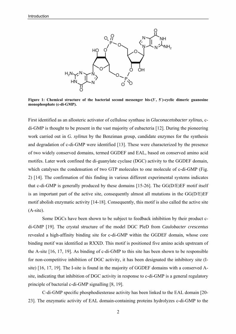
Figure 1: Chemical structure of the bacterial second messenger bis-(3`, 5`)-cyclic dimeric guanosine monophosphate (c-di-GMP).
First identified as an allosteric activator of cellulose synthase in Gluconacetobacter xylinus, c-
di-GMP is thought to be present in the vast majority of eubacteria [12]. During the pioneering
work carried out in G. xylinus by the Benziman group, candidate enzymes for the synthesis
and degradation of c-di-GMP were identified [13]. These were characterized by the presence
of two widely conserved domains, termed GGDEF and EAL, based on conserved amino acid
motifes. Later work confined the di-guanylate cyclase (DGC) activity to the GGDEF domain,
which catalyses the condensation of two GTP molecules to one molecule of c-di-GMP (Fig.
2) [14]. The confirmation of this finding in various different experimental systems indicates
that c-di-GMP is generally produced by these domains [15-26]. The GG(D/E)EF motif itself
is an important part of the active site, consequently almost all mutations in the GG(D/E)EF
motif abolish enzymatic activity [14-18]. Consequently, this motif is also called the active site
(A-site).
Some DGCs have been shown to be subject to feedback inhibition by their product c-
di-GMP [19]. The crystal structure of the model DGC PleD from Caulobacter crescentus
revealed a high-affinity binding site for c-di-GMP within the GGDEF domain, whose core
binding motif was identified as RXXD. This motif is positioned five amino acids upstream of
the A-site [16, 17, 19]. As binding of c-di-GMP to this site has been shown to be responsible
for non-competitive inhibition of DGC activity, it has been designated the inhibitory site (I-
site) [16, 17, 19]. The I-site is found in the majority of GGDEF domains with a conserved A-
site, indicating that inhibition of DGC activity in response to c-di-GMP is a general regulatory
principle of bacterial c-di-GMP signalling [8, 19].
C-di-GMP specific phosphodiesterase activity has been linked to the EAL domain [20-
23]. The enzymatic activity of EAL domain-containing proteins hydrolyzes c-di-GMP to the

Introduction
3
linear molecule 5`-pGpG. It is believed that this molecule in turn is rapidly hydrolyzed to
GMP by other, unknown phosphoesterases in the cell (Fig. 2) [20-22, 24]. Recently another
domain family, called HD-GYP, was shown to possess phosphodiesterase activity that is
specific for c-di-GMP. This domain family is a member of the metal-dependent
phosphohydrolase superfamily, and is able to degrade c-di-GMP directly to GMP (Fig. 2)
[25].
Figure 2: Scheme of the general c-di-GMP signalling pathway.
C-di-GMP is synthesised from two molecules of GTP by proteins harbouring DGC activity. Two alternative degradation pathways are known, either via HD-GYP domain containing proteins to GTP, or by EAL domain containing proteins to pGpG and then further to GMP by other phosphoesterases. The c-di-GMP concentration is detected by c-di-GMP sensors, which then control cellular functions. The black line indicates non-competitive inhibition of DGC activity. This figure was adapted from [10].
C-di-GMP sensory proteins
To translate the cellular concentration of c-di-GMP into a physiological output, binding
molecules are required. Two classes of binding proteins have been identified so far (Fig. 2).
Firstly, c-di-GMP can bind to the I-site as described above. As not all GGDEF domains
harbouring an I-site also contain a conserved A-site, it has been suggested that these
molecules can transduce signals upon binding to c-di-GMP [8]. Indeed, the GGDEF domain
protein PopA from C. crescentus requires an intact I-site but not its degenerate A-site to be
correctly sequestered to the cell pole and to maintain its function in cell cycle control [26].
Secondly, the PilZ domain, named after the PilZ protein from Pseudomonas aeruginosa, was
Introduction
4
implicated in c-di-GMP binding, with several examples of different cellular outputs
characterised [27-34]. PilZ domains were identified in glycosyl transferases, like the cellulose
synthases of G. xylinus, Escherichia coli and Salmonella enterica [31, 33, 34] and in proteins
that are likely to interact with glycosyl transferases like in the alginate and PEL
polysaccharide synthase systems of P. aeruginosa [35, 36]. This indicates a direct link
between c-di-GMP and exopolysaccharid (EPS) production. Furthermore, the C. crescentus
PilZ domain protein DgrA, which has been shown to bind c-di-GMP in vitro, negatively
regulates flagellar rotation upon binding [33], suggesting a role for PilZ in the control of
motility.
Regulation of c-di-GMP metabolism
In contrast to archea and eukaryotes, which do not employ c-di-GMP signalling, the majority
of eubacteria harbour a multitude of GGDEF, EAL or HD-GYP domain proteins [12]. Most
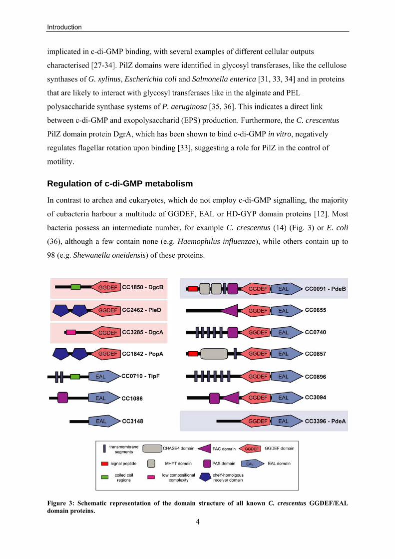
bacteria possess an intermediate number, for example C. crescentus (14) (Fig. 3) or E. coli
(36), although a few contain none (e.g. Haemophilus influenzae), while others contain up to
98 (e.g. Shewanella oneidensis) of these proteins.
Figure 3: Schematic representation of the domain structure of all known C. crescentus GGDEF/EAL domain proteins.

Introduction
5
The domain prediction was retrieved from the SMART database [37]. Proteins with biochemically characterised DGC and PDE activities are highlighted in light red and blue, respectively. The annotation for PopA (CC1842) was manually adjusted.
One question arising from this distribution is why such diversity is needed. Do all proteins
regulate one global c-di-GMP pool, or does each enzyme act on a separate, local pool? And if
so, what are the mechanisms that circumvent cross-talk between different c-di-GMP
dependent regulatory pathways?
Strikingly, most GGDEF, EAL, and HD-GYP domains are fused with known or
hypothetical signal input domains [7, 12]. Among these are components of the bacterial two-
component regulatory system as well as one-component systems, making it likely that these
domains regulate the activity of their fused GGDEF, EAL, and/or HD-GYP domains. It was
also shown that the c-di-GMP signalling domains themselves can act as regulatory domains.
Examples for this are the phosphodiesterases FimX from P. aeruginosa and PdeA for C.
crescentus, where GTP binding to a non-conserved GG(D/E)EF motif was shown to activate
the PDE activity of these composite GGDEF/EAL domain proteins [20, 23]. Another example
is the GGDEF domain protein PopA, where c-di-GMP binding to its I-site leads to altered
subcellular localisation [26]. This variety of different input domains likely allows sensing of a
range of (so far largely unknown) environmental and internal signals that can be integrated in
a c-di-GMP mediated response, enabling the cell to adapt to diverse environmental niches.
This gives a first explanation for the abundance of c-di-GMP metabolising enzymes within
each species.
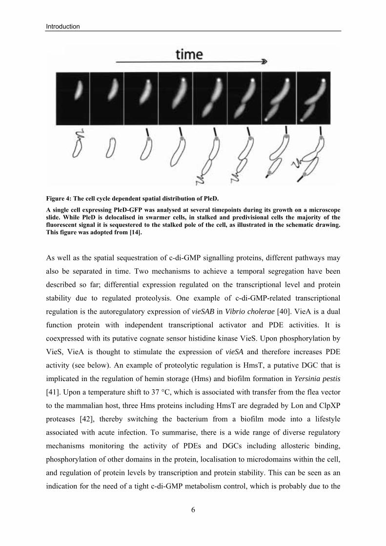
Furthermore, a subcellular localisation was shown for some components of the c-di-
GMP regulatory system [14, 26, 30, 38]. Most notable is the localisation of the C. crescentus
DGC PleD, from a diffuse cytoplasmic distribution to the incipient stalked pole upon the
swarmer-to-stalked cell differentiation (Fig. 4) [14]. Another example is the previously
mentioned c-di-GMP sensor PopA, which shows a complex localisation pattern in a cell cycle
dependent manner and in response to c-di-GMP binding [26]. This localisation of proteins
important for c-di-GMP signalling was interpreted as evidence for microcompartmentalisation
and reveals a possible mechanism to separate different c-di-GMP dependent pathways that are
active at the same time in the cell [7-10, 39].
Introduction
6
Figure 4: The cell cycle dependent spatial distribution of PleD.
A single cell expressing PleD-GFP was analysed at several timepoints during its growth on a microscope slide. While PleD is delocalised in swarmer cells, in stalked and predivisional cells the majority of the fluorescent signal it is sequestered to the stalked pole of the cell, as illustrated in the schematic drawing. This figure was adopted from [14].
As well as the spatial sequestration of c-di-GMP signalling proteins, different pathways may
also be separated in time. Two mechanisms to achieve a temporal segregation have been
described so far; differential expression regulated on the transcriptional level and protein
stability due to regulated proteolysis. One example of c-di-GMP-related transcriptional
regulation is the autoregulatory expression of vieSAB in Vibrio cholerae [40]. VieA is a dual
function protein with independent transcriptional activator and PDE activities. It is
coexpressed with its putative cognate sensor histidine kinase VieS. Upon phosphorylation by
VieS, VieA is thought to stimulate the expression of vieSA and therefore increases PDE
activity (see below). An example of proteolytic regulation is HmsT, a putative DGC that is
implicated in the regulation of hemin storage (Hms) and biofilm formation in Yersinia pestis
[41]. Upon a temperature shift to 37 °C, which is associated with transfer from the flea vector
to the mammalian host, three Hms proteins including HmsT are degraded by Lon and ClpXP
proteases [42], thereby switching the bacterium from a biofilm mode into a lifestyle
associated with acute infection. To summarise, there is a wide range of diverse regulatory
mechanisms monitoring the activity of PDEs and DGCs including allosteric binding,
phosphorylation of other domains in the protein, localisation to microdomains within the cell,
and regulation of protein levels by transcription and protein stability. This can be seen as an
indication for the need of a tight c-di-GMP metabolism control, which is probably due to the

Introduction
7
fact that this second messenger regulates processes critical to many aspects of bacterial
survival in diverse and often dynamic environments [10].
From the phenotypes described so far, it was concluded that high cellular levels of c-
di-GMP generally activate biofilm formation, while inhibiting motility. Therefore, the
paradigm in the field is that c-di-GMP functions as a switch, regulating the transition between
sessile and motile lifestyle (Fig. 2) [7-10]. In addition, c-di-GMP has been shown to influence
the expression of virulence factors [43-45] and affects the cell cycle [26]. Although the
specific target molecules and effector mechanisms involved in c-di-GMP signalling remain
largely unknown, c-di-GMP has been demonstrated to control cellular functions at the
transcriptional, translational, and posttranslational level [46-50].
Regulation of biofilm formation by c-di-GMP
One major process regulated by c-di-GMP is the promotion of surface adhesion and biofilm
formation. Bacteria either exist as free planktonic cell or they form surface-attached
communities known as biofilms [51, 52]. The latter lifestyle is associated with persistence in
adverse environments, as biofilms have been shown to increase bacterial tolerance against
toxic compounds, antibiotics, stress factors and predators [51, 53-55]. The organization of
biofilms can vary from a simple monolayer of cells attached to biotic or abiotic surfaces to a
sessile microbial community with a complex three-dimensional structure [56]. The generation
of biofilms is a multistep process that generally contains three distinct stages (Fig. 5).
Initially, motile bacteria move towards surfaces until they are able to overcome surface
tension and bind. After this initial attachment, the cells irreversibly attach by the production
of an adhesive matrix. The young biofilm eventually matures, building a tight pack with
additional cells, finally resulting in microcolonies. The last stage of the biofilm is
characterized by the release of new planktonic cells and is therefore termed the detachment
state [56-58].

Introduction
8
Figure 5: Illustration of the developmental steps of a biofilm.
Motile cells overcome surface tension and make first reversible contact to the surface in stage 1. In stage 2, the cells become irreversibly attached to the surface by synthesising adhesive matrices. In the next steps, microcolonies are formed. The biofilm matures and builds its three dimensional, sometimes complex structures. In the fifth step, single motile cells are released from the biofilm and invade new resources. This figure was adopted from [56].
During biofilm formation, three structures were shown to be of interspecies importance: pili,
flagella, and adhesive matrices [6]. One prominent class of adhesive material is known as
exopolysaccharides (EPS). These may account for 50 % to 90 % of the total organic carbon of
biofilms and are therefore considered to be the main matrix material of the biofilm [59]. The
nature of these polymers shows great variability, with a multitude of different chemical and
physical properties [60]. Numerous reports have documented the influence of c-di-GMP on
biofilm formation [29, 42, 45, 47, 50, 61-72]. In addition to its effect on adhesion factors, for
example pili [23, 30] or curli fimbriae [71], the synthesis of EPS components was shown to be
increased by high cellular c-di-GMP in various species including Pseudomonas putida [73,
74], Pseudomonas fluorescens SBW25 [68, 74-76], Salmonella typhimurium [67, 71, 77],
Thermotoga maritima [78], and Vibrio cholerae [63, 79, 80].
One example of how EPS production is regulated on a transcriptional level was
described in V. cholerae. The phosphodiesterase VieA was shown to influence the
transcription of vpsR and vpsT [72, 81]. These two transcriptional activators regulate operons
coding for Vibrio exopolysaccharide (VPS) biosynthesis enzymes. VPS is an exopoly-
saccharide used as a biofilm matrix component in V. cholerae [82, 83]. The absence of VieA
leads to increased transcription of vps genes and therefore an increase in biofilm development
[72]. This effect can also be achieved via overexpression of the DGC VCA0956 [81].

Introduction
9
A second example linking exopolysaccharide synthesis to increased cellular c-di-GMP
levels is the Wsp system in P. aeruginosa and P. fluorescens. The Wsp system is a
chemosensory system with homology to the chemotaxis pathway of E. coli [84], but instead
of altered flagellar rotation, the output is c-di-GMP production [18, 85, 86]. The current
model describes the transmembrane protein WspA as a chemoreceptor that changes
conformation upon binding to an unknown molecule. This signal is transduced via either
WspB or WspD to the histidine kinase WspE. WspE then phosphorylates the receiver
domains of the response regulators WspR as well as WspF. Upon phosphorylation the
methylesterase WspF, in concert with the methyltransferase WspC, becomes involved in
signal adaptation, while the DGC activity of WspR is activated [18, 68, 85, 86]. Therefore,
signalling through the Wsp system leads to the production of c-di-GMP by WspR. It has been
suggested that the activation of WspR is mediated by release of its C-terminal GGDEF
domain from the inhibitory N-terminal CheY-like receiver domain upon phosphorylation [18,
68]. Among other influences, including inhibition of twitching and swimming motility and
regulation of Cup fimbriae [85, 86], signalling through the Wsp system regulates biofilm
formation. In Pseudomonas fluorescens the production of partially acetylated cellulose is
stimulated [75, 76] and in Pseudomonas aeruginosa increased expression of the
exopolysaccharide operons pel and psl was shown [87, 88]. These operons encode two sets of
genes predicted to be involved in the production of two distinct carbohydrate-rich biofilm
matrix components. The pel gene cluster is involved in the production of a glucose-rich
matrix material, while the psl gene cluster is involved in the production of a mannose-rich
exopolysaccharide [87]. Both molecules are required for biofilm maturation (reviewed in
[89]). In addition to its regulation on the transcriptional level, the production of the PEL
exopolysaccharide has been shown to be allosterically regulated by c-di-GMP, via the c-di-
GMP binding protein PelD [90]. These examples illustrate how increased levels of c-di-GMP
propagate biofilm formation, a process that has been shown in a variety of species.
Furthermore, the mechanisms described here demonstrate regulation on a transcriptional level
and on a direct allosteric level.
Regulation of motility by c-di-GMP
The ability to move is important for the lifestyle of many bacteria, as it permits the
scavenging of nutrient resources, the exploration of new niches and the avoidance of toxic
environments. It is therefore not surprising that bacteria employ a multitude of different ways
to enable locomotion, including twitching, swarming, and swimming. While twitching

Introduction
10
motility involves a cycle of assembly, attachment, and retraction of Type IV pili [91, 92],
swarming and swimming are mediated by flagella. In many bacteria one kind of flagella is
used for both swimming and swarming, but some species employ distinct flagella for the two
modes of motility [93]. All of these types of locomotion have been shown to be regulated by
c-di-GMP in various ways (reviewed in [8-10, 94]).
An example how c-di-GMP interferes with twitching motility was characterised in
P. aeruginosa. FimX is a composite GGDEF/EAL domain protein with a degenerate A-site. It
was shown to function as a phosphodiesterase, whose activity can be stimulated by GTP,
presumably by binding to the degenerate A-site, as a mutation in this motif abolishes the
activation by GTP [23]. The fully functional PDE activity of FimX is needed to enable
twitching motility in P. aeruginosa [23]. Although the levels of the major subunit of pili,
PilA, are unchanged in a fimX mutant, these cells fail to efficiently assemble their pili, as
almost no PilA is detectable on the outside of the bacteria [23, 30]. Therefore, FimX is
believed to regulate the assembly of pili via c-di-GMP.
In Vibrio parahaemolyticus it was reported that c-di-GMP, produced by the composite
GGDEF/EAL protein ScrC, specifically controls the expression of lateral flagella [50]. These
flagella are arranged randomly on the cell surface of V. parahaemolyticus and, in contrast to
the polar flagella that are only used for swimming motility, permit the cell to migrate across
hydrated, viscous semisolid surfaces, a process called swarming [2, 95]. Another composite
protein was recently identified to also play a role in lateral flagella expression. ScrG is a
composite protein like ScrC, but with a degenerate GG(D/E)EF motif [96]. Mutation analysis
as well as biochemical evidence suggest that ScrG has PDE, but no DGC activity [96]. Both
systems are believed to react to different stimuli and were shown to inversely regulate lateral
flagella expression. This activity is cumulative and operates on the level of transcription via a
yet uncharacterised mechanism.
The final example given here is the effect of c-di-GMP on swimming; the best
understood form of motility in bacteria. Swimming allows movement through a liquid
environment with low viscosity by rotation of flagella, which generate thrust and propel the
bacteria forward. Upon artificially increasing the cellular c-di-GMP level, several studies have
shown that bacteria stop rotating their flagella [14, 19, 33, 61, 97]. In C. crescentus this
paralysis can be mediated by the PilZ domain proteins DgrA and DgrB [33]. In E. coli as well
as in S. enterica serovar Typhimurium the PilZ domain protein YcgR and the PDE YhjH are
suggested to function in the same pathway, regulating flagellar rotation on a functional level
[29, 34, 98]. These publications suggest a model in which YhjH, together with a yet unknown

Introduction
11
DGC, inversely regulates c-di-GMP levels. The concentration of c-di-GMP is sensed by
YcgR, which transduces the signal further to the flagellum. Initial evidence suggests that the
YhjH/YcgR system may control the interaction between the flagellar motor protein MotA and
its stator counterpart FliG (reviewed in [94]). Taken together, these examples show that low
concentrations of c-di-GMP are associated with locomotion of cells by flagellar motors or
retracting pili. Furthermore, they show that this regulation can acts on different levels,
including assembly, transcription, and function.
Caulobacter crescentus is a model organism for cell cycle control and bacterial development
Understanding the inverse regulation of motility and biofilm formation by c-di-GMP is
greatly assisted by an experimental setting where this transition occurs under defined
conditions, in a reliable manner, and synchronous in a sufficiently large population. The
unique asymmetric cell cycle of C. crescentus with its obligate transition from motile
swarmer cell to sessile stalked cell makes it a particularly convenient experimental organism
for the examination of both cell cycle and cellular differentiation events. C. crescentus is a
gram negative bacterium belonging to the α-proteobacteria and can be isolated from
freshwater environments, including oceans, streams and lakes [99]. The cells are hetero-
oligotrophic aerobe, have vibrioid morphology and contain a single, polar cytoplasmic
protuberance surrounded by cell wall material; the stalk [100]. In their natural habitat, they
can be found attached to submerged biotic and abiotic surfaces including other micro
organisms like bacteria and algae [100].

Introduction
12
Figure 6: The Caulobacter crescentus lifecycle.
The developmental stages of the C. crescentus division cycle are shown schematically. Additionally, the spatial distribution of the cell cycle regulator CtrA, as well as its degradation by the protease ClpXP are depicted. CtrA is shown in blue, quiescent chromosomes are represented by circles and replicating chromosomes are indicated by “θ” structures. Morphogenetic events are indicated by grey bars, cell cycle events by black bars. The length and positioning of the bars indicate the initiation and period of events. This figure was adapted from [101].
During its life cycle, C. crescentus undergoes an asymmetric cell division (Fig. 6). Each
division gives rise to two genetically identical, but morphologically and physiologically
distinct daughter cells with different developmental programs. These two cell types comprise
a motile swarmer cell and a non-motile stalk bearing cell. The stalked cell progeny can
reinitiate chromosome replication almost immediately following cell division [102, 103] and
therefore functions as a stem cell, continuously generating new swarmer cells. The planktonic
swarmer cells are equipped with a single polar flagellum, polar adhesive pili, are able to
perform chemotaxis and their replicative program is blocked [100, 102-105]. This block is
mediated by the master cell cycle regulator CtrA [106]. In addition to the direct positive and
negative regulation of 96 genes, which are organized in 55 operons (e.g. flagellar class II
genes) [107], activated CtrA binds directly to the origin of replication and thereby inhibits
replication initiation [106]. To be able to reinitiate chromosome replication and cell division,
the swarmer cell has to undergo an obligate differentiation into a stalked cell. During this

Introduction
13
transition, CtrA is desphosphorylated and simultaneously located to the incipient stalked pole
of the cell, where it is degraded by the ATP dependent protease ClpXP [108-111]. The loss of
active CtrA derepresses the origin of replication and DNA replication is initiated. At the same
time the flagellum is shed, the pili retract, and the chemosensory apparatus is lost [112].
These organelles are replaced by the stalk, with an adhesive material, the holdfast, located at
its tip [113]. The holdfast contains adhesive polysaccharides, whose exact composition is
unknown, and mediates strong irreversible attachment of C. crescentus cells to solid
substrates [57]. The optimal attachment of cells is enabled only during a short window of
development, when the flagellum, pili, and the adhesive holdfast are all present at the same
pole. Therefore, it has been suggested that the precise timing of the retraction of pili, loss of
flagellar activity and assembly of the holdfast is critical for surface colonization (Fig. 7) [47].
During the stalked phase of the cell cycle, the chromosome is completely replicated and both
the cell and the stalk continually grow. In the predivisional cell the two chromosomes
segregate, a constriction forms and a new flagellum, chemotaxis apparatus, and pilus
secretion apparatus are assembled at the pole opposite the stalk (Fig. 6) [101]. Cell division
completes the cycle, again resulting in a new motile swarmer cell scavenging for new nutrient
resources, and a surface adherent stalked cell.
Figure 7: The order of developmental events is important for optimal surface attachment in Caulobacter crescentus.
Schematic representation of the development of polar appendices in C. crescentus, their timing during the cell cycle and their role in surface attachment.

Introduction
14
The morphological and physiological changes that take place during the cell cycle make it
easy to visualise the current status of individual cells. This, together with the ability to isolate
a pure population of swarmer cells by density gradient centrifugation and to follow their
synchronised development facilitates the analysis of the C. crescentus cell cycle [114]. The
couplings of developmental processes, especially the transition from a motile, planktonic into
a non-motile, sessile state, with the cell cycle, as well as its asymmetric division are special
features of C. crescentus. Together, these unique features made C. crescentus one of the
preferred bacterial model systems to analyse cell cycle progression, generation of asymmetry
and development.
The structure of the Caulobacter crescentus flagellum
One possibility of responding to changes in the environment is to move away from adverse, or
towards promising conditions. A wide spread way of facilitating locomotion in bacteria is the
synthesis of a rotating propeller at the cell surface; the flagellum. During its swarmer and late
predivisional phase, C. crescentus possesses a single, polar flagellum. Electron microscopy
revealed that its structure is similar to the well studied flagella of enteric bacteria [115-117].
The flagellar rotor can be divided into three major components; basal body, hook, and
filament (Fig. 8) (reviewed in [118]). The basal body is located in the cytoplasmic membrane
and consists of a series of rings attached to each other by a rod. This structure spans both
membranes and the peptidoglycan layer and acts as flagellar rotor. The rings are named
according to their position in the cell envelope. The C ring is formed by the cytoplasmic part
of the basal body, the MS ring lies within the inner membrane and sticks in the periplasm,
whereas the P ring is located in the peptidoglycan layer. A C. crescentus specific E ring is
located between MS and P rings [115]. Finally, the L ring is located in the outer membrane.
The rod is attached to the hook, a flexible joint that connects the rod to the filament with the
help of adaptor proteins, the hook associated proteins (HAP) (review in [118]). In C.
crescentus the filament is composed of three different flagellins and converts rotary motion
into thrust [119]. The filament is a semi-rigid, hollow tube that comprises most of the flagellar
mass and can be up to 10 μM long. It was shown that at least 20 individual proteins are
integrated into the complex flagellar structure and another 30 are used for its assembly and
function [120].

Introduction
15
Figure 8: Schematic representation of the flagellar structure.
The flagellum can be subdivided into three substructures: a basal body complex that spans the bacterial membranes, the hook and an external filament. The rotor part of the basal body is associated with the motor force generating proteins MotA and MotB that form the stator. Rotation of the flagellum is achieved via interaction of the motor force generators, which function as proton pumps, with the C-ring. Inner membrane (IM); outer membrane (OM); peptidoglycan layer (PG). This figure was adapted from [121].
The regulation of flagellar gene expression
The regulation of flagellar gene expression is subject to a complex regulatory hierarchy, in
which the expression of genes encoding early structures and the functional assembly of their
gene products is required for the expression of late gene products [122-127]. In C. crescentus,
four distinct hierarchical classes can be distinguished (Fig. 9) [128]. The first level of control
is mediated by CtrA, which promotes the transcription of the class II genes and links this
process to the cell cycle [106, 109, 128, 129]. Class II genes encode the structures of the basal
body that are assembled earliest, as well as components of the flagellum-specific type III
secretory system and transcription factors (reviewed in [118, 130]). The alternative sigma
factor σ54 and the transcription activator FlbD, a class II gene product, are essential for the
expression of class III genes encoding both the outer basal body and hook structure [131-
134]. FlbD and σ54 are also a prerequisite for the transcription of the class IV genes encoding
the three flagellin subunits of the flagellar filament [131-134]. In contrast to class III genes,
the translation of class IV genes requires the completion of the basal body-hook assembly

Introduction
16
[135, 136]. At least one flagellin transcript was shown to be destabilized by the negative
regulator FlbT prior to the functional assembly of class II and III gene products, suggesting a
general role of posttranscriptional control [137, 138].
Figure 9: A schematic diagram of the Caulobacter crescentus flagellar regulatory hierarchy.
The hierarchical order of flagellar gene expression is depicted with respect to the cell cycle and DNA replication. Example genes for each class are given within blocks that represent the period during the cell cycle in which they are expressed. CtrA coordinates cell cycle events like DNA replication to the synthesis of the flagellum, as it controls the transcription of early, class II flagellar genes. Among others, the transcriptional activator FlbD is a class II gene and after functional assembly of the inner basal body it drives the expression of class III and class IV genes. The class IV genes are further regulated by a posttranscriptional mechanism, which acts on the functional assembly of class III gene products (see text for details). Alternative sigma factors required for gene expression of the respective class are depicted next to the transcription factors. The chromosome is indicated as red circle, as in Fig. 6. This figure was adapted from [139].

Introduction
17
The expression of flagellar genes is interconnected twice with the cell cycle. First, CtrA
expression, stabilisation and activation by phosphorylation are cell cycle dependent [108, 109,
140, 141]. Second, in addition to its regulated expression, FlbD is also activated by
phosphorylation in a cell cycle specific manner [132]. Furthermore, the differential activation
of FlbD restricts the expression of class III/IV genes to the incipient swarmer cell
compartment in late predivisional cells [132, 142-145].
The assembly of the flagellum
The order of flagellar assembly proceeds, generally speaking, from inner to outer structures
(Fig. 10) (reviewed in [118, 130]).
Figure 10: Schematic drawing of the order of flagellar assembly.
The scheme describes the flagellar assembly as it is investigated in the enterics E. coli and S. enterica serovar Typhimurium. Three major substructures are distinguished, the basal body, the hook, and the filament. The assembly of the basal body is subdivided into four steps, the integration of FliF, the assembly of flagellar specific type three secretion system (TTSS), the C-ring forming switch device and the rod and L/P ring assembly. The order of events is shown as progression from left to right. Inner membrane (IM); peptidoglycan (PG); outer membrane (OM); hook associated proteins (HAP). This figure was adapted from [94].
The first step in assembly is believed to be the insertion of the class II gene product FliF in
the inner membrane, where it forms the MS ring [146]. The MS ring is the central structure
that houses the flagellar specific type III secretion (TTSS) apparatus, which is assembled
next. The C ring is probably assembled on the MS ring by the proteins FliM, FliN, and FliG
that together form the switch complex [118], which is required to change the direction of
flagellar rotation. The TTSS apparatus is highly selective in transporting its substrates. As
soon as the export apparatus is functionally assembled in the MS ring, the rod forming
proteins are exported and assembled in the periplasm. Onto the rod, the P ring and the L ring,
which are either located in the peptidoglycan layer (P) or in the outer membrane (L),
Introduction
18
assemble. These depend on the presence of both the assembled export apparatus and the
switch, although the P and L ring proteins are exported by the secretory pathway and not like
the other components by the TTSS system. After the whole rod has formed, the completed
basal body can now serve as a channel through which subsequent proteins travel across the
inner and outer membrane and through the peptidoglycan layer to their assembly site. The
first molecules to be secreted through this envelope-spanning channel are the hook subunits,
which are polymerized at the distal end of the basal body outside of the cell. After export and
assembly of hook associated proteins (HAP), the flagellins are secreted and assembled
between the HAPs to produce the filament. The stator complex consisting of MotA and MotB
proteins that form the proton channel energising the flagellum is assembled at some point
during the flagellar synthesis. Flagellar associated stator complexes are in equilibrium with
non-associated complexes diffusing in the inner membrane (reviewed in [118]).
The physiology of the flagellar rotation
In some species the flagellum can rotate with a frequency of up to 100,000 rpm [147]. To be
able to rotate, the proton (or sodium) gradient that energises the process has to be converted
into torque. The protons flow through an ion channel formed by the stators [148-153] which
are composed of MotA and MotB proteins with the stochiometry MotA4MotB2 [118, 154,
155]. Rotation is probably driven by conformational changes in these membrane protein
complexes. It is thought that changes occur as protons move on and off a critical aspartate
residue in the stator protein MotB [156] and the resulting forces are applied to the rotor
protein FliG, which was shown to interact with the stator protein MotA [157]. While the
stator protein MotB can interact with the peptidoglycan and is therefore anchored in a fixed
position, the rotor part of the flagellum is free to rotate [149, 158-162]. Each stator complex
assembles and works independently, so each complex can generate torque on its own [163-
166]. In the absence of either MotA or MotB, the flagellum is completely assembled, but
paralysed [148, 150, 167-169].
This paralysis of the flagellum resembles a situation with artificially high cellular c-di-
GMP levels in C. crescentus [14, 33, 61]. Although the exact mechanism leading to the
inhibition of flagellar rotation is not clear, the c-di-GMP binding proteins DgrA and DgrB
have been implicated in this process [33]. In addition, c-di-GMP interacts with flagellar
function on a second level in C. crescentus. At the swarmer-to-stalked cell transition, the
flagellum is shed off and released from the cell to turn off motility in the stalked cell. This
process is dependent on the DGC activity of the GGDEF domain protein PleD [170].

Introduction
19
The activation of PleD, a response regulator with a di-guanylate cyclase output domain provides a link between cell cycle cues and polar development
PleD is an unconventional member of the response regulator family of two component signal
transduction systems. It contains two N-terminal receiver domains (D1 and D2) arranged in
tandem, and a C-terminal GGDEF output domain [14, 16, 17]. Several genetic experiments
have indicated that the two sensor histidine kinases PleC and DivJ, and the single domain
response regulator DivK act as upstream components required for the activation of the PleD
di-guanylate cyclase activity [61, 171, 172]. In agreement with this, direct biochemical
experiments have shown phosphotransfer from both DivJ and PleC to PleD, leading to its
activation [14, 61, 173]. The essential cell fate determinator protein DivK is transcriptionally
coupled to PleD [174]. Although a direct connection to PleD has not yet been established, it
has been shown that DivK is regulated by the same sensor histidine kinases as PleD [172,
174, 175]. As in the case of PleD, DivJ transfers phosphate to DivK [174, 175]. In contrast,
PleC has the function of a phosphatase [175, 176]. Due to the presence of all three
components, DivJ, PleC, and DivK, at the same time in the dividing cell, DivJ and PleC
compete for the phosphorylation status of DivK. Because DivJ activity is restricted to the
stalked cell, while PleC activity is restricted to the newborn swarmer cell upon cell division
(see below), phosphorylated and unphosphorylated DivK are asymmetrically separated [176].
As DivK influences the phosphorylation as well as degradation of the cell cycle regulator
CtrA, the asymmetric DivK phosphorylation status directly influences the cell cycle [140].
Dynamical localisation of PleD and its upstream components during the cell cycle
PleD, like its upstream components PleC, DivJ, and DivK, is dynamically localised during the
cell cycle. Whereas DivJ is located at the stalked pole of the stalked and predivisional cell,
PleC is located at the swarmer pole of swarmer and predivisional cells (Fig. 12) [175]. DivK
is recruited to the swarmer pole in newborn swarmer cells and to the stalked pole of stalked
cells, whereas in predivisional cells it is located to both poles (Fig. 12) [177]. PleD is
delocalised in swarmer cells, but as soon as the cells differentiate into stalked cells it is
sequestered to the incipient stalked pole (Fig. 12) [14]. To enable this localisation, wild-type
PleD has to be phosphorylated, as mutations of the phosphoryl acceptor (PleDD53N), as well as
deletions of the sensor histidine kinases DivJ and PleC abolish polar sequestration [14].
Although critical, phosphorylation is not sufficient for localisation, as shown by the

Introduction
20
hyperlocalised PleD mutant PleD*D53N that misses the phosphorylation site but mimics the
conformation of an activated PleD [14]. Beyond this insight, the mechanism of subcellular
sequestration is unknown. Whereas the differential localisation of PleC and DivJ results in a
uneven distribution of phosphorylated DivK upon cell division, which then leads to
asymmetry and is believed to orchestrate the different cell fates of the C. crescentus progenies
[176], the role of PleD localisation is currently unclear. It has been speculated that localised
PleD could restrict c-di-GMP production to one locus, thereby enabling the specificity of its
signalling pathway [7, 8, 14]. PleD localisation could also ensure that upon division, active
PleD stays in the stalked progeny to keep the swarmer cell free of active PleD.
Figure 11: Schematic drawing of the subcellular localisation of PleD and its upstream components during the Caulobacter crescentus cell cycle.
PleC is depicted in red, DivJ in green, DivK in yellow, and PleD in blue. Delocalised (and unphosphorylated) PleD is shown in light blue. The enzymatic activities of DivJ and PleC towards DivK and PleD, the cell cycle and key developmental events are schematically represented. While PleD is not phosphorylated and therefore not localised in the swarmer cell, it is activated by PleC and DivJ during the swarmer-to-stalked cell transition. Upon activation PleD is sequestered to the incipient stalked pole of the cell, where it stays for the rest of the cell cycle. Delocalised PleC, DivJ and DivK are not shown for clarity.
Introduction
21
Structural insights in the function of PleD
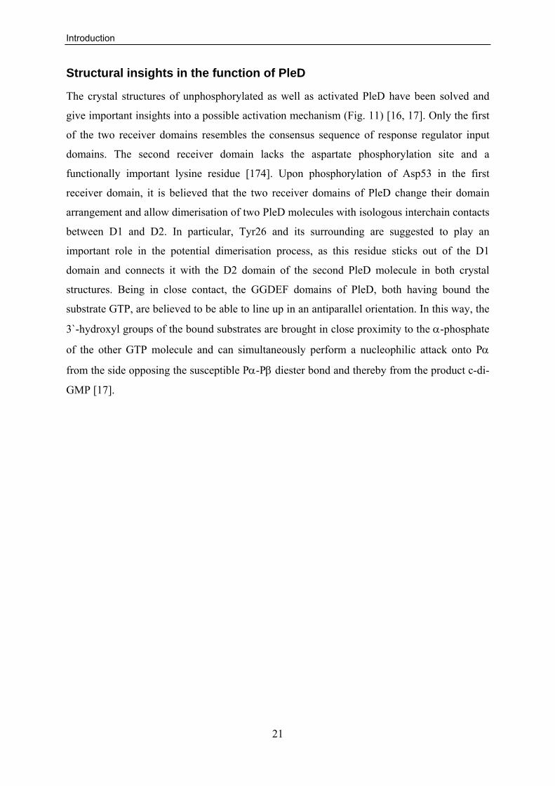
The crystal structures of unphosphorylated as well as activated PleD have been solved and
give important insights into a possible activation mechanism (Fig. 11) [16, 17]. Only the first
of the two receiver domains resembles the consensus sequence of response regulator input
domains. The second receiver domain lacks the aspartate phosphorylation site and a
functionally important lysine residue [174]. Upon phosphorylation of Asp53 in the first
receiver domain, it is believed that the two receiver domains of PleD change their domain
arrangement and allow dimerisation of two PleD molecules with isologous interchain contacts
between D1 and D2. In particular, Tyr26 and its surrounding are suggested to play an
important role in the potential dimerisation process, as this residue sticks out of the D1
domain and connects it with the D2 domain of the second PleD molecule in both crystal
structures. Being in close contact, the GGDEF domains of PleD, both having bound the
substrate GTP, are believed to be able to line up in an antiparallel orientation. In this way, the
3`-hydroxyl groups of the bound substrates are brought in close proximity to the α-phosphate
of the other GTP molecule and can simultaneously perform a nucleophilic attack onto Pα
from the side opposing the susceptible Pα-Pβ diester bond and thereby from the product c-di-
GMP [17].

Introduction
22
Figure 12: Ribbon diagrams of the crystal structures of non-activated PleD and activated PleD.
The first receiver domain (D1) is coloured in red, the second receiver domain (D2) in yellow and the GGDEF domain in green. The GG(D/E)F motif in the GGDEF domain is highlighted in blue. Disordered parts are depicted in grey, two-fold symmetry axes are indicated by a grey line. The active site (A-site), allosteric inhibition site (I-site) and phosphorylation site (P-site) are indicated; symmetry-related elements are specified by a prime. (A) and (B) present perpendicular views to the two-fold axis of the stem, whereas in (C) and (D) the view is rotated by 90° around a horizontal axis with respect to the top panels, showing the bottom view of the (D1/D2)2 stem with the DGC domains in the rear clipped off for clarity. (A and C) Non-activated PleD [16]. Intercalated (c-di-GMP)2 dimers are bound to allosteric inhibition sites I and I`, which are formed by the primary inhibition site in the GGDEF domain (Ip: Arg359, Asp362, Arg390) and a secondary site on the adaptor domain (IS,D2: R148, R178). (B and D) In the activated structure [17], the phosphorylation site (P site) is modified by BeF3
- and Mg2+. The active site (A site)
harbours GTPαS/Mg2+. Intercalated (c-di-GMP)2 dimers are bound to the I and I` sites. In this structure,
the site are comprised of the primary IP site, as in the non-activated structure, and a secondary I site of the symmetry-related DGC (IS,DGC: R313). In both structures, the crosslinking of two half I-sites by c-di-GMP results in the immobilisation of the GGDEF domains in a way that the A-sites are not productively arranged, rendering the enzyme catalytically incompetent. This figure was adopted from [17].

Introduction
23
PleD not only possesses an enzymatic active site (A-site), which binds GTP and catalyses its
condensation to c-di-GMP, but also an inhibitory site (I-site) that mediates feedback
inhibition upon binding to c-di-GMP [16, 17, 19]. In PleD the I-site is split into three different
motifs. Firstly, c-di-GMP is bound by the residues Arg359 and Asp362 of the RXXD motif
together with Arg392, all located in the GGDEF domain. These form the so called primary I-
site (IP) [16, 17] Secondly, two alternative secondary binding sites, one formed by residues
Arg148 and Arg178 of the D2 domain (IS, D2) [16] and one formed by Arg313 of the adjacent
GGDEF domain (IS, DGC) [17], can bind to c-di-GMP. This binding of c-di-GMP between two
sites crosslinks two domains, either the GGDEF with the D2 or alternatively the two GGDEFs
of a presumed PleD dimer. This crosslinking reduces the mobility of the GGDEF domain and
prevents productive arrangements of the GGDEF active sites. This mechanism was therefore
called “inhibition by domain immobilisation” [16, 17]. In addition, the comparison of atomic
simulations of PleD having bound c-di-GMP to the I-site with PleD having an unoccupied I-
site indicated reduced flexibility of PleD domains upon c-di-GMP binding. This led to the
conclusion that c-di-GMP binding to the I-site may influence the dimerisation rate of the
molecule and therefore also activity [177]. The finding that mutations in the IS, D2 site
displayed a 20-fold higher DGC activity compared to wild-type PleD is consistent with that
interpretation [19]. Thus, it is possible that c-di-GMP binding to the I-site affects PleD
activity in several ways.
Cellular functions of PleD
PleD influences several aspects of polar development in C. crescentus. As described above, in
the absence of PleD, the flagellum is not released during the swarmer-to-stalked cell
transition. In the wild-type situation, this process coincides with degradation of FliF by the
ClpAP protease, which is also inhibited in the absence of PleD [170, 178, 179]. Thus, deletion
of PleD leads to ectopic flagella at the tip of the stalk during the stalked and predivisional
phase of the cell cycle [61]. These flagella are functional, resulting in a “hypermotile”
phenotype, characterised by swimming of all cell types in liquid culture [61, 170]. In contrast,
on semi-solid agar plates ΔpleD cells form smaller colonies compared to the wild type, which
was explained by a chemotaxis defect of the mutant strain [14, 61, 180]. When a constantly
activated mutant of PleD (PleD*) is expressed in C. crescentus, this leads to normally
flagellated but non-motile cells, as the rotation of the flagellum is inhibited [61]. Whereas a
ΔpleD mutant possesses short stalks, the PleD* expressing cells have increased stalks,
indicating a function for pleD in stalk growth or length determination [61, 170]. It was further

Introduction
24
shown that the production of the holdfast is not properly timed in a ΔpleD strain. Instead of
holdfast synthesis during the swarmer-to-stalked cell transition, the onset of synthesis is
delayed, which leads to suboptimal attachment to surfaces [47]. Taken together, these
findings illustrate that the DGC PleD is involved in the inverse regulation of motility and
surface attachment. Its activity is controlled on multiple levels to ensure correct timing at the
swarmer-to-stalked cell transition. Furthermore, PleD activation is linked to a system
controlling asymmetry in C. crescentus and itself is involved in polar development.
Aim of the thesis
25
2. Aim of the thesis The second messenger c-di-GMP is a key signalling molecule that has a major influence on
the lifestyle of bacteria as it antagonistically regulates motility and non-motility, as well as
single cell dispersal and biofilm formation. In this work, the model organism for bacterial
development, Caulobacter crescentus, and its cell cycle embedded transition between motile
swarmer cell and sessile stalked cell will be employed to gain a deeper insight into the
enzymatic regulation and biological role of c-di-GMP in the control of lifestyle change. One
approach used will be the systematic functional analysis of all 14 GGDEF and/or EAL
domain proteins from C. crescentus with a focus on phenotypes relevant for the swarmer-to-
stalked cell transition. The key regulators identified herein will be more closely analysed to
understand their role and interconnection in this developmental process. In another approach,
the di-guanylate cyclase PleD, that is already known to be involved in the regulation of the
swarmer-to-stalked cell transition of C. crescentus, will be characterized in more detail. Here,
especially the mechanisms and environmental influences underlying its activation and
localisation will be investigated, to understand more details of its regulation and the function
of polar localisation.

Results I A network controlling polar development via c-di-GMP
26
3. Results
3.1 A Phosphodiesterase and its Cognate Di-guanylate Cyclases Antagonistically Control Polar Development in Caulobacter crescentus Sören Abel, Marc Folcher, Urs Jenal
Statement of my work
All plasmids and strains used in this study have been generated by me, unless otherwise stated
in table S1. I also performed all motility (Fig. 1, 2A, 3A, 5), attachment (Fig. 1, 5), holdfast
stain (Fig. 6A, 6B), Western blot (Fig. 2B, 7A, 7B, 7C, 9B), synchronisation (including
dominant negative ClpX expression) (Fig. 6B, 7A, 7C, 8A, 8B, 9B), and light-/fluorescence
microscopy (Fig. 8A, 8B, 9A) experiments of this study. Furthermore, I generated and
analysed the motile suppressors of the ΔpdeA mutant (Fig. 3B, 3C), performed the
computational analysis of EAL/GGDEF domains (Fig. S2), and determined the strain growth
rates (Fig. S3) given in this manuscript. Finally, I analysed the enzymatic assays of DgcB and
the electron micrographs (Fig. 2C).

Results I A network controlling polar development via c-di-GMP
27
Abstract The second messenger c-di-GMP positively regulates surface attachment and negatively
regulates single cell based motility. In Caulobacter crescentus c-di-GMP plays a key role in
the developmental transition from the planktonic swarmer cell to the sessile stalked cell,
which involves remodelling of the cell poles. As this irreversible process determines the fate
of the bacterium, the decision when to undergo this obligate transition is of critical
importance and has to be carefully regulated. To gain further insight into the regulation of
pole morphogenesis in this organism, a systematic functional analysis of all GGDEF, EAL
and GGDEF/EAL composite proteins from C. crescentus was conducted. This identified the
phosphodiesterase PdeA as a novel component involved in the regulation of the motile-to-
sessile transition. Mutants lacking PdeA are non-motile, fail to restrict holdfast synthesis to
the stalked cell type and, as a result, show an increased propensity for surface attachment.
PdeA is only present in the late predivisional and swarmer phase of the cell cycle. During
transition into stalked cells, it is recruited to the incipient stalked cell pole, where it is
degraded by the ATP dependent protease ClpXP. Furthermore, we identify the novel di-
guanylate cyclase DgcB that acts as a specific antagonist of PdeA. Evidence is provided that
this pair exerts its function in flagellar control via a pathway that does not converge with an
already known pathway comprising the di-guanylate cyclase DgcA and the c-di-GMP effector
proteins DgrA and DgrB. DgcB controls holdfast biogenesis and surface attachment together
with the well-characterised di-guanylate cyclase PleD. Genetic data suggest that both di-
guanylate cyclases feed into a c-di-GMP pool that is degraded by PdeA. Cumulatively, these
findings result in a model in which PdeA acts as a gatekeeper that prevents the premature
surface attachment and loss of motility of the swarmer cell by reducing the concentration of c-
di-GMP produced by DgcB. The specific degradation of PdeA together with the activation of
PleD trigger the developmental processes towards a non-motile, surface attached stalked cell.

Results I A network controlling polar development via c-di-GMP
28
Introduction
C-di-GMP is a ubiquitous second messenger in eubacteria. Several studies have proposed this
molecule as key regulator controlling the switch between a planktonic, free living lifestyle
and sessile community behaviour of bacterial cells. In particular, an increasing number of
reports have linked high levels of c-di-GMP to sessility and biofilm production via synthesis
of a number of different adhesion factors, including exopolysaccharides, pili, curli fimbriae,
or surface exposed proteinaceous adhesins [23, 30, 46, 49, 72, 81, 86, 181, 182]. In contrast,
low levels of c-di-GMP were shown to promote cell motility and a unicellular lifestyle
(reviewed in [7-10]).
Synthesis of c-di-GMP is catalyzed by proteins containing a GGDEF domain, whereas
the degradation of c-di-GMP is mediated by EAL or HD-GYP domain proteins [13, 14, 20-
22, 25, 62]. These domains are highly conserved and widespread in the bacterial kingdom
[12]. Most bacteria possess an intermediate number (5-20) of GGDEF or EAL domain
proteins (e.g. Caulobacter crescentus), a few contain none (e.g. Haemophilus influenzae), and
others contain up to 100 (e.g. Shewanella oneidensis, Vibrio vulnificus) of these proteins. The
GGDEF, EAL and HD-GYP domains are often found fused to each other in various
combinations, as well as associated in a modular fashion with a wide range of regulatory,
sensory input and structural domains. It is therefore believed that the production of c-di-GMP
is directly controlled by environmental or internal stimuli [7, 8, 12].
C-di-GMP controls cellular functions via an interaction with downstream receptors.
Despite an allosteric c-di-GMP binding site (I-site) in the GGDEF domain itself [16, 17, 19],
the PilZ domain has been shown to effect cellular functions upon c-di-GMP binding [27, 28,
33, 34, 36]. This small effector domain can be found alone or associated with regulatory
proteins and enzymes and may function as switch protein or as an allosteric regulator [27].
The PilZ domain was found to be involved in regulation of exopolysaccharide (EPS)
production [31, 33, 34, 36, 90] as well as flagellar rotation [33]. The latter was found to be
regulated by the only C. crescentus PilZ domain proteins DgrA and DgrB [33].
The motile-to-sessile switch of C. crescentus that is integrated in its division cycle make this
organism a suitable model system to investigate c-di-GMP signalling. Upon division the
bacterium produces two distinct daughter cells with different morphologies and
developmental programs: a sessile stalked cell and a motile swarmer cells [100]. While the
newborn stalked cell immediately initiates a new round of chromosome replication and cell

Results I A network controlling polar development via c-di-GMP
29
division, the swarmer cell is not able to replicate its chromosome, as the origin of replication
is blocked by the cell cycle regulator CtrA [102, 103, 106]. The swarmer cell is propelled by
its polar flagellum, performs chemotaxis, and is piliated. Before the cell starts to initiate a new
round of cell division, it undergoes a differentiation step from motile to sessile cell. During
this transition pili retract, the flagellum is shed, and a stalk with an adhesive holdfast at its tip
is produced [101]. The holdfast contains exopolysaccharides of unknown composition
through which the cells irreversibly attach to surfaces. Surface attachment peaks during a
short window of development, when the flagellum, pili, and the adhesive holdfast are all
present at the same pole. It was proposed that the precise timing of assembly and loss of polar
organelles is critical for optimal C. crescentus surface colonization [47]. At the same time
CtrA is inactivated and localised to the developing stalked pole that is occupied by its cognate
protease ClpXP, where it is proteolytically degraded. This deblocks the origin of replication
and allows the duplication of the chromosome. As the stalked cell elongates and initiates cell
division, a new flagellum is synthesised. Flagellar biogenesis in C. crescentus is a
hierarchically ordered process, which requires the functional assembly of one regulation class,
before the genes of the following class are expressed (reviewed in [118, 126, 130]). Prior to
cell division, the new flagellum is completed and together with the chemotaxis apparatus and
pilus secretion apparatus assembled at the pole opposite the stalk. A constriction is formed
and cells divide, again resulting in a swarmer and a stalked cell.
The genome of C. crescentus contains 14 genes encoding GGDEF, EAL, or GGDEF/EAL
composite proteins (Fig. S1). Three of these proteins and their function in the regulation of
motility and attachment have already been characterised in more detail. The EAL domain
protein TipF was shown to be a prerequisite of flagellar assembly and positioning and is also
required for proper pili expression. Therefore, a ΔtipF strain is non-motile and can be
assumed to have an attachment defect [183]. Although TipF was predicted to contain an EAL
domain, important residues for enzymatic activity are not conserved, making it unlikely that
TipF has PDE activity (Fig. S2). In agreement with that, in vitro studies failed to show any
enzymatic activity involved in c-di-GMP metabolism (M. Folcher unpublished).
PopA is predicted to contain a GGDEF domain. However, like in TipF, widely
conserved residues that have been shown to be critical for enzymatic activity are missing (Fig.
S2) [14, 18, 19]. Indeed, in vitro experiments failed to demonstrate DGC activity [26]. In
contrast, residues of the I-site implicated in allosteric binding of c-di-GMP that normally lead
to feedback inhibition of the enzymatic activity, are present and still function as c-di-GMP
Results I A network controlling polar development via c-di-GMP
30
binding site in PopA [19, 26]. Regulated by c-di-GMP and together with the adaptor protein
RcdA [26, 184], PopA was shown to recruit CtrA and perhaps other proteins during the
swarmer-to-stalked cell transition and to localise them to the stalked pole, where they are
degraded by the protease ClpXP [26]. Furthermore, the deletion of popA leads to reduced
motility on semi-solid agar plates [185]. The reason for this is unclear, although the presence
of a degenerate A-site makes it likely that the observed phenotype of the ΔpopA strain is
rather caused by the disrupted degradation of ClpXP substrates than by direct variations of the
c-di-GMP level.
One GGDEF domain protein was recently implicated in the periodic remodelling of
the cell pole. The di-guanylate cyclase PleD promotes the transition from swarmer-to-stalked
cell by directing flagellar ejection as well as stalk formation and timing of holdfast biogenesis
[14, 47, 61, 186]. Like many enzymes involved in c-di-GMP metabolism, PleD has a modular
structure with a catalytic GGDEF domain and two regulatory N-terminal CheY-like receiver
domains [16, 17]. PleD is activated during the swarmer-to-stalked cell transition through
phosphorylation-mediated dimerisation by two sensor histidine kinases, DivJ and PleC [61,
171, 172, 174, 186, 187]. Dimerisation of PleD is necessary for its ability to produce c-di-
GMP and leads to its localisation to the incipient stalked pole of the cell [14, 186].
Differential phosphorylation coupled to polar sequestration ensures that PleD activity is
restricted to the correct time and place during the C. crescentus cell cycle and development.
Consistent with its role in timing of holdfast biogenesis, a deletion in pleD reduced surface
attachment to about 40 % compared to wild type. Furthermore, it leads to a significant
reduction of the colony size on motility plates. This unexpected negative effect on motility
was suggested to be the result of a chemotaxis defect [61, 180].
The finding that three of the 14 GGDEF/EAL domain proteins are engaged in different
aspects of the motile-to-sessile transition and cell cycle control, raised the question if and how
the remaining proteins of this family take part in these processes. In particular, the role of
phosphodiesterases that counteract the activity of di-guanylate cyclases is still poorly defined.
Here, we present a systematic functional analysis of all C. crescentus genes predicted to code
for GGDEF/EAL domain protein with a focus on the regulation of motility and attachment.
This led to the identification of a new pair of enzymes, a di-guanylate cyclase and a
phosphodiesterase, which antagonistically control pole morphogenesis during the swarmer-to-
stalked cell transition. First insights are gained on how the phosphodiesterase activity is
limited to the motile cell cycle phases, where it acts as a gatekeeper, preventing the cells from

Results I A network controlling polar development via c-di-GMP
31
premature development into the sessile form. Furthermore, we present evidence for a spatial
or temporal separation of enzyme activities towards c-di-GMP within the cell. Together, these
findings add an additional layer of complexity to the regulation of pole morphogenesis in
C. crescentus and the role of the second messenger c-di-GMP therein.

Results I A network controlling polar development via c-di-GMP
32
Results
A systematic mutational analysis of genes coding for GGDEF and/or EAL domain proteins reveals proteins that inversely regulate motility and attachment
According to the paradigm of c-di-GMP signalling, the deletion of a di-guanylate cyclase
should result in decreased surface attachment and increased motility. In contrast, elevated c-
di-GMP levels in the absence of a phosphodiesterase should have the opposite phenotype. To
systematically investigate the role of GGDEF and EAL domain proteins in Caulobacter
crescentus, single in frame deletions of the 14 genes predicted to code for proteins containing
GGDEF and/or EAL domains were generated by allelic exchange. Systematic assays for
motility and attachment phenotypes were established. To control for a possible influence on
growth, which would confound the attachment and motility assays, the growth rate was
determined for all mutant strains (Fig. S3). Importantly, none of the deletions affected growth.
Mutations in tipF, pleD, and popA had been described before and served as a controls
for the experiments. For motility, all three deletion strains behaved as described in the
literature. The absence of PleD reduced colony size to about 60 % of the wild type due to its
chemotaxis effect and a ΔpopA strain showed a about 50 % reduction (Fig. 1) [185]. The tipF
deletion is non-motile as it can not assemble a flagellum and the observed colony size of
about 5 % of the wild type reflects this (Fig. 1) [183]. The attachment of a ΔpleD strain (about
40 % of the wild type) is in good agreement with the published data (Fig. 1) [47].
Interestingly, while it was described that the ΔpopA strain has no attachment phenotype [185],
we found that this mutant showed a decrease of attached biomass to about 50 % compared to
wild type (Fig. 1). Similarly, a ΔtipF mutant showed a severely decreased attachment to about
20 % of the wild type (Fig. 1).
The other 11 deletion strains can be grouped into three classes according to their
phenotypes: a) decreased attachment and increased motility, b) increased attachment and
reduced motility, and c) no obvious phenotype. The first class includes deletions in cc0740,
cc0857, dgcA, and dgcB, which all reduced attachment to about 70 % and increased motility
to about 150 % of the wild type (Fig. 1). The second class of mutants contains deletions in the
pdeA and cc0091 genes. While the ΔpdeA strain severely reduced motility to about 20 % of
the wild type, the motility phenotype of the Δcc0091 mutant is very mild (about 90 % of the
wild type) (Fig. 1). The other five deletion strains, Δcc0655, Δcc0896, Δcc1086, Δcc3094, and

Results I A network controlling polar development via c-di-GMP
33
Δcc3148, showed no apparent phenotype in surface attachment or cell motility under the
conditions tested. The reason why deletions of these genes did not show a phenotype remains
unclear. They might not be expressed under the assay conditions, there might be redundancy
so a phenotype is only detectable upon deletion of more than one gene or they simply do not
influence the measured phenotypes.
Strains lacking pdeA show reduced motility although the flagellum is completely assembled
As the ΔpdeA strain showed an attachment and motility phenotype expected for a
phosphodiesterase mutant it was decided to further investigate this gene. It was shown earlier
that increased cellular levels of c-di-GMP result in a paralysed flagellum [33, 61]. Therefore,
it was reasoned that the motility defect of the pdeA mutant would have a similar effect, since a
reduction of the overall PDE activity is likely to increase the cellular c-di-GMP concentration.
To make sure that the observed phenotype was indeed caused by a mutation in pdeA, a
plasmid harbouring the pdeA gene was constructed and this construct was introduced both
into wild type and into the mutant strain. In a wild type background, plasmid-driven
expression of pdeA in trans slightly increased the swarm size on semi-solid agar plates,
arguing that PdeA has indeed a direct role in motility control (Fig. 2A). In support of this,
motility of the ΔpdeA mutant strain was completely restored to wild-type levels (Fig. 2A).
Next, the abundance of several proteins involved in building the flagellum or in the regulation
of motility was analysed. Because flagellar biogenesis is organized in a hierarchical manner
(reviewed in [126, 130]), the levels of members of all four regulation classes of the flagellar
hierarchy were compared in the wild type and the ΔpdeA mutant. As shown in Fig. 2B, no
difference was observed for CtrA (class I), FliF, FliM, and FlbD (class II), FlgH (class III), or
the flagellins (class IV). This indicates that the structural and regulatory flagellar components
are normally expressed and that the first two checkpoint substructures of the flagellum are
assembled properly. The level of FliL, a protein required for motor function but not assembly,
whose gene is not part of the flagellar hierarchy [188] was also analysed. Like for the other
flagellar proteins tested, FliL levels showed no differences in the ΔpdeA strain as compared to
the wild type (Fig. 2B).
As the ΔpdeA strain has no apparent defect in expressing flagellar proteins, the motility defect
of the mutant could either be caused by inhibiting flagellar function or by an impaired
assembly of class IV filament proteins. To investigate the latter, electron microscopy was
used to analyse flagellar filament structures of mutant and wild type. As indicated in Fig. 2C,

Results I A network controlling polar development via c-di-GMP
34
the pdeA mutant strain had visible flagella that were morphologically indistinguishable from
wild type. Taken together, these experiments indicate that in the ΔpdeA mutant strain the
flagellum is assembled properly. Thus, the motility defect of this strain is likely caused by an
impaired motor function.
Mutations in dgcB but not in dgrA and dgrB suppress the motility defect
of ΔpdeA
It was shown recently that ectopic expression of the di-guanylate cyclase dgcA leads to a non-
motile phenotype and a paralysed flagellum [33]. The motility block in this strain was
partially suppressed when either dgrA or dgrB, both coding for small c-di-GMP effector
proteins, were deleted [33]. To be able to test whether these small effector proteins are also
involved in motility control in the ΔpdeA mutant, strains were constructed that in addition to
the absence of PdeA contained deletions in dgrA and/or dgrB. The deletion of dgrA, dgrB, or
both failed to restore motility in the ΔpdeA mutant, arguing that the inhibition of flagellar
rotation in the absence of PdeA is not mediated via the DgrA or DgrB effector proteins (Fig.
3A). Consistent with this, a point mutation in rpsA, coding for the ribosomal protein S1, was
able to restore motility in cell overexpressing dgrA [33], but failed to restore motility in the
ΔpdeA mutant (data not shown). These results imply that an increased cellular concentration
of c-di-GMP can inhibit motility via different non-converging pathways and, more
specifically, argue that the di-guanylate cyclase DgcA and the phosphodiesterase PdeA do not
interfere with the same pool of c-di-GMP to control motility.
To unravel components of the signal transduction pathway that together with PdeA
control C. crescentus cell motility, we used the paralyzed ΔpdeA strain to screen for motile
suppressors. In a first approach, a transposon mutagenesis was conducted by randomly
integrating tnMariner into the genome of the ΔpdeA strain and isolating mutants that regained
motility. In a second approach, spontaneous suppressors were generated by selecting for
motile colonies on semi-solid agar plates. The following classes of suppressors were
expected: a) upregulation of phosphodiesterases that decrease the cellular c-di-GMP
concentration; b) downregulation or elimination of di-guanylate cyclases that contribute to the
c-di-GMP pool normally degraded by PdeA; and c) mutation of effector proteins that
transduce the c-di-GMP signal to the flagellum. While the transposon mutagenesis most likely
only generates loss-of-function mutations, the spontaneous mutations allow also the
identification of gain-of-function mutations. In the transposon screen about 50,000 mutants
were tested, resulting in 286 motile suppressor strains. Due to an intermediate selection

Results I A network controlling polar development via c-di-GMP
35
procedure (see materials and methods) it is not clear if these are independent events. So far,
only two targets could be identified (Fig. 3B). One transposon insertion was mapped to the
intergenic region between open reading frame cc2147 (annotated as a D-isomer specific 2-
hydroxyacid dehydrogenase family protein) and cc2148 (predicted to code for the ATP-
binding protein of an ABC transporter) (Fig. 3B). The insertion led to wild-type-like colonies
on motility agar plates (data not shown). Marker linkage experiments by transduction
confirmed that suppression was due to this transposon insertion. The second target isolated in
the screen was dgcB. Four independent transposon insertions were mapped in the open
reading frame coding for this presumable di-guanylate cyclase (Fig. 3B). Two of them
mapped to the 3` end of the gene, which codes for the GGDEF domain, implying that
disruption of DgcB mediated c-di-GMP synthesis is the cause for the suppression of the
paralyzed flagellum. All four insertions increased motility to about 80 % of the wild-type
strain (data not shown).
In addition to the transposon-derived suppressors, eight spontaneous motile suppressor
mutants of the ΔpdeA strain were isolated. Interestingly, individual mutants showed different
levels of suppression and were grouped into three classes according to their performance on
motility agar plates. Whereas two of the strains formed colonies on semi-solid agar plates of
similar size to wild type, three exhibited intermediate motility of about 80 %, while three
others showed increased motility of up to about 150 % of the wild type level (Fig. 3C). Based
on the results of the transposon screen, it was first tested if any of the spontaneous suppressor
mutations mapped to dgcB. A resistance marker in a neutral locus next to dgcB (~100 %
linkage) was transferred to the suppressor by means of general transduction, replacing the
dgcB region of the suppressor strain with wild-type dgcB. Two of the three suppressors with
an intermediate motility could be reverted to the reduced motility phenotype of ΔpdeA
(Fig. 3C), arguing that the original suppressor mutations mapped to the dgcB gene. Western
blot analysis failed to show a crossreacting band for DgcB in these two mutants, whereas the
protein was detectable in the other six isolated strains (data not shown). This result was
further confirmed by sequence analysis, demonstrating that one strain contained a nonsense
mutation in dgcB and the other contained a deletion of the dgcB region (Fig. 3B). The same
test was performed with a marker ~100 % linked to cc2147, but none of the eight suppressor
strains could be reverted to the original paralyzed phenotype of the ΔpdeA mutant (data not
shown). The heterogeneity of the suppressor phenotypes together with the transduction data
indicated that different (possibly downstream) targets are involved. Future work will have to
concentrate on mapping the chromosomal loci altered in these additional suppressors.
Results I A network controlling polar development via c-di-GMP
36
Because both suppressor screens identified DgcB as a candidate di-guanylate cyclase
interacting with PdeA, our studies concentrated on the role of these two proteins in pole
morphogenesis.
DgcB is a di-guanylate cyclase
Based on the motility and surface attachment phenotype of a dgcB mutant and based on its
domain organisation, the DgcB protein was postulated to be a di-guanylate cyclase. To test
this, a hexa-histidine-tagged version of DgcB was expressed in Escherichia coli and purified
to homogeneity on a nickel affinity column. The protein was then used in a standard DGC
assay [14, 19]. In the presence of purified DgcB protein, GTP was converted to c-di-GMP
(Fig. 4). As control reactions the already characterized DGCs PleD* and DgcA from
C. crescentus were tested (Fig. 4). Under the assay conditions (~5 μM GTP) both showed a
higher specific activity as DgcB. The KM for GTP conversion was determined to be in the
range of about 35 μM. While the active site (GGEEF) is conserved, the RXXD motif of the I-
site is not conserved in DgcB (Fig. S2). In agreement with this, DgcB was not subject to non-
competitive feedback inhibition by c-di-GMP (data not shown). These results together with
the genetic data presented above indicate that DgcB is a di-guanylate cyclase that specifically
controls pole morphogenesis in C. crescentus.
PdeA and DgcB antagonistically control Caulobacter crescentus surface attachment and motility
Because mutations in the di-guanylate cyclase dgcB can suppress the phenotype of a mutant
strain lacking the phosphodiesterase PdeA, we reasoned that these two opposing enzymes act
in the same regulatory pathway by controlling the cellular levels of c-di-GMP. To confirm the
suppression data and to rule out polar effects of the transposon insertions, attachment and
motility were scored in double mutants with markerless, in frame deletions of both pdeA and
dgcB. As shown in Fig. 5, the ΔpdeA mutant showed an increased relative attachment and
decreased motility, whereas the opposite phenotype was observed for the ΔdgcB mutant. The
double deletion strain showed an intermediate phenotype for both readouts and was similar to
wild type. This indicated that the two opposing enzyme activities antagonistically control
motility and surface attachment. This finding also suggests that DgcB and PdeA activities are
not required for motility and surface attachment per se, but rather to regulate these cellular
functions during the C. crescentus life cycle.

Results I A network controlling polar development via c-di-GMP
37
To test if PdeA exclusively interacts with DgcB or if other DGCs also contribute to
this pathway, additional mutations were analysed in the ΔpdeA mutant background. DgcA is
also known to be a DGC in vitro [19] and a strain with a single deletion in dgcA behaves like
a ΔdgcB mutant in terms of attachment and motility (Fig. 1, 5). Therefore, a double deletion
of pdeA and dgcA was created and tested. However, as shown in Fig. 5, a deletion in dgcA had
no effect on attachment or on motility in the ΔpdeA mutant background. This result indicates
that DgcB and PdeA form a specific pair of antagonists that control motility and attachment
through the same molecular pathway. Apparently, DgcA, although influencing motility and
attachment, does not interfere with the PdeA pathway. In contrast, a mutation in the DGC
pleD decreased attachment in the ΔpdeA background almost to levels of the ΔpleD strain and
a dgcB pleD double mutant showed only background levels of attachment independent of the
presence or absence of PdeA (Fig.5). This suggests that that the di-guanylate cyclases DgcB
and PleD collectively induce attachment in C. crescentus and that the phosphodiesterase
PdeA counteracts the action of these two enzymes. In contrast, the motility on semi-solid agar
plates of the ΔpleD ΔpdeA strain was on the level of a ΔpdeA mutant. This phenotype is
comparable to the ΔdgcA ΔpdeA strain, indicating that PleD and PdeA influence motility on
two non-converging pathways. The colony size formed by the dgcB pleD deletion strains
shows intermediate motility behaviour between ΔdgcB and ΔpleD. This is likely because a
pleD mutant, although being hypermotile in liquid culture, does not perform accordingly on
semisolid agar plates [180]. Therefore the deletion of pleD reduces the increased motility of
the ΔdgcB strain. According to the motility behaviour of the ΔpleD ΔpdeA strain, the
additional deletion of pdeA should have no influence on motility in the dgcB pleD mutant.
Indeed, the colony size on semi-solid agar plates of the ΔdgcB ΔpleD ΔpdeA mutant did not
differ from the ΔdgcB ΔpleD strain.
PdeA, DgcB, and PleD control the timing of holdfast biosynthesis during the Caulobacter crescentus cell cycle
As the holdfast is the primary adhesin of C. crescentus we wondered if PdeA, DgcB and PleD
might influence surface attachment via controlling its synthesis. For this, all possible single,
double, and triple mutant strains were analysed by staining with a fluorophore coupled to the
lectin wheat germ agglutinin, which specifically binds holdfast polysaccharides [47]. The
fraction of cells with a strong fluorescence signal was compared in all strains and related to
their attachment behaviour. In all cases, attachment directly correlated with the fraction of

Results I A network controlling polar development via c-di-GMP
38
cells displaying a stainable holdfast structure (Fig. 6A). Whereas the pleD and dgcB single
mutants both showed reduced holdfast formation, the dgcB pleD double mutant showed an
almost complete loss of holdfast synthesis (Fig. 6A). Like the attachment defect of the dgcB
pleD double mutant holdfast formation could not be rescued by an additional deletion of pdeA
(Fig. 6A). Interestingly, the dgcB pleD double and dgcB pleD pdeA triple mutant also showed
a significant proportion of the cells with filamentous morphology and alterations in the
position and number of cell division constriction sites, suggesting a defect in the coordination
of cell division events (Fig. 6A).
It has been shown recently that the onset of holdfast production during the swarmer-
to-stalked cell transition is delayed in mutants lacking PleD [47], which in turn leads to a
reduced number of cells with detectable holdfast in a growing culture. To investigate if DgcB
and PdeA are also involved in the timing of holdfast production, the respective mutant strains
were analysed. Swarmer cells from wild type, ΔdgcB, ΔpleD and ΔpdeA mutant strains were
isolated and the appearance of holdfast was analysed in synchronised populations through one
cell cycle. As reported before [47] wild-type swarmer cells have no detectable holdfast
adhesin. However, after 20 minutes a large proportion of differentiating cells already had
produced a holdfast and after 40 minutes, when no motile cells could be detected anymore,
the majority of cells showed strong staining at the cell pole. This behaviour corresponds well
with the observed peak of attachment during the swarmer-to-stalked cell transition [47]. A
deletion of pleD was shown to result in a delay in holdfast synthesis [47]. Consistent with
this, the first holdfasts of a ΔpleD mutant in this analysis were detectable only after one third
of the cell cycle (Fig. 6B). Similarly, a dgcB mutant also caused a delayed holdfast synthesis
with holdfasts being detectable at the same time when it appeared in a ΔpleD strain, although
the staining is weak and only very few cells with a holdfast could be detected (Fig.6B). A
strong holdfast signal, which is comparable to the wild type, can only be found in early
predivisional cells after almost two thirds of the cell cycle. While dgcB pleD double mutants
failed to develop a holdfast during the cell cycle, holdfast structures appeared prematurely in
the ΔpdeA mutant strain, with the majority of freshly isolated swarmer cells showing a
fluorescence signal (Fig.6B). Together, these experiments indicate that all three enzymes
influence the same cellular pathway that triggers holdfast formation and irreversible
attachment to surfaces. The di-guanylate cyclases DgcB and PleD positively regulate holdfast
synthesis, whereas the phosphodiesterase PdeA negatively regulates the holdfast production.
Together, these enzymes are important for the correct timing of holdfast production during the
C. crescentus cell cycle.
Results I A network controlling polar development via c-di-GMP
39
PdeA is subjected to cell cycle dependent proteolysis
To mediate the timing of pole morphogenesis, the activity of regulators involved in this
process requires proper temporal control. It has recently been shown that PleD plays an
important role in polar morphogenesis and underlies a complex regulatory mechanism that
restricts the DGC activity of PleD to the stalked and predivisional cells [14, 186, 187].
However, PleD activity alone is apparently not sufficient to explain the timing of the holdfast
production. This raised questions about DgcB and PdeA regulation and their coordination
with PleD. Interestingly, transcription of dgcB appears to be controlled by the master cell
cycle regulator CtrA leading to a sharp burst of dgcB transcription in the swarmer cell [141].
To substantiate this apparent temporal control of dgcB expression, polyclonal antibodies were
raised against the DgcB protein and the distribution of the protein was determined during the
cell cycle (Fig. 7A). Despite the transcriptional regulation of dgcB, the DgcB protein levels
remained constant over the cell cycle. In contrast, PdeA levels fluctuated during the cell cycle.
PdeA was present in swarmer cells but then rapidly disappeared until it reappeared in late
predivisional cells (Fig. 7A). The cell cycle pattern of the protein level was similar to CtrA
and FilF. Like for CtrA and FliF [108, 178], the sharp drop of PdeA levels during the
swarmer-to-stalked cell transition suggests that this protein is subject to cell cycle specific
proteolytic degradation. Interestingly, a PdeA-YFP fusion did not to fluctuate during the cell
cycle (Fig. 7A), arguing that the C-terminal YFP stabilizes the protein. It is well known that
certain ATP dependant proteases recognize their substrates via “floppy tails” at either N- or
C-termini [109]. The bulky C-terminal YFP could thus inhibit the recognition of PdeA by its
protease by masking a C-terminal signal sequence. Such an effect was already described for
the ClpXP substrate CtrA [109] and for the ClpAP substrate FliF [178]. To test which of the
known ATP-dependent proteases from C. crescentus is responsible for PdeA turnover, several
mutant strains were analysed, including ftsH, lon, hslU, clpA, and clpX. PdeA levels were
determined by immunoblot in strains with a disruption in one of the genes coding for a
protease or chaperone. As a control for stabilized PdeA, the chromosomal copy of pdeA was
replaced with a pdeA-gfp fusion. As shown in Fig. 7B, stabilization of PdeA by GFP tagging
led to increased protein levels in a mixed population of cells. In contrast, the PdeA levels
were unaltered in ftsH, lon, hslU, and clpA mutants (Fig. 7B).
The chaperon ClpX, like its cognate protease ClpP [189], is essential for C. crescentus
growth and survival [108]. Therefore, its potential involvement in PdeA degradation was
investigated using an inducible mutant allele coding for a dominant negative ClpX variant
[190]. Upon induction, this ClpX mutant forms non-functional hetero-oligomers with wild-

Results I A network controlling polar development via c-di-GMP
40
type ClpX [190, 191]. As shown in Fig. 7C, this lead to a rapid reduction of PdeA
degradation. Swarmer cells that expressed the toxic clpX mutant allele were not able to
efficiently degrade PdeA as they developed into stalked cells. In contrast, FliF, which is
degraded by ClpAP showed a normal turnover, indicating that the block in PdeA degradation
did not stem from an unspecific cell cycle block, but rather was the direct result of depletion
of ClpX activity. In contrast, swarmer cells that did not express the mutant allele of clpX
showed normal degradation of PdeA during the transition (Fig. 7C). Similar to PdeA,
degradation of CtrA, a known ClpXP substrate [108], was severely hampered when ClpX
activity is reduced and both substrate proteins follow the same degradation kinetics (Fig. 7C).
Taken together, these experiments indicate that PdeA is specifically degraded by the ClpXP
protease during swarmer-to-stalked cell transition.
Dynamic localization of PdeA and DgcB during the cell cycle
It has been reported recently that ClpXP and one of its substrates, CtrA, transiently sequester
to the stalked pole as C. crescentus cells enter S-phase [111, 184]. Because PdeA appears to
be a ClpXP substrate we thought to analyse its distribution throughout the cell cycle. A low
copy number plasmid harbouring pdeA-yfp was introduced in the ΔpdeA strain. The resulting
cells were synchronised and the distribution of the PdeA-YFP fusion was determined by
fluorescence microscopy throughout the cell cycle (Fig. 8A). Whereas swarmer cells showed
a delocalised signal, PdeA was localised to the ClpXP occupied incipient stalked pole during
cell differentiation. Interestingly, PdeA-YFP remained at this subcellular site for the rest of
the cell cycle, likely because this fusion protein cannot be removed by the ClpXP protease
(Fig. 8A). It will be interesting to analyse the degradation and subcellular distribution of an
YFP-PdeA fusion that has an accessible C-terminus.
To examine the subcellular localisation of DgcB, a strain was constructed in which the
chromosomal copy of dgcB was replaced by a dgcB-cfp fusion. DgcB-CFP was delocalised in
swarmer and stalked cells, but sequestered to the stalked pole in late stalked cells. As cells
elongate and divide, a second focus of DgcB-CFP appeared at the opposite pole of the cell
(Fig. 8B). In newly divided cells, DgcB apparently remained at the old poles for a very short
period of time, before being delocalised again. Although it is unclear at this point if the
distribution of the DgcB-CFP resembles the pattern of DgcB wild type and what the
molecular basis and significance of DgcB localization is, the observed localization might
reflect different activity states of the protein as was shown for PleD [186].

Results I A network controlling polar development via c-di-GMP
41
CtrA localisation factors are not required for the polar sequestration of PdeA
CtrA, the master regulator of the C. crescentus cell cycle, requires the two recruitment factors
RcdA and PopA to be delivered to the ClpXP occupied stalked pole during swarmer-to-
stalked cell transition [26, 184]. Because of the similar localisation pattern of CtrA and PdeA
and because both proteins are degraded by ClpXP, it was tested if PdeA localisation and
degradation is also dependent on any of these recruitment factors. As indicated in Fig. 9A
localisation of the PdeA-YFP fusion was independent of RcdA and PopA. Furthermore,
degradation of PdeA during the cell cycle did not require PopA (Fig. 9B). This suggests that
degradation of PdeA, although being dependent on the ClpXP protease, is mediated by a
different mechanism than CtrA.

Results I A network controlling polar development via c-di-GMP
42
Discussion
Surface attachment peaks during cell differentiation when flagella, adhesive pili, and holdfast
are concomitantly present at one cell pole [47]. The correct timing of exposure and removal of
polar organelles is thus of critical importance for the motile-sessile transition and subject to
careful control in response to internal and external cues. It becomes increasingly clear that c-
di-GMP is an important component of the regulatory mechanism controlling pole
morphogenesis in Caulobacter crescentus (reviewed in [7, 8, 94]). One member of the c-di-
GMP signalling pathway is the di-guanylate cycle PleD, which is subject to complex
regulation restricting its activity to the differentiating cell pole [14, 186, 187]. Here, we
examined the role of two additional c-di-GMP signalling proteins, DgcB and PdeA, in pole
morphogenesis. We present evidence that the phosphodiesterase PdeA acts as gatekeeper in
the swarmer cell to prevent premature loss of motility and holdfast formation. PdeA activity is
restricted to the motile cell type through specific proteolysis by the protease ClpXP. The di-
guanylate cyclase DgcB, together with PleD, acts as an antagonist of PdeA, initiating pole
morphogenesis during the swarmer-to-stalked cell transition upon removal of PdeA.
A systematic functional screen revealed potential di-guanylate cyclases and phosphodiesterases that contribute to cell differentiation
A systematic functional analysis of mutations in genes encoding GGDEF, EAL, or
GGDEF/EAL composite proteins revealed several candidates involved in motility and
attachment control (Fig. 1). The motility phenotypes of ΔtipF, ΔpleD, and ΔpopA mutants
have been described before [61, 183, 185] and showed the expected behaviour in this
analysis. Similarly, the attachment phenotype of the ΔpleD mutant was also as described
earlier [47]. In contrast to previous reports [185], surface attachment of the ΔpopA mutant was
reduced to about 50 % compared to wild-type cells (Fig. 1). This discrepancy might be
explained by different wild type strain background used in the two studies. While in the
former study the CB15 wild type strain LS1250 was used, a strain that might have
experienced some laboratory adaptations, this study used a CB15 strain directly derived from
the American type tissue collection (ATTC19089). Interestingly, although the popA gene is
present and unaltered in the genome of strain LS1250 the PopA protein was not detectable by
Western blot analysis (A. Dürig unpublished). It is possible that popA expression was lost by
repeated culturing in the lab and it is therefore not surprising that a popA deletion has no
phenotype in this strain background. The role of PopA in motility control and surface
Results I A network controlling polar development via c-di-GMP
43
attachment is unclear. Since PopA does not appear to be an active di-guanylate cyclase [26],
these effects are unlikely to be the result of a direct variation of c-di-GMP levels. The finding
that PopA regulates protein degradation upon binding to c-di-GMP [26] suggests that the
observed effect may be a cause of disrupted protein hydrolysis of yet uncharacterised proteins
by the protease ClpXP. Beside its described loss of motility [183], a deletion in tipF also
showed a severe reduction of surface attachment (Fig. 1), although the holdfast biosynthesis
appeared normal (data not shown). An active flagellum as well as the presence of adhesive
pili is required for optimal surface adhesion [47, 192]. Because both flagellar assembly and
pili biogenesis are disturbed in a ΔtipF mutant [183], this could explain the observed
reduction in attachment.
Several mutants including ΔdgcA, ΔdgcB, Δcc0740, and Δcc0857 showed an increase
in motility and a reduced surface attachment propensity (Fig. 1). According to the c-di-GMP
signalling paradigm, these genes were anticipated to encode for di-guanylate cyclases.
Consistent with this, all four genes code for proteins containing a GGDEF domain (Fig. S1).
Furthermore, DgcA has recently been shown to possess DGC activity in vitro [19]. These
findings were complemented in this study by demonstrating that also DgcB is a DGC in vitro.
The remaining two members of this group, CC0740 and CC0857, show conservation of the
residues in their GGDEF domains known to be involved in catalysis (Fig. S2). It is thus
anticipated that the two proteins are also active DGCs that contribute the C. crescentus cell
differentiation.
Two mutants that harbour deletions in pdeA and cc0091, respectively, showed reduced
motility and increased attachment and therefore were predicted to contain a defective PDE
(Fig. 1). Both open reading frames code for GGDEF/EAL composite proteins (Fig. S1).
Despite the presence of both domains, PdeA is a phosphodiesterase and lacks DGC activity in
vitro [20]. Similarly, CC0091 has PDE but no DGC activity (A. Levi unpublished). Thus,
there is indeed a correlation of the motility/attachment mutant phenotype and the enzymatic
activities of the respective gene products. Importantly, because of its pronounced mutant
phenotype, the PdeA phosphodiesterase appears to have a key role in controlling attachment
and motility.
The loss of PdeA paralyses the flagellum via increased c-di-GMP levels in a distinct pool
As described above, a deletion in PdeA resulted in a severe reduction of motility on semi-
solid agar plates (Fig. 1), yet selected flagellar proteins of different classes of the flagellar
Results I A network controlling polar development via c-di-GMP
44
regulation hierarchy showed wild-type levels (Fig. 2B) and transmission electron microscopy
revealed a morphologically normal flagellum (Fig. 2C). These findings argue that the
assembly of the flagellar rotor is not altered in the ΔpdeA strain, but rather the rotation of the
flagellum is inhibited. The expression of a constitutively active PleD mutant or the
overexpression of the di-guanylate cyclase DgcA were shown to paralyse the flagellum via
increased c-di-GMP levels [19, 61]. In analogy, the observed reduction in motility of a ΔpdeA
strain is maybe due to an increased cellular c-di-GMP concentration as it would be expected
for the deletion of a PDE. This interpretation is further strengthened by the finding, that the
motility defect of the ΔpdeA mutant can be partially overcome by the deletion of a DGC
(Fig. 5).
For the overexpression of DgcA, it was shown that the paralysis of the flagellum is
mediated by the small c-di-GMP effector proteins DgrA and DgrB, probably via the
regulation of FliL levels [33]. The deletion of DgrA and/or DgrB did not suppress the motility
phenotype of a ΔpdeA strain (Fig. 3A). Neither was the FliL level altered (Fig. 2B), nor had a
motile suppressor mutant in rpsA, which was isolated by Christen et al., any influence on
motility in a ΔpdeA strain (data not shown). These results match the finding that, in contrast to
the solitary deletion of DgcA, its deletion in the ΔpdeA background had no effect (Fig. 5).
These results indicate that the DgcA-DgrA/B pathway and the PdeA pathway are separated.
To be able to act on different non-converging pathways the c-di-GMP pools affected by DgcA
and PdeA, respectively, have to be separated. Therefore, these findings present initial
evidence on a spatial or temporal segregation of c-di-GMP mediated signalling pathways in
the same cell.
Which receptor then transmits the elevated c-di-GMP levels in the ΔpdeA strain? So
far, two types of c-di-GMP sensors are known (reviewed in [8]), PilZ domain proteins [19,
27, 28, 33, 34] and GGDEF domain proteins with an I-site [19, 26]. Despite DgrA and DgrB,
current in silico analysis does not predict any more PilZ domain proteins in C. crescentus.
Candidate receptor proteins possessing an I-site would be PopA, PleD, DgcA, CC0655, and
CC3094 (Fig. S2), though DgcA and PleD can be excluded as a double deletion of PdeA and
either of these DGCs did not re-establish motility (Fig. 5).
PdeA prevents premature holdfast synthesis in the swarmer cell
The surface attachment of a ΔpdeA strain was increased compared to the wild type (Fig. 1),
which would be consistent with a high cellular c-di-GMP level. Since the holdfast is a critical
component for surface attachment and c-di-GMP is a known allosteric activator of several

Results I A network controlling polar development via c-di-GMP
45
EPS producing enzymes [31, 33, 34, 36, 47, 90], the analysis of the holdfast structure was
apparent. Indeed, the amount of cells with a stainable holdfast in a mixed population was
increased and could be correlated to the attachment level of a ΔpdeA strain (Fig. 6A). It was
also observed that the holdfast structure appeared earlier in the cell cycle in the absence of
PdeA (Fig. 6B). This explains the increased amount of cells with detectable strong holdfast in
a mixed population. Together with the premature holdfast synthesis, the fact that in the wild-
type situation the drop of PdeA levels (Fig. 7A) and the synthesis of the holdfast (Fig. 6B)
concurred, suggests that PdeA activity prevents holdfast synthesis. A candidate protein that
regulates holdfast production in response to c-di-GMP is CC0095. This protein was shown to
bind to c-di-GMP, is predicted to be a glycosyl transferase and is essential for attachment and
holdfast synthesis (A. Levi unpublished). Furthermore, the artificial increase of c-di-GMP
results in increased holdfast production in dependence on CC0095 (A. Levi unpublished). It
remains to be elucidated whether this protein is also involved in holdfast production in
response to a pdeA deletion.
The localisation of PdeA to the ClpXP occupied stalked pole concurs with its degradation
Western blot analysis showed that PdeA is only present during the late predivisional and
swarmer phase of the cell cycle (Fig. 7A). Furthermore, the expression of a dominant negative
ClpX mutant lead to a stabilisation of PdeA (Fig. C), suggesting that its rapid decrease during
the swarmer-to-stalked cell transition is mediated by the protease ClpXP. A well characterised
example for the cell cycle dependent degradation by ClpXP is the response regulator CtrA
[108]. The localisation pattern of the stabilized PdeA-YFP investigated here resembled the
localisation pattern of a non-degradable CtrA-YFP fusion [111]. Like CtrA [110, 111, 184],
PdeA localisation to the ClpXP occupied stalked pole at the swarmer-to-stalked cell transition
concurred with its degradation (Fig. 8A). Due to the stabilization of the PdeA-YFP fusion
(Fig. 7A), the YFP focus stayed at the stalked pole (Fig. 8A). Nevertheless, due to the finding
that an YFP fusion to PdeA was able to partially complement the ΔpdeA phenotype for
attachment and motility (data not shown), the localisation pattern during swarmer and late
predivisional phase may reflect the wild type situation.
Deletions in the CtrA recruiting factors RcdA and PopA [26, 184] did not abolish
localisation of PdeA (Fig. 9A). Furthermore unlike CtrA [26], PdeA was normally degraded
in a ΔpopA strain (Fig. 9B). These results indicate that there are different ways of targeting
ClpXP substrates to the protease. One could speculate that other yet uncharacterised
Results I A network controlling polar development via c-di-GMP
46
recruitment factors might be existent or that the localisation of PdeA is an intrinsic feature of
PdeA itself.
Disruption of DgcB can suppress the ΔpdeA phenotype
The so far characterised ΔpdeA phenotypes are likely caused by increased c-di-GMP levels
during the swarmer phase of the cell cycle. By down-regulating c-di-GMP production of
corresponding DGCs these phenotypes should be suppressed. In the screen for motile
suppressor mutants of the ΔpdeA phenotype, either spontaneously arisen or generated by
transposon mutagenesis, DgcB was revealed as a counterpart for PdeA. In addition to DgcB,
only one more insertion locus was mapped in the transposon screen, which resides in the
intergenic region between cc2147 and cc2148. Although no open reading frame and no overt
upstream regulatory element were directly disrupted by the transposon insertion, the
possibility remains that the result of this insertion could be due to polar effects on the
neighbouring genes. However, for both an effect in motility upon increased c-di-GMP
concentration is not overt. Alternatively, the size of the intergenic region between the two
genes (141 bp) would be sufficient to harbour a regulatory RNA. As it was suggested that c-
di-GMP might bind to RNAs due to its chemical similarity [8], this option has to be further
investigated. In this context, it is interesting to note that only one transposon insertion was
found in the cc2147/cc2148 region, while four independent hits were found in dgcB.
Furthermore, none of the eight isolated spontaneous suppressors harboured a mutation linked
to the cc2147/cc2148 region, yet two strains were found to have a mutation in dgcB. Although
the number of isolated suppressors is relatively low, one would expect that a target of similar
size as dgcB, like it is the case for cc2147 and cc2148 respectively, should also appear in the
screen. This can be taken as a further hint that the transposon integration in the
cc2147/cc2148 intergenic region altered a small target like e.g. a small sized regulatory RNA
or caused a specific alteration rather than a disruption.
The finding that despite DgcB and the transposon insertion between cc2147 and
cc2148 at least three more suppressors, as judged by their performance on semi-solid agar
plates, could be isolated in the selection for spontaneous motile suppressors (Fig. 3C)
indicates that more components are involved in motility regulation in response to c-di-GMP.
As the design of the screen should allow identification of downstream targets of PdeA, the
characterisation of these suppressors is of high interest. Especially the class of suppressor that
elevate motility levels to 150 % could lead to the identification of such c-di-GMP effector

Results I A network controlling polar development via c-di-GMP
47
proteins, as the motility behaviour resembles a single DGC knock out (Fig. 1), without the
additional deletion of pdeA and could therefore indicate a c-di-GMP “blind” phenotype.
Is DgcB a constitutive active di-guanylate cyclase or is its activity regulated?
The direct comparison of DGC activities between purified, recombinant DgcB, PleD*, and
DgcA, showed a relative low specific activity for DgcB under the conditions of this in vitro
assay (~5 μM GTP) (Fig. 4). This is likely, at least partially, due to the relatively high KM for
GTP. While the KM for GTP is 8.3 μM in the case of PleD* [193], DgcB, with a KM for GTP
of ~35 μM, requires more than four times more GTP to be half maximal active. This could be
relevant for the regulation of its activity in the cell cycle, as GTP concentration in
C. crescentus was estimated to be in the range of 50 μM in the swarmer cells and drops to
about 10 μM for the rest of the cell cycle at the swarmer-to-stalked cell transition [194].
As predicted by the absence of an intact I-site (Fig. S3), addition of c-di-GMP did not
decrease enzymatic activity in a non-competitive way. Is DgcB a constantly active DGC with
a low KM for GTP and therefore low turnover, which is why no feedback inhibition is
required [8], or is its DGC activity further regulated? Initial evidence indicates that the
addition of c-di-GMP may induce the DGC activity, perhaps by changing the oligomerisation
state of the protein (M. Folcher unpublished). A coiled-coil region, which are often involved
in protein-protein interactions [195], can be found in the N-terminus of DgcB and it remains
to be tested if this region plays a role in the regulation of DgcB activity. Furthermore, in the
suppressor screens non-functional DgcB resulted in a partial suppression of the motility
phenotype of ΔpdeA, resulting in 80 % motility compared to the wild type (Fig. 3C). Another
group of spontaneous mutants also showed 80 % motility in the ΔpdeA background (Fig. 3C),
although their mutations were not linked to dgcB (Fig. 3C) and DgcB was expressed to wild
type levels (data not shown). This raises the possibility, that the mutation altered an activator
or repressor of DgcB in a way that DgcB is not active anymore and therefore behaves like a
functional knock out.
PdeA and DgcB form a cognate pair of antagonistic enzymes involved in c-di-GMP metabolism
The finding that the deletion of dgcB can partially suppress the ΔpdeA phenotype argues that
these two proteins are active in the same pathway. As DgcB was shown to be a DGC and
PdeA a PDE, it is likely that both enzymes influence the cell by inversely regulating the

Results I A network controlling polar development via c-di-GMP
48
cellular c-di-GMP concentration. This is further strengthened by the finding that two
transposon insertions in the 5`-end of dgcB could suppress the ΔpdeA phenotype (Fig. 3B).
These insertions are expected to alter only the GGDEF domain and leave the N-terminal part
of the protein intact, which was confirmed by Western blot analysis showing a DgcB specific
band at slightly lowered molecular weight indicating a truncated DgcB in these mutants (data
not shown).
What do we know about the molecular targets of c-di-GMP regulated by DgcB and
PdeA? In the absence of PdeA, cells showed a decreased motility due to inhibition of flagellar
rotation. In contrast, the deletion of dgcB increased motility on semi-solid agar plates (Fig. 1).
The direct measurement of swimming speed in liquid culture showed that ΔdgcB cells swim
faster (111 ±2 % of the wild type swimming speed), which would be consistent with a
flagellum that turns faster [185]. Therefore, these results argue that PdeA and DgcB inversely
regulate motility by influencing the rotation speed of the flagellum.
A single deletion in dgcB decreased attachment and as for ΔpdeA, this attachment
effect was correlated with the proportion of cells with a stainable holdfast (Fig. 6A).
Furthermore, the combined deletion of pdeA and dgcB showed intermediate attachment levels
between the single deletions, close to the wild-type attachment (Fig. 5). Again, this was
reflected on the level of holdfast production (Fig. 6A). While in ΔpdeA cells the holdfast was
already detectable in swarmer cells, the synthesis of the holdfast was delayed in ΔdgcB (Fig.
6B), indicating that a certain threshold concentration of c-di-GMP has to be reached to start
holdfast production. In the absence of PdeA, this concentration is reached earlier in the cell
cycle, whereas in the absence of DgcB this concentration is reached after a longer time. Taken
together, these data support a model in which the opposing activities of DgcB and PdeA
inversely influence the production and timing of the holdfast.
DgcB is not the only di-guanylate cyclase in the PdeA pathway
If DgcB was the only DGC delivering c-di-GMP to the cellular pool that is accessible to
PdeA, then it would be expected that a deletion of pdeA has no effect in the ΔdgcB
background. This is because in the absence of any c-di-GMP influx in the corresponding pool,
it would not matter if degrading activity is present or not. The data presented here show that a
pdeA deletion has a partial influence on motility and attachment in the absence of DgcB
(Fig. 5). This finding argues that an additional c-di-GMP source feeds in the cellular pool that
is accessible to PdeAs degrading activity. Furthermore, the finding that holdfast production
was not completely abolished in ΔdgcB mutant, but only delayed also indicates that at least

Results I A network controlling polar development via c-di-GMP
49
one other DGC is required for holdfast synthesis (Fig. 6B). The DGC PleD was already
shown to affect holdfast synthesis in a way that resembles DgcB (Fig. 6B) [47] and indeed,
the double deletion of DgcB and PleD showed a nearly complete abolishment of holdfast
production (Fig. 6A). This cumulative phenotype of the two DGCs indicates that they feed
into the same c-di-GMP pool which affects attachment and are strictly required for this
process. Furthermore, the additional elongated morphology of a dgcB and pleD double
deletion strain indicates a role for c-di-GMP produced by these two enzymes in the regulation
of cell division (Fig. 6A). The finding that the additional deletion of pdeA in a ΔdgcB ΔpleD
background did not increase attachment or the number of holdfast bearing cells argues that
DgcB and PleD are the major, perhaps the only DGCs feeding into the c-di-GMP pool that
regulates attachment and is normally degraded by PdeA (Fig. 6A). The situation differs for
the regulation of motility. Here, PleD and PdeA are not in the same pathway, as a deletion of
pleD in the ΔpdeA background did not affect the ΔpdeA phenotype (Fig. 5). Still, ΔdgcB only
partially suppressed the motility phenotype of ΔpdeA (Fig. 5), arguing hat a third, yet
uncharacterised DGC must be involved in this process
What is the implication of cell cycle specific regulation of PleD, DgcB and PdeA?
PdeA could only be detected in the late predivisional and swarmer cell (Fig. 7A), whereas
PleD is inactive in the swarmer cell and activated at the swarmer-to-stalked cell transition [14,
186]. Therefore activities of the PDE and DGC overlap only during a short window at the
swarmer-to-stalked cell transition. While this timepoint is critical for surface attachment of
the cell [47], the regulation of flagellar rotation takes place in the swarmer cell. This can
explain why PdeA and PleD act in the same pathway that regulates surface attachment, but in
separated pathways that regulate motility.
Can we deduce from the current data when DgcB is active? The finding that a strain
with a deletion of dgcB produced a holdfast later in the cell cycle (Fig. 6B) together with the
results that a ΔpleD strain also had a similar phenotype (Fig. 6B) [47] and DgcB and PleD are
the major DGC required for holdfast synthesis (Fig. 5) indicates that DgcB is active during
the swarmer-to-stalked cell transition and in the stalked cell. It must also be active in the
predivisional cell and/or the swarmer cell, as its deletion accelerates swimming speed (Fig. 1)
[185]. Furthermore, the finding that the holdfast in ΔpdeA cells could be prematurely detected
in the swarmer cells but not at the swarmer pole of the late predivisional cell (Fig. 6B) could
indicate the DGC activity that is normally counteracted by PdeA is only present in the

Results I A network controlling polar development via c-di-GMP
50
swarmer cell. Alternatively, the machinery for holdfast production could be dysfunctional in
predivisional cells. Although the assembly of this machinery is not investigated, it has been
reported that at least all proteins required for holdfast synthesis are already present in the
predivisional cell [47]. The last indication that DgcB might be regulated in a cell cycle
specific manner is its dynamic localisation pattern (Fig. 8B). In analogy to PleD, which is
only active when localised [186], one could speculate that this localisation pattern might also
reflect the activity status of DgcB, but in contrast to PleD the current data would argue that
DgcB is inactive while localised to the poles in the predivisional cell.
Taken together, the data discussed so far can be integrated into the following model (Fig. 10).
In the swarmer cell DgcB and (an) unknown DGC(s) are active, but they fail to increase the
cellular c-di-GMP pool, because PdeA is distributed all over the cell and acts as a gatekeeper
that degrades the synthesised second messenger. Therefore the flagellum rotates in the
swarmer cell and no holdfast is synthesised. Upon transition of the swarmer cell into a stalked
cell, PdeA localises to the ClpXP occupied, incipient stalked pole, where it is degraded.
Degradation of PdeA decreases PDE activity in the cell, which allows the increase of the
cellular c-di-GMP concentration. At the same time the DGC PleD is activated. Together with
DgcB, these enzymes synthesise c-di-GMP, which finally results in activation of holdfast
production and leads to non-motility, as the flagellum stops rotation and is shed. As soon as a
constriction is formed and the cell starts to divide, DgcB is localised to both cell poles and
may loose activity, while active PleD is localised to the stalked pole and therefore restricted to
the stalked compartment of the predivisional cell. At the same time, PdeA protein levels
increase, the flagellum is assembled and starts to rotate shortly prior to cell division.
Results I A network controlling polar development via c-di-GMP
51
Materials and methods
Growth conditions
E. coli strains were grown in Luria-Bertani (LB) broth at 37 °C. Caulobacter crescentus
strains were routinely grown in peptone yeast extract (PYE) or minimal medium (M2) [196].
All media were supplemented with antibiotics for selection when required. For
synchronisation experiments, newborn swarmer cells were isolated by Ludox gradient
centrifugation according to Jenal et al. [179], and released into fresh minimal medium either
supplemented with 0.2 % glucose (M2G) or 0.3 % D-xylose (M2X). All C. crescentus
incubation steps were performed at 30 °C. To determine the optical density of C. crescentus
cultures the optical density was measured at 660 nm using a photo spectrometer (Genesys6,
Thermo Spectronic, WI, USA)
Strain generation
The bacterial strains and plasmids used in this study are shown in Table S1. Molecular
biology techniques were used as described elsewhere [197]. Plasmids were propagated in
E. coli strain DH5a or DH10B. For conjugational transfer into C. crescentus, plasmids were
mobilised from E. coli strain S17-1 [196]. For protein purification, recombinant proteins were
expressed in BL21(DE3) (pLysS). Markerless in frame deletion was generated by integration
of the respective pNPTS128 based deletion constructs followed by double recombination
upon sacB counter selection. The presence of the deletion was confirmed by PCR. The exact
procedure of strain and plasmid construction is available on request.
Phenotypic assays
Motility was scored by stabbing C. crescentus strains into PYE semi-solid agar plates (0.3 %
agar). After three days, the colony size was quantified by scanning the plates on an Epson
Perfection 4870 Photo (Epson, Japan) and using Photoshop CS v8.0 (Adobe, CA, USA) and
ImageJ 1.34 (NIH, USA [198]) software package. For all motility experiments the mean of at
least five independent colony sizes is shown. Error bars represent the standard deviation.
Attachment was quantified according to the method of O’Toole and Kolter [47, 199].
In brief, cells were grown in PYE filled 96well microtiterplates (Falcon, NJ, USA) for 24 h at
30 °C under shaking (200 rpm). Attached biomass to the polystyrene surface was staining by
crystal violet. The staining was quantified using an Elisa plate reader (Molecular Devices) at

Results I A network controlling polar development via c-di-GMP
52
600 nm after dissolving the dye with 20 % acetic acid. For each strain the mean of at least six
independent experiments is shown. Error bars represent the standard deviation.
The holdfast was stained by fixing cells with 1.5 % formaldehyde and stained with
Oregon Green-conjugated wheat germ agglutinin (0.2 mg/ml) (Molecular Probes) for 20
minutes. After washing with water, an appropriate volume of resuspended bacteria were
placed on a microscope slide for fluorescence microscopy.
Protein purification and DGC activity assays
E. coli BL21 pLysS cells carrying the expression plasmid pET21c::dgcB were grown in LB
medium with ampicillin (100 μg/ml), and expression was induced by adding isopropyl 1-thio-
β-D-galactopyranoside at OD600 0.4 to a final concentration of 0.1 mM. After harvesting by
centrifugation, cells were resuspended in buffer containing 50 mM Tris-HCl, pH 8.0, 250 mM
NaCl, 5 mM Di-thiothreitol, lysed by sonication, and the suspension was clarified by
centrifugation for 10 min at 5000 g. Soluble and insoluble protein fractions were separated by
a high-spin centrifugation step (100,000 g, 1 h). The supernatant was loaded onto nickel-
nitrilotriacetic acid affinity resin (Qiagen, Germany), washed with buffer, and eluted with an
imidazol gradient. Protein preparations were examined for purity by SDS-PAGE, and
fractions containing pure protein were pooled and dialyzed for 12 h at 4 °C. To determine di-
guanylate cyclase activity, the reaction mixtures with 2.5 μg purified hexa-histidine-tagged
DgcB contained 25 mM Tris-HCl, pH 8.0, 250 mM NaCl, 10 mM MgCl2 and were started by
the addition of 100 μM [33P]GTP (Amersham Biosciences, UK; 3000 Camel). At indicated
time intervals the reaction was stopped with an equal volume of 0.5 M EDTA, pH 8.0 and
reaction products were analysed on polyethyleneimine-cellulose chromatography plates.
Samples were dissolved in 5 μl of running buffer containing 1:1.5 (v/v) saturated NH4SO4 and
1.5 M KH2PO4, pH 3.6, and blotted on Polygram® CEL 300 polyethyleneimine-cellulose
thin-layer chromatography plates (Macherey-Nagel, Germany). Plates were developed in
1:1.5 (v/v) saturated NH4SO4 and 1.5 M KH2PO4, pH 3.6, dried, and exposed on a Storage
PhosphorScreen (Amersham Biosciences, UK). The intensity of the various radioactive
species was calculated by quantifying the intensities of the relevant spots using ImageJ
software, version 1.34.
The KM for GTP was determined by adding increasing amounts of cold GTP (5 μM,
10 μM, 20 μM, 50 mM, 100 μM, and 500 μM). The time dependent production of c-di-GMP
was quantified and the initial reaction velocity (V0) was determined. The initial reaction
velocities were potted as a function of the total GTP concentration and the resulting curve was

Results I A network controlling polar development via c-di-GMP
53
fitted according to a Michaelis-Menten kinetics using GraphPad Prism 5.01 software
(GraphPad Software, CA, USA). For the determination of allosteric inhibition of c-di-GMP,
increasing amounts of cold c-di-GMP (1 μM, 5 μM, 10 μM, 25 μM, 50 μM) were added to the
reaction mixture. The initial reaction velocities were determined and compared considering
the time dependent production of c-di-GMP from radioactive labelled GTP.
Motile suppressor analysis
For the generation of motile suppressors by transposon mutagenesis, a tnMarinerdcat was
randomly introduced into the genome of CB15N ΔpdeA (UJ4454) delivered by the suicide
plasmid pALMAR2. Resulting chloramphenicol resistant C. crescentus were pooled and
motile strains were isolated on semi-solid agar plates after incubation for three days. The
integration site of the transposon was determined by arbitrary PCR followed by sequencing.
For the generation of spontaneous suppressors, CB15N ΔpdeA (UJ4454) was stabbed into
semi-solid agar PYE plates and incubated for four days. Motile mutants appeared as flares
and were isolated.
For the identification of mutations in dgcB or cc2147/cc2148, ΦCR30 lysates grown
on CB15N::CMS21 (UJ1682) and CB15N::CMS24 (UJ1685), respectively, were used
according to the method described by West et al. [200]. Both strains (CB15N::CMS21 and
CB15N::CMS24) contained a Kanr marker that is about 100 % linked to cc1850 or
cc2147/cc2148. The presence of mutations was confirmed by sequencing relevant strains.
Antibody Production and Immunoblots
PdeA or DgcB fused to a C-terminal hexa-histidine-tag was purified as described above and
each injected into rabbits for polyclonal antibody production (Laboratoire d’Hormonologie,
Marloie, Belgium). For immunoblots anti-PdeA serum was diluted 1:1000 and anti-DgcB
serum was diluted 1:10000.
Western Blot analysis of whole cell lysates was performed as described elsewhere
[33]. Primary antibodies were detected by HRP-conjugated swine anti-rabbit secondary
antibodies (Dako Cytomation, Denmark). Western blots were developed with ECL detection
reagents (Western Lightning, Perkin Elmer, MA, USA).
Light-, fluorescence- and electron microscopy
For the visualisation of the flagellar morphology, C. crescentus strains were grown in PYE till
exponential phase, harvested by centrifugation and washed with water. Samples were applied
Results I A network controlling polar development via c-di-GMP
54
for 1 min to glow-discharged, carbon-coated grids and negatively stained with 2 % (w/v)
uranylacetate. The samples were viewed in a Philips Morgagni 268D electron microscope at a
nominal magnification of 20,000× and an acceleration voltage of 80 kV.
For fluorescence imaging bacterial culture was placed on a microscope slide layered
with a pad of 2 % agarose dissolved in water. An Olympus IX71 microscope equipped with
an UPlanSApo 100x/1.40 Oil objective (Olympus, Japan) and a coolSNAP HQ (Photometrics,
AZ, USA) CCD camera was used to take differential interference contrast (DIC) (exposure
time 0.15 sec) and fluorescence photomicrographs. For Oregon-Green fluorescence FITC
filter sets (Ex 490/20 nm, Em 528/38 nm), for YFP fluorescence YFP filter sets (Ex 500/20
nm, Em 535/30 nm), and for CFP fluorescence CFP filter sets (Ex 436/10 nm, Em 470/30 nm)
with an exposure time of 1.0 sec were used. The signal to noise ratio for weak fluorescence
signals (e.g. PdeA-YFP) was improved by averaging five serial exposures. Images were
processed with softWoRx v3.3.6 (Applied Precision, WA, USA) and Photoshop CS v8.0
softwares.

Results I A network controlling polar development via c-di-GMP
55
Figure legends
Figure 1: Systematic assays reveal novel candidate di-guanylate cyclases and
phosphodiesterases.
Clean in frame deletions of genes predicted to code for GGDEF and/or EAL domain proteins
were generated in the C. crescentus wild type background CB15. Their attachment behaviour
to polystyrene surfaces and motility on semi-solid agar plates was quantified and plotted
relative to the wild type. Error bars represent the standard deviation.
Figure 2: The motility defect of ΔpdeA is caused by a paralysed flagellum.
(A) The motility phenotype of a ΔpdeA strain can be complemented by expressing a plasmid
borne copy of pdeA. The motility of CB15 ΔpdeA harbouring the plasmid ppdeA was
compared to wild type cells with an empty control plasmid and CB15 ΔpdeA with the same
plasmid on semis solid agar plates. As control the wild type harbouring ppdeA was used. Area
of the colony size is given as relative value compared to the wild type. Error bars represent
the standard deviation.
(B) The levels of flagellar proteins in ΔpdeA are like in the wild type.
Western blot analysis of whole cell lysates of mid exponential phase cultures from wild type
(CB15N) and ΔpdeA were probed with different antibodies raised against the indicated
proteins of different flagellar regulation classes. The proteins belong to the following classes:
CtrA (class I), FliF, FliM, and FlbD (class II), FlgH (class III), and the flagellins (class IV).
(C) The pdeA mutant strain comprises a morphologically normal flagellum.
Representative transmission electron micrographs of negatively stained wild type (CB15N)
and ΔpdeA cells grown under the same conditions are shown.
Figure 3: Mutations in dgcB, but not in dgrA/B can suppress the motility phenotype of a
pdeA deletion.
(A) Deletions in genes coding for the c-di-GMP effector proteins DgrA, DgrB or both do not
rescue the ΔpdeA motility defect. Clean deletions of dgrA, dgrB and both genes in the wild
type background (CB15N) were compared on semi-solid agar plates to the same deletion in
the ΔpdeA background. Values are normalized to the wild type. Error bars represent the
standard deviation.

Results I A network controlling polar development via c-di-GMP
56
(B) Transposon insertions, making the ΔpdeA strain motile were isolated in dgcB and in the
intergenic region between cc2147 and cc2148.
Schematic representation of the genomic locus of cc2147/cc2148 and dgcB retrieved from the
KEGG database (http://www.genome.jp/kegg/). The domain structure of DgcB drawn to scale
is schematically represented below the gene. Transposon insertion sites are indicated by
arrows. Stop codons identified in the screen for spontaneous suppressors (see Fig. 3C) are
indicated by a *. The genes are annotated as: cc2144-2146 = hypothetical proteins, cc2147 =
D-isomer specific 2-hydroxyacid dehydrogenase family protein, cc2148 = putative ABC
transporter ATP-binding protein, cc2149 = TonB-dependent receptor, cc1849 = probable
coniferyl aldehyde dehydrogenase, cc1850 = DgcB, cc1851 enoyl-CoA hydratase/isomerase
family protein.
(C) Spontaneous suppressor of the motility defect of ΔpdeA show different levels of
suppression and only one class maps to dgcB.
Suppressor mutants of ΔpdeA were isolated by selection on semi-solid agar plates and tested
for their motility (black bars). They were grouped into three classes according to their
performance on semi-solid agar plates. A representative of each class is shown (class I =
UJ4711, class II = UJ4712, class IIIa = UJ4713, and class IIIb = UJ4714). The dgcB region of
the suppressors was replaced by a wild-type copy of dgcB by means of general transduction
and the motility of the resulting strain was quantified (grey bars). Only a subpopulation of
class IIII reverts to ΔpdeA level motility and was indicated as subclass IIIb. Values are
normalized to the wild type. Error bars represent the standard deviation.
Figure 4: Purified recombinant DgcB converts GTP into c-di-GMP.
DgcB-His (2.5 μg) was tested for di-guanylate cyclase activity, and the products of the
enzymatic reactions were analysed for 0, 5, 15, 30, 60, 90, and 120 min incubation time as
indicated. As control reactions purified DgcA (2.5 μg) and PleD* (2.5 μg) were used and the
same time points are shown. Radioactive products and educts were identified and labelled
according to their RF values.
Figure 5: DgcB and PdeA from a cognate pair of antagonistic enzymes.
The epistasis of DgcA, DgcB, PleD and PdeA was tested. Clean deletions of the different
dgcs were combined with a deletion in pdeA and a double deletion of dgcB and pleD was
investigated. The attachment behaviour on polystyrene plates, as well as the motility on semi-

Results I A network controlling polar development via c-di-GMP
57
solid agar plates of the resulting strains was assayed. The quantified data of the indicated
strains are presented relative to the wild type. Error bars represent the standard deviation.
Figure 6: DgcB and PdeA inversely regulate the onset of holdfast formation during the
cell cycle together with PleD.
(A) The amount of cells with a detectable holdfast reflects the attachment behaviour of the
cells. Batch cultures of cells in logarithmic growth phase were stained with an Oregon green
coupled lection binding to holdfast exopolysaccharides. The relative number of cells with a
stainable holdfast was determined and compared with the attachment data from Fig. 5. For
each strain, at least 370 cells were counted. The error bars represent the standard deviation.
Representative images of the holdfast stained cells are shown below the graph. Red colour
represents the DIC channel, green colour the Oregon green channel.
(B) In contrast to a ΔdgcB and ΔpleD strain, a ΔpdeA mutant shows premature holdfast
synthesis. Swarmer cells of wild type (CB15), ΔdgcB, ΔpleD, and ΔpdeA were isolated and
resuspended in M2G. The synchronised population was followed through a cell cycle. At
indicated phases of the cell cycle, aliquots of cells were probed for the presence of holdfast
exopolysaccharides. White arrows highlight the first appearance of detectable holdfast.
Figure 7: PdeA is degraded by the ClpXP protease upon swarmer-to-stalked cell
transition and resynthesised in predivisional cells.
(A) In contrast to DgcB, the protein level of PdeA shows cell cycle specific variations. Wild
type cells (CB15N) were synchronised and aliquots were taken at the indicated phases of the
cell cycle. These were probed in a Western blot analysis with polyclonal antiserum raised
against DgcB or PdeA, respectively.
A strain expressing PdeA-YFP from a plasmid in the presence of a chromosomal copy of
PdeA was synchronised under the same conditions and cell lysates were tested in a Western
blot analysis for PdeA levels.
(B) The C. crescentus proteases Lon, FtsH and the chaperons HslU and ClpA are not involved
in the degradation of PdeA. Strains with disrupted genes coding for Lon, FtsH, HslU, and
ClpA were grown to the same optical density, probed for the PdeA content in a Western blot
analysis and compared to the wild type (CB15N). As control for undegradable PdeA, a strain
with a pdeA-yfp fusion replacing the wild-type chromosomal pdeA copy was assayed.
(C) Decreased ClpX activity stabilizes PdeA during the cell cycle. A dominant negative
mutant of ClpX on a low copy number plasmid under the control of the xylose promotor was

Results I A network controlling polar development via c-di-GMP
58
introduced in wild-type C. crescentus (CB15N). The resulting cells were or were not induced
with xylose 2 h prior to isolating swarmer cells as indicted. These were followed through a
cell cycle and aliquots were taken at indicated phases of the cell cycle. Whole cell lysates
were tested with antisera against PdeA, CtrA or FliF in a Western blot analysis.
Figure 8: PdeA and DgcB are dynamically localised during the cell cycle.
(A) PdeA localisation to the incipient stalked pole of the cell coincides with ClpXP
localisation during the swarmer-to-stalked cell transition. A C-terminal YFP fusion expressed
from a low copy number plasmid under the control of its own promotor was introduced into a
pdeA deletion strain. These cells were synchronised and the distribution of the fluorescent
signal was recorded at indicated phases of the cell cycle using a fluorescence microscope.
Differential interference contrast images and fluorescent images were shown as well as a
schematic representation.
(B) DgcB is homogenously distributed in swarmer and stalked cell, but localises to both cell
poles as soon as cell form a constriction. A suicide plasmid containing the 3`-part of dgcB
fused to cfp was integrated into the chromosomal dgcB. The cell cycle dependent localisation
of DgcB-CFP was investigated in synchronised cells under the same conditions as described
for Fig. 8A.
Figure 9: The mechanism localising and degrading PdeA differs from the CtrA
degradation and localisation pathway.
(A) The PdeA localisation is independent of the CtrA localisation factors PopA and RcdA.
PdeA-YFP is expressed from a low copy number plasmid in the wild type (CB15N) or in
strains with deleted popA and disrupted rcdA, respectively. The PdeA-YFP distribution is
investigated during exponential growth phase and fluorescence as well as differential
interference contrast pictures are shown. Fluorescent foci at the stalked pole of the cell are
indicated by arrows.
(B) PopA is not required to allow cell cycle specific regulation of PdeA. The PdeA levels
were analysed during the cell cycle progression of synchronised ΔpopA cells at indicated
timepoints using Western blot analysis with a PdeA specific antiserum.
Figure 10: Model for the role of PdeA, DgcB, and PleD in the regulation of polar
development in C. crescentus.

Results I A network controlling polar development via c-di-GMP
59
The enzymatic activities of PdeA, DgcB and PleD towards c-di-GMP, together with the effect
on the cell are indicated in the box. Furthermore, the cell cycle resolved localisation and
regulation of the three enzymes are depicted. The model proposes that PdeA is regulated by
the protease ClpXP. Upon degradation of PdeA, the DGC activity of PleD and DgcB can
initiate paralysis and shedding of the flagellum and holdfast synthesis. Hatched areas indicate
the delocalisation of two proteins. Inactive, delocalised PleD is not depicted in this scheme for
clarity.
Figure S1: Schematic representation of the in silico predicted domain architecture of
C. crescentus GGDEF and/or EAL domain proteins.
The prediction of the domain architecture was retrieved from the SMART database [37]. The
annotation for CC1842-PopA was manually adjusted. In vitro characterised DGCs are
highlighted in red, PDEs in blue.
Figure S2: Multiple alignment of either all in silico predicted GGDEF or all EAL
domains from C. crescentus.
The protein sequences were retrieved from the KEGG database
(http://www.genome.jp/kegg/). Domain barriers were determined by including all highly
conserved residues as determined by Galperin et al. [12] and amino acid positions were
indicated behind the protein name. Multiple alignments of all GGDEF or all EAL domains
were performed using ClustalW [201]. Black boxes highlight the GG(D/E)F, RXXD and EAL
motifs, respectively.
Figure S3: The individual deletion of genes coding for predicted GGDEF and/or EAL
domain proteins does not alter the growth rate of cells.
Deletion strains were grown in complex medium (PYE) at 30 °C, the optical density at
660 nm was recorded every 60 min for 12 h and the doubling time was calculated from these
data. The mean of three independent experiments is shown. Error bars represent the standard
deviation.

Results I A network controlling polar development via c-di-GMP
60
Figure 1

Results I A network controlling polar development via c-di-GMP
61
Figure 2

Results I A network controlling polar development via c-di-GMP
62
Figure 3
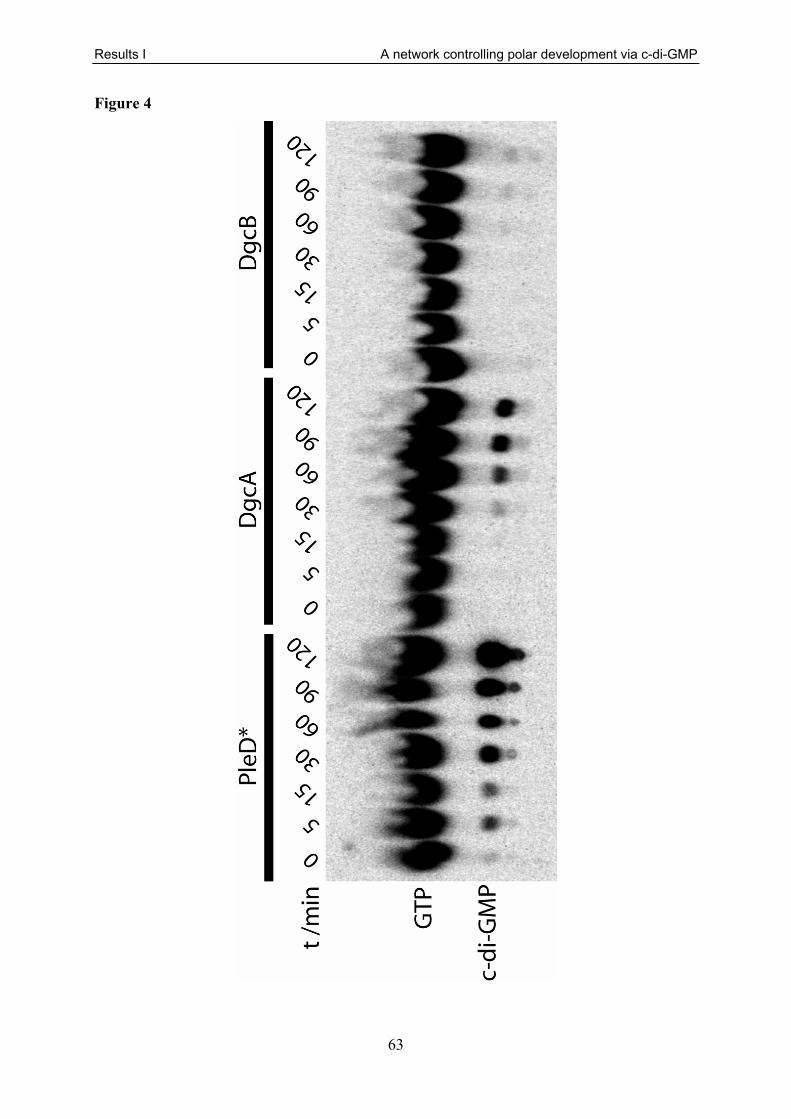
Results I A network controlling polar development via c-di-GMP
63
Figure 4

Results I A network controlling polar development via c-di-GMP
64
Figure 5
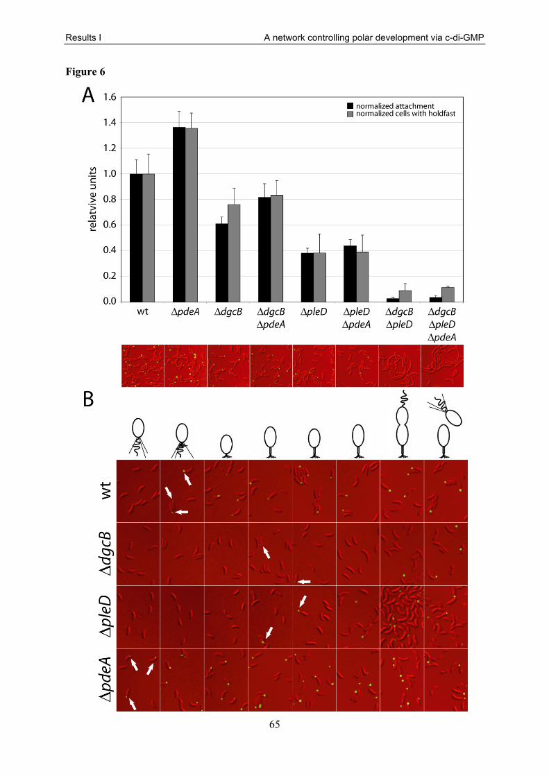
Results I A network controlling polar development via c-di-GMP
65
Figure 6

Results I A network controlling polar development via c-di-GMP
66
Figure 7

Results I A network controlling polar development via c-di-GMP
67
Figure 8

Results I A network controlling polar development via c-di-GMP
68
Figure 9

Results I A network controlling polar development via c-di-GMP
69
Figure 10
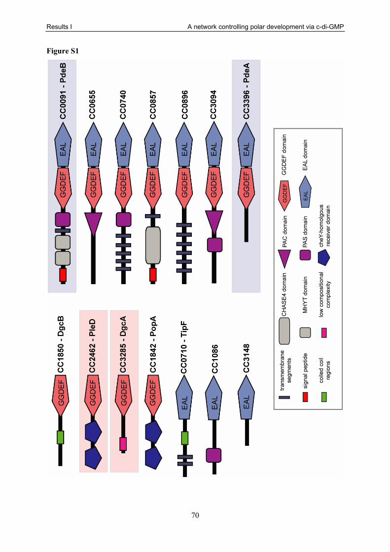
Results I A network controlling polar development via c-di-GMP
70
Figure S1

Results I A network controlling polar development via c-di-GMP
71
Figure S2

Results I A network controlling polar development via c-di-GMP
72
Figure S3

Results I A network controlling polar development via c-di-GMP
73
Table S1
Strain or plasmid Relevant genotype and description Reference or source
C. crescentus strains UJ134 CB15N hslU::kanr A. Stotz UJ838 CB15N clpA::Ω M. Osteras UJ945 CB15N ftsH::Ω [202] UJ1249 CB15N xylX::pMO88 M. Osteras UJ1267 CB15N - synchronizable variant strain of CB15 [114] UJ1682 CB15N::CMS21 [200] UJ1685 CB15N::CMS24 [200] UJ1855 CB15N lon::Ω [203] UJ3056 CB15 ∆cc0091 A. Levi UJ3241 CB15 - C. crescentus wild type ATCC19089 UJ4182 BL21(DE3) + pET21c::dgcB M. Christen UJ4448 CB15N ∆popA this study UJ4454 CB15N ∆pdeA this study UJ4455 CB15 ∆cc0655 this study UJ4456 CB15 ∆tipF this study UJ4457 CB15 ∆cc0740 this study UJ4458 CB15 ∆cc0857 this study UJ4459 CB15 ∆cc0896 this study UJ4460 CB15 ∆cc1086 this study UJ4461 CB15 ∆popA this study UJ4462 CB15 ∆dgcB this study UJ4463 CB15 ∆pleD this study UJ4464 CB15 ∆cc3094 this study UJ4465 CB15 ∆cc3148 this study UJ4466 CB15 ∆dgcA this study UJ4467 CB15 ∆pdeA this study UJ4709 CB15 + pBBR1MCS-2 this study UJ4710 CB15 ∆pdeA + pBBR1MCS-2 this study UJ4711 CB15N ∆pdeA motile suppressor class I this study UJ4712 CB15N ∆pdeA motile suppressor class II this study UJ4713 CB15N ∆pdeA motile suppressor class IIIa this study UJ4714 CB15N ∆pdeA motile suppressor class IIIb this study UJ4715 CB15 ∆dgcB ∆pdeA this study UJ4716 CB15 ∆dgcA ∆pdeA this study UJ4717 CB15 + pAL53 A. Levi UJ4718 CB15 ∆pdeA + pAL53 this study UJ4719 CB15N ∆dgrA this study UJ4720 CB15N ∆dgrA ∆pdeA this study UJ4721 CB15N ∆dgrB this study UJ4722 CB15N ∆dgrB ∆pdeA this study UJ4723 CB15 ∆dgcB ∆pleD this study UJ4724 CB15 ∆dgcB ∆pleD ∆pdeA this study UJ4725 CB15N ∆dgrA ∆dgrB this study UJ4726 CB15N ∆dgrA ∆dgrB ∆pdeA this study UJ4727 CB15N pdeA::pSA116 this study
UJ4728 CB15N ∆pdeA dgcB::tnMarinerdCat integrated at position +2048028 bp this study
UJ4729 CB15N ∆pdeA cc2147::tnMarinerdCat integrated at position +2351967 bp this study

Results I A network controlling polar development via c-di-GMP
74
UJ4730 CB15N ∆pdeA dgcB::tnMarinerdCat integrated at position +2047422 bp this study
UJ4731 CB15N + pSA120 this study UJ4732 CB15N ∆pdeA + pSA120 this study UJ4733 CB15N dgcB::pSA117 this study
UJ4734 CB15N ∆pdeA dgcB::tnMarinerdCat integrated at position +2047990 bp this study
UJ4735 CB15N ∆pdeA dgcB::tnMarinerdCat integrated at position +2047337 bp this study
UJ4736 CB15N ∆popA + pSA120 this study UJ4737 CB15N rcdA::hygr + pSA120 this study E. coli strains BL21(DE3) (pLysS) F- dcm ompT hsdS(rB-mB-) gal l (DE3) [pLysS CAMr] Stratagene
DH10B
F- mcrA ∆(mrr- hsd RMS- mcrBC) Φ80dlacZ∆M15 ∆lacX74 endA1 recA1 deoR ∆ (ara, leu)7697 araD139 galU galK nupG rpsL thi pro hsdR- hsd+ recA RP4-2-Tc::Mu-Tn7 Stratagene
DH5α supE44 ΔlacU169(Φ80 lacZΔM15) hsdR17 recA1 endA1gyrA96 thi-1 relA1 [204]
S17-1 RP4-2, Tc::Mu, KM-Tn7 [205] Plasmids pALMAR2 pLRS60 carrying a TnMariner delivering a Cat cassette A. Levi pBBR1MCS-2 Kanr broad range medium copy cloning vector [206] pET21c::dgcB pET21c carrying dgcB with a C-terminal 6xHis tag M. Christen
pMO88 pMR20 carrying clpX with mutation in ATP binding site under the control of the xylose promotor
M. Osteras and U. Jenal
pNPTS::KO1599 pNPTS138 based deletion construct for cc1599 - DgrA B. Christen pNPTS::KO3165 pNPTS138 based deletion construct for cc3165 - DgrB B. Christen ppdeA pBBR1MCS-2 carrying pdeA A. Levi
pSA116 pNPTS138 carrying 323 bp of the 3`end of pdeA fused to gfp this study
pSA117 pPHU281 carrying 620 bp of the 3`end of dgcB fused to cfp this study
pSA120 pMR10 carrying pdeA under the control of its own promotor fused to yfp this study
pDM1 pNPTS138 based deletion construct for cc0091 D. Meyer pSA93 pNPTS138 based deletion construct for cc0655 this study pSA94 pNPTS138 based deletion construct for cc0710 - tipF this study pAD2 pNPTS138 based deletion construct for cc0740 A. Dürig pSA91 pNPTS138 based deletion construct for cc0857 this study pSA102 pNPTS138 based deletion construct for cc0896 this study pSA79 pNPTS138 based deletion construct for cc1086 this study pAD8 pNPTS138 based deletion construct for cc1842 - popA A. Dürig pAD7 pNPTS138 based deletion construct for cc1850 - dgcB A. Dürig pSA95 pNPTS138 based deletion construct for cc2462 - pleD this study pSA96 pNPTS138 based deletion construct for cc3094 this study pSA90 pNPTS138 based deletion construct for cc3148 this study pSA80 pNPTS138 based deletion construct for cc3285 - DgcA this study pSA81 pNPTS138 based deletion construct for cc3396 - PdeA this study

Results II CC1064, a pleitropic assembly and localisation factor
75
3.2 CC1064, a Transmembrane Protein Required for Flagellar Rotation and Subcellular Protein Localisation Under Environmental Stress Conditions in Caulobacter crescentus Sören Abel, Alexander Böhm, Thomas Egli, Urs Jenal
Statement of my work
The majority of plasmids and strains used in this study have been generated by me. I also
performed motility (Fig. 1A, 1B, 1C, 4C, 5, Tab. 2), swimming speed determination (Fig.
3C), attachment (Fig. 1B, 2C, 2D), chemostat (Fig. 2A, 2B), β-galacdosidase activity (Fig.
3A), light-/fluorescence microscopy (Fig. 6, 7, 8, 11), synchronies (Fig. 9C), holdfast stain
(Fig. 9C), stalk length determination (Fig. 9A), and growth experiments (S2, S3) of this study.
Furthermore, I analysed the electron micrographs (Fig. 3B, 9B), the fatty acid analysis results
(Tab. 1) and determined several suppressor mutations in motA and motB (Fig. 4A).

Results II CC1064, a pleitropic assembly and localisation factor
76
Abstract
Adaptation to changing environmental conditions requires, among other things, constant
remodelling of the bacterial membrane. However, these changes consecutively affect the
solvent properties of the lipid bilayer and thus function of membrane embedded and
associated proteins. Here, we describe the transmembrane protein CC1064 from Caulobacter
crescentus that is required for optimal motility, surface attachment and correct subcellular
localisation of the di-guanylate cyclase PleD. It is shown that the paralysis the flagellum, as
well as the failure to recruit PleD to the developing stalked pole of the cell in the absence of
CC1064 is conditional for oxygen and temperature stress conditions. Spontaneous motile
suppressors that can compensate for the lack of CC1064 were found to harbour amino acid
exchanges in the flagellar motor proteins MotA or MotB. Furthermore, genetic tests suggest
that the functional assembly of MotA and MotB is disrupted in the Δcc1046 mutant grown
under stress conditions and model is discussed that links changes of the cellular fatty acids
composition of the membrane to this assembly defect. We demonstrate that mislocalisation of
PleD is independent of flagellar paralysis and vice versa. Taken together, we provide evidence
for pleiotropic effects of the transmembrane protein CC1064 that influences motor protein
assembly, surface attachment and PleD localisation via diverging pathways.

Results II CC1064, a pleitropic assembly and localisation factor
77
Introduction
Caulobacter crescentus is an obligate aerobe fresh water bacterium with a characteristic
asymmetric cell cycle. Upon division, C. crescentus cells give rise to two morphologically
distinct progenitor cells, a motile swarmer cell and a sessile stalked cell. The latter can act
similarly to a stem cell and continuously sprout off motile swarmer cells. These cannot divide
and are able to swim with the help of a polar flagellum [99, 100]. In response to external and
internal signals the swarmer cells initiates a developmental program and differentiates into a
stalked cell. Upon differentiation, the stalked cell gains the ability to undergo DNA
replication and initiates a new round of cell division. During this swarmer-to-stalked cell
transition, several polar rearrangements take place. The pili retract, the flagellum is released,
the stalk starts to develop, and the adhesive holdfast is synthesized. Optimal surface
attachment requires the presence and correctly timed development of all these polar
organelles [47, 192].
The atypical response regulator PleD, which is localised to the stalked pole, is one factor that
was described to control this polar development and therefore influences motility and
attachment in C. crescentus [14, 47, 61, 170, 174]. PleD is a modular protein with two N-
terminal receiver domains arranged in tandem (Rec1 and Rec2) and a C-terminal di-guanylate
cyclase output domain (GGDEF domain) [14, 16, 17]. It was shown that in the absence of
PleD, the flagellar ejection, the stalk length control and timing of holdfast synthesis during
swarmer-to-stalked cell transition are disturbed [47, 61]. The failure of flagellar ejection leads
to the occurrence of ectopic flagella at the tip of the stalk and thus hypermotility, while the
delayed holdfast synthesis leads to reduced attachment of ΔpleD mutants. In contrast, the
presence of the constitutively active mutant PleD* results in increased holdfast production,
increased stalk length and flagellar paralysis [61].
Activation of c-di-GMP synthesis occurs via phosphorylation of Asp53 in the first
receiver domain of PleD. Multiple lines of evidence demonstrate that the sensor histidine
kinases DivJ and PleC, as well as the single domain response regulator DivK control the
timed activation of PleD [61, 171, 172, 187]. Like PleD, all these signalling proteins change
their subcellular localisation in a cell cycle specific manner [175, 177]. Whereas both PleC
and DivJ directly phosphorylate PleD [14, 61, 173], DivK was shown to enhance the kinase
activity of PleC and DivJ [187]. Phosphorylation leads to dimerisation, bringing two educt
charged GGDEF domains into close proximity to allow c-di-GMP synthesis [14, 16, 17].

Results II CC1064, a pleitropic assembly and localisation factor
78
Dimerisation of PleD was also shown to be a prerequisite for the sequestration of activated
PleD to the differentiating C. crescentus cell pole [186]. In agreement to this, the hyperactive
PleD* was shown have a higher propensity to dimerize in vitro and displays hyperlocalisation
in vivo [14]. However, although the regulators of PleD activity and localisation are defined,
the nature of the factor(s) that sequesters active PleD to the cell pole remains unclear.
Another factor that influences motility, but also attachment behaviour of C. crescentus is the
flagellum itself. The flagellum is a complex structure in the bacterial cell. It spans both the
inner and the outer membrane of gram negative bacteria and converts a proton gradient across
the inner membrane into mechanical work [207, 208]. Assembly of the flagellum is a highly
regulated process involving coordinated expression of over 50 genes in a hierarchical manner
[118, 126, 127, 130]. C. crescentus flagellar genes can be divided into four regulation classes
[128]. By various mechanisms it is ensured, that the gene products of each class are only
present in the cell, if the gene products of the preceding regulation class have been
functionally assembled [209].
The flagellum can be divided into two substructures; the rotor and the stator. The rotor
consists of the C ring, the MS ring, the rod, and the filament [118]. The stator is composed of
two integral inner membrane proteins, MotA and MotB [158, 210, 211]. MotA was shown to
contain four transmembrane helices [167, 212]. The second and third transmembrane helices
are connected by one long cytoplasmic sequence [150], where conserved charged residues are
believed to make direct electrostatic contact to FliG, a protein of the cytoplasmatic C-ring of
the rotor [157, 213]. MotB has one transmembrane domain with a large C-terminal part in the
periplasm [158], where it is believed to anchor the motor proteins to the peptidoglycan cell
wall [150, 158, 159, 161, 162, 214]. MotA and MotB form a multimer [160, 214, 215] with
the composition MotA4MotB2 [118, 154, 155] and it has been shown that their transmembrane
domains are in close proximity [216, 217]. Individual MotA4MotB2 complexes form proton
channels across the inner membrane that couple proton influx to rotation of the flagellum
[148-153]. Therefore, strains lacking MotA or MotB posses paralyzed flagella [148, 150, 167-
169].
It is believed that a single flagellar motor is energized by 8-12 MotA4MotB2 stator
complexes [164, 165, 211, 218] and that each complex can act independently from the other
stator complexes to generate torque [163-166]. MotA and MotB can pre-assemble in the
membrane [219] where about 200 MotB proteins build a pool, that can laterally diffuse in the
membrane and rapidly exchange with motor assembled MotB [218]. It was demonstrated that

Results II CC1064, a pleitropic assembly and localisation factor
79
the pre-assembled MotA4MotB2 complexes can as a whole be incorporated into pre-
assembled flagella [163-165]. The proton channels of pre-formed stator complexes which are
not docked to the rotor yet are blocked by an amphipathic helix in MotB, named “the plug”, to
prevent premature proton influx [220].
Little is known about specific interactions between the lipid bilayer and the flagellar basal
body. Nevertheless, mutations in genes involved in lipid metabolism have been found to cause
motility phenotypes in E. coli [221] and it is known from a number of integral membrane
proteins that they have a preferred lipid composition of the bilayer for optimal functioning
[222-226]. Environmental factors such as temperature, oxygen partial pressure, pH, water
activity, nutrient availability, pressure and the presence of certain chemicals demand
adaptation of the lipid bilayer to ensure both integrity and fluidity of the membrane.
Membrane adaptations in response to environmental perturbations are known to involve the
alteration of the fatty acid composition (reviewed in [227, 228]). Thus, the question remains if
and how the functioning of the highly sophisticated flagellum and other membrane factors is
affected by adaptive alterations of the lipid bilayer.

Results II CC1064, a pleitropic assembly and localisation factor
80
Results
CC1064 affects surface attachment and motility of Caulobacter
crescentus
To identify genes that are involved in C. crescentus surface attachment and motility, a
transposon insertion mutagenesis and subsequent screen for attachment mutants was
conducted. One mutant harbouring a Tn5 insertion in the open reading frame cc1064
displayed an attachment reduced to 40 % of the wild type. The cc1064 gene encodes a
predicted 297 aa protein with a transmembrane helix at the N-terminus and a large
periplasmic domain. We became interested in this gene because it is located next to divJ
(cc1063), a gene encoding a sensor histidine kinase involved in cell division, asymmetry
generation and also motility and attachment (Fig. S1). In addition to its attachment phenotype,
the cc1064 mutant also displayed severely impaired motility, leading to a colony size on semi-
solid agar plates of only about 20 % compared to the wild type on semi-solid agar plates (Fig
1A/B).
To confirm that the described phenotypes are actually caused by loss of cc1064
function rather than by a polar effect on a nearby gene, a markerless, in frame deletion of the
cc1064 gene was constructed and the resulting strain was tested for its attachment and
motility behaviour. Both effects could be reproduced with this non-polar deletion mutant. In
addition, the motility and attachment phenotypes could be restored to wild type by providing a
copy of cc1064 from a low-copy number plasmid (Fig. 1A/B). These complementation
experiments demonstrate that CC1064 is required for full motility and attachment in
C. crescentus.
To examine if the reduced motility on semi-solid agar plates was caused by a defect in
motility or a chemotaxis defect, the swimming behaviour of the cells in liquid medium was
analysed with the help of light microscopy. Only a very small subpopulation (about 3 %) of
cc1064 mutant cells was found to be motile (Fig. 1C). This was despite the fact that the
distribution of swarmer, stalked and pre-divisional cells was not distinguishable from wild
type (data not shown) and there were no differences in the growth rate of Δcc1064 cells
compared to the wild type (Fig. S2). Thus, the CC1064 protein is required for proper motility.

Results II CC1064, a pleitropic assembly and localisation factor
81
The Δcc1064 phenotype is conditional
Surprisingly, it was discovered that the motility phenotype and the attachment phenotype of
the cc1064 mutant is dependent on the culture conditions. When cultures were incubated with
vigorous shaking, the fraction of motile mutant cells was indistinguishable from the wild type
(Fig. 1C). This suggested that the oxygen concentration in the medium might influence the
motility of the mutant cells. Alternatively, an increase in growth rate in response to higher
oxygen concentrations might render a larger fraction of the mutant cells motile.
To distinguish between these two possibilities, continuous culture experiments in a chemostat
under carbon-limited growth conditions were performed. During the experiment, growth rate
and temperature were kept constant and small aliquots of cells were regularly removed and
analysed microscopically to determine their motility. When the oxygen supply was reduced
without changing the growth rate, mutant cells stopped swimming after about 75 min. This
phenomenon was fully reversible, since replenishment of the oxygen supply restored motility,
again within about 75 min minutes (Fig. 2A). A control culture of wild-type cells did not
show any change in motility upon alterations of the oxygen feed (Fig. 2B). This indicates that
the oxygen concentration, but not growth rate is influencing the Δcc1064 phenotype and that
CC1064 is required for C. crescentus motility under conditions of low oxygen concentration.
Since motility was found to be oxygen dependent, it was tested if the attachment defect of the
Δcc1064 mutant was also oxygen dependent. Cells were grown under high and low oxygen
conditions to the same OD660 and then tested for attachment to polystyrene surfaces (Fig 2C).
While the attachment of cells grown under low oxygen conditions was reduced to 40 % of the
wild type, as observed before, the growth with high oxygen supply led to an increase of
attachment to about 75 % compared to wild type cells grown under the same conditions. Thus,
the attachment phenotype can only be partially restored by increasing the oxygen supply
during cell culturing.
It appeared possible that the observed motility defect was caused by alterations of the
cell membrane, as environmental parameters are known to influence membrane composition.
Apart from oxygen, another factor involved in the regulation of the membrane composition is
ambient temperature (reviewed in [227, 229, 230]. Consequently, the motility and attachment
behaviour of the Δcc1064 strain grown under high oxygen conditions, but at reduced
temperatures (16 °C) was tested. Despite the high oxygen concentrations under these
conditions, cc1064 mutant cells showed very few motile cells (data not shown). Similarly,
cc1064 mutant cells grown under high oxygen but low temperature conditions, displayed
reduced attachment to the same levels like at optimal growth temperature but low oxygen

Results II CC1064, a pleitropic assembly and localisation factor
82
(Fig. 2D). This is despite the fact that the oxygen solubility in aqueous solutions is higher at
low temperatures [231]. Taken together, both oxygen supply and growth temperature
influence motility and surface attachment of the Δcc1064 mutant. Whereas the motility defect
could be completely restored to wild-type levels, the attachment deficit could only be partially
restored upon growth under optimal conditions.
The composition of the lipid bilayer changes upon manipulating the oxygen level of the culture
As pointed out before, it has been shown for several species that changes in the growth
temperature or changes of the oxygen supply lead to alterations the composition of the lipid
bilayer (reviewed in [227, 229, 230]). The findings described above are consistent with either
of two models. CC1064 could play a role for the adjustment of the proper membrane lipid
composition under low oxygen and/or low temperature conditions, which in turn might be a
necessity for optimal flagellar functioning or assembly. Alternatively CC1064 might by a
factor necessary for optimal flagellar functioning or assembly in an altered membrane lipid
environment at low oxygen and/or low temperature conditions. To investigate which of these
possibilities is correct, an analysis of the total cellular fatty acid content was performed.
Samples of wild-type and Δcc1064 mutant bacteria were either taken under high or low
oxygen growth conditions, the fatty acids were extracted, methylated, and subjected to gas
chromatography. It was found that the fatty acid composition differed between the different
growth conditions, but no difference between the cc1064 mutant and the wild type was
detected in these experiments (Tab. 1). Low and high oxygen samples mainly differed in the
content of unsaturated fatty acids, which was higher under low oxygen conditions.
Furthermore, hydroxylated and branched species increased and uneven chain-length fatty
acids decreased. This indicates that CC1064 has no influence on the composition of the fatty
acids, but may rather be an important factor for flagellar functioning in an altered lipid
environment in response to low oxygen and/or low temperature.
The flagellum of a Δcc1064 mutant is paralyzed
The expression of bacterial flagellar genes is organised in a highly regulated cascade with
different checkpoints. To investigate if the lack of cc1064 affects one of these flagellar gene
expression checkpoints and therefore flagellar function, the activity of representative class II,
III and IV promotors and class IV mRNA stability was analysed under conditions where the
cc1064 mutants are non-motile (Fig. 3A). No differences for promotor activities between the

Results II CC1064, a pleitropic assembly and localisation factor
83
wild type and the Δcc1064 mutant could be observed. This indicates full assembly of the
flagellar basal body and hook complex. To corroborate this finding and to further examine
flagellum morphology, cc1064 mutant cells were examined with the help of transmission
electron microscopy. Flagella of mutant cells grown under conditions where they are non-
motile showed no obvious morphological difference compared to the wild type (Fig. 3B).
Taken together, the results indicate that cc1064 is not important for assembly of the basal
body, hook or filament, but rather for flagellar functioning, hence flagellar rotation.
An analysis of the swimming speed of the rare motile cells that can be found in a low
oxygen culture of a Δcc1064 strain showed no difference compared to wild type (Fig 3C).
This indicates that the lack of CC1064 leads to either complete loss of flagellar rotation or, in
rare cases has no influence at all. Thus, the reason for small swarms on motility agar plates is
the strongly reduced proportion of motile cells rather than an overall reduction of the
swimming speed.
Suppressor mutations rendering the Δcc1064 mutant motile are in motA
and motB
To define the flagellar structures that are altered in the Δcc1064 background and lead to non-
motility, a screen for spontaneous motile suppressor mutants was performed. Motile
suppressors were isolated from motility agar plates after incubation for three days. Genetic
mapping experiments and subsequent sequencing identified all suppressor mutants to harbour
a single amino acid substitution in MotA or MotB (Fig 4A). All mutants displayed swarming
behaviour in semi-solid agar plates that was indistinguishable from wild type (Fig. 4B).
Interestingly, all amino acid exchanges in the suppressor mutants are located in or very close
(maximal distance 13 aa) to the transmembrane regions of MotA and MotB. It is worth
pointing out that several suppressor mutants harboured substitutions of the same amino acid to
chemically unrelated amino acids (e.g. Ser52 to Cys, Gly or Arg).
Next, the attachment phenotypes of the motile suppressor mutants were assayed.
Attachment for all tested motile suppressor mutants was found to be only partially restored
with relative attachment levels of about 70 % compared to the wild type (Fig. 2C/D). These
attachment values correspond to levels observed for the parental Δcc1064 strain grown under
high oxygen conditions, where motility is not affected. Also, the attachment levels of the
motile suppressor mutant were found to be insensitive to oxygen as well as temperature (Fig.
2C/D). Together these findings argue for a model where in addition to its motility dependent

Results II CC1064, a pleitropic assembly and localisation factor
84
attachment phenotype, CC1064 also plays a role for attachment that is independent of its
function in motility.
As MotA and MotB form a proton channel across the membrane it appeared possible
that the suppressor mutations lead to an increased proton influx, which then might
compensate the Δcc1064 motility defect. It was shown that elevated ion leakage can lead to
growth impairment and is therefore costly [220]. To test if the motile suppressors might
exhibit increased influx and therefore might show impaired growth, growth rates of several
isolated suppressor mutants were determined. All tested mutants showed the same growth rate
as the wild type. To rule out that the suppressor mutations might have a deteriorating effect on
growth in the presence of cc1064 one particular suppressor mutant with a G58D substitution
in MotB was chosen and it was tested if the suppressor mutation causes a growth defect in the
cc1064+ background (Fig. S3). In parallel the motility of the generated strain on semi-solid
agar plates was investigated (Fig. 4C). Neither growth rate nor swarm size of the motB point
mutant in the cc1064+ background was altered compared to the wild type. Together these
results indicate that the mutations that render the Δcc1064 strain motile have no measurable
costs under the conditions of the assay. Furthermore, it can be concluded from these data that
the point mutations are no bypass suppressors that would generally increase motility in any
strain background. Instead it is likely that the suppressor mutations have a specific influence
in the Δcc1064 background.
motA or motB suppressor alleles are dominant over the respective wild-type alleles
It was shown that the number of functional stator complexes in the flagellum influences the
colony size on semi-solid agar plates [160, 164, 165]. This feature of the flagellar motor was
employed to find out if wild-type MotB can assemble in a Δcc1064 background. Therefore,
motBwt and motBsup alleles were co-expressed. It was reasoned that if in the absence of cc1064
non-functional wild-type MotB can still assemble and form homo-multimers or hetero-
multimers with co-expressed mutant MotB proteins this would result in reduced swimming
speed due to reduced number of functional stator complexes per motor [164].
Different motB suppressor alleles were expressed from a plasmid in the presence of a
chromosomal wild-type allele or vice versa and the motility of the respective strains was
tested (Fig 5). Overexpression of motB, either the wild-type or a mutant, had no effect on
motility in the cc1064+ background. Also in a Δcc1064 background the overexpression of
wild-type motB did not increase the swarm size compared to a control plasmid. Hence,

Results II CC1064, a pleitropic assembly and localisation factor
85
increased amounts of motB do not change swimming behaviour in the wild type or cc1064
mutant background. However, expression of any motBsup allele in the Δcc1064 background
led to full restoration of motility, irrespective of the presence or absence of the wild-type
motB allele on the chromosome or a plasmid. Thus, under all tested conditions the suppressor
alleles are fully dominant over wild-type motB.
Combinations of motA and motB suppressor alleles show allele specific motility phenotypes
A possible explanation for the molecular mechanism by which the amino acid exchanges in
MotA and MotB lead to suppression of the Δcc1064 motility phenotype is that the affected
amino acids interfere with the correct functional assembly in the absence of CC1064. To
allow function in the Δcc1064 background the amino acid substitutions must alter the protein
is some way. To investigate the nature of these changes, it was tested if all mutations in motA
and motB are compatible with each other. 45 combinations of point mutations in motA and
motB were created in the in the Δcc1064 background and their motility was tested. The
individual motA motB double mutants showed motility phenotypes ranging from less motile
than the Δcc1064 motA+ motB+ strain over intermediate to fully motile (Tab. 2). Interestingly,
the motility phenotypes were highly allele specific. For example motAA42T combined with
motBV29L, motBV29M or motBA40V led to fully motile cells, while the combination of motAA42T
with motBA40T or motBG58D led to intermediate motility behaviour and all other combinations
with motAA42T led to a severely reduced motility (Tab. 2). Likewise motBL44S, which shows
severely reduced motility in combination with motAA42T, showed the whole range of motility
phenotypes when combined with the other available motA alleles (Tab. 2). Surprisingly, the
pattern of allele specificity does not seem to be dependent on the position of the amino acid
exchange nor on structural or chemical properties of the side chains of the amino acids. Even
mutations that are predicted to locate on different sites of the inner membrane (e.g. motAI28F
and motBV29M) showed reduced motility. These findings indicate a complex interconnection
between the different motor protein variants.
Under low oxygen conditions CC1064 is required for correct subcellular distribution of the response regulator PleD
Because cc1064 is located next to the divJ gene on the C. crescentus genome and because
DivJ is known to influence PleD phosphorylation and thereby localisation to the incipient
stalked pole during swarmer-to-stalked cell transition, the subcellular localization of PleD was

Results II CC1064, a pleitropic assembly and localisation factor
86
tested. A C-terminal GFP fusion of the response regulator PleD was introduced into wild-type
and Δcc1064 cells, respectively. PleD localisation of the resulting strains grown under high
oxygen conditions was compared to low oxygen conditions. As cultures reached mid
exponential phase, the subcellular localisation of PleD was analysed using fluorescence
microscopy (Fig. 6). In stalked and predivisional cells grown under high oxygen conditions,
PleD was sequestered to the stalked pole in both wild-type and Δcc1064 background. About
55 % of the cells showed a localised polar fluorescent focus. In contrast, under low oxygen
conditions PleD was found to be uniformly distributed in the majority (91 %) of cc1064
mutant cell, while the number of cells showing a PleD focus at the stalked pole was only
mildly reduced in the wild type under these conditions (37 %). Together, these data suggest
that CC1064 either directly or indirectly functions as a localisation factor for PleD under
certain environmental conditions, such as low oxygen.
The localisation of the proteins responsible for PleD activation is not affected by the cc1064 deletion
Polar sequestration of proteins in general may be abolished in a cc1064 mutant under low
oxygen conditions. To test this hypothesis, different GFP fusion proteins, that are known to
localise to different addresses in the cell (DivJ, PleC, and DivK), were tested for their
subcellular localization under low oxygen conditions. Neither of these proteins showed an
altered localisation pattern in the Δcc1064 mutant compared to the wild type (Fig. 7). From
this it is concluded that the cc1064 mutant exhibits no general localisation defect.
A phosphorylation independent, hyperlocalised PleD mutant is localised
in the Δcc1064 background under all conditions
As the cc1064 deletion mutant seems to have a PleD specific localisation defect, the next
question was if CC1064 affects a feature of PleD that is a prerequisite for localisation, for
example the phosphorylation or the dimerisation status, or might alternatively affect or
constitute itself a PleD receptor at the stalked cell pole. To distinguish between these two
possibilities the localisation of the constitutively active, super localised PleD mutant,
PleD*D53N, was investigated. PleD*
D53N-GFP clearly displayed polar localisation when cultures
were grown under low oxygen conditions, even in the absence of cc1064 (Fig. 8). Thus it is
unlikely that CC1064 affects a receptor or itself functions as a receptor for PleD. Instead,
these results support the idea that either phosphorylation or dimer formation of PleD is
impaired in the Δcc1064 background.

Results II CC1064, a pleitropic assembly and localisation factor
87
Cells with a deletion in cc1064 do not show the developmental
phenotypes of a ΔpleD mutant
ΔpleD mutant strains are known to display several developmental phenotypes, including
reduced stalk length, failure of efficient flagellar ejection during swarmer-to-stalked cell
transition, which results in hypermotility in liquid culture, and finally delayed holdfast
synthesis [47, 170, 174]. The latter phenotype has been described as the reason for the
observed attachment phenotype of pleD mutants [47]. Therefore, it was tested if the Δcc1064
mutant displays any features of pleD mutants. First, the stalk length of cells grown under
oxygen limitation was quantified using light microscopy. In contrast to the ΔpleD mutant,
which showed a reduced stalk length of 0.6 μm in average, the Δcc1064 had a mean stalk
length identical to the wild type (1.0 μm) (Fig. 9A).
Cells grown under the same conditions were also investigated under the electron
microscope to visualise the position of the flagellum. As described, the ΔpleD mutants
showed aberrant flagellar positioning at the tip of the stalk. This morphological distinctive
feature was not detectable in the cc1064 deletion strain (Fig. 9B).
Also, newborn swarmer cells were isolated and initiation timepoint of holdfast
synthesis of the synchronised population was examined. Again, the Δcc1064 strain behaved
like the wild type, producing a detectable holdfast as cells differentiated into a stalked cell,
whereas the holdfast synthesis in a pleD deletion strain was clearly delayed (Fig. 9C). Thus,
all organelles known to be controlled by PleD are unimpaired in the cc1064 mutant. As a
consequence, one must conclude that the activity of PleD is not strongly reduced in the
cc1064 mutant background.
Double deletions of cc1064 and pleD lead to an additive motility phenotype
As shown above, the deletion of cc1064 leads to a paralysed flagellum. The same phenotype
is known for the constitutively active PleD* mutant [61]. This is thought to be caused by the
ectopic DGC activity of PleD* in the swarmer cell [14, 61, 186]. Following this idea the
aberrant PleD localisation pattern of the cc1064 mutant might lead to PleD activity in the
flagellated swarmer cell, which in turn would lead to premature flagellar arrest. If this
hypothesis is correct, a pleD mutant should be epistatic over Δcc1064. To test this, a cc1064,
pleD double mutant was constructed and its motility behaviour was assayed. It is important to
note that, despite the hypermotility phenotype in liquid culture, the ΔpleD mutant displays

Results II CC1064, a pleitropic assembly and localisation factor
88
smaller colonies on semisolid agar plates compared to the wild type size [61]. It was found
that the cc1064 pleD double mutant displayed an even smaller swarm size compared to the
single Δcc1064 and single ΔpleD mutant, indicating that both deletions lead to a cumulative
phenotype (Fig. 10). From this, it is concluded that CC1064 and PleD influence motility via
different pathways and that mislocalisation of PleD is not the molecular base for the motility
phenotype of the Δcc1064 strain
Mutations in motA or motB cause suppression of the motility phenotype, but do not restore PleD localisation
Little is known about the environmental or internal cues that control PleD activation via
DivJ/PleC. Since it has been shown that the flagellum can – in addition to its role for cell
locomotion – act as a sensor, that provides the cell with information about for example the
viscosity or the wetness of the medium [232, 233] and since cc1064 was shown here to affect
motility and PleD localisation, it appeared possible that the aberrant PleD localisation pattern
might be caused by flagellar malfunction. To test this idea PleD localisation was analysed in
motile suppressor mutants of a Δcc1064 strain. One mutant harbouring a mutation in motA
and one harbouring a mutation in motB were chosen and PleD localisation was tested (Fig.
11). As for the non-motile Δcc1064 parental strain, no localised PleD could be observed in the
motile suppressors. Thus, it appears unlikely that paralysis of the flagellum is the cause for the
PleD localisation defect.

Results II CC1064, a pleitropic assembly and localisation factor
89
Discussion
CC1064 plays a pleitropic role for motility, PleD localisation and surface attachment
Among many genes that were discovered in a screen for loss of attachment mutants [35] the
open reading frame cc1064 caught our attention because it is located next to divJ (Fig. S1)
and affected, in addition to attachment, also motility (Fig. 1A, B). Furthermore, both
phenotypes were found to be temperature and oxygen dependent (Fig. 1 C, 2A-D). The
genetic proximity of cc1064 and the gene encoding the PleD activator DivJ, together with the
involvement of PleD in both motility and attachment control [47, 61] suggested that PleD and
CC1064 might act in the same pathway. However, several lines of evidence indicate that
CC1064 rather plays a pleitropic role which affects several systems independently.
Firstly, ΔpleD Δcc1064 double mutants showed an even smaller swarm size than the
pleD or cc1064 single mutants (Fig. 10). Hence, the combination of both mutations causes a
cumulative motility phenotype which argues against a role for both gene products in the same
linear pathway. It rather indicates that the few cells that are motile despite the lack of cc1064
(Fig. 1C) and therefore are likely responsible for the residual swarming behaviour of the
Δcc1064 mutants are now experiencing an additional chemotaxis defect caused by the lack of
PleD [180].
Secondly, whereas motility and PleD localisation was completely restored under high
oxygen conditions at 30 °C (Fig. 1C, 2A, and 6), the attachment behaviour of the ∆cc1064
mutant could only be partially recovered under these optimal conditions (Fig. 2C, D). It is
worth noting in this context that reduced surface attachment has been described as results of
loss of motility [47, 192]. The finding that optimal growth conditions could restore surface
attachment to ~75 % in a Δcc1064 mutant (Fig. 2C, D), together with the finding that fully
motile suppressor mutants showed the same relative level of surface attachment independent
on the growth conditions (Fig. 2C, D) makes it likely that the conditional part of the Δcc1064
attachment defect is caused by the reduced motility. Nevertheless, neither optimal growth
conditions nor the motile suppressor mutations could completely restore attachment behaviour
to wild-type levels. Thus it is concluded that cc1064 is required for optimal attachment under
any tested condition in addition to its conditional effect on motility.
Thirdly, it appeared possible that malfunction of the flagellar motor might lead to the
PleD described mislocalisation. This idea was based on several reports that describe an

Results II CC1064, a pleitropic assembly and localisation factor
90
additional role of the flagellum as a sensor. One study reports that a decrease in the flagellar
rotation rate of the sodium-driven motor of Vibrio parahaemolyticus results in a changed flux
of sodium ions. The decrease in the rotation rate is explained by sterical inhibition of flagellar
turning due to a surface in vicinity. The sodium flux difference was shown to be coupled to
the induction of lateral flagellum transcription [232]. Another report suggests that the block of
flagellar rotation in Vibrio cholerae is a signal for exopolysaccharide synthesis, presumably
by influencing the phosphorylation of the regulatory response regulator VpsR [234]. Due to
these findings it was considered before that the flagellum of C. crescentus could also act as a
mechanosensor for surfaces and be part of a signalling cascade required for optimal
expression of adhesive properties upon surface contact [47]. PleD is a key mediator for the
timing of the transition from swarmer-to-stalked cell [47]. Although delocalisation of PleD is
not the reason for the motility defect of the Δcc1064 mutant (see above), it appeared possible
that the paralysis of the flagellum might cause the localisation defect of PleD. The isolation of
motile suppressors of the Δcc1064 mutant enabled a test of this idea. However, the finding
that PleD is still delocalised in these suppressor mutants (Fig. 11) indicates that it is not motor
malfunction which is causing the PleD localisation defect. Localisation experiments in motA
deletion strains further support this finding and excludes the motor protein itself from being a
PleD localisation factor (data not shown). These data indicate that CC1064 influences the
spatial distribution of PleD and motor functioning via divergent mechanisms. In summary, the
motility phenotype, the delocalisation of PleD and the attachment phenotype are not linked to
each other.
Are changes of the lipid bilayer composition responsible for the
conditional nature of the Δcc1064 phenotype?
The Δcc1064 mutant showed similar phenotypes at low ambient temperature and at low
oxygen supply (Fig. 2 C, D). Therefore, it was reasoned that the molecular cause for the
pleitropic effects must be influenced by both growth conditions. As low oxygen supply (Fig.
S2) and low temperature both lead to a decrease of the growth rate it was considered that both
external parameters might cause the pleitropic phenotypes by slowing down C. crescentus
growth. However, in a carbon limited Δcc1064 chemostat cultures at a constant, slow growth
rate the motility of the cells could be modulated by regulating the oxygen supply (Fig. 2A).
This finding indicates a specific function for oxygen in the regulation of motility, despite its
influence on growth rate. Therefore, it is concluded that low oxygen supply and likely also

Results II CC1064, a pleitropic assembly and localisation factor
91
low growth temperature are causing the motility defect independently of their effect on
growth rate.
What else could be the reason for the pleitropic phenotype of a cc1064 mutant and
why can the motility and PleD localisation phenotypes only be observed under certain
environmental conditions? Several findings point at an involvement of the lipid bilayer.
Firstly, temperature and oxygen supply are known to alter the fatty acid composition of the
bacterial membrane (reviewed in [227, 229, 230]). Generally speaking, the reduction of
growth temperature requires the increase of membrane fluidity. A common response to
preserve membrane fluidity is an increase of the proportion of short chain fatty acids and to
increase the degree of non-saturated and branched fatty acids [235-239]. Furthermore, the
ratio of even and odd chain-length fatty acids has also been demonstrated to increase in
response to decreased growth temperature [240]. For the effect of an alteration of the oxygen
supply during growth, the fatty acid analysis performed here showed that C. crescentus
increased the proportion of hydroxylated and branched fatty acids upon decreased oxygen
levels (Tab. 1). Although the relative abundance of different unsaturated fatty acid species
showed a heterogeneous picture, the overall level of unsaturated fatty acids evidently
increased under low oxygen conditions while the number of uneven chain length species
droped (Tab. 1). Thus, the changes in the fatty acid composition are qualitatively comparable
for both, growth at low temperature and growth at low oxygen.
The second hint towards an involvement of the membrane comes from the time scale
on which the bacterial swimming behaviour responds to changes of the oxygen supply. After
changing the oxygen input in chemostat experiments, Δcc1064 cells responded in
approximately 75 min with altered swimming behaviour, which corresponds to 0.25
generation times under these conditions (Fig. 2A). Thus, the changes in swimming behaviour
do not reflect a division cycle, as it would be expected for e.g. the de novo synthesis of a
flagellum. The time scale is rather consistent with the remodelling of a complex system, for
example the rearrangement of the lipid bilayer composition.
The last and strongest indication that CC1064 is needed for adaptation of membrane
proteins to changing lipid environments comes from the characterisation of motile suppressors
mutants. All mutations were found to change residues in or very close to transmembrane
regions of the motor proteins MotA and MotB. This close proximity of the position of amino
acid exchanges to the membrane suggests a functional relationship. Based on these data, it
was reasoned that cc1064 might have an influence on the membrane composition or
alternatively might be required to react adequately to changes in the lipid bilayer. The former
Results II CC1064, a pleitropic assembly and localisation factor
92
was tested by the analysis of the fatty acid composition in a Δcc1064 strain grown under low
and high oxygen conditions. Because no severe difference was found when compared to the
wild type under the respective conditions (Tab. 1), it was concluded that CC1064 has no
influence on the overall fatty acid composition, unless CC1064 does affect the headgroups of
the lipids, which have not been examined in the fatty acid analysis, rather than the fatty acid
tails. However, one argument speaks against this possibility. A decrease in temperature
triggers the Δcc1064 phenotype. Therefore, CC1064 must be required under these conditions.
However, changes of the lipid headgroup composition are rarely documented under the
influence of low temperature [241]. CC1064 may also affect a low abundance fatty acid,
which might be below the detection limit in the global fatty acid analysis. Such a fatty acid
may be distributed unevenly in the cell in a way that a small global change may lead to a
strong local concentration change. Localised lipid species have been reported recently in
Bacillus subtilis and Escherichia coli, where different lipid species specific fluorescent dyes
revealed the heterogeneity of the bacterial membrane [242-244]. The same dyes (FM4-64 and
NAO) have been tested in C. crescentus, but no irregular staining was observed (data not
shown). Although this does not rule out the possibility that such lipid domains exist in
C. crescentus, so far no experimental evidence could be found.
Flagellar paralysis is caused by a stator assembly defect
The transcription of selected flagellar genes in a Δcc1064 strain was comparable to the wild
type (Fig. 3A) and also examination of the flagellum of cc0164 mutant cells by electron
microscopy showed a morphologically normal filament structure (Fig. 3B). Since flagellar
assembly proceeds in a hierarchical manner and because the filament is only assembled after
the entire basal body and hook structures are functionally assembled [122-126] this indicates
that the cause for the motility defect of the Δcc1064 strain is on a functional level or due to
the misassembly of proteins that are not part of the flagellar regulation hierarchy. This notion
is further corroborated by the finding that all motile suppressors of the Δcc1064 strain harbour
changes in MotA and MotB (Fig. 4A) and non-functional motA or motB alleles cause a
paralysis of the flagellum [148, 150, 167-169].
It is interesting to note in this context that even under low oxygen conditions and under low
temperature conditions, a small subpopulation of cells was found able to swim and that these
cells showed a fully functional flagellum as judged by their wild-type swimming speed (Fig.
3C). This digital phenotype, completely non-motile or fully functional, indicates that CC1064
itself is not required for motility. It rather facilitates some molecular event, like e.g. motor

Results II CC1064, a pleitropic assembly and localisation factor
93
assembly, that is required for motility. By chance, few cells may be able to carry out this
function even in the absence of CC1064 and these cells then display wild-type swimming
behaviour.
A total of 18 amino acid exchanges in either MotA or MotB were found to suppress
the motility defect of Δcc1064 cells. Interestingly, all of these mutations were equally
effective in overcoming the flagellar blockage and no intermediate phenotypes could be
observed. It is also worth noting here that the suppressor mutations are specific for the cc1064
background, as they do not increase swimming performance in general e.g. in the wild type
(cc1064+) background (Fig. 4C) and are thus unlikely to be bypass suppressors. Together, the
finding that the flagellum is paralysed in a Δcc1064 strain grown under certain conditions
together with the finding that MotA and MotB can specifically suppress this defect indicates
that wild-type MotA and MotB are not functional under these conditions.
But what is the nature of the defect of the motor proteins and how can we link this
defect to the finding that CC1064 is required for adaptation to changes of the membrane
composition? All suppressor mutations in motA and motB lead to amino acid exchanges that
are in or very close to the membrane and it is known that changes in the lipid composition, for
example in response to low temperature or oxygen, change the solvent properties of the lipid
bilayer for many membrane-embedded or -associated proteins [245]. Furthermore, the
conformational state of membrane proteins is dependent on these solvent features [246] and
molecular assembly in the membrane milieu depends on specific characteristics of the
molecules in the lipid fraction [245]. Thus it was considered that the MotA/B complex might
either not function correctly or might not be assembled correctly in the absence of CC1064 in
an altered lipid environment.
To test if motor unit assembly is affected, wild-type motB and suppressor alleles of
motB were coexpressed. If one assumes that complex formation occurs with comparable
probability for both MotB species, one would expect different stator complex species. These
would consist of MotBwt homodimers, MotBwtMotBsup heterodimers and MotBsup
homodimers. Block et al. [165] showed that the successive incorporation of functional
MotA4MotB2 complexes in a paralyzed motor is able to restore rotation in a stepwise manner.
This experiment is known as motility resurrection. Importantly, it was shown that the rotation
speed of the flagellum increases stepwise and linearly with the number of assembled stator
complexes. Reid et al. [164] found that swimming speed changes were consistent with the
incorporation of stators and Garza et al. [160] showed that the colony size on a semi-solid
agar plate is correlated with the rotational velocities of tethered cells. Thus one can infer the

Results II CC1064, a pleitropic assembly and localisation factor
94
number of functional MotA/B complexes per flagellar motor from the swarm size on semi-
solid agar plates. In the experiment performed here, overexpression of a mutant motB in the
motB+ Δcc1064 background and vice versa the wild-type motB overexpression in a motBsup
Δcc1064 background showed wild-type motility behaviour (Fig. 5). This finding indicates that
all assembled stator complexes in the flagellar structure are functional and thus argues for a
model where only MotBsup molecules and no wild-type MotB can be assembled into the
flagellum in the Δcc1064 background under low oxygen or low temperature conditions.
Interestingly, one suppressor mutation characterised here lead to an amino acid
substitutions in MotB that has been characterised before in a different context in E. coli. It
was reported that E. coli MotBTyr61 is essential for the plug function that prevents proton flux
in unassembled stator complexes [220]. When this residue was exchanged for cysteine and
overexpressed together with wild type MotA, the cells stopped growing, probably due to a
massive influx of protons that acidified the cytoplasm. E. coli MotBTyr61 corresponds to
MotBTyr62 in C. crescentus, which was also found to be exchanged against cysteine in one of
the suppressors isolated in this work (Fig. 4A). Although no connection between the plug-
function as described by Hosking et al. and the suppression of the Δcc1064 motility
phenotype could be found here, it was reported that this residue in addition to other residues
between Pro52 and Pro65 form an amphipathic helix that is crucial for protein-membrane
lipid interaction. This helix and its amphipathic character appear to be conserved in
C. crescentus. In addition to the Tyr62Cys mutation, two more exchanges have been found in
this region, Gly58D and Asp60Thr (Fig. 4A). Both mutations lead to a substitution of
uncharged amino acids for charged and polar residues, respectively. Thus, it is concluded that
this region of MotB is not only preventing premature proton flux, but is also important for
assembly.
Which feature of motor assembly is defective?
Interestingly, a mutation in either MotA or MotB can overcome the stator assembly defect of
the cc1064 mutant. This argues for a model in which not MotA or MotB alone are
dysfunctional, but the internal or external interactions of the MotA/B complex are disturbed.
To elucidate the nature of this disturbance it was tested whether combinations of the
suppressor mutants in motA are compatible with those in motB. Combinations of different
motA and motB suppressor alleles led to a variety of motility phenotypes ranging from wild-
type like colony sizes to less motility than in the Δcc1064 strain expressing wild-type motA
and motB (Tab. 2). Furthermore, no obvious pattern was observed when compatible and

Results II CC1064, a pleitropic assembly and localisation factor
95
incompatible combinations were analysed with respect to the nature and position of the
affected amino acid. The most striking example comes from the Ser52 in MotB that could be
exchanged for the physico-chemically unrelated amino acids cysteine, glycine, and arginine
(Fig. 4A). This led to the conclusion that is not important to gain a specific function, but
rather to loose the serine or a small polar amino acid at this particular position. All three
Ser52 variants were incompatible with almost all motA suppressor mutants (Tab. 2). However,
MotAT189A was compatible with the three Ser52 exchanges. This indicates that MotAT189A
differs in some respect from all other mutants, though the molecular mechanism is not
obvious. In contrast, the exchanges at positions MotBV29L and MotBV29M or MotBA40T and
MotBA40V showed completely different compatibility patterns with MotA variants depending
on the nature of the amino acid substitution (Tab. 2). Whereas in some cases exchanges with
drastically different physico-chemical properties like G58D were widely tolerated, such mild
differences like I204L were largely incompatible (Tab. 2). Furthermore, even exchanges that
are predicted to be located at different sites of the membrane like for example MotAI28F and
MotBV29M were incompatible (Fig. 4A, Tab. 2). It is therefore unlikely that the observed allele
specificity reflects direct interaction of the altered amino acids.
Despite the lack of a molecular explanation for these findings, at least two scenarios
could explain these results. For example, the different mutations could suppress the Δcc1064
phenotype in a number of independent ways of which some are compatible, but some are not.
If this explanation were true, one would expect the substitutions to fall into different
categories. These categories should result in compatibility patterns that allowed distinguishing
between pathways that tolerate and those that exclude each other. The fact that the found
suppressor mutations each seem to form a unique category argues against this hypothesis.
However, it is conceivable that such a pattern would emerge when extending the suppressor
mutant combinations. Alternatively, all the different substitutions may allow motility by
targeting one common mechanism. In this case, combination of mutants would likely result in
a cumulative effect on this mechanism. As one mutation already renders the motor proteins in
a functional state, the combination of two such mutations may lead to a change that exceeds
some tolerance threshold over which the stators are not fully functional anymore.
In summary this leads to a model in which CC1064 is required for the correct assembly of
motor protein complexes in a lipid environment that is established upon oxygen or
temperature stress. The absence of CC1064 results in non- or misassembled stator complexes
that can not interact with the rotor of the flagellum. Consequently, the flagellum is paralysed
and cells can not swim.

Results II CC1064, a pleitropic assembly and localisation factor
96
CC1064 is required for PleD localisation under adverse conditions
In addition to the motility and attachment defect, it was realised that the GGDEF domain
protein PleD did not localise in the Δcc1064 background. Like flagellar paralysis, but unlike
the attachment defect, this phenotype is conditional and the localisation of PleD could be re-
established under high oxygen conditions. This effect does not appear to be based on a
general disturbance of polar identity, as the spatial distribution of polar organelles like the
stalk and the flagellum and the subcellular localisation of DivJ, DivK, and PleC was found to
be normal. Rather, this defect appears to be specific for PleD. Although the polar identity of
C. crescentus is not affected by the lack of CC1064, it is striking that all affected processes,
like attachment, stator assembly, and PleD localisation are polar processes and employ
localised components. What could be the cause for the permanent delocalisation in the
Δcc1064 background? Either prerequisites for PleD localisation that act on PleD itself, for
example phosphorylation [14] or dimerisation [186], are insufficient or a putative factor that
recruits PleD to the pole is altered or missing. The finding that PleD*D53N variant which is
known to be constitutively active and has a strong propensity to form dimers [14, 61, 186],
was localised even under low oxygen conditions in the Δcc1064 background (Fig. 8) is an
argument that putative recruitment factors are not altered. Therefore it is likely, that the PleD
phosphorylation or dimerisation is insufficient in Δcc1064 cells.
What is the role of cell cycle dependent PleD localisation?
According to the current knowledge, a defect in phosphorylation or dimerisation is predicted
to disrupt PleD activity. Thus, if the absence of polar sequestration of PleD in a
cc1064 mutant at low oxygen conditions is a result of reduced phosphorylation or reduced
dimerisation, at least a partial pleD phenotype would be expected. However, none of the
known pleD phenotypes could be detected in a Δcc1064 strain (Fig. 4A-C). Especially, the
finding that the timing of the holdfast is not delayed, together with the conditional nature of
PleD localisation, rule out PleD as the cause for the reduced attachment defect of the Δcc1064
mutant under all conditions. Furthermore, the normal localisation pattern of DivK in the
Δcc1064 cells under low oxygen conditions (Fig. 7) is an indication that the kinase function of
the cognate PleD kinase DivJ is not affected, as DivK localisation is dependent on DivJ
catalytic activity [177, 247]. Together, these data indicate that the activity of PleD is not
altered by the deletion of cc1064. These findings raise questions regarding the role of PleD
localisation for C. crescentus development.

Results II CC1064, a pleitropic assembly and localisation factor
97
What causes the delocalisation of PleD in the Δcc1064 background?
While the localisation experiments with PleD*D53N (Fig. 8) indicate that the PleD localisation
defect is caused by an intrinsic feature of PleD, the finding that no ΔpleD phenotype could be
detected (Fig. 9A-C) argues for the opposite. The molecular reason for the delocalised PleD in
a Δcc1064 background at low oxygen conditions is therefore unclear. From the finding that
PleD can localise at high oxygen growth conditions (Fig. 6), as well as from the PleD*D53N
data, one can conclude that CC1064 itself can not be the receptor for PleD. Instead it is
possible that CC1064 can influence a polar receptor, perhaps by a similar mechanism as
discussed for the motor proteins, without completely abolishing its function. While the
hyperlocalised PleD*D53N may still show full polar sequestration, the affinity of wild-type
PleD for a polar receptor may be insufficient to be detected. Alternatively, the absence of
CC1064 could also reduce the activation of PleD in a way that its activity is still sufficient to
maintain PleD function and prevent a ΔpleD phenotype. Still, the residual active and therefore
localised PleD could be present at too low levels to allow a visualisation in the background
noise when expressed as a GFP fusion from a plasmid. Finally, PleD*D53N differs from wild-
type PleD not only in its propensity to form dimers, but it also lacks the phosphoryl acceptor
site and can therefore not be phosphorylated. This difference may be relevant to establish a
localisation phenotype in the Δcc1064 background.

Results II CC1064, a pleitropic assembly and localisation factor
98
Materials and Methods
Growth conditions
E. coli strains were grown at 37 °C in Luria-Bertani (LB) media, Caulobacter crescentus
strains were grown at 30 °C in peptone-yeast extract (PYE) supplemented with the
appropriate antibiotics [196, 197]. For low oxygen conditions 5 ml cell cultures were grown
in 14 ml reaction tubes on a rotary wheel at 100 rpm and for high oxygen conditions 5 ml cell
cultures were grown 100 ml bevelled Erlenmeyer flasks were used on a gyratory shaker at 200
rpm. Cell density of bacterial cultures were determined by measuring the OD660 using a photo
spectrometer (Genesys6, Thermo Spectronic, WI, USA).
Continuous culture
To ensure carbon limited growth for chemostat experiments a modified M2 minimal media
[196] was used. The basal medium consisted of 12.25 mM Na2HPO4, 7.75 mM KH2PO4, and
18.70 mM NH4Cl. The following salts and trace elements were added as a 100-fold
concentrated solution after autoclaving the basal medium: 0.50 mM MgSO4, 0.05 mM FeSO4
stabilized with 0.05 mM Na4EDTA, 0.50 mM CaCl2. Glucose was always autoclaved
separately and added after cooling to a final concentration of 1.11 mM. The medium also
contained 0.15 ml silicone anti-foam (Fluka) per litre. For continuous culture, computer-
controlled glass and stainless steel bioreactors (MBR, Wetzikon, Switzerland) were used and
run at a volume of 1 l. pH was kept at 6.5±0.2 by automatic addition of a solution of 0.5 M
NaOH and 0.5 M KOH and the temperature was set at 30±0.1 °C. The stirrer speed was set to
25 rpm and the dilution rate was 0.021±0.006 h-1. Oxygen was supplemented at a rate of
50 ml min-1 for high oxygen conditions and 15 ml min-1 for low oxygen condition. At the
indicated time points 1 ml aliquots were taken for further analysis of changes in motility and
OD660.
Strain and plasmid generation
The bacterial strains and plasmids used in this study are shown in Table S1. Molecular
biology techniques were used as described elsewhere [197]. Plasmids were propagated in
Escherichia coli strain DH5α or DH10B and introduced into C. crescentus via S17-1 by
conjugation [196]. The markerless Δcc1064 in frame deletion was generated by integration of
pNPTS128-deltacc1064 followed by double recombination upon sacB counter selection. The
presence of the deletion was confirmed by PCR. The different motA and motB alleles were

Results II CC1064, a pleitropic assembly and localisation factor
99
transferred between strains by means of general transduction with phage CR30 [196]. As
selectable markers, highly cotransducible (~100 % linkage) tn5dcat transposon insertions in
cc0762 for motA, or tn5dkan insertions in cc1578 for motB were used, respectively. The
presence of the mutation was either confirmed with the help of functional assays or
sequencing. Exact procedures for strain and plasmid constructions are available upon request.
Phenotypic assays
For attachment assays 200 μl aliquots of logarithmically growing C. crescentus cells were
transferred to 96 well microtiter plates (Falcon, NJ, USA). Comparable cell density was
confirmed (OD660 ±0.05) and cells were allowed to bind to the plastic surface for exactly
15 minutes either with (high oxygen conditions) or without (low oxygen conditions) shaking
at 200 rpm on a rocking platform for at 30 °C, unless otherwise stated. Attached cells were
subsequently quantified by staining with a 0.1 % crystal violet solution (in 5 % 2-propanol
and 5 % methanol) and quantification of the retained dye with the help of an Elisa plate reader
(Molecular Devices, CA, USA) at 600 nm after dissolving the dye with 20 % acetic acid as
described before [47]. For all attachment experiments the mean of at least eight measures is
displayed. Error bars represent the standard deviation.
Motility of C. crescentus strains was determined using semi-solid agar plates
composed of PYE plus 0.3 % agar. Semi-solid agar plates were routinely incubated at 30 °C
for 3 days. The colony size was quantified by scanning the plates on an Epson Perfection
4870 Photo scanner (Epson, Japan) and using Photoshop CS v8.0 (Adobe, CA, USA) and
ImageJ 1.34 (NIH, USA [198]) software package. For all motility experiments the mean of at
least five independent colony sizes is shown. Error bars represent the standard deviation.
Light-, fluorescence- and electron microscopy
An Olympus IX71 microscope equipped with an UPlanSApo 100x/1.40 Oil objective
(Olympus, Germany) and a coolSNAP HQ (Photometrics, AZ, USA) CCD camera were used
to take fluorescence images. For GFP and Oregon green fluorescence FITC filter sets (Ex
490/20 nm, Em 528/38 nm) were used with an exposure time of 0.5 sec and 1.0 sec
respectively. To determine the number of motile cells and the velocity of individual cells, a
microchamber using double sided tape (Tesa, USA) was build on a microscope slide. 20 μl of
logarithmically growing C. crescentus expressing GFP constitutively from plasmid pAD6
were placed in the chamber, which was sealed with a coverslip. Motile cells appeared as
bright trail on the photomicrograph. The length of the trail was taken as measure for

Results II CC1064, a pleitropic assembly and localisation factor
100
swimming velocity. Images were processed with softWoRx v3.3.6 (Applied Precision, WA,
USA) and Photoshop CS v8.0 (Adobe, CA, USA) softwares. The same microscopic setup was
used to subjectively determine motility of the cells under differential interference contrast
(DIC).
To visualise the flagellum of Caulobacter crescentus strains, logarithmically growing
cultures were washed three times with water, applied for 1 min to glow-discharged, carbon-
coated grids and negatively stained with 2 % (w/v) uranylacetate. The samples were viewed in
a Philips Morgagni 268D electron microscope at a nominal magnification of 20,000× and an
acceleration voltage of 80 kV.
Fatty acid analysis
Cellular fatty acid analysis was performed at the DSMZ (Germany) using a fatty acid methyl-
ester analysis (FAME) according to the MIDI microbial identification system (Kim 2001). In
brief, exponentially growing bacteria were harvested by centrifugation and lyophilised. The
dried bacteria were resuspended in 1 ml 3.8 M NaOH in 50 % methanol and boiled for 35
min. For methylation of fatty acids 2 ml of a 55 % hydrochloric acid solution in methanol was
added and heated for 10 ±1 minutes at 80 ± 1 °C. The fatty acid methyl esters were extracted
in 1.25 ml 50 % hexane 50 % methyl tert-butyl ether. The organic phase is washed with 3 ml
0.3 M NaOH and subjected to gas chromatography. A 25 m x 0.2 mm phenyl methyl silicone
fused silica capillary column was used in combination with a flame ionisation detector and the
Sherlock automated pattern recognition software attached to a database (MIDI, DE, USA).
The temperature profile for each run was 170 °C to 270 °C at a ramp of 5 °C per minute.
Hydrogen is used as carrier gas.
β-Galactosidase activity assays
For β-Galactosidase assays Caulobacter crescentus strains were grown in PYE medium,
harvested at OD660 of 0.4, permeabilized with chloroform, and assayed for β-galactosidase
activity according to the method of Miller [248]. The resulting o-nitrophenol product
absorption was measured at 420 nm in a photo spectrometer (Genesys6, Thermo Spectronic,
WI, USA).

Results II CC1064, a pleitropic assembly and localisation factor
101
Suppressor generation and mapping
To isolate motile suppressor mutants of Δcc1064, UJ2864 was spotted on a semi-solid agar
plate. After 3-4 days of incubation at 30 °C, flares of motile cells became visible and were
isolated by restreaking twice on PYE plates.
For determining the locus of suppressor mutations, a Mariner transposon was randomly
integrated into the mutant chromosome delivered by pALMAR-1 or pALMAR-3,
respectively. A ΦCR30 lysate was prepared on the pooled resulting colonies and transduced
in UJ2864 (CB15N Δcc1064). Recombinants were screened for a linked motile phenotype on
semi-solid agar plates and the cotransduction frequency of positive hits was determined. The
integration site of the transposon was mapped with arbitrary PCR followed by sequencing.
The distance between transposon insertion site and mutation was estimated according to West
et al. [200]. The resulting area of interest was sequenced.
Synchronization and holdfast staining
Homogenous populations of swarmer cells were isolated as described before [179],
resuspended in M2G minimal medium [196] to an optical density of approximately OD660 0.3,
and allowed to proceed synchronously throughout the cell cycle. Aliquots of cells were taken
every 30 minutes, fixed with 1.5 % formaldehyde and stained with Oregon Green-conjugated
wheat germ agglutinin (0.2 mg/ml) for 20 minutes. After washing with water, an appropriate
volume of resuspended bacteria was placed on a microscope slide layered with a pad of 2 %
agarose dissolved in water and analysed.

Results II CC1064, a pleitropic assembly and localisation factor
102
Figure legends
Figure 1: CC1064 is required for surface attachment and motility.
(A) An in frame non-polar deletion of cc1064 can be complemented in trans. Wild type
(CB15N) and Δcc1064 cells with a control vector (pMR10) or a plasmid encoded copy of
cc1064 were spotted in duplicates on a semi-solid agar plate and incubated for 3 days.
(B) Quantification of the motility and attachment phenotypes of Δcc1064 with and without
trans complementation. Swarm size and relative attachment for the indicated strains are
displayed. All parameters are normalized to the wild type (CB15N for motility, CB15 for
attachment). Error bars represent the standard deviation.
(C) A Δcc1064 culture has a strongly reduced subpopulation of motile cells.
The percentage of motile and non-motile cells for a batch culture of the wild type (CB15N)
and Δcc1064 were determined as described in material and methods under low oxygen or high
oxygen conditions. Note that the high percentage of non-motile cells for the wild type culture
is due to the presence of non-motile stalked and predivisional cells.
Figure 2: The ∆cc1064 phenotypes are oxygen and temperature conditional.
(A/B) Continuous culture experiments. The Δcc1064 mutant (A) or the wild type
(CB15N) (B) were grown in a chemostat under carbon limitation and constant growth rate as
described in material and methods. The oxygen input (air feed given in ml min-1), cell density
(OD600) and dilution rate (D given in h-1) are indicated. Motility behaviour of the cells at
various time points during continuous culture is indicated by plus (motile) or minus (non-
motile) signs below the graph.
(C) The attachment phenotype of Δcc1064 cells could only be partially rescued by growth at
high oxygen levels. Fast attachment assays with cultures grown at high or low oxygen
conditions (see material and methods) with wild type (CB15), cc1064::tn5 and different
motile suppressors harbouring motA or motB suppressor alleles were performed. Values are
normalized to the wild type grown under the respective conditions. Error bars represent the
standard deviation.
(D) Low growth temperatures lead to an attachment defect of the Δcc1064 mutant. Strains and
experimental setup are as for Fig. 1C except that incubations were carried out under high
oxygen conditions but 16°C (experimental culture) or 30°C (control culture).
Figure 3: The Δcc1064 cells harbour a morphologically intact but paralysed flagellum.
Results II CC1064, a pleitropic assembly and localisation factor
103
(A) Δcc1064 mutants do not display defective flagellar gene expression. Specific β-
galactosidase activities of Δcc1064 cells and wild type control cells (CB15N) carrying
plasmids that harbour lacZ reporter fusions to the indicated representative flagellar
genes/promotors or a control plasmid (pMR20) are shown. The mean of at least three
independent experiments is shown. Error bars represent the standard deviation.
(B) Δcc1064 mutants have an apparently intact flagellum. Representative transmission
electron micrographs of negatively stained bacteria grown under conditions where the
Δcc1064 strain is non-motile are shown.
(C) The rare motile cells in Δcc1064 cultures under low oxygen conditions display the same
swimming speed as wild type cells (CB15N). Swimming speed for individual cells in cultures
grown under high or low oxygen conditions was determined as described in material and
methods. Average swimming speed of wild type cells and Δcc1064 mutant cells are displayed.
For wild type and Δcc1064 under high oxygen conditions at least 180 cells, for Δcc1064
under low oxygen conditions 25 cells were measured. Error bars represent the standard
deviation.
Figure 4: Single amino acid exchanges in or close to the transmembrane regions of the
flagellar motor proteins suppress the non-motility phenotype of a Δcc1064 mutant.
(A) Schematic drawing of the membrane topology of MotA and MotB. Residues predicted to
be in the membrane are drawn in light blue; residues altered in the suppressor mutants are
shown in red. Transmembrane region were predicted based on the following programs:
HMMTOP [249], DAS [250], SOSUI [251-253], TMHMM [254, 255], Mpred [256].
(B) The motility behaviour of motA and motB suppressor mutants is indistinguishable from
wild type. Wild-type Caulobacter crescentus (CB15N), the Δcc1064 mutant and different
motile suppressor mutants were spotted on a semi-solid agar plate and incubated for 3 days.
Suppressor mutations in motA are indicated in red; those in motB are labelled in black.
(C) The suppressor mutations neither cause a growth disadvantage nor increased motility in
the cc1064+ background. The colony areas of wild type (CB15N), Δcc1064,
Δcc1064 motBG58D and motBG58D in the cc1064+ background from a motility-agar plate are
displayed. Values are normalized to the wild type. Error bars represent the standard deviation.
Figure 5: motB suppressor alleles are fully dominant over the motB wild-type allele

Results II CC1064, a pleitropic assembly and localisation factor
104
The swarm sizes for the indicated mutants or the wild type control (CB15N) harbouring the
indicated motB alleles on a plasmid or the empty vector (pMR20), respectively, are indicated.
Swarm sizes are normalized to the wild type harbouring the control plasmid. Error bars
represent the standard deviation.
Figure 6: PleD is delocalised in a Δcc1064 background under low oxygen conditions.
The wild type (CB15N) and Δcc1064 expressing a plasmid borne copy of pleD-egfp were
grown under high or low oxygen conditions and analysed for their PleD localisation.
Representative images are shown. Arrows indicate PleD-GFP foci.
Figure 7: The cc1064 mutant has no general localisation defect.
Δcc1064 cells expressing either divJ-gfp, pleC-gfp or divK-gfp, respectively, were grown
under low oxygen conditions and the subcellular localisation of the various fusion proteins
was checked with fluorescence microscopy. Representative pictures are shown. Arrows
indicate polar foci of GFP fusion proteins.
Figure 8: A constitutively active PleD mutant is still localised in Δcc1064.
Fluorescence microscopic pictures of wild type (CB15) and Δcc1064 cells expressing a
plasmid driven copy of PleD*D53N-GFP grown under low oxygen conditions. Arrows indicate
PleD*D53N-GFP foci at the stalked pole of the cells in representative images.
Figure 9: Phenotypes attributed to loss of PleD activity are not altered in Δcc1064
mutants.
(A) The stalk length of Δcc1064 mutants is not reduced.
The stalk length of the wild type (CB15N), a Δcc1064 and a ΔpleD mutant grown under low
oxygen conditions was measured from differential interference contrast microscopic pictures.
Mean stalk length values of at least 50 cells is displayed. Error bars represent the standard
deviation.
(B) Δcc1064 mutants do not show misplaced flagella.
Cells were grown under low oxygen conditions till mid exponential phase and the
morphology of Δcc1064 and ΔpleD mutants was analysed with the help of transmission
electron microscopy. Please note, that due to preparation artefacts flagellar filaments were

Results II CC1064, a pleitropic assembly and localisation factor
105
frequently sheared, but short fragments attached to the stalk were still detectable in ΔpleD
mutants as indicated by the arrow.
(C) The onset of holdfast formation is not delayed in the Δcc1064 mutant.
Swarmer cells from low oxygen cultures of the wild type (CB15), a ΔpleD mutant and a
cc1064::tn5 mutant were isolated and followed through a cell cycle. At indicated phases of the
cell cycle, aliquots of cells were probed for the presence of holdfast exopolysaccharides with
the help of an Oregon green coupled lection. Red colour represents the DIC channel, green
colour the Oregon green channel.
Figure 10: CC1064 does not affect motility via the same pathway as PleD.
The motility behaviour on semi-solid agar plates of the indicated mutant strains was
quantified. Values are normalized to the swarm size of the wild type (CB15N) and present
means of five independent experiments with the standard deviations indicated as error bars.
Figure 11: Motile suppressor mutants of Δcc1064 display dispersed PleD.
PleD localisation was analysed in the indicated motile suppressor mutants of the parental
Δcc1064 strain expressing a plasmid borne copy of pleD-gfp. The strains were grown under
low oxygen conditions. Representative pictures are shown.
Figure S1: cc1064 is located next to divJ.
Schematic drawing of the genomic locus of cc1064. Structure and annotation were retrieved
from the KEGG database (www.genome.jp/kegg).
Figure S2: The Δcc1064 mutant has the same growth rate as the wild type.
Wild type (CB15N) and Δcc1064 cultures were grown in complex medium at low or high
oxygen conditions as described in material and methods and the optical density at 660 nm was
determined at indicated time points. The natural logarithm of the cell density is plotted over
time.
Figure S3: Growth rates of wild type, Δcc1064 mutant or motile suppressor mutants are
identical.
Cultures of the indicated strains were grown as described in material and methods. The
natural logarithm of the cell density is plotted over time.

Results II CC1064, a pleitropic assembly and localisation factor
106
Table 1: The oxygen level, but not the absence of cc1064 has an influence on the fatty
acid composition of the Caulobacter crescentus membrane.
Wild type (CB15N) and Δcc1064 were grown under high and low oxygen conditions to the
same OD660, lyophilised and the fatty acid composition was analysed as described in material
and methods. Given retention time (RT) values relate to the wild type at high oxygen and fatty
acid concentrations are peak areas in arbitrary units.
Table 2: Combinations of motAsup and motBsup alleles show an allele specific motility
phenotype in the Δcc1064 background.
Mean swarm sizes normalized to the wild type (CB15N) ± standard deviation of Δcc1064
strains harbouring the indicated combinations of motA and motB alleles are shown.

Results II CC1064, a pleitropic assembly and localisation factor
107
Figure 1
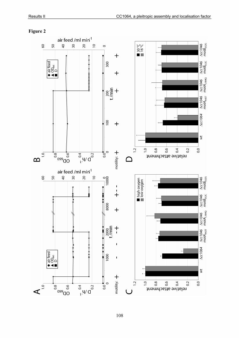
Results II CC1064, a pleitropic assembly and localisation factor
108
Figure 2

Results II CC1064, a pleitropic assembly and localisation factor
109
Figure 3

Results II CC1064, a pleitropic assembly and localisation factor
110
Figure 4

Results II CC1064, a pleitropic assembly and localisation factor
111
Figure 5

Results II CC1064, a pleitropic assembly and localisation factor
112
Figure 6

Results II CC1064, a pleitropic assembly and localisation factor
113
Figure 7

Results II CC1064, a pleitropic assembly and localisation factor
114
Figure 8

Results II CC1064, a pleitropic assembly and localisation factor
115
Figure 9
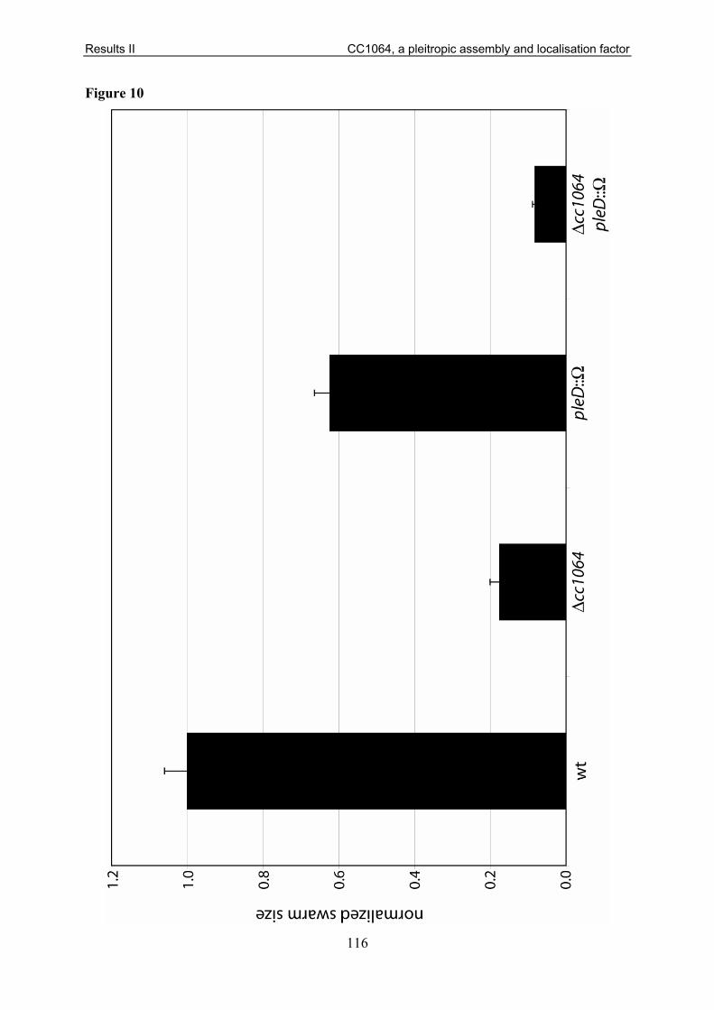
Results II CC1064, a pleitropic assembly and localisation factor
116
Figure 10

Results II CC1064, a pleitropic assembly and localisation factor
117
Figure 11

Results II CC1064, a pleitropic assembly and localisation factor
118
Table 1

Results II CC1064, a pleitropic assembly and localisation factor
119
Table 2

Results II CC1064, a pleitropic assembly and localisation factor
120
Figure S1

Results II CC1064, a pleitropic assembly and localisation factor
121
Figure S2
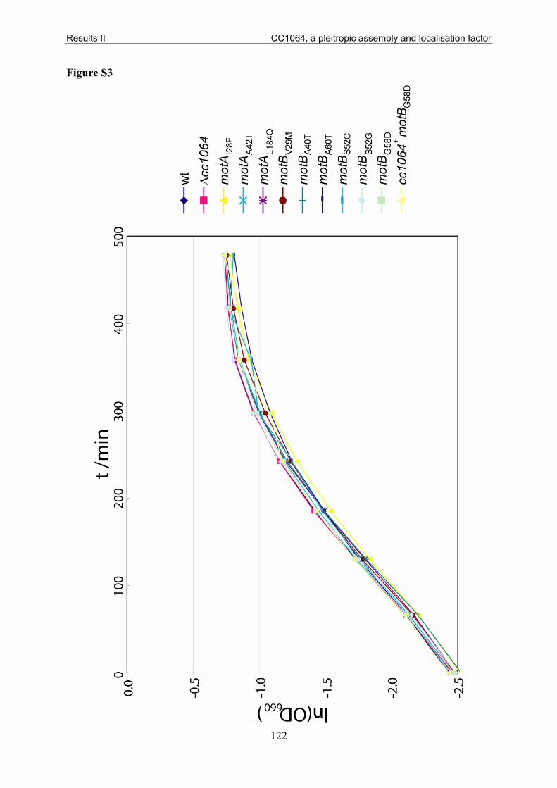
Results II CC1064, a pleitropic assembly and localisation factor
122
Figure S3

Results II CC1064, a pleitropic assembly and localisation factor
123
Table S1
Strain or plasmid Relevant genotype and description Reference or source
C. crescentus strains UJ195 CB15N pleD::Ω [170] UJ418 CB15N + pMR20 P. Aldridge UJ730 CB15 ∆pleD [47] UJ998 CB15N divJ::Ω [61] UJ1267 CB15N - synchronizable variant strain of CB15 [114] UJ2591 CB15 motA::tn5 A. Levi UJ2712 CB15N + pOP-290-2 A. Stotz UJ2713 CB15N + pCM4 [257] UJ2714 CB15N + pflgL-lacZ [132] UJ2715 CB15N ∆cc1064 + pOP-290-2 this study UJ2716 CB15N ∆cc1064 + pCM4 this study UJ2717 CB15N ∆cc1064 + pflgL-lacZ this study UJ2718 CB15N ∆cc1064 + pMR20 this study UJ2719 CB15N ∆cc1064 divJ::pdivJgfp this study UJ2720 CB15N ∆cc1064 + pMR10divK-egfp this study UJ2724 CB15N + pSA41 this study UJ2730 CB15N ∆cc1064 + pcc1064 this study UJ2731 CB15N + pMR10 this study UJ2742 CB15N ∆cc1064 + ppleC-egfp this study UJ2743 CB15 cc1064::tn5 motAA42T this study UJ2747 CB15 cc1064::tn5 motAL184Q this study UJ2749 CB15 cc1064::tn5 motBS52C this study UJ2750 CB15 cc1064::tn5 motBS52G this study UJ2815 CB15N + pAD6 A. Dürig UJ2864 CB15N ∆cc1064 this study UJ2865 CB15N ∆cc1064 pleD::Ω this study UJ2912 CB15N ∆cc1064 + pSA41 this study UJ2926 CB15N ∆cc1064 divJ::Ω this study UJ2929 CB15N ∆cc1064 motBG58D this study UJ2930 CB15N ∆cc1064 motBA60T this study UJ2931 CB15N ∆cc1064 cc1578::tnMarinerdNptI motBG58D this study UJ2935 CB15N ∆cc1064 motBS52C this study UJ2938 CB15N ∆cc1064 motBA40V this study UJ2939 CB15N ∆cc1064 motBV29M this study UJ2940 CB15N ∆cc1064 motBS52R this study UJ2941 CB15N ∆cc1064 motBS52G this study UJ2946 CB15N ∆cc1064 cc1578::tn5 this study UJ2947 CB15N cc1578::tn5 motBG58D this study UJ2997 CB15N ∆cc1064 + pMR10 this study UJ3024 CB15N ∆cc1064 motAA42T this study UJ3025 CB15N ∆cc1064 motAL184Q this study UJ3026 CB15N ∆cc1064 motAI28F this study UJ3028 CB15N ∆cc1064 motBY62C this study UJ3029 CB15N ∆cc1064 motBT51S this study

Results II CC1064, a pleitropic assembly and localisation factor
124
UJ3030 CB15N ∆cc1064 motBA40T this study UJ3110 CB15N ∆cc1064 + pAD6 this study UJ3142 CB15N ∆cc1064 motBS52C + pSA41 this study UJ3143 CB15N ∆cc1064 motAA42T + pSA41 this study UJ3157 CB15N ∆cc1064 motAI204L cc0762::tnMarinerdCat this study UJ3158 CB15N ∆cc1064 motAT189A cc0762::tnMarinerdCat this study UJ3197 CB15N + pmotBwt this study UJ3199 CB15N + pmotBG58D this study UJ3201 CB15N + pmotBA60T this study UJ3203 CB15N ∆cc1064 + pmotBwt this study UJ3205 CB15N ∆cc1064 + pmotBG58D this study UJ3207 CB15N ∆cc1064 + pmotBA60T this study UJ3208 CB15N ∆cc1064 motBG58D + pMR20 this study UJ3210 CB15N ∆cc1064 motBG58D + pmotBwt this study UJ3212 CB15N ∆cc1064 motBG58D + pmotBG58D this study UJ3214 CB15N ∆cc1064 motBG58D + pmotBA60T this study UJ3215 CB15N ∆cc1064 motBA60T + pMR20 this study UJ3217 CB15N ∆cc1064 motBA60T + pmotBwt this study UJ3219 CB15N ∆cc1064 motBA60T + pmotBG58D this study UJ3221 CB15N ∆cc1064 motBA60T + pmotBA60T this study UJ3241 CB15 - C. crescentus wild type ATCC19089 UJ3385 CB15 cc1064::tn5 this study UJ3712 CB15N cc1578::tn5 this study
UJ3748 CB15N motAwt motBwt cc0762::tnMarinerdCat cc1578::tnMarinerdNptI this study
UJ3749 CB15N ∆cc1064 motAwt motBwt cc0762::tnMarinerdCat cc1578::tnMarinerdNptI this study
UJ3750 CB15N ∆cc1064 motAwt motBG58D cc0762::tnMarinerdCat cc1578::tnMarinerdNptI this study
UJ3751 CB15N ∆cc1064 motAwt motBA60T cc0762::tnMarinerdCat cc1578::tnMarinerdNptI this study
UJ3752 CB15N ∆cc1064 motAwt motBS52C cc0762::tnMarinerdCat cc1578::tnMarinerdNptI this study
UJ3753 CB15N ∆cc1064 motAwt motBA40V cc0762::tnMarinerdCat cc1578::tnMarinerdNptI this study
UJ3754 CB15N ∆cc1064 motAwt motBV29M cc0762::tnMarinerdCat cc1578::tnMarinerdNptI this study
UJ3755 CB15N ∆cc1064 motAwt motBS52R cc0762::tnMarinerdCat cc1578::tnMarinerdNptI this study
UJ3756 CB15N ∆cc1064 motAA42T motBwt cc0762::tnMarinerdCat cc1578::tnMarinerdNptI this study
UJ3757 CB15N ∆cc1064 motAA42T motBG58D cc0762::tnMarinerdCat cc1578::tnMarinerdNptI this study
UJ3758 CB15N ∆cc1064 motAA42T motBA60T cc0762::tnMarinerdCat cc1578::tnMarinerdNptI this study
UJ3759 CB15N ∆cc1064 motAA42T motBS52C cc0762::tnMarinerdCat cc1578::tnMarinerdNptI this study

Results II CC1064, a pleitropic assembly and localisation factor
125
UJ3760 CB15N ∆cc1064 motAA42T motBS52R cc0762::tnMarinerdCat cc1578::tnMarinerdNptI this study
UJ3761 CB15N ∆cc1064 motAL184Q motBwt cc0762::tnMarinerdCat cc1578::tnMarinerdNptI this study
UJ3762 CB15N ∆cc1064 motAL184Q motBS52C cc0762::tnMarinerdCat cc1578::tnMarinerdNptI this study
UJ3763 CB15N ∆cc1064 motAL184Q motBS52R cc0762::tnMarinerdCat cc1578::tnMarinerdNptI this study
UJ3764 CB15N ∆cc1064 motAI28F motBwt cc0762::tnMarinerdCat cc1578::tnMarinerdNptI this study
UJ3765 CB15N ∆cc1064 motAI28F motBA60T cc0762::tnMarinerdCat cc1578::tnMarinerdNptI this study
UJ3766 CB15N ∆cc1064 motAI28F motBS52C cc0762::tnMarinerdCat cc1578::tnMarinerdNptI this study
UJ3767 CB15N ∆cc1064 motAI28F motBV29M cc0762::tnMarinerdCat cc1578::tnMarinerdNptI this study
UJ3768 CB15N ∆cc1064 motAI28F motBS52R cc0762::tnMarinerdCat cc1578::tnMarinerdNptI this study
UJ3769 CB15N ∆cc1064 motAI204L motBwt cc0762::tnMarinerdCat cc1578::tnMarinerdNptI this study
UJ3770 CB15N ∆cc1064 motAI204L motBA60T cc0762::tnMarinerdCat cc1578::tnMarinerdNptI this study
UJ3771 CB15N ∆cc1064 motAI204L motBS52C cc0762::tnMarinerdCat cc1578::tnMarinerdNptI this study
UJ3772 CB15N ∆cc1064 motAI204L motBA40V cc0762::tnMarinerdCat cc1578::tnMarinerdNptI this study
UJ3773 CB15N ∆cc1064 motAI204L motBV29M cc0762::tnMarinerdCat cc1578::tnMarinerdNptI this study
UJ3774 CB15N ∆cc1064 motAI204L motBS52R cc0762::tnMarinerdCat cc1578::tnMarinerdNptI this study
UJ3775 CB15N ∆cc1064 motAL189A motBwt cc0762::tnMarinerdCat cc1578::tnMarinerdNptI this study
UJ3776 CB15N ∆cc1064 motAL189A motBA60T cc0762::tnMarinerdCat cc1578::tnMarinerdNptI this study
UJ3777 CB15N ∆cc1064 motAL189A motBA40V cc0762::tnMarinerdCat cc1578::tnMarinerdNptI this study
UJ3778 CB15N ∆cc1064 motAL189A motBV29M cc0762::tnMarinerdCat cc1578::tnMarinerdNptI this study
UJ3786 CB15N ∆cc1064 motAwt motBS52G cc0762::tnMarinerdCat cc1578::tnMarinerdNptI this study
UJ3789 CB15N ∆cc1064 motAwt motBA40T cc0762::tnMarinerdCat cc1578::tnMarinerdNptI this study
UJ3790 CB15N ∆cc1064 motAA42T motBS52G cc0762::tnMarinerdCat cc1578::tnMarinerdNptI this study
UJ3791 CB15N ∆cc1064 motAA42T motBA40T cc0762::tnMarinerdCat cc1578::tnMarinerdNptI this study
UJ3792 CB15N ∆cc1064 motAL184Q motBS52G cc0762::tnMarinerdCat cc1578::tnMarinerdNptI this study

Results II CC1064, a pleitropic assembly and localisation factor
126
UJ3793 CB15N ∆cc1064 motAI28F motBS52G cc0762::tnMarinerdCat cc1578::tnMarinerdNptI this study
UJ3794 CB15N ∆cc1064 motAI204L motBS52G cc0762::tnMarinerdCat cc1578::tnMarinerdNptI this study
UJ3795 CB15N ∆cc1064 motAA42T motBL44S cc0762::tnMarinerdCat cc1578::tnMarinerdNptI this study
UJ3796 CB15N ∆cc1064 motAL184Q motBL44S cc0762::tnMarinerdCat cc1578::tnMarinerdNptI this study
UJ3797 CB15N ∆cc1064 motAI28F motBL44S cc0762::tnMarinerdCat cc1578::tnMarinerdNptI this study
UJ3798 CB15N ∆cc1064 motAL189A motBL44S cc0762::tnMarinerdCat cc1578::tnMarinerdNptI this study
UJ3799 CB15N ∆cc1064 motAI204L motBV29L cc0762::tnMarinerdCat cc1578::tnMarinerdNptI this study
UJ3802 CB15N ∆cc1064 ∆motAwt motBL44S cc0762::tnMarinerdCat cc1578::tnMarinerdNptI this study
UJ3803 CB15N ∆cc1064 ∆motAwt motBV29L cc0762::tnMarinerdCat cc1578::tnMarinerdNptI this study
UJ4703 CB15 + pMR10 this study UJ4704 CB15 Δcc1064+ pMR10 this study UJ4705 CB15 Δcc1064+ pcc1064 this study UJ4706 CB15 ∆cc1064 this study UJ4707 CB15 Δcc1064 + pSW7 this study UJ4708 CB15 + pSW7 this study E. coli strains
DH10B
F- mcrA ∆(mrr- hsd RMS- mcrBC) Φ80dlacZ∆M15 ∆lacX74 endA1 recA1 deoR ∆ (ara, leu)7697 araD139 galU galK nupG rpsL thi pro hsdR- hsd+ recA RP4-2-Tc::Mu-Tn7 Stratagene
DH5α supE44 ΔlacU169(Φ80 lacZΔM15) hsdR17 recA1 endA1gyrA96 thi-1 relA1 [204]
S17-1 RP4-2, Tc::Mu, KM-Tn7 [205] Plasmids
pAD6 pMR20 carrying egfp under the control of the Lac promotor A. Dürig
pALMAR-1 tnMariner harbouring a kanamycin resistance cassette A. Levi
pALMAR-3 tnMariner harbouring a chloramphenicol resistance cassette A. Levi
pcc1064 pMR10 carrying wild-type cc1064 under the control of its own promotor this study
pCM4 flgH transcriptional fusion to LacZ on pRKlac290 [257] pdivJgfp pEGFP-N2 carrying a C-terminal egfp fusion to divJ [175] pflgL-lacZ flgL translational fusion to LacZ on pRKlac290 [132] pUT_Km2 Mini-Tn5 transposon delivery vector [258]
pmotBA60T pMR20 carrying motBA60T under the control of its own promotor this study
pmotBG58D pMR20 carrying motBG58D under the control of its own promotor this study
pmotBwt pMR20 carrying motBwt under the control of its own promotor this study

Results II CC1064, a pleitropic assembly and localisation factor
127
pMR10 Kanr broad range low copy cloning vector [259] pMR10divK-egfp pMR10 carrying a C-terminal egfp fusion to divK [177] pMR20 Tetr broad range low copy cloning vector [259]
pNPTS128-deltacc1064
Kanr pLitmus38-derived vector with oriT and sacB carrying ~830 bp homology region upstream and downstream of cc1064, including the first 134 bp and the last 10 bp of the cc1064 open reading frame this study
pOP-290-2 fliF promotor transcriptional fusion to LacZ on pRKlac290 A. Stotz ppleC-egfp pMR20 carrying a C-terminal egfp fusion to pleC [175]
pSA41 pBBR1MSC-2nolac carrying a C-terminal egfp fusion to pleD this study
pSW7 pMR20 carrying a C-terminal gfp fusion to pleD*D53N [61]

Results III Activation mechanisms of PleD
128
3.3 Activation of the Di-guanylate Cyclase PleD by Phosphorylation-mediated Dimerisation Ralf Paul, Sören Abel, Paul Wassmann, Andreas Beck, Heiko Heerklotz, Urs Jenal
JBC 282:29170-29177 (2007)
Statement of my work
I contributed to this work by generating GFP fusion constructs of diverse PleD mutants.
Furthermore, I performed all microscopy experiments required for this work (Fig. 6).

Activation of the Diguanylate Cyclase PleD byPhosphorylation-mediated Dimerization*S
Received for publication, June 7, 2007, and in revised form, July 19, 2007 Published, JBC Papers in Press, July 19, 2007, DOI 10.1074/jbc.M704702200
Ralf Paul, Soren Abel, Paul Wassmann, Andreas Beck, Heiko Heerklotz1, and Urs Jenal2
From Biozentrum, University of Basel, Klingelbergstrasse 70, Basel CH-4056, Switzerland
Diguanylate cyclases (DGCs) are key enzymes of second mes-
senger signaling in bacteria. Their activity is responsible for the
condensation of two GTP molecules into the signaling com-
pound cyclic di-GMP. Despite their importance and abundance
in bacteria, catalytic and regulatory mechanisms of this class of
enzymes are poorly understood. In particular, it is not clear if
oligomerization is required for catalysis and if it represents a
level for activity control. To address this questionwe perform in
vitro and in vivo analysis of theCaulobacter crescentus diguany-
late cyclase PleD. PleD is a member of the response regulator
family with two N-terminal receiver domains and a C-terminal
diguanylate cyclase output domain. PleD is activated by phos-
phorylation but the structural changes inflicted upon activation
of PleD are unknown. We show that PleD can be specifically
activated by beryllium fluoride in vitro, resulting in dimeriza-
tion and c-di-GMP synthesis. Cross-linking and fractionation
experiments demonstrated that theDGC activity of PleD is con-
tained entirely within the dimer fraction, confirming that the
dimer represents the enzymatically active state of PleD. In con-
trast to the catalytic activity, allosteric feedback regulation of
PleD is not affected by the activation status of the protein, indi-
cating that activation by dimerization and product inhibition
represent independent layers of DGC control. Finally, we pres-
ent evidence that dimerization also serves to sequester activated
PleD to the differentiating Caulobacter cell pole, implicating
protein oligomerization in spatial control and providing a
molecular explanation for the coupling of PleD activation and
subcellular localization.
Cyclic 3,5-guanylyl and adenylyl nucleotides function assecond messengers in signal transduction pathways ofeukaryotes and prokaryotes. The synthesis of thesemolecules iscatalyzed by a wide variety of nucleotidyl cyclases, which areactive as homo- or heterodimers (1). Monocyclic nucleotidylcyclases that catalyze the formation of cAMP or cGMP are reg-ulated by small molecules, endogenous domains, or exogenousprotein partners, many of which alter the interface of the cata-
lytic domains and therefore the integrity of the catalytic site.Much less is known about catalysis and regulationmechanismsof the recently discovered family of diguanylate cyclases(DGCs).3 DGCs are responsible for the synthesis of cyclic di-GMP, a ubiquitous secondmessenger involved in bacterial bio-film formation and persistence (2). Cellular levels of c-di-GMPare controlled through the opposing activities of DGCs andphosphodiesterases, which form two large families of outputdomains found in bacterial one- and two-component systems(3). The DGC activity is contained within the highly conservedGGDEF domain, whose three-dimensional fold is similar to thecatalytic core of adenylate cyclase and the “palm” domain ofDNA polymerases (4, 5). Because GGDEF domains are oftenassociated with sensory input domains, it was proposed thatthese regulatory proteins serve to directly couple environmen-tal or internal stimuli to a specific cellular response through thesynthesis of the secondmessenger c-di-GMP. Similar tomono-cyclic nucleotidyl cyclases, the controlled formation of catalyt-ically competent GGDEF domain dimers may be a key mode ofDGC regulation (2, 5, 6). A simple model proposes that dimer-ization mediates an antiparallel arrangement of two DGCdomains, each of which is loaded with one GTP substrate mol-ecule. Such an arrangement would allow deprotonation of theGTP 3OHgroups and subsequent intermolecular nucleophilicattacks onto the -phosphate to occur (5).The diguanylate cyclase PleD controls pole morphogenesis
during theCaulobacter crescentus cell cycle (4, 7–10). PleD is anunorthodox member of the response regulator family of two-component signal transduction systems with two receiverdomains arranged in tandem fused to a GGDEF output domain(5). Phosphorylation by two cognate kinases, PleC and DivJ, isrequired for the activation and dynamic sequestration of PleDto the differentiating pole (4, 9). Although the first receiverdomain (Rec1) serves as phosphoryl acceptor (at the conservedAsp-53 residue), the second receiver domain (Rec2) was pro-posed to function as an adaptor for dimerization of activatedPleD (5, 9). A simple mechanistic model for the activation ofPleD proposes that phosphorylation at the conservedAsp-53 ofRec1 induces repacking of the Rec1/Rec2 interface. This in turnwould mediate dimer formation by isologous Rec1-Rec2 con-tacts across the interface and thereby facilitate reorientationand assembly of two C-terminal DGC domains (5). Here wedemonstrate that PleD activity can be greatly stimulated in vitro
* This work was supported by Swiss National Science Foundation Fellowship3100A0-108186 (to U. J.). The costs of publication of this article weredefrayed in part by the payment of page charges. This article must there-fore be hereby marked “advertisement” in accordance with 18 U.S.C. Sec-tion 1734 solely to indicate this fact.
S The on-line version of this article (available at http://www.jbc.org) containssupplemental Figs. S1–S3 and Table S1.
1 Present address: Leslie Dan Faculty of Pharmacy, University of Toronto,Toronto, Ontario M5S 3M2, Canada.
2 To whom correspondence should be addressed: Tel.: 41-61-267-2135; Fax:41-61-267-2118; E-mail: [email protected].
3 The abbreviations used are: DGC, diguanylate cyclase; c-di-GMP, cyclicdiguanylic acid; SEC, size-exclusion chromatography; ITC, isothermal titra-tion calorimetry; DSS, disuccinimidyl suberate; PVDF, polyvinylidene diflu-oride; GFP, green fluorescent protein.
THE JOURNAL OF BIOLOGICAL CHEMISTRY VOL. 282, NO. 40, pp. 29170 –29177, October 5, 2007© 2007 by The American Society for Biochemistry and Molecular Biology, Inc. Printed in the U.S.A.
29170 JOURNAL OF BIOLOGICAL CHEMISTRY VOLUME 282 • NUMBER 40 • OCTOBER 5, 2007
at ME
DIZ
INB
IBLIO
TH
EK
on March 3, 2008
ww
w.jbc.org
Dow
nloaded from
http://www.jbc.org/cgi/content/full/M704702200/DC1Supplemental Material can be found at:

by the phosphoryl mimic BeF3 and that activation of PleDresults in dimer formation. Cross-linking experiments revealedthat the DGC activity resides entirely in the dimer fraction ofactivated PleD. Furthermore, controlled dimerization not onlymodulates DGC activity but is also employed to couple PleDactivity to its subcellular sequestration. This is the first demon-stration that GGDEF protein dimers represent the active con-formation of diguanylate cyclases and confirms that oligomer-ization can be used to regulate the activity of this abundant classof signaling proteins.
MATERIALS AND METHODS
Strains, Plasmids, and Media—Bacterial strains and plas-mids used in this study are shown in Table 1. Escherichia coli
strains were grown in Luria Broth (LB) media supplementedwith antibiotics for selection, when necessary. The exact pro-cedure of strain and plasmid construction is available onrequest.Expression and Purification of PleD—E. coli cells carrying the
respective expression plasmid were grown in 200 ml of LBmedium with ampicillin (100 g/ml), and expression wasinduced by adding isopropyl 1-thio--D-galactopyranoside to0.4 mM final concentration. After harvesting by centrifugation,the cells were resuspended in TN buffer (50 mM Tris-HCl atpH 8.0, 500 mM NaCl, 5 mM -mercaptoethanol) and lysedby passage through a French press cell. The suspension wasclarified by centrifugation, followed by a high spin centrifu-gation step (100,000 g, 1 h). The supernatant was loadedonto Ni-NTA affinity resin (Qiagen), washed with TN buffer,and eluted with an imidazole gradient. Elution fractions wereexamined for purity by SDS-PAGE, and fractions containingpure protein were pooled. PleD was extensively dialyzed firstagainst 500mMNaCl, 50mMTris-HCl, pH8.0, 5mMEDTA, pH8.0, 5 mM -mercaptoethanol, and than against 250 mM NaCl,25 mM Tris-HCl, pH 7.8, 5 mM -mercaptoethanol. Prior tocross-linking experiments PleD was dialyzed against a buffercontaining 250mMNaCl, 5mM PO4, and 5mM -mercaptoeth-anol. Analytical size exclusion chromatography (SEC) was per-formed with a Superdex 200 column on a SMART system(Amersham Biosciences) at a flow rate of 50 l/min. Prepara-tive SEC to quantitatively strip nickel-nitrilotriacetic acid-pu-rified PleD from bound c-di-GMP was performed on a prepar-ative scale Superdex 200 column on an AEKTA system(Amersham Biosciences).Enzymatic Assays—Diguanylate cyclase assays were adapted
fromprocedures described previously (Paul et al. 4). The stand-ard reactionmixtureswith purified PleD contained 50mMTris-
HCl, pH 7.8, 250mMNaCl, 10mMMgCl2 in a 50-l volume andwere started by adding 100 MGTP/[-33P]GTP (PerkinElmerLife Sciences, 0.01 Ci/l). To calculate the initial velocity ofproduct formation, aliquots were withdrawn at regular timeintervals, and the reaction was stopped with an equal volume of50 mM EDTA, pH 6.0. Reaction products (2 l) were separatedon polyethyleneimine-cellulose plates (Macherey-Nagel) in 1.5M KH2PO4/5.5 M (NH4)2SO4 (pH 3.5), mixed in a 2:1 ratio.Plates were exposed to a phosphorimaging screen, and theintensity of the various radioactive species was calculated byquantifying the intensities of the relevant spots using Image-QuaNT software (Amersham Biosciences). Measurementswere always restricted to the linear range of product formation.Cross-linking Assays—The purified protein (20 or 25 M in
100 mM NaCl, 5 mM NaPO4, pH 7.8, 10 mM MgCl2, 5 mM
-mercaptoethanol, 1 mM BeCl2/10 mMNaF) was incubatedwith 2mM disuccinimidyl suberate (DSS, Pierce) for 0, 1, 5, and10 min. The cross-linker was inactivated by adding Tris-HCl,pH 7.8, to 50 mM final concentration. After separation on 10%SDS-PAGE and transfer to a PVDF membrane, PleD mono-meric and dimeric forms were detected by staining with ananti-PleD antibody (8).Isothermal Titration Calorimetry—The interaction of PleD
with cyclic-di-GMP was measured with a VP-ITC isothermaltitration calorimeter fromMicroCal (Northampton,MA), with3MPleD in the cell and 90M c-di-GMP in the syringe (buffer:100 mMNaCl, 25 mMTris-HCl, pH 7.8, 5 mMMgCl2, and 1mM
-mercaptoethanol). All solutions were thoroughly degassedand equilibrated to 25 °C before filling into the calorimeter.The delay between the injections was set to 5–10 min toensure complete re-equilibration between subsequent injec-tions. The heat capacity of the interaction between the inhib-itor and the protein was estimated through measurementsbetween 5 and 25 °C.Microscopy and Photography—C. crescentus strains were
grown in 5 ml of peptone-yeast extract media containing 5g/ml tetracycline (PYE/tet) for 18 h at 30 °C on a roller incu-bator. The stationary phase cultures were diluted 1/50 andgrown for another 8–10 h in 5 ml of PYE/tet. For fluorescenceimaging 1 l of bacterial culture was placed on a microscopeslide layered with a pad of 2% agarose dissolved in water. AnOlympus IX71 microscope equipped with an UPlanSApo100/1.40 oil objective (Olympus) and a coolSNAP HQ (Pho-tometrics) charge-coupled device camera were used to take dif-ferential interference contrast and fluorescence photomicro-graphs. For GFP fluorescence fluorescein isothiocyanate filtersets (Ex 490/20 nm, Em 528/38 nm) were used with exposuretimes of 0.15 and 1.0 s, respectively. Images were processedwith softWoRx version 3.3.6 and Photoshop CS version 8.0.
RESULTS
Activation of the PleD Diguanylate Cyclase by Beryllium
Fluoride—To investigate the specific requirements for PleDDGC activity and activation in vitro, we first set out to definethe optimal reaction conditionswith respect to pH, the concen-trations of monovalent and divalent cations, and protein con-centration. PleD was enzymatically active between pH 6.5 and10.0, with maximal activity between pH 7.5 and 8.5 (supple-
TABLE 1
Activities of PleD wild-type and mutant proteins5M of each proteinwas incubated in reaction buffer (see “Materials andMethods”)with 100 M
33P-labeled GTP. For BeF3 activation 1mM BeCl2 and 10mMNaFwereadded to the reactions.
Protein Without BeF3 With BeF3
nanomoles of c-di-GMP min1 mg1
PleD 3.32 (0.7)a 159.97 (22.6)PleDD53N 2.38 (0.3) 1.10 (0.2)PleDY26A NDb 0.036 (0.017)
a In parentheses: standard deviation.b ND, not detectable.
Controlled Dimerization of Diguanylate Cyclase
OCTOBER 5, 2007 • VOLUME 282 • NUMBER 40 JOURNAL OF BIOLOGICAL CHEMISTRY 29171
at ME
DIZ
INB
IBLIO
TH
EK
on March 3, 2008
ww
w.jbc.org
Dow
nloaded from

mental Fig. S1). Enzymatic activity was strictly dependent onthe presence of either Mg2 (supplemental Fig. S1) or Mn2
(not shown), with Mn2 resulting in a slightly higher activitycompared with Mg2. The strict requirement for bivalent cat-ions is in agreement with the recent finding of coordinatingmetal ions in the catalytic center of the PleDDGC (11). Activitydecreased with increasing NaCl concentration (supplementalFig. S1). Also, the addition of KCl (25mM) to reaction assays (4)slightly decreased the enzymatic activity and was omitted insubsequent experiments.Activation of the PleD DGC in vitro and in vivo requires the
transfer of a phosphoryl group onto the aspartic acid acceptorresidue Asp-53 of the first receiver domain (Fig. 1) (4). How-ever, in vitro phosphorylation experiments with PleD resultedin an exiguous increase of DGC activity, possibly due to subop-timal assay conditions or to low stability of the phosphorylatedform (4). For this reason we tested activation of PleD by beryl-lium fluoride (BeF3) a molecular mimic of a phosphoryl groupthat has been widely used for biochemical and structural stud-ies of bacterial response regulators (12–18). As shown in Fig. 2,BeF3 significantly stimulated the enzymatic activity of PleD,
FIGURE 1. Crystal structure of the non-activated PleD (5). A front view ofa PleD dimer is shown in A with the first receiver domain (Rec1) in red, thesecond receiver domain (Rec2) in yellow, and the GGDEF domain in green.The active site (A-site) loop (blue), the phosphoryl acceptor Asp-53, andthe position of Tyr-26 are indicated. The 2-fold symmetry axis is drawn indark blue. B, bottom view along the axis of the Rec1-Rec2 dimerizationstem. The domain coloring is equivalent to A.
FIGURE 2. Beryllium fluoride-mediated activation of the PleD diaguany-late cyclase. A, PleD (5 M) activity (nanomoles of c-di-GMP min1 mg1) asa function of BeCl2 concentration in the presence of 5 mM NaF. B, PleD (2.5 M)activity (nanomoles of c-di-GMP min1 mg1) as a function of NaF concen-tration in the presence of 1 mM BeCl2. C, PleDD53N (5 M) activity (nanomolesof c-di-GMP min1 mg1) as a function of BeCl2 concentration in the presenceof 5 mM NaF. D, specific activities (nanomoles of c-di-GMP min1 mg1) forPleD (open circles), PleD activated with BeF3 (filled circles), and the constitu-tively active PleD* mutant protein (triangles) as a function of the proteinconcentration.
Controlled Dimerization of Diguanylate Cyclase
29172 JOURNAL OF BIOLOGICAL CHEMISTRY VOLUME 282 • NUMBER 40 • OCTOBER 5, 2007
at ME
DIZ
INB
IBLIO
TH
EK
on March 3, 2008
ww
w.jbc.org
Dow
nloaded from

with optimal concentrations of 1mMBeCl2 (Fig. 2A) and 10mM
NaF (Fig. 2B), respectively. DGC activation was reversible andwas immediately abolished upon removal of BeF3 (data notshown). A PleD mutant protein lacking the phosphorylacceptor site Asp-53 (PleDD53N) could not be activated, sug-gesting that BeF3 activates the protein by specifically interact-ing with this residue in the first receiver domain (Fig. 2C). Aconstitutively active mutant of PleD, PleD* (4) was also stimu-lated by BeF3, but only by a factor of 2 (data not shown). This isconsistent with the view that PleD* is locked in an active state(see below). Concentrations of BeCl2 orNaF above 1mM and 10mM, respectively, had a negative effect on DGC activity (Fig. 2,A–C). At these concentrations BeF3 probably interacts non-specifically with surface residues that are required for diguany-late cyclase activity.BeF3 Activation Results in PleD Dimerization—The specific
activities of non-activated and BeF3-activated PleD wild-typeprotein and of the constitutively active PleD* mutant increasedwith increasing protein concentration (Fig. 2D). This suggestedthat PleD might be active in a dimeric (or oligomeric) form,with dimer formation being concentration-dependent. Theobservation that PleD* and BeF3 activated PleD reach an activ-ity plateau at much lower protein concentrations than non-activated PleD further suggested that the PleD dimerizationconstant is affected either by genetic changes or by chemicalactivation of the protein. To test this hypothesis, cross-linkingstudies were performed with PleD using the chemical cross-linker DSS (see “Materials and Methods”). We reasoned thatthe amount of covalently cross-linked dimers was proportionalto the amount of dimers in solution.When theDSS cross-linkerwas incubated with non-activated wild-type PleD or PleDD53N
at a protein concentration of 20 M (well below the estimateddissociation constant Kd of dimerization of 104 M (11)),only a minor fraction of the protein was captured ascovalently cross-linked dimers (Fig. 3A). This is consistentwith the low basal level of enzymatic activity observed fornon-activated PleD (Table 1). Activation of PleD by BeF3 notonly increased DGC activity (Table 1) but also the amount ofcross-linked dimer species (Fig. 3A). In contrast, the non-activable PleDD53N (Fig. 2 and Table 1) showed no increase incross-linked dimers (Fig. 3A).
The crystal structure of non-activated PleD predicted a spe-cific dimerization interface in the Rec1-Rec2 receiver domainstemwith a small contact patch around the surface exposedTyrresidue at position 26 of the first receiver domain (Fig. 1).Tyr-26 is strictly conserved in PleD homologs that share aRec1-Rec2-DGC domain structure, but not in response regula-tors with a different domain architecture (supplemental Fig.S2). To test if this residue plays a role in PleD dimerization,DGC activity and dimerization behavior of the PleDY26A
mutant protein were analyzed. Indeed, PleDY26A was com-pletely inactive in the absence and only marginally active in thepresence of BeF3 (Table 1). Consistent with this, only a minorfraction of the protein could be cross-linked in the dimer form,irrespectively of the presence of BeF3 (Fig. 3A). In agreementwith these in vitro data, the pleDY26A allele failed to comple-ment the pleiotropic developmental defects of a C. crescentus
pleD null mutant. Together these results strongly support the
view that Tyr-26 residue forms part of the interaction surface ofPleD dimers. This is consistent with the finding that, althoughadditional inter-chain contacts are formed in the crystal struc-ture of BeF3 activated PleD, the specific contact around Tyr-26is maintained (11).TheDiguanylate CyclaseActivity of PleDResides in theDimer
Fraction—As indicated by cross-linking, PleD forms dimers inthe presence of BeF3. To investigate if the enzymatic activitycoincided with the cross-linked PleD fractions, reactions werediluted 10-fold immediately after DSS treatment and quench-ing. At this BeF3 concentration PleD showed only residualDGCactivity (Fig. 2). As shown in Fig. 3B, cross-linking of BeF3-activated PleD resulted in a 6-fold increase of DGC activity as
FIGURE 3. Beryllium fluoride induces PleD dimerization and activation.A, cross-link assays with PleD, PleDD53N, and PleDY26A. 20 M of the purifiedproteins with or without BeF3 was incubated with 2 mM DSS (Pierce) for 0, 1, 5,and 10 min. PleD monomers and dimers were separated on SDS-polyacryl-amide gels, transferred to PVDF membranes, and detected by immunoblotanalysis with an anti-PleD antibody. Arrows mark the monomeric and dimericforms of PleD. B, PleD cross-linked in the presence of BeF3 shows increasedDGC activity. 20 M of PleD (circles) or PleDD53N (squares) with (filled symbols)or without BeF3 (open symbols) were incubated with 2 mM DSS for 0, 1, 5, 10,and 60 min. The reactions were diluted 1:10, and DGC activities were deter-mined as indicated under “Materials and Methods.”
Controlled Dimerization of Diguanylate Cyclase
OCTOBER 5, 2007 • VOLUME 282 • NUMBER 40 JOURNAL OF BIOLOGICAL CHEMISTRY 29173
at ME
DIZ
INB
IBLIO
TH
EK
on March 3, 2008
ww
w.jbc.org
Dow
nloaded from

compared with non-cross-linked samples. In contrast, cross-linking of PleD in the absence of BeF3 and cross-linking ofPleDD53N either in the presence or absence of BeF3 did notresult in increased DGC activity. Taken together, these resultssuggested that, in the presence of BeF3, a fraction of the PleDprotein is trapped by DSS cross-link in a dimerized form andthat the dimer represents the active conformation of PleD. Tofurther substantiate this idea we attempted to separate activedimers from inactive monomers by SEC (see “Materials andMethods”). Only PleD, but not PleDD53N, changed its apparentmolecular size in the presence of BeF3 (Fig. 4, A and B). How-ever, in contrast to the constitutive active PleD*, which is pres-ent predominantly as a dimer (Fig. 4C), BeF3-activated PleD didnot show resolved monomer and dimer peaks. Rather, thebroadening of the PleD peak and its shift to a higher apparentmolecular mass are consistent with a rapid equilibriumbetween monomers and dimers for the activated PleD. Onlywhen BeF3-activated PleD was cross-linked with DSS beforeSEC, defined monomer and dimer peaks were observed (Fig.4D). The peak corresponding to cross-linked PleD dimers wasconsiderably smaller than the monomer peak indicating thatonly a minor fraction of PleD was trapped in the dimeric form.This was confirmed by immunoblot experiments with anti-PleD antibodies that also showed an increase of the dimer form(fractions 11 and 12) in BeF3-treated samples (Fig. 4F). Never-theless, the dimer fractions almost exclusively accounted forthe detectable DGC activity (Fig. 4E). The observations thatBeF3 treatment increased the proportion of PleD dimers and
that isolated dimers exhibited high DGC activity are in supportof an “activation by dimerization” mechanism.DGC Activity Is Not Required for PleD Dimerization—To
demonstrate that dimerization is required, but not sufficient fordiguanylate cyclase activity, we analyzed if the PleDE370Q
mutant formed dimers. The conserved Glu-370 residue is partof the A-site signature sequence GG(E/D)EF of the PleD digua-nylate cyclase domain and was proposed to coordinate a Mg2
ion in the active site required for deprotonation of the 3-OHgroup of one GTP substrate molecule and its subsequentnucleophilic attack onto the -phosphate of the other GTPmolecule. As predicted, the PleDE370Q mutant lacked detecta-ble enzymatic activity both in the presence and absence of BeF3(data not shown).We even failed to detect any enzymatic activ-ity when the protein concentration was increased to 50 M
and with prolonged incubation times. However, in contrast toPleDY26A the lack of activitywas not due to a failure to dimerize.When BeF3-activated PleDE370Q was used in cross-link experi-ments, the behavior of the mutant form was indistinguishablefrom PleDwild type (supplemental Fig. S3). Thus, dimerizationis clearly a prerequisite for, rather than a consequence of digua-nylate cyclase activity.Activation of the PleDDiguanylate Cyclase Does Not Interfere
with Feedback Inhibition—The diguanylate cyclase activity ofPleD is subject to strong product inhibition through binding ofc-di-GMP to an allosteric I-site widely conserved amongGGDEF domain proteins (5, 19). To test if PleD activation withBeF3 interferes with allosteric control, binding of c-di-GMP tothe I-site was directly measured using ITC (Fig. 5). Integration
FIGURE 4. The enzymatic activity of PleD is contained within the dimerfraction. SEC of PleD (50 g) in the presence (stippled line) or absence (solidline) of BeF3 (A); PleDD53N (50 g) in the presence (stippled line) or absence(solid line) of BeF3 (B); PleD (solid line) and PleD* (stippled line) (C); and PleDcross-linked with 2 mM DSS in the absence (solid line) or presence (stippledline) of BeF3 (D). E, enzymatic activity (nanomoles of c-di-GMP min1 mg1) ofthe PleD SEC fractions 11–15 from D with (open bars) or without (black bars)BeF3. F, immunoblot analysis of PleD SEC fractions 11–15 from D with anti-PleD antibody. Arrows mark the monomeric and dimeric forms of PleD.
FIGURE 5. c-di-GMP binding to the allosteric site of PleD is not affected byBeF3-mediated activation of the protein. A, raw data of ITC with 90 M
c-di-GMP titrated into 3 M non-activated PleD at 25 °C. B, integration of thetitration peaks for the binding of c-di-GMP (90 M) to non-activated (opensymbols) and BeF3-activated PleD (solid symbols). The fitted binding curvesare indicated by the stippled line for the non-activated, and by the solid line forthe BeF-activated protein.
Controlled Dimerization of Diguanylate Cyclase
29174 JOURNAL OF BIOLOGICAL CHEMISTRY VOLUME 282 • NUMBER 40 • OCTOBER 5, 2007
at ME
DIZ
INB
IBLIO
TH
EK
on March 3, 2008
ww
w.jbc.org
Dow
nloaded from

of the titration peaks of c-di-GMP injected from the syringeinto the cell of the calorimeter containing PleD produced asigmoidal enthalpy curve for the interaction between PleD andc-di-GMP.The slope of the binding curve implies a dissociationconstant of 0.3 M (0.1 M). This is in good agreement withthe Ki of 0.5 M determined earlier (5, 19). In support of ac-di-GMP dimer bound at each I-site (5) the binding stoichi-ometry was measured as 2.1:1 (0.2) (c-di-GMP:PleD) (Fig. 5).When binding of c-di-GMP to PleD was compared in the
non-activated and BeF3-activated conformation, both bindingaffinity (0.4 0.1 M) and stoichiometry (2.1:1 0.2) did notchange significantly upon activation. In agreement with this,the IC50 values for PleD inhibition measured at a protein con-centration of 5 M were very similar for the non-activated(5.1 1.4 M) and the BeF3-activated (5.9 1.3 M) PleD.From these data we conclude that activation of PleD by dimer-ization does not interfere with allosteric control of the protein.It should be noted that the calorimetric data show a deviation atvery low c-di-GMP concentrations that cannot be described interms of the simple binding model used here. This may berelated to the varying degree of dimerization of c-di-GMP insolution (20). Because this effect is limited to the first few injec-tions and does not interfere with the sigmoidal part of the bind-ing curve, we have eliminated the first three data points fromthe fit (Fig. 5B) rather than introducing a more complex modelwith additional parameters of questionable relevance. A sur-prising result was obtained when measuring the heat capacityfor the interaction between c-di-GMP and PleD by plotting thebinding enthalpy versus the temperature between 5 °C and25 °C (dCp dH/dT). The heat capacity was fitted as 0.43kJ/(mol K) (data not shown). The negative value suggests ahydrophobic interaction between the inhibitor and the protein.However, in the crystal structure c-di-GMP interacts predom-inantly in a hydrophilic manner with charged amino acid resi-dues (5). It is conceivable that the binding of c-di-GMP to PleDmight cause a conformational change in the protein, with acorresponding change of the protein-solvent interactions.A PleD Mutant Unable to Dimerize Fails to Sequester to the
Cell Pole—During the C. crescentus cell cycle PleD dynamicallysequesters to the developing pole in response to activation byphosphorylation (4). However, themolecular basis for pole dis-crimination between active and inactive PleD is unclear. Toanalyze if phosphorylation itself or rather phosphorylation-in-duced dimerization is required for polar sequestration, wefused PleDY26A to GFP and compared its subcellular localiza-tion to GFP fused versions of PleDD53N and PleD*D53N. Asshown in Fig. 6, PleDD53N fails to localize to the pole, whereasPleD*D53N, the non-phosphorylatable but constitutive activeform is found exclusively at the C. crescentus stalked cell pole.Similar to PleDD53N, PleDY26A fails to sequester to the cell pole,arguing that the ability to form dimers is critical for polar local-ization of PleD (Fig. 6). When the Y26A mutation was intro-duced into the constitutive active form PleD*, the resultingPleD*Y26A-GFP fusion protein also failed to localize to the pole(Fig. 6). Levels of both PleDY26A and PleD*Y26AGFP fusion pro-teins were normal, arguing that the Y26A mutation did notaffect the protein stability in vivo (data not shown). In sum-mary, these data suggested that the oligomerization state of
PleD provides the structural basis for cell cycle dependentdynamic recruitment of the protein to the cell pole.
DISCUSSION
Dimerization is a common property of proteins utilized toprovide stability, transmit signals, or channel reagents acrossmembranes (21). Dimer formation can also be a mechanism tocontrol enzyme activity, as exemplified by the cell death prote-ase caspase 9 (22) or the cytokine antagonist p40 (23). Here wepresent an example for dimerization-induced activation of anenzyme, diguanylate cyclase, which takes advantage of an asso-ciated regulatory domain that couples dimerization to phos-phorylation input.The crystal structure of the non-activated form of the PleD
response regulator suggested an “activation by dimerization”mechanism (5). However, because monomeric proteins canform non-physiological dimers or higher order oligomersin crystals, this hypothesis required experimental validation.In the crystal structure PleD forms a dimer with the tworeceiver domains mediating weak monomer-monomer inter-actions between a small contact patch around Tyr-26 of Rec1and 3-4 of Rec2 (Fig. 1). Thus, the apo structure not onlyprovided the structural basis for activity control of PleDbut alsoproposed the interaction surface for dimerization. Here wetested the hypothesis that, upon activation, PleD engages in
FIGURE 6. A PleD dimerization mutant fails to dynamically localize to thecell pole. Differential interference contrast (left panels) and fluorescentimages (right panels) are shown for C. crescentus NA1000 wild-type expressingGFP fusion proteins of the indicated PleD variants. Polar fluorescent foci aremarked by arrows.
Controlled Dimerization of Diguanylate Cyclase
OCTOBER 5, 2007 • VOLUME 282 • NUMBER 40 JOURNAL OF BIOLOGICAL CHEMISTRY 29175
at ME
DIZ
INB
IBLIO
TH
EK
on March 3, 2008
ww
w.jbc.org
Dow
nloaded from

dimer formation, and we analyzed the specific role of Tyr-26 inthis process. Because we failed to efficiently activate PleD in
vitro by phosphorylation through one of its cognate kinases (4)we used the phosphoryl mimic BeF3 to analyze the effect ofactivation on PleD oligomerization and activity (12, 15). Ourresults not only provide biochemical evidence for an “activationby dimerization” mechanism but also confirm an importantrole forTyr-26 in dimerization. Both a genetically active formofPleD, PleD* (4), andBeF3 activated PleD-stimulatedDGCactiv-ity and oligomerization. Because stimulation of PleD DGCactivity by BeF3 specifically required the phosphoryl acceptorside Asp-53, the changes in structure and activity observedmost likely reflect the activation mechanism normally evokedby phosphorylation. Because the constitutive active form PleD*still showed 10-fold higher DGC activity and formed morestable dimers than BeF3-modified PleD, it is possible that PleDis only partially activated by BeF3 in the non-toxic concentra-tion range used (Fig. 2). Alternatively, the PleD* mutant pro-tein, which contains several amino acid changes contributing tothe “locked-on” state (4, 9), might form particularly stabledimers. To demonstrate that theDGC activity specifically asso-ciates with PleD dimers, we chemically cross-linked BeF3-acti-vated protein and subsequently separated PleD dimers fromnon-cross-linked monomers by SEC. The finding that thecross-linked dimer fraction had a greatly increased enzymaticactivity as compared with monomers strongly implied thatPleD dimers represent the active form of the enzyme andargued that phosphorylation-mediated dimerization repre-sents the main mechanism of PleD diguanylate cyclase activitycontrol. A possible role for dimerization or oligomerization ofdiguanylate cyclases was suggested previously. Several full-lengthGGDEFdomain proteins, and isolatedGGDEFdomains,that were expressed as fusions to maltose-binding protein,behaved as dimers or trimers when analyzed by SEC (6). How-ever, although the significance of trimer formation is unclear,no correlation was reported between the oligomeric state andenzymatic activity of these proteins.In contrast to PleD wild type, the PleDY26A mutant failed to
efficiently formdimers upon activationwith BeF3. This, and theobservation that, in the presence of BeF3, PleDY26A showed analmost 10,000-fold lower DGC activity as compared with PleDwild type, is consistent with a specific requirement of residueTyr-26 for PleD oligomerization. The weak monomer interac-tions observed in the PleD apo structure around Tyr-26 pre-dicted that if this residue is part of the dimerization interfaceadditional contacts would have to be formed upon activation tostabilize the complex. In agreement with the data presentedhere, the interaction surface aroundTyr-26 ismaintained in thecrystal structure of the activated form of PleD (11) with Tyr-26making specific contacts to Asp-209 and Arg-212 in the secondreceiver domain of the other chain. However, a series of addi-tional inter-chain contacts are formed in the activated structureresulting in a tightening of the dimer interface (11). The Tyrresidue at position 26 of the first receiver domain is strictlyconserved in PleD homologs with an identical Rec1-Rec2-GGDEF domain structure (supplemental Fig. S2). In contrast,this residue is not conserved in response regulators with a dif-ferent domain structure or composition (supplemental Fig. S2).
Similarly, residues Asp-209 and Arg-212, the interaction part-ners of Tyr-26 in the crystal structure of activated PleD, showstrict conservation only in proteins with a PleD-like domainstructure (data not shown). Together with the experimentaldata presented here, this strongly suggested that Tyr-26 formspart of the dimerization surface of this protein family.Diguanylate cyclases catalyze the formation of a symmetric
product by condensing two identical GTP substrate molecules.In contrast to monocyclic nucleotidyl cyclases, which formnon-symmetric products, dimerization is an apparent necessityfor the catalytic mechanism of DGCs, because it creates a fullysymmetrical active site at the interface of two subunits. In thesimplest model two substrate-charged GGDEF domains wouldmeet in a symmetric but antiparallel arrangement to properlyposition the 3-OH groups for an intermolecular nucleophilicattack onto the -phosphate of the opposite substrate mole-cule. Moreover, because it is a prerequisite for catalysis, oli-gomerization of the DGC domains is obviously exploited tocontrol PleD enzyme activity. Although phosphorylation-me-diated dimerization of PleD represents the first example of con-trolled dimerization of a DGC, it is possible that promoting orinhibiting dimerization is a key mechanism of DGC activitycontrol in general. The preponderance of potential DGCs pres-ent in many bacteria predicts complex signaling mechanismsand makes it obligatory for the cell to tightly control theseenzymes (2). Although most DGCs have been postulated to besubject to strict product inhibition (19), little is known abouthow DGCs are activated in response to specific environmentalor internal signals. Although it can be assumed that all GGDEFdomains that are fused to receiver domains of two-componentsystems exhibit PleD-like phosphorylation-mediated control,GGDEF domains, which are associated with other signal inputdomains like GAF (24, 25), PAS (26), BLUF (27), or HAMP (28,29), might function similarly. It is worth mentioning that mostregulatorymechanisms used to control monocyclic nucleotidylcyclases involve the formation or dissolution of catalyticallycompetent active sites, caused by rearrangement of the twocatalytic domains of the dimer relative to each other (reviewedin Ref. 1). Future studies will show if this regulatory principlecan be extended to the large family of bacterial DGCs.Oligomerization of PleD is not only used to temporally con-
trol DGC activity but also contributes to its spatial distribution.PleD activity is required for the morphological changes thattake place during the C. crescentus swarmer-to-stalked celltransition (7–9). During this cell differentiation step, PleD isactivated by phosphorylation and as a result sequesters to thedifferentiating pole (4). The observation that non-phosphory-latable forms of PleD fail to localize to the cell pole, whereas theconstitutively activated mutant form PleD* is predominantlyfound at this subcellular site, suggested that activation of PleDduring development is directly coupled to its dynamic subcel-lular positioning (4). However, themolecular basis for this cou-pling event and for PleD recognition at the pole remainedunclear. In principle, a polar interaction partner could recog-nize activated PleD by its phosphorylation status, by an alteredmonomer conformation, or by its oligomerization state. Theobservation, that PleDmolecules lacking Tyr-26 not only fail todimerize but also fail to sequester to the pole, suggested that the
Controlled Dimerization of Diguanylate Cyclase
29176 JOURNAL OF BIOLOGICAL CHEMISTRY VOLUME 282 • NUMBER 40 • OCTOBER 5, 2007
at ME
DIZ
INB
IBLIO
TH
EK
on March 3, 2008
ww
w.jbc.org
Dow
nloaded from

oligomerization state dictates subcellular distribution of PleDduring the C. crescentus cell cycle. Enzymatically active dimersof PleD would thus specifically sequester to the differentiatingcell pole resulting in the predominant formation of c-di-GMPat this cellular localization. Because Caulobacter possessesmarkers that are laid downduring or after cell division to tag thenew poles (30–32), it is reasonable to assume that PleD inter-acts with one or several pre-existing proteins, which are able todiscriminate between its monomeric and dimeric forms.Dimerization of PleD might increase the interaction diversityby enabling simultaneous binding of two interacting proteins orby creating new binding sites for additional proteins (21). Bothof these possibilities could provide a molecular explanation forthe discrimination of PleD oligomers at the differentiating pole.Spatial discrimination based on receiver domain-mediated oli-gomerization could very well be a general cellular phenomenonin bacteria. Like PleD, the response regulator DivK dynamicallylocalizes to theC. crescentus cell poles in a phosphorylation-de-pendent manner (33). It is not clear how the poles discriminatebetween activated and non-activatedDivK, but it is attractive tospeculate that oligomerization might also play a role in thisbehavior. CikA, a sensor histidine kinase and a key componentof the circadian clock input pathway in the cyanobacteriumSynecococcus elongatus, also localizes to the pole where it isbelieved to interact with a complex of clock-related proteins(34). Polar sequestration of CikA depends on a C-terminalpseudo-receiver domain that lacks the conserved phosphorylacceptor side. It has been proposed that, through a docking/activation mechanism, pseudo-receiver domain couples theactivity of CikA to its subcellular location (34). Because histi-dine kinases are active as dimers, the pseudo-receiver domainmight serve as an adaptor between the oligomeric state andpolar positioning of CikA. Like the pseudo-receiver domain,the Rec2 receiver domain of PleD is not conserved and likelyfulfills an adaptor function. It is possible that the Rec1-Rec2-GGDEF domain structure that arose through duplication of thereceiver domains in PleD homologs has evolved to provide foradditional surface for the interaction with specific polar recep-tors. In such a scenario, Rec1 would be interacting with thehistidine kinase and would provide dimerization surface. Rec2,in turn, would also be engaged in dimerization but in additionwouldmediate interactionwith a polar receptor. If so, a distinctsubcellular localization of enzymatically active DGCs might becommon to all PleD homologs. Future studies are geared atidentifying the polar receptor(s) for PleD, characterizing themolecular mechanisms required for the discriminationbetween PleDmonomers and dimers, and analyzing the biolog-ical relevance of sequestering an enzymatically active form ofPleD to this particular subcellular site.
Acknowledgments—We thank Jacob Malone and Tilman Schirmer
for helpful discussions.
REFERENCES
1. Sinha, S. C., and Sprang, S. R. (2006) Rev. Physiol. Biochem. Pharmacol.
157, 105–140
2. Jenal, U., and Malone, J. (2006) Annu. Rev. Genet. 40, 385–407
3. Ulrich, L. E., Koonin, E. V., and Zhulin, I. B. (2005) Trends Microbiol. 13,
52–56
4. Paul, R., Weiser, S., Amiot, N. C., Chan, C., Schirmer, T., Giese, B., and
Jenal, U. (2004) Genes Dev. 18, 715–727
5. Chan, C., Paul, R., Samoray, D., Amiot, N. C., Giese, B., Jenal, U., and
Schirmer, T. (2004) Proc. Natl. Acad. Sci. U. S. A. 101, 17084–17089
6. Ryjenkov, D. A., Tarutina, M., Moskvin, O. V., and Gomelsky, M. (2005) J.
Bacteriol. 187, 1792–1798
7. Hecht, G. B., and Newton, A. (1995) J. Bacteriol. 177, 6223–6229
8. Aldridge, P., and Jenal, U. (1999)Mol. Microbiol. 32, 379–391
9. Aldridge, P., Paul, R., Goymer, P., Rainey, P., and Jenal, U. (2003) Mol.
Microbiol. 47, 1695–1708
10. Levi, A., and Jenal, U. (2006) J. Bacteriol. 188, 5315–5318
11. Wassmann, P., Chan, C., Beck, A., Heerklotz, H., Paul, R., Jenal, U., and
Schirmer, T. (2007) Structure 15, 915–927
12. Yan, D., Cho, H. S., Hastings, C. A., Igo, M. M., Lee, S. Y., Pelton, J. G.,
Stewart, V., Wemmer, D. E., and Kustu, S. (1999) Proc. Natl. Acad. Sci.
U. S. A. 96, 14789–14794
13. Cho, H., Wang, W., Kim, R., Yokota, H., Damo, S., Kim, S. H., Wemmer,
D., Kustu, S., and Yan, D. (2001) Proc. Natl. Acad. Sci. U. S. A. 98,
8525–8530
14. Hastings, C. A., Lee, S. Y., Cho, H. S., Yan, D., Kustu, S., and Wemmer,
D. E. (2003) Biochemistry 42, 9081–9090
15. Wemmer, D. E., and Kern, D. (2005) J. Bacteriol. 187, 8229–8230
16. Toro-Roman, A., Wu, T., and Stock, A. M. (2005) Protein Sci. 14,
3077–3088
17. Toro-Roman, A., Mack, T. R., and Stock, A. M. (2005) J. Mol. Biol. 349,
11–26
18. Bachhawat, P., Swapna, G. V., Montelione, G. T., and Stock, A. M. (2005)
Structure 13, 1353–1363
19. Christen, B., Christen, M., Paul, R., Schmid, F., Folcher, M., Jenoe, P.,
Meuwly, M., and Jenal, U. (2006) J. Biol. Chem. 281, 32015–32024
20. Zhang, Z. Y., Kim, S., Gaffney, B. L., and Jones, R. A. (2006) J. Am. Chem.
Soc. 128, 7015–7024
21. Marianayagam, N. J., Sunde, M., and Matthews, J. M. (2004) Trends Bio-
chem. Sci. 29, 618–625
22. Renatus, M., Stennicke, H. R., Scott, F. L., Liddington, R. C., and Salvesen,
G. S. (2001) Proc. Natl. Acad. Sci. U. S. A. 98, 14250–14255
23. Heinzel, F. P., Hujer, A. M., Ahmed, F. N., and Rerko, R. M. (1997) J. Im-
munol. 158, 4381–4388
24. Aravind, L., and Ponting, C. P. (1997) Trends Biochem. Sci. 22, 458–459
25. Hurley, J. H. (2003) Sci. STKE 2003, PE1
26. Ponting, C. P., and Aravind, L. (1997) Curr. Biol. 7, R674–R677
27. Gomelsky, M., and Klug, G. (2002) Trends Biochem. Sci. 27, 497–500
28. Aravind, L., and Ponting, C. P. (1999) FEMSMicrobiol. Lett. 176, 111–116
29. Galperin, M. Y., Nikolskaya, A. N., and Koonin, E. V. (2001) FEMSMicro-
biol. Lett. 203, 11–21
30. Viollier, P. H., Sternheim, N., and Shapiro, L. (2002) Proc. Natl. Acad. Sci.
U. S. A. 99, 13831–13836
31. Huitema, E., Pritchard, S., Matteson, D., Radhakrishnan, S. K., and Viol-
lier, P. H. (2006) Cell 124, 1025–1037
32. Lam, H., Schofield, W. B., and Jacobs-Wagner, C. (2006) Cell 124,
1011–1023
33. Jacobs, C., Hung, D., and Shapiro, L. (2001) Proc. Natl. Acad. Sci. U. S. A.
98, 4095–4100
34. Zhang, X., Dong, G., andGolden, S. S. (2006)Mol.Microbiol. 60, 658–668
Controlled Dimerization of Diguanylate Cyclase
OCTOBER 5, 2007 • VOLUME 282 • NUMBER 40 JOURNAL OF BIOLOGICAL CHEMISTRY 29177
at ME
DIZ
INB
IBLIO
TH
EK
on March 3, 2008
ww
w.jbc.org
Dow
nloaded from

SUPPLEMENTAL MATERIAL:
Figure S1: Characterization of PleD DGC activity as a function of pH (A), MgCl2
concentration (B), and NaCl concentration (C). The specific activity was determined as nmol
c-di-GMP min-1 mg-1. 10 M PleD was assayed in 50 l volume (250 mM NaCl, 50 mM Tris-
HCl pH 7.8, 10 mM MgCl2, 5mM - Mercaptoethanol) and 100 M 33P-labelled GTP, with
varying NaCl or MgCl2 concentrations in the respective assays. The pH-dependence was
determined with assays containing 75 mM Tris-Propane (pH 6-7, 9-10.5), or 75 mM Tris-HCl
(pH 7-9).
Figure S2: Exclusive conservation of Tyr26 in the phosphoryl-acceptor domain of PleD-like
response regulators. Sequence logos representing amino acid conservation of different
response regulator families were generated by the WebLogo tool
(http://weblogo.berkeley.edu; (35)) from alignments produced by BLAST and edited by
Jalview (36). The domain structure of the different families was generated with SMART (37)
and is indicated above the WebLogos. (A) PleD-like family of response regulators sharing the
same Rec1-Rec2-GGDEF (DUF1) domain structure and composition. The numbering of the
amino acid residues refers to C. crescentus PleD. (B) Response regulators with duplicated
receiver domains, a PleD-like GGDEF domain (DUF1), and additional signaling domains.
The numbering of the amino acid residues refers to the N-terminal receiver domain of protein
NSE_0512 from Neorickettsia sennetsu (C) Response regulators with a single receiver
domain and a GGDEF output domain. The numbering of the amino acid residues refers to
Pseudomonas fluorescens WspR (38). (D) Response regulators consisting of a single receiver
domain. The numbering of the amino acid residues refers to C. crescentus DivK (39).
Figure S3: Beryllium fluoride induces dimerization of a catalytically inactive PleD mutant
protein. (A) Size exclusion chromatography (SEC) of PleDE370Q crosslinked with 2 mM DSS
in the presence (stippled line) or absence (solid line) of BeF3. (B) Immunoblot analysis of
PleD SEC fractions 11-15 from (A) with anti-PleD antibody. Arrows mark the monomeric
and dimeric forms of PleD.

Table S1: Strains and Plasmids Strain Relevant genotype or description Reference or source Escherichia coli
DH10B F- mcrA (mrr- hsd RMS- mcrBC) (40) 80dlacZ M15 lacX74 endA1 recA1
deoR (ara, leu)7697 araD139 galU galK nupG rpsL thi pro hsdR- hsd+ recA RP4-2-Tc Mu-Tn7
BL21 (DE3) F- dcm ompT hsdS(rB-mB-) Stratagene pLysS gal (DE3) [pLysS CAMr] Caulobacter crescentus
UJ626 CB15N pleD pPA53-4 (4) UJ1909 CB15N pleD pSW6 (4) UJ1910 CB15N pleD pSW7 (4) UJ2722 CB15N pleD pSA38 This study UJ2745 CB15N pleD pSA39 This study Plasmid Relevant genotype or description Reference or source pCC2 pET11; pleD, C-terminal His6 tag (4) pRP87 pET11; pleDD53N, C-terminal His6 tag (4) pRP89 pET11; pleD*, C-terminal His6 tag (4) pRP100 pET11; pleDE370Q, C-terminal His6 tag This study pRP106 pET11; pleDY26A, C-terminal His6 tag This study pPA53-4 pMR10; pleD-gfp (4) pSA38 pMR10; pleDY26A-gfp This study pSA39 pMR10; pleD*Y26A-gfp This study pSW6 pMR10; pleDD53N-gfp (4) pSW7 pMR10; pleD*D53N-gfp (4)

0
20
40
60
80
100
6.0 6.5 7.0 7.5 8.0 8.5 9.0 9.5 10.0
0 5 10 15 20 25 30 35 40 450
0.51
1.52
2.53
3.5
Sp
ecific
activity
00.5
11.5
22.5
33.5
NaCl (M)0 0.4 0.8 1.2 1.6 2
50
pH
MgCl (mM)
Rel
ativ
e ac
tivity
A
B
C
Sp
ecific
activity
2
Figure S1
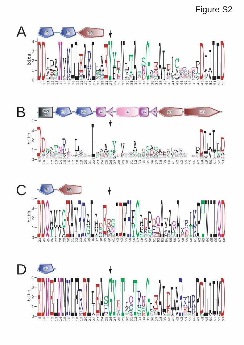
A
B
Figure S2
C
D

AP
rote
in
1411 1312
-BeF
+BeF
B
Fraction
dimer
monomer
dimer
monomer
1411 1312 15
0.04
0.02
01.4 1.5 1.6 1.7 1.8
Elution volume (ml)
0.06
0.08
Figure S3

Results IV The network controlling PleD activity
142
3.4 Allosteric Regulation of Histidine Kinases by their Cognate Response Regulator Determines Cell Fate Ralf Paul, Tina Jaeger*, Sören Abel*, Irene Wiederkehr, Marc Folcher, Emanuele G.
Biondi, Michael T. Laub, Urs Jenal
*Authors contributed equally to the manuscript
Cell, manuscript accepted
Statement of my work
I contributed to this work by generating strains expressing fluorescent protein fusions to DivJ,
PleC and DivK. In addition, I performed the cell cycle resolved colocalisation experiments
(Fig. 6C) and took part in all further microscopy analysis required for this work (Fig. 6A
S9A).

Allosteric Regulation of HistidineKinases by Their Cognate ResponseRegulator Determines Cell FateRalf Paul,1 Tina Jaeger,1,3 Soren Abel,1,3 Irene Wiederkehr,1 Marc Folcher,1 Emanuele G. Biondi,2 Michael T. Laub,2
and Urs Jenal1,*1Biozentrum, University of Basel, Klingelbergstrasse 70, 4056 Basel, Switzerland2Massachusetts Institute of Technology, Department of Biology, 77 Massachusetts Avenue Cambridge, MA 02139, USA3These authors contributed equally to this manuscript.
*Correspondence: [email protected]
DOI 10.1016/j.cell.2008.02.045
SUMMARY
The two-component phosphorylation network is of
critical importance for bacterial growth and physiol-
ogy. Here, we address plasticity and interconnection
of distinct signal transduction pathways within this
network. In Caulobacter crescentus antagonistic ac-
tivities of the PleC phosphatase and DivJ kinase lo-
calized at opposite cell poles control the phosphory-
lation state and subcellular localization of the cell fate
determinator protein DivK. We show that DivK func-
tions as an allosteric regulator that switches PleC
from a phosphatase into an autokinase state and
thereby mediates a cyclic di-GMP-dependent mor-
phogenetic program. Through allosteric activation
of the DivJ autokinase, DivK also stimulates its own
phosphorylation and polar localization. These data
suggest that DivK is the central effector of an inte-
grated circuit that operates via spatially organized
feedback loops to control asymmetry and cell fate
determination in C. crescentus. Thus, single domain
response regulators can facilitate crosstalk, feed-
back control, and long-range communication among
members of the two-component network.
INTRODUCTION
Asymmetric cell division underlies the fundamental basis for the
developmental evolution of organisms. It refers to the capability
of stem cells to simultaneously produce a continuous output of
differentiated cells and to maintain their own population of undif-
ferentiated cells. The regulation of asymmetric division is
achieved by the controlled segregation of basally localized cell
fate determinants, which leads to the polarization of the stem
cell along its axis (Betschinger and Knoblich, 2004). In the bacte-
rium Caulobacter crescentus, asymmetry is established by
members of the two-component signaling systems, which con-
trol various aspects of bacterial physiology including cell differ-
entiation and virulence. Two sensor histidine kinases, DivJ and
PleC, are positioned to opposing poles of theCaulobacter predi-
visional cell (McAdams and Shapiro, 2003). A cylindrical exten-
sion of the cell body, the stalk, and an adhesive holdfast occupy
the DivJ-marked pole, while the PleC-occupied pole bears a ro-
tating flagellum and adhesive pili (Figure 7A). Upon division the
stalked cell re-enters S-phase immediately, whereas the mo-
tile-swarmer cell takes advantage of the replication inert G1
phase to spread out before it undergoes reprogramming into
a surface adherent stalked cell.
C. crescentus cell fate is implemented by the essential single
domain response regulator DivK (Hecht et al., 1995; Matroule
et al., 2004). DivK localizes to both poles of the predivisional
cell in a phosphorylation-dependent manner, but is released
from the flagellated pole after completion of cytokinesis
(Figure 7A) (Jacobs et al., 2001). While the DivJ autokinase is
the main phosphodonor for DivK and responsible for its seques-
tration to the cell poles (Lam et al., 2003), the PleC phosphatase
activity displaces DivK from the flagellated pole by maintaining
DivKP levels low in the swarmer cell (Lam et al., 2003;
Matroule et al., 2004). Compartmentalization of the DivJ kinase
and the PleC phosphatase during cell division results in the
sudden reduction of DivKP levels in the swarmer cell and the
initiation of the swarmer-specific developmental program
(Matroule et al., 2004). Conversely, a rapid DivJ-mediated
increase of DivK phosphorylation during the G1-to-S transition
is critical for G1-to-S transition and cell differentiation (Hung
and Shapiro, 2002; Jacobs et al., 2001; Wu et al., 1998). Acti-
vated DivK has recently been proposed to control cell cycle pro-
gression and development via the CckA-ChpT pathway, which
regulates the activity of the master cell cycle regulator CtrA
(Biondi et al., 2006).
Although it is clear that DivK phosphorylation by DivJ and de-
phosphorylation by PleC are vital for C. crescentus cell cycle
control and development, the significance of the spatial behavior
of DivK and its molecular role remain unclear. Here we propose
that DivK together with PleC and DivJ form the core of an inte-
grated regulatory circuitry that operates via spatially organized
cellular feedback loops. We show that DivK directly interacts
with both polar proteins to strongly boost their kinase activities.
By switching PleC from the phosphatase into the autokinase
mode and by forming a strong positive feedback loop with the
Cell 133, 1–10, May 2, 2008 ª2008 Elsevier Inc. 1
CELL 3852
Please cite this article in press as: Paul et al., Allosteric Regulation of Histidine Kinases by Their Cognate Response Regulator Determines Cell
Fate, Cell (2008), doi:10.1016/j.cell.2008.02.045

DivJ autokinase, DivK effectively and robustly mediates G1-to-S
transition.
One of the readouts of the DivK-driven network is the synthe-
sis of the second messenger cyclic di-GMP via the activation of
the response regulator PleD (Aldridge et al., 2003; Paul et al.,
2004). PleD phosphorylation results in dimerization-based acti-
vation of the C-terminal diguanylate cyclase domain and se-
questration of the regulator to the differentiating pole where it di-
rects flagellar ejection, holdfast biogenesis, and stalk formation
(Levi and Jenal, 2006; Paul et al., 2007, 2004). Genetic experi-
ments indicated that DivJ, PleC, and DivK are upstream compo-
nents required for the activation of the PleD diguanylate cyclase
(Aldridge et al., 2003; Sommer and Newton, 1991). Consistent
with this, in vivo and in vitro experiments had shown a direct
role for PleC and DivJ in modulating phosphorylation, diguany-
late cyclase activity, and polar localization of PleD (Aldridge
et al., 2003; Paul et al., 2004). This indicated that PleC, in addition
to functioning as phosphatase in swarmer cells, also plays a role
as autokinase and contributes to the stalked cell-specific pro-
gram via PleD activation. We present in vitro and in vivo evidence
that DivK together with the DivJ and PleC kinases serves to ac-
tivate and sequester the PleD diguanylate cyclase during theG1-
to-S transition. DivK contributes to the specific phosphorylation
of PleD by acting as a specific and effective enhancer of the PleC
and DivJ autokinases. Our findings propose a regulatory role for
single domain response regulators in two component signal
transduction pathways as diffusible modulators of their cognate
sensor histidine kinases.
RESULTS
DivK Stimulates the PleD Diguanylate Cyclase Activity
in a PleC-Dependent Manner
To explore the regulatory link between PleD, PleC, and DivK, ac-
tivation of PleD diguanylate cyclase activity was assayed in vitro
in the presence of PleC and DivK. In line with earlier results (Paul
et al., 2004) PleC alone only marginally stimulated PleD activity
(Figure 1A). Surprisingly, in the presence of DivK or DivKD53N,
a mutant that cannot be phosphorylated because it lacks the
phosphoryl acceptor site, PleD diguanylate cyclase activity was
dramatically stimulated (Figures 1A and 1B). Stimulation by DivK
wild-type was less effective, presumably because DivK itself
can use PleCP as phosphodonor (Hecht et al., 1995) and thus
can sequester someof the available phosphoryl groups. DivK-de-
pendent stimulation of PleD activity required ATP, PleC autoki-
nase activity, and the PleD phosphoryl acceptor site Asp53
(Figure 1B). In particular, purified PleCF778L, a mutant that lacks
autokinase activity but shows normal phosphatase activity
(Matroule et al., 2004) failed to support DivK-dependent stimula-
tion of PleD. Because PleD is activated by dimerization (Paul
et al., 2007), residual diguanylate cyclase activity can be detected
at this protein concentration (5 mM) even in the absence of phos-
phorylation (theKd for dimerizationof nonactivatedPleD is100mM
[Wassmann et al., 2007]) (Figure 1B). It is interesting to note that
whenall three proteinswere present PleDdiguanylate cyclase ac-
tivity decreased below the basal level of nonactivated PleD under
conditions that did not allow phosphorylation (no ATP, no PleC
autokinase activity) (Figure 1B). Altogether, these experiments
demonstrate that the single domain response regulator DivK is
able to efficiently stimulate the in vitro activity of PleD and that
this activation requires an active histidine protein kinase PleC.
DivK Stimulates PleC Autokinase but Not
Phosphatase Activity
The above experiments suggested that DivK activates PleD by in-
terferingwithPleCautokinaseactivity.To test this,PleCautophos-
phorylation activitywasmonitored in thepresenceandabsenceof
DivK. Because PleC readily phosphorylates DivK in vitro (Hecht
etal., 1995;Wuetal., 1998),weusedDivKD53N toavoida reduction
of PleCPbyphosphotransfer toDivK. As shown in Figure 2A and
Figure S1A (available online), DivKD53N stimulated levels of
PleCP in a concentration dependent manner. Because the sta-
bility of PleCP was not affected by DivK (Figure S1B), DivKD53N
seems to specifically stimulate PleC autophosphorylation activity.
As a consequence of this stimulation, phosphotransfer from PleC
to PleD (Figure 2B) as well as PleD dimerization (Figure S2) was
increased when both response regulators were present in the re-
action. This is consistent with the observed increase in c-di-GMP
synthesis in reactions containing PleD, PleC and DivK (Figure 1).
Likewise, phosphotransfer from PleCP to DivK was stimulated
in the presence of DivKD53N (Figure 3A). Stimulation of PleC auto-
kinase activity did not result from a DivK-mediated in vitro artifact
(e.g., through DivK assisted folding of PleC), as solubility, quarter-
nary structure, and activity of PleC preincubated with or without
DivKD53N was indistinguishable (Figure 3). Finally, we tested
if DivKD53Nwas able to stimulate PleC phosphatase activity. Puri-
fied DivKP was mixed with PleC in the presence or absence of
DivKD53N to assay dephosphorylation rates. In contrast to PleC
mediated phosphorylation of DivK, dephosphorylation of DivK
was not increased in the presence of DivKD53N (Figures 3A–3C).
Rather, the rate of DivKP dephosphorylation was reduced in
the presence of DivKD53N. In conclusion, these experiments
provide strong evidence that the response regulator DivK is able
to selectively stimulate the kinasebut not the phosphataseactivity
of its cognate histidine kinase PleC.
DivK and PleD Compete for Phosphorylation
by the DivJ Kinase
Because DivK and PleD also interact with DivJ, we tested if the
response regulators also showed some synergistic behavior
with respect to DivJ or if they would compete for DivJ kinase.
As shown in Figure 1C, the PleD diguanylate cyclase was acti-
vated by DivJ, but the addition of DivK efficiently blocked PleD
activation. PleD activation was restored only when DivK was di-
luted below a molar ratio of 1:10. DivKD53N also reduced DivJ-
mediated PleD activation but was less efficient than DivK wild-
type (Figure 1C). These results suggested that PleD and DivK
compete for the phosphodonor DivJP. This was confirmed
bymonitoring phosphotransfer fromDivJP to the two response
regulators. Although DivJP readily served as phosphodonor for
PleD in the absence of DivK, phosphoryl groupswere transferred
exclusively to DivK when both response regulators were present
in the reaction mixture (Figure 2D). Altogether, these experi-
ments show that DivK and PleD compete for phosphorylation
by the stalked pole specific kinase DivJ, and that in vitro DivJP
prefers to transfers phosphate to DivK.
2 Cell 133, 1–10, May 2, 2008 ª2008 Elsevier Inc.
CELL 3852
Please cite this article in press as: Paul et al., Allosteric Regulation of Histidine Kinases by Their Cognate Response Regulator Determines Cell
Fate, Cell (2008), doi:10.1016/j.cell.2008.02.045

DivK Stimulates Autophosphorylation of DivJ
The observation that DivK is able to stimulate PleC autokinase
activity in vitro raised the possibility that DivK might also modu-
late the activity of its other cognate kinase, DivJ. Similar to its ob-
served effect on PleC, DivKD53N efficiently stimulated DivJ auto-
phosphorylation (Figures 4A and S1C), and did not affect the
stability of DivJP (Figure S1D). As for PleC, pre-incubation of
DivJ with DivKD53N did not affect kinase solubility or quarternary
structure, and stimulation by DivK was indistinguishable in un-
treated and DivK pre-treated samples (Figure S4). Activation of
DivJ by DivKD53N led to increased phosphotransfer to the re-
sponse regulators DivK and PleD (Figure 4B). In conclusion,
DivK acts both as phosphoryl acceptor and as a potent activator
of the stalked pole-specific kinase DivJ. A nonphosphorylatable
form of PleD, PleDD53N, had no stimulatory effect on DivJ or PleC
(Figure S1C), arguing that the activation of the DivJ and PleC au-
tokinase activities by DivK is specific for this response regulator.
The Developmental Mutant DivKD90G Fails to Stimulate
PleD Activation by PleC
Consistent with the postulated cell-cycle role of DivK during the
G1-to-S transition, the cold-sensitive divKD90G mutant arrests in
G1 at the restrictive temperature (Wu et al., 1998). At the permis-
sive temperature, this strain shows a developmental phenotype
strikingly similar to mutants lacking PleD (see below). To test if
altered interactions of the mutant protein with DivJ or PleC ac-
count for this phenotype we analyzed the ability of DivKD90G to
Figure 1. In Vitro Activation of the Diguanylate Cyclase PleD by
the Histidine Kinase PleC and the Response Regulator DivK
(A) PleC and PleDwere incubated with DivK or DivKD53N in the presence of ATP
and [a-33P]GTP. The formation of c-di-GMP was determined as initial veloci-
ties of the enzymatic reactions (Paul et al., 2007). Reactions contained
1.25 mM PleD, 10 mM PleC, and increasing amounts of DivKD53N or DivK
(0.1 mM, 0.3 mM, 1 mM, 10 mM or 30 mM). Error bars represent the mean ± stan-
dard deviation (SD).
(B) Reaction conditions were as indicated in (A). Reactions contained 10 mM
PleC wild-type or PleCF778L, 5 mM PleD or PleDD53N, and 10 mM DivKD53N or
DivKD53ND90G.
(C) Reaction conditions were as indicated in (A). Reactions contained DivJ
(10 mM), PleD (10 mM), and DivK, DivKD53N, or DivKD90G (10 mM, 1 mM, 0.3 mM,
or 0.1 mM).
Figure 2. DivK Stimulates Autophosphorylation of the PleC Histidine
Kinase
(A) In vitro autophosphorylation with PleC (10 mM) and DivKD53N (0.3, 10 mM).
(B) Phosphotransfer reactions with PleC (10 mM), DivKD53N (10 mM), and PleD
(10 mM).
(C) In vitro autophosphorylation of PleC (10 mM) in the presence of DivKD53N
(10 mM) or DivKD53ND90G (10 mM).
(D) In vitro phosphotransfer between DivJ (2.5 mM) and the response regulators
DivK (2.5 mM) and PleD (2.5 mM or 50 mM). The bands corresponding to the
phosphorylated proteins are marked.
Cell 133, 1–10, May 2, 2008 ª2008 Elsevier Inc. 3
CELL 3852
Please cite this article in press as: Paul et al., Allosteric Regulation of Histidine Kinases by Their Cognate Response Regulator Determines Cell
Fate, Cell (2008), doi:10.1016/j.cell.2008.02.045

activate the DivJ and PleC autokinases. DivKD90G was slightly
less efficient in competing with PleD for the activation by DivJ
(Figure 1C). To analyze the interaction of DivKD90G with the
DivJ and PleC kinases, we constructed a double mutant protein
that lacked the phosphoryl acceptor Asp53. DivKD53ND90G was
still able to stimulate DivJ autophosphorylation and phospho-
transfer to DivK or PleD (Figure 4B). In contrast, DivKD53ND90G
was unable to stimulate PleD and PleC autophosphorylation
(Figures 1B and 2C), and failed to stimulate PleD activation by
PleC (Figure 1B). Phosphotransfer from PleCP to DivKD90G
was similar to DivK wild-type (Figure S5), and DivKD53ND90G did
not affect PleC phosphatase activity (Figure S6). Together, these
experiments show that the D90G mutation specifically affects
the ability of DivK to stimulate PleC autokinase.
PleD-Dependent Pole Morphogenesis Requires
the PleC Autokinase
Although the phosphatase activity of PleC is sufficient to initiate
pole development and induce motility in the swarmer cell (Ma-
troule et al., 2004), it has not been tested if PleC kinase activity
plays a role during the next, PleD-dependent step of pole mor-
phogenesis. Because PleC and DivK are able to efficiently acti-
vate PleD in vitro, we analyzed the role of PleC kinase in promot-
ing PleD-dependent developmental processes like motility, stalk
formation, holdfast synthesis, and surface attachment (Aldridge
and Jenal, 1999; Aldridge et al., 2003). For this purpose, we gen-
erated a DpleC single and a DpleCDpleD double mutant in the
surface-binding wild-type strain CB15 (ATCC19089) and used
these strains for complementation experiments with pleC alleles
encoding PleC variants with kinase and phosphatase activity
(K+P+; pleCWT), with only phosphatase activity (KP+; pleCF778L),
or lacking both activities (KP;pleCT614R) (Matroule et al., 2004).
To corroborate that the F778L mutation specifically affects PleC
autokinase but not phosphatase activity in vivo, we showed that
DivKP levels were similarly reduced in swarmer cells of strains
harboring pleCWT or pleCF778L, respectively (Figure S7).
C. crescentus surface binding is developmentally controlled
and requires an active flagellum, the formation of polar pili, and
synthesis of an adhesive holdfast (Levi and Jenal, 2006). Be-
cause of its block in pole development, the DpleC mutant com-
pletely failed to attach to plastic surfaces, whereas a DpleD
mutant inefficiently adhered to surfaces because of a delay in
holdfast formation (Levi and Jenal, 2006) (Figure 5A). Comple-
mentation with pleC wild-type fully restored surface attachment
of the DpleC, but not of the DpleCDpleD double mutant. In con-
trast, the pleCF778L (KP+) allele partially restored surface bind-
ing of the DpleC mutant to the same level observed for a DpleD
mutant (Figure 5A), arguing that in strains lacking PleC kinase ac-
tivity development is initiated but cannot proceed past the stage
of PleD activation. In comparison, expression of PleCT614R
(KP) in theDpleC strain showed no effect (Figure 5A). Reduced
attachment was the result of inefficient formation of the primary
cell adhesin, the holdfast, during development. Synchronized
cultures of DpleC mutant cells complemented with pleC
Figure 3. DivK Does not Stimulate PleC Phosphatase Activity
(A) PleC-mediated phosphorylation of DivK. PleC (10 mM) and DivK (10 mM)
were incubated with [g-32P]ATP in the presence or absence of DivKD53N
(15 mM) for the times indicated.
(B) PleC-mediated dephosphorylation of DivK. Purified DivKPwas incubated
alone (top row), with DivKD53N (15 mM; second row), with PleC (2.5 mM; third
row), or with DivKD53N and PleC (bottom row) for the times indicated.
(C) Quantification of PleC-mediated DivK phosphorylation (squares) and
dephosphorylation (circles) in the presence (open symbols) or absence of
DivKD53N (closed symbols). The phosphatase assays were normalized for
DivK auto-phosphatase activity.
Figure 4. A Positive Feedback Mechanism Stimulates DivK Phos-
phorylation by DivJ
(A) DivK stimulates DivJ autophosphorylation. DivJ (10 mM)was incubated with
[g-32P]ATP with DivKD53N (30 mM) or DivKD53ND90G (30 mM) for the indicated
times.
(B) Phosphotransfer reactions with DivJ (10 mM), DivK (10 mM), PleD (10 mM),
DivKD53N (10 mM), and DivKD53ND90G (10 mM). The bands corresponding to
the phosphorylated proteins are indicated. The band labeled with an asterisk
is not visible on the Coomassie-stained gel and most likely represents a gel
artifact.
4 Cell 133, 1–10, May 2, 2008 ª2008 Elsevier Inc.
CELL 3852
Please cite this article in press as: Paul et al., Allosteric Regulation of Histidine Kinases by Their Cognate Response Regulator Determines Cell
Fate, Cell (2008), doi:10.1016/j.cell.2008.02.045

wild-type developed a visible holdfast 15–30 min after swarmer
cells were released into fresh medium (Figure 6A). A similar pat-
tern was observed for C. crescentus wild-type cells (Levi and
Jenal, 2006). In contrast, cells expressing pleCF778L (KP+)
showed a severe delay in holdfast synthesis and reduced sur-
face attachment during the swarmer-to-stalked cell differentia-
tion (Figure 6A). Because activation of the PleD response regula-
tor results in the production of c-di-GMP (Paul et al., 2004), we
next tested if PleD-dependent pole morphogenesis correlates
with the production of the second messenger during develop-
ment. A pronounced peak of c-di-GMP was observed early in
the C. crescentus cell cycle coincident with the onset of holdfast
biogenesis both in wild-type (data not shown) and in the DpleC
mutant complemented with wild-type pleC (Figure 6B). Because
c-di-GMP levels failed to increase in pleDmutant cells or in cells
lacking PleC kinase activity (KP+), we conclude that both activ-
ities are necessary to increase secondmessenger concentration
during Caulobacter cell differentiation.
Likewise, motility control and stalk biogenesis during C. cres-
centus cell differentiation required both PleD and an active PleC
autokinase (Figure S8). Together these findings suggest that
while PleC phosphatase activity is sufficient to activate the
motility program in newborn swarmer cells, consecutive steps
in pole development during the G1-to-S transition require PleC
autokinase activity to activate the PleD diguanylate cyclase.
DivK Stimulates the PleC-PleD Signal Transduction
Pathway Involved in Cell Fate Determination
To investigate the in vivo role of DivK in pole development we first
tested if increased cellular concentrations of DivKD53N could
stimulate PleC- and PleD-dependent surface attachment. Cells
containing an additional plasmid-borne copy ofdivKD53N showed
increased surface attachment as compared to cells harboring
a control plasmid (Figures 5B and 5E). Importantly, attachment
was not increased in strains lacking PleD or PleC autokinase
activity, arguing that higher levels of DivKD53N increased attach-
ment in a PleC- and PleD-dependent manner.
Next, we compared surface attachment of C. crescentuswild-
type with that of a divKD90G mutant strain. The observation that
DivKD90G failed to stimulate PleD activity through the PleC kinase
in vitro suggested that the PleD-dependent pathway controlling
pole development might be inactive in the divKD90G mutant at
the permissive temperature. As shown in Figure 5C, the com-
plete deficiency of the DpleC mutant to attach to surfaces was
Figure 5. DivK Stimulates PleC and DivJ Autokinase Activities in Vivo
(A) PleC kinase activity is required for PleD-dependent surface attachment. Attachment of cells to polystyrene was measured for the following strains as de-
scribed in (Levi and Jenal, 2006): CB15 wild-type, DpleC (DC), and DpleC DpleD (DCD). As indicated, the strains contained an empty vector (pMR) or a plasmid
copy of the following pleC alleles: pleC wild-type (pCWT), pleCF778L (pCF778L), or pleCT614R (pCT614R). Error bars represent the mean ± SD.
(B) and (C) Surface attachment of the following strains was measured: CB15 wild-type, DpleD (DD), DpleC (DC), divKD90G DpleC (KD90GDC), and divKD90G DpleC
DpleD (KD90GDCD). The strains contained an empty vector (pMR) or a plasmid copy of the pleC wild-type (pCWT) or pleCT614R (pCT614R) allele as indicated.
(D) Levels of PleCP in theDpleC deletion strain containing an empty vector (DC+pMR), theDpleC deletion strain containing a plasmid copy of the pleCwild-type
(DC+pCWT), and the divKD90G DpleC double mutant containing a plasmid copy of the pleC wild-type (KD90GDC+pCWT). The band corresponding to PleCP is
marked.
(E) Levels of DivJP per cell in a C. crescentus divK+ strain and a divK:U null mutant (left panel). Immunoblots with anti-DivJ and anti-DivK antibodies,
respectively, are shown on the right. Error bars represent the mean ± SD.
Cell 133, 1–10, May 2, 2008 ª2008 Elsevier Inc. 5
CELL 3852
Please cite this article in press as: Paul et al., Allosteric Regulation of Histidine Kinases by Their Cognate Response Regulator Determines Cell
Fate, Cell (2008), doi:10.1016/j.cell.2008.02.045

partially suppressed in the presence of the divKD90G allele. This is
consistent with the observation that divKD90G is able to bypass
the cell division checkpoint of cells lacking the PleC phospha-
tase (Matroule et al., 2004; Sommer and Newton, 1991). How-
ever, attachment of a DpleC divKD90G double mutant remained
at an intermediary level typically observed for cells lacking
PleD, and an extrachromosomal copy of the pleC wild-type
gene could not restore wild-type levels of attachment in this
mutant background (Figure 5C). Finally, a DpleC DpleD divKD90G
triple mutant when complemented with pleC wild-type showed
the same intermediary attachment level, arguing that PleD was
not activated in the presence of the divKD90G allele. Like mutants
lacking PleD or the PleC autokinase activity, divKD90G mutant
cells showed a severe delay in holdfast synthesis during the
swarmer-to-stalked cell differentiation (Figure 6A) and were
unable to produce the characteristic PleD-dependent peak of
c-di-GMP during the G1-S transition (Figure 6B).
Altogether, these experiments corroborate the results ob-
tained in vitro and suggest that DivK is required for the activation
of the PleC autokinase in vivo. Based on this we propose that
PleC, DivK and PleD define a pathway required for c-di-GMP
mediated pole morphogenesis during C. crescentus cell differ-
entiation.
DivK Stimulates PleC and DivJ Kinase Activity In Vivo
An attractive model to explain how DivK activates PleC autoki-
nase during the G1-to-S transition combines the in vitro and
in vivo data presented above with the spatial dynamics of DivK
during the cell cycle (Jacobs et al., 2001; Matroule et al., 2004).
In such a model the appearance of DivJ during swarmer cell
differentiation activates DivK and mediates its localization to
the PleC-occupied pole. An increase of DivK levels at this site
would then activate the PleC autokinase and trigger c-di-GMP
production through PleD phosphorylation. If so, one would pre-
dict that in differentiating cells the autokinase activities of both
PleC and DivJ are dependent on DivK. To test this we measured
the in vivo levels of PleCP and DivJP in dependence of DivK.
PleCP was readily detectable in wild-type cells but was greatly
reduced in divKD90G mutant cells at the permissive conditions
(Figure 5D). Because DivKD90G was still able to stimulate DivJ in
vitro (Figure 4), we generated a divK null mutant strain to test
the influence of DivK on DivJ activation in vivo. The divK gene
was inactivated in the ctrA401mutant background, which shows
reduced activity of the cell cycle master regulator CtrA (Quon
et al., 1998). Consistent with the observation that ctrAmutant al-
leles can restore viability of divK null mutants (Wu et al., 1998),
a divK::U ctrA401 double mutant was fully viable. As shown in
Figure 5E, DivJP levels were significantly reduced in DdivK
ctrA401 doublemutant as compared to the isogenic ctrA401 sin-
gle mutant. These results support the conclusion that DivK is re-
quired to stimulate PleC and DivJ autokinase activities in vivo.
The model outlined above predicts that PleC and DivJ coloc-
alize at the differentiating pole at the time when PleD is activated
and the holdfast is synthesized. To test this we analyzed the
localization patterns of PleC and DivJ in synchronized cells
expressing a PleC-YFP and a DivJ-tDimer2 fusion (Matroule
et al., 2004; Wheeler and Shapiro, 1999). PleC-YFP is localized
in swarmer cells but disperses from the emerging stalked pole
between 30 and 45 min after the initiation of development
when cellular levels of PleC drop (Figures 6C and S9). At later
stages of the cell cycle, PleC-YFP concentrates at the pole op-
posite the stalk (Figure 6C). In contrast, in newborn swarmer cells
DivJ levels are low and DivJ-tDimer2 is absent at the flagellated
pole. In parallel with an increase of DivJ, DivJ-tDimer2 concen-
trates at the emerging stalked pole already after 15 min (Figures
6C and S9). As a result, DivJ-tDimer2 and PleC-YFP coexist at
Figure 6. PleD-Dependent c-di-GMP Synthesis and Holdfast Forma-
tion during Development Requires DivK and PleC Kinase Activity
(A) Holdfast formation (open symbols) and attachment (closed symbols) during
the G1-to-S transition. Swarmer cells of the following strains were isolated and
suspended in M2G medium: DpleC deletion strain containing a plasmid copy
of the pleC wild-type (squares), DpleC deletion strain containing a plasmid
copy of the pleCF778L allele (diamonds), and a divKD90GDpleC double mutant
containing a plasmid copy of the pleC wild-type (circles). Error bars represent
the mean ± SD.
(B) Measurements of c-di-GMP levels during the G1-to-S transition. The
following strains were analyzed: DpleC deletion strain containing a plasmid
copy of the pleC wild-type (squares), DpleC deletion strain containing a plas-
mid copy of the pleCF778L allele (circles), DpleC DpleD deletion strain contain-
ing a plasmid copy of the pleC wild-type (diamonds), and a divKD90GDpleC
double mutant containing a plasmid copy of the pleC wild-type (gray circles).
(C) PleC-mYFP and DivJ-tDimer2 localization was examined by epi-fluores-
cence microscopy in synchronized cell populations of UJ4507 at the indicated
time points. Arrows indicate polar foci.
6 Cell 133, 1–10, May 2, 2008 ª2008 Elsevier Inc.
CELL 3852
Please cite this article in press as: Paul et al., Allosteric Regulation of Histidine Kinases by Their Cognate Response Regulator Determines Cell
Fate, Cell (2008), doi:10.1016/j.cell.2008.02.045
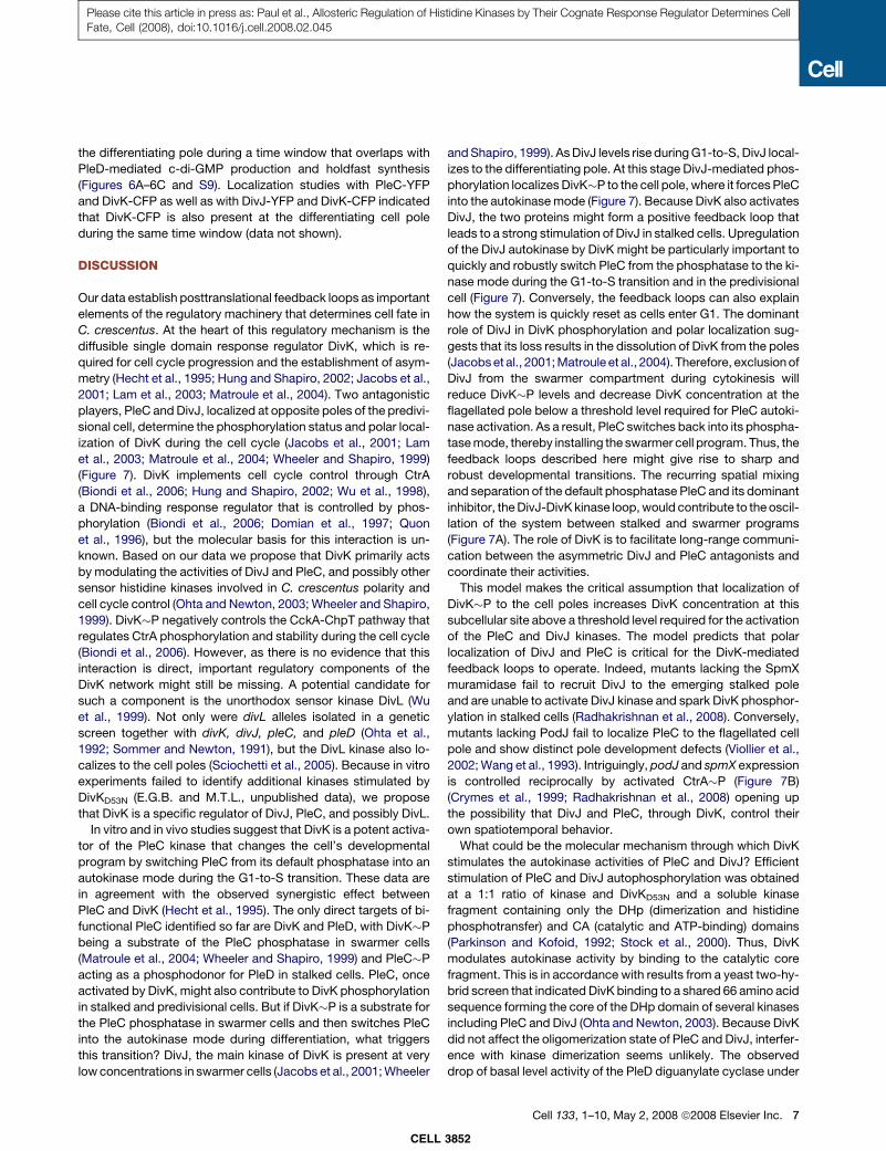
the differentiating pole during a time window that overlaps with
PleD-mediated c-di-GMP production and holdfast synthesis
(Figures 6A–6C and S9). Localization studies with PleC-YFP
and DivK-CFP as well as with DivJ-YFP and DivK-CFP indicated
that DivK-CFP is also present at the differentiating cell pole
during the same time window (data not shown).
DISCUSSION
Our data establish posttranslational feedback loops as important
elements of the regulatory machinery that determines cell fate in
C. crescentus. At the heart of this regulatory mechanism is the
diffusible single domain response regulator DivK, which is re-
quired for cell cycle progression and the establishment of asym-
metry (Hecht et al., 1995; Hung and Shapiro, 2002; Jacobs et al.,
2001; Lam et al., 2003; Matroule et al., 2004). Two antagonistic
players, PleC andDivJ, localized at opposite poles of the predivi-
sional cell, determine the phosphorylation status and polar local-
ization of DivK during the cell cycle (Jacobs et al., 2001; Lam
et al., 2003; Matroule et al., 2004; Wheeler and Shapiro, 1999)
(Figure 7). DivK implements cell cycle control through CtrA
(Biondi et al., 2006; Hung and Shapiro, 2002; Wu et al., 1998),
a DNA-binding response regulator that is controlled by phos-
phorylation (Biondi et al., 2006; Domian et al., 1997; Quon
et al., 1996), but the molecular basis for this interaction is un-
known. Based on our data we propose that DivK primarily acts
by modulating the activities of DivJ and PleC, and possibly other
sensor histidine kinases involved in C. crescentus polarity and
cell cycle control (Ohta and Newton, 2003; Wheeler and Shapiro,
1999). DivKP negatively controls the CckA-ChpT pathway that
regulates CtrA phosphorylation and stability during the cell cycle
(Biondi et al., 2006). However, as there is no evidence that this
interaction is direct, important regulatory components of the
DivK network might still be missing. A potential candidate for
such a component is the unorthodox sensor kinase DivL (Wu
et al., 1999). Not only were divL alleles isolated in a genetic
screen together with divK, divJ, pleC, and pleD (Ohta et al.,
1992; Sommer and Newton, 1991), but the DivL kinase also lo-
calizes to the cell poles (Sciochetti et al., 2005). Because in vitro
experiments failed to identify additional kinases stimulated by
DivKD53N (E.G.B. and M.T.L., unpublished data), we propose
that DivK is a specific regulator of DivJ, PleC, and possibly DivL.
In vitro and in vivo studies suggest that DivK is a potent activa-
tor of the PleC kinase that changes the cell’s developmental
program by switching PleC from its default phosphatase into an
autokinase mode during the G1-to-S transition. These data are
in agreement with the observed synergistic effect between
PleC and DivK (Hecht et al., 1995). The only direct targets of bi-
functional PleC identified so far are DivK and PleD, with DivKP
being a substrate of the PleC phosphatase in swarmer cells
(Matroule et al., 2004; Wheeler and Shapiro, 1999) and PleCP
acting as a phosphodonor for PleD in stalked cells. PleC, once
activated by DivK, might also contribute to DivK phosphorylation
in stalked and predivisional cells. But if DivKP is a substrate for
the PleC phosphatase in swarmer cells and then switches PleC
into the autokinase mode during differentiation, what triggers
this transition? DivJ, the main kinase of DivK is present at very
lowconcentrations in swarmer cells (Jacobs et al., 2001;Wheeler
andShapiro, 1999). AsDivJ levels rise duringG1-to-S, DivJ local-
izes to the differentiating pole. At this stage DivJ-mediated phos-
phorylation localizes DivKP to the cell pole, where it forces PleC
into the autokinasemode (Figure 7). Because DivK also activates
DivJ, the two proteins might form a positive feedback loop that
leads to a strong stimulation of DivJ in stalked cells. Upregulation
of the DivJ autokinase by DivK might be particularly important to
quickly and robustly switch PleC from the phosphatase to the ki-
nase mode during the G1-to-S transition and in the predivisional
cell (Figure 7). Conversely, the feedback loops can also explain
how the system is quickly reset as cells enter G1. The dominant
role of DivJ in DivK phosphorylation and polar localization sug-
gests that its loss results in the dissolution of DivK from the poles
(Jacobset al., 2001;Matroule et al., 2004). Therefore, exclusionof
DivJ from the swarmer compartment during cytokinesis will
reduce DivKP levels and decrease DivK concentration at the
flagellated pole below a threshold level required for PleC autoki-
nase activation. As a result, PleC switches back into its phospha-
tasemode, thereby installing the swarmer cell program. Thus, the
feedback loops described here might give rise to sharp and
robust developmental transitions. The recurring spatial mixing
and separation of the default phosphatase PleC and its dominant
inhibitor, theDivJ-DivK kinase loop,would contribute to theoscil-
lation of the system between stalked and swarmer programs
(Figure 7A). The role of DivK is to facilitate long-range communi-
cation between the asymmetric DivJ and PleC antagonists and
coordinate their activities.
This model makes the critical assumption that localization of
DivKP to the cell poles increases DivK concentration at this
subcellular site above a threshold level required for the activation
of the PleC and DivJ kinases. The model predicts that polar
localization of DivJ and PleC is critical for the DivK-mediated
feedback loops to operate. Indeed, mutants lacking the SpmX
muramidase fail to recruit DivJ to the emerging stalked pole
and are unable to activate DivJ kinase and spark DivK phosphor-
ylation in stalked cells (Radhakrishnan et al., 2008). Conversely,
mutants lacking PodJ fail to localize PleC to the flagellated cell
pole and show distinct pole development defects (Viollier et al.,
2002;Wang et al., 1993). Intriguingly, podJ and spmX expression
is controlled reciprocally by activated CtrAP (Figure 7B)
(Crymes et al., 1999; Radhakrishnan et al., 2008) opening up
the possibility that DivJ and PleC, through DivK, control their
own spatiotemporal behavior.
What could be the molecular mechanism through which DivK
stimulates the autokinase activities of PleC and DivJ? Efficient
stimulation of PleC and DivJ autophosphorylation was obtained
at a 1:1 ratio of kinase and DivKD53N and a soluble kinase
fragment containing only the DHp (dimerization and histidine
phosphotransfer) and CA (catalytic and ATP-binding) domains
(Parkinson and Kofoid, 1992; Stock et al., 2000). Thus, DivK
modulates autokinase activity by binding to the catalytic core
fragment. This is in accordance with results from a yeast two-hy-
brid screen that indicated DivK binding to a shared 66 amino acid
sequence forming the core of the DHp domain of several kinases
including PleC and DivJ (Ohta and Newton, 2003). Because DivK
did not affect the oligomerization state of PleC and DivJ, interfer-
ence with kinase dimerization seems unlikely. The observed
drop of basal level activity of the PleD diguanylate cyclase under
Cell 133, 1–10, May 2, 2008 ª2008 Elsevier Inc. 7
CELL 3852
Please cite this article in press as: Paul et al., Allosteric Regulation of Histidine Kinases by Their Cognate Response Regulator Determines Cell
Fate, Cell (2008), doi:10.1016/j.cell.2008.02.045
conditions where autophosphorylation is absent or inefficient, in-
dicates the formation of a nonproductive ternary complex that
engages PleD in an inactive monomeric form (Paul et al.,
2007). Possibly, DivK and PleD monomers interact with the
same kinase dimer and DivK bound to one kinase protomer
can activate autophosphorylation and phosphotransfer to PleD
via the other protomer. The failure of the DivKD90G mutant to
stimulate PleC kinase activity in vitro and in vivo could result
from an altered interaction with the DHp domain of the kinase.
This view is consistent with the position of Asp90 at the N termi-
nus of the DivK receiver domain helix a4. Because this region
undergoes structural changes upon phosphorylation (Robinson
et al., 2000) and makes specific contacts with the DHp domain
(Zapf et al., 2000) it is a good candidate for the interaction
surface that mediates the phosphatase-kinase switch of PleC.
In conclusion, our results indicate a role for the abundant class
of single domain response regulators in spatially interconnecting
different components of the two-component signal transduction
circuitry. While recent system level approaches indicated that
Figure 7. Model for the Regulation of the PleC-DivJ-
DivK Cell Fate Control System
(A) Schematic of theC. crescentus cell cycle with polar appen-
dices and localization patterns for PleC, DivJ, DivK, DivKP,
PleD, and PleDP. A summary of the posttranslational feed-
back circuitry with the DivJ kinase, the PleC phosphatase,
and the DivK response regulator is indicated.
(B) Diagram of the phosphorylation circuit controlling cell fate
and cellular asymmetry in Caulobacter crescentus. The model
predicts that the sessile-stalked cell program is directed
through polar DivKP interacting with and activating both
PleC and DivJ kinases. Likewise, the motile-swarmer cell pro-
gram results from the absence of the DivJ kinase, dissolution
of DivK from the cell pole, and establishment of the PleC phos-
phatase activity. The two knownmolecular targets of the PleC-
DivJ-DivK cell fate machinery, PleD and CtrA, are indicated.
Stippled lines indicate PleC and DivJ polar localization control
through CtrAP-regulated expression of podJ and spmX,
respectively (Crymes et al., 1999; Radhakrishnan et al., 2008).
a one-to-one relationship between histidine ki-
nases and their cognate response regulators pre-
vents unwanted cross-talk (Skerker et al., 2005;
Yamamoto et al., 2005) our results add a new level
of complexity to this sensory network demonstrat-
ing the possibility of widespread inter-connections
between apparently insolated signaling systems
through retrograde information transfer from
response regulators to cognate kinases.
EXPERIMENTAL PROCEDURES
Strains, Plasmids, and Media
Thebacterial strains andplasmids used in this study are shown
in Table S1. Caulobacter crescentus strains were grown in
peptone yeast extract (PYE), or in minimal glucose media
(M2G, [Ely, 1991]). Newborn SW cells were isolated by Ludox
or Percoll gradient centrifugation (Jenal and Shapiro, 1996),
and released into fresh M2G medium. For conjugal transfer
into C. crescentus, Escherichia coli strain S17-1 was used as
donor. The divKD90G mutation (Sommer and Newton, 1991) was introduced
into CB15DpleC (UJ731) by allelic exchange. To generate the divK null mutant,
the divK::U allele was transduced from strain CJ403 (Lam et al., 2003) into the
C. crescentus ctrA401mutant strain (Quon et al., 1996). The exact procedures
of strain and plasmid construction are available on request. Unless stated oth-
erwise, pooled data consist of at least three independent experiments and are
represented as mean ± standard deviation (SD).
Attachment Assays and Holdfast Staining
For attachment assays, logarithmically growing C. crescentus cells were
diluted 1:30 in PYE, and cultivated for 24 hr in 96-well microtiter plates at 200
rpm on a rocking platform. Attachment of cells to the polystyrene surface
was quantified as described (Levi and Jenal, 2006). For attachment assays
of synchronized populations, cell aliquots were transferred to microtiter plates
at the timepoints indicated, and allowed tobind to the plastic surface for 15min
at room temperature before staining. C. crescentus holdfast was stained with
a mixture of Oregon Green-conjugated wheat-germ agglutinin (0.2 mg/ml)
and Calcofluor White (0.1 mg/ml) and was visualized by fluorescence imaging
on an Olympus AX70 microscope with a Hamamatsu C4742-95 digital camera
(Levi and Jenal, 2006). Images were recorded and processed with Improvision
Openlab and the Photoshop CS v8.0 (Adobe, CA) software packages.
8 Cell 133, 1–10, May 2, 2008 ª2008 Elsevier Inc.
CELL 3852
Please cite this article in press as: Paul et al., Allosteric Regulation of Histidine Kinases by Their Cognate Response Regulator Determines Cell
Fate, Cell (2008), doi:10.1016/j.cell.2008.02.045

Fluorescence Microscopy
For fluorescence imaging bacteria were placed on a microscope slide coated
with 2% agarose dissolved in water. An Olympus IX71 microscope equipped
with an UPlanSApo 1003/1.40 Oil objective (Olympus, Germany) and a cool-
SNAP HQ CCD camera (Photometrics, AZ, United States) were used to take
differential interference contrast (DIC) and fluorescence photomicrographs.
YFP (Ex 500/20 nm, EM 535/30 nm) Rhodamine filter sets (Ex 555/28 nm,
EM 617/73 nm) were used. DIC pictures were taken with 0.15 s and fluores-
cence pictures with 1.0 s exposure time. Images were processed with soft-
WoRx v3.3.6 (Applied Precision, WA) and Photoshop CS v8.0 softwares.
In Vivo Phosphorylation
In vivo phosphorylation experiments were performed as described previously
(Domian et al., 1997; Jacobs et al., 2001) with the following modifications. The
strains were grown in M5G minimal medium supplemented with 1 mM gluta-
mate until an optical density at 660 nm of 0.15, collected by centrifugation
and resuspended in filtered culture medium to an optical density of 0.3. Cells
were labeled at 30C for 5 min with 100 mCi [32P]H3PO4 or 30 mCi [32P]ATP, and
after lysis immunoprecipitated with 10 ml anti-PleC, anti-DivK, or anti-DivJ se-
rum. Radiolabeled proteins were separated on a 10% polyacryamide gel and
visualized with a phosphoimager (Molecular Dynamics, GE Healthcare, NJ).
Nucleotide Analysis
For the analysis of c-di-GMP levels in synchronized C. crescentus cultures
(OD660 0.4), nucleotides were extracted with 1M formic acid. Lyophilized sam-
ples were analyzed with a 125/4 Nucleosil 4000-1 PEI column (Macherey-Na-
gel, Germany) on SMART- (GE Healthcare, NJ) or Pro Star HPLC-Systems
(Varian, CA). The nucleotides were applied to the column dissolved in buffer
A (7 mM KH2PO4 [pH 4]), and eluted with a gradient of buffer B (0.5 M
KH2PO4, 1 M Na2SO4 [pH 5.5]) at a flow rate of 50 ml/min. Concentrations
were determined by comparison with a standard of chemically synthesized
c-di-GMP.
Expression and Purification of Proteins
E. coli cells carrying the respective expression plasmid were grown in LB me-
dium with ampicillin (100 mg/ml), and expression was induced by adding either
arabinose (final concentration of 0.2%) or IPTG (final concentration of 0.4 mM).
After harvesting by centrifugation, the cells were resuspended in TN-buffer
(50 mM Tris-HCl [pH 8.0], 500 mM NaCl, 5 mM b-mercaptoethanol), lysed
by passage through a French pressure cell, and clarified by centrifugation.
The supernatant was loaded onto Ni-NTA affinity resin (QIAGEN, Germany),
washedwith TN-buffer, and elutedwith an imidazol-gradient. PleD, DivK, a cat-
alytic fragment of DivJ‘ (C-terminal 296 amino acids), and a catalytic PleC‘
fragment containing the complete cytoplasmic domains (540 amino acids)
remained soluble and were purified under native conditions. Two additional
catalytic PleC‘ fragments (containing the C-terminal 313 or 489 amino acids,
respectively) were solubilized from inclusion bodies and renatured after purifi-
cation (Hecht et al., 1995; Paul et al., 2004). Proteins were examined for purity
by SDS-PAGE and fractions containing pure protein were pooled and dialyzed.
DivKwas concentrated by precipitationwith 60%saturated ammonium sulfate
(pH 7.0) prior to dialysis; the kinase fragments and PleD were concentrated
using Amicon Ultra- or Microcon- Centrifugal Filter Units (Millipore, MA). Ana-
lytical size exclusion chromatographywas performedwith a Superdex 200 col-
umn on an AKTApurifier (GE Healthcare) system at a flow rate of 0.5 ml/min.
Concentrations and molecular weights of proteins were determined by an
online refractometer (Optilab rEX, Wyatt Technology) and a miniDAWN light
scattering instrumentation (Wyatt Technology, CA). Untagged DivK for in vitro
phosphatase assays was obtained by cleaving His-DivK with Thrombin (Nova-
gen, WI, United States) for 1 hr at 25C, followed by preparative size exclusion
chromatography with a Superdex 75 column on an AKTApurifier (GE Health-
care, NJ) at a flow rate of 1.5 ml/min.
Enzymatic Assays
Diguanylate cyclase assays were adapted from procedures described previ-
ously (Paul et al., 2007, 2004). The standard diguanylate cyclase reaction
mixtures contained 50 mM Tris-HCl (pH 7.8), 250 mM NaCl, 10 mM MgCl2 in
50 ml volume and was started by the addition of a mixture of 100 mM
GTP/[a-33P]GTP (PerkinElmer; 0.01 mCi/ml). All kinase reactions were supple-
mented with 25 mM KCl. To calculate the initial velocity of product formation,
aliquots were withdrawn at regular time intervals and the reaction was stopped
with an equal volume of 50 mM EDTA (pH 6.0). Reaction products (2 ml) were
separated on polyethyleneimine-cellulose plates (Macherey-Nagel) in 1.5 M
KH2PO4/5.5M (NH4)2SO4 (pH 3.5) in a 2:1 ratio. Plateswere exposed to a phos-
phorimager screen, and the intensity of the various radioactive species was
calculated by quantifying the intensities of the relevant spots using the Image-
Quant software (Molecular Dynamics). Measurements were always restricted
to the linear range of product formation. In vitro kinase and phosphatase as-
says were performed as described earlier (Hecht et al., 1995; Matroule et al.,
2004; Paul et al., 2004; Skerker et al., 2005). For kinase assays the proteins
were incubated at 25C for 15 min in phosphorylation buffer (50 mM Tris-
HCl at pH 7.8, 25 mM NaCl, 25 mM KCl, 5 mM MgCl2) containing 5 mCi
[g-32P]ATP (Amersham Biosciences, GE Healthcare, NJ), unless stated other-
wise. Reactions were stopped with 43 SDS-PAGE sample buffer (250 mM
Tris-HCl at pH 6.8, 40% glycerol, 8% SDS, 2.4 M b-mercaptoethanol,
0.06% bromophenol blue, 40 mM EDTA), and 32P-labeled proteins were
separated by electrophoresis on 10% SDS-PAGE gels followed by autoradi-
ography on a phosphor-imager screen. DivK-P for phosphatase assays was
purified from contaminating His-tagged PleC and DivKD53N by two rounds of
batch purification with Ni-NTA resin (QIAGEN, Germany), followed by gel filtra-
tion for the removal of ATP.
SUPPLEMENTAL DATA
Supplemental Data include nine figures, one table, and Supplemental Refer-
ences and can be found with this article online at http://www.cell.com/cgi/
content/full/133/3/---/DC1/.
ACKNOWLEDGMENTS
We thank all members of the Jenal group for valuable discussions and Arnaud
Basle, Assaf Levi, Dietrich Samurai, and Paul Wassmann for technical assis-
tance. We are grateful to Y. Brun, T. Costa, C. Jacobs-Wagner, J.-Y. Matroule,
A. Newton, N. Ohta, D. Pierce, and P. Viollier for providing mutant strains,
plasmids, and antisera. This work was supported by Swiss National Science
Foundation Fellowship 3100A0-108186 to U.J.
Received: August 14, 2007
Revised: December 21, 2007
Accepted: February 11, 2008
Published: May 1, 2008
REFERENCES
Aldridge, P., and Jenal, U. (1999). Cell cycle-dependent degradation of a flagel-
lar motor component requires a novel-type response regulator. Mol. Microbiol.
32, 379–391.
Aldridge, P., Paul, R., Goymer, P., Rainey, P., and Jenal, U. (2003). Role of the
GGDEF regulator PleD in polar development of Caulobacter crescentus. Mol.
Microbiol. 47, 1695–1708.
Betschinger, J., and Knoblich, J.A. (2004). Dare to be different: asymmetric cell
division in Drosophila, C. elegans and vertebrates. Curr. Biol. 14, R674–R685.
Biondi, E.G., Reisinger, S.J., Skerker, J.M., Arif, M., Perchuk, B.S., Ryan, K.R.,
and Laub, M.T. (2006). Regulation of the bacterial cell cycle by an integrated
genetic circuit. Nature 444, 899–904.
Crymes,W.B., Jr., Zhang, D., and Ely, B. (1999). Regulation of podJ expression
during the Caulobacter crescentus cell cycle. J. Bacteriol. 181, 3967–3973.
Domian, I.J., Quon, K.C., and Shapiro, L. (1997). Cell type-specific phosphor-
ylation and proteolysis of a transcriptional regulator controls the G1-to-S tran-
sition in a bacterial cell cycle. Cell 90, 415–424.
Ely, B. (1991). Genetics of Caulobacter crescentus. Methods Enzymol. 204,
372–384.
Cell 133, 1–10, May 2, 2008 ª2008 Elsevier Inc. 9
CELL 3852
Please cite this article in press as: Paul et al., Allosteric Regulation of Histidine Kinases by Their Cognate Response Regulator Determines Cell
Fate, Cell (2008), doi:10.1016/j.cell.2008.02.045
Hecht, G.B., Lane, T., Ohta, N., Sommer, J.M., and Newton, A. (1995). An es-
sential single domain response regulator required for normal cell division and
differentiation in Caulobacter crescentus. EMBO J. 14, 3915–3924.
Hung, D.Y., and Shapiro, L. (2002). A signal transduction protein cues proteo-
lytic events critical to Caulobacter cell cycle progression. Proc. Natl. Acad. Sci.
USA 99, 13160–13165.
Jacobs, C., Hung, D., and Shapiro, L. (2001). Dynamic localization of a cyto-
plasmic signal transduction response regulator controls morphogenesis dur-
ing the Caulobacter cell cycle. Proc. Natl. Acad. Sci. USA 98, 4095–4100.
Jenal, U., and Shapiro, L. (1996). Cell cycle-controlled proteolysis of a flagellar
motor protein that is asymmetrically distributed in the Caulobacter predivi-
sional cell. EMBO J. 15, 2393–2406.
Lam, H., Matroule, J.Y., and Jacobs-Wagner, C. (2003). The asymmetric spa-
tial distribution of bacterial signal transduction proteins coordinates cell cycle
events. Dev. Cell 5, 149–159.
Levi, A., and Jenal, U. (2006). Holdfast formation in motile Swarmer cells opti-
mizes surface attachment during Caulobacter crescentus development. J.
Bacteriol. 188, 5315–5318.
Matroule, J.Y., Lam, H., Burnette, D.T., and Jacobs-Wagner, C. (2004). Cyto-
kinesismonitoring during development; rapid pole-to-pole shuttling of a signal-
ing protein by localized kinase and phosphatase in Caulobacter. Cell 118, 579–
590.
McAdams, H.H., and Shapiro, L. (2003). A bacterial cell-cycle regulatory net-
work operating in time and space. Science 301, 1874–1877.
Ohta, N., Lane, T., Ninfa, E.G., Sommer, J.M., and Newton, A. (1992). A histi-
dine protein kinase homologue required for regulation of bacterial cell division
and differentiation. Proc. Natl. Acad. Sci. USA 89, 10297–10301.
Ohta, N., and Newton, A. (2003). The core dimerization domains of histidine ki-
nases contain recognition specificity for the cognate response regulator. J.
Bacteriol. 185, 4424–4431.
Parkinson, J.S., and Kofoid, E.C. (1992). Communication modules in bacterial
signaling proteins. Annu. Rev. Genet. 26, 71–112.
Paul, R., Abel, S., Wassmann, P., Beck, A., Heerklotz, H., and Jenal, U. (2007).
Activation of the diguanylate cyclase pleD by phosphorylation-mediated di-
merization. J. Biol. Chem. 282, 29170–29177.
Paul, R., Weiser, S., Amiot, N.C., Chan, C., Schirmer, T., Giese, B., and Jenal,
U. (2004). Cell cycle-dependent dynamic localization of a bacterial response
regulator with a novel di-guanylate cyclase output domain. Genes Dev. 18,
715–727.
Quon, K.C., Marczynski, G.T., and Shapiro, L. (1996). Cell cycle control by an
essential bacterial two-component signal transduction protein. Cell 84, 83–93.
Quon, K.C., Yang, B., Domian, I.J., Shapiro, L., and Marczynski, G.T. (1998).
Negative control of bacterial DNA replication by a cell cycle regulatory protein
that binds at the chromosome origin. Proc. Natl. Acad. Sci. USA 95, 120–125.
Radhakrishnan, S.K., Thanbichler, M., and Viollier, P.H. (2008). The dynamic
interplay between a cell fate determinant and a lysozyme homolog drives
the asymmetric division cycle of Caulobacter crescentus. Genes Dev. 22,
212–225.
Robinson, V.L., Buckler, D.R., and Stock, A.M. (2000). A tale of two compo-
nents: a novel kinase and a regulatory switch. Nat. Struct. Biol. 7, 626–633.
Sciochetti, S.A., Ohta, N., and Newton, A. (2005). The role of polar localization
in the function of an essential Caulobacter crescentus tyrosine kinase. Mol.
Microbiol. 56, 1467–1480.
Skerker, J.M., Prasol, M.S., Perchuk, B.S., Biondi, E.G., and Laub, M.T. (2005).
Two-component signal transduction pathways regulating growth and cell cy-
cle progression in a bacterium: a system-level analysis. PLoS Biol. 3, e334. 10.
1371/journal.pbio.0030334.
Sommer, J.M., andNewton, A. (1991). Pseudoreversion analysis indicates a di-
rect role of cell division genes in polar morphogenesis and differentiation in
Caulobacter crescentus. Genetics 129, 623–630.
Stock, A.M., Robinson, V.L., and Goudreau, P.N. (2000). Two-component sig-
nal transduction. Annu. Rev. Biochem. 69, 183–215.
Viollier, P.H., Sternheim, N., and Shapiro, L. (2002). Identification of a localiza-
tion factor for the polar positioning of bacterial structural and regulatory pro-
teins. Proc. Natl. Acad. Sci. USA 99, 13831–13836.
Wang, S.P., Sharma, P.L., Schoenlein, P.V., and Ely, B. (1993). A histidine
protein kinase is involved in polar organelle development in Caulobacter cres-
centus. Proc. Natl. Acad. Sci. USA 90, 630–634.
Wassmann, P., Chan, C., Paul, R., Beck, A., Heerklotz, H., Jenal, U., and
Schirmer, T. (2007). Structure of BeF(3)(-)-Modified Response Regulator
PleD: Implications for Diguanylate Cyclase Activation, Catalysis, and Feed-
back Inhibition. Structure 15, 915–927.
Wheeler, R.T., and Shapiro, L. (1999). Differential localization of two histidine
kinases controlling bacterial cell differentiation. Mol. Cell 4, 683–694.
Wu, J., Ohta, N., and Newton, A. (1998). An essential, multicomponent signal
transduction pathway required for cell cycle regulation in Caulobacter. Proc.
Natl. Acad. Sci. USA 95, 1443–1448.
Wu, J., Ohta, N., Zhao, J.L., and Newton, A. (1999). A novel bacterial tyrosine
kinase essential for cell division and differentiation. Proc. Natl. Acad. Sci. USA
96, 13068–13073.
Yamamoto, K., Hirao, K., Oshima, T., Aiba, H., Utsumi, R., and Ishihama, A.
(2005). Functional characterization in vitro of all two-component signal trans-
duction systems from Escherichia coli. J. Biol. Chem. 280, 1448–1456.
Zapf, J., Sen, U., Madhusudan, Hoch, J.A., and Varughese, K.I. (2000). A tran-
sient interaction between two phosphorelay proteins trapped in a crystal
lattice reveals the mechanism of molecular recognition and phosphotransfer
in signal transduction. Structure 8, 851–862.
10 Cell 133, 1–10, May 2, 2008 ª2008 Elsevier Inc.
CELL 3852
Please cite this article in press as: Paul et al., Allosteric Regulation of Histidine Kinases by Their Cognate Response Regulator Determines Cell
Fate, Cell (2008), doi:10.1016/j.cell.2008.02.045

Supplementary material
Figure S1: DivK Stimulates Autophosphorylation of the PleC and DivJ Histidine
Kinases.
(A) Quantification of PleC autophosphorylation as shown in Fig. 2A with the
following concentrations of DivKD53N, 0 µM (open circles), 0.3 µM (open squares),
10 µM (filled triangles), 30 µM (filled squares), or 10 µM DivKD53ND90G (open

triangles). Values are averages from three experiments and are normalized to the level
of PleC autophosphorylation in the absence of DivKD53N at time point 15 min.
(B) DivK does not affect stability of PleC~P. PleC was phosphorylated in the
presence of [γ-32P]ATP, purified, and PleC~P levels were measured after incubation
with or without DivKD53N.
(C) Quantification of DivJ autophosphorylation as shown in Fig. 4A without
additional proteins (open circles), in the presence of DivKD53N (30 µM; closed
squares), of DivKD53ND90G (30 µM; open squares), or of PleDD53N (30 µM, diamonds).
Values were normalized to the phosphorylation levels at 15 min in the absence of
additional protein. The data are averages of two experiments.
(C) DivK does not affect stability of DivJ~P. DivJ was phosphorylated in the presence
of [γ-32P]ATP, purified, and DivJ~P levels were measured after incubation with or
without DivKD53N.
Figure S2: PleC-Mediated Dimerization of PleD Requires DivK.
Crosslink assays with PleD, PleC, and DivKD53N were carried out as described in
(Paul et al., 2007). 20 µM of the purified proteins were incubated with ATP and cross-
linked for 0, 1, and 5 minutes. PleD monomers and dimers were separated on SDS-
polyacrylamide gels, transferred to PVDF-membranes, and detected by immunoblot
analysis with an anti-PleD antibody. Arrows mark the monomeric and dimeric forms
of PleD.

Figure S3: Quarternary Structure and Activity of PleC in the Presence or Absence of
DivK.
(A) Size exclusion chromatography of PleC pre-incubated in the presence and
absence of DivK. Multi-angle light scattering (MALS) was used to measure the

molecular weight of purified proteins. Measured values are indicated with theoretical
values shown in brackets. PleCM refers to the peak corresponding to monomeric PleC,
PleCD marks the peak corresponding to a PleC dimer. The distribution of PleC
monomer and dimer fraction was not affected by preincubation with DivK.
(B) Measurement of kinase activity of purified PleCM from (A) in the presence or
absence of DivK.
(C) Measurement of kinase activity of purified PleCD from (A) in the presence or
absence of DivK.
Figure S4: Quarternary Structure and Activity of DivJ in the Presence or Absence of
DivK.

(A) Size exclusion chromatography of DivJ pre-incubated in the presence and absence
of DivK. Multi-angle light scattering (MALS) was used to measure the molecular
weight of purified proteins. Measured values are indicated with theoretical values
shown in brackets. DivJ behaved as stable dimer under all conditions tested.
(B) Measurement of kinase activity of purified DivJ from (A) in the presence or
absence of DivK.
Figure S5: Phosphotransfer from PleC to DivK and DivKD90G.
Phosphotransfer reactions with PleC (10 µM), DivK (10 µM), DivKD90G (10 µM),
DivKD53N (10 µM), and DivKD53ND90G (10 µM). The bands corresponding to the
phosphorylated proteins are indicated.

Figure S6: DivKD90G Does not Affect PleC Phosphatase Activity.
(A) PleC-mediated dephosphorylation of DivK. Purified DivK~P was incubated alone
(top row), with PleC (second row), with DivKD53N and PleC (third row), or with
DivKD53ND90G and PleC (bottom row) for the time indicated. Quantification of PleC-
mediated DivK dephosphorylation is indicated in the panel below.

Figure S7: PleC Wild Type and PleCF778L Have Similar Phosphatase Activities in
Vivo.
Levels of DivK~P were determined in synchronized swarmer cells of ∆pleC (∆C)
containing a plasmid copy of pleC wild type (pCWT) or pleCF778L (pCF778L). Cells were
released into fresh medium, pulse labeled at the indicated time points, and extracts
were used for immunoprecipitation experiments with anti-DivK antibody.
Figure S8: PleD-Dependent Pole Morphogenesis Requires the PleC Kinase.
(A) PleC kinase affects motility in a PleD-dependent manner. The relative motility of
the following strains was determined by comparing colony size on semi-solid agar
plates after 3 days incubation at 30ºC: CB15N, ∆pleC (∆C), and ∆pleC ∆pleD (∆CD).
As indicated the strains contained an empty vector (pMR) or a plasmid copy of the
following pleC alleles: pleC wild type (pCWT), pleCF778L (pCF778L), or pleCT614R
(pCT614R). Error bars represent the mean +/- standard deviation.
(B) PleC kinase activity is required for PleD-dependent stalk biogenesis. The
percentage of predivisional cells carrying a stalk was determined for the strains
indicated by light microscopy. Strain and plasmid designations are as outlined in (A).

Figure S9: Polar Localization of DivJ and PleC during the C. crescentus Cell Cycle.
(A) Quantification of PleC-mYFP and DivJ-tDimer2 localization to the cell poles
(Matroule et al., 2004; Ohta and Newton, 2003; Wheeler and Shapiro, 1999).
Synchronized populations of cells were examined by epi-fluorescence microscopy at
the indicated time points and scored visually. Polar foci of PleC-mYFP (open circles)
and DivJ-tDimer2 (closed circles) are indicated. The numbers of cells with PleC-
mYFP and DivJ-tDimer2 foci at the same (open squares) or opposite poles (open
diamonds) are shown. Error bars represent the mean +/- standard deviation.
(B) Immunoblot analysis of synchronized cell populations using anti-CtrA, anti-DivJ,
and anti-PleC antibodies.

Table S1: Strains and Plasmids
Strain or
Plasmid Relevant genotype or description Reference or source
Caulobacter crescentus strains
CB15 C. crescentus wild type ATCC19089
CB15N synchronizable variant strain of CB15 (Evinger and Agabian, 1977)
UJ730 CB15 ∆ pleD (Levi and Jenal, 2006)
UJ731 CB15 ∆pleC (Levi and Jenal, 2006)
UJ1983 CB15N divKD90G (Hung and Shapiro, 2002)
UJ3165 CB15N ∆pleC + pCWT This study
UJ3166 CB15N ∆pleC + pCF778L This study
UJ3167 CB15N ∆pleC + pCT614R This study
UJ3168 CB15N ∆pleC ∆pleD + pCWT This study
UJ3169 CB15N ∆pleC ∆pleD + pCF778L This study
UJ3170 CB15N ∆pleC ∆pleD + pCT614R This study
UJ3190 CB15 ∆pleC + pCWT This study
UJ3191 CB15 ∆pleC and pCF778L This study
UJ3192 CB15 ∆pleC and pCT614R This study
UJ3234 CB15 ∆pleC ∆pleD This study
UJ3528 CB15 ∆pleC ∆pleD + pCWT This study
UJ3529 CB15 ∆pleC ∆pleD + pCF778L This study
UJ3530 CB15 ∆pleC ∆pleD + pCT614R This study
UJ3572 CB15 + pKD53N This study
UJ3578 CB15 ∆pleC + pKD53N This study
UJ3579 CB15 ∆pleD + pKD53N This study
UJ3611 CB15 ∆pleC divKD90G This study
UJ3629 CB15 ∆pleC divKD90G + pCWT This study
UJ3631 CB15 ∆pleC divKD90G + pCT614R This study
UJ3905 CB15 ∆pleC ∆pleD divKD90G + pCWT This study
UJ4507 pBGentdivJ-tdimer2,pleC-mYFP in NA1000 (Matroule et al., 2004)
UJ4518 NA1000 ctrA401 divK::Ω This study
Escherichia coli strains
BL21(DE3) F- dcm ompT hsdS(rB-mB-) Stratagene
(pLysS) gal λ (DE3) [pLysS CAMr]
BL21 RIL F− ompT hsdS(rB-mB-) dcm+ Tetr gal Stratagene
(codonPlus) λ (DE3) endA Hte [argU ileY leuW Camr]

DH10B F- mcrA ∆(mrr- hsd RMS- mcrBC) φ80dlacZ∆M15 Stratagene
∆lacX74 endA1 recA1 deoR ∆(ara, leu)7697 araD139
galU galK nupG rpsL thi pro hsdR- hsd+ recA
RP4-2-Tc::Mu-Tn7
S17-1 RP4-2, Tc::Mu, KM-Tn7 (Simon et al., 1983)
Plasmid
pMR20 TetR low copy number vector (Roberts et al., 1996)
pBAD AmpR expression plasmid Invitrogen
pCC2 pET11; pleD, C-terminal His6 tag (Paul et al., 2004)
pET11 AmpR expression plasmid Stratagene
pET28 AmpR expression plasmid Stratagene
pET28H6-K DivK -His tag with thrombin site (Matroule et al., 2004)
pJS14 pBBR1-derived medium copy number J. Skerker
broad host range vector
pCWT pJS14 carrying pleC (Matroule et al., 2004)
pCF778L pJS14 carrying pleCF778L (Matroule et al., 2004)
pCT614R pJS14 carrying pleCT614R (Matroule et al., 2004)
pPA89 pMR20 carrying divKD53N (Aldridge et al., 2003)
pRP49 pBAD; pleC489, N-terminal His6 tag (Paul et al., 2004)
pRP63 pBAD; divJ296, N-terminal His6 tag (Paul et al., 2004)
pRP87 pET11; pleDD53N, C-terminal His6 tag (Paul et al., 2004)
pRP111 pET11; divK, C-terminal His6 tag This study
pRP112 pET11; divKD53N, C-terminal His6 tag This study
pRP122 pBAD; pleC313, N-terminal His6 tag This study
pRP123 pET11; divKD53N D90G This study
pRP124 pNPT138S; divKD90G This study
pTRX-HIS-
CC2482 pleC540, His6 and Trx tags (Skerker et al., 2005)
REFERENCES:
Aldridge, P., Paul, R., Goymer, P., Rainey, P., and Jenal, U. (2003). Role of the
GGDEF regulator PleD in polar development of Caulobacter crescentus. Mol
Microbiol 47, 1695-1708.
Evinger, M., and Agabian, N. (1977). Envelope-associated nucleoid from Caulobacter
crescentus stalked and swarmer cells. J Bacteriol 132, 294-301.
Hung, D. Y., and Shapiro, L. (2002). A signal transduction protein cues proteolytic
events critical to Caulobacter cell cycle progression. Proc Natl Acad Sci U S A 99,
13160-13165.
Levi, A., and Jenal, U. (2006). Holdfast formation in motile Swarmer cells optimizes
surface attachment during Caulobacter crescentus development. J Bacteriol 188,
5315-5318.
Matroule, J. Y., Lam, H., Burnette, D. T., and Jacobs-Wagner, C. (2004). Cytokinesis
monitoring during development; rapid pole-to-pole shuttling of a signaling protein by
localized kinase and phosphatase in Caulobacter. Cell 118, 579-590.
Ohta, N., and Newton, A. (2003). The core dimerization domains of histidine kinases
contain recognition specificity for the cognate response regulator. J Bacteriol 185,
4424-4431.
Paul, R., Abel, S., Wassmann, P., Beck, A., Heerklotz, H., and Jenal, U. (2007).
Activation of the diguanylate cyclase pleD by phosphorylation-mediated
dimerization. J Biol Chem.
Paul, R., Weiser, S., Amiot, N. C., Chan, C., Schirmer, T., Giese, B., and Jenal, U.
(2004). Cell cycle-dependent dynamic localization of a bacterial response regulator
with a novel di-guanylate cyclase output domain. Genes Dev 18, 715-727.
Roberts, R. C., Toochinda, C., Avedissian, M., Baldini, R. L., Gomes, S. L., and
Shapiro, L. (1996). Identification of a Caulobacter crescentus operon encoding hrcA,
involved in negatively regulating heat-inducible transcription, and the chaperone gene
grpE. J Bacteriol 178, 1829-1841.
Simon, R., Prieffer, U., and Puhler, A. (1983). A broad host range mobilization
system for in vivo genetic engineering: Transposon mutagenesis in gram negative
bacteria. Biotechnology 1, 784-790.

Skerker, J. M., Prasol, M. S., Perchuk, B. S., Biondi, E. G., and Laub, M. T. (2005).
Two-component signal transduction pathways regulating growth and cell cycle
progression in a bacterium: a system-level analysis. PLoS Biol 3, e334.
Wheeler, R., and Shapiro, L. (1999). Differential localization of two histidine kinases
controlling bacterial cell differentiation. Molecular Cell 4, 683- 694.

Results V Spatiotemporal control of proteolysis by PopA
165
3.5 Second messenger mediated spatiotemporal control of protein degradation during the bacterial cell cycle
Anna Dürig, Marc Folcher, Sören Abel, Torsten Schwede, Nicolas Amiot, Bernd Giese,
Urs Jenal
Genes and Development, manuscript submitted
Statement of my work
I contributed to this work by performing the fluorescence resonance energy transfer (FRET)
based in vivo interaction study between PopA and RcdA (Fig. 3D).

Results V Spatiotemporal control of proteolysis by PopA
166
Second messenger mediated spatiotemporal control of protein degradation
during the bacterial cell cycle
Anna Dürig, Marc Folcher, Sören Abel, Torsten Schwede, Nicolas Amiot1, Bernd Giese1, and
Urs Jenal2
Biozentrum, University of Basel, Klingelbergstrasse 70, 4056 Basel, Switzerland, 1Department of Chemistry, University of Basel, St. Johanns-Ring 19, 4056 Basel, Switzerland
2For correspondence: Phone: ++41 (0)61 267 2135; Fax: ++41 (0)61 267 2118; e-mail:
Short title: c-di-GMP mediated protein degradation control
Key Words: PopA, cyclic di-GMP, CtrA, protein degradation, Caulobacter crescentus, cell cycle, polar
localization, second messenger

Results V Spatiotemporal control of proteolysis by PopA
167
Summary
Second messengers control a wide range of important cellular functions in eukaryotes and
prokaryotes. Here we show that cyclic di-GMP, a global bacterial second messenger promotes
cell cycle progression in Caulobacter crescentus by mediating the degradation of the
replication initiation inhibitor CtrA. During the G1-to-S phase transition both CtrA and its
cognate protease ClpXP dynamically localise to the old cell pole where CtrA is degraded.
Sequestration of CtrA to the cell pole depends on PopA, a newly identified c-di-GMP effector
protein. PopA itself dynamically localises to cell pole and directs CtrA to this subcellular site
via interaction with the RcdA adaptor protein. PopA mutants that are unable to bind c-di-GMP
fail to sequester to the cell pole and, as a consequence, to promote CtrA degradation. Thus, c-
di-GMP facilitates CtrA degradation during the cell cycle by controlling the dynamic
sequestration of the PopA recruitment factor to the cell pole. Finally, we present evidence that
CtrA degradation and G1-to-S cell cycle progression rely on converging pathways responsible
for substrate and protease localization to the old cell pole. This is the first report that links c-
di-GMP to protein dynamics and cell cycle control in bacteria.

Results V Spatiotemporal control of proteolysis by PopA
168
Introduction
Regulated proteolysis has a major impact on cellular physiology as it plays a primordial role
in cell cycle control, stress response, and cell differentiation in both pro- and eukaryotes. To
avoid unwanted protein destruction eukaryotic cells largely restrict proteolysis to specific
cellular compartments. In bacteria several energy-dependent cytoplasmic proteases and their
associated factors are responsible for the rapid degradation of a number of key cellular
regulators [260, 261]. These so called self-compartmentalizing proteases select their substrates
through an ATPase complex, which gates the access to the proteolytic active site. To
specifically select target proteins destined for degradation bacteria have evolved a series of
regulatory mechanisms, including trans-translation [262], interference of small effector
molecules [263], pre-processing [264, 265], protein association [266, 267], or the use of
specific targeting factors [268]. The recent observation that in Caulobacter crescentus the
master cell cycle regulator CtrA dynamically sequesters to the old cell pole, where it is
degraded by the polarly localised ClpXP protease complex, suggested that a spatial
concurrence might also play a role in protein degradation control in bacteria [184, 269].
In C. crescentus protein degradation plays a significant role in controlling cell cycle
progression [109, 270]. Caulobacter cells divide asymmetrically to produce two distinct
daughter cells, a smaller motile swarmer cell and a larger surface adherent stalked cell.
Whereas the newborn stalked cell enters S-phase and reinitiates chromosome replication
immediately, the chromosome of the swarmer cell remains quiescent for an extended period,
equivalent to the G1-phase of eukaryotic cells. Concurrent with the morphological
transformation of the swarmer cell into a stalked cell, the replication block is suspended and
cells proceed into S-phase. Differential activity of the essential response regulator CtrA is
critical to control the Caulobacter G1-to-S phase transition. Phosphorylated CtrA, CtrA~P,
blocks the initiation of replication by directly binding to five sites in the chromosomal OriC
region where it apparently restricts access of replication initiation factors [106]. The activity
of CtrA is redundantly controlled at the levels of expression, phosphorylation, and degradation
[109, 271]. Importantly, to initiate chromosome replication activated CtrA~P is eliminated
from the cell by two redundant mechanisms, temporally controlled dephosphorylation and
proteolysis [109].
In vivo and in vitro experiments have demonstrated that the essential ClpXP protease
complex degrades CtrA during G1-to-S transition [108, 272]. The observation that ClpXP
rapidly degrades CtrA in vitro without the requirement for additional stimulatory factors

Results V Spatiotemporal control of proteolysis by PopA
169
indicated that control of CtrA degradation might involve an inhibitory mechanism [272].
Moreover, recent findings suggested that cell cycle-dependent degradation of CtrA involves
spatial control. Intriguingly, the ClpXP protease complex transiently sequesters to the
incipient stalked cell pole during the G1-to-S transition [184]. At the same time CtrA
transiently localises to the same pole where it is degraded by ClpXP (Fig. 1A) [111, 184].
Two distinct factors, which themselves sequester to the stalked cell pole, are responsible for
the dynamic localization of the protease and its substrate. RcdA, a protein that interacts with
CtrA in vivo helps to localise CtrA to the pole [184]. Similarly, polarly localised CpdR tags
ClpXP to the incipient stalked cell pole [269]. CpdR is a member of the response regulator
family of two-component signal transduction systems that lacks a dedicated output domain
and consists of a receiver domain module with a conserved Asp51 phosphoryl acceptor
residue. Phosphorylation controls CpdR localization to the cell pole and by that the cellular
dynamics of ClpXP [140, 269]. The observation that phosphorylation and localization of
CpdR inversely correlate during the cell cycle together with the finding that a CpdRD51A
mutant almost exclusively localises to the pole, suggested that CpdR sequestration and ClpXP
recruitment are negatively controlled by phosphorylation [269]. Strikingly, phosphorylation of
CtrA and CdpR are catalyzed by the same cell cycle phosphorelay comprising the CckA
sensor kinase and the ChpT phosphotransferase [140]. Under conditions where the CckA-
ChpT pathway is active, CtrA is activated by phosphorylation and concomitantly appears to
be stabilized through the phosphorylation of CdpR and delocalization of ClpXP. Inversely,
CckA downregulation would prevent the phosphorylation of CtrA and CpdR and, as a
consequence, would lead to ClpXP localization and CtrA degradation [140].
These studies suggested that the timing of CtrA degradation during the G1-to-S
transition is intimately linked to its dynamic localization to the cell pole. But what are the
molecular mechanisms that mediate RcdA and CtrA localization to this subcellular site and
how is this event temporally controlled during the cell cycle? Here we propose that the second
messenger c-di-GMP critically contributes to temporal and spatial control of CtrA degradation
during the C. crescentus cell cycle. C-di-GMP has recently been recognized as ubiquitous
second messenger in bacteria controlling the transition between a motile, single-cell state and
a sessile, surface-attached biofilm mode in a wide range of organisms [8, 10]. Two opposing
enzyme activities, diguanylate cyclase (DGC) and phosphodiesterase (PDE) control the
cellular level of c-di-GMP. The DGC and PDE activities are contained within the highly
conserved GGDEF and EAL domains, respectively [14, 20]. GGDEF and EAL domains are
often associated with sensory input domains and it is assumed that these regulatory proteins

Results V Spatiotemporal control of proteolysis by PopA
170
serve to directly couple environmental or internal stimuli to a specific cellular response
through the synthesis or degradation of c-di-GMP. But how these two enzyme classes are
regulated is still largely unclear. DGCs are activated through dimerisation of two GGDEF
protomers [17, 186]. In addition, many DGCs are tightly controlled by product inhibition
through the binding of c-di-GMP to an allosteric I-site, which is distinct from the catalytic
active A-site [16, 19].
We have recently shown that C. crescentus pole morphogenesis during the swarmer-to-
stalked cell transition is controlled by the DGC PleD [14, 47, 61]. PleD is an unorthodox
member of the response regulator family of two-component signal transduction systems with
two receiver domains arranged in tandem fused to a GGDEF output domain [16]. During
development PleD is activated by phosphorylation and in response is sequestered to the
differentiating pole [14, 186]. The observation that phosphorylation-mediated dimerisation not
only leads to DGC activation but also to PleD polar localization, suggested a coupling of these
two events and a spatially confinement of PleD mediated c-di-GMP signaling to the old cell
pole [14, 186]. Here we have analysed the role of PopA, a PleD paralog with identical Rec1-
Rec2-GGDEF domain structure, in C. crescentus development and cell cycle progression.
Similar to PleD, PopA is sequestered to the old cell pole. But in contrast to PleD, PopA
localization does not require phosphorylation but depends on c-di-GMP binding to the
conserved I-site of its GGDEF output domain. We demonstrate that PopA directly interacts
with RcdA and helps to recruit both RcdA and CtrA to the cell pole. Based on our data we
postulate that upon c-di-GMP binding PopA dynamically sequesters to the old cell pole where
it helps to recruit the machinery responsible for cell cycle-dependent degradation of CtrA.
This establishes the GGDEF domain as bona fide c-di-GMP effector module and discovers a
novel role for c-di-GMP in interfering with the central machinery driving cell proliferation.

Results V Spatiotemporal control of proteolysis by PopA
171
Results
PopA is a structural homolog of the PleD diguanylate cyclase
In the course of the functional characterization of C. crescentus proteins involved in c-di-
GMP turnover, we analysed open reading frame CC1842. This gene codes for a response
regulator with two receiver domains and a GGDEF output domain (Fig. 1B). Because of its
homology to the diguanylate cyclase PleD [14, 16, 61, 186], CC1842 was renamed popA
(paralog of pleD). Based on this homology relationship, the overall fold of the receiver
domains and the GGDEF domain can be expected to be conserved, and a 3-D model of the
PopA structure was build using the crystal structure of PleD [16] as template (23% identity;
Fig. S1). Despite the low sequence conservation, the modeled PopA structure was similar to
PleD (Fig. 1C), suggesting that the overall fold of the receiver domains and the GGDEF
domain is conserved. Sequence comparison of PleD and PopA revealed that the phosphoryl
acceptor site (Asp55; P-site) of the first receiver domain and the I-site motif (RVED) of the
GGDEF domain were conserved, while the catalytic A-site motif was degenerate (Fig. 1B).
PopA is required for cell cycle dependent degradation of CtrA
A chromosomal popA in-frame deletion mutant was generated and analysed for a number of
morphological and cell cycle-associated markers. The mutant strain showed a significantly
reduced motility on semisolid agar plates compared to wild type (data not shown), suggesting
a specific defect in motor function or in timing of motility during the cell cycle. Moreover, the
ΔpopA mutant failed to degrade the cell cycle regulator CtrA upon entry into S-phase (Fig.
2A). Cell cycle-dependent degradation of the chemoreceptor McpA, another ClpX substrate
[273], was not affected (Fig. 2A). Thus, PopA appears to be specifically required for CtrA
degradation during the cell cycle. To test if PopA phosphorylation or the GGDEF output
domain are required for CtrA degradation, we generated mutations in the conserved P-
(D55N) and I-site (R357G), and in the degenerate A-site motif (E368Q) (Fig. 1C). Analysis of
CtrA turnover in the popAD55N, popAR357G, or popAE368Q mutant strains revealed that CtrA was
degraded normally in the P- and A-site mutants but stabilized in the I-site mutant (Fig. 2A).
To confirm that PopA interferes with CtrA stability, wild type and ΔpopA mutant strains
were engineered that expressed the YFP-CtrARD+15 allele from the xylose-inducible
promoter PxylX. The YFP-CtrARD+15 fusion protein is a fluorescent CtrA derivative, which
contains the minimal requirements for cell cycle-regulated proteolysis and polar sequestration
[110]. Cells grown in the presence of xylose were synchronized and released into fresh

Results V Spatiotemporal control of proteolysis by PopA
172
minimal medium lacking xylose. In wild-type cells both full-length CtrA and YFP-
CtrARD+15 were degraded normally during the G1-to-S transition (Fig. 2B, Fig. S2).
However, synthesis of the fusion protein did not resume after cells had entered S-phase,
confirming that the yfp-ctrARD+15 allele was not expressed under these conditions (Fig. 2B).
In the ΔpopA mutant the YFP-CtrARD+15 fusion protein was stabilized (Fig. 2B, Fig. S2).
Mutants that are unable to remove active CtrA during the cell cycle show a distinct G1
arrest and cell division block [109]. To test if popA mutants display a similar cell cycle arrest,
plasmid-borne copies of ctrA wild-type and ctrAD51E, which codes for a constitutive active
form of the regulator [109], were expressed from the xylose inducible promoter. While the
expression of ctrA or ctrAD51E had no effect in C. crescentus wild-type cells, the ΔpopA
mutant showed a pronounced cell division block upon induction by xylose (Fig. 2C).
To conclude, these data suggest that PopA is required for the cell cycle-dependent
degradation of the CtrA master regulator and that PopA is required to promote the G1-to-S
phase transition in C. crescentus.
PopA is required for CtrA and RcdA sequestration to the cell pole
To analyse at which level PopA interferes with CtrA degradation we first examined the
cellular position of the YFP-CtrARD+15 fusion protein in the ΔpopA mutant. Whereas YFP-
CtrARD+15 transiently localises to the cell poles in C. crescentus wild type cells, CtrA foci
were not present in stalked or predivisional cells of the ΔpopA mutant (Fig. 3A). Instead,
diffuse fluorescence was observed in all ΔpopA mutant cells, indicative of a stabilized CtrA
fusion protein. These results indicated that PopA is involved in polar sequestration of CtrA.
We then asked if PopA was required for the localization of RcdA. As shown in Fig. 3B an
RcdA-GFP fusion localises to the old pole in wild type cells, but fails to sequester to the pole
in the ΔpopA mutant. Based on these results we propose that PopA is positioned upstream of
RcdA in the signal transduction cascade leading to cell cycle-dependent degradation of CtrA
and that PopA directs CtrA to the cell pole via the localization of RcdA.
Because RcdA interacts with CtrA and ClpX in vivo [184] we examined if PopA also
interacts with any of these factors. For this we used the bacterial adenylate cyclase two-hybrid
(BACTH) system, which is based on the interaction-mediated reconstruction of a cyclic AMP
(cAMP) signaling cascade [274]. Fusions between PopA, RcdA, CpdR, CtrA, ClpX, or ClpP
and two complementary fragments, T25 and T18, that constitute the catalytic domain of
Bordetella pertussis adenylate cyclase, were generated in all possible combinations and
assayed for cAMP production on maltose MacConkey agar plates. Strong signals indicating

Results V Spatiotemporal control of proteolysis by PopA
173
interaction were obtained for the following protein pairs: ClpX/ClpX, ClpP/ClpP, ClpX/CpdR,
and CpdR/CpdR (data not shown). This is in agreement with earlier results demonstrating
ClpX-CpdR interaction by co-immunoprecipitation [269] or with ClpX and ClpP forming
oligomeric complexes [275, 276]. The observation that CpdR also strongly interacts with itself
suggests that this protein is able to form oligomers. In addition, we obtained a strong positive
signal for the interaction between PopA and RcdA (Fig. 3C). Interaction with RcdA as
measured by the two-hybrid system did not require an intact PopA I-site or A-site (Fig. 3C).
Relatively weak but reproducible interaction signals were obtained for PopA/PopA. No
interactions were detected between PopA and CtrA (data not shown). This result adds PopA to
the protein-protein interaction map of the CtrA degradation machinery as outlined in Fig. S3.
To measure the interaction between PopA and RcdA in vivo, we used fluorescence
resonance energy transfer (FRET), which relies on the distance-dependent transfer of energy
from an excited donor fluorophore to an acceptor fluorophore [277-279]. We engineered two
C. crescentus strains containing plasmid-born copies of either popA-cfp and rcdA-yfp or popA-
yfp and rcdA-cfp. Both strains were used to perform FRET measurements by quantifying the
difference of the CFP donor fluorescence before and after specific bleaching of the YFP
acceptor (see experimental procedures) [279]. For both strains clear differences in CFP
fluorescence intensity were measured, indicative of a direct interaction between the two
partners (Fig. 3D). To conclude, we propose that PopA directly interacts with RcdA and by
mediating RcdA localization to the old cell pole directs CtrA to this subcellular site during the
G1-to-S transition.
PopA localises to the new and old cell pole in an RcdA- and ClpX-independent manner
The observation that PopA directly interacts with RcdA and directs this small protein to the
cell pole prompted us to test if PopA itself is sequestered to the C. crescentus cell poles where
it could act as pole specificity factor for RcdA. To observe the dynamic intracellular position
of PopA, we constructed a popA-egfp fusion expressed from its own promoter on a low copy
number plasmid. The PopA-eGfp fusion protein was fully functional as ΔpopA mutant cells
carrying a plasmid-borne popA-egfp allele showed a wild type motility and CtrA turnover
phenotype (data not shown). As shown in Figs. 4A and S4, PopA-eGFP localises to the cell
poles throughout the cell cycle. A single focus appeared at one pole of the incipient swarmer
cell. Because predivisional cells show a focus at both the stalked and the flagellated pole, we
presume that the focus observed in newborn swarmer cells occupies the old flagellated pole.

Results V Spatiotemporal control of proteolysis by PopA
174
During the G1-to-S transition PopA-eGFP dynamically positions to the new cell pole resulting
in a bipolar distribution pattern in stalked and predivisional cells (Figs. 4A, S4, movies S10).
It is important to note that in stalked and predivisional cells the fluorescence intensity is
different at the two cell poles with stronger foci normally marking the old stalked cell pole.
After cell division PopA-eGFP asymmetrically positions to the new pole of the daughter
stalked cell, while the new pole inherited by the swarmer progeny remains unoccupied during
most of the G1 phase (Fig. 4A, S4).
Because polar positioning of RcdA requires both ClpX [184] and PopA (Fig. 3B) we
wanted to test if PopA localization also requires one of these factors. As shown in Figs. 4B
and 4C, PopA-eGFP localization was unaltered in mutants either lacking RcdA or being
depleted for ClpX. Based on this we propose that PopA is at the top of the recruitment and
degradation hierarchy for CtrA and that this factor is primarily responsible for the
spatiotemporal behavior associated with CtrA degradation during the cell cycle.
PopA localization to the old cell pole requires an intact c-di-GMP binding site
If PopA alone is responsible for the temporal and spatial control of RcdA and CtrA upon entry
into S-phase one would expect that the dynamic sequestration of the three proteins to the cell
pole more or less coincides. However, PopA localises to the new pole long before RcdA and
CtrA are sequestered to the same subcellular site. It is possible that PopA works in
conjunction with (an) additional factor(s) responsible for temporal control of CtrA
degradation. Alternatively, the cellular dynamics and specificities of PopA might be more
complex and thus not apparent by analyzing its overall distribution in wild type cells. PopA is
a bifunctional protein involved in motility and cell cycle-dependent degradation of CtrA and
could for instance have different function-specific addresses in the cell. To test this possibility
we analysed the molecular basis of PopA sequestration to the cell poles. The observation that
in the popAR357G I-site mutant RcdA failed to mediate CtrA degradation even though
PopAR357G was still able to interact with RcdA, suggested that an intact I-site might be
required for polar localization of PopA rather than for the subsequent recruitment of RcdA. To
test this hypothesis, low copy number plasmids containing the mutant alleles popAD55N-egfp
(P-site), popAR357G-egfp (I-site), and popAE368Q-egfp (A-site) were constructed and were
introduced into the C. crescentus wild type and ΔpopA mutant strains. In accordance with
their wild type-like CtrA degradation behavior, both PopA P- and A-site mutants showed a
localization pattern indistinguishable from PopA wild type (Figs. 5, S5). In contrast,

Results V Spatiotemporal control of proteolysis by PopA
175
PopAR357G-eGfp showed a characteristic unipolar localization pattern (Figs. 5, S5B).
Noticeably, the PopA I-site mutant failed to localise to the stalked cell pole but was still able
to sequester to the opposite pole of the cell. As a result of PopAR357G-eGfp loss from the
stalked cell pole an increased diffuse fluorescence was observed in all cells. Because of the
asymmetric positioning of PopAR357G-eGfp in predivisional cells, newborn swarmer cells
inherited a fluorescent focus at the old flagellated pole. During the G1-to-S transition
PopAR357G-eGfp was rapidly lost from the old pole after 20 to 40 minutes and appeared at the
opposite new pole after 40 to 60 minutes (Fig. S5B). The relatively high number of cells with
no detectable polar PopAR357G-eGfp focus at the beginning of S-phase coincides with the
fading of the fluorescent signal at the old pole and the subsequent appearance of a fluorescent
focus at the new pole (Fig. S5B). However, as polar signals are relatively weak at this stage of
the cell cycle and can easily be missed by selecting the wrong focal plane during data
acquisition, the number of cells without polar foci is most likely overestimated.
In summary, the PopAR357G-eGfp mutant appears to specifically recognize the new pole
of the cell but disappears from this site as cells undergo the G1-to-S transition at a time
corresponding to RcdA and CtrA recruitment to the old pole. Based on this we propose that I-
site specific binding of c-di-GMP is required for spatiotemporal control of PopA during the
cell cycle and that ligand binding either sequesters PopA to the old stalked pole or retains pre-
localised protein at this subcellular site during cell differentiation. In agreement with this, the
C-terminal GGDEF domain is required for polar localization of PopA as both a Rec1-Rec2-
eGfp and a Rec1-eGfp fusion failed to localise to the cell pole (Fig. S6, Tab. S1).
The localization pattern of PopAR357G-eGfp suggested that an additional mechanism is
required to sequester PopA to the new cell pole. Because the motility defect of the ΔpopA
mutant is similar to the phenotype described for a podJ mutant [280] and because PodJ
functions as a swarmer pole-specific protein localization factor [281], we next analysed if
PopA localization to the new cell pole was dependent on PodJ. Similar to a PopA I-site
mutant, PopA wild type showed a unipolar pattern in a ΔpodJ mutant. But unlike PopAR357G-
eGfp, PopA-eGFP primarily localised to the pole opposite the stalk under these conditions
(Fig. 5, Tab. S2). Moreover, when the I-site mutant PopAR357G-eGfp was analysed in the
ΔpodJ mutant the polar foci were replaced by a strong diffuse fluorescence throughout the cell
(Fig. 5, Tab. S2). Based on these results we conclude that PodJ is responsible for PopA
recruitment to the new cell pole where it might engage in motility-specific functions. Together
these data suggested that PopA has two function-specific addresses in the cell. While PodJ
directs the protein to the new cell pole, binding of c-di-GMP to the PopA I-site is responsible

Results V Spatiotemporal control of proteolysis by PopA
176
for PopA recruitment to or retention at the ClpXP-occupied old pole during the G1-to-S
transition.
PopA lacks DGC activity but binds c-di-GMP specifically and with high affinity
The phenotype of the popAR357G mutant indicated that the conserved I-site plays an important
role in temporal and spatial control of PopA. More specifically, these data suggested that
PopA specifically binds c-di-GMP at the I-site and, in response, alter its dynamic cellular
behavior. Because the I-site was originally identified as an allosteric binding site of the
GGDEF domain that regulates diguanylate cyclase activity [16, 17, 19], we first analysed if
PopA, despite its degenerate A-site, shows enzymatic activity. A hexahistidine-tagged version
of PopA was purified and used for DGC in vitro activity assays [14, 19]. Because DGCs are
active as a dimers, which are able to form spontaneously at high protein concentrations (Kd of
100 µM) [17, 186] we assayed PopA at increasing concentrations. However, PopA failed to
show DGC activity even at the highest protein concentrations used (data not shown). This is in
agreement with the observation that most amino acid changes in the highly conserved GGDEF
signature motif abolished enzyme activity of an active DGC [18].
Next we used a UV crosslink assay [19] with radiolabeled c-di-GMP to assay ligand
binding of PopA. The following proteins were purified and analysed: PopA wild type,
PopAE368Q (A-site mutant), and PopAD357G (I-site mutant). While both PopA wild type and
PopAE368Q bound c-di-GMP, ligand binding was abolished in the I-site mutant protein
PopAD357G (Fig. 6A). Binding of radiolabeled ligand was then assayed in the presence of
increasing concentrations of non-labeled c-di-GMP to determine binding affinity. As shown in
Figs. 6B and 6C, PopA binds c-di-GMP with a Kd of about 2 µM. Other nucleotides like GTP
or GDP were not able to chase radiolabeled c-di-GMP suggesting that binding of c-di-GMP to
PopA is highly specific (data not shown). Together this suggested that PopA is a bona fide c-
di-GMP binding protein and that it exploits the conserved I-site to modulate its own activity
and cellular behavior in response to fluctuating levels of c-di-GMP.
CpdR and PopA pathways converge leading to cell cycle-dependent degradation of CtrA.
To analyse if PopA, in addition to its role in RcdA and CtrA sequestration, is also involved in
the recruitment of the ClpXP protease to the cell pole, we assayed ClpX localization during
the C. crescentus cell cycle. As reported previously [269], in C. crescentus wild type ClpX
Results V Spatiotemporal control of proteolysis by PopA
177
localises to the old cell pole during the G1-to-S transition, coinciding with CtrA degradation
(Figs. 7, 7S; note that the cell timing in Figs. 7 and 7S is identical to the experiments shown in
Fig. 2A). ClpX localization, although not completely abolished, was significantly impaired in
a ΔpopA mutant (Figs. 7, 7S). Importantly, a popA P-site mutant (popAD55N) showed an
equally impaired ClpX localization (Figs. 7, 7S) arguing that PopA is being phosphorylated in
vivo and that phosphorylation of PopA, directly or indirectly, influences ClpX localization.
Reduction of ClpX localization in popA mutants was associated with a similar reduction in the
localization of the ClpXP targeting factor CpdR (Tab. 1). In contrast, localization of
CpdRD51A, a constitutively active mutant that can no longer be phosphorylated, is not reduced
in popA mutants (Fig. 8A, B). Thus, we conclude that PopA interferes with the polar
recruitment of the ClpXP protease by stimulating polar sequestration of CpdR in a
phosphorylation-dependent manner.
The observation that the ClpXP protease and its substrate CtrA have distinct targeting
factors raised the questions if these two polar recruitment pathways converge and if they are
ultimately responsible for the timing of CtrA degradation during the G1-to-S transition. To
test this we analysed CtrA stability in popA mutants that also carried a cpdRD51A-YFP mutant
allele. The CpdRD51A mutant more effectively localises to the cell pole as compared to wild
type CpdR and to cause an increased CtrA turnover via a more efficient polar recruitment of
the ClpXP protease [269]. In agreement with this, we find severely reduced levels of CtrA in
cells expressing the cpdRD51A (Fig. 8C). This was due to increased degradation of CtrA, as
normal CtrA levels were restored in cells co-expressing the stable variant CtrA::W (Fig. 8C).
Surprisingly, while cells carrying the cpdRD51A allele showed a severe filamentation and
bulging phenotype in a popA wild type background, they had a normal morphology in a
ΔpopA mutant (Fig. 8A, B). This effect was not due to reduced localization of CpdRD51A-YFP
in the DpopA mutant (Fig. 8B). Strikingly, in the ΔpopA null mutant or the popA I-site mutant
(R357G) normal CtrA levels and cell morphology was restored even when the expression of
the cpdRD51A allele was induced (Fig. 8C).
In summary, these data provide evidence that the CpdR-ClpXP and PopA-RcdA-CtrA
localization pathways converge and that CtrA degradation is mediated through the
concomitant dynamic localization of these factors to the old cell pole during the G1-to-S
transition.

Results V Spatiotemporal control of proteolysis by PopA
178
Discussion
In C. crescentus G1-to-S cell cycle progression is mediated by the irreversible destruction of
the master regulator CtrA. To understand how this event is regulated and to elucidate the
general control mechanisms operating during the bacterial cell cycle we have examined the
spatiotemporal behavior of CtrA degradation. CtrA is degraded by the ClpXP protease
complex [108, 272], which dynamically positions to the old cell pole coincident with CtrA
turnover [184]. The observation that CtrA itself sequesters to the same pole before being
degraded suggested that the timing of CtrA degradation might be dictated by a dynamic
spatial convergence of substrate and protease at this subcellular site [111, 184]. Here we
present evidence that the timing of CtrA degradation is ultimately mediated by the bacterial
second messenger c-di-GMP via the dynamic polar localization of a c-di-GMP specific
binding protein, PopA. Our data suggest that PopA, in its c-di-GMP ligated form, is
sequestered to the cell pole where it acts as polar recruitment factor for CtrA. This is the first
report that links c-di-GMP to the dynamic spatiotemporal control of the bacterial cell cycle
and is reminiscent of the function of eukaryotic second messengers in cell polarity and
behavior [282-284].
PopA is responsible for polar recruitment of CtrA during the G1-to-S transition
CtrA localization to the cell pole is mediated by RcdA, a small stalked pole-specific protein
that interacts with CtrA and ClpXP in vivo [184]. We show here that RcdA also interacts with
PopA, a GGDEF domain protein required for CtrA degradation and RcdA localization. The
strong positive signals observed with the bacterial two-hybrid system and by FRET analysis,
indicated that PopA and RcdA interaction is direct. Furthermore, epistasis experiments
positioned PopA upstream of RcdA. Thus, we propose that PopA directs CtrA to the cell pole
via its interaction with RcdA, which in turn might play an intermediary role between CtrA and
PopA (Fig. 9A). It is important to note that in vitro experiments have argued against the idea
that RcdA enhances CtrA degradation by tethering the substrate to its protease [272].
However, RcdA could contribute to the timing of CtrA polar localization and/or confer
substrate specificity to the cellular machinery that recruits proteins destined for degradation to
the ClpXP tagged cell pole.
But if PopA is at the top of the cascade that determines CtrA sequestration and
degradation, what controls its activity during the cell cycle? We found that PopA, like the

Results V Spatiotemporal control of proteolysis by PopA
179
other components involved in CtrA degradation, dynamically localises to the C. crescentus
cell poles. PopA localization control appears to be complex in that the protein is sequestered
to both the new and the old cell poles at distinct times of the cell cycle. Remarkably, PopA
localization to these two subcellular sites relies on distinct mechanisms and might serve two
distinct cellular functions. PopA localization to the incipient swarmer pole requires PodJ, a
cell polarity determinant that also recruits the PleC histidine kinase/phosphatase and
components of the pili assembly machinery to the flagellated pole [281, 285, 286]. Upon cell
division full-length PodJ, PodJL, is processed into a truncated form, PodJS, which is needed
for chemotaxis of the newborn swarmer cell [280, 281, 286]. Localization of PopA to the
swarmer pole requires the cytoplasmic portion of PodJS (A. Moser and U. Jenal, unpublished).
Hence, PodJS mediated targeting of PopA to the flagellated pole might be important for the
proper functioning of the flagellar motor. Although PodJ is cleared from the cell pole during
the G1-to-S transition [287], PopA persists at the incipient stalked pole. This strongly
suggested the existence of a second, PodJ-independent polar localization mechanism for
PopA, which is specific for the incipient stalked cell pole and for the CtrA degradation
pathway. In support of this, a PopA I-site mutant (PopAD357G) was able to localise to the
swarmer pole but failed to localise to the stalked cell pole independent of the presence or
absence of PodJ.
Several experiments suggested that for CtrA sequestration and degradation the important
functional element of the PopA GGDEF output domain is not the catalytic active A-site, but
rather the conserved I-site. Firstly, PopA sequestration to the incipient stalked cell pole
required an intact I-site, but not the P- or A-site. Secondly, PopA lacks the highly conserved
GGDEF signature motif and biochemistry experiments failed to detect PopA DGC activity,
even when high protein concentrations were used. This is consistent with the finding that the
catalytic activity of DGCs requires a highly conserved GGDEF active site [17, 18], and argues
that PopA is not involved in the synthesis of c-di-GMP. Thirdly, PopA is a c-di-GMP binding
protein. Binding studies with PopA wild type and mutant proteins demonstrated that the
protein is able to specifically bind c-di-GMP with high affinity and that an intact I-site, but not
the A-site, is required for binding. The binding affinity (Kd 2 µM) is similar to the affinities
determined for the allosteric I-sites of two enzymatically active DGCs [16, 17, 19]. It has been
proposed that product inhibition of DGCs represents a major control element for c-di-GMP
signaling establishing threshold levels of the second messenger in the cell [19]. From this it
can be inferred that PopA binds c-di-GMP in a physiologically relevant concentration range
[19]. The observation that an intact I-site is required for PopA sequestration to the stalked cell

Results V Spatiotemporal control of proteolysis by PopA
180
pole, but not for protein-protein interaction with RcdA, argues that c-di-GMP binding
specifically influences the timing of PopA sequestration to the old cell pole, rather than its
interaction with downstream components.
If a transient increase of c-di-GMP during the G1-to-S transition is responsible for the
timing of PopA, RcdA and CtrA localization, cell cycle control must be mediated by one or
several DGCs and/or PDEs. We have recently shown that c-di-GMP levels peak during the
G1-to-S transition and that the PleD diguanylate cyclase is mainly responsible for this
fluctuation (R. Paul and U. Jenal, unpublished). However, PopA localization was unaltered in
a pleD mutant. Also, PopA localization was not affected in mutants lacking any of the other
12 C. crescentus proteins harboring a GGDEF or EAL domain (S. Abel and U. Jenal,
unpublished). This leaves the possibility that several DGCs redundantly contribute to the c-di-
GMP pool required for PopA activation. It is also possible that cell cycle timing of c-di-GMP
levels and PopA localization to the stalked cell pole might ultimately be determined by a c-di-
GMP specific phosphodiesterase, which specifically reduces c-di-GMP levels in G1.
A possible mechanism for c-di-GMP mediated PopA localization to the cell pole
PopA and the diguanylate cyclase PleD show a similar dynamic localization to the incipient
stalked cell pole. But do they also take advantage of a similar localization mechanism? PleD
localization and activation during the swarmer-to-stalked cell transition requires
phosphorylation-mediated dimerisation [14, 17, 186]. Based on the finding that the ability to
dimerize is critical for PleD activation and polar localization we have proposed a simple
model for the coupling of PleD DGC activity to its subcellular distribution [186]. The model
predicts that the timing of PleD polar localization during the cell cycle is determined by
phosphorylation-dependent dimerisation. Despite of its conserved phosphoryl acceptor site,
PopA phosphorylation does not appear to be required for polar localization. PopA and PleD
share the same Rec1-Rec2-GGDEF domain structure but are only 23% identical. Intriguingly,
the amino residues that contribute to the Rec1-Rec2 interdomain interface in activated PleD
dimers [17] are strictly conserved in PopA (Fig. S1). In vivo interaction (Fig. S3) and
biochemical experiments (A. Moser and U. Jenal, unpublished) suggested that PopA can
oligomerize. This raises the possibility that PopA oligomerization also influences its dynamic
cellular behavior. But how would c-di-GMP binding affect PopA oligomerization? Atomic
simulations of ligated and unligated PleD have suggested reduced flexibility of all three
domains upon c-di-GMP binding to the I-site. Strikingly, simulations found stronger

Results V Spatiotemporal control of proteolysis by PopA
181
correlations between D1 and D2 for unligated PleD, which may affect the dimerisation rate
[288]. Consistent with the idea that I-site occupancy negatively influences PleD dimerisation,
mutation of two residues of the Rec2 domain involved in c-di-GMP binding displayed a 20-
fold higher DGC activity compared with wild-type PleD [19]. Hence, it is possible that
binding of c-di-GMP to the I-site affects the oligomerization behavior of PleD and PopA in a
similar manner and by that influences the dynamic positioning of these proteins during the cell
cycle.
Converging localization pathways for substrate and protease mediate cell cycle-dependent degradation of CtrA
Two response regulators, CpdR and PopA, are involved in directing the protease ClpXP and
its substrate CtrA to the emerging stalked cell pole. Whereas PopA is responsible for the
transient localization of CtrA, CpdR controls ClpX localization. CpdR and ClpX polar
recruitment depends on the phosphorylation state of CpdR [269]. In particular, in the presence
of CpdRD51A, a mutant that can no longer be phosphorylated, the proportion of cells with ClpX
and CpdR at the cell pole is dramatically increased and, as a result, cellular levels of CtrA are
severely reduced [269] (Fig. 8). Expression of CpdRD51A also results in a severe cell
morphology and cell growth phenotype. The finding that cell morphology, viability, and CtrA
levels are restored in popA mutant cells expressing cpdRD51A argues that this phenotype is a
direct consequence of reduced levels of CtrA and possibly additional ClpXP substrates. The
latter can be inferred from the observation that the effect of cpdRD51A is not completely
abolished in cells expressing a stable CtrA variant. In contrast, the expression of a PopA I-site
mutant that fails to localise to the stalked cell pole fully suppresses the cpdRD51A phenotype.
Together this argues that the two pathways responsible for the polar localization of substrate
and protease converge and together are responsible for the accurate cell cycle timing of CtrA
degradation (Fig. 9A). In addition, the convergent CpdR and PopA pathways might be
interlinked. We found that PopA also contributes to CpdR and ClpX localization. In DpopA
mutants CpdR and ClpX frequently mislocalise. This effect is most pronounced during G1-to-
S transition, when CtrA is being degraded. Intriguingly, a PopA mutant lacking the conserved
phosphoryl acceptor site Asp55 showed the same localization defect for CdpR and ClpX
arguing that this effect is somehow mediated through PopA phosphorylation. Although we
failed to provide evidence for a direct interaction between PopA and CpdR, we cannot exclude
that PopA is part of a macromolecular complex at the stalked cell pole thereby contributing to
CpdR and ClpX localization. Alternatively, mislocalisation of CdpR and ClpX in popA

Results V Spatiotemporal control of proteolysis by PopA
182
mutants could stem from increased phosphorylation of CpdR under these conditions. The
observation that CpdRD51A localises normally to the stalked cell pole even when PopA is
absent (Fig. 8) is in line with this hypothesis and argues against a direct involvement of PopA
in CpdR localization. Thus, we propose that PopA activity and polar localization, in addition
to being stimulated by c-di-GMP binding to the I-site of its output domain, is modulated
negatively by phosphorylation of the first receiver domain. Such a mechanism would allow
PopA to integrate distinct signals from the cell cycle via phosphorylation and c-di-GMP
binding. Recently, the CckA-ChpT phosphorelay was shown to phosphorylate both CtrA and
CpdR in response to cell cycle cues [140]. This elegant mechanism allows the cells to
inversely control CtrA activation and degradation during the cell cycle. It is possible that the
CckA-ChpT pathway controls CtrA stability not only by turning off CpdR and preventing
ClpXP localization, but in parallel downregulates CtrA recruitment through the
phosphorylation of PopA.
Materials and methods
Strains, plasmids, and media
The bacterial strains and plasmids used in this study are listed in Table S1. Caulobacter
crescentus strains were either grown in peptone yeast extract (PYE), in minimal glucose
media (M2G, [196], or minimal xylose media (M2X) at 30oC, unless stated otherwise. Where
necessary, growth medium was supplemented with D-xylose varying from 0.1-0.3%.
Newborn swarmer cells (SW cells) were isolated by Ludox gradient centrifugation [179], and
released into the appropriate minimal medium. Plasmids were introduced into C. crescentus
either by conjugation or electroporation.
E. coli strains were grown in Luria Broth (LB) media. Antibiotics for selection were added to
the media where necessary. The exact procedure of strain and plasmid construction is
available on request.
Microscopy
For fluorescence imaging cells were placed on a microscope slide layered with a pad of 1%
agarose dissolved in water or in PYE for time laps microscopy. An Olympus IX71 microscope
equipped with an UPlanSApo 100x/1.40 Oil objective (Olympus, Germany) and a coolSNAP
HQ (Photometrics, AZ, United States) CCD camera was used to take differential interference
contrast (DIC) and fluorescence photomicrographs. For GFP fluorescence FITC filter sets (Ex
490/20 nm, Em 528/38 nm), for YFP (Ex 500/20 nm, Em 535/30 nm) and for CFP (Ex436/10,

Results V Spatiotemporal control of proteolysis by PopA
183
Em 470/30 nm) were used with an exposure time of 1.0 sec. Images were processed with
softWoRx v3.3.6 (Applied Precision, WA, United States) and Photoshop CS2 (Adobe, CA,
United States) softwares.
Bacterial Two-Hybrid Analysis
Proteins of interest were fused in frame to the 3’end of the T25 fragment (pKT25) and to the
3’ end (pUT18C) or 5’ end (pUT18) of the T18 fragment of the B. pertussis adenylate cyclase
[274]. pKT25-zip and pUT18C-zip were used as positive controls. The adenylate cyclase
deficient E. coli strain MM337 was used to screen for positive interactions. pKT25 derivatives
were transformed together with pUT18 or pUT18C derivatives into MM337 and the
transformants selected on LB with ampicillin (100 ug/ml) and kanamycin (50 ug/ml). To
screen for protein-protein interaction single colonies were streaked on McConkey Agar Base
supplemented with maltose (1%), ampicillin (100 µg/ml) and kanamycin (50 µg/ml).
Protein Expression and Purification
Expression plasmids (pET21C, Novagen) were transformed into E. coli BL21 (DE3)
ArcticExpress (Stratagen). The strains were grown in LB with ampicilin 100 µg/l at 17°C.
Expression was induced with IPTG (0.1 mM) over night. Cells were collected by
centrifugation, resuspended in cold sonication buffer SB (20 mM Tris pH 8.0, 150 mM NaCl,
10 mM MgCl2, 5 mM imidazole). Cells were disrupted by sonication using a Branson Sonifier
and cell debris were removed by centrifugation. The clear lysate was incubated for 1 h with 1
ml Ni-NTA agarose (Qiagen). The matrix was washed with SB containing 250 mM NaCl and
proteins were eluted with SB buffer containing 50 mM and 250 mM imidazole, respectively.
Proteins were dialyzed against 20 mM Tris pH 8, 150 mM NaCl, 10 mM MgCl2, 1 mM DTT,
10 % glycerol and dialyzed fractions were concentrated using Amicon ultrafiltration cell.
The concentrated proteins were further purified by gel filtration using a Superdex 75 PC 3.2
/30 column on Smart system (GE healthcare) equilibrated with 20 mM Tris pH 8, 250 mM
NaCl, 10 mM MgCl2, 1 mM DTT. The monomer fractions were collected and used for UV
cross-linking assay.
Antibody Production and Immunoblots
PopA fused to a C-terminal hexa-histidine tag was purified as described above and injected
into rabbits for polyclonal antibody production (Laboratoire d’Hormonologie, Marloie,
Belgium). For immunoblots anti-PopA serum was diluted 1:5'000. Antibodies against CtrA
and McpA were used as described [109, 289].
Results V Spatiotemporal control of proteolysis by PopA
184
Fluorescence Resonance Energy Transfer
Cultures of strains UJ4329 and UJ4330 were grown in PYE supplemented with 2.5 μg/ml
tetracycline and 2.5 μg/ml gentamycin until they reached an OD660 of 0.3. 5 ml of this culture
were harvested by centrifugation, washed with and resuspended in 50 μl tethering buffer
(10mM potassium phosphate, 0.1mM EDTA, 1 mM L-methionine, 10mM sodium lactate, pH
7). For FRET analysis the cell suspension was placed on thin agarose pads (1 % agarose in
tethering buffer) on microscopy slides and allowed to immobilise for 5–10 min. The agarose
pads were then covered with cover slips and were sealed with an Apiezon grease.
Fluorescence of 300-500 cells was monitored in each experiment. Fluorescence signals in
cyan and yellow channels were detected using two photon-counting photomultipliers (H7421-
40, Hamamatsu,Bridgewater, NJ) whose outputs were converted to analog signals by
ratemeters (RIS-375, Rowland Institute).
UV Cross-linking with [33P] c-di-GMP
The 33P-labeled c-di-GMP was enzymatically produced using [33P]GTP (300 Ci/mmol) and
purified as described [19]. Purified protein samples were incubated 10 min on ice in reaction
buffer (25 mM Tris-HCl pH 8.0, 250 mM NaCl, 10 mM MgCl2, and 5 mM β-
mercaptoethanol) together with 1 µM c-di-GMP and 33P-radiolabeled c-di-GMP (0.75 µCi,
6000 Ci/mmol). Samples were then UV irradiated and analysed as previously described [19].
The c-di-GMP binding constant of PopA was determined as described previously [19].
Comparative Modeling of PopA
A comparative three-dimensional model of Caulobacter crescentus PopA was built based on
the crystal structure of the response regulator PleD in complex with c-di-GMP (PDB: 1W25;
[16]. Template identification and alignment was performed by scanning the PDB database
[290] for suitable template structures using a PSI-BLAST [291] sequence profile for the target
based on the NCBI non-redundant protein sequence database [175]. Model coordinates were
generated in Swiss-Model Workspace following visual assessment of placements of insertions
and deletions in the alignment [292]. The orientation of c-di-GMP in the model was inferred
from the PleD - c-di-GMP template structure complex.
Acknowledgements
We thank A. Iniesta and L. Shapiro for strains. This work was supported by Swiss National
Science Foundation Fellowship 3100A0-108186 to U.J

Results V Spatiotemporal control of proteolysis by PopA
185
Figure legends Figure 1: Dynamic protein localization and CtrA degradation during the cell cycle.
(A) Schematic of CtrA, ClpXP, RcdA, and CpdR localization during the C. crescentus cell
cycle. (B) Sequence alignment of the PleD and PopA paralogs. The amino acid sequence
flanking the phosphoryl acceptor site (P-site), I-site, and A-site are shown with the conserved
residues colored in red and the signature motifs boxed. (C) Comparison of the 3-D structure of
the GGDEF domains of PleD (as determined by x-ray crystallography [16]) and PopA (as
determined by modeling). A- and I-sites are marked and the position of a dimer of c-di-GMP
bound to the I-site is indicated. In the PleD crystal structure a c-di-GMP monomer is found in
the A-site [16].
Figure 2: Cell cycle dependent degradation of CtrA requires PopA.
(A) Immunoblots of synchronized cultures of C. crescentus wild type and popA mutant
strains. The upper panels show immunoblots stained with anti-CtrA antibodies, immunoblots
shown in the lower panels were stained with anti-McpA antibodies. (B) Synchronized
swarmer cells of strains expressing yfp-ctrARD+15 from the xylose-inducible promoter Pxyl
were released into M2G minimal glucose medium and monitored throughout the cell cycle.
Samples of C. crescentus wild type (upper panel) and popA mutant (lower panel) were
analysed by immunoblots using anti-CtrA antibodies. The YFP-CtrARD+15 fusion protein
and wild type CtrA are marked. (C) Morphology of C. crescentus wild type and ΔpopA
mutant expressing ctrA (left panels) or ctrAD51E (right panels) from the xylose inducible
promoter Pxyl. Cells were harvested under inducing (xylose) or non-inducing (glucose)
conditions and analysed by light microscopy.
Figure 3: PopA mediates polar localization of RcdA and CtrA.
(A) CtrA localization to the cell pole requires PopA. Wild-type and ΔpopA mutant cells
expressing yfp-ctrARD+15 were analysed by DIC and fluorescence microscopy. Polar foci of
Yfp-CtrARD+15 are marked by arrows and shown schematically in the right panel. (B) RcdA
localization to the cell pole requires PopA. Wild type and ΔpopA mutant cells expressing
rcdA-yfp were analysed by DIC and fluorescence microscopy. Polar foci of RcdA-Yfp are
shown schematically in the right panel. (C) PopA directly interacts with RcdA. The red color
on McConkey agar base maltose plates is an indicator for protein-protein interaction. 1) pT18-
zip + pT25-zip (positive control); 2) pT25-PopA + pT18-RcdA; 3) pT25-PopA + pT18; 4)

Results V Spatiotemporal control of proteolysis by PopA
186
pT25-PopAE368Q (A-site mutant) + pT18-RcdA; 5) pT25-PopAE368Q (A-site mutant) + pT18;
6) pT25-PopAR357G (I-site mutant) + pT18-RcdA; 7) pT25-PopAR357G (I-site mutant) + pT18,
8) pT25 + pT18-RcdA. (D) In vivo FRET analysis demonstrating direct protein-protein
interaction between PopA and RcdA. C. crescentus cells expressing rcdA-ecfp and popA-eyfp
(left panel) or popA-ecfp and rcdA-eyfp (right panel) were analysed. The intensity of the CFP
channel was recorded before and after YFP-specific bleaching. The increase of signal intensity
of the CFP channel after specific bleaching of the YFP channel is a measure of the FRET
efficiency (%).
Figure 4: PopA localises to the old and new cell poles.
(A) PopA dynamically localises to the old and new cell pole during the C. crescentus cell
cycle. Representative time-laps experiment with C. crescentus wild-type expressing popA-
egfp. DIC images (top), fluorescent images (middle), and a schematic representation (bottom)
are shown. (B) RcdA is dispensable for PopA localization to the cell poles. Mixed cultures of
C. crescentus wild type and ΔrcdA mutant cells expressing popA-egfp were analysed by DIC
and fluorescence microscopy. The polar localization pattern of PopA-eGFP was
indistinguishable in the two strains. (C) ClpX is dispensable for PopA localization to the cell
poles. Mixed cultures of the C. crescentus conditional clpX mutant strain UJ271 expressing
popA-egfp were analysed by DIC and fluorescence microscopy under permissive (PYEX) and
restrictive conditions (PYEG). The polar localization pattern of PopA-eGFP was
indistinguishable under these conditions.
Figure 5: Distinct mechanisms mediate PopA localization to the new and old pole.
C. crescentus wild type and podJ mutant expressing GFP fusion proteins to PopA wild type
and the following PopA mutants were analysed by DIC and fluorescence microscopy:
PopAD55N (P-site mutant), PopAE368Q (A-site mutant), PopAR357G (I-site mutant). Polar
localization is indicated schematically in the panels on the right.
Figure 6: PopA specifically binds c-di-GMP at the conserved I-site of the GGDEF
domain.
(A) UV crosslink experiment of purified hexahistidine-tagged PopA with [33P] labeled c-di-
GMP. The following proteins were used: PopA, PopAE368D (A-site mutant), PopAR357G (I-site
mutant). The Coomassie blue stained gel (left) and the autoradiograph (right) are shown. (B)
Results V Spatiotemporal control of proteolysis by PopA
187
UV crosslinking of purified PopA with [33P] labeled c-di-GMP and increasing concentrations
of non-labeled c-di-GMP (0 – 80 µM). Coomassie blue-stained gel (top panel) and
Autoradiograph (bottom panel) are shown.
Figure 7: PopA is required for proper localization of ClpX to the cell pole.
Cultures of C. crescentus wild type and popA mutants expressing a clpX-egfp were
synchronized and cells were analysed by DIC and fluorescence microscopy as they progressed
through the cell cycle. At intervals, cells were scored for polar localization of ClpX. The
upper panel shows the ratio of cells with a polar ClpX-GFP focus. The lower panel shows the
ratio of cells with mis- or delocalised ClpX-GFP. The timing of cell cycle progression is
equivalent to the experiments shown in Fig. 2A.
Figure 8: CpdR and PopA constitute two converging pathways leading to cell cycle-
dependent degradation of CtrA.
Cultures of C. crescentus DcpdR single (A) and DcpdRDpopA double mutants (B) expressing
cpdRD51A-yfp under the control of the xylose-inducible promoter Pxyl, were grown in the
presence of xylose and analysed microscopically. (C) Cultures of C. crescentus DcpdR single
(left panels) and DcpdRDpopA double mutants (right panels) expressing cpdRD51A-yfp under
the control of the xylose-inducible promoter Pxyl, were grown in the presence (PYEX) or
absence of xylose (PYEG) and analysed by immunoblots using anti-CtrA (upper panels) and
anti-CC1850 (lower panels) antibodies, respectively.
Figure 9: Model for the role of PopA in cell cycle-dependent degradation of CtrA.
(A) Converging pathways involved in polar sequestration of ClpXP and its substrate CtrA.
The CckA-ChpT phosphorelay inversely regulates CtrA activity and stability through the
phosphorylation of CtrA and CpdR. A possible link of the CckA phosphorelay with PopA
activity is indicated. Sensor histidine kinase and phosphotransfer protein are shown in blue,
response regulators are highlighted green. The model proposes that cell cycle-dependent
localization of PopA to the stalked cell pole involves the timed synthesis and/or hydrolysis of
c-di-GMP by one or several as yet unidentified DGCs or PDEs. Upon binding of c-di-GMP
PopA sequesters to the cell pole, where it recruits RcdA and CtrA (B).

Results V Spatiotemporal control of proteolysis by PopA
188
Figure 1

Results V Spatiotemporal control of proteolysis by PopA
189
Figure 2

Results V Spatiotemporal control of proteolysis by PopA
190
Figure 3
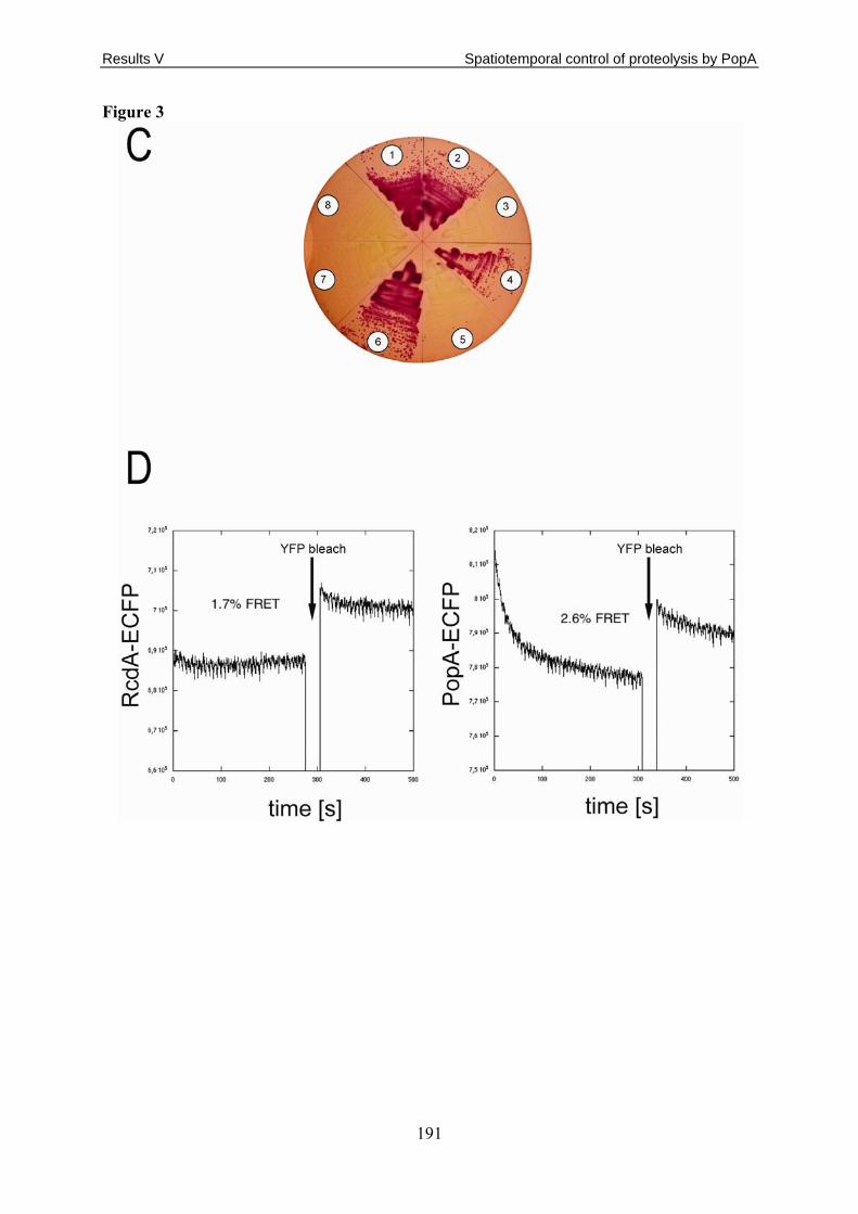
Results V Spatiotemporal control of proteolysis by PopA
191
Figure 3

Results V Spatiotemporal control of proteolysis by PopA
192
Figure 4

Results V Spatiotemporal control of proteolysis by PopA
193
Figure 5

Results V Spatiotemporal control of proteolysis by PopA
194
Figure 6

Results V Spatiotemporal control of proteolysis by PopA
195
Figure 7

Results V Spatiotemporal control of proteolysis by PopA
196
Figure 8
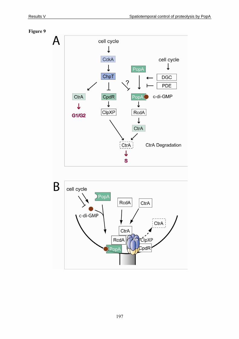
Results V Spatiotemporal control of proteolysis by PopA
197
Figure 9

Results V Spatiotemporal control of proteolysis by PopA
198
Table 1

Results V Spatiotemporal control of proteolysis by PopA
199
Supplementary Material:
Figure S1: Sequence comparison of the diguanylate cyclase PleD and the c-di-GMP
binding protein PopA.
A sequence alignment of PopA (Query) and PleD (Sbjct) is shown. The three domains are
indicated by different colors. Conserved residues of Rec1 known to be important for intra-
molecular signaling are boxed green, conserved residues of the putative dimerisation interface
are boxed red, and residues important for GGDEF domain function are highlighted in purple.
Figure S2: CtrA is stabilized in a ΔpopA mutant.
Cell cycle time lapse experiment with wild-type and ΔpopA mutant cells expressing yfp-
ctrARD+15 from the xylose inducible promoter Pxyl. Cells grown in the presence of the
inducer xylose were synchronized and released into fresh M2G minimal glucose medium
lacking the inducer. Samples were removed at intervals and analysed by DIC and fluorescence
microscopy.
Fig. S3: Protein – protein interaction map of the C. crescentus CtrA degradation
machinery.
Arrows indicate interactions between proteins. Interactions shown by co-immunoprecipitation
are in blue, interactions shown by the bacterial two-hybrid system (BACTH) are in red.
Stippled arrows indicate weak interactions.
Fig. S4: Polar localization of PopA-eGFP during the C. crescentus cell cycle.
(A) C. crescentus wild-type cells expressing popA-egfp were synchronized and samples
removed at 20 minute intervals were analysed by fluorescence microscopy. Cell cycle
progression is shown schematically. (B) Statistics of PopA-eGFP localization as shown in (A).
Cells with no focus, with a PopA-eGFP focus at one pole, and with a bipolar pattern were
scored throughout the cell cycle as indicated.
Fig. S5: Polar localization of PopA I- and A-site mutants during the C. crescentus cell
cycle.
C. crescentus popA mutant cells expressing either popAE368Q-egfp (A) or popAD357G-egfp (B)
were synchronized and samples removed at 20 minute intervals were analysed by DIC and
Results V Spatiotemporal control of proteolysis by PopA
200
fluorescence microscopy. The statistics of PopAE368Q-eGFP and PopAD357G-eGFP localization
is shown underneath the microscopy panels. Cells with no focus, with a focus at one pole, and
with a bipolar pattern were scored throughout the cell cycle as indicated.
Fig. S6: The GGDEF domain is required for efficient localization of PopA to the pole.
C. crescentus popA mutant cells expressing popA-rec1-egfp (receiver domain 1 of PopA),
popA-rec1-rec2-egfp (receiver domains 1 and 2 of PopA), or popA-egfp (full-length PopA)
were analysed by DIC (left) and fluorescence microscopy (middle). The localization patterns
are indicated on the right.
Fig. S7: PopA is required for proper localization of ClpX to the cell pole.
Cultures of C. crescentus wild type and popA mutants expressing a clpX-egfp were
synchronized and cells were analysed by DIC and fluorescence microscopy as they progressed
through the cell cycle. Polar localization of ClpX is shown schematically in the middle panel.
Progression of cells through the cell cycle is indicated on the left.
The timing of cell cycle progression is equivalent to the experiments shown in Fig. 2A.
Fig. S8: CpdR is not required for localization of PopA to the cell pole.
Cultures of C. crescentus wild type and DcpdRmutant expressing popA-egfp were analysed by
DIC (middle) and fluorescence microscopy (left). The localization patterns are indicated on
the right.
Fig. S9: PopA is required for proper CpdR localization to the cell pole.
Cultures of C. crescentus DcpdR (upper panels) and DcpdRDpopA (lower panels) mutants
expressing cpdR-yfp were synchronized and cells were analysed by DIC and fluorescence
microscopy throughout the cell cycle. The localization patterns are shown schematically.
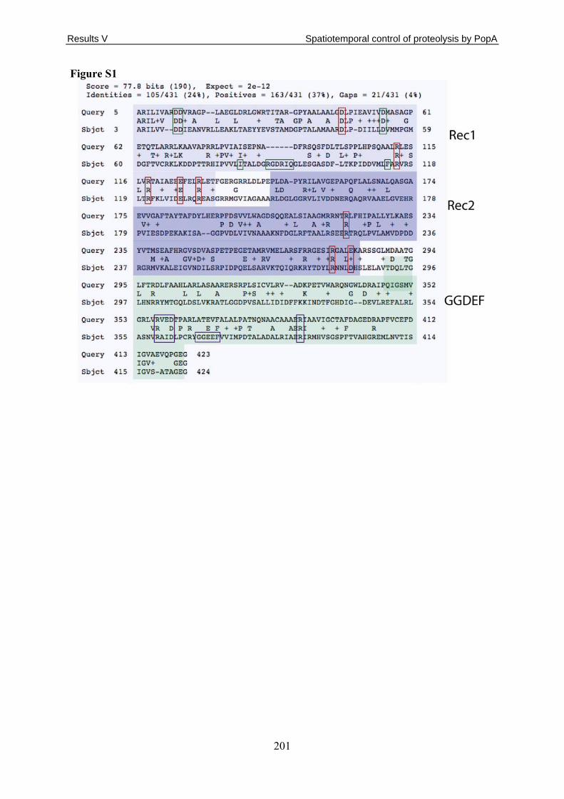
Results V Spatiotemporal control of proteolysis by PopA
201
Figure S1

Results V Spatiotemporal control of proteolysis by PopA
202
Figure S2

Results V Spatiotemporal control of proteolysis by PopA
203
Figure S3

Results V Spatiotemporal control of proteolysis by PopA
204
Figure S4

Results V Spatiotemporal control of proteolysis by PopA
205
Figure S5

Results V Spatiotemporal control of proteolysis by PopA
206
Figure S6

Results V Spatiotemporal control of proteolysis by PopA
207
Figure S7

Results V Spatiotemporal control of proteolysis by PopA
208
Figure S8

Results V Spatiotemporal control of proteolysis by PopA
209
Figure S9

Results V Spatiotemporal control of proteolysis by PopA
210
Table S1
Strain or plasmid Relevant genotype and description Reference or source
C. crescentus strains NA1000 Synchronizable laboratory strain of CB15 [114] UJ271 NA1000 clpX::Ω::pUJ168 [108] UJ2765 NA1000 ΔpodJ and plasmid pAD5 this study UJ2796 NA1000 and plasmid pAD5 this study UJ2827 NA1000 ΔpopA this study UJ3125 NA1000 and plasmid pEJ146 this study UJ3127 NA1000 ΔpopA and plasmid pEJ146 this study UJ3159 NA1000 ΔpopA and plasmid pAD19 this study UJ3563 NA1000 ΔpopA and plasmid pAD32 this study UJ3565 NA1000 ΔpopA and plasmid pAD30 this study UJ3640 NA1000 ΔpopA ΔpodJ this study UJ3665 NA1000 ΔpopA ΔrcdA and plasmid pAD5 this study UJ3666 NA1000 ΔpopA ΔpodJ and plasmid pAD5 this study UJ3672 NA1000 ΔpopA ΔpodJ and plasmid pAD30 this study UJ3742 NA1000 ΔpopA rcdA::prcdA-egfp this study UJ3743 NA1000 ΔpopA xylX::pX-clpX-egfp this study UJ3966 NA1000 and plasmid pID42 this study UJ3967 NA1000 and plasmid pIDC42 this study UJ3969 NA1000 ΔpopA and plasmid pID42 this study UJ3970 NA1000 ΔpopA and plasmid pIDC42 this study UJ3972 NA1000 clpX::Ω::pUJ168 and plasmid pAD5 this study UJ4329 NA1000 and plasmids pAD105, pAD82 this study UJ4330 NA1000 and plasmids pAD106, pAD83 this study UJ4331 NA1000 ΔpopA and plasmid pAD128 this study UJ4333 NA1000 ΔpopA and plasmid pAD130 this study UJ4374 NA1000 ΔcpdR and plasmid pAD147 this study UJ4417 NA1000 ΔpopA xylX::pX-clpX-egfp and plasmid pAD150 this study UJ4401 NA1000 ΔpopA ΔcpdR this study UJ4434 NA1000 ΔcpdR and plasmid pcpdR-yfp this study UJ4435 NA1000 ΔpopA ΔcpdR and plasmid pcpdR-yfp this study UJ4471 NA1000 ΔpopA ΔcpdR and plasmid pX-cpdRD51A-yfp this study UJ4473 NA1000 ΔcpdR and plasmid pX-cpdRD51A-yfp this study LS4183 NA1000 xylX::pX-clpX-egfp [184] LS4191 NA1000 rcdA::prcdA-egfp [184] NA1000 ΔcpdR Disruption of cpdR (tetR) [173] NA1000 ΔpodJ Deletion of podJ [281] E. coli strains
MM337 E. coli K-12 araD139 flbB5301 ptsF25 rbsR relA1 rpsL150 (argF-lac)U169 cya M. Manson
DH10B
F- mcrA ∆(mrr- hsd RMS- mcrBC) Φ80dlacZ∆M15 ∆lacX74 endA1 recA1 deoR ∆ (ara, leu)7697 araD139 galU galK nupG rpsL thi pro hsdR- hsd+ recA RP4-2-Tc::Mu-Tn7 [205]
S17 RP4-2, Tc::Mu, KM-Tn7 [205]
DH5α supE44 ΔlacU169(Φ80 lacZΔM15) hsdR17 recA1 endA1gyrA96 thi-1 relA1 [204]
ArcticR BL21 (DE3) arctic express strain for protein expression Stratagene Plasmids pAD5 pMR20; popA-eGfp under control of popA promoter this study

Results V Spatiotemporal control of proteolysis by PopA
211
pAD8 pNPTS138; used for clean deletion of popA this study pAD19 pMR20; popAD55N-eGfp under control of popA promoter this study pAD30 pMR20; popAR357G-eGfp under control of popA promoter this study pAD32 pMR20; popAE368Q-eGfp under control of popA promoter this study pAD33 pET21C; popAR357G C-terminal His6 tag this study pAD34 pET21C; popAE368Q C-terminal His6 tag this study pAD44 pUT18C; clpX C-terminal fused to the T18 fragment this study pAD45 pKT25; clpX C-terminal fused to the T25 fragment this study pAD47 pUT18C; clpP C-terminal fused to the T18 fragment this study pAD48 pKT25; clpP C-terminal fused to the T25 fragment this study pAD50 pUT18C; rcdA C-terminal fused to the T18 fragment this study pAD51 pKT25; rcdA C-terminal fused to the T25 fragment this study pAD53 pUT18C; cpdR C-terminal fused to the T18 fragment this study pAD54 pKT25; cpdR C-terminal fused to the T25 fragment this study pAD56 pUT18; rcdA N-terminal fused to the T18 fragment this study pAD58 pUT18; clpX N-terminal fused to the T18 fragment this study pAD60 pUT18; clpP N-terminal fused to the T18 fragment this study pAD62 pUT18; cpdR N-terminal fused to the T18 fragment this study pAD65 pUT18; ctrA N-terminal fused to the T18 fragment this study pAD67 pUT18; popA N-terminal fused to the T18 fragment this study pAD82 pMR20; popA-eYfp under control of popA promoter this study pAD83 pMR10; popA-eCfp under control of popA promoter this study pAD90 pKT25; popAR357G C-terminal fused to the T25 fragment this study pAD91 pKT25; popAE368Q C-terminal fused to the T25 fragment this study pAD105 pBBR-MCS-5; rcdA-eCfp under the control of rcdA promoter this study pAD106 pBBR-MCS-5; rcdA-eYfp under the control of rcdA promoter this study pAD128 pMR20; popA-Rec1-eGfp under the control of popA promoter this study pAD130 pMR20; popA-Rec1Rec2-eGfp under the control of popA promoter this study pAD140 pKT25; ctrA C-terminal fused to the T25 fragment this study pAD141 pKT25; popA C-terminal fused to the T25 fragment this study pAD142 pUT18C; ctrA C-terminal fused to the T18C fragment this study pAD143 pUT18C; popA C-terminal fused to the T18C fragment this study pAD147 pMR10; popA-eGfp under control of popA promoter this study
pAD150 pMR20; popAD55N under the control of popA promoter, chloramphenicol cassette integrated this study
pAD153 pMR20; popAR357G under the control of popA promoter,kanamycin cassette integrated this study
pBBR1MCS-5 GentR, broad host range cloning vector [206] pcpdR-yfp pMR11; cpdR 3’ fused to eyfp with cpdR promoter [269] pECFP AmpR vector for creation of eCFP-fusion proteins Clontech pEGFP AmpR vector for creation of eGFP-fusion proteins Clontech pEJ146 pMR10-Pxyl::yfp-ctrA RD+15 [110] pET21C AmpR expression vector, high copy number Novagen pET21C::PopA pET21C; popA C-terminal His6 tag this study pEYFP AmpR vector for creation of eYFP-fusion proteins Clontech pID42 pJS14; Pxyl::ctrA [109] pIDC42 pJS14; Pxyl::ctrAD51E [109] pJS14 ChlorR high copy number expression vector J. Skerker pKT25 pSU40 derivative with T25 fragment of CyaA [293] pKT25-zip pKT25 derivative with leucine zipper of GCN4 [293] pMR10 KanR low copy number and broad host range vector [259] pMR20 TetR low copy number and broad host range vector [259] pNPTS138 KanR, suicide vector with sacB gene and oriT D. Alley prcdA-egfp pXGFP4 with xylX promoter replaced with last 300 bp of rcdA [184]

Results V Spatiotemporal control of proteolysis by PopA
212
pUT18C-zip pUT18C derivative with leucine zipper of GCN4 [293] pUT18C pUC19 derivative with T18 fragment of CyaA. C-terminal fusions [293] pUT18 pUC19 derivative with T18 fragment of CyaA. N-terminal fusions [293] pX-clpX-egfp pXGFP4 with clpX [184] pX-cpdRD51A-yfp pMR31; cpdRD51A 3’ fused to eyfp with xylX promoter [269]

Outlook
213
4. Outlook In this work we characterised a network of enzymes involved in c-di-GMP turnover that
regulates the developmental transition form free-living, motile swarmer cells into sessile
stalked cell of the model organism Caulobacter crescentus. We identified the
phosphodiesterase PdeA and the di-guanylate cyclase DgcB as two important antagonistic
regulatory components in this process and analysed the already known DGC PleD in more
detail. As the correct timing of intertwined developmental events is important for the
swarmer-to-stalked cell transition, the activity of regulatory components has to be carefully
controlled. Based on the findings presented in this work, it would be of interest to investigate
the regulation of the activities of PdeA and DgcB more closely. A first question would be
whether PdeA is restricted to the swarmer cell compartment in the predivisional cell, which
could be addressed by investigating the spatial distribution of a degradable PdeA-GFP fusion
during the cell cycle. The finding that PdeA localises to the ClpXP occupied developing
stalked pole concurring its degradation raises the question for the role of this localisation. Has
it a regulatory function, in analogy to the PopA-RcdA-CtrA pathway? Is there a PdeA specific
polar receptor or is its localisation mediated by ClpXP itself? The former may be solved by
the generation of delocalised PdeA mutants and the investigation of their degradation pattern.
The latter could be addressed by the examination of PdeA localisation in a ΔcpdR mutant, in
which ClpXP is not localised to the stalked pole [269]. Additionally it would be intriguing to
examine whether there is a second layer of PdeA activity control despite the proteolytic
regulation. Furthermore, it is of interest to unravel the mechanisms underlying the activation
of the PdeA antagonist DgcB. Is DgcB a constitutive or regulated di-guanylate cyclase and
how is its activity profile during the cell cycle? What is the nature of the regulatory
mechanism; oligomerisation in dependence of c-di-GMP? Are further factors involved the
regulation of DgcB and can we make use of the motile suppressors in the ΔpdeA mutant
background to identify them?
Next to the activity control of the components in this regulatory network it is also of
importance to define whether, which and how further regulatory components are involved in
the control of the swarmer-to-stalked cell development via c-di-GMP. It was suggested that
CC0091 may act as an additional antagonist to PleD (A. Levi unpublished). Furthermore, the
performed epistasis experiments argue for an unknown DGC that, in addition to DgcB, feeds
in the c-di-GMP pool accessible to PdeA and is involved in the motility control. Thus, one
perspective is to address the possible role of the phosphodiesterase CC0091 and the potential

Outlook
214
di-guanylate cyclase in the regulatory network presented here to build a more comprehensive
model.
In a broader perspective, it is important to define the downstream targets that are
controlled by the c-di-GMP regulated by the components of the network described here.
Especially the finding that the known c-di-GMP effector proteins DgrA and DgrB are not
involved in the motility regulation by PdeA raises the question for the nature of additional c-
di-GMP receptors. Again, the answer to this question may reside in the motile suppressors in
the ΔpdeA mutant background. Another important question would be, whether c-di-GMP
effector proteins that regulate motility also affect attachment and vice versa or whether these
processes are individually regulated. A candidate c-di-GMP binding protein effecting holdfast
synthesis is the glycosyltransferase CC0095, which also has to be further investigated and
eventually integrated into a global model of the developmental aspects of the swarmer-to-
stalked cell transition.
A major question is how different c-di-GMP mediated signalling pathways are separated
from each other. The finding that the paralysis of the flagellum upon deletion of pdeA is not
mediated via DgrA/B could provide an insight into the underlying mechanisms, if it is
possible to solve the presence, activity and spatial distribution of the individual components.
In the same context it would also be interesting to further investigate the role of PleD
localisation, as we have shown here that despite PleD delocalisation in a Δcc1064 mutant
background, these cells do not show the known developmental phenotypes of a ΔpleD mutant.
This unexpected phenomenon may be addressed by generating PleD mutants that retain
activity without being localised. In contrast to the PleD activity in a Δcc1064 strain, their
activity could be directly quantified in an in vitro assay and allow a better interpretation of
their in vivo phenotypes.
Finally, another major, yet widely uncharacterised problem is the nature of upstream
signals. What regulates DGC and PDE activity? Are environmental or cell cycle signals
responsible for the timing of the swarmer-to-stalked cell transition; or both, and how are these
signals transmitted?
Despite its importance to study the localisation of PleD, the further understanding of the
pleitropic phenotypes of a Δcc1064 mutant is of interest, as CC1064 could be part of a general
mechanism used by C. crescentus to react to changing environmental conditions. Most
important would be to assign a direct molecular function to CC1064 and investigate how this
molecule is able to regulate such diverse functions like motor protein assembly, surface
attachment regulation and PleD localisation.

Outlook
215
Taken together, the suggested follow-up experiments based on the work presented here
would take us a step closer to understand the spatiotemporal dynamics of the network of c-di-
GMP metabolizing and effector proteins, how this integrates external and internal signals and
thereby allows bacteria to decide over their further lifestyle.

Bibliography
216
5. Bibliography 1. Kolter, R. and E.P. Greenberg, Microbial sciences: the superficial life of microbes.
Nature, 2006. 441(7091): p. 300-2. 2. Harshey, R.M., Bacterial motility on a surface: many ways to a common goal. Annu
Rev Microbiol, 2003. 57: p. 249-73. 3. Pratt, L.A. and R. Kolter, Genetic analyses of bacterial biofilm formation. Curr Opin
Microbiol, 1999. 2(6): p. 598-603. 4. Fux, C.A., et al., Survival strategies of infectious biofilms. Trends Microbiol, 2005.
13(1): p. 34-40. 5. Kolter, R., Surfacing views of biofilm biology. Trends Microbiol, 2005. 13(1): p. 1-2. 6. Stewart, P.S. and M.J. Franklin, Physiological heterogeneity in biofilms. Nat Rev
Microbiol, 2008. 6(3): p. 199-210. 7. Jenal, U., Cyclic di-guanosine-monophosphate comes of age: a novel secondary
messenger involved in modulating cell surface structures in bacteria? Curr Opin Microbiol, 2004. 7(2): p. 185-91.
8. Jenal, U. and J. Malone, Mechanisms of cyclic-di-GMP signaling in bacteria. Annu Rev Genet, 2006. 40: p. 385-407.
9. Romling, U. and D. Amikam, Cyclic di-GMP as a second messenger. Curr Opin Microbiol, 2006. 9(2): p. 218-28.
10. Tamayo, R., J.T. Pratt, and A. Camilli, Roles of cyclic diguanylate in the regulation of bacterial pathogenesis. Annu Rev Microbiol, 2007. 61: p. 131-48.
11. Cotter, P.A. and S. Stibitz, c-di-GMP-mediated regulation of virulence and biofilm formation. Curr Opin Microbiol, 2007. 10(1): p. 17-23.
12. Galperin, M.Y., A.N. Nikolskaya, and E.V. Koonin, Novel domains of the prokaryotic two-component signal transduction systems. FEMS Microbiol Lett, 2001. 203(1): p. 11-21.
13. Tal, R., et al., Three cdg operons control cellular turnover of cyclic di-GMP in Acetobacter xylinum: genetic organization and occurrence of conserved domains in isoenzymes. J Bacteriol, 1998. 180(17): p. 4416-25.
14. Paul, R., et al., Cell cycle-dependent dynamic localization of a bacterial response regulator with a novel di-guanylate cyclase output domain. Genes Dev, 2004. 18(6): p. 715-27.
15. Ryjenkov, D.A., et al., Cyclic diguanylate is a ubiquitous signaling molecule in bacteria: insights into biochemistry of the GGDEF protein domain. J Bacteriol, 2005. 187(5): p. 1792-8.
16. Chan, C., et al., Structural basis of activity and allosteric control of diguanylate cyclase. Proc Natl Acad Sci U S A, 2004. 101(49): p. 17084-9.
17. Wassmann, P., et al., Structure of BeF3- -modified response regulator PleD: implications for diguanylate cyclase activation, catalysis, and feedback inhibition. Structure, 2007. 15(8): p. 915-27.
18. Malone, J.G., et al., The structure-function relationship of WspR, a Pseudomonas fluorescens response regulator with a GGDEF output domain. Microbiology, 2007. 153(Pt 4): p. 980-94.
19. Christen, B., et al., Allosteric control of cyclic di-GMP signaling. J Biol Chem, 2006. 281(42): p. 32015-24.
20. Christen, M., et al., Identification and characterization of a cyclic di-GMP-specific phosphodiesterase and its allosteric control by GTP. J Biol Chem, 2005. 280(35): p. 30829-37.

Bibliography
217
21. Schmidt, A.J., D.A. Ryjenkov, and M. Gomelsky, The ubiquitous protein domain EAL is a cyclic diguanylate-specific phosphodiesterase: enzymatically active and inactive EAL domains. J Bacteriol, 2005. 187(14): p. 4774-81.
22. Tamayo, R., A.D. Tischler, and A. Camilli, The EAL domain protein VieA is a cyclic diguanylate phosphodiesterase. J Biol Chem, 2005. 280(39): p. 33324-30.
23. Kazmierczak, B.I., M.B. Lebron, and T.S. Murray, Analysis of FimX, a phosphodiesterase that governs twitching motility in Pseudomonas aeruginosa. Mol Microbiol, 2006. 60(4): p. 1026-43.
24. Chang, A.L., et al., Phosphodiesterase A1, a regulator of cellulose synthesis in Acetobacter xylinum, is a heme-based sensor. Biochemistry, 2001. 40(12): p. 3420-6.
25. Ryan, R.P., et al., Cell-cell signaling in Xanthomonas campestris involves an HD-GYP domain protein that functions in cyclic di-GMP turnover. Proc Natl Acad Sci U S A, 2006. 103(17): p. 6712-7.
26. Dürig, A., et al., Second messenger mediated spatiotemporal control of protein degradation during the bacterial cell cycle. Cell, 2008. manuscript ready for submission.
27. Amikam, D. and M.Y. Galperin, PilZ domain is part of the bacterial c-di-GMP binding protein. Bioinformatics, 2006. 22(1): p. 3-6.
28. Pratt, J.T., et al., PilZ domain proteins bind cyclic diguanylate and regulate diverse processes in Vibrio cholerae. J Biol Chem, 2007. 282(17): p. 12860-70.
29. Ko, M. and C. Park, Two novel flagellar components and H-NS are involved in the motor function of Escherichia coli. J Mol Biol, 2000. 303(3): p. 371-82.
30. Huang, B., C.B. Whitchurch, and J.S. Mattick, FimX, a multidomain protein connecting environmental signals to twitching motility in Pseudomonas aeruginosa. J Bacteriol, 2003. 185(24): p. 7068-76.
31. Ross, P.A., Y; Weinhouse, H; Michaeli, D; Weinberger-Ohana, P; et al., Control of cellulose synthesis Acetobacter xylinum. A unique guanyl oligonucleotide is the immediate activator of the cellulose synthase. Carbohydrate Research, 1986. 149: p. 101–117.
32. Alm, R.A., et al., Identification of a novel gene, pilZ, essential for type 4 fimbrial biogenesis in Pseudomonas aeruginosa. J Bacteriol, 1996. 178(1): p. 46-53.
33. Christen, M., et al., DgrA is a member of a new family of cyclic diguanosine monophosphate receptors and controls flagellar motor function in Caulobacter crescentus. Proc Natl Acad Sci U S A, 2007. 104(10): p. 4112-7.
34. Ryjenkov, D.A., et al., The PilZ domain is a receptor for the second messenger c-di-GMP: the PilZ domain protein YcgR controls motility in enterobacteria. J Biol Chem, 2006. 281(41): p. 30310-4.
35. Levi, A., Genetic Dissection of Caulobacter crescentus Surface Colonization, in Biocenter. 2007, University of Basel: Basel.
36. Merighi, M., et al., The second messenger bis-(3'-5')-cyclic-GMP and its PilZ domain-containing receptor Alg44 are required for alginate biosynthesis in Pseudomonas aeruginosa. Mol Microbiol, 2007. 65(4): p. 876-95.
37. Schultz, J., et al., SMART, a simple modular architecture research tool: identification of signaling domains. Proc Natl Acad Sci U S A, 1998. 95(11): p. 5857-64.
38. Guvener, Z.T. and C.S. Harwood, Subcellular location characteristics of the Pseudomonas aeruginosa GGDEF protein, WspR, indicate that it produces cyclic-di-GMP in response to growth on surfaces. Mol Microbiol, 2007. 66(6): p. 1459-73.
39. Ross, P.A., Y; Weinhouse, H; Michaeli, D; Weinberger-Ohana, P; et al., An unusual guanyl oligonucleotide regulates cellulose synthesis in Acetobacter xylinum. FEBS Lett., 1985. 186: p. 191–196.

Bibliography
218
40. Lee, S.H., et al., Nucleotide sequence and spatiotemporal expression of the Vibrio cholerae vieSAB genes during infection. J Bacteriol, 1998. 180(9): p. 2298-305.
41. Perry, R.D., M.L. Pendrak, and P. Schuetze, Identification and cloning of a hemin storage locus involved in the pigmentation phenotype of Yersinia pestis. J Bacteriol, 1990. 172(10): p. 5929-37.
42. Perry, R.D., et al., Temperature regulation of the hemin storage (Hms+) phenotype of Yersinia pestis is posttranscriptional. J Bacteriol, 2004. 186(6): p. 1638-47.
43. Tischler, A.D. and A. Camilli, Cyclic diguanylate regulates Vibrio cholerae virulence gene expression. Infect Immun, 2005. 73(9): p. 5873-82.
44. Hisert, K.B., et al., A glutamate-alanine-leucine (EAL) domain protein of Salmonella controls bacterial survival in mice, antioxidant defence and killing of macrophages: role of cyclic diGMP. Mol Microbiol, 2005. 56(5): p. 1234-45.
45. Kulasakara, H., et al., Analysis of Pseudomonas aeruginosa diguanylate cyclases and phosphodiesterases reveals a role for bis-(3'-5')-cyclic-GMP in virulence. Proc Natl Acad Sci U S A, 2006. 103(8): p. 2839-44.
46. Kader, A., et al., Hierarchical involvement of various GGDEF domain proteins in rdar morphotype development of Salmonella enterica serovar Typhimurium. Mol Microbiol, 2006. 60(3): p. 602-16.
47. Levi, A. and U. Jenal, Holdfast formation in motile swarmer cells optimizes surface attachment during Caulobacter crescentus development. J Bacteriol, 2006. 188(14): p. 5315-8.
48. Ross, P., et al., The cyclic diguanylic acid regulatory system of cellulose synthesis in Acetobacter xylinum. Chemical synthesis and biological activity of cyclic nucleotide dimer, trimer, and phosphothioate derivatives. J Biol Chem, 1990. 265(31): p. 18933-43.
49. Weber, H., et al., Cyclic-di-GMP-mediated signalling within the sigma network of Escherichia coli. Mol Microbiol, 2006. 62(4): p. 1014-34.
50. Boles, B.R. and L.L. McCarter, Vibrio parahaemolyticus scrABC, a novel operon affecting swarming and capsular polysaccharide regulation. J Bacteriol, 2002. 184(21): p. 5946-54.
51. Costerton, J.W., et al., Microbial biofilms. Annu Rev Microbiol, 1995. 49: p. 711-45. 52. Potera, C., Biofilms invade microbiology. Science, 1996. 273(5283): p. 1795-7. 53. Mah, T.F., et al., A genetic basis for Pseudomonas aeruginosa biofilm antibiotic
resistance. Nature, 2003. 426(6964): p. 306-10. 54. Matz, C., et al., Biofilm formation and phenotypic variation enhance predation-driven
persistence of Vibrio cholerae. Proc Natl Acad Sci U S A, 2005. 102(46): p. 16819-24.
55. Weitere, M., et al., Grazing resistance of Pseudomonas aeruginosa biofilms depends on type of protective mechanism, developmental stage and protozoan feeding mode. Environ Microbiol, 2005. 7(10): p. 1593-601.
56. Stoodley, P., et al., Biofilms as complex differentiated communities. Annu Rev Microbiol, 2002. 56: p. 187-209.
57. O'Toole, G., H.B. Kaplan, and R. Kolter, Biofilm formation as microbial development. Annu Rev Microbiol, 2000. 54: p. 49-79.
58. Watnick, P. and R. Kolter, Biofilm, city of microbes. J Bacteriol, 2000. 182(10): p. 2675-9.
59. Donlan, R.M., Biofilms: microbial life on surfaces. Emerg Infect Dis, 2002. 8(9): p. 881-90.
60. Sutherland, I., Biofilm exopolysaccharides: a strong and sticky framework. Microbiology, 2001. 147(Pt 1): p. 3-9.

Bibliography
219
61. Aldridge, P., et al., Role of the GGDEF regulator PleD in polar development of Caulobacter crescentus. Mol Microbiol, 2003. 47(6): p. 1695-708.
62. Bobrov, A.G., O. Kirillina, and R.D. Perry, The phosphodiesterase activity of the HmsP EAL domain is required for negative regulation of biofilm formation in Yersinia pestis. FEMS Microbiol Lett, 2005. 247(2): p. 123-30.
63. Bomchil, N., P. Watnick, and R. Kolter, Identification and characterization of a Vibrio cholerae gene, mbaA, involved in maintenance of biofilm architecture. J Bacteriol, 2003. 185(4): p. 1384-90.
64. Choy, W.K., et al., MorA defines a new class of regulators affecting flagellar development and biofilm formation in diverse Pseudomonas species. J Bacteriol, 2004. 186(21): p. 7221-8.
65. Drenkard, E. and F.M. Ausubel, Pseudomonas biofilm formation and antibiotic resistance are linked to phenotypic variation. Nature, 2002. 416(6882): p. 740-3.
66. Enos-Berlage, J.L., et al., Genetic determinants of biofilm development of opaque and translucent Vibrio parahaemolyticus. Mol Microbiol, 2005. 55(4): p. 1160-82.
67. Garcia, B., et al., Role of the GGDEF protein family in Salmonella cellulose biosynthesis and biofilm formation. Mol Microbiol, 2004. 54(1): p. 264-77.
68. Goymer, P., et al., Adaptive divergence in experimental populations of Pseudomonas fluorescens. II. Role of the GGDEF regulator WspR in evolution and development of the wrinkly spreader phenotype. Genetics, 2006. 173(2): p. 515-26.
69. Guvener, Z.T. and L.L. McCarter, Multiple regulators control capsular polysaccharide production in Vibrio parahaemolyticus. J Bacteriol, 2003. 185(18): p. 5431-41.
70. Kirillina, O., et al., HmsP, a putative phosphodiesterase, and HmsT, a putative diguanylate cyclase, control Hms-dependent biofilm formation in Yersinia pestis. Mol Microbiol, 2004. 54(1): p. 75-88.
71. Simm, R., et al., GGDEF and EAL domains inversely regulate cyclic di-GMP levels and transition from sessility to motility. Mol Microbiol, 2004. 53(4): p. 1123-34.
72. Tischler, A.D. and A. Camilli, Cyclic diguanylate (c-di-GMP) regulates Vibrio cholerae biofilm formation. Mol Microbiol, 2004. 53(3): p. 857-69.
73. Gjermansen, M., et al., Characterization of starvation-induced dispersion in Pseudomonas putida biofilms. Environ Microbiol, 2005. 7(6): p. 894-906.
74. Ude, S., et al., Biofilm formation and cellulose expression among diverse environmental Pseudomonas isolates. Environ Microbiol, 2006. 8(11): p. 1997-2011.
75. Spiers, A.J., et al., Adaptive divergence in experimental populations of Pseudomonas fluorescens. I. Genetic and phenotypic bases of wrinkly spreader fitness. Genetics, 2002. 161(1): p. 33-46.
76. Spiers, A.J., et al., Biofilm formation at the air-liquid interface by the Pseudomonas fluorescens SBW25 wrinkly spreader requires an acetylated form of cellulose. Mol Microbiol, 2003. 50(1): p. 15-27.
77. Simm, R., et al., Phenotypic convergence mediated by GGDEF-domain-containing proteins. J Bacteriol, 2005. 187(19): p. 6816-23.
78. Johnson, M.R., et al., Population density-dependent regulation of exopolysaccharide formation in the hyperthermophilic bacterium Thermotoga maritima. Mol Microbiol, 2005. 55(3): p. 664-74.
79. Rashid, M.H., et al., Identification of genes involved in the switch between the smooth and rugose phenotypes of Vibrio cholerae. FEMS Microbiol Lett, 2003. 227(1): p. 113-9.
80. Kovacikova, G., W. Lin, and K. Skorupski, Dual regulation of genes involved in acetoin biosynthesis and motility/biofilm formation by the virulence activator AphA

Bibliography
220
and the acetate-responsive LysR-type regulator AlsR in Vibrio cholerae. Mol Microbiol, 2005. 57(2): p. 420-33.
81. Beyhan, S., et al., Transcriptome and phenotypic responses of Vibrio cholerae to increased cyclic di-GMP level. J Bacteriol, 2006. 188(10): p. 3600-13.
82. Casper-Lindley, C. and F.H. Yildiz, VpsT is a transcriptional regulator required for expression of vps biosynthesis genes and the development of rugose colonial morphology in Vibrio cholerae O1 El Tor. J Bacteriol, 2004. 186(5): p. 1574-8.
83. Yildiz, F.H., N.A. Dolganov, and G.K. Schoolnik, VpsR, a Member of the Response Regulators of the Two-Component Regulatory Systems, Is Required for Expression of vps Biosynthesis Genes and EPS(ETr)-Associated Phenotypes in Vibrio cholerae O1 El Tor. J Bacteriol, 2001. 183(5): p. 1716-26.
84. Bantinaki, E., et al., Adaptive divergence in experimental populations of Pseudomonas fluorescens. III. Mutational origins of wrinkly spreader diversity. Genetics, 2007. 176(1): p. 441-53.
85. D'Argenio, D.A., et al., Autolysis and autoaggregation in Pseudomonas aeruginosa colony morphology mutants. J Bacteriol, 2002. 184(23): p. 6481-9.
86. Hickman, J.W., D.F. Tifrea, and C.S. Harwood, A chemosensory system that regulates biofilm formation through modulation of cyclic diguanylate levels. Proc Natl Acad Sci U S A, 2005. 102(40): p. 14422-7.
87. Friedman, L. and R. Kolter, Two genetic loci produce distinct carbohydrate-rich structural components of the Pseudomonas aeruginosa biofilm matrix. J Bacteriol, 2004. 186(14): p. 4457-65.
88. Jackson, K.D., et al., Identification of psl, a locus encoding a potential exopolysaccharide that is essential for Pseudomonas aeruginosa PAO1 biofilm formation. J Bacteriol, 2004. 186(14): p. 4466-75.
89. Furukawa, S., S.L. Kuchma, and G.A. O'Toole, Keeping their options open: acute versus persistent infections. J Bacteriol, 2006. 188(4): p. 1211-7.
90. Lee, V.T., et al., A cyclic-di-GMP receptor required for bacterial exopolysaccharide production. Mol Microbiol, 2007. 65(6): p. 1474-84.
91. Merz, A.J., M. So, and M.P. Sheetz, Pilus retraction powers bacterial twitching motility. Nature, 2000. 407(6800): p. 98-102.
92. Mattick, J.S., Type IV Pili and Twitching Motility. Annu. Rev. Microbiol., 2002. 56: p. 289–314.
93. McCarter, L., The multiple identities of Vibrio parahaemolyticus. J Mol Microbiol Biotechnol, 1999. 1(1): p. 51-7.
94. Wolfe, A.J. and K.L. Visick, Get the message out: cyclic-Di-GMP regulates multiple levels of flagellum-based motility. J Bacteriol, 2008. 190(2): p. 463-75.
95. Merino, S., J.G. Shaw, and J.M. Tomas, Bacterial lateral flagella: an inducible flagella system. FEMS Microbiol Lett, 2006. 263(2): p. 127-35.
96. Kim, Y.K. and L.L. McCarter, ScrG, a GGDEF-EAL protein, participates in regulating swarming and sticking in Vibrio parahaemolyticus. J Bacteriol, 2007. 189(11): p. 4094-107.
97. Mendez-Ortiz, M.M., et al., Genome-wide transcriptional profile of Escherichia coli in response to high levels of the second messenger 3',5'-cyclic diguanylic acid. J Biol Chem, 2006. 281(12): p. 8090-9.
98. Frye, J., et al., Identification of new flagellar genes of Salmonella enterica serovar Typhimurium. J Bacteriol, 2006. 188(6): p. 2233-43.
99. Poindexter, J.S. and J.G. Hagenzieker, Constriction and septation during cell division in caulobacters. Can J Microbiol, 1981. 27(7): p. 704-19.
100. Poindexter, J.S., Biological Properties and Classification of the Caulobacter Group. Bacteriol Rev, 1964. 28: p. 231-95.

Bibliography
221
101. Jacobs-Wagner, C., Regulatory proteins with a sense of direction: cell cycle signalling network in Caulobacter. Mol Microbiol, 2004. 51(1): p. 7-13.
102. Degnen, S.T. and A. Newton, Dependence of cell division on the completion of chromosome replication in Caulobacter. J Bacteriol, 1972. 110(3): p. 852-6.
103. Marczynski, G.T., A. Dingwall, and L. Shapiro, Plasmid and chromosomal DNA replication and partitioning during the Caulobacter crescentus cell cycle. J Mol Biol, 1990. 212(4): p. 709-22.
104. Brun, Y.V. and R. Janakiraman, The dimorphic life cycle of Caulobacter and stalked bacteria., in Prokaryotic development, Y.V.a.S. Brun, L.J., Editor. 2000, ASM Press: Washington, DC. p. 297-317.
105. Skerker, J.M. and L. Shapiro, Identification and cell cycle control of a novel pilus system in Caulobacter crescentus. Embo J, 2000. 19(13): p. 3223-34.
106. Quon, K.C., et al., Negative control of bacterial DNA replication by a cell cycle regulatory protein that binds at the chromosome origin. Proc Natl Acad Sci U S A, 1998. 95(1): p. 120-5.
107. Laub, M.T., et al., Global analysis of the genetic network controlling a bacterial cell cycle. Science, 2000. 290(5499): p. 2144-8.
108. Jenal, U. and T. Fuchs, An essential protease involved in bacterial cell-cycle control. Embo J, 1998. 17(19): p. 5658-69.
109. Domian, I.J., K.C. Quon, and L. Shapiro, Cell type-specific phosphorylation and proteolysis of a transcriptional regulator controls the G1-to-S transition in a bacterial cell cycle. Cell, 1997. 90(3): p. 415-24.
110. Ryan, K.R., E.M. Judd, and L. Shapiro, The CtrA response regulator essential for Caulobacter crescentus cell-cycle progression requires a bipartite degradation signal for temporally controlled proteolysis. J Mol Biol, 2002. 324(3): p. 443-55.
111. Ryan, K.R., S. Huntwork, and L. Shapiro, Recruitment of a cytoplasmic response regulator to the cell pole is linked to its cell cycle-regulated proteolysis. Proc Natl Acad Sci U S A, 2004. 101(19): p. 7415-20.
112. Alley, M.R., J.R. Maddock, and L. Shapiro, Polar localization of a bacterial chemoreceptor. Genes Dev, 1992. 6(5): p. 825-36.
113. Merker, R.I. and J. Smit, Characterization of the Adhesive Holdfast of Marine and Freshwater Caulobacters. Appl Environ Microbiol, 1988. 54(8): p. 2078-2085.
114. Evinger, M. and N. Agabian, Envelope-associated nucleoid from Caulobacter crescentus stalked and swarmer cells. J Bacteriol, 1977. 132(1): p. 294-301.
115. Stallmeyer, M.J., et al., Image reconstruction of the flagellar basal body of Caulobacter crescentus. J Mol Biol, 1989. 205(3): p. 511-8.
116. Trachtenberg, S. and D.J. DeRosier, Three-dimensional reconstruction of the flagellar filament of Caulobacter crescentus. A flagellin lacking the outer domain and its amino acid sequence lacking an internal segment. J Mol Biol, 1988. 202(4): p. 787-808.
117. Wagenknecht, T., et al., Three-dimensional reconstruction of the flagellar hook from Caulobacter crescentus. J Mol Biol, 1981. 151(3): p. 439-65.
118. Kojima, S. and D.F. Blair, The bacterial flagellar motor: structure and function of a complex molecular machine. Int Rev Cytol, 2004. 233: p. 93-134.
119. Lagenaur, C. and N. Agabian, Caulobacter flagellins. J Bacteriol, 1977. 132(2): p. 731-3.
120. Macnab, R.M., Genetics and biogenesis of bacterial flagella. Annu Rev Genet, 1992. 26: p. 131-58.
121. Aldridge, P. and K.T. Hughes, Regulation of flagellar assembly. Current Opinion in Microbiology, 2002. 5: p. 160–65.
Bibliography
222
122. Newton, A., et al., Genetic switching in the flagellar gene hierarchy of Caulobacter requires negative as well as positive regulation of transcription. Proc Natl Acad Sci U S A, 1989. 86(17): p. 6651-5.
123. Ramakrishnan, G., J.L. Zhao, and A. Newton, Multiple structural proteins are required for both transcriptional activation and negative autoregulation of Caulobacter crescentus flagellar genes. J Bacteriol, 1994. 176(24): p. 7587-600.
124. Komeda, Y., Transcriptional control of flagellar genes in Escherichia coli K-12. J Bacteriol, 1986. 168(3): p. 1315-8.
125. Kutsukake, K., Y. Ohya, and T. Iino, Transcriptional analysis of the flagellar regulon of Salmonella typhimurium. J Bacteriol, 1990. 172(2): p. 741-7.
126. Wu, J. and A. Newton, Regulation of the Caulobacter flagellar gene hierarchy; not just for motility. Mol Microbiol, 1997. 24(2): p. 233-9.
127. Kalir, S., et al., Ordering genes in a flagella pathway by analysis of expression kinetics from living bacteria. Science, 2001. 292(5524): p. 2080-3.
128. Quon, K.C., G.T. Marczynski, and L. Shapiro, Cell cycle control by an essential bacterial two-component signal transduction protein. Cell, 1996. 84(1): p. 83-93.
129. Reisenauer, A., K. Quon, and L. Shapiro, The CtrA response regulator mediates temporal control of gene expression during the Caulobacter cell cycle. J Bacteriol, 1999. 181(8): p. 2430-9.
130. Gober, J.W. and M.V. Marques, Regulation of cellular differentiation in Caulobacter crescentus. Microbiol Rev, 1995. 59(1): p. 31-47.
131. Ramakrishnan, G. and A. Newton, FlbD of Caulobacter crescentus is a homologue of the NtrC (NRI) protein and activates sigma 54-dependent flagellar gene promoters. Proc Natl Acad Sci U S A, 1990. 87(6): p. 2369-73.
132. Wingrove, J.A., E.K. Mangan, and J.W. Gober, Spatial and temporal phosphorylation of a transcriptional activator regulates pole-specific gene expression in Caulobacter. Genes Dev, 1993. 7(10): p. 1979-92.
133. Brun, Y.V. and L. Shapiro, A temporally controlled sigma-factor is required for polar morphogenesis and normal cell division in Caulobacter. Genes Dev, 1992. 6(12A): p. 2395-408.
134. Anderson, D.K., et al., Regulation of the Caulobacter crescentus rpoN gene and function of the purified sigma 54 in flagellar gene transcription. Mol Gen Genet, 1995. 246(6): p. 697-706.
135. Xu, H., A. Dingwall, and L. Shapiro, Negative transcriptional regulation in the Caulobacter flagellar hierarchy. Proc Natl Acad Sci U S A, 1989. 86(17): p. 6656-60.
136. Minnich, S.A. and A. Newton, Promoter mapping and cell cycle regulation of flagellin gene transcription in Caulobacter crescentus. Proc Natl Acad Sci U S A, 1987. 84(5): p. 1142-6.
137. Mangan, E.K., et al., FlbT couples flagellum assembly to gene expression in Caulobacter crescentus. J Bacteriol, 1999. 181(19): p. 6160-70.
138. Anderson, P.E. and J.W. Gober, FlbT, the post-transcriptional regulator of flagellin synthesis in Caulobacter crescentus, interacts with the 5' untranslated region of flagellin mRNA. Mol Microbiol, 2000. 38(1): p. 41-52.
139. Muir, R.E. and J.W. Gober, Mutations in FlbD that relieve the dependency on flagellum assembly alter the temporal and spatial pattern of developmental transcription in Caulobacter crescentus. Molecular Microbiology, 2002. 43(3): p. 597-615.
140. Biondi, E.G., et al., Regulation of the bacterial cell cycle by an integrated genetic circuit. Nature, 2006. 444(7121): p. 899-904.
141. Laub, M.T., et al., Genes directly controlled by CtrA, a master regulator of the Caulobacter cell cycle. Proc Natl Acad Sci U S A, 2002. 99(7): p. 4632-7.

Bibliography
223
142. Gober, J.W., et al., Identification of cis and trans-elements involved in the timed control of a Caulobacter flagellar gene. J Mol Biol, 1991. 217(2): p. 247-57.
143. Gober, J.W. and L. Shapiro, A developmentally regulated Caulobacter flagellar promoter is activated by 3' enhancer and IHF binding elements. Mol Biol Cell, 1992. 3(8): p. 913-26.
144. Wingrove, J.A. and J.W. Gober, Identification of an asymmetrically localized sensor histidine kinase responsible for temporally and spatially regulated transcription. Science, 1996. 274(5287): p. 597-601.
145. Muir, R.E. and J.W. Gober, Regulation of late flagellar gene transcription and cell division by flagellum assembly in Caulobacter crescentus. Mol Microbiol, 2001. 41(1): p. 117-30.
146. Ueno, T., K. Oosawa, and S. Aizawa, M ring, S ring and proximal rod of the flagellar basal body of Salmonella typhimurium are composed of subunits of a single protein, FliF. J Mol Biol, 1992. 227(3): p. 672-7.
147. Magariyama, Y., et al., Very fast flagellar rotation. Nature, 1994. 371(6500): p. 752. 148. Blair, D.F. and H.C. Berg, The MotA protein of E. coli is a proton-conducting
component of the flagellar motor. Cell, 1990. 60(3): p. 439-49. 149. Blair, D.F., D.Y. Kim, and H.C. Berg, Mutant MotB proteins in Escherichia coli. J
Bacteriol, 1991. 173(13): p. 4049-55. 150. Blair, D.F. and H.C. Berg, Mutations in the MotA protein of Escherichia coli reveal
domains critical for proton conduction. J Mol Biol, 1991. 221(4): p. 1433-42. 151. Blair, D.F., Flagellar movement driven by proton translocation. FEBS Lett, 2003.
545(1): p. 86-95. 152. Stolz, B. and H.C. Berg, Evidence for interactions between MotA and MotB, torque-
generating elements of the flagellar motor of Escherichia coli. J Bacteriol, 1991. 173(21): p. 7033-7.
153. Wilson, M.L. and R.M. Macnab, Co-overproduction and localization of the Escherichia coli motility proteins motA and motB. J Bacteriol, 1990. 172(7): p. 3932-9.
154. Braun, T.F. and D.F. Blair, Targeted disulfide cross-linking of the MotB protein of Escherichia coli: evidence for two H(+) channels in the stator Complex. Biochemistry, 2001. 40(43): p. 13051-9.
155. Sato, K. and M. Homma, Functional reconstitution of the Na(+)-driven polar flagellar motor component of Vibrio alginolyticus. J Biol Chem, 2000. 275(8): p. 5718-22.
156. Zhou, J., et al., Function of protonatable residues in the flagellar motor of Escherichia coli: a critical role for Asp 32 of MotB. J Bacteriol, 1998. 180(10): p. 2729-35.
157. Zhou, J., S.A. Lloyd, and D.F. Blair, Electrostatic interactions between rotor and stator in the bacterial flagellar motor. Proc Natl Acad Sci U S A, 1998. 95(11): p. 6436-41.
158. Chun, S.Y. and J.S. Parkinson, Bacterial motility: membrane topology of the Escherichia coli MotB protein. Science, 1988. 239(4837): p. 276-8.
159. De Mot, R. and J. Vanderleyden, The C-terminal sequence conservation between OmpA-related outer membrane proteins and MotB suggests a common function in both gram-positive and gram-negative bacteria, possibly in the interaction of these domains with peptidoglycan. Mol Microbiol, 1994. 12(2): p. 333-4.
160. Garza, A.G., et al., Mutations in motB suppressible by changes in stator or rotor components of the bacterial flagellar motor. J Mol Biol, 1996. 258(2): p. 270-85.
161. Khan, I.H., T.S. Reese, and S. Khan, The cytoplasmic component of the bacterial flagellar motor. Proc Natl Acad Sci U S A, 1992. 89(13): p. 5956-60.

Bibliography
224
162. Muramoto, K. and R.M. Macnab, Deletion analysis of MotA and MotB, components of the force-generating unit in the flagellar motor of Salmonella. Mol Microbiol, 1998. 29(5): p. 1191-202.
163. Blair, D.F. and H.C. Berg, Restoration of torque in defective flagellar motors. Science, 1988. 242(4886): p. 1678-81.
164. Reid, S.W., et al., The maximum number of torque-generating units in the flagellar motor of Escherichia coli is at least 11. Proc Natl Acad Sci U S A, 2006. 103(21): p. 8066-71.
165. Block, S.M. and H.C. Berg, Successive incorporation of force-generating units in the bacterial rotary motor. Nature, 1984. 309(5967): p. 470-2.
166. Yuan, J. and H.C. Berg, Resurrection of the flagellar rotary motor near zero load. Proc Natl Acad Sci U S A, 2008. 105(4): p. 1182-5.
167. Dean, G.E., et al., Gene sequence and predicted amino acid sequence of the motA protein, a membrane-associated protein required for flagellar rotation in Escherichia coli. J Bacteriol, 1984. 159(3): p. 991-9.
168. Enomoto, M., Genetic Studies of Paralyzed Mutants in Salmonella. II. Mapping of Three mot Loci by Linkage Analysis. Genetics, 1966. 54(5): p. 1069-1076.
169. Enomoto, M., Genetic studies of paralyzed mutant in Salmonella. I. Genetic fine structure of the mot loci in Salmonella typhimurium. Genetics, 1966. 54(3): p. 715-26.
170. Aldridge, P. and U. Jenal, Cell cycle-dependent degradation of a flagellar motor component requires a novel-type response regulator. Mol Microbiol, 1999. 32(2): p. 379-91.
171. Sommer, J.M. and A. Newton, Turning off flagellum rotation requires the pleiotropic gene pleD: pleA, pleC, and pleD define two morphogenic pathways in Caulobacter crescentus. J Bacteriol, 1989. 171(1): p. 392-401.
172. Sommer, J.M. and A. Newton, Pseudoreversion analysis indicates a direct role of cell division genes in polar morphogenesis and differentiation in Caulobacter crescentus. Genetics, 1991. 129(3): p. 623-30.
173. Skerker, J.M., et al., Two-component signal transduction pathways regulating growth and cell cycle progression in a bacterium: a system-level analysis. PLoS Biol, 2005. 3(10): p. e334.
174. Hecht, G.B. and A. Newton, Identification of a novel response regulator required for the swarmer-to-stalked-cell transition in Caulobacter crescentus. J Bacteriol, 1995. 177(21): p. 6223-9.
175. Wheeler, R.T. and L. Shapiro, Differential localization of two histidine kinases controlling bacterial cell differentiation. Mol Cell, 1999. 4(5): p. 683-94.
176. Matroule, J.Y., et al., Cytokinesis monitoring during development; rapid pole-to-pole shuttling of a signaling protein by localized kinase and phosphatase in Caulobacter. Cell, 2004. 118(5): p. 579-90.
177. Jacobs, C., D. Hung, and L. Shapiro, Dynamic localization of a cytoplasmic signal transduction response regulator controls morphogenesis during the Caulobacter cell cycle. Proc Natl Acad Sci U S A, 2001. 98(7): p. 4095-100.
178. Grunenfelder, B., et al., Identification of the protease and the turnover signal responsible for cell cycle-dependent degradation of the Caulobacter FliF motor protein. J Bacteriol, 2004. 186(15): p. 4960-71.
179. Jenal, U. and L. Shapiro, Cell cycle-controlled proteolysis of a flagellar motor protein that is asymmetrically distributed in the Caulobacter predivisional cell. Embo J, 1996. 15(10): p. 2393-406.
180. Burton, G.J., G.B. Hecht, and A. Newton, Roles of the histidine protein kinase pleC in Caulobacter crescentus motility and chemotaxis. J Bacteriol, 1997. 179(18): p. 5849-53.

Bibliography
225
181. Zogaj, X., et al., The multicellular morphotypes of Salmonella typhimurium and Escherichia coli produce cellulose as the second component of the extracellular matrix. Mol Microbiol, 2001. 39(6): p. 1452-63.
182. Zogaj, X., et al., Production of cellulose and curli fimbriae by members of the family Enterobacteriaceae isolated from the human gastrointestinal tract. Infect Immun, 2003. 71(7): p. 4151-8.
183. Huitema, E., et al., Bacterial birth scar proteins mark future flagellum assembly site. Cell, 2006. 124(5): p. 1025-37.
184. McGrath, P.T., et al., A dynamically localized protease complex and a polar specificity factor control a cell cycle master regulator. Cell, 2006. 124(3): p. 535-47.
185. Dürig, A., Second messenger mediated spatiotemporal control of cell cycle and development in Biocenter. 2008, University of Basel: Basel.
186. Paul, R., et al., Activation of the diguanylate cyclase PleD by phosphorylation-mediated dimerization. J Biol Chem, 2007. 282(40): p. 29170-7.
187. Paul, R., et al., Allosteric Regulation of Histidine Kinases by Their Cognate Response Regulator Determines Cell Fate. Cell, 2008. in press.
188. Jenal, U., J. White, and L. Shapiro, Caulobacter flagellar function, but not assembly, requires FliL, a non-polarly localized membrane protein present in all cell types. J Mol Biol, 1994. 243(2): p. 227-44.
189. Gottesman, S., et al., ClpX, an alternative subunit for the ATP-dependent Clp protease of Escherichia coli. Sequence and in vivo activities. J Biol Chem, 1993. 268(30): p. 22618-26.
190. Potocka, I., et al., Degradation of a Caulobacter soluble cytoplasmic chemoreceptor is ClpX dependent. J Bacteriol, 2002. 184(23): p. 6635-41.
191. Gorbatyuk, B. and G.T. Marczynski, Regulated degradation of chromosome replication proteins DnaA and CtrA in Caulobacter crescentus. Mol Microbiol, 2005. 55(4): p. 1233-45.
192. Bodenmiller, D., E. Toh, and Y.V. Brun, Development of surface adhesion in Caulobacter crescentus. J Bacteriol, 2004. 186(5): p. 1438-47.
193. Christen, M., Mechanisms of Cyclic-di-GMP Signaling, in Biocenter. 2007, University of Basel: Basel.
194. Wiederkehr, I., Methodology for the analysis of in vivo levels of cyclic-di-GMP in Caulobacter crescentus, in Biocenter. 2007, University of Basel: Basel.
195. Cohen, C., Parry, D. A. D. , Alpha-helical coiled coils - a widespread motif in proteins. Trends Biochem. Sci., 1986. 11: p. 245-48.
196. Ely, B., Genetics of Caulobacter crescentus. Methods Enzymol, 1991. 204: p. 372-84. 197. Sambrook, J., E.F. Fritsch, and T. Maniatis, Molecular cloning. A laboratory manual.
1989, New York: Cold Spring Harbor Laboratory Press. 198. Abramoff, M.D., Magelhaes, P.J., Ram, S.J., Image Processing with ImageJ.
Biophotonics International, 2004. 11(7): p. 36-42. 199. O'Toole, G.A. and R. Kolter, Initiation of biofilm formation in Pseudomonas
fluorescens WCS365 proceeds via multiple, convergent signalling pathways: a genetic analysis. Mol Microbiol, 1998. 28(3): p. 449-61.
200. West, L., D. Yang, and C. Stephens, Use of the Caulobacter crescentus genome sequence to develop a method for systematic genetic mapping. J Bacteriol, 2002. 184(8): p. 2155-66.
201. Chenna, R., et al., Multiple sequence alignment with the Clustal series of programs. Nucleic Acids Res, 2003. 31(13): p. 3497-500.
202. Fischer, B., et al., The FtsH protease is involved in development, stress response and heat shock control in Caulobacter crescentus. Mol Microbiol, 2002. 44(2): p. 461-78.

Bibliography
226
203. Wright, R., et al., Caulobacter Lon protease has a critical role in cell-cycle control of DNA methylation. Genes Dev, 1996. 10(12): p. 1532-42.
204. Hanahan, D., Studies on transformation of Escherichia coli with plasmids. J Mol Biol, 1983. 166(4): p. 557-80.
205. Simon, R., U. Prieffer, and A. Puhler, A broad host range mobilization system for in vivo genetic engineering: Transposon mutagenesis in gram negative bacteria. Biotechnology, 1983. 1: p. 784-90.
206. Kovach, M.E., et al., Four new derivatives of the broad-host-range cloning vector pBBR1MCS, carrying different antibiotic-resistance cassettes. Gene, 1995. 166(1): p. 175-6.
207. Larsen, S.H., et al., Change in direction of flagellar rotation is the basis of the chemotactic response in Escherichia coli. Nature, 1974. 249(452): p. 74-7.
208. Manson, M.D., et al., A protonmotive force drives bacterial flagella. Proc Natl Acad Sci U S A, 1977. 74(7): p. 3060-4.
209. Brun, Y.V., G. Marczynski, and L. Shapiro, The expression of asymmetry during Caulobacter cell differentiation. Annu Rev Biochem, 1994. 63: p. 419-50.
210. Ridgway, H.G., M. Silverman, and M.I. Simon, Localization of proteins controlling motility and chemotaxis in Escherichia coli. J Bacteriol, 1977. 132(2): p. 657-65.
211. Khan, S., M. Dapice, and T.S. Reese, Effects of mot gene expression on the structure of the flagellar motor. J Mol Biol, 1988. 202(3): p. 575-84.
212. Zhou, J., R.T. Fazzio, and D.F. Blair, Membrane topology of the MotA protein of Escherichia coli. J Mol Biol, 1995. 251(2): p. 237-42.
213. Zhou, J. and D.F. Blair, Residues of the cytoplasmic domain of MotA essential for torque generation in the bacterial flagellar motor. J Mol Biol, 1997. 273(2): p. 428-39.
214. Garza, A.G., et al., Extragenic suppression of motA missense mutations of Escherichia coli. J Bacteriol, 1996. 178(21): p. 6116-22.
215. Garza, A.G., et al., Motility protein interactions in the bacterial flagellar motor. Proc Natl Acad Sci U S A, 1995. 92(6): p. 1970-4.
216. Sharp, L.L., J. Zhou, and D.F. Blair, Features of MotA proton channel structure revealed by tryptophan-scanning mutagenesis. Proc Natl Acad Sci U S A, 1995. 92(17): p. 7946-50.
217. Sharp, L.L., J. Zhou, and D.F. Blair, Tryptophan-scanning mutagenesis of MotB, an integral membrane protein essential for flagellar rotation in Escherichia coli. Biochemistry, 1995. 34(28): p. 9166-71.
218. Leake, M.C., et al., Stoichiometry and turnover in single, functioning membrane protein complexes. Nature, 2006. 443(7109): p. 355-8.
219. Van Way, S.M., et al., Mot protein assembly into the bacterial flagellum: a model based on mutational analysis of the motB gene. J Mol Biol, 2000. 297(1): p. 7-24.
220. Hosking, E.R., et al., The Escherichia coli MotAB proton channel unplugged. J Mol Biol, 2006. 364(5): p. 921-37.
221. Inoue, T., et al., Genome-wide screening of genes required for swarming motility in Escherichia coli K-12. J Bacteriol, 2007. 189(3): p. 950-7.
222. McAuley, K.E., et al., Structural details of an interaction between cardiolipin and an integral membrane protein. Proc Natl Acad Sci U S A, 1999. 96(26): p. 14706-11.
223. Bogdanov, M., P.N. Heacock, and W. Dowhan, A polytopic membrane protein displays a reversible topology dependent on membrane lipid composition. Embo J, 2002. 21(9): p. 2107-16.
224. Williamson, I.M., et al., The potassium channel KcsA and its interaction with the lipid bilayer. Cell Mol Life Sci, 2003. 60(8): p. 1581-90.

Bibliography
227
225. Zhang, W., et al., Reversible topological organization within a polytopic membrane protein is governed by a change in membrane phospholipid composition. J Biol Chem, 2003. 278(50): p. 50128-35.
226. Zhang, W., et al., Phospholipids as determinants of membrane protein topology. Phosphatidylethanolamine is required for the proper topological organization of the gamma-aminobutyric acid permease (GabP) of Escherichia coli. J Biol Chem, 2005. 280(28): p. 26032-8.
227. Denich, T.J., et al., Effect of selected environmental and physico-chemical factors on bacterial cytoplasmic membranes. J Microbiol Methods, 2003. 52(2): p. 149-82.
228. Chintalapati, S., M.D. Kiran, and S. Shivaji, Role of membrane lipid fatty acids in cold adaptation. Cell Mol Biol (Noisy-le-grand), 2004. 50(5): p. 631-42.
229. Chattopadhyay, M.K., Mechanism of bacterial adaptation to low temperature. J Biosci, 2006. 31(1): p. 157-65.
230. Zhang, Y.M. and C.O. Rock, Membrane lipid homeostasis in bacteria. Nat Rev Microbiol, 2008. 6(3): p. 222-33.
231. Atkins, P.W., Physical Chemistry. 5 ed. 1994, Oxford: Oxford University Press. 232. Kawagishi, I., et al., The sodium-driven polar flagellar motor of marine Vibrio as the
mechanosensor that regulates lateral flagellar expression. Mol Microbiol, 1996. 20(4): p. 693-9.
233. Wang, Q., et al., Sensing wetness: a new role for the bacterial flagellum. Embo J, 2005. 24(11): p. 2034-42.
234. Lauriano, C.M., et al., The sodium-driven flagellar motor controls exopolysaccharide expression in Vibrio cholerae. J Bacteriol, 2004. 186(15): p. 4864-74.
235. Cronan, J.E. and P.R. Vagelos, Metabolism and function of the membrane phospholipids of Escherichia coli. Biochim Biophys Acta, 1972. 265(1): p. 25-60.
236. Haest, C.W., et al., The effect of lipid phase transitions on the architecture of bacterial membranes. Biochim Biophys Acta, 1974. 356(1): p. 17-26.
237. Sinensky, M., Homeoviscous adaptation--a homeostatic process that regulates the viscosity of membrane lipids in Escherichia coli. Proc Natl Acad Sci U S A, 1974. 71(2): p. 522-5.
238. McElhaney, R.N. and K.A. Souza, The relationship between environmental temperature, cell growth and the fluidity and physical state of the membrane lipids in Bacillus stearothermophilus. Biochim Biophys Acta, 1976. 443(3): p. 348-59.
239. Russell, N.J., et al., Membranes as a target for stress adaptation. Int J Food Microbiol, 1995. 28(2): p. 255-61.
240. Russell, N.J., Psychrophilic bacteria--molecular adaptations of membrane lipids. Comp Biochem Physiol A Physiol, 1997. 118(3): p. 489-93.
241. Russell, N.J. and N. Fukunaga, A comparison of thermal adaptation of membrane lipids in psychrophilic and thermophilic
bacteria. FEMS Microbiol. Rev., 1990. 75: p. 171-82. 242. Fishov, I. and C.L. Woldringh, Visualization of membrane domains in Escherichia
coli. Mol Microbiol, 1999. 32(6): p. 1166-72. 243. Mileykovskaya, E. and W. Dowhan, Visualization of phospholipid domains in
Escherichia coli by using the cardiolipin-specific fluorescent dye 10-N-nonyl acridine orange. J Bacteriol, 2000. 182(4): p. 1172-5.
244. Kawai, F., et al., Cardiolipin domains in Bacillus subtilis marburg membranes. J Bacteriol, 2004. 186(5): p. 1475-83.
245. Helms, V., Attraction within the membrane. Forces behind transmembrane protein folding and supramolecular complex assembly. EMBO Rep, 2002. 3(12): p. 1133-8.
246. Lee, A.G., How lipids and proteins interact in a membrane: a molecular approach. Mol Biosyst, 2005. 1(3): p. 203-12.

Bibliography
228
247. Lam, H., J.Y. Matroule, and C. Jacobs-Wagner, The asymmetric spatial distribution of bacterial signal transduction proteins coordinates cell cycle events. Dev Cell, 2003. 5(1): p. 149-59.
248. Miller, F., Glycopeptides of human immunoglobulins. 3. The use and preparation of specific glycosidases. Immunochemistry, 1972. 9(3): p. 217-28.
249. Tusnady, G.E. and I. Simon, The HMMTOP transmembrane topology prediction server. Bioinformatics, 2001. 17(9): p. 849-50.
250. Cserzo, M., et al., Prediction of transmembrane alpha-helices in prokaryotic membrane proteins: the dense alignment surface method. Protein Eng, 1997. 10(6): p. 673-6.
251. Hirokawa, T., S. Boon-Chieng, and S. Mitaku, SOSUI: classification and secondary structure prediction system for membrane proteins. Bioinformatics, 1998. 14(4): p. 378-9.
252. Mitaku, S., et al., Proportion of membrane proteins in proteomes of 15 single-cell organisms analyzed by the SOSUI prediction system. Biophys Chem, 1999. 82(2-3): p. 165-71.
253. Mitaku, S., T. Hirokawa, and T. Tsuji, Amphiphilicity index of polar amino acids as an aid in the characterization of amino acid preference at membrane-water interfaces. Bioinformatics, 2002. 18(4): p. 608-16.
254. Sonnhammer, E.L., G. von Heijne, and A. Krogh, A hidden Markov model for predicting transmembrane helices in protein sequences. Proc Int Conf Intell Syst Mol Biol, 1998. 6: p. 175-82.
255. Krogh, A., et al., Predicting transmembrane protein topology with a hidden Markov model: application to complete genomes. J Mol Biol, 2001. 305(3): p. 567-80.
256. Hofmann K., S.W., TMbase - A database of membrane spanning proteins segments. Biol. Chem. Hoppe-Seyler, 1993. 374: p. 166.
257. Mohr, C.D., U. Jenal, and L. Shapiro, Flagellar assembly in Caulobacter crescentus: a basal body P-ring null mutation affects stability of the L-ring protein. J Bacteriol, 1996. 178(3): p. 675-82.
258. de Lorenzo, V., et al., Mini-Tn5 transposon derivatives for insertion mutagenesis, promoter probing, and chromosomal insertion of cloned DNA in gram-negative eubacteria. J Bacteriol, 1990. 172(11): p. 6568-72.
259. Roberts, R.C., et al., Identification of a Caulobacter crescentus operon encoding hrcA, involved in negatively regulating heat-inducible transcription, and the chaperone gene grpE. J Bacteriol, 1996. 178(7): p. 1829-41.
260. Gottesman, S., Proteolysis in bacterial regulatory circuits. Annu Rev Cell Dev Biol, 2003. 19: p. 565-87.
261. Jenal, U. and R. Hengge-Aronis, Regulation by proteolysis in bacterial cells. Curr Opin Microbiol, 2003. 6: p. 163-172.
262. Keiler, K.C., P.R. Waller, and R.T. Sauer, Role of a peptide tagging system in degradation of proteins synthesized from damaged messenger RNA. Science, 1996. 271(5251): p. 990-3.
263. Zhu, J. and S.C. Winans, The quorum-sensing transcriptional regulator TraR requires its cognate signaling ligand for protein folding, protease resistance, and dimerization. Proc Natl Acad Sci U S A, 2001. 98(4): p. 1507-12.
264. Alba, B.M., et al., DegS and YaeL participate sequentially in the cleavage of RseA to activate the sigma(E)-dependent extracytoplasmic stress response. Genes Dev, 2002. 16(16): p. 2156-68.
265. Kanehara, K., K. Ito, and Y. Akiyama, YaeL (EcfE) activates the sigma(E) pathway of stress response through a site-2 cleavage of anti-sigma(E), RseA. Genes Dev, 2002. 16(16): p. 2147-55.

Bibliography
229
266. Gonzalez, M., et al., Subunit-specific degradation of the UmuD/D' heterodimer by the ClpXP protease: the role of trans recognition in UmuD' stability. Embo J, 2000. 19(19): p. 5251-8.
267. Johansson, J. and B.E. Uhlin, Differential protease-mediated turnover of H-NS and StpA revealed by a mutation altering protein stability and stationary-phase survival of Escherichia coli. Proc Natl Acad Sci U S A, 1999. 96(19): p. 10776-81.
268. Turgay, K., et al., Competence in Bacillus subtilis is controlled by regulated proteolysis of a transcription factor. EMBO J, 1998. 17(22): p. 6730-6738.
269. Iniesta, A.A., et al., A phospho-signaling pathway controls the localization and activity of a protease complex critical for bacterial cell cycle progression. Proc Natl Acad Sci U S A, 2006. 103(29): p. 10935-40.
270. Grünenfelder, B., et al., Proteomic analysis of the bacterial cell cycle. Proc Natl Acad Sci U S A, 2001. 98(8): p. 4681-4686.
271. Domian, I.J., A. Reisenauer, and L. Shapiro, Feedback control of a master bacterial cell-cycle regulator. Proc Natl Acad Sci USA, 1999. 96(12): p. 6648-53.
272. Chien, P., et al., Direct and adaptor-mediated substrate recognition by an essential AAA+ protease. Proc Natl Acad Sci U S A, 2007. 104(16): p. 6590-5.
273. Tsai, J.W. and M.R. Alley, Proteolysis of the Caulobacter McpA chemoreceptor is cell cycle regulated by a ClpX-dependent pathway. J Bacteriol, 2001. 183(17): p. 5001-7.
274. Karimova, G., et al., A bacterial two-hybrid system based on a reconstituted signal transduction pathway. Proc Natl Acad Sci U S A, 1998. 95(10): p. 5752-6.
275. Wang, J., J.A. Hartling, and J.M. Flanagan, Crystal structure determination of Escherichia coli ClpP starting from an EM-derived mask. J Struct Biol, 1998. 124(2-3): p. 151-63.
276. Kim, Y.R., et al., Characterization and pathogenic significance of Vibrio vulnificus antigens preferentially expressed in septicemic patients. Infect Immun, 2003. 71(10): p. 5461-71.
277. Miyawaki, A. and R.Y. Tsien, Monitoring protein conformations and interactions by fluorescence resonance energy transfer between mutants of green fluorescent protein. Methods Enzymol, 2000. 327: p. 472-500.
278. Selvin, P.R., The renaissance of fluorescence resonance energy transfer. Nat Struct Biol, 2000. 7(9): p. 730-4.
279. Sourjik, V. and H.C. Berg, Receptor sensitivity in bacterial chemotaxis. Proc Natl Acad Sci U S A, 2002. 99(1): p. 123-7.
280. Wang, S.P., et al., A histidine protein kinase is involved in polar organelle development in Caulobacter crescentus. Proc Natl Acad Sci USA, 1993. 90(2): p. 630-4.
281. Viollier, P.H., N. Sternheim, and L. Shapiro, Identification of a localization factor for the polar positioning of bacterial structural and regulatory proteins. Proc Natl Acad Sci U S A, 2002. 99(21): p. 13831-6.
282. Janetopoulos, C., et al., Temporal and spatial regulation of phosphoinositide signaling mediates cytokinesis. Dev Cell, 2005. 8(4): p. 467-77.
283. Evans, J.H. and J.J. Falke, Ca2+ influx is an essential component of the positive-feedback loop that maintains leading-edge structure and activity in macrophages. Proc Natl Acad Sci U S A, 2007. 104(41): p. 16176-81.
284. Insall, R. and N. Andrew, Chemotaxis in Dictyostelium: how to walk straight using parallel pathways. Curr Opin Microbiol, 2007. 10(6): p. 578-81.
285. Hinz, A.J., et al., The Caulobacter crescentus polar organelle development protein PodJ is differentially localized and is required for polar targeting of the PleC development regulator. Mol Microbiol, 2003. 47(4): p. 929-41.

Bibliography
230
286. Lawler, M.L., et al., Dissection of functional domains of the polar localization factor PodJ in Caulobacter crescentus. Mol Microbiol, 2006. 59(1): p. 301-16.
287. Chen, J.C., et al., Cytokinesis signals truncation of the PodJ polarity factor by a cell cycle-regulated protease. Embo J, 2006. 25(2): p. 377-86.
288. Schmid, F.F. and M. Meuwly, All-atom simulations of structures and energetics of c-di-GMP-bound and free PleD. J Mol Biol, 2007. 374(5): p. 1270-85.
289. Tsai, J.W. and M.R. Alley, Proteolysis of the McpA chemoreceptor does not require the Caulobacter major chemotaxis operon. J Bacteriol, 2000. 182(2): p. 504-7.
290. Berman, H.M., et al., The Protein Data Bank. Nucleic Acids Res, 2000. 28(1): p. 235-42.
291. Altschul, S.F., et al., Gapped BLAST and PSI-BLAST: a new generation of protein database search programs. Nucleic Acids Res, 1997. 25(17): p. 3389-402.
292. Schwede, T., et al., SWISS-MODEL: An automated protein homology-modeling server. Nucleic Acids Res, 2003. 31(13): p. 3381-5.
293. Karimova, G., A. Ullmann, and D. Ladant, Protein-protein interaction between Bacillus stearothermophilus tyrosyl-tRNA synthetase subdomains revealed by a bacterial two-hybrid system. J Mol Microbiol Biotechnol, 2001. 3(1): p. 73-82.

Acknowledgements
231
6. Acknowledgements First of all, I would like to thank my supervisor Professor Urs Jenal for the opportunity to
work in his group on such an interesting topic, the inspiring scientific atmosphere with lots of
freedom for own thoughts and ideas, his advice and - last but not least - for making sure I
could concentrate on science instead of finance during the last time. I also want to thank the
members of my PhD committee, Professor Tilman Schirmer and Professor Christoph Dehio,
for their scientific advice.
I would like to thank Dr. Anna Dürig for her companionship and friendship during the last
four years, joined fights with the microscope and lunch breaks in the hospital park (when it
still existed). I am especially grateful to Dr. Alex Böhm for his advice, fruitful scientific
discussions, the good collaboration, and for reading and correcting the manuscript. Dr. Jacob
Malone helped me a lot by proof-reading and correcting the thesis and eliminating one-to-one
translated German phrases. He also accompanied me to great movies whose quality is widely
underestimated by most other people. I am very grateful to Fabienne Hamburger and her
extraordinary cloning abilities as well as for her funny cutting remarks. Also, I would like to
thank Professor Thomas Egli for letting me perform chemostat experiments in his lab and
Marc Folcher for his help and knowledge about biochemistry.
I really enjoyed the nice and friendly atmosphere in the lab and I would like to thank all
former and current members of our group: Alain, (f)Alex, Annina, Assaf, Dominique, Flora,
Irene, Jenny, Lucy, Martha, Micael, Ralf, Samuel, Sharif, Sue, Thierry, Tina, Wanda.
I am indebted to my friends and family, especially my parents for their support, understanding
and untiring encouragement, as well as Dr. Björn Titz for not only being a good friend, but
also for inspiring discussions.
Finally, I would like to thank Pia Schulz zur Wiesch for all the love and support during the
last years. Especially during the writing process of this thesis she has been a constant source
of inspiration and motivation, without which I would have never managed to complete this
project the way it is now.

Curriculum vitae
232
7. Curriculum vitae
Personal data Name Sören Abel Birth May 29th, 1978 in Kiel, Germany Nationality German Civil status single/no children
Education 01.06.2008 – Postdoctoral researcher at the Biozentrum of the University of
Basel, Switzerland in the group of Prof. Dr. Urs Jenal 04/2004 – 05/2008 Doctoral thesis “Analysis of the c-di-GMP mediated cell fate
determination in Caulobacter crescentus“ at the Biozentrum of the University of Basel, Switzerland in the group of Prof. Dr. Urs Jenal
09/2003 – 10/2003 Practical training in real-time at the biochemical institute of the
Christian-Albrechts-University Kiel, Germany in the group of Prof. Dr. Rose John under the supervision of Dr. Andreas Ludwig
10/2002 – 07/2003 Diploma thesis, “Klonierung, Expression und Charakterisierung
der proteolytischen Regulation des transmembranen CXC-Chemokins CXCL16” at the biochemical institute of the Christian-Albrechts-University Kiel, Germany in the group of Prof. Dr. Rose John under the supervision of Dr. Andreas Ludwig
10/1998 – 07/2003 Studies in biochemistry and molecular biology at the Christian-
Albrechts-University Kiel, Germany 08/1988 – 05/1997 High School, Heinrich-Heine-Gymnasium Heikendorf,
Germany 08/1984 – 08/1988 Elementary School, Grund- und Hauptschule Heikendorf,
Germany
Teaching experience 01/2008 – Supervision of the Master thesis of Micael Nicollier “The
development and cell cycle of a c-di-GMP depleted Caulobacter crescentus” at the Biozentrum of the University of Basel, Switzerland in the group of Prof. Dr. Urs Jenal

Curriculum vitae
233
04/2004 – 03/2008 Assistant at annual practical courses in microbiology for students of biology at the Biozentrum of the University of Basel, Switzerland
10/2002 – 02/2003 Assistant at a practical course in biochemistry for students of
medicine at the biochemical institute of the Christian-Albrechts-University Kiel, Germany
Scholarship and awards 14.10.2004 Poster prize at the “Biozentrumssymposium” 01.09.2002 – 14.09.2002 Summer school “Protein Evolution” of the “Studienstiftung des
Deutsche Volkes” under the supervision of Prof Dr. A. Lupas and Dr. R. Russell
01.02.2002 – 30.07.2003 Scholarship of the “Studienstiftung des Deutschen Volkes” 03/1997 16th rank at the german “VIII. Internationalen Bioolympiade”
Publication record 01.01.2009 Duerig A, Abel S, Folcher M, Nicollier M, Schwede T, Amiot
N, Giese B, Jenal U. Second messenger-mediated spatiotemporal control of protein degradation regulates bacterial cell cycle progression. GENES & DEVELOPMENT, 23:93–104.
02.05.2008 Paul R, Jaeger T*, Abel S*, Wiederkehr I, Folcher M, Biondi
EG, Laub MT, Jenal U. Allosteric Regulation of Histidine Kinases by Their Cognate Response Regulator Determines Cell Fate. CELL, 133:452–61.
19.07.2007 Paul R, Abel S, Wassmann P, Beck A, Heerklotz H, Jenal U.
Activation of the diguanylate cyclase PleD by phosphorylation-mediated dimerization. J BIOL CHEM. 2007, 282(40):29170-7.
15.05.2004 Abel S*, Hundhausen C*, Mentlein R, Schulte A, Berkhout TA,
Broadway N, Hartmann D, Sedlacek R, Dietrich S, Muetze B, Schuster B, Kallen KJ, Saftig P, Rose-John S, Ludwig A. The transmembrane CXC-chemokine ligand 16 is induced by IFN-gamma and TNF-alpha and shed by the activity of the disintegrin-like metalloproteinase ADAM10. J IMMUNOL, 2004, 172(10):6362-72.
* authors contributed equally
During my PhD studies I attended courses by the following lecturers: A G Rolink, C Arrieumerlou, C Dehio, D Bumann, G Cornelis, J Pieters, M Dürrenberger, T Hohn, U A Meyer, U Jenal, W Gehrig

234
Ich erkläre, dass ich die Dissertation „Analysis of the c-di-GMP mediated cell fate determination in Caulobacter crescentus“ nur mit der darin angegebenen Hilfe verfasst und bei keiner anderen Universität und keiner anderen Fakultät der Universität Basel eingereicht habe. Basel, den 01.05.08 Sören Abel