Fate, Transport and Transformation Test ... - Regulations.gov
Upload
independentCategory
view
1download
0
Chapter 2
Neural Stem and Progenitor Cells: Lineage and Cell FateDetermination
Stephen N. Sansom, Sabhi Rahman, Uruporn Thammongkol andFrederick J. Livesey
INTRODUCTION
Neurogenesis, the process by which postmitotic neurons are generated frompools of mitotic progenitor cells, is a highly regulated process in all organismsstudied (Edlund and Jessell, 1999; Livesey and Cepko, 2001). Different typesof neurons are produced in a temporal sequence that is conserved in differentspecies, and different types of neurons are produced in different parts of thenervous system (Cepko et al., 1996). Discrete phenotypes or identities are as-signed to the postmitotic progeny of neural progenitor cells through a processof cell fate determination. To a significant degree, the fates of those progeny aredecided within the mitotic progenitor cell before it divides. Thus, progenitorcells have an integrative function whereby they combine extrinsic informationin the form of extracellular signals with information intrinsic to the cell to de-cide the fates of their daughter cells, as will be discussed in more detail below.
Given the emergence of findings in recent years illustrating the conserva-tion of mechanisms controlling neural cell fate determination in vertebrate andinvertebrate development, it is unlikely that alternative mechanisms are actingin adult neural stem cells. Therefore, an understanding of the cellular and mo-lecular mechanisms involved in this process during development will be of di-rect benefit to efforts to exploit neural stem cells for therapeutic uses. The de-velopmental biology of neural cell fate determination can be broadly dividedinto a series of processes: the induction or appearance of neurogenic tissue(s),that is tissue containing neural stem and progenitor cells; the division of thistissue into distinct territories or regions that go on to form different componentsof the adult nervous system; and the ordered production of region-specific neu-rons within each territory. Several striking recent studies have clearly shownthat this process can be recapitulated in vitro, generating particular classes ofneurons from embryonic stem (ES) cells through a series of discrete steps aimed
From: Neural Stem Cells: Development and TransplantationEditor: Jane E. Bottenstein © 2003 Kluwer Academic Publishers, Norwell, MA

Neural Stem Cells: Development and Transplantation56
at guiding cells through each stage in this process (Kim et al., 2002; Wichterleet al., 2002).
In contrast with our increasing understanding of lineage and cell fate de-termination during neural development, much less is known of the origins, lin-eage choices and cell fate determination mechanisms operating in adult neuralstem cells (NSCs). Therefore, this chapter will discuss what is known of neurallineage and cell fate determination mechanisms in both vertebrates and inverte-brates, comparing this with what is known in adult NSCs.
THE ORIGINS OF NEURAL LINEAGES DURINGEMBRYONIC DEVELOPMENT
The neural lineages of Drosophila
In the fruit fly, Drosophila, the neural lineages of the central nervous sys-tem (CNS) originate from bilaterally symmetrical neurogenic regions on eitherside of the embryonic ventral midline. This ventral neurogenic region will giverise to the ventral nerve cord (Bate, 1976; Bossing et al., 1996). The neuroecto-dermal cells of this region have the potential to become either epidermal orneural progenitor cells (Doe and Goodman, 1985). Initially this ventral neuro-genic region is characterized by the broad expression of a group of genes thatconfer a neural fate, referred to as the proneural genes and described in detailbelow (Alonso and Cabrera, 1988; Cabrera, 1990; Cabrera and Alonso, 1991;Cabrera et al., 1987; Jimenez and Campos-Ortega, 1990; Martin-Bermudo etal., 1991). Not all cells within the neurogenic region become neural progeni-tors. Instead, only some nonadjacent cells continue to express proneural genesand proneural gene expression in the surrounding cells is lost (Cabrera, 1990;Cabrera et al., 1987).
Developmental origins of the vertebrate nervous system
In vertebrates the central nervous system derives from a neural plate thatis induced in the dorsal ectoderm. The induction of this neuroectoderm has beenmost intensively studied in Xenopus and it is understood to take place accordingto a process known as the default model (Wilson and Hemmati-Brivanlou, 1997).In the default model, neural fate represents the default state of the ectoderm ofthe early embryo, that is normally repressed by factors of the bone morphoge-netic protein (BMP) family. In Xenopus, neural induction is achieved by thesecretion of BMP inhibitors, including chordin and noggin, from the organizer(Harland, 2000). The overexpression of Bmp2/4 prevents neural induction and

Sansom, Rahman, Thammongkol & Livesey 57
promotes the formation of ectoderm, whilst the ectopic expression of the BMPinhibitors promotes neural induction at the expense of the ectodermal fate (Wil-son and Edlund, 2001). The vertebrate neural plate, like the neurogenic regionsof the Drosophila embryo, is characterised, by the expression of proneural genes(Blader et al., 1997; Henrique et al., 1997; Ma et al., 1996).
Notch signaling is responsible for the selection of neuralprogenitor cells
In the neuroectoderm of Drosophila lateral inhibition through the notchsignaling pathway is responsible for the process by which the broad initial ex-pression of the proneural genes becomes restricted to the subset of progenitorcells which will give rise to the nervous system (Artavanis-Tsakonas et al.,1999; Chitnis and Kintner, 1996; Heitzler and Simpson, 1991; Lewis, 1998).Although the overexpression of notch or its ligand delta in the neuroectodermdoes not affect neurogenesis, the ectopic activation of notch signaling preventsthe formation of neural progenitor cells, while the inactivation of notch sigallingresults in the generation of ectopic neural progenitors (Hartenstein and Posakony,1990; Heitzler and Simpson, 1991; Lehmann et al., 1983; Lieber et al., 1993;Nakao and Campos-Ortega, 1996; Parks and Muskavitch, 1993; Rebay et al.,1993; Seugnet et al., 1997; Struhl et al., 1993).
A situation comparable to that in Drosophila is found in the neural platesof Xenopus and zebrafish where lateral inhibition by notch signaling also re-stricts proneural gene expression and neural fate to a subset of cells (Blader etal., 1997; Chitnis et al., 1995; Chitnis and Kintner, 1996; Ma et al., 1996). Inboth flies and vertebrates, the lateral inhibition of neural fate in the prospectiveneural territories is due to an upregulation of the notch ligand, delta, by theproneural genes in cells fated to become neural precursors (Cau et al., 2002;Chitnis and Kintner, 1996; Fode et al., 1998; Lewis, 1998; Ma et al., 1999;Perron and Harris, 2000a). The notch target genes, hairy and enhancer of splitE(spl) in Drosophila, and the vertebrate hairy and enhancer of split homolog(Hes), hairy and enhancer of split related (Her), and enhancer of split related(Esr) are all repressors of proneural gene expression (Casarosa et al., 1999; Cauet al., 2002; Chitnis and Kintner, 1996; Fode et al., 1998; Heitzler et al., 1996;Hinz et al., 1994; Kunisch et al., 1994; Ma et al., 1998).
The precise mechanism by which notch signaling selects neural progeni-tor cells is still unclear. In Drosophila, notch and its ligand, delta, are initiallyexpressed evenly in the neuroectoderm (Fehon et al., 1991; Kooh et al., 1993)but, unlike other cells of the neuroectoderm, prospective neural precursors donot express the notch target gene E(spl) (Baker et al., 1996; Dokucu et al., 1996;Jennings et al., 1994). It is therefore thought that the cells of the Drosophila

Neural Stem Cells: Development and Transplantation58
neuroectodem are specified as neural progenitors by a spatial inactivation of thenotch pathway (Baker, 2000).
ROLES OF PRONEURAL GENES IN VERTEBRATE ANDINVERTEBRATE NEUROGENESIS
Proneural gene families in flies and vertebrates
The proneural genes have been clearly implicated in neurogenesis in bothflies and vertebrates [for review see Bertrand et al. (2002)]. Proneural genes areboth necessary and sufficient to initiate the development of neuronal lineagesand to promote the generation of progenitors which are committed to neuronaldifferentiation. Proneural genes are transcription factors which contain a basichelix-loop-helix (bHLH) domain which confers dimerization and DNA bindingproperties (Murre et al., 1989). The proneural genes were originally identifiedin Drosophila in the early 1970s as a complex of genes involved in the earlystages of neural development (Garcia-Bellido, 1979; Ghysen and Dambly-Chaudiere, 1988).
Two classes of proneural genes are known in Drosophila. The achaete-scute (asc) family consists of four genes: achaete, scute, lethal of scute andasense (Gonzalez et al., 1989; Villares and Cabrera, 1987). The second, theatonal (ato) family, has three members, atonal, amos and cato (Goulding et al.,2000a,b; Huang et al., 2000b; Jarman et al., 1993). In vertebrates there are sev-eral families of proneural genes named according to their homology with thosein Drosophila: these are the achaete-scute homologs (ath), the atonal homologs(ath), and the atonal-related (atr) gene families (Guillemot, 1999; Lee, 1997).The vertebrate ash family consists of four members, ash1-4, which are prefixedin vertebrates by the first letter of the species name such that: ash1 in mice isMash1, in Xenopus is Xasth1, and in zebrafish is Zash1. The vertebrate ath genefamily is larger but only two of its members are considered true orthologs of theDrosophila ato genes (these are Math1 and Math5 in mice). Examples of thevertebrate atonal-related families are the NeuroD, Neurogenin and Olig genefamilies (Hassan and Bellen, 2000; Lee, 1997). These family relations are basedon the presence of specific residues within the bHLH domain.
How do proneural genes function?
Proneural genes function by binding to DNA as heterodimers with theubiquitously expressed bHLH ‘E’ proteins: E2A, HEB and E2-2 in vertebrates,and daughterless (da) in Drosophila (Cabrera and Alonso, 1991; Johnson et al.,

Sansom, Rahman, Thammongkol & Livesey 59
1992; Massari and Murre, 2000). The bHLH domain of the proneural genescontains a stretch of ten DNA binding residues, of which nine are conservedbetween all proneural genes (Bertrand et al., 2002; Chien et al., 1996). Theseconserved DNA binding residues recognise the E-box (CANNT) promoter ele-ment. Most proneural genes function as activators of target gene transcription,with the exception of Olig2, which is a repressor (Cabrera and Alonso, 1991;Johnson et al., 1992; Mizuguchi et al., 2001; Novitch et al., 2001).
Repression of proneural function can be achieved by disruption of theirheterodimerisation with the ubiquitous E proteins. The Drosophila extramacrochaetae (emc) and vertebrate inhibitor of differentiation (Id) genes pos-sess bHLH domains but lack DNA binding motifs, and are thought to competewith proneural proteins for E proteins, thus inhibiting proneural gene function(Cabrera and Alonso, 1991; Campuzano, 2001; Yokota, 2001). The Drosophilahairy and enhancer of split (Espl), and the vertebrate hairy and enhancer of splithomolog (Hes), hairy and enhancer of split related (Her), and enhancer of splitrelated (Esr) genes are transcriptional repressors of proneural genes and arealso thought to repress proneural function by the disruption of heterodimer for-mation (Davis and Turner, 2001; Kageyama and Nakanishi, 1997).
Expression of the proneural genes
In Drosophila, expression of the proneural genes begins in the quiescentcells (the cells of the neuroectoderm are not actively cycling at this stage) of theneuroectoderm which are competent to adopt both epidermal and neural fates.Proneural gene expression begins in clusters of cells in the neuroectoderm, whichreflect the later distribution of neural progenitor cells in the peripheral and cen-tral nervous systems (Campuzano and Modolell, 1992). The refinement ofproneural gene expression by notch signaling results in the selection and delami-nation of neural progenitors from the neuroectoderm (Jan and Jan, 1994; Jimenezand Campos-Ortega, 1990). In vertebrates, proneural genes are first expressedin the neural plate, the cells of which, in contrast to those of the neuroectodermof Drosophila, are actively cycling and have already been specified for a neuralfate. Proneural gene expression thus acts in combination with notch signaling tospecify the the formation of neuronal progenitor cells in the neural plate thatpossess a limited mitotic potential (Casarosa et al., 1999; Cau et al., 2002; Fodeet al., 1998; Fode et al., 2000; Horton et al., 1999; Ma et al., 1998; Ma et al.,1999).

Neural Stem Cells: Development and Transplantation60
Proneural genes are responsible for the specification of neuralprogenitor cells
In Drosophila a major role of the proneural genes is to promote the speci-fication of neural progenitors in both the peripheral nervous system (PNS) andcentral nervous system (CNS). Mutations that disrupt proneural gene functionin Drosophila result in a reduction in the numbers of neural progenitors gener-ated, whereas the overexpression of proneural genes results in the ectopic for-mations of neural progenitors (Dominguez and Campuzano, 1993; Jimenez andCampos-Ortega, 1990; Rodriguez et al., 1990). In vertebrates, the ash, atoh andngn genes have a proneural role which is similar to the role of their Drosophilahomologues. The loss of neural progenitors in vertebrate models mutant forproneural gene function is correlated with premature astrocyte generation, andthere is evidence that proneural genes promote the neuronal fate and repress theglial fate in vertebrates (Casarosa et al., 1999; Cau et al., 2002; Fode et al.,1998; Guillemot and Joyner, 1993; Horton et al., 1999; Ma et al., 1998; Ma etal., 1999; Scardigli et al., 2001).
Other vertebrate proneural genes, for example NeuroD and Math3/NeuroM,have characteristics more similar to those of neural differentiation genes, butare also implicated in dictating a neuronal rather than a glial cell fate choice insome regions (Morrow et al., 1999; Tomita et al., 2000). As in Drosophila, theover-expression of many vertebrate proneural genes has the opposite effect toloss of function studies, promoting neuronal differentiation (Blader et al., 1997;Ma et al., 1996; Mizuguchi et al., 2001). However, direct evidence for theproneural function of some vertebrate proneural genes is lacking. For example,Math1 and Math5 are involved in specifying neuronal identity, but do not seemto have a proneural function (Bermingham et al., 1999; Gowan et al., 2001;Hassan and Bellen, 2000). Mutational studies in the mouse have only estab-lished classical proneural function for a few genes, including Mash1, Ngn1 andNgn2. Furthermore, the known vertebrate proneural genes do not account forthe generation of all the known neural lineages (Fode et al., 1998; Ma et al.,1998; Ma et al., 1999; Sommer et al., 1995). There are therefore many similari-ties, but also clear differences in the roles of the proneural genes in progenitorcell selection in vertebrates and flies.
The role of proneural genes in neuronal differentiation
After selection, neural progenitor cells further upregulate proneural geneexpression before becoming commited to differentiation (Culi and Modolell,1998; Kintner, 2002; Koyano-Nakagawa et al., 1999; Vaessin et al., 1994). Posi-tive feedback loops serve to maintain and upregulate proneural gene expression

Sansom, Rahman, Thammongkol & Livesey 61
in prospective progenitor cells. For example, the transcription factors senselessin Drosophila and Xcoe2 and Hes6 in vertebrates are induced by proneuralgenes and upregulate proneural gene expression (Bae et al., 2000; Dubois et al.,1998; Koyano-Nakagawa et al., 2000; Nolo et al., 2000). Some proneural genesare subject to autoregulation, such as the vertebrate atonal homolog, Math1,and conversely other vertebrate proneural genes are known not to autoregulatesuch as Mash1 and Ngn1 (Guillemot et al., 1993; Helms et al., 2000; Nieto etal., 2001; Sun et al., 1998; Van Doren et al., 1992).
Whilst the proneural genes have a role in the promotion of neural fate,proneural gene expression in neural progenitors is transient. In vertebrates,proneural genes are downregulated before progenitors exit the proliferative zoneof the neural tube and begin to differentiate (Ben-Arie et al., 1996; Gradwohl etal., 1996; Ma et al., 1998). In Drosophila, proneural genes are downregulatedbefore progenitors start to generate the sense organs of the PNS and the gan-glion mother cells of the CNS (Cubas et al., 1991; Jarman et al., 1993; Skeathand Carroll, 1991). Proneural genes therefore function to confer a neural fate byswitching on downstream genes, known as the neuronal differentiation genes.
Many neuronal differentiation genes possess bHLH domains, and are re-lated to the proneural genes, and this has given rise to the idea that, as is thesituation in muscle differentiation, cascades of different bHLH genes are re-sponsible for neural cell fate determination and differentiation (Jan and Jan,1993; Kintner, 2002; Lee, 1997; Weintraub, 1993). bHLH neuronal differentia-tion genes are expressed later than the proneural genes, are under the transcrip-tional control of proneural genes, and can promote neuronal differentiation ifectopically expressed. In the fly, Asense is a direct transcriptional target ofAchaete and Scute and is involved in sense organ differentiation (Dominguezand Campuzano, 1993; Jarman et al., 1993). In vertebrates, bHLH genes of theNeuroD family are downstream of the neurogenins (Fode et al., 1998; Huang etal., 2000a; Ma et al., 1998) and have the characteristics of neural differentiationgenes (Farah et al., 2000; Lee et al., 1995; Liu et al., 2000a; Miyata et al., 1999;Olson et al., 2001; Schwab et al., 2000). Because proneural genes and neuronaldifferentiation genes are structurally related, it is plausible that their distinctfunctions may be due to the different times at which they are expressed. Thispossibility has not been fully investigated, although there is evidence that sev-eral proneural genes control differentiation steps in neuronal lineages.
Proneural genes have a role in the specification of neuronalidentity
As well as their role in progenitor selection, a role for proneural genes inthe specification of neuronal identity has emerged. Proneural genes are often

Neural Stem Cells: Development and Transplantation62
expressed in restricted progenitor domains that will give rise to particular typesof neurons. In the dorsal vertebrate spinal cord, Math1, Ngn1 and Mash1 areexpressed in distinct dorsoventral progenitor domains that produce distinct typesof interneurons (Gowan et al., 2001). Mutant analysis in the mouse has shownthat Math1 and Ngn1 are necessary for the correct specification of some neuralprogenitor domains, further linking proneural gene expression to neural cellfate determination. (Bermingham et al., 2001; Gowan et al., 2001). In Droso-phila, loss of function studies have shown that different types of proneural genesare involved both in the formation of different types of sense organs (Huang etal., 2000b; Jarman et al., 1993; Jarman et al., 1994), and in the formation ofdifferent types of neurons in the CNS (Parras et al., 1996; Skeath and Doe,1996).
In vertebrates a well studied example of the role of the proneural genes inthe specification of neuronal identity is the role of Mash1 in the generation ofnoradrenergic neurons. In the PNS, loss and gain of function experiments haveshown that Mash1 acts together with the homeodomain protein Phox2b to in-duce the expression of Phox2a, a related homeobox gene, and dopamine b-hydroxylase (DBH), in the specification of noradrenergic neurons in the sym-pathetic ganglia (Goridis and Rohrer, 2002; Hirsch et al., 1998; Lo et al., 1998;Pattyn et al., 1999). By contrast, in the noradrenergic centres of the brain, Mash1induces the expression of both Phox2b and Phox2a (Goridis and Brunet, 1999;Pattyn et al., 2000). Mash1 has also been implicated in the specification of otherneuronal identities, for example, in the ventral forebrain Mash1 is expressed indomains which give rise γ-amino butyric acid (GABA) GABAergic neurons(Fode et al., 2000; Parras et al., 2002). The involvement of Mash1 in the speci-fication of different kinds of neurons indicates that it must interact with region-ally expressed factors that modify its specificity.
The vertebrate Neurogenin genes are also thought to be involved in thespecification of neuronal identity. In the PNS a role has been established for theNeurogenins in the specification of sensory neurons, and in the CNS Ngn2 hasbeen shown to cooperate with Olig2 in motor neuron induction (Lo et al., 2002).In the retina NeuroD and Math3 are necessary and sufficient for the generationof amacrine interneurons (Inoue et al., 2002; Morrow et al., 1999), whilst Math3and Mash1 are involved in specifying bipolar fate (Hatakeyama et al., 2001).The specification of neuronal fate can therefore be carried out by non-proneuralbHLH proteins, and is uncoupled from the selection of progenitors in someneural lineages.
Evidence from Drosophila indicates that different proneural genes regu-late different target genes. For example, the gene cut, expressed in the progeni-tors of external sense organs, is induced by the asc genes but is repressed byatonal (Blochlinger et al., 1991; Jarman and Ahmed, 1998). Specificity in the

Sansom, Rahman, Thammongkol & Livesey 63
regulation of target genes is thought to be conferred both by the different DNAbinding properties of the different proneural genes, and by regionally expressedcofactors. Sequence analysis of E-box motifs has revealed that different proneuralproteins recognise distinct E-box sequences (Bertrand et al., 2002; Chien et al.,1996). In Drosophila, the regionally expressed cofactors Pannier and Chip havebeen identified and shown to modulate Achaete/Scute-Daughterless mediatedactivation of achaete transcription (Ramain et al., 2000).
THE GENERATION OF NEURONAL TYPES:PROGENITOR REGIONALIZATION
A primary event in the construction of a nervous system is the division ofthe nascent CNS into a number of discrete territories or regions, typically byconferring distinct regional identities on neural progenitor cells. A further roundof spatial patterning of progenitor cells then occurs within each region, as dis-cussed below. As already described in both Drosophila and vertebrates, manyneural progenitor cells acquire a regional identity during their initial inductionor generation in the neuroectoderm and neural plate.
Regionalization of the ventral neuroectoderm in Drosophila
The ventral neurogenic region of the Drosophila embryo gives rise to thirtyneuroblasts per hemisegment during neurogenesis. These neuroblasts delami-nate from the neuroepithelium in five successive waves along the ventrodorsaland anterior-posterior axes in rows and columns in a stereotyped spatiotempo-ral pattern (Bate, 1976; Bossing et al., 1996; Doe, 1992; Hartenstein and Cam-pos-Ortega, 1984; Schmidt et al., 1997). Upon formation, each neuroblast has aunique fate, and each neuroblast that arises in a particular position at a particu-lar time during development always has the same fate (Bossing et al., 1996;Schmidt et al., 1997). The neural progenitors of the ventral neurogenic regionthus have an identity and fate that is conferred in the neuroepithelium.
The initial dorsoventral patterning of neuroblasts is conferred by a set oftranscription factors known as the columnar genes. These are the homeodomainproteins encoded by ventral nervous system defective (vnd), intermediateneuroblasts defective (ind), msh and dichaete (Buescher and Chia, 1997; Isshikiet al., 1997; McDonald et al., 1998; Skeath et al., 1994; Skeath et al., 1995).These factors are expressed in ventral to dorsal stripes within the ventral neuro-genic region. The anteroposterior patterning of neuroblasts is understood to beunder control of the segment polarity genes wingless (wg), gooseberry (gsb),patched (ptc), and hedgehog (hh) (Bhat, 1996; Bhat, 1999; Bhat and Schedl,1997; Chu-LaGraff and Doe, 1993; Skeath et al., 1995). It is thought the seg-

Neural Stem Cells: Development and Transplantation64
ment polarity genes and the columnar genes function together with the proneuralgenes to confer regional identity during neural progenitor formation (Skeathand Thor, 2003). In this interpretation, the interaction of proneural proteins withthe regionally expressed factors forms a code to specify the unique fate of everyneuroblast.
Regionalization of the vertebrate neural tube
The primary regionalization event in the vertebrate CNS is its divisioninto the broad territories of forebrain, midbrain, hindbrain and spinal cord. Withineach territory, a fine-scale regionalization of neural progenitors takes place. Invertebrates the best understood example of fine-scale neural progenitorregionalisation is found in developing the neural tube. This structure is pat-terned dorsoventrally by molecules homologous to those responsible for pat-terning the ventral neurogenic region of the fly. The dorsoventral axis of theventral half of the neural tube can be subdivided into five progenitor domainsknown as p0, p1, p2, MN and p3, based on differential gene expression (Briscoeet al., 2000). Each of these domains gives rise to a distinct class of neurons (fordetails, see Figure1). Domains p0-p3 give rise to V0-V3 interneurons, whilstthe pMN domain gives rise to motor neurons (MNs) (Briscoe et al., 2000; Ericsonet al., 1997a; Pierani et al., 2001; Sharma et al., 1998).
The five progenitor domains are initially specified by a gradient of thesignaling molecule sonic hedgehog (SHH), secreted from the ventral floor plate(Ericson et al., 1996; Roelink et al., 1995). The progenitors of the neural tubeare highy sensitive to the ambient concentration of SHH, and this results in thegraded expression of a group of transcription factors (hereafter referred to asspinal cord TFs) by the neural tube progenitor cells (Briscoe et al., 2001; Briscoeet al., 2000; Ericson et al., 1996). Many of the spinal cord TFs possesshomeodomains, although one is a bHLH factor (Olig2) (Lee and Pfaff, 2001).These spinal cord TFs can be divided into two classes: Class I factors (Pax6,Irx3, Dbx2, Dbx1, Pax3/7) are repressed by SHH signaling whereas Class IIfactors (Nkx2.2/2.9, Olig2, Nkx6.1, Nkx6.2) are induced by SHH (Briscoe etal., 2000).
Furthermore, Class I and II spinal cord TFs are antagonistic anddownregulate the expression of one another, in a process known as cross regu-lation, which functions to establish sharp boundaries in gene expression (Briscoeet al., 2000; Ericson et al., 1997b; Mizuguchi et al., 2001; Novitch et al., 2001;Sander et al., 2000; Vallstedt et al., 2001). The expression of combinations ofthese spinal cord TFs defines five progenitor domains (Jessell, 2000). Func-tional studies have demonstrated that these transcription factors act in a combi-natorial manner to specify distinct neural identities (Briscoe et al., 2000; Briscoe

Sansom, Rahman, Thammongkol & Livesey 65
Fig
ure
1. R
egio
naliz
atio
n of
spi
nal
cord
pro
geni
tor
cells
. (A
) So
nic
hedg
ehog
, pr
oduc
ed b
y th
e fl
oor
plat
e, m
edia
tes
the
inhi
bitio
n of
expr
essi
on o
f cla
ss I
hom
eodo
mai
n pr
otei
ns a
nd th
e in
duct
ion
of e
xpre
ssio
n of
cla
ss II
pro
tein
s w
ithin
pro
geni
tor c
ells
. The
exp
ress
ion
of e
ach
fact
or h
as d
iffe
ring
sen
sitiv
ities
to S
HH
con
cent
ratio
n. (
B) H
omeo
dom
ain
prot
eins
cro
ss-r
epre
ss o
ne a
noth
er a
t a c
omm
on p
roge
nito
r dom
ain
boun
dary
(C
) C
ross
rep
ress
ion
betw
een
clas
s I
and
II p
rote
ins
sets
up
five
dis
tinct
pro
geni
tor
dom
ains
(p0
, p1
, p2
, pM
N a
nd p
3).
The
sepr
ogen
itor
dom
ains
hav
e be
en s
how
n to
act
in
a co
mbi
nato
rial
man
ner
to s
peci
fy d
istin
ct n
eura
l id
entit
ies
(V0,
V1,
V2
and
V3
repr
esen
tin
tern
euro
ns a
nd M
N d
enot
es m
otor
neu
rons
) w
ithin
the
spin
al c
ord.
Ven
tric
ular
zon
e (V
Z).
For
det
aile
d re
view
, see
Jes
sell,
200
0.
Pax3
/7, D
bx2
Db
x1/2
, Irx
3Pa
x6D
bx2
, Irx
3
Nkx
6.1,
Irx3
Nkx
6.1
Nkx
2.2
VZ
Pax6
Nkx
6.1
Nkx
2.2
Db
x2
Pax7
Db
x1D
bx2
Irx3
Pax6
Nkx
6.1
Nkx
2.2
Cla
ss II
Cla
ss I
(A)
(B)
(C)
SHH
SHH
Do
rsal
Ven
tral
p0
p1
p2
pM
N
p3
V0
V1
V2
VM
N
V3
SHH

Neural Stem Cells: Development and Transplantation66
et al., 1999; Ericson et al., 1997b; Mansouri and Gruss, 1998; Sander et al.,2000). Several of the spinal cord TFs are homologous to genes involved in thedorsoventral patterning of the Drosophila ventral neurogenic region: nkx2.2 isrelated to vnd, gsh-1/2 to ind and msx is related to msh(dr) (Cornell and Ohlen,2000).
The intracellular mechanisms by which spinal cord TFs are expressed inresponse to the SHH gradient has not been fully elucidated. However membersof the Gli/ci gene family, which is known to be downstream of SHH signalingin Drosophila, have been implicated in this process (Ding et al., 1998; Litingtungand Chiang, 2000; Matise et al., 1998). It is also known that SHH is able toinduce the expression of target genes via Gli/ci independent mechanisms(Krishnan et al., 1997).
How do spinal cord TFs function in the specification ofneuronal identity?
Spinal cord TFs, contrary to expectation, are understood to act by the re-pression of their target genes. Eight of the eleven spinal cord TFs possess aengrailed homology (eh1) domain, conserved with the engrailed repressor (Muhret al., 2001; Smith and Jaynes, 1996). This domain is understood to interactwith the Groucho-TLE (Gro/TLE) corepressors, which are broadly expressedin the developing neural tube (Allen and Walsh, 1999; Muhr et al., 2001). TheGro/TLE repressors are thought to mediate gene regulation by promoting inter-action with histone deacetylases to modulate chromatin structure or possibly bymore direct interaction with the transcription machinery (Chen et al., 1999;Edmondson and Roth, 1998; Edmondson et al., 1996; Lee and Pfaff, 2001). It ispossible that additional corepressors function with spinal cord TFs in the devel-oping neural tube.
Spinal cord TFs are thus understood to act to confer neural fate by thenegative regulation of their target genes. Differential target gene expression inthe five progenitor regions might be achieved by the presence of different tran-scription factor binding sites in the promoters of different target genes (Lee andPfaff, 2001). In this model the downstream target genes of the spinal cord TFsare initially broadly expressed, but become restricted to permissive progenitordomains. Examples of targets of spinal cord TFs are evx1 in VO cells, en1 inV1 cells, Lhx3/4 and Chx10 in V2 cells, MNR2/HB0 and Isl1/2 in motor neu-rons and Sim1 in V3 interneurons (Lee and Pfaff, 2001). Expression of thesedownstream genes begins in the spinal cord neurons when they exit the cellcycle, and is responsible for conferring specific neuronal phenotypes (Lee andPfaff, 2001). Studies of mouse mutant models of the spinal cord TF target geneshave revealed that whilst some of these genes dictate all aspects of cell identity,

Sansom, Rahman, Thammongkol & Livesey 67
others only act to specify certain features of cell fate, such as axon guidanceproperties (Matise and Joyner, 1997; Moran-Rivard et al., 2001; Saueressig etal., 1999).
The rostrocaudal patterning of neural progenitors in the spinal cord is notas well understood as their dorsoventral patterning. Although most kinds ofneuron are represented at the different segmental levels of the spinal cord, strik-ingly some classes of motor neurons are not (Jessell, 2000). Grafting experi-ments have shown that the initial rostrocaudal patterning of the neural tube iscarried out by interactions with the paraxial mesoderm (Appel et al., 1995; Ensiniet al., 1998; Itasaki et al., 1996; Lance-Jones et al., 2001; Lumsden and Krumlauf,1996). Although the signals involved in this patterning are still being eluci-dated, signaling by retinoic acid is known to be important (Muhr et al., 1999).
A class of genes understood to be important for the rostrocaudal pattern-ing of neurons in the spinal cord are the classical Homeobox (Hox) genes. Thereare four Hox gene clusters: a, b, c, and d in vertebrates, which are related to thehomeotic genes of Drosophila. In vertebrates, Hox genes have a well estab-lished role in axial patterning (Burke et al., 1995). Members of the Hox-c andHox-d gene clusters are expressed at different rostrocaudal levels of the spinalcord and there is evidence that they are necessary for the specification of someclasses of neuron in the developing neural tube, indicating that Hox genes playa role in the rostrocaudal regionalisation of the nervous system (Belting et al.,1998; de la Cruz et al., 1999; Keynes and Krumlauf, 1994).
Progenitor cells within a given domain are multipotent
An important feature of the progenitor domains in the developing spinalcord is that whilst they may be defined by the expression of regional factors andproneural genes, these domains are not resticted to the generation of a singletype of neuron. In the pMN domain of the ventral vertebrate spinal cord pro-genitors are known to undergo a switch from motor neuron production to oligo-dendrocyte generation over time (Lu et al., 2000; Pringle et al., 1998; Richardsonet al., 1997; Soula et al., 2001; Zhou et al., 2001; Zhou et al., 2000). In additionto temporal changes it is possible that there is heterogeneity within individualprogenitor domains.
CONTROL OF THE TEMPORAL ORDER OFNEUROGENESIS
During neurogenesis, multipotent neural progenitors give rise to a seriesof differentiated neurons (Bossing et al., 1996; Schmidt et al., 1997). The knownmechanisms by which neural progenitors gain a spatial identity, through the
Neural Stem Cells: Development and Transplantation68
activity of regionalised factors and the proneural genes have been discussed.However, the generation of the individual neurons of a specific neural lineageover time presents a new problem. How does a neural progenitor give rise to aseries of neurons with distinct identities over time? A key mechanism used inall nervous systems studied is asymmetric division of stem and progenitor cells.
Asymmetric cell division in Drosophila
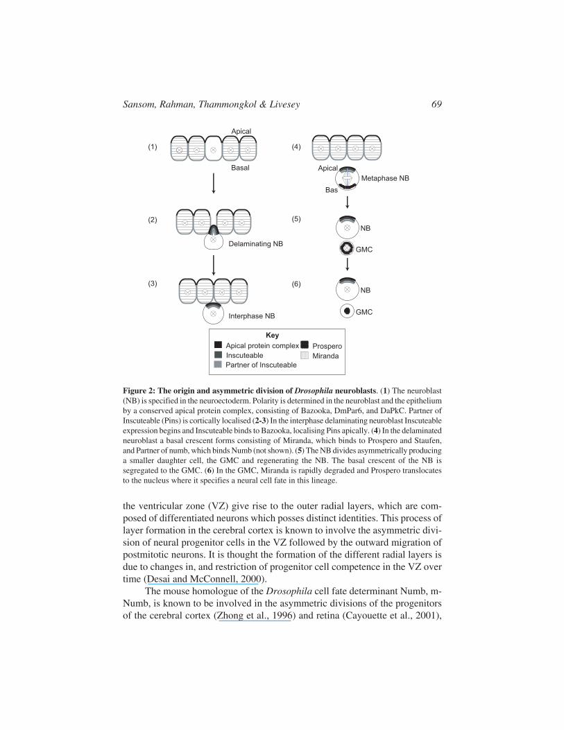
In Drosophila both the CNS progenitors, neuroblasts (NBs), and the PNSprogenitors, the sensory organ precursors (SOPs) undergo a series of asymmet-ric divisions in order to generate characteristic lineages of neurons and glia(Bossing et al., 1996; Gho et al., 1999; Reddy and Rodrigues, 1999; Schmid etal., 1999; Schmidt et al., 1997). After delaminating from the neuroectoderm,Drosophila neuroblasts undergo a series of apical/basal orientated asymmetricdivisions (Figure 2). These asymmetric divisions give rise to a smaller daughtercell, the ganglion mother cell (GMC), which buds off from the dorsal/lateralcortex of the neuroblast. GMCs then divide terminally to give rise to two neu-rons or glia.
The asymmetric division of neuroblasts requires the asymmetric localisationof cell fate determinants and the correct orientation of the mitotic spindle inorder for the proper segregation of cell fate determinants to the GMC daughtercell. The polarity of neuroblasts is established by an apical protein complexconsisting of Bazooka, DaPKC and DmPar6, which also mediates polarity inthe epithelium (Petronczki and Knoblich, 2001; Schober et al., 1999; Wodarz etal., 2000; Wodarz et al., 1999). The cell-fate determinants Prospero, prosperomRNA and Numb, and the adapter molecules that help to localise them whichare Miranda, Staufen, and Partner of numb, respectively, form a basal crescentwithin the neuroblast (Broadus et al., 1998; Hirata et al., 1995; Ikeshima-Kataokaet al., 1997; Knoblich et al., 1995; Li et al., 1997; Lu et al., 1998; Rhyu et al.,1994; Schuldt et al., 1998; Shen et al., 1997; Spana and Doe, 1995). This basalcrescent, which overlies the basal spindle pole of mitotic NBs, segregates to theGMC daughter cell. The neural lineages of the peripheral nervous system aregenerated by the SOPs which undergo a series of asymmetric cell divisions togive rise to four different cell types which together constitute an external senseorgan (Bodmer et al., 1989).
Asymmetric cell division in vertebrates
In vertebrates, the asymmetric cell division of neural progenitors has beenreported in the cortex and in the retina of the rat (Cayouette et al., 2001; Chennand McConnell, 1995). In the mammalian cerebral cortex, neural progenitors of
Sansom, Rahman, Thammongkol & Livesey 69
the ventricular zone (VZ) give rise to the outer radial layers, which are com-posed of differentiated neurons which posses distinct identities. This process oflayer formation in the cerebral cortex is known to involve the asymmetric divi-sion of neural progenitor cells in the VZ followed by the outward migration ofpostmitotic neurons. It is thought the formation of the different radial layers isdue to changes in, and restriction of progenitor cell competence in the VZ overtime (Desai and McConnell, 2000).
The mouse homologue of the Drosophila cell fate determinant Numb, m-Numb, is known to be involved in the asymmetric divisions of the progenitorsof the cerebral cortex (Zhong et al., 1996) and retina (Cayouette et al., 2001),
Figure 2: The origin and asymmetric division of Drosophila neuroblasts. (1) The neuroblast(NB) is specified in the neuroectoderm. Polarity is determined in the neuroblast and the epitheliumby a conserved apical protein complex, consisting of Bazooka, DmPar6, and DaPkC. Partner ofInscuteable (Pins) is cortically localised (2-3) In the interphase delaminating neuroblast Inscuteableexpression begins and Inscuteable binds to Bazooka, localising Pins apically. (4) In the delaminatedneuroblast a basal crescent forms consisting of Miranda, which binds to Prospero and Staufen,and Partner of numb, which binds Numb (not shown). (5) The NB divides asymmetrically producinga smaller daughter cell, the GMC and regenerating the NB. The basal crescent of the NB issegregated to the GMC. (6) In the GMC, Miranda is rapidly degraded and Prospero translocatesto the nucleus where it specifies a neural cell fate in this lineage.
(1)
(2)
(3)
(4)
(5)
(6)
GMC
Interphase NB
Metaphase NB
NB
Apical
Basal
GMC
NB
Delaminating NB
Apical
Bas
Partner of Inscuteable
Inscuteable
Apical protein complex
Key
Miranda
Prospero

Neural Stem Cells: Development and Transplantation70
and is capable of rescuing numb mutant flies (Zhong et al., 1996). In cultures ofcortical progenitor cells, m-Numb has been shown to preferentially localise tothe postmitotic cell in progenitor-neuron divisions, and, in comparison m-Numbinheritance is unbiased in progenitor-progenitor divisions (Shen et al., 2002).Recent video microscopy analysis of retinal progenitor explants has demon-strated that the asymmetric inheritance of Numb between two retinal daughtercells promotes a different fate for each daughters, whereas the symmetric inher-itance of Numb tend to leads to the same fate for both daughter cells (Cayouetteand Raff, 2003). These observations indicate that there is conservation of Numbfunction in the developing nervous systems of Drosophila and vertebrates, andthat the generation of neural lineages by a series of asymmetric divisions is acommon feature of neurogenesis.
Temporal aspects of neural cell fate determination in theDrosophila retina
In the Drosophila retina, neural differentiation is initiated in a posterior toanterior wave which sweeps across the retina, following the morphogenetic fur-row (Ready et al., 1976; Tomlinson and Ready, 1987). In front of the morpho-genetic furrow differentiation is inhibited by notch signaling via hairy (Brownet al., 1995). Behind the furrow, photoreceptor differentiation occurs in an in-variant sequence, starting with the differentiation of single precisely spaced‘founder’ R8 photoreceptor cells (Jarman et al., 1994; Tomlinson and Ready,1987). The R8 photoreceptor founder cells each contribute to one ommatidium,of which there are 750 in the adult retina (Ready et al., 1976). The founder cellsare recruited by a process of lateral inhibition involving notch signaling in whichthe broad initial expression of the proneural gene, atonal, becomes restricted tothe prospective R8 photoreceptor cells (Frankfort and Mardon, 2002).
Unlike the generation of the CNS and sensory organs, the different retinalneurons in Drosophila do not arise by the asymmetric division of a progenitorcell, but are understood to be specified over time by bursts of epidemeral growthfactor (EGF) signaling, which have been shown to be necessary and sufficientfor the generation of the different photoreceptor neurons in the retina (Freeman,1997). It has been suggested that the ability of the retinal precursor cells torespond to EGF signaling changes over time, either due to intrinsic or extrinsicfactors, such that the age of the retinal cell exposed to the EGF signal deter-mines the type of neuron born. In this model, temporal intrinsic information isintegrated with an extrinsic signal to determine the type of neuron generated(Freeman, 1997). In this situation where the same extra cellular signal is used tostimulate each wave of differentiation, the changing internal configuration ofthe neural precursor is likely to be crucial to determine the cell fate it will adopt.

Sansom, Rahman, Thammongkol & Livesey 71
Temporal aspects of neural cell fate determination in thevertebrate retina
In the vertebrate retina, the initial steps of neural cell fate determinationare remarkably similar to those in Drosophila. The first-born neurons are reti-nal ganglion cells, and their production requires the expression of the atonalhomolog, ath5, which as in the Drosophila retina is induced by sonic hedgehogand opposed by a gradient of notch signaling (Neumann and Nuesslein-Volhard,2000). In the vertebrate retina, six types of neurons and one type of glial cell aregenerated during development. The order in which these cell types appear isinvariant across vertebrate species, with the retinal ganglion cells being pro-duced first and rods, bipolar and Müller glial cells last (Carter-Dawson andLaVail, 1979; Cepko et al., 1996a; LaVail et al., 1991; Stiemke and Hollyfield,1995; Young, 1985). Unlike in Drosophila, the neurons of the vertebrate retinaare generated from a pool of actively cycling neural progenitor cells.Neurogenesis in the vertebrate retina is characterised by several features. Firstlyretinal progenitors are multipotent, and can generate more than one or two celltypes (Holt et al., 1988; Turner and Cepko, 1987; Turner et al., 1990; Wetts andFraser, 1988). Secondly, despite the conserved birth order, there is an overlap inthe generation of different retinal cell types (LaVail et al., 1991; Stiemke andHollyfield, 1995; Young, 1985).
Vertebrate retinal progenitor cells are only able to give rise to certain sub-sets of cell types at different stages in development (Austin et al., 1995; Belliveauand Cepko, 1999; Belliveau et al., 2000). It has been shown that whilst extrinsicsignals can regulate the proportion of different cell types being made, they can-not alter the range of cell types generated. Combined with the multipotency ofretinal progenitor cells, these observations led to the proposal of a competencemodel for retinal development (Figure 3), which suggested that retinal progeni-tor cells pass through a series of intrinsically determined configurations, or com-petence states, in each of which they are able to give rise to only a subset of celltypes in response to extracellular signals (Cepko et al., 1996a).
Potentially, competence states might be determined by chromatin modu-lation, transcriptional states, gene expression profiles, translational regulation,protein accumulation/degradation, and by post-translational protein modifica-tion (Livesey and Cepko, 2001). There is some evidence for a transcriptional ortranslational control of progenitor competence. Two markers have been identi-fied which show heterogeneity of expression in retinal progenitors, syntaxin-1aand VC1.1 (Alexiades and Cepko, 1997). Retinal progenitors also display achanging response to mitogens over time, and the level of epidermal growthfactor receptor (EGFR) expression is known to change over time in the retina(Lillien, 1995; Lillien and Cepko, 1992). In addition, the cyclin kinase inhibi-

Neural Stem Cells: Development and Transplantation72
tors (CKI’s) p27 and p57 which regulate cell cycle exit are expressed in differ-ent subsets of progenitors (Dyer and Cepko, 2000b). It has also been suggestedthat the level of p27xic1, another cyclin kinase inhibitor, increases over time inretinal progenitors, and that its accumulation over a certain level is responsiblefor driving the formation of the final retinal cell type, the Müller glial cell(Ohnuma et al., 1999).
The mechanisms that govern the switch between different progenitor statesor competences are unknown. It is possible that intrinsic factors, extrinsic fac-tors or a combination of the two are responsible for changes in progenitor compe-tence. Several types of retinal neuron are known to produce signals that nega-tively feedback on the retinal progenitor cells, regulating the types of neuronswhich they can generate (Bermingham et al., 1999; Reh and Tully, 1986; Waidand McLoon, 1998). A complicating factor in understanding retinal progenitorcompetence is the existence of heterogenous progenitor states at any one time(Brown et al., 1998; Dyer and Cepko, 2000a; Jasoni and Reh, 1996; Levine etal., 2000; Lillien and Cepko, 1992). This progenitor heterogeneity raises thecomplicating possibility that functionally different subsets of retinal progeni-tors exist, and that each subset of progenitors may generate only a selection ofretinal cell types.
Figure 3. The competence model of neural development. Progenitors have been proposed toundergo changes in competence to generate different cell types during the course of development(Cepko et al., 1996). Evidence exists for at least two distinct competence states in the developingvertebrate retina: an early progenitor competence state and a late state. Little is currently knownof how many competence states exist, how they are defined at the molecular level and howprogenitor cells shift between competence states, the latter is illustrated by the middle arrowbetween the two competence states shown.
time

Sansom, Rahman, Thammongkol & Livesey 73
A temporal identity for neural progenitors and their progeny
In the asymmetrically dividing neuroblasts of the Drosophila embryo asequentially expressed group of transcription factors encoded by hunchback,kruppel, castor, pdm and grainyhead has recently been reported (Brody andOdenwald, 2000; Isshiki et al., 2001). While the expression of these factorsoccurs as a temporal sequence in neuroblasts, the daughter ganglion mothercells (GMCs) that they give rise to maintain the expression of the transcriptionfactor expressed in the mother neuroblast at the time that they were born. Hunch-back and Kruppel have been shown to confer birth order specificity on manyneuroblast lineages regardless of whether these lineages result in a neuronal orglial cell fate. How is this cascade of transcription factors regulated?Misexpression studies indicate that hunchback activates the expression ofkruppel, and that Kruppel activates the expression of castor (Isshiki et al., 2001).Hunchback and Kruppel are also known to repress the expression of the nextplus one gene in the sequence. Hunchback represses castor and Kruppel re-presses pdm. Interestingly the overexpression of hunchback has recently beenshown to reset the sequential expression of these transcription factors (Pearsonand Doe, 2003).
The expression of this transcriptional cascade in many different neuro-blast lineages, some of which give rise to neurons and some to glia, suggeststhat it is responsible for conferring a temporal, rather than absolute identity oneach GMC as it is born. As yet no such transcriptional cascade has been identi-fied in vertebrates, although given the similarity of the other steps of neural cellfate determination between flies and vertebrates, the existence of similar mecha-nisms would not be a surprise.
A role is emerging for a novel group of regulatory genes, the microRNAs,in this process. The nematode worm C.elegans homologue of hunchback, hbl-1, has recently been shown to control developmental time and to be regulatedby the microRNA let7 (Abrahante et al., 2003; Lin et al., 2003). Regulatorysites exist in the Drosophila hunchback 3’-untranslated region for the homolo-gous Drosophila microRNAs and it is therefore likely that it too is temporallyregulated in this way. Regulation by microRNA genes may therefore offer anovel mechanism for the temporal control of neurogenesis, in conjunction withthe temporal transcription factor sequence outlined above.
Cell fate determination within a given competence state
Within an intrinsically defined progenitor competence state, cell fate hasbeen shown to be influenced by extrinsic factors, for example by feedback inhi-bition from postmitotic neurons (Belliveau and Cepko, 1999; Reh and Tully,

Neural Stem Cells: Development and Transplantation74
1986; Waid and McLoon, 1998). Such a feedback mechanism has been shownto act on progenitor cells before M phase in order to affect daughter cell fate(Belliveau and Cepko, 1999) and a similar mechanism has been proposed forthe developing neocortex (Desai and McConnell, 2000). It is noteworthy thatextrinsic factors can also act to determine or respecify the fate of postmitoticcells, at least in vitro. For example, it is known that cilary neurotrophic factor(CNTF) and leukaemia inhibitory factor (LIF) can cause cells destined to be-come rods to adopt aspects of the bipolar cell phenotype (Ezzeddine et al., 1997).
Notch signaling is known to be involved in the differentiation of neuronsand glia of the vertebrate retina and developing forebrain. However it is unclearwhether notch signaling has a permissive or instructive role in these processes(Livesey and Cepko, 2001). In the neural crest, transient notch signaling is in-structive in switching neural crest progenitors to neurogenesis, and then togliogenesis (Morrison et al., 2000). In the late retina, notch acts to signal thetransition between neurogenesis and gliogenesis (Furukawa et al., 2000), as italso does in the developing forebrain (Gaiano et al., 2000). Notch signaling istherefore likely to be important in regulating cell fate determination in verte-brates.
NEURAL CELL FATE DETERMINATION ELSEWHEREIN THE VERTEBRATE NERVOUS SYSTEM
In other regions of the developing vertebrate nervous system, cell fatedetermination is similar to the situation in the retina. Both the progenitor cellsof the cortex and spinal cord are multipotent (Briscoe et al., 1999; Leber et al.,1990). In the developing cortex, as in the retina, progenitors give rise to neuronsbefore generating glial cell types (Morrison et al., 2000; Qian et al., 2000b).Cortical progenitors progress through phases reminiscent of the competencestates of retinal progenitors, in which they are competent to produce cells of agiven laminar fate (Morrison et al., 2000; Qian et al., 2000b). However, unlikethe situation in the retina, cortical neural progenitor cells are capable of gener-ating later, but not earlier cell types upon heterochronic transplantation (Desaiand McConnell, 2000; McConnell, 1988). This has lead to the concept of theprogressive restriction model in cortical cell fate determination (Desai andMcConnell, 2000).
Both cortical and spinal cord progenitors can respond to extrinsic factorsthat regulate their cell fate choices. Cortical progenitors are competent to re-spond to extrinsic signals until late S/early G2 in the cell cycle (McConnell,1988), in agreement with the finding that retinal progenitors make cell fatechoices prior to M phase (Belliveau and Cepko, 1999). As in the retina, feed-back signaling is used as a mechanism of neural cell fate determination in the

Sansom, Rahman, Thammongkol & Livesey 75
spinal cord where postmitotic motor neurons induce the genesis of interneurons(Pfaff et al., 1996). Heterogeneity also appears to be a conserved feature ofvertebrate neurogenesis, as different populations of spinal cord progenitors canbe distinguished by the expression of different transcription factors (Briscoe etal., 2000).
Vertebrate neural cell fate determination has many striking parallels withcell fate determination in Drosophila. As in the fly, precursor cells give rise todifferent types of neurons over time, implying that changes in competence (orcell fate potential) are common mechanisms of cell fate determination in bothinvertebrates and vertebrates. Other conserved features of neural cell fate deter-mination are feedback inhibition, multipotency and progenitor heterogeneity.Whilst much still remains to be understood about neural cell fate determination,our existing knowledge and paradigms suggest that with the advent of genomicstechnologies we might soon have a much deeper understanding of key aspectssuch as the changes in neural progenitor cell fate potential over time.
TERMINAL DIFFERENTIATION PROGRAMS
When a postmitotic neuron has been specified to assume a particular fate,it must then terminally differentiate to realize that fate. This process is under-stood to involve locking a cell into a terminal transcriptional program. The tran-scriptional networks involved in terminal differentiation are only just begin-ning to be understood in vertebrates, and several transcription factors have beenidentified which are specific for different neurons. For example, in the retinathe transcription factors Brn3a-c are specific to ganglion cells, Crx is specific tophotoreceptors, and each is required for the full phenotypic differentiation oftheir respective neurons (Furukawa et al., 1997; Furukawa et al., 1999; Gan etal., 1999; Liu et al., 2000b). As discussed above, combinations of homeodomain-containing transcription factors control survival and differentiation of manyclasses of spinal cord neurons (for review, see (Jessell, 2000)).
Several different transcription factors regulating terminal differentiationhave been identified in the worm C.elegans, including the Chx10 homologueceh-10 and several different LIM-domain containing transcription factors (Altun-Gultekin et al., 2001; Hobert and Westphal, 2000). Single transcription factorshave been shown to control distinct aspects of neuronal phenotypes, the moststriking example being definition of dendritic arbor morphology in Drosophilaneurons by different levels of expression of the cut transcription factor (Grueberet al., 2003). It is thought that terminal transcription programs are likely toinvolve autoregulatory loops to maintain the cell specific transcriptional pro-gram.

Neural Stem Cells: Development and Transplantation76
ORIGINS AND LINEAGE RELATIONSHIPS OF ADULTNEURAL STEM CELLS
A key issue for neural stem cell biology, discussed in more detail else-where in this book, is the origin of neural stem cells and their lineage relation-ships to the progenitor cells present during embryonic development. As hasbeen well described, neurosphere-forming cells can be isolated from diverseregions of the adult CNS, including the lateral wall of the lateral ventricle, thehippocampus and the ciliary margin of the retina (for review, see Kuhn andSvendsen, 1999; Temple, 2001; and elsewhere in this volume). The preciselocation of the primary neural stem cell within the lateral wall of the lateralventricle has been studied in some detail (Doetsch et al., 1999; Johansson et al.,1999), with a consensus emerging that a subpopulation of SVZ astrocytes aretrue stem cells. However, there is still much to know about the developmentalorigins of these cells.
Firstly, it is not clear when neural stem cells are generated during devel-opment. Using the classic neurosphere formation assay, typically it is not pos-sible to harvest stem cells until relatively late in development. However, usingthe alternative approach of adherent clone generation, rather than sphere forma-tion, for identifying NSCs suggests that stem cells are present from the earlieststages of neural development (Qian et al., 2000a). Is it possible that NSCs are alate generated cell type, or that key NSC properties are not acquired by endog-enous cycling neural progenitor and stem cells until late in development? Thislatter possibility raises the other pivotal question of which cells are NSCs re-lated to and derived from. Are NSCs actually astrocytes, or a distinct subpopu-lation of astrocyte-like cells, generated late in development? There are strikingsimilarities between astrocytes and progenitor cells in terms of gene expres-sion, as noted by several authors (Fischer and Reh, 2001; Furukawa et al., 2000),and astrocytes are typically the last-born cell type. Radial glia within the devel-oping cerebral cortex have been demonstrated to not only give rise to astrocyteslate in development, but are also neurogenic progenitor cells earlier in develop-ment (Campbell and Gotz, 2002; Hartfuss et al., 2001; Malatesta et al., 2000).So are astrocytes, or a subpopulation thereof, effectively quiescent late stageneural progenitor cells?
If neural stem cells are late stage progenitor cells, this would not be com-patible with multipotency, as it has been demonstrated in both the retina andspinal cord that late progenitor cells are only capable of generating late-borncell types. The potential of NSCs isolated from different regions of the CNS hasnot been analysed in detail, particularly with respect to the temporal order ofnormal neurogenesis. One exception is the ciliary marginal zone of the retina,where the stem cells within this region in amphibia and fish are multipotent,

Sansom, Rahman, Thammongkol & Livesey 77
and give rise to mitotic progenitor cells to go on to produce all of the cells of theadult retina (Perron and Harris, 2000b; Perron et al., 1998).
Thus, it would appear that NSCs are not simply the functional equivalentof late stage progenitor cells. One alternative possibility for the origin of NSCsis that there is a population of neural stem cells distinct from the much largerpopulation of mitotic neural progenitor cells, and that adult NSCs are the directdescendants of these stem cells (Temple, 2001). If this is the case, the ability togenerate neurospheres in culture may be a property that is lacking in early em-bryonic NSCs, but which has been gained by adult NSCs.
CELL FATE DETERMINATION IN ADULT NEURALSTEM CELLS
In contrast with developing neural stem and progenitor cells, little is knownof the basic mechanisms regulating cell fate decision-making in adult NSCs.Most strategies for inducing the neural differentiation of cultured NSCs rely ongrowth factor withdrawal or retinoid exposure, and assay the relative degree ofneuronal and glial differentiation within the culture system, rather than the di-versity of neuronal cell types (Kuhn and Svendsen, 1999). Thus much interesthas focused on the efficiency of neuron generation from neural stem cells, ratherthan on the mechanisms controlling neurogenesis and cell fate determination.However, it is clear that NSCs express many of the same markers as developingneural progenitor cells, and do use the delta-notch pathway to regulate neuraldifferentiation (Morrison et al., 2000). It is likely, therefore, that similar or iden-tical cellular mechanisms and key genes are redeployed in NSCs for the genera-tion of discrete types of neurons.
With regard to the developmental potential of NSCs, transplantation ofNSCs into the adult CNS results in differentiation into regionally appropriatecell types after an initial culture period of weeks to months, typically asneurospheres (Englund et al., 2002; Fricker et al., 1999). However, the degreeof neurogenesis depends on transplantation into one of the areas of adultneurogenesis, such as the SVZ. NSCs that are transplanted outside of theseregions generate mostly glial cells (Wu et al., 2002). Time spent in culture priorto transplantation appears to have striking effects on the potential and/or com-petence of NSCs, suggesting that some form of reprogramming or dedifferen-tiation may take place. Human NSCs are capable of generating region-specificneuronal progeny when transplanted into the adult rat CNS following a series ofin vitro treatments, including growth factor exposure (Wu et al., 2002). In con-trast, in vitro studies indicate that NSCs isolated from different regions of theforebrain are intrinsically different in terms of gene expression and responses tomitogens (Parmar et al., 2002).

Neural Stem Cells: Development and Transplantation78
The ability of tissue culture and growth factor exposure to alter the poten-tial of NSCs appears to be a general phenomenon. Peripheral nerve stem cellsdemonstrate a striking difference in potential when transplanted immediatelyafter harvesting, compared to transplantation following a period of time in cul-ture (Bixby et al., 2002; Kruger et al., 2002; White et al., 2001). Such a changein potential is reminiscent of the change in potential of oligodendrocyte precur-sor cells in vitro from being dedicated to the production of oligodendrocytes tothe ability to form astrocytes and neurons, again through a series of growthfactor treatments (Kondo and Raff, 2000).
Those studies have important implications for our understanding of thebiology underlying progenitor and stem cell potential and competence, and alsofor the practical aspects of manipulating these cells for replacement therapies.Several reports have been made of the plasticity of neural stem cells both withinand outside the nervous system and in the developing chick embryo (Bjornsonet al., 1999; Blau et al., 2001; Clarke et al., 2000; Vescovi et al., 2002). Anumber of alternative mechanisms have been proposed and demonstrated forthis putative plasticity, including cell fusion (Terada et al., 2002; Wurmser andGage, 2002; Ying et al., 2002). However, it is also clear that culture conditionscan have marked effects on the potential of these cells, through unknown cellu-lar mechanisms, a phenomenon that could possibly be harnessed for therapeuticbenefits, but could also be of concern for experimental design and interpreta-tion.
CONCLUSION: IS THERE A GENERAL BLUEPRINTFOR CONTROLLING NEURAL CELL FATEDETERMINATION FROM ES CELLS AND NS CELLS?
Recent findings on the generation of particular classes of neurons from EScells have emphasized the importance of a thorough understanding of the mecha-nisms controlling cell fate determination during development (Kim et al., 2002;Wichterle et al., 2002). In both cases, specific types of neurons were generatedfrom ES cells by first inducing neural differentiation in the ES cells, and thenusing the available knowledge of the extracellular signals and transcription fac-tors required in the production of each cell type. In one case, overexpression ofa transcription factor normally expressed in midbrain dopaminergic neuron pro-genitor cells was used to bias progeny towards that fate (Kim et al., 2002). Inthe other, ES cells were exposed to a series of treatments designed to mimicnormal in vivo development, such that ES cells were first induced to form neu-rogenic tissue and then this tissue was exposed to signals that confer caudal, orspinal cord, fates. Lastly, this tissue was treated with a combination of factors

Sansom, Rahman, Thammongkol & Livesey 79
known to induce motor neuron production and differentiation in the developingneural tube (Wichterle et al., 2002).
These studies have raised the possibility that there is a single frameworkfor generating classes of neurons which can be applied to both ES cells andNSCs isolated from different regions of the CNS. However, they also highlightthe potential difficulties of working with NSCs, as opposed to ES cells. In thecase of ES cells, it is possible to prospectively walk cultured neural tissue downdevelopmental pathways appropriate to the cell type one wishes to produce.This may not be the case for regionally-derived NSCs. As discussed above, weknow little about the cellular status of these NSCs, especially after periods oftime in culture. In particular, it is not clear if NSCs with increased potentialafter time spent in culture are the equivalent of developing neural progenitorcells of a distinct development stage, or whether this represents an independentpathway for neural development or differentiation. Understanding this aspectof NSC biology will be essential for efforts to manipulate NSCs to generatedesired cell types at high efficiencies.
Finally, given the presence of endogenous stem cells in the adult mamma-lian CNS, it is also of interest to ask whether approaches that manipulate suchstem cells are likely to be more successful for stimulating repair than the trans-plantation of exogenous stem cells (See Chapter 12 in this book). In this case, asin the manipulation of NSCs in vitro, an understanding of the competence andpotential of these cells, allied to our increasing knowledge of the general mecha-nisms regulating neurogenesis, will be essential for the rational development oftherapeutics aimed at stimulating those cells to repair the damaged CNS.
REFERENCES
Abrahante, J. E., Daul, A. L., Li, M., Volk, M. L., Tennessen, J. M., Miller, E. A., and Rougvie,A. E. (2003). The Caenorhabditis elegans hunchback-like Gene lin-57/hbl-1 ControlsDevelopomental Time and Is Regulated by MicroRNAs. Developmental Cell 4, 625-637.
Alexiades, M. R., and Cepko, C. L. (1997). Subsets of retinal progenitors display temporallyregulated and distinct biases in the fates of their progeny. Development 124, 1119-1131.
Allen, K. M., and Walsh, C. A. (1999). Genes that regulate neuronal migration in the cerebralcortex. Epilepsy Res 36, 143-154.
Alonso, M. C., and Cabrera, C. V. (1988). The achaete-scute gene complex of Drosophilamelanogaster comprises four homologous genes. Embo J 7, 2585-2591.
Altun-Gultekin, Z., Andachi, Y., Tsalik, E. L., Pilgrim, D., Kohara, Y., and Hobert, O. (2001). Aregulatory cascade of three homeobox genes, ceh-10, ttx-3 and ceh-23, controls cell fatespecification of a defined interneuron class in C. elegans. Development 128, 1951-1969.
Appel, B., Korzh, V., Glasgow, E., Thor, S., Edlund, T., Dawid, I. B., and Eisen, J. S. (1995).Motoneuron fate specification revealed by patterned LIM homeobox gene expression in em-bryonic zebrafish. Development 121, 4117-4125.
Artavanis-Tsakonas, S., Rand, M. D., and Lake, R. J. (1999). Notch signaling: cell fate controland signal integration in development. Science 284, 770-776.

Neural Stem Cells: Development and Transplantation80
Austin, C. P., Feldman, D. E., Ida, J. A., and Cepko, C. L. (1995). Vertebrate retinal ganglioncells are selected from competent progenitors by the action of Notch. Development 121,3637-3650.
Bae, S., Bessho, Y., Hojo, M., and Kageyama, R. (2000). The bHLH gene Hes6, an inhibitor ofHes1, promotes neuronal differentiation. Development 127, 2933-2943.
Baker, N. E. (2000). Notch signaling in the nervous system. Pieces still missing from the puzzle.Bioessays 22, 264-273.
Baker, N. E., Yu, S., and Han, D. (1996). Evolution of proneural atonal expression during distinctregulatory phases in the developing Drosophila eye. Curr Biol 6, 1290-1301.
Bate, C. M. (1976). Embryogenesis of an insect nervous system. I. A map of the thoracic andabdominal neuroblasts in Locusta migratoria. J Embryol Exp Morphol 35, 107-123.
Belliveau, M. J., and Cepko, C. L. (1999). Extrinsic and intrinsic factors control the genesis ofamacrine and cone cells in the rat retina. Development, In press.
Belliveau, M. J., Young, T. L., and Cepko, C. L. (2000). Late retinal progenitor cells show intrin-sic limitations in the production of cell types and the kinetics of opsin synthesis. J Neurosci20, 2247-2254.
Belting, H. G., Shashikant, C. S., and Ruddle, F. H. (1998). Multiple phases of expression andregulation of mouse Hoxc8 during early embryogenesis. J Exp Zool 282, 196-222.
Ben-Arie, N., McCall, A. E., Berkman, S., Eichele, G., Bellen, H. J., and Zoghbi, H. Y. (1996).Evolutionary conservation of sequence and expression of the bHLH protein Atonal suggestsa conserved role in neurogenesis. Hum Mol Genet 5, 1207-1216.
Bermingham, N. A., Hassan, B. A., Price, S. D., Vollrath, M. A., Ben-Arie, N., Eatock, R. A.,Bellen, H. J., Lysakowski, A., and Zoghbi, H. Y. (1999). Math1: an essential gene for thegeneration of inner ear hair cells. Science 284, 1837-1841.
Bermingham, N. A., Hassan, B. A., Wang, V. Y., Fernandez, M., Banfi, S., Bellen, H. J., Fritzsch,B., and Zoghbi, H. Y. (2001). Proprioceptor pathway development is dependent on Math1.Neuron 30, 411-422.
Bertrand, N., Castro, D. S., and Guillemot, F. (2002). Proneural genes and the specification ofneural cell types. Nat Rev Neurosci 3, 517-530.
Bhat, K. M. (1996). The patched signaling pathway mediates repression of gooseberry allowingneuroblast specification by wingless during Drosophila neurogenesis. Development 122, 2921-2932.
Bhat, K. M. (1999). Segment polarity genes in neuroblast formation and identity specificationduring Drosophila neurogenesis. Bioessays 21, 472-485.
Bhat, K. M., and Schedl, P. (1997). Requirement for engrailed and invected genes reveals novelregulatory interactions between engrailed/invected, patched, gooseberry and wingless duringDrosophila neurogenesis. Development 124, 1675-1688.
Bixby, S., Kruger, G. M., Mosher, J. T., Joseph, N. M., and Morrison, S. J. (2002). Cell-intrinsicdifferences between stem cells from different regions of the peripheral nervous system regu-late the generation of neural diversity. Neuron 35, 643-656.
Bjornson, C. R., Rietze, R. L., Reynolds, B. A., Magli, M. C., and Vescovi, A. L. (1999). Turningbrain into blood: a hematopoietic fate adopted by adult neural stem cells in vivo. Science 283,534-537.
Blader, P., Fischer, N., Gradwohl, G., Guillemont, F., and Strahle, U. (1997). The activity ofneurogenin1 is controlled by local cues in the zebrafish embryo. Development 124, 4557-4569.
Blau, H. M., Brazelton, T. R., and Weimann, J. M. (2001). The evolving concept of a stem cell:entity or function? Cell 105, 829-841.
Blochlinger, K., Jan, L. Y., and Jan, Y. N. (1991). Transformation of sensory organ identity byectopic expression of Cut in Drosophila. Genes Dev 5, 1124-1135.

Sansom, Rahman, Thammongkol & Livesey 81
Bodmer, R., Carretto, R., and Jan, Y. N. (1989). Neurogenesis of the peripheral nervous systemin Drosophila embryos: DNA replication patterns and cell lineages. Neuron 3, 21-32.
Bossing, T., Udolph, G., Doe, C. Q., and Technau, G. M. (1996). The embryonic central nervoussystem lineages of Drosophila melanogaster. I. Neuroblast lineages derived from the ventralhalf of the neuroectoderm. Dev Biol 179, 41-64.
Briscoe, J., Chen, Y., Jessell, T. M., and Struhl, G. (2001). A hedgehog-insensitive form of patchedprovides evidence for direct long-range morphogen activity of sonic hedgehog in the neuraltube. Mol Cell 7, 1279-1291.
Briscoe, J., Pierani, A., Jessell, T. M., and Ericson, J. (2000). A homeodomain protein codespecifies progenitor cell identity and neuronal fate in the ventral neural tube. Cell 101, 435-445.
Briscoe, J., Sussel, L., Serup, P., Hartigan-O’Connor, D., Jessell, T. M., Rubenstein, J. L., andEricson, J. (1999). Homeobox gene Nkx2.2 and specification of neuronal identity by gradedSonic hedgehog signalling. Nature 398, 622-627.
Broadus, J., Fuerstenberg, S., and Doe, C. Q. (1998). Staufen-dependent localization of prosperomRNA contributes to neuroblast daughter-cell fate. Nature 391, 792-795.
Brody, T., and Odenwald, W. F. (2000). Programmed transformations in neuroblast gene expres-sion during Drosophila CNS lineage development. Dev Biol 226, 34-44.
Brown, N. L., Kanekar, S., Vetter, M. L., Tucker, P. K., Gemza, D. L., and Glaser, T. (1998).Math5 encodes a murine basic helix-loop-helix transcription factor expressed during earlystages of retinal neurogenesis. Development 125, 4821-4833.
Brown, N. L., Sattler, C. A., Paddock, S. W., and Carroll, S. B. (1995). Hairy and emc negativelyregulate morphogenetic furrow progression in the Drosophila eye. Cell 80, 879-887.
Buescher, M., and Chia, W. (1997). Mutations in lottchen cause cell fate transformations in bothneuroblast and glioblast lineages in the Drosophila embryonic central nervous system. De-velopment 124, 673-681.
Burke, A. C., Nelson, C. E., Morgan, B. A., and Tabin, C. (1995). Hox genes and the evolution ofvertebrate axial morphology. Development 121, 333-346.
Cabrera, C. V. (1990). Lateral inhibition and cell fate during neurogenesis in Drosophila: theinteractions between scute, Notch and Delta. Development 110, 733-742.
Cabrera, C. V., and Alonso, M. C. (1991). Transcriptional activation by heterodimers of theachaete-scute and daughterless gene products of Drosophila. Embo J 10, 2965-2973.
Cabrera, C. V., Martinez-Arias, A., and Bate, M. (1987). The expression of three members of theachaete-scute gene complex correlates with neuroblast segregation in Drosophila. Cell 50,425-433.
Campbell, K., and Gotz, M. (2002). Radial glia: multi-purpose cells for vertebrate brain develop-ment. Trends Neurosci 25, 235-238.
Campuzano, S. (2001). Emc, a negative HLH regulator with multiple functions in Drosophiladevelopment. Oncogene 20, 8299-8307.
Campuzano, S., and Modolell, J. (1992). Patterning of the Drosophila nervous system: the achaete-scute gene complex. Trends Genet 8, 202-208.
Carter-Dawson, L. D., and LaVail, M. M. (1979). Rods and cones in the mouse retina. II. Auto-radiographic analysis of cell generation using tritiated thymidine. J Comp Neurol 188, 263-272.
Casarosa, S., Fode, C., and Guillemot, F. (1999). Mash1 regulates neurogenesis in the ventraltelencephalon. Development 126, 525-534.
Cau, E., Casarosa, S., and Guillemot, F. (2002). Mash1 and Ngn1 control distinct steps of deter-mination and differentiation in the olfactory sensory neuron lineage. Development 129, 1871-1880.

Neural Stem Cells: Development and Transplantation82
Cayouette, M., and Raff, M. (2003). The orientation of cell division influences cell-fate choice inthe developing mammalian retina. Development 130, 2329-2339.
Cayouette, M., Whitmore, A. V., Jeffery, G., and Raff, M. (2001). Asymmetric segregation ofNumb in retinal development and the influence of the pigmented epithelium. J Neurosci 21,5643-5651..
Cepko, C. L., Austin, C. P., Yang, X., Alexiades, M., and Ezzeddine, D. (1996). Cell fate de-termination in the vertebrate retina. Proc Natl Acad Sci USA 93, 589-595.
Chen, G., Fernandez, J., Mische, S., and Courey, A. J. (1999). A functional interaction betweenthe histone deacetylase Rpd3 and the corepressor groucho in Drosophila development. GenesDev 13, 2218-2230.
Chenn, A., and McConnell, S. K. (1995). Cleavage orientation and the asymmetric inheritance ofNotch1 immunoreactivity in mammalian neurogenesis. Cell 82, 631-641.
Chien, C. T., Hsiao, C. D., Jan, L. Y., and Jan, Y. N. (1996). Neuronal type information encodedin the basic-helix-loop-helix domain of proneural genes. Proc Natl Acad Sci U S A 93, 13239-13244.
Chitnis, A., Henrique, D., Lewis, J., Ish-Horowicz, D., and Kintner, C. (1995). Primary neurogenesisin Xenopus embryos regulated by a homologue of the Drosophila neurogenic gene Delta.Nature 375, 761-766.
Chitnis, A., and Kintner, C. (1996). Sensitivity of proneural genes to lateral inhibition affects thepattern of primary neurons in Xenopus embryos. Development 122, 2295-2301.
Chu-LaGraff, Q., and Doe, C. Q. (1993). Neuroblast specification and formation regulated bywingless in the Drosophila CNS. Science 261, 1594-1597.
Clarke, D. L., Johansson, C. B., Wilbertz, J., Veress, B., Nilsson, E., Karlstrom, H., Lendahl, U.,and Frisen, J. (2000). Generalized potential of adult neural stem cells. Science 288, 1660-1663.
Cornell, R. A., and Ohlen, T. V. (2000). Vnd/nkx, ind/gsh, and msh/msx: conserved regulators ofdorsoventral neural patterning? Curr Opin Neurobiol 10, 63-71.
Cubas, P., de Celis, J. F., Campuzano, S., and Modolell, J. (1991). Proneural clusters of achaete-scute expression and the generation of sensory organs in the Drosophila imaginal wing disc.Genes Dev 5, 996-1008.
Culi, J., and Modolell, J. (1998). Proneural gene self-stimulation in neural precursors: an essen-tial mechanism for sense organ development that is regulated by Notch signaling. Genes Dev12, 2036-2047.
Davis, R. L., and Turner, D. L. (2001). Vertebrate hairy and Enhancer of split related proteins:transcriptional repressors regulating cellular differentiation and embryonic patterning.Oncogene 20, 8342-8357.
de la Cruz, C. C., Der-Avakian, A., Spyropoulos, D. D., Tieu, D. D., and Carpenter, E. M. (1999).Targeted disruption of Hoxd9 and Hoxd10 alters locomotor behavior, vertebral identity, andperipheral nervous system development. Dev Biol 216, 595-610.
Desai, A. R., and McConnell, S. K. (2000). Progressive restriction in fate potential by neuralprogenitors during cerebral cortical development. Development 127, 2863-2872.
Ding, Q., Motoyama, J., Gasca, S., Mo, R., Sasaki, H., Rossant, J., and Hui, C. C. (1998). Dimin-ished Sonic hedgehog signaling and lack of floor plate differentiation in Gli2 mutant mice.Development 125, 2533-2543.
Doe, C. Q. (1992). Molecular markers for identified neuroblasts and ganglion mother cells in theDrosophila central nervous system. Development 116, 855-863.
Doe, C. Q., and Goodman, C. S. (1985). Early events in insect neurogenesis. I. Development andsegmental differences in the pattern of neuronal precursor cells. Dev Biol 111, 193-205.

Sansom, Rahman, Thammongkol & Livesey 83
Doetsch, F., Caille, I., Lim, D. A., Garcia-Verdugo, J. M., and Alvarez-Buylla, A. (1999).Subventricular zone astrocytes are neural stem cells in the adult mammalian brain. Cell 97,703-716.
Dokucu, M. E., Zipursky, S. L., and Cagan, R. L. (1996). Atonal, rough and the resolution ofproneural clusters in the developing Drosophila retina. Development 122, 4139-4147.
Dominguez, M., and Campuzano, S. (1993). asense, a member of the Drosophila achaete-scutecomplex, is a proneural and neural differentiation gene. Embo J 12, 2049-2060.
Dubois, L., Bally-Cuif, L., Crozatier, M., Moreau, J., Paquereau, L., and Vincent, A. (1998).XCoe2, a transcription factor of the Col/Olf-1/EBF family involved in the specification ofprimary neurons in Xenopus. Curr Biol 8, 199-209.
Dyer, M. A., and Cepko, C. L. (2000a). Control of Müller glial cell proliferation and activationfollowing retinal injury. Nat Neurosci 3, 873-880.
Dyer, M. A., and Cepko, C. L. (2000b). p57(Kip2) regulates progenitor cell proliferation andamacrine interneuron development in the mouse retina. Development 127, 3593-3605.
Edlund, T., and Jessell, T. M. (1999). Progression from extrinsic to intrinsic signaling in cell fatespecification: a view from the nervous system. Cell 96, 211-224.
Edmondson, D. G., and Roth, S. Y. (1998). Interactions of transcriptional regulators with his-tones. Methods 15, 355-364.
Edmondson, D. G., Smith, M. M., and Roth, S. Y. (1996). Repression domain of the yeast globalrepressor Tup1 interacts directly with histones H3 and H4. Genes Dev 10, 1247-1259.
Englund, U., Bjorklund, A., Wictorin, K., Lindvall, O., and Kokaia, M. (2002). Grafted neuralstem cells develop into functional pyramidal neurons and integrate into host cortical cir-cuitry. Proc Natl Acad Sci U S A 99, 17089-17094.
Ensini, M., Tsuchida, T. N., Belting, H. G., and Jessell, T. M. (1998). The control of rostrocaudalpattern in the developing spinal cord: specification of motor neuron subtype identity is initi-ated by signals from paraxial mesoderm. Development 125, 969-982.
Ericson, J., Briscoe, J., Rashbass, P., van Heyningen, V., and Jessell, T. M. (1997a). Graded sonichedgehog signaling and the specification of cell fate in the ventral neural tube. Cold SpringHarb Symp Quant Biol 62, 451-466.
Ericson, J., Morton, S., Kawakami, A., Roelink, H., and Jessell, T. M. (1996). Two critical peri-ods of Sonic Hedgehog signaling required for the specification of motor neuron identity. Cell87, 661-673.
Ericson, J., Rashbass, P., Schedl, A., Brenner-Morton, S., Kawakami, A., van Heyningen, V.,Jessell, T. M., and Briscoe, J. (1997b). Pax6 controls progenitor cell identity and neuronalfate in response to graded Shh signaling. Cell 90, 169-180.
Ezzeddine, Z. D., Yang, X., DeChiara, T., Yancopoulos, G., and Cepko, C. L. (1997). Postmitoticcells fated to become rod photoreceptors can be respecified by CNTF treatment of the retina.Development 124, 1055-1067.
Farah, M. H., Olson, J. M., Sucic, H. B., Hume, R. I., Tapscott, S. J., and Turner, D. L. (2000).Generation of neurons by transient expression of neural bHLH proteins in mammalian cells.Development 127, 693-702.
Fehon, R. G., Johansen, K., Rebay, I., and Artavanis-Tsakonas, S. (1991). Complex cellular andsubcellular regulation of notch expression during embryonic and imaginal development ofDrosophila: implications for notch function. J Cell Biol 113, 657-669.
Fischer, A. J., and Reh, T. A. (2001). Müller glia are a potential source of neural regeneration inthe postnatal chicken retina. Nat Neurosci 4, 247-252.
Fode, C., Gradwohl, G., Morin, X., Dierich, A., LeMeur, M., Goridis, C., and Guillemot, F.(1998). The bHLH protein NEUROGENIN 2 is a determination factor for epibranchial pla-code-derived sensory neurons. Neuron 20, 483-494.

Neural Stem Cells: Development and Transplantation84
Fode, C., Ma, Q., Casarosa, S., Ang, S. L., Anderson, D. J., and Guillemot, F. (2000). A role forneural determination genes in specifying the dorsoventral identity of telencephalic neurons.Genes Dev 14, 67-80.
Frankfort, B. J., and Mardon, G. (2002). R8 development in the Drosophila eye: a paradigm forneural selection and differentiation. Development 129, 1295-1306.
Freeman, M. (1997). Cell determination strategies in the Drosophila eye. Development 124, 261-270.
Fricker, R. A., Carpenter, M. K., Winkler, C., Greco, C., Gates, M. A., and Bjorklund, A. (1999).Site-specific migration and neuronal differentiation of human neural progenitor cells aftertransplantation in the adult rat brain. J Neurosci 19, 5990-6005.
Furukawa, T., Morrow, E. M., and Cepko, C. L. (1997). Crx, a novel otx-like homeobox gene,shows photoreceptor-specific expression and regulates photoreceptor differentiation. Cell 91,531-541.
Furukawa, T., Morrow, E. M., Li, T., Davis, F. C., and Cepko, C. L. (1999). Retinopathy andattenuated circadian entrainment in Crx-deficient mice. Nat Genet 23, 466-470.
Furukawa, T., Mukherjee, S., Bao, Z. Z., Morrow, E. M., and Cepko, C. L. (2000). rax, Hes1, andnotch1 promote the formation of Müller glia by postnatal retinal progenitor cells. Neuron 26,383-394.
Gaiano, N., Nye, J. S., and Fishell, G. (2000). Radial glial identity is promoted by Notch1 signal-ing in the murine forebrain. Neuron 26, 395-404.
Gan, L., Wang, S. W., Huang, Z., and Klein, W. H. (1999). POU domain factor Brn-3b is essen-tial for retinal ganglion cell differentiation and survival but not for initial cell fate specifica-tion. Dev Biol 210, 469-480.
Garcia-Bellido, A. (1979). Genetic analysis of the achaete-scute system of DrosophilaMelanogaster. Genetics 91, 491-520.
Gho, M., Bellaiche, Y., and Schweisguth, F. (1999). Revisiting the Drosophila microchaete lin-eage: a novel intrinsically asymmetric cell division generates a glial cell. Development 126,3573-3584.
Ghysen, A., and Dambly-Chaudiere, C. (1988). From DNA to form: the achaete-scute complex.Genes Dev 2, 495-501.
Gonzalez, F., Romani, S., Cubas, P., Modolell, J., and Campuzano, S. (1989). Molecular analysisof the asense gene, a member of the achaete-scute complex of Drosophila melanogaster, andits novel role in optic lobe development. Embo J 8, 3553-3562.
Goridis, C., and Brunet, J. F. (1999). Transcriptional control of neurotransmitter phenotype. CurrOpin Neurobiol 9, 47-53.
Goridis, C., and Rohrer, H. (2002). Specification of catecholaminergic and serotonergic neurons.Nat Rev Neurosci 3, 531-541.
Goulding, S. E., White, N. M., and Jarman, A. P. (2000a). cato encodes a basic helix-loop-helixtranscription factor implicated in the correct differentiation of Drosophila sense organs. DevBiol 221, 120-131.
Goulding, S. E., zur Lage, P., and Jarman, A. P. (2000b). amos, a proneural gene for Drosophilaolfactory sense organs that is regulated by lozenge. Neuron 25, 69-78.
Gowan, K., Helms, A. W., Hunsaker, T. L., Collisson, T., Ebert, P. J., Odom, R., and Johnson, J.E. (2001). Crossinhibitory activities of Ngn1 and Math1 allow specification of distinct dorsalinterneurons. Neuron 31, 219-232.
Gradwohl, G., Fode, C., and Guillemot, F. (1996). Restricted expression of a novel murine atonal-related bHLH protein in undifferentiated neural precursors. Dev Biol 180, 227-241.
Grueber, W. B., Jan, L. Y., and Jan, Y. N. (2003). Different levels of the homeodomain proteincut regulate distinct dendrite branching patterns of Drosophila multidendritic neurons. Cell112, 805-818.
Sansom, Rahman, Thammongkol & Livesey 85
Guillemot, F. (1999). Vertebrate bHLH genes and the determination of neuronal fates. Exp CellRes 253, 357-364.
Guillemot, F., and Joyner, A. L. (1993). Dynamic expression of the murine Achaete-Scute homo-logue Mash-1 in the developing nervous system. Mechanisms of Development 42, 171-185.
Guillemot, F., Lo, L. C., Johnson, J. E., Auerbach, A., Anderson, D. J., and Joyner, A. L. (1993).Mammalian achaete-scute homolog 1 is required for the early development of olfactory andautonomic neurons. Cell 75, 463-476.
Harland, R. (2000). Neural induction. Curr Opin Genet Dev 10, 357-362.Hartenstein, V., and Campos-Ortega, J. A. (1984). Early neurogenesis in wild type Drosophila
melanogaster. Roux’s Arch Dev Biol 193, 308-325.Hartenstein, V., and Posakony, J. W. (1990). A dual function of the Notch gene in Drosophila
sensillum development. Developmental Biology 142, 13-30.Hartfuss, E., Galli, R., Heins, N., and Gotz, M. (2001). Characterization of CNS precursor sub-
types and radial glia. Dev Biol 229, 15-30.Hassan, B. A., and Bellen, H. J. (2000). Doing the MATH: is the mouse a good model for fly
development? Genes Dev 14, 1852-1865.Hatakeyama, J., Tomita, K., Inoue, T., and Kageyama, R. (2001). Roles of homeobox and bHLH
genes in specification of a retinal cell type. Development 128, 1313-1322.Heitzler, P., Bourouis, M., Ruel, L., Carteret, C., and Simpson, P. (1996). Genes of the Enhancer
of split and achaete-scute complexes are required for a regulatory loop between Notch andDelta during lateral signalling in Drosophila. Development 122, 161-171.
Heitzler, P., and Simpson, P. (1991). The choice of cell fate in the epidermis of Drosophila. Cell64, 1083-1092.
Helms, A. W., Abney, A. L., Ben-Arie, N., Zoghbi, H. Y., and Johnson, J. E. (2000). Autoregu-lation and multiple enhancers control Math1 expression in the developing nervous system.Development 127, 1185-1196.
Henrique, D., Tyler, D., Kintner, C., Heath, J., Lewis, J., Ish-Horowicz, D., and Storey, K. (1997).cash4, a novel achaete-scute homolog induced by Hensen’s node during generation of theposterior nervous system. Genes Dev 11, 603-615.
Hinz, U., Giebel, B., and Campos-Ortega, J. A. (1994). The basic-helix-loop-helix domain ofDrosophila lethal of scute protein is sufficient for proneural function and activates neuro-genic genes. Cell 76, 77-87.
Hirata, J., Nakagoshi, H., Nabeshima, Y., and Matsuzaki, F. (1995). Asymmetric segregation ofthe homeodomain protein Prospero during Drosophila development. Nature 377, 627-630.
Hirsch, M. R., Tiveron, M. C., Guillemot, F., Brunet, J. F., and Goridis, C. (1998). Control ofnoradrenergic differentiation and Phox2a expression by MASH1 in the central and peripheralnervous system. Development 125, 599-608.
Hobert, O., and Westphal, H. (2000). Functions of LIM-homeobox genes. Trends Genet 16, 75-83.
Holt, C. E., Bertsch, T. W., Ellis, H. M., and Harris, W. A. (1988). Cellular determination in theXenopus retina is independent of lineage and birth date. Neuron 1, 15-26.
Horton, S., Meredith, A., Richardson, J. A., and Johnson, J. E. (1999). Correct coordination ofneuronal differentiation events in ventral forebrain requires the bHLH factor MASH1. MolCell Neurosci 14, 355-369.
Huang, H. P., Liu, M., El-Hodiri, H. M., Chu, K., Jamrich, M., and Tsai, M. J. (2000a). Regula-tion of the pancreatic islet-specific gene BETA2 (neuroD) by neurogenin 3. Mol Cell Biol 20,3292-3307.
Huang, M. L., Hsu, C. H., and Chien, C. T. (2000b). The proneural gene amos promotes multipledendritic neuron formation in the Drosophila peripheral nervous system. Neuron 25, 57-67.

Neural Stem Cells: Development and Transplantation86
Ikeshima-Kataoka, H., Skeath, J. B., Nabeshima, Y., Doe, C. Q., and Matsuzaki, F. (1997). Mirandadirects Prospero to a daughter cell during Drosophila asymmetric divisions. Nature 390, 625-629.
Inoue, T., Hojo, M., Bessho, Y., Tano, Y., Lee, J. E., and Kageyama, R. (2002). Math3 andNeuroD regulate amacrine cell fate specification in the retina. Development 129, 831-842.
Isshiki, T., Pearson, B., Holbrook, S., and Doe, C. Q. (2001). Drosophila neuroblasts sequen-tially express transcription factors which specify the temporal identity of their neuronal prog-eny. Cell 106, 511-521.
Isshiki, T., Takeichi, M., and Nose, A. (1997). The role of the msh homeobox gene during Droso-phila neurogenesis: implication for the dorsoventral specification of the neuroectoderm. De-velopment 124, 3099-3109.
Itasaki, N., Sharpe, J., Morrison, A., and Krumlauf, R. (1996). Reprogramming Hox expressionin the vertebrate hindbrain: influence of paraxial mesoderm and rhombomere transposition.Neuron 16, 487-500.
Jan, Y. N., and Jan, L. Y. (1993). HLH proteins, fly neurogenesis, and vertebrate myogenesis.Cell 75, 827-830.
Jan, Y. N., and Jan, L. Y. (1994). Genetic control of cell fate specification in Drosophila periph-eral nervous system. Annu Rev Genet 28, 373-393.
Jarman, A. P., and Ahmed, I. (1998). The specificity of proneural genes in determining Droso-phila sense organ identity. Mech Dev 76, 117-125.
Jarman, A. P., Grau, Y., Jan, L. Y., and Jan, Y. N. (1993). atonal is a proneural gene that directschordotonal organ formation in the Drosophila peripheral nervous system. Cell 73, 1307-1321.
Jarman, A. P., Grell, E. H., Ackerman, L., Jan, L. Y., and Jan, Y. N. (1994). Atonal is the proneuralgene for Drosophila photoreceptors. Nature 369, 398-400.
Jasoni, C. L., and Reh, T. A. (1996). Temporal and spatial pattern of MASH-1 expression in thedeveloping rat retina demonstrates progenitor cell heterogeneity. J Comp Neurol 369, 319-327.
Jennings, B., Preiss, A., Delidakis, C., and Bray, S. (1994). The Notch signalling pathway isrequired for Enhancer of split bHLH protein expression during neurogenesis in the Droso-phila embryo. Development 120, 3537-3548.
Jessell, T. M. (2000). Neuronal specification in the spinal cord: inductive signals and transcrip-tional codes. Nat Rev Genet 1, 20-29.
Jimenez, F., and Campos-Ortega, J. A. (1990). Defective neuroblast commitment in mutants ofthe achaete-scute complex and adjacent genes of D. melanogaster. Neuron 5, 81-89.
Johansson, C. B., Momma, S., Clarke, D. L., Risling, M., Lendahl, U., and Frisen, J. (1999).Identification of a neural stem cell in the adult mammalian central nervous system. Cell 96,25-34.
Johnson, J. E., Birren, S. J., Saito, T., and Anderson, D. J. (1992). DNA binding and transcrip-tional regulatory activity of mammalian achaete-scute homologous (MASH) proteins revealedby interaction with a muscle-specific enhancer. Proc Natl Acad Sci U S A 89, 3596-3600.
Kageyama, R., and Nakanishi, S. (1997). Helix-loop-helix factors in growth and differentiationof the vertebrate nervous system. Curr Opin Genet Dev 7, 659-665.
Keynes, R., and Krumlauf, R. (1994). Hox genes and regionalization of the nervous system.Annu Rev Neurosci 17, 109-132.
Kim, J. H., Auerbach, J. M., Rodriguez-Gomez, J. A., Velasco, I., Gavin, D., Lumelsky, N., Lee,S. H., Nguyen, J., Sanchez-Pernaute, R., Bankiewicz, K., and McKay, R. (2002). Dopamineneurons derived from embryonic stem cells function in an animal model of Parkinson’s dis-ease. Nature 418, 50-56.

Sansom, Rahman, Thammongkol & Livesey 87
Kintner, C. (2002). Neurogenesis in embryos and in adult neural stem cells. J Neurosci 22, 639-643.
Knoblich, J. A., Jan, L. Y., and Jan, Y. N. (1995). Asymmetric segregation of Numb and Prosperoduring cell division. Nature 377, 624-627.
Kondo, T., and Raff, M. (2000). Oligodendrocyte precursor cells reprogrammed to become mul-tipotential CNS stem cells. Science 289, 1754-1757.
Kooh, P. J., Fehon, R. G., and Muskavitch, M. A. (1993). Implications of dynamic patterns ofDelta and Notch expression for cellular interactions during Drosophila development. Devel-opment 117, 493-507.
Koyano-Nakagawa, N., Kim, J., Anderson, D., and Kintner, C. (2000). Hes6 acts in a positivefeedback loop with the neurogenins to promote neuronal differentiation. Development 127,4203-4216.
Koyano-Nakagawa, N., Wettstein, D., and Kintner, C. (1999). Activation of Xenopus genes re-quired for lateral inhibition and neuronal differentiation during primary neurogenesis. MolCell Neurosci 14, 327-339.
Krishnan, V., Pereira, F. A., Qiu, Y., Chen, C. H., Beachy, P. A., Tsai, S. Y., and Tsai, M. J.(1997). Mediation of Sonic hedgehog-induced expression of COUP-TFII by a protein phos-phatase. Science 278, 1947-1950.
Kruger, G. M., Mosher, J. T., Bixby, S., Joseph, N., Iwashita, T., and Morrison, S. J. (2002).Neural crest stem cells persist in the adult gut but undergo changes in self-renewal, neuronalsubtype potential, and factor responsiveness. Neuron 35, 657-669.
Kuhn, H. G., and Svendsen, C. N. (1999). Origins, functions, and potential of adult neural stemcells. Bioessays 21, 625-630.
Kunisch, M., Haenlin, M., and Campos-Ortega, J. A. (1994). Lateral inhibition mediated by theDrosophila neurogenic gene delta is enhanced by proneural proteins. Proc Natl Acad Sci U SA 91, 10139-10143.
Lance-Jones, C., Omelchenko, N., Bailis, A., Lynch, S., and Sharma, K. (2001). Hoxd10 induc-tion and regionalization in the developing lumbosacral spinal cord. Development 128, 2255-2268.
LaVail, M. M., Rapaport, D. H., and Rakic, P. (1991). Cytogenesis in the monkey retina. J CompAnat 309, 86-114.
Leber, S., Breedlove, S., and Sanes, J. (1990). Lineage, arrangement, and death of clonally re-lated motoneurons in the chick spinal cord. J Neuroscience 10, 2451-2462.
Lee, J. E. (1997). Basic helix-loop-helix genes in neural development. Curr Opin Neurobiol 7,13-20.
Lee, J. E., Hollenberg, S. M., Snider, L., Turner, D. L., Lipnick, N., and Weintraub, H. (1995).Conversion of Xenopus ectoderm into neurons by NeuroD, a basic helix-loop-helix protein.Science 268, 836-844.
Lee, S. K., and Pfaff, S. L. (2001). Transcriptional networks regulating neuronal identity in thedeveloping spinal cord. Nat Neurosci 4 Suppl, 1183-1191.
Lehmann, R., Jimenez, F., Dietrich, U., and Campos-Ortega, J. A. (1983). On the phenotype anddevelopment of mutants of early neurogenesis in Drosophila Melanogaster. 192, 62-74.
Levine, E. M., Close, J., Fero, M., Ostrovsky, A., and Reh, T. A. (2000). p27(Kip1) regulates cellcycle withdrawal of late multipotent progenitor cells in the mammalian retina. Dev Biol 219,299-314.
Lewis, J. (1998). Notch signalling and the control of cell fate choices in vertebrates. Semin CellDev Biol 9, 583-589.
Li, P., Yang, X., Wasser, M., Cai, Y., and Chia, W. (1997). Inscuteable and Staufen mediateasymmetric localization and segregation of prospero RNA during Drosophila neuroblast celldivisions. Cell 90, 437-447.
Neural Stem Cells: Development and Transplantation88
Lieber, T., Kidd, S., Alcamo, E., Corbin, V., and Young, M. W. (1993). Antineurogenic pheno-types induced by truncated Notch proteins indicate a role in signal transduction and maypoint to a novel function for Notch in nuclei. Genes Dev 7, 1949-1965.
Lillien, L. (1995). Changes in retinal cell fate induced by overexpression of EGF receptor. Nature377, 158-162.
Lillien, L., and Cepko, C. (1992). Control of proliferation in the retina: temporal changes inresponsiveness to FGF and TGF alpha. Development 115, 253-266.
Lin, S., Johnson, S. M., Abraham, M., Vella, M. C., Pasquinelli, A., Gamberi, C., Gottlieb, E.,and Slack, F. (2003). The C.elegans hunchback homolog, hbl-1, Controls Temporal Pattern-ing and Is a Probable MicroRNA Target. Developmental Cell 4, 639-650.
Litingtung, Y., and Chiang, C. (2000). Specification of ventral neuron types is mediated by anantagonistic interaction between Shh and Gli3. Nat Neurosci 3, 979-985.
Liu, M., Pleasure, S. J., Collins, A. E., Noebels, J. L., Naya, F. J., Tsai, M. J., and Lowenstein, D.H. (2000a). Loss of BETA2/NeuroD leads to malformation of the dentate gyrus and epilepsy.Proc Natl Acad Sci U S A 97, 865-870.
Liu, W., Khare, S. L., Liang, X., Peters, M. A., Liu, X., Cepko, C. L., and Xiang, M. (2000b). AllBrn3 genes can promote retinal ganglion cell differentiation in the chick. Development 127,3237-3247.
Livesey, F. J., and Cepko, C. L. (2001). Vertebrate neural cell-fate determination: lessons fromthe retina. Nat Rev Neurosci 2, 109-118.
Lo, L., Dormand, E., Greenwood, A., and Anderson, D. J. (2002). Comparison of the genericneuronal differentiation and neuron subtype specification functions of mammalian achaete-scute and atonal homologs in cultured neural progenitor cells. Development 129, 1553-1567.
Lo, L., Tiveron, M. C., and Anderson, D. J. (1998). MASH1 activates expression of the pairedhomeodomain transcription factor Phox2a, and couples pan-neuronal and subtype-specificcomponents of autonomic neuronal identity. Development 125, 609-620.
Lu, B., Rothenberg, M., Jan, L. Y., and Jan, Y. N. (1998). Partner of Numb colocalizes withNumb during mitosis and directs Numb asymmetric localization in Drosophila neural andmuscle progenitors. Cell 95, 225-235.
Lu, Q. R., Yuk, D., Alberta, J. A., Zhu, Z., Pawlitzky, I., Chan, J., McMahon, A. P., Stiles, C. D.,and Rowitch, D. H. (2000). Sonic hedgehog—regulated oligodendrocyte lineage genes en-coding bHLH proteins in the mammalian central nervous system. Neuron 25, 317-329.
Lumsden, A., and Krumlauf, R. (1996). Patterning the Vertebrate Neuraxis. Science 274, 1109-1115.
Ma, Q., Chen, Z., del Barco Barrantes, I., de la Pompa, J. L., and Anderson, D. J. (1998).neurogenin1 is essential for the determination of neuronal precursors for proximal cranialsensory ganglia. Neuron 20, 469-482.
Ma, Q., Fode, C., Guillemot, F., and Anderson, D. J. (1999). Neurogenin1 and neurogenin2control two distinct waves of neurogenesis in developing dorsal root ganglia. Genes Dev 13,1717-1728.
Ma, Q., Kintner, C., and Anderson, D. J. (1996). Identification of neurogenin, a vertebrate neu-ronal determination gene. Cell 87, 43-52.
Malatesta, P., Hartfuss, E., and Gotz, M. (2000). Isolation of radial glial cells by fluorescent-activated cell sorting reveals a neuronal lineage. Development 127, 5253-5263.
Mansouri, A., and Gruss, P. (1998). Pax3 and Pax7 are expressed in commissural neurons andrestrict ventral neuronal identity in the spinal cord. Mech Dev 78, 171-178.
Martin-Bermudo, M. D., Martinez, C., Rodriguez, A., and Jimenez, F. (1991). Distribution andfunction of the lethal of scute gene product during early neurogenesis in Drosophila. Devel-opment 113, 445-454.

Sansom, Rahman, Thammongkol & Livesey 89
Massari, M. E., and Murre, C. (2000). Helix-loop-helix proteins: regulators of transcription ineucaryotic organisms. Mol Cell Biol 20, 429-440.
Matise, M. P., Epstein, D. J., Park, H. L., Platt, K. A., and Joyner, A. L. (1998). Gli2 is requiredfor induction of floor plate and adjacent cells, but not most ventral neurons in the mousecentral nervous system. Development 125, 2759-2770.
Matise, M. P., and Joyner, A. L. (1997). Expression patterns of developmental control genes innormal and Engrailed-1 mutant mouse spinal cord reveal early diversity in developing inter-neurons. J Neurosci 17, 7805-7816.
McConnell, S. K. (1988). Fates of visual cortical neurons in the ferret after isochronic andheterochronic transplantation. J Neurosci 8, 945-974.
McDonald, J. A., Holbrook, S., Isshiki, T., Weiss, J., Doe, C. Q., and Mellerick, D. M. (1998).Dorsoventral patterning in the Drosophila central nervous system: the vnd homeobox genespecifies ventral column identity. Genes Dev 12, 3603-3612.
Miyata, T., Maeda, T., and Lee, J. E. (1999). NeuroD is required for differentiation of the granulecells in the cerebellum and hippocampus. Genes Dev 13, 1647-1652.
Mizuguchi, R., Sugimori, M., Takebayashi, H., Kosako, H., Nagao, M., Yoshida, S., Nabeshima,Y., Shimamura, K., and Nakafuku, M. (2001). Combinatorial roles of olig2 and neurogenin2in the coordinated induction of pan-neuronal and subtype-specific properties of motoneu-rons. Neuron 31, 757-771.
Moran-Rivard, L., Kagawa, T., Saueressig, H., Gross, M. K., Burrill, J., and Goulding, M. (2001).Evx1 is a postmitotic determinant of v0 interneuron identity in the spinal cord. Neuron 29,385-399.
Morrison, S. J., Perez, S. E., Qiao, Z., Verdi, J. M., Hicks, C., Weinmaster, G., and Anderson, D.J. (2000). Transient Notch activation initiates an irreversible switch from neurogenesis togliogenesis by neural crest stem cells. Cell 101, 499-510.
Morrow, E. M., Furukawa, T., Lee, J. E., and Cepko, C. L. (1999). NeuroD regulates multiplefunctions in the developing neural retina in rodent. Development 126, 23-36.
Muhr, J., Andersson, E., Persson, M., Jessell, T. M., and Ericson, J. (2001). Groucho-mediatedtranscriptional repression establishes progenitor cell pattern and neuronal fate in the ventralneural tube. Cell 104, 861-873.
Muhr, J., Graziano, E., Wilson, S., Jessell, T. M., and Edlund, T. (1999). Convergent inductivesignals specify midbrain, hindbrain, and spinal cord identity in gastrula stage chick embryos.Neuron 23, 689-702.
Murre, C., McCaw, P. S., Vaessin, H., Caudy, M., Jan, L. Y., Jan, Y. N., Cabrera, C. V., Buskin,J. N., Hauschka, S. D., Lassar, A. B., and et al. (1989). Interactions between heterologoushelix-loop-helix proteins generate complexes that bind specifically to a common DNA se-quence. Cell 58, 537-544.
Nakao, K., and Campos-Ortega, J. A. (1996). Persistent expression of genes of the enhancer ofsplit complex suppresses neural development in Drosophila. Neuron 16, 275-286.
Neumann, C. J., and Nuesslein-Volhard, C. (2000). Patterning of the zebrafish retina by a waveof sonic hedgehog activity. Science 289, 2137-2139.
Nieto, M., Schuurmans, C., Britz, O., and Guillemot, F. (2001). Neural bHLH genes control theneuronal versus glial fate decision in cortical progenitors. Neuron 29, 401-413.
Nolo, R., Abbott, L. A., and Bellen, H. J. (2000). Senseless, a Zn finger transcription factor, isnecessary and sufficient for sensory organ development in Drosophila. Cell 102, 349-362.
Novitch, B. G., Chen, A. I., and Jessell, T. M. (2001). Coordinate regulation of motor neuronsubtype identity and pan-neuronal properties by the bHLH repressor Olig2. Neuron 31, 773-789.
Ohnuma, S., Philpott, A., Wang, K., Holt, C. E., and Harris, W. A. (1999). p27Xic1, a Cdkinhibitor, promotes the determination of glial cells in Xenopus retina. Cell 99, 499-510.

Neural Stem Cells: Development and Transplantation90
Olson, J. M., Asakura, A., Snider, L., Hawkes, R., Strand, A., Stoeck, J., Hallahan, A., Pritchard,J., and Tapscott, S. J. (2001). NeuroD2 is necessary for development and survival of centralnervous system neurons. Dev Biol 234, 174-187.
Parks, A. L., and Muskavitch, M. A. (1993). Delta function is required for bristle organ determi-nation and morphogenesis in Drosophila. Dev Biol 157, 484-496.
Parmar, M., Skogh, C., Bjorklund, A., and Campbell, K. (2002). Regional specification ofneurosphere cultures derived from subregions of the embryonic telencephalon. Mol CellNeurosci 21, 645-656.
Parras, C., Garcia-Alonso, L. A., Rodriguez, I., and Jimenez, F. (1996). Control of neural precur-sor specification by proneural proteins in the CNS of Drosophila. Embo J 15, 6394-6399.
Parras, C. M., Schuurmans, C., Scardigli, R., Kim, J., Anderson, D. J., and Guillemot, F. (2002).Divergent functions of the proneural genes Mash1 and Ngn2 in the specification of neuronalsubtype identity. Genes Dev 16, 324-338.
Pattyn, A., Goridis, C., and Brunet, J. F. (2000). Specification of the central noradrenergic pheno-type by the homeobox gene Phox2b. Mol Cell Neurosci 15, 235-243.
Pattyn, A., Morin, X., Cremer, H., Goridis, C., and Brunet, J. F. (1999). The homeobox genePhox2b is essential for the development of autonomic neural crest derivatives. Nature 399,366-370.
Pearson, B. J., and Doe, C. Q. (2003). What time is it? Controlling the neuroblast clock with thetranscription factor hunchback. Abstract for 44th Annual Drosophila Research Conference.
Perron, M., and Harris, W. A. (2000a). Determination of vertebrate retinal progenitor cell fate bythe Notch pathway and basic helix-loop-helix transcription factors. Cell Mol Life Sci 57,215-223.
Perron, M., and Harris, W. A. (2000b). Retinal stem cells in vertebrates. Bioessays 22, 685-688.Perron, M., Kanekar, S., Vetter, M. L., and Harris, W. A. (1998). The genetic sequence of retinal
development in the ciliary margin of the Xenopus eye. Dev Biol 199, 185-200.Petronczki, M., and Knoblich, J. A. (2001). DmPAR-6 directs epithelial polarity and asymmetric
cell division of neuroblasts in Drosophila. Nat Cell Biol 3, 43-49.Pfaff, S. L., Mendelsohn, M., Stewart, C. L., Edlund, T., and Jessell, T. M. (1996). Requirement
for LIM homeobox gene Isl1 in motor neuron generation reveals a motor neuron-dependentstep in interneuron differentiation. Cell 84, 309-320.
Pierani, A., Moran-Rivard, L., Sunshine, M. J., Littman, D. R., Goulding, M., and Jessell, T. M.(2001). Control of interneuron fate in the developing spinal cord by the progenitorhomeodomain protein Dbx1. Neuron 29, 367-384.
Pringle, N. P., Guthrie, S., Lumsden, A., and Richardson, W. D. (1998). Dorsal spinal cord neu-roepithelium generates astrocytes but not oligodendrocytes. Neuron 20, 883-893.
Qian, X., Shen, Q., Goderie, S. K., He, W., Capela, A., Davis, A. A., and Temple, S. (2000a).Timing of CNS cell generation: a programmed sequence of neuron and glial cell productionfrom isolated murine cortical stem cells. Neuron 28, 69-80.
Qian, X., Shen, Q., Goderie, S. K., He, W., Capela, A., Davis, A. A., and Temple, S. (2000b).Timing of CNS cell generation: a programmed sequence of neuron and glial cell productionfrom isolated murine cortical stem cells [In Process Citation]. Neuron 28, 69-80.
Ramain, P., Khechumian, R., Khechumian, K., Arbogast, N., Ackermann, C., and Heitzler, P.(2000). Interactions between chip and the achaete/scute-daughterless heterodimers are re-quired for pannier-driven proneural patterning. Mol Cell 6, 781-790.
Ready, D. F., Hanson, T. E., and Benzer, S. (1976). Development of the Drosophila retina, aneurocrystalline lattice. Dev Biol 53, 217-240.
Rebay, I., Fehon, R. G., and Artavanis-Tsakonas, S. (1993). Specific truncations of DrosophilaNotch define dominant activated and dominant negative forms of the receptor. Cell 74, 319-329.

Sansom, Rahman, Thammongkol & Livesey 91
Reddy, G. V., and Rodrigues, V. (1999). A glial cell arises from an additional division within themechanosensory lineage during development of the microchaete on the Drosophila notum.Development 126, 4617-4622.
Reh, T. A., and Tully, T. (1986). Regulation of tyrosine hydroxylase-containing amacrine cellnumber in larval frog retina. Developmental Biology 114, 463-469.
Rhyu, M. S., Jan, L. Y., and Jan, Y. N. (1994). Asymmetric distribution of numb protein duringdivision of the sensory organ precursor cell confers distinct fates to daughter cells. Cell 76,477-491.
Richardson, W. D., Pringle, N. P., Yu, W. P., and Hall, A. C. (1997). Origins of spinal cordoligodendrocytes: possible developmental and evolutionary relationships with motor neu-rons. Dev Neurosci 19, 58-68.
Rodriguez, I., Hernandez, R., Modolell, J., and Ruiz-Gomez, M. (1990). Competence to developsensory organs is temporally and spatially regulated in Drosophila epidermal primordia. EmboJ 9, 3583-3592.
Roelink, H., Porter, J. A., Chiang, C., Tanabe, Y., Chang, D. T., Beachy, P. A., and Jessell, T. M.(1995). Floor plate and motor neuron induction by different concentrations of the amino-terminal cleavage product of sonic hedgehog autoproteolysis. Cell 81, 445-455.
Sander, M., Paydar, S., Ericson, J., Briscoe, J., Berber, E., German, M., Jessell, T. M., andRubenstein, J. L. (2000). Ventral neural patterning by Nkx homeobox genes: Nkx6.1 controlssomatic motor neuron and ventral interneuron fates. Genes Dev 14, 2134-2139.
Saueressig, H., Burrill, J., and Goulding, M. (1999). Engrailed-1 and netrin-1 regulate axon path-finding by association interneurons that project to motor neurons. Development 126, 4201-4212.
Scardigli, R., Schuurmans, C., Gradwohl, G., and Guillemot, F. (2001). Crossregulation betweenNeurogenin2 and pathways specifying neuronal identity in the spinal cord. Neuron 31, 203-217.
Schmid, A., Chiba, A., and Doe, C. Q. (1999). Clonal analysis of Drosophila embryonicneuroblasts: neural cell types, axon projections and muscle targets. Development 126, 4653-4689.
Schmidt, H., Rickert, C., Bossing, T., Vef, O., Urban, J., and Technau, G. M. (1997). The embry-onic central nervous system lineages of Drosophila melanogaster. II. Neuroblast lineagesderived from the dorsal part of the neuroectoderm. Dev Biol 189, 186-204.
Schober, M., Schaefer, M., and Knoblich, J. A. (1999). Bazooka recruits Inscuteable to orientasymmetric cell divisions in Drosophila neuroblasts. Nature 402, 548-551.
Schuldt, A. J., Adams, J. H., Davidson, C. M., Micklem, D. R., Haseloff, J., St Johnston, D., andBrand, A. H. (1998). Miranda mediates asymmetric protein and RNA localization in thedeveloping nervous system. Genes Dev 12, 1847-1857.
Schwab, M. H., Bartholomae, A., Heimrich, B., Feldmeyer, D., Druffel-Augustin, S., Goebbels,S., Naya, F. J., Zhao, S., Frotscher, M., Tsai, M. J., and Nave, K. A. (2000). Neuronal basichelix-loop-helix proteins (NEX and BETA2/Neuro D) regulate terminal granule cell differ-entiation in the hippocampus. J Neurosci 20, 3714-3724.
Seugnet, L., Simpson, P., and Haenlin, M. (1997). Transcriptional regulation of Notch and Delta:requirement for neuroblast segregation in Drosophila. Development 124, 2015-2025.
Sharma, K., Sheng, H. Z., Lettieri, K., Li, H., Karavanov, A., Potter, S., Westphal, H., and Pfaff,S. L. (1998). LIM homeodomain factors Lhx3 and Lhx4 assign subtype identities for motorneurons. Cell 95, 817-828.
Shen, C. P., Jan, L. Y., and Jan, Y. N. (1997). Miranda is required for the asymmetric localizationof Prospero during mitosis in Drosophila. Cell 90, 449-458.

Neural Stem Cells: Development and Transplantation92
Shen, Q., Zhong, W., Jan, Y. N., and Temple, S. (2002). Asymmetric Numb distribution is criti-cal for asymmetric cell division of mouse cerebral cortical stem cells and neuroblasts. Devel-opment 129, 4843-4853.
Skeath, J. B., and Carroll, S. B. (1991). Regulation of achaete-scute gene expression and sensoryorgan pattern formation in the Drosophila wing. Genes Dev 5, 984-995.
Skeath, J. B., and Doe, C. Q. (1996). The achaete-scute complex proneural genes contribute toneural precursor specification in the Drosophila CNS. Curr Biol 6, 1146-1152.
Skeath, J. B., Panganiban, G. F., and Carroll, S. B. (1994). The ventral nervous system defectivegene controls proneural gene expression at two distinct steps during neuroblast formation inDrosophila. Development 120, 1517-1524.
Skeath, J. B., and Thor, S. (2003). Genetic control of Drosophila nerve cord development. CurrOpin Neurobiol 13, 8-15.
Skeath, J. B., Zhang, Y., Holmgren, R., Carroll, S. B., and Doe, C. Q. (1995). Specification ofneuroblast identity in the Drosophila embryonic central nervous system by gooseberry-dis-tal. Nature 376, 427-430.
Smith, S. T., and Jaynes, J. B. (1996). A conserved region of engrailed, shared among all en-, gsc-, Nk1-, Nk2- and msh-class homeoproteins, mediates active transcriptional repression in vivo.Development 122, 3141-3150.
Sommer, L., Shah, N., Rao, M., and Anderson, D. J. (1995). The cellular function of MASH1 inautonomic neurogenesis. Neuron 15, 1245-1258.
Soula, C., Danesin, C., Kan, P., Grob, M., Poncet, C., and Cochard, P. (2001). Distinct sites oforigin of oligodendrocytes and somatic motoneurons in the chick spinal cord: oligodendro-cytes arise from Nkx2.2-expressing progenitors by a Shh-dependent mechanism. Develop-ment 128, 1369-1379.
Spana, E. P., and Doe, C. Q. (1995). The prospero transcription factor is asymmetrically localizedto the cell cortex during neuroblast mitosis in Drosophila. Development 121, 3187-3195.
Stiemke, M. M., and Hollyfield, J. G. (1995). Cell birthdays in Xenopus laevis retina. Differentia-tion 58, 189-193.
Struhl, G., Fitzgerald, K., and Greenwald, I. (1993). Intrinsic activity of the Lin-12 and Notchintracellular domains in vivo. Cell 74, 331-345.
Sun, Y., Jan, L. Y., and Jan, Y. N. (1998). Transcriptional regulation of atonal during develop-ment of the Drosophila peripheral nervous system. Development 125, 3731-3740.
Temple, S. (2001). The development of neural stem cells. Nature 414, 112-117.Terada, N., Hamazaki, T., Oka, M., Hoki, M., Mastalerz, D. M., Nakano, Y., Meyer, E. M.,
Morel, L., Petersen, B. E., and Scott, E. W. (2002). Bone marrow cells adopt the phenotypeof other cells by spontaneous cell fusion. Nature 416, 542-545.
Tomita, K., Moriyoshi, K., Nakanishi, S., Guillemot, F., and Kageyama, R. (2000). Mammalianachaete-scute and atonal homologs regulate neuronal versus glial fate determination in thecentral nervous system. Embo J 19, 5460-5472.
Tomlinson, A., and Ready, D. F. (1987). Neuronal differentiation in the Drosophila ommatidium.Devel Biol 120, 366-376.
Turner, D. L., and Cepko, C. L. (1987). A common progenitor for neurons and glia persists in ratretina late in development. Nature 328, 131-136.
Turner, D. L., Snyder, E. Y., and Cepko, C. L. (1990). Lineage-independent determination of celltype in the embryonic mouse retina. Neuron 4, 833-845.
Vaessin, H., Brand, M., Jan, L. Y., and Jan, Y. N. (1994). daughterless is essential for neuronalprecursor differentiation but not for initiation of neuronal precursor formation in Drosophilaembryo. Development 120, 935-945.

Sansom, Rahman, Thammongkol & Livesey 93
Vallstedt, A., Muhr, J., Pattyn, A., Pierani, A., Mendelsohn, M., Sander, M., Jessell, T. M., andEricson, J. (2001). Different levels of repressor activity assign redundant and specific roles toNkx6 genes in motor neuron and interneuron specification. Neuron 31, 743-755.
Van Doren, M., Powell, P. A., Pasternak, D., Singson, A., and Posakony, J. W. (1992). Spatialregulation of proneural gene activity: auto- and cross-activation of achaete is antagonized byextramacrochaetae. Genes Dev 6, 2592-2605.
Vescovi, A. L., Rietze, R., Magli, M. C., and Bjornson, C. (2002). Hematopoietic potential ofneural stem cells. Nat Med 8, 535; author reply 536-537.
Villares, R., and Cabrera, C. V. (1987). The achaete-scute gene complex of D. melanogaster:conserved domains in a subset of genes required for neurogenesis and their homology tomyc. Cell 50, 415-424.
Waid, D. K., and McLoon, S. C. (1998). Ganglion cells influence the fate of dividing retinal cellsin culture. Development 125, 1059-1066.
Weintraub, H. (1993). The MyoD family and myogenesis: redundancy, networks, and thresh-olds. Cell 75, 1241-1244.
Wetts, R., and Fraser, S. E. (1988). Multipotent precursors can give rise to all major cell types ofthe frog retina. Science 239, 1142-1145.
White, P. M., Morrison, S. J., Orimoto, K., Kubu, C. J., Verdi, J. M., and Anderson, D. J. (2001).Neural crest stem cells undergo cell-intrinsic developmental changes in sensitivity to instruc-tive differentiation signals. Neuron 29, 57-71.
Wichterle, H., Lieberam, I., Porter, J. A., and Jessell, T. M. (2002). Directed differentiation ofembryonic stem cells into motor neurons. Cell 110, 385-397.
Wilson, P. A., and Hemmati-Brivanlou, A. (1997). Vertebrate neural induction: inducers, inhibi-tors, and a new synthesis. Neuron 18, 699-710.
Wilson, S. I., and Edlund, T. (2001). Neural induction: toward a unifying mechanism. Nat Neurosci4 Suppl, 1161-1168.
Wodarz, A., Ramrath, A., Grimm, A., and Knust, E. (2000). Drosophila atypical protein kinase Cassociates with Bazooka and controls polarity of epithelia and neuroblasts. J Cell Biol 150,1361-1374.
Wodarz, A., Ramrath, A., Kuchinke, U., and Knust, E. (1999). Bazooka provides an apical cuefor Inscuteable localization in Drosophila neuroblasts. Nature 402, 544-547.
Wu, P., Tarasenko, Y. I., Gu, Y., Huang, L. Y., Coggeshall, R. E., and Yu, Y. (2002). Region-specific generation of cholinergic neurons from fetal human neural stem cells grafted in adultrat. Nat Neurosci 5, 1271-1278.
Wurmser, A. E., and Gage, F. H. (2002). Stem cells: cell fusion causes confusion. Nature 416,485-487.
Ying, Q. L., Nichols, J., Evans, E. P., and Smith, A. G. (2002). Changing potency by spontaneousfusion. Nature 416, 545-548.
Yokota, Y. (2001). Id and development. Oncogene 20, 8290-8298.Young, R. W. (1985). Cell differentiation in the retina of the mouse. The Anat Record 212, 199-
205.Zhong, W., Feder, J. N., Jiang, M. M., Jan, L. Y., and Jan, Y. N. (1996). Asymmetric localization
of a mammalian numb homolog during mouse cortical neurogenesis. Neuron 17, 43-53.Zhou, Q., Choi, G., and Anderson, D. J. (2001). The bHLH transcription factor Olig2 promotes
oligodendrocyte differentiation in collaboration with Nkx2.2. Neuron 31, 791-807.Zhou, Q., Wang, S., and Anderson, D. J. (2000). Identification of a novel family of oligodendro-
cyte lineage-specific basic helix-loop-helix transcription factors. Neuron 25, 331-343.

Neural Stem Cells: Development and Transplantation94
Copyright © 2022 FDOKUMEN