Cell fate decisions within the mouse organizer are governed by graded Nodal signals
18
Cell fate decisions within the mouse organizer are governed by graded Nodal signals Stéphane D. Vincent, N. Ray Dunn, Shigemi Hayashi, Dominic P. Norris, 1 and Elizabeth J. Robertson 2 Department of Molecular and Cellular Biology, Harvard University, Cambridge, Massachusetts 02138, USA It is well known that cell fate decisions in the mouse organizer region during gastrulation ultimately govern gut formation and patterning, left–right axis determination, and development of the central nervous system. Previous studies suggest that signaling pathways activated by Nodal, bone morphogenetic protein (BMP), and Wnt ligands coordinately regulate patterning of the streak and the formation of midline organizing tissues, but the specific contributions of these molecules within discrete cell lineages are poorly defined. Here we removed Smad2 activity in the epiblast, using a conditional inactivation strategy. Abrogation of Smad2 does not compromise primitive streak (PS) formation or gastrulation movements, but rather results in a failure to correctly specify the anterior definitive endoderm (ADE) and prechordal plate (PCP) progenitors. To selectively lower Nodal activity in the posterior epiblast, we generated a novel allele lacking the proximal epiblast enhancer (PEE) governing Nodal expression in the PS. As for conditional inactivation of Smad2, germ-line deletion of the PEE selectively disrupts development of the anterior streak. In striking contrast, the node and its midline derivatives, the notochord and floor plate, develop normally in both categories of mutant embryos. Finally, we show that removal of one copy of Smad3 in the context of a Smad2-deficient epiblast results in a failure to specify all axial midline tissues. These findings conclusively demonstrate that graded Nodal/Smad2 signals govern allocation of the axial mesendoderm precursors that selectively give rise to the ADE and PCP mesoderm. [Keywords: Nodal; Smad2; organizer; mouse embryo; axis patterning] Received April 3, 2003; revised version accepted May 9, 2003. Shortly after implantation, reciprocal signaling between the three cell populations of the early mouse embryo, namely, the epiblast, primitive visceral endoderm (VE), and extraembryonic ectoderm, establishes the initial proximal–distal (P–D) axis. The anterior–posterior (A–P) axis emerges gradually as a result of subsequent cell movements, and first becomes evident when a discrete population of proximal epiblast cells is induced to form mesoderm, initiating primitive streak (PS) formation and marking the posterior side of the embryo (for reviews, see Beddington and Robertson 1999; Lu et al. 2001). Over the next 12–24 h, the PS gradually elongates to reach the distal tip of the epiblast. Fate mapping studies have shown that cells entering the streak proximally give rise to extraembryonic mesoderm. The lateral plate and par- axial mesoderm emerge from intermediate levels (for re- view, see Lawson 1999), whereas cells situated in the anterior streak give rise to the so termed “organizer re- gion”. First identifiable at midstreak stages by the ex- pression of the transcription factors Gsc and Foxa2 (for review, see Camus and Tam 1999), these most anterior cells include “axial mesendoderm” precursors that give rise to the anterior definitive endoderm (ADE) and pre- chordal plate (PCP) mesoderm, as well as progenitors of the node and its derivatives, the notochord and floor plate. These early cell fate decisions during gastrulation ultimately govern gut formation and patterning, left– right (L–R) axis determination, and development of the central nervous system (CNS). In the mouse, initial anterior identity is imposed by a population of specialized VE cells, termed the anterior visceral endoderm (AVE), that overlies the prospective anterior side of the epiblast. Previous studies have shown that the TGF (transforming growth factor ) family member Nodal acts from the epiblast to activate the intracellular effector molecule Smad2 in the VE and to promote formation of the AVE (Brennan et al. 2001). Failure to form the AVE results in a complete loss of anterior markers in the epiblast (Waldrip et al. 1998; Perea-Gomez et al. 2002). As gastrulation proceeds, the AVE is displaced proximally (Thomas and Beddington 1996), and patterning of the neurectoderm is assumed by 1 Present address: MRC Mammalian Genetics Unit, Oxfordshire, OX11 ORD United Kingdom. 2 Corresponding author. E-MAIL [email protected]; FAX (617) 496-6770. Article and publication are at http://www.genesdev.org/cgi/doi/10.1101/ gad.1100503. 1646 GENES & DEVELOPMENT 17:1646–1662 © 2003 by Cold Spring Harbor Laboratory Press ISSN 0890-9369/03 $5.00; www.genesdev.org Cold Spring Harbor Laboratory Press on July 22, 2016 - Published by genesdev.cshlp.org Downloaded from
-
Upload
independent -
Category
Documents
-
view
0 -
download
0
Transcript of Cell fate decisions within the mouse organizer are governed by graded Nodal signals

Cell fate decisions within the mouseorganizer are governed by gradedNodal signalsStéphane D. Vincent, N. Ray Dunn, Shigemi Hayashi, Dominic P. Norris,1
and Elizabeth J. Robertson2
Department of Molecular and Cellular Biology, Harvard University, Cambridge, Massachusetts 02138, USA
It is well known that cell fate decisions in the mouse organizer region during gastrulation ultimately governgut formation and patterning, left–right axis determination, and development of the central nervous system.Previous studies suggest that signaling pathways activated by Nodal, bone morphogenetic protein (BMP), andWnt ligands coordinately regulate patterning of the streak and the formation of midline organizing tissues, butthe specific contributions of these molecules within discrete cell lineages are poorly defined. Here we removedSmad2 activity in the epiblast, using a conditional inactivation strategy. Abrogation of Smad2 does notcompromise primitive streak (PS) formation or gastrulation movements, but rather results in a failure tocorrectly specify the anterior definitive endoderm (ADE) and prechordal plate (PCP) progenitors. To selectivelylower Nodal activity in the posterior epiblast, we generated a novel allele lacking the proximal epiblastenhancer (PEE) governing Nodal expression in the PS. As for conditional inactivation of Smad2, germ-linedeletion of the PEE selectively disrupts development of the anterior streak. In striking contrast, the node andits midline derivatives, the notochord and floor plate, develop normally in both categories of mutant embryos.Finally, we show that removal of one copy of Smad3 in the context of a Smad2-deficient epiblast results in afailure to specify all axial midline tissues. These findings conclusively demonstrate that graded Nodal/Smad2signals govern allocation of the axial mesendoderm precursors that selectively give rise to the ADE and PCPmesoderm.
[Keywords: Nodal; Smad2; organizer; mouse embryo; axis patterning]
Received April 3, 2003; revised version accepted May 9, 2003.
Shortly after implantation, reciprocal signaling betweenthe three cell populations of the early mouse embryo,namely, the epiblast, primitive visceral endoderm (VE),and extraembryonic ectoderm, establishes the initialproximal–distal (P–D) axis. The anterior–posterior (A–P)axis emerges gradually as a result of subsequent cellmovements, and first becomes evident when a discretepopulation of proximal epiblast cells is induced to formmesoderm, initiating primitive streak (PS) formation andmarking the posterior side of the embryo (for reviews,see Beddington and Robertson 1999; Lu et al. 2001). Overthe next 12–24 h, the PS gradually elongates to reach thedistal tip of the epiblast. Fate mapping studies haveshown that cells entering the streak proximally give riseto extraembryonic mesoderm. The lateral plate and par-axial mesoderm emerge from intermediate levels (for re-view, see Lawson 1999), whereas cells situated in theanterior streak give rise to the so termed “organizer re-
gion”. First identifiable at midstreak stages by the ex-pression of the transcription factors Gsc and Foxa2 (forreview, see Camus and Tam 1999), these most anteriorcells include “axial mesendoderm” precursors that giverise to the anterior definitive endoderm (ADE) and pre-chordal plate (PCP) mesoderm, as well as progenitors ofthe node and its derivatives, the notochord and floorplate. These early cell fate decisions during gastrulationultimately govern gut formation and patterning, left–right (L–R) axis determination, and development of thecentral nervous system (CNS).In the mouse, initial anterior identity is imposed by a
population of specialized VE cells, termed the anteriorvisceral endoderm (AVE), that overlies the prospectiveanterior side of the epiblast. Previous studies haveshown that the TGF� (transforming growth factor �)family member Nodal acts from the epiblast to activatethe intracellular effector molecule Smad2 in the VE andto promote formation of the AVE (Brennan et al. 2001).Failure to form the AVE results in a complete loss ofanterior markers in the epiblast (Waldrip et al. 1998;Perea-Gomez et al. 2002). As gastrulation proceeds, theAVE is displaced proximally (Thomas and Beddington1996), and patterning of the neurectoderm is assumed by
1Present address: MRC Mammalian Genetics Unit, Oxfordshire, OX11ORD United Kingdom.2Corresponding author.E-MAIL [email protected]; FAX (617) 496-6770.Article and publication are at http://www.genesdev.org/cgi/doi/10.1101/gad.1100503.
1646 GENES & DEVELOPMENT 17:1646–1662 © 2003 by Cold Spring Harbor Laboratory Press ISSN 0890-9369/03 $5.00; www.genesdev.org
Cold Spring Harbor Laboratory Press on July 22, 2016 - Published by genesdev.cshlp.orgDownloaded from
the anterior streak derivatives including the anterior-most definitive endoderm, PCP and notochord. Geneticstudies provide limited insight into the developmentalrelationships among these specific cell types. For ex-ample, loss of the transcription factor Hex, expressed inthe ADE, disrupts forebrain patterning (Martinez Barberaet al. 2000), whereas loss of Lhx1 activity in the PCPresults in severe anterior truncations (Shawlot et al.1999). Interestingly, the node is not required for initialA–P axis specification per se, because the neural tuberetains rudimentary patterning along the A–P axis afterphysical or genetic removal of the node (Dufort et al.1998; Davidson et al. 1999; Klingensmith et al. 1999).Collectively, these findings demonstrate that derivativesof the anterior streak precisely pattern the overlying neu-rectoderm, yet their discrete functional activities arepoorly understood.The induction and patterning of the anterior streak
requires the activities of two fork head transcription fac-tors, Foxa2 and FoxH1 (Ang and Rossant 1994; Wein-stein et al. 1994; Hoodless et al. 2001; Yamamoto et al.2001). Foxa2 is broadly expressed throughout the VE andlocally in anterior streak cells, where it is required toform the node (Ang and Rossant 1994; Weinstein et al.1994; Dufort et al. 1998). FoxH1 is expressed throughoutthe epiblast and VE (Weisberg et al. 1998; Saijoh et al.2000) and collaborates with phosphorylated Smad2/3/4complexes to regulate targets of the Nodal pathway (forreview, see Whitman 2001). FoxH1 promotes Foxa2 ex-pression levels in the anterior streak and consequentlyanterior streak derivatives are missing in FoxH1 mutantembryos (Hoodless et al. 2001; Yamamoto et al. 2001).Thus, it appears likely that FoxH1 transduces Nodal sig-nals responsible for patterning the anterior streak andestablishing of the mouse organizer region. The Nodalantagonists Lefty1 and Cerl are necessary for correct po-sitioning and patterning of PS tissues (Perea-Gomez et al.2002). In contrast, the bone morphogenetic protein(BMP) antagonists Noggin and Chordin are required laterfor axial mesendoderm formation (Bachiller et al. 2000),whereas specification of the anterior neurectoderm de-pends on expression of the Wnt antagonist Dkk1 in theanterior mesendoderm (Mukhopadhyay et al. 2001).Thus studies of induced mutations suggest that signalingpathways activated by Nodal, BMP, and Wnt ligands co-ordinately regulate patterning of the streak and the for-mation of midline organizing tissues, but the specificactivities of these molecules during cell type specifica-tion have not been elucidated.
Nodal signaling via the Alk4 or Alk7 type I receptorsin association with either the ActRIIA or ActRIIB type IIreceptors activates the intracellular effectors Smad2 andSmad3, which in turn associate with Smad4 and trans-locate to the nucleus to regulate target gene expression(for reviews, see Massagué et al. 2000; Whitman 2001).Smad2 signals contributed by the extraembryonic tis-sues are essential for establishing the AVE and henceA–P identity within the epiblast (Waldrip et al. 1998). Inthe absence of Smad2, the entire epiblast adopts an ex-traembryonic mesodermal fate, giving rise to a normal
yolk sac and fetal blood cells (Waldrip et al. 1998; Heyeret al. 1999). In chimeric embryos, Smad2 mutant cellsextensively colonize ectodermal and mesodermal popu-lations without disturbing normal development but arenot recruited into the definitive endoderm (DE) lineageduring gastrulation (Tremblay et al. 2000). Thus, itseems likely that the closely related effector Smad3 actsdownstream of essential Nodal signals during gastrula-tion and mesodermal patterning. However, defects ob-served in chimeric embryos potentially reflect the devel-opmental bias of embryonic stem (ES) cells during tissuecolonization as opposed to functional contributions ofthe Nodal/Smad2 pathway during cell fate decisions.Here we remove Smad2 activity in the epiblast using a
conditional gene inactivation strategy. In the absence ofSmad2, PS formation and gastrulation proceed normally,but correct specification of the ADE and PCP progenitorsfails. To selectively lower Nodal activity in the posteriorepiblast, we generated a novel Nodal allele lacking theproximal epiblast enhancer (PEE) governing expressionin the proximal epiblast (Norris and Robertson 1999). Asfor conditional inactivation of Smad2, germ line deletionof the PEE selectively disrupts development of the ante-rior streak. In striking contrast, the node and its midlinederivatives, the notochord and floor plate, develop nor-mally in both categories of mutant embryos. Finally, weshow that removal of one copy of Smad3 in the contextof a Smad2-deficient epiblast results in a failure tospecify all axial midline tissues. Thus, we conclude thatcell fate decisions within the anterior streak are gov-erned by graded Nodal/Smad2 signals, and that Smad2-dependent signals regulate formation of the definitiveendoderm and PCP precursor populations.
Results
Generation of a Smad2 conditional allele
Our previous studies have shown that Smad2 isuniquely required in the VE, where it functions in thepathway that specifies the AVE (Waldrip et al. 1998;Brennan et al. 2001). Accordingly, in Smad2-deficientembryos the epiblast forms extensive extraembryonicmesoderm (Waldrip et al. 1998; Heyer et al. 1999; Bren-nan et al. 2001). Smad2-deficient ES cells efficiently giverise to embryonic mesoderm and ectoderm, as well asthe node and its derivatives, but rarely contribute to theDE (Tremblay et al. 2000). To further evaluate Smad2functions during specification of these discrete cell lin-eages during gastrulation, we designed a strategy to de-lete Smad2 activity from the epiblast prior to gastrula-tion.We generated a conditional Smad2 allele (Smad2RobCA,
designated Smad2CA) by flanking the first coding exonwith loxP sites (Fig. 1A,B). Smad2CA/CA animals are vi-able and fertile (Fig. 1C), and the locus efficiently under-goes Cre-mediated recombination in tail cartilage tissuefrom animals harboring a Col2a1Cre transgene (Fig. 1D;Ovchinnikov et al. 2000). To confirm that the remainingloxP sites do not compromise transcriptional activity,
Nodal signals pattern the mouse organizer
GENES & DEVELOPMENT 1647
Cold Spring Harbor Laboratory Press on July 22, 2016 - Published by genesdev.cshlp.orgDownloaded from
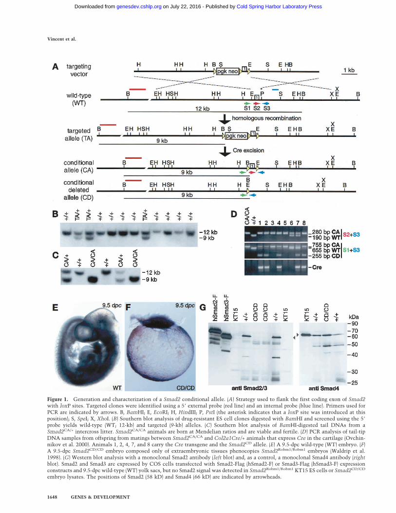
Figure 1. Generation and characterization of a Smad2 conditional allele. (A) Strategy used to flank the first coding exon of Smad2with loxP sites. Targeted clones were identified using a 5� external probe (red line) and an internal probe (blue line). Primers used forPCR are indicated by arrows. B, BamHI; E, EcoRI; H, HindIII; P, PstI (the asterisk indicates that a loxP site was introduced at thisposition); S, SpeI; X, XhoI. (B) Southern blot analysis of drug-resistant ES cell clones digested with BamHI and screened using the 5�
probe yields wild-type (WT; 12-kb) and targeted (9-kb) alleles. (C) Southern blot analysis of BamHI-digested tail DNAs from aSmad2CA/+ intercross litter. Smad2CA/CA animals are born at Mendelian ratios and are viable and fertile. (D) PCR analysis of tail-tipDNA samples from offspring from matings between Smad2CA/CA and Col2a1Cre/+ animals that express Cre in the cartilage (Ovchin-nikov et al. 2000). Animals 1, 2, 4, 7, and 8 carry the Cre transgene and the Smad2CD allele. (E) A 9.5-dpc wild-type (WT) embryo. (F)A 9.5-dpc Smad2CD/CD embryo composed only of extraembryonic tissues phenocopies Smad2Robm1/Robm1 embryos (Waldrip et al.1998). (G) Western blot analysis with a monoclonal Smad2 antibody (left blot) and, as a control, a monoclonal Smad4 antibody (rightblot). Smad2 and Smad3 are expressed by COS cells transfected with Smad2-Flag (hSmad2-F) or Smad3-Flag (hSmad3-F) expressionconstructs and 9.5-dpc wild-type (WT) yolk sacs, but no Smad2 signal was detected in Smad2Robm1/Robm1 KT15 ES cells or Smad2CD/CD
embryo lysates. The positions of Smad2 (58 kD) and Smad4 (66 kD) are indicated by arrowheads.
Vincent et al.
1648 GENES & DEVELOPMENT
Cold Spring Harbor Laboratory Press on July 22, 2016 - Published by genesdev.cshlp.orgDownloaded from
we crossed the Smad2CA allele into the Smad2Robm1 nullbackground (Waldrip et al. 1998) and obtained viable
Smad2CA/Robm1 progeny at the expected Mendelianratio (data not shown). Moreover, germ line excision
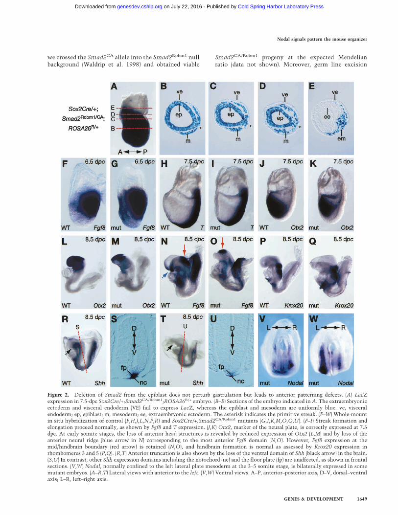
Figure 2. Deletion of Smad2 from the epiblast does not perturb gastrulation but leads to anterior patterning defects. (A) LacZexpression in 7.5-dpc Sox2Cre/+;Smad2CA/Robm1;ROSA26R/+ embryo. (B–E) Sections of the embryo indicated in A. The extraembryonicectoderm and visceral endoderm (VE) fail to express LacZ, whereas the epiblast and mesoderm are uniformly blue. ve, visceralendoderm; ep, epiblast; m, mesoderm; ee, extraembryonic ectoderm. The asterisk indicates the primitive streak. (F–W) Whole-mountin situ hybridization of control (F,H,J,L,N,P,R) and Sox2Cre/+;Smad2CA/Robm1 mutants (G,I,K,M,O,Q,U). (F–I) Streak formation andelongation proceed normally, as shown by Fgf8 and T expression. (J,K) Otx2, marker of the neural plate, is correctly expressed at 7.5dpc. At early somite stages, the loss of anterior head structures is revealed by reduced expression of Otx2 (L,M) and by loss of theanterior neural ridge (blue arrow in N) corresponding to the most anterior Fgf8 domain (N,O). However, Fgf8 expression at themid/hindbrain boundary (red arrow) is retained (N,O), and hindbrain formation is normal as assessed by Krox20 expression inrhombomeres 3 and 5 (P,Q). (R,T) Anterior truncation is also shown by the loss of the ventral domain of Shh (black arrow) in the brain.(S,U) In contrast, other Shh expression domains including the notochord (nc) and the floor plate (fp) are unaffected, as shown in frontalsections. (V,W) Nodal, normally confined to the left lateral plate mesoderm at the 3–5 somite stage, is bilaterally expressed in somemutant embryos. (A–R,T) Lateral views with anterior to the left. (V,W) Ventral views. A–P, anterior–posterior axis; D–V, dorsal–ventralaxis; L–R, left–right axis.
Nodal signals pattern the mouse organizer
GENES & DEVELOPMENT 1649
Cold Spring Harbor Laboratory Press on July 22, 2016 - Published by genesdev.cshlp.orgDownloaded from
of the loxP flanked exon using a Prm1-Cre deleterstrain (O’Gorman et al. 1997) generates a null allele:Smad2CD/CD or Smad2CD/Robm1 embryos phenocopySmad2
Robm1null mutants (Fig. 1F). Finally, Western blot
analysis confirms that Smad2CD/CD embryos lack de-tectable Smad2 protein (Fig. 1G).
Inactivation of Smad2 in the epiblast selectivelydisturbs development of anterior streak derivatives
For conditional inactivation of Smad2, we used aSox2Cre transgene selectively expressed in the epiblastfrom implantation stages onward (Hayashi et al. 2002).As expected, crossing the Sox2Cre transgene into theROSA26R conditional reporter background (Soriano1999) results in rapid activation of LacZ expressionthroughout the entire epiblast from the earliest postim-plantation stages onward (Fig. 2A–E; data not shown).The Sox2Cre transgene was first introduced into theSmad2Robm1 null strain (Waldrip et al. 1998), and nextSox2Cre/+;Smad2Robm1/+ and Smad2CA/CA animalswere intercrossed.Mutant Sox2Cre/+;Smad2Robm1/CA embryos (desig-
nated conditional mutant embryos) gastrulate normallyas assessed by expression of Fgf8 (Fig. 2F,G) and T mRNA(Fig. 2H,I) and at 7.5 d postcoitum (dpc) are morphologi-cally indistinguishable from control littermates. How-ever, by headfold stages (8.5 dpc) the mutant embryosappear overtly abnormal, and specifically display a re-duction in rostral neurectoderm tissue. To examine theonset and nature of these patterning defects, we analyzedthe expression of diagnostic markers. Otx2 is widely ex-pressed in the developing neurectoderm from pregastru-lation stages onward and becomes confined to the fore-brain and midbrain at early somite stages. Wild-type andmutant embryos display indistinguishable Otx2 expres-sion patterns at 7.5 dpc (Fig. 2J,K). However, 12 h later,the Otx2 expression domain is reduced in mutant em-bryos, and by 8.5 dpc is confined to a small region at themost rostral extent of the CNS (Fig. 2L,M). Similar con-clusions were reached analyzing Six3 (data not shown), atranscription factor expressed in the anterior neurecto-derm.To further evaluate anterior CNS defects, we analyzed
Fgf8 expression, which is normally confined to the ante-rior neural ridge and defines the most rostral neurepithe-lium, as well as the isthmus marking the junction be-
tween the developing midbrain and hindbrain. In condi-tional mutant embryos, Fgf8 expression is confined to asmall patch of tissue representing the remnants of theisthmus (Fig. 2N,O), but fails to be expressed rostrally.Similarly, the ventral Shh domain is absent (Fig. 2R,T).In contrast, hindbrain formation is unaffected, as judgedby expression of Krox20 (Fig. 2P,Q). Thus, we concludethat conditional loss of Smad2 in the epiblast has littleeffect on induction and initial specification of the neuralplate. Rather, subsequent growth and anterior patterningare disturbed so that by 8.5 dpc mutant embryos lack allprospective forebrain and most midbrain tissue.In the mouse, initial anterior identity in the underly-
ing epiblast is imposed by the AVE (Kimura et al. 2000;Perea-Gomez et al. 2002) and then, as gastrulation pro-ceeds, it is stabilized by anterior streak derivatives. Toevaluate the onset of anterior patterning defects in theSmad2 conditional mutant embryos, we first examinedearly postimplantation stages. As assessed by the expres-sion of Cerl at 6.5 dpc (Fig. 3A,B), the AVE is inducedcorrectly and rotates appropriately in conditional mu-tants. Gsc and Foxa2 expression normally marks boththe AVE and the anterior PS (Fig. 3C,E). Interestingly,Gsc and Foxa2 expression in the AVE is unaffected, butmutant embryos only weakly express these markers inthe posterior epiblast (Fig. 3D,F). This result suggeststhat the AVE functions normally and that loss of Smad2in the epiblast selectively disrupts the patterning of theanterior PS. Therefore, the anterior defects observed inSmad2 conditional mutant embryos most likely resultfrom impaired production of, or disrupted signalingwithin, the anterior streak derivatives such as the ADE,PCP, and node that guide anterior development after gas-trulation.Next, we assessed the development of the anterior
streak derivatives. Blimp1, a zinc finger transcriptionfactor belonging to the Krüppel family, is normally ex-pressed in the AVE and ADE (Fig. 3G; de Souza et al.1999). Blimp1 transcripts are present at barely detectablelevels in the ADE of mutant embryos (Fig. 3H). Simi-larly, expression of the homeodomain gene Hex (Fig.3K,L), a marker of the midline DE (Thomas et al. 1998),is markedly reduced. The DAN family member Cerl,which transiently marks the majority of nascent DE (Fig.3I; Belo et al. 1997), is weakly expressed in fewer cellscompared with wild type, and the Cerl ADE expressiondomain is virtually eliminated (Fig. 3J).
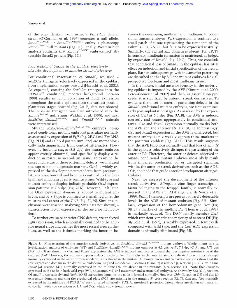
Figure 3. Mispatterning of the anterior streak derivatives in Sox2Cre/+;Smad2CA/Robm1 mutant embryos. Whole-mount in situhybridization analysis of wild-type (WT) and Sox2Cre/+;Smad2CA/null mutant embryos at 6.5 dpc (A–F), 7.5 dpc (G–R), and 7.75 dpc(S–X). (A–D) As shown by Cerl and Foxa2 expression, the AVE is induced and rotates toward the presumptive anterior side of theembryo. (C–F) However, the mutants express reduced levels of Foxa2 and Gsc in the anterior streak (indicated by red lines). Blimp1normally expressed in the anterior mesendoderm (K) is absent in the mutant (L). Frontal views and transverse sections show that theCerl expression domain in the definitive endoderm (DE) and mesoderm (I, sections I1 and I2) is reduced (J, sections J1, J2). Hex (K) andFoxa2 (M, section M1), normally expressed in the midline DE, are absent in the mutants (L,N, section N1). Note that Foxa2 isexpressed in the node in both wild-type (WT; M, section M2) and mutant (N and section N2) embryos. As shown by Shh (O,P, sectionsO1 and P1, respectively) and Nodal (Q,R) expression domains, the node is formed normally. However, Shh (O, section O2) and Gsc (S)expression domains marking the prechordal plate (PCP) are missing in the mutant (P, cross-section P2; T). Chd and Nog normallyexpressed in the midline and PCP (U,W) are truncated anteriorly (V,X). A, anterior; P, posterior. Lateral views are shown with anteriorto the left, with the exception of I, J, and S–X, which show frontal views.
Vincent et al.
1650 GENES & DEVELOPMENT
Cold Spring Harbor Laboratory Press on July 22, 2016 - Published by genesdev.cshlp.orgDownloaded from
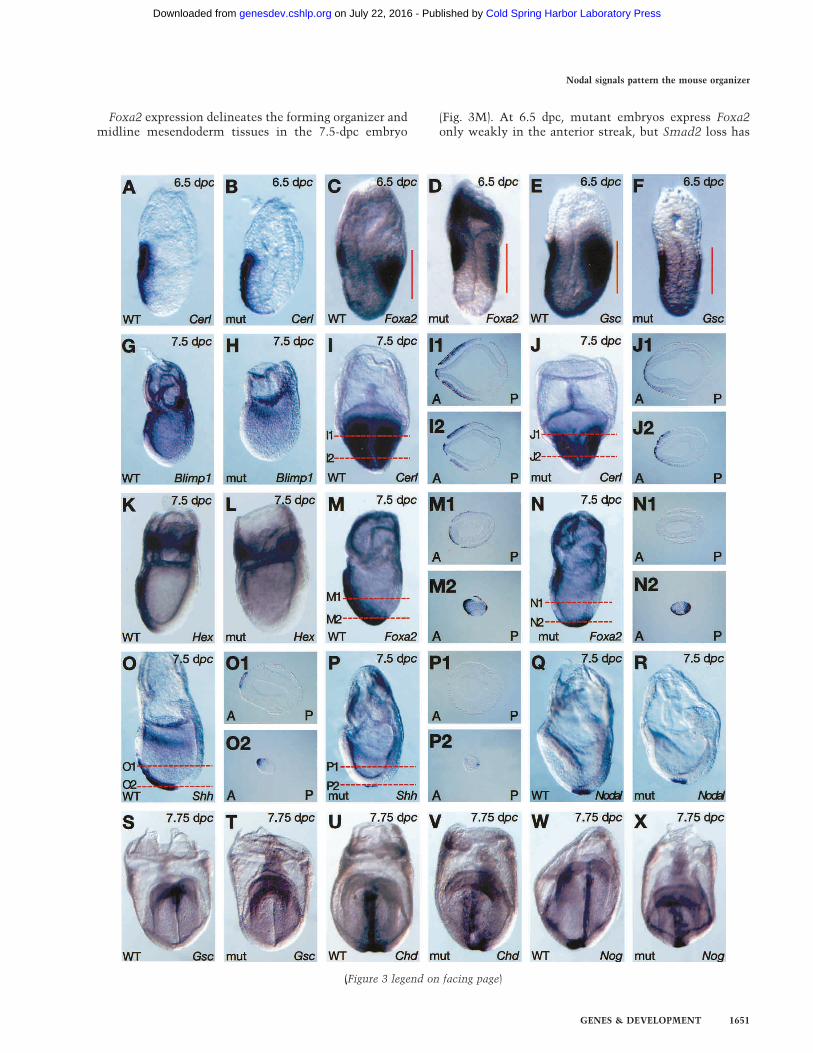
Foxa2 expression delineates the forming organizer andmidline mesendoderm tissues in the 7.5-dpc embryo
(Fig. 3M). At 6.5 dpc, mutant embryos express Foxa2only weakly in the anterior streak, but Smad2 loss has
(Figure 3 legend on facing page)
Nodal signals pattern the mouse organizer
GENES & DEVELOPMENT 1651
Cold Spring Harbor Laboratory Press on July 22, 2016 - Published by genesdev.cshlp.orgDownloaded from
no noticeable effect on Foxa2 expression levels in thedeveloping node. Strikingly, Foxa2 transcripts are notdetected anterior to the node. Thus, it appears that an-terior midline tissue is not specified. Alternatively,Foxa2 induction could potentially depend on Smad2 ac-tivity.To further distinguish these possibilities, we analyzed
Shh expression in the developing node and PCP at 7.5dpc (Fig. 3O). As for Foxa2, in mutant embryos Shh iscorrectly expressed in the node but absent from the an-terior midline tissue (Fig. 3P). To confirm that formationof the node and notochord is unperturbed in the condi-tional mutants, we examined Nodal (Fig. 3Q,R) and T(data not shown) expression, as well as Shh expression at8.5 dpc as a marker for the floor plate and notochord (Fig.2R–T). These structures develop normally but the con-ditional mutant embryos display decreased levels of Shhtranscripts (Fig. 2T,U).The most rostral population of midline axial meso-
derm gives rise to a histologically distinct population ofcells termed the PCP (Sulik et al. 1994), which plays animportant role as an organizing center responsible forpatterning the developing brain. The loss of both Foxa2and Shh expression at 7.5 dpc in the PCP progenitorsemerging from the distal tip of the streak (Fig. 3) stronglysuggests that Smad2 is essential for specification of thisdiscrete cell type. Additional markers Gsc, and the BMPantagonists Chordin and Noggin diagnostic for the PCP,were examined in 7.75-dpc mutant embryos. As shownin Figure 3S–X, the prominent domain of Gsc is lost, andexpression of both Noggin and Chordin is severely at-tenuated along the midline in conditional mutant em-bryos.To assess the functional consequences of perturbed ex-
pression of these midline markers, we examined Nodalexpression in early somite stage embryos. Asymmetricexpression of Nodal in the left lateral plate mesoderm(LPM) is known to be dependent on the presence of afunctional “midline barrier”. Consistent with disturbedfunction of the midline, a high proportion (60%; 6/10) ofmutant embryos show bilateral Nodal expression (Fig.2V,W). Thus, conditional loss of Smad2 selectively dis-rupts formation of the most anterior midline popula-tions, namely, the PCP and ADE.
Targeted removal of the PEE decreases Nodalexpression in the primitive streak
Smad2 and Smad3 are activated by type I receptors thatbind TGF�/activin/Nodal ligands (for reviews, see Mas-sagué et al. 2000; Whitman 2001). To date, the only li-gands of this class expressed in the early mouse embryoare Nodal (Zhou et al. 1993; Conlon et al. 1994) and Gdf1(Wall et al. 2000). Gdf1 is required for establishment ofthe L–R axis (Rankin et al. 2000), whereas Nodal actsmuch earlier during A–P axis establishment (Conlon etal. 1994; Brennan et al. 2001). To test whether Nodal/Smad2 signals are solely responsible for patterning of theanterior streak, we used a genetic strategy to selectively
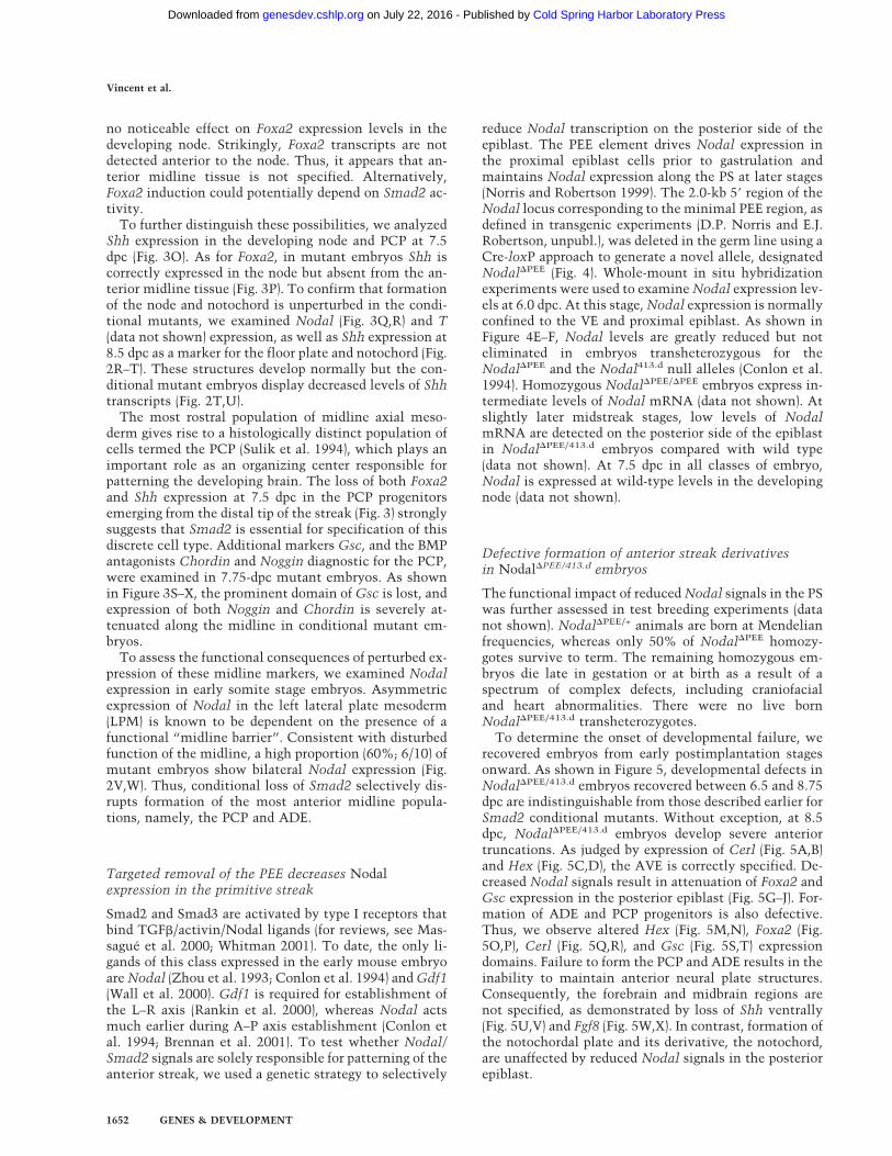
reduce Nodal transcription on the posterior side of theepiblast. The PEE element drives Nodal expression inthe proximal epiblast cells prior to gastrulation andmaintains Nodal expression along the PS at later stages(Norris and Robertson 1999). The 2.0-kb 5� region of theNodal locus corresponding to the minimal PEE region, asdefined in transgenic experiments (D.P. Norris and E.J.Robertson, unpubl.), was deleted in the germ line using aCre-loxP approach to generate a novel allele, designatedNodal�PEE (Fig. 4). Whole-mount in situ hybridizationexperiments were used to examine Nodal expression lev-els at 6.0 dpc. At this stage, Nodal expression is normallyconfined to the VE and proximal epiblast. As shown inFigure 4E–F, Nodal levels are greatly reduced but noteliminated in embryos transheterozygous for theNodal�PEE and the Nodal413.d null alleles (Conlon et al.1994). Homozygous Nodal�PEE/�PEE embryos express in-termediate levels of Nodal mRNA (data not shown). Atslightly later midstreak stages, low levels of NodalmRNA are detected on the posterior side of the epiblastin Nodal�PEE/413.d embryos compared with wild type(data not shown). At 7.5 dpc in all classes of embryo,Nodal is expressed at wild-type levels in the developingnode (data not shown).
Defective formation of anterior streak derivativesin Nodal�PEE/413.d embryos
The functional impact of reduced Nodal signals in the PSwas further assessed in test breeding experiments (datanot shown). Nodal�PEE/+ animals are born at Mendelianfrequencies, whereas only 50% of Nodal�PEE homozy-gotes survive to term. The remaining homozygous em-bryos die late in gestation or at birth as a result of aspectrum of complex defects, including craniofacialand heart abnormalities. There were no live bornNodal�PEE/413.d transheterozygotes.To determine the onset of developmental failure, we
recovered embryos from early postimplantation stagesonward. As shown in Figure 5, developmental defects inNodal�PEE/413.d embryos recovered between 6.5 and 8.75dpc are indistinguishable from those described earlier forSmad2 conditional mutants. Without exception, at 8.5dpc, Nodal�PEE/413.d embryos develop severe anteriortruncations. As judged by expression of Cerl (Fig. 5A,B)and Hex (Fig. 5C,D), the AVE is correctly specified. De-creased Nodal signals result in attenuation of Foxa2 andGsc expression in the posterior epiblast (Fig. 5G–J). For-mation of ADE and PCP progenitors is also defective.Thus, we observe altered Hex (Fig. 5M,N), Foxa2 (Fig.5O,P), Cerl (Fig. 5Q,R), and Gsc (Fig. 5S,T) expressiondomains. Failure to form the PCP and ADE results in theinability to maintain anterior neural plate structures.Consequently, the forebrain and midbrain regions arenot specified, as demonstrated by loss of Shh ventrally(Fig. 5U,V) and Fgf8 (Fig. 5W,X). In contrast, formation ofthe notochordal plate and its derivative, the notochord,are unaffected by reduced Nodal signals in the posteriorepiblast.
Vincent et al.
1652 GENES & DEVELOPMENT
Cold Spring Harbor Laboratory Press on July 22, 2016 - Published by genesdev.cshlp.orgDownloaded from
Defects in anterior streak specification compromisedevelopment of the anterior gut tube
As shown earlier, formation of the DE lineage is severelycompromised in both categories of mutant embryo. Al-though the complete loss of this tissue is strongly sug-gested by the absence of Hex expression along the mid-line, Cerl, which normally delineates the DE, is dimin-ished but not eliminated (Figs. 3, 5). To test whetherthese mutants fail to form DE or whether the tissue issimply mispatterned, we examined embryos histologi-cally at 9.5 dpc. As shown in Figure 6, the gut tube formsbut is very poorly elaborated in the anterior region. Nor-
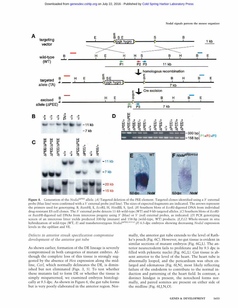
mally, the anterior gut tube extends to the level of Rath-ke’s pouch (Fig. 6C). However, no gut tissue is evident insimilar sections of mutant embryos (Fig. 6G,L). The an-terior neurectoderm fails to proliferate and by 9.5 dpc isfilled with pyknotic nuclei (Fig. 6G,L). Gut tissue is ab-sent anterior to the level of the heart. The heart tube isabnormally looped, and the pericardium was often en-larged and edematous (Fig. 6I,N), most likely reflectingfailure of the endoderm to contribute to the normal in-duction and patterning of the heart field. In contrast, adistinct hindgut is present, the notochord forms nor-mally, and paired somites are present on either side ofthe midline (Fig. 6I,J,N,O).
Figure 4. Generation of the Nodal�PEE allele. (A) Targeted deletion of the PEE element. Targeted clones identified using a 3� externalprobe (blue line) were confirmed with a 5� external probe (red line). The sizes of expected fragments are indicated. The arrows representthe primers used for genotyping. B, BamHI; E, EcoRI; H, HindIII; S, SpeI. (B) Southern blots of EcoRI-digested DNA from individualdrug-resistant ES cell clones. The 3� external probe detects 11-kb wild-type (WT) and 9-kb targeted alleles. (C) Southern blots of EcoRI-or BamHI-digested tail DNAs from intercross progeny using 3� (blue) or 5� (red) external probes, as indicated. (D) PCR genotypingscreen of an intercross litter yields predicted 330-bp (mutant) and 158-bp (wild-type, WT) products. (E,F,G) Whole-mount in situhybridization of wild-type (WT; E) and transheterozygous Nodal�PEE/413.d (F) 6.5-dpc embryos showing decreasing Nodal expressionlevels in the epiblast and VE.
Nodal signals pattern the mouse organizer
GENES & DEVELOPMENT 1653
Cold Spring Harbor Laboratory Press on July 22, 2016 - Published by genesdev.cshlp.orgDownloaded from

Figure 5. Nodal�PEE/413.d mutants closely resemble Sox2Cre/+;Smad2CA/Robm1 mutant embryos. Whole-mount in situ analysis hy-bridization of wild-type (WT) and Nodal�PEE/413.d mutant embryos at 6.5 dpc (A–J), 7.5 dpc (K–R), 7.75 dpc (S,T), 8.5 dpc (U–V), and 8.75dpc (W–X). The AVE is specified and rotates toward the anterior side of the embryo (A–D,G–I), as shown by Cerl (A,B),Hex (C,D), Foxa2(G,H), and Gsc (I,J) expression. Reduced Nodal activity has no effect on expression of FoxH1, a coeffector of Nodal signaling (E,F), orstreak induction and elongation, as shown by T expression (K,L). As for the Smad2 conditional mutant embryos, expression of Foxa2(G,H) and Gsc (I,J) is decreased in the anterior streak (indicated by red lines) compared with wild type (WT). Consequently, expressionof the midline definitive endoderm markers Hex (M,N) and Foxa2 (O,P) is absent and the Cerl expression domain is reduced (Q,R). Asfor Smad2 conditional mutants, the Nodal�PEE/413.d mutants express Foxa2 in the forming node. (S,T) Frontal views show that Gsc isabsent or severely reduced in the prechordal plate in Nodal�PEE/413.d mutant embryos. (U–W) Truncation of the anterior part of thebrain is revealed at 8.5 dpc by the absence of the ventral forebrain domain of Shh (red arrow in U,V) and loss of Fgf8 marking theanterior neural ridge (blue arrow in W). (W,X) However, a residual Fgf8 isthmic region (red arrow) is still present in the mutants.
Vincent et al.
1654 GENES & DEVELOPMENT
Cold Spring Harbor Laboratory Press on July 22, 2016 - Published by genesdev.cshlp.orgDownloaded from
Extraembryonic endoderm contributes to gutformation in Smad2 conditional mutant embryos
The DE emerging from the anterior streak during gastru-lation normally displaces the VE into the visceral yolksac (Thomas and Beddington 1996). In the absence ofNodal/Smad2 signals, the gut tube evident at 9.5 dpc inmutant embryos might be derived in part from the ex-
traembryonic VE as a consequence of improper definitiveendoderm specification. Consistent with this idea, usingHnf4� as a marker of extraembryonic endoderm, we con-sistently observed that the Hnf4� positive cell popula-tion extended more distally in Smad2 conditional mu-tants in comparison with wild-type embryos (Fig. 7A,D).Thus, it appears that VE derivatives become incorpo-rated into embryonic gut tissues.
Figure 6. Gut tube formation in the Nodal�PEE/413.d and Sox2Cre/+;Smad2CA/Robm1 mutant embryos. Wild-type (WT; A–E), Sox2Cre/+;Smad2CA/Robm1 mutant (F–J), and Nodal�PEE/413.d mutant (K–O) 9.5-dpc embryos. (B,G,L) Sections at the level of the head show lossof anterior neurectoderm in both categories of mutant embryos and, as shown in high magnification in lower left (G,L), residualanterior tissue contains many pyknotic cells. (C) Normally the gut tube (arrowhead) extends into the head territories). In both classesof mutants (H,M) the anterior gut tube (arrowhead) is shortened and fails to extend anterior to the heart (D,I,N). Sections at the levelof the heart reveal that the neural tube is highly abnormal. A poorly elaborated gut tube-like structure is present (brackets) but theheart fails to undergo correct looping morphogenesis and has an abnormal trifolium shape. (E,J,O) Sections at the level of the posteriorsomites. (E) In the wild-type (WT) embryo, the somites have already started to differentiate into dermomyotome and sclerotome.Distinct bilateral somites and hindgut (brackets) are present in both categories of mutant embryos. (I,M) The notochord (red arrow) isclearly detectable in the Smad2 and Nodal�PEE/413.d mutant embryos.
Nodal signals pattern the mouse organizer
GENES & DEVELOPMENT 1655
Cold Spring Harbor Laboratory Press on July 22, 2016 - Published by genesdev.cshlp.orgDownloaded from
To unequivocally determine the origins of the gut en-doderm, we used an in vivo fate mapping strategy. TheROSA26R conditional allele (Soriano 1999) was intro-
duced into the Smad2CA background. Smad2CA/CA;ROSA26R/R and Sox2Cre/+;Smad2Robm1/CA mice werethen intercrossed. Using this strategy, the Sox2Cre
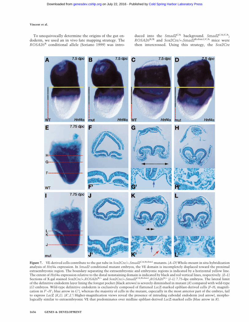
Figure 7. VE-derived cells contribute to the gut tube in Sox2Cre/+;Smad2CA/Robm1 mutants. (A–D) Whole-mount in situ hybridizationanalysis of Hnf4� expression. In Smad2 conditional mutant embryos, the VE domain is incompletely displaced toward the proximalextraembryonic region. The boundary separating the extraembryonic and embryonic regions is indicated by a horizontal yellow line.The extent of Hnf4� expression relative to the distal nonstaining domain is indicated by black and red vertical lines, respectively. (E–L)Sections of X-gal stained Sox2Cre/+;ROSA26R/+ and Sox2Cre/+;Smad2CA/Robm1;ROSA26R/+ (I–L) 7.75-dpc embryos. The lateral limitof the definitive endoderm layer lining the foregut pocket (black arrows) is severely diminished in mutant (K) compared with wild-type(G) embryos. Wild-type definitive endoderm is exclusively composed of flattened LacZ-marked epiblast-derived cells (F–H; magnifi-cation in F�–H�; blue arrow in G�), whereas the majority of cells in the mutant, especially in the most anterior part of the embryo, failto express LacZ (K,L). (K�,L�) Higher-magnification views reveal the presence of intruding cuboidal endoderm (red arrow), morpho-logically similar to extraembryonic VE that predominates over midline epiblast-derived LacZ-marked cells (blue arrow in K).
Vincent et al.
1656 GENES & DEVELOPMENT
Cold Spring Harbor Laboratory Press on July 22, 2016 - Published by genesdev.cshlp.orgDownloaded from
transgene activates the ROSA26R reporter allele exclu-sively in derivatives of the epiblast (Fig. 2A–E) and not inthe VE and extraembryonic ectoderm. As expected, incontrol embryos, the gut tube is uniformly composed ofLacZ-positive DE cells (Fig. 7E–H). In contrast, the ante-rior gut tube in conditional mutants predominantlycomprises LacZ-negative VE derivatives (Fig. 7K,L).Moreover, the mid- and posterior gut tissues are a mosaicof both VE-derived and epiblast-derived cell populations(Fig. 7J). Thus, Smad2-deficient epiblast cells have theability to form definitive endoderm-like cells, but fewerprogenitors are specified. These reduced numbers ofADE cells fail to efficiently displace primitive VE, sothat VE cells become incorporated into the superficialtissue layer overlying the embryonic region. These per-sisting VE-derived cells lack the ability to maintain ex-pression of key regulators including Hex and Cerl, andare functionally unable to substitute for the DE in pat-terning the neurectoderm, resulting in complex tissuedefects that selectively disrupt formation of anteriorstructures.Thus Smad2-deficient epiblast cells have the ability to
form DE-like cells, but fewer progenitors are specified.Consequently, the VE is not efficiently displaced. Thesepersistent VE cells fail to express a number of key regu-lators including Hex and Cerl and are unable to func-tionally substitute for the DE in patterning the neurec-toderm.
Smad2 conditional mutant embryos lacking one copyof Smad3 show loss of node-derived structures
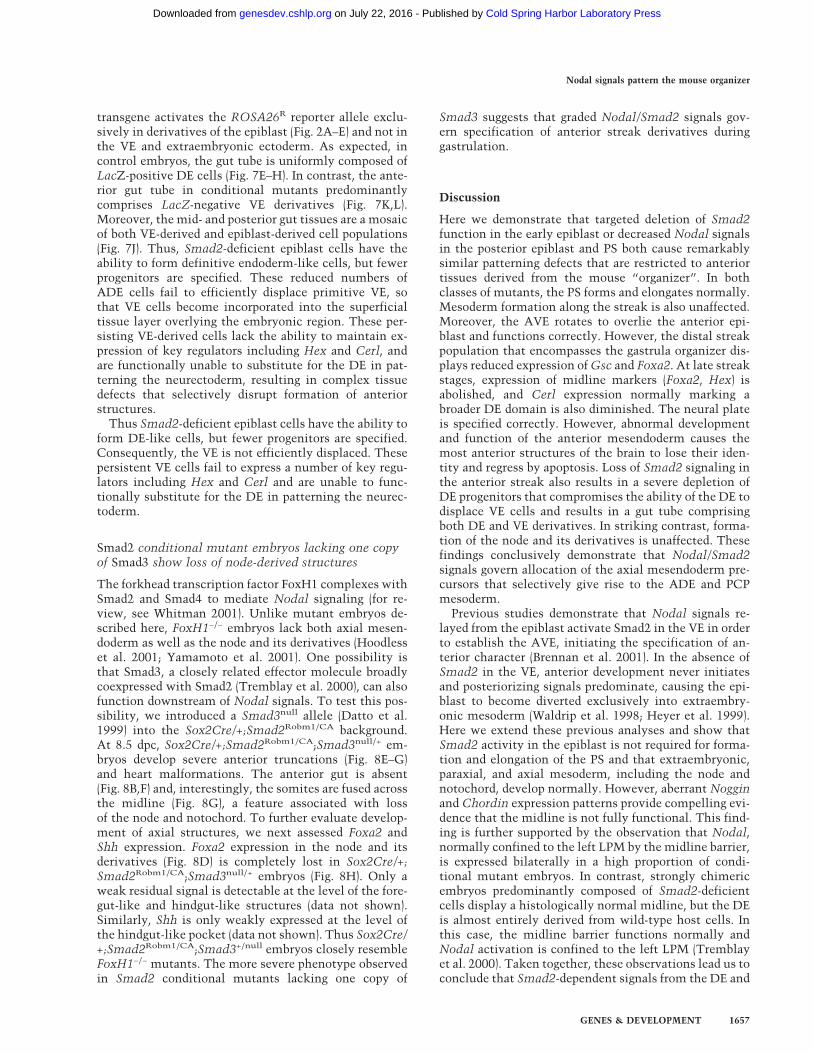
The forkhead transcription factor FoxH1 complexes withSmad2 and Smad4 to mediate Nodal signaling (for re-view, see Whitman 2001). Unlike mutant embryos de-scribed here, FoxH1−/− embryos lack both axial mesen-doderm as well as the node and its derivatives (Hoodlesset al. 2001; Yamamoto et al. 2001). One possibility isthat Smad3, a closely related effector molecule broadlycoexpressed with Smad2 (Tremblay et al. 2000), can alsofunction downstream of Nodal signals. To test this pos-sibility, we introduced a Smad3null allele (Datto et al.1999) into the Sox2Cre/+;Smad2Robm1/CA background.At 8.5 dpc, Sox2Cre/+;Smad2Robm1/CA;Smad3null/+ em-bryos develop severe anterior truncations (Fig. 8E–G)and heart malformations. The anterior gut is absent(Fig. 8B,F) and, interestingly, the somites are fused acrossthe midline (Fig. 8G), a feature associated with lossof the node and notochord. To further evaluate develop-ment of axial structures, we next assessed Foxa2 andShh expression. Foxa2 expression in the node and itsderivatives (Fig. 8D) is completely lost in Sox2Cre/+;Smad2Robm1/CA;Smad3null/+ embryos (Fig. 8H). Only aweak residual signal is detectable at the level of the fore-gut-like and hindgut-like structures (data not shown).Similarly, Shh is only weakly expressed at the level ofthe hindgut-like pocket (data not shown). Thus Sox2Cre/+;Smad2Robm1/CA;Smad3+/null embryos closely resembleFoxH1−/− mutants. The more severe phenotype observedin Smad2 conditional mutants lacking one copy of
Smad3 suggests that graded Nodal/Smad2 signals gov-ern specification of anterior streak derivatives duringgastrulation.
Discussion
Here we demonstrate that targeted deletion of Smad2function in the early epiblast or decreased Nodal signalsin the posterior epiblast and PS both cause remarkablysimilar patterning defects that are restricted to anteriortissues derived from the mouse “organizer”. In bothclasses of mutants, the PS forms and elongates normally.Mesoderm formation along the streak is also unaffected.Moreover, the AVE rotates to overlie the anterior epi-blast and functions correctly. However, the distal streakpopulation that encompasses the gastrula organizer dis-plays reduced expression of Gsc and Foxa2. At late streakstages, expression of midline markers (Foxa2, Hex) isabolished, and Cerl expression normally marking abroader DE domain is also diminished. The neural plateis specified correctly. However, abnormal developmentand function of the anterior mesendoderm causes themost anterior structures of the brain to lose their iden-tity and regress by apoptosis. Loss of Smad2 signaling inthe anterior streak also results in a severe depletion ofDE progenitors that compromises the ability of the DE todisplace VE cells and results in a gut tube comprisingboth DE and VE derivatives. In striking contrast, forma-tion of the node and its derivatives is unaffected. Thesefindings conclusively demonstrate that Nodal/Smad2signals govern allocation of the axial mesendoderm pre-cursors that selectively give rise to the ADE and PCPmesoderm.Previous studies demonstrate that Nodal signals re-
layed from the epiblast activate Smad2 in the VE in orderto establish the AVE, initiating the specification of an-terior character (Brennan et al. 2001). In the absence ofSmad2 in the VE, anterior development never initiatesand posteriorizing signals predominate, causing the epi-blast to become diverted exclusively into extraembry-onic mesoderm (Waldrip et al. 1998; Heyer et al. 1999).Here we extend these previous analyses and show thatSmad2 activity in the epiblast is not required for forma-tion and elongation of the PS and that extraembryonic,paraxial, and axial mesoderm, including the node andnotochord, develop normally. However, aberrant Nogginand Chordin expression patterns provide compelling evi-dence that the midline is not fully functional. This find-ing is further supported by the observation that Nodal,normally confined to the left LPM by the midline barrier,is expressed bilaterally in a high proportion of condi-tional mutant embryos. In contrast, strongly chimericembryos predominantly composed of Smad2-deficientcells display a histologically normal midline, but the DEis almost entirely derived from wild-type host cells. Inthis case, the midline barrier functions normally andNodal activation is confined to the left LPM (Tremblayet al. 2000). Taken together, these observations lead us toconclude that Smad2-dependent signals from the DE and
Nodal signals pattern the mouse organizer
GENES & DEVELOPMENT 1657
Cold Spring Harbor Laboratory Press on July 22, 2016 - Published by genesdev.cshlp.orgDownloaded from
PCP are required for normal function of midline struc-tures.It is well known from studies in Xenopus that verte-
brate head development occurs as a two-step inductionprocess (for review, see Stern 2001). In mouse, inductionof the AVE shortly after implantation initiates a Smad2-dependent pathway that inhibits the posteriorizing in-fluence of Nodal signals on the anterior side of the epi-blast and maintains a labile, preneural and preforebrainstate (Kimura et al. 2000; Brennan et al. 2001; Perea-Gomez et al. 2002). Following the onset of gastrulation,a second signal from the anterior streak-derived mesen-doderm is required to maintain and stabilize the neuraland forebrain region. Here we show in embryos selec-tively lacking Smad2 function in the epiblast that theAVE is induced and the neural plate is specified as judgedby Otx2 and Six3 expression. However, formation of thePCP and the ADE precursors is severely compromised.Consequently, anterior specification is not maintained.The anterior neurectoderm is deprived of essential sig-nals, and all forebrain markers are lost or reduced. Thus,the Nodal/Smad2 signaling cascade specifies the ante-
rior mesendoderm required for maintaining forebrainspecification.The role of the anterior mesendoderm in maintaining
the anterior neural plate/forebrain domains has previ-ously been described in studies of Foxa2 and Hex mutantembryos (Dufort et al. 1998; Martinez Barbera et al.2000). Here we demonstrate that the Nodal/Smad2pathway acts upstream of these genes in determiningcell fate. One possibility is that these genes are directtargets of Nodal/Smad2 signaling. Consistent withthis, the AVE and ADE specific enhancer mapped withinthe Hex locus contains Foxa2 binding sites and se-quences bound by SIP-1, a protein that interacts withSmad2 (Rodriguez et al. 2001). Similarly, a Smad2 re-sponsive enhancer has been characterized within theXenopus XFKH1 locus (Howell and Hill 1997). However,studies to date have failed to identify FoxH1 and/orSmad binding sites within the mouse Foxa2 locus(Sasaki and Hogan 1996; Hoodless et al. 2001), and Foxa2is weakly induced in the anterior streak and PCP tissuesof FoxH1 mutant embryos (Hoodless et al. 2001; Yama-moto et al. 2001). These findings suggest that in mouse,
Figure 8. Decreasing Smad3 expression in Smad2 conditional mutant embryos results in loss of axial structures. (A) Frontal view ofa wild-type (WT) 8.5-dpc embryo showing the presence of a midline. (B,C) Hematoxylin-eosin (HE)-stained sections of A. The level ofthe sections is indicated in A. (E) Frontal view of a Sox2Cre/+;Smad2Robm1/CA;Smad3null/+ 8.5-dpc mutant embryo showing absence ofmidline structures. (F–G) HE-stained sections of E. The level of sections is indicated in E. (D,H) Foxa2 whole-mount in situ analysisof wild-type (WT; D) and Sox2Cre/+;Smad2Robm1/CA;Smad3null/+ (H) 8.5 dpc embryos. (F) In the Sox2Cre/+;Smad2Robm1/CA;Smad3null/+
mutant embryo, the neural tissues remain as a neural plate, the heart (red asterisk) has not looped, and the foregut pocket (bracketsin B) is absent. The absence of a midline is shown by the unfolded neural tube (arrow; cf. F and B) and the fused somites (red arrows;cf. G and C). Foxa2 is expressed in the node (arrow in D) and in the axial mesendoderm of the wild-type (WT) embryo. Neither of theseexpression domains is present in Sox2Cre/+;Smad2Robm1/CA;Smad3null/+ mutant embryos, confirming the absence of a node and itsderivatives.
Vincent et al.
1658 GENES & DEVELOPMENT
Cold Spring Harbor Laboratory Press on July 22, 2016 - Published by genesdev.cshlp.orgDownloaded from
Foxa2 may be a secondary target of the Nodal/Smad2pathway.Interestingly, Foxa2 mutant embryos lack a node and
midline derivatives (Ang and Rossant 1994; Weinstein etal. 1994). Similarly, chimeric embryos generated by ag-gregating tetraploid wild-type embryos with Foxa2-defi-cient ES cells (Dufort et al. 1998) not only lack a nodeandmidline derivatives, but also lack foregut andmidguttissues and contain only a rudimentary hindgut. Further-more, deletion of Foxa2 in the axial mesendoderm leadsto loss of the most anterior structures of the brain (Hal-lonet et al. 2002). The present results demonstrate de-creased Nodal signaling in the anterior streak down-regulates Foxa2 expression. Considering that node mor-phogenesis is unperturbed, we conclude that residualFoxa2 expression in the anterior streak is sufficient topromote node formation. In contrast, Arkadia mutantsfail to activate Foxa2 in the anterior streak, and theseembryos lack both a node and axial mesendoderm(Episkopou et al. 2001). Similarly, loss of Foxa2 expres-sion and node-derived structures was observed in Smad2conditional mutant embryos lacking one copy of Smad3.Collectively, these observations implicate graded Foxa2requirements during specification of the anterior streakderivatives. Thus, it appears that PCP and ADE progeni-tors selectively require increased Nodal/Smad2/Foxa2expression. Similarly, pharynx organogenesis in Cae-norhabditis elegans is controlled by pha-4, a Foxa2 ho-molog, and sequential activation of diverse downstreamtarget genes by pha-4 is regulated by the relative affini-ties of PHA-4 for variants of its canonical binding site(Gaudet and Mango 2002). Here we demonstrate that re-duced Foxa2 expression selectively disrupts allocation ofcells into the PCP and ADE cell lineages. It is temptingto speculate that activation of distinct Foxa2 targetgenes guiding cell fate decisions within the mouse orga-nizer is similarly regulated as a result of different affini-ties for activated Foxa2 complexes.
Nodal expression in the epiblast becomes restricted tothe presumptive posterior side of the embryo shortly be-fore the onset of gastrulation (Varlet et al. 1997). Twoenhancers, a conserved intronic enhancer (ASE) contain-ing binding sites for the FoxH1 transcription factor andthe PEE element, contribute to Nodal expression inthe epiblast (Norris and Robertson 1999). The ASEautoregulatory enhancer promotes activation and up-regulation of Nodal expression in the epiblast and VE.Deletion of this element attenuates Nodal signalsin the epiblast, and allows specification of the AVE butfails to promote cell movements required to convert theP–D to the A–P axis (Norris et al. 2002). Here we showthat decreased Nodal expression in the posterior epiblastof Nodal�PEE/413.d mutant embryos causes loss of PCPand a reduction in DE progenitors. Thus, we concludethat the strength of Nodal signaling selectively regulatescell fate decisions in the streak.In lower vertebrates, distinct aspects of early embry-
onic patterning require graded doses of Nodal signaling.For example, in Xenopus, increased levels of Nodal sig-naling are required for formation of dorsal mesoderm,
whereas lower levels are sufficient for development ofventral mesodermal cell types (Agius et al. 2000). Simi-larly, in Zebrafish, specification of the PCP requires highlevels of Nodal, whereas notochord specification occursat lower levels (Gritsman et al. 2000). In Xenopus em-bryos, Activin concentration gradients are perceived bythe number of occupied receptors per cell, and activationof target genes like Xbra or Xgsc depends on the degree ofActivin receptor occupancy (Dyson and Gurdon 1998). Asimilar mechanism may be responsible for interpretingNodal signaling thresholds and activating subsets of tar-get genes in discrete cell types during distinct aspects ofNodal-dependent A–P and L–R patterning. Moreover,sustained Nodal/Smad2 signaling may be required tomaintain specific programs of gene expression over time.For example, in Nodal�600/�600 mutant embryos inwhich asymmetric Nodal expression is greatly reduced,Lefty2 is not activated in the left LPM, whereas Pitx2 isrobustly expressed, although with a slightly delayed on-set (Norris et al. 2002). Because both Lefty2 and Pitx2 aredirect Nodal targets, differential gene induction appearsto reflect locus specific response to differing levels ofactivated Smad complexes. Interestingly, although acti-vation of Pitx2 transcription is known to be mediated viaa FoxH1 responsive enhancer, a second regulatory ele-ment that specifically binds Nkx2.1 at slightly later de-velopmental stages is required for continued mainte-nance of transcription (Shiratori et al. 2001).
Nodal signals play a conserved role in specification ofthe DE lineage in vertebrates (David and Rosa 2001;Lowe et al. 2001; Norris et al. 2002). In mutant embryosdescribed here, we fail to observe expression of midlineDE markers (Hex, Foxa2). Cerl, a broader DE marker, isonly weakly expressed and is absent from the ADE.However, histological analysis clearly shows that ven-tral closure occurs and a gut structure is present. In chi-meric embryos, neither Smad2−/− nor FoxH1−/− ES cellscontribute to the gut tube, strongly suggesting that bothactivities are necessary for DE specification (Tremblay etal. 2000; Hoodless et al. 2001). Here we show that theanterior part of the gut tube is truncated and mainlyderives from extraembryonic VE cells, whereas the mid-gut and hindgut contain mainly epiblast derivatives.These results demonstrate that the VE compensatesphysically but cannot functionally replace DE cells.Strikingly, despite numerous genes expressed in com-mon in both extraembryonic and DE cell populations,the VE appears to lack signaling capabilities. It will beinteresting to identify DE-specific signals and learn moreabout the mechanism by which they pattern the overly-ing neuroectoderm.
Materials and methods
Generating a Smad2 conditional allele
The Smad2CA allele was generated by flanking the first codingexon with loxP sites (Fig. 1A). The targeting construct containsa 5-kb (SpeI–EcoRI) 5� and a 4.9-kb (EcoRI–XhoI) 3� homologyarm containing a loxP site that replaces a PstI site with an EcoRI
Nodal signals pattern the mouse organizer
GENES & DEVELOPMENT 1659
Cold Spring Harbor Laboratory Press on July 22, 2016 - Published by genesdev.cshlp.orgDownloaded from
site, and loxP flanked pgk-neomycin and pgk-dta selection cas-settes (Fig. 1A). Linearized vector was electroporated into CCEES cells, and drug-resistant colonies were genotyped by South-ern blot analysis as described (Waldrip et al. 1998). Approxi-mately 6% of the clones were correctly targeted. The pgk-neo-mycin cassette was excised by transient expression of Cre re-combinase. Two independent Smad2CA/+ ES cell clones wereused to generate germ line chimeric mice that were subse-quently bred to C57BL/6 mice.
Generation of the Nodal�PEE allele
The PEE element corresponds to a 2-kb (HindIII–SpeI) regionlocated ∼10 kb 5� of the first exon of Nodal (Norris and Robert-son 1999). The PEE targeting vector was designed using a con-ditional strategy (Fig. 4A) and contains a 3-kb (HindIII–HindIII)5� and a 6.2-kb (HindIII–EcoRI) 3� homology arm. The SpeI sitewithin the 3� arm was replaced by a loxP site and an EcoRI site.The homology arms were subcloned into a vector containing aloxP-flanked pgk-hygro cassette and hsv-tk and pgk-dta nega-tive selection cassettes. Linearized vector was electroporatedinto CCE cells and drug-resistant colonies were screened bySouthern blot analysis using a 3� external (BamHI–PstI) probe.Approximately 5% of the clones were correctly targeted. Thepgk-hygro cassette and 2-kb PEE element were excised fromtargeted clones by transient expression of Cre recombinase. Re-sulting subclones were screened by PCR and Southern analysisusing 5� (SacI–PstI) and 3� external probes. Four correctly ex-cised ES cell clones were used to generate germ line chimericmice that were subsequently bred to C57BL/6 mice.
Genotyping procedures
Mice were genotyped by PCR screening of genomic tail DNA.Embryos were individually genotyped prior to whole-mount insitu hybridization and/or histological analysis by digesting afragment of extraembryonic tissue in 20–50 µL of 1× PerkinElmer PCR buffer (Roche) containing 0.45% NP-40, 0.45%Tween 20, and 1 µg/µL Proteinase K for 9 h at 56°C. Lysateswere boiled and 0.5–1 µL was used for PCR analysis.The Nodal413.d, Smad2Robm1, and ROSA26R alleles and the
Cre transgene were genotyped as described (Conlon et al. 1994;Vidal et al. 1998; Waldrip et al. 1998; Soriano 1999). Primersused to genotype the Smad2CA and Smad2CD alleles were asfollows (Fig. 1A): S1, 5�-CCCGGTAAATCTACCCTAG-3�; S2,5�-GCTTAAAAGTCACTACTCAAG-3�; S3, 5�-TTTCAAAACTATATTTGCCCAAG-3�.The S2 + S3 pair amplifies 220-bp and 320-bp fragments for
the wild-type and the Smad2CA alleles, respectively. TheS1 + S3 pair amplifies 650-bp, 700-bp, and 200-bp fragments forthe wild-type, Smad2CA, and Smad2CD alleles, respectively.The Nodal�PEE allele was genotyped using the following
primers (Fig. 4A): P1, 5�-AGTGCTGGGATCACAGAAG-3�; P2,5�-GAGATAGGTCTTGTGTGGC-3�; P3, 5�-TATGATAATGATCAGGTCAGG-3�.The combination of the P1, P2, and P3 primers amplifies 160-
bp and 330-bp fragments from the wild-type and the Nodal�PEE
alleles, respectively.The strategy to genotype the Smad3null allele will be de-
scribed elsewhere (N.R. Dunn and E.J. Robertson, in prep.).
Mouse strains
Smad2CA/CA, Smad2CA/CA;ROSA26R/R (Soriano 1999), andSmad2CA/CA;Smad3null/null (Datto et al. 1999) were maintainedon a (129 X C57BL/6) hybrid background. Nodal413.d/+–129/Sv//
Ev mice have been described (Conlon et al. 1994). Sox2Cre/+;Smad2Robm1/+ mice were obtained from crosses betweenSmad2Robm1/+ (Waldrip et al. 1998) and Sox2Cre/+ (Hayashi etal. 2002) parents and maintained on an outbred CD1 back-ground. Mice carrying the Smad2CD allele were generated bymating Smad2CA/CA animals with a Prm1-Cre deleter strain(O’Gorman et al. 1997) and were maintained on a CD1 back-ground.
Western blots
Control extracts from KT15 Smad2-deficient ES cells (Tremblayet al. 2000) and COS cells transfected with human Smad2-Flagor Smad3-Flag expression constructs (kindly provided by C.Koonce) were prepared in 2× sample buffer. Yolk sac tissue fromwild-type and Smad2CD/CD mutant embryos was sonicated in2× sample buffer. Extracts were loaded on a 10% SDS-PAGE gel.After transfer onto nitrocellulose membranes (Schleicher &Schuell BioScience), the membranes were incubated with eithera Smad2/3 antibody (2 µg/µL mouse monoclonal antibody spe-cific for the C-terminal part of the MH1 and the linker domains,BD Transduction Laboratories, no. 610842) or a Smad4 antibody(1/1000 mouse monoclonal antibody, Santa Cruz Biotechnol-ogy, Inc., no. sc-7966). A horse radish peroxydase-conjugatedsheep anti-mouse IgG (Amersham Pharmacia Biotech, no.NA931) was used as the secondary antibody. The blots werewashed in TBST and developed by chemiluminescence usingECL (Amersham Pharmacia Biotech).
X-gal staining, whole-mount in situ hybridization,and histology
X-gal staining and whole-mount in situ hybridization were per-formed according to standard procedures (Hogan et al. 1994).Probes for the following genes were used in this study: Gsc(Blum et al. 1992), Chordin (Klingensmith et al. 1999), Foxa2(Sasaki and Hogan 1996), Nodal (Conlon et al. 1994), Shh (Ech-elard et al. 1993), T (Herrmann 1991),Cerl (Belo et al. 1997), Six3(Oliver et al. 1995), Fgf8 (Crossley and Martin 1995), Hex (Tho-mas et al. 1998), Krox20 (Sham et al. 1993), and Otx2 (Ang et al.1994). Noggin and Hnf4� probe templates were generated byPCR. The Blimp-1 probe was derived from the EST mB3 (ESTclone image: 1165721). For each probe and embryonic stage ex-amined, between four and six mutant embryos were subject towhole-mount in situ hybridization and compared with a similargroup of age-matched controls. For histology, embryos werefixed in 4% paraformaldehyde, dehydrated through an ethanolseries, and embedded in wax before sectioning. Hematoxylinand eosin staining was performed according to standard proto-cols.
Acknowledgments
We are grateful to Liz Bikoff for discussions and for valuablecomments on the manuscript. We thank Phil Soriano for gen-erously providing the ROSA26R mice, Josh Frederick and Xia-Fan Wang for generously providing the Smad3null mice, JohnKlingensmith for the Chordin probe, Chad Koonce for advice onWestern analysis, Debbie Pelusi for genotyping assistance, andmembers of the lab for discussion. S.D.V. was supported by aFellowship from the ARC and a Long Term Fellowship from theHFSP. N.R.D. was supported by a postdoctoral fellowship fromthe NICHD. This work was supported by a grant from the NIHto E.J.R.The publication costs of this article were defrayed in part by
Vincent et al.
1660 GENES & DEVELOPMENT
Cold Spring Harbor Laboratory Press on July 22, 2016 - Published by genesdev.cshlp.orgDownloaded from
payment of page charges. This article must therefore be herebymarked “advertisement” in accordance with 18 USC section1734 solely to indicate this fact.
References
Agius, E., Oelgeschlager, M., Wessely, O., Kemp, C., and DeRobertis, E.M. 2000. Endodermal Nodal-related signals andmesoderm induction in Xenopus. Development 127: 1173–1183.
Ang, S.L. and Rossant, J. 1994. HNF-3 � is essential for node andnotochord formation in mouse development. Cell 78: 561–574.
Ang, S.L., Conlon, R.A., Jin, O., and Rossant, J. 1994. Positiveand negative signals from mesoderm regulate the expressionof mouse Otx2 in ectoderm explants. Development120: 2979–2989.
Bachiller, D., Klingensmith, J., Kemp, C., Belo, J.A., Anderson,R.M., May, S.R., McMahon, J.A., McMahon, A.P., Harland,R.M., Rossant, J., et al. 2000. The organizer factors Chordinand Noggin are required for mouse forebrain development.Nature 403: 658–661.
Beddington, R.S. and Robertson, E.J. 1999. Axis developmentand early asymmetry in mammals. Cell 96: 195–209.
Belo, J.A., Bouwmeester, T., Leyns, L., Kertesz, N., Gallo, M.,Follettie, M., and De Robertis, E.M. 1997. Cerberus-like is asecreted factor with neutralizing activity expressed in theanterior primitive endoderm of the mouse gastrula. Mech.Dev. 68: 45–57.
Blum, M., Gaunt, S.J., Cho, K.W., Steinbeisser, H., Blumberg, B.,Bittner, D., and De Robertis, E.M. 1992. Gastrulation in themouse: The role of the homeobox gene goosecoid. Cell69: 1097–1106.
Brennan, J., Lu, C.C., Norris, D.P., Rodriguez, T.A., Beddington,R.S., and Robertson, E.J. 2001. Nodal signalling in the epi-blast patterns the early mouse embryo. Nature 411: 965–969.
Camus, A. and Tam, P.P. 1999. The organizer of the gastrulatingmouse embryo. Curr. Top. Dev. Biol. 45: 117–153.
Conlon, F.L., Lyons, K.M., Takaesu, N., Barth, K.S., Kispert, A.,Herrmann, B., and Robertson, E.J. 1994. A primary require-ment for nodal in the formation and maintenance of theprimitive streak in the mouse. Development 120: 1919–1928.
Crossley, P.H. and Martin, G.R. 1995. The mouse Fgf8 geneencodes a family of polypeptides and is expressed in regionsthat direct outgrowth and patterning in the developing em-bryo. Development 121: 439–451.
Datto, M.B., Frederick, J.P., Pan, L., Borton, A.J., Zhuang, Y., andWang, X.F. 1999. Targeted disruption of Smad3 reveals anessential role in transforming growth factor �-mediated sig-nal transduction. Mol. Cell. Biol. 19: 2495–2504.
David, N.B. and Rosa, F.M. 2001. Cell autonomous commit-ment to an endodermal fate and behaviour by activation ofNodal signalling. Development 128: 3937–3947.
Davidson, B.P., Kinder, S.J., Steiner, K., Schoenwolf, G.C., andTam, P.P. 1999. Impact of node ablation on the morphogen-esis of the body axis and the lateral asymmetry of the mouseembryo during early organogenesis. Dev. Biol. 211: 11–26.
de Souza, F.S., Gawantka, V., Gomez, A.P., Delius, H., Ang, S.L.,and Niehrs, C. 1999. The zinc finger gene Xblimp1 controlsanterior endomesodermal cell fate in Spemann’s organizer.EMBO J. 18: 6062–6072.
Dufort, D., Schwartz, L., Harpal, K., and Rossant, J. 1998. Thetranscription factor HNF3beta is required in visceral endo-
derm for normal primitive streak morphogenesis. Develop-ment 125: 3015–3025.
Dyson, S. and Gurdon, J.B. 1998. The interpretation of positionin a morphogen gradient as revealed by occupancy of activinreceptors. Cell 93: 557–568.
Echelard, Y., Epstein, D.J., St-Jacques, B., Shen, L., Mohler, J.,McMahon, J.A., and McMahon, A.P. 1993. Sonic hedgehog, amember of a family of putative signaling molecules, is im-plicated in the regulation of CNS polarity. Cell 75: 1417–1430.
Episkopou, V., Arkell, R., Timmons, P.M., Walsh, J.J., Andrew,R.L., and Swan, D. 2001. Induction of the mammalian noderequires Arkadia function in the extraembryonic lineages.Nature 410: 825–830.
Gaudet, J. and Mango, S.E. 2002. Regulation of organogenesis bythe Caenorhabditis elegans FoxA protein PHA-4. Science295: 821–825.
Gritsman, K., Talbot, W.S., and Schier, A.F. 2000. Nodal signal-ing patterns the organizer. Development 127: 921–932.
Hallonet, M., Kaestner, K.H., Martin-Parras, L., Sasaki, H., Betz,U.A., and Ang, S.L. 2002. Maintenance of the specification ofthe anterior definitive endoderm and forebrain depends onthe axial mesendoderm: A study using HNF3beta/Foxa2conditional mutants. Dev. Biol. 243: 20–33.
Hayashi, S., Lewis, P., Pevny, L., and McMahon, A.P. 2002.Efficient gene modulation in mouse epiblast using a Sox2Cretransgenic mouse strain. Gene Expr. Patterns 2: 93–97.
Herrmann, B.G. 1991. Expression pattern of the Brachyury genein whole-mount TWis/TWis mutant embryos. Development113: 913–917.
Heyer, J., Escalante-Alcalde, D., Lia, M., Boettinger, E., Edel-mann, W., Stewart, C.L., and Kucherlapati, R. 1999. Postgas-trulation Smad2-deficient embryos show defects in embryoturning and anterior morphogenesis. Proc. Natl. Acad. Sci.96: 12595–12600.
Hogan, B.L., Beddington, R.S.,.Costantini, F., and Lacy, E. 1994.Manipulating the mouse embryo: A laboratory manual.Cold Spring Harbor Laboratory Press, Cold Spring Harbor,NY.
Hoodless, P.A., Pye, M., Chazaud, C., Labbe, E., Attisano, L.,Rossant, J., and Wrana, J.L. 2001. FoxH1 (Fast) functions tospecify the anterior primitive streak in the mouse. Genes &Dev. 15: 1257–1271.
Howell, M. and Hill, C.S. 1997. XSmad2 directly activates theactivin-inducible, dorsal mesoderm gene XFKH1 in Xenopusembryos. EMBO J. 16: 7411–7421.
Kimura, C., Yoshinaga, K., Tian, E., Suzuki, M., Aizawa, S., andMatsuo, I. 2000. Visceral endoderm mediates forebrain de-velopment by suppressing posteriorizing signals. Dev. Biol.225: 304–321.
Klingensmith, J., Ang, S.L., Bachiller, D., and Rossant, J. 1999.Neural induction and patterning in the mouse in the absenceof the node and its derivatives. Dev. Biol. 216: 535–549.
Lawson, K.A. 1999. Fate mapping the mouse embryo. Int. J.Dev. Biol. 43: 773–775.
Lowe, L.A., Yamada, S., and Kuehn, M.R. 2001. Genetic dissec-tion of nodal function in patterning the mouse embryo. De-velopment 128: 1831–1843.
Lu, C.C., Brennan, J., and Robertson, E.J. 2001. From fertiliza-tion to gastrulation: Axis formation in the mouse embryo.Curr. Opin. Genet. Dev. 11: 384–392.
Martinez Barbera, J.P., Clements, M., Thomas, P., Rodriguez,T., Meloy, D., Kioussis, D., and Beddington, R.S. 2000. Thehomeobox gene Hex is required in definitive endodermaltissues for normal forebrain, liver and thyroid formation.Development 127: 2433–2445.
Nodal signals pattern the mouse organizer
GENES & DEVELOPMENT 1661
Cold Spring Harbor Laboratory Press on July 22, 2016 - Published by genesdev.cshlp.orgDownloaded from
Massagué, J., Blain, S.W., and Lo, R.S. 2000. The TGFbeta sig-naling in growth control, cancer, and heritable disorders.Cell 103: 295–309.
Mukhopadhyay, M., Shtrom, S., Rodriguez-Esteban, C., Chen,L., Tsukui, T., Gomer, L., Dorward, D.W., Glinka, A., Grin-berg, A., Huang, S.P., et al. 2001. Dickkopf1 is required forembryonic head induction and limb morphogenesis in themouse. Dev. Cell 1: 423–434.
Norris, D.P. and Robertson, E.J. 1999. Asymmetric and node-specific nodal expression patterns are controlled by two dis-tinct cis-acting regulatory elements. Genes & Dev.13: 1575–1588.
Norris, D.P., Brennan, J., Bikoff, E.K., and Robertson, E.J. 2002.The Foxh1-dependent autoregulatory enhancer controls thelevel of Nodal signals in the mouse embryo. Development129: 3455–3468.
O’Gorman, S., Dagenais, N.A., Qian, M., and Marchuk, Y. 1997.Protamine–Cre recombinase transgenes efficiently recom-bine target sequences in the male germ line of mice, but notin embryonic stem cells. Proc. Natl. Acad. Sci. 94: 14602–14607.
Oliver, G., Mailhos, A., Wehr, R., Copeland, N.G., Jenkins,N.A., and Gruss, P. 1995. Six3, a murine homologue of thesine oculis gene, demarcates the most anterior border of thedeveloping neural plate and is expressed during eye develop-ment. Development 121: 4045–4055.
Ovchinnikov, D.A., Deng, J.M., Ogunrinu, G., and Behringer,R.R. 2000. Col2a1-directed expression of Cre recombinase indifferentiating chondrocytes in transgenic mice. Genesis26: 145–146.
Perea-Gomez, A., Vella, F.D., Shawlot, W., Oulad-Abdelghani,M., Chazaud, C., Meno, C., Pfister, V., Chen, L., Robertson,E., Hamada, H., et al. 2002. Nodal antagonists in the anteriorvisceral endoderm prevent the formation of multiple primi-tive streaks. Dev. Cell 3: 745–756.
Rankin, C.T., Bunton, T., Lawler, A.M., and Lee, S.J. 2000.Regulation of left-right patterning in mice by growth/differ-entiation factor-1. Nat. Genet. 24: 262–265.
Rodriguez, T.A., Casey, E.S., Harland, R.M., Smith, J.C., andBeddington, R.S. 2001. Distinct enhancer elements controlHex expression during gastrulation and early organogenesis.Dev. Biol. 234: 304–316.
Saijoh, Y., Adachi, H., Sakuma, R., Yeo, C.Y., Yashiro, K., Wa-tanabe, M., Hashiguchi, H., Mochida, K., Ohishi, S., Kawa-bata, M., et al. 2000. Left-right asymmetric expression oflefty2 and nodal is induced by a signaling pathway that in-cludes the transcription factor FAST2. Mol. Cell 5: 35–47.
Sasaki, H. and Hogan, B.L. 1996. Enhancer analysis of the mouseHNF-3 � gene: Regulatory elements for node/notochord andfloor plate are independent and consist of multiple sub-ele-ments. Genes Cells 1: 59–72.
Sham, M.H., Vesque, C., Nonchev, S., Marshall, H., Frain, M.,Gupta, R.D., Whiting, J., Wilkinson, D., Charnay, P., andKrumlauf, R. 1993. The zinc finger gene Krox20 regulatesHoxB2 (Hox2.8) during hindbrain segmentation. Cell72: 183–196.
Shawlot, W., Wakamiya, M., Kwan, K.M., Kania, A., Jessell,T.M., and Behringer, R.R. 1999. Lim1 is required in bothprimitive streak-derived tissues and visceral endoderm forhead formation in the mouse. Development 126: 4925–4932.
Shiratori, H., Sakuma, R., Watanabe, M., Hashiguchi, H., Mo-chida, K., Sakai, Y., Nishino, J., Saijoh, Y., Whitman, M., andHamada, H. 2001. Two-step regulation of left-right asym-metric expression of Pitx2: Initiation by nodal signaling andmaintenance by Nkx2. Mol. Cell 7: 137–149.
Soriano, P. 1999. Generalized lacZ expression with the ROSA26Cre reporter strain. Nat. Genet. 21: 70–71.
Stern, C.D. 2001. Initial patterning of the central nervous sys-tem: How many organizers? Nat. Rev. Neurosci. 2: 92–98.
Sulik, K., Dehart, D.B., Iangaki, T., Carson, J.L., Vrablic, T.,Gesteland, K., and Schoenwolf, G.C. 1994. Morphogenesis ofthe murine node and notochordal plate. Dev. Dyn. 201: 260–278.
Thomas, P. and Beddington, R. 1996. Anterior primitive endo-derm may be responsible for patterning the anterior neuralplate in the mouse embryo. Curr. Biol. 6: 1487–1496.
Thomas, P.Q., Brown, A., and Beddington., R.S 1998. Hex: Ahomeobox gene revealing peri-implantation asymmetry inthe mouse embryo and an early transient marker of endo-thelial cell precursors. Development 125: 85–94.
Tremblay, K.D., Hoodless, P.A., Bikoff, E.K., and Robertson, E.J.2000. Formation of the definitive endoderm in mouse is aSmad2-dependent process. Development 127: 3079–3090.
Varlet, I., Collignon, J., and Robertson, E.J.. 1997. nodal expres-sion in the primitive endoderm is required for specificationof the anterior axis during mouse gastrulation. Development124: 1033–1044.
Vidal, F., Sage, J., Cuzin, F., and Rassoulzadegan, M. 1998. Creexpression in primary spermatocytes: A tool for genetic en-gineering of the germ line. Mol. Reprod. Dev. 51: 274–280.
Waldrip, W.R., Bikoff, E.K., Hoodless, P.A., Wrana, J.L., andRobertson, E.J. 1998. Smad2 signaling in extraembryonic tis-sues determines anterior-posterior polarity of the earlymouse embryo. Cell 92: 797–808.
Wall, N.A., Craig, E.J., Labosky, P.A., and Kessler, D.S. 2000.Mesendoderm induction and reversal of left-right pattern bymouse Gdf1, a Vg1-related gene. Dev. Biol. 227: 495–509.
Weinstein, D.C., Ruiz i Altaba, A., Chen, W.S., Hoodless, P.,Prezioso, V.R., Jessell, T.M., and Darnell Jr., J.E. 1994. Thewinged-helix transcription factor HNF-3 � is required fornotochord development in the mouse embryo. Cell 78: 575–588.
Weisberg, E., Winnier, G.E., Chen, X., Farnsworth, C.L., Hogan,B.L., and Whitman, M. 1998. A mouse homologue of FAST-1transduces TGF � superfamily signals and is expressed dur-ing early embryogenesis. Mech. Dev. 79: 17–27.
Whitman, M. 2001. Nodal signaling in early vertebrate em-bryos. Themes and variations. Dev. Cell. 1: 605–617.
Yamamoto, M., Meno, C., Sakai, Y., Shiratori, H., Mochida, K.,Ikawa, Y., Saijoh, Y., and Hamada, H. 2001. The transcrip-tion factor FoxH1 (FAST) mediates Nodal signaling duringanterior-posterior patterning and node formation in themouse. Genes & Dev. 15: 1242–1256.
Zhou, X., Sasaki, H., Lowe, L., Hogan, B.L., and Kuehn, M.R.1993. Nodal is a novel TGF-�-like gene expressed in themouse node during gastrulation. Nature 361: 543–547.
Vincent et al.
1662 GENES & DEVELOPMENT
Cold Spring Harbor Laboratory Press on July 22, 2016 - Published by genesdev.cshlp.orgDownloaded from
10.1101/gad.1100503Access the most recent version at doi: 2003 17: 1646-1662 Genes Dev.
Stéphane D. Vincent, N. Ray Dunn, Shigemi Hayashi, et al. Nodal signalsCell fate decisions within the mouse organizer are governed by graded
References
http://genesdev.cshlp.org/content/17/13/1646.full.html#ref-list-1
This article cites 61 articles, 23 of which can be accessed free at:
ServiceEmail Alerting
click here.right corner of the article orReceive free email alerts when new articles cite this article - sign up in the box at the top
http://genesdev.cshlp.org/subscriptionsgo to: Genes & Development To subscribe to
Cold Spring Harbor Laboratory Press
Cold Spring Harbor Laboratory Press on July 22, 2016 - Published by genesdev.cshlp.orgDownloaded from