Functional sexual dimorphism of the nucleolar organizer regions in the tuberomamillary nucleus
6
BRAIN RESEARCH ELSEVIER Brain Research 736 (1996) 1-6 Research report Functional sexual dimorphism of the nucleolar organizer regions in the tuberomamillary nucleus Sonia Gonzfilez-Gonzfilez a, Fernando Dfaz b, Guillermo Vallejo a, Jorge L. Arias a, * a Psychobiology Laboratory, Psychology Faculty, University of Oviedo, Aniceto Sela s / n , 33005 Oviedo, Spain b Department of Functional Biology (Physiology), University of Oviedo, Oviedo, Spain Accepted 21 May 1996 Abstract The tuberomamillary nucleus (TM) is a cluster of magnocellular neurons that are located close to the mamillary recess or basal surface of the mamillary body (MBs) and is the only known source of histamine (HA). The nucleolar organizer regions (NORs) of the nervous cells of the subnuclei El, E2 and E3 of the TM are quantified in this paper to discern the existence of possible sexual differences in the overall neuronal protein synthesis between male and female rats in two phases of the oestrous cycle (oestrous-dioestrous). We have used the argentic impregnation technique of the NORs (AgNORs) in both bilateral nuclei of the TM and found functional bilateral symmetry in its AgNORs. Furthermore, we demonstrate the existence of significant differences (P < 0.001) between sexes and between two phases of the oestrous cycle studied (oestrous-dioestrous) in the parameter of number of AgNORs per neuron. Hormonal manipulation of the TM in the rat during the two periods, postnatal or adult, shows the importance of the activational effect of the hormones on the activity of cellular protein synthesis. Keywords: Tuberomamillary nucleus; AgNOR; Nucleolar organizer region; Estrous cycle; Hormonal manipulation; Activational effect 1. Introduction The TM nucleus is formed by a cluster of magnocellu- lar neurons adjacent to the mamillary bodies, just above the ventral surface of the hypothalamus and is the only known source of histamine (HA) in the brain. The cerebral histaminergic system is formed by five subgroups of cells known as El-E5 according to the nomenclature used by Inagaki et al. [15] (Fig. 1). It has efferent projections to almost all parts of the brain, from the olfactory bulb to the spinal cord and receives inputs mainly from the limbic system [21]. Besides the HA, several transmitters have been identified in this nucleus: adenosine [24], GABA [11] and substance-P [18]. However, little is known about its function. At present, studies of a unilateral electrolytic lesion of the TM have provided information on the existence of an inhibitory system of this nucleus on the lateral hypothala- mus [27]. When the TM was removed by a lesion, this * Corresponding author. Fax: (34) (8) 510-3216. resulted in an increase in the rate of lateral hypothalamic self-stimulation. Thus, the TM system is interpreted as an area of inhibition of reinforcement and its lesion seems to facilitate the reinforcement processed. In fact, behavioral studies show that the lesions of the TM nucleus may have a facilitatory effect on learning and mnemonic functioning which is possibly related to a lesion-induced disinhibition or facilitation of the reinforcement processes [17]. On the other hand, in all the studies, it is assumed that the TM nuclei present bilateral structural and functional symmetry so that the use of unilateral lesions of the TM nuclei is frequent [6,27]. These data, together with the existence of sexual differences in spatial skills [4] and the fact that the nearby structures that have connections to the TM nucleus present sexual dimorphism [9], make us pro- pose the possible existence of functional sexual dimor- phism and functional symmetry in the neurons of the TM nucleus. In the present study, we have used the argentic impreg- nation technique of the nucleolar organizer regions (NORs) that has been described by Ploton [23]. This technique makes it possible to mark the fibrillar centers (FCs), a component of the nucleolus that is important in the synthe- 0006-8993/96/$15.00 Copyright © 1996 Elsevier Science B.V. All rights reserved. PII S0006-8993(96)00602-6
-
Upload
independent -
Category
Documents
-
view
1 -
download
0
Transcript of Functional sexual dimorphism of the nucleolar organizer regions in the tuberomamillary nucleus

BRAIN RESEARCH
E L S E V I E R Brain Research 736 (1996) 1-6
R e s e a r c h r e p o r t
Functional sexual dimorphism of the nucleolar organizer regions in the tuberomamillary nucleus
S o n i a G o n z f i l e z - G o n z f i l e z a, F e r n a n d o D f a z b, G u i l l e r m o V a l l e j o a, J o r g e L. A r i a s a, *
a Psychobiology Laboratory, Psychology Faculty, University of Oviedo, Aniceto Sela s /n , 33005 Oviedo, Spain b Department of Functional Biology (Physiology), University of Oviedo, Oviedo, Spain
Accepted 21 May 1996
Abstract
The tuberomamillary nucleus (TM) is a cluster of magnocellular neurons that are located close to the mamillary recess or basal surface of the mamillary body (MBs) and is the only known source of histamine (HA). The nucleolar organizer regions (NORs) of the nervous cells of the subnuclei El, E2 and E3 of the TM are quantified in this paper to discern the existence of possible sexual differences in the overall neuronal protein synthesis between male and female rats in two phases of the oestrous cycle (oestrous-dioestrous). We have used the argentic impregnation technique of the NORs (AgNORs) in both bilateral nuclei of the TM and found functional bilateral symmetry in its AgNORs. Furthermore, we demonstrate the existence of significant differences (P < 0.001) between sexes and between two phases of the oestrous cycle studied (oestrous-dioestrous) in the parameter of number of AgNORs per neuron. Hormonal manipulation of the TM in the rat during the two periods, postnatal or adult, shows the importance of the activational effect of the hormones on the activity of cellular protein synthesis.
Keywords: Tuberomamillary nucleus; AgNOR; Nucleolar organizer region; Estrous cycle; Hormonal manipulation; Activational effect
1. Introduct ion
The TM nucleus is formed by a cluster of magnocellu- lar neurons adjacent to the mamil lary bodies, just above the ventral surface of the hypothalamus and is the only known source of histamine (HA) in the brain. The cerebral histaminergic system is formed by five subgroups of cells known as E l - E 5 according to the nomenclature used by Inagaki et al. [15] (Fig. 1). It has efferent projections to almost all parts of the brain, from the olfactory bulb to the spinal cord and receives inputs mainly from the l imbic system [21]. Besides the HA, several transmitters have been identified in this nucleus: adenosine [24], G A B A [11] and substance-P [18]. However, little is known about its function.
At present, studies of a unilateral electrolytic lesion of the TM have provided information on the existence of an inhibitory system of this nucleus on the lateral hypothala- mus [27]. When the TM was removed by a lesion, this
* Corresponding author. Fax: (34) (8) 510-3216.
resulted in an increase in the rate of lateral hypothalamic self-stimulation. Thus, the TM system is interpreted as an area of inhibition of reinforcement and its lesion seems to facilitate the reinforcement processed. In fact, behavioral studies show that the lesions of the TM nucleus may have a facilitatory effect on learning and mnemonic functioning which is possibly related to a lesion-induced disinhibition or facilitation of the reinforcement processes [17].
On the other hand, in all the studies, it is assumed that the TM nuclei present bilateral structural and functional symmetry so that the use of unilateral lesions of the TM nuclei is frequent [6,27]. These data, together with the existence of sexual differences in spatial skills [4] and the fact that the nearby structures that have connections to the TM nucleus present sexual dimorphism [9], make us pro- pose the possible existence of functional sexual dimor- phism and functional symmetry in the neurons of the TM nucleus.
In the present study, we have used the argentic impreg- nation technique of the nucleolar organizer regions (NORs) that has been described by Ploton [23]. This technique makes it possible to mark the fibrillar centers (FCs), a component of the nucleolus that is important in the synthe-
0006-8993/96/$15.00 Copyright © 1996 Elsevier Science B.V. All rights reserved. PII S0006-8993(96)00602-6

2 S. Gonzdlez-Gonzdlez et al. / Brain Research 736 (1996) 1-6
The possible organizational and /o r activational effect of sexual steroids on a functional level in the activity of neuronal protein synthesis was observed by the hormonal manipulation of male and female rats treated in neonatal (gonadectomized males and injected with TP in females) or adult periods (gonadectomized in both sexes).
8"
- x .... " . . ' ( mm
..- . , : - - - /
E3 Fig. 1. A series of schematic drawings of frontal sections of the posterior hypothalamic region of the rat. Location of histaminergic neurons and the subgroups E l -E3 in the tuberomamillary nucleus. Abbreviations: MM, medial mamillary nucleus; LM, lateral mamillary nucleus; RM, recessus mamillary; Arc, arcuatus nucleus; DM, dorsomedial hypothalamic nu- cleus; 3v, third ventricle.
sis of the ribosomal RNA. The FCs maintain a morpholog- ical correlation with the NORs [14], sites formed by ribo- somal RNA (rRNA) and also certain proteins involved in the transcription of the genes encoding this RNA, many of which are argyrophilic [26]. The argentic impregnation technique selectively marks the proteins associated to the NORs and the result of this stained structure is called AgNORs. The AgNORs represent an indirect indicator of NORs, regions of acrocentric chromosomes containing the main rRNA genes. A NOR itself is not amenable to silver staining, but the amount of protein within the nucleolus that is positive with silver techniques depends on the activity of the NORs. Thus, the amount of AgNOR is an indicator of the activity of the cell when it is expressed in terms of protein synthesis. In fact, the application of this technique has been demonstrated to be effective during normal development and undernourishment [12], dehydra- tion [19] and in the clinical field [7].
2. Materials and methods
The subjects were 56 Wistar rats (250-300 g) that were housed in a room illuminated daily from 08.00 to 20.00 h (a 12:12 h l ight /dark cycle) and maintained at 21 _+ I°C. The rats were allowed free access to food and water. Seven groups were formed: a first group of eight male rats, two groups of eight female rats in each one of the two phases of the oestrous cycle: oestrous and dioestrous. A fourth group of newborn male rats was castrated on the first day of life and killed 3 months later, and a fifth group of eight newborn female rats was injected subcutaneously with testosterone propionate (TP) (1.5 mg; Sigma, St. Louis, MO), dissolved in 0.2 ml of sesame oil, on the first day of life and killed 3 months later. The sex of the newborn was distinguished on the bases of ano-genital distances. A sixth group of eight adult males was castrated and killed 4 weeks later. Finally, a seventh group of eight adult female rats was ovariectomized and killed 4 weeks later.
Oestrous-dioestrous cyclicity was monitored in all the female rats by daily vaginal smears that were taken in the morning (between 09.00 and 10.00) and the stages were determined by microscopic analysis of the vaginal smears. The female rats that were in one of the two phases studied were sacrificed immediately.
The animals were anesthetized with ethylic ether (Probus, Spain) prior to the vascular perfusion with 10% formaldehyde in phosphate buffer (0.1 M, pH 7.4). Then they were decapitated, their brains quickly removed, and the caudal part of the hypothalamus was dissected with a coronal cut of the tissue. Immediately after this, the pieces obtained were placed into the same fixative used for the perfusion and later embedded in paraffin.
The coronal sections of 4 ixm that contained the TM, according to the atlas of Paxinos and Watson [22], were selected. One out of every two sections was selected from the entire mamillary region in order to obtain a sample of the region that contains tuberomamillary cells of the sub- nuclei El , E2 and E3. The TM was divided into two nuclei: right TM (rTM) and left TM (1TM) to study its possible functional symmetry. The sections were silver stained, according to the method of Ploton [23] and im- proved by McLemore and Lord [20], which, briefly de- scribed, consists in immersing the slides in a solution of one volume of 2% gelatine (Panreac, Spain) in 16 aqueous formic acid and two volumes of 50% silver nitrate (Merck, Germany) for 20 min at room temperature and in darkness (Fig. 2). This is an improved silver staining method,

S. Gonzdlez-Gonzdlez et al. / Brain Research 736 (1996) 1-6 3
Fig. 2. Ag-NOR staining on cells from the tuberomamillary nucleus of adult Wistar rat. Bar = 5 txm. X 2000.
precipitate free, which raises the pH to 3.0, optimizes visualization by eliminating silver precipitation and im- proving contrast between AgNORs and surrounding tissue components. To facilitate the process of recognizing the subnuclei El, E2 and E3 of the TM nucleus, alternate sections were stained with toluidine blue stain, which makes it possible to clearly distinguish the cluster of magnocellular neurons adjacent to the MBs.
The stained slides were quantified with a computer-aided image analysis system (IMCO 2, Microm, Spain) using a software (MIP) that was especially developed to measure cellular dimensional parameters (area, perimeter, etc.) in each selected image. The following variables were consid- ered for the analysis: the mean number of AgNORs per neuron and the relative AgNOR area (AgNOR area/nucleus area; AgNORs/N). We selected 100 neu- rons from each one of the eight animals, for a total of 800 cells for each group studied.
We proceeded, as we have already described [12], with a systematic scanning of the subnuclei using microscopical fields of the rTM. These always have equidistant fields in order to avoid repetition of the measures. Only 10-15 cells were taken per slide until the required 100 cells of an animal were obtained. Once the quantification of the rTM was completed, the same procedure was begun with the 1TM. The persons performing the quantification never knew which was the assigned group.
As for the quantified variables, we used the mean number of AgNOR and percent (%) area AgNOR/neuron. The latter description is often used for prognosis in the clinical field [8] because it is a relative index of transcrip- tional activity. To a certain extent, this parameter also measures the changes between the NORs and nuclear membrane [5]. The statistical analysis of the measures obtained was performed with the analysis of variance (ANOVA) and a post-hoc Tukey's HSD test was used to assess the differences in more detail.
3. R e s u l t s
3.1. Relative % area AgNOR / neuron
An analysis of variance (ANOVA) of the parameter AgNORs/N surface revealed that no statistically signifi- cant differences existed between sexes for the untreated group of TM, F095(6/49) = 2.33, P > 0.05; however, there were statistically significant differences (P < 0.05) between the treated and untreated groups.
The post-hoc t-test (Tukey) was used to assess the differences in the parameter AgNORs/N in greater detail (Fig. 3). The AgNORs/N parameter reflects statistically significant differences (P < 0.001) between the group of newborn females injected with propionate testosterone and the untreated group of male rats, but no differences appear in the two untreated groups of females (dioestrous-oe- strous). However, the group of newborn castrated males not only presented statistical differences with the untreated group of male rats but also with the untreated group of females in the dioestrous phase. Notwithstanding, both the adult castrated female group and the adult castrated male group had statistically significant differences (P < 0.001) with the untreated groups of males and females (dioe-
14,00
10.00
Z
IT 8,00 0 Z 0) 6 , o o <
4, O 0
2 . 0 0
O
I~!~ M A L E S I ~ N C M
]i"i!iil'i[ *-r- ~:.~ n b FT P ~ A C M
Ngi! .NN!iE + NNi~iii ii~-n!iii.. . ~, " i i i~. iNi '~ii
'iiir'iiNi' [i~[[i[ l lNll[
,~!!NN. .NiNUE ~ ~NN- NNi '
~N ~-- - i~N~-
TM
Fig. 3. Relative % area AgNOR/neuron-I-S.E.M. Values represent the mean of the percentage of the area of the nuclei that is occupied by the AgNORs. The data were analysed by an ANOVA, which reflects the existence of significant differences (P < 0.05) between the treated and
untreated groups. The post-hoc t-test (Tukey) reflects statistically signifi- cant differences (P < 001) between: (°) untreated adult males with nbFTP, NCM, ACF and ACM but not with the rest of the groups; (°°) untreated dioestrous females with NCM, ACF and ACM but not with the rest of the groups; (*) untreated oestrous females with ACF and ACM but not with the rest of the groups; (* *) newborn females TP with untreated adult males, ACF and ACM but not with the rest of the groups; ( + ) newborn castrated males with untreated adult males, untreated oestrus females and ACM but not with the rest of the groups.

4 S. Gonzdlez-Gonzdlez et al./Brain Research 736 (1996) 1-6
s t r o u s - o e s t r o u s ) and also wi th the two n e w b o r n t rea ted
g roups of ma le s and females .
A n ana lys i s of va r i ance ( A N O V A ) o f the p a r a m e t e r
A g N O R s / N sur face and the pos t -hoc t- test (Tuk ey ) be-
tween the un t rea ted adul t ma le g roup and the un t r ea t ed
adul t f e m a l e g roup ( d i o e s t r o u s - o e s t r o u s ) for the two nu-
clei into w h i c h the T M was d iv ided ( the r igh t T M ( r T M )
and the left one ( ITM)) , s h o w e d that no s ta t is t ica l ly s ignif i -
can t d i f f e rences ex i s ted - F 0 9 5 ( 2 / 2 1 ) = 3.47, P > 0.05 -
for the r T M and 1TM. No s ign i f i can t func t iona l d i f f e rences
were f o u n d b e t w e e n the r T M and the ITM.
3.2. M e a n n u m b e r o f A g N O R s p e r neuron
T h e ana lys i s of va r i ance ( A N O V A ) o f the p a r a m e t e r
A g N O R s n u m b e r pe r nuc l eus r evea l ed the ex i s t ence of
s ta t is t ica l ly s ign i f i can t sexua l d i m o r p h i c d i f fe rences be-
t w e e n the non - t r ea t ed groups , F0.gs ( 6 / 4 9 ) = 2.33, P <
0.05. T h e p o s t - h o c t- test (Tukey ) r evea l ed the ex i s t ence o f
s ta t is t ica l ly s ign i f i can t d i f f e rences ( P < 0 . 0 0 1 ) b e t w e e n
the un t r ea t ed g roup o f ma le rats and the two un t rea ted
g roups o f f e m a l e rats ( d i o e s t r o u s - o e s t r o u s ) (Fig. 4). T h e r e
were a lso d i f f e rences b e t w e e n the two un t r ea t ed g roups o f
f e m a l e rats in the two phases of the cycle s tudied. T h e
g roup o f n e w b o r n f e m a l e rats in jec ted wi th p rop iona t e
t e s tos t e rone and the n e w b o r n g roup of cas t ra ted ma le rats
1 ~ , 0 0
1 4 ° 0 0
1 2 . 0 0
Z r~ 1 , o o
0 Z 8 . 0 0
0 ~.00
4 . 0 0
2 , 0 0
0
TT
i
I
F T M
MALES DI ESTRUS ESTRUS
I T M
Fig. 5. Relative % area AgNOR/neuron + S.E.M. for the right nucleus tuberomamillary (rTM) and the left nucleus tuberonmmillary (1TM). Data were analysed by an ANOVA which reflects statistically significant differences (P < 0.05) between sexes. The post-hoc t-test (Tukey) re- flects statistically significant differences (P < 0.001) between the male group and females groups (dioestrous-oestrus). No statistically signifi- cant differences existed between the rTM and 1TM.
5
e - 4 £
e - -
e'~ 3
O Z
O3, <
O
E c -
e - -
0
iiliii!i ....... ,=,=: i i i i l l i i ! i i i i t i ! i i i i i i i ! i llEi~IIli~ iHi lNq i i g t ! ! f iiNii oo Nliii -'I-- I~FItlNI
I liii i 7 -
2 . . . . . . . . . . . . . . . . . . ! i i i i i i i
, J! i!Hii! i . ,
i i i ~ i i iiiili!
. . . E I I ! ! ~ ! I , . .
ii!i4ii 1 liiNtl~iii '
i i~i i i l i iiiN i i i l N ! i i i ! i i
iiNi . . . i i@Jii, . . IN!~!II
o T M
M A L E S ~ N C M [ ] D I E S T R U S [%---N A C F [ 1 E S T R U S l i r a A C M
n b F T P
1 li Fig. 4. Mean number of AgNORs per neuron _+ S.E.M. for each one of the nuclei of the MBs studied. Data were analysed by ANOVA, which reflects the existence of statistically significant (P < 0.05) sexual dimor- phic differences between the non-treated groups. The post-hoc t-test (Tukey) revealed the existence of statistically significant differences (P < 0.001) between: (°) untreated adult males with untreated dioestrous females and the rest of the groups; (°°) untreated dioestrous females with untreated adult males and the rest of the groups; ( * ) newborn females TP with untreated adult males and untreated dioestrous females but not with the rest of the groups; (* *) newborn castrated males with untreated adults males and untreated dioestrous but not with the rest of the groups.
s h o w e d s ign i f i can t d i f f e rences wi th the g roup of un t rea ted
adul t ma les and the un t rea ted f ema le g roup in d ioes t rous
phase , bu t no t wi th the un t rea ted g roup o f f ema le s in
MALES r IDIESTRUS [ i ESTF1US
0
a.~ O 0 O 0 - I - "q-
0 Z " "
- - ~ " ' - 1 -
0 2
E
O
r T M I T M
Fig. 6. Values represent mean number of Ag-NORs per neuron + S.E.M. for the right nucleus tuberomamillary (rTM) and the left nucleus tubero- mamillary (ITM). Data were analysed by an ANOVA which reflects statistiscally significant differences (P < 0.05) between sexes. The post- hoc t-test (Tukey) reflects statistically significant differences (P < 0.001) between male group and females groups (dioestrous-oestrous) and be- tween two phases of the oestrous cycle. No statistically significant differences existed between the rTM and ITM.
S. Gonzrlez-Gonzrlez et al. / Brain Research 736 (1996) 1-6 5
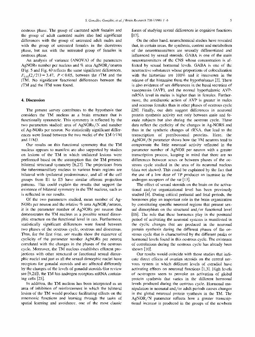
oestrous phase. The group of castrated adult females and the group of adult castrated males also had significant differences with the group of untreated adult males and with the group of untreated females in the dioestrous phase, but not with the untreated group of females in oestrous phase.
An analysis of variance (ANOVA) of the parameters AgNORs number per nucleus and % area AgNOR/neuron (Fig. 5 and Fig. 6) reflects the same significant differences, F0.95(2/21) = 3.47, P < 0.05, between the rTM and the ITM. No significant functional differences between the rTM and the 1TM were found.
4. Discussion
The present survey contributes to the hypothesis that considers the TM nucleus as a brain structure that is functionally symmetric. This symmetry is reflected by the two parameters studied: area of AgNORs /N and number of Ag-NORs per neuron. No statistically significant differ- ences were found between the two nuclei of the TM (rTM and 1TM).
Our results on this functional symmetry that the TM nucleus appears to manifest are also supported by studies on lesions of the TM in which unilateral lesions were performed based on the assumption that the TM presents bilateral structural symmetry [6,27]. The projections from the tuberomamillary nucleus to various brain regions are bilateral with ipsilateral predominance, and all of the cell groups from E1 to E5 have similar efferent projection patterns. This could explain the results that support the existence of bilateral symmetry in the TM nucleus, such as is reflected in our results.
Of the two parameters studied, mean number of Ag- NORs per neuron and the relative % area AgNOR/neuron, it is the parameter number of AgNORs per neuron that demonstrates the TM nucleus as a possible sexual dimor- phic structure on the functional level in rats. Furthermore, statistically significant differences were found between two phases of the oestrous cycle, oestrous and dioestrous. Thus, for the first time, our results show the existence of cyclicity of the parameter number AgNORs per neuron correlated with the changes in the phases of the oestrous cycle. Moreover, the TM nucleus establishes efferent pro- jections with other structural or functional sexual dimor- phic nuclei and just as all the sexual dimorphic nuclei have receptors for gonadal steroids and are affected differently by the changes of the levels of gonadal steroids (for review see [9,21]), the TM has androgen receptors mRNA contain- ing cells [25].
In addition, the TM nucleus has been interpreted as an area of inhibition of reinforcement in which the bilateral lesion of the TM would produce facilitating effects on the mnemonic functions and learning through the tasks of spatial learning and avoidance, one of the most classic
forms of studying sexual differences in cognitive functions [17].
On the other hand, neurochemical studies have revealed that, in certain areas, the synthesis, content and metabolism of the neurotransmitters are sexually differentiated and influenced by sexual steroids. GABA is one of the main neurotransmitters of the CNS whose concentration is af- fected by sexual hormonal levels. GABA is one of the neuroactive substances whose proportions of colocalization with the histamine are 100% and it intervenes in the release of the histamine from the hypothalamus [2]. There is also evidence of sex differences in the basal secretion of vasopressin (AVP), and the normal hypothalamic AVP- mRNA level in males is higher than in females. Further- more, the antidiuretic action of AVP is greater in males and oestrous females than in other phases of oestrous cycle [28]. Finally, our data suggest differences in neuronal protein synthesis activity not only between male and fe- male subjects but also during the oestrous cycle. These data reflect the cyclicity of the changes in Ag-NORS and thus in the synthetic changes of rRNA, that lead to the transcription of preribosomal proteins. Here, the A g N O R / N parameter shows how the TM neurons tend to compensate the little neuronal activity reflected in the parameter number of AgNOR per neuron with a greater transcription process, keeping in mind that there are no differences between sexes or between phases of the oe- strous cycle studied in the area of its neuronal nucleus (data not shown). This could be explained by the fact that the use of a low dose of TP produces an increase in the androgen receptors of the rat [13].
The effect of sexual steroids on the brain on the activa- tional and/or organizational level has been previously studied [4]. During critical perinatal and fetal periods, the hormones play an important role in the brain organization by constituting specific neuronal regions that present sex- ual dimorphism on the structural and/or functional level [16]. The role that these hormones play in the postnatal period of activating the neuronal systems is manifested in the cyclic changes that are produced in the neuronal protein synthesis during the different phases of the oe- strous cycle that is characterized by the different peaks or hormonal levels found in this oestrous cycle. The existence of correlations during the oestrous cycle has already been shown [ 10].
Our results would coincide with those studies that indi- cate direct effects of ovarian steroids on the central ner- vous system in which different levels of estradiol have activating effects on neuronal functions [1,3]. High levels of oestrogens seem to provoke an activation of global protein synthesis that varies in the different hormonal levels produced during the oestrous cycle. Hormonal ma- nipulation in neonatal and /or adult periods causes changes in the global neuronal protein synthesis in the TM. The A g N O R / N parameter reflects how a greater transcrip- tional increase is produced in the groups of the newborn

6 S. Gon¢(deg-Gonz~lez et a l . /Brain Research 736 (1996) I - 6
with hormonal manipulation and cannot be equalled by the adult groups that are hormonally manipulated. Thus, we conclude that the hormonal balance in the TM is especially important in the adult state, and that it is important to know more about the histaminergic system and its relation to the gonadal hormones.
Acknowledgements
We thank the following persons for their help: Angel Nistal and his group (Image Analysis Service. Univ. of Oviedo), Fernando S4nchez-Santed for his help in the management of the surgical techniques, Piedad Burgos and Laudino L6pez for their technical assistance and Barbara Shapiro for her translation of the article into English. This study was supported by grants from the FEDER and the University of Oviedo (TA/94) , Spain.
References
Ill Apud, J.A., Cocchi, D., Masotto, C., Penalva, A., Miiller, E.E. and Racagni, G., Effect of single or repeated estrogen administration on tuberoinfundibular GABA neurons and anterior pituitary GABA receptors: Biochemical and functional studies, Brain Res., 361 (1985) 146-153.
[2] Airaksinem, M.S., Alanen, S., Szabat, E., Visser, T.J. and Panula, P., Multiple neurotransmitters in the tuberomammillary nucleus: comparison of rat, mouse and guinea pig, J. Comp. Neurol., 323 (1992) 103-116.
[3] Bloch, G.J., Kurth, S.M., Adesson, T.R. and Micevych, P.E., Estro- gen-concentrating cells within cell groups of the medial preoptic area: sex differences and co-localization with galanin-immunoreac- tire cells, Brain Res., 595(1992) 301-308.
[4] Beatty, W., Gonadal hormones and sex differences in nonreproduc- tire behaviors. In Sexual difJerentiation, Handbook Behat:ioral Neu- robiology, Vol. 11, Plenum Press, New York, 1992, pp. 85-128.
[5] Bourgueous, C.A. and Hubert, J., Spatial relationship between the nucleolus and nuclear envelope: structural aspects and functional significant, lnt. ReL'. Cytol., l l (1988) 1-52.
[6] Carey R.J., Unilateral 6-hydroxydopamine lesions of dopamine neu- rons produce bilateral self-stimulation deficits, Behat. Brain Res., 6 (1982) 101-114.
[7] Croker, J. and Nar, P., Nucleolar organizer regions in lymphomas, J. Pathol., 151 (1987) 111-118.
[8] Derenzini, M., Farabegoli, F. and Trerb, D., Relationship between interphase AgNOR distribution and nucleolar size in cancer cells, Histochem. J., 24 (1992)951-956.
[9] Dulce, M.M. and Lieberman, A.R., Sexual dimorphism in the mam malian limbic system, Prog. Neurobiol., 45 (1995) 275-333.
[10] Egozi, Y., Kloog, Y. and Sokolovsky, M., Acetylcholine rhythm in the preoptic area of the rat hypothalamus is synchronized with the estrous cycle, Brain Res., 383 (1986) 310-313.
[11] Ericson, H., Blomqvist, A. and K6hler, C., Brainstem afferents to the tuberomammillary nucleus in the rat brain with special reference
to monoaminergic innervation, J. Comp. Neurol., 281 (1989) 169- 192.
[12] Gonzfilez-Pardo, H., GutiErrez, S., Men6ndez, A. and Arias, J.L., Postnatal development of argyrophilic nucleolar organizer regions in the mammillary body of undernourished rats, Brain Res., 527 (1994) 116-122.
[13] Gupta, D., Haldar, Ch., Coeleveld, M. and Roth, J., Ontogeny, circadian rhythm pattern, and hormonal modulation of 5-dihydro- testosterone receptors in the rat pineal, Neuroendocrinalogy, 57 (1993) 45-53.
[14] Hernfinde-Verdum, D., The nucleolar organizer regions, Biol. Cell., 49 (1983) 191-21)2.
[15] lnagaki, N., Toda, K., Taniuchi, l., Panula, P., Yamatodani, A., Tohyama, M., Watanabe, T. and Wada, H., An analysis of histamin- ergic efferents of the tuberomammillary nucleus to the medial preoptic area and inferior colliculus of the rat, Exp. Brain Res., 80 (1990) 374-380.
[16] Keefer, D. and Holderegger, C., The ontogeny of estrogen receptors: brain and pituitary, Det,. Brain Res., 19 (1985) 183-194.
[17] Klapdor, K., HaseniShrl, R.U. and Huston, J.P., Facilitation of learning in adult and aged rats following bilateral lesions of the tuberomammillary nucleus region, Behal:. Brain Res., 61 (1994) 113-116.
[18] KiShler, C., Swanson, L.W., Haglund, L. and Wu, J.Y., The cytoar- chitecture, histochemistry and projections of the tuberomammillary nucleus in the rat, Neuroscience, 16 (1985) 85-110.
[19] Lafarga, M., AndrOs, M.A., Berciano, M.T. and Maquieri, E., Orga- nization of nucleoli and nucleolar bodies in osmotically stimulated supraoptic neurons of the rat, J. Comp. Neurol., 308 (1991) 329-333.
[20] McLemore, D. and Lord, B., Argyrophilic nucleolar organizer rgions: An improved technique for visualization, J, Histoteeh., 3 (1991) 187-189.
[21] Onodera, K., Yamatodani, A., Watanabe, T. and Wada, H., Neu- ropharmacology of the histaminergic neuron system in the brain and its relationship with behavioral disorders, Prog Neurobiol., 42 (1994) 685 -702.
[22] Paxinos, G. and Watson, C., The Rat Brain in Stereotaxic Caordi- nates. 2nd edn., Harcourt Brace, Jovanovich Publishers, Academic Press, New York, 1986, 166 pp.
[23] Ploton, D., Menager, M., Jeannesson, P., Himber, G., Pigeon, F. and Adneu, J.J.. hnprovement in the staining and in the visualization of the argyrophilic proteins of the nuclear organizer region at the optical level, Histochem. J., 18 (1986) 5-14.
[24] Senba, E., Daddona, P.E., Watanabe, T., Wu, J.Y. and Nagy, J.[., Coexistence of adenosine deaminase, histidine decarboxylase in hypothalamic neurons of the rat, J. Neurosci., 5 (1985) 3393-3402.
[25] Simerly, R.B., Chang, C., Muramatsu, M. and Swanson, L.W., Distribution of androgen and estrogen receptor mRNA-containing cells in the rat brain: an in situ hybridization study, J. Comp. Neurol., 294 (1990) 76-95.
[26] Tr{Sster, H., Spring, H., Meissner, B., Schultz, P., Onder, P. and Trendelenburg, M.F., Structural organization of an active chromoso- mal nucleolar organizer region (NOR) identified by light microscopy and subsequent TEM and STEM electron microscopy, Chromosoma, 91 (1985) 151-163.
[27] Wagner, U., Segura-Torres, P., Weiler. T. and Huston, J.P., The tuberomammillary nucleus region as a reinforcement inhibiting sub- strate: facilitation of ipsihypothalamic self-stimulation by unilateral ibotenic acid lesions, Brain Res., 613 (1993) 269-274.
[28] Wen-Jie, D. and Yao, T., Effects of dehydration and salt-loading on hypothalamic vasopressin mRNA level in male and female rats, Brain Res., 676 (1995) 178-182.