
Expression profiling identifies the cytoskeletal organizer ezrin and the developmental homeoprotein...
7
ARTICLES NATURE MEDICINE VOLUME 10 | NUMBER 2 | FEBRUARY 2004 175 RMS, a skeletal muscle cancer, accounts for 5–10% of all pediatric neoplasms and >50% of pediatric soft-tissue sarcomas 1 . Despite recent improvements in long-term survival rates, about one third of RMS patients continue to experience relapses, and the majority of these patients die from disseminated metastatic disease 2,3 , illustrating the need for new therapeutic strategies. Metastasis is a complex and multistage process involving local invasion, intravasation, successful transit through the bloodstream, extravasation, and eventual prolifer- ation and survival within a favorable target organ. Although individ- ual candidate genes have been implicated in the metastatic process, few such discoveries have led to substantial clinical improvement. Advances in microarray-based expression profiling have provided technology robust enough to enable simultaneous analysis of the expression of large groups and combinations of genes. This technol- ogy has recently been brought to bear on the challenge of tumor pro- gression and metastasis 4–6 . Microarray-based expression profiling has been most commonly used to compare and contrast heterogeneous groups of human tumors. But such analyses are more difficult in RMS and other relatively rare tumors for which it is difficult to procure tissue samples of sufficient numbers to ensure statistical significance. The availability of an authen- tic mouse model that accurately reflects human disease would offer the possibility of analyzing larger numbers of homogeneous tumor sam- ples, thus increasing the likelihood of identifying crucial metastasis genes and pathways. We recently reported the development of a trans- genic mouse, overexpressing the c-Met ligand HGF/SF and deficient in Ink4a/Arf, in which skeletal muscle tumors reminiscent of those in embryonic RMS arise with very high penetrance and short latency 7 . All HGF/SF-transgenic, Ink4a/Arf-deficient mice succumb to multifocal, highly invasive RMS tumors by ∼4 months of age. Here we used this transgenic model system to establish a panel of highly and poorly metastatic RMS cell lines, which we subjected to microarray-based expression profiling to generate hypotheses as to which genes con- tribute to the metastatic process. We then validated several candidate genes using a variety of functional studies. Establishment of highly and poorly metastatic RMS cell lines Early-passage RMS cell lines were derived predominantly from RMS tumors arising in a variety of primary sites in HGF/SF-transgenic, Ink4a/Arf-deficient mice (Table 1). The metastatic potential of each of 19 of these cell lines was assessed in vivo by tail vein injection and orthotopic transplantation with spontaneous metastases. There was good general agreement between the results obtained using these two techniques. Nine cell lines were designated as highly metastatic to the lung, while ten others were deemed to be poorly metastatic (Table 1). Metastasis was noted in other organs, including kidney, heart, liver and skin. The highly and poorly metastatic cell lines could not be distin- guished in vitro by morphology or growth characteristics (data not shown). An assessment of expression and activity of obvious candidate pathway members, such as c-Met, AKT and MAPK, revealed no consis- tent differences (Supplementary Fig. 1 online). We therefore analyzed global gene expression patterns using a cDNA microarray approach. 1 Laboratory of Molecular Biology and 2 Pediatric Oncology Branch, National Cancer Institute, National Institutes of Health, Bethesda, Maryland 20892, USA. 3 Cancer Genetics Branch, National Human Genome Research Institute, National Institutes of Health, Bethesda, Maryland 20892, USA. 4 These authors contributed equally to this work. Correspondence should be addressed to G.M. ([email protected]). Published online 4 January 2004; doi:10.1038/nm966 Expression profiling identifies the cytoskeletal organizer ezrin and the developmental homeoprotein Six-1 as key metastatic regulators Yanlin Yu 1,4 , Javed Khan 2,4 , Chand Khanna 2 , Lee Helman 2 , Paul S Meltzer 3 & Glenn Merlino 1 Patients presenting with metastatic rhabdomyosarcoma (RMS), the most common soft-tissue sarcoma in children, have a very poor clinical prognosis. This is due, in large part, to our rudimentary knowledge of the molecular events that dictate metastatic potential. We used cDNA microarray analysis of RMS cell lines, derived from Ink4a/Arf-deficient mice transgenic for hepatocyte growth factor/scatter factor (HGF/SF), to identify a set of genes whose expression was significantly different between highly and poorly metastatic cells. Subsequent in vivo functional studies revealed that the actin filament–plasma membrane linker ezrin (encoded by Vil2) and the homeodomain-containing transcription factor Six-1 (sine oculis–related homeobox-1 homolog) had essential roles in determining the metastatic fate of RMS cells. VIL2 and SIX1 expression was enhanced in human RMS tissue, significantly correlating with clinical stage. The identification of ezrin and Six-1 as critical regulators of metastasis in RMS provides new mechanistic and therapeutic insights into this pediatric cancer. © 2004 Nature Publishing Group http://www.nature.com/naturemedicine
-
Upload
independent -
Category
Documents
-
view
1 -
download
0
Transcript of Expression profiling identifies the cytoskeletal organizer ezrin and the developmental homeoprotein...

A R T I C L E S
NATURE MEDICINE VOLUME 10 | NUMBER 2 | FEBRUARY 2004 175
RMS, a skeletal muscle cancer, accounts for 5–10% of all pediatricneoplasms and >50% of pediatric soft-tissue sarcomas1. Despiterecent improvements in long-term survival rates, about one third ofRMS patients continue to experience relapses, and the majority ofthese patients die from disseminated metastatic disease2,3, illustratingthe need for new therapeutic strategies. Metastasis is a complex andmultistage process involving local invasion, intravasation, successfultransit through the bloodstream, extravasation, and eventual prolifer-ation and survival within a favorable target organ. Although individ-ual candidate genes have been implicated in the metastatic process,few such discoveries have led to substantial clinical improvement.Advances in microarray-based expression profiling have providedtechnology robust enough to enable simultaneous analysis of theexpression of large groups and combinations of genes. This technol-ogy has recently been brought to bear on the challenge of tumor pro-gression and metastasis4–6.
Microarray-based expression profiling has been most commonlyused to compare and contrast heterogeneous groups of human tumors.But such analyses are more difficult in RMS and other relatively raretumors for which it is difficult to procure tissue samples of sufficientnumbers to ensure statistical significance. The availability of an authen-tic mouse model that accurately reflects human disease would offer thepossibility of analyzing larger numbers of homogeneous tumor sam-ples, thus increasing the likelihood of identifying crucial metastasisgenes and pathways. We recently reported the development of a trans-genic mouse, overexpressing the c-Met ligand HGF/SF and deficient in
Ink4a/Arf, in which skeletal muscle tumors reminiscent of those inembryonic RMS arise with very high penetrance and short latency7. AllHGF/SF-transgenic, Ink4a/Arf-deficient mice succumb to multifocal,highly invasive RMS tumors by ∼ 4 months of age. Here we used thistransgenic model system to establish a panel of highly and poorlymetastatic RMS cell lines, which we subjected to microarray-basedexpression profiling to generate hypotheses as to which genes con-tribute to the metastatic process. We then validated several candidategenes using a variety of functional studies.
Establishment of highly and poorly metastatic RMS cell linesEarly-passage RMS cell lines were derived predominantly from RMStumors arising in a variety of primary sites in HGF/SF-transgenic,Ink4a/Arf-deficient mice (Table 1). The metastatic potential of each of19 of these cell lines was assessed in vivo by tail vein injection andorthotopic transplantation with spontaneous metastases. There wasgood general agreement between the results obtained using these twotechniques. Nine cell lines were designated as highly metastatic to thelung, while ten others were deemed to be poorly metastatic (Table 1).Metastasis was noted in other organs, including kidney, heart, liver andskin. The highly and poorly metastatic cell lines could not be distin-guished in vitro by morphology or growth characteristics (data notshown). An assessment of expression and activity of obvious candidatepathway members, such as c-Met, AKT and MAPK, revealed no consis-tent differences (Supplementary Fig. 1 online). We therefore analyzedglobal gene expression patterns using a cDNA microarray approach.
1Laboratory of Molecular Biology and 2Pediatric Oncology Branch, National Cancer Institute, National Institutes of Health, Bethesda, Maryland 20892, USA. 3CancerGenetics Branch, National Human Genome Research Institute, National Institutes of Health, Bethesda, Maryland 20892, USA. 4These authors contributed equally tothis work. Correspondence should be addressed to G.M. ([email protected]).
Published online 4 January 2004; doi:10.1038/nm966
Expression profiling identifies the cytoskeletal organizerezrin and the developmental homeoprotein Six-1 as keymetastatic regulatorsYanlin Yu1,4, Javed Khan2,4, Chand Khanna2, Lee Helman2, Paul S Meltzer3 & Glenn Merlino1
Patients presenting with metastatic rhabdomyosarcoma (RMS), the most common soft-tissue sarcoma in children, have a verypoor clinical prognosis. This is due, in large part, to our rudimentary knowledge of the molecular events that dictate metastaticpotential. We used cDNA microarray analysis of RMS cell lines, derived from Ink4a/Arf-deficient mice transgenic for hepatocytegrowth factor/scatter factor (HGF/SF), to identify a set of genes whose expression was significantly different between highly andpoorly metastatic cells. Subsequent in vivo functional studies revealed that the actin filament–plasma membrane linker ezrin(encoded by Vil2) and the homeodomain-containing transcription factor Six-1 (sine oculis–related homeobox-1 homolog) hadessential roles in determining the metastatic fate of RMS cells. VIL2 and SIX1 expression was enhanced in human RMS tissue,significantly correlating with clinical stage. The identification of ezrin and Six-1 as critical regulators of metastasis in RMSprovides new mechanistic and therapeutic insights into this pediatric cancer.
©20
04 N
atur
e P
ublis
hing
Gro
up
http
://w
ww
.nat
ure.
com
/nat
urem
edic
ine
A R T I C L E S
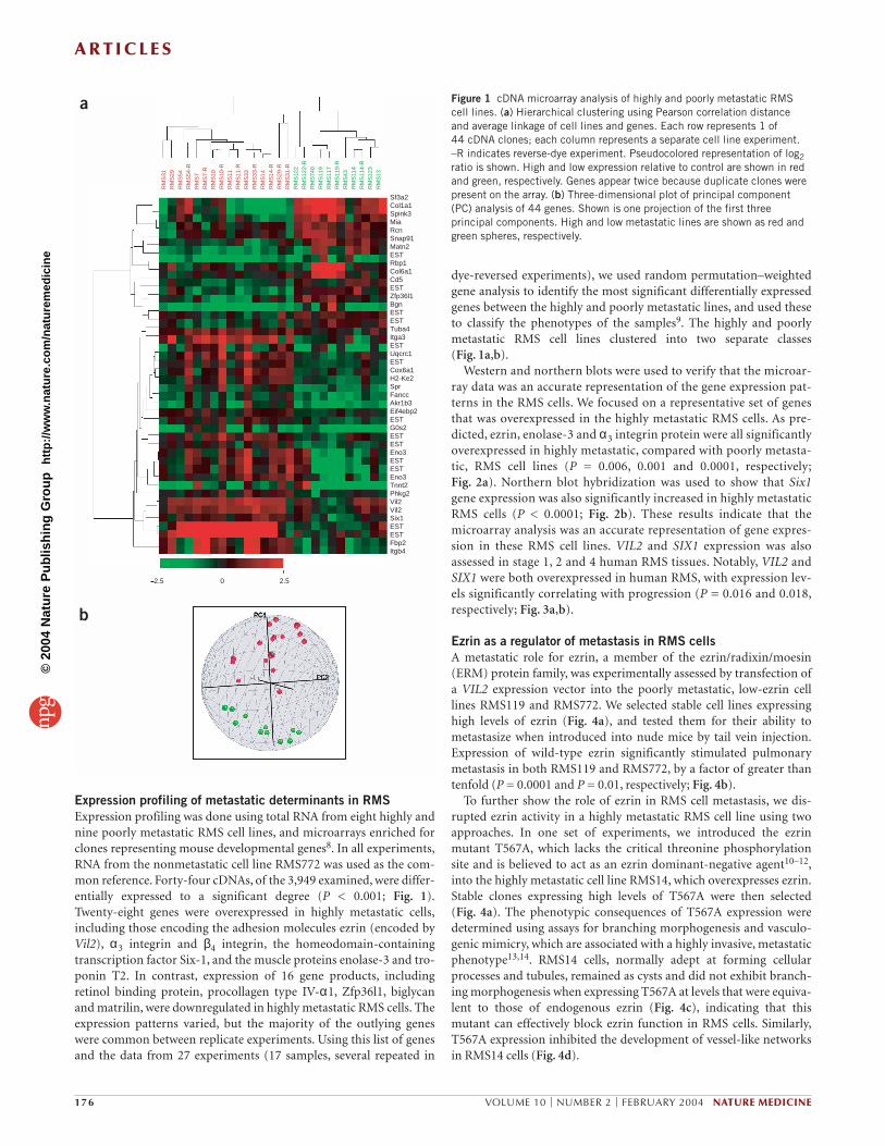
Expression profiling of metastatic determinants in RMSExpression profiling was done using total RNA from eight highly andnine poorly metastatic RMS cell lines, and microarrays enriched forclones representing mouse developmental genes8. In all experiments,RNA from the nonmetastatic cell line RMS772 was used as the com-mon reference. Forty-four cDNAs, of the 3,949 examined, were differ-entially expressed to a significant degree (P < 0.001; Fig. 1).Twenty-eight genes were overexpressed in highly metastatic cells,including those encoding the adhesion molecules ezrin (encoded byVil2), α3 integrin and β4 integrin, the homeodomain-containingtranscription factor Six-1, and the muscle proteins enolase-3 and tro-ponin T2. In contrast, expression of 16 gene products, includingretinol binding protein, procollagen type IV-α1, Zfp36l1, biglycanand matrilin, were downregulated in highly metastatic RMS cells. Theexpression patterns varied, but the majority of the outlying geneswere common between replicate experiments. Using this list of genesand the data from 27 experiments (17 samples, several repeated in
dye-reversed experiments), we used random permutation–weightedgene analysis to identify the most significant differentially expressedgenes between the highly and poorly metastatic lines, and used theseto classify the phenotypes of the samples9. The highly and poorlymetastatic RMS cell lines clustered into two separate classes (Fig. 1a,b).
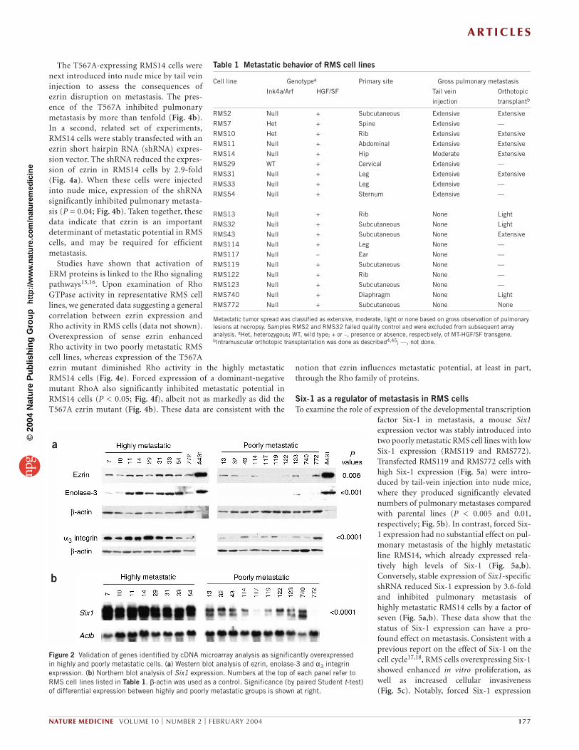
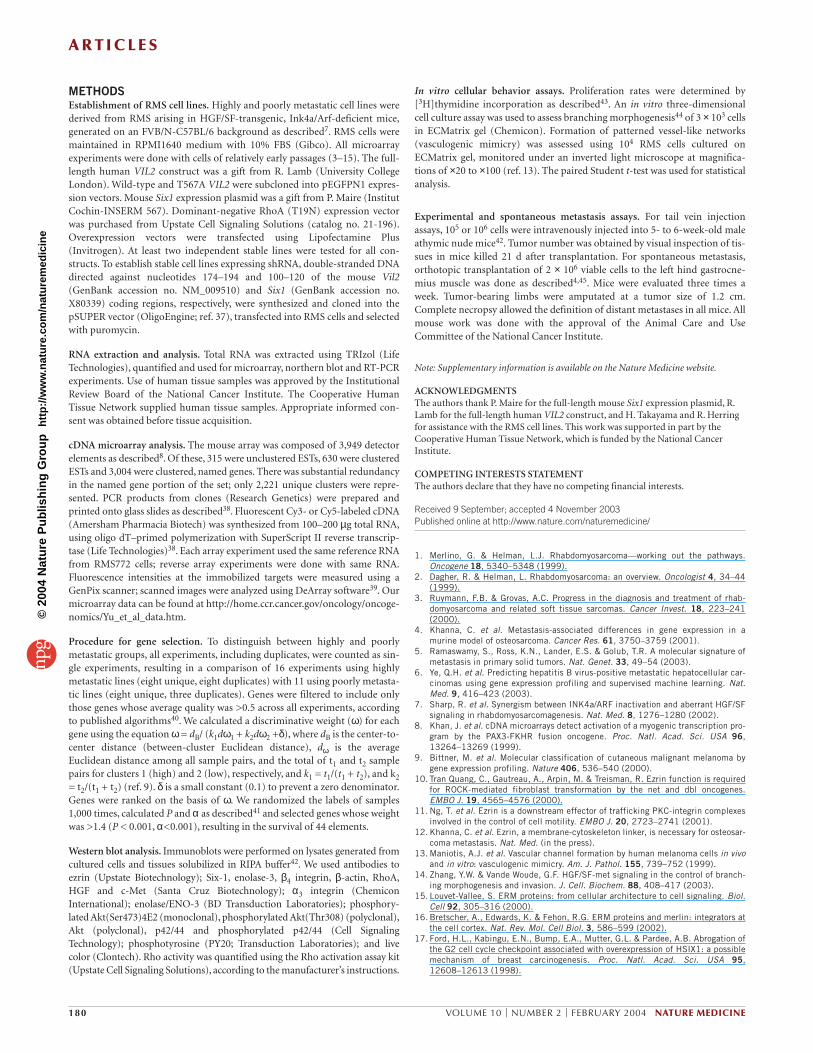
Western and northern blots were used to verify that the microar-ray data was an accurate representation of the gene expression pat-terns in the RMS cells. We focused on a representative set of genesthat was overexpressed in the highly metastatic RMS cells. As pre-dicted, ezrin, enolase-3 and α3 integrin protein were all significantlyoverexpressed in highly metastatic, compared with poorly metasta-tic, RMS cell lines (P = 0.006, 0.001 and 0.0001, respectively;Fig. 2a). Northern blot hybridization was used to show that Six1gene expression was also significantly increased in highly metastaticRMS cells (P < 0.0001; Fig. 2b). These results indicate that themicroarray analysis was an accurate representation of gene expres-sion in these RMS cell lines. VIL2 and SIX1 expression was alsoassessed in stage 1, 2 and 4 human RMS tissues. Notably, VIL2 andSIX1 were both overexpressed in human RMS, with expression lev-els significantly correlating with progression (P = 0.016 and 0.018,respectively; Fig. 3a,b).
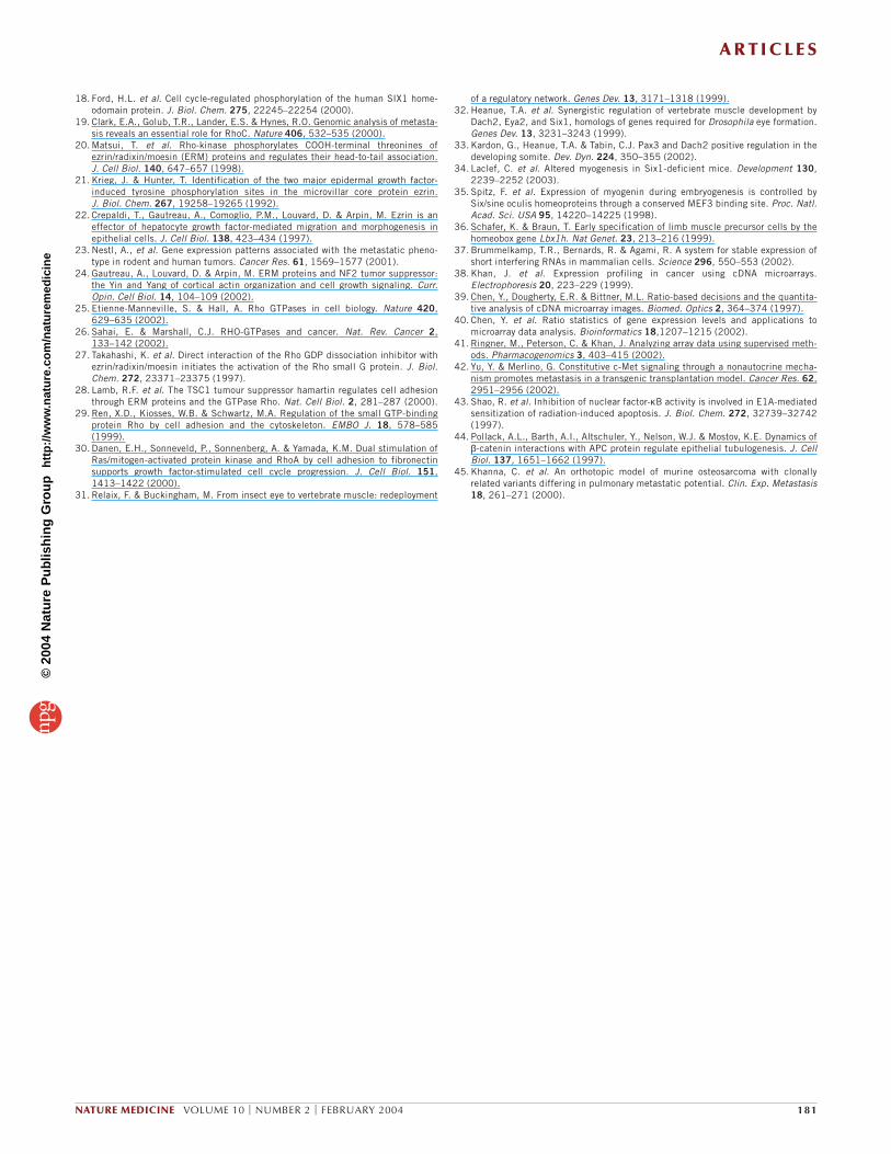
Ezrin as a regulator of metastasis in RMS cellsA metastatic role for ezrin, a member of the ezrin/radixin/moesin(ERM) protein family, was experimentally assessed by transfection ofa VIL2 expression vector into the poorly metastatic, low-ezrin celllines RMS119 and RMS772. We selected stable cell lines expressinghigh levels of ezrin (Fig. 4a), and tested them for their ability tometastasize when introduced into nude mice by tail vein injection.Expression of wild-type ezrin significantly stimulated pulmonarymetastasis in both RMS119 and RMS772, by a factor of greater thantenfold (P = 0.0001 and P = 0.01, respectively; Fig. 4b).
To further show the role of ezrin in RMS cell metastasis, we dis-rupted ezrin activity in a highly metastatic RMS cell line using twoapproaches. In one set of experiments, we introduced the ezrinmutant T567A, which lacks the critical threonine phosphorylationsite and is believed to act as an ezrin dominant-negative agent10–12,into the highly metastatic cell line RMS14, which overexpresses ezrin.Stable clones expressing high levels of T567A were then selected (Fig. 4a). The phenotypic consequences of T567A expression weredetermined using assays for branching morphogenesis and vasculo-genic mimicry, which are associated with a highly invasive, metastaticphenotype13,14. RMS14 cells, normally adept at forming cellularprocesses and tubules, remained as cysts and did not exhibit branch-ing morphogenesis when expressing T567A at levels that were equiva-lent to those of endogenous ezrin (Fig. 4c), indicating that thismutant can effectively block ezrin function in RMS cells. Similarly,T567A expression inhibited the development of vessel-like networksin RMS14 cells (Fig. 4d).
176 VOLUME 10 | NUMBER 2 | FEBRUARY 2004 NATURE MEDICINE
RM
S31
RM
S29
RM
S54
RM
S54
-RR
MS
7R
MS
7-R
RM
S10
RM
S10
-RR
MS
11R
MS
11-R
RM
S33
RM
S33
-RR
MS
14R
MS
14-R
RM
S29
-RR
MS
31-R
RM
S12
2R
MS
122-
RR
MS
740
RM
S11
9R
MS
117
RM
S11
9-R
RM
S43
RM
S11
4R
MS
114-
RR
MS
123
RM
S13
Sf3a2Col1a1Spink3MiaRcnSnap91Matn2ESTRbp1Col6a1Cd5ESTZfp36l1BgnESTESTTuba4Itga3ESTUqcrc1ESTCox6a1H2-Ke2SprFanccAkr1b3Eif4ebp2ESTG0s2ESTESTEno3ESTESTEno3Tnnt2Phkg2Vil2Vil2Six1ESTESTFbp2Itgb4
2.5 0 2.5
a
b PC1
Figure 1 cDNA microarray analysis of highly and poorly metastatic RMScell lines. (a) Hierarchical clustering using Pearson correlation distance and average linkage of cell lines and genes. Each row represents 1 of 44 cDNA clones; each column represents a separate cell line experiment.–R indicates reverse-dye experiment. Pseudocolored representation of log2ratio is shown. High and low expression relative to control are shown in redand green, respectively. Genes appear twice because duplicate clones werepresent on the array. (b) Three-dimensional plot of principal component(PC) analysis of 44 genes. Shown is one projection of the first threeprincipal components. High and low metastatic lines are shown as red andgreen spheres, respectively.
©20
04 N
atur
e P
ublis
hing
Gro
up
http
://w
ww
.nat
ure.
com
/nat
urem
edic
ine
A R T I C L E S
The T567A-expressing RMS14 cells werenext introduced into nude mice by tail veininjection to assess the consequences ofezrin disruption on metastasis. The pres-ence of the T567A inhibited pulmonarymetastasis by more than tenfold (Fig. 4b).In a second, related set of experiments,RMS14 cells were stably transfected with anezrin short hairpin RNA (shRNA) expres-sion vector. The shRNA reduced the expres-sion of ezrin in RMS14 cells by 2.9-fold(Fig. 4a). When these cells were injectedinto nude mice, expression of the shRNAsignificantly inhibited pulmonary metasta-sis (P = 0.04; Fig. 4b). Taken together, thesedata indicate that ezrin is an importantdeterminant of metastatic potential in RMScells, and may be required for efficientmetastasis.
Studies have shown that activation ofERM proteins is linked to the Rho signalingpathways15,16. Upon examination of RhoGTPase activity in representative RMS celllines, we generated data suggesting a generalcorrelation between ezrin expression andRho activity in RMS cells (data not shown).Overexpression of sense ezrin enhancedRho activity in two poorly metastatic RMScell lines, whereas expression of the T567Aezrin mutant diminished Rho activity in the highly metastaticRMS14 cells (Fig. 4e). Forced expression of a dominant-negativemutant RhoA also significantly inhibited metastatic potential inRMS14 cells (P < 0.05; Fig. 4f), albeit not as markedly as did theT567A ezrin mutant (Fig. 4b). These data are consistent with the
notion that ezrin influences metastatic potential, at least in part,through the Rho family of proteins.
Six-1 as a regulator of metastasis in RMS cellsTo examine the role of expression of the developmental transcription
factor Six-1 in metastasis, a mouse Six1expression vector was stably introduced intotwo poorly metastatic RMS cell lines with lowSix-1 expression (RMS119 and RMS772).Transfected RMS119 and RMS772 cells withhigh Six-1 expression (Fig. 5a) were intro-duced by tail-vein injection into nude mice,where they produced significantly elevatednumbers of pulmonary metastases comparedwith parental lines (P < 0.005 and 0.01,respectively; Fig. 5b). In contrast, forced Six-1 expression had no substantial effect on pul-monary metastasis of the highly metastaticline RMS14, which already expressed rela-tively high levels of Six-1 (Fig. 5a,b).Conversely, stable expression of Six1-specificshRNA reduced Six-1 expression by 3.6-foldand inhibited pulmonary metastasis ofhighly metastatic RMS14 cells by a factor ofseven (Fig. 5a,b). These data show that thestatus of Six-1 expression can have a pro-found effect on metastasis. Consistent with aprevious report on the effect of Six-1 on thecell cycle17,18, RMS cells overexpressing Six-1showed enhanced in vitro proliferation, aswell as increased cellular invasiveness (Fig. 5c). Notably, forced Six-1 expression
NATURE MEDICINE VOLUME 10 | NUMBER 2 | FEBRUARY 2004 177
a
b
Figure 2 Validation of genes identified by cDNA microarray analysis as significantly overexpressed in highly and poorly metastatic cells. (a) Western blot analysis of ezrin, enolase-3 and α3 integrinexpression. (b) Northern blot analysis of Six1 expression. Numbers at the top of each panel refer toRMS cell lines listed in Table 1. β-actin was used as a control. Significance (by paired Student t-test)of differential expression between highly and poorly metastatic groups is shown at right.
Table 1 Metastatic behavior of RMS cell lines
Cell line Genotypea Primary site Gross pulmonary metastasis
Ink4a/Arf HGF/SF Tail vein Orthotopic
injection transplantb
RMS2 Null + Subcutaneous Extensive Extensive
RMS7 Het + Spine Extensive —
RMS10 Het + Rib Extensive Extensive
RMS11 Null + Abdominal Extensive Extensive
RMS14 Null + Hip Moderate Extensive
RMS29 WT + Cervical Extensive —
RMS31 Null + Leg Extensive Extensive
RMS33 Null + Leg Extensive —
RMS54 Null + Sternum Extensive —
RMS13 Null + Rib None Light
RMS32 Null + Subcutaneous None Light
RMS43 Null + Subcutaneous None Extensive
RMS114 Null + Leg None —
RMS117 Null – Ear None —
RMS119 Null + Subcutaneous None —
RMS122 Null + Rib None —
RMS123 Null + Subcutaneous None —
RMS740 Null + Diaphragm None Light
RMS772 Null + Subcutaneous None None
Metastatic tumor spread was classified as extensive, moderate, light or none based on gross observation of pulmonarylesions at necropsy. Samples RMS2 and RMS32 failed quality control and were excluded from subsequent arrayanalysis. aHet, heterozygous; WT, wild type; + or –, presence or absence, respectively, of MT-HGF/SF transgene.bIntramuscular orthotopic transplantation was done as described4,45; —, not done.
©20
04 N
atur
e P
ublis
hing
Gro
up
http
://w
ww
.nat
ure.
com
/nat
urem
edic
ine

A R T I C L E S
also induced upregulation of ezrin in two poorly metastatic RMS celllines, suggesting that Vil2 lies downstream of Six1 (Fig. 5d).
DISCUSSIONTumor progression to a fully malignant, metastatic phenotype is ahighly complex process involving alterations in the expression of amultitude of genes. The advent of microarray technology, in which theexpression of tens of thousands of genes can be assessed simultane-ously, has greatly facilitated the dissection of this process4–6,9,19. Herewe describe the use of cDNA microarray analysis to identify a set ofgenes whose enhanced expression is associated with metastasis in celllines established from a recently developed mouse model of humanRMS7. We experimentally demonstrate in vivo, for the first time, thefunctional metastatic role of two of these genes, Vil2 and Six1.
Ezrin is a member of the ERM family, which shares the commonmembrane-binding N-terminal FERM domain with band-4.1 familymembers15,16. Ezrin provides a functional link between the plasmamembrane and the cortical actin cytoskeleton of the cell, and parti-cipates in crucial signal transduction pathways. ERM membersundergo phosphorylation at a C-terminal threonine through kinasessuch as Rho kinase10,20, and at N-terminal tyrosines through recep-tor tyrosine kinases such as the epidermal growth factor receptor andc-MET21,22, promoting cytoskeletal reorganization and subsequentmorphogenetic alterations. Ezrin has been implicated in themetastatic spread of osteosarcoma and mammary and pancreaticadenocarcinomas4,12,23. Here we reported that ezrin expression sig-nificantly correlates with metastasis in our mouse RMS model sys-tem. We also demonstrated experimentally that forced ezrinexpression induces a highly metastatic state in poorly metastaticRMS cell lines.
Ezrin resides at the nexus of multiple pathways regulating cellularbehaviors that can influence metastatic potential, including cell sur-vival, motility, invasion and adherence12,15,16,24. Analysis of the phe-notype of RMS14 cells transfected with the dominant-negativeezrin mutant T567A strongly supports the association of ezrinexpression with aggressive malignancy. Our data also suggest thatthe prometastatic activity of ezrin is mediated, at least in part,through Rho. The Rho family of small G proteins regulates a variety
178 VOLUME 10 | NUMBER 2 | FEBRUARY 2004 NATURE MEDICINE
a
b
Figure 3 Analysis of expression of VIL2 and SIX1 in staged human RMStissue. (a) Quantitative RT-PCR analysis of expression of human genesencoding ezrin and Six-1 in representative stage 1, 2 and 4 RMS neoplasms,compared with normal human skeletal muscle (SM). Expression of 18SrRNA (18S) was used as internal control. Northern blot analysis confirmedthese expression patterns (data not shown). M, molecular weight markers.(b) Relative expression of human VIL2 and SIX1 in staged human RMStumors compared with normal skeletal muscle. Bars indicate s.e.m. P valueswere assigned using Kruskal-Wallis one-way nonparametric ANOVA.
a
b
c d
e f
Figure 4 Phenotypic consequences of altered Ezrin function. (a) Western blot analysis of stable RMS cell lines transfected with plasmids expressing senseVil2 (sEzrin), T567A ezrin mutant, or ezrin-targeted shRNA (shEzrin). RMS772 and RMS119 are poorly metastatic; RMS14 is highly metastatic. sEzrinand T567A mutant were expressed as GFP fusion proteins (GEz and GMEz, respectively). Ez, ezrin; βA, β-actin control. (b) Gross pulmonary metastasesfrom transfected cells. Bars indicate s.e.m.; >50 indicates too numerous to count. (c,d) Light microscopy showing that T567A inhibits branchingmorphogenesis (c) and formation of vessel-like networks (d) C, vector control. Magnification, approximately ×100. (e) Alterations in functional ezrin levelsinduce corresponding changes in Rho activity. (f) Effect of dominant-negative RhoA (dnRhoA) on pulmonary metastasis of RMS14 cells.
©20
04 N
atur
e P
ublis
hing
Gro
up
http
://w
ww
.nat
ure.
com
/nat
urem
edic
ine

A R T I C L E S
of cellular functions through actin cytoskeletal reorganization, isderegulated in many tumor types, and is capable of inducing a vari-ety of cellular behaviors associated with tumor progression25,26.ERM proteins such as ezrin function both upstream and down-stream of Rho, forming an amplification feedback system15,16. Forexample, ERM proteins can bind to the Rho negative regulatorRhoGDI, thereby releasing inactive Rho and facilitating its activa-tion27. ERM interaction with hamartin, the protein product of thetumor suppressor gene TSC1, activates Rho and promotes focaladhesion formation28. In this report, we showed that using T567Ato compromise ezrin function in highly metastatic RMS14 cellsinhibits both Rho activity and metastatic potential, an effect alsoachieved through expression of dominant-negative RhoA. Notably,Rho proteins are also activated through interactions with inte-grins29,30, several of which are characteristically upregulated in ourmetastatic RMS cells.
In contrast to the cytoskeletal organizer ezrin, Six-1 is a homeo-domain-containing transcription factor, a vertebrate homolog of theDrosophila gene sine oculis gene that is implicated in the developmentof muscle, eye and brain31. In cultured human cells, Six-1 expressionis normally upregulated during S-phase entry, and forced Six-1expression attenuates a DNA damage-induced G2 cell cycle check-point17. Six-1 is also phosphorylated by casein kinase II, which mayregulate its cell cycle control function18. In support of these studies,we found that RMS cells overexpressing Six-1 showed enhanced pro-liferative capacity in vitro. However, the enhancing effect of Six-1expression on in vitro invasiveness suggests a more complex role incellular behavior. In fact, Six-1 is expressed in migrating limb precur-sor cells, acts as part of a regulatory network along with Eya-2, Dach-2 and Pax-3 (refs. 32,33), and is required for primary myogenesis34.Six-1 was itself induced in NIH3T3 cells by forced expression of thealveolar RMS–derived fusion oncogene Pax3-Foxo1 (ref. 8). Six-1 alsoactivates myogenin expression by interacting with MEF-3 bindingsites, which have been implicated in the regulation of other skeletalmuscle–related genes35. Another potential transcriptional target ofthe Six-1/Eya-2/Dach-2/Pax-3 regulatory network is Lbx1h, a home-obox gene required for migration of muscle precursor cells36. Thisraises the possibility that overexpression of Six-1 may inducemetastatic spread by subverting the normal role of Lbx1h in regulat-ing myoblast migration.
The sharp behavioral separation observed between our highly andpoorly metastatic cell lines constitutes genetic evidence supportingthe notion that a relatively small number of crucial regulatory factorsmay dictate metastatic potential. The existence of such key metastasisregulators could account, in part, for the detection of commonmetastatic signatures by microarray analysis of human tumors5,6, aswell as the coordinated upregulation of prometastatic genes, such asVil2 and certain integrins, in our RMS model. Six-1, a transcriptionfactor that helps control development-related cellular behavior, is apossible candidate for such a metastasis regulator. In fact, ezrin
expression was enhanced in poorly metastatic RMS cell lines inwhich Six-1 expression was experimentally elevated. Six-1 was alsoreported to be upregulated in primary and metastatic breast can-cer17. Thus, this transcription factor may influence expression of abattery of genes associated with cell proliferation, survival, motilityand/or invasiveness.
Here we demonstrate the power of applying microarray-basedgenomic analysis in combination with genetically engineered mousemodels to the study of metastasis. The successful identification andfunctional confirmation of ezrin and Six-1 as important metastaticregulators offers new mechanistic insights into RMS. The HGF/SF-transgenic, Ink4a/Arf-deficient mouse model can also serve as apotential preclinical model system. Both Vil2 and Six1 are membersof a panel of genes whose predictive value with respect to metastaticpotential can now be validated prospectively using our mouse model,as well as retrospectively in human RMS samples. In addition, celllines with highly metastatic and poorly metastatic profiles can be usedin syngeneic transplantation assays to test the efficacy of promisingrational antimetastatic agents derived from expression profiling data.The degree to which ezrin influences metastatic potential in thismodel system raises the possibility that it could be used as a moleculartarget in antimetastasis therapy. A successful outcome from studiessuch as these should brighten the therapeutic prospects for advancedRMS cases.
NATURE MEDICINE VOLUME 10 | NUMBER 2 | FEBRUARY 2004 179
a
b
c
d
Figure 5 Phenotypic consequences of altered Six-1 expression. (a) Westernblot analysis of representative RMS cell lines stably transfected withconstructs expressing either sense, full-length Six-1 or a Six-1-targetedshRNA (shSix-1). RMS772 and RMS119 are poorly metastatic; RMS14 ishighly metastatic. C, vector control. (b) Gross pulmonary metastases fromtransfected cell lines. (c) Forced expression of Six-1 stimulated cellularproliferation (left) and invasiveness (right) in vitro. Bars indicate s.e.m. (not visible if low); >50 indicates too numerous to count. (d) Western blotanalysis of ezrin expression in poorly metastatic RMS cells transfected witha sense full-length Six-1 expression vector. βA, β-actin control.
©20
04 N
atur
e P
ublis
hing
Gro
up
http
://w
ww
.nat
ure.
com
/nat
urem
edic
ine
A R T I C L E S
METHODSEstablishment of RMS cell lines. Highly and poorly metastatic cell lines werederived from RMS arising in HGF/SF-transgenic, Ink4a/Arf-deficient mice,generated on an FVB/N-C57BL/6 background as described7. RMS cells weremaintained in RPMI1640 medium with 10% FBS (Gibco). All microarrayexperiments were done with cells of relatively early passages (3–15). The full-length human VIL2 construct was a gift from R. Lamb (University CollegeLondon). Wild-type and T567A VIL2 were subcloned into pEGFPN1 expres-sion vectors. Mouse Six1 expression plasmid was a gift from P. Maire (InstitutCochin-INSERM 567). Dominant-negative RhoA (T19N) expression vectorwas purchased from Upstate Cell Signaling Solutions (catalog no. 21-196).Overexpression vectors were transfected using Lipofectamine Plus(Invitrogen). At least two independent stable lines were tested for all con-structs. To establish stable cell lines expressing shRNA, double-stranded DNAdirected against nucleotides 174–194 and 100–120 of the mouse Vil2(GenBank accession no. NM_009510) and Six1 (GenBank accession no.X80339) coding regions, respectively, were synthesized and cloned into thepSUPER vector (OligoEngine; ref. 37), transfected into RMS cells and selectedwith puromycin.
RNA extraction and analysis. Total RNA was extracted using TRIzol (LifeTechnologies), quantified and used for microarray, northern blot and RT-PCRexperiments. Use of human tissue samples was approved by the InstitutionalReview Board of the National Cancer Institute. The Cooperative HumanTissue Network supplied human tissue samples. Appropriate informed con-sent was obtained before tissue acquisition.
cDNA microarray analysis. The mouse array was composed of 3,949 detectorelements as described8. Of these, 315 were unclustered ESTs, 630 were clusteredESTs and 3,004 were clustered, named genes. There was substantial redundancyin the named gene portion of the set; only 2,221 unique clusters were repre-sented. PCR products from clones (Research Genetics) were prepared andprinted onto glass slides as described38. Fluorescent Cy3- or Cy5-labeled cDNA(Amersham Pharmacia Biotech) was synthesized from 100–200 µg total RNA,using oligo dT–primed polymerization with SuperScript II reverse transcrip-tase (Life Technologies)38. Each array experiment used the same reference RNAfrom RMS772 cells; reverse array experiments were done with same RNA.Fluorescence intensities at the immobilized targets were measured using aGenPix scanner; scanned images were analyzed using DeArray software39. Ourmicroarray data can be found at http://home.ccr.cancer.gov/oncology/oncoge-nomics/Yu_et_al_data.htm.
Procedure for gene selection. To distinguish between highly and poorlymetastatic groups, all experiments, including duplicates, were counted as sin-gle experiments, resulting in a comparison of 16 experiments using highlymetastatic lines (eight unique, eight duplicates) with 11 using poorly metasta-tic lines (eight unique, three duplicates). Genes were filtered to include onlythose genes whose average quality was >0.5 across all experiments, accordingto published algorithms40. We calculated a discriminative weight (ω) for eachgene using the equation ω= dB/ (k1dω1 + k2dω2 +δ), where dB is the center-to-center distance (between-cluster Euclidean distance), dω is the averageEuclidean distance among all sample pairs, and the total of t1 and t2 samplepairs for clusters 1 (high) and 2 (low), respectively, and k1 = t1/(t1 + t2), and k2= t2/(t1 + t2) (ref. 9). δ is a small constant (0.1) to prevent a zero denominator.Genes were ranked on the basis of ω. We randomized the labels of samples1,000 times, calculated P and α as described41 and selected genes whose weightwas >1.4 (P < 0.001, α<0.001), resulting in the survival of 44 elements.
Western blot analysis. Immunoblots were performed on lysates generated fromcultured cells and tissues solubilized in RIPA buffer42. We used antibodies toezrin (Upstate Biotechnology); Six-1, enolase-3, β4 integrin, β-actin, RhoA,HGF and c-Met (Santa Cruz Biotechnology); α3 integrin (ChemiconInternational); enolase/ENO-3 (BD Transduction Laboratories); phosphory-lated Akt(Ser473)4E2 (monoclonal), phosphorylated Akt(Thr308) (polyclonal),Akt (polyclonal), p42/44 and phosphorylated p42/44 (Cell SignalingTechnology); phosphotyrosine (PY20; Transduction Laboratories); and livecolor (Clontech). Rho activity was quantified using the Rho activation assay kit(Upstate Cell Signaling Solutions), according to the manufacturer’s instructions.
In vitro cellular behavior assays. Proliferation rates were determined by[3H]thymidine incorporation as described43. An in vitro three-dimensionalcell culture assay was used to assess branching morphogenesis44 of 3 × 10³ cellsin ECMatrix gel (Chemicon). Formation of patterned vessel-like networks(vasculogenic mimicry) was assessed using 104 RMS cells cultured onECMatrix gel, monitored under an inverted light microscope at magnifica-tions of ×20 to ×100 (ref. 13). The paired Student t-test was used for statisticalanalysis.
Experimental and spontaneous metastasis assays. For tail vein injectionassays, 105 or 106 cells were intravenously injected into 5- to 6-week-old maleathymic nude mice42. Tumor number was obtained by visual inspection of tis-sues in mice killed 21 d after transplantation. For spontaneous metastasis,orthotopic transplantation of 2 × 10⁶ viable cells to the left hind gastrocne-mius muscle was done as described4,45. Mice were evaluated three times aweek. Tumor-bearing limbs were amputated at a tumor size of 1.2 cm.Complete necropsy allowed the definition of distant metastases in all mice. Allmouse work was done with the approval of the Animal Care and UseCommittee of the National Cancer Institute.
Note: Supplementary information is available on the Nature Medicine website.
ACKNOWLEDGMENTSThe authors thank P. Maire for the full-length mouse Six1 expression plasmid, R.Lamb for the full-length human VIL2 construct, and H. Takayama and R. Herringfor assistance with the RMS cell lines. This work was supported in part by theCooperative Human Tissue Network, which is funded by the National CancerInstitute.
COMPETING INTERESTS STATEMENTThe authors declare that they have no competing financial interests.
Received 9 September; accepted 4 November 2003Published online at http://www.nature.com/naturemedicine/
1. Merlino, G. & Helman, L.J. Rhabdomyosarcoma—working out the pathways.Oncogene 18, 5340–5348 (1999).
2. Dagher, R. & Helman, L. Rhabdomyosarcoma: an overview. Oncologist 4, 34–44(1999).
3. Ruymann, F.B. & Grovas, A.C. Progress in the diagnosis and treatment of rhab-domyosarcoma and related soft tissue sarcomas. Cancer Invest. 18, 223–241(2000).
4. Khanna, C. et al. Metastasis-associated differences in gene expression in amurine model of osteosarcoma. Cancer Res. 61, 3750–3759 (2001).
5. Ramaswamy, S., Ross, K.N., Lander, E.S. & Golub, T.R. A molecular signature ofmetastasis in primary solid tumors. Nat. Genet. 33, 49–54 (2003).
6. Ye, Q.H. et al. Predicting hepatitis B virus-positive metastatic hepatocellular car-cinomas using gene expression profiling and supervised machine learning. Nat.Med. 9, 416–423 (2003).
7. Sharp, R. et al. Synergism between INK4a/ARF inactivation and aberrant HGF/SFsignaling in rhabdomyosarcomagenesis. Nat. Med. 8, 1276–1280 (2002).
8. Khan, J. et al. cDNA microarrays detect activation of a myogenic transcription pro-gram by the PAX3-FKHR fusion oncogene. Proc. Natl. Acad. Sci. USA 96,13264–13269 (1999).
9. Bittner, M. et al. Molecular classification of cutaneous malignant melanoma bygene expression profiling. Nature 406, 536–540 (2000).
10. Tran Quang, C., Gautreau, A., Arpin, M. & Treisman, R. Ezrin function is requiredfor ROCK-mediated fibroblast transformation by the net and dbl oncogenes.EMBO J. 19, 4565–4576 (2000).
11. Ng, T. et al. Ezrin is a downstream effector of trafficking PKC-integrin complexesinvolved in the control of cell motility. EMBO J. 20, 2723–2741 (2001).
12. Khanna, C. et al. Ezrin, a membrane-cytoskeleton linker, is necessary for osteosar-coma metastasis. Nat. Med. (in the press).
13. Maniotis, A.J. et al. Vascular channel formation by human melanoma cells in vivoand in vitro: vasculogenic mimicry. Am. J. Pathol. 155, 739–752 (1999).
14. Zhang, Y.W. & Vande Woude, G.F. HGF/SF-met signaling in the control of branch-ing morphogenesis and invasion. J. Cell. Biochem. 88, 408–417 (2003).
15. Louvet-Vallee, S. ERM proteins: from cellular architecture to cell signaling. Biol.Cell 92, 305–316 (2000).
16. Bretscher, A., Edwards, K. & Fehon, R.G. ERM proteins and merlin: integrators atthe cell cortex. Nat. Rev. Mol. Cell Biol. 3, 586–599 (2002).
17. Ford, H.L., Kabingu, E.N., Bump, E.A., Mutter, G.L. & Pardee, A.B. Abrogation ofthe G2 cell cycle checkpoint associated with overexpression of HSIX1: a possiblemechanism of breast carcinogenesis. Proc. Natl. Acad. Sci. USA 95,12608–12613 (1998).
180 VOLUME 10 | NUMBER 2 | FEBRUARY 2004 NATURE MEDICINE
©20
04 N
atur
e P
ublis
hing
Gro
up
http
://w
ww
.nat
ure.
com
/nat
urem
edic
ine
A R T I C L E S
18. Ford, H.L. et al. Cell cycle-regulated phosphorylation of the human SIX1 home-odomain protein. J. Biol. Chem. 275, 22245–22254 (2000).
19. Clark, E.A., Golub, T.R., Lander, E.S. & Hynes, R.O. Genomic analysis of metasta-sis reveals an essential role for RhoC. Nature 406, 532–535 (2000).
20. Matsui, T. et al. Rho-kinase phosphorylates COOH-terminal threonines ofezrin/radixin/moesin (ERM) proteins and regulates their head-to-tail association.J. Cell Biol. 140, 647–657 (1998).
21. Krieg, J. & Hunter, T. Identification of the two major epidermal growth factor-induced tyrosine phosphorylation sites in the microvillar core protein ezrin. J. Biol. Chem. 267, 19258–19265 (1992).
22. Crepaldi, T., Gautreau, A., Comoglio, P.M., Louvard, D. & Arpin, M. Ezrin is aneffector of hepatocyte growth factor-mediated migration and morphogenesis inepithelial cells. J. Cell Biol. 138, 423–434 (1997).
23. Nestl, A., et al. Gene expression patterns associated with the metastatic pheno-type in rodent and human tumors. Cancer Res. 61, 1569–1577 (2001).
24. Gautreau, A., Louvard, D. & Arpin, M. ERM proteins and NF2 tumor suppressor:the Yin and Yang of cortical actin organization and cell growth signaling. Curr.Opin. Cell Biol. 14, 104–109 (2002).
25. Etienne-Manneville, S. & Hall, A. Rho GTPases in cell biology. Nature 420,629–635 (2002).
26. Sahai, E. & Marshall, C.J. RHO-GTPases and cancer. Nat. Rev. Cancer 2,133–142 (2002).
27. Takahashi, K. et al. Direct interaction of the Rho GDP dissociation inhibitor withezrin/radixin/moesin initiates the activation of the Rho small G protein. J. Biol.Chem. 272, 23371–23375 (1997).
28. Lamb, R.F. et al. The TSC1 tumour suppressor hamartin regulates cell adhesionthrough ERM proteins and the GTPase Rho. Nat. Cell Biol. 2, 281–287 (2000).
29. Ren, X.D., Kiosses, W.B. & Schwartz, M.A. Regulation of the small GTP-bindingprotein Rho by cell adhesion and the cytoskeleton. EMBO J. 18, 578–585(1999).
30. Danen, E.H., Sonneveld, P., Sonnenberg, A. & Yamada, K.M. Dual stimulation ofRas/mitogen-activated protein kinase and RhoA by cell adhesion to fibronectinsupports growth factor-stimulated cell cycle progression. J. Cell Biol. 151,1413–1422 (2000).
31. Relaix, F. & Buckingham, M. From insect eye to vertebrate muscle: redeployment
of a regulatory network. Genes Dev. 13, 3171–1318 (1999).32. Heanue, T.A. et al. Synergistic regulation of vertebrate muscle development by
Dach2, Eya2, and Six1, homologs of genes required for Drosophila eye formation.Genes Dev. 13, 3231–3243 (1999).
33. Kardon, G., Heanue, T.A. & Tabin, C.J. Pax3 and Dach2 positive regulation in thedeveloping somite. Dev. Dyn. 224, 350–355 (2002).
34. Laclef, C. et al. Altered myogenesis in Six1-deficient mice. Development 130,2239–2252 (2003).
35. Spitz, F. et al. Expression of myogenin during embryogenesis is controlled bySix/sine oculis homeoproteins through a conserved MEF3 binding site. Proc. Natl.Acad. Sci. USA 95, 14220–14225 (1998).
36. Schafer, K. & Braun, T. Early specification of limb muscle precursor cells by thehomeobox gene Lbx1h. Nat Genet. 23, 213–216 (1999).
37. Brummelkamp, T.R., Bernards, R. & Agami, R. A system for stable expression ofshort interfering RNAs in mammalian cells. Science 296, 550–553 (2002).
38. Khan, J. et al. Expression profiling in cancer using cDNA microarrays.Electrophoresis 20, 223–229 (1999).
39. Chen, Y., Dougherty, E.R. & Bittner, M.L. Ratio-based decisions and the quantita-tive analysis of cDNA microarray images. Biomed. Optics 2, 364–374 (1997).
40. Chen, Y. et al. Ratio statistics of gene expression levels and applications tomicroarray data analysis. Bioinformatics 18,1207–1215 (2002).
41. Ringner, M., Peterson, C. & Khan, J. Analyzing array data using supervised meth-ods. Pharmacogenomics 3, 403–415 (2002).
42. Yu, Y. & Merlino, G. Constitutive c-Met signaling through a nonautocrine mecha-nism promotes metastasis in a transgenic transplantation model. Cancer Res. 62,2951–2956 (2002).
43. Shao, R. et al. Inhibition of nuclear factor-κB activity is involved in E1A-mediatedsensitization of radiation-induced apoptosis. J. Biol. Chem. 272, 32739–32742(1997).
44. Pollack, A.L., Barth, A.I., Altschuler, Y., Nelson, W.J. & Mostov, K.E. Dynamics ofβ-catenin interactions with APC protein regulate epithelial tubulogenesis. J. CellBiol. 137, 1651–1662 (1997).
45. Khanna, C. et al. An orthotopic model of murine osteosarcoma with clonallyrelated variants differing in pulmonary metastatic potential. Clin. Exp. Metastasis18, 261–271 (2000).
NATURE MEDICINE VOLUME 10 | NUMBER 2 | FEBRUARY 2004 181
©20
04 N
atur
e P
ublis
hing
Gro
up
http
://w
ww
.nat
ure.
com
/nat
urem
edic
ine