Functional specialization of transcription elongation factors
Upload
independentCategory
view
0download
0
Discovery, optimization and validation of an optimalDNA-binding sequence for the Six1 homeodomaintranscription factorYubing Liu1,2, Soumyadeep Nandi1,2, Andre Martel1,2, Alen Antoun1,2, Ilya Ioshikhes1,2
and Alexandre Blais1,2,*
1Ottawa Institute of Systems Biology and 2Biochemistry, Microbiology and Immunology Department, Faculty ofMedicine, University of Ottawa, Ottawa, Ontario, Canada
Received March 12, 2012; Revised May 9, 2012; Accepted May 24, 2012
ABSTRACT
The Six1 transcription factor is a homeodomainprotein involved in controlling gene expressionduring embryonic development. Six1 establishesgene expression profiles that enable skeletalmyogenesis and nephrogenesis, among others.While several homeodomain factors have been ex-tensively characterized with regards to theirDNA-binding properties, relatively little is known ofthe properties of Six1. We have used the genomicbinding profile of Six1 during the myogenic differen-tiation of myoblasts to obtain a better understandingof its preferences for recognizing certain DNA se-quences. DNA sequence analyses on our genomicbinding dataset, combined with biochemical charac-terization using binding assays, reveal that Six1 has amuch broader DNA-binding sequence spectrum thanhad been previously determined. Moreover, using aposition weight matrix optimization algorithm, wegenerated a highly sensitive and specific matrix thatcan be used to predict novel Six1-binding sites withhighest accuracy. Furthermore, our results supportthe idea of a mode of DNA recognition by thisfactor where Six1 itself is sufficient for sequence dis-crimination, and where Six1 domains outside of itshomeodomain contribute to binding site selection.Together, our results provide new light on theproperties of this important transcription factor,and will enable more accurate modeling of Six1function in bioinformatic studies.
INTRODUCTION
A defining characteristic of transcription factors (TFs) istheir ability to recognize and bind to specific DNA se-quences in the genome, within the regulatory region of
the target genes they control. Multiple structural classesof TFs exist, among which are homeodomain factors. Theprimary function of the homeodomain is DNA binding.This group of regulatory factors were first discovered dueto their involvement in homeotic conversions inDrosophila, but were later found to exist in essentially alleukaryote species, from yeast to humans. Bioinformaticsanalyses indicate that 235 homeodomain TFs are encodedin the human genome (not counting splice variants andpseudogenes) (1). This diversity is manifested by variousexpression profiles, protein domain composition andinteraction partners. However, a large number ofhomeodomains seemingly recognize the same DNAsequence TAAT, or close variants, leading to thequestion of how redundancy in binding site selection isavoided by these proteins. The prototypical 60 aminoacids long homeodomain assumes a three-dimensionalstructure composed of an unstructured N-terminal armfollowed by three alpha-helices. The N-terminal arm andthe third helix contribute most of the DNA sequence-binding specificity, and their amino acid composition arethought to contribute to this property (2,3). Two recentlarge-scale surveys considered the question of binding spe-cificity of homeodomain TFs from mouse (4) orDrosophila (5). It was found that indeed, whenhomeodomains are considered globally, their amino acidsequence correlates with their DNA sequence-bindingpreferences within and adjacent to the TAAT core.However, domains residing outside of the homeodomaincan also influence DNA binding, either through directDNA contacts or interaction with dimerization partners(6–9). Therefore, it remains to be established whetherhomeodomain TF-binding site predictions based solelyon in vitro DNA-binding preferences are sufficientlyaccurate to allow to predict which target genes theyregulate.The Six family of homeodomain TFs is conserved from
flies to humans, and in mammalians counts six members,from Six1 to Six6. Like most homeodomain TFs, they are
*To whom correspondence should be addressed. Tel: +1 613 562 5800 (Ext. 8463); Fax: +1 613 562 5452; Email: [email protected]
Published online 22 June 2012 Nucleic Acids Research, 2012, Vol. 40, No. 17 8227–8239doi:10.1093/nar/gks587
� The Author(s) 2012. Published by Oxford University Press.This is an Open Access article distributed under the terms of the Creative Commons Attribution Non-Commercial License (http://creativecommons.org/licenses/by-nc/3.0), which permits unrestricted non-commercial use, distribution, and reproduction in any medium, provided the original work is properly cited.

involved in controlling the development of various tissuetypes; for instance, Six1 and Six4 are involved in the de-velopment of the eyes, ears, kidneys and skeletal muscle(reviewed in (10,11)). We became interested in this groupof TFs when we found that the DNA motif they recognize,the MEF3 sequence element, is enriched within thepromoter region of Myogenic Regulatory Factors(MRFs) target genes, in muscle precursor cells (myoblasts)(12). The MEF3 element is a phylogenetically conservedmotif (DNA consensus TCAGGTTTC) that was origin-ally identified within the regulatory region of only a fewmuscle-specific genes (13,14). The Six1 and Six4 membersof the Six family, two factors essential to myogenesis, weresubsequently found to bind specifically to the MEF3sequence within the myogenin (Myog) gene promoterand activate this gene’s expression during embryogenesis(15–19). MRFs control myogenesis by activating the ex-pression of a large cohort of genes necessary for differen-tiation and function of muscle cells, and in some instancesthey accomplish this task by cooperating with transcrip-tion factors of the Mef2 and Pbx families (20,21). Theconnection between the MRF target genes and MEF3sites led us to postulate that Six family members canalso cooperate with the MRFs to regulate myogenesis.This was confirmed by the genome-wide identification ofSix1 target genes in mouse myoblasts and myotubes, andby functional assays which showed that Six factors canactivate transcription with the MRFs in a synergisticfashion (22).The wealth of DNA-binding information contained
within our genomic Six1-binding profile (22) gave us theopportunity to examine the DNA sequence-binding pref-erences of this TF in myoblasts and myotubes and toanalyze it in the context of the previously identifiedMEF3 sequence motif. Here, using de novo motif findingand position weight matrix (PWM) optimization, wereport that Six1 has a broader than anticipated DNA-binding profile, which extends well beyond the canonicalMEF3 consensus sequence. In vitro binding data corrob-orate these in vivo DNA-binding sequence preferences,suggesting that sequence-specific DNA binding by Six1does not require interaction with dimerization partners.However, we find a discrepancy between our results andthose of an in vitro DNA-binding screen for the Six1homeodomain, performed by others (4), suggesting thatSix1 homeodomain sequence is not the sole determinantof its binding to DNA.
MATERIALS AND METHODS
De novo motif finding
The Amadeus program was used for de novo DNAsequence motif discovery (23). The ‘bound’ sets corres-ponded to the sequences bound by Six1 in mousemyoblasts (MB, total of 1022 sites) or myotubes (MT,total of 1853 sites) with a false discovery rate (FDR) lessthan 10% (22). These two sets of bound sequences werebroken down into three (for MB) or five (for MT)randomly assigned subgroups, respectively (i.e. MB-A,MB-B, MB-C and so on). The purpose of this
sub-grouping was to obtain one subgroup for motif dis-covery, and to reserve the other sequences for subsequentvalidation of the discovered motifs. The Amadeusprogram was run on each of these eight subgroups, per-forming ‘large’ searches of 12 base pairs motifs andexamining both DNA strands. For these eight searches,the background sequence set corresponded to a randomlyselected subset of the genomic regions surveyed in ourChIP-on-chip analyses (20% of surveyed loci, approxi-mately 108Mb of sequence). Both sets of sequences wererepeat-masked using RepeatMasker (24). The top rankingsequence motif (lowest corrected P-value after 25 cycles ofboot-strapping where ‘bound’ and ‘background’ se-quences are randomly interchanged) was retained.Searches for motifs less than 12 base pairs in lengthwere also run and yielded very similar motifs (notshown). Finally, an averaged PWM (Six1_MB+MT)was calculated by aligning the eight PWMs and averagingthe frequencies at each position. PWMs were representedgraphically using the Weblogo program (25).
Motif abundance within sets of sequences was estimatedusing the CisGenome program (26). The number of PWM‘hits’ within ‘bound’ and ‘background’ sequences wasdetermined with a likelihood ratio setting of 500. Thebound set of sequences for this purpose was composedof those excluded from the initial motif discovery (e.g.for the matrix identified using subgroup MB-A, the se-quences in MB-B and MB-C were combined and used totest its performance). Here the background sequenceswere a distinct set of 20% of all surveyed loci. Therelative enrichment score (the ratio of frequency ofPWM hits among bound regions over that in backgroundregions) was calculated. We also calculated the cumulativehypergeometric probability, the chance of finding at leasta certain number of hits to a PWM among the boundregions (sample) given that a certain number of hits existamong the background (population) sequences (sampleand population sizes expressed as total base pair length).Finally, searches were also repeated on the top 5% mostphylogenetically conserved portion of the sequences.
PWM optimization
The methods described by Staden (27) and Bucher (28)were utilized to calculate the PWM. We adopted thebase frequencies reported by Amadeus in the de novomotif searches (above). These base frequencies were con-verted into odds scores by dividing the frequencies byexpected frequency which is calculated from theDatabase of Transcription Start Sites (DBTSS) formouse using the formula described in (27):
ebi ¼
PLi¼1
nbi
L
where b is one of 4 nucleotides (A,C,G or T) at positioni, nbi is the number of times base b occurs at the i-thposition of the motif and L is the length of the sequence.
The sequences used from DBTSS were of the length1201 base pairs (�1000 to+201 from the TSS) and werealigned with respect to the TSS.
8228 Nucleic Acids Research, 2012, Vol. 40, No. 17
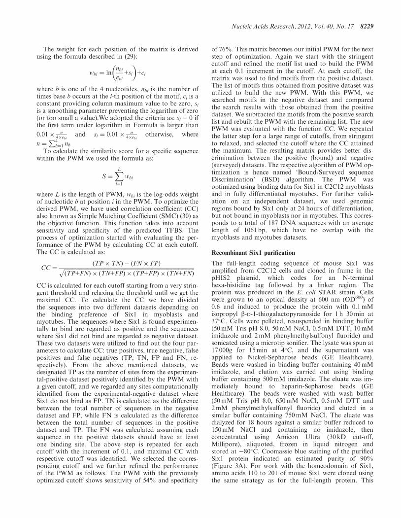
The weight for each position of the matrix is derivedusing the formula described in (29):
wbi ¼ lnnbiebi
+si
� �+ci
where b is one of the 4 nucleotides, nbi is the number oftimes base b occurs at the i-th position of the motif, ci is aconstant providing column maximum value to be zero, siis a smoothing parameter preventing the logarithm of zero(or too small a value).We adopted the criteria as: si=0 ifthe first term under logarithm in Formula is larger than
0:01� n4�ebi
and si ¼ 0:01� n4�ebi
otherwise, where
n ¼P4
b¼1 nbTo calculate the similarity score for a specific sequence
within the PWM we used the formula as:
S ¼XLi¼1
wbi
where L is the length of PWM, wbi is the log-odds weightof nucleotide b at position i in the PWM. To optimize thederived PWM, we have used correlation coefficient (CC)also known as Simple Matching Coefficient (SMC) (30) asthe objective function. This function takes into accountsensitivity and specificity of the predicted TFBS. Theprocess of optimization started with evaluating the per-formance of the PWM by calculating CC at each cutoff.The CC is calculated as:
CC ¼ðTP� TNÞ � ðFN� FPÞffiffiffiffiffiffiffiffiffiffiffiffiffiffiffiffiffiffiffiffiffiffiffiffiffiffiffiffiffiffiffiffiffiffiffiffiffiffiffiffiffiffiffiffiffiffiffiffiffiffiffiffiffiffiffiffiffiffiffiffiffiffiffiffiffiffiffiffiffiffiffiffiffiffiffiffiffiffiffiffiffiffiffiffiffiffiffiffiffiffiffiffiffiffiffiffi
ðTP+FNÞ � ðTN+FPÞ � ðTP+FPÞ � ðTN+FNÞp
CC is calculated for each cutoff starting from a very strin-gent threshold and relaxing the threshold until we get themaximal CC. To calculate the CC we have dividedthe sequences into two different datasets depending onthe binding preference of Six1 in myoblasts andmyotubes. The sequences where Six1 is found experimen-tally to bind are regarded as positive and the sequenceswhere Six1 did not bind are regarded as negative dataset.These two datasets were utilized to find out the four par-ameters to calculate CC: true positives, true negative, falsepositives and false negatives (TP, TN, FP and FN, re-spectively). From the above mentioned datasets, wedesignated TP as the number of sites from the experimen-tal-positive dataset positively identified by the PWM witha given cutoff, and we regarded any sites computationallyidentified from the experimental-negative dataset whereSix1 do not bind as FP. TN is calculated as the differencebetween the total number of sequences in the negativedataset and FP, while FN is calculated as the differencebetween the total number of sequences in the positivedataset and TP. The FN was calculated assuming eachsequence in the positive datasets should have at leastone binding site. The above step is repeated for eachcutoff with the increment of 0.1, and maximal CC withrespective cutoff was identified. We selected the corres-ponding cutoff and we further refined the performanceof the PWM as follows. The PWM with the previouslyoptimized cutoff shows sensitivity of 54% and specificity
of 76%. This matrix becomes our initial PWM for the nextstep of optimization. Again we start with the stringentcutoff and refined the motif list used to build the PWMat each 0.1 increment in the cutoff. At each cutoff, thematrix was used to find motifs from the positive dataset.The list of motifs thus obtained from positive dataset wasutilized to build the new PWM. With this PWM, wesearched motifs in the negative dataset and comparedthe search results with those obtained from the positivedataset. We subtracted the motifs from the positive searchlist and rebuilt the PWM with the remaining list. The newPWM was evaluated with the function CC. We repeatedthe latter step for a large range of cutoffs, from stringentto relaxed, and selected the cutoff where the CC attainedthe maximum. The resulting matrix provides better dis-crimination between the positive (bound) and negative(surveyed) datasets. The respective algorithm of PWM op-timization is hence named ‘Bound/Surveyed sequenceDiscrimination’ (BSD) algorithm. The PWM wasoptimized using binding data for Six1 in C2C12 myoblastsand in fully differentiated myotubes. For further valid-ation on an independent dataset, we used genomicregions bound by Six1 only at 24 hours of differentiation,but not bound in myoblasts nor in myotubes. This corres-ponds to a total of 187 DNA sequences with an averagelength of 1061 bp, which have no overlap with themyoblasts and myotubes datasets.
Recombinant Six1 purification
The full-length coding sequence of mouse Six1 wasamplified from C2C12 cells and cloned in frame in thepHIS2 plasmid, which codes for an N-terminalhexa-histidine tag followed by a linker region. Theprotein was produced in the E. coli STAR strain. Cellswere grown to an optical density at 600 nm (OD600) of0.6 and induced to produce the protein with 0.1mMisopropyl b-D-1-thiogalactopyranoside for 1 h 30min at37�C. Cells were pelleted, resuspended in binding buffer(50mM Tris pH 8.0, 50mM NaCl, 0.5mM DTT, 10mMimidazole and 2mM phenylmethylsulfonyl fluoride) andsonicated using a microtip sonifier. The lysate was spun at17 000g for 15min at 4�C, and the supernatant wasapplied to Nickel-Sepharose beads (GE Healthcare).Beads were washed in binding buffer containing 40mMimidazole, and elution was carried out using bindingbuffer containing 500mM imidazole. The eluate was im-mediately bound to heparin-Sepharose beads (GEHealthcare). The beads were washed with wash buffer(50mM Tris pH 8.0, 650mM NaCl, 0.5mM DTT and2mM phenylmethylsulfonyl fluoride) and eluted in asimilar buffer containing 750mM NaCl. The eluate wasdialyzed for 18 hours against a similar buffer reduced to150mM NaCl and containing no imidazole, thenconcentrated using Amicon Ultra (30 kD cut-off,Millipore), aliquoted, frozen in liquid nitrogen andstored at �80�C. Coomassie blue staining of the purifiedSix1 protein indicated an estimated purity of 90%(Figure 3A). For work with the homeodomain of Six1,amino acids 110 to 201 of mouse Six1 were cloned usingthe same strategy as for the full-length protein. This
Nucleic Acids Research, 2012, Vol. 40, No. 17 8229

Six1-HD protein therefore contains the homeodomainand 15 amino acids of flanking sequence on each side,which conforms to what Berger et al. have used.Purification of the histidine-tagged Six1-HD proteinfrom E. coli was performed in the same was as for thefull-length protein, except that dialysis and concentrationwere performed using devices with smaller pore sizes (3 kDcut-off).
Electrophoretic mobility shift assays and calculation ofdissociation constant (Kapp
d )
EMSA experiments were performed using His-Six1 andfluorescently-labelled double-stranded DNA probes,which were prepared by end labelling of double-strandedDNA containing a G nucleotide overhang at each end ofthe molecule, with the Klenow enzyme Exo- (NEB) andCy5-labelled deoxycytidine triphosphate (dCTP) (GEHealthcare). In all cases, the probe sequence context wasthat of the mouse myogenin MEF3 site, with the sequencegTTAGAGGGGGGCTCAGGTTTCTGTGGCGTTGGC as thetop strand. The initial small script ‘g’ represents the add-itional nucleotide overhang used for labelling, while theunderlined nucleotides constitute the MEF3 site. In orderto test the influence of various MEF3 nucleotide substitu-tions on Six1 binding, and to disregard the putative influ-ence of surrounding nucleotides, we changed the sequenceof the MEF3 site while retaining the same surroundingsequence context. EMSA-binding reactions containedvarying amounts of His-Six1 protein in a fixed volume(4ml in 50mM Tris, 150mM NaCl and 1mM DTT) and20 fmoles of probe in a final volume of 10 ml. The bindingbuffer was composed of Hepes 25mM pH 7.6, KCl 8mM,dIdC 1 mg, MgCl2 5mM, Glycerol 10% v/v. The reactionswere set up on ice, then incubated at 37�C for 5min, andloaded on a 5% w/v acrylamide gel (29:1 ratio acrylamideto bis-acrylamide) containing 2% glycerol, with TGE0.5� (12.5mM Tris, 95mM Glycine, 0.5mM EDTA) asthe running buffer. After separation, the gels were rinsedin water, and the fluorescent signal was quantitated usinga Typhoon Phosphorimager (GE Healthcare), adjustingthe photomultiplier tube voltage so that none of thesignal is saturated. The ratio of the volume of shiftedprobes over that of the total (shifted and free probes)was calculated using ImageQuant software (GEHealthcare) for each concentration of Six1. To determinethe Kapp
d of protein-DNA binding, we determined the con-centration of Six1 protein (in nanomolar) required toreach half maximal binding, using the function of onesite saturation in SigmaPlot and following recommenda-tions outlined in (31).
RESULTS
Identification of a novel MEF3-like motif
We have previously obtained ChIP-on-chip data for Six1binding in the C2C12 cell line of mouse myoblasts (22).The experiment was performed in proliferating myoblastsand in differentiated myotubes and led to the identifica-tion of 1022 and 1853 high-confidence bound genomic lociin these two cell types, respectively. The average length of
Six1-bound loci is 1230 bp; we used Amadeus, a de novomotif finding program, to precisely identify the DNAsequence motif most likely recognized by Six1 in thesegenomic regions. Our assumption is that the mostabundant motif in these bound regions should be theone directly bound by Six1. For the purpose of motif dis-covery and subsequent testing, the search was run multipletimes on the Six1 ChIP-on-chip target sequences and par-titioned in eight subsets of approximately 330–370 se-quences (see materials and methods for details). Asshown in Figure 1, we obtained eight similar PWMswith little if any difference between the PWMs ofSix1-bound genomic sequences detected in myoblastsand myotubes. Additionally, to summarize these resultswe also combined these eight PWMs to obtain anaverage matrix, called Six1_MB+MT (Figure 1 andSupplementary Table S1).
Figure 1 clearly shows that our novel matrices share astrong resemblance with the previously identified MEF3sequence motif represented in the TRANSFAC database.However, the new PWMs are clearly more degenerate,since at multiple positions more than one nucleotide isallowed; this is most obvious near the center of themotif, at positions 6–8, where the preference for theAGG nucleotides is weaker than in the TRANSFACmotif. In contrast, positions 4 (T) and 12 (C) display thelowest variety. These differences have important implica-tions for the prediction of target gene binding by Six1:using the inflexible TRANSFAC MEF3 element wouldpossibly overlook a large number of true targets.
To determine if these differences are significant and ifthe increased degeneracy in our novel PWMs has animpact of their predictive value, we evaluated the sensitiv-ity and specificity of the PWMs. We compared the numberof matches to each PWM that can be found in boundsequences and in control sequences using the Cisgenomemotif mapping program: a larger number of matchesindicate higher sensitivity, while the specificity is givenby the enrichment ratio (frequency of matches in boundsequences divided by that in control sequences). We firstverified that hits to the Six1_MB+MT PWM are substan-tially enriched among all strata of the Six1-bound locifound by ChIP-on-chip, not solely among the top(highest confidence) loci (Figure 2). Table 1 gives theresults of the comparison of our de novo-identifiedmotifs and reveals that all nine motifs we generatedfrom our ChIP-on-chip data are present in largenumbers among the bound genomic regions (i.e. thePWMs are sensitive) and are characterized by substantialenrichment levels (i.e. they are also specific). Importantly,the results presented in Table 1 clearly show that the novelmatrices outperform the TRANSFAC motif in both spe-cificity (higher enrichment levels) and sensitivity (largernumber of sites). These analyses were performed givingequal consideration to all genomic sequences. When onlythe phylogenetically conserved regions of the bound lociwere studied, the enrichment level of the PWMs increased,as can be expected for the binding sites of a developmen-tally important TF (32,33). Here again, our de novoPWMs outperformed the TRANSFAC matrix with their
8230 Nucleic Acids Research, 2012, Vol. 40, No. 17

Figure 1. Position weight matrices for Six1 and other homeodomain TFs. (A) PWMs generated in this study. (B) Previously reported PWMs forSix1. (C) The PWMs of other well-characterized homeodomain TFs. The PWMs were represented graphically using sequence logos.
Nucleic Acids Research, 2012, Vol. 40, No. 17 8231

higher enrichment levels (Table 1, enrichment—conservedsites only).Secondly, we also compared our novel matrices to that
identified by Berger et al. (4) by probing protein-bindingmicroarrays with the bacterially expressed Six1homeodomain in isolation, excluding the N-terminal Sixdomain as well as the C-terminal region (11). Again, ourde novo matrices outperform this motif, both when all se-quences or only the conserved subset were considered(Table 1). We note that the similarity between our andBerger’s matrices is limited to positions 4–6 of our motif(10–12 of their motif, consensus TCA).Finally, we also verified whether the binding sites of
other homeodomain transcription factors, includingsome that are involved in controlling myogenesis(Nkx2.5, Msx1, Pbx1, Pax3), are enriched among thegenomic sites bound by Six1. None of these wereenriched to a significant level within the Six1-boundgenomic regions (Table 1). The canonical ‘ATTA’(reverse-complement of TAAT) DNA sequence motifrecognized by homeodomain transcription factors (e.g.Nkx2.5, in Figure 1) is observed in the Six1_MB+MTmatrix at positions 8 to 11 (consensus (A/G)TT(T/A)).However, among the matches to the PWM that we haveidentified, only 222 out of 1873 conform to the canonicalTAAT sequence at these positions; this sub-motif ranksfourth in frequency, behind GTTT, GTTA and ATTT(536, 374 and 268 hits respectively, Supplementary TableS2). Together, the results of this analysis suggest that
Six1-bound DNA elements are not limited to the canon-ical ‘ATTA’ DNA sequence motif shared by severalhomeodomain TFs and provide a PWM that characterizesSix1 DNA-binding preferences with improved accuracyover all other existing matrices.
Broad sequence specificity of DNA binding by Six1
The Six1 PWM that we established is substantially differ-ent from other homeodomain TFs as well as from thePWMs previously reported for Six1 (TRANSFAC andBerger et al.). We were especially intrigued by the ratherdegenerate nature of the central portion of the matrix.Consequently, we used electrophoretic mobility shiftassays (EMSA) to probe Six1’s ability to bind a range ofDNA sequences that is wider than previously expected,avoiding the contribution of other confounding factors.
First, we set out to determine the apparent equilibriumdissociation constant (Kapp
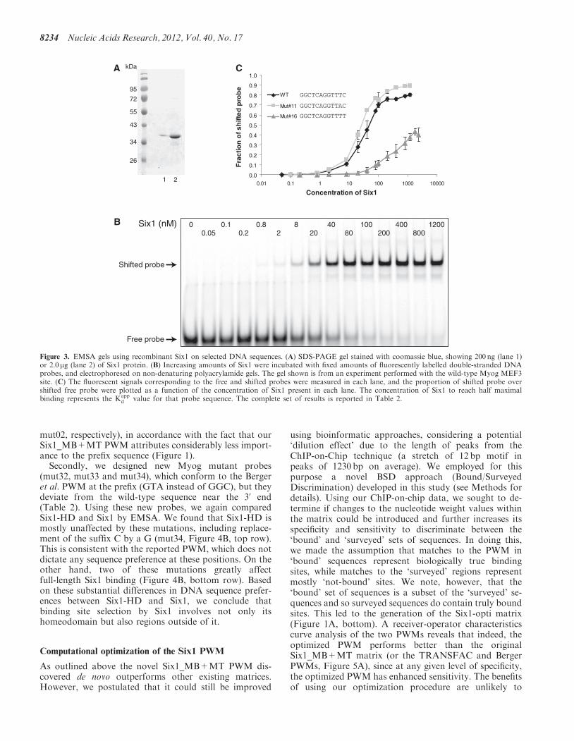
d ) of purified Six1 for the MEF3site present in the Myog proximal promoter (15,22,34,35),a site that is identical to the consensus DNA motif estab-lished by the TRANSFAC PWM, and which is to date thebest characterized Six1-binding site. EMSA reactions wereperformed in the presence of fixed amounts ofCy5-labelled probe and increasing amounts of the Six1protein (Figure 3A). We found that Six1 binds to themouse Myog MEF3 site with a Kapp
d of 35 nM(Figure 3B). Next, we aimed to verify whether thesequence preferences given by our PWM reflect the
0.0
0.2
0.4
0.6
0.8
1.0
1.2
1.4
0
10
20
30
40
50
60
70
80
Nu
mb
er
of
PW
M h
it p
er
kb
of
seq
uen
ce
Nu
mb
er
of
PW
M h
it p
er
bin
of
50 b
ou
nd
lo
ci
Bound genomic regions (or control loci)
PWM Hits per 50 peaks
PWM Hits per kb of sequence
Figure 2. Enrichment of the Six1_MB+MT matrix hits within the loci bound by Six1 in myotubes, as a function of their score in the ChIP-on-chipexperiment. The 1853 genomic loci bound by Six1 in myotubes were ranked in decreasing order of ChIP enrichment and subdivided in bins of 50regions. Matches to the Six1_MB+MT matrix were identified, and the numbers of PWM hit per bin (left-hand y axis) or per base pair (right-hand yaxis) were calculated. As controls, five sets of randomly selected genomic regions were also scanned for PWM hits. Additionally, two sets ofsequences totaling 108 Mb and originally surveyed in the ChIP-on-chip experiments were also scanned here.
8232 Nucleic Acids Research, 2012, Vol. 40, No. 17

affinity of the protein to DNA. Accordingly, we designeda library of Cy5-labelled DNA duplexes corresponding toMEF3 site derivatives with a focus on the sequencesdiverging between Six1_MB+MT and the MEF3 PWMfrom TRANSFAC (Table 2). We found that, as suggestedby the relative degeneracy of our novel MEF3-like matrix,many sequences differing from the TRANSFAC motif canbe bound with high avidity by Six1 (Table 2, Myog_mut07-11). These results further support the ideas that theTRANSFAC PWM is too stringent, and that de novomotif more accurately captures the DNA sequence prefer-ence of the Six1 transcription factor. Interestingly, wefound that the C nucleotide ‘suffix’ of the motif (TCAGGTTTC) is essential for high affinity binding of Six1 toDNA; mutation to any other nucleotide leads to a sharpdecrease in binding (Table 2, Myog_mut 14 to 16, andFigure 3C). This is an important observation consideringthat a shorter MEF3 element, amputated of this cytosinesuffix, has often been described (36–38). Other variants arealso indicative of Six1-binding preference. For example,even though the Six1_MB+MT has a high level of degen-eracy at positions 2-3 and 6, changing the prefix GGC toGAT (Mut03, at positions 2-3) or position 6 from an A toa T (Mut06) abolishes binding.
Regions outside the homeodomain contribute toDNA-binding sequence specificity
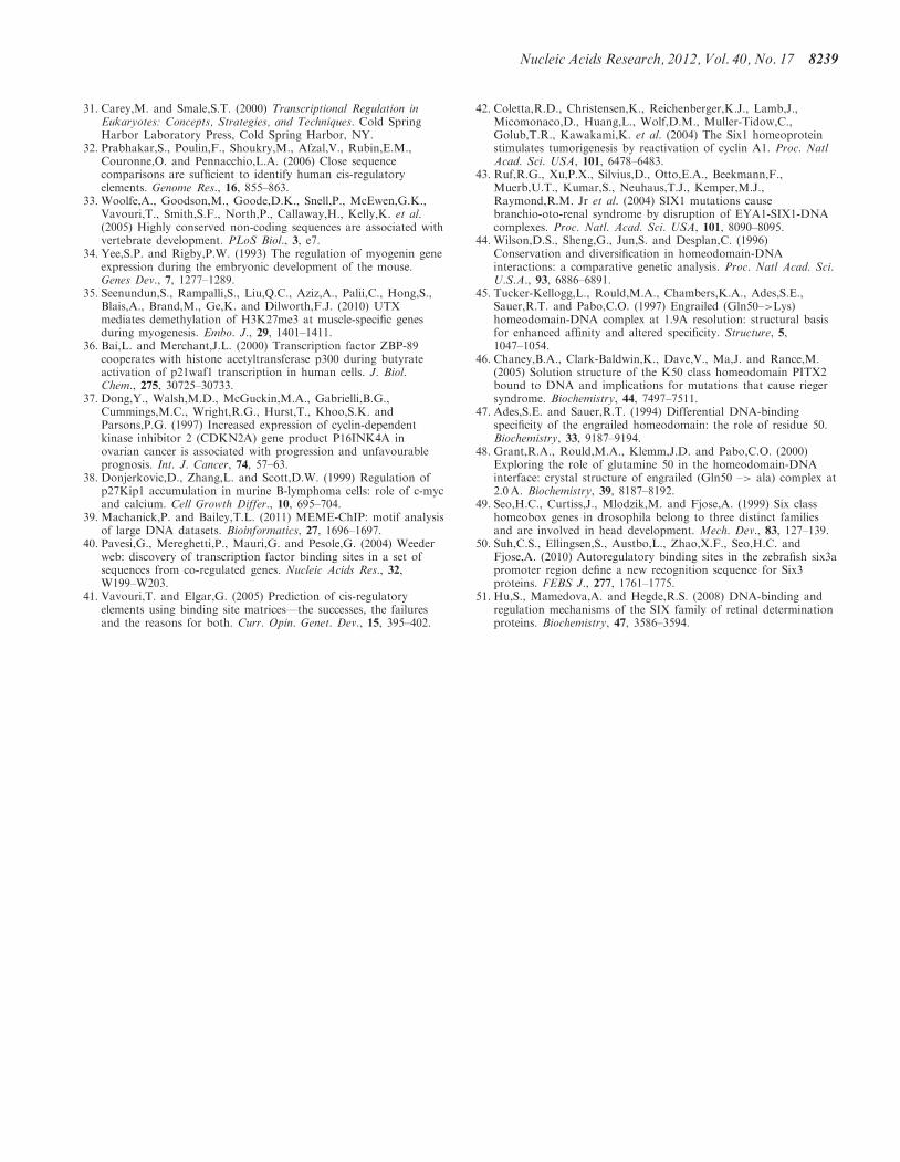
In their large-scale study of mouse homeodomains, Bergeret al. reported a DNA sequence motif preferred by theSix1 homeodomain (Six1-HD, Figure 1B) that is ratherdifferent from the one we report here for the full-lengthprotein (Figure 1A). This has important implications forthe possible mode of DNA binding by Six1, and suggeststhat protein regions outside of its homeodomain may par-ticipate in binding site selection. We therefore addressedthis question using EMSA, by comparing the affinities ofSix1-HD and Six1 for certain DNA sequences.First, we tested binding of the two proteins on the Myog
wild-type and mut02 probes, since the latter conforms tothe Berger et al. preferred sequence. We observed thatwhile binding of Six1-HD on the mut02 probe occurswith a Kapp
d of 690 nM, only very weak binding occurredbetween the homeodomain and the wild-type MyogMEF3 site (Kapp
d 7700 nM, Figure 4A). This is consistentwith the fact that the Berger et al. PWM gives a substan-tial importance to the GTA prefix, which is present inmut02 but absent in the wild-type probe. In contrast, thefull-length Six1 protein binds both sequences with com-parable affinities (Table 2, 34.7 and 28.7 nM for WT and
Table 1. Results of the sensitivity and specificity searches for new and existing Six1 PWMs
Name Target lista No. of sites (all)b Enrichment(all sites)c
P-value(all sites)d
No. of sites(conserved only)e
Enrichment(conservedsites only)
P-value(conservedsites)
Six1_MB-A_m01.mat MB_BC 485 4.73 <1E-16 139 6.49 <1E-16Six1_MB-B_m01.mat MB_AC 449 4.45 <1E-16 141 7.21 <1E-16Six1_MB-C_m01.mat MB_AB 471 5.23 <1E-16 144 7.31 <1E-16Six1_MT-A_m01.mat MT_BCDE 977 2.58 <1E-16 246 4.60 <1E-16Six1_MT-B_m01.mat MT_ACDE 1043 2.96 <1E-16 260 4.52 <1E-16Six1_MT-C_m01.mat MT_ABDE 888 2.73 2.2E-16 240 4.70 <1E-16Six1_MT-D_m01.mat MT_ABCE 891 3.08 <1E-16 248 4.92 1.3E-15Six1_MT-E_m01.mat MT_ABCD 895 2.36 <1E-16 242 3.93 <1E-16Six1_MB+MT.mat MB_ABC 1144 3.50 <1E-16 321 4.99 <1E-16
MT_ABCDE 1873 2.93 3.3E-16 489 4.67 7.8E-16Berger_Six1_0935.mat MB_ABC 308 1.38 2.8E-08 51 1.52 2.6E-03
MT_ABCDE 544 1.25 2.5E-07 84 1.54 9.5E-05M00319-V$MEF3_B02.mat MB_ABC 26 2.29 1.2E-04 9 5.02 8.6E-05
MT_ABCDE 51 2.29 8.5E-08 11 3.77 1.7E-04M00510-V$LHX3_01-Lhx3a.mat MB_ABC 350 0.67 1.0E+00 116 0.67 1.0E+00
MT_ABCDE 782 0.77 1.0E+00 203 0.72 1.0E+00M00640-V$HOXA4_Q2-HOXA4.mat MB_ABC 697 0.89 1.0E+00 185 0.99 5.9E-01
MT_ABCDE 1429 0.93 1.0E+00 317 1.04 2.6E-01M00241-V$NKX25_02-Nkx2-5.mat MB_ABC 472 0.77 1.0E+00 116 0.69 1.0E+00
MT_ABCDE 866 0.72 1.0E+00 188 0.68 1.0E+00M00360-V$PAX3_01-Pax-3.mat MB_ABC 27 1.13 2.9E-01 2 0.39 9.7E-01
MT_ABCDE 63 1.35 1.3E-02 14 1.67 4.5E-02M00394-V$MSX1_01-Msx-1.mat MB_ABC 241 1.00 5.2E-01 52 0.80 9.5E-01
MT_ABCDE 528 1.12 6.4E-03 105 1.00 5.2E-01M00096-V$PBX1_01-Pbx1a.mat MB_ABC 541 0.80 1.0E+00 147 0.94 7.7E-01
MT_ABCDE 997 0.76 1.0E+00 211 0.83 1.0E+00
Enrichment of binding sites predicted by PWMs discovered for Six1, for existing Six1 PWMs and for other homeodomain transcription factors hasbeen illustrated.aList of target genomic regions scanned with a given PWM. MB indicates Six1-bound targets in myoblasts, and MT those bound in myotubes.Subgroups of targets are given as letters (e.g. MB_AB refers to the combination of myoblast targets subgroups A and B).bNumber of sites corresponding to ‘hits’ to the PWM, irrespective of their phylogenetic conservation.cThe enrichment is given as the ratio of hits found in the indicated target set over those found in a fraction of the ChIP-surveyed sequence space,pro-rated by the length of each group of sequences in base pairs.dThe P-value represents the cumulative hypergeometric probability subtracted from 1.eSame as for b, but limited to genomic regions among the top 5% most phylogenetically conserved among 45 vertebrate species.
Nucleic Acids Research, 2012, Vol. 40, No. 17 8233
mut02, respectively), in accordance with the fact that ourSix1_MB+MT PWM attributes considerably less import-ance to the prefix sequence (Figure 1).Secondly, we designed new Myog mutant probes
(mut32, mut33 and mut34), which conform to the Bergeret al. PWM at the prefix (GTA instead of GGC), but theydeviate from the wild-type sequence near the 30 end(Table 2). Using these new probes, we again comparedSix1-HD and Six1 by EMSA. We found that Six1-HD ismostly unaffected by these mutations, including replace-ment of the suffix C by a G (mut34, Figure 4B, top row).This is consistent with the reported PWM, which does notdictate any sequence preference at these positions. On theother hand, two of these mutations greatly affectfull-length Six1 binding (Figure 4B, bottom row). Basedon these substantial differences in DNA sequence prefer-ences between Six1-HD and Six1, we conclude thatbinding site selection by Six1 involves not only itshomeodomain but also regions outside of it.
Computational optimization of the Six1 PWM
As outlined above the novel Six1_MB+MT PWM dis-covered de novo outperforms other existing matrices.However, we postulated that it could still be improved
using bioinformatic approaches, considering a potential‘dilution effect’ due to the length of peaks from theChIP-on-Chip technique (a stretch of 12 bp motif inpeaks of 1230 bp on average). We employed for thispurpose a novel BSD approach (Bound/SurveyedDiscrimination) developed in this study (see Methods fordetails). Using our ChIP-on-chip data, we sought to de-termine if changes to the nucleotide weight values withinthe matrix could be introduced and further increases itsspecificity and sensitivity to discriminate between the‘bound’ and ‘surveyed’ sets of sequences. In doing this,we made the assumption that matches to the PWM in‘bound’ sequences represent biologically true bindingsites, while matches to the ‘surveyed’ regions representmostly ‘not-bound’ sites. We note, however, that the‘bound’ set of sequences is a subset of the ‘surveyed’ se-quences and so surveyed sequences do contain truly boundsites. This led to the generation of the Six1-opti matrix(Figure 1A, bottom). A receiver-operator characteristicscurve analysis of the two PWMs reveals that indeed, theoptimized PWM performs better than the originalSix1_MB+MT matrix (or the TRANSFAC and BergerPWMs, Figure 5A), since at any given level of specificity,the optimized PWM has enhanced sensitivity. The benefitsof using our optimization procedure are unlikely to
0.0
0.1
0.2
0.3
0.4
0.5
0.6
0.7
0.8
0.9
1.0
0.01 0.1 1 10 100 1000 10000
Concentration of Six1
Fra
ctio
n o
f sh
ifted
pro
be
WT
Mut#11
Mut#16
72
55
43
34
26
95
kDa
1 2
A
B 0 0.10.2
0.82
820
4080
100200
4008000.05
1200Six1 (nM)
C
GGCTCAGGTTTT
GGCTCAGGTTTC
GGCTCAGGTTAC
Shifted probe
Free probe
Figure 3. EMSA gels using recombinant Six1 on selected DNA sequences. (A) SDS-PAGE gel stained with coomassie blue, showing 200 ng (lane 1)or 2.0 mg (lane 2) of Six1 protein. (B) Increasing amounts of Six1 were incubated with fixed amounts of fluorescently labelled double-stranded DNAprobes, and electrophoresed on non-denaturing polyacrylamide gels. The gel shown is from an experiment performed with the wild-type Myog MEF3site. (C) The fluorescent signals corresponding to the free and shifted probes were measured in each lane, and the proportion of shifted probe overshifted free probe were plotted as a function of the concentration of Six1 present in each lane. The concentration of Six1 to reach half maximalbinding represents the Kapp
d value for that probe sequence. The complete set of results is reported in Table 2.
8234 Nucleic Acids Research, 2012, Vol. 40, No. 17

originate from having started with a poor initial PWMgenerated by Amadeus, since PWMs obtained byMEME-ChIP (39) and Weeder (40), two popular motiffinding programs, did not perform any better than theone obtained with Amadeus (Six1_MB+MT).
The improved Six1-opti PWM allowed us to identifynew potential binding sites that may have been missedusing the original Six1_MB+MT matrix. At similar sen-sitivity and specificity (�60% and �73%, respectively),the Six1-opti matrix identified 322 novel putativebinding sequences (occurring a total of 505 times among
our Six1-bound genomic loci) that were missed with thestarting matrix (Figure 5B). On the other hand, thestarting matrix identified only 11 sequences (17 occur-rences) not found by the optimized PWM. It is also note-worthy to consider these results in terms of putative targetgene identification, since this is a common use of PWMscanning programs. Using the Six1-opti PWM wouldallow to identify 1051 target genes (i.e. sequences with atleast one hit to the PWM), while the original matrix wouldonly recognize 747 of them. This represents a 40.7%increase in sensitivity.
Table 2. Summary of EMSA experiments
Namea Sequenceb Rationalec Kappd (nM)d
WTMyog_WT gTTAGAGGGGGGCTCAGGTTTCTGTGGCGTTGGC Wild-type sequence 34.7±7.9
Prefix changesMyog_mut01 gTTAGAGGGGGGATCAGGTTTCTGTGGCGTTGGC Most frequent prefix 28.7±4.3Myog_mut02 gTTAGAGGGGGTATCAGGTTTCTGTGGCGTTGGC Prefix conforms to Berger et al. 16.8±2.4Myog_mut03 gTTAGAGGGGGATTCAGGTTTCTGTGGCGTTGGC Least frequent prefix >350
Core changesMyog_mut04 gTTAGAGGGGGGCTCGGGTTTCTGTGGCGTTGGC G is second most frequent after A 40.9±4.1Myog_mut05 gTTAGAGGGGGGCTCCGGTTTCTGTGGCGTTGGC C is least frequent nucleotide 63.8±7.7Myog_mut06 gTTAGAGGGGGGCTCTGGTTTCTGTGGCGTTGGC T is third most frequent after A >350Myog_mut07 gTTAGAGGGGGGCTCAGATTTCTGTGGCGTTGGC Very frequent dinucleotide 52.3±5.6Myog_mut08 gTTAGAGGGGGGCTCAAGTTTCTGTGGCGTTGGC Very frequent dinucleotide 34.2±1.8Myog_mut09 gTTAGAGGGGGGCTCATGTTTCTGTGGCGTTGGC Very frequent dinucleotide 29.4±2Myog_mut10 gTTAGAGGGGGGCTCAAATTTCTGTGGCGTTGGC Very frequent dinucleotide 32.4±2.1Myog_mut11 gTTAGAGGGGGGCTCAGGTTACTGTGGCGTTGGC A is second most frequent after T 24.2±1.3Myog_mut12 gTTAGAGGGGGGCTCAGGTTCCTGTGGCGTTGGC Rare nucleotide 81.6±10Myog_mut13 gTTAGAGGGGGGCTCAGGTTGCTGTGGCGTTGGC Rare nucleotide 72.2±3.1
Suffix changesMyog_mut14 gTTAGAGGGGGGCTCAGGTTTATGTGGCGTTGGC Very rare nucleotide >350Myog_mut15 gTTAGAGGGGGGCTCAGGTTTGTGTGGCGTTGGC Very rare nucleotide >350Myog_mut16 gTTAGAGGGGGGCTCAGGTTTTTGTGGCGTTGGC Very rare nucleotide >350
Multiple changesMyog_mut32 gTTAGAGGGGGTATCAGGGTTCTGTGGCGTTGGC mut02 with change near 3’ end >350Myog_mut33 gTTAGAGGGGGTATCAGGTGTCTGTGGCGTTGGC mut02 with change near 3’ end 130±50Myog_mut34 gTTAGAGGGGGTATCAGGTTTGTGTGGCGTTGGC mut02 with change at 3’ end >350
Tests of Six1-opti PWM prediction of binding sites (changes to the core, suffix and/or prefix)Myog_mut17 gTTAGAGGGGATCTCATATTACTGTGGCGTTGGC Unique to Six1-opti 25±3.3Myog_mut18 gTTAGAGGGGAGATCACATTTCTGTGGCGTTGGC Unique to Six1-opti 39.4±2.1Myog_mut19 gTTAGAGGGGAGATCACATTACTGTGGCGTTGGC Unique to Six1-opti 48.2±1.1Myog_mut20 gTTAGAGGGGTTCTCAAATTACTGTGGCGTTGGC Unique to Six1-opti 46.7±1.1Myog_mut21 gTTAGAGGGGGTATAAAATTTCTGTGGCGTTGGC Unique to Six1-opti 74.3±9Myog_mut22 gTTAGAGGGGAGCTCTGGTTACTGTGGCGTTGGC Unique to Six1-opti 85.4±10Myog_mut23 gTTAGAGGGGAGATCAGGTTTATGTGGCGTTGGC Unique to Six1-opti 69.3±8.2Myog_mut24 gTTAGAGGGGGGGTCAGGTGACTGTGGCGTTGGC Unique to Six1-opti >350Myog_mut25 gTTAGAGGGGATATCAGATATCTGTGGCGTTGGC Unique to Six1-opti 29.1±5.3Myog_mut26 gTTAGAGGGGGTATCAAATAACTGTGGCGTTGGC Unique to Six1-opti 10.8±3Myog_mut27 gTTAGAGGGGGCCTCGGGTTTCTGTGGCGTTGGC Unique to Six1_MB+MT >350Myog_mut28 gTTAGAGGGGGGATCGGGTTCCTGTGGCGTTGGC Unique to Six1_MB+MT 42.4±7.6Myog_mut29 gTTAGAGGGGGTTTCAGGTTCCTGTGGCGTTGGC Unique to Six1_MB+MT 75.4±8.3Myog_mut30 gTTAGAGGGGGTCTCGGCTTTCTGTGGCGTTGGC Unique to Six1_MB+MT >350Myog_mut31 gTTAGAGGGGGATTCAGGTTTCTGTGGCGTTGGC Unique to Six1_MB+MT >350
aMyog_WT is the Myog probe with wild-type MEF3 consensus in the center. Myog_mut01 to Myog_mut30 are probes with various mutations in theMEF3 consensus. Myog_mut31 is the same probe as Myog_mut03 cited as a different rationale.bMutated nucleotides in the MEF3 consensus are highlighted in black. The lower cap ‘g’ nucleotide was added for fluorescent labelling purposes.The natural sequence would be a ‘C’ at that position.cRationales to choose the corresponding sequences are listed. Mut01 the most frequent MEF3 sequence found in Six1_MB and Six1_MT bindingdata. Mut02 contains TA at position 2 and 3, which is found in the Berger et al. study. Myog03 has the least frequency of dinucleotides (AT) atposition 2 and 3. The MEF3 in Mut05 is found in the Myod core enhancer region. Mut07 to10 are selected with different dinucleotide combinationat position 7 and 8. Mut04, 06, and 11 to 16 are chosen based on the frequency of the nucleotide at a certain position. Mut17 to 26 are MEF3sequences found only using Six1-opti MEF3 motif. Mut27 to 31 are MEF3 sequences found only using Six1_MB+MT MEF3 motif. Of note, Mut03and Mut31 contain the same MEF3 sequence.dDissociation constant (Kapp
d ) and standard error of mean are calculated for each probe based on at least three independent experiments. >350 nM,not accurately determined due to very weak binding.
Nucleic Acids Research, 2012, Vol. 40, No. 17 8235

To determine if Six1 binds the new sequences identifiedby the optimized matrix, we used EMSA to assess thebinding affinity of Six1 to them. We selected 10 novelsites unique to Six1-opti and 5 sites uniquely identifiedwith Six1_MB+MT for validation (Table 2).Interestingly, 9 out of 10 sites unique to Six1-opti havecomparable affinity to the Myog probe, whereas only 2out of 5 sites unique to Six1_MB+MT are bound by Six1with a measurable Kapp
d , suggesting that our optimizationapproach improved the discriminatory power of our Six1PWM. Finally, as an ultimate test of the relevance of thebinding site predictions made by the optimized PWM, werepeated the search on an independent set of Six1 targetloci that are bound by Six1 24 hours after the onset ofmyoblast differentiation, but not in myoblasts ormyotubes (see materials and methods). As expected,with 49 common binding sequences out of 187Six1-bound regions, 23 binding sites were identifiedsolely with the Six1-opti PWM, while only 2 binding se-quences were uniquely defined with Six1_MB+MT(Figure 5C). This confirms the superiority of the optimizedPWM over the original matrix.
DISCUSSION
Using bioinformatic analysis of the genomic bindingprofile of Six1 in muscle cells, we have found that thistranscription factor has the ability to bind to a muchbroader range of DNA sequences than anticipated.While the previously reported MEF3 sequence motif isclearly enriched among genomic regions bound by Six1,other sequences that diverge substantially from this DNAelement are also found preferentially at Six1-bound loci.We used an in vitro binding assay with recombinant Six1to confirm that the protein indeed binds to these divergentsequences with high affinity, thereby ruling out an artifac-tual effect of the de novo sequence discovery algorithm weused. The novel PWM should prove to be useful to studiesthat employ TF target site prediction to elucidate thestructure of regulatory networks (41). For Six1, a TFinvolved in the genesis of multiple tissue types, a moreprecise DNA binding motif may contribute to discoveringnovel direct targets, elucidating composite regulatorynetworks and rationalizing its implication in diseasessuch as breast cancer or Branchio-oto-renal syndrome(7,42,43).
A
B
Protein: Six1-HD(µM)
Probe: mut32 ...GTATCAGGGTTC Probe: mut34 ...GTATCAGGTTTGProbe: mut33 ...GTATCAGGTGTC
Protein: Six1(nM)
0.0
0.1
0.2
0.8
2.0
8.0 20 40 80 100
200
400
800
0.05
1200
Kd : 1100 +/- 670 nMapp
Kd : 260 +/- 20 nMapp
Kd : 490 +/- 210 nMapp
Kd : > 350 nMapp
Kd : > 130 +/- 50 nMapp
Kd : > 350 nMapp
0.0
0.05 0.1
0.2
0.4
0.8
1.2
1.6
2.4
3.2
4.8
5.6
6.4
9.6
12.8
Protein: Six1-HDProbe: wild-type ...GGCTCAGGTTTC...Kd : 7700 +/- 2900 nMapp
Shifted probe
Free probe
Protein: Six1-HDProbe: mut02 ...GTATCAGGTTTC...Kd : 690 +/- 190 nMapp
0.0
0.05 0.1
0.2
0.4
0.8
1.2
1.6
2.4
3.2
4.8
5.6
6.4
9.6
12.8
Figure 4. Substantial differences in DNA sequence selectivity between Six1-HD and Six1. (A) EMSA gels were performed with increasing amountsof Six1 homeodomain (Six1-HD) using the Myogenin WT probe (left), or the mut02 probe (right), which conforms to the consensus reported byBerger et al. using protein-binding microarrays. (B) EMSA experiments performed with the Six1-HD (top row) or Six1 (bottom row) proteins, onthree derivatives of the mut02 probe (mutated positions are underlined, compared with the WT probe). Note that because the Six1-HD has arelatively low affinity for DNA in these assays, the amounts of protein used are higher than those used for the full-length Six1 protein. The Kapp
dvalues (all in nanomolars) for each protein on each probe are given underneath the respective gel images.
8236 Nucleic Acids Research, 2012, Vol. 40, No. 17

The results of our analyses provide useful informationthat may guide structure-function studies of Six1-DNAinteractions. The Six family homeodomains, includingthat of Six1, belong to the K50 class of homeodomains,as they differ from the majority of other homeodomainsclasses by the change of a key DNA-binding residue, as-paragine at position 50 of the HD, to a lysine. The im-portance of that residue in encoding DNA-bindingspecificity has been highlighted by a number of biochem-ical and structural studies (44–48). Other residuesimplicated in DNA-binding specificity or stability, forexample those within the N-terminal arm, differ betweenSix and other homeodomains or within Six familymembers [discussed in (49–51)]. Yet, it remains to bedetermined precisely how DNA-binding specificity is es-tablished by Six family TFs, and by Six1 in particular. Thevarious family members have been shown to bind to dif-ferent sequences: Six3 and Six6 can bind to the canonicalTAAT sequence that is bound by most homeodomainTFs, while Six1/2/4/5 have all been shown to be able tobind to sequences resembling the MEF3 element [reviewedin (11)]. Some Six proteins can also bind DNA asheterodimers with Eya family proteins, and these inter-actions are thought to enhance their affinity for
DNA (7,51). Most TFs, including several homeodomainfactors, bind to DNA in vivo as homo- or heterodimers,and stabilization of such oligomeric states has been putforth as one possible mechanism to explain the influenceof Eya proteins in regulating Six proteins (51). In our ex-periments, one predominant oligomeric state of Six1 wasdetected in EMSA; however, the oligomeric state of Six1in solution remains unclear (i.e. whether Six1 binds DNAas a monomer or as a multimer in our assays), although atvery high protein concentrations slower migratingcomplexes became visible on EMSA gels (data notshown). We cannot tell at this point if these species repre-sent aggregates or functionally and physiologicallyrelevant oligomers. Further analysis of the precise modeof DNA recognition by Six1, the influence of interactionpartners and possible involvement of oligomeric states,should help us understand the function of this proteinand rationalize its implication in diseased states.ChIP-on-chip analysis captures a snapshot of protein-
DNA interactions as they occur in live cells, and althoughchromatin is immunoprecipitated with an antibodyagainst Six1, putative DNA-binding partners of Six1were possibly involved in the interactions we have dis-covered. It would be interesting to determine thegenomic binding profiles of Eya proteins in myoblasts tosee if indeed these proteins tend to bind DNA along withSix1, and if co-binding with Eya proteins alters DNA-binding preferences in any way. However, these experi-ments may prove excessively difficult to perform in vivo:while it is possible to ChIP a specific Eya-Sixchromatin-bound complex (using sequential ChIPassays), it would be much more challenging to devise away of pulling-down only Six1 chromatin complexesthat do not contain Eya proteins. Interestingly, however,the results of our in vitro experiments corroborate those ofour ChIP-on-chip experiments: the variety of sequencemotifs enriched among Six1-bound loci is reflected inEMSA using DNA probes and Six1 alone. This leads usto postulate that protein domain(s) within Six1 itself arethe main determinant of DNA sequence selection by Six1in vivo (at least in muscle cells, where our analysis wasdone). As shown by others and noted above, Eyabinding could influence predominantly the bindingaffinity rather than sequence selectivity (7,51).The PWM we have generated is fairly close to the initial
MEF3 PWM reported more than 15 years ago. The strin-gency (and therefore low sensitivity) of the TRANSFACMEF3 PWM comes from the fact that it was derived fromthe DNA sequences of only five sites within muscle genepromoters. One can easily imagine that with largersampling, the PWM would perform better at predictingSix1-binding sites.The significant discrepancy between our data and those
reported by Berger et al. was more puzzling. The authorsused protein-binding microarrays to determine the se-quence specificity of the mouse Six1 homeodomain andreported a Six1 PWM that has only limited resemblanceto our PWM or to the TRANSFAC MEF3 element (4).We reason that these differences originate from the factthat only the homeodomain region of Six1 was studied,whereas our ChIP-on-chip and binding studies were
A
B
Found bySix1-opti
Found bySix1_MB+MT
Found bySix1-opti
Found bySix1_MB+MT
C
0.0
0.2
0.4
0.6
0.8
1.0
0.0 0.2 0.4 0.6 0.8 1.0
1-specificity
Sen
siti
vity
Six1-OptiSix1_MB+MT (Amadeus)
TRANSFAC MEF-3Berger et al.
MEME MB+MTWeeder MB+MT
Figure 5. Performance comparison of the Six1_MB+MT andSix1-opti PWMs. (A) ROC curves. The y-axis is the sensitivity andthe x-axis is the 1-specificity value. Performances of the originalTRANSFAC MEF3 and Berger et al. PWMs for Six1 are also givenfor comparison. (B) Venn diagram indicating the number of hits toeach PWM, and their overlap, at their respective optimal thresholds,among loci targeted by Six1 in myotubes. Numbers in parentheses arethe number of unique sequences (one sequence can occur more thanonce). (C) Results for the comparison between predictions made withthe Six1-opti and Six1_MB+MT PWMs, on sequences bound by Six1only at 24 hours post-differentiation.
Nucleic Acids Research, 2012, Vol. 40, No. 17 8237

performed with full-length Six1. Indeed, we confirmedwith EMSA experiments that Six1-HD exhibits a DNAsequence preference that is in line with what Bergeret al. reported, but that is substantially different fromthat exhibited by the full-length protein. We thereforeconclude that regions outside of the Six1 homeodomainparticipate in DNA binding, either through direct DNAcontacts or indirectly, perhaps by enabling structuralstabilization.
SUPPLEMENTARY DATA
Supplementary Data are available at NAR Online:Supplementary Tables 1–2.
ACKNOWLEDGEMENTS
The authors thank Jean-Francois Couture and SabinaSarvan for advice and help with protein purification andKapp
d determination.
FUNDING
The Canadian Natural Science and Engineering ResearchCouncil (NSERC) (to A.B., S.N. and I.I.); LeadersOpportunity Fund grant of Canadian Foundation ofInnovation and Ontario Research Foundation (to I.I.and S.N.); a NSERC graduate scholarship (to Y.L.)Funding for open access charge: NSERC operating(to A.B.).
Conflict of interest statement. None declared.
REFERENCES
1. Holland,P.W., Booth,H.A. and Bruford,E.A. (2007) Classificationand nomenclature of all human homeobox genes. BMC Biol., 5,47.
2. Gehring,W.J., Qian,Y.Q., Billeter,M., Furukubo-Tokunaga,K.,Schier,A.F., Resendez-Perez,D., Affolter,M., Otting,G. andWuthrich,K. (1994) Homeodomain-DNA recognition. Cell, 78,211–223.
3. Svingen,T. and Tonissen,K.F. (2006) Hox transcription factorsand their elusive mammalian gene targets. Heredity, 97, 88–96.
4. Berger,M.F., Badis,G., Gehrke,A.R., Talukder,S.,Philippakis,A.A., Pena-Castillo,L., Alleyne,T.M., Mnaimneh,S.,Botvinnik,O.B., Chan,E.T. et al. (2008) Variation inhomeodomain DNA binding revealed by high-resolution analysisof sequence preferences. Cell, 133, 1266–1276.
5. Noyes,M.B., Christensen,R.G., Wakabayashi,A., Stormo,G.D.,Brodsky,M.H. and Wolfe,S.A. (2008) Analysis of homeodomainspecificities allows the family-wide prediction of preferredrecognition sites. Cell, 133, 1277–1289.
6. Li,T., Stark,M.R., Johnson,A.D. and Wolberger,C. (1995) Crystalstructure of the MATa1/MAT alpha 2 homeodomain heterodimerbound to DNA. Science, 270, 262–269.
7. Patrick,A.N., Schiemann,B.J., Yang,K., Zhao,R. and Ford,H.L.(2009) Biochemical and functional characterization of six SIX1branchio-oto-renal syndrome mutations. J. Biol. Chem., 284,20781–20790.
8. Chang,C.P., Brocchieri,L., Shen,W.F., Largman,C. andCleary,M.L. (1996) Pbx modulation of hox homeodomainamino-terminal arms establishes different DNA-bindingspecificities across the hox locus. Mol. Cell. Biol., 16, 1734–1745.
9. Shen,W.F., Montgomery,J.C., Rozenfeld,S., Moskow,J.J.,Lawrence,H.J., Buchberg,A.M. and Largman,C. (1997) AbdB-like
hox proteins stabilize DNA binding by the Meis1 homeodomainproteins. Mol. Cell. Biol., 17, 6448–6458.
10. Kawakami,K., Sato,S., Ozaki,H. and Ikeda,K. (2000) Six familygenes—structure and function as transcription factors and theirroles in development. Bioessays, 22, 616–626.
11. Kumar,J.P. (2009) The sine oculis homeobox (SIX) family oftranscription factors as regulators of development and disease.Cell Mol. Life Sci., 66, 565–583.
12. Blais,A., Tsikitis,M., Acosta-Alvear,D., Sharan,R., Kluger,Y. andDynlacht,B.D. (2005) An initial blueprint for myogenicdifferentiation. Genes Dev., 19, 553–569.
13. Parmacek,M.S., Ip,H.S., Jung,F., Shen,T., Martin,J.F., Vora,A.J.,Olson,E.N. and Leiden,J.M. (1994) A novel myogenic regulatorycircuit controls slow/cardiac troponin C gene transcription inskeletal muscle. Mol. Cell Biol., 14, 1870–1885.
14. Spitz,F., Salminen,M., Demignon,J., Kahn,A., Daegelen,D. andMaire,P. (1997) A combination of MEF3 and NFI proteinsactivates transcription in a subset of fast-twitch muscles. Mol.Cell Biol., 17, 656–666.
15. Spitz,F., Demignon,J., Porteu,A., Kahn,A., Concordet,J.P.,Daegelen,D. and Maire,P. (1998) Expression of myogenin duringembryogenesis is controlled by Six/sine oculis homeoproteinsthrough a conserved MEF3 binding site. Proc. Natl Acad. Sci.USA, 95, 14220–14225.
16. Laclef,C., Hamard,G., Demignon,J., Souil,E., Houbron,C. andMaire,P. (2003) Altered myogenesis in Six1-deficient mice.Development, 130, 2239–2252.
17. Grifone,R., Demignon,J., Houbron,C., Souil,E., Niro,C.,Seller,M.J., Hamard,G. and Maire,P. (2005) Six1 and Six4homeoproteins are required for Pax3 and mrf expression duringmyogenesis in the mouse embryo. Development, 132, 2235–2249.
18. Li,X., Oghi,K.A., Zhang,J., Krones,A., Bush,K.T., Glass,C.K.,Nigam,S.K., Aggarwal,A.K., Maas,R., Rose,D.W. et al. (2003)Eya protein phosphatase activity regulates Six1-dach-eyatranscriptional effects in mammalian organogenesis. Nature, 426,247–254.
19. Bessarab,D.A., Chong,S.W., Srinivas,B.P. and Korzh,V. (2008)Six1a is required for the onset of fast muscle differentiation inzebrafish. Dev Biol, 323, 216–228.
20. Molkentin,J.D. and Olson,E.N. (1996) Combinatorial control ofmuscle development by basic helix-loop-helix and MADS-boxtranscription factors. Proc. Natl Acad. Sci. USA, 93, 9366–9373.
21. Berkes,C.A., Bergstrom,D.A., Penn,B.H., Seaver,K.J.,Knoepfler,P.S. and Tapscott,S.J. (2004) Pbx marks genes foractivation by MyoD indicating a role for a homeodomain proteinin establishing myogenic potential. Mol. Cell, 14, 465–477.
22. Liu,Y., Chu,A., Chakroun,I., Islam,U. and Blais,A. (2010)Cooperation between myogenic regulatory factors and SIX familytranscription factors is important for myoblast differentiation.Nucleic Acids Res., 38, 6857–6871.
23. Linhart,C., Halperin,Y. and Shamir,R. (2008) Transcription factorand microRNA motif discovery: The amadeus platform and acompendium of metazoan target sets. Genome Res., 18,1180–1189.
24. Tarailo-Graovac,M. and Chen,N. (2009) Using RepeatMasker toidentify repetitive elements in genomic sequences. Curr. Protoc.Bioinformatics, Chapter 4, Unit 4.10.
25. Crooks,G.E., Hon,G., Chandonia,J.M. and Brenner,S.E. (2004)WebLogo: a sequence logo generator. Genome Res., 14,1188–1190.
26. Ji,H., Jiang,H., Ma,W., Johnson,D.S., Myers,R.M. andWong,W.H. (2008) An integrated software system for analyzingChIP-chip and ChIP-seq data. Nat. Biotechnol., 26, 1293–1300.
27. Staden,R. (1989) Methods for calculating the probabilities offinding patterns in sequences. Comput. Appl. Biosci., 5, 89–96.
28. Bucher,P. (1990) Weight matrix descriptions of four eukaryoticRNA polymerase II promoter elements derived from 502unrelated promoter sequences. J. Mol. Biol., 212, 563–578.
29. Gershenzon,N.I., Stormo,G.D. and Ioshikhes,I.P. (2005)Computational technique for improvement of the position-weightmatrices for the DNA/protein binding sites. Nucleic Acids Res.,33, 2290–2301.
30. Burset,M. and Guigo,R. (1996) Evaluation of gene structureprediction programs. Genomics, 34, 353–367.
8238 Nucleic Acids Research, 2012, Vol. 40, No. 17
31. Carey,M. and Smale,S.T. (2000) Transcriptional Regulation inEukaryotes: Concepts, Strategies, and Techniques. Cold SpringHarbor Laboratory Press, Cold Spring Harbor, NY.
32. Prabhakar,S., Poulin,F., Shoukry,M., Afzal,V., Rubin,E.M.,Couronne,O. and Pennacchio,L.A. (2006) Close sequencecomparisons are sufficient to identify human cis-regulatoryelements. Genome Res., 16, 855–863.
33. Woolfe,A., Goodson,M., Goode,D.K., Snell,P., McEwen,G.K.,Vavouri,T., Smith,S.F., North,P., Callaway,H., Kelly,K. et al.(2005) Highly conserved non-coding sequences are associated withvertebrate development. PLoS Biol., 3, e7.
34. Yee,S.P. and Rigby,P.W. (1993) The regulation of myogenin geneexpression during the embryonic development of the mouse.Genes Dev., 7, 1277–1289.
35. Seenundun,S., Rampalli,S., Liu,Q.C., Aziz,A., Palii,C., Hong,S.,Blais,A., Brand,M., Ge,K. and Dilworth,F.J. (2010) UTXmediates demethylation of H3K27me3 at muscle-specific genesduring myogenesis. Embo. J., 29, 1401–1411.
36. Bai,L. and Merchant,J.L. (2000) Transcription factor ZBP-89cooperates with histone acetyltransferase p300 during butyrateactivation of p21waf1 transcription in human cells. J. Biol.Chem., 275, 30725–30733.
37. Dong,Y., Walsh,M.D., McGuckin,M.A., Gabrielli,B.G.,Cummings,M.C., Wright,R.G., Hurst,T., Khoo,S.K. andParsons,P.G. (1997) Increased expression of cyclin-dependentkinase inhibitor 2 (CDKN2A) gene product P16INK4A inovarian cancer is associated with progression and unfavourableprognosis. Int. J. Cancer, 74, 57–63.
38. Donjerkovic,D., Zhang,L. and Scott,D.W. (1999) Regulation ofp27Kip1 accumulation in murine B-lymphoma cells: role of c-mycand calcium. Cell Growth Differ., 10, 695–704.
39. Machanick,P. and Bailey,T.L. (2011) MEME-ChIP: motif analysisof large DNA datasets. Bioinformatics, 27, 1696–1697.
40. Pavesi,G., Mereghetti,P., Mauri,G. and Pesole,G. (2004) Weederweb: discovery of transcription factor binding sites in a set ofsequences from co-regulated genes. Nucleic Acids Res., 32,W199–W203.
41. Vavouri,T. and Elgar,G. (2005) Prediction of cis-regulatoryelements using binding site matrices—the successes, the failuresand the reasons for both. Curr. Opin. Genet. Dev., 15, 395–402.
42. Coletta,R.D., Christensen,K., Reichenberger,K.J., Lamb,J.,Micomonaco,D., Huang,L., Wolf,D.M., Muller-Tidow,C.,Golub,T.R., Kawakami,K. et al. (2004) The Six1 homeoproteinstimulates tumorigenesis by reactivation of cyclin A1. Proc. NatlAcad. Sci. USA, 101, 6478–6483.
43. Ruf,R.G., Xu,P.X., Silvius,D., Otto,E.A., Beekmann,F.,Muerb,U.T., Kumar,S., Neuhaus,T.J., Kemper,M.J.,Raymond,R.M. Jr et al. (2004) SIX1 mutations causebranchio-oto-renal syndrome by disruption of EYA1-SIX1-DNAcomplexes. Proc. Natl. Acad. Sci. USA, 101, 8090–8095.
44. Wilson,D.S., Sheng,G., Jun,S. and Desplan,C. (1996)Conservation and diversification in homeodomain-DNAinteractions: a comparative genetic analysis. Proc. Natl Acad. Sci.U.S.A., 93, 6886–6891.
45. Tucker-Kellogg,L., Rould,M.A., Chambers,K.A., Ades,S.E.,Sauer,R.T. and Pabo,C.O. (1997) Engrailed (Gln50–>Lys)homeodomain-DNA complex at 1.9A resolution: structural basisfor enhanced affinity and altered specificity. Structure, 5,1047–1054.
46. Chaney,B.A., Clark-Baldwin,K., Dave,V., Ma,J. and Rance,M.(2005) Solution structure of the K50 class homeodomain PITX2bound to DNA and implications for mutations that cause riegersyndrome. Biochemistry, 44, 7497–7511.
47. Ades,S.E. and Sauer,R.T. (1994) Differential DNA-bindingspecificity of the engrailed homeodomain: the role of residue 50.Biochemistry, 33, 9187–9194.
48. Grant,R.A., Rould,M.A., Klemm,J.D. and Pabo,C.O. (2000)Exploring the role of glutamine 50 in the homeodomain-DNAinterface: crystal structure of engrailed (Gln50 –> ala) complex at2.0A. Biochemistry, 39, 8187–8192.
49. Seo,H.C., Curtiss,J., Mlodzik,M. and Fjose,A. (1999) Six classhomeobox genes in drosophila belong to three distinct familiesand are involved in head development. Mech. Dev., 83, 127–139.
50. Suh,C.S., Ellingsen,S., Austbo,L., Zhao,X.F., Seo,H.C. andFjose,A. (2010) Autoregulatory binding sites in the zebrafish six3apromoter region define a new recognition sequence for Six3proteins. FEBS J., 277, 1761–1775.
51. Hu,S., Mamedova,A. and Hegde,R.S. (2008) DNA-binding andregulation mechanisms of the SIX family of retinal determinationproteins. Biochemistry, 47, 3586–3594.
Nucleic Acids Research, 2012, Vol. 40, No. 17 8239
Copyright © 2022 FDOKUMEN