Intestinal Specific Gene Regulation by Transcription Factors ...
202
Intestinal Specific Gene Regulation by Transcription Factors Gata4 and Hnf1α in vivo Tjalling Bosse
-
Upload
khangminh22 -
Category
Documents
-
view
3 -
download
0
Transcript of Intestinal Specific Gene Regulation by Transcription Factors ...

Intestinal Specific Gene Regulation by Transcription
Factors Gata4 and Hnf1α in vivo
Tjalling Bosse

© 2006 Tjalling Bosse, Leiden, The Netherlands
On the cover: This painting by Marcelo Lavellen was presented to the author during the
preparations of his thesis.
The printing of this thesis was financially supported by:
Astra Zeneca, Tramedico B.V. and Nutricia
Print: Gildeprint BV, Enschede

Intestinal Specific Gene Regulation by Transcription
Factors Gata4 and Hnf1α in vivo
Darm specifieke gen regulatie door transcriptie factoren Gata4 en Hnf1α in
vivo
Proefschrift
ter verkrijging van de graad van doctor aan de
Erasmus Universiteit Rotterdam
op gezag van de
rector magnificus
Prof. dr. S.W.J. Lamberts
en volgens besluit van het College voor Promoties.
De openbare verdediging zal plaatsvinden op
woensdag 29 november 2006 om 11.45 uur
door
Tjalling Bosse
geboren te Alphen aan den Rijn

Promotiecommissie
Promotoren: Prof.dr. H.A. Büller
Prof. R.J. Grand MD
Overige leden: Prof.dr. J.B. van Goudoever
Prof.dr. D. Tibboel
Prof.dr. E.J. Kuipers

……………..Alleen het kind,
Is nog hetzelfde als voor duizend jaren
Nieuw en verwonderd ligt het rond te staren
Alsof de wereld pas vandaag begint.
O makker in ditzelfde grauw getij
Nog altijd komt het kind tot jouw en mij
Nog altijd kan de wereld nieuw beginnen
In ieder kind kan het opnieuw beginnen,
Zolang God kinderen in ons midden zendt,
Heeft hij zich nog niet van ons afgewend.
Uit het geboorteboekje van mijn moeder
ter nagedachtenis aan mijn broer:
Rutger Bosse


PREFACE
This study was a joint effort between the Division of Pediatric Gastroenterology and Nutrition,
Department of Pediatrics, Erasmus MC/Sophia Kinderziekenhuis, Rotterdam, The Netherlands,
and the Division of Gastroenterology and Nutrition, Department of Medicine, Children’s
Hospital Boston, Boston MA, 02115; Department of Pediatrics, Harvard Medical School, USA.
Research described was supported by Numico International Training Fellowship, Zoetermeer,
The Netherlands (T. Bosse); NIH Research Grant #R01-DK061382 (S.D. Krasinski) and R37-
DK32658 (R.J. Grand); grants from the Harvard Digestive Disease Center (5P30-DK34854).


CONTENTS
PREFACE
page
Chapter 1 Introduction 11
Chapter 2 Complex regulation of the lactase-phlorizin hydrolase
promoter by Gata4
American Journal of Physiology (2004): 287(4), G899-909 47
Chapter 3 Hepatocyte nuclear factor-1alpha is required for
gene-expression, but dispensable for histone acetylation
of the lactase-phlorizin hydrolase gene
American Journal of Physiology (2006): 290(5):G1016-24 73
Chapter 4 Gata4 is essential for the maintenance of jejunal-ileal
identities in the adult mouse small intestine
Molecular Cell Biology (2006): Aug. 28[Epub ahead of print] 95
Chapter 5 Gata4 and Hnf1α are dispensable for the expression of
specific intestinal genes during development
American Journal of Physiology; Submitted Aug. 2006 119
Chapter 6 Gata4 dependent repression of ileal genes in the adult
small intestine is mediated by Fog interactions
Manuscript in preparation 147
Chapter 7 Summary and discussion of future perspectives 167
Chapter 8 Samenvatting 185
Acknowledgements – Dankwoord 195
Curriculum vitea and References 199
Colorfigures 203


CHAPTER 1
Introduction
Regulation of intestinal differentiation
Tjalling Bosse1, Richard J. Grand
2, Hans A. Büller
3 and Stephen D. Krasinski
2
From the 1University of Amsterdam, Department of Pediatrics, Amsterdam Medical Center, Amsterdam, 1100DD;
The Netherlands; 2Division of Gastroenterology and Nutrition, Department of Pediatrics, Children’s Hospital
Boston, Boston MA, 02115; Department of Pediatrics, Harvard Medical School and the 3Erasmus University
Rotterdam, Department of Pediatrics, Erasmus MC/Sophia Children’s hospital, Rotterdam, The Netherlands

CHAPTER 1
12

Introduction
13
Introduction
1 Introduction: Regulation of intestinal differentiation
Significance of studying intestinal differentiation
1.1 Intestinal development and differentiation
1.1.1 Organization of the mouse Crypt-villus structure
1.1.2 Regional specialization along the length of the small intestine
1.2 Genetic and epigenetic regulation of gene expression
1.2.1 Transcriptional regulation
1.2.2 Epigenetics: Histone acetylation and DNA methylation
1.3 Lactase-phlorizin hydrolase and sucrase-isomaltase
1.3.1 The expression patterns of LPH and SI genes mark the development and
differentiation phases of the small intestine
1.3.2 Lactose persistence vs. lactose non-persistence
1.3.3 LPH and SI gene expression is “hard-wired”
1.4 Homeodomain containing transcription factors:
Cdx2 and HNF-1
1.4.1 Cdx2 transcription factor
1.4.2 The HNF-1 transcription factor family
1.4.3 Role of Hnf1α in chromatin dynamics
1.5 GATA zinc finger containing transcription factor family
1.5.1 Functional domains in Gata4, 5 and 6
1.5.2 Differential functions revealed by GATA knockout studies in mice
1.5.3 GATA factors in tissue specific gene expression
1.5.4 GATA interacting factors
1.6 Complex interaction between tissue restricted transcription factors
1.6.1 Evolutionarily conserved mechanism of functional synergy between
intestinal GATA factors and Hnf1α
1.6.2 Intestinal specific expression patterns of Gata4, Hnf1α and Cdx2
1.7 Specific Aims

CHAPTER 1
14
1 Introduction: Regulation of intestinal differentiation
Significance of studying intestinal differentiation
The mammalian small intestine is responsible for the terminal digestion and absorption of
nutrients, water homeostasis, and the elimination of waste products, which in turn, are essential
processes for life. These processes however, are easily disrupted by infection, inflammatory
processes such as Crohn’s disease, cancer, and resection. The small intestine is equipped with
specific proteins, such as enzymes to digest nutrients (digestion) and ‘transporters’ to carry the
nutrients into the body (absorption). These tools for digestion and absorption are specifically
expressed in the enterocytes of the small intestine and this expression is regulated by a complex
of regulatory proteins among which intestinal transcription factors. These regulatory proteins are
proposed to be important for intestinal gene expression, differentiation and development and are
central to intestinal function. A better insight into the role that specific transcription factors play
in these processes will thus complement our understanding of the regulation of intestinal
function. Such fundamental knowledge will provide critical insight into disease processes and
repair mechanisms of the intestinal epithelium, and identify potential avenues of intervention to
correct lost or deficient intestinal function. The research described in this thesis investigates the
role of the transcription factors Gata4 and Hnf1α in intestinal gene expression in vivo.
1.1 Intestinal development and differentiation
The mammalian small intestine develops from a simple tubular structure in the early embryo to a
mature, highly specialized and organized organ system. Beginning on embryonic day (E) 8.5 in
mice, anterior and posterior invaginations of the visceral endoderm occur that eventually fuse
forming a primitive gut tube lined by an undifferentiated, stratified epithelium. Between E9.5
and E14.5, interactions between visceral endoderm and mesoderm result in organ specification,
and the gut tube undergoes considerable longitudinal growth. The primitive gut tube can be
divided into three parts: foregut, midgut and hindgut. The mid-gut is destined to become the
small intestine, and between E14.5 and E19.5, the midgut endoderm goes through rapid
remodeling converting the stratified epithelium into a simple columnar epithelium in a proximal-
to-distal wave called cytodifferentiation. During this time, upward growth of the underlining
mesenchyme forms nascent finger-like projections (villi) that are separated from one another by
a proliferating compartments known as the intervillus epithelium. It is during this important
transition that the expression of certain intestine-specific genes is first detected (108). After birth,
the mouse epithelium has an immature architecture that goes through considerable
morphological and functional changes during the first 3 weeks of life after which it reaches a
mature morphology. The timed developmental transitions of the gut are reflected in the well-
orchestrated changes in the intestinal gene expression of certain gene sets (also referred to as the

Introduction
15
developmental axis). A better understanding in the regulation of these specific intestinal genes
will therefore provide insight into the underlying mechanism responsible for intestinal
development and differentiation.
1.1.1 Organization of the mouse Crypt-villus structure
The epithelium of the mature intestine is a highly dynamic system in which the cells are
continuously renewed by a process involving cell generation, proliferation in the crypts and
migration associated differentiation along the villi. A stepwise differentiation process from
intestinal stem cells forms the four major cell lineages: absorptive enterocytes, goblet cells,
enteroendocrine cells and Paneth cells (Fig. 1).
Fig. 1. Schematic representation of intestinal crypt-villus
structure. This figure shows a schematic of a “ finger-like
projection” villus (above the dotted line) and crypt (below
dotted line) that characterizes the luminal surface of the
small intestine. The four prinicipal cells that originate from
the presumptive stem cells are depicted. The epithelium is
lined by absorptive enterocytes (light yellow), goblet cells
(blue) and enteroendocrine cells (pink) which originate in
the crypt and migrate up (red arrow) the villus where they
are eventually shed into the lumen. The paneth cells reside
in the bottom of the crypts (brown) below the presumptive
stem cells. Generally, proliferative immature cells can be
found in the crypts, whereas differentiated and mature cells
line the villus.
Absorptive enterocytes, comprising approximately 90% of the villus associated epithelium, are
polarized cells with an apical microvillus membrane that contains transporters, receptors, and
membrane-anchored hydrolases, including lactase-phlorizin hydrolase (LPH) and sucrase
isomaltase (SI). Goblet cells synthesize mucins, which provide protection of the epithelium and a
medium in which terminal digestion and absorption can occur optimally. Enteroendocrine cells
produce a variety of hormones necessary for regulation of intestinal processes, such as
peptideYY (PYY), secretin and cholecystokinin (CCK). Paneth cells produce antimicrobial
enzymes and peptides such as, cryptidins and defensins (107). While the three other
differentiated cell types migrate upward from the upper third of the crypts to the apex of the
Proliferation
Differentiation
Migration
Absorptive enterocytes
Entero-endocrine cells
Goblet cells
Paneth cells
Presumptive stem cell
Proliferation
Differentiation
Migration
Absorptive enterocytes
Entero-endocrine cells
Goblet cells
Paneth cells
Presumptive stem cell
Absorptive enterocytes
Entero-endocrine cells
Goblet cells
Paneth cells
Presumptive stem cell

CHAPTER 1
16
villus, the Paneth cells characteristically migrate to the bottom of the crypt where they reside for
about 20 days (18, 39). At the tip of the villus, the differentiated epithelial cells become
apoptotic and are exfoliated into the intestinal lumen (43). The entire villus epithelium is
renewed every 3 days in the mouse (5 days in man) (150). This cell replenishment and migration
process along the crypt/villus (vertical) axis guarantees a continuous and rapid renewal of the
intestinal epithelium, ensuring maintenance of barrier and absorptive functions, which define the
term homoeostasis in this organ.
1.1.2 Regional specialization along the length of the small intestine
For appropriate function it is imperative that the intestine maintain functionally diverse regions
along the antero-posterior or horizontal axis. The adult small intestine anatomically starts at the
pyloric junction (the most distal point of the stomach) and ends at the ileocecal valve (start of the
colon) and is divided into duodenum, jejunum and ileum. Although these transitions are not
clearly demarcated, these structures have obvious functional and morphological differences.
Many studies, using intestinal isografts, have contributed to the concept that position-appropriate
differentiation along the horizontal axis of the small intestine can occur in the absence of
exposure to luminal contents, and that such regulation in the small intestine is pre-determined
(40). However, little is known about the intrinsic molecular mechanisms that maintain regional
diversity in the adult small intestine, which is a principal subject in this thesis (Chapter 4).
The duodenum is the most proximal part of the small intestine and is the first to receive
the partly digested chyme from the stomach. Its major function is to provide a medium in which
virtually all digestible macromolecules are being digested into forms that are capable of, or
nearly capable of, being absorbed by the absorptive enterocytes in the jejunum and ileum. The
acidic content of the stomach triggers numerous enteroendocrine cells present in the duodenum
to release hormones such as cholecystokinin (CCK) and secretin. These hormones stimulate the
secretion of pancreatic fluids and bile into the duodenum. Pancreatic secretions contain digestive
enzymes, such as proteases, lipases and amylase, which are responsible for the digestion of
proteins, lipids and carbohydrates into small absorbable molecules. Bile acid is the critical
component of bile which has the ability to emulsify fat globules to increase lipase action and to
solublize fat and fat-soluble vitamins in an aqueous environment by the formation of micelles
(aggregates of lipid such as fatty acids, cholesterol and monoglycerides). To protect the
epithelium against the acidic fluids, specialized glands (Brunner’s glands) secreting basic fluids,
are specifically found in the submucosa of the duodenum. Together the epithelial lining of the
duodenum is specialized in the digestion of stomach content and the release of hormones that
activate gastro-intestinal processes.
In considering the digestive system, the jejunum is central of the three divisions of the
small intestine and lies between the duodenum and the ileum. The jejunum is the prime position
for the absorption of carbohydrates, proteins and lipids. Therefore the absorptive enterocytes in
the jejunum express high levels of integral membrane proteins responsible for the absorption of

Introduction
17
disaccharides and peptides. The jejunum differs from the duodenum due to lack of Brunner's
glands and a generally lower number of enteroendocrine cells. Together the jejunum functions as
the major site of nutrient absorption.
The ileum is the most distal part of the small intestine and ends at the ileocecal valve, a
structure that is designed to prevent the backward movement of substances from the large to the
small intestine. It is specialized in the absorption of vitamin (B12) and bile acids. To
accommodate these functions, the ileal enterocytes specifically express bile acid transporters,
and a transporter responsible for vitamin B12 absorption. The function of the ileum becomes
clinically apparent in patients in whom the ileum is diseased or resected. These patients suffer
from disturbed bile-acid metabolism resulting in steatorrhea and eventually gallstone formation.
The ileal epithelium contains a relative high number of enteroendocrine cells that produce
hormones that repress gastrointestinal processes, and display the highest number of goblet cells.
It is currently thought that this relative high number of goblet cells is functionally significant in
the protection against the higher bacterial load in the ileum (98). Further, the ileum differs from
other regions of the small intestine in its lymphoid tissues. While the length of the intestinal tract
contains lymphoid tissue, only the ileum has abundant aggregates of lymphoid cells called
Peyer's patches. After passing though the ileum, the luminal content is emptied in the colon in
which the terminal phase of digestion and absorption occurs. The functional differences between
the three major compartments of the small intestine are well described, and the genes expressed
in these regions facilitating the differential functions are well known. The mechanisms, however,
responsible for the maintenance of these regional diversities in adult homeostasis are only
beginning to be elucidated and are a focus of the present thesis.
1.2 Genetic and epigenetic regulation of gene expression
1.2.1 Transcriptional regulation
When studying the mechanisms of gene regulation processes it is crucial to understand the basics
of genetic regulation. In conventional genetic theories the expression of genes in a cell are
primarily regulated by the information that lie within the DNA. The DNA sequence can be
divided into the genes, which encode for the production of proteins, and the regulatory regions
which contain the information for the control of gene expression. In the most simplified model,
genes are regulated primarily by a stretch of DNA flanking the genes, called the 5′-flanking
region or colloquially the promoter. The anatomy of a typical promoter includes a transcriptional
start site, TATA-box, protein binding elements in the proximal promoter and more distal
regulatory areas. To initiate the expression of a gene (transcription), a complex of general
transcription factors together with RNA polymerase is formed on the TATA box. Specific
regulation is accomplished by proteins that recognize and bind the conserved regulatory elements
on the proximal promoter of the gene. These proteins, called specific transcription factors, are
capable of activating or repressing the transcriptional process. The gene regulation by general-

CHAPTER 1
18
and specific- transcription factors is further controlled by proteins called co-factors which
interact with the transcription factors, but do not bind to the DNA. This mixture of transcription
factors and co-factors present in a cell at a certain time, combined with the sequence in the
regulatory region of a gene, orchestrates the subset of genes that are expressed. In the central
dogma, DNA is transcribed into RNA in the nucleus, the RNA is transported into the cytoplasm
where ribosomes translate the RNA into functional proteins. Although the gene expression is
regulated at the level of transcription, in some instances genes are also regulated by the steps
following transcription. This type of regulation, collectively called “posttranslational regulation”,
is a mechanism often used for proteins that are required in quick responses to extra-cellular
signals.
1.2.2. Epigenetic regulation; histone acetylation and DNA methylation
Next to the genetic regulation by information that lies within the DNA, other forms of regulation
by information outside the DNA are starting to be identified and have been collectively called
“epigenetic”. Epigenetic regulation is the regulation of gene expression that occurs “in addition
to” (epi-, Greek for “in addition”) gene sequences in the DNA as described above. Examples of
epigenetic regulation are histone acetylation and DNA methylation. The effects of these
epigenetic processes on gene regulation are increasingly well understood. Histone acetylation
and DNA methylation are further discussed in the following paragraphs.
In the nuclei of eukaryotic cells, genomic DNA is highly folded and compacted by
histone and nonhistone proteins in a dynamic polymer called chromatin (122). The distinct levels
of chromatin organization are dependent on the dynamic higher order structuring of
nucleosomes, which represent the basic repeating unit of chromatin. For each nucleosome,
roughly 146 bp of DNA are coiled around an octamer of core histone proteins formed by four
histone partners: an H3-H4 tetramer and two H2A-H2B dimers (73). Currently, it is thought that
chromatin structure plays an important regulatory role and that multiple signaling pathways
converge on histones (149). Modification of the charged histone N-terminus (“tail”), such as
acetylation, phosphorylation and methylation, allow regulatable contacts with the underlying
DNA. For example, acetylation of lysine residues on the histone tails neutralize the positive
charge and thereby decrease their affinity for negatively-charged DNA (50). Consequently,
histone acetylation alters nucleosomal conformation (95) which increases the accessibility of
transcriptional regulatory proteins to chromatin templates (68) (Fig. 2).

Introduction
19
Fig. 2. Histone acetylation (A) Schematic representation of the dynamic
state of histone acetylation and deacetylation that is maintained by two
enzyme families; histone acetyl-transferases (HATS) and histone
deacetylases (HDACs). HAT activity results in acetylated histone tails
(Ac) and “opens” the DNA. (B) Acetyl-transferases catalyse the linkage
of the acetyl group from acetyl-CoA to the beta-amino group of specific
lysine residues, effectively neutralising a positive charge within the N-
terminal part of the histone molecule. This modification is reversed by the
action of histone deacetylases (HDAC).
When this occurs, complexes of transcriptional factors can bind to the DNA and initiate
transcription of the gene. Although this represents the concept of current thinking, little is
presently known about the cause and effect relationship between histone acetylation and
transcriptional activity or about the underlying molecular mechanism. For example, does
transcription factor binding cause acetylation and opening, or does this have to happen first so
that transcription factors have access? In chapter 3 of this thesis the acetylation states of the
promoters of LPH and SI in the adult mouse jejunum are investigated.
DNA methylation is also part of epigenetic regulation and involves a type of chemical
modification of DNA (addition of a methyl group) without changing the DNA sequence,
typically occurring in CpG islands present in the 5'-flanking region (Fig.3).
Fig. 3. Mechanism of DNA methylation. 5-Methylcytosine is produced
by the action of the DNA methyltransferases, which catalyse the transfer
of a methyl group (CH3) from S-adenosylmethionine to the carbon-5
position of cytosine.
A CpG island is a DNA region of at least 200bp with a GC percentage greater than 50%. The
methylation state of the CpG island in the regulatory region is closely linked to the chromatin
structure of the promoter and subsequently the activity of the gene. Unmethylated regulatory
regions are associated with active genes and methylated regulatory regions are associated with
inactive genes. It is currently thought that when CpG islands are methylated, specific proteins
can bind to the methylated area and can recruit proteins with histone deacetylation activity
resulting in closed chromatin and the inhibition of gene expression (156). It has now been shown
A
B
A
B

CHAPTER 1
20
that promoter hypermethylation can play a role in cancer formation by inhibiting the expression
of tumor suppressor genes (8). Methylation may therefore be a target for future therapies.
1.3 Lactase-phlorizin hydrolase (LPH or lactase) and sucrase isomaltase (SI or
sucrase)
LPH and SI are critical intestinal enzymes for nutrient absorption, and are established markers
for intestinal differentiation and development; therefore, they serve as model genes in this thesis.
LPH and SI are both brush border hydrolases that protrude from the apical membrane of the
absorptive enterocyte into the intestinal lumen. LPH is a β-galactosidase responsible for the
cleavage of the disaccharide lactose, the principal carbohydrate of mammalian milk, into its
absorbable monosaccharides, glucose and galactose. The phlorizin hydrolase activity of LPH
also shows specificity for lactosyl residues in glycolipids and phlorizin, a naturally occurring
bitter white crystalline glucoside found in the root bark of apple, pear, cherry and mushrooms
(20). SI contains isomaltase and sucrase acitivities; cleaves sucrose into glucose and fructose. SI
plays a key role in the final degradation of glycogen and starch.
Our interest in the regulation of the lactase gene has emerged from the importance of
lactase for human health and the intriguing phenotypic differences observed within certain
human populations (explained below). The lactase research however has now evolved into using
LPH as a model gene to dissect mechanisms responsible for maintenance of intestinal
differentiation.
1.3.1 The expression patterns of LPH and SI genes mark the developmental and
differentiation phases of the small intestine
The LPH and SI genes display a complex pattern of intestinal specific gene expression along the
vertical, horizontal and developmental axes, indicating that these genes are under tight control
mechanisms. LPH mRNA expression is first detected in the enterocytes of fetal rats, at E18,
coincident with the formation of the first primitive villi (84, 108). Since the occurrence of LPH
mRNA parallels fetal cytodifferentiation, LPH is a marker for this event in the developing gut.
At birth and preweaning, LPH is highly expressed throughout the immature intestine facilitating
the requirement for lactose absorption at this developmental time point. During the third
postnatal week in rat, changes occur in the level and distribution of LPH and SI gene expression.
The LPH expression per enterocyte is reduced and the expression in the intestine is restricted to
the jejunum. In contrast, SI gene expression increases from low levels at the time of weaning to
high adult levels throughout the small intestine. These enzymatic changes during postnatal
developmental are functionally relevant since they coincide with the transition from a milk-based
diet, in which the primary carbohydrate is lactose, to a diet of solid foods that contain alpha-
disaccharides. Therefore LPH and SI are excellent markers for the final phase of intestinal

Introduction
21
differentiation, since they both are specifically expressed in the small intestine and display highly
regulated patterns of expression along the horizontal and temporal axes, indicating common as
well as specific control mechanisms (6, 59, 108, 109).
Finally, along the crypt-villus axis (vertical) LPH and SI are both restricted to the
differentiated enterocytes. In fetal rats the mRNA is uniformly expressed on the villi, but not
expressed in the intervillus regions. After birth and into adulthood the mRNA is localized to the
lower half of the villus, whereas the protein is expressed from crypt-villus junction to the tip of
the villi (108). This indicates that the membrane-bound protein is stable for the further lifespan
of the enterocyte and illustrates the differentiation-specific nature of LPH and SI gene
expression. Together, these data demonstrate an intricate, highly regulated pattern of expression
that coincides with critical mechanisms of gene regulation in the mammalian small intestine.
Accordingly, the LPH and SI genes were used as the principal model for defining the
mechanisms of intestine-specific gene expression in this thesis.
1.3.2 Lactase persistence vs. lactase non-persistence
Lactase plays a critical role in the nutrition of human and other mammalian neonates, since this
is the only small intestinal brush border enzyme capable of digesting lactose. In the majority of
the human population, the pattern of lactase activity during development is characterized by an
increase during late fetal period to high levels around birth. In humans, lactase activity decreases
around age 5, leading to low activity levels in adulthood. A minority of the human population,
however, retains high levels of activity throughout adult life (36, 83). The Northern European
population and some nomadic populations of north and central Africa and Arabia (e.g. the
Tuareg, Fulbe, Beja and Bedouin people), consider the ability to digest milk in adulthood
without complaints as normal. However, in the rest of the world’s population and all mammals,
significant milk ingestion results in mild to severe gastrointestinal complaints (abdominal pain,
cramps, nausea, flatulence and diarrhea), caused by low lactase levels and the inability to digest
lactose (113). This has been misnamed, late onset lactase deficiency in the older literature. It is
however now clear that low lactase activity is not a deficiency, but a condition found normally in
most adult humans. Extensive population studies have indicated that the majority of the world’s
populations have low lactase levels in adulthood (36). Therefore the terminology used to
describe this phenotype is now named lactase non-persistence, and the phenotype of the small
group of people in whom lactase levels remain high throughout life is termed lactase persistence.
Since all mammals tested display a down-regulation of lactase after suckling, the lactase
non-persistent phenotype is regarded as the ancestral type (63, 108, 114, 132). The ability for
humans to drink milk after suckling only became advantageous after milk from domesticated
animals became available. It has therefore been suggested that a mutation event 5000-10,000
years ago (coincident with the domestification of cattle) has resulted in the positive survival
benefit for those who could use milk as a nutritional source throughout life (49). In order to be
lactase persistent it is sufficient to have one active lactase gene (146). By the use of “marker”

CHAPTER 1
22
single nucleotide polymorphism (SNP) in the coding region of the lactase gene it has been
demonstrated that the majority of the lactase mRNA present in lactase persistent heterozygous
individuals originates from only one allele consistent with their heterozygous status (146). This
demonstrates that lactase non-persistence is caused by a cis-acting transcriptional silencing of the
lactase gene and that the individual lactase alleles are regulated independently. The sequence
responsible for the silencing of the lactase alleles must therefore be located in the regulatory
region of the lactase gene.
Several SNP’s have been suggested to play a role in this silencing and have been
described in and around the LPH gene locus (32, 49). Two particular SNPs have been shown to
be tightly linked with lactase non-persistense phenotype. A C at position -13910 (C-13910)
upstream of the lactase gene is 100% associated and a G at position -22018 (G-22018) is more
than 95% associated with lactase non-persistence in the Finnish population (31). The T-13910
and A-22018 variants are associated with lactase persistence. The identification of SNPs
associated with the lactase non-persistence population may provide the basis for an
understanding of the mechanism responsible for this phenotype. Although this phenomenon is
not the subject of this thesis, the experiments described complement the knowledge on lactase
gene regulation and therefore will aid future studies directed towards an understanding of lactase
non-persistence in humans.
1.3.3 LPH and SI gene expression is “hard-wired”
Historically there have been three central hypotheses on the mechanism driving LPH and SI gene
expression. These are a) regulation by luminal content, b) regulation by hormonal signals and c)
regulation at the level of gene transcription.
The first hypothesis was particularly appealing since the postnatal changes during
weaning in LPH and SI gene expression coincide with dietary changes. However, studies using
organ transplants showed that in the absence of luminal content the enzymatic changes still
occurred (48, 85). In addition, prolonging the period of suckling in rats caused a delay in decline
of LPH gene expression, but did not alter the timing of increased SI gene expression (67). These
studies therefore clearly demonstrate that LPH and SI gene expression are independent of their
substrate.
The second hypothesis (b) was of interest since weaning also coincides with an upsurge
in glucocorticoids levels in the growing sucklings (47). In addition, studies in which levels of
thyroxine and glucocorticoid hormones were administered in early postnatal rats showed that
these hormones cooperatively stimulate SI expression and reduce LPH expression (154). In
contrast, intestinal explant studies showed that the postweaning changes in LPH and SI activity
occurred normally in the transplanted intestine despite constant hormone levels of the adult host
(85), indicating an intrinsic program of expression patterns. These and other studies have
established that hormonal signals can modulate the expression of LPH and SI, but the hormonal

Introduction
23
effect cannot account for the magnitude of the change that occurs during normal postnatal
development.
The remaining hypothesis (c), and the current belief is that LPH and SI gene expression is
regulated at the level of gene transcription which may in part be modulated by hormonal
signaling. Evidence for this hypothesis has accumulated throughout the last decade. The first
studies in accordance with this hypothesis indicate that the developmental patterns of LPH and SI
in rats (19), and the genetic LPH patterns in humans (32) were found to be coordinated with the
abundance of its mRNA, findings subsequently extended by us (33, 58) and confirmed by others
(34, 45, 53, 63, 72). In addition, we showed that protein activity, protein, mRNA, pre-mRNA,
and transcriptional rate for both LPH, as well as SI, are tightly linked along the length of the
intestine and during development as determined by nuclear run-on assays (58). These data
demonstrate that LPH and SI biosynthesis is regulated predominantly by transcriptional
regulation (58, 109), which has been the basis of the recently published studies.
Transcriptional control of LPH and SI was further supported by in vivo experiments using
transgenic mice which showed that both the LPH and SI promoters direct reporter expression to
absorptive enterocytes on small intestinal villi (59, 135, 140). Sequence analysis of the promoter
regions of the human, rat, mouse, and pig LPH genes (12, 137, 143) as well as the human and
mouse SI promoter regions reveal consensus binding sites for the transcription factors, Cdx-2,
GATA, and HNF-1 (Fig. 4), all of which are expressed in the intestinal epithelium.
-100 -40
• GATA HNF-1 GATA Cdx-2 • Cdx-2 hLPH
5’-ATCATAGATAACCCAGTTAAATATTAAGTCTTAATTATCACTTAGTATTTTACAACCTCAGTTGCAGTTATAAAGTAAG-3’
3'-AATAGT-5'
GATA HNF-1 Cdx-2 Cdx-2 rLPH
5’-TATCTATCCTAGATAACCCAGTTAAATATTGTGTGGATAATCACTATGTTTTACAGCCTTGGCTGTGCTCATAAAGTTA-3’
GATA HNF-1 Cdx-2 Cdx-2 mLPH
5’-TATCTGTCCTAGATAACCCAGTTAAATATTGTATGGATAATCACTGTATTTTACAGCCTTGGCTGTGCTCATAAAGTCA-3’
GATA HNF-1 Cdx-2 Cdx-2 pLPH
5’-ATCACAGATAACCCAGTTACATATTAAGTGTTAAAAATCACCTAGTATTTTACAACCTCAGTTGTAGTATAAAGTAAGT-3’
HNF-1 GATA Cdx-2 Cdx-2 hSI
5’-TGTATGTTGAGCAGAAGATTATTAAACACTGATAGGCTGGTGAGGGTGCAATAAAACTTTATGAGTAGGTCAATATAT-3’
GATA Cdx-2 Cdx-2 mSI
5’-TTGAATGTTGAACAGAAGAATATTAAACATTGATAGGCTTGTGAAAGTGCAATAAAACTTTATGAGTAGTCAATATAT-3’
Fig. 4. The –100 to –20 bp region of the human (h)(13), rat (r)(143), mouse (m), and pig (p) LPH and mouse
and human SI 5'-flanking sequences. These promoters contain conserved binding sites for GATA
(A/TGATAA/G), HNF-1 (GTTAATNATTAAC), and Cdx-2 (TTTAT/C) (underlined). TATA-boxes are in
boldface type. The mouse sequence was obtained from the genetic database of the Celera Discovery System
(Rockville, MD) (Dr. M. Fleming, licensee, Dept. Pathology, Children's Hospital, Boston).

CHAPTER 1
24
Generation of transgenic mice with mutations in the HNF-1 site of a short evolutionarily
conserved SI promoter (-201) resulted in the attenuation of transgene expression, indicating an
important regulatory role for this cis-element (15). In a similar approach the GATA site was
mutated which led to the identification of a novel putative repressor of SI expression Cux/CDP in
the distal gut (14). Together these studies demonstrate that information on tissue-, cell-type-, and
differentiation-specific LPH and SI gene expression is “hard-wired” and contained in its 5′-
flanking regions. Furthermore, the conserved position of binding sites for GATA zinc finger
proteins and the homeobox proteins HNF-1 and Cdx2, in the LPH and SI promoters, and the
close proximity of these sites to the TATA-box and to each other have led to the hypothesis that
the factors that bind these sites are important and act in concert to modulate LPH and SI gene
expression in vivo. Although Cdx2 is likely an important factor in the regulation of intestinal
gene expression, in this thesis we have chosen to specifically study GATA and HNF-1
transcription factors.
1.4 Homeodomain containing transcription factors: Cdx2 and HNF-1 family
1.4.1 Cdx2 transcription factor
Although Cdx2 is not the central focus of the chapters described in this thesis, its importance in
differentiation and development of the intestinal epithelium is well established and Cdx2 may
function in combination with GATA and HNF-1 factors. Cdx2 is a homeobox gene and the
mouse homologue of the Drosophila transcription factor, Caudal, which is essential for the
formation of posterior structures. In mice, Cdx2 is first detected at E3.5 in extra-embryonic
tissue and is later found in the embryo (E8.5) in the posterior gut, tailbud, posterior region of the
neural tube, and unsegemented paraxial mesoderm (22). In adult mammals, Cdx2 is highly
expressed in small intestine and colon and is also detected in pancreas. Specifically, Cdx2 is
expressed in all epithelial cells in crypts and on villi, but only at low levels in Paneth cells at the
bases of crypts. Stable transfection of Cdx2 into IEC-6 cells, which are undifferentiated crypt-
like cells that do not express Cdx2, results in inhibition of cell growth, induction of marked
morphological differentiation, and activation of intestine-specific gene expression. Mice
homozygous for the Cdx2 null mutation fail to implant, and die between E3.5 and E5.5, whereas
heterozygous knockout mice develop adenocarcinoma of the small intestine and colon (22).
Together, these data suggest that Cdx2 is important for implantation during early embryogenesis
as well as development and maintenance of a differentiated intestinal epithelium.
Initial studies implicated Cdx2 in enterocyte differentiation due to the regulation of
intestinal gene expression. SI was the first intestinal gene identified as a Cdx2 transcriptional
target, and it remains the best characterized (124, 133). Subsequently, other intestine-specific
genes have found to rely upon Cdx2 for their developmental and tissue-specific expression. Cdx2
target genes typically possess one or more copies of a Cdx2-responsive element with a sequence

Introduction
25
TTTA(T/C). Other target genes are LPH (35, 46, 60, 96, 136, 137), clusterin (123), calbindin-
D9K (CaBP9K) (5, 64), carbonic anhydrase 1 (27, 28), HOX C8 (129), the vitamin D receptor
(152), guanylyl cyclase C (100), and human apolipoprotein B (69). Cdx2 has also been shown to
activate the glucagon promoter in a pancreatic islet cell line (65). Together, these studies indicate
that Cdx2 is an essential factor in modulating the expression of many intestinal specific genes.
1.4.2 The HNF-1 transcription factors family
The proteins Hnf1α and -β (hepatocyte nuclear factor-1α and –β) belong to the HNF-1
homeodomain containing family of transcriptional activators that are involved in tissue restricted
gene expression of the liver as well as the kidney, intestine, stomach and pancreas (104). Both
transcription factors are composed of 3 functional domains: a N-terminal dimerization domain, a
POU-homeobox DNA binding domain, and two C-terminal transactivation domains (75)(Fig. 5).
Fig. 5. Schematic representation of the structure of the HNF-1 transcription factor family. HNF-1 proteins
contain a dimerization domain (DD), POU-domain (POU), homeodomain (HD), and two activation domains (AD) at
the C-terminus.
The DNA binding domain of Hnf1α has the unique feature that it has an insertion of 21 amino
acids between the second and third helices of the classical homeodomain. Hnf1α binds as a
dimer to the consensus sequence GTTAATNATTAAC (75, 138), although this consensus is
highly variable. Dimerizing occurs through the dimerization domain with itself or with Hnf1β,
and is stabilized by a cofactor of HNF-1 (DCoH) (44, 76). Hnf1α and Hnf1β share a similar
structure with 75% sequence identity in the dimerization domain and 93% in the DNA binding
domain. Their C-terminal transactivation domains are more divergent (47%). Both bind the same
targets and can act as positive transcriptional regulators; however, Hnf1α is a generally more
potent activator. We have also confirmed this in cell culture systems where Hnf1α strongly
activated intestinal promoters like LPH and SI, however Hnf1β did not (further discussed in
section 1.6).
Hnf1α is expressed in the polarized epithelia of liver, kidney, intestine, pancreas and
stomach in mice (7, 61, 75). The expression pattern of Hnf1β overlaps that of Hnf1α with a few
exceptions. For example, Hnf1β is low in the liver but more abundant in the kidney. In the
mouse gut, Hnf1α mRNA is uniformly distributed in the stratified epithelium at E10.5 and at
E15.5 suggestive for a role in gut morphogenesis (12). In the adult mouse intestine Hnf1α mRNA
N CHDDD ADІІPOU ADІ
transcriptional
activation
DNA binding
and co-factor
interaction
HNF-1 transcription factor family
N CHDDD ADІІPOU ADІ
transcriptional
activation
DNA binding
and co-factor
interaction
HNF-1 transcription factor family

CHAPTER 1
26
is expressed in the crypt and lower villus and Hnf1α protein is expressed in the differentiated
enterocytes on the villus (14). Preweaning, Hnf1α nuclear protein abundance is low and localized
in single cells at the bottom of the villi, suggesting a role for Hnf1α in the developmental
transition during weaning (14). Despite these studies, little is known about the exact
developmental and horizontal patterns of expression and localization of Hnf1α protein and
mRNA. In this thesis (chapter 5) a detailed analysis of Hnf1α expression in mouse development
is described.
Hnf1β knockout mice have disorganized visceral endoderm and die at E7.5, and therefore
do not provide any information about its role in intestinal differentiation (4, 24). Inactivation of
the Hnf1α gene in mice has been performed by two laboratories (71, 105). These mice are viable
and sterile and displayed decreased growth rate, liver enlargement, renal dysfunction and
developed non-insulin-dependent diabetes mellitus. This is consistent with haploinsufficiency of
the Hnf1α allele in humans, which causes diabetes of the young type-3 (MODY3) (151). The
Hnf1α knockout mice have therefore been used as a model for this human disease. The Hnf1α
knockout mice have reduced expression of a variety of liver genes, such as albumin, α1-
antitrypsin, phenylalanine hydroxylase (pah), and liver fatty-acid binding protein (Fabp1) (1,
105), however pah was unaffected in the pancreas where Hnf1α is also expressed suggesting
tissue specific regulation (101). Further expression analysis using microchip oligonucleotides
revealed alterations in more than 800 liver genes in the Hnf1α null mice (117). The enlarged liver
of these animals is attributed to the mis-expression of genes in lipid metabolism, and the diabetes
is due to alterations in the pathways that regulate beta cell responses to glucose and arginine
(105). The overall gastrointestinal morphology is normal in these mice, despite the reduction in
intestinal gene expression of Claudin2 (112) and the apical sodium dependent bile acid
transporter (Asbt)(117). In this thesis the Hnf1α null mice were used to analyze the role of Hnf1α
in the expression of intestinal gene expression, specifically LPH and SI, during intestinal
development and differentiation.
1.4.3 Role of Hnf1α in chromatin dynamics
The molecular mechanisms involved in how Hnf1α controls transcription are still poorly
understood. However, there is evidence which suggests that Hnf1α may be responsible for
regulating the chromatin dynamics of its target genes (101, 106, 111, 121). In Hnf1α null mice
pah expression is attenuated in liver tissue, and this is associated with an abnormal methylation
pattern of CpG islands and disappearance of nuclease hypersensitivity sites in the pah gene 5′-
flanking region in hepatic cells (106). In addition, Hnf1α is required to obtain chromatin opening
and transcription of the α1-antitrypsin gene in a hepatoma cell line in vitro (111). A molecular
basis to these findings came from work in which it was found that Hnf1α can interact with
proteins with intrinsic histone acetyltransferease (HAT) activity, such as CREB-binding protein
p300/CBP-associated factor, and that this HAT activity was relevant for the activation potential
of a genome-integrated promoter containing HNF-1 binding sites (121). Finally, using Hnf-1α

Introduction
27
null mice, Parrizas et. al. (101) demonstrated that promoter occupancy by Hnf1α does not
correlate with its requirement for gene expression. They further showed a close linkage between
tissue-specific Hnf1α-dependent gene activity and localized acetylation of histone tails.
Therefore these studies together suggest that Hnf1α occupies target gene promoters in diverse
tissues, but plays an obligate role in transcriptional activation only in cellular- and promoter-
specific contexts in which it is required to recruit HAT activity. In this thesis we have utilized
chromatin immunoprecipitation assays (ChIP) on mouse jejunal extracts to test the hypothesis
that Hnf1α regulates LPH and SI gene expression by modifying the acetylation state of these
gene sets (chapter 3).
1.5 The GATA zinc finger containing transcription factor family
The GATA family is a small group of transcriptional regulators that has been implicated in cell
lineage differentiation, organ development, and cell-specific gene expression (62, 78, 82, 90, 99,
103, 119, 148). The GATA family are characterized by an evolutionarily conserved domain
consisting of two zinc fingers that direct binding to the nucleotide sequence element
(A/T)GATA(A/G) (56, 77). Based on their expression pattern, the GATA family has been
divided into two subgroups, Gata1, 2, and 3 and Gata4, 5, and 6. The Gata1, 2 and 3 genes are
predominantly expressed in the hematopoietic system where they regulate differentiation specific
gene expression in megakaryocytes, T-lymphocytes, and erythroid cells (97). Gata4, 5, and 6
have a more diverse pattern of expression and are found in small intestine, heart, liver, lung, and
gonads where they play critical roles in regulating tissue-specific gene expression (3, 54, 66, 87,
88, 125). Despite an abundance of knowledge on GATA factors in heart development and
hematopoiesis, our understanding of GATA factors in the intestinal epithelium is currently
limited. Since Gata4, 5 and 6 are the “intestinal GATAs”, they will be the topic of further
discussion.
1.5.1 Functional domains of Gata4, 5 and 6
Several functional domains within the GATA family have been identified using protein deletion
studies and sequence homology comparisons. The mouse Gata4, 5, and 6 genes all contain 2
activation domains in the C-terminus and two highly conserved zinc finger motifs and an
adjacent stretch of basic amino acids, characteristic of the family (Fig. 6).

CHAPTER 1
28
Fig. 6. Schematic representation of the functional domains of Gata4, 5 and 6. Intestinal GATA factors contain a
pair of activation domain (AD) in the N-terminus, two zinc fingers (ZnΙ and ZnΙΙ) and a basic region (B) that are
required for DNA binding and interactions with other factors.
Within the domains of the zinc-fingers and the adjacent C-terminal basic region, Gata4, 5 and 6
show a high degree of homology (85-95%) (78), suggesting conservation of function and an
ancestral relationship. Using mutation and deletion constructs in this region, it has been shown
that the C-terminal zinc finger (ZnІІ) and basic region (B) are sufficient and necessary for DNA
binding (89, 142, 153). The N-terminal zinc finger (ZnІ) is not required for DNA-binding; it can
however can also interact with GATA DNA sequence elements and has been shown to interact
with cofactors (89, 148). Within the basic region a strong nuclear localization sequence is present
in Gata4 (89). In the N-terminus, two separate transcription activation domains (AD) have been
identified by fusing these regions to a yeast Gal4-DNA binding domain and measuring the
activity in cell culture (89). Together these studies have mapped several homologous domains
with conserved functions within this GATA subfamily, suggesting similar regulation
mechanisms.
1.5.2 Differential functions revealed by GATA knockout studies in mice
To reveal the individual functions of these GATA members, mice have been generated in which
the Gata4, 5 or 6 genes were individually inactivated (57, 62, 79, 82, 90). Mice homozygous for
the Gata4 null allele die by E9.5 because of defects in heart morphogenesis and ventral closure
of the foregut (62, 79). Specifically, the ventral closure of the lateral aspects is ineffective in the
Gata4 null mice resulting in a cardia bifida. The malformation of the cardiac structures is likely a
secondary effect associated with an intrinsic defect in the folding of the visceral endoderm (92).
This interpretation is further supported by the observation that Gata4 null embryonic stem cells
can generate cardiac myocytes but are partially defective in their ability to generate visceral
endoderm and definitive endoderm of the foregut (62, 92, 120). Finally, a role for Gata4 in heart
development is further suggested by the identification of a deletion in human chromosome
8p23.1 that contains the Gata4 gene and is associated with congenital heart disease (102). Mice
homozygous for a Gata5 null allele were viable and fertile, but females displayed pronounced
genitourinary abnormalities (82), consistent with the observed pattern of Gata5 expression in the
developing genitourinary system. Targeted disruption of the Gata6 gene in the mouse was lethal
very early in embryonic development (E5.5-E7.5), as a result of defects in visceral endoderm
N CBZnІADІІ ZnІІADІ
transcriptional
activation
DNA binding
and co-factor
interaction
Gata4, 5 and 6
N CBZnІADІІ ZnІІADІ
transcriptional
activation
DNA binding
and co-factor
interaction
Gata4, 5 and 6

Introduction
29
function and subsequent extra embryonic development (57, 90), a phenotype consistent with the
expression pattern. Collectively these gene targeting studies indicate that Gata5 is likely not
critical in intestinal function. However a role for Gata4 or Gata6 in the intestine cannot be
excluded from these studies due to the early lethal phenotypes. To overcome these limitations
and investigate the role of these two GATA factors in the intestine, conditional knockout
approaches are necessary.
1.5.3 GATA factors in tissue specific gene expression
As stated above, Gata4, 5 and 6 have been implicated as important regulators of gene expression
in heart, liver, gonads, lung and gut epithelium. GATA factors regulate the expression of a
number of intestinal genes such as LPH, SI, adenosine deaminase (ADA), liver fatty-acid binding
protein (Fabp1), intestinal fatty-acid binding protein (Fabp2), H+/K+-ATPase, trefoil factor and
sodium-hydrogen exchanger isoform 3 (NHE3) (2, 14, 30, 35, 38, 55, 94, 128). Moreover, the C.
elegans GATA, Elt2, regulates intestinal specific genes in the gut, extending the role of GATA
factors as regulators of intestinal gene expression to include invertebrates (16). Together, these
studies indicate that GATA factors are important regulators of essential genes in the small
intestine. At the start of this thesis, it was unknown which intestinal GATA was responsible for
enterocyte gene expression in mice. However, during the course of this thesis it became clear
that Gata4 is the principal GATA factor responsible for enterocyte gene expression in adult
mouse intestine.
GATA factors have also been implicated in a variety of other non-intestinal genes.
Cardiac genes such as α-myosin heavy chain, cardiac troponin-C, atrial natriuretic factors (ANF),
brain natriuretic peptide (BNP), cardiac troponin-І, cardiac transcription factor Nkx2.5 and many
others (11, 23, 26, 41, 51, 74, 79, 91, 93, 110, 131) have been shown to be regulated by Gata4.
The importance for Gata4 in cardiac gene expression is reflected in the phenotype of the Gata4
null mice (62, 80). Genes expressed in the epithelium of the respiratory tract of the lung such as
surfactant protein A and thyroid transcription factor-1 (TTF1) are regulated by Gata6 (17, 116).
That Gata6 is an important lung-specifying factor is further supported by the observation that
Gata6 null embryonic stem cells fail to contribute to the lung epithelium in chimeric mouse
embryos (90). Liver-specific genes such as albumin, vitellogenin ІІ and liver-enriched homeobox
Hex have also been implicated to be regulated by GATA factors. Furthermore, GATA factors
have been implicated in expression of genes in the gonads (118, 134, 144, 147). Collectively, the
studies described above suggest that GATA factors are essential regulators of tissue-specific
genes expression in a large group of organ systems. However, it is likely that GATA factors
regulate tissue-specific gene expression among these different cell types by interactions with
other semi-restricted transcription factors or cofactors (see below).

CHAPTER 1
30
1.5.4 GATA interacting factors
A large cluster of proteins have been described that can interact with GATA factors, including
DNA binding factors, co-factors and general transcription factors (9, 10, 21, 25, 29, 70, 81, 86,
115, 134). This wide variety of GATA interaction molecules likely reflects transcriptional
mechanisms whereby tissue-specific gene expression is coordinated among various cell types.
An example from the cardiac literature is the interaction between Gata4 and Nkx2.5, which is a
means to achieve high levels of atrial natriuretic factor (ANF) and cardiac α-actin activity (29,
70, 115). This interaction is mediated by the C-terminal zinc finger of Gata4 and the
homeodomain of Nkx2.5. These studies suggest a model in which Gata4 regulates heart-specific
gene expression through complexes with other semi-restricted heart-expressed transcription
factors. A similar mechanism was found in the regulation of the Mullerian inhibiting substance
(MIS) gene promoter in Sertoli cells (134). In these cells the physical interaction of Gata4 with
the nuclear receptor SF1 resulted in the synergistic activation of the MIS promoter. We have
shown that this paradigm is also applicable to intestinal gene expression as we found that
intestinal GATA factors physically associate with Hnf1α resulting in high levels of intestinal
specific gene expression (further discussed in section 1.6).
GATA function can also be modulated by interaction with the general transcription
machinery (52, 145) and with co-factors (42). A recently identified cofactor of GATA, named
friend of GATA (FOG), has been shown to modulate GATA function by interacting with the N-
terminal zinc finger. This interaction is conserved in Drosophila where the FOG homologue, U-
shaped, interacts with pannier, a GATA homologue (42). Two FOG co-factors have currently
been identified and named Fog1 and Fog2, based on protein structure and expression patterns
(127, 130). Fog1 expression corresponds well with the expression of Gata1 (139), while Fog2
generally parallels the expression of Gata4 (130). Targeted disruption of Fog1 or Fog2 in mice
results in early lethal phenotypes that closely mimic the phenotypes of the Gata1 and Gata4
germline knockout mice respectively (103, 126), indicating an essential role for FOG in GATA-
dependent gene expression. Interestingly, a targeted mutation in the N-terminal zinc finger of
Gata4 specifically disrupts FOG interaction and results in cardiac malformation reminiscent to
the germline Gata4 null mice (25). Together these data demonstrate that FOG is an essential
factor in GATA function during heart development. It is therefore possible that FOG co-factors
also play a role in GATA function in the intestine. There are, however, currently no reports on
FOG expression or function in the small intestine. In chapter 6 we describe the intestinal
expression of FOG co-factors and their role in mediating intestinal Gata4 function.
In conclusion, these studies indicate that GATA factors orchestrate the expression of
many genes in multiple tissues, and recent evidence suggesting GATA factors as important
regulators of gene expression in the intestinal epithelium. The studies discussed above also
establish the paradigm that interactions between GATA transcription factors and other semi-
restricted transcription factors or co-factors are a means to achieve high levels of tissue-specific
gene regulation.

Introduction
31
1.5 Complex interactions between tissue restricted transcription factors
The sequence analyses of the proximal promoters of LPH and SI (Fig. 4) have led to the
identification of binding sites for the transcription factor families GATA, HNF-1 and Cdx2 in
close proximity to the TATA-box. Indeed, family members of these transcription factors are
expressed in the intestinal epithelium (Gata4/5/6, Hnf1α/β and Cdx2) and have been shown to
influence the regulation of cellular differentiation in intestinal as well as non-intestinal systems.
This resulted in the hypothesis that these transcription factors together are important regulators
of LPH and SI gene expression. Recent efforts by us and others have been directed towards
defining the most critical intestinal transcription factors and the mechanism by which they
regulate intestinal genes (14, 60, 142). In these studies, we used the conserved proximal
promoters of LPH and SI in cell culture systems to identify the activation potential of GATA,
HNF-1 and Cdx2 transcription factors by transient co-transfection assays (60). As a cell culture
system, a Caco2 cell line was utilized which is derived from human adenocarcinoma and served
as a model for absorptive enterocytes. Caco2 cells differentiate upon confluence exhibiting
characteristics of small intestinal absorptive enterocytes, including a microvillus membrane and
expression of small intestinal genes, including LPH and SI (141). As shown in Table 1, the
transcriptional responses for short conserved LPH and SI promoters when co-transfected with
individual expression vectors for Gata4, 5 and 6, Hnf1α and β, and Cdx2 show differential
effects.
Table 1. Transcriptional activities of LPH and
SI promoter-reporter constructs co-
transfected with expression vectors in Caco-2
cells. Values are mean (+ SEM) transcriptional
activity (n = 4), expressed as total hGH secreted
into the media relative to total CAT in cell lysates
per well (mg hGH/Unit CAT activity). The
transcriptional activities after co-transfection of
individual expression vectors were compared
among rLPH108, hLPH118, and hSI183. Values
with the same symbol in each row are
significantly different; a,bP<0.05,
c,dP<0.001. Courtesy of S.D. Krasinsiki.
Gata5 and Hnf1α had the strongest transactivation potential for the LPH promoter, whereas Cdx2
and Hnf1α were the strongest for SI activation. The lowest transactivation response was reported
for Hnf1β on the LPH promoters, however Gata4 was the lowest for the SI promoter. These data
demonstrated differential effects by multiple transcription factors on the expression of LPH and
SI, indicating a different mechanism of expression.
Expression
vector
rLPH108 hLPH118 hSI183
pRC-CMV 0.02 + 0.01a 0.01 + 0.004
b 0.08 + 0.02
a,b
GATA-4 0.28 + 0.05 0.36 + 0.06a
0.15 + 0.02a
GATA-5 0.55 + 0.11 0.70 + 0.13 0.40 + 0.05
GATA-6 0.34 + 0.06 0.13 + 0.03 0.12 + 0.03
HNF-1α 0.34 + 0.03c
0.32 + 0.03d 1.64 + 0.25
c,d
HNF-1β 0.07 + 0.02c
0.02 + 0.01d 0.28 + 0.01
c,d
Cdx-2 0.08 + 0.01c
0.14 + 0.02d 0.67 + 0.13
c,d

CHAPTER 1
32
1.6.1 Evolutionarily conserved mechanism of functional synergy between intestinal
GATA factors and Hnf1a.
Among different species and in different organ systems it has been observed that zinc finger
families (such as GATA) and homeodomain factors (such as HNF) can physically interact and
cooperatively activate target gene expression, suggesting that the interaction between zinc-finger
and homeodomains is an evolutionarily conserved mechanism of gene regulation (37, 155).
Therefore, it was hypothesized that GATA and HNF-1 proteins in the intestine may
cooperatively activate intestinal genes. To test this hypothesis Gata5 and Hnf1α were co-
transfected singly and together with a human LPH promoter-reporter vector in HeLa cells. Gata5
and Hnf1α were used, because they had the strongest transactivation effect on both the LPH and
SI promoters when co-transfected individually as described above. Indeed, when Gata5 and
Hnf1α were cotransfected together they activated the LPH promoter more strongly than the
additive effect of each of the expression vectors co-transfected singly as shown in Figure 7.
Fig. 7. GATA/Hnf1α synergistic activation of the human
LPH promoter requires intact HNF-1-binding sites on the
DNA. Transient cotransfection assays were carried out in
HeLa cells using wild-type Gata5 and Hnf1α expression
vectors cotransfected with wild-type and mutant promoter-
reporter constructs. The h118mG1G2 promoter-reporter
construct contains mutations in both GATA sites, whereas
h118mH contains a mutation in the HNF-1-binding site. The
h118mG1G2H construct contains mutations in all three
binding sites. The dashed line indicates the sum of
transcriptional activities of Gata5 and Hnf1α expression
vectors cotransfected individually with h118wt.
Transcriptional activities that extend above the dashed line
indicate synergistic activation. Data are means ± S.E. (n = 5).
*, p < 0.05 compared with h118wt cotransfected with Gata5 and Hnf1α. Courtesy of H.M. van Wering.
In addition figure 7 shows that this cooperative activation required an intact HNF-1 site;
however, an intact GATA site was not necessary. The same cooperative response on the LPH
promoter was found in chapter 2 of this thesis where Gata4 was used, indicating that both Gata4
and Gata5 are equally capable of cooperatively activating the LPH promoter in combination with
Hnf1α. This stronger activation by the combination of GATA and Hnf1α proteins can be defined
as functional synergy. Therefore it can be concluded that GATA factors together with Hnf1α
synergistically activate the LPH promoter.
Next, a thorough characterization of the molecular mechanism of the functional synergy
between GATA factors and Hnf1α factors was undertaken using Gata5 as a model (142). These
studies revealed that Gata5 and Hnf1α physically associated both in vivo and in vitro and this

Introduction
33
interaction was required for the Gata5/Hnf1α cooperative activation. Additionally, this physical
interaction was mediated by the C-terminal zinc finger and basic region of Gata5 and the
homeodomain of Hnf1α. Deletion of the Hnf1α activation domains resulted in an absolute loss of
cooperativity, whereas a deletion of Gata5 activation domains resulted in a reduction, but not an
attenuation, of cooperativity. In this thesis, an identical cooperativity and physical interaction
with Hnf1α was found for Gata4 (Chapter 2).
1.6.2 Intestinal specific expression patterns of Gata4, Hnf1a and Cdx2
In order for transcription factors to cooperatively activate intestinal genes, their expression
pattern must overlap with each other and with the expression of the intestinal genes they activate.
Using this argument, Boudreau et al. (14) compared the postnatal expression patterns of Gata4,
Hnf1α and Cdx2 with the expression of SI in the mouse intestine. Gata4, Hnf1α and Cdx2 were
chosen because they were the primary factors from adult mouse intestinal epithelium that
interacted with the SI promoter, providing key data delineating the intestinal factors critical for
SI gene expression. Immunolocalization studies revealed that Hnf1α protein is rarely detected in
suckling mice and becomes progressively expressed in the villous epithelial cells during the
suckling/weaning transition, a pattern correlating with the postnatal induction of SI gene
expression. Gata4 protein was detected in differentiated epithelial cells on villi of the proximal
intestine; however, lower levels were noted in the distal ileum. This low level of Gata4
expression in the distal ileum was further defined in this thesis (chapter 2 and 4) and correlates
well with the absence of LPH gene expression in this segment. Cdx2 proteins are detected on the
entire villi and in colon at all ages, paralleling the horizontal SI expression pattern in adults. In
this report it is suggested that the most important transcription factors for the temporal and
position-dependent regulation of SI in the mouse intestine are Gata4, Hnf1α and Cdx2 (14).
While, this study provided the first intestinal protein expression data on these transcription
factors, the presented data are limited. A detailed analysis of the intestinal expression of Gata4
and Hnf1α on both mRNA and protein level during intestinal differentiation and development
will be necessary to better correlate these factors with the differentiation processes of the small
intestine, and these data are provided in this thesis (chapters 2,4 and 5).

CHAPTER 1
34
1.7 Specific Aims of thesis
This introduction describes how during intestinal development and in the adult tissue the cellular
phenotypes are defined by the expression of specific genes in the individual cells. The sets of
genes expressed in the intestinal epithelial cell are principally determined by transcriptional
initiation and shift in well-orchestrated patterns during development, differentiation, and
adaptive processes in the intestinal mucosa. Understanding the molecular mechanisms that
regulate transcription of the cellular gene sets is the foundation for understanding intestinal
development and differentiation events. Current understanding of transcriptional regulatory
machinery in the small intestine is based on the analysis of a few genes expressed in restricted
cellular and developmental patterns. These genes encoding the disaccharidases SI and LPH have
many attributes that makes them excellent marker genes for the investigation of the mechanisms
that direct intestine-specific gene expression. Their expression is limited to the cells of the small
intestine enterocytic lineage. In addition, they have a complex pattern of expression that is
intrinsically directed by information in the 5′-flanking region. The discovery of conserved
binding sites for GATA, HNF-1 and Cdx2 transcription factors in the proximal promoters of
LPH and SI has directed our research towards these transcription factor families. The
overlapping expression of these transcription factors in the small intestine has led to the
paradigm that interactions between these factors may be a means to achieve high levels of
intestine specific gene expression. Recently we demonstrated that intestinal GATA factors and
Hnf1α physically associate and functionally cooperate in the activation of the LPH promoter in
vitro. During the course of this thesis, Gata4 emerged as the critical absorptive enterocyte GATA
factor. While in vitro approaches are necessary to understand underlying mechanisms of gene
expression, subtractive technology is the preferred approach to define the function of a
transcription factor in vivo. Therefore, the central aim of this thesis was to translate the in vitro
findings of cooperativity between Gata4 and Hnf1α into an in vivo model using knockout mouse
technology for both Gata4 and Hnf1α.
Specific Aim 1:
To determine the mechanism that establishes independent functions for individual GATA factors
in the activation of the LPH promoter
Specific Aim 2:
To study the importance of Hnf1α and its binding site for the regulation of LPH and SI gene
expression in vivo, and delineate the function of Hnf1α-dependent recruitment of histone
acetylase activity.

Introduction
35
Specific Aim 3:
To define the role of Hnf1α in intestinal gene expression using a germline Hnf1α-/- mouse line as
an experimental model.
Specific Aim 4:
To establish a conditional Gata4 knockout mouse as a model to study the role of Gata4 in the
maintenance of intestinal differentiation.
Specific Aim 5:
To delineate the importance of FOG co-factors for mediating Gata4 function in the intestine by
defining the regulatory pathways altered in vivo by a Gata4 knock-in that cannot bind FOG co-
factors.
Specific Aim 6:
To define the role of Gata4 and Hnf1α during intestinal differentiation and development using
the differentiation markers LPH and SI.

CHAPTER 1
36
REFERENCES
1. Akiyama TE, Ward JM, and Gonzalez FJ. Regulation of the liver fatty acid-binding protein gene by hepatocyte
nuclear factor 1alpha (HNF1alpha). Alterations in fatty acid homeostasis in HNF1alpha-deficient mice. J Biol Chem
275: 27117-27122, 2000.
2. Al-azzeh ED, Fegert P, Blin N, and Gott P. Transcription factor GATA-6 activates expression of
gastroprotective trefoil genes TFF1 and TFF2. Biochimica et Biophysica Acta 1490: 324-332, 2000.
3. Arceci RJ, King AA, Simon MC, Orkin SH, and Wilson DB. Mouse GATA-4: a retinoic acid-inducible
GATA-binding transcription factor expressed in endodermally derived tissues and heart. Mol Cell Biol 13: 2235-
2246, 1993.
4. Barbacci E, Reber M, Ott MO, Breillat C, Huetz F, and Cereghini S. Variant hepatocyte nuclear factor 1 is
required for visceral endoderm specification. Development 126: 4795-4805, 1999.
5. Barley NF, Prathalingam SR, Zhi P, Legon S, Howard A, and Walters JR. Factors involved in the duodenal
expression of the human calbindin-D9k gene. Biochem J 341: 491-500, 1999.
6. Barth JA, Li W, Krasinski SD, Montgomery RK, Verhave M, and Grand RJ. Asymmetrical localization of
mRNAs in enterocytes of human jejunum. J Histochem Cytochem 46: 335-343, 1998.
7. Baumhueter S, Mendel DB, Conley PB, Kuo CJ, Turk C, Graves MK, Edwards CA, Courtois G, and
Crabtree GR. HNF-1 shares three sequence motifs with the POU domain proteins and is identical to LF-B1 and
APF. Genes Dev 4: 372-379, 1990.
8. Baylin SB. DNA methylation and gene silencing in cancer. Nat Clin Pract Oncol 2 Suppl 1: S4-11, 2005.
9. Belaguli NS, Sepulveda JL, Nigam V, Charron F, Nemer M, and Schwartz RJ. Cardiac tissue enriched
factors serum response factor and GATA-4 are mutual coregulators. Mol Cell Biol 20: 7550-7558, 2000.
10. Bhalla SS, Robitaille L, and Nemer M. Cooperative activation by GATA-4 and YY1 of the cardiac B-type
natriuretic peptide promoter. J Biol Chem 276: 11439-11445, 2001.
11. Bhavsar PK, Dellow KA, Yacoub MH, Brand NJ, and Barton PJ. Identification of cis-acting DNA elements
required for expression of the human cardiac troponin I gene promoter. J Mol Cell Cardiol 32: 95-108, 2000.
12. Blumenfeld M, Maury M, Chouard T, Yaniv M, and Condamine H. Hepatic nuclear factor 1 (HNF1) shows
a wider distribution than products of its known target genes in developing mouse. Development 113: 589-599, 1991.
13. Boll W, Wagner P, and Mantei N. Structure of the chromosomal gene and cDNAs coding for lactase-phlorizin
hydrolase in humans with adult-type hypolactasia or persistence of lactase. Am J Hum Genet 48: 889-902, 1991.
14. Boudreau F, Rings EH, van Wering HM, Kim RK, Swain GP, Krasinski SD, Moffett J, Grand RJ, Suh
ER, and Traber PG. Hepatocyte nuclear factor-1 alpha, GATA-4, and caudal related homeodomain protein Cdx2
interact functionally to modulate intestinal gene transcription. Implication for the developmental regulation of the
sucrase-isomaltase gene. J Biol Chem 277: 31909-31917, 2002.
15. Boudreau F, Zhu Y, and Traber PG. Sucrase-isomaltase gene transcription requires the hepatocyte nuclear
factor-1 (HNF-1) regulatory element and is regulated by the ratio of HNF-1 alpha to HNF-1 beta. J Biol Chem 276:
32122-32128, 2001.
16. Britton C, McKerrow JH, and Johnstone IL. Regulation of the Caenorhabditis elegans gut cysteine protease
gene cpr-1: requirement for GATA motifs. J Mol Biol 283: 15-27, 1998.

Introduction
37
17. Bruno MD, Korfhagen TR, Liu C, Morrisey EE, and Whitsett JA. GATA-6 activates transcription of
surfactant protein A. Journal 275: 1043-1049, 2000.
18. Bry L, Falk P, Huttner K, Ouellette A, Midtvedt T, and Gordon JI. Paneth cell differentiation in the
developing intestine of normal and transgenic mice. Proc Natl Acad Sci USA 91: 10335-10339, 1994.
19. Buller HA, Kothe MJ, Goldman DA, Grubman SA, Sasak WV, Matsudaira PT, Montgomery RK, and
Grand RJ. Coordinate expression of lactase-phlorizin hydrolase mRNA and enzyme levels in rat intestine during
development [published erratum appears in J Biol Chem 1990 Aug 5;265(22):13410]. J Biol Chem 265: 6978-6983,
1990.
20. Buller HA, Rings EH, Pajkrt D, Montgomery RK, and Grand RJ. Glycosylation of lactase-phlorizin
hydrolase in rat small intestine during development. Gastroenterology 98: 667-675, 1990.
21. Charron F, Paradis P, Bronchain O, Nemer G, and Nemer M. Cooperative interaction between GATA-4 and
GATA-6 regulates myocardial gene expression. Mol Cell Biol 19: 4355-4365, 1999.
22. Chawengsaksophak K, James R, Hammond VE, Kontgen F, and Beck F. Homeosis and intestinal tumours
in Cdx2 mutant mice. Nature 386: 84-87, 1997.
23. Cheng G, Hagen TP, Dawson ML, Barnes KV, and Menick DR. The role of GATA, CArG, E-box, and a
novel element in the regulation of cardiac expression of the Na+-Ca2+ exchanger gene. J Biol Chem 274: 12819-
12826, 1999.
24. Coffinier C, Thepot D, Babinet C, Yaniv M, and Barra J. Essential role for the homeoprotein
vHNF1/HNF1beta in visceral endoderm differentiation. Development 126: 4785-4794, 1999.
25. Crispino JD, Lodish MB, Thurberg BL, Litovsky SH, Collins T, Molkentin JD, and Orkin SH. Proper
coronary vascular development and heart morphogenesis depend on interaction of GATA-4 with FOG cofactors.
Genes Dev 15: 839-844, 2001.
26. Di Lisi R, Millino C, Calabria E, Altruda F, Schiaffino S, and Ausoni S. Combinatorial cis-acting elements
control tissue-specific activation of the cardiac troponin I gene in vitro and in vivo. J Biol Chem 273: 25371-25380,
1998.
27. Drummond F, Sowden J, Morrison K, and Edwards YH. The caudal-type homeobox protein Cdx-2 binds to
the colon promoter of the carbonic anhydrase 1 gene. Eur J Biochem 236: 670-681, 1996.
28. Drummond FJ, Sowden J, Morrison K, and Edwards YH. Colon carbonic anhydrase 1: transactivation of
gene expression by the homeodomain protein Cdx2. FEBS Lett 423: 218-222, 1998.
29. Durocher D, Charron F, Warren R, Schwartz RJ, and Nemer M. The cardiac transcription factors Nkx2-5
and GATA-4 are mutual cofactors. EMBO J 16: 5687-5696, 1997.
30. Dusing MR, Florence EA, and Wiginton DA. High-level activation by a duodenum-specific enhancer requires
functional GATA binding sites. Am J Physiol 284: G1053-1065, 2003.
31. Enattah NS, Sahi T, Savilahti E, Terwilliger JD, Peltonen L, and Jarvela I. Identification of a variant
associated with adult-type hypolactasia. Nat Genet 30: 233-237, 2002.
32. Escher JC, de Koning ND, van Engen CG, Arora S, Buller HA, Montgomery RK, and Grand RJ.
Molecular basis of lactase levels in adult humans. J Clin Invest 89: 480-483, 1992.
CHAPTER 1
38
33. Estrada G, Krasinski SD, Montgomery RK, Grand RJ, Garcia-Valero J, and Lopez-Tejero MD.
Quantitative analysis of lactase-phlorizin hydrolase expression in the absorptive enterocytes of newborn rat small
intestine. J Cell Physiol 167: 341-348, 1996.
34. Fajardo O, Naim HY, and Lacey SW. The polymorphic expression of lactase in adults is regulated at the
messenger RNA level [see comments]. Gastroenterology 106: 1233-1241, 1994.
35. Fang R, Olds LC, Santiago NA, and Sibley E. GATA family transcription factors activate the lactase gene
promoter in intestinal Caco-2 cells. Am J Physiol 260: G58-G67, 2001.
36. Flatz G. Genetics of lactose digestion in humans. Adv Hum Gen 16: 1-77, 1987.
37. Florence B, Guichet A, Ephrussi A, and Laughon A. Ftz-F1 is a cofactor in Ftz activation of the Drosophila
engrailed gene. Development 124: 839-847, 1997.
38. Gao X, Sedgwick T, Shi YB, and Evans T. Distinct functions are implicated for the GATA-4, -5, and -6
transcription factors in the regulation of intestine epithelial cell differentiation. Mol Cell Biol 18: 2901-2911, 1998.
39. Garabedian EM, Roberts LJJ, McNevin MS, and Gordon JI. Examining the Role of Paneth Cells in the
Small Intestine by Lineage Ablation in Transgenic Mice 10.1074/jbc.272.38.23729. J Biol Chem 272: 23729-23740,
1997.
40. Gordon JI. Understanding gastrointestinal epithelial cell biology: lessons from mice with help from worms and
flies. Gastroenterology 105: 315-324, 1993.
41. Grepin C, Robitaille L, Antakly T, and Nemer M. Inhibition of transcription factor GATA-4 expression
blocks in vitro cardiac muscle differentiation. Mol Cell Biol 15: 4095-4102, 1995.
42. Haenlin M, Cubadda Y, Blondeau F, Heitzler P, Lutz Y, Simpson P, and Ramain P. Transcriptional activity
of pannier is regulated negatively by heterodimerization of the GATA DNA-binding domain with a cofactor
encoded by the u-shaped gene of Drosophila. Genes Dev 11: 3096-3108, 1997.
43. Hall CA. The neurologic aspects of transcobalamin II deficiency. British Journal of Haematology 80: 117-120,
1992.
44. Hansen LP and Crabtree GR. Regulation of the HNF-1 homeodomain proteins by DCoH. Curr Opin Gen Dev
3: 246-253, 1993.
45. Harvey CB, Hollox EJ, Poulter M, Wang Y, Rossi M, Auricchio S, Iqbal TH, Cooper BT, Barton R,
Sarner M, Korpela R, and Swallow DM. Lactase haplotype frequencies in Caucasians: association with the lactase
persistence/non-persistence polymorphism. Annals of Human Genetics 62: 215-223, 1998.
46. Hecht A, Torbey CF, Korsmo HA, and Olsen WA. Regulation of sucrase and lactase in developing rats: role
of nuclear factors that bind to two gene regulatory elements. Gastroenterology 112: 803-812, 1997.
47. Henning SJ. Ontogeny of enzymes in the small intestine. Annu Rev Physiol 47: 231-245, 1985.
48. Henning SJ. Physiology of the Gastrointestinal Tract. New York: Raven Press, 1987.
49. Hollox EJ, Poulter M, Zvarik M, Ferak V, Krause A, Jenkins T, Saha N, Kozlov AI, and Swallow DM.
Lactase haplotype diversity in the Old World. American 68: 160-172, Journal of Human Genetics 2001 Jan.
50. Hong L, Schroth GP, Matthews HR, Yau P, and Bradbury EM. Studies of the DNA binding properties of
histone H4 amino terminus. Thermal denaturation studies reveal that acetylation markedly reduces the binding
constant of the H4 "tail" to DNA. J Biol Chem 268: 305-314, 1993.

Introduction
39
51. Ip HS, Wilson DB, Heikinheimo M, Tang Z, Ting CN, Simon MC, Leiden JM, and Parmacek MS. The
GATA-4 transcription factor transactivates the cardiac muscle-specific troponin C promoter-enhancer in nonmuscle
cells. Mol Cell Biol 14: 7517-7526, 1994.
52. Kakita T, Hasegawa K, Morimoto T, Kaburagi S, Wada H, and Sasayama S. p300 protein as a coactivator
of Gata-5 in the transcription of cardiac-restricted atrial natriuretic factor gene. J Biol Chem 274: 34096-34102,
1999.
53. Keller P, Zwicker E, Mantei N, and Semenza G. The levels of lactase and of sucrase-isomaltase along the
rabbit small intestine are regulated both at the mRNA level and post-translationally. FEBS Lett 313: 265-269, 1992.
54. Kelley C, Blumberg H, Zon LI, and Evans T. GATA-4 is a novel transcription factor expressed in
endocardium of the developing heart. Development 118: 817-827, 1993.
55. Kiela PR, LeSueur J, Collins JF, and Ghishan FK. Transcriptional regulation of the rat NHE3 gene.
Functional interactions between GATA-5 and Sp family transcription factors. J Biol Chem 278: 5659-5668, 2003.
56. Ko LJ and Engel JD. DNA-binding specificities of the GATA transcription factor family. Mol Cell Biol 13:
4011-4022, 1993.
57. Koutsourakis M, Langeveld A, Patient R, Beddington R, and Grosveld F. The transcription factor GATA6
is essential for early extraembryonic development [corrected and republished with original paging, article originally
printed in Development 1999 Feb;126(4):723-32]. Development 126: 723-732, 1999.
58. Krasinski SD, Estrada G, Yeh KY, Yeh M, Traber PG, Rings EH, Buller HA, Verhave M, Montgomery
RK, and Grand RJ. Transcriptional regulation of intestinal hydrolase biosynthesis during postnatal development in
rats. Am J Physiol 267: G584-594, 1994.
59. Krasinski SD, Upchurch BH, Irons SJ, June RM, Mishra K, Grand RJ, and Verhave M. Rat lactase-
phlorizin hydrolase/human growth hormone transgene is expressed on small intestinal villi in transgenic mice.
Gastroenterology 113: 844-855, 1997.
60. Krasinski SD, van Wering HM, Tannemaat MR, and Grand RJ. Differential activation of intestinal gene
promoters: Demonstration of functional interactions between GATA-5 and HNF-1a. Am J Physiol 281: G69-G84,
2001.
61. Kuo CJ, Conley PB, Hsieh CL, Francke U, and Crabtree GR. Molecular cloning, functional expression, and
chromosomal localization of mouse hepatocyte nuclear factor 1. Proc Natl Acad Sci USA 87: 9838-9842, 1990.
62. Kuo CT, Morrisey EE, Anandappa R, Sigrist K, Lu MM, Parmacek MS, Soudais C, and Leiden JM.
GATA4 transcription factor is required for ventral morphogenesis and heart tube formation. Genes Dev 11: 1048-
1060, 1997.
63. Lacey SW, Naim HY, Magness RR, Gething MJ, and Sambrook JF. Expression of lactase-phlorizin
hydrolase in sheep is regulated at the RNA level. Biochem J 302: 929-935, 1994.
64. Lambert M, Colnot S, Suh E, F LH, Blin C, Calliot ME, Raymondjean M, Thomasset M, Traber PG, and
Perret C. cis-Acting elements and transcription factors involved in the intestinal specific expression of the rat
calbindin-D9k gene: binding of the intestine-specific transcription factor Cdx-2 to the Tata box. Eur J Biochem 236:
778-788, 1996.
65. Laser B, Meda P, Constant I, and Philippe J. The caudal-related homeodomain protein Cdx-2/3 regulates
glucagon gene expression in islet cells. J Biol Chem 271: 28984-28994, 1996.
CHAPTER 1
40
66. Laverriere AC, MacNeill C, Mueller C, Poelmann RE, Burch JB, and Evans T. GATA-4/5/6, a subfamily
of three transcription factors transcribed in developing heart and gut. J Biol Chem 269: 23177-23184, 1994.
67. Lebenthal E, Sunshine P, and Kretchmer N. Effect of prolonged nursing on the activity of intestinal lactase in
rats. Gastroenterology 64: 1136-1141, 1973.
68. Lee DY, Hayes JJ, Pruss D, and Wolffe AP. A positive role for histone acetylation in transcription factor
access to nucleosomal DNA. Cell 72: 73-84, 1993.
69. Lee SY, Nagy BP, Brooks AR, Wang DM, Paulweber B, and Levy-Wilson B. Members of the caudal family
of homeodomain proteins repress transcription from the human apolipoprotein B promoter in intestinal cells. J Biol
Chem 271: 707-718, 1996.
70. Lee Y, Shioi T, Kasahara H, Jobe SM, Wiese RJ, Markham BE, and Izumo S. The cardiac tissue-restricted
homeobox protein Csx/Nkx2.5 physically associates with the zinc finger protein GATA4 and cooperatively activates
atrial natriuretic factor gene expression. Mol Cell Biol 18: 3120-3129, 1998.
71. Lee YH, Sauer B, and Gonzalez FJ. Laron dwarfism and non-insulin-dependent diabetes mellitus in the Hnf-
1alpha knockout mouse. Mol Cell Biol 18: 3059-3068, 1998.
72. Lloyd M, Mevissen G, Fischer M, Olsen W, Goodspeed D, Genini M, Boll W, Semenza G, and Mantei N.
Regulation of intestinal lactase in adult hypolactasia. J Clin Invest 89: 524-529, 1992.
73. Luger K, Rechsteiner TJ, Flaus AJ, Waye MM, and Richmond TJ. Characterization of nucleosome core
particles containing histone proteins made in bacteria. J Mol Biol 272: 301-311, 1997.
74. McGrew MJ, Bogdanova N, Hasegawa K, Hughes SH, Kitsis RN, and Rosenthal N. Distinct gene
expression patterns in skeletal and cardiac muscle are dependent on common regulatory sequences in the MLC1/3
locus. Mol Cell Biol 16: 4524-4534, 1996.
75. Mendel DB and Crabtree GR. HNF-1, a member of a novel class of dimerizing homeodomain proteins. J Biol
Chem 266: 677-680, 1991.
76. Mendel DB, Khavari PA, Conley PB, Graves MK, Hansen LP, Admon A, and Crabtree GR.
Characterization of a cofactor that regulates dimerization of a mammalian homeodomain protein. Science 254: 1762-
1767, 1991.
77. Merika M and Orkin SH. DNA-binding specificity of GATA family transcription factors. Mol Cell Biol 13:
3999-4010, 1993.
78. Molkentin JD. The zinc finger-containing transcription factors GATA-4, -5, and -6. J Biol Chem 275: 38949-
38952, 2000.
79. Molkentin JD, Kalvakolanu DV, and Markham BE. Transcription factor GATA-4 regulates cardiac muscle-
specific expression of the alpha-myosin heavy-chain gene. Mol Cell Biol 14: 4947-4957, 1994.
80. Molkentin JD, Lin Q, Duncan SA, and Olson EN. Requirement of the transcription factor GATA4 for heart
tube formation and ventral morphogenesis. Genes Dev 11: 1061-1072, 1997.
81. Molkentin JD, Lu JR, Antos CL, Markham B, Richardson J, Robbins J, Grant SR, and Olson EN. A
calcineurin-dependent transcriptional pathway for cardiac hypertrophy. Cell 93: 215-228, 1998.
82. Molkentin JD, Tymitz KM, Richardson JA, and Olson EN. Abnormalities of the genitourinary tract in
female mice lacking GATA5. Mol Cell Biol 20: 5256-5260, 2000.

Introduction
41
83. Montgomery RK, Buller HA, Rings EH, and Grand RJ. Lactose intolerance and the genetic regulation of
intestinal lactase-phlorizin hydrolase. FASEB J 5: 2824-2832, 1991.
84. Montgomery RK, Rings EH, Thompson JF, Schuijt CC, Aras KM, Wielenga VJ, Kothe MJ, Buller HA,
and Grand RJ. Increased C/EBP in fetal rat small intestine precedes initiation of differentiation marker mRNA
synthesis. Am J Physiol 272: G534-544, 1997.
85. Montgomery RK, Sybicki MA, and Grand RJ. Autonomous biochemical and morphological differentiation in
fetal rat intestine transplanted at 17 and 20 days of gestation. Dev Biol 87: 76-84, 1981.
86. Morin S, Charron F, Robitaille L, and Nemer M. GATA-dependent recruitment of MEF2 proteins to target
promoters. EMBO J 19: 2046-2055, 2000.
87. Morrisey EE, Ip HS, Lu MM, and Parmacek MS. GATA-6: a zinc finger transcription factor that is expressed
in multiple cell lineages derived from lateral mesoderm. Dev Biol 177: 309-322, 1996.
88. Morrisey EE, Ip HS, Tang Z, Lu MM, and Parmacek MS. GATA-5: a transcriptional activator expressed in a
novel temporally and spatially-restricted pattern during embryonic development. Dev Biol 183: 21-36, 1997.
89. Morrisey EE, Ip HS, Tang Z, and Parmacek MS. GATA-4 activates transcription via two novel domains that
are conserved within the GATA-4/5/6 subfamily. J Biol Chem 272: 8515-8524, 1997.
90. Morrisey EE, Tang Z, Sigrist K, Lu MM, Jiang F, Ip HS, and Parmacek MS. GATA6 regulates HNF4 and
is required for differentiation of visceral endoderm in the mouse embryo. Genes Dev 12: 3579-3590, 1998.
91. Murphy AM, Thompson WR, Peng LF, and Jones L, 2nd. Regulation of the rat cardiac troponin I gene by
the transcription factor GATA-4. Biochem J 322: 393-401, 1997.
92. Narita N, Bielinska M, and Wilson DB. Cardiomyocyte differentiation by GATA-4-deficient embryonic stem
cells. Development 124: 3755-3764, 1997.
93. Nicholas SB and Philipson KD. Cardiac expression of the Na(+)/Ca(2+) exchanger NCX1 is GATA factor
dependent. Am J Physiol 277: H324-330, 1999.
94. Nishi T, Kubo K, Hasebe M, Maeda M, and Futai M. Transcriptional activation of H+/K+-ATPase genes by
gastric GATA binding proteins. Journal of Biochemistry 121: 922-929, 1997.
95. Norton VG, Imai BS, Yau P, and Bradbury EM. Histone acetylation reduces nucleosome core particle linking
number change. Cell 57: 449-457, 1989.
96. Olsen WA, Lloyd M, Korsmo H, and He YZ. Regulation of sucrase and lactase in Caco-2 cells: relationship to
nuclear factors SIF-1 and NF-LPH-1. Am J Physiol 271: G707-713, 1996.
97. Orkin SH. Embryonic stem cells and transgenic mice in the study of hematopoiesis. International Journal of
Developmental Biology 42: 927-934, 1998.
98. Pabst R. The anatomical basis for the immune function of the gut. Anat Embryol (Berl) 176: 135-144, 1987.
99. Pandolfi PP, Roth ME, Karis A, Leonard MW, Dzierzak E, Grosveld FG, Engel JD, and Lindenbaum
MH. Targeted disruption of the GATA3 gene causes severe abnormalities in the nervous system and in fetal liver
haematopoiesis. Nature Genetics 11: 40-44, 1995.
100. Park J, Schulz S, and Waldman SA. Intestine-specific activity of the human guanylyl cyclase C promoter is
regulated by Cdx2. Gastroenterology 119: 89-96, 2000.

CHAPTER 1
42
101. Parrizas M, Maestro MA, Boj SF, Paniagua A, Casamitjana R, Gomis R, Rivera F, and Ferrer J. Hepatic
nuclear factor 1-alpha directs nucleosomal hyperacetylation to its tissue-specific transcriptional targets. Mol Cell
Biol 21: 3234-3243, 2001.
102. Pehlivan T, Pober BR, Brueckner M, Garrett S, Slaugh R, Van Rheeden R, Wilson DB, Watson MS, and
Hing AV. GATA4 haploinsufficiency in patients with interstitial deletion of chromosome region 8p23.1 and
congenital heart disease. Am J Med Genet 83: 201-206, 1999.
103. Pevny L, Simon MC, Robertson E, Klein WH, Tsai SF, V DA, Orkin SH, and Costantini F. Erythroid
differentiation in chimaeric mice blocked by a targeted mutation in the gene for transcription factor GATA-1.
Nature 349: 257-260, 1991.
104. Pontoglio M. Hepatocyte nuclear factor 1, a transcription factor at the crossroads of glucose homeostasis. J Am
Soc Nephrol 11 Suppl 16: S140-143, 2000.
105. Pontoglio M, Barra J, Hadchouel M, Doyen A, Kress C, Bach JP, Babinet C, and Yaniv M. Hepatocyte
nuclear factor 1 inactivation results in hepatic dysfunction, phenylketonuria, and renal Fanconi syndrome. Cell 84:
575-585, 1996.
106. Pontoglio M, Faust DM, Doyen A, Yaniv M, and Weiss MC. Hepatocyte nuclear factor 1alpha gene
inactivation impairs chromatin remodeling and demethylation of the phenylalanine hydroxylase gene. Mol Cell Biol
17: 4948-4956, 1997.
107. Porter EM, Bevins CL, Ghosh D, and Ganz T. The multifaceted Paneth cell. Cellular & Molecular Life
Sciences 59: 156-170, 2002.
108. Rings EH, de Boer PA, Moorman AF, van Beers EH, Dekker J, Montgomery RK, Grand RJ, and Buller
HA. Lactase gene expression during early development of rat small intestine. Gastroenterology 103: 1154-1161,
1992.
109. Rings EH, Krasinski SD, van Beers EH, Moorman AF, Dekker J, Montgomery RK, Grand RJ, and
Buller HA. Restriction of lactase gene expression along the proximal-to-distal axis of rat small intestine occurs
during postnatal development [see comments]. Gastroenterology 106: 1223-1232, 1994.
110. Rivkees SA, Chen M, Kulkarni J, Browne J, and Zhao Z. Characterization of the murine A1 adenosine
receptor promoter, potent regulation by GATA-4 and Nkx2.5. J Biol Chem 274: 14204-14209, 1999.
111. Rollini P and Fournier RE. The HNF-4/HNF-1alpha transactivation cascade regulates gene activity and
chromatin structure of the human serine protease inhibitor gene cluster at 14q32.1. Proc Natl Acad Sci USA 96:
10308-10313, 1999.
112. Sakaguchi T, Gu X, Golden HM, Suh ER, Rhoads DB, and Reinecker HC. Cloning of the human claudin-2
5'-flanking region revealed a TATA-less promoter with conserved binding sites in mouse and human for caudal-
related homeodomain proteins and hepatocyte nuclear factor-1a. J Biol Chem 277: 21361-21370, 2002.
113. Scrimshaw NS and Murray E. [Lactose tolerance and milk consumption: myths and realities]. Arch Latinoam
Nutr 38: 543-567, 1988.
114. Sebastio G, Villa M, Sartorio R, Guzzetta V, Poggi V, Auricchio S, Boll W, Mantei N, and Semenza G.
Control of lactase in human adult-type hypolactasia and in weaning rabbits and rats. Am J Hum Genet 45: 489-497,
1989.
115. Sepulveda JL, Belaguli N, Nigam V, Chen CY, Nemer M, and Schwartz RJ. GATA-4 and Nkx-2.5
coactivate Nkx-2 DNA binding targets: role for regulating early cardiac gene expression. Mol Cell Biol 18: 3405-
3415, 1998.

Introduction
43
116. Shaw-White JR, Bruno MD, and Whitsett JA. GATA-6 activates transcription of thyroid transcription
factor-1. J Biol Chem 274: 2658-2664, 1999.
117. Shih DQ, Bussen M, Sehayek E, Ananthanarayanan M, Shneider BL, Suchy FJ, Shefer S, Bollileni JS,
Gonzalez FJ, Breslow JL, and Stoffel M. Hepatocyte nuclear factor-1alpha is an essential regulator of bile acid
and plasma cholesterol metabolism. Nature Genetics 27: 375-382, 2001.
118. Silverman E, Eimerl S, and Orly J. CCAAT enhancer-binding protein beta and GATA-4 binding regions
within the promoter of the steroidogenic acute regulatory protein (StAR) gene are required for transcription in rat
ovarian cells. J Biol Chem 274: 17987-17996, 1999.
119. Simon TC and Gordon JI. Intestinal epithelial cell differentiation: new insights from mice, flies and
nematodes. Curr Opin Gen Dev 5: 577-586, 1995.
120. Soudais C, Bielinska M, Heikinheimo M, MacArthur CA, Narita N, Saffitz JE, Simon MC, Leiden JM,
and Wilson DB. Targeted mutagenesis of the transcription factor GATA-4 gene in mouse embryonic stem cells
disrupts visceral endoderm differentiation in vitro. Development 121: 3877-3888, 1995.
121. Soutoglou E, Papafotiou G, Katrakili N, and Talianidis I. Transcriptional activation by hepatocyte nuclear
factor-1 requires synergism between multiple coactivator proteins. J Biol Chem 275: 12515-12520, 2000.
122. Struhl K. Histone acetylation and transcriptional regulatory mechanisms. Genes Dev 12: 599-606, 1998.
123. Suh E, Chen L, Taylor J, and Traber PG. A homeodomain protein related to caudal regulates intestine-
specific gene transcription. Mol Cell Biol 14: 7340-7351, 1994.
124. Suh E and Traber PG. An intestine-specific homeobox gene regulates proliferation and differentiation. Mol
Cell Biol 16: 619-625, 1996.
125. Suzuki E, Evans T, Lowry J, Truong L, Bell DW, Testa JR, and Walsh K. The human GATA-6 gene:
structure, chromosomal location, and regulation of expression by tissue-specific and mitogen-responsive signals.
Genomics 38: 283-290, 1996.
126. Svensson EC, Huggins GS, Lin H, Clendenin C, Jiang F, Tufts R, Dardik FB, and Leiden JM. A
syndrome of tricuspid atresia in mice with a targeted mutation of the gene encoding Fog-2. Nature 25: 353-356,
2000.
127. Svensson EC, Tufts RL, Polk CE, and Leiden JM. Molecular cloning of FOG-2: a modulator of transcription
factor GATA-4 in cardiomyocytes. Proc Natl Acad Sci USA 96: 956-961, 1999.
128. Tamura S, Wang XH, Maeda M, and Futai M. Gastric DNA-binding proteins recognize upstream sequence
motifs of parietal cell-specific genes. Proc Natl Acad Sci USA 90: 10876-10880, 1993.
129. Taylor JK, Levy T, Suh ER, and Traber PG. Activation of enhancer elements by the homeobox gene Cdx2
is cell line specific. Nucleic Acids Res 25: 2293-2300, 1997.
130. Tevosian SG, Deconinck AE, Cantor AB, Rieff HI, Fujiwara Y, Corfas G, and Orkin SH. FOG-2: A novel
GATA-family cofactor related to multitype zinc-finger proteins Friend of GATA-1 and U-shaped. Proc Natl Acad
Sci USA 96: 950-955, 1999.
131. Thuerauf DJ, Hanford DS, and Glembotski CC. Regulation of rat brain natriuretic peptide transcription. A
potential role for GATA-related transcription factors in myocardial cell gene expression. J Biol Chem 269: 17772-
17775, 1994.
CHAPTER 1
44
132. Torp N, Rossi M, Troelsen JT, Olsen J, and Danielsen EM. Lactase-phlorizin hydrolase and aminopeptidase
N are differentially regulated in the small intestine of the pig. Biochem J 295: 177-182, 1993.
133. Traber PG, Wu GD, and Wang W. Novel DNA-binding proteins regulate intestine-specific transcription of
the sucrase-isomaltase gene. Mol Cell Biol 12: 3614-3627, 1992.
134. Tremblay JJ and Viger RS. Transcription factor GATA-4 enhances Mullerian inhibiting substance gene
transcription through a direct interaction with the nuclear receptor SF-1. Molecular Endocrinology 13: 1388-1401,
1999.
135. Troelsen JT, Mehlum A, Olsen J, Spodsberg N, Hansen GH, Prydz H, Noren O, and Sjostrom H. 1 kb of
the lactase-phlorizin hydrolase promoter directs post-weaning decline and small intestinal-specific expression in
transgenic mice. FEBS Lett 342: 291-296, 1994.
136. Troelsen JT, Mitchelmore C, Spodsberg N, Jensen AM, Noren O, and Sjostrom H. Regulation of lactase-
phlorizin hydrolase gene expression by the caudal-related homoeodomain protein Cdx-2. Biochem J 322: 833-838,
1997.
137. Troelsen JT, Olsen J, Noren O, and Sjostrom H. A novel intestinal trans-factor (NF-LPH1) interacts with
the lactase-phlorizin hydrolase promoter and co-varies with the enzymatic activity. J Biol Chem 267: 20407-20411,
1992.
138. Tronche F and Yaniv M. HNF1, a homeoprotein member of the hepatic transcription regulatory network.
Bioessays 14: 579-587, 1992.
139. Tsang AP, Visvader JE, Turner CA, Fujiwara Y, Yu C, Weiss MJ, Crossley M, and Orkin SH. FOG, a
multitype zinc finger protein, acts as a cofactor for transcription factor GATA-1 in erythroid and megakaryocytic
differentiation. Cell 90: 109-119, 1997.
140. Tung J, Markowitz AJ, Silberg DG, and Traber PG. Developmental expression of SI is regulated in
transgenic mice by an evolutionarily conserved promoter. Am J Physiol 273: G83-92, 1997.
141. Van Beers EH, Al RH, Rings EH, Einerhand AW, Dekker J, and Buller HA. Lactase and sucrase-
isomaltase gene expression during Caco-2 cell differentiation. Biochem J 308: 769-775, 1995.
142. van Wering HM, Huibregtse IL, van der Zwan SM, de Bie MS, Dowling LN, Boudreau F, Rings EH,
Grand RJ, and Krasinski SD. Physical interaction between GATA-5 and hepatocyte nuclear factor-1alpha results
in synergistic activation of the human lactase-phlorizin hydrolase promoter. J Biol Chem 277: 27659-27667, 2002.
143. Verhave M, Krasinski SD, Maas SM, Smiers FJ, Mishra K, Breeuwsma NG, Naim HY, and Grand RJ.
Further characterization of the 5'-flanking region of the rat lactase-phlorizin hydrolase gene. Biochem Biophys Res
Comm 209: 989-995, 1995.
144. Viger RS, Mertineit C, Trasler JM, and Nemer M. Transcription factor GATA-4 is expressed in a sexually
dimorphic pattern during mouse gonadal development and is a potent activator of the Mullerian inhibiting substance
promoter. Development 125: 2665-2675, 1998.
145. Wada H, Hasegawa K, Morimoto T, Kakita T, Yanazume T, and Sasayama S. A p300 protein as a
coactivator of GATA-6 in the transcription of the smooth muscle-myosin heavy chain gene. J Biol Chem 275:
25330-25335, 2000.
146. Wang Y, Harvey CB, Pratt WS, Sams VR, Sarner M, Rossi M, Auricchio S, and Swallow DM. The
lactase persistence/non-persistence polymorphism is controlled by a cis-acting element. Human Molecular Genetics
4: 657-662, 1995.

Introduction
45
147. Watanabe K, Clarke TR, Lane AH, Wang X, and Donahoe PK. Endogenous expression of Mullerian
inhibiting substance in early postnatal rat sertoli cells requires multiple steroidogenic factor-1 and GATA-4-binding
sites. Proc Natl Acad Sci U S A 97: 1624-1629, 2000.
148. Weiss MJ and Orkin SH. GATA transcription factors: key regulators of hematopoiesis. Experimental
Hematology 23: 99-107, 1995.
149. Wolffe AP. Histone deacetylase: a regulator of transcription. Science 272: 371-372, 1996.
150. Wright NA and Irwin M. The kinetics of villus cell populations in the mouse small intestine. I. Normal villi:
the steady state requirement. Cell Tissue Kinet 15: 595-609, 1982.
151. Yamagata K, Oda N, Kaisaki PJ, Menzel S, Furuta H, Vaxillaire M, Southam L, Cox RD, Lathrop GM,
Boriraj VV, Chen X, Cox NJ, Oda Y, Yano H, Le Beau MM, Yamada S, Nishigori H, Takeda J, Fajans SS,
Hattersley AT, Iwasaki N, Hansen T, Pedersen O, Polonsky KS, Bell GI, and et al. Mutations in the hepatocyte
nuclear factor-1alpha gene in maturity-onset diabetes of the young (MODY3). Nature 384: 455-458, 1996.
152. Yamamoto H, Miyamoto K, Li B, Taketani Y, Kitano M, Inoue Y, Morita K, Pike JW, and Takeda E.
The caudal-related homeodomain protein Cdx-2 regulates vitamin D receptor gene expression in the small intestine.
J Bone Mineral Res 14: 240-247, 1999.
153. Yang HY and Evans T. Distinct roles for the two cGATA-1 finger domains. Mol Cell Biol 12: 4562-4570,
1992.
154. Yeh KY, Yeh M, Montgomery RK, Grand RJ, and Holt PR. Cortisone and thyroxine modulate intestinal
lactase and sucrase mRNA levels and activities in the suckling rat. Biochem Biophys Res Comm 180: 174-180, 1991.
155. Yussa M, Lohr U, Su K, and Pick L. The nuclear receptor Ftz-F1 and homeodomain protein Ftz interact
through evolutionarily conserved protein domains. Mech Dev 107: 39-53, 2001.
156. Zhang Y and Reinberg D. Transcription regulation by histone methylation: interplay between different
covalent modifications of the core histone tails. Genes Dev 15: 2343-2360, 2001.

CHAPTER 1
46

CHAPTER 2
Complex regulation of the lactase-phlorizin hydrolase
promoter by Gata4
Herbert M. van Wering1, Tjalling Bosse
2, Anna Musters
2, Evelien de Jong
1, Naomi
de Jong2, Caroline Hogen Esch
3, Francois Boudreau
4, Gary P. Swain
5, Lauren N.
Dowling6, Robert K. Montgomery
6,7, Richard J. Grand
6,7, Stephen D. Krasinski
6,7,8
From 1the Department of Pediatrics, Free University of Amsterdam, Amsterdam, The Netherlands 1081HV,
2the
Department of Pediatrics, University of Amsterdam, Amsterdam, The Netherlands 1100DD, 3 the Department of
Pediatrics, Leiden University, Leiden, The Netherlands 2300RC, 4the Departement d'Anatomie et Biologie
Cellulaire, Faculte de Medecine, Universite de Sherbrooke, Quebec J1H 5N4, Canada, 5
the Division of
Gastroenterology, Department of Medicine, University of Pennsylvania, Philadelphia, Pennsylvania, 19104,the 6Division of Gastroenterology and Nutrition, Department of Medicine, Childrens Hospital, Boston, Massachusetts,
02115, 7the Department of Pediatrics, Harvard Medical School, Boston, Massachusetts, 02115 and
8the Dorothy R.
Friedman School of Nutrition Science and Policy, Tufts University, Boston, Massachusetts, 02111.

CHAPTER 2
48
ABSTRACT
Lactase-phlorizin hydrolase (LPH), a marker of intestinal differentiation, is expressed in
absorptive enterocytes on small intestinal villi in a tightly regulated pattern along the proximal-
distal axis. The LPH promoter contains binding sites that mediate activation by members of the
Gata4, -5, -6 subfamily, but little is known about their individual contribution to LPH regulation
in vivo. Here, we show that Gata4 is the principal GATA factor from adult mouse intestinal
epithelial cells that binds to the mouse LPH promoter, and its expression is highly correlated
with that of LPH mRNA in jejunum and ileum. Gata4 cooperates with Hnf1α to synergistically
activate the LPH promoter by a mechanism identical to that previously characterized for
Gata5/Hnf1α, requiring physical association between Gata4 and Hnf1α, and intact HNF-1
binding sites on the LPH promoter. Gata4 also activates the LPH promoter independently of
Hnf1α, in contrast to Gata5, which is unable to activate the LPH promoter in the absence of
Hnf1α. Gata4-specific activation requires intact GATA binding sites on the LPH promoter, and
was mapped by domain swapping experiments to the zinc finger and basic regions. However, the
difference in the capacity of Gata4 and Gata5 to activate the LPH promoter was not due to a
difference in affinity for binding to the GATA binding sites on the LPH promoter. These data
indicate that Gata4 is a key regulator of LPH gene expression that may function through an
evolutionarily conserved mechanism involving cooperativity with Hnf1α and/or via a GATA-
specific pathway that is independent of Hnf1α.

Gata4 is a key regulator of LPH
49
INTRODUCTION
The absorptive enterocyte, comprising over 95% of the cells on the villus epithelium, is a highly
differentiated columnar cell that expresses specialized proteins required for intestinal function,
including digestive enzymes, membrane transporters, and a variety of receptors and carriers
(18,68). These proteins also display complex topographic patterns resulting in distinct functions
along the length of the small intestine. Lactase-phlorizin hydrolase (LPH), the enzyme
responsible for the digestion of milk lactose into the absorbable monosaccharides, glucose and
galactose, is an excellent model for the study of intestine-specific gene expression and intestinal
differentiation because in adult mammals LPH is expressed only in the small intestine (45) and is
confined to absorptive enterocytes on villi, not proliferating cells of the crypt compartment
(57,58). LPH demonstrates ‘positional’ regulation, as exhibited by a tightly controlled pattern of
expression along the proximal-distal axis of rodents (6,16,25,27,35,58,69) and humans (52), with
high levels in the jejunum and proximal ileum, and reduced levels in the duodenum and distal
ileum. The pattern of LPH gene expression is regulated mainly by gene transcription (25). In
transgenic mice, 1 kb of the pig LPH 5′-flanking sequence (69) and 2 kb of rat LPH 5′-flanking
sequence (27,35) are sufficient for tissue-, cell-type-, and differentiation-specific transgene
expression. The LPH 5′-flanking sequence of mice, rats, and humans contains conserved GATA,
HNF-1, and Cdx binding sites within 100 bp of the transcription initiation site (Fig. 1) suggesting
that the factors that bind these sites may be important and act in concert. Recent studies have
focused on the role of the Gata4, -5, -6 subfamily of transcription factors in the regulation of
LPH gene expression (13,15,28,73).
Fig. 1: The LPH gene contains conserved transcription factor binding sites in the 5′-flanking sequence. The -
100 to -25 bp regions of the human (2), rat (75), and mouse (Celera mouse genomic DNA database, courtesy of Dr.
M. Fleming, Department of Pathology, Children’s Hospital Boston) LPH 5′-flanking sequence are shown. GATA,
HNF-1 and Cdx binding sites are underlined and the TATA-box is in bold-face type. Previous studies have shown
that there are two functional GATA sites in the human LPH promoter, but only one in the rat and mouse LPH
promoters (28).
The Gata4, -5, -6 subfamily of GATA factors, like the Gata1, -2, -3 subfamily, contains a
pair of zinc fingers of the four-cysteine type and an adjacent basic region that together mediate
binding to the consensus DNA sequence, WGATAR. Gata1, -2, and -3 are expressed in
developing blood cells and are critical for hematopoiesis (54), whereas Gata4, -5, and -6 are
-100 -40 -25
• GATA HNF-1 GATA Cdx • Cdx •Human 5’-ATCATAGATAACCCAGTTAAATATTAAGTCTTAATTATCACTTAGTATTTTACAACCTCAGTTGCAGTTATAAAGT-3’
Rat 5’-TATCTATCCTAGATAACCCAGTTAAATATTGTGTGGATAATCACTATGTTTTACAGCCTTGGCTGTGCTCATAAAG-3’
Mouse 5’-TATCTGTCCTAGATAACCCAGTTAAATATTGTATGGATAATCACTGTATTTTACAGCCTTGGCTGTGCTCATAAAG-3’
3’-AAATG-5’
3’-AATAGT-5’ 3’-AAATG-5’
3’-AAATG-5’

CHAPTER 2
50
expressed in more diverse patterns that include cardiac tissue, small intestine, stomach, liver,
lung, spleen, ovary, testis, and bladder (1,21,32,47,48,66) where they are thought to regulate
tissue-specific gene expression (42). Gene knockout studies indicate that Gata4 and Gata6 are
critical for embryonic development as Gata4 null mice die during early cardiogenesis (31,43)
and Gata6 null mice fail at the time of gastrulation (24,51). Gata5 knockout mice display minor
genitourinary abnormalities in females, but normal intestinal structure (44); intestinal gene
expression has not been reported. In post-weaning and adult mice, Gata4, -5, -6 mRNAs are
expressed throughout the small intestine (1,12), although only Gata4 and Gata6 protein have thus
far been detected (5,10,12). Gata4, -5, -6 subfamily members have been shown to bind and
activate the promoters of several intestinal genes including LPH (13,15,28,73), sucrase-
isomaltase (SI) (4,28), intestinal fatty acid binding protein (Fabp2) (17), liver fatty acid binding
protein (Fabp1) (9), sodium-hydrogen exchanger isoform 3 (Nhe3) (22), adenosine deaminase
(Ada) (12) and trehalase (Treh) (53). GATA factors may interact with hepatocyte nuclear factor-
1α (Hnf1α), a homeodomain protein, to synergistically activate intestinal gene promoters
(4,10,28,73). We have found that Gata5 physically associates with Hnf1α to synergistically
activate the LPH promoter by an evolutionarily conserved mechanism (73), but is incapable of
activating the LPH promoter independently of Hnf1α (28). Together, these studies support an
important role for GATA factors in the regulation of intestinal gene expression.
The goal of the present study was to define the importance and underlying mechanism of
action of Gata4 in the regulation of LPH gene expression. Our data show that Gata4 is the
principal intestinal GATA factor that binds to the GATA binding site on the mouse LPH
promoter, and its expression is highly correlated with that of LPH in the jejunum and ileum.
Gata4 activates the LPH promoter synergistically with Hnf1α, similar to Gata5, as well as
independently of Hnf1α, distinguishing it from Gata5. Structure-function studies reveal that the
zinc finger and basic regions mediate differential functions between Gata4 and Gata5, but the
underlying mechanism is not due to differences in affinity for binding to the GATA sites on the
LPH promoter. These data indicate that Gata4 is a key regulator of LPH gene expression, and
reveal for the first time independent functions among individual intestinal GATA factors.
MATERIALS AND METHODS
Plasmids. Previously characterized expression vectors for mouse Gata4 (31), Gata5 (48)
(gifts of M. Parmacek, University of Pennsylvania) and Hnf1α (30) (gift of G. Crabtree, Stanford
University) were obtained for these studies. Since the original Hnf1α expression vector replicates
inefficiently during bacterial amplification, the Hnf1α coding region was subcloned into
pcDNA1, as previously described (73).
For transfection studies, the human LPH promoter containing 118 bp of 5′-flanking
region was fused 5′ to the human growth hormone reporter (h118wt) (28). This promoter region

Gata4 is a key regulator of LPH
51
contains two GATA binding sites, an HNF-1 site, and a Cdx site, as previously described (28,74)
(Fig. 1). In specified experiments, previously constructed plasmids (28) were used in which
mutations were introduced into the two GATA sites (h118mG1G2), the HNF-1 site (h118mH), or
in all three sites together (h118mG1HG2). Mutations introduced into these sites have been
previously shown to disrupt specific protein-DNA interactions (28). pRC-CMV (Invitrogen,
Carlsbad, CA) served as a negative control expression vector for all co-transfection experiments.
To construct plasmid templates for the synthesis of RNA probes for RNase protection
assays, mouse cDNA sequence (LPH, bp 285 to 535; Gata4, bp 456 to 736; Hnf1α, bp 2936 to
3174) was amplified and subcloned into pBluescript II KS(+). Templates for antisense probes
were linearized and transcribed using [α-32P] UTP (800Ci/mmol, PerkinElmer Life Sciences,
Boston, MA) - LPH antisense: XbaI, SP6 polymerase; Gata4 antisense: EcoRI, T3 polymerase;
Hnf1α antisense: KpnI, T7 polymerase. A mouse β-actin probe (27) was used as a control for
tissue RNA.
All plasmids used in these studies were amplified in E. coli DH5α under ampicillin
selection and isolated under endotoxin-free conditions using the High Purity Plasmid Maxiprep
System (Marligen Biosciences, Inc, Ijamsville, MD).
Mice. Approval was obtained from the Institutional Animal Care and Use Committee for
all experiments involving mice. Mice were housed under standard conditions in the Animal
Research at Children’s Hospital (ARCH) animal facility and provided food and water ad libitum.
All study animals were 8-16 weeks of age, and all tissue was collected between 1300 and 1600
hr to avoid any fluctuation in gene expression due to circadium cycles (56).
Isolation of nuclear extracts from intestinal epithelium. Nuclear extracts were isolated
from the intestinal epithelium of adult mouse intestine as previously described (55). Mice were
anesthetized with avertin anesthesia (240 mg/kg), the abdomens opened longitudinally, and the
intestine was removed and transferred to a glass plate on a bed of ice. The samples used for
experimentation included 8 cm segments centered at the geometric center of the small intestine
(jejunum), or pooled samples from the first cm adjacent to the pylorus (segment 1), two cm
centered at the first and second quarter junction (segment 2), two cm centered at the geometric
center (segment 3), two cm centered at the third and fourth quarter junction (segment 4), and 3
cm adjacent to the ileocecal junction (segment 5). Segments were isolated, rinsed with ice-cold
PBS containing fresh 2 mM phenylmethanesulonyl fluoride (PMSF), 1 mM benzamidine, and 60
IU/µl aprotinin, opened longitudinally, and cut into 5-mm cross-sectional segments. The
segments were transferred to a 50 ml tube containing 5 ml of BD Cell Recovery Solution (BD
Bioscience, Bedford, MA), 2 mM PMSF, 2 mM benzamidine and 120 IU/µl aprotinin, and
incubated at 4oC for 18-20 h. After gentle manual inversion for 1 min, the mucosal pieces were
removed and a sample was visualized under trypan blue staining to confirm the dissociation of
epithelial cells while the remaining cells were collected by centrifugation and washed with ice-
cold PBS. Nuclear extracts were then isolated as previously described (65), and the quality was

CHAPTER 2
52
determined by expected GATA and HNF-1 complex formation in EMSAs and by detection of
Gata4 and Hnf1α using Western analysis.
In vitro transcription and translation. Unlabeled and labeled wild type and mutated
Gata4, Gata5, and Hnf1α proteins were synthesized as previously described (73) using the
TNT™ transciption/translation system (Promega, Madison, WI). Labeled proteins were
synthesized using [35S]-methionine (Redivue, Amersham-Pharmacia Biotech, Piscataway, NJ).
Electrophoretic mobility shift assays (EMSAs). To define protein-DNA interactions,
EMSAs were carried out as previously described (28) using oligonucleotides shown in Fig. 2A
as probes and/or competitors. For competition or supershift EMSAs, competitors or antibodies
(0.1 µg/µl), respectively, were pre-incubated with the nuclear extract or TNT™ protein for 20
minutes prior to the addition of the probe. The antibodies used in EMSAs were purchased from
Santa Cruz Biotechnology (Santa Cruz, CA) as the concentrated gel shift stock (Gata4, sc-
1237X; Gata5, sc-7280X; Gata6, sc-7244X; Hnf1α, sc-6547X). In certain experiments, band
densities from autoradiography were quantified using the Chemi-Doc gel document system and
Quantity One software (Bio-Rad, Hercules, CA). All experiments were conducted on at least
three different animals.
RNase protection assays. The quantitative pattern of LPH, Gata4, and Hnf1α mRNA
expression was defined by RNase protection assays using a previously described protocol
(25,27). For proximal-distal analysis, five 1-cm segments were taken from regions as described
for the isolation of nuclear extracts (the only distinction being that segment 5 was the most distal
cm adjacent to the ileocecal junction). Total RNA was isolated using the RNeasy™ kit (Qiagen,
Valencia, CA), quantified by optical density at A260 nm, and checked for degradation on an
agarose gel. [32P]-labeled probes were hybridized at 68
oC in 50% formamide overnight, digested
with RNase A and T1, and the protected fragments were separated on 6% denaturing
polyacrylamide gels and revealed by autoradiography. All experiments were conducted on at
least three different animals.
Western analysis. To quantify Gata4 and Hnf1α protein along the proximal-distal axis,
western analysis was conducted. Nuclear extracts of mouse intestine (15 µg) were
electrophoresed in 10% SDS-polyacrylamide gels, and transferred onto Immobilon-P PVDF
membrane (Millipore Corporation, Bedford, MA) by electroblotting. After transfer, gels were
stained with Coomassie Blue to check for transfer efficiency. The membranes were blocked at
room temperature for 1 hour or overnight at 4oC in 5% nonfat dried milk in PBS. The antibodies
used in Western analysis were purchased from Santa Cruz Biotechnology (Santa Cruz, CA) and
included goat anti-Gata4 (sc-1237, 1:2000 dilution) and goat anti-Hnf1α (sc-6547, 1:3000
dilution). Gata4 and Hnf1α were visualized with horseradish peroxidase-linked secondary
antibodies and the Supersignal West Femto Maximum Sensitivity Substrate (Pierce
Biotechnology, Rockford, IL) following the manufacturers instructions. To verify equal loading
of samples, the blots were stripped in 62.5 mM Tris-HCl (pH 6.7), 2% SDS and 100 mM β-
mercaptoethanol at room temperature for one hour, blocked, and reprobed with mouse

Gata4 is a key regulator of LPH
53
monoclonal anti-β-actin (Sigma-Aldrich, St. Louis, MO) at 1:3000. Experiments with non-
immune serum were used as negative controls. Positive controls were assays conducted using
TNT™ proteins. All experiments were conducted on at least three different animals.
Immunohistochemistry. To define co-expression of Gata4 and Hnf1α, and to verify their
proximal-distal expression patterns, immunohistochemistry was performed on serial sections of
adult mouse jejunum (segment 2) and ileum (segment 5). Intestinal segments were isolated,
rinsed in ice-cold PBS, fixed in 4% paraformaldehyde in PBS, embedded in paraffin as
longitudinal segments, and sectioned (6 µm) onto Superfrost Plus™ (Fischer) slides. After
heating for 30 min at 37oC and deparaffinization, sections were boiled in a microwave for 6 min
in 10 mM sodium citrate (pH 6.0), rinsed, blocked with protein blocking agent (Coulter-
Immunotech, Miami, FL) for Gata4 immunostaining, or TENG-T (10 mM Tris, pH 7.5, 5 mM
EDTA, 0.15 mM sodium chloride, 0.25% gelatin, 0.05% Tween-20) for Hnf1α immunostaining,
and incubated overnight at 4oC with affinity-purified primary antibodies from Santa Cruz
Biotechnology. Goat anti-Gata4 (sc-1237) and goat anti-Hnf1α (sc-6547) were each used at a
concentration of 0.2 µg/ml. The primary antibodies were visualized using a biotinylated
secondary antibody and an avidin-horseradish peroxidase conjugate (Vectastain ABC kit, Vector
Labs). Signal development was achieved using 3,3΄-diamino benzidine (Sigma) for 4-6 min. The
tissue was lightly counterstained with methyl green. Sections were imaged under bright field
microscopy using SPOT™ image capture software (Diagnostic Instruments, Inc, Sterling
Heights, MI). All experiments were conducted on at least three different animals.
Cell culture and transient co-transfection assays. To characterize the function of GATA
factors on the LPH promoter, transient co-transfection assays were conducted. HeLa cells were
used because these cells do not synthesize endogenous Hnf1α (28), and are therefore a model for
both Hnf1α-independent and Hnf1α-dependent (co-transfected) activation. Cells were transfected
using Effectene™ reagent (Qiagen, Valencia, CA) according to the manufacturer's instructions.
Optimal conditions were 1 µg total DNA (0.4 µg of promoter-reporter construct, 0.2 µg of
expression vector when included, and pBluescript as carrier) and 4 µl of Effectene™ reagent for
transfection in 6-well plates. Media were replaced after 24 h, and collected for human growth
hormone analysis after an additional 24 h. Human growth hormone was quantified using an [125
I]
radioimmunoassay kit (Allegro hGH, Nichols Institute Diagnostics, San Clemente, CA). All
plates were confluent at the time of harvest. To control for transfection efficiency, all
transcriptional activities were expressed relative to pXGH5, as documented previously (73). All
HeLa cells used in the present experiment were mycoplasma-free, as determined using the
Mycoplasma Detection Kit (ATCC, Manassas, VA).
Site-directed mutagenesis. To delete or disrupt specific structures within Gata4, site-
directed mutagenesis as originally described by Kunkel (29) and modified (73) was carried out
on the Gata4 and Gata5 expression vectors. Mutagenic oligonucleotides are outlined in Fig. 2B
as the forward strand for clarity, although the reverse strand was used for all uracil templates.
G4(-AD) resulted in the insertion of a BamHI site and a new start codon. The activation domains,

CHAPTER 2
54
which are flanked by BamHI sites after mutagenesis, were removed by a BamHI digest followed
by an intramolecular ligation. G4(C290S) resulted in a cysteine-to-serine substitution in the C-
terminal zinc finger at amino acid position 290. G4(AD/Nhe), G4(CTD/Nhe), G5(AD/Nhe), and
G5(CTD/Nhe) incorporated NheI sites between the activation domains and zinc fingers, or the
zinc fingers and C-terminal domains of both Gata4 and Gata5 for domain swapping experiments.
The zinc finger and basic regions were removed by digestion with NheI, interchanged, re-ligated
and checked for proper orientation and codon integrity by sequencing.
GST pull-down assays. To partially map the structures necessary for protein-protein
interactions, GST pull-down assays were carried out as previously described (4,73). GST-Hnf1α
protein synthesis was induced in E. coli DH5α using 0.1 µM isopropyl-1-thio-D-
galatcopyranoside for 3 h (62), and purified using GSH-Sepharose beads (Amersham) according
to the manufacturer's instructions. For pull-down assays, GST-Hnf1α was incubated with
labeled wild-type or mutated Gata4 and glutathione-coated beads in binding buffer (73) for 2 h at
4oC. The beads were washed 5 times in PBS, and bound proteins were released by boiling in
SDS sample buffer and resolved by SDS-PAGE.
AOligonucleotides for EMSA
Name Oligonucleotide Description
XGATA 5'-GGAGATCCCTGTACAGATATGGGGAGAC-3' GATA b inding site from Xenopus I-FABP gene
hG1 5'-GTGATCATAGATAACCCAGTTAAA-3' 5' GATA binding site in human LPH p romoter
hG2 5'-ATACTAAGTGATAATTAAGACTTA-3' 3' GATA binding site in human LPH p romoter
mG1 5'-TCTGTCCTAGATAACCCAGTTAAA-3' GATA binding site in mouse LPH promoter
BMutagenic Oligonucleotides
Name Oligonucleotide Mutation Effect
G4(-AD) 5'-GAGCATCCTGGGGATCCATGGCTGCCGCCTC-3‘ BamHI, Start Delet ion of activation domains
G4(C290S) 5'-GGTGAGCCTGTATCTAATGCCTGCGGC-3' C290S Cysteine-to-serine substitution
G4(AD/Nhe) 5'-CGGCGACGGCGATCGCGGCCGGGGAA-3' NheI Incorporation of NheI site
between activation domains
and zinc fingers
G4(CTD/Nhe) 5'-ACCACTCTGGGAGCGATCGAGGTCACCACG-3' NheI Incorporation of NheI site
between zinc fingers and C-
terminal domain
G5(AD/Nhe) 5'-CGGGGAAGCACCCGATCGCCTCGGAAGCT-3' NheI Incorporation of NheI site
between activation domains
and zinc fingers
G5(CTD/Nhe) 5'-GTTTGTGGTGTCGATCGAGAGGGTGAGAGGG-3‘ NheI Incorporation of NheI site
between zinc fingers and C-
terminal domain

Gata4 is a key regulator of LPH
55
Fig. 2. Oligonucleotides used in this study. A. EMSA oligonucleotides. The forward strand is shown. GATA
binding sites are indicated in boldface type. hG1 corresponds to the 5′ GATA site and hG2 depicts the 3′ GATA site
in the human LPH gene; mG1, the only functional GATA binding site in the proximal mouse promoter, is conserved
in position with that of hG1. B. Mutagenic oligonucleotides. The forward strand sequence is shown. Incorporation
of new codons is indicated in boldface type and new restriction sites are shown in underlined italics.
Statistics. Means were compared by one-way analysis of variance (ANOVA). For
statistically significant ANOVA, specific differences among groups were determined by multiple
comparison analysis. The Dunnett's multiple comparison test was used when data were compared
to a specified control. Otherwise, the Tukey-Kramer multiple comparison test was employed.
All analyses were conducted using InStat software (GraphPad Software, San Diego CA).

CHAPTER 2
56
RESULTS
Gata4 is emerging as an important activator of intestinal genes (4,10,12), but little is known
about its importance and underlying mechanism in the activation of the LPH gene. To begin to
define the importance of Gata4 in the regulation of LPH gene expression, the ability of Gata4
from mouse intestine to bind to the GATA binding site in the mouse LPH promoter was defined
by supershift EMSAs (Fig. 3A). Using nuclear extracts from the epithelial cells of mouse
jejunum and the mouse GATA binding site as a probe (mG1), a specific protein-DNA complex
was formed that supershifted with the addition of a Gata4 antibody. Faster mobility complexes
which we believe are degradation products of Gata4, also disappeared with the addition of the
Gata4 antibody. No supershift was formed using Gata5 or Gata6 antibodies, or with non-
immune serum. The antibodies used in this assay recognized their respective antigens, as shown
by supershift analysis of nuclear extracts from transfected Caco-2 cells (Fig. 3B). Although
other proteins, including Gata4, Hnf1α, β-actin (see below), and Cdx-2 (not shown) are readily
detected in these extracts, it remains a possibility that Gata5 and Gata6 are preferentially
degraded during nuclear extract isolation. Quantitative analysis from jejunal extracts of four
different mice using scanning densitometry revealed that 75% (range, 60-92%) of the complex
that formed in the absence of the Gata4 antibody supershifted when the Gata4 antibody was
added. These data indicate that Gata4 in nuclear extracts from epithelial cells of the adult mouse
jejunum is the primary GATA protein that binds to the GATA site on the mouse LPH promoter
implicating Gata4 as the principal intestinal GATA regulator of LPH gene expression.
Fig. 3. Gata4 isolated from mouse
epithelium binds the GATA site on the
mouse LPH gene. A. Supershift EMSAs were
carried out using nuclear extracts (20 µg)
isolated from the epithelial cells of the jejunum
of adult mice and the mG1 site (conserved 5'
GATA site at -89 bp) as a probe. A GATA
complex (GC) was formed that supershifted
(supershift complex, SC) with the anti-Gata4
antibody, but not with the anti-Gata5 or anti-
Gata6 antibodies, or non-immune (NI) serum.
B. Antibodies used in this study recognize
their specific antigens. Supershift EMSAs
were carried out using nuclear extracts (10 µg)
isolated from Caco-2 cells untransfected (lane
2) or transfected with expression vectors for
Gata4 (lanes 3&4), Gata5 (lanes 5&6) or Gata6
(lanes 7&8). The GATA complexes (GC) and supershift complexes (SC) are indicated.
1 2 3 4 5 6
Antibody: G4 G5 G6 NI
Jejunal
Extract: + + + + +
GC
SC
-
-- Antibody: G6
Caco-2
Extract: -
-- G5-G4--
+
Gata4
Gata5
Gata6
1 2 3 4 5 6 7 8
GC
SC
A BmG1 probe XGATA probe
1 2 3 4 5 6
Antibody: G4 G5 G6 NI
Jejunal
Extract: + + + + +
GC
SC
-
-- Antibody: G6
Caco-2
Extract: -
-- G5-G4--
+
Gata4
Gata5
Gata6
1 2 3 4 5 6 7 8
GC
SC
A BmG1 probe XGATA probe

Gata4 is a key regulator of LPH
57
To delineate the function of Gata4 in the regulation of LPH gene expression in vivo, it is
necessary to describe the pattern of Gata4 expression relative to that of LPH. Since Gata4
demonstrates cooperativity with Hnf1α (10,73), characterization of the pattern of Hnf1α gene
expression was also undertaken. All experiments were conducted in adult mice and focused on
the proximal-distal pattern. LPH mRNA abundance, as determined by RNase protection assays
(Fig. 4A), was highest in the jejunum and proximal ileum, and reduced in the duodenum and
distal ileum, corroborating previous results in rodents (6,16,25,27,35,58,69). Gata4 mRNA
abundance was highest in the proximal and middle segments of intestine, and reduced in distal
segments, similar to that previously described using Northern analysis (12). Neither LPH nor
Gata4 was expressed in colon. Hnf1α mRNA abundance was similar throughout the small
intestine and colon, as previously shown (60). RNase protection assays also revealed that Gata5
and Gata6 mRNAs are expressed in all segments, with Gata5 being highest in the distal intestine
(SD Krasinski, unpublished observation), as previously reported (12). Western analysis (Fig. 4B)
demonstrated that the patterns of Gata4 and Hnf1α abundance were correlated with their
respective mRNAs. Gata4 was highest in proximal segments and reduced in distal segments,
whereas Hnf1α was expressed relatively evenly throughout the mouse small intestine, as
previously shown for duodenum and jejunum in 22-day old mice (5). To demonstrate co-
expression of Gata4 and Hnf1α in the same cells, and also to confirm the proximal-distal patterns
of protein expression, immunohistochemistry on serial sections was carried out on jejunum and
ileum (Fig. 4C).
A
Probe
Colon
tRNA
Gata4
Hnf1α
1 2 3 4 5
Small intestine
β-actin
Proximal DistalHnf1αGata4
Jejunum
Ileum
C
LPH
Jejunum
Ileum
1
Ileum
B
Gata4
Hnf1α
β-actin
1 2 3 4 5
Small intestine
Proximal Distal
50 kD
75 kD
44 kD
44 kD
β-actin
A
Probe
Colon
tRNA
Gata4
Hnf1α
1 2 3 4 5
Small intestine
β-actin
Proximal DistalHnf1αGata4
Jejunum
Ileum
C
LPH
Jejunum
Ileum
1
Ileum
B
Gata4
Hnf1α
β-actin
1 2 3 4 5
Small intestine
Proximal Distal
50 kD
75 kD
44 kD
44 kD
β-actin
B
Gata4
Hnf1α
β-actin
1 2 3 4 5
Small intestine
Proximal Distal
50 kD
75 kD
44 kD
44 kD
β-actin

CHAPTER 2
58
Fig. 4. LPH expression is correlated with that of Gata4 in jejunum and ileum, but not in duodenum or crypts.
(A). LPH, Gata4, and Hnf1α mRNAs are expressed in distinct patterns along the length of the adult mouse small
intestine. RNase protection assays were carried out using 5 µg of total RNA from 5 equidistant segments along the
length of the small intestine of an adult mouse as described in Materials and Methods. (B). Gata4 and Hnf1α are
expressed in patterns similar to that of their respective mRNAs. Western analysis was conducted on nuclear extracts
(20 µg) isolated from 5 segments along the length of the adult mouse small intestine as described in Materials and
Methods. (C). Gata4 and Hnf1α are co-expressed in the nuclei of the same cells on intestinal epithelium.
Immunostaining for Gata4 and Hnf1α was conducted on serial sections of adult mouse jejunum (segment 2) and
ileum (segment 5) using anti-Gata4 and anti-Hnf1α antibodies, as described in Materials and Methods.
In jejunum, Gata4 and Hnf1α were co-expressed in the nuclei of villus enterocytes, in agreement
with previous studies (5,10), although we also noted crypt enterocyte staining for both of these
proteins. Serial dilutions of both antibodies (not shown) revealed an absence of background at
higher concentrations (up to 0.8 µg/ml), and an equal disappearance of crypt and villus staining
at lower concentrations (~0.02 µg/ml), verifying the crypt staining. The reason for the
differences with published data is unknown, but could be due to differences in sensitivity. In the
ileum, Gata4 was not detected, whereas Hnf1α was expressed in all villus enterocytes, in general
agreement with RNase protection (Fig. 4A) and Western (Fig. 4B) data. These data indicate that
LPH expression is correlated with that of Gata4 in jejunum and ileum, but not in duodenum.
Gata4 has already been shown to activate the LPH promoter in the Caco-2 intestinal cell
line (13,28), but the underlying mechanism has not yet been defined. Thus, transient co-
transfection assays using expression vectors for Gata4, Gata5, and Hnf1α were conducted in
HeLa cells, which do not synthesize endogenous Hnf1α (28) and are therefore a model for both
Hnf1α-independent and cooperative activation. A previously described plasmid containing 118
bp of the human LPH promoter, which includes the conserved GATA and HNF-1 binding sites
(Fig. 1) fused to the human growth hormone reporter (h118wt) (28,73) was used in these studies.
As shown in Fig. 5, Gata5 did not activate the human LPH promoter significantly, consistent
with previous data in HeLa cells (28), but Gata4 independently activated this promoter over
basal expression (>25-fold, P<0.05). Both Gata4 and Gata5 synergistically activated the human
LPH promoter with Hnf1α, as previously shown (73). Gata4 and Gata5 were synthesized in
similar amounts as indicated by band densities in EMSAs using the XGATA probe, which binds
both proteins equally (17,28,73), and nuclear extracts from transfected HeLa cells (not shown).
These data indicate that Gata4 activates the human LPH promoter by dual mechanisms, one that
is independent of Hnf1α (Gata4-specific activation) and one that demonstrates cooperativity with
Hnf1α (GATA/Hnf1α cooperative activation).

Gata4 is a key regulator of LPH
59
Fig. 5. Gata4 activates the human LPH promoter both
independently and cooperatively with Hnf1α. A previously
described promoter-reporter construct in which 118 bp of the
human LPH promoter fused to the human growth hormone reporter
(28,73) was co-transfected into HeLa cells with pRC-CMV (basal
expression) or expression vectors for wild type Gata4, Gata5, or
Hnf1α, singly and in combination (indicated along the x-axis).
Transcriptional activity was determined as described in Materials
and Methods. The dotted line indicates the sum of the
transcriptional activities of GATA and Hnf1α expression vectors
co-transfected individually. Transcriptional activities above the
dotted line indicate synergistic activation. Data are means ± SEM
of n = 3. *P<0.05, **P<0.01, as compared to basal (pRC-CMV)
expression by Dunnett's multiple comparison test.
To define the underlying mechanisms of each pathway, the requirement of particular
structures in Gata4 necessary for Gata4-specific and GATA/Hnf1α cooperative activation was
determined by the introduction of mutations into Gata4 that delete or disrupt these structures
(Fig. 6). Gata4 is comprised of a pair of N-terminal activation domains (AD), a pair of zinc
fingers (Zn) and a basic region (B) that mediate protein-DNA (49) and protein-protein
(11,33,37,59,67) interactions, and a C-terminal domain (CTD) of uncertain function. Deletion of
the activation domains (-AD) resulted in a 60% reduction in transcriptional activity in the
absence of Hnf1α, and a 50% reduction in the presence of Hnf1α, although cooperative
activation in the presence of Hnf1α remained above the summation (dotted) line indicating
synergy (Fig. 6A). A mutation in a critical cysteine residue (C290S), which disrupts the
structure of the C-terminal zinc finger, resulted in the elimination of both Gata4-specific and
GATA/Hnf1α-cooperative activation (P<0.05 for each, Fig. 6A). EMSAs (Fig. 6B) and GST
pull-down assays (Fig. 6C) indicated that Gata4(-AD) is capable of binding to DNA and
physically associating with Hnf1α, respectively, whereas Gata4(C290S) is incapable of binding
to DNA (Fig. 6B), as previously shown (49), or physically associating with Hnf1α (Fig. 6C).
These data indicate that the C-terminal zinc finger of Gata4 is necessary for Gata4-specific and
GATA/Hnf1α-cooperative activation, and is likely due to the inability to bind DNA and
physically associate with Hnf1α, respectively.
0
0.5
1
pRC-CMV
Gata4
Gata5
Hnf1α
Gata4
Gata5
Expression vector
Transcriptional activity
+ Hnf1α
*
** **
0
0.5
1
pRC-CMV
Gata4
Gata5
Hnf1α
Gata4
Gata5
Expression vector
Transcriptional activity
+ Hnf1α
*
** **

CHAPTER 2
60
Fig. 6. The C-terminal zinc finger of
Gata4 is critical for transcriptional
activation. (A). The C-terminal zinc
finger is necessary for Gata4-specific
and GATA/Hnf1α cooperative
activation. HeLa cells were co-
transfected with h118wt and wild type
or mutated Gata4, in the absence or
presence of co-transfected Hnf1α
(indicated in the boxes within each
bargraph). Gata4(-AD) is an activation
domain deletion and Gata4(C290S)
contains a cysteine-to-serine
substitution in the C-terminal zinc
finger. Data are means ± SEM of n = 5.
*P<0.05, as compared to co-transfection
with wild type Gata4 by Dunnett's
multiple comparison test. (B). The C-
terminal zinc finger of Gata4 is
necessary for DNA binding. EMSAs
were carried out using a probe
containing a GATA binding site
(XGATA) (73) and in vitro transcribed
and translated wild type and mutated Gata4 proteins, as indicated across the top of the figure. A protein-DNA
complex using wild type Gata4 (filled arrowhead, lane 2) forms a supershift complex (SC) with an anti-Gata4
antibody (lane 3). Gata4(-AD) (open arrowhead) binds to DNA, whereas Gata4(C290S) does not bind to DNA. (C).
The C-terminal zinc finger of Gata4 is required for physical interaction with Hnf1α. GST pull-down assays (upper
panel) were carried out using GST-Hnf1α incubated with labeled in vitro transcribed and translated wild type (lane
1) and mutated Gata4 (lanes 2 and 3). Gata4 wt (filled arrowhead) and Gata4(-AD) (open arrowhead) were pulled
down by GST-Hnf1α, whereas Gata4(C290S) was not pulled down by GST-Hnf1α. Gata4 was not pulled down by
GST alone (lane 4), indicating that Gata4 does not interact with GST. All proteins were synthesized as shown by
direct loading of labeled TNT™ products (lower panel).
To define the importance of GATA and HNF-1 binding sites on the LPH promoter for
Gata4-specific and GATA/Hnf1α cooperative activation, transient co-transfection assays were
carried out in HeLa cells using h118 LPH-hGH constructs in which the GATA and/or HNF-1
binding sites were mutated (Fig. 7). In a promoter construct in which both GATA sites were
mutated, Gata4-specific activation was significantly reduced to less than 20% of that of the wild
type promoter (P<0.05), whereas GATA/Hnf1α cooperative activation was reduced to
approximately 50% of that of the wild type promoter, but remained above the summation
(dotted) line indicating synergy. In a promoter construct in which only the HNF-1 binding site
was mutated, Gata4 activation was not compromised, whereas GATA/Hnf1α cooperative
activation was reduced to approximately 25% of that of the wild type promoter, and fell below
the summation line indicating loss of synergy. In a construct where all three binding sites were
Transcriptional activity
0 0.5 1.0 0 2.0 4.0
Gata4 expression vector
-Hnf1α* * +Hnf1α
C290S
-AD
C290S
AD I AD II Zn I
Zn II
B
wtCTD
B
A
C
1 2 3 4 5
-Gata4: wt
-AD
C290S
Anti-Gata4: - - + - -
SC
wt
-AD
C290S
84
52
36
30
Gata4:
1 2 3
Gata4:
GST-H1α GST
120
84
52
36
301
wt
-AD
C290S
wt
2 3 4
-
Transcriptional activity
0 0.5 1.0 0 2.0 4.0
Gata4 expression vector
-Hnf1α-Hnf1α* * +Hnf1α+Hnf1α
C290S
-AD
C290S
AD I AD II Zn I
Zn II
B
wtCTD
B
A
C
1 2 3 4 5
-Gata4: wt
-AD
C290S
Anti-Gata4: - - + - -
SC
wt
-AD
C290S
84
52
36
30
Gata4:
1 2 3
Gata4:
GST-H1α GST
120
84
52
36
301
wt
-AD
C290S
wt
2 3 4
-

Gata4 is a key regulator of LPH
61
mutated, both Gata4-specific and GATA/Hnf1α cooperative activation were significantly
reduced (P<0.05 for each). These findings demonstrate that the GATA binding sites are
necessary for Gata4-specific activation, and the HNF-1 binding site is required for GATA/Hnf1α
cooperative activation.
Fig. 7. Different binding sites on the human LPH promoter are required for Gata4-specific and
GATA/Hnf1α cooperative activation. HeLa cells were co-transfected with wild type and mutated h118 LPH-
human growth hormone (hGH) promoter-reporter constructs (indicated on the left) and expression vectors for Gata4
alone or Gata4 and Hnf1α together (indicated in the boxes within each bargraph). The h118mG1G2, h118mH, and
h118 mG1HG2 constructs contain mutations introduced into both GATA binding sites, the HNF-1 site, and all three
sites together, respectively. These mutations have been previously shown to disrupt specific protein-DNA
interactions (28). Data are means ± SEM of n = 5. *P<0.05, as compared to h118wt by Dunnett's multiple
comparison test.
Since binding of Gata4 to DNA is a requirement for Gata4-specific activation (Fig. 7),
and since the zinc finger and basic regions of GATA factors mediate DNA binding (42,49,73),
we next tested the hypothesis that the zinc finger and basic regions are responsible for the
differential abilities of Gata4 and Gata5 to activate the LPH promoter in absence of Hnf1α. In
these studies, expression vectors were constructed in which the zinc finger and basic regions of
Gata4 and Gata5 were interchanged and tested for their ability to activate the LPH promoter in
HeLa cells. Gata4(ZnG5), which contains the activation and C-terminal domains of Gata4, but
the zinc finger and basic region of Gata5, failed to activate the LPH promoter (Fig. 8A).
In contrast, Gata5(ZnG4), which contains the activation and C-terminal domains of
Gata5, but the zinc finger and basic region of Gata4, demonstrated an activity that was similar to
that of wild type Gata4. These expression vectors synthesized similar amounts of GATA protein,
as shown by EMSA using nuclear extracts from transfected HeLa cells and the XGATA probe
(Fig. 8B). These proteins were all capable of binding GATA sites on DNA (Fig. 8B), were
recognized by epitope-specific antibodies (supershift, not shown), and synergistically activated
G1 G2H hGH
G1 G2H hGH
G1 G2H hGH
Transcriptional activity
0 0.5 1.0
Gata4
0 2.5 5.0
mH
wt
mG1G2
mG1HG2
h118
Gata4 +
Hnf1α
*
* *
G1 G2H hGH
G1 G2H hGH
G1 G2H hGH
G1 G2H hGH
Transcriptional activity
0 0.5 1.0
Gata4
0 2.5 5.0
mH
wt
mG1G2
mG1HG2
h118
Gata4 +
Hnf1α
*
* *
G1 G2H hGH

CHAPTER 2
62
the LPH promoter with Hnf1α (>3-fold synergy for all, not shown), indicating that the proteins
were structurally intact. These findings reveal that Gata4-specific activation of the LPH promoter
is mediated by the zinc finger and basic regions.
Fig. 8. Gata4-specific activation is mediated by the zinc finger and basic regions. (A) Heterologous Gata5
containing the Gata4 zinc finger region demonstrates Gata4-specific activation. HeLa cells were co-transfected with
h118wt and wild type or heterologous Gata4 or Gata5 containing zinc finger swaps as indicated on the left. Data are
means ± SEM of n = 4. *P<0.01, as compared to Gata4 wt and Gata5(ZnG4) by the Tukey-Kramer multiple
comparison test. (B) Mutated GATA factors bind GATA sites on DNA. EMSAs carried out using nuclear extracts
from transfected HeLa cells (5 µg) and the XGATA probe reveal similar intensities of specific complexes.
Since the zinc finger and basic regions of GATA factors mediate binding to DNA
(42,49,73), we hypothesized that the differences in the ability of Gata4 and Gata5 to activate the
LPH promoter in the absence of Hnf1α is due to a greater affinity for the GATA binding sites on
the human LPH promoter by Gata4 as compared to Gata5. To test this hypothesis, a series of
affinity EMSAs were conducted (Fig. 9). In the first experiment, competition EMSAs were
carried out in which the ability of the GATA binding sites on the LPH promoter to preferentially
compete Gata4 or Gata5 away from a standardized GATA binding site was determined (Fig.
9A). Previous studies indicated that the GATA binding site in the Xenopus Fabp2 gene (i.e.,
XGATA) binds both Gata4 and Gata5 similarly (17,28,73), and thus was used as the standard
control probe. Gata4 and Gata5 are different in size (48), and thus can be added together and
analyzed simultaneously. Supershift analysis verified that the slower mobility complex was
Gata4 (lane 3) and the faster mobility complex was Gata5 (lane 4). XGATA competed both
bands similarly (lanes 5-7), as anticipated. The hG1 and hG2 oligonucleotides also competed each
band relatively equally, suggesting that they bind Gata4 and Gata5 with similar affinities. In the
A
1 2 3 4 5
Probe
Gata4 wt
Gata5 wt
Gata4(ZnG5)
Gata5(ZnG4)B
0 0.25 0.5 0.75
Transcriptional activity
*
*
Gata4 wt
Gata5 wt
Gata4(ZnG5)
Gata5(ZnG4)
GATA expression vector
AD Zn CTD
A
1 2 3 4 5
Probe
Gata4 wt
Gata5 wt
Gata4(ZnG5)
Gata5(ZnG4)B
0 0.25 0.5 0.75
Transcriptional activity
*
*
Gata4 wt
Gata5 wt
Gata4(ZnG5)
Gata5(ZnG4)
GATA expression vector
AD Zn CTD

Gata4 is a key regulator of LPH
63
second experiment, affinity was tested directly as previously described (7) using constant
amounts of synthetic Gata4 or Gata5 and limiting amounts of probe (Fig. 9B). Probe titrations
revealed no difference in complex intensities between Gata4 and Gata5 for both hG1 and hG2.
Together, these data indicate that the GATA sites on the human LPH promoter bind Gata4 and
Gata5 with similar affinities, and thus do not account for the differences in the ability of these
two transcription factors to activate the LPH promoter in co-transfection experiments.
Experiments to identify alternative mechanisms underlying these differences are in progress in
our laboratory.
Fig. 9. Gata4 and Gata5 have similar affinities for the GATA binding sites on the human LPH promoter. (A)
EMSAs showing similar competition of Gat4 and Gata5 complexes by the GATA binding sites in the LPH
promoter. Using the XGATA probe and a mixture of in vitro transcribed and translated Gata4 and Gata5 proteins
(lanes 2-13), competition EMSAs were carried out with titrated amounts of oligonucleotide competitors (5-, 20-,
100-fold molar excess), including XGATA (lanes 5-7), hG1 (lanes 8-10) and hG2 (lanes 11-13). Supershift complex
(SC) formation confirmed that the band with the slower mobility was the Gata4 complex (G4C, lane 3) and that with
the faster mobility was the Gata5 complex (G5C), as anticipated based on the size of the proteins. (B) EMSAs
showing similar affinities of Gata4 and Gata5 for the binding sites on the LPH promoter. Constant amounts of
synthetic Gata4 or Gata5 were incubated with limiting amounts of XGATA, hG1, and hG2 probes.
B
X-GATA hG1 hG2
G4 Ab
G5 Ab
- -
- Gata4 + Gata5Protein:
Antibody or
Competitor:
1 2 3 4 5 6 7 8 9 10 11 12 13
A
G4C
G5C
SC
X-GATA
hG1
hG2
Gata4 Gata5
Probe:
Protein:
B
X-GATA hG1 hG2
G4 Ab
G5 Ab
- -
- Gata4 + Gata5Protein:
Antibody or
Competitor:
1 2 3 4 5 6 7 8 9 10 11 12 13
A
G4C
G5C
SC
X-GATA
hG1
hG2
Gata4 Gata5
Probe:
Protein:

CHAPTER 2
64
DISCUSSION
Intestinal differentiation occurs as a result of a precisely orchestrated program involving complex
interactions among general as well as tissue-restricted transcription factors and co-factors.
Markers of intestinal differentiation, namely those genes whose expression is confined to the
differentiated villus epithelium, are used as models to study the underlying mechanisms that
direct this critical process (68). LPH is an excellent model for the study of intestinal
differentiation because in small intestine of adult mammals it is expressed only in absorptive
enterocytes on villi in a precisely regulated pattern along the proximal-distal axis
(6,16,25,27,35,45,52,57,58,69). In the present study, we show that Gata4 is the principal GATA
factor that binds to the LPH promoter (Fig. 3), and correlates well with LPH mRNA in jejunum
and ileum (Fig. 4), implicating Gata4 as the primary GATA regulator of LPH gene expression in
vivo. Gata4 synergistically activates the LPH promoter by a mechanism identical to that
previously characterized for Gata5/Hnf1α (73) suggesting a conserved pathway among intestinal
GATA factors. Gata4 also activates the LPH promoter by a mechanism that is distinct from that
of Gata5, demonstrating for the first time independent functions among individual intestinal
GATA factors.
Gata4 is emerging as the principal GATA regulator of intestinal genes. Gata4 was the
primary GATA factor in nuclear extracts isolated from adult mouse jejunum that binds the
GATA sites in the SI (4), ADA (12), Fabp2 (9), and LPH (Fig. 3) promoters. In all of these
studies, binding of Gata5 and Gata6 could not be detected. Although Gata4, -5, -6, mRNAs are
all expressed in the intestine of adult mice (SD Krasinski, unpublished observation) (12), only
Gata4 and Gata6 protein have thus far been detected in post-weaning mice (4,10,12) (Fig. 3 & 4).
Indeed, intestinal Gata5 could not be detected by immunostaining in spite of positive control
immunostaining of Gata5 in stomach (10), although its expression could be below the detection
limits of the assay. Gata6 is present in adult mouse epithelium as shown by immunostaining (10),
but binding to intestinal gene promoters was not detected by us (Fig. 3) or others (4,10,12).
Gata6 was found to bind the treh promoter in 8 day old mice (53). Thus, it is possible that Gata5
and/or Gata6 are critical for LPH gene expression at developmental time-points other than adults.
In chimeric mice harboring intestinal epithelial cells that do not express Gata4, Divine et al (9)
showed that Fabp2 expression was absent as compared to wild-type cells in the same animal,
providing key in vivo data demonstrating a critical importance of Gata4 for intestinal Fabp2
expression, and a lack of overlapping functions with other intestinal GATA factors. Thus,
although all three members of the Gata4, -5, -6 subfamily activate intestinal gene promoters in
over-expression experiments in cell culture models (9,13,17,22,28,73), data in adult mice support
a principal role for Gata4 as the key GATA regulator of intestinal gene expression in vivo.
LPH gene expression in the adult mouse small intestine is highly correlated with that of
Gata4 in the jejunum and ileum, but not in the duodenum (Fig. 4). In the jejunum and proximal
ileum, LPH and Gata4 are both expressed at high levels, and in the distal ileum, LPH and Gata4

Gata4 is a key regulator of LPH
65
are both expressed at reduced levels, consistent with the hypothesis that Gata4 is an activator of
LPH gene expression in these regions. In the duodenum where LPH expression is low, Gata4
expression is high. Reduced LPH gene expression in the duodenum could be due to a reduction
or modification of a critical activator, or to activation of a repressor pathway. Hnf1α, a proposed
activator of the LPH promoter (28,40,41,64), is expressed equally throughout the small intestine
(Fig. 4), as previously shown (60), and is thus unlikely to account for differential LPH
expression along the proximal-distal axis. Cdx2, also a proposed activator of the LPH promoter
(14,28,41,71,72,74), is reduced in proximal vs. distal intestinal segments (5,61), and thus may be
responsible for the reduced duodenal expression of LPH. It is also possible that LPH is
specifically repressed in the duodenum. Preliminary data suggest that Pdx1, whose intestinal
expression is confined to the duodenum (19), represses LPH gene expression in this region of
small intestine (76). Precedence for a repressor pathway is supported by Cux/CDP, a
homeodomain-containing transcriptional repressor closely related to the cut protein in
Drosophila melanogaster, which represses SI gene expression in distal ileum and colon, as
shown by ectopic ileal and colonic SI expression in mice that do not express Cux/CDP (3).
Together, these data support the hypothesis that the pattern of LPH gene expression along the
proximal-distal axis is defined, in part, by Gata4, but that other pathways are also involved.
Gata4 may activate the LPH promoter in concert with Hnf1α through an evolutionarily
conserved mechanism. Serial sectioning experiments (Fig. 4) demonstrate that Gata4 and Hnf1α
are co-expressed in the same absorptive enterocytes on villi, as previously indicated (4,10),
providing the topographic basis for molecular interactions. Indeed, LPH expression in adult
mice always occurs in regions that co-express Gata4 and Hnf1α, although LPH is not always
expressed where Gata4 and Hnf1α are co-expressed, such as in crypts, suggesting that Gata4 and
Hnf1α are necessary, but not sufficient for LPH gene expression. Preliminary data in our
laboratory using germline hnf-1α null mice demonstrate ~90% reduction in LPH gene expression
in adult jejunum (26), supporting an important role for Hnf1α as an activator of LPH gene
expression in vivo. In cell culture models, Gata4 (Fig. 5 & 6) and Gata5 (73) physically associate
with Hnf1α to synergistically activate the LPH promoter, and all three GATA factors interact
with Hnf1α to synergistically activate the Fabp2 promoter (9). Gata4, Hnf1α, and Cdx2 may all
cooperate together in the activation of the SI promoter (4,28). For both Gata4 and Gata5,
physical association with Hnf1α is mediated by their C-terminal zinc fingers and basic regions
(73) (Fig. 6). Binding sites on the LPH promoter for Hnf1α, but not for GATA, are necessary for
both Gata4/Hnf1α and Gata5/Hnf1α synergy (73) (Fig. 7). Together, these studies suggest that
cooperative interaction with Hnf1α is a conserved pathway among intestinal GATA factors for
the activation of intestinal genes.
Gata4 and Gata5 also demonstrate differential regulation of the LPH promoter in that in
the absence of Hnf1α, Gata4 is capable of activating the LPH promoter alone, but Gata5 is not
(Fig. 5). Differences in the ability of individual members of the Gata4, -5, -6 subfamily to
activate target genes have been previously reported for non-intestinal genes. Using affinity

CHAPTER 2
66
EMSAs performed with increasing amounts of radiolabeled probes and a constant amount of
protein as was conducted in the present investigation (Fig. 9), Charron et al (7) showed that
preferential regulation of the α-myosin heavy chain (MHC) gene by Gata4 was due in part to a
greater affinity of Gata4 compared to Gata6 for the GATA site on the α-MHC promoter. In
contrast, Morrissey et al (50) found that when the zinc finger DNA binding domains of Gata4 or
Gata6 were fused to the VP16 activation domain, a Dab2 promoter-reporter construct containing
a GATA-binding site was activated equally indicating that differential regulation was not due to
the zinc fingers, and therefore, unlikely to be due to differences in DNA binding affinity. Using
the differences in the capacity of Gata4 and Gata5 to activate the LPH promoter in the absence of
Hnf1α as a model in structure-function studies involving a domain swapping strategy, we
determined that Gata4-specific activation of the LPH promoter is mediated by the zinc finger and
basic regions (Fig. 8). However, although the zinc finger/basic regions mediate DNA binding,
Gata4 and Gata5 surprisingly demonstrate virtually identical affinities for the binding sites on the
LPH promoter (Fig. 9) suggesting that the underlying differences in the ability of Gata4 and
Gata5 to activate the LPH promoter is not due to differences in affinity for binding to DNA.
Thus, GATA subfamily members demonstrate multiple pathways for specific regulation of target
genes.
Several issues remain a challenge for future studies. First, the contribution of
GATA/Hnf1α cooperativity vs. Gata4-specific activation of LPH gene expression in vivo
remains to be determined. Although Gata4 and Hnf1α are co-expressed in the same cells (Fig. 4),
and synergistically activate intestinal gene promoters in over-expression experiments in cell
culture assays (9,28,73), the importance of Gata4/Hnf1α cooperativity at stoichmetric levels in
vivo for intestinal gene expression remains unknown. Second, the mechanisms underlying the
complex topographic patterns of intestinal gene expression are yet to be defined. Although LPH
expression occurs where Gata4 and Hnf1α are highly co-expressed, LPH expression is low or
absent in duodenum and in crypts (Fig. 4), respectively, where Gata4 and Hnf1α are also highly
co-expressed implicating the involvement of other pathways. Finally, the mechanisms underlying
the differential functions of Gata4 and Gata5 remain to be determined. We mapped the
functional components to the zinc finger/basic regions of Gata4 and Gata5 (Fig. 8), but showed
that the differential function was not due to differences in binding affinity to the LPH promoter
(Fig. 9). Alternative mechanisms for all of these issues include the participation of other
transcription factors and/or other proteins that may act as co-activators or co-repressors, as well
as modification pathways that could alter protein function. Other transcription factors and co-
factors include Cdx2 (40,41,64,74), SP1 (22,38), HoxC11 (40), friend of GATA (FOG) (37,67),
and the dimerization co-factor of Hnf1α (DcoH) (39). GATA subfamily members and Hnf1α
may also recruit coactivators such as CBP and p300/CBP-associated factor (20,63), forming a
complex that activates transcription by coupling nucleosome modification with recruitment of
proteins for the general transcription machinery. Gata4 may be directly modulated by
phosphorylation (23,36,46) or acetylation (77), which may influence both DNA binding affinity

Gata4 is a key regulator of LPH
67
and trans-activation potential. Other information further upstream in the LPH promoter
(8,35,64,70) may also be important to interpret in vivo LPH gene expression. The future
challenge will be to delineate the combinatorial roles of Gata4 and Hnf1α, and their interaction
with other factors and cofactors, as well as their possible modification, in the spatial and
temporal regulation of intestinal differentiation.
ACKNOWLEDGEMENTS
We would like to thank Christina M. Piaseckyj and John J. Fialkovich for their expert technical
assistance. We would also like to thank Dr. M. Parmacek (University of Pennylvania) for the
mouse Gata4, Gata5, and Gata6 expression vectors and Dr. G. Crabtree (Stanford University) for
the Hnf1α expression vector.
This work was supported by R01-DK061382 (SDK), R37-DK32658 (RJG), the Harvard
Digestive Disease Center (5P30-DK34854), and grants from the Nutricia Research Foundation
(HMvW, TB)

CHAPTER 2
68
REFERENCES
1. Arceci RJ, King AA, Simon MC, Orkin SH and Wilson DB. Mouse GATA-4: a retinoic acid-inducible
GATA-binding transcription factor expressed in endodermally derived tissues and heart. Mol. Cell. Biol. 13: 2235-
2246, 1993.
2. Boll W, Wagner P and Mantei N. Structure of the chromosomal gene and cDNAs coding for lactase-phlorizin
hydrolase in humans with adult-type hypolactasia or persistence of lactase. Am. J. Hum. Genet. 48: 889-902, 1991.
3. Boudreau F, Rings EH, Swain GP, Sinclair AM, Suh ER, Silberg DG, Scheuermann RH and Traber PG. A
novel colonic repressor element regulates intestinal gene expression by interacting with Cux/CDP. Mol. Cell. Biol.
22: 5467-5478, 2002.
4. Boudreau F, Rings EH, van Wering HM, Kim RK, Swain GP, Krasinski SD, Moffett J, Grand RJ, Suh ER
and Traber PG. Hepatocyte nuclear factor-1a, GATA-4, and caudal related homeodomain protein Cdx2 interact
funtionally to modulate intestinal gene transcription. Implications for the developmental regulation of the sucrase-
isomaltase gene. J. Biol. Chem. 277: 31909-31917, 2002.
5. Boudreau F, Zhu Y and Traber PG. Sucrase-isomaltase gene transcription requires the hepatocyte nuclear
factor-1 (HNF-1) regulatory element and is regulated by the ratio of HNF-1 alpha to HNF-1 beta. J. Biol. Chem.
276: 32122-32128, 2001.
6. Buller HA, Kothe MJ, Goldman DA, Grubman SA, Sasak WV, Matsudaira PT, Montgomery RK and
Grand RJ. Coordinate expression of lactase-phlorizin hydrolase mRNA and enzyme levels in rat intestine during
development [published erratum appears in J Biol Chem 1990 Aug 5;265(22):13410]. J. Biol. Chem. 265: 6978-
6983, 1990.
7. Charron F, Paradis P, Bronchain O, Nemer G and Nemer M. Cooperative interaction between GATA-4 and
GATA-6 regulates myocardial gene expression. Mol. Cell. Biol. 19: 4355-4365, 1999.
8. Chitkara DK, Chumpitazi B, Grand RJ and Montgomery RK. Regulation of human lactase-phlorizin
hydrolase (LPH) gene by proteins binding to sites 5' to the Alu sequence. Gastroenterology 120: A304, 2001.
9. Divine JK, McCaul SP and Simon TC. HNF-1α and endodermal transcription tactors cooperatively activate
Fabpl: MODY3 mutations abrogate cooperativity. Am. J. Physiol. 285: G62-G72, 2003.
10. Divine JK, Staloch LJ, Haveri H, Jacobsen CM, Wilson DB, Heikinheimo M and Simon TC. GATA-4,
GATA-5, and GATA-6 activate the rat liver fatty acid binding protein gene in concert with HNF-1α. Am. J. Physiol.
In Press, 2004.
11. Durocher D and Nemer M. Combinatorial interactions regulating cardiac transcription. Dev. Genetics 22: 250-
262, 1998.
12. Dusing MR, Florence EA and Wiginton DA. High-level activation by a duodenum specific enhancer requires
functional GATA binding sites. Am. J. Physiol. 284: G1053-1065, 2003.
13. Fang R, Olds LC, Santiago NA and Sibley E. GATA family transcription factors activate the lactase gene
promoter in intestinal Caco-2 cells. Am. J. Physiol. 260: G58-G67, 2001.
14. Fang R, Santiago NA, Olds LC and Sibley E. The homeodomain protein Cdx2 regulates lactase gene promoter
activity during enterocyte differentiation. Gastroenterology 118: 115-127, 2000.
15. Fitzgerald K, Bazar L and Avigan MI. GATA-6 stimulates a cell line-specific activation element in the human
lactase promoter. Am. J. Physiol. 274: G314-324, 1998.

Gata4 is a key regulator of LPH
69
16. Freund JN, Duluc I and Raul F. Lactase expression is controlled differently in the jejunum and ileum during
development in rats. Gastroenterology 100: 388-394, 1991.
17. Gao X, Sedgwick T, Shi YB and Evans T. Distinct functions are implicated for the GATA-4, -5, and -6
transcription factors in the regulation of intestine epithelial cell differentiation. Mol. Cell. Biol. 18: 2901-2911, 1998.
18. Gordon JI and Hermiston ML. Differentiation and self-renewal in the mouse gastrointestinal epithelium. Curr.
Opin. Cell Biol. 6: 795-803, 1994.
19. Guz Y, Montminy MR, Stein R, Leonard J, Gamer LW, Wright CVE and Teitelman G. Expression of
murine STF-1, a putative insulin gene transcription factor, in β cells of pancreas, duodenal epithelium and pancreatic
exocrine and endocrine progenitors during ontogeny. Development 121: 11-18, 1995.
20. Kakita T, Hasegawa K, Morimoto T, Kaburagi S, Wada H and Sasayama S. p300 protein as a coactivator
of Gata-5 in the transcription of cardiac-restricted atrial natriuretic factor gene. J. Biol. Chem. 274: 34096-34102,
1999.
21. Kelley C, Blumberg H, Zon LI and Evans T. GATA-4 is a novel transcription factor expressed in
endocardium of the developing heart. Development 118: 817-827, 1993.
22. Kiela PR, LeSueur J, Collins JF and Ghishan FK. Transcriptional regulation of the rat NHE3 gene:
Functional interactions between GATA-5 and Sp family transcription factors. J. Biol. Chem. 278: 5659-5668, 2003.
23. Kitta K, Day RM, Kim Y, Torregroza I, Evans T and Suzuki YJ. Hepatocyte growth factor induces GATA-4
phosphorylation and cell survival in cardiac muscle cells. J. Biol. Chem. 278: 4705-4712, 2002.
24. Koutsourakis M, Langeveld A, Patient R, Beddington R and Grosveld F. The transcription factor GATA6 is
essential for early extraembryonic development [corrected and republished with original paging, article originally
printed in Development 1999 Feb;126(4):723-32]. Development 126: 723-732, 1999.
25. Krasinski SD, Estrada G, Yeh KY, Yeh M, Traber PG, Rings EH, Buller HA, Verhave M, Montgomery
RK and Grand RJ. Transcriptional regulation of intestinal hydrolase biosynthesis during postnatal development in
rats. Am. J. Physiol. 267: G584-594, 1994.
26. Krasinski SD, Gielen M, Dowling LN, Bosse T, van Wering HM, Akiyama TE, Gonzalez FJ, Montgomery
MK and Grand RJ. Hepatocyte nuclear factor-1α (HNF-1α) differentially activates lactase-phlorizin hydrolase
(LPH) gene expression during weaning in mice. Gastroenterology 124: A91, 2003.
27. Krasinski SD, Upchurch BH, Irons SJ, June RM, Mishra K, Grand RJ and Verhave M. Rat lactase-
phlorizin hydrolase/human growth hormone transgene is expressed on small intestinal villi in transgenic mice.
Gastroenterology 113: 844-855, 1997.
28. Krasinski SD, Van Wering HM, Tannemaat MR and Grand RJ. Differential activation of intestinal gene
promoters: functional interactions between GATA-5 and HNF-1 alpha. Am. J. Physiol. 281: G69-84, 2001.
29. Kunkel TA. Rapid and efficient site-specific mutagenesis without phenotypic selection. Proc. Natl. Acad. Sci.
U.S.A. 82: 488-492, 1985.
30. Kuo CJ, Conley PB, Hsieh CL, Francke U and Crabtree GR. Molecular cloning, functional expression, and
chromosomal localization of mouse hepatocyte nuclear factor 1. Proc. Natl. Acad. Sci. U.S.A. 87: 9838-9842, 1990.
31. Kuo CT, Morrisey EE, Anandappa R, Sigrist K, Lu MM, Parmacek MS, Soudais C and Leiden JM.
GATA4 transcription factor is required for ventral morphogenesis and heart tube formation. Genes Dev. 11: 1048-
1060, 1997.

CHAPTER 2
70
32. Laverriere AC, MacNeill C, Mueller C, Poelmann RE, Burch JB and Evans T. GATA-4/5/6, a subfamily of
three transcription factors transcribed in developing heart and gut. J. Biol. Chem. 269: 23177-23184, 1994.
33. Lee Y, Shioi T, Kasahara H, Jobe SM, Wiese RJ, Markham BE and Izumo S. The cardiac tissue-restricted
homeobox protein Csx/Nkx2.5 physically associates with the zinc finger protein GATA4 and cooperatively activates
atrial natriuretic factor gene expression. Mol. Cell. Biol. 18: 3120-3129, 1998.
34. Lee YH, Sauer B and Gonzalez FJ. Laron dwarfism and non-insulin-dependent diabetes mellitus in the Hnf-
1alpha knockout mouse. Mol. Cell. Biol. 18: 3059-3068, 1998.
35. Lee YS, Wang Z, Lin CK, Contag CH, Olds LC, Cooper AD and Sibley E. Regulation of intestine-specific
spatiotemporal expression by the rat lactase promoter. J. Biol. Chem. 277: 13099-13105, 2002.
36. Liang Q, Wiese RJ, Bueno OF, Dai YS, Markham BE and Molkentin JD. The transcription factor GATA4 is
activated by extracellular signal-regulated kinase 1- and 2-mediated phosphorylation of serine 105 in
cardiomyocytes. Mol. Cell. Biol. 21: 7460-7469, 2001.
37. Lu JR, McKinsey TA, Xu H, Wang DZ, Richardson JA and Olson EN. FOG-2, a heart- and brain-enriched
cofactor for GATA transcription factors. Mol. Cell. Biol. 19: 4495-4502, 1999.
38. Martin MG, Wang J, Solorzano-Vargas RS, Lam JT, Turk E and Wright EM. Regulation of the human
Na(+)-glucose cotransporter gene, SGLT1, by HNF-1 and Sp1. Am. J. Physiol. 278: G591-603, 2000.
39. Mendel DB, Khavari PA, Conley PB, Graves MK, Hansen LP, Admon A and Crabtree GR.
Characterization of a cofactor that regulates dimerization of a mammalian homeodomain protein. Science 254: 1762-
1767, 1991.
40. Mitchelmore C, Troelsen JT, Sjostrom H and Noren O. The HOXC11 homeodomain protein interacts with
the lactase-phlorizin hydrolase promoter and stimulates HNF1alpha-dependent transcription. J. Biol. Chem. 273:
13297-13306, 1998.
41. Mitchelmore C, Troelsen JT, Spodsberg N, Sjostrom H and Noren O. Interaction between the homeodomain
proteins Cdx2 and HNF1alpha mediates expression of the lactase-phlorizin hydrolase gene. Biochem. J. 346: 529-
535, 2000.
42. Molkentin JD. The zinc finger-containing transcription factors GATA-4, -5, and -6. J. Biol. Chem. 275: 38949-
38952, 2000.
43. Molkentin JD, Lin Q, Duncan SA and Olson EN. Requirement of the transcription factor GATA4 for heart
tube formation and ventral morphogenesis. Genes Dev. 11: 1061-1072, 1997.
44. Molkentin JD, Tymitz KM, Richardson JA and Olson EN. Abnormalities of the genitourinary tract in female
mice lacking GATA5. Mol. Cell. Biol. 20: 5256-5260, 2000.
45. Montgomery RK, Buller HA, Rings EH and Grand RJ. Lactose intolerance and the genetic regulation of
intestinal lactase-phlorizin hydrolase. FASEB J. 5: 2824-2832, 1991.
46. Morimoto T, Hasegawa K, Kaburagi S, Kakita T, Wada H, Yanazume T and Sasayama S. Phosphorylation
of GATA-4 is involved in alpha 1-adrenergic agonist-responsive transcription of the endothelin-1 gene in cardiac
myocytes. J. Biol. Chem. 275: 13721-13726, 2000.
47. Morrisey EE, Ip HS, Lu MM and Parmacek MS. GATA-6: a zinc finger transcription factor that is expressed
in multiple cell lineages derived from lateral mesoderm. Dev. Biol. 177: 309-322, 1996.
48. Morrisey EE, Ip HS, Tang Z, Lu MM and Parmacek MS. GATA-5: a transcriptional activator expressed in a
novel temporally and spatially-restricted pattern during embryonic development. Dev. Biol. 183: 21-36, 1997.

Gata4 is a key regulator of LPH
71
49. Morrisey EE, Ip HS, Tang Z and Parmacek MS. GATA-4 activates transcription via two novel domains that
are conserved within the GATA-4/5/6 subfamily. J. Biol. Chem. 272: 8515-8524, 1997.
50. Morrisey EE, Musco S, Chen MY, Lu MM, Leiden JM and Parmacek MS. The gene encoding the mitogen-
responsive phosphoprotein Dab2 is differentially regulated by GATA-6 and GATA-4 in the visceral endoderm. J.
Biol. Chem. 275: 19949-19954, 2000.
51. Morrisey EE, Tang Z, Sigrist K, Lu MM, Jiang F, Ip HS and Parmacek MS. GATA6 regulates HNF4 and is
required for differentiation of visceral endoderm in the mouse embryo. Genes Dev. 12: 3579-3590, 1998.
52. Newcomer AD and McGill DB. Distribution of disaccharidase activity in the small bowel of normal and
lactase-deficient subjects. Gastroenterology 51: 481-488, 1966.
53. Oesterreicher TJ and Henning SJ. Rapid induction of GATA transcription factors in developing mouse
intestine following glucocorticoid administration. Am. J. Physiol. In Press, 2004.
54. Orkin SH, Shivdasani RA, Fujiwara Y and McDevitt MA. Transcription factor GATA-1 in megakaryocyte
development. Stem Cells 16: 79-83, 1998.
55. Perreault N and Beaulieu JF. Primary cultures of fully differentiated and pure human intestinal epithelial cells.
Exp. Cell Res. 245: 34-42, 1998.
56. Rhoads DB, Rosenbaum DH, Unsal H, Isselbacher KJ and Levitsky LL. Circadian periodicity of intestinal
Na+/glucose cotransporter 1 mRNA levels is transcriptionally regulated. J. Biol. Chem. 273: 9510-9516, 1998.
57. Rings EH, de Boer PA, Moorman AF, van Beers EH, Dekker J, Montgomery RK, Grand RJ and Buller
HA. Lactase gene expression during early development of rat small intestine. Gastroenterology 103: 1154-1161,
1992.
58. Rings EH, Krasinski SD, van Beers EH, Moorman AF, Dekker J, Montgomery RK, Grand RJ and Buller
HA. Restriction of lactase gene expression along the proximal-to-distal axis of rat small intestine occurs during
postnatal development [see comments]. Gastroenterology 106: 1223-1232, 1994.
59. Sepulveda JL, Belaguli N, Nigam V, Chen CY, Nemer M and Schwartz RJ. GATA-4 and Nkx-2.5
coactivate Nkx-2 DNA binding targets: role for regulating early cardiac gene expression. Mol. Cell. Biol. 18: 3405-
3415, 1998.
60. Serfas MS and Tyner AL. HNF-1 alpha and HNF-1 beta expression in mouse intestinal crypts. Am. J. Physiol.
265: G506-513, 1993.
61. Silberg DG, Swain GP, Suh ER and Traber PG. Cdx1 and Cdx2 expression during intestinal development.
Gastroenterology 119: 961-971, 2000.
62. Smith DB and Johnson KS. Single-step purification of polypeptides expressed in Escherichia coli as fusions
with glutathione S-transferase. Gene 67: 31-40, 1988.
63. Soutoglou E, Papafotiou G, Katrakili N and Talianidis I. Transcriptional activation by hepatocyte nuclear
factor-1 requires synergism between multiple coactivator proteins. J. Biol. Chem. 275: 12515-12520, 2000.
64. Spodsberg N, Troelsen JT, Carlsson P, Enerback S, Sjostrom H and Noren O. Transcriptional regulation of
pig lactase-phlorizin hydrolase: involvement of HNF-1 and FREACs. Gastroenterology 116: 842-854, 1999.
65. Suh E, Chen L, Taylor J and Traber PG. A homeodomain protein related to caudal regulates intestine-specific
gene transcription. Mol. Cell. Biol. 14: 7340-7351, 1994.

CHAPTER 2
72
66. Suzuki E, Evans T, Lowry J, Truong L, Bell DW, Testa JR and Walsh K. The human GATA-6 gene:
structure, chromosomal location, and regulation of expression by tissue-specific and mitogen-responsive signals.
Genomics 38: 283-290, 1996.
67. Svensson EC, Tufts RL, Polk CE and Leiden JM. Molecular cloning of FOG-2: a modulator of transcription
factor GATA-4 in cardiomyocytes. Proc. Natl. Acad. Sci. U.S.A. 96: 956-961, 1999.
68. Traber PG and Silberg DG. Intestine-specific gene transcription. Ann. Rev. Physiol. 58: 275-297, 1996.
69. Troelsen JT, Mehlum A, Olsen J, Spodsberg N, Hansen GH, Prydz H, Noren O and Sjostrom H. 1 kb of
the lactase-phlorizin hydrolase promoter directs post-weaning decline and small intestinal-specific expression in
transgenic mice. FEBS Lett. 342: 291-296, 1994.
70. Troelsen JT, Mitchelmore C and Olsen J. An enhancer activates the pig lactase phlorizin hydrolase promoter
in intestinal cells. Gene 305: 101-111, 2003.
71. Troelsen JT, Mitchelmore C, Spodsberg N, Jensen AM, Noren O and Sjostrom H. Regulation of lactase-
phlorizin hydrolase gene expression by the caudal-related homoeodomain protein Cdx-2. Biochem. J. 322: 833-838,
1997.
72. Troelsen JT, Olsen J, Mitchelmore C, Hansen GH, Sjostrom H and Noren O. Two intestinal specific
nuclear factors binding to the lactase-phlorizin hydrolase and sucrase-isomaltase promoters are functionally related
oligomeric molecules. FEBS Lett. 342: 297-301, 1994.
73. van Wering HM, Huibregtse IL, van der Zwan SM, de Bie MS, Dowling LN, Boudreau F, Rings EHHM,
Grand RJ and Krasinski SD. Physical interaction between GATA-5 and HNF-1α results in synergistic activation
of the human lactase-phlorizin hydrolase promoter. J. Biol. Chem. 277: 27659-27667, 2002.
74. van Wering HM, Moyer L, Grand RJ and Krasinski SD. Novel interaction at the Cdx-2 binding sites of the
lactase-phlorizin hydrolase promoter. Biochem. Biophys. Res. Comm. 299: 587-593, 2002.
75. Verhave M, Krasinski SD, Maas SM, Smiers FJ, Mishra K, Breeuwsma NG, Naim HY and Grand RJ.
Further characterization of the 5'-flanking region of the rat lactase-phlorizin hydrolase gene. Biochem. Biophys. Res.
Comm. 209: 989-995, 1995.
76. Wang Z and Sibley E. PDX-1 interacts with the lactase promoter and functions as a repressor.
Gastroenterology 124: A126, 2003.
77. Yanazume T, Hasegawa K, Morimoto T, Kawamura T, Wada H, Matsumori A, Kawase Y, Hirai M and
Kita T. Cardiac p300 is involved in myocyte growth with decompensated heart failure. Mol. Cell. Biol. 23: 3593-
3606, 2003.

CHAPTER 3
Hepatocyte nuclear factor-1alpha is required for gene-
expression, but dispensable for histone acetylation of
the lactase-phlorizin hydrolase gene
Tjalling Bosse1, Herbert M. van Wering
2, Marieke Gielen
1, Lauren N. Dowling
3,
John J. Fialkovich3, Christina Piaseckyj
3, Frank J. Gonzalez
4, Taro E. Akiyama
5,
Robert K. Montgomery3,6
, Richard J. Grand3,6
, Stephen D. Krasinski3,6,7
From the 1Department of Pediatrics, University of Amsterdam, Amsterdam, The Netherlands 1100DD; and
2Department of Medicine, Free University of Amsterdam, Amsterdam, The Netherlands 1081HV; and
3Division of
Gastroenterology and Nutrition, Department of Medicine, Childrens Hospital, Boston, Massachusetts, 02115; and 4National Cancer Institute, National Institute of Heath, Bethesda, MD 20892-0001; and
5 Merck Research
Laboratories, Rahway, NJ 07065; and 6Department of Pediatrics, Harvard Medical School, Boston, Massachusetts,
02115; and 7Dorothy R. Friedman School of Nutrition Science and Policy, Tufts University, Boston, Massachusetts,
02111
CHAPTER 3
74
ABSTRACT
Hepatocyte nuclear factor-1alpha (Hnf1α) is a modified homeodomain-containing transcription
factor that has been implicated in the regulation of intestinal genes. To define the importance and
underlying mechanism of Hnf1α for the regulation of intestinal gene expression in vivo, we
analyzed the expression of the intestinal differentiation markers and putative Hnf1α targets,
lactase-phlorizin hydrolase (LPH) and sucrase-isomaltase (SI), in Hnf1α null mice. We found
that in adult jejunum, LPH mRNA in Hnf1α -/- mice was reduced 95% as compared to wild-type
controls (P<0.01, n=4), whereas SI mRNA was virtually identical to that in wild-type mice.
Further, SI mRNA abundance was unchanged in the absence of Hnf1α along the length of the
adult mouse small intestine as well as in newborn jejunum. We found that Hnf1α occupies the
promoters of both the LPH and SI genes in vivo. However, in contrast to liver and pancreas
where Hnf1α regulates target genes by recruitment of histone acetyl transferase activity to the
promoter, the histone acetylation state of the LPH and SI promoters was not affected by the
presence or absence of Hnf1α. Finally, we show that a subset of hypothesized intestinal target
genes are regulated by Hnf1α in vivo, and this regulation occurs in a defined tissue-specific and
developmental context. These data indicate that Hnf1α is an activator of a subset of intestinal
genes, and induces these genes through an alternative mechanism in which it is dispensable for
chromatin remodeling.

Hnf1α regulates a subset of intestinal genes
75
INTRODUCTION
Hepatocyte nuclear factor-1α (Hnf1α), a modified homeodomain-containing transcription factor
originally identified in liver by its ability to interact with regulatory elements in the albumin and
β-fibrinogen genes (7, 8), has since been implicated in the regulation of gastrointestinal genes.
Hnf1α is expressed in liver, pancreas and kidney (2,3), as well as in the epithelium of the
stomach, small intestine and colon (19). In the small intestine of adult rodents, Hnf1α is
expressed evenly from duodenum to ileum in the proliferative crypt compartment as well as in
differentiated cells on villi (4, 39, 49). It is expressed in the small intestine throughout
development (4, 39). Hnf1α binds in vitro to specific regulatory regions in multiple genes
expressed in the small intestinal epithelium (4,6, 9, 11, 12, 15, 18, 20, 23, 27, 37, 38, 43, 52, 54),
and has been shown to activate the promoters of most of these genes in over-expression
experiments in cell culture transient co-transfection assays (4, 6, 9, 14, 15, 18, 37, 52, 54). In
germline hnf1α null mice, the mRNAs for the cystic fibrosis transmembrane conductance
regulator (CFTR), Claudin-2 and calbindin D9k were modestly reduced (~50%) (27, 37, 52),
whereas that of the apical sodium-dependent bile acid transporter (ASBT) was silenced (40). In
transgenic mice expressing reporters under the control of the sucrase-isomaltase (SI) or liver
fatty acid binding protein (Fabp1) promoters, mutations in well characterized HNF-1 binding
sites resulted in significant reductions in transgene expression (6, 10). Hnf1α may interact with
other intestinal transcription factors, including Hox C11 (24), Cdx-2 (4, 18, 25), and GATA
factors (4, 10, 18, 49, 50). Taken together, these data suggest that Hnf1α controls the expression
of multiple intestinal genes through direct binding to the promoter, and likely regulates these
genes through cooperation with other transcription factors.
Lactase-phlorizin hydrolase (LPH) and SI are absorptive enterocyte-specific, microvillus
membrane disaccharidases necessary for the terminal digestion of certain carbohydrates in
mammals: LPH hydrolyzes the β-linkage in lactose, whereas SI hydrolyses the α-linkage in α-
dissacharides (13). The genes encoding these disaccharidases are widely used as models to study
intestine-specific gene expression and intestinal differentiation (16, 17, 35, 36, 44). In adult
mammals, LPH and SI expression is confined to absorptive enterocytes on villi, and is highest in
the jejunum and proximal ileum, although SI is also expressed in the distal ileum (16, 36). LPH
is highly expressed just prior to birth through suckling and declines at weaning, whereas SI is
low prior to weaning and increases during weaning. Although this pattern coincides with a
change from a milk-based diet to a diet of solid foods containing α-disaccharides, it is well
known that LPH and SI are not regulated by their substrates (13). LPH and SI have well
characterized HNF-1 binding sites in their 5’-flanking regions, called cis-element-2c (CE-2c) in
LPH (47) (-73 to -61 in the mouse LPH promoter) and SI footprint 3 (SIF3) in SI (55) (-174 to -
155 bp in the mouse SI promoter). These sites mediate Hnf1α activation in transient
cotransfection assays in cell culture models (4, 18), and SIF3 is necessary in vivo for the
expression of a transgene under the control of the SI promoter (6). However, the association of

CHAPTER 3
76
Hnf1α with CE-2c or SIF3 of the endogenous gene in vivo has not yet been reported. We have
shown that Hnf1α interacts with GATA factors, namely Gata4, to cooperatively activate the LPH
and SI promoters (4, 18, 49, 50) through an evolutionarily conserved mechanism that requires the
DNA binding and activation domains of Hnf1α, and the HNF-1 binding sites on target promoters
(49, 50). Together, these data support a role for Hnf1α as a transcriptional activator of LPH and
SI gene expression, and validate the use of LPH and SI as models for Hnf1α regulation in the
mammalian small intestine.
Targeted disruption of hnf1α has been reported by two laboratories from which the
importance and underlying mechanism of Hnf1α regulation is beginning to be elucidated (21,
31). Hnf1α null mice display delayed growth, liver dysfunction, diabetes and sterility, but there is
no structural defect in the intestine (40). In these mice, hepatic expression of phenylalanine
hydroxylase (pah) and Fabp1 was markedly reduced (1, 32), whereas the expression of pah in
the pancreas was unaffected (29). In contrast, hepatic expression of the glucose transporter 2
(glut2) and L-type pyruvate kinase (L-PK) genes was expressed normally in hnf1α null mice, but
markedly attenuated in pancreas. These findings reveal a differential regulation of target genes in
liver and pancreas by Hnf1α (29). Although Hnf1α was associated with these promoters in all
tissues where these genes are expressed, Hnf1α was indispensable for transcriptional activation
only in cellular- and promoter-specific contexts in which it was required to recruit histone acetyl
transferase (HAT) activity (29). Whether Hnf1α is required for the recruitment of HAT activity
in the activation of intestinal genes is unknown.
In the present study, we hypothesized that Hnf1α is required for the expression of the
LPH and SI genes as well as other putative Hnf1α targets in vivo, and activates these genes by
mediating local histone acetylation at the promoters. In our experimental model, we used mice
that do not express hnf1α rather than by relying on cell culture models dependent on over-
expression of transcription factors and extra-chromosomal promoter reporter plasmids. We found
that Hnf1α is essential for the expression of the LPH gene, but is not necessary for that of SI. We
further show that Hnf1α is associated with the binding sites on the LPH and SI promoters in vivo,
but this interaction does not necessarily correlate with transcriptional activation, as has been
shown for genes expressed in liver and pancreas (29). Finally, we demonstrate that the histone
acetylation state of the LPH and SI promoters is not affected by the presence or absence of
Hnf1α suggesting that in contrast to liver and pancreatic genes (29), Hnf1α is not required for
histone acetylation and subsequent activation of specific intestinal targets. These data are
consistent with the hypothesis that Hnf1α regulates a subset of intestinal genes by a mechanism
in which it is dispensable for chromatin modification.

Hnf1α regulates a subset of intestinal genes
77
MATERIALS AND METHODS
Mice. Mice segregating a null hnf1α allele on a C57BL/6J background were generated by
deletion of the first exon using Cre-LoxP technology (21). Mice were housed under standard
conditions in the Animal Research at Children’s Hospital (ARCH) facility and provided food and
water ad libitum. To identify hnf1α wild-type and null alleles, DNA was obtained from tail snips,
and a 3-primer PCR strategy was employed (T. Akiyama, unpublished). Absence of Hnf1α
expression in the intestine of hnf1α-/- mice was confirmed by Western analysis and
immunohistochemistry as described (49). To obtain tissue for study, mice were anesthetized and
tissue was extracted through a midline incision. All adult study animals were 8-16 weeks of age,
and all tissue was collected between 1300 and 1600 hr to avoid any fluctuations in gene
expression due to circadian cycles (34). Approval was obtained from the Institutional Animal
Care and Use Committee for all experiments involving mice. RNA isolation. RNA was isolated
from 30-50 mg of mouse small intestine, colon and liver using the RNeasy™ kit (Qiagen,
Valencia, CA). To ensure that all traces of DNA were removed, RNA samples were treated with
DNase (DNA-free, Ambion, Austin, TX) for 1 h at 37oC following the manufacturer’s
instructions. RNA samples were quantified by optical density at A260 nm, and checked for
absence of degradation on an agarose gel.
RNase protection assays. To determine the effect of the absence of hnf1α on LPH and SI
mRNA abundance, RNase protection assays were conducted as described (16, 17). To construct
a plasmid template for the synthesis of an antisense mouse LPH RNA probe, mouse LPH cDNA
sequence (+285 to +535bp) was amplified and subcloned into pBluescript II KS(+). The plasmid
was linearized with XbaI and transcribed using SP6 RNA polymerase. The template for a mouse
SI probe was kindly provided by Dr. P. Traber (University of Pennsylvania) (22). A mouse β-
actin probe (17) was used as a control for tissue RNA. Gel-isolated [32P]-labeled probes were
hybridized to RNA at 68oC in 50% formamide overnight, digested with RNase A and T1, and
the protected fragments were separated on 6% denaturing polyacrylamide gels and revealed by
autoradiography.
Real-time quantitative RT-PCR (qRT-PCR). To quantify LPH and SI mRNA levels in
wild-type and hnf1α null mice, real-time qRT-PCR was performed using an iCycler and iQ
SYBR Green Supermix (Biorad). Specific primer pairs (Fig. S1) were designed using Beacon
Designer software (Biosoft International), which enables selection of primers that are highly
specific for intended targets, and do not form primer-dimers. Temperature gradient and melt
curves were obtained for all primer pairs to define optimal cycling conditions and to confirm the
absence of primer-dimer formation, respectively. All reactions were conducted in triplicate using
10 pmol of both forward and reverse primers at an annealing temperature of 59ºC and an
extension temperature of 72ºC. To calculate the efficiency of the PCR reaction, a standard curve
was produced using 10-fold dilutions of calibrator RNA. Calibrator RNA was a pool of intestinal
RNA obtained from wild-type adult mouse jejunum. RNA abundance was corrected for gapdh

CHAPTER 3
78
and expressed relative to the calibrator using the Pfaffl method (30), which takes into account the
efficiency of amplication of each primer pair. No-RT controls were used for all samples to
confirm the absence of DNA contamination. Significant differences in mRNA abundance
between hnf1α wild-type and null mice were determined by the student t test.
Semi-quantitative RT-PCR. To define the effects on other transcriptional regulators and
potential intestinal Hnf1α targets, semi-quantitative RT-PCR was performed on total RNA from
jejunum and duodenum of wild-type and hnf1α null mice. Primers as shown in Fig. S1 were
designed with Beacon Designer software as described above. Optimal temperature and cycling
conditions in the linear range were determined for each primer set with wild-type cDNA using an
iCycler (Biorad). cDNA was prepared with RNA (0.8µg) from two wild-type and two hnf1α null
mice using the iScript cDNA synthesis kit (Biorad) following the manufacturer’s instructions.
For each reaction, 2µl of cDNA was used in a 25µl PCR reaction. The PCR products were
separated on a 2% agarose gel.
Electrophoretic mobility shift assays (EMSAs). To define protein-DNA interactions,
EMSAs were carried out as previously described (18) using probes and/or competitors shown in
Fig. S1, and nuclear extracts from isolated epithelial cells (49). For supershift analyses,
antibodies (0.1 µg/µl) were pre-incubated with the nuclear extracts for 20 min prior to the
addition of the probe. The antibodies used in EMSAs were purchased from Santa Cruz
Biotechnology (Santa Cruz, CA) as the concentrated gel shift stock (Hnf1α, sc-6547X; Hnf1β,
sc-7411X). Both antibodies were verified by the specific supershift complex produced in control
experiments using in vitro transcribed and translated proteins. All experiments were conducted
on at least three different animals.
Chromatin Immunoprecipitations (ChIP) assays. To analyze the association of Hnf1α and
the acetylated forms of histone 3 and histone 4 with the mouse LPH and SI promoters, ChIP
assays were performed by a procedure adapted from protocols used for mammalian cells (29,
42). The small intestine of anesthetized mice was removed and placed on a glass plate on a bed
of ice, rinsed with ice cold 1X PBS and 5cm of midjejunum was isolated and opened
longitudinally. Epithelial cells were collected by scraping the mucosa with glass microscope
slides, washed in 1X PBS, and cross-linked for 10 min at room temperature in a final
concentration of 1% formaldehyde (Sigma) in 1XPBS. Fixation was terminated by replacing the
formaldehyde solution with 0.125 M glycine in 1X PBS. Cells were collected and washed with
ice cold 1X PBS and allowed to swell on ice for 10 min in lysis buffer (25mM Hepes, pH 7.8,
1.5mM MgCl2, 10 mM KCl, 0.1% NP-40, 1mM DTT and protease inhibitor cocktail (Sigma)).
Following homogenization with a Dounce homogenizer (10 strokes), the nuclei were collected
by centrifugation, resuspended in sonication buffer (50mM Hepes, pH 7.8, 140mM NaCl, 1mM
EDTA, 1% Triton X-100, 0.1% sodium deoxycholate, 0.1% SDS and protease inhibitors) and
sonicated on ice (10 times 20 sec pulses, Ultrasonics, model W-220F ) until the chromosomal
DNA ranged from 0.1-1.0 kb in length. Cellular debris was removed by centrifugation at 14K
rpm, and the supernatant containing the fragmented chromatin was precleared with protein-G-

Hnf1α regulates a subset of intestinal genes
79
Sepharose (Active Motif). Immunoprecipitation was conducted on the precleared chromatin
(20µg) using 7.5µl of anti-Hnf1α antibody (sc-6547, SantaCruz), or 4µl of anti-acetylated histone
H3 or anti-acetylated histone H4 antibodies (06-599, 06-866, Upstate). The immune complexes
were purified by adsorption to protein-GSepharose beads, which were washed once with
sonication buffer, once with sonication buffer containing 500mM NaCl, once with 20mM Tris,
pH 7.8, 1mM EDTA, 250mM LiCl, 0.5% NP-40, 0.5% Na-deoxycholate, and twice with Tris-
EDTA (TE) buffer. SDS was omitted in the wash buffers for the Hnf1α immunoprecipitations.
The beads were incubated in elution buffer (50mM Tris, pH 8.0, 1mM EDTA and 1% SDS) at
65oC for 15min, then elution buffer containing 200mM NaCl at 65oC for 4 h to reverse the
formaldehyde cross-links. Samples were then treated with 1µg of proteinase K (25mg/ml)for 1 h
at 45oC followed by purification with PCR purification columns (Qiagen). PCR reactions were
performed in a 25µl reaction volume containing 2µl of immunoprecipitate or input DNA
(fragmented, reverse cross-linked chromatin), and primer mixtures designed to amplify segments
containing the transcription initiation site and mCE-2c for LPH or mSIF3 for SI. Amplification
of LPH or SI coding regions was used as negative controls.

CHAPTER 3
80
RESULTS
LPH mRNA is reduced in hnf1α null mice – Based on in vitro, cell culture, and transgenic
data, Hnf1α is hypothesized to be an activator of the LPH and SI genes in vivo. To test this
hypothesis, the abundance of LPH and SI mRNAs in the adult jejunum of hnf1α null mice was
compared to that in heterozygous or wild-type littermates. As shown in Fig. 1A, LPH mRNA
was greatly reduced in an hnf1α-/- mouse as compared to an hnf1α-/+ littermate using RNase
protection assays, whereas SI mRNA was not affected by the absence of Hnf1α.
Fig. 1. LPH mRNA abundance is reduced in the absence of Hnf1α. (A) RNase protection assays using probes
specific for mouse LPH (lane 1-5) and SI (lane 6-10) mRNAs were carried out on RNA isolated from the jejunum of
mice homozygous (-/-, lanes 2&7) and heterozygous (-/+, lanes 3&8) for the hnf1α null allele. Mouse liver RNA
(lanes 4&9) and tRNA (lanes 5&10) were used as negative controls. (B) Real-time qRTPCR of LPH and SI mRNA
from jejunum revealed a 95% reduction in LPH mRNA abundance in the null mice (-/-) as compared to wild-type
mice (+/+) (*P<0.01, n=4 in each group). All samples were corrected for gapdh and expressed relative to the
calibrator, which was pooled RNA from adult mouse jejunum.
To quantitatively determine the magnitude of mRNA reduction in the absence of Hnf1α, real-
time qRT-PCR was conducted on RNA isolated from adult mid-jejunum using optimized primers
for both LPH and SI mRNAs. LPH mRNA was reduced 95% in hnf1α null mice as compared to
wild-type controls (P<0.01, n=4) whereas SI mRNA in hnf1α null mice was similar to that in
wild-type mice (Fig. 1B). In all analyses, LPH and SI expression was similar between wild-type
and hnf1α-/+ mice.
0.0
0.2
0.4
0.6
0.8
1.0
1.2
*
LPH
0.0
0.2
0.4
0.6
0.8
1.0
1.2
SI
+/+ -/- +/+ -/-
mRNA abundance
(relative to calibrator)
A
1 2 3 4 5 6 7 8 9 10
-/- +/- Liver
tRNA
Probe Jej
LPH
-/- +/- Liver
tRNA
Probe Jej
SIB
0.0
0.2
0.4
0.6
0.8
1.0
1.2
*
LPH
0.0
0.2
0.4
0.6
0.8
1.0
1.2
SI
+/+ -/- +/+ -/-
mRNA abundance
(relative to calibrator)
A
1 2 3 4 5 6 7 8 9 10
-/- +/- Liver
tRNA
Probe Jej
LPH
-/- +/- Liver
tRNA
Probe Jej
SIB

Hnf1α regulates a subset of intestinal genes
81
Hnf1α intestinal target genes are reduced throughout the length of the small
intestine – To define the importance of Hnf1α for LPH and SI gene expression throughout the
length of the intestine, both of which display regulated proximal-to-distal patterns (16, 36), LPH
and SI mRNA abundance was determined throughout the intestine of wild-type and hnf1α null
mice using RNase protection assays. As shown in Fig. 2, LPH mRNA was greatly reduced
throughout the length of the mouse small intestine of hnf1α-/- mice as compared to hnf1α+/+
controls, whereas SI mRNA of hnf1α null mice was similar to that of wild-type mice in all
segments, with the possible exception of the proximal intestine where SI mRNA was higher in
the hnf1α null mice. This pattern was confirmed by real-time qRT-PCR on post-weaning mice (n
= 4 in each group), which also revealed a greatly reduced LPH mRNA abundance, and a variable
increase in SI mRNA levels throughout the intestine of hnf1α null mice (not shown).
Fig. 2. LPH mRNA is reduced throughout the length of the mouse small intestine of
adult hnf1α null mice. RNase protection assays were conducted using probes specific for mouse LPH, SI and β-
actin mRNAs and RNA isolated from 5 equidistant segments along the length of the small intestine as described
(49), and in distal colon of adult wild-type (+/+) and hnf1α null (-/-) mice, as indicated across the top of the figure.
Segment 1 corresponds to proximal duodenum; segment 2, proximal jejunum; segment 3, mid jejunum; segment 4,
proximal ileum; segment 5, distal ileum. Liver RNA was included as a negative control.
Absence of hnf1α does not affect the expression of other intestinal transcription
factors – To ensure specificity of the hnf1α null effect on its target genes, the expression of other
intestinal transcription factors implicated in the regulation of LPH or SI was determined in wild-
type and hnf1α-/- mice (Fig. 3A). As expected, hnf1α mRNA was expressed in the adult jejunum
of wild-type mice, but was absent in that of the hnf1α null mice (Fig. 3A). Absence of Hnf1α
was also confirmed by Western analysis and immunohistochemistry (Fig. 3B&C, respectively),
as previously documented in the liver of these mice (1). None of the other intestinal transcription
LPH
β-actin
Liver
1 2 3 4 5 Colon
+/+
1 2 3 4 5 Colon
-/-
SI
Probe
Prox Dist Prox Dist
LPH
β-actin
Liver
1 2 3 4 5 Colon
+/+
1 2 3 4 5 Colon
-/-
SI
Probe
Prox DistProx Dist Prox DistProx Dist

CHAPTER 3
82
factors expressed in jejunum, including hnf1β, gata4, gata5, gata6, cdx1 and cdx2, was affected
by the absence of hnf1α. However, duodenal pdx1, a putative target of Hnf1α (11) and repressor
of LPH gene expression (53), was reduced in the hnf1α null mice. Since duodenal expression of
LPH is attenuated rather than increased in the hnf1α null mice, it is unlikely that decreased pdx1
affects duodenal LPH expression in this model. Noteworthy, is the lack of a compensatory
response in hnf1β gene expression as shown in the liver of hnf1α null mice (41). Together, these
data demonstrate that the observed effects on the expression of LPH are unlikely due to
secondary effects by these transcriptional regulators. However, these findings do not rule out the
possibility that as yet unidentified transcriptional regulators of LPH are affected by the absence
of Hnf1α.
Fig. 3. Intestinal transcription factors are not affected by the absence of Hnf1α. (A) Semiquantitative RT-PCR
analysis was carried out using primers specific for hnf1α, hnf1β, gata4, gata5, gata6, cdx1, cdx2, hnf4α, pdx-1 and
gapdh with RNA from the jejunum and duodenum of two wild-type (+/+ lanes 1&2) and two hnf1α null (-/-
lanes3&4) mice. A reaction without reverse transcriptase (No RT) served as a control for DNAcontamination (lane
5). (B) Western analysis for Hnf1α protein was performed as described previously (49). The expression of Hnf1α in
wild-type jejunum and HepG2 cells, a hepatocyte-like cell line, was confirmed by the presence of ~90 kD band
(lanes1&2) that was absent in the jejunal nuclear extract from an hnf1α null mouse (lane 3). As a loading control, the
blot was stripped and re-probed with anti-β-actin antibody. (C)Immunostaining with a specific Hnf1α antibody was
performed as described previously (49). Nuclei in the epithelial cells of crypts and villi from a wild-type mouse
jejunum (+/+) stained positive for Hnf1α, whereas no specific staining was detected in the sections from hnf1α null
mice (-/-).
Hnf1α
Hnf1β
Cdx2
Gata4
Gata6
1 2 3 4 5
Gata5
Cdx1
No RT
+/+ -/-
Gapdh
Pdx1
Gapdh
C
+/+ -/-
HNF-1α
β-actin
1 2 3
B+/+ -/-HepG2
A
Jejunum
Duodenum
Hnf1α
Hnf1β
Cdx2
Gata4
Gata6
1 2 3 4 5
Gata5
Cdx1
No RT
+/+ -/-
Gapdh
Pdx1
Gapdh
C
+/+ -/-
HNF-1α
β-actin
1 2 3
B+/+ -/-HepG2
A
Jejunum
Duodenum

Hnf1α regulates a subset of intestinal genes
83
Hnf1α binds the mouse LPH and SI promoters both in vitro and in vivo. Although
previous reports using EMSAs demonstrated that Hnf1α interacts with specificsites on the LPH
and SI promoters in vitro (4, 18), these studies have generally been conducted using rat, pig or
human probes and in vitro transcribed and translated proteins or proteins derived from cells
grown in culture. Thus, to define the association of Hnf1α with the previously identified HNF-1
binding sites on the mouse LPH and SI promoters under stoichiometric conditions similar to
those present in the intestinal epithelium, EMSAs were conducted using mouse probes (mCE-2c
& mSIF3) and nuclear extracts obtained from isolated intestinal epithelial cells from adult mouse
jejunum (Fig. 4A). For both the LPH and SI probes, a specific protein-DNA complex was
formed, as indicated by specific competition, that completely supershifted when an Hnf1α
antibody was added; no detectable supershift complex was formed when an Hnf1β antibody was
added. These data indicate that both promoters are capable of binding Hnf1α at physiological
levels in vitro, and that Hnf1α is the predominant protein in nuclear extracts from intestinal
epithelium that binds to these promoters. To test the hypothesis that Hnf1α is associated with the
LPH and SI promoters in vivo, ChIP assays were conducted on chromatin isolated from the
epithelium of adult mouse jejunum using an Hnf1α antibody. A critical step for valid ChIP
assays on chromatin isolated from animal tissue is to establish consistent conditions for
chromatin fragmentation prior to immunoprecipitation. Optimal results were achieved when
chromatin fragments ranged from 0.1 to 1.0 kb (Fig. 4B). In formaldehyde cross-linked
chromatin from hnf1α+/+
mice, both the LPH and SI promoters were immunoprecipitated with the
Hnf1α antibody (Fig. 4C). These data, which were replicated on three different animals,
demonstrate that Hnf1α is associated with the promoters of both the LPH and SI genes in vivo.

CHAPTER 3
84
Fig. 4. Hnf1α binds to the LPH and SI promoters both in vitro and in vivo. (A) EMSAs were carried out using
nuclear extracts (20 µg) isolated from the epithelial cells of the jejunum of adult wild-type mice and a probes
containing the HNF-1 binding sites of the mouse LPH and SI promoters (mCE-2c & mSIF3, respectively). An HNF-
1 complex (HC) was formed that supershifted (supershift complex, SC) with the anti Hnf1α antibody (lanes 3 and
9), but not with anti-Hnf1β antibody (lanes 6 and 12). TheHC was competed away by adding an unlabeled specific
(sp) oligonucleotide (lanes 4 and10), but was not competed away when a non-specific (ns) competitor was added
(lanes 5 and 11). (B) Chromosomal fragmentation was carried out on formaldehyde cross-linked DNA from
enterocytes of wild-type (+/+, lane 2) and hnf1α null (-/-, lane 3) mouse jejunum by sonication until fragment sizes
ranged from 0.1 to 1.0 kb. DNA was separatedin a 1.5% ethidium bromide-stained agarose gel along with a size
marker (lane 1). (C) ChIP assays were conducted on fragmented chromatin from wild-type (+/+, lanes 1-3) and
hnf1α null (-/-, lanes 4-6) mice using an Hnf1α antibody (lanes 3&6). Diluted input DNA samples were assayed in
parallel to illustrate that the promoters were present in equimolar amounts in the chromatin of wild-type and hnf1α
null mice (lane 1&4). Immunoprecipitated samples were analyzed by PCR using primers for the LPH or SI
promoter. Immunoprecipitations using non-immune sereum (NI, lanes 2&5) and amplification of the LPH and SI
coding regions were used as negative controls.
The histone acetylation state at the LPH and SI promoters is not regulated by Hnf1α - In
liver and pancreas, Hnf1α has been shown to be indispensable for the recruitment of HAT
activity to the promoters of target genes in these tissues. To test the hypothesis that Hnf1α is also
mCE-2c
Antibody
Competitor
- H1α -
-
H1β-
- - sp ns
Probe
1 2 3 4 5
SC
HC
A
LPH promoter
SI promoter
SI coding region
1 2 3 4 5 6
Input
NI
Hnf-1αIP
+/+
LPH coding region
C
6
-
-
Protein - + + + + +
mSIF3
7 8 9 10 11 12
0.1
0.5
1.0
+/+ -/-
B
1 2 3
- H1α -
-
H1β-
- - sp ns
-
-
- + + + + +
Input
NI
Hnf-1αIP
-/-
Size

Hnf1α regulates a subset of intestinal genes
85
necessary for histone acetylation and subsequent activation of intestinal target genes, ChIP
assays were conducted on the LPH and SI promoters using antibodies specific for acetylated tails
of histone 3 and histone 4 (Fig. 5). In wild-type mice, the histones at both the LPH and SI
promoters were highly acetylated which is consistent with an activated or poised state. However,
in the absence of Hnf1α, histone 3 and histone 4 remained highly acetylated at both promoters.
These data were confirmed in two other pairs of wild-type and hnf1α null mice. Although these
data are consistent with the hypothesis that Hnf1α is dispensable for the acetylation of histones at
the LPH promoter, they do not rule out the possibility that subtle changes in histone acetylation
occur that are below the limits of detection of this assay, or that redundant or compensatory
mechanisms take over in the absence of Hnf1α.
Fig. 5. Nucleosomes at the LPH and SI promoters remain hyperacetylated in the absence of Hnf1α ChIP
assays were conducted on fragmented chromatin from wild-type (+/+, lanes 1-5) and hnf1α null (-/-, lanes 6-10)
mice using anti-acetylated histone H3 (AcH3, lanes 3&8) and anti-acetylated histone H4 (AcH4, lanes 4&9)
antibodies. Diluted input DNA (lanes 1&6) and immunoprecipitations using non-immune serum (NI, lanes 2&7) are
also shown. ChIP assays using an Hnf1α antibody (lanes 5&10) were used as a validated control (Fig. 4C).
Hnf1α regulates a subset of intestinal genes in vivo – The differential effect on LPH and
SI gene expression in the hnf1α null mice has led us to investigate other genes expressed in the
intestine, including those hypothesized to be regulated by Hnf1α in cell cultures studies, and/or
those expressed in the intestine, but shown to be regulated by Hnf1α in non-intestinal tissues in
vivo (Fig. 6). In addition to LPH and SI, these include Fabp1(1, 9), α1-antitrypsin (15), aldolase
B (48), guanylin (14), sodium-glucose cotransporter 1(SGLT1) (23), hnf4α (12), L-PK (41) and
neuroD1 (41). Surprisingly, of 8 putative Hnf1α target genes indicated by cell culture assays,
only LPH, Fabp1, α1-antitrypsin and guanylin mRNAs were reduced (Fig. 6) in the absence of
mLPH promoter
+/+ -/-
mSI promoter
mLPH coding
mSI coding
Input
NI
H3Ac IP
H4Ac IP
1 2 3 4 5
HNF-1αIP
6 7 8 9 10
Input
NI
H3Ac IP
H4Ac IP
HNF-1αIP
mLPH promoter
+/+ -/-
mSI promoter
mLPH coding
mSI coding
Input
NI
H3Ac IP
H4Ac IP
1 2 3 4 5
HNF-1αIP
6 7 8 9 10
Input
NI
H3Ac IP
H4Ac IP
HNF-1αIP

CHAPTER 3
86
Hnf1α in adult mouse jejunum; expression of the other putative target genes was
indistinguishable from that in wild-type mice. Of the 5 target genes whose expression is reduced
in the absence of Hnf1α in adult pancreas or liver, the mRNAs of two (Fabp1 and α1-
antitrypsin) were also reduced in the intestine revealing a tissue specificity associated with
Hnf1α regulation. Noteworthy, SGLT1 expression was modestly increased in adult jejunum of
hnf1α null mice, which we believe is a response to the diabetic phenotype (see Discussion).
Since many of the putative Hnf1α targets are developmentally regulated, the importance of
Hnf1α in newborn mice was also determined (Fig. 6). LPH, Fabp1, α1- antitrypsin and guanylin
were all reduced in the absence of Hnf1α in both the newborn and adult small intestine.
However, although α1-antitrypsin in newborn hnf1α-/- mice was reduced similarly to that in
adults, LPH and Fabp1 were reduced modestly in newborn hnf1α-/- mice, but almost completely
in adult hnf1α-/- mice. Guanylin, on the other hand, was greatly reduced in newborn hnf1α-/-
mice, but only modestly in adults. These data suggest that Hnf1α is required for the expression of
a particular subset of intestinal target genes, but in a specific developmental context.
Fig. 6. Hnf1α is required for the
expression of a subset of intestinal
genes. Semiquantitative RT-PCR was
conducted on other genes expressed in
the intestine, including those
hypothesized to be regulated by Hnf1α
in cell cultures studies, and/or those
shown to be regulated by HNF-1α in
non-intestinal tissues using hnf1α null
mice (indicated along the left side of
the figure). RT-PCR was conducted on
RNA isolated from two adult and two
newborn wild-type (+/+) and hnf1α
null (-/-) mice, as indicated across the
top of the figure. All targets were
optimized to be in the linear range.
Reactions without reverse transcriptase
(No RT) served as a control for DNA
contamination.
LPH
SI
fabpl
Gapdh
1 2 3 4 5
α1-antitrypsin
Guanylin
No RT
+/+ -/-
NeuroD1
L-PK
Adult
6 7 8 9 10
No RT
+/+ -/-
Newborn
SGLT1
Hnf4α
Aldolase B
LPH
SI
fabpl
Gapdh
1 2 3 4 5
α1-antitrypsin
Guanylin
No RT
+/+ -/-
NeuroD1
L-PK
Adult
6 7 8 9 10
No RT
+/+ -/-
Newborn
SGLT1
Hnf4α
Aldolase B

Hnf1α regulates a subset of intestinal genes
87
DISCUSSION
Establishment and maintenance of intestinal function requires a complex interplay of multiple
transcription factors that together coordinate intestinal gene expression. Hnf1α is a
transcriptional regulator that has been implicated as an activator of intestinal genes, but whose
function in the intestine in vivo is only beginning to be elucidated (27, 37, 40, 52). In the present
study, we defined the importance of Hnf1α for the expression of intestinal genes using mice that
do not express hnf1α rather than by relying on in vitro and cell culture models. We found that
LPH gene expression was markedly reduced as anticipated, whereas SI gene expression was
surprisingly unaffected by the absence of Hnf1α. Despite the differential requirement of Hnf1α
for LPH and SI gene expression, both the LPH and SI promoters bind Hnf1α in the in vivo
context of enterocyte chromatin. Analysis of the histone acetylation state at the LPH and SI
promoters revealed that, in contrast to Hnf1α target genes in liver and pancreas where Hnf1α is
for recruitment of HAT activity and subsequent transcriptional activation, histone acetylation
was not affected by the presence or absence of Hnf1α. Finally, we show that a subset of
hypothesized intestinal target genes is regulated by Hnf1α, and this regulation occurs in a defined
tissue-specific and developmental context.
Due to compelling in vitro and cell culture data demonstrating that Hnf1α binds and
activates the LPH and SI promoters, it was hypothesized that Hnf1α regulates LPH and SI gene
expression in vivo. While a significantly reduced expression of LPH in the absence of Hnf1α was
anticipated, it was unexpected that SI mRNA was unaffected by the absence of Hnf1α in the
small intestines of either adult or newborn hnf1α null mice. An analyses of others genes
previously shown to be targets of Hnf1α using in vitro and cell culture assays revealed that some
are in vivo targets of Hnf1α, includin, α1-antitrypsin, and guanylin, whereas others are not
regulated by Hnf1α in vivo, including SGLT1, aldolase B, hnf4α, and L-PK. Thus, these data
indicate that cell culture studies, while valuable to define putative importance and underlying
mechanisms of transcriptional activation, should be interpreted in the context of the defined
model, and may not always predict in vivo function.
In transgenic mice expressing a reporter under the control of the SI or Fabp1 promoters,
mutations in well characterized HNF-1 binding sites reveal a strong reduction in promoter
activation in comparison to mice carrying a wild-type transgene (6, 10), demonstrating that these
sites are necessary for promoter activation in vivo. Since Hnf1α was the predominant protein
from intestinal nuclear extracts that binds to these sites (Fig. 4) (4), it was hypothesized that
interaction of Hnf1α with these sites is required for SI and Fabp1 gene expression in vivo (4, 10).
Our data showing that Fabp1 is a transcriptional target of Hnf1α in vivo (Fig. 6) supports this
hypothesis for Fabp1 gene expression, but the lack of a requirement of Hnf1α for SI gene
expression (Fig. 1&2) is inconsistent with this hypothesis. It is possible that the SI promoter used
in these transgenes does not contain all of the HNF-1 sites necessary for activation. It is also
possible that the mutations in SIF3 interrupt the binding of another critical activator, such as

CHAPTER 3
88
Hnf1β. Although the ability of Hnf1β to activate SI in cell culture models is minimal (6, 18), it
binds to SIF3 in vitro (6) and is thus a candidate activator of SI gene expression in vivo. It is also
possible that an as yet unknown protein binds to SIF3. Precedence for alternative binding to well
characterized cis-acting elements is supported by similar data on the SI promoter where CDP-
Cux, a transcriptional repressor of SI in vivo (5), binds to sequence that overlaps a conserved
GATA binding site in the 5’-flanking region. In conclusion, SI is not a transcriptional target of
Hnf1α, but its well characterized HNF-1 binding site, SIF3, may be required to mediate SI
activation in vivo.
It can be argued that the effects we observed in the intestine of the hnf1α null mice are
secondary to the diabetic phenotype. It has been well established in numerous studies in diabetic
humans and rodent models that enzymes and transporters involved in intestinal glucose
metabolism are increased (26, 28, 45, 56), which probably is a secondary response to
hyperphagia, hypoinsulinemia or sensing of glucosuria. Consistent with these studies, we
detected a modest increase in the mRNA abundance of SI (Fig. 3 and data not shown), and
SGLT1 (Fig. 6) of adult hnf1α null mice. However, LPH mRNA levels were significantly
reduced, and we therefore conclude that the reduced expression of LPH in adult hnf1α null mice
is a direct effect of the lack of Hnf1α, rather than secondary to diabetes.
In ChIP assays, we found in adult wild-type mice that Hnf1α was associated with both the
LPH and SI promoters in vivo, yet only LPH is regulated by Hnf1α. This discordant relationship
between in vivo Hnf1α binding to target promoters and Hnf1α gene activation was also found for
genes expressed in liver and pancreas. For example, Hnf1α is associated with the promoters of
pah, glut2 and L-PK in liver and pancreas where these genes are all expressed. However, Hnf1α
is only required for pah expression in liver, and glut2 and L-PK expression in pancreas. These
and our data therefore reveal that association of Hnf1α with a putative target promoter does not
necessarily predict regulation by Hnf1α.
Our data reveal that in the absence of Hnf1α, the LPH promoter remains hyperacetylated
(Fig. 5), and thus the 95% reduction in LPH gene expression in adult intestine is not a result of
local hypoacetylation of the promoter. This finding contrasts with that in liver and pancreas,
where Hnf1α dependency was always associated with histone hyperacetylation of the promoters.
For example, in the presence of Hnf1α, such as in wild-type mice, the pah gene was expressed in
liver where the histones at the promoter were highly acetylated, and the DNA in this region was
hypomethylated indicating an active or poised state (29). However, in hnf1α null mice, pah gene
expression was silenced, and the histones and DNA at the promoter were hypoacetylated and
hypermethylated, respectively (29, 32). The glut2 and L-PK genes are endogenously expressed in
multiple tissues, but in the absence of Hnf1α, glut2 and L-PK gene expression is silenced only in
the pancreas. The histones at the glut2 and L-PK promoters, which are normally hyperacetylated
in pancreas and liver, were hypoacetylated only in the pancreas of hnf1α null mice (29). From
these studies, a model was proposed in which Hnf1α promotes and maintains the formation of a
transcriptionally competent state through local recruitment of HAT activity (51). Our data show

Hnf1α regulates a subset of intestinal genes
89
that the acetylation state of the LPH promoter is not different in the epithelium of adult hnf1α
null mice as compared to controls (Fig. 6). Although it is possible that modest alterations in
histone acetylation are below the detection limits of the assay, or that Hnf1α plays a role in
histone acetylation, but in its absence, other factors compensate, our data for LPH are
fundamentally consistent with the hypothesis that Hnf1α is dispensable for the recruitment of
HAT activity. These data therefore indicate that other factors are responsible for chromatin
modification of the LPH promoter.
Our data show that Hnf1α regulates target genes in defined tissue-specific and
developmental contexts. For example, hnf4α, L-PK, and neuroD1 are all targets of Hnf1α in non-
intestinal tissue in vivo, but are not regulated by Hnf1α in the intestine. Further, LPH, Fabp1 and
guanylin reveal a developmental-specific reduction in gene expression in the absence of Hnf1α
in adult intestine. Differential tissue regulation by Hnf1α has been previously shown for target
enes expressed in both liver and pancreas (29, 41). Together, these data argue that Hnf1α
controls the tissue- and developmental specific expression of target genes through a complex
mechanism, likely involving other transcriptional regulators.
The data presented here and in the literature (33) support a role for Hnf1α as a regulator
of terminal metabolic genes in different tissues. The intestinal genes shown to be regulated by
Hnf1α in the present study have diverse functions, including terminal digestion of nutrients
(LPH), intracellular lipid transport (FABP-1), inflammatory response(α1-antitrypsin), and fluid
and electrolyte balance (guanylin). Thus, disruption in intestinal Hnf1α regulatory pathways
could alter important metabolic processes within the intestine, though this was not specifically
studied in the present investigation. Specific mutations in Hnf1α in humans are associated with
MODY3 (33), and some of these mutations result in a disrupted interaction with other
transcription factors causing a decreased activation of intestinal target genes, such as Fabp1 (9).
Thus, it is possible that individuals with Hnf1α mutations that cause MODY3 could have
metabolic consequences in the small intestine.
Hnf1α selectively regulates specific genes in diverse tissues and cell types by a
mechanism that is dependent on information in the promoters of these target genes, and the panel
of transcriptional regulators present in the tissues or cell types that express these genes. Many
intestinal gene promoters have conserved binding sites not only for HNF-1, but also for the Cdx
and GATA families of transcription factors (46). We have previously shown that Hnf1α
physically interacts with GATA factors, namely Gata4, to cooperatively activate specific
promoters, including that of LPH and Fabp1 (10, 18, 49, 50) through an evolutionarily conserved
mechanism (49, 50). We have hypothesized that the overlapping expression and subsequent
interaction of Hnf1α and Gata4 in the intestinal epithelium is a means to achieve high levels of
intestine-specific gene expression of specific targets (49, 50). Hnf1α expression is constant
throughout the length of the adult mouse small intestine (49), whereas Gata4 expression declines
in distal intestine coincident with a distal decline in LPH gene expression. Thus, we propose that
although Hnf1α is necessary for LPH gene expression in the adult mouse small intestine, its

CHAPTER 3
90
specific pattern is defined by co-regulation with Gata4. Like LPH, the Fabp1 promoter contains
conserved HNF-1 and GATA binding sites (10), and is regulated in vivo by both Hnf1α (Fig. 6)
and Gata4 (10), further supporting a mechanism of cooperativity between these two transcription
factors. These studies indicate that Hnf1α is necessary for the expression of a subset of intestinal
genes, and likely interacts with other transcriptional regulators such as Gata4 to mediate the
specific pattern of target gene expression. The specific pattern of expression is therefore
dependent on a dynamic relationship between the target gene promoter and the transcriptional
regulators present within a specific spatial and developmental context.
ACKNOWLEDGMENTS
We thank C.E. Hogen Esch (Leiden University) for her help with immunohistochemistry and Dr.
P.G. Traber (University of Pennsylvania) for providing us with the mouse SI probe. This work
was supported by National Institute of Diabetes and Digestive and Kidney Diseses Grants R01-
DK-061382 (to S.D. Krasinski) and R37-DK-32658 (to R.J. Grand), the Harvard Digestive
Disease Center (5P30-DK-34854), and from the Nutricia Research Foundation (to T. Bosse).

Hnf1α regulates a subset of intestinal genes
91
REFERENCES
1. Akiyama TE, Ward JM, and Gonzalez FJ. Regulation of the liver fatty acidbinding protein gene by hepatocyte
nuclear factor 1alpha (HNF1alpha). Alterations in fatty acid homeostasis in HNF1alpha-deficient mice. J Biol Chem
275: 27117-27122, 2000.
2. Baumhueter S, Courtois G, Morgan JG, and Crabtree GR. The role of HNF-1 in liver-specific gene
expression. Am N Y Acad Sci 557: 272-278, discussion 279, 1989.
3. Baumhueter S, Mendel DB, Conley PB, Kuo CJ, Turk C, Graves MK, Edwards CA, Courtois G, and
Crabtree GR. HNF-1 shares three sequence motifs with the POU domain proteins and is identical to LF-B1 and
APF. Genes Dev 4: 372-379, 1990.
4. Boudreau F, Rings EH, van Wering HM, Kim RK, Swain GP, Krasinski SD, Moffett J, Grand RJ, Suh ER,
and Traber PG. Hepatocyte nuclear factor-1 alpha, GATA-4, and caudal related homeodomain protein Cdx2
interact functionally to modulate intestinal gene transcription. Implication for the developmental regulation of the
sucrase isomaltase gene. J Biol Chem 277: 31909-31917, 2002.
5. Boudreau F, Rings EHHM, Swain GP, Sinclair AM, Suh ER, Silberg DG, Scheuermann RH, and Traber
PG. A Novel Colonic Repressor Element Regulates Intestinal Gene Expression by Interacting with Cux/CDP. Mol
Cell Biol 22: 5467-5478, 2002.
6. Boudreau F, Zhu Y, and Traber PG. Sucrase-isomaltase gene transcription requires the hepatocyte nuclear
factor-1 (HNF-1) regulatory element and is regulated by the ratio of HNF-1 alpha to HNF-1 beta. J Biol Chem 276:
32122-32128, 2001.
7. Cereghini S, Blumenfeld M, and Yaniv M. A liver-specific factor essential for albumin transcription differs
between differentiated and dedifferentiated rat hepatoma cells. Genes Dev 2: 957-974, 1988.
8. Courtois G, Morgan JG, Campbell LA, Fourel G, and Crabtree GR. Interaction of a liver-specific nuclear
factor with the fibrinogen and alpha 1-antitrypsin promoters. Science 238: 688-692, 1987.
9. Divine JK, McCaul SP, and Simon TC. HNF-1alpha and endodermal transcription factors cooperatively
activate Fabpl: MODY3 mutations abrogate cooperativity. Am J Physiol Gastrointest Liver Physiol 285: G62-72,
2003.
10. Divine JK, Staloch LJ, Haveri H, Jacobsen CM, Wilson DB, Heikinheimo M, and Simon TC. GATA-4,
GATA-5, and GATA-6 activate the rat liver fatty acid binding protein gene in concert with HNF-1alpha. Am J
Physiol Gastrointest Liver Physiol 287: G1086-1099, 2004.
11. Gerrish K, Cissell MA, and Stein R. The Role of Hepatic Nuclear Factor 1alpha and PDX-1 in Transcriptional
Regulation of the pdx-1 Gene. J Biol Chem 276: 47775- 47784, 2001.
12. Hatzis P and Talianidis I. Regulatory mechanisms controlling human hepatocyte nuclear factor 4alpha gene
expression. Mol Cell Biol 21: 7320-7330, 2001.
13. Henning SJ. Postnatal development: coordination of feeding, digestion, and metabolism. Am J Physiol
Gastrointest Liver Physiol 241: G199-214, 1981.
14. Hochman JA, Sciaky D, Whitaker TL, Hawkins JA, and Cohen MB. Hepatocyte nuclear factor-1alpha
regulates transcription of the guanylin gene. Am J Physiol Gastrointest Liver Physiol 273: G833-841, 1997.
15. Hu C and Perlmutter DH. Regulation of alpha1-antitrypsin gene expression in human intestinal epithelial cell
line caco-2 by HNF-1alpha and HNF-4. Am J Physiol Gastrointest Liver Physiol 276: G1181-1194, 1999.

CHAPTER 3
92
16. Krasinski SD, Estrada G, Yeh KY, Yeh M, Traber PG, Rings EH, Buller HA, Verhave M, Montgomery
RK, and Grand RJ. Transcriptional regulation of intestinal hydrolase biosynthesis during postnatal development in
rats. Am J Physiol Gastrointest Liver Physiol 267: G584-594, 1994.
17. Krasinski SD, Upchurch BH, Irons SJ, June RM, Mishra K, Grand RJ, and Verhave M. Rat lactase-
phlorizin hydrolase/human growth hormone transgene is expressed on small intestinal villi in transgenic mice.
Gastroenterology 113: 844-855, 1997.
18. Krasinski SD, van Wering HM, Tannemaat MR, and Grand RJ. Differential activation of intestinal gene
promoters: Demonstration of functional interactions between GATA-5 and HNF-1α. Am J Physiol Gastrointest
Liver Physiol 281: G69-G84, 2001.
19. Kuo CJ, Conley PB, Hsieh CL, Francke U, and Crabtree GR. Molecular cloning, functional expression, and
chromosomal localization of mouse hepatocyte nuclear factor 1. Proc Natl Acad Sci 87: 9838-9842, 1990.
20. Lambert M, Colnot S, Suh E, F LH, Blin C, Calliot ME, Raymondjean M, Thomasset M, Traber PG, and
Perret C. cis-Acting elements and transcription factors involved in the intestinal specific expression of the rat
calbindin-D9k gene: binding of the intestine-specific transcription factor Cdx-2 to the Tata box. Eur J Biochem 236:
778-788, 1996.
21. Lee YH, Sauer B, and Gonzalez FJ. Laron dwarfism and non-insulin-dependent diabetes mellitus in the Hnf-
1alpha knockout mouse. Mol Cell Biol 18: 3059-3068, 1998.
22. Markowitz AJ, Wu GD, Birkenmeier EH, and Traber PG. The human sucraseisomaltase gene directs
complex patterns of gene expression in transgenic mice. Am J Physiol Gastrointest Liver Physiol 265: G526-539,
1993.
23. Martin MG, Wang J, Solorzano-Vargas RS, Lam JT, Turk E, and Wright EM. Regulation of the human
Na(+)-glucose cotransporter gene, SGLT1, by HNF-1 and Sp1. Am J Physiol Gastrointest Liver Physiol 278: G591-
603, 2000.
24. Mitchelmore C, Troelsen JT, Sjostrom H, and Noren O. The HOXC11 homeodomain protein interacts with
the lactase-phlorizin hydrolase promoter and stimulates HNF1alpha-dependent transcription. J Biol Chem 273:
13297-13306, 1998.
25. Mitchelmore C, Troelsen JT, Spodsberg N, Sjostrom H, and Noren O. Interaction between the
homeodomain proteins Cdx2 and HNF1alpha mediates expression of the lactase-phlorizin hydrolase gene. Biochem
J 346: 529-535, 2000.
26. Miyamoto K HK, Taketani Y, Minami H, Oka T, Nakabou Y, Hagihira H. Diabetes and glucose transporter
gene expression rat small intestine. Biochem Biophys Res Commun 181: 1110-1117, 1991.
27. Mouchel N, Henstra SA, McCarthy VA, Williams SH, Phylactides M, and Harris A. HNF1alpha is
involved in tissue-specific regulation of CFTR gene expression. Biochem J 378: 909-918, 2004.
28. Olsen WA, Perchellet E, and Malinowski RL. Intestinal mucosa in diabetes: synthesis of total proteins and
sucrase-isomaltase. Am J Physiol Gastrointest Liver Physiol 250: G788-793, 1986.
29. Parrizas M, Maestro MA, Boj SF, Paniagua A, Casamitjana R, Gomis R, Rivera F, and Ferrer J. Hepatic
nuclear factor 1-alpha directs nucleosomal hyperacetylation to its tissue-specific transcriptional targets. Mol Cell
Biol 21: 3234 3243, 2001.
30. Pfaffl MW. A new mathematical model for relative quantification in real-time RTPCR. Nucl Acids Res 29: e45,
2001.

Hnf1α regulates a subset of intestinal genes
93
31. Pontoglio M, Barra J, Hadchouel M, Doyen A, Kress C, Bach JP, Babinet C, and Yaniv M. Hepatocyte
nuclear factor 1 inactivation results in hepatic dysfunction, phenylketonuria, and renal Fanconi syndrome. Cell 84:
575-585, 1996.
32. Pontoglio M, Faust DM, Doyen A, Yaniv M, and Weiss MC. Hepatocyte nuclear factor 1alpha gene
inactivation impairs chromatin remodeling and demethylation of the phenylalanine hydroxylase gene. Mol Cell Biol
17: 4948-4956, 1997.
33. Pontoglio M, Prie D, Cheret C, Doyen A, Leroy C, Froguel P, Velho G, Yaniv M, and Friedlander G.
HNF1alpha controls renal glucose reabsorption in mouse and man. EMBO Reports 1: 359-365, 2000.
34. Rhoads DB, Rosenbaum DH, Unsal H, Isselbacher KJ, and Levitsky LL. Circadian periodicity of intestinal
Na+/glucose cotransporter 1 mRNA levels is transcriptionally regulated. J Biol Chem 273: 9510-9516, 1998.
35. Rings EH, de Boer PA, Moorman AF, van Beers EH, Dekker J, Montgomery RK, Grand RJ, and Buller
HA. Lactase gene expression during early development of rat small intestine. Gastroenterology 103: 1154-1161,
1992.
36. Rings EH, Krasinski SD, van Beers EH, Moorman AF, Dekker J, Montgomery RK, Grand RJ, and Buller
HA. Restriction of lactase gene expression along the proximal-to-distal axis of rat small intestine occurs during
postnatal development [see comments]. Gastroenterology 106: 1223-1232, 1994.
37. Sakaguchi T, Gu X, Golden HM, Suh ER, Rhoads DB, and Reinecker HC. Cloning of the human claudin-2
5'-flanking region revealed a TATA-less promoter with conserved binding sites in mouse and human for caudal-
related homeodomain proteins and hepatocyte nuclear factor-1a. J Biol Chem 277: 21361-21370, 2002.
38. Schjerven H, Brandtzaeg P, and Johansen F-E. Hepatocyte NF-1 and STAT6 Cooperate with Additional
DNA-Binding Factors to Activate Transcription of the Human Polymeric Ig Receptor Gene in Response to IL-4. J
Immunol 170: 6048-6056, 2003.
39. Serfas MS and Tyner AL. HNF-1 alpha and HNF-1 beta expression in mouse intestinal crypts. Am J Physiol
Gastrointest Liver Physiol 265: G506-513, 1993.
40. Shih DQ, Bussen M, Sehayek E, Ananthanarayanan M, Shneider BL, Suchy FJ, Shefer S, Bollileni JS,
Gonzalez FJ, Breslow JL, and Stoffel M. Hepatocyte nuclear factor-1alpha is an essential regulator of bile acid
and plasma cholesterol metabolism. Nat Genet 27: 375-382, 2001.
41. Shih DQ, Screenan S, Munoz KN, Philipson L, Pontoglio M, Yaniv M, Polonsky KS, and Stoffel M. Loss
of HNF-1alpha function in mice leads to abnormal expression of genes involved in pancreatic islet development and
metabolism. Diabetes 50: 2472-2480, 2001.
42. Shivdasani RA. Molecular and transcriptional regulation of megakaryocyte differentiation. Stem Cells 19: 397-
407, 2001.
43. Spodsberg N, Troelsen JT, Carlsson P, Enerback S, Sjostrom H, and Noren O. Transcriptional regulation of
pig lactase-phlorizin hydrolase: involvement of HNF-1 and FREACs. Gastroenterology 116: 842-854, 1999.
44. Suh E and Traber PG. An intestine-specific homeobox gene regulates proliferation and differentiation. Mol
Cell Biol 16: 619-625, 1996.
45. Tandon RK, Srivastava LM, and Pandey SC. Increased disaccharidase activity in human diabetics. Am J Clin
Nutr 28: 621-625, 1975.
46. Traber PG and Silberg DG. Intestine-specific gene transcription. Ann Rev Physiol 58: 275-297, 1996.

CHAPTER 3
94
47. Troelsen JT. Adult-type hypolactasia and regulation of lactase expression. Biochim Biophys Acta (BBA) -
General Subjects 1723: 19-32, 2005.
48. Vallet V, Bens M, Antoine B, Levrat F, Miquerol L, Kahn A, and Vandewalle A. Transcription Factors and
Aldolase B Gene Expression in Microdissected Renal Proximal Tubules and Derived Cell Lines. Exp Cell Res 216:
363-370, 1995.
49. van Wering HM, Bosse T, Musters A, de Jong E, de Jong N, Hogen Esch CE, Boudreau F, Swain GP,
Dowling LN, Montgomery RK, Grand RJ, and Krasinski SD. Complex regulation of the lactase-phlorizin
hydrolase promoter by GATA-4. Am J Physiol Gastrointest Liver Physiol 287: G899-909, 2004.
50. van Wering HM, Huibregtse IL, van der Zwan SM, de Bie MS, Dowling LN, Boudreau F, Rings EH,
Grand RJ, and Krasinski SD. Physical interaction between GATA-5 and hepatocyte nuclear factor-1alpha results
in synergistic activation of the human lactase-phlorizin hydrolase promoter. J Biol Chem 277: 27659-27667, 2002.
51. Viollet B, Yaniv M, and Pontoglio M. Embryonic but not postnatal reexpression of hepatocyte nuclear factor
1alpha (HNF1alpha) can reactivate the silent phenylalanine hydroxylase gene in HNF1alpha-deficient hepatocytes.
Mol Cell Biol 21: 3662-3670, 2001.
52. Wang L, Klopot A, Freund JN, Dowling LN, Krasinski SD, and Fleet JC. Control of differentiation-induced
calbindin-D9k gene expression in Caco-2 cells by cdx2 and HNF-1alpha. Am J Physiol Gastrointest Liver Physiol
287: G943-953, 2004.
53. Wang Z, Fang R , Olds LC, and Sibley E. Transcriptional regulation of the lactase-phlorizin hydrolase
promoter by PDX-1. Am J Physiol Gastrointest Liver Physiol 287: G555-561, 2004.
54. Wu GD, Chen L, Forslund K, and Traber PG. Hepatocyte nuclear factor-1 alpha (HNF-1 alpha) and HNF-1
beta regulate transcription via two elements in an intestine specific promoter. J Biol Chem 269: 17080-17085, 1994.
55. Wu GD, Wang W, and Traber PG. Isolation and characterization of the human sucrase-isomaltase gene and
demonstration of intestine-specific transcriptional elements. J Biol Chem 267: 7863-7870, 1992.

CHAPTER 4
Gata4 is essential for the maintenance of jejunal-ileal
identities in the adult mouse small intestine
Tjalling Bosse1, Christina M. Piaseckyj
2, Ellen Burghard
1, John J. Fialkovich
2,
Satish Rajagopal3, William T. Pu
3, Stephen D. Krasinski
2,4
From the1Department of Pediatrics, University of Amsterdam, Amsterdam, The Netherlands 1100DD; and
Departments of 2Medicine and
3Cardiology, Childrens Hospital and Harvard Medical School, Boston,
Massachusetts, 02115; and 4Dorothy R. Friedman School of Nutrition Science and Policy, Tufts University, Boston,
Massachusetts, 02111

CHAPTER 4
96
ABSTRACT
Gata4, a member of the zinc finger family of GATA transcription factors, is highly expressed in
duodenum and jejunum, but is nearly undetectable in distal ileum of adult mice. We show here
that the caudal reduction of Gata4 is conserved in humans. To test the hypothesis that the
regional expression of Gata4 is critical for the maintenance of jejunal-ileal homeostasis in the
adult small intestine in vivo, we established an inducible, intestine-specific model that results in
the synthesis of a transcriptionally inactive Gata4 mutant. Synthesis of mutant Gata4 in jejunum
of 6-8 week old mice resulted in an attenuation of absorptive enterocyte genes normally
expressed in jejunum but not in ileum, including the anticipated targets liver fatty acid binding
protein (Fabp1) and lactase-phlorizin hydrolase (LPH), and a surprising induction of genes
normally silent in jejunum but highly expressed in ileum, specifically those involved in bile acid
metabolism. In the jejunum of Gata4 mutant mice, goblet cell population was increased ~40%
(P<0.05), and mRNAs for specific enteroendocrine subpopulations were redistributed toward an
ileal phenotype. Math1, a known activator of the secretory cell fate, was induced ~75%
(P<0.05). Gata4 is thus an important positional signal required for the maintenance of jejunal-
ileal identities in the adult mouse small intestine.
GATA4 is required for a jejunal identity
97
INTRODUCTION
The mammalian small intestine is lined by a highly specialized epithelium that displays a wide-
ranging, yet tightly regulated functional diversity along its cephalo-caudal axis (13). The
functional diversity is linked to a continuous renewal process in which stem cells located near
the base of crypts produce transit amplifying cells that ultimately differentiate into four principal
cell types. Absorptive enterocytes, which constitute the majority of intestinal epithelial cells,
goblet cells, and enteroendocrine cells, migrate up the villi and are shed into the intestinal lumen
every 3-5 days, whereas Paneth cells reside at the base of crypts and turn over more slowly.
Absorptive enterocyte genes that encode proteins responsible for the terminal digestion and
absorption of most nutrients are expressed primarily in duodenum and jejunum, whereas those
that mediate absorption of conjugated bile salts and intrinsic factor-bound vitamin B12 are
localized to the distal ileum. Goblet and Paneth cells are more numerous in distal small intestine
presumably to provide greater protection against bacterial infiltration, and enteroendocrine
subpopulations display a functional diversity characterized by the regional segregation of
hormones that activate or repress gastrointestinal processes. Although gene knockout and over-
expression models have resulted in the discovery that Wnt, Hedgehog, and Notch signaling are
critical regulatory pathways for intestinal differentiation in vivo (1), the embryonic lethality of
many of these models has hampered our ability to understand the processes involved in the
maintenance of regional identities in the mature intestine.
Gata4 is a member of an evolutionarily conserved family of zinc-finger transcription
factors necessary for a variety of developmental processes including embryonic morphogenesis
and cellular differentiation (24). Inactivation of GATA homologs in lower animals, such as elt-2
in C. elegans (11), serpent in D. melanogaster (29), or gata4 in zebrafish (15), result in defects
in heart development and gastrointestinal organogenesis demonstrating a conservation of
function. In mice, Gata4 is expressed in visceral endoderm in embryos, and in developing and
mature heart, ovary, testis, pancreas, liver, stomach, and small intestine. Gata4 inactivation
models in mice reveal abnormalities in ventral morphogenesis resulting in a failure to form a
primitive heart tube and foregut (20, 25), as well as specific defects in cardiac morphogenesis
and proliferation later in embryonic development (28, 45, 47). However, due to the embryonic
lethality of these models, the importance of Gata4 for intestinal function in vivo in mature mice
is currently unknown.
Gata4 is highly expressed in duodenum and jejunum, but is greatly reduced in distal
ileum of adult mice (43) implying a regulatory role along the cephalo-caudal axis. It is not
known if this regional pattern occurs in humans. Gata4 is expressed in absorptive enterocytes on
villi (3, 7, 9, 43) where it is thought to trans-activate specific target genes (3, 7, 8, 10, 12, 26, 43).
Isolated Gata4-/- cells in the small intestine of chimeric E18.5 mice do not express the intestinal
differentiation marker, liver fatty acid binding protein (Fabp1) (7), providing key in vivo data
demonstrating a critical importance for Gata4 in the regulation of intestinal gene expression, and

CHAPTER 4
98
a lack of overlapping functions with other intestinal GATA factors. We have shown that Gata4
binds and activates in vitro the promoter of the putative absorptive enterocyte target, lactase-
phlorizin hydrolase (LPH). We have further shown that Gata4 physically associates with
hepatocyte nuclear factor-1α (Hnf1α), a homeodomain transcription factor also expressed in the
intestinal epithelium (2, 3, 33), and synergistically activates the LPH promoter through an
evolutionarily conserved mechanism (43, 44). Finally, we have hypothesized that the known
distal decline in LPH gene expression is linked to the caudal reduction in Gata4 expression (43).
In the present study, we established an inducible, intestine-specific Gata4 knockout model to test
the hypothesis that Gata4 is required for the maintenance of jejunal-ileal patterns of gene
expression in the adult small intestine in vivo.
MATERIALS AND METHODS
Mice. Mice were housed under standard conditions in the Animal Research at Children’s
Hospital (ARCH) facility and provided food and water ad libitum. Gata4flox/flox
mice (28) were
crossed with transgenic Villin-CreERT2 mice (21) (generous gift of Dr. S. Robine, Institut Curie,
Paris) to generate experimental Gata4flox/flox
, Villin-CreERT2 mice. In these mice, Cre is
expressed in the epithelial cells of the small and large intestine. The expressed Cre is fused to a
mutated estrogen receptor, and resides in the cytoplasm until it is translocated into the nucleus
upon TamoxifenTM treatment, where it then excises floxed DNA. Gata4
flox/flox mice positive for
the Villin-CreERT2 transgene were designated as Gata4 mutant mice, whereas Gata4
flox/flox /
Villin-CreERT2-negative littermates were used as controls.
Genotyping was conducted on tail DNA as described (2) using specific primers for Cre
and exon 2 of Gata4. Nuclear translocation of Cre was induced in mice at 6-8 weeks of age by
five daily intraperitoneal injections of TamoxifenTM (100µl, 10mg/ml) as described (14, 21), and
study mice were sacrificed 14 days after the last injection. During this 14-day span, mice were
weighed, and were observed daily for general activity, skin and hair outlook, and stool firmness.
At the time of sacrifice, mice were anesthetized with avertin anesthesia, and the intestine was
removed from a midline incision, placed on a glass plate on a bed of ice, and rinsed with ice-cold
PBS. Intestinal tissue was obtained along the length of the small intestine for isolation of RNA
and nuclear extracts, and sectioning as described (2, 43). Approval was obtained from the
Institutional Animal Care and Use Committee for all mouse experiments described here.
Immunofluorescence. Intestinal segments were flushed with ice cold PBS, fixed for 4 h
in 4% paraformaldehyde at 4oC, rinsed with 70% ethanol in PBS, and embedded in paraffin.
Five µm sections were heated for 15 min at 65oC, deparaffinized and rehydrated. Antigen
retrieval was performed by boiling slides for 10 min in 10mM sodium citrate, pH 6.0. After
cooling, slides were washed 3X for 5 min in PBS and incubated in a blocking solution containing
10% donkey serum in PBS for 1 h in a humidified chamber at room temperature. Blocking serum
was replaced by the primary antibody in 10% donkey serum/PBS, and incubated overnight at
GATA4 is required for a jejunal identity
99
4oC in a humidified chamber. Slides were then washed 3X for 5 min in PBS and incubated with
the secondary antibody 10% donkey serum/PBS for 4 h in a humidified chamber at room
temperature. In some experiments, a solution containing DAPI (4′,6-diamino-2-phenylindole
dihydrochloride) nucleic acid stain (2ug/ml; D1306, Molecular Probes) in PBS was added and
incubated for 15 min at room temperature. Slides were washed in PBS, mounted in Mowiol
Mounting Media (Calbiochem, San Diego, CA) and allowed to dry overnight.
The primary antibodies included mouse anti-Gata4 (1:250, sc-25310, Santa Cruz), rabbit
anti-Gata4 (1:100, sc-9053, Santa Cruz), goat anti-Gata4 (1:500, sc-1237, Santa Cruz), rabbit
anti-Gata6 (1:20, sc-9055, Santa Cruz), goat anti-Gata5 (1:40, 1:400, sc-7280, Santa Cruz),
rabbit anti-Gata5 (1:40, 1:400, sc-9054), rabbit anti-chromogranin A (1:1000, 20085,
Immunostar), rabbit anti-lysozyme (1:200, 18-0039, Zymed), rabbit anti-Ki67 (1:100, RM-9106,
Lab Vision), rabbit anti-Fabp1 (1:1000, kind gift of Dr. J. Gordon, Washington University),
rabbit anti-Asbt (1:500, kind gift of Dr. P. Dawson, Wake Forest University), and anti-cleaved
caspase-3 (1:100, 9664, Cell Signaling Technology). The secondary antibodies used were Alexa
fluor 594 donkey anti-goat antibody, Alexa fluor 488 donkey anti-mouse antibody, and Alexa
fluor 488 donkey anti-rabbit antibody (1:500, Molecular Probes).
Semi-quantitative and real-time RT-PCR. Semi-quantitative and real-time RT-PCR was
conducted as previously described (2). Briefly, RNA was isolated using the RNeasy kit (Qiagen)
with DNase treatment (Ambion), quantified by optical density at A260nm, and checked on an
agarose gel for intact ribosomal RNA bands. Complementary DNA (cDNA) was synthesized
using iScript (BioRad) for semi-quantitative RT-PCR, and intrinsically using the one-step
method for real-time RT-PCR. Primer pairs were designed using Beacon Design software
(Biosoft International) and optimized as described (2). Semi-quantitative RT-PCR was
terminated in the linear range of amplification and real-time RT-PCR was carried out using an
iCycler and iQ SYBR Green Supermix (Bio-Rad) on 4-6 animals in each group, corrected for
Gapdh and expressed relative to the calibrator, which was either adult jejunal or adult ileal RNA.
RT-PCR primer sequences are available upon request.
Western Blot. Western blot analysis was performed as previously described (43) using
25µg of nuclear extracts from the jejunum of control and Gata4 mutant mice, and an affinity-
purified goat polyclonal antibody directed against the C-terminal domain of Gata4 (sc-1237,
Santa Cruz).
Electrophoretic mobility shift assays (EMSAs). EMSAs were carried out as previously
described (43) using nuclear extracts from isolated epithelial cells and a probe that specifically
binds GATA proteins. For supershift analyses, a goat polyclonal Gata4 antibody (0.1 µg/µl, sc-
1237X, Santa Cruz Biotechnology,) was pre-incubated with the nuclear extracts for 20 min prior
to the addition of the probe.
Cloning and construction of a mutant Gata4 expression vector. During the course of
these studies, we found that the mutant mice produced a truncated form of Gata4, termed
Gata4∆ex2
, in the intestine. To clone Gata4∆ex2
for further analysis, total RNA was isolated from

CHAPTER 4
100
Gata4wt/∆ex2
heart tissue, and reverse transcribed using random hexamers and Superscript ΙΙ
(Invitrogen). Complementary DNA was PCR-amplified using primers spanning exon 2, and a
450bp amplicon containing the mutant 5´ end was subcloned into the pCRΙΙ-TOPO plasmid
(Invitrogen). The mutant 5´ end was then excised by digestion of internal HindIII and MfeI
restriction sites, and used to replace the 5´ end of the full-length, wild-type Gata4 cDNA.
HindIII/NotI digests were used to transfer the 1.3kb mutant Gata4 cDNA into the pcDNA3.1(+)
expression vector.
Transient co-transfection. Transient co-transfection assays were carried out in HeLa
cells using expression vectors for wild-type or mutant Gata4, and Gata6, and a 118 bp mouse
LPH promoter-human growth hormone reporter construct (hLPH-hGH) as described (43).
Expression of the promoter-reporter construct by wild-type or mutant Gata4 was expressed
relative to that obtained from the empty expression vector, pRC-CMV. Experiments were
repeated three times in triplicate, and statistically significant differences were determined by the
Student’s t-test. HeLa cells were tested before and after experimentation to document an absence
of mycoplasma infection using the MycoTect™ kit (Invitrogen).
Statistical analyses. Statistically significant differences were determined by the
Student’s t-test or analysis of variance followed by the Tukey-Kramer multiple comparison test.
Goblet cell counts were performed by 8 independent, masked observers counting a total of 10
villi on PAS-stained slides. Data were expressed as the mean of 6 control and 6 Gata4 mutant
mice.
RESULTS
Gata4 is expressed in absorptive enterocytes on villi and in crypt epithelium. Although it is
well recognized that Gata4 is highly expressed in absorptive enterocytes on villi (3, 7, 9, 43), its
expression in proliferating crypt cells and its co-expression with other Gata factors has not been
fully appreciated. We compared three different anti-Gata4 antibodies by immunofluorescence,
and found that goat anti-Gata4 produced immunostaining with the highest sensitivity and lowest
background. Using this antibody, and an antibody for the enteroendocrine marker, chromogranin
A (Fig. 1A-D), we found no evidence of Gata4 in this lineage, as previously shown (9). Using
serial sections that included PAS-stained slides, and a criterion that goblet cell nuclei must be
outside of the plane of absorptive enterocyte nuclei, we also found no evidence of Gata4 in this
lineage (Fig. 1E-H), also as previously shown (9). Gata4 immunofluorescence, however, was
found throughout the crypt epithelium (Fig. 1I and J), though at a lower intensity than that which
occurs on villi. Gata4 was co-expressed with the marker for proliferating cells, Ki-67, in the
nuclei of epithelial cells of the upper crypt (Fig. 1J-M), and lysozyme-positive Paneth cells at the
base of the crypt (Fig. 1N-P). Blocking experiments using the specific peptide that produced the
goat anti-Gata4 antibody demonstrated that the crypt staining was specific for the Gata4
antibody. Although background immunofluorescence in secretory granules of Paneth cells

GATA4 is required for a jejunal identity
101
remained detectable (Fig. 1Q), the blocking peptide completely attenuated Gata4
immunostaining in crypt nuclei (Fig. 1Q and R), demonstrating that the Gata4 immunostaining in
crypts was not due to non-specific immunofluorescence. Gata6 was co-expressed with Gata4 in
absorptive enterocytes on villi (Fig. 1S-U), in agreement with previous studies (7, 37), but the
presence of Gata6 in either enteroendocrine cells, as previously reported (9), or in crypts (not
shown), could not be documented. Although Gata5 mRNA is expressed in mouse small intestine
(8), we could not demonstrate Gata5 protein using two different commercial antibodies, despite
positive staining in stomach with one of these antibodies (not shown). Thus, on villi, Gata4 is
localized specifically to the absorptive enterocyte lineage where it is co-expressed with Gata6.
In crypts, Gata4 is expressed throughout the epithelium, including proliferating Ki-67-positive
cells and non-proliferating Paneth cells.
Fig. 1. Gata4 is co-expressed with Gata6 in absorptive enterocytes on villi, and throughout the crypt
epithelium in the adult mouse jejunum. (A-D) Immunofluorescence showing that Gata4 (red) is not expressed in
the nuclei (DAPI, blue) of chromogranin A (green) enteroendocrine cells. Arrowheads indicate the absence of
Gata4 in the nucleus of a specific enteroendocrine cell. (E-H) Serial section of a PAS-stained goblet cell showing
absence of Gata4 immunofluorescence (red) in the nucleus (DAPI, blue) of this cell (arrowhead). (I-M) Co-
immunofluorescence for Gata4 (red) and Ki67 (green) showing that Gata4 is expressed in proliferating epithelial
cells of the upper crypt (yellow). (N-P) Co-immunofluorescence for Gata4 (red) and lysozyme (green) showing that
Gata4 is expressed in the nucleus (DAPI, blue) of Paneth cells. (Q,R) Serial section using a blocking peptide
showing that the nuclear Gata4 expression is not due to non-specific fluorescence. (S-U) Co-immunofluorescence
for Gata6 (green) and Gata4 (red) showing that these Gata factors are co-expressed in the absorptive enterocytes on
villi (yellow).

CHAPTER 4
102
Gata4
Chromogranin
Gata4Chromogranin
Gata4/DAPI
Gata4 Gata4/DAPIPAS
A B C
E F
Gata4 Ki67 Gata4/Ki67
Gata4Gata4
Lysozyme Gata4/DAPIGata4+Block
DAPIGata4+BlockLysozyme/DAPI
Gata4Gata6 Gata6/Gata4
N O P Q R
Chromogranin
Gata4/DAPI
Gata4/DAPI
G H
I J
D
K L M
S T U
Gata4
Chromogranin
Gata4Chromogranin
Gata4/DAPI
Gata4 Gata4/DAPIPAS
A B C
E F
Gata4 Ki67 Gata4/Ki67
Gata4Gata4
Lysozyme Gata4/DAPIGata4+Block
DAPIGata4+BlockLysozyme/DAPI
Gata4Gata6 Gata6/Gata4
N O P Q R
Chromogranin
Gata4/DAPI
Gata4/DAPI
G H
I J
D
K L M
S T U

GATA4 is required for a jejunal identity
103
Absence of Gata4 in the ileum is conserved in humans. Quantification of Gata4 mRNA in
five equidistant segments along the length of the adult mouse small intestine by real-time RT-
PCR revealed that Gata4 mRNA abundance in the most distal segment (segment 5) was ~5% of
that in all other intestinal segments studied (P<0.05) demonstrating a sharp decline in Gata4
expression localized to the distal ileum (Fig. 2A). This is consistent with our previous Western
analysis which showed a greatly reduced abundance of Gata4 protein in distal ileum as compared
to all other segments of adult mouse small intestine (43). Gata4 was expressed in the epithelial
cells of the adult human jejunum (Fig. 2B), but was not detected in human ileum (Fig. 2C), as
determined by immunofluorescence. H&E staining (Fig. 2D and E), and positive nuclear staining
for Hnf1α (data not shown) confirmed the integrity of the tissue sections. These data
demonstrate that the regional expression of Gata4 is conserved in humans, and that data obtained
from mice on the regulation of anterior-posterior homeostasis by Gata4 is likely relevant to
humans.
Fig. 2. Absence of Gata4 in ileum is conserved in humans. (A) Real-time RT-PCR analysis of Gata4 mRNA
abundance along the length of the adult mouse small intestine showing that Gata4 mRNA is significantly lower in
the distal ileum as compared to all other segments (*P<0.05, mean±SD, n=3). (B,C) Immunofluorescence of adult
human intestinal epithelium for Gata4 (red) and nucleic acid by DAPI (blue) showing the presence of Gata4 in
jejunum (B), and absence of Gata4 in ileum (C). (D,E). H&E staining of adult human jejunum and ileum showing
intact morphology.
Establishment of an inducible, intestine-specific Gata4 inactivation model. To overcome the
limitation of the early lethal phenotype in germline Gata4 null mice, we established an inducible,
intestine-specific Gata4 inactivation model (Fig. 3). In this model, a previously validated mouse
line was used in which a portion of exon 2 containing the translational start site and N-terminal
activation domains of Gata4 was flanked by loxP sites (floxed) (Fig. 3A) (28). Germline Cre
A
mRNA abundance
(relative to calibrator)
0.0
0.4
0.8
1.2
1 2 3 4 5
*
small intestineproximal distal
mouse Gata4
human
jejunumhuman
ileumH&E
D E
Gata4/DAPI B C
A
mRNA abundance
(relative to calibrator)
0.0
0.4
0.8
1.2
1 2 3 4 5
*
small intestineproximal distal
mouse Gata4
mRNA abundance
(relative to calibrator)
0.0
0.4
0.8
1.2
1 2 3 4 5
*
small intestineproximal distal
mRNA abundance
(relative to calibrator)
0.0
0.4
0.8
1.2
1 2 3 4 5
*
small intestineproximal distal
mouse Gata4
human
jejunumhuman
ileumH&E
D E
Gata4/DAPI B C
human
jejunumhuman
ileumH&E
D E
Gata4/DAPI B C

CHAPTER 4
104
expression in mice homozygous for the Gata4 floxed allele (Gata4flox/flox
) exhibited a phenotype
that was indistinguishable from that in published germline Gata4 knockout models (20, 25) (data
not shown) verifying these mice. Gata4flox/flox
mice were mated with an established transgenic
line (Villin-CreERT2) in which Cre is expressed in the epithelial cells of the small and large
intestine under the control of the villin promoter (Fig. 3A) (21). The expressed Cre is fused to a
mutated estrogen receptor, and resides in the cytoplasm until it is translocated into the nucleus
upon TamoxifenTM treatment, where it then excises floxed DNA. The Villin-CreER
T2 transgene
is expressed in stem cells since recombination is stably maintained for up to 60 days after
TamoxifenTM treatment (21). As shown in Fig. 3B, Gata4 was expressed normally in stomach,
pancreas, and heart, but was inactivated in the jejunum of Gata4 mutant mice. Recombination
was complete along the length of the small intestine with the exception of the duodenum where
exon 2 was not excised completely (Fig. 3B). This was confirmed by experiments using Rosa26
mice (38) which revealed Cre excision specifically in small and large intestine, but not in
duodenum (data not shown). Inactivation of Gata4 in the jejunum was complete from 2 days to 4
weeks after the completion of Tamoxifen™ treatment (data not shown).
Fig. 3. Villin-CreERT2-mediated recombination
of the Gata4floxed
allele in mouse small intestine.
(A) Schematic representation of the Gata4 and
Villin-CreERT2 alleles used. The Gata4
floxed allele
encodes wild-type Gata4 whereas the Gata4∆ex2
allele represents the Gata4 locus after Cre-
mediated recombination. Arrows indicate location
of primers used for genotyping and the inset shows
the PCR products that distinguish the wild-type
(350bp) from the floxed (390bp) Gata4 allele. (B)
Semi-quantitative RT-PCR analyses of exon 2
using the F1 and F2 primers reveal normal Gata4
expression in stomach, pancreas, and heart, but null
expression in the small intestine of the mutant
mice, with the exception of duodenum (segment 1)
where Gata4 mRNA remains detectable.
Western analysis using nuclear extracts isolated from the jejunum of control and Gata4
mutant mice and an antibody specific for the C-terminal domain of Gata4 revealed that the
mutant mice synthesize a truncated form of Gata4 (~33kD, Gata4∆ex2
) in the jejunum (Fig. 4A).
Quantitative analysis of nuclear extracts revealed that the abundance of Gata4∆ex2
in mutant
jejunum was similar to that of Gata4 in control jejunum (n=4 in each group, data not shown).
Although Gata4∆ex2
was capable of binding to DNA as shown using EMSAs (Fig. 4B), it was
unable to activate the LPH promoter in cell culture co-expression experiments (Fig. 4C),
3
mutant
A
B
stomach
jejunum
colon
pancreas
kidney
heart
Gata4floxed
Gata4∆ex2
1 2
creF1 F2
1 3
ATG
ERT2 SV40pA
cre1 cre2
Villin-CreERT2
1 2 3 4 5
small intestineproximal distal
controlGata4
Gapdh
wt/flox flox/flox
Gata4
Gapdh
390
350
Villin promoter Cre
3
mutant
A
B
stomach
jejunum
colon
pancreas
kidney
heart
Gata4floxed
Gata4∆ex2
1 2
creF1 F2
1 3
ATG
ERT2 SV40pA
cre1 cre2
Villin-CreERT2
1 2 3 4 5
small intestineproximal distal
controlGata4
Gapdh
wt/flox flox/flox
Gata4
Gapdh
390
350
Villin promoter Cre

GATA4 is required for a jejunal identity
105
presumably due to the deletion of its N-terminal activation domains. However, it clearly
demonstrated dominant-negative activity in vitro, as shown by its ability to compete the Gata4 or
Gata6 activation of the human LPH promoter in transient co-transfection assays (Fig. 4C). The
predicted structure (Fig. 4D) is based on excision of the native translational start site and
activation domains in exon 2, translation of a predicted in-frame ATG in exon 3, the size of the
protein on Western blots (Fig. 4A), and recognition by a C-terminal-directed antibody. These
data demonstrate that treatment of Gata4 mutant mice with TamoxifenTM results in the synthesis
of a truncated, transcriptionally inactive form of Gata4 in the jejunum that has the potential to act
as a dominant-negative Gata transcription factor.
Fig. 4. An inactive, truncated form of
Gata4 with dominant-negative activity is
synthesized in the jejunum of Gata4 mutant
mice. (A) Western blot analysis using an
antibody specific for the C-terminal domain of
Gata4 demonstrates a specific band of ~54 kD
in the jejunum of control mice (Gata4) and
absence of this band in Gata4 mutant animals.
In the Gata4 mutant mice, another specific
band of ~33 kD reveals the presence of a
truncated Gata4 protein (Gata4∆ex2
). Identical
results were obtained from three other mutant
animals. (B) EMSA using a standardized
GATA binding site as a probe (43) and nuclear
extracts from control and Gata4 mutant mice
showing that both Gata4 and Gata4∆ex2
bind
DNA. Formation of a supershift complex
using an antibody directed against the C-
terminal domain demonstrates that both Gata4
proteins contain intact C-termini. (C) Transient
co-transfection assay in HeLa cells
demonstrating that Gata4∆ex2
is
transcriptionally inactive, but has dominant-
negative properties. Gata6 and Gata4
individually activated the human LPH
promoter/human growth hormone reporter
(hLPH-hGH), but Gata4∆ex2
did not. Gata6
and Gata4 activation of hLPH-hGH was
attenuated when Gata4∆ex2
was co-transfected
indicating dominant-negative activity. Co-transfection of Gata6 and Gata4 resulted in an antagonistic response
suggesting that Gata6 and Gata4 compete for binding to the hLPH promoter. (D) Schematic representation of the
predicted Gata4 protein synthesized in the jejunum of the Gata4 mutant mice showing the deletion of the N-terminal
activation domains (ActΙ & ActΙΙ), but intact zinc fingers (ZnI & ZnII), basic region (BR), and C-terminal domain
(CTD).
1 2 3 4
Gata4 antibody - +
Gata4
Gata4∆ex2
control mutant
B
- +
A
Gata4∆ex2
kD size
control
mutant
50
60
70
Gata4
D
N- -C
N- -C
ActΙ ActΙΙ ZnΙ ZnΙΙ
BR
Gata4
Gata4∆ex2ZnΙ ZnΙΙ
BR
CTD
CTD
C
0
0.1
0.2
0.3
0.4
pRC-C
MV
Gata6
Gata4
Gata4∆ex2
Gata6 + G
ata4∆ex2
Gata4 + G
ata4∆ex2
Gata6 + G
ata4
Transcription activity
1 2 3 4
Gata4 antibody - +
Gata4
Gata4∆ex2
control mutant
B
- +
A
Gata4∆ex2
kD size
control
mutant
50
60
70
Gata4
D
N- -C
N- -C
ActΙ ActΙΙ ZnΙ ZnΙΙ
BR
Gata4
Gata4∆ex2ZnΙ ZnΙΙ
BR
CTD
CTD
D
N- -C
N- -C
ActΙ ActΙΙ ZnΙ ZnΙΙ
BR
Gata4
Gata4∆ex2ZnΙ ZnΙΙ
BR
CTD
CTD
C
0
0.1
0.2
0.3
0.4
0
0.1
0.2
0.3
0.4
pRC-C
MV
Gata6
Gata4
Gata4∆ex2
Gata6 + G
ata4∆ex2
Gata4 + G
ata4∆ex2
Gata6 + G
ata4
Transcription activity

CHAPTER 4
106
Absorptive enterocytes in the jejunum acquire an ileal-like gene expression program in the
Gata4 mutant mice. During the two-week interval between the completion of Tamoxifen
treatment and the end of the study, it was noted that body weight, general activity, skin and hair
outlook, and stool firmness of the Gata4 mutant mice were indistinguishable from controls.
Immunostaining for Fabp1, a marker of intestinal differentiation that is highly expressed in
jejunum but nearly undetectable in ileum of adult mice (36), and a confirmed Gata4 target both
in vitro and in vivo (7), revealed that Fabp1 was expressed normally in the cytoplasm of
absorptive enterocytes of control jejunum (Fig. 5A), but was greatly reduced in absorptive
enterocytes of Gata4 mutant jejunum (Fig. 5B), corresponding to levels characteristic in distal
ileum of control mice (Fig. 5C). To test whether the inactivation of Gata4 in the jejunum results
in the induction of enterocyte genes normally absent in the jejunum but expressed in ileum, the
expression of the ileal apical sodium-dependent bile acid transporter (Asbt=SLC10A) was
analyzed. In jejunum, Asbt is normally absent (Fig. 5D), but in the jejunum of Gata4 mutant
mice, Asbt was markedly induced (Fig. 5E), approaching the expression in control ileum (Fig.
5F).
Quantitative analysis revealed that jejunal Fabp1 mRNA in the Gata4 mutant mice was
~20% (P<0.05) of that in control mice (Fig. 5G), confirming the decrease in
immunofluorescence signal. Other hypothesized targets of Gata4 include the genes that encode
the disaccharidases LPH (43) and sucrase-isomaltase (SI) (3, 19), as well as intestinal fatty acid
binding protein (Fabp2) (12). In mice, LPH is normally highly expressed in jejunum but is low
or undetectable in distal ileum (18), whereas SI and Fabp2 are normally expressed in both
jejunum and ileum (22, 41). In Gata4 mutant mice, LPH mRNA abundance was significantly
attenuated (~10% of controls, P<0.05), whereas the mRNAs for SI and Fabp2 were only
marginally reduced by the inactivation of Gata4 (Fig. 5G).
The unexpected induction in Asbt was confirmed by real-time RT-PCR, which revealed a
20-fold increase (P<0.01) in Asbt mRNA in the absence of Gata4 in the jejunum (Fig. 5G). The
expression of the ileal lipid binding protein (Ilbp=Fabp6), an ileal absorptive enterocyte marker
(6) thought to be important for intracellular bile acid transport, was also markedly induced (644-
fold, P<0.001) in the jejunum of Gata4 mutant mice (Fig. 5G) demonstrating that the induction
of ileal genes is not exclusive to Asbt.
To define the extent of the transformation in jejunum to an ileal-like phenotype, the
mRNA abundance of LPH and Asbt in mutant mice was quantitatively compared to that in
control ileum. As shown in Fig. 5H, both LPH and Asbt mRNA changed in the direction of the
ileum, but did not attain ileal levels, indicating that the transformation to an ileal gene expression
program in absorptive enterocytes is not complete. It is possible that Gata4 is not completely
inactivated, and thus, could account for an incomplete transformation, although our RT-PCR
data indicate complete Gata4 inactivation. It is also possible that Gata4 is only partially required
for transformation, and that other as yet unknown factors are also involved.

GATA4 is required for a jejunal identity
107
Gata5, Gata6, and Cdx2 have been implicated as transcriptional activators of Fabp1
and/or LPH in vitro (7, 42), and Hnf1α is indispensable for the expression of these two genes in
vivo (2). We found no difference in Gata5, Gata6, Cdx2, or Hnf1α mRNA levels between
control and Gata4 mutant mice indicating that the attenuation of Fabp1 and LPH is not
secondary to changes in the expression of these regulators (Fig. 5I). In cardiac tissue, Gata6 is
up-regulated in the absence of Gata4 (20, 25), but this does not appear to be the case in the small
intestine.
The molecular control of Asbt gene expression in the mouse ileum has been studied in
detail, and Hnf1α (34), the AP-1 proteins c-Jun and c-Fos (4), and liver receptor homologue-1
(Lrh1) (5) have been implicated as activators of Asbt gene expression. In the jejunum of Gata4
mutant mice, the mRNA abundance of Hnf1α, c-Jun, c-Fos and Lrh1 was indistinguishable from
that in control jejunum (Fig. 5H) and ileum (data not shown), indicating that the induction of
Asbt is not secondary to an increase in these activator
Inactivation of Gata4 results in a jejunal-to-ileal shift in secretory cell composition.
Although differential expression of specific absorptive enterocyte genes between jejunum and
ileum is readily detectable, differences in composition and gene expression of the secretory
lineages (Paneth, goblet, and enteroendocrine cells) between jejunum and ileum are more
difficult to quantify due to the subtlety of the regional differences. This is particularly true of
Paneth cells, in which we have been unable to document differences in cryptdin1, cryptdin4, and
lysozyme mRNA abundance between control jejunum and ileum (data not shown). As a
consequence, we found no difference in Paneth-specific expression of these genes in the Gata4
mutant mice (data not shown).
Perhaps the most distinguishable change in the secretory cells is the volume density of
goblet cells, which normally increases from duodenum to distal ileum, and is reflected by an
increase in goblet cell number on villi and an increase in abundance of small intestinal goblet cell
markers such as mucins (39). In the jejunum of Gata4 mutant mice, the number of periodic-acid
Schiff (PAS)-positive goblet cells on villi (Fig. 6A-C) was significantly increased ~40% from
11.3±3.5 goblet cells/villus (mean±SD) in control jejunum to 15.8±2.6 goblet cells/villus in
Gata4 mutant jejunum (P<0.05, n=6), as determined by unbiased counting. Quantification of
mucin-2 (Muc2) mRNA, the principal small intestinal goblet marker and indicator of goblet cell
volume, revealed an increase toward ileal levels (Fig. 6D), but, like the absorptive enterocytes
genes, did not demonstrate a complete transformation to ileal levels. These data show that
inactivation of Gata4 in the adult jejunum results in an increase in the goblet cell population,
supporting a partial jejunal-to-ileal transformation in cell fate allocation.

CHAPTER 4
108
Asbt/DAPI
Fabp1/DAPI A B C
D E F
G
No R
T
control mutant
Gapdh
target
gene
control
jejunum
mutant
jejunum
control
ileum
J>I
J=I
J<I
0.2*
0.1*
0.9
0.6
20**
644***
Fabp1
LPH
SI
Fabp2
Asbt
Ilbp
fold
change
normal
pattern
jejunum
Hnf1α
Gapdh
Gata6
Gata5
Gata4
c-Jun
c-Fos
Lrh1
Cdx2
No R
T
control mutant
jejunumH
mRNA abundance
(relative to calibrator)
a,b
control ileum
control jejunum
mutant jejunum
Asbt
0.0
1.5
1.0
0.5c
c,d
d
0.0
1.5
2.0
1.0
0.5
LPH
ab
I
Asbt/DAPI
Fabp1/DAPI A B C
D E F
G
No R
T
control mutant
Gapdh
target
gene
control
jejunum
mutant
jejunum
control
ileum
J>I
J=I
J<I
0.2*
0.1*
0.9
0.6
20**
644***
Fabp1
LPH
SI
Fabp2
Asbt
Ilbp
fold
change
normal
pattern
jejunum
Hnf1α
Gapdh
Gata6
Gata5
Gata4
c-Jun
c-Fos
Lrh1
Cdx2
No R
T
control mutant
jejunum
Hnf1α
Gapdh
Gata6
Gata5
Gata4
c-Jun
c-Fos
Lrh1
Cdx2
No R
T
control mutant
jejunum
No R
T
control mutant
jejunumH
mRNA abundance
(relative to calibrator)
a,b
control ileum
control jejunum
mutant jejunum
Asbt
0.0
1.5
1.0
0.5c
c,d
d
0.0
1.5
2.0
1.0
0.5
LPH
ab
mRNA abundance
(relative to calibrator)
a,b
control ileum
control jejunum
mutant jejunum
Asbt
0.0
1.5
1.0
0.5c
c,d
d
Asbt
0.0
1.5
1.0
0.5c
c,d
d
0.0
1.5
2.0
1.0
0.5
LPH
ab
0.0
1.5
2.0
1.0
0.5
LPH
ab
I

GATA4 is required for a jejunal identity
109
Fig. 5. Absorptive enterocyte gene expression program in jejunum is transformed into an ileal-like pattern in
Gata4 mutant mice. (A-C) Cytoplasmic Fabp1 immunofluorescence (green) and DAPI nuclear staining (blue) of
the absorptive enterocytes of control jejunum (A), mutant jejunum (B), and control ileum (C) showing a reduced
expression of Fabp1 in mutant jejunum. (D-F) Microvillus membrane Asbt immunofluorescence (green) and DAPI
nuclear staining (blue) of control jejunum (D), mutant jejunum (E), and control ileum (F) showing an induction of
Asbt in mutant jejunum. (G) Real-time and semi-quantitative RT-PCR conducted on jejunal RNA from control and
Gata4 mutant mice. Real-time RT-PCR (left) is shown as a ratio of mRNA abundance of Gata4 mutant jejunum as
compared to controls (*P<0.05, **P<0.01, ***P<0.001) of genes normally expressed higher in jejunum than ileum
(J>I), equally in jejunum and ileum (J=I), and lower in jejunum than ileum (J<I). Semi-quantitative RT-PCR (right)
is shown for two representative samples from control and Gata4 mutant mice. A reaction without reverse
transcriptase (No RT) served as a control for DNA contamination. (H) Comparison of LPH and Asbt mRNA in
control jejunum, mutant jejunum and control ileum showing that the transformation to an ileal-like phenotype is not
complete. (I) Semi-quantitative RT-PCR analysis for Gata5, Gata6, Cdx2, hepatocyte nuclear factor-1α (Hnf1α), c-
Jun, c-Fos, liver receptor homologue-1 (Lrh1) and Gapdh on RNA from jejunum showing that the mRNA
abundance between control and Gata4 mutant mice are not different. A reaction without reverse transcriptase (No
RT) served as a control for DNA contamination.
Enteroendocrine cells are equally distributed throughout the small intestine, but
subpopulations are localized to distinct regions based on function. For example, hormones that
stimulate pancreatic secretions, such as cholecystokinin (CCK) are synthesized and secreted
from enteroendocrine cells in the duodenum and jejunum, whereas hormones that inhibit
pancreatic secretions, including peptide YY (PYY) are scarce in the proximal small intestine but
more abundant distally. To test the hypothesis that the absence of Gata4 causes a jejunum-to-
ileum transformation in enteroendocrine subpopulations, real-time RT-PCR for CCK and PYY
mRNA was undertaken. In the jejunum of Gata4 mutant mice, CCK mRNA was decreased,
whereas PYY mRNA was increased, both shifting towards ileal levels (Fig. 6E and F). These data
suggest that the inactivation of Gata4 in the adult jejunum results in a partial redistribution of
enteroendocrine subpopulations towards an ileal-like composition.

CHAPTER 4
110
Fig. 6. Secretory lineages are redistributed towards an ileal-like composition. (A-C) Periodic acid Schiff (PAS)
staining of control jejunum (A), mutant jejunum (B), and control ileum (C) showing expansion of the goblet cell
population in Gata4 mutant jejunum. (D-F) Real-time RT-PCR of Muc2 (D), CCK (E), and PYY (F) mRNA
abundance in control jejunum (solid bars), mutant jejunum (open bars), and control ileum (hatched bars) (*P<0.05,
as compared to control jejunum, mean±SD).
There was no evidence of abnormal proliferation or apoptosis identified by anti-Ki67 and
anti-cleaved caspase3 staining in the jejunum of Gata4 mutant mice as compared to controls
(Fig. 7A-D). This was true not only 2 weeks after the completion of Tamoxifen™ treatment, but
also 1, 2, and 3 days after the beginning of Tamoxifen™ treatment (data not shown). We also
found no difference in the abundance in the mRNAs for Wnt and Hedgehog signaling proteins,
including T-cell factor-4 (Tcf4), cyclinD1, bone morphogenetic protein-4, (Bmp4), sonic
hedgehog (Shh), and Indian hedgehog (Ihh) between control and Gata4 mutant mice (Fig. 7E).
Together, these data indicate that inactivation of Gata4 in the small intestine does not
appreciably alter proliferation or the normal apoptotic fate of differentiated cells, and likely does
not involve Wnt and Hedgehog signaling pathways.
Cell lineage allocation is determined in large part by the Notch signaling pathway,
whereby Notch activates Hes1 in intestinal progenitor cells which, in turn, represses Math1
resulting in the specification of the absorptive enterocyte cell fate (31). In occasional cells that do
not express Hes1, Math1 is expressed resulting in a commitment to the secretory lineages.
Inactivation of the Hes1 gene results in low numbers of absorptive enterocytes and an expansion
of the secretory lineages (17) whereas deletion of the Math1 gene results in an elimination of
PAS
control
jejunum
mutant
jejunum
control
ileum
A
D
B C
E FMuc2 CCK PYY
mRNA abundance
(relative to calib
rator)
0
2
4 *
0
1
2
*
0
1
2*
control ileumcontrol jejunum mutant jejunum
PAS
control
jejunum
mutant
jejunum
control
ileum
A
D
B C
E FMuc2 CCK PYY
mRNA abundance
(relative to calib
rator)
0
2
4 *
0
1
2
*
0
1
2*
control ileumcontrol jejunum mutant jejunum

GATA4 is required for a jejunal identity
111
secretory cells (46). Expression of neurogenin-3 (Ngn3) in Math1-positive cells specifies the
enteroendocrine lineage. Notch1, Hes1, and Ngn3 mRNA abundance was unaffected by the
inactivation of Gata4 as shown by semi-quantitative (Fig. 7F) and real-time RT-PCR (data not
shown), but Math1 mRNA in the jejunum of the Gata4 mutant mice was significantly increased
(P<0.05), and comparable to higher levels of Math1 mRNA in the control ileum (Fig. 7G), which
is consistent with an expansion of the goblet cell lineage.
Fig. 7. Proliferation, apoptosis, and signaling pathways in Gata4 mutant mice. (A,B) Immunofluorescence for
the proliferation marker Ki67 (green) and DAPI (blue) showing that the proliferative compartment in the jejunum of
control (A) and mutant (B) jejunum were not different. (C,D) Immunofluorescence for cleaved caspase3 (green)
and DAPI (blue) showing that cell death in control (C) and mutant (D) jejunum were not different. (E) Semi-
quantitative RT-PCR analysis of T-cell factor-4 (Tcf4), Cyclin D1, bone morphogenic protein-4 (Bmp4), sonic
hedgehog (Shh), Indian hedgehog (Ihh), and Gapdh showing that the mRNA abundance between control and Gata4
mutant mice are not different. (F) Semi-quantitative RT-PCR analysis on Notch1, Hes1, Math1, neurogenin3
(Ngn3), and Gapdh showing that jejunal mRNA abundance between control and Gata4 mutant mice are not
different, with the exception of Math1, which is significantly greater by real-time RT-PCR (G) in both mutant
jejunum (P<0.05) and control ileum (P<0.01) as compared to control jejunum (mean±SD).
E
No R
T
control
jejunum
Ki67/DAPI
Caspase3/DAPI
A B
C D
mutant
jejunum
Bmp4
Tcf4
CyclinD1
Gapdh
Shh
Ihh
control mutant
jejunum
F
Math1
Notch1
Hes1
Ngn3
Gapdh
No R
T
control mutant
jejunum
mRNA abundance
(relative to calibrator)
Math1G
0.0
1.5
3.0
***
control ileum
control jejunum
mutant jejunum
E
No R
T
control
jejunum
Ki67/DAPI
Caspase3/DAPI
A B
C D
mutant
jejunum
Bmp4
Tcf4
CyclinD1
Gapdh
Shh
Ihh
Bmp4
Tcf4
CyclinD1
Gapdh
Shh
Ihh
control mutant
jejunum
control mutant
jejunum
F
Math1
Notch1
Hes1
Ngn3
Gapdh
No R
T
control mutant
jejunum
mRNA abundance
(relative to calibrator)
Math1G
0.0
1.5
3.0
***
control ileum
control jejunum
mutant jejunum
CHAPTER 4
112
DISCUSSION
Gata4, a member of an evolutionarily conserved transcription factor family, is expressed in
multiple tissues throughout development and into adulthood, but due to the embryonic lethality
of published Gata4 knockout models (20, 25, 28, 45, 47), its function in vivo in adults is
unknown. In the mature small intestine of mice, Gata4 is expressed in the absorptive enterocytes
on villi and in all epithelial cells of the crypt (Fig. 1). In both mice and humans, Gata4 is highly
expressed in jejunum, but nearly undetectable in distal ileum (Fig. 2), suggesting a role in the
regulation of proximal-distal homeostasis within the intestine. In the present study, we
established an inducible, intestine-specific inactivation model in which a truncated,
transcriptionally inactive Gata4 mutant is synthesized (Figs 3 and 4). Adult mice expressing
mutant Gata4 in place of wild type Gata4 in the jejunum display a partial jejunum-to-ileum
transformation manifested by an attenuation of absorptive enterocytes gene normally expressed
in jejunum, but not in ileum, an induction of genes not normally expressed in jejunum, but highly
expressed in ileum (Fig. 5), and a redistribution of secretory cells toward an ileal phenotype (Fig.
6). Although the jejunum-to-ileum transformation is not complete, indicating that Gata4 is not
the only regulatory protein involved in distinguishing jejunum from ileum, these data indicate
that Gata4 is necessary, in part, for the maintenance of jejunual-ileal identities in the adult mouse
small intestine.
The synthesis of a truncated form of Gata4 in the mutant mice was unexpected. This
protein, indicated as Gata4∆ex2
, is transcriptionally inactive, but is able to bind to DNA and act as
a dominant-negative Gata factor. Gata4∆ex2
in mutant mice and Gata4 in control mice are each
co-expressed with Gata6 in absorptive enterocytes on villi (Fig 1 and data not shown), and
possibly in crypts. Thus, the dominant-negative activity of Gata4∆ex2
could conceivably mask
underlying compensation by Gata6. However, we argue that the same competition for DNA
elements that might occur between Gata4∆ex2
and Gata6 in mutant mice would also occur
between Gata4 and Gata6 in control mice, especially since Gata4∆ex2
and Gata4 are synthesized
in similar amounts. We believe that under these conditions, Gata4∆ex2
would occupy the identical
cadre of binding sites in mutant mice as Gata4 would in control mice, and thus would not
interfere with normal Gata6 regulation. We therefore conclude that although we are unable to
assess the compensatory function of Gata6, the phenotype observed in the present study in the
Gata4 mutant mice is highly likely to be reflective of Gata4 function.
Our data indicate that Gata4 is involved in multiple pathways (Fig. 8) including (a)
activation of a jejunal gene expression program in absorptive enterocytes, (b) repression of an
ileal gene expression program in absorptive enterocytes, and (c) alteration in cell fate
specification. Gata4 could act as a global regulator at an early step in cellular differentiation
whereby cells of all lineages are fated toward a jejunal phenotype in the presence of Gata4, but
toward an ileal phenotype in the absence of Gata4. This hypothesis is supported by the
expression of Gata4 throughout the crypt, but does not take into account a function for Gata4 in

GATA4 is required for a jejunal identity
113
absorptive enterocytes on villi. Gata4 could also act via direct effects on promoters in mature
absorptive enterocytes to activate or repress specific target genes, and in crypts to regulate cell
fate allocation. This hypothesis accounts for a Gata4 function in both absorptive enterocytes on
villi and progenitor cells in crypts, and is thus the favored hypothesis at this time.
Fig. 8. Model showing that Gata4 maintains a jejunal phenotype through (a) activation of a jejunal gene expression
program in absorptive enterocyte, (b) repression of an ileal gene expression program in absorptive enterocytes, and
(c) alteration in cell fate specification.
Activation of absorptive enterocyte genes in the jejunum by Gata4 has been hypothesized
in our previous work to occur in cooperation with Hnf1α through an evolutionarily conserved
mechanism requiring physical interactions between these two transcription factors (43, 44). We
have further hypothesized that overlapping expression and subsequent co-regulation by these two
transcription factor families is a means to achieve high levels of absorptive enterocyte-specific
gene expression (19). Cooperative activation by Gata4 and Hnf1α has been demonstrated in cell
culture over-expression experiments not only for LPH (19, 43, 44), but also for Fabp1 (7) and SI
(3, 19, 43, 44)We found that in the jejunum of adult Hnf1α-/- mice, gene expression of both
Fabp1 and LPH was attenuated, whereas that of SI and Fabp2 was similar to that in wild-type
mice (2). The identical effects in both the adult Gata4 mutant and Hnf1α null mice is supportive
of co-regulation by Gata4 and Hnf1α, and is further consistent with our hypothesis that these two
transcription factors act in concert to activate a subset of absorptive enterocyte target genes.
Although the induction of ileal genes such as Asbt and Ilbp in the jejunum of Gata4
mutant mice is consistent with our hypothesis that Gata4 maintains a jejunal identity, this finding
was nevertheless surprising because it indicates that Gata4 is responsible for the repression in the
jejunum of ileal-specific genes. Gata4 is generally thought of as an activator of gene
transcription, although it may recruit friend of GATA (FOG) cofactors and repress the
expression of certain Gata4 target genes(40). Gata1 demonstrates multiple functions in
hematopoeisis where it is required for the activation of erythroid genes as well as repression of
early hematopoietic genes (30). Using a biotinylated tagging/proteomics approach in mouse
Gata4
Jejunal-specific genes Ileal-specific genes
Ileal-specific cell fate
Hnf1α
Math1 ?
a b
c
Gata4
Jejunal-specific genes Ileal-specific genes
Ileal-specific cell fate
Hnf1α
Math1 ?
a b
c

CHAPTER 4
114
erythroleukemic (MEL) cells, Gata1 was found to co-exist in multiple complexes, and divergent
complexes were found to associate in vivo with discrete Gata1 target genes, thereby linking
specific Gata1 partners to distinct aspects of its functions. We propose an analogy with Gata1 in
that Gata4 can function as both an activator and repressor depending on the context of the
promoter and the transcription factors and cofactors with which Gata4 interacts within the cell.
To our knowledge, this is the first report showing an induction in Asbt and Ilbp gene
expression in the adult jejunum. This finding may be clinically relevant for patients who have
undergone resection of the terminal ileum or who have suffered disease-related changes in the
ileal mucosa, as in cystic fibrosis and Crohn’s disease. These patients develop severe bile acid
malabsorption characterized by diarrhea and eventually gall stone and kidney stone formation
(23). Further characterization of the Gata4-dependent inhibitory pathway that blocks ileal gene
expression in the jejunum may reveal specific targets that can be used for therapeutic
intervention in order to induce bile acid absorption in the jejunum of patients with impaired ileal
function.
The redistribution in the number of goblet cells and type of enteroendocrine cells in
jejunum to an ileal-like pattern in the absence of Gata4 indicates that Gata4 plays a role in
defining cell fate, and likely explains the function of Gata4 in crypt progenitor cells where this
process occurs. Math1 is expressed in secretory cells on villi as well as isolated progenitor cells
in crypts, and is responsible for commitment of the secretory lineages (27, 46). Thus, the
increase in Math1 mRNA in the jejunum of Gata4 mutant mice is consistent with an expansion
of the goblet cell composition on jejunal villi. However, the higher levels of Math1 mRNA in
the jejunum of the Gata4 mutant mice as well as in control ileum could be a reflection of the
increase in goblet cell number, or could represent a novel regulatory pathway whereby Gata4
controls the composition of the secretory cell lineages in jejunum vs. ileum through a Math1-
dependent process.
Inactivation of jejunal Gata4 resulted in a ~40% decrease in CCK mRNA and a ~40%
increase in PYY mRNA, all within the same set of RNA samples. It is well known that in
addition to the specification of enteroendocrine cells by Math1/Ngn3, other factors, including
Pax4, Pax6, BETA2/NeuroD, and pancreatic-duodenal homeobox-1 (Pdx1), regulate the specific
subsets of enteroendocrine genes along the cephalo-caudal axis (32). Our data suggest that
Gata4 is yet another factor involved in distinguishing the regional fate of enteroendocrine
subpopulations, specifically between jejunum and ileum. Since Gata4 is not expressed in mature
enteroendocrine cells (Fig. 1)(9), it is likely that this process occurs within the progenitor crypt
compartment.
Although there are no other known in vivo examples of a regional shift in phenotype
within the small intestinal system, there are examples of homeotic shifts in other organs of the
gastrointestinal tract. These include Cdx2, which is normally expressed in the small and large
intestine, and Pdx1, which is normally expressed in pancreas and duodenum. Ectopic expression
of Cdx2 in the stomach of transgenic mice gives rise to patches of intestinal-like tissue (35),

GATA4 is required for a jejunal identity
115
whereas ectopic expression Pdx1 in Xenopus transforms part of the liver into pancreas (16).
These data demonstrate that specific transcription factors expressed in restricted patterns along
the anterior-posterior axis are responsible for the maintenance of regional identities along the
gastrointestinal tract, and that Gata4 is one of these factors.
The adult intestinal epithelium is a rapidly renewing tissue with differential functions
along the cephalo-caudal axis and therefore constant positional information to newly emerging
cells is essential for the maintenance of local functions. Despite significant advances in our
understanding of the molecular control governing intestinal differentiation and development,
little is known about the molecular signals required for maintenance of proximal-distal identities
in adult intestinal homeostasis. Insight into these molecular signals may be of clinical value in
situations where local functions are lost due to disease or resection. In the present study, we have
now uncovered a member of the well studied GATA family, Gata4, as a major and essential
positional signal that is required for the maintenance of differential functions between jejunum
and ileum in vivo.
ACKNOWLEDGEMENTS
We thank L. Liu and S. White for assistance with histology; J. Wagner of the Harvard Digestive
Disease Imaging Core for help with immunofluorescence; Dr. J. Glickman for the sections of
adult human intestine; Dr. S. Robine for providing the Villin-CreERT2 mice; Dr. J. Gordon for
the Fabp1 antibody; Dr. P. Dawson for the Asbt antibody; Drs. M. R. Neutra, A. B. Leiter, R. A.
Shivdasani, Y. Fujiwara, and S. H. Orkin for advice and guidance; Drs. R. K. Montgomery and
R. J. Grand for critical reading and valuable support; and E. Beuling for valuable insight and
suggestions.
This work was supported by National Institute of Diabetes and Digestive and Kidney
Diseases Grants R01-DK-061382 (S.D.K.), the Harvard Digestive Disease Center (5P30-DK-
34854), and the Nutricia Research Foundation (T.B.).

CHAPTER 4
116
REFERENCES
1. Bjerknes, M., and H. Chang. 2005. Gastrointestinal Stem Cells. II. Intestinal stem cells. Am. J. Physiol.
289:G381-G387.
2. Bosse, T., H. M. van Wering, M. Gielen, L. N. Dowling, J. J. Fialkovich, C. M. Piaseckyj, F. J. Gonzalez, T.
E. Akiyama, R. K. Montgomery, R. J. Grand, and S. D. Krasinski. 2006. Hepatocyte nuclear factor-1α is
required for expression, but dispensable for histone acetylation of the lactase-phlorizin hydrolase gene in vivo. Am.
J. Physiol. 290:1016-1024.
3. Boudreau, F., E. H. Rings, H. M. van Wering, R. K. Kim, G. P. Swain, S. D. Krasinski, J. Moffett, R. J.
Grand, E. R. Suh, and P. G. Traber. 2002. Hepatocyte nuclear factor-1α, GATA-4, and caudal related
homeodomain protein Cdx2 interact funtionally to modulate intestinal gene transcription. Implications for the
developmental regulation of the sucrase-isomaltase gene. J. Biol. Chem. 277:31909-31917.
4. Chen, F., L. Ma, N. Al-Ansari, and B. L. Shneider. 2001. The role of AP-1 in the transctiptional regulation of
the rat apical sodium-dependent bile acid transporter. J. Biol. Chem. 276:38703-38714.
5. Chen, F., L. Ma, P. A. Dawson, C. J. Sinal, E. Sehayek, F. J. Gonzalez, J. L. Breslow, M.
Ananthanarayanan, and B. L. Shneider. 2003. Liver receptor homologue-1 mediates species- and cell line-
specific bile acid-dependent negative feedback regulation of the apical sodium-dependent bile acid transporter. J.
Biol. Chem. 278:19909-19916.
6. Crossman, M. W., S. M. Hauft, and J. I. Gordon. 1994. The mouse ileal lipid-binding protein gene: a model
for studying axial patterning during gut morphogenesis. J. Cell Biol. 126:1547-64.
7. Divine, J. K., L. J. Staloch, H. Haveri, C. M. Jacobsen, D. B. Wilson, M. Heikinheimo, and T. C. Simon.
2004. GATA-4, GATA-5, and GATA-6 activate the rat liver fatty acid binding protein gene in concert with HNF-
1α. Am. J. Physiol. 287:G1086-G1099.
8. Dusing, M. R., E. A. Florence, and D. A. Wiginton. 2003. High-level activation by a duodenum specific
enhancer requires functional GATA binding sites. Am. J. Physiol. 284:G1053-1065.
9. Dusing, M. R., and D. A. Wiginton. 2005. Epithelial lineages of the small intestine have unique patterns of
GATA expression. J. Mol. Hist. 36:15-24.
10. Fang, R., L. C. Olds, N. A. Santiago, and E. Sibley. 2001. GATA family transcription factors activate the
lactase gene promoter in intestinal Caco-2 cells. Am. J. Physiol. 260:G58-G67.
11. Fukushige, T., M. G. Hawkins, and J. D. McGhee. 1998. The GATA-factor elt-2 is essential for formation of
the Caenorhabditis elegans intestine. Dev. Biol. 198:286-302.
12. Gao, X., T. Sedgwick, Y. B. Shi, and T. Evans. 1998. Distinct functions are implicated for the GATA-4, -5,
and -6 transcription factors in the regulation of intestine epithelial cell differentiation. Mol. Cell. Biol. 18:2901-11.
13. Gordon, J. I., G. H. Schmidt, and K. A. Roth. 1992. Studies of intestinal stem cells using normal, chimeric,
and transgenic mice. FASEB J. 6:3039-3050.
14. Hayashi, S., and A. P. McMahon. 2002. Efficient recombination in diverse tissues by a tamoxifen-inducible
form of Cre: a tool for temporally regulated gene activation/inactivation in the mouse. Dev. Biol. 244:305-318.
15. Holtzinger, A., and T. Evans. 2005. Gata4 regulates the formation of multiple organs. Development 132:4005-
4014.
16. Horb, M. E., C. N. Shen, D. Tosh, and J. M. Slack. 2003. Experimental conversion of liver to pancreas. Curr.
Biol. 13:105-115.

GATA4 is required for a jejunal identity
117
17. Jensen, J., E. E. Pedersen, P. Galante, J. Hald, R. S. Heller, M. Ishibashi, R. Kageyama, F. Guilemot, P.
Serup, and O. D. Madsen. 2000. Control of endodermal endocrine development by Hes-1. Nat. Genet. 24:36-44.
18. Krasinski, S. D., B. H. Upchurch, S. J. Irons, R. M. June, K. Mishra, R. J. Grand, and M. Verhave. 1997.
Rat lactase-phlorizin hydrolase/human growth hormone transgene is expressed on small intestinal villi in transgenic
mice. Gastroenterology 113:844-55.
19. Krasinski, S. D., H. M. Van Wering, M. R. Tannemaat, and R. J. Grand. 2001. Differential activation of
intestinal gene promoters: functional interactions between GATA-5 and HNF-1 alpha. Am. J. Physiol. 281:G69-84.
20. Kuo, C. T., E. E. Morrisey, R. Anandappa, K. Sigrist, M. M. Lu, M. S. Parmacek, C. Soudais, and J. M.
Leiden. 1997. GATA4 transcription factor is required for ventral morphogenesis and heart tube formation. Genes
Dev. 11:1048-60.
21. Marjou, F. E., K.-P. Janssen, B. H.-J. Chang, M. Li, V. Hindie, L. Chan, D. Louvard, P. Chambon, D.
Metzger, and S. Robine. 2004. Tissue-specific and inducible Cre-mediated recombination in the gut epithelium.
Genesis 39:186-193.
22. Markowitz, A. J., G. D. Wu, E. H. Birkenmeier, and P. G. Traber. 1993. The human sucrase-isomaltase gene
directs complex patterns of gene expression in transgenic mice. Am. J. Physiol. 265:G526-39.
23. Merrick, M. V. 1988. Bile acid malabsorption. Clinical presentations and diagnosis. Dig. Dis. 6:159-169.
24. Molkentin, J. D. 2000. The zinc finger-containing transcription factors GATA-4, -5, and -6. J. Biol. Chem.
275:38949-52.
25. Molkentin, J. D., Q. Lin, S. A. Duncan, and E. N. Olson. 1997. Requirement of the transcription factor
GATA4 for heart tube formation and ventral morphogenesis. Genes Dev. 11:1061-72.
26. Oesterreicher, T. J., and S. J. Henning. 2004. Rapid induction of GATA transcription factors in developing
mouse intestine following glucocorticoid administration. Am. J. Physiol. 286:G947-G953.
27. Pinto, D., A. Gregorieff, H. PBegthel, and H. Clevers. 2003. Canonical Wnt signals are essential for
homeostasis of the intestinal epithelium. Genes Dev. 17:1709-1713.
28. Pu, W. T., T. Ishiwata, A. L. Juraszek, and S. Izumo. 2004. GATA4 is a dosage sensitive regulator of cardiac
morphogenesis. Dev. Biol. 275:235-244.
29. Rehorn, K.-P., H. Thelen, A. M. Michelson, and R. Reuter. 1996. A molecular aspect of hematopoiesis and
endoderm development common to vertebrates and Drosophila. Development 122:4023-4031.
30. Rodriguez, P., E. Bonte, J. Krijgsveld, K. E. Kolodziej, B. Guyot, A. J. R. Heck, P. Vyas, E. de Boer, F.
Grosveld, and J. Strouboulis. 2005. GATA-1 forms distinct activating and repressive complexes in erythroid cells.
EMBO J. 24:2354-2366.
31. Sancho, E., E. Batlle, and H. Clevers. 2003. Live and let die in the intestinal epithelium. Curr. Opin. Cell Biol.
15:763-770.
32. Schonhoff, S. E., M. Giel-Moloney, and A. B. Leiter. 2004. Minireview: Development and differentiation of
gut endocrine cells. Endocrinology 145:2639-2644.
33. Serfas, M. S., and A. L. Tyner. 1993. HNF-1 alpha and HNF-1 beta expression in mouse intestinal crypts. Am.
J. Physiol. 265:G506-13.

CHAPTER 4
118
34. Shih, D. Q., M. Bussen, E. Sehayek, M. Ananthanarayanan, B. L. Shneider, F. J. Suchy, S. Shefer, J. S.
Bollileni, F. J. Gonzalez, J. L. Breslow, and M. Stoffel. 2001. Hepatocyte nuclear factor-1alpha is an essential
regulator of bile acid and plasma cholesterol metabolism. Nat. Genet. 27:375-82.
35. Silberg, D. G., J. Sullivan, E. Kang, G. P. Swain, J. Moffett, N. J. Sund, S. D. Sackett, and K. H. Kaestner.
2002. Cdx2 ectopic expression induces gastric intestinal metaplasia in transgenic mice. Gastroenterology 122:689-
696.
36. Simon, T. C., K. A. Roth, and J. I. Gordon. 1993. Use of transgenic mice to map cis-acting elements in the
liver fatty acid-binding protein gene (Fabpl) that regulate its cell lineage-specific, differentiation-dependent, and
spatial patterns of expression in the gut epithelium and in the liver acinus. J. Biol. Chem. 268:18345-58.
37. Sodhi, C. P., J. Li, and S. A. Duncan. 2006. Generation of mice harbouring a conditional loss-of-function
allele of Gata6. BMC Dev. Biol. 6:1-19.
38. Soriano, P. 1999. Generalized lacZ expression with ROSA26 Cre reporter strain. Nat. Genet. 21:70-71.
39. Specian, R. D., and M. G. Oliver. 1991. Functional biology of intestinal goblet cells. Am. J. Physiol.
260:C183-C193.
40. Svensson, E. C., R. L. Tufts, C. E. Polk, and J. M. Leiden. 1999. Molecular cloning of FOG-2: a modulator of
transcription factor GATA-4 in cardiomyocytes. Proc. Natl. Acad. Sci. U.S.A. 96:956-61.
41. Sweetser, D. A., E. H. Birkenmeier, I. J. Klisak, S. Zollman, R. S. Sparkes, T. Mohandas, A. J. Lusis, and
J. I. Gordon. 1987. The human and rodent intestinal fatty acid binding protein genes. A comparative analysis of
their structure, expression, and linkage relationships. J. Biol. Chem. 262:16060-71.
42. Troelsen, J. T. 2005. Adult-type hypolactasia and regulation of lactase expression. Biochimica et Biophysica
Acta 1723:19-32.
43. van Wering, H. M., T. Bosse, A. Musters, E. de Jong, N. de Jong, C. E. Hogen Esch, F. Boudreau, G. P.
Swain, L. N. Dowling, R. K. Montgomery, R. J. Grand, and S. D. Krasinski. 2004. Complex regulation of the
lactase-phlorizin hydrolase promoter by GATA-4. Am. J. Physiol. 287:G899-G909.
44. van Wering, H. M., I. L. Huibregtse, S. M. van der Zwan, M. S. de Bie, L. N. Dowling, F. Boudreau, E. H.
H. M. Rings, R. J. Grand, and S. D. Krasinski. 2002. Physical interaction between GATA-5 and HNF-1α results
in synergistic activation of the human lactase-phlorizin hydrolase promoter. J. Biol. Chem. 277:27659-27667.
45. Watt, A. J., M. A. Battle, J. Li, and S. A. Duncan. 2004. GATA4 is essential for formation of the
proepicardium and regulates cardiogenesis. Proc. Natl. Acad. Sci. U.S.A. 101:12573-12578.
46. Yang, Q., B. N. A., F. M. J., and Z. H. Y. 2001. Requirement of math1 for secretory cell lineage commitment
in the mouse intestine. Science 294:2155-8.
47. Zeisberg, E. M., Q. Ma, A. L. Juraszek, K. Moses, R. J. Schwartz, S. Izumo, and W. T. Pu. 2005.
Morphogenesis of the right ventricle requires myocardial expression of Gata4. J. Clin. Invest. 115:1522-1531.

CHAPTER 5
Gata4 and Hnf1α are partially required for the
expression of specific intestinal genes during
development
Tjalling Bosse1, John J. Fialkovich
2, Christina M. Piaseckyj
2, Eva Beuling
3,
Henrike Broekman4, Richard J. Grand
2,5, Robert K. Montgomery
2,5, Stephen D.
Krasinski2,5,6
1School of Medicine, University of Amsterdam, Amsterdam, The Netherlands 1100DD; and
2Division of
Gastroenterology and Nutrition, Department of Medicine, Children’s Hospital Boston, Boston, Massachusetts,
02115; 3School of Medicine, Erasmus University Rotterdam, Rotterdam, The Netherlands, 3000DR;
4School of
Medicine, University of Utrecht, Utrecht, The Netherlands, 3508TC; 5Department of Pediatrics, Harvard Medical
School, Boston, Massachusetts, 02115; and 6Dorothy R. Friedman School of Nutrition Science and Policy, Tufts
University, Boston, Massachusetts, 02111

CHAPTER 5
120
ABSTRACT
The terminal differentiation phases of intestinal development in mice occur during
cytodifferentiation and the weaning transition. Lactase-phlorizin hydrolase (LPH), liver fatty
acid binding protein (Fabp1) and sucrase-isomaltase (SI) are well characterized markers of these
transitions. We have previously shown in mature jejunum using gene inactivation models that
Gata4 and Hnf1α are each indispensable for LPH and Fabp1 gene expression, but are both
dispensable for SI gene expression. In the present study, we used these models to test the
hypothesis that Gata4 and Hnf1α regulate LPH, Fabp1 and SI gene expression during
development, specifically focusing on cytodifferentiation and the weaning transition.
Inactivation of Gata4 had no effect on LPH gene expression during either cytodifferentiation or
suckling, whereas inactivation of Hnf1α resulted in a 50% reduction in LPH gene expression
during these same time intervals. Inactivation of Gata4 or Hnf1α had a partial effect (~50%
reduction) on Fabp1 gene expression during cytodifferentiation and suckling, but no effect on SI
gene expression at any time during development. During a narrow window just prior to
weaning, we found a surprising and dramatic reduction in Gata4 and Hnf1α protein in the nuclei
of absorptive enterocytes of the jejunum despite high levels of mRNA. Finally, we show that
neither Gata4 nor Hnf1α mediates the glucocorticoid-induced precocious maturation of the
intestine, but rather are downstream targets of this process. Together, these data demonstrate
that specific intestinal genes have differential requirements for Gata4 and Hnf1α that are
dependent on the developmental time-frame in which they are expressed.

Developmental regulation of intestinal genes by Gata4 and Hnf1α
121
INTRODUCTION
The mature mammalian small intestine is lined by a continuously renewing epithelium that is
established through a series of programmed developmental transitions (reviewed in (36)). In
mice, beginning on embryonic day (E) 8.5, anterior and posterior invaginations of the visceral
endoderm occur that eventually fuse forming a primitive gut tube. Between E9.5 and E14.5, the
gut tube undergoes considerable longitudinal growth, and interactions between visceral
endoderm and mesoderm result in organ specification. Between E14.5 and E17.5, the process of
cytodifferentiation occurs in which the mouse gut endoderm lining the presumptive small
intestine is transformed from an undifferentiated, stratified epithelium to a highly differentiated
columnar epithelium with villus outgrowth. Dividing cells segregate to the intervillus regions
whereas differentiated cells migrate up the villi and begin to express proteins that are critical for
intestinal function after birth. During the first week of life, crypts of Lieberkühn develop from
the flat intervillus regions resulting in the formation of a distinct proliferating compartment, and
the differentiated cells on villi express proteins that are critical for the digestion and absorption
of nutrients in milk (reviewed in (18)). During the third week of life corresponding to the
weaning transition, the proteins expressed on villi undergo a final transition to an adult pattern
designed for the efficient digestion and absorption of nutrients in solid foods. Although the
precise timing of events in intestinal development differs between rodents and humans, the
fundamental mechanisms underlying cytodifferentiation and the control of villus protein
expression during postnatal development are thought to be highly conserved (reviewed in (28)).
Fundamental insight into these processes is essential for understanding gut function and the
processes that fail in intestinal disease, as well as for creating possible avenues for therapeutic
intervention.
Lactase-phlorizin hydrolase (LPH), liver fatty acid binding protein (Fabp1), and sucrase-
isomaltase (SI) are intestinal proteins important for nutrition during different stages of
development, and are also established markers for the transitions that occur in intestinal
development (17, 19, 24, 34, 35, 37, 38, 42, 44). LPH and SI are microvillus membrane
disaccharidases that hydrolyze milk lactose and α-disaccharides found in solid foods,
respectively, whereas Fabp1 is a cytoplasmic protein important for intracellular lipid transport.
In rodents, LPH and Fabp1 are first detected at the beginning of cytodifferentation in preparation
for a critical function after birth, continue to be expressed at high levels during the suckling
period, and decline during weaning. In contrast, SI is undetectable before weaning and increases
to adult levels during weaning. Thus, LPH and Fabp1 are markers of cytodifferentiation during
fetal development, whereas LPH, Fabp1, and SI are indicators of the well-orchestrated patterns
of absorptive enterocyte gene expression that occur during postnatal development.
Although much is known about the patterns of LPH, Fabp1, and SI gene expression
during development, the mechanisms underlying these patterns remain to be fully elucidated.
Transgenic studies have shown that the 5'-flanking regions of LPH, Fabp1, and SI direct

CHAPTER 5
122
appropriate tissue, cell-type, and temporal patterns of expression (20, 23, 25, 26, 38, 45). Highly
conserved transcription factor binding sites in the proximal promoters of LPH, Fabp1, and SI
have been identified for the Gata family of zinc finger transcription factors, and the hepatocye
nuclear factor-1 (Hnf1) and caudal (Cdx) families of homeodomain proteins (5, 6, 8-10, 12, 14,
15, 21, 27, 40, 43, 50). Gata4 and Hnf1α are the predominant family members in nuclear
extracts from mouse intestinal epithelial cells that bind to the LPH and SI promoters (4, 5, 46),
and both Gata4 and Hnf1α activate the LPH, Fabp1, and SI promoters in cell culture over-
expression experiments (5, 6, 8, 9, 12, 21, 27, 40, 46). Gata4 and Hnf1α physically associate and
synergistically activate the LPH, Fabp1, and SI genes (5, 9, 46) through an evolutionarily
conserved pathway (46, 47). We have postulated that this interaction is a means to achieve high
levels of intestine-specific gene expression in vivo (20). Gata4 and Hnf1α are co-expressed in
the nuclei of epithelial cells of the adult jejunum establishing a topographic basis of co-
regulation. Recently, we found that inactivation of Gata4 in adult mouse jejunum produces a
shift to an ileal-like phenotype, but no obvious consequences in weight, behaviour, skin, or
general physiology (3). Germline Hnf1α knockout mice survive into adulthood and demonstrate
sterility, diabetes, delayed growth rate, and liver dysfunction (22). In both models, LPH and
Fabp1 mRNA abundances in adult jejunum were reduced ~90%, whereas that of SI was
surprisingly not affected by the inactivation of either Gata4 or Hnf1α (3, 4). These data thus
indicate that, in adult mouse intestine, both Gata4 and Hnf1α are necessary for the expression of
the LPH and Fabp1 genes consistent with our model of co-regulation, but are dispensable for SI
gene expression. The goal of the present study is to use these models to define the requirement
of Gata4 and Hnf1α for the regulation of LPH, Fabp1, and SI gene expression in the developing
mouse small intestine, specifically focusing on cytodifferentation and the weaning transition.

Developmental regulation of intestinal genes by Gata4 and Hnf1α
123
MATERIALS AND METHODS
Mice. All animal studies were performed under protocols approved by the Institutional
Animal Care and Use Committee of Children’s Hospital Boston. Mice were housed in the
Animal Research at Children’s Hospital facility under standard conditions with 12 h light/dark
cycles and were given food and water ad libitum. Two gene inactivation mouse models were
used in the present study, including an inducible, intestine-specific Gata4 inactivation model (3)
and a germline Hnf1α null mouse model (4, 22), all in a C56BL/6 background.
To inactivate Gata4 in the small intestinal epithelium, Gata4flox/flox
mice (32) were
crossed with transgenic Villin-CreERT2 mice (11) to generate Gata4
flox/flox, Villin-CreER
T2
positive (Cre+) study animals (mutant) and Gata4flox/flox
, Villin-CreERT2 negative (Cre-)
controls. Mice were screened for specific genotypes using PCR on tail DNA as previously
described (3). To inactivate Gata4 in the intestine, Tamoxifen TM (1 mg/20g BW, Sigma-Aldrich
Chemical Co., St. Louis, MO) was administered to timed-pregnant females for 5 consecutive
days beginning at E12.5, or for 4 consecutive days beginning at postnatal day (P) 7 as described
(2). Control and Gata4 mutant animals were sacrificed for study at E17.5, P10, P20, P30 or 6-10
wk (adult). Gata4 mutant mice produce a truncated, transcriptionally inactive form of Gata4 that
is capable of site-specific binding to DNA elements, and thus has the potential for dominant
negative activity in vivo (3).
Hnf1α-/- mice survive into adulthood, but are sterile, requiring mating of Hnf1α+/-
parents to generate both null and wild-type study animals (22). All mice were genotyped using
PCR on tail DNA as previously described (4). Hnf1α+/+ control and Hnf1α-/- littermates were
sacrificed for study at E17.5, P1, P4, P7, P14, P21 or P28.
Study mice or pregnant females were anesthetized with avertin anesthesia (2,2,2-
Tribromoethanol, 240 mg/kg BW, Sigma) prior to dissection. For fetal mice, embryos were
removed from the mother, transferred to a Petri dish containing 1X PBS, and tissue was isolated
using a dissecting microscope. For postnatal mice, tissue was extracted through a midline
incision and transferred to a glass plate on a bed of wet ice. All tissues were collected between
1300 and 1600 hr to avoid any fluctuations in gene expression due to circadian cycles (33).
RNA isolation. RNA was isolated from snap frozen fetal and postnatal mouse tissues.
From fetal animals, RNA was isolated from either the entire small intestine, or in the case of
selected E17.5 pups, from intestinal segments separated into equal lengths where segment 1 was
the most proximal 20%, segment 2 the next 20%, segment 3 the middle 20%, segment 4 the next
20%, and segment 5 the most distal 20% of the small intestine. From postnatal mice, RNA was
isolated from 30-50 mg of small intestine (0.5 to 1.0 cm) obtained from the geometric center
(segment 3, jejunum). RNA was isolated using the RNeasy™ kit (Qiagen, Valencia, CA). To
ensure that all traces of DNA were removed, RNA samples were treated with DNase (DNA-free,
Ambion, Austin, TX) for 1 h at 37oC following the manufacturer’s instructions. RNA samples

CHAPTER 5
124
were quantified by optical density at A260 nm, and checked for absence of degradation on an
agarose gel.
Semi-quantitative and real-time RT-PCR. Semi-quantitative and real-time RT-PCR was
conducted as previously described (4, 46). For both PCR reactions, complementary DNA
(cDNA) was synthesized using iScript (BioRad). Primer pairs were designed using Beacon
Design software (PREMIER Biosoft International, Palo Alto, CA) and optimized as described (3,
4). Primer sequences are available upon request. Semi-quantitative RT-PCR experiments were
terminated in the linear range of amplification. Real-time RT-PCR was conducted using an
iCycler and iQ SYBR Green Supermix (Bio-Rad Laboratories, Inc, Hercules, CA). All real-time
RT-PCR data were corrected for Gapdh and expressed relative to the calibrator, which was adult
jejunal RNA from a single mouse, unless otherwise indicated.
Immunohistochemistry. Immunofluorescence was conducted on mouse tissue as
previously described (3). Following dissection, mouse tissues were immediately immersed in a
freshly made solution of buffered 4% paraformaldehyde and incubated for 4 h at 4oC, then
resuspended in 70% ethanol overnight. Tissue was embedded in paraffin and 5 µm sections
were prepared for immunohistochemistry in the Department of Pathology, Children’s Hospital
Boston. Following tissue deparaffinization and rehydration, antigen retrieval was conducted by
boiling slides for 10 min in 10mM sodium citrate (pH 6). The slides were then slow-cooled and
washed in 1X PBS. After blocking (10% donkey serum in 1X PBS) for 1 h in a humidified
chamber, the primary antibody was pipetted onto slides and incubated overnight at 4oC. After
washing, the fluorescent secondary antibody was pipetted onto slides and incubated for 4 h at
room temperature.
The primary antibodies used were goat anti-Hnf1α (1:250; Santa Cruz Biotechnology,
Inc., Santa Cruz, CA), mouse anti-Gata4 (1:500; Santa Cruz), goat anti-Gata4 (1:400; Santa
Cruz), rabbit anti-Cdx2 (1:500; gift from D. Silberg, University of Pensylvania), rabbit anti-LPH
(1:500; gift from K-Y. Yeh, Louisiana State University), rabbit anti-Fabp1 (1:1000; gift from J.
Gordon, Washington University), and rabbit anti-SI (1:500; gift from K-Y. Yeh, Louisiana State
University). In most experiments, a solution containing 4’,6-diamino-2-phenylindole
dihydrochloride (DAPI) nucleic acid stain (2ug/ml, Molecular Probes) in PBS was added to
reveal the nuclei.
Isolation of nuclear and non-nuclear extracts. Nuclear extracts were isolated as
previously described (46) from pooled mucosal scrapings of 4 cm segments at the geometric
center of the small intestine (mid-jejunum) from P7, P14, P21, P28, and adult (6-12 wk) mice. In
selected experiments, nuclear and non-nuclear fractions were isolated from pooled mucosal
scrapings of 4 cm segments at the geometric center of the small intestine (mid-jejunum) from
P10 mice. The epithelial scrapings were resuspended in hypotonic buffer (10 mM Hepes (pH
7.9), 10 mM KCl, 1.5 mM MgCl2, protease inhibitor cocktail, 1.0 mM PMSF and 1.0 mM DTT)
and centrifuged at 5K rpm for 5 min at 4°C. The cell pellet was resuspended in hypotonic buffer,
incubated on ice for 5 min and homogenized in a pre-cooled Dounce homogenizer with 20

Developmental regulation of intestinal genes by Gata4 and Hnf1α
125
strokes using the loose pestle. After centrifuging at 8K rpm for 5 min at 4°C, the supernatant was
saved (-70°C) as the non-nuclear fraction. The nuclei were resuspended in low salt buffer
(20mM Hepes (pH 7.9), 20 mM KCl, 1.5 mM MgCl2, 25% glycerol, protease inhibitor cocktail,
1.0 mM PMSF and 1.0 mM DTT) followed by the slow addition of high salt buffer (20 mM
Hepes (pH 7.9), 1.2 M KCl, 1.5 mM MgCl2, 25% glycerol, protease inhibitor cocktail, 1.0 mM
PMSF and 1.0 mM DTT). After extracting at 4°C for 30 min with vigorous mixing every 5 min,
the sample was centrifuged at 14K rpm for 15 min at 4°C and the supernatant was saved as the
nuclear fraction (-70°C).
Western blotting. Western blot analysis was performed as described previously (4) using
20-25 µg of nuclear or non-nuclear extracts. The primary antibodies included affinity-purified
goat polyclonal antibodies for Gata4 or Hnf1α (Santa Cruz). All blots were stripped and re-
probed with anti-mouse β-actin.
EMSA. EMSAs were performed using labelled, double-stranded oligonucleotides
containing well characterized binding sites for Gata or Hnf1 families of transcription factors, as
described previously (21). These included the Gata binding site present in the Xenopus Fabp1
promoter (X-GATA, 5’-GGAGATCCCTGTACAGATATGGGGAGAC-3’) (16), and the Hnf1
binding site present in the rat β-fibinogen promoter (β-Fib, 5’-
CAAACTGTCAAATATTAACTAAAGGGAG-3’) (7). Supershift EMSAs were conducted
using affinity-purified goat polyclonal antibodies for Gata4 or Hnf1α (Santa Cruz).
Dexamethasone treatment. To investigate the role of Gata4 and Hnf1α in hormonally
induced precocious weaning, a model was used in which wild-type, Hnf1α-/- and Gata4 mutant
mice were treated with dexamethasone (Sigma-Aldrich) at P10 essentially as described (24).
Dexamethasone was injected intraperitoneally at 1.0 µg/g body weight. Negative controls
included littermates injected with vehicle (0.8% ethanol in 1X PBS). After 4 or 24 h, mice were
sacrificed and the jejuna (segment 3) were collected for the isolation of RNA and nuclear
extracts as well as for sectioning.
Statistical analyses. Statistically significant differences were determined by the
Student’s t-test or analysis of variance followed by the Tukey-Kramer multiple comparison test.
RESULTS
Gata4 and Hnf1α differentially regulate LPH and Fabp1 gene expression during
cytodifferentiation - The mouse intestinal epithelium undergoes rapid cytodifferentiation
beginning at E14.5 resulting in the formation of nascent villi and synthesis of intestinal
differentiation markers by E17.5. The mRNAs for LPH and Fabp1, markers of
cytodifferentiation (17, 34), were not detectable by real-time RT-PCR in whole intestine at E13.5
or E14.5, were just detectable at E15.5, and reached their highest levels by E17.5 (Fig. 1A).
LPH and Fabp1 mRNAs were highest throughout the proximal half of the small intestine

CHAPTER 5
126
(segments 1-3) and declined to nearly undetectable levels in the distal ileum (segment 5) (Fig.
1B). These data confirm that LPH and Fabp1 are markers for cytodifferentiation of the mouse
midgut.
Fig. 1. LPH and Fabp1 mRNAs are induced in the mouse small intestine during cytodifferenation. A. LPH
and Fabp1 mRNAs are first detected in mouse small intestine at E15.5. LPH and Fabp1 mRNAs were quantified by
real-time RT-PCR in whole intestine of mouse embryos obtained from timed-pregnant mothers. Data were obtained
from a single mouse at each age group. B. LPH and Fabp1 mRNAs at E17.5 are highly expressed in the proximal
half of mouse small intestine and decline distally. RNA was isolated from 5 equidistant segments of a mouse
embryo at E17.5 as described in Materials and Methods and the abundances of LPH and Fabp1 mRNAs were
determined by semi-quantitative RT-PCR. Gapdh is shown as a positive control.
Inactivation of Gata4 or Hnf1α in the jejunum of adult mice results in an attenuation
(~90% reduction) of both LPH and Fabp1 gene expression (3, 4). To define the importance of
Gata4 or Hnf1α for LPH and Fabp1 gene expression during cytodifferentiation, we quantified
the expression of LPH and Fabp1 mRNAs in our knockout models during this time-frame. To
inactivate Gata4 in the midgut, pregnant mothers carrying Gata4flox/flox
embryos that were either
E13.5
E14.5
E15.5
E16.5
E17.5
0.2
0.4
mRNA abundance
(relative to calibrator)
A
0
0.5
1.0
Fabp1
LPH
B
LPH
Gapdh
1 2 3 4 5
Proximal Distal
Fabp1
E13.5
E14.5
E15.5
E16.5
E17.5
E13.5
E14.5
E15.5
E16.5
E17.5
0.2
0.4
mRNA abundance
(relative to calibrator)
A
0
0.5
1.0
Fabp1
LPH
B
LPH
Gapdh
1 2 3 4 5
Proximal Distal
Fabp1

Developmental regulation of intestinal genes by Gata4 and Hnf1α
127
positive (mutant) or negative (control) for the Villin-CreERT2 transgene, were treated with 5
daily doses of Tamoxifen™ beginning when pups were E12.5 (Fig. 2A). At E17.5, Gata4 was
specifically inactivated in the midgut of Gata4 mutant mice (Fig. 2B). Body weight, gross
intestinal structure, and overall intestinal histology, as indicated by H&E staining, of Gata4
mutant mice was indistinguishable from controls. LPH mRNA abundance in the midgut of
Gata4 mutant mice was also indistinguishable from controls, whereas Fabp1 mRNA abundance
was reduced ~50% (Fig. 2C, top). In Hnf1α-/- mice, both LPH and Fabp1 mRNA abundances
were significantly reduced (P<0.05) by ~50% as compared to Hnf1α+/+ wild-type controls (Fig.
2C, bottom). These data indicate that during cytodifferentiation, Gata4 is dispensable for LPH
gene expression but partially required for Fabp1 gene expression, whereas Hnf1α is partially
required for both LPH and Fabp1 gene expression.
Fig. 2. Gata4 and Hnf1α are differentially required for LPH
and Fabp1 gene expression during cytodifferentiation. A.
Schematic representation showing that timed-pregnant mothers
with litters containing both control (Gata4flox/flox
, Cre negative) and
Gata4 mutant (Gata4flox/flox
, Cre positive) mice were treated with 5
daily injections of Tamoxifen™ (filled circles) beginning at E12.5.
Mice were sacrificed for analysis at E17.5 (arrow). B. Gata4 is
specifically inactivated in the midgut of Gata4 mutant mice at
E17.5. Semi-quantitative RT-PCR for Gata4 was conducted on
RNA obtained from the midgut and heart of a representative
control and Gata4 mutant mouse as indicated. C. Gata4 is
dispensable for LPH gene expression but partially required for
Fabp1 gene expression, whereas Hnf1α is partially required for
both LPH and Fabp1 gene expression. Real-time RT-PCR for LPH
(left panels) and Fabp1 (right panels) mRNA was conducted on
RNA isolated from E17.5 jejunum (segment 3) of control and
Gata4 mutant mice (top panels), and wild-type (+/+) and Hnf1α
null (-/-) mice (bottom panels). The calibrator was midgut RNA
from one of the control mice. Data are mean ± SD of n = 3-5 mice.
*P<0.05 as compared to control or wild-type mice.
To further understand the regulation of target genes by Gata4 and Hnf1α during
cytodifferentiation, real-time RT-PCR was used to quantify the expression patterns of these
transcription factors during this time-frame more completely. Gata4 and Hnf1α mRNAs were
B
C
A
E0.5 E10.5
E17.5
Tamoxifen™Birth
Heart
Control Mutant
Heart
Jejunum
Jejunum
Gata4
Gapdh
mRNA abundance
(relative to calibrator)
*
0.5
1.0
1.5
*
0
*
LPH Fabp1
0.5
1.0
1.5
Gata4
Control
Mutant
+/+-/-
Hnf1α
B
C
A
E0.5 E10.5
E17.5
Tamoxifen™Birth
Heart
Control Mutant
Heart
Jejunum
Jejunum
Gata4
Gapdh
mRNA abundance
(relative to calibrator)
*
0.5
1.0
1.5
*
0
*
LPH Fabp1
0.5
1.0
1.5
Gata4
Control
Mutant
+/+-/-
Hnf1α

CHAPTER 5
128
expressed throughout cytofifferentiation, with the highest levels occurring at E17.5 (Fig. 3A).
Along the cephalo-caudal axis at E17.5, Gata4 mRNA displayed a declining proximal-to-distal
gradient being nearly undetectable in the most distal segment, whereas Hnf1α mRNA
demonstrated a generally increasing proximal-to-distal gradient, as shown by semi-quantitative
RT-PCR (Fig. 3B). Gata4 and Hnf1α protein were co-localized on nascent villi and intervillus
regions of the midgut at E16.5 (Fig. 3C-E), and were specifically localized to the nucleus at this
age (Fig. 3F-H), as previously shown for Gata4 (30). Together, these data demonstrate a
topographic basis for possible co-regulation by Gata4 and Hnf1α during cytodifferentiation.
Fig. 3. Gata4 and Hnf1α are co-expressed in the nuclei of small intestinal epithelial cells during
cytodifferentiation. A. Gata4 and Hnf1α mRNAs demonstrate an increasing pattern during cytodifferentiation.
Gata4 and Hnf1α mRNA was quantified by real-time RT-PCR in whole intestine of mouse embryos obtained from
timed-pregnant mothers. Data were obtained from a single mouse at each age group. B. Gata4 mRNA at E17.5 is
highly expressed in the proximal half of mouse small intestine and declines distally, whereas Hnf1α mRNA is low in
proximal intestine and increases distally. RNA was isolated from 5 equidistant segments of a mouse embryo at
E17.5 as described in Materials and Methods, and the abundance of Gata4 and Hnf1α mRNA was determined by
semi-quantitative RT-PCR. C-H. Gata4 and Hnf1α are co-expressed in the nuclei of epithelial cells of midgut
(segment 3) at E16.5. Immunofluorescence was conducted using rabbit anti-Gata4 (C,E,G) and goat anti-Hnf1α
(D,E,H), and nuclei were revealed using DAPI (F-H) as indicated. Co-localization of Gata4 and Hnf1α in nuclei are
indicated by merged photomicrographs as indicated (E,G,H). White arrowheads indicate nuclei.
E13.5
E14.5
E15.5
E16.5
E17.5
1.2
0.6
0.8
1.6
0
mRNA abundance
(relative to calibrator)
Hnf1α
Gata4
A
Gata4/DAPI
Merge
Hnf1α/DAPIDAPI
Gata4 Hnf1α
C D
G HF
E
Gata4
Hnf1α
Gapdh
1 2 3 4 5
Proximal Distal
B E17.5 small intestine
E16.5
E13.5
E14.5
E15.5
E16.5
E17.5
1.2
0.6
0.8
1.6
0
mRNA abundance
(relative to calibrator)
Hnf1α
Gata4
A
Gata4/DAPI
Merge
Hnf1α/DAPIDAPI
Gata4 Hnf1α
C D
G HF
E
Gata4
Hnf1α
Gapdh
1 2 3 4 5
Proximal Distal
B E17.5 small intestine
E16.5
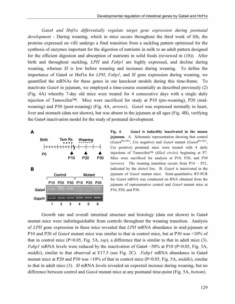
Developmental regulation of intestinal genes by Gata4 and Hnf1α
129
Gata4 and Hnf1α differentially regulate target gene expression during postnatal
development - During weaning, which in mice occurs throughout the third week of life, the
proteins expressed on villi undergo a final transition from a suckling pattern optimized for the
synthesis of enzymes important for the digestion of nutrients in milk to an adult pattern designed
for the efficient digestion and absorption of nutrients in solid foods (reviewed in (18)). After
birth and throughout suckling, LPH and Fabp1 are highly expressed, and decline during
weaning, whereas SI is low before weaning and increases during weaning. To define the
importance of Gata4 or Hnf1α for LPH, Fabp1, and SI gene expression during weaning, we
quantified the mRNAs for these genes in our knockout models during this time-frame. To
inactivate Gata4 in jejunum, we employed a time-course essentially as described previously (2)
(Fig. 4A) whereby 7-day old mice were treated for 4 consecutive days with a single daily
injection of Tamoxifen™. Mice were sacrificed for study at P10 (pre-weaning), P20 (mid-
weaning) and P30 (post-weaning) (Fig. 4A, arrows). Gata4 was expressed normally in heart,
liver and stomach (data not shown), but was absent in the jejunum at all ages (Fig. 4B), verifying
the Gata4 inactivation model for the study of postnatal development.
Fig. 4. Gata4 is inducibly inactivated in the mouse
jejunum. A. Schematic representation showing that control
(Gata4flox/flox
, Cre negative) and Gata4 mutant (Gata4flox/flox
,
Cre positive) postnatal mice were treated with 4 daily
injections of Tamoxifen™ (filled circles) beginning at P7.
Mice were sacrificed for analysis at P10, P20, and P30
(arrows). The weaning transition occurs from P14 – P21,
indicated by the dotted line. B. Gata4 is inactivated in the
jejunum of Gata4 mutant mice. Semi-quantitative RT-PCR
for Gata4 mRNA was conducted on RNA obtained from the
jejunum of representative control and Gata4 mutant mice at
P10, P20, and P30.
Growth rate and overall intestinal structure and histology (data not shown) in Gata4
mutant mice were indistinguishable from controls throughout the weaning transition. Analysis
of LPH gene expression in these mice revealed that LPH mRNA abundance in mid-jejunum at
P10 and P20 of Gata4 mutant mice was similar to that in control mice, but at P30 was <10% of
that in control mice (P<0.05, Fig. 5A, top), a difference that is similar to that in adult mice (3).
Fabp1 mRNA levels were reduced by the inactivation of Gata4 ~50% at P10 (P<0.05, Fig. 5A,
middle), similar to that observed at E17.5 (see Fig. 2C). Fabp1 mRNA abundance in Gata4
mutant mice at P20 and P30 was <10% of that in control mice (P<0.05, Fig. 5A, middle), similar
to that in adult mice (3). SI mRNA levels revealed an expected increase during weaning, but no
difference between control and Gata4 mutant mice at any postnatal time-point (Fig. 5A, bottom).
P10 P20 P30
Gata4
Gapdh
Control
P10 P20 P30
Mutant
1 2 3 4 5 6
P0P10 P20 P30
WeaningTam RxBirth
A
B
P10 P20 P30
Gata4
Gapdh
Control
P10 P20 P30
Mutant
1 2 3 4 5 6
P0P10 P20 P30
WeaningTam RxBirth
A
B

CHAPTER 5
130
Immunofluorescence for LPH, Fabp1, and SI in control and Gata4 mutant mice during
weaning followed the results of their mRNA abundances. LPH immunoflouresence was specific
to the microvillus membrane in the P10 jejunum (Fig. 5B), and was not affected by the
inactivation of Gata4 (Fig. 5C), consistent with mRNA levels. LPH immunofluorescence was
also present in the jejunum of P30 control mice (Fig. 5D), but absent in the Gata4 mutant mice
(Fig. 5E), which is also consistent with mRNA levels. Fabp1 immunofluorescence in the
cytoplasm of villus enterocytes was reduced by the inactivation of Gata4 at both P10 and P30
(Fig. 5F-I), consistent with its mRNA levels. SI was not detected at P10 in either control or
Gata4 mutant mice (Fig. 5J and K), and was localized to the microvillus membrane at P30 with
no apparent difference in intensity between control and Gata4 mutant mice (Fig. 5L and M),
consistent with mRNA levels. These data demonstrate that Gata4 is: not required for LPH gene
expression before weaning, but indispensable after weaning; at least partially required for Fabp1
gene expression throughout development; and not required for SI gene expression at any time
during development.
LPH/DAPI
Control Mutant
D E
Control Mutant
CB
P10 P30
SI/DAPI
A
L MKJ
0.0
0.4
0.8
SI
LPH
0.0
1.0
2.0
3.0
P20P10 P30
mRNA abundance (relative to calibrator)
Control Mutant
1.2
*
Fabp1
0.0
3.0
6.0
9.0
**
*
Fabp1/DAPI
H IGF
LPH/DAPI
Control Mutant
D E
Control Mutant
CB
P10 P30
SI/DAPI
A
L MKJ
0.0
0.4
0.8
SI
LPH
0.0
1.0
2.0
3.0
P20P10 P30
mRNA abundance (relative to calibrator)
Control Mutant
1.2
*
Fabp1
0.0
3.0
6.0
9.0
**
*
Fabp1/DAPI
H IGF

Developmental regulation of intestinal genes by Gata4 and Hnf1α
131
Fig. 5. Inactivation of Gata4 in jejunum reveals gene-specific, developmental regulation. A. LPH, Fabp1, and
SI mRNA abundances are differentially affected by the inactivation of Gata4 during development. Real-time RT-
PCR for LPH (top), Fabp1 (middle) and SI (bottom) mRNAs were conducted on RNA isolated from jejunum
(segment 3) of control and Gata4 mutant mice at P10, P20 and P30. Data are mean ± SD of n = 3-5 mice. *P<0.05,
as compared to controls. B-M. Immunofluorescence for LPH (B-E), Fabp1 (F-I), and SI (J-M) in P10 control
(B,F,J), P10 Gata4 mutant (C,G,K), P30 control (D,H,L), and P30 Gata4 mutant (E,I,M) mice is consistent with
mRNA levels.
In Hnf1α -/- mice, LPH mRNA abundance was ~50% of that in wild-type jejunum at P7,
P14, and P21, similar to that at E17.5, but was <10% of that in wild-type jejunum at P28 (Fig.
6A, top), similar to that in adult Hnf1α null mice (4). Fabp1 mRNA was reduced ~50% in the
Hnf1α null mice at P7 and P14, similar to that at E17.5 (see Fig. 2C), and was barely detectable
at P21 and P28 (Fig. 6A, middle), similar to that in adults (4). SI mRNA increased during
weaning in Hnf1α+/+ and Hnf1α-/- mice with no significant difference between the two groups
(Fig. 6A, bottom).
LPH immunofluorescence was most intense in the P7 jejunum of wild-type mice (Fig.
6B), and was consistently less intense in the P7 jejunum of Hnf1α null mice (n=3, Fig. 6C), in
agreement with the decrease in LPH mRNA levels at this age. LPH immunofluorescence was
present in the P28 jejunum of wild-type mice (Fig. 6D), but was not detectable in the jejunum of
P28 Hnf1α null mice (Fig. 6E), again consistent with mRNA levels. Fabp1 immunofluorescence
was reduced in Hnf1α null mice at both P7 and P28, consistent with its mRNA levels (Fig. 6F-I).
SI immunofluorescence was not detected in either control or Hnf1α null mice at P7 (Fig. 6J and
K), and was similarly intense in the jejunum of both mice at P28 (Fig. 6L and M), correlating
with its mRNA levels. These data indicate that Hnf1α, although indispensable for LPH and
Fabp1 gene expression after weaning (4), is only partially required before weaning. These data
also show that Hnf1α is not required for the endogenous increase in SI gene expression during
weaning.

CHAPTER 5
132
Fig. 6. Null expression of Hnf1α demonstrates gene-specific, developmental regulation. A. LPH, Fabp1, and SI
mRNA abundances are differentially affected by null expression of Hnf1α during development. Real-time RT-PCR
for LPH (top), Fabp1 (middle) and SI (bottom) mRNAs were conducted on RNA isolated from jejunum (segment 3)
of Hnf1α+/+ and Hnf1α-/- mice at P7, P14, P21, and P28. Data are mean ± SD of n = 3-5 mice. *P<0.05, as
compared to controls. B-M. Immunofluorescence for LPH (B-E), Fabp1 (F-I), and SI (J-M) in P7 Hnf1α+/+ (B,F,J),
P7 Hnf1α-/- (C,G,K), P28 Hnf1α+/+ (D,H,L), and P28 Hnf1α-/- (E,I,M) mice is consistent with mRNA levels.
Nuclear Gata4 and Hnf1α are paradoxically lost prior to weaning - To begin to define
the underlying mechanism by which Gata4 and Hnf1α differentially regulate intestinal genes
during postnatal development, the patterns of expression of Gata4 and Hnf1α mRNAs and
proteins were determined in wild-type mice at P7, P14, P21, and P28. Gata4 mRNA was highest
before weaning, and gradually declined during weaning to significantly lower levels at P21
(P<0.05) (Fig. 7A, upper). Hnf1α mRNA was highest at P7, and was significantly lower at all
time-points thereafter (Fig. 7A, bottom). These patterns are generally consistent with recently
published data (13).
LPH/DAPI
+/+ -/-
D E
+/+ -/-
CB
P7 P28
SI/DAPI
0.0
0.5
1.0SI
LPH
1.0
2.0
3.0
* *
**
P14 P28P7 P21
mRNA abundance (relative to calibrator)
A
L MKJ
+/+ -/-
Fabp1
1.0
2.0
4.0
*
* *
3.0
Fabp1/DAPI
H IGF
LPH/DAPI
LPH/DAPI
+/+ -/-
D E
+/+ -/-
CB
P7 P28
SI/DAPI
0.0
0.5
1.0SI
LPH
1.0
2.0
3.0
* *
**
P14 P28P7 P21
mRNA abundance (relative to calibrator)
A
L MKJ
+/+ -/-
Fabp1
1.0
2.0
4.0
*
* *
3.0
Fabp1/DAPI
H IGF

Developmental regulation of intestinal genes by Gata4 and Hnf1α
133
Fig. 7. Gata4 and Hnf1α mRNA abundance do not correlate with their nuclear protein levels during
postnatal development. A. Gata4 and Hnf1α mRNA abundance decreases during the weaning transition. Gata4
and Hnf1α mRNA was quantified in wild-type jejunum (segment 3) at P7, P14, P21, and P28 by real-time RT-PCR.
Data are mean ± SD of n = 3-5 mice. *P<0.05, as compared to P7 as determined by the Tukey-Kramer multiple
comparison test. B. Gata4 and Hnf1α protein levels increase during the weaning transition. Gata4 and Hnf1α were
quantified in nuclear extracts isolated from wild-type jejunum at P7, P14, P21, and P28 by Western analysis. Blots
were re-probed with a β-actin antibody to demonstrate similar protein loading in each lane. C. Gata4 and Hnf1α
were not detected in nuclear or non-nuclear fractions of P10 mouse jejunum. Gata4 and Hnf1α were quantified in
nuclear and non-nuclear extracts isolated from wild-type jejunum at P10, and in nuclear extracts from wild-type
knockout adult jejunum by Western analysis. Fabp1 immunoblot was conducted to verify the enrichment of
cytoplasmic proteins in the non-nuclear fractions. D. Gata and Hnf1 binding activity was not detected in nuclear
extracts from P7 mouse jejunum. Using well characterized sites for Gata (X-GATA) and Hnf1 (β-Fib) binding,
EMSAs were conducted using nuclear extracts obtained from P7 and P28 mouse jejunum. Specific complexes
(filled arrowhead) that supershifted (open arrowhead) with specific Gata4 (G4) or Hnf1α (H1a) antibodies were
found only for extracts from P28 mice.
A
Hnf1α
Gata4
0.5
1.0
1.5
mRNA abundance (relative to calibrator)
0.0
1.0
2.0
3.0
* ****
*
P14
P21
P28
P7
B
Gata4β-actin
50 kD
44 kD
Hnf1α
β-actin
75 kD
44 kD
P14
P28
P21
P7
- +- +- +- +- -Antibody:
Extract: P7 P7P28 P28- -
Probe: X-GATA β-FibD
44 kD
C
Gata4 50 kD
44 kD
Hnf1α 75 kD
44 kD
nuclear
knockout
WT
non-nuclear
P10 Adult
nuclear
β-actin
β-actin
Fabp1 17 kD
β-actin
A
Hnf1α
Gata4
0.5
1.0
1.5
mRNA abundance (relative to calibrator)
0.0
1.0
2.0
3.0
* ****
*
P14
P21
P28
P7
Hnf1α
Gata4
0.5
1.0
1.5
mRNA abundance (relative to calibrator)
0.0
1.0
2.0
3.0
* ****
*
P14
P21
P28
P7
B
Gata4β-actin
50 kD
44 kD
Hnf1α
β-actin
75 kD
44 kD
P14
P28
P21
P7
- +- +- +- +- -Antibody:
Extract: P7 P7P28 P28- -
Probe: X-GATA β-Fib
- +- +- +- +- -Antibody:
Extract: P7 P7P28 P28- -
Probe: X-GATA β-FibD
44 kD
C
Gata4 50 kD
44 kD
Hnf1α 75 kD
44 kD
nuclear
knockout
WT
non-nuclear
P10 Adult
nuclear
β-actin
β-actin
Fabp1 17 kD
β-actin

CHAPTER 5
134
To determine if the mRNA abundance correlates with nuclear protein levels, Western
blot analyses using nuclear extracts from mid-jejunum of wild-type mice were performed. Gata4
and Hnf1α were surprisingly undetectable at P7 and very low at P14, but increased markedly at
P21 and P28 (Fig. 7B), similar to that previously shown (5), and sharply contrasting with their
decreasing mRNA levels during this time interval (see Fig. 7A). The differential abundance of
nuclear Gata4 and Hnf1α between preweaning and postweaning jejunum was replicated in at
least 2 additional sets of mice. To determine if Gata4 and Hnf1α are localized outside of the
nucleus before weaning, Western analyses were performed on nuclear and non-nuclear fractions
isolated from jejunal enterocytes at P10. As shown in Fig. 7C, neither Gata4 nor Hnf1α was
detected in either the nuclear or non-nuclear fractions at this age. As controls, both Gata4 and
Hnf1α were readily detected in nuclear extracts of wild-type jejunum from adult mice, but not in
the mature jejunum of the respective knockout models. The relative abundance of Fabp1 in the
non-nuclear fraction verifies the enrichment of cytoplasmic protein in this fraction. The inability
to detect Gata4 or Hnf1α in nuclear and non-nuclear fractions before weaning was replicated on
pooled samples of P7 mouse jejunum. Nuclear extracts from both P7 (Fig. 7D) and P10 (data
not shown) were further tested by EMSAs using well characterized DNA binding sites for Gata
(16) and Hnf1 (7) transcription factors. Although nuclear extracts from P28 mouse jejunum
isolated in parallel showed specific complexes (filled arrowheads) that supershifted (open
arrowheads) with specific antibodies, neither complex could be detected in nuclear extracts from
P7 jejunum. Together, these data indicate that Gata4 and Hnf1α are absent, or present at very
low levels, in the epithelial nuclei before weaning.
To define the expression patterns of Gata4 and Hnf1α at the cellular level during
weaning, and to verify the attenuated levels of Gata4 and Hnf1α before weaning,
immunofluorescence experiments were conducted throughout this time interval. Gata4 and
Hnf1α were localized to the nucleus of crypt and villus epithelium at P1 (data not shown) and P4
(Fig. 8A-C), revealing a similar pattern to that at E16.5 (see Fig. 3C-H). Neither Gata4 (Fig. 8D
and E) nor Hnf1α (Fig. 8F and G) was detected in the nuclei of P7 jejunum. Positive nuclear
immunofluorescence for Cdx2 (Fig. 8H and I) in serial sections served as a positive control for
nuclear immunostaining (white arrowheads) at this age. At P14, Hnf1α was not detected (data
not shown), whereas Gata4 was detected only in the nuclei of cells in the crypts and lower villi
(Fig. 8J and K). At P21, Gata4 was expressed throughout the nuclei of the crypt and villus
epithelium (data not shown), whereas Hnf1α was expressed in the nuclei of cells in the crypt and
lower half of the villi, but was absent in the cells at the villus tip (Fig. 8L and M). At P28, Gata4
and Hnf1α were expressed throughout the nuclei of the crypt and villus epithelium (data not
shown), similar to that in adults. Together, these studies demonstrate that both the Gata4 and
Hnf1α genes are expressed at high levels throughout postnatal development as indicated by their
high levels of mRNA (see Fig. 7A), but their protein products are expressed at low levels during
suckling, specifically beginning the second week of life, just prior to the weaning transition.

Developmental regulation of intestinal genes by Gata4 and Hnf1α
135
Fig. 8. Gata4 and Hnf1α expression in jejunum is attenuated before weaning. A-C. Immunofluorescence in P4
jejunum using rabbit anti-Gata4 (A), goat anti-Hnf1α (B) and a merged photomicrograph (C). D-I.
Immunofluorescence in P7 jejunum using goat anti-Gata4 (D,E), goat anti-Hnf1α (F,G) and rabbit anti-Cdx2 (H,I)
along with DAPI nuclear stain (E,G,I). J-M. Immunofluorescence in P14 jejunum using goat anti-Gata4 (J,K), and
in P21 jejunum using goat anti-Hnf1α (L,M) without (J,L) and with (K,M) DAPI nuclear stain. White arrowheads
show specific nuclear immunofluorescence and yellow arrowheads indicate the absence of specific nuclear
immunofluorescence.
P4
Gata4 Hnf1α Merge
Gata4/DAPIGata4 Hnf1α/DAPIHnf1α Cdx2 Cdx2/DAPI
P7
A B C
D E F G H I
Gata4/DAPIGata4 Hnf1α/DAPIHnf1α
P14
P21
J K L M
P4
Gata4 Hnf1α Merge
Gata4/DAPIGata4 Hnf1α/DAPIHnf1α Cdx2 Cdx2/DAPI
P7
A B C
D E F G H I
Gata4/DAPIGata4 Hnf1α/DAPIHnf1α
P14
P21
J K L M

CHAPTER 5
136
Gata4 and Hnf1α do not mediate the precocious weaning induced by glucocorticoids -
Glucocorticoids are known to induce maturation of the small intestine resulting in the precocious
induction of intestinal enzymes such as SI (24). However, because this induction is
characterized by a 8 h lag, it is thought to be a secondary effect. The primary response is likely
mediated by intestinal transcription factors and Gata factors have been implicated (31). To
define the possible role of Gata4 as well as Hnf1α in mediating the glucocorticoid response in
preweaning mice, we characterized the dexamethasone-mediated response in the context of null
intestinal expression of Gata4 or Hnf1α. As shown in Fig 9A (top), SI mRNA abundance was
similarly induced ~80-fold 24 h after dexamethasone administration in wild-type, Gata4 mutant,
and Hnf1α-/- mice suggesting that neither Gata4 nor Hnf1α mediates the dexamethasone effect
on SI in vivo. LPH mRNA abundance was not affected by dexamethasone in wild-type mice,
but was significantly reduced by dexamethasone in both Gata4 mutant and Hnf1α-/- mice (Fig.
9A, bottom). These data indicate that dexamethasone
induces a process in which Gata4 and Hnf1α become
regulatory for LPH gene expression, as in the post-
weaning situation. In wild-type mice, the mRNAs for
both Gata4 and Hnf1α were significantly reduced by
dexamethasone, both at 4 h (data not shown) and 24 h
(Fig. 9B) after dexamethasone treatment, similar to that
which occurs after weaning. Taken together, these data
indicate that Gata4 and Hnf1α expression profiles change
according to a precocious maturation process induced by
glucocorticoids.
Fig. 9. Dexamethasone induces a precocious weaning response in
jejunum in the presence or absence of Gata4 or Hnf1α. A.
Dexamethasone induces SI mRNA levels in preweaning mice
independently of Gata4 or Hnf1α, but attenuates LPH mRNA levels
specifically in the absence of Gata4 or Hnf1α. Mice were treated
with dexamethasone (1 µg/g BW) on P10 and RNA was collected 24
h later from the jejunum of wild-type, Gata4 mutant, and Hnf1α-/-
mice. SI (top) and LPH (bottom) mRNA levels were quantified by
real-time RT-PCR in n = 3-5 mice. *P<0.05, **P<0.01 as compared
to untreated mice. B. Dexamethasone attenuates Gata4 and Hnf1α
mRNA levels in jejunum of preweaning mice. Wild-type mice were
treated with dexamethasone on P10 and RNA was collected 24 h
later from the jejunum. Gata4 and Hnf1α mRNA levels were
quantified by real-time RT-PCR in n = 3-5 mice. *P<0.05, as
compared to untreated mice.
0
0.5
1.0
1.5
2.0
0.2
0.4
0.6
Wild-
type
Gata4
mutant
Hnf1α
-/-
- Dex
+ Dex
SI
LPH
*
***
**
**
0
1
2
3
4
Gata4 Hnf1α
mRNA abundance
(relative to calibrator)
* *
A
B
0
0.5
1.0
1.5
2.0
0.2
0.4
0.6
Wild-
type
Gata4
mutant
Hnf1α
-/-
- Dex
+ Dex
SI
LPH
*
***
**
**
0
1
2
3
4
Gata4 Hnf1α
mRNA abundance
(relative to calibrator)
* *
A
B

Developmental regulation of intestinal genes by Gata4 and Hnf1α
137
DISCUSSION
The establishment of a fully functioning mature mammalian gut is the result of a series of
ordered developmental transitions (reviewed in (36)). The terminal differentiation phases of
intestinal development, characterized in part by the expression of proteins necessary for the
digestion and absorption of nutrients, occur during cytodifferentiation and the weaning transition
(reviewed in (18)). LPH, Fabp1 and SI are well characterized markers of these transitions (17,
19, 24, 34, 35, 37, 38, 42), and the Gata and Hnf1 families of transcription factors have been
implicated in their regulation (5, 6, 8-10, 12, 15, 21, 27, 40, 50). We have previously shown in
mature jejunum that inactivation of either Gata4 or Hnf1α results in a nearly complete
attenuation of LPH and Fabp1 gene expression, but no effect on SI gene expression (3, 4).
In the present study, we found that the regulation of target genes by Gata4 and Hnf1α
during development differs from that in adults (Fig. 10). Before weaning, Gata4 and Hnf1α are
only partially required for LPH and Fabp1 gene expression, and their patterns of regulation
differ for each of these genes during this time-frame. The partial Gata4 requirement for Fabp1
gene expression during cytodifferentiation is consistent with data from E18.5 mosaic Gata4
knockout mice in which Fabp1 gene expression is attenuated in intestinal epithelial cells that do
not express Gata4 as indicated by in situ hybridization (9). We further found that Gata4 and
Hnf1α are dispensable for SI gene expression throughout development. During the suckling
period, there is a surprising and dramatic reduction in Gata4 and Hnf1α protein in the nuclei of
absorptive enterocytes of the jejunum despite high levels of mRNA. Finally, we show that Gata4
or Hnf1α do not mediate the precocious maturation of the intestine induced by glucocorticoids.
Together, these data demonstrate that specific intestinal genes, including LPH and Fabp1, have
differential requirements for Gata4 and Hnf1α that are dependent on the developmental time-
frame in which they are expressed.
Our data show that Gata4 or Hnf1α are partially required for LPH and Fabp1 gene
expression before weaning, which contrasts with their indispensability for LPH and Fabp1 gene
expression after weaning. These findings suggest that redundant or overlapping mechanisms
maintain the expression of LPH and Fabp1 during this time interval. Functionally, LPH is
necessary for the digestion of milk lactose, and is thus critical for nutrition during suckling,
while Fabp1 plays a role in intracellular lipid transport, and is likely important for the transport
of the lipid load present in milk. Thus, a redundant mechanism for the maintenance of gene
expression during this critical developmental time interval is a plausible hypothesis. These data
also suggest that other factors are involved in LPH and Fabp1 gene expression before weaning.
Candidate transcription factors include Gata5, Gata6, Hnf1β, and members of the Cdx, Hnf3, and
C/EBP families, all of which have been previously implicated as activators of LPH and/or Fabp1
gene transcription in vitro (5, 9, 10, 12, 14, 15, 21, 29, 41, 43, 48). Additional studies will be
necessary to define the requirement in vivo of these transcription factors and/or identify other
factors involved in the developmental regulation of LPH and Fabp1 gene expression.

CHAPTER 5
138
Fig. 10. Summary of Gata4 and Hnf1α regulation of LPH, Fabp1 and SI gene expression during intestinal
development. Schematic representation of LPH (top), Fabp1 (middle), and SI (bottom) gene expression in wild-
type, Gata4 mutant, and Hnf1α-/- during development.
These data also reveal differential mechanisms underlying the regulation of genes whose
expression patterns during development are strikingly similar. Both LPH and Fabp1 are highly
expressed in more proximal regions of small intestine than distal regions, and at higher levels
Wild-type
Gata4 mutant
Hnf1α-/-
Cytodifferentiation
Suckling
Weaning
Post-weaning
Adult
LPH
SI
Fabp1
Wild-type
Gata4 mutant
Hnf1α-/-
Cytodifferentiation
Suckling
Weaning
Post-weaning
Adult
LPH
SI
Fabp1

Developmental regulation of intestinal genes by Gata4 and Hnf1α
139
before weaning than after weaning (Fig. 1,5 and 6, and (17, 19, 34, 35, 38)), and their promoters
contain binding sites for similar sets of transcription factors (8, 21). However, although both are
similarly regulated by Gata4 and Hnf1α in mature intestine (3, 4), they are differentially
regulated by these two transcription factors before weaning. Both LPH and Fabp1 mRNAs are
reduced ~50% in Hnf1α null mice before weaning, but only Fabp1 mRNA is reduced in the
Gata4 mutant mice before weaning. Further, Fabp1 mRNA abundance is attenuated ~90% by
the inactivation of Gata4 at P20 or Hnf1α at P21, similar to that in adult mice, whereas LPH
mRNA abundance is reduced <50% at these time points, similar to preweaning mice, indicating a
differential in the timing of regulation by Gata4 or Hnf1α. The differential regulation of LPH
and Fabp1 by Gata4 before weaning highlights a target-specific gene regulation during
development.
We have previously hypothesized that the induction of SI gene expression during
postnatal development is regulated by the combinatory effect of a complex of transcription
factors including Gata4 and Hnf1α (5, 21). In addition, the abundance of nuclear Gata4 and
Hnf1α proteins in the jejunum increases during the weaning transition, paralleling SI gene
expression (Fig. 8 and (5, 21)). Despite these compelling data for a combinatory role for Gata4
and Hnf1α in the activation of SI gene expression, we recently reported that the inactivation of
Gata4 or Hnf1α had no effect on SI gene expression in adult mice (3, 4). Here, we show that the
inactivation of Gata4 or Hnf1α had no effect on the initiation of SI gene expression during
weaning. Together, these data demonstrate that Gata4 and Hnf1α are dispensable for SI gene
expression throughout development. The future challenge, therefore, is to identify transcription
factors essential for SI gene expression.
In our studies, we identified a paradoxical loss of Gata4 and Hnf1α protein in the nuclei
of absorptive enterocytes during a narrow window just prior to the onset of weaning. At P7-P14,
Gata4 and Hnf1α were greatly reduced in the nuclear fraction of jejunal extracts as determined
by Western analysis (Fig. 7B and C) and EMSA (Fig. 7D), and neither could be detected in the
nuclei of villus enterocytes in the jejunum by immunofluorescence (Fig. 8). This reduction in
Gata4 and Hnf1α nuclear protein occurs over a narrow time-frame at the end of the first week
(P4-P7), and regenerates from crypts during the third week of life (Fig. 8). Interestingly, Gata4
and Hnf1α mRNA abundance remains high during this time interval (Fig. 7A) suggesting that
the reduction in Gata4 and Hnf1α protein is not due to a decrease in transcription rate. We thus
believe that either the mRNAs for these proteins are not being transcribed, and/or that their
translation products are being actively catabolized. However, because Gata4 and Hnf1α are
partially regulatory during this time interval, we hypothesize that at least some Gata4 and Hnf1α
is normally present in the nucleus, but below the detection limits of our assays. Further,
regulation in the knockout models at P7-P14 (Fig. 5 and 6) is virtually identical to that which
occurs at E17.5 (Fig. 2), when Gata4 and Hnf1α are normally present in epithelial nuclei,
suggesting that the loss of nuclear Gata4 and Hnf1α before weaning is not a critical regulatory

CHAPTER 5
140
mechanism for LPH and Fabp1 gene expression. We believe that the process of nuclear Gata4
and Hnf1α loss in the second week of life in mice is a regulatory process, but for other as yet
unknown targets of Gata4 and Hnf1α.
Glucocorticoids like dexamethasone can induce the precocious maturation of the
intestine, but the underlying mechanism is not understood (1, 51). Characteristic of this process
is a dramatic increase in SI mRNA abundance 24 h after the administration of glucocorticoids
(24). Since the induction in SI mRNA is not apparent within the first 8 h (24), it is thought that
the SI induction is not a direct response to glucocorticoids, but rather a secondary effect of early
response genes on SI gene transcription (1). Recently, Oesterreicher and Henning (31) showed
that in 8-day old mice, Gata4 and Gata6 were both induced 4 h after dexamethasone treatment, as
shown by supershift EMSAs and Western analysis, suggesting a role for Gata factors in the
glucocorticoid-induced maturation of the intestine. To test the hypothesis that Gata4 or Hnf1α
mediates this process, we conducted dexamethasone-induced precocious maturation experiments
in our knockout models. SI mRNA was strongly induced in the presence or absence of Gata4 or
Hnf1α (Fig. 9A) indicating that these proteins are not required for mediating the dexamethasone
response on SI. We next defined the effect of dexamethasone on LPH mRNA abundance in our
knockout models, and found that LPH mRNA was reduced in the Gata4 mutant and Hnf1α-/-
mice (Fig. 9A), but not in wild-type controls. Our interpretation of these data is that
dexamethasone induces precocious maturation to the point where Gata4 and Hnf1α become
more regulatory for LPH gene expression. We also found a decrease in both Gata4 and Hnf1α
mRNA abundance with dexamethasone treatment (Fig. 9B), which is also consistent with a
maturation of the intestine as Gata4 and Hnf1α mRNAs decline normally during weaning (Fig.
7A). Taken together, we believe that neither Gata4 nor Hnf1α mediates the glucocorticoid
response but rather are downstream targets of this response.
Cre-mediated inactivation of Gata4 in our current model results in the synthesis of a
trancriptionally inactive, truncated form of Gata4 (mutant Gata4) that is missing its N-terminal
activation domains, but contains its functional zinc finger region (3). Since mutant Gata4
continues to bind DNA, it has the potential to act as a dominant-negative Gata factor (3) masking
the activity of other enterocyte Gata factors, such as Gata6, which is co-expressed with Gata4 in
villus enterocytes (3, 9, 39). Further, mutant Gata4 contains the functional zinc finger region,
which has been shown to interact not only with DNA, but also with other proteins, such as Hnf1α
(46). Indeed, we have shown that although the activation domains of Hnf1α are absolutely
required for synergy, the Gata activation domains are dispensable for this activity (46, 47),
suggesting that mutant Gata4 maintains the ability to mediate Gata4/Hnf1α synergy. Thus, the
phenotype attributed to the inactivation of Gata4 in our current model could represent
specifically Gata4 function, a masked Gata6 function, and/or a function independent of
Gata4/Hnf1α interactions. Further, because Gata4 is inducibly inactivated rather than null for
Gata4 from the earliest phases of intestinal development, it is possible that gene expression that

Developmental regulation of intestinal genes by Gata4 and Hnf1α
141
is dependent on the presence of Gata4 early in development (before administration of
Tamoxifen™) may not be revealed in the current model. Precedence for such a mechanism is
shown in the liver where embryonic, but not postnatal re-expression of Hnf1α is capable of re-
activating the silent phenylalanine hydrolxylase gene in Hnf1α-deficient hepatocytes (49).
Nevertheless, since the same Gata4 model (induction of mutant Gata4) is used throughout our
studies, the differential regulation of target genes at diverse developmental time-points continues
to support different underlying mechanisms of regulation during development.
Our data show that the induction and maintenance of terminal differentiation by Gata4
and Hnf1α is highly dependent on the developmental time-frame under study. Although Gata4
and Hnf1α are indispensable for the maintenance of expression of specific genes in the mature
intestine (3, 4), these transcription factors are only partially required for the same genes prior to
the final maturation that occurs at weaning, implicating other mechanisms in the development of
gut function. Understanding the individual and combined effects of intestinal transcription
factors during development will continue to reveal important regulatory pathways essential for
intestinal differentiation.

CHAPTER 5
142
ACKNOWLEDGEMENTS
We thank Drs. W. T. Pu (Children’s Hospital Boston, Boston, MA) for the floxed Gata4 mouse
line, S. Robine (Institut Curie, Paris, France) for the VillinCreERT2 transgenic mouse line, and F.
J. Gonzalez (National Institutes of Health, Bethesda, MD) for the Hnf1α null mouse line. We
also thank Drs. J. Gordon (Washington University, St. Louis, MO) for the Fabp1 antibody, K-Y.
Yeh (Louisiana State University, Shreveport, LA) for the LPH and SI antibodies, and D. Silberg
(University of Pennsylvania, Philadelphia, PA) for the Cdx-2 antibody. We also thank Lena Liu
(Department of Pathology, Children’s Hospital Boston, Boston, MA) for technical support.
GRANTS
This work was supported by National Institute of Diabetes and Digestive and Kidney Diseases
Grants R01-DK-061382 (S.D. Krasinski) and R37-DK-32658 (R.J. Grand), the Harvard
Digestive Disease Center (5P30-DK-34854), and the Nutricia Research Foundation (T. Bosse
and E. Beuling).

Developmental regulation of intestinal genes by Gata4 and Hnf1α
143
REFERENCES
1. Agbemafle BM, Oesterreicher TJ, Shaw CA, and Henning SJ. Immediate early genes of glucocorticoid action
on the developing intestine. Am J Physiol - Gastrointest & Liver Physiol 288: 897-906, 2005.
2. Bettess MD, Dubois N, Murphy MJ, Dubey C, Roger C, Robine S, and Trumpp A. c-Myc is required for the
formation of intestinal crypts but dispensable for homeostasis of the adult intestinal epithelium. Mol Cell Biol 25:
7868-7878, 2005.
3. Bosse T, Piaseckyj CM, Burghard E, Fialkovich JJ, Rajagopal S, Pu WT, and Krasinski SD. Gata4 is
essential for the maintenance of jejunal-ileal identities in the adult mouse jejunum. Mol Cell Biol In Press, 2006.
4. Bosse T, van Wering HM, Gielen M, Dowling LN, Fialkovich JJ, Piaseckyj CM, Gonzalez FJ, Akiyama TE,
Montgomery RK, Grand RJ, and Krasinski SD. Hepatocyte nuclear factor-1alpha is required for expression but
dispensable for histone acetylation of the lactase-phlorizin hydrolase gene in vivo. Am J Physiol Gastrointest Liver
Physiol 290: G1016-1024, 2006.
5. Boudreau F, Rings EH, van Wering HM, Kim RK, Swain GP, Krasinski SD, Moffett J, Grand RJ, Suh ER,
and Traber PG. Hepatocyte nuclear factor-1 alpha, GATA-4, and caudal related homeodomain protein Cdx2
interact functionally to modulate intestinal gene transcription. Implication for the developmental regulation of the
sucrase-isomaltase gene. J Biol Chem 277: 31909-31917, 2002.
6. Boudreau F, Zhu Y, and Traber PG. Sucrase-isomaltase gene transcription requires the hepatocyte nuclear
factor-1 (HNF-1) regulatory element and is regulated by the ratio of HNF-1 alpha to HNF-1 beta. J Biol Chem 276:
32122-32128, 2001.
7. Courtois G, Morgan JG, Campbell LA, Fourel G, and Crabtree GR. Interaction of a liver-specific nuclear
factor with the fibrinogen and alpha 1-antitrypsin promoters. Science 238: 688-692, 1987.
8. Divine JK, McCaul SP, and Simon TC. HNF-1alpha and endodermal transcription factors cooperatively
activate Fabpl: MODY3 mutations abrogate cooperativity. Am J Physiol Gastrointest Liver Physiol 285: G62-72,
2003.
9. Divine JK, Staloch LJ, Haveri H, Jacobsen CM, Wilson DB, Heikinheimo M, and Simon TC. GATA-4,
GATA-5, and GATA-6 activate the rat liver fatty acid binding protein gene in concert with HNF-1alpha. Am J
Physiol Gastrointest Liver Physiol 287: G1086-1099, 2004.
10. Divine JK, Staloch LJ, Haveri H, Rowley CW, Heikinheimo M, and Simon TC. Cooperative interactions
among intestinal GATA factors in activating the rat liver fatty acid binding protein gene. Am J Physiol Gastrointest
Liver Physiol, 2006.
11. el Marjou F, Janssen KP, Chang BH, Li M, Hindie V, Chan L, Louvard D, Chambon P, Metzger D, and
Robine S. Tissue-specific and inducible Cre-mediated recombination in the gut epithelium. Genesis 39: 186-193,
2004.
12. Fang R, Olds LC, Santiago NA, and Sibley E. GATA family transcription factors activate the lactase gene
promoter in intestinal Caco-2 cells. Am J Physiol 260: G58-G67, 2001.
13. Fang R, Olds LC, and Sibley E. Spatio-temporal patterns of intestine-specific transcription factor expression
during postnatal mouse gut development. Gene Expr Patterns 6: 426-432, 2006.
14. Fang R, Santiago NA, Olds LC, and Sibley E. The homeodomain protein Cdx2 regulates lactase gene
promoter activity during enterocyte differentiation. Gastroenterology 118: 115-127, 2000.

CHAPTER 5
144
15. Fitzgerald K, Bazar L, and Avigan MI. GATA-6 stimulates a cell line-specific activation element in the
human lactase promoter. Am J Physiol 274: G314-324, 1998.
16. Gao X, Sedgwick T, Shi YB, and Evans T. Distinct functions are implicated for the GATA-4, -5, and -6
transcription factors in the regulation of intestine epithelial cell differentiation. Mol Cell Biol 18: 2901-2911, 1998.
17. Gordon JI, Elshourbagy N, Lowe JB, Liao WS, Alpers DH, and Taylor JM. Tissue specific expression and
developmental regulation of two genes coding for rat fatty acid binding proteins. J Biol Chem 260: 1995-1998,
1985.
18. Henning SJ. Postnatal development: coordination of feeding, digestion, and metabolism. American Journal of
Physiology - Gastrointestinal & Liver Physiology 241: 199-214, 1981.
19. Krasinski SD, Estrada G, Yeh KY, Yeh M, Traber PG, Rings EH, Buller HA, Verhave M, Montgomery
RK, and Grand RJ. Transcriptional regulation of intestinal hydrolase biosynthesis during postnatal development in
rats. Am J Physiol 267: G584-594, 1994.
20. Krasinski SD, Upchurch BH, Irons SJ, June RM, Mishra K, Grand RJ, and Verhave M. Rat lactase-
phlorizin hydrolase/human growth hormone transgene is expressed on small intestinal villi in transgenic mice.
Gastroenterology 113: 844-855, 1997.
21. Krasinski SD, van Wering HM, Tannemaat MR, and Grand RJ. Differential activation of intestinal gene
promoters: Demonstration of functional interactions between GATA-5 and HNF-1a. Am J Physiol 281: G69-G84,
2001.
22. Lee YH, Sauer B, and Gonzalez FJ. Laron dwarfism and non-insulin-dependent diabetes mellitus in the Hnf-
1alpha knockout mouse. Mol Cell Biol 18: 3059-3068, 1998.
23. Lee YS, Wang Z, Lin CK, Contag CH, Olds LC, Cooper AD, and Sibley E. Regulation of intestine-specific
spatiotemporal expression by the rat lactase promoter. J Biol Chem 277: 13099-13105, 2002.
24. Leeper LL and Henning SJ. Development and tissue distribution of sucrase-isomaltase mRNA in rats. Am J
Physiol - Gastrointest Liver Physiol 258, 1990.
25. Markowitz AJ, Wu GD, Bader A, Cui Z, Chen L, and Traber PG. Regulation of lineage-specific
transcription of the sucrase-isomaltase gene in transgenic mice and cell lines. Am J Physiol 269: G925-939, 1995.
26. Markowitz AJ, Wu GD, Birkenmeier EH, and Traber PG. The human sucrase-isomaltase gene directs
complex patterns of gene expression in transgenic mice. Am J Physiol 265: G526-539, 1993.
27. Mitchelmore C, Troelsen JT, Spodsberg N, Sjostrom H, and Noren O. Interaction between the
homeodomain proteins Cdx2 and HNF1alpha mediates expression of the lactase-phlorizin hydrolase gene. Biochem
J 346: 529-535, 2000.
28. Montgomery RK, Mulberg AE, and Grand RJ. Development of the human gastrointestinal tract: twenty years
of progress. Gastroenterology 116: 702-731, 1999.
29. Montgomery RK, Rings EH, Thompson JF, Schuijt CC, Aras KM, Wielenga VJ, Kothe MJ, Buller HA,
and Grand RJ. Increased C/EBP in fetal rat small intestine precedes initiation of differentiation marker mRNA
synthesis. Am J Physiol 272: G534-544, 1997.
30. Nemer G and Nemer M. transcriptional activation of BMP-4 and regulation of mammalian organogenesis by
GATA-4 and -6. Dev Biol 254: 131-148, 2003.
31. Oesterreicher TJ and Henning SJ. Rapid induction of Gata transcription factors in developing mouse intestine
following glucocorticoid administration. Am J Physiol - Gastrointest Liver Physiol 286: G947-G953, 2004.

Developmental regulation of intestinal genes by Gata4 and Hnf1α
145
32. Pu WT, Ishiwata T, Juraszek AL, Ma Q, and Izumo S. GATA4 is a dosage-sensitive regulator of cardiac
morphogenesis. Dev Biol 275: 235-244, 2004.
33. Rhoads DB, Rosenbaum DH, Unsal H, Isselbacher KJ, and Levitsky LL. Circadian periodicity of intestinal
Na+/glucose cotransporter 1 mRNA levels is transcriptionally regulated. J Biol Chem 273: 9510-9516, 1998.
34. Rings EH, de Boer PA, Moorman AF, van Beers EH, Dekker J, Montgomery RK, Grand RJ, and Buller
HA. Lactase gene expression during early development of rat small intestine. Gastroenterology 103: 1154-1161,
1992.
35. Rings EH, Krasinski SD, van Beers EH, Moorman AF, Dekker J, Montgomery RK, Grand RJ, and Buller
HA. Restriction of lactase gene expression along the proximal-to-distal axis of rat small intestine occurs during
postnatal development [see comments]. Gastroenterology 106: 1223-1232, 1994.
36. Roberts DJ. Molecular mechanisms of development of the gastrointestinal tract. Dev Dyn 219: 109-120, 2000.
37. Sebastio G, Villa M, Sartorio R, Guzzetta V, Poggi V, Auricchio S, Boll W, Mantei N, and Semenza G.
Control of lactase in human adult-type hypolactasia and in weaning rabbits and rats. Am J Hum Gen 45: 489-497,
1989.
38. Simon TC, Roth KA, and Gordon JI. Use of transgenic mice to map cis-acting elements in the liver fatty acid-
binding protein gene (Fabpl) that regulate its cell lineage-specific, differentiation-dependent, and spatial patterns of
expression in the gut epithelium and in the liver acinus. J Biol Chem 268: 18345-18358, 1993.
39. Sodhi CP, Li J, and Duncan SA. Generation of mice harbouring a conditional loss-of-function allele of Gata6.
BMC Dev Biol 6: 1-19, 2006.
40. Spodsberg N, Troelsen JT, Carlsson P, Enerback S, Sjostrom H, and Noren O. Transcriptional regulation of
pig lactase-phlorizin hydrolase: involvement of HNF-1 and FREACs. Gastroenterology 116: 842-854, 1999.
41. Staloch LJ, Divine JK, Witten JT, and Simon TC. C/EBP and Cdx family factors regulate liver fatty acid
binding protein transgene expression in the small intestinal epithelium. Biochim Biophys Acta 1731: 168-178, 2005.
42. Traber PG. Regulation of sucrase-isomaltase gene expression along the crypt-villus axis of rat small intestine.
Biochem Biophys Res Com 173: 765-773, 1990.
43. Traber PG, Wu GD, and Wang W. Novel DNA-binding proteins regulate intestine-specific transcription of the
sucrase-isomaltase gene. Mol Cell Biol 12: 3614-3627, 1992.
44. Troelsen JT. Adult-type hypolactasia and regulation of lactase expression. Biochim Biophys Acta 1723: 19-32,
2005.
45. Troelsen JT, Mehlum A, Olsen J, Spodsberg N, Hansen GH, Prydz H, Noren O, and Sjostrom H. 1 kb of
the lactase-phlorizin hydrolase promoter directs post-weaning decline and small intestinal-specific expression in
transgenic mice. FEBS Lett 342: 291-296, 1994.
46. van Wering HM, Bosse T, Musters A, de Jong E, de Jong N, Hogen Esch CE, Boudreau F, Swain GP,
Dowling LN, Montgomery RK, Grand RJ, and Krasinski SD. Complex regulation of the lactase-phlorizin
hydrolase promoter by GATA-4. Am J Physiol Gastrointest Liver Physiol 287: G899-909, 2004.
47. van Wering HM, Huibregtse IL, van der Zwan SM, de Bie MS, Dowling LN, Boudreau F, Rings EH,
Grand RJ, and Krasinski SD. Physical interaction between GATA-5 and hepatocyte nuclear factor-1alpha results
in synergistic activation of the human lactase-phlorizin hydrolase promoter. J Biol Chem 277: 27659-27667, 2002.

CHAPTER 5
146
48. Verhave M, Krasinski SD, Christian SI, van Schaik S, van den Brink GR, Doting EMH, Maas SM,
Wolthers KC, Grand RJ, and Montgomery RK. Regulatory regions in the rat lactase-phlorizin hydrolase gene
that control cell-specific expression. JPGN 39: 275-285, 2004.
49. Viollet B, Yaniv M, and Pontoglio M. Embryonic but not postnatal reexpression of hepatocyte nuclear factor
1alpha (HNF1alpha) can reactivate the silent phenylalanine hydroxylase gene in HNF1alpha-deficient hepatocytes.
Mol Cell Biol 21: 3662-3670, 2001.
50. Wu GD, Chen L, Forslund K, and Traber PG. Hepatocyte nuclear factor-1 alpha (HNF-1 alpha) and HNF-1
beta regulate transcription via two elements in an intestine-specific promoter. J Biol Chem 269: 17080-17085, 1994.
51. Yaylaoglu MB, Agbemafle BM, Oesterreicher TJ, Finegold MJ, Thaller C, and Henning SJ. Diverse
patterns of cell-specific gene expression in response to glucocorticoid in the developing small intestine. Am J
Physiol - Gastrointest Liver Physiol` In Press, 2006.

CHAPTER 6
Gata4 dependent repression of ileal gene expression
is mediated through FOG interactions
(Manuscript in preparation)
Tjalling Bosse1, Eva Beuling
2, Daan aan de Kerk
1, Christina M. Piaseckyj
3, Y.
Fujiwara3,4, Stephen D. Krasinski
3,5
From the1Department of Pediatrics, University of Amsterdam, Amsterdam, The Netherlands 1100DD;
2Erasmus
University Rotterdam, Department of Pediatrics, Erasmus MC/Sophia children’s hospital, Rotterdam, The
Netherlands; 3Division of Gastroenterology and Nutrition, Department of Pediatrics, Children’s Hospital, Boston,
Massachusetts, 02115; 4Department of Hematology and Oncology, Children’s Hospital Boston, Massachusetts
02115, USA and 5Dorothy R. Friedman School of Nutrition Science and Policy, Tufts University, Boston,
Massachusetts, 02111
CHAPTER 6
148
ABSTRACT
GATA-family transcription factors are critical for the differentiation and development of diverse
tissues. In the small intestine, Gata4 has been demonstrated to be critical in the maintenance of
jejunal-ileal identities. Inactivation of Gata4 leads to an increase in goblet cells, attenuation of
jejunal genes (such as lactase phlorizin-hydrolase, LPH) and repression of ileal genes (such as
apical sodium bile-acid transporter, Asbt) in the adult mouse jejunum. In this report we use
Gata4 mutant mice to show that Gata4 is a critical factor in the establishment of the horizontal
distribution of Asbt gene expression in adult mice, by repressing Asbt expression in the proximal
and mid-jejunum during weaning. This Gata4 dependent repression acts through the first 1.1kb
of the Asbt 5'-flanking region as shown by transient transfections in CT-26 cells. In other organ
systems GATA factors function has been shown to be modulated by interactions with other
regulators, specifically by the interaction with Friend of GATA (FOG) co-factors. We therefore
hypothesized that intestinal Gata4 functions may be mediated by intestinal FOG co-factors. We
first determined the expression profile of Fog1 and Fog2 in the small intestine and found that
Fog1 is the principal FOG co-factor in the adult small intestine. As an experimental model to
search for a requirement for FOG co-factors in the intestinal Gata4 function, we established a
mouse line that upon tamoxifenTM
induction harbors a single amino acid replacement in Gata4
that impairs its physical interaction with FOG co-factors only in the intestine (Gata4 knock-in).
No gross abnormalities could be identified in the overall structure of the epithelium of these
mice, in particular we could not detect an increase in goblet cell population. To determine the
role of FOG co-factors in the Gata4 dependent gene expression, mRNA levels of LPH and Asbt
were compared between wild-type, mutant and knock-in jejunum and wild-type ileum. This
revealed that FOG co-factors facilitate in the Gata4 dependent repression of Asbt in the adult
jejunum, however do not play a role in the activation of LPH by Gata4. Therefore we infer that
the interaction of FOG co-factors, presumably Fog1, with Gata4 in the adult jejunum is
necessary for the complete Gata4 repression of ileal-specific gene expression.

FOG co-factors and intestinal Gata4 function
149
INTRODUCTION
This manuscript is in preparation, however the experiments presented were carried out during the
process of the thesis preparation and demonstrate significant results with respect to the
experiments described in this thesis and therefore were appropriate to include.
Gata4 is a member of an evolutionarily conserved zinc finger containing transcription
factor family that plays a crucial role in the maintenance of jejunal-ileal identities in the adult
mouse small intestine through a variety of molecular pathways (3). In the adult small intestine,
Gata4 is normally expressed in jejunum, but is greatly reduced or absent in distal ileum(3, 36).
Recently we showed that mice harboring an intestinal inactive Gata4 mutant have a jejunal
phenotype with ileal characteristics. The jejunal epithelium of these mice had an increase number
of goblet cells and a redistribution of enteroendocrine subtypes towards an ileal composition,
implicating a role for Gata4 in cell-lineage allocation along the horizontal axis. The jejunum also
displayed an attenuation in the expression of genes normally highly expressed in the jejunum,
such as liver fatty-acid binding protein (Fabp1) and lactase-phlorizin hydrolase (LPH),
indicating a role for Gata4 in the activation of these genes. The jejunum of these Gata4 mutant
mice further showed a significant induction of the ileal specific genes, ileal sodium bile-acid
transporter (Asbt) and ileal lipid binding protein (Ilbp). The mechanism of activating jejunal
genes like LPH and Fabp1 has been extensively studied in cell-culture systems and is likely a
result of the physical interaction of the C-terminal zinc finger of Gata4 with the homeodomain
region of hepatocytes nuclear factor (Hnf1α)(36). The molecular mechanism involved in the
Gata4 dependent repression of ileal genes in the adult jejunum and the role of Gata4 in cell-fate
determination however remains to be elucidated. Together these data present the paradigm that
Gata4 acts as an activator and a repressor at the same time in the absorptive enterocytes of the
adult jejunal epithelium. We hypothesized previously that this duality may be coordinated by
specific interactions with other semi-restricted transcription factors or co-factors in the context of
the specific promoter sequences.
Friend of GATA (FOG) is a recently discovered multi-zinc finger co-factor family that
physically associates with the N-terminal zinc finger of GATA factors, and mediates GATA
function. Depending on the context, FOG co-factors can act to further activate or repress the
GATA function of target genes (17, 24, 34, 35). The GATA:FOG interaction is conserved in
Drosophila where the FOG homologue, U-shaped, physically associates with pannier, a GATA
homologue indicating that this interaction is evolutionarily conserved and therefore
fundamentally important (10, 15). Two members of the FOG family, Fog1 and Fog2, have been
described based on protein structure and expression patterns (31, 32). Fog1 is co-expressed with
Gata1 in erythroid and megakaryocytic cells, whereas Fog2 is co-expressed with Gata4 in
cardiac tissue. Germline knock-out mice of Gata1 or Fog1, as well as a knock-in of a mutation
into Gata1 that disrupts interactions with FOG co-factors, all result in embryonic lethal
phenotypes characterized by failure during hematopoiesis (8, 13, 34). Likewise, germline knock-

CHAPTER 6
150
out mice of Gata4 or Fog2, as well as a knock-in mutation in Gata4 that disrupts FOG
interactions, all result in embryonic death due to failure during early cardiogenesis (9, 21, 25, 30,
33). Noteworthy in all of these examples, however, is that FOG co-factors mediate some, but not
all GATA functions, indicating that GATA factors have some intrinsic (FOG-independent)
activity.
Expression of FOG co-factors in the small intestine has not been reported, however in a
recent study evidence was provided for a role of FOG co-factors in the gastrointestinal system. In
this study the developing stomach epithelium of Gata4 knock-in (Gata4ki/ki
) embryos (at E11.5)
was analyzed (18). This study showed that in wild-type embryos, Fog1 is specifically co-
expressed with Gata4 in the developing hindstomach and absent in the forestomach. Fog2
expression was not evident in the gastric epithelium during fetal or postnatal development,
implicating Fog1 as the principal FOG co-factor in the stomach. The stomach epithelium of the
Gata4ki/ki
embryos did not show any gross abnormalities, however a significant induction of the
endoderm derived signaling molecule sonic hedgehog (Shh) was noted in the hindstomach
(normally expressed in the forestomach). The findings in the stomach of these Gata4ki/ki
mice
support a role for the Gata4:FOG interaction in specifying the differences between fore- and
hindstomach during early embryogenesis. This study was the first to establish a role for a
member of the FOG family, presumably Fog1, in the gastrointestinal system.
In the current study we hypothesized that some of Gata4 functions in the adult small
intestine may also be mediated by interactions with members of the FOG family. The expression
of the GATA and FOG family overlap in a variety of organ systems, however the expression of
FOG family members in the adult small intestine has not yet been analyzed. In the differentiation
and development of the cardiac, gonadal, stomach and heamatopoetic system it has been well
established that some critical GATA functions are mediated through the interaction with FOG
co-factors. In this study we show that Gata4 plays a critical role in determining the proximal
boundary of Asbt expression during postnatal development of the small intestine. We further
show that 1.1kb of the mouse Asbt 5'-flanking region contains the necessary information for the
Gata4 dependent repression. In addition we show for the first time that the FOG family
members, Fog1 and Fog2, are expressed in the adult small intestine, providing a topographical
basis for interactions with Gata4. We further define the role of the Gata4:FOG interaction in the
adult small intestine, by establishing an experimental mouse model in which a mutated Gata4
that is unable to physically interact with FOG co-factors (Gataki) is only expressed in the adult
small intestine after tamoxifenTM
induction. No gross abnormalities could be detected in the
overall structure of the intestinal epithelium of these mice, in particular we could not detect an
increase in goblet cell population. However, the analysis of LPH and Asbt mRNA levels revealed
that FOG co-factors facilitate in the Gata4 dependent repression of Asbt in the adult jejunum, but
do not play a role in the activation of LPH. Therefore we conclude that the interaction of FOG
co-factors, presumably Fog1, with Gata4 in the intestine is necessary for the complete Gata4
repression of ileal specific gene expression in the adult jejunum.

FOG co-factors and intestinal Gata4 function
151
METHODS
Mice. Mice were housed under standard conditions in the Animal Research at Children’s
Hospital (ARCH) facility and provided food and water ad libitum. To generate Gata4flox/ki
mice
we crossed Gata4flox/flox
mice (27) with Gata4wt/ki
mice (9)(generous gift from dr. S. Orkin,
Children’s Hospital Boston). These mice were generated in a VillinCreERT2 (12) transgenic
background as depicted in Fig. 4. In these mice, Cre is expressed in the epithelial cells of the
small and large intestine. The expressed Cre is fused to a mutated estrogen receptor, and resides
in the cytoplasm until it is translocated into the nucleus upon TamoxifenTM
treatment, where it
then excises floxed DNA. Treatment of Gata4flox/ki
Cre+ mice with tamoxifenTM
effectively
results in a Gata4∆ex2/ki
and these mice wer designated intestinal Gata4 knock-in mice. The
phenotype of these mice are compared to the earlier described Gata4∆ex2/∆ex2
(mutant) mice that
harbor a truncated, inactive Gata4 protein in the small intestine(3). Control mice are Gata4flox/flox
mice that are treated with tamoxifenTM
, but do not carry the VillinCreERT2 transgene and are
therefore are phenotypically wild-type mice.
Tamoxifen treatment. Nuclear translocation of Cre was induced by daily intraperitoneal
injections of TamoxifenTM
(100µl, 10mg/ml) as described (12). To study Asbt expression in the
Gata4 mutant mice during weaning, mice were injected for 4 days starting at postnatal day 10
(P10) as described previously (2). The knock-in animals were all 6-8 weeks of age and were
injected for 5 consecutive days, and harvested 2 weeks after the last injection, exactly as
described previously (3). At the time of sacrifice, mice were anesthetized with avertin anesthesia,
and the intestine was removed from a midline incision, placed on a glass plate on a bed of ice,
and rinsed with ice-cold PBS. Intestinal tissue was obtained along the length of the small
intestine for mRNA isolation and sectioning as described (36). Jejunum (segment 3) and distal
ileum (segment 5) were the focus of this study.
Genotyping. Genotyping was conducted on genomic tail DNA to distinguish between
Gata4 wild-type, Gata4 floxed and Gata4 knock-in alleles. Using primers directed to exon2
(F1/F2) the floxed allele could be distinguished from wild-type or knock-in alleles as described
previously (3). Addition PCR-genotyping was conducted to identify the knock-in allele using
primers directs towards intron 5, that holds additional sequence to mark the knock-in allele (9).
To confirm effective recombination of the floxed alleles intestinal RNA was obtained and RT-
PCR using the F1/F2 primers (Fig. 4.) was performed. Finally, the knock-in mutation was
confirmed by sequencing at the Molecular Genetic Core (Children’s Hospital, Boston). Jejunal
cDNA obtained from Gata4∆ex2/ki
mice and a sequencing primer directed towards exon3 (N-
terminal zinc finger) was used for sequencing. Primer sequences are available upon request.
RNA isolation. RNA was isolated as described previously[van Wering, 2004 #1025].
Briefly, RNA was isolated using the RNeasy kit (Qiagen) with DNase treatment, quantified by
optical density at A260nm, and checked on an agarose gel for intact ribosomal RNA bands.
Complementary DNA (cDNA) was synthesized using iScript (BioRad). From postnatal mice,

CHAPTER 6
152
RNA was isolated from 30-50 mg of small intestine (0.5 to 1.0 cm) obtained from the most
proximal region adjacent to the pylorus (segment 1), the 25% point (segment 2), the geometric
center (segment 3), the 75% point (segment 3), and the most distal region adjacent to the
ileocecal junction (segment 5). For all analyses, segments 1-5 generally correspond to
duodenum, proximal jejunum, mid-jejunum, proximal ileum, and distal ileum, respectively.
Real-time RT-PCR. Real-time RT-PCR was conducted exactly as described previously (3,
4). Primer pairs were designed using Beacon Design software (Biosoft International) and
optimized as described (4). Real-time RT-PCR was carried out using an iCycler and iQ SYBR
Green Supermix (Bio-Rad), and all data were corrected for Gapdh and expressed relative to the
calibrator, which was adult jejunal RNA unless indicated otherwise (indicated in figure legends).
RT-PCR primer sequences are available upon request.
Immunofluoresence (IF). Immunofluoresence was performed exactly as described
previously (3). Paraffin embedded intestinal segments were heated for 15 min at 650C,
deparaffinized and rehydrated. Antigen retrieval was performed by boiling slides for 10 min in
10mM sodium citrate, pH 6.0. After cooling, slides were washed 3X for 5 min in PBS and
incubated in a blocking solution containing 10% donkey serum in PBS for 1 h in a humidified
chamber at room temperature. Blocking serum was replaced by the primary antibody in 10%
donkey serum/PBS, and incubated overnight at 4oC in a humidified chamber. Slides were then
washed 3X for 5 min in PBS and incubated with the secondary antibody 10% donkey serum/PBS
for 4 h in a humidified chamber at room temperature. In some experiments, a solution containing
DAPI (4′,6-diamino-2-phenylindole dihydrochloride) nucleic acid stain (2ug/ml; D1306,
Molecular Probes) in PBS was added and incubated for 15 min at room temperature. Slides were
washed in PBS, mounted in Mowiol Mounting Media (Calbiochem, San Diego, CA) and allowed
to dry overnight.
The primary antibodies included rabbit anti-Asbt (1:500, kind gift from Dr. P. Dawson,
Wake Forest University), and rabbit anti-LPH (1:500, kind from K-Y. Yeh, Louisiana State
University). The secondary antibodies used was Alexa fluor 488 donkey anti-rabbit antibody
(1:500, Molecular Probes).
Plasmids. Previously characterized expression vectors for mouse Gata4 (G4-CMV)(21),
and a mouse 1.1 Kb mouse Asbt-luciferase promoter-reporter construct was used as described
previously (7).
Sequence analyses. The 5'-flanking region of the mouse Asbt gene (accession number
NM_0011388) was analyzed for GATA sites using Vector NTI software (InvitrogenTM
).
Cell culture, transient transfections, and luciferase assays. Transient transfection in the
mouse colon carcinoma cell line, CT-26, was carried out as described by Chen et. al.(7). Briefly,
confluent cells were harvested and resuspended in 700 µl of PBS containing 4 µg of mouse Asbt-
luciferase reporter constructs and 0.1 µg of a control plasmid (pRL-TK) encoding the Renilla
luciferase gene under the control of the thymidine kinase promoter (Promega). The
cells were
transfected by electroporation. Luciferase activities were determined by the dual luciferase

FOG co-factors and intestinal Gata4 function
153
reporter assay system (Promega) according to manufacturer’s instructions,
using a Turner 20/20
n
Luminometer (Turner BioSystems) with a 10-s counting window.
In situ hybridization (ISH). The ISH experiments shown in this chapter are performed in
the laboratory of dr. S. Orkin, Department of Hematology and Oncology, Children’s Hospital
Boston and shown with permission.
RESULTS
Previously we found that ileal genes, such as the apical sodium bile-acid transporter (Asbt), are
induced in the jejunum of adult mice in which the activation domains of Gata4 are deleted (3),
implicating Gata4 as an important regulator of the ileal restricted expression of Asbt in adult
mice. Asbt expression however is not only under tight horizontal control, it also demonstrates an
evolutionarily conserved developmental expression profile. In humans, dogs, rats, rabbits and
guinea pigs an up regulation of Asbt during postnatal development in the ileum has been
described(1, 11, 16, 22, 23, 26, 28), however the underlying mechanism for this temporal
expression remains to be elucidated. Recently we showed efficient intestinal specific Gata4
inactivation during weaning by tamoxifenTM
treatment of the inducible, intestinal Gata4 mutant
mice between postnatal day 7 (P7) and P10 (2). Making use of this Gata4 inactivation model we
now sought to define the role of Gata4 in the horizontal Asbt gene expression during weaning by
quantifying Asbt mRNA levels with real-time RT-PCR, and protein expression by
immunofluoresence in control and Gata4 mutant mice (Fig. 1).

CHAPTER 6
154
Fig. 1. Effects on Asbt expression after Gata4 inactivation during weaning. Asbt mRNA and protein expression
was determined in control and Gata4 mutant mice at postnatal day 10 (P10, preweaning), P20 (weaning), P30
(postweaning). (A) Real-time RT-PCR analysis of Asbt mRNA levels at P10 (top, N=4), P20 (middle, N=2) and P30
(bottom, N=7) along the length of the small intestine in control (open bars) and Gata4 mutant mice (filled bars). The
x-axis indicates the position along the length of the small intestine (1=proximal duodenum, 2=proximal jejunum,
3=mid-jejunum, 4=proximal ileum, 5=distal ileum) and all data are controlled by Gapdh and expressed relative to a
calibrator (pool of mRNA obtained from control adult ileum;seg5). (B-D) Immunofluoresence using an Asbt-
antibody showed the lack of Asbt expression in the mid-jejunum (seg3) sections of P10, P20, P30 control mice, and
(E)abundant Asbt expression (green) in the ileum of a P30 control section. (F-H) Asbt immunostaining of mutant
jejunal sections at P10, P20 and P30, showed matching with the mRNA data the jejunal expression of Asbt in the
P20 and P30. Slides were co-stained with DAPI nuclear stain (blue) indicating the nuclei.
Preweaning, at P10, both control and Gata4 mutant showed nearly undetectable levels of Asbt
mRNA throughout the small intestine (Fig.1A), indicating that Gata4 does not play a critical role
in the repression of mouse Asbt expression at this time. During weaning, endogenous Asbt
mRNA expression (as indicated by the open bars) increased significantly between P10 and P20
and is limited to the ileum (segment 4 and 5) of the mouse small intestine, parallel to the
developmental expression profile described in rats (29). In the Gata4 mutant mice (filled bars)
we detected an increase of Asbt mRNA expression during weaning similar to the control group,
however in contrast to the control mice the increase was not limited to segment 4 and 5 but
0
1 2 3 4 5
0.5
1.0
1.5
2.0
Proximal Distal
AsbtmRNA abundance
(relative to calibrator)
Control
Gata4 mutantP10
P20
P30 *
*
*
0
0.5
1.0
1.5
0
0.5
1.0
1.5
small intestine
A
B C D E
F HG
P10 P20 P30
Control
Gata
4mutant
0
1 2 3 4 5
0.5
1.0
1.5
2.0
Proximal Distal
AsbtmRNA abundance
(relative to calibrator)
Control
Gata4 mutantP10
P20
P30 *
*
*
0
0.5
1.0
1.5
0
0.5
1.0
1.5
small intestine
A
B C D E
F HG
P10 P20 P30
Control
Gata
4mutant

FOG co-factors and intestinal Gata4 function
155
extended into segment 3 of the P20 mutant mice. This finding in the P20 mutant mice is
consistent with the role of Gata4 as a repressor of Asbt gene expression in the small intestine.
The horizontal distribution of Asbt mRNA after weaning (P30) in control animals showed a
pattern similar to that of the P20 mice, with high levels in the ileum (segment 4 and 5) and
undetectable low levels in the rest of the small intestine. The Asbt mRNA abundance in the ileal
segments (4 and 5) of the Gata4 mutant mice at P30 was not significantly different from control
mice. However the induction of Asbt mRNA in the mid-jejunum (segment 3) at P20 had
increased further in the P30 mutant mice reaching near ileal levels (N=7, P<0.05). In addition in
the P30 mutant mice, Asbt mRNA was also detectable in the proximal jejunum (segment 2),
indicating that the inactivation of Gata4 has resulted in a distal shift of ileal gene expression. To
investigate if the Asbt protein follows the Asbt mRNA levels immunofluoresence experiments
were conducted (Fig. 1B-H). In the P30 control ileum (Fig. 1E), Asbt staining was specific to the
microvillus membrane consistent with normal high mRNA levels. Asbt protein was absent in the
jejunum of control mice at all developmental time-points (Fig. 1B-D), matching the mRNA
results. Conversely, but consistent with the mRNA data, Asbt protein was detectable on some
enterocytes in the jejunum of the Gata4 mutant mice at P20 (Fig. 1G). Furthermore, section of
the jejunum of Gata4 mutant mice at P30 (Fig. 1H), strongly expressed Asbt protein on the
brushborder, similar to the control ileum. Together these data are consistent with the results
obtained in adult mice, identifying Gata4 as a potent repressor of Asbt expression in the jejunum.
These data further show that Gata4 is a critical factor in determining the distal boundary of Asbt
expression along the horizontal axis established during weaning in mice.
Although the repressor function of Gata4 has now been identified, the underlying
mechanism of Asbt repression remains to be determined. We have shown previously that other
potential regulators of Asbt, including Hnf1α, c-Jun, c-Fos and liver receptor homologue-1
(lrh1) are not effected by the inactivation of Gata4, providing evidence that the repression is a
direct effect of Gata4 on the promoter of Asbt. Keeping with this hypothesis, we searched the
mouse Asbt 5'-flanking region (NM_0011388) for putative Gata4 binding sites (Fig2A). This
analysis revealed the presence of 6 GATA binding sites in the first 1.1 kb Asbt promoter, of
which 2 conserved sites in the first 300 bp downstream of the transcriptional start site (conserved
in human, rat and rabbit). To determine if the Gata4 dependent repression acts through the 1.1 kb
mouse Asbt promoter, we used this promoter fused to a luciferase reporter in transient co-
transfection assay using CT-26 cells (Fig. 2B).

CHAPTER 6
156
Fig. 2. The mouse 1.1 kb Asbt promoter
sequence contains the information for
Gata4 specific repression. (A) Sequence
analysis of the mouse 1.1 Kb Asbt 5'-flanking
region revealed the presence of 6 putative
GATA sites. (B) Transient co-transfections
assay was carried out in CT-26 cells using a
1.1 Kb mouse Asbt-luciferase promoter-
reporter construct transfected singly and
together with a wild-type expression vector for
Gata4. The data are presented as the luciferase
activity relative to the luciferase activity of the
Asbt promoter transfected singly. (N=2)
The CT-26 cell line is a cell line isolated from a mouse adenocacrinoma of the colon, and was
used since these cells display ileal-like characteristics (5). When we transfect the Asbt-luciferase
promoter-reporter construct in these cells, they produce high luciferase levels indicating that the
transcriptional machinery to activate the Asbt promoter is present in the CT-26 cells. We next co-
transfected the Asbt promoter construct together with a wild-type Gata4 expression vector. This
resulted in a 70% reduction of luciferase activity compared to transfection of the Asbt promoter
constructs without Gata4. Together these results suggest that the Gata4 dependent repression is
regulated through the first 1.1 kb mouse Asbt promoter and show that the CT-26 cell line is a
adequate in vitro model system to further investigate the underlying mechanisms.
Since Gata4 is generally considered an activator we hypothesized that the observed
ability to repress ileal genes may be the result of specific interaction with other factors that
modulate Gata4 function. One family of regulators that is emerging as a family that modulates
GATA function in many organ systems is the friend of GATA (FOG) family of co-factors. To
mediate GATA function, FOG co-factors are mostly co-expressed with GATA factors during
mouse development in a variety of organ systems (31, 32, 35). Despite many reports describing
the expression of Gata4, 5 and 6 in the adult mouse intestine, the expression of FOG co-factors
in the adult small intestine has not yet been reported. To provide a topographical basis for a
functional interaction with Gata4 in the adult intestine, we determined the expression patterns of
Fog1 and Fog2 in the adult gastrointestinal system using in situ hybridization. Sections of adult
0
40
80
120
Transcriptional 1.1Kb m
Asbt
promoter activity (%)
+Gata4
expression vector
B
A
TATA
-211
+1
GATA
-297
GATA
-235
GATA
-702
GATA
-875
GATA
-1014
GATA
-1054
5‘-
0
40
80
120
Transcriptional 1.1Kb m
Asbt
promoter activity (%)
+Gata4
expression vector
B
A
TATA
-211
+1
GATA
-297
GATA
-235
GATA
-702
GATA
-875
GATA
-1014
GATA
-1054
5‘-

FOG co-factors and intestinal Gata4 function
157
40X
sense
Stomach Duodenum Jejunum Ileum
40X
antisense
Fog1 ISHA
Stomach Duodenum Jejunum
Ileum Cecum Colon
Fog2 ISHB
40X
sense
Stomach Duodenum Jejunum Ileum
40X
antisense
Fog1 ISHA
Stomach Duodenum Jejunum
Ileum Cecum Colon
Fog2 ISHB
Stomach Duodenum Jejunum
Ileum Cecum Colon
Stomach Duodenum Jejunum
Ileum Cecum Colon
Stomach Duodenum Jejunum
Ileum Cecum Colon
Fog2 ISHB
stomach, duodenum, jejunum and ileum, cecum and colon of wild-type mice were hybridized
with a probe specific for Fog1 and Fog2 mRNA (Fig. 3A and B).
Fig. 3. Expression of FOG co-factors in the gastrointestinal system. Fog1 and fog2 mRNAs are expressed in the
epithelium of adult mouse gastrointestinal tract. (A) In situ hybridization using a probe specific for Fog1 showed
high levels of Fog1 in stomach, duodenum, jejunum and ileum. The anti-sense probe was used as a negative control.
(B) In situ hybridization using a probe for Fog2 revealed low levels of Fog2 expression in duodenum, jejunum and
ileum and no expression in cecum and colon. These experiments were conducted in the laboratory of our
collaborator, Dr. S. Orkin (Children’s Hospital Boston) and shown here with permission.
We found expression of both Fog1 and Fog2 in the mouse stomach epithelium and along the
length of the small intestinal epithelium with signals being stronger in crypts and at the base of
villi than at the villus tips. In situ signals for Fog1 mRNA were more intense in duodenum than
ileum, and overall, Fog1 signals were much stronger than Fog2 signals, implicating Fog1 as the
principal FOG co-factor in the small intestinal epithelium. The horizontal pattern of Fog1 mRNA
expression (distal decline) parallels the expression of Gata4 in the adult intestine, suggestive for
associate functions.
The investigate the role of a potential interaction between FOG co-factors and Gata4 in
the adult small intestine, we overcame the early lethal phenotype of homozygous Gata4 knock-in
mice (9) by establishing an inducible, intestine-specific Gata4 knock-in model (Fig. 4). In this
model, homozygous floxed Gata4 (Gata4flox/flox
) mice (27) are crossed with heterozygous Gata4
knock-in (Gata4wt/ki
) mice to generate mice heterozygous for the floxed Gata4 and the knock-in
Gata4 allele (Gata4flox/ki
). The knock-in mutation results in a previously validated mutant Gata4
protein with a valine to glycine substitution on position 217 (V217G) in the N-terminal zinc
finger, that specifically disrupts the binding of FOG co-factors (9). To enable us to induce

CHAPTER 6
158
Tamoxifen
2 5 6 7*
V217G
2 5 6 7
Gata4KI
Gata4flox
ERT2SV40pAVillinCreERT2 Villin promoter Cre
F1 F2
Genomic DNA
2 5 6 7*
5 6 7
Gata4KI
Gata4∆ex2
Intestinal DNA
Gata4floxed
Gata4KI
Jejunum (seg 3)
DNA RNA
Tail
DNA
A B
1 2 3
Intestinal Gata4∆ex2/ki
C
Gata4∆ex2/ki
Gata4flox/flox3΄-UAA-CUG-UGU-΄5
3΄-UAA-CGG-UGU-΄5
218 217 216
Leu Val Cys
218 217 216
Leu Gly Cys
V217G
Tamoxifen
2 5 6 7*
V217G
2 5 6 7
Gata4KI
Gata4flox
ERT2SV40pAVillinCreERT2 Villin promoter Cre
F1 F2
Genomic DNA
2 5 6 7*
5 6 7
Gata4KI
Gata4∆ex2
Intestinal DNA
Gata4floxed
Gata4KI
Gata4floxed
Gata4KI
Jejunum (seg 3)
DNA RNA
Tail
DNA
A B
1 2 3
Intestinal Gata4∆ex2/ki
C
Gata4∆ex2/ki
Gata4flox/flox3΄-UAA-CUG-UGU-΄5
3΄-UAA-CGG-UGU-΄5
218 217 216
Leu Val Cys
218 217 216
Leu Gly Cys
V217G
intestinal specific recombination these mice were also bred with a transgenic line (Villin-
CreERT2) in which Cre is expressed in the epithelial cells of the small and large intestine under
the control of the villin promoter (12). The expressed Cre is fused to a mutated estrogen receptor,
and resides in the cytoplasm until it is translocated into the nucleus upon TamoxifenTM
treatment,
where it then excises floxed DNA. As previously shown for the Gata4flox/flox
mice (3), upon
TamoxifenTM
treatment the recombination of exon2 is intestinal specific and results in the
Gata4∆ex2
allele.
Fig. 4. VillinCreERT2 mediated recombination of the Gata4
floxed/ki allele in the mouse small intestine results in
intestinal, inducible Gata4 knock-in mice (Gata4 ∆ex2/ ∆ex2
). (A) Schematic representation of the Gata4 and
VillinCreERT2
alleles used. The Gata4flox
allele has 2 loxP sites (indicated by the red arrowheads) flanking the 2nd
exon of Gata4. The Gata4ki allele has a mutation in exon5 that results in a valine to glycine substitution on position
217 (V217G). Gata4∆ex2
allele represents the Gata4 locus after intestinal specific Cre mediated recombination.
Arrows indicate location of primers used for genotyping. (B) Genotyping of the Gata4 ∆ex2/ki
using the F1 and F2
primers. In lane 1, PCR with tail DNA of these mice shows the presence of both the floxed (top band) and the
knock-in (bottom band) alleles in genomic DNA. Intestinal DNA (lane 2) showed that the floxed allele is
recombined (remaining signal is the result of contamination of the non-epithelial fraction) and the knock-in allele is
unaffected. RT-PCR with jejunal RNA (lane 3) shows efficient recombination of the floxed allele (absence of top
band) and the presence of the Gata4 knock-in allele. (C) Sequence analysis of exon5 of jejunal RNA from control
(Gata4flox/flox
) and the intestinal knock-in (Gata4 ∆ex2/ki
) mice confirmed the presence of the knock-in mutation that
results in a valine to glycine substitution on position 217 in the N-terminal zinc finger of Gata4.

FOG co-factors and intestinal Gata4 function
159
This allele synthesizes a transcriptionally inactive Gata4 mutant protein that lacks the Gata4
activation domains as described previously(3). In the current experiment, mice harboring the
VillinCreERT2 transgene and Gata4
flox/ki
were treated with TamoxifenTM
for 5 consecutive days
resulting in Gata4∆ex2/ki
, and tissue was harvested 2 weeks after the last injection. The genotype
of these mice was confirmed by PCR using tail DNA and primers flanking the 3'-LoxP site as
shown in figure 4 (lane 1). Intestinal specific recombination was verified by (RT-)PCR on
intestinal DNA and cDNA, and showed the specific deletion of exon2 in the floxed allele (lane 2
and 3). To confirm the presence of the knock-in mutation (V217G) we sequenced exon3 of
jejunal RNA from the Gata4∆ex2/ki
mice and compared it with the sequence of jejunal RNA from
Gata4flox/flox
(Fig. 4C), this showed the presence of a T to G substitution resulting in a valine to
glycine substitution at position 217 of Gata4. Together these data demonstrate the successful
generation of mice that upon tamoxifenTM
treatment express two mutant Gata4 proteins
specifically in the small intestine. One inactive truncated form (Gata4∆ex2
), and one Gata4 mutant
that lost the ability to interact with FOG co-factors (Gata4ki) effectively resulting in an inducible,
intestinal Gata4 knock-in mouse model.
The overall structure of the jejunum of the Gata4∆ex2/ki
mice did not show gross
abnormalities compared to control mice (Gata4flox/flox
) (Fig.5).
Fig. 5. No gross abnormalities in epithelium morphology of the intestinal Gata4 knock-in mice. Hematoxilin
and eosin (H&E) staining of (A) control jejunum (Gata4flox/flox
), (B) mutant jejunum (Gata4∆ex2/ ∆ex2
) and (C) the
jejunum of Gata4 knock-in mice (Gata4∆ex2/ki
) revealed no gross abnormalities in the Gata4 knock-in mice.
Because we previously showed that the deletion of the activation domains of Gata4 in the
intestine results in the increase in goblet cell number, we specifically explored the number of
Control
Jejunum
(G4flox/flox)
Mutant
Jejunum
(G4∆ex2/∆ex2)
Knock-In
Jejunum
(G4∆ex2/ki)
A B C
Control
Jejunum
(G4flox/flox)
Mutant
Jejunum
(G4∆ex2/∆ex2)
Knock-In
Jejunum
(G4∆ex2/ki)
A B C

CHAPTER 6
160
goblet cells in the jejunum of the Gata4∆ex2/ki
mice. Although, we could confirm the increase in
goblet cells in the jejunum of age-matched Gata4∆ex2/∆ex2
mutant mice (Fig. 5B), we did not see
an increase in goblet cells in the jejunum of the intestinal Gata4 knock-in mice (Fig. 5C) as
compared to control mice (Fig 5A). These results suggest that the ability of Gata4 to repress the
goblet cell number in the adult jejunum is mediated through the N-terminal activation domains of
Gata4 and does not require the interaction with FOG co-factors.
To determine if the interaction of FOG co-factors with Gata4 in the intestine plays a role
in the Gata4 dependent gene expression in the adult jejunum, we compared the expression of
LPH and Asbt in the control (Gata4flox/flox
), mutant (Gata4∆ex2/∆ex2
) and knock-in
jejunum(Gata4∆ex2/ki
) and control ileum using real-time RT-PCR and immunofluoresence (Fig.
6.). The deletion of the activation domains of Gata4 in the jejunum as in the Gata4 mutant mice
resulted in a strong reduction of LPH mRNA levels (Fig 6A), towards ileal levels consistent with
our previous report (3). The LPH mRNA levels in the jejunum of the Gata4 knock-in mice,
however, were not significantly different from the levels in the jejunum of control mice. The
LPH protein levels in these mice followed the mRNA results as shown by immunofluoresence
(data not shown). These indicate that the disruption of the interaction between FOG co-factors
and Gata4 in the adult jejunum has no effect on LPH expression. Therefore these data show that
the role of Gata4 as an critical activator of jejunal genes (such as LPH) is independent of FOG
co-factors.
We next determined the role of FOG co-factors in the Gata4 dependent repression of the
ileal specific gene Asbt in the jejunum of adult mice (Fig. 6B and C). The Asbt mRNA levels in
the jejunum of the Gata4 mutant mice were induced compared to undetectable low levels in the
control jejunum, confirming our previous data (3). Interestingly, in the jejunum of the Gata4
knock-in mice we also detected an increase in Asbt expression compared to the control jejunum.
This increase in Asbt expression was also confirmed by immunofluoresence, in which positive
Asbt immunostaining could be detected in the jejunal sections of both Gata4 mutant and Gata4
knock-in mice (Fig. 6C). Together these data implicate that the Gata4 dependent repression of
Asbt expression in the adult jejunum is partly dependent on the interaction of FOG co-factors
with the N-terminal zinc finger of Gata4.

FOG co-factors and intestinal Gata4 function
161
Fig. 6. The role of FOG co-factors in Gata4 dependent gene expression in adult jejunum. (A) LPH mRNA
levels in control jejunum, Gata4 mutant jejunum, Gata4 knock-in jejunum and control ileum as determined by real-
time RT-PCR (N=1). LPH mRNA levels are controlled for Gapdh and expressed relative to a calibrator (pool of
mRNA from control jejunum, seg3). (B) Asbt mRNA levels in control jejunum, Gata4 mutant jejunum, Gata4
knock-in jejunum and control ileum as determined by real-time RT-PCR (N=1). Asbt mRNA levels are controlled
for Gapdh and expressed relative to a calibrator (pool of mRNA from control ileum, seg5). (C) Asbt
immunofluoresence (green) on section of a control jejunum, Gata4 mutant jejunum, Gata4 knock-in jejunum and
control ileum. Nuclei are co-stained with DAPI nuclear stain.
0
1
2LPH mRNA
mRNA abundance
(relative to calibrator)
Control
Jejunum
(G4flox/flox)
Mutant
Jejunum
(G4∆ex2/∆ex2)
Knock-In
Jejunum
(G4∆ex2/ki)
Control
Ileum
(G4flox/flox)
A
0
0.4
0.8
1.2
mRNA abundance
(relative to calibrator) Asbt mRNA
Control
Jejunum
(G4flox/flox)
Mutant
Jejunum
(G4∆ex2/∆ex2)
Knock-In
Jejunum
(G4∆ex2/ki)
Control
Ileum
(G4flox/flox)
ASBT/DAPI
B
C
G4flox/flox G4∆ex2/∆ex2 G4∆ex2/ki G4flox/flox
0
1
2LPH mRNA
mRNA abundance
(relative to calibrator)
Control
Jejunum
(G4flox/flox)
Mutant
Jejunum
(G4∆ex2/∆ex2)
Knock-In
Jejunum
(G4∆ex2/ki)
Control
Ileum
(G4flox/flox)
A
0
0.4
0.8
1.2
mRNA abundance
(relative to calibrator) Asbt mRNA
Control
Jejunum
(G4flox/flox)
Mutant
Jejunum
(G4∆ex2/∆ex2)
Knock-In
Jejunum
(G4∆ex2/ki)
Control
Ileum
(G4flox/flox)
ASBT/DAPI
B
C
G4flox/flox G4∆ex2/∆ex2 G4∆ex2/ki G4flox/flox

CHAPTER 6
162
DISCUSSION
The precisely coordinated spatial and temporal control of intestinal gene expression necessary
for the maintenance of intestinal differentiation is likely the result of the interplay of multiple
regulatory mechanisms. Gata4 is emerging as a major positional factor involved in establishing
horizontal gene expression boundaries in the adult small intestine. In this report we defined the
role of Gata4 and FOG co-factors in the determination of the proximal boundary of Asbt gene
expression in the mouse small intestine. We show that in mice where Gata4 is inactivated before
weaning the Asbt expression after weaning extends into the proximal jejunum, whereas in
control animals Asbt gene expression is restricted to the ileum. We also show that the repression
of Asbt can be recapitulated in an in vitro model system, using CT-26 cells and 1.1kb mouse
Asbt promoter, indicating that the Gata4 repression acts through this 1.1kb promoter. In addition
we identified Fog1 as the principal intestinal FOG co-factor, as suggested by the strong in situ
hybridization signal compared to very low Fog2 levels. The expression of Fog1 together with
Gata4 in the intestine provides a topographic basis for function interaction. The function of the
assumed interaction between intestinal FOG co-factors and Gata4 was tested by the
establishment of an intestinal specific Gata4 knock-in mouse in which a Gata4 protein is
expressed that can not interact with FOG co-factors. This revealed that the role of Gata4 as an
activator of jejunal genes and a repressor of goblet cell number in the adult jejunum is
independent of FOG co-factors. However, these mice also revealed that the Asbt repression by
Gata4 is partly dependent on the interaction with intestinal FOG co-factors, presumably Fog1.
Given the overall importance of Asbt function in health and human disease, precise
understanding of the molecular mechanisms of its regulation is of great importance. Exploitation
of endogenous mechanisms of the jejunal down-regulation of this gene will provide
novel
approaches to the treatment of patient with lost ileal function, such as in inflammatory bowel
disease. The experiments described in this manuscript identify intestinal Gata4 and FOG co-
factors as key factors involved in directing the proximal boundary of Asbt
expression. The
identification of the mechanism of Gata4 dependent Asbt repression, may lead to new ways to
treat patients with short guts, where ileal function has irreversibly been lost. Similarly,
identification of factors involved in the developmental regulation of Asbt expression
will lead to
new methods to care for premature infants, where intestinal function is immature.
Functional GATA:FOG interactions have been characterized in other tissues, including
bone marrow, testes, heart and stomach. Cooperation between Gata1 and Fog1 is required for
normal erythropoiesis (8) and within the megakaryocyte lineage Fog1 acts with either Gata1 or
Gata2 to direct differentiation of early precursors(6, 14) (Nichols, Chang, Cantor, Garriga-Canut
and Orkin). Fog1 is co-expressed with Gata3 in naïve T-cells and may repress T-helper 2 cell
development (37). Interactions between Gata4 and Fog2 are required for early testis development
and stomach regionalization. Normal cardiogenesis requires interactions between members of the
Gata4, 5, and 6 subfamily and both Fog1 and Fog2 (9, 20). In this study we show consistent with
FOG co-factors and intestinal Gata4 function
163
these studies in other organ systems, that also in the small intestine FOG co-factors play a role in
GATA function. Specifically we show that the Gata4 dependent repression of the ileal gene,
Asbt, in the adult jejunum is partly dependent upon interactions with FOG family members,
presumably Fog1.
In the gastro-intestinal system Gata4 is emerging as a key factor in establishing
horizontal boundaries of gene expression. Previously the role of Gata4 in stomach differentiation
and development has been described (19), using mosaic Gata4 null mice. This report first shows
that Gata4 is specifically expressed in the epithelium of the hind-stomach and not in the fore-
stomach. The patches of Gata4 null cells failed to properly differentiate, and these patches
expressed high Shh levels in the hind-stomach whereas endogenously Shh expression is
restricted to the fore-stomach. This study established Gata4 as a critical regulator in the
differentiation and regional specification of the stomach epithelium in mice. Recently another
report analyzed the expression of FOG co-factors in the stomach epithelium and found that Fog1
is the predominant FOG factor in the stomach. In this report, Gata4 knock-in embryos were used
to show that they also had increased levels of Shh in the hind-stomach, indicating that the role of
Gata4 in specifying the regional diversity between fore- and hind-stomach is dependent on
interaction with FOG-cofactors. Our results in the adult small intestine as describe in this report
show similarities with the gastric literature. We demonstrate that Gata4:FOG interactions play a
role in maintaining the jejunal-ileal differences by repressing the ileal gene, Asbt, in the jejunum
of adult mice. The analyses of the phenotypes of the knock-in mice described, has been biased
towards hypothesized target genes based on previous results. Collectively these studies therefore
implicate the interplay of Gata4 with FOG co-factors (likely Fog1) in the gastro-intestinal
epithelium as a mechanism to establish and maintain regional diversity.
ACKNOWLEDGEMENTS
We thank L. Liu and S. White for assistance with histology; Dr. S.H. Orkin for providing us with
the Gata4ki/wt
mice. Dr. S. Robine for providing the VillinCreERT2
mice; Dr. P. Dawson for the
ASBT antibody; R. A. Shivdasani, Y. Fujiwara, and S. H. Orkin for advice and guidance; Drs. R.
K. Montgomery and R. J. Grand for critical reading and valuable support. This work was
supported by National Institute of Diabetes and Digestive and Kidney Diseases Grants R01-DK-
061382 (S.D.K.), the Harvard Digestive Disease Center (5P30-DK-34854), and the Nutricia
Research Foundation (T.B. and E.B.)

CHAPTER 6
164
REFERENCES
1. Barnard JA, Ghishan FK, and Wilson FA. Ontogenesis of taurocholate transport by rat ileal brush border
membrane vesicles. J Clin Invest 75: 869-873, 1985.
2. Bosse T, Fialkovich JJ, Piaseckyj CM, Beuling E, Broekman H, Montgomery MK, and Krasinski SD. The
role of Gata4 and Hnf1a in the regulation of gene expression during intestinal differentiation and development. Am.
J Physiol Gastrointest Liver Physiol submitted, 2006.
3. Bosse T, Piaseckyj CM, Burghard E, Fialkovich JJ, Rajagopal S, Pu WT, and Krasinski SD. Gata4 is
essential for the maintenace of jejunal-ileal identities in the adult mouse jejunum. Mol Cell Biol, in press, Aug.
2006.
4. Bosse T, van Wering HM, Gielen M, Dowling LN, Fialkovich JJ, Piaseckyj CM, Gonzalez FJ, Akiyama TE,
Montgomery RK, Grand RJ, and Krasinski SD. Hepatocyte nuclear factor-1alpha is required for expression but
dispensable for histone acetylation of the lactase-phlorizin hydrolase gene in vivo. Am J Physiol Gastrointest Liver
Physiol 290: G1016-1024, 2006.
5. Brattain MG, Strobel-Stevens J, Fine D, Webb M, and Sarrif AM. Establishment of mouse colonic carcinoma
cell lines with different metastatic properties. Cancer Res 40: 2142-2146, 1980.
6. Cantor AB and Orkin SH. Hematopoietic development: a balancing act. Curr Opin Gen Dev 11: 513-519, 2001.
7. Chen F, Ma L, Dawson PA, Sinal CJ, Sehayek E, Gonzalez FJ, Breslow J, Ananthanarayanan M, and
Shneider BL. Liver receptor homologue-1 mediates species- and cell line-specific bile acid-dependent negative
feedback regulation of the apical sodium-dependent bile acid transporter. J Biol Chem 278: 19909-19916, 2003.
8. Crispino JD, Lodish MB, MacKay JP, and Orkin SH. Use of altered specificity mutants to probe a specific
protein-protein interaction in differentiation: the GATA-1:FOG complex. Mol Cell 3: 219-228, 1999.
9. Crispino JD, Lodish MB, Thurberg BL, Litovsky SH, Collins T, Molkentin JD, and Orkin SH. Proper
coronary vascular development and heart morphogenesis depend on interaction of GATA-4 with FOG cofactors.
Genes Dev 15: 839-844, 2001.
10. Cubadda Y, Heitzler P, Ray RP, Bourouis M, Ramain P, Gelbart W, Simpson P, and Haenlin M. u-shaped
encodes a zinc finger protein that regulates the proneural genes achaete and scute during the formation of bristles in
Drosophila. Genes Dev 11: 3083-3095, 1997.
11. de Belle RC, Vaupshas V, Vitullo BB, Haber LR, Shaffer E, Mackie GG, Owen H, Little JM, and Lester
R. Intestinal absorption of bile salts: immature development in the neonate. J Pediatr 94: 472-476, 1979.
12. el Marjou F, Janssen KP, Chang BH, Li M, Hindie V, Chan L, Louvard D, Chambon P, Metzger D, and
Robine S. Tissue-specific and inducible Cre-mediated recombination in the gut epithelium. Genesis 39: 186-193,
2004.
13. Fujiwara Y, Browne CP, Cunniff K, Goff SC, and Orkin SH. Arrested development of embryonic red cell
precursors in mouse embryos lacking transcription factor GATA-1. Proceedings of the National Academy of
Sciences of the United States of America 93: 12355-12358, 1996.
14. Garriga-Canut M and Orkin SH. Transforming acidic coiled-coil protein 3 (TACC3) controls friend of
GATA-1 (FOG-1) subcellular localization and regulates the association between GATA-1 and FOG-1 during
hematopoiesis. J Biol Chem 279: 23597-23605, 2004.

FOG co-factors and intestinal Gata4 function
165
15. Haenlin M, Cubadda Y, Blondeau F, Heitzler P, Lutz Y, Simpson P, and Ramain P. Transcriptional activity
of pannier is regulated negatively by heterodimerization of the GATA DNA-binding domain with a cofactor
encoded by the u-shaped gene of Drosophila. Genes Dev 11: 3096-3108, 1997.
16. Heubi JE and Fondacaro JD. Postnatal development of intestinal bile salt transport in the guinea pig. Am J
Physiol 243: G189-194, 1982.
17. Holmes M, Turner J, Fox A, Chisholm O, Crossley M, and Chong B. hFog-2, a novel zinc finger protein,
binds the co-repressor mCtBp2 and modulates Gata-mediated activation. J Biol Chem 274: 23491-23498, 1999.
18. Jacobsen CM, Mannisto S, Porter-Tinge S, Genova E, Parviainen H, Heikinheimo M, Adameyko, II,
Tevosian SG, and Wilson DB. GATA-4:FOG interactions regulate gastric epithelial development in the mouse.
Dev Dyn 234: 355-362, 2005.
19. Jacobsen CM, Narita N, Bielinska M, Syder AJ, Gordon JI, and Wilson DB. Genetic mosaic analysis
reveals that GATA-4 is required for proper differentiation of mouse gastric epithelium. Dev Biol 241: 34-46, 2002.
20. Katz SG, Cantor AB, and Orkin SH. Interaction between FOG-1 and the corepressor C-terminal binding
protein is dispensable for normal erythropoiesis in vivo. Molecular & Cellular Biology 22: 3121-3128, 2002.
21. Kuo CT, Morrisey EE, Anandappa R, Sigrist K, Lu MM, Parmacek MS, Soudais C, and Leiden JM.
GATA4 transcription factor is required for ventral morphogenesis and heart tube formation. Genes Dev 11: 1048-
1060, 1997.
22. Lester R, Smallwood RA, Little JM, Brown AS, Piasecki GJ, and Jackson BT. Fetal bile salt metabolism.
The intestinal absorption of bile salt. J Clin Invest 59: 1009-1016, 1977.
23. Little JM and Lester R. Ontogenesis of intestinal bile salt absorption in the neonatal rat. Am J Physiol 239:
G319-G323, 1980.
24. Lu JR, McKinsey TA, Xu H, Wang DZ, Richardson JA, and Olson EN. FOG-2, a heart- and brain-enriched
cofactor for GATA transcription factors. Mol Cell Biol 19: 4495-4502, 1999.
25. Molkentin JD and Olson EN. GATA4: a novel transcriptional regulator of cardiac hypertrophy? [editorial;
comment]. Circulation 96: 3833-3835, 1997.
26. Moyer MS, Heubi JE, Goodrich AL, Balistreri WF, and Suchy FJ. Ontogeny of bile acid transport in brush
border membrane vesicles from rat ileum. Gastroenterology 90: 1188-1196, 1986.
27. Pu WT, Ishiwata T, Juraszek AL, Ma Q, and Izumo S. GATA4 is a dosage-sensitive regulator of cardiac
morphogenesis. Dev Biol 275: 235-244, 2004.
28. Schwarz SM, Watkins JB, and Ling SC. Taurocholate transport by brush-border membrane vesicles from the
developing rabbit ileum: structure/function relationships. J Pediatr Gastroenterol Nutr 10: 482-489, 1990.
29. Shneider BL, Dawson PA, Christie DM, Hardikar W, Wong MH, and Suchy FJ. Cloning and molecular
characterization of the ontogeny of a rat ileal sodium-dependent bile acid transporter. J Clin Invest 95: 745-754,
1995.
30. Svensson EC, Huggins GS, Lin H, Clendenin C, Jiang F, Tufts R, Dardik FB, and Leiden JM. A syndrome
of tricuspid atresia in mice with a targeted mutation of the gene encoding Fog-2. Nature 25: 353-356, 2000.
31. Svensson EC, Tufts RL, Polk CE, and Leiden JM. Molecular cloning of FOG-2: a modulator of transcription
factor GATA-4 in cardiomyocytes. Proc Natl Acad Sci USA 96: 956-961, 1999.

CHAPTER 6
166
32. Tevosian SG, Deconinck AE, Cantor AB, Rieff HI, Fujiwara Y, Corfas G, and Orkin SH. FOG-2: A novel
GATA-family cofactor related to multitype zinc-finger proteins Friend of GATA-1 and U-shaped. Proc Natl Acad
Sci USA 96: 950-955, 1999.
33. Tevosian SG, Deconinck AE, Tanaka M, Schinke M, Litovsky SH, Izumo S, Fujiwara Y, and Orkin SH.
FOG-2, a cofactor for GATA transcription factors, is essential for heart morphogenesis and development of
coronary vessels from epicardium. Cell 101: 729-739, 2000.
34. Tsang AP, Fujiwara Y, Hom DB, and Orkin SH. Failure of megakaryopoiesis and arrested erythropoiesis in
mice lacking the GATA-1 transcriptional cofactor FOG. Genes Dev 12: 1176-1188, 1998.
35. Tsang AP, Visvader JE, Turner CA, Fujiwara Y, Yu C, Weiss MJ, Crossley M, and Orkin SH. FOG, a
multitype zinc finger protein, acts as a cofactor for transcription factor GATA-1 in erythroid and megakaryocytic
differentiation. Cell 90: 109-119, 1997.
36. van Wering HM, Bosse T, Musters A, de Jong E, de Jong N, Hogen Esch CE, Boudreau F, Swain GP,
Dowling LN, Montgomery RK, Grand RJ, and Krasinski SD. Complex regulation of the lactase-phlorizin
hydrolase promoter by GATA-4. Am J Physiol Gastrointest Liver Physiol 287: G899-909, 2004.
37. Zhou M, Ouyang W, Gong Q, Katz SG, White JM, Orkin SH, and Murphy KM. Friend of GATA-1
represses GATA-3-dependent activity in CD4+ T cells. J Exp Med 194: 1461-1471, 2001.

CHAPTER 7
Summary and discussion of future perspectives
Tjalling Bosse1, Richard J. Grand
2, Hans A. Büller
3 and Stephen D. Krasinski
2
From the 1University of Amsterdam, Department of Pediatrics, Amsterdam Medical Center, Amsterdam, 1100DD;
The Netherlands; 2Division of Gastroenterology and Nutrition, Department of Pediatrics, Childrens Hospital Boston,
Boston MA, 02115; Department of Pediatrics, Harvard Medical School;3Erasmus University Rotterdam, Department
of Pediatrics, Erasmus MC/Sophia Children’s hospital, Rotterdam, The Netherlands

CHAPTER 7
168
7. Summary and discussion of future perspective
7.1 Summary
This thesis describes the role of transcription factors Gata4 and Hnf1α in the regulation of
intestinal gene expression in mice, using lactase-phlorizin hydrolase (LPH) and sucrase
isomaltase (SI) as the principal marker genes for intestinal differentiation and development.
Chapter 1 is a general review on the current understanding of intestinal differentiation and
development and describes in detail previous work from the laboratory of R.J. Grand and S.D.
Krasinski as well as work from other laboratories with respect to the complex mechanisms
regulating intestinal gene expression. Experiments are discussed that established LPH and SI as
excellent markers for intestinal differentiation and show that expression of these genes is
regulated on the level of gene transcription. LPH and SI are both brush border hydrolases of the
absorptive enterocyte, which are responsible for the digestion of lactose (the principal
carbohydrate of mammalian milk) and α-disaccharides (mainly found in solid foods)
respectively. During intestinal development, critical transitions are marked by the changes in
LPH and SI expression facilitating the changes in dietary content. Earlier work showed a close
correlation between transcriptional rate of the LPH and SI gene with the protein activity, protein,
mRNA and pre-mRNA along the length of the intestine and during development. In addition
transgenic studies indicated that information for the expression patterns of LPH and SI along the
horizontal, vertical and temporal axes are under intrinsic control of the 5′-flanking regions.
Together these studies have established that LPH and SI biosynthesis is regulated predominantly
by transcriptional regulation. DNA sequence analysis of the 5′-flanking regions (proximal
promoters) of LPH and SI revealed the presence of evolutionarily conserved cis-elements in
close proximity to each other and the TATA-box. Among these elements, binding sites for the
zinc finger containing GATA family and the homeodomain containing HNF-1 and Cdx families
were found. Recent studies by us showed that GATA and HNF-1 factors are capable of
physically interacting with each other and that this association is required to achieve functional
synergy of the LPH and SI promoters in vitro. This fundamental finding by dr. Herbert M. van
Wering has led to the central paradigm of this thesis, that the overlapping expression of these
two conserved transcription factor families is necessary for intestine-specific expression of
certain target genes in vivo and consequently may contribute to intestinal differentiation and
development. This thesis reveals the role of Gata4 and Hnf1α in intestinal specific gene
expression, by using mouse knock-out models, and represents an advance in our understanding
of the mechanisms controlling gut differentiation, development and function.
7.1.1 Gata4 is a key regulator of LPH gene expression
In chapter 2 it is revealed that Gata4 is the principal intestinal GATA factor that binds to the
GATA binding site on the mouse LPH promoter and its expression is highly correlated with that
of LPH in the jejunum and ileum. To delineate function, the relationship between LPH mRNA
Summary and future perspectives
169
and Gata4 protein was determined along the length of the mouse small intestine using
quantitative RNase protection assays (RPA), western blots and qualitative
immunohistochemistry (IHC). Intestinal Hnf1α expression patterns were also determined,
because we have previously shown that Gata4 cooperatively activates intestinal genes together
with Hnf1α. Whereas Hnf1α was found to be expressed equally throughout the small intestine,
Gata4 was nearly undetectable in the distal ileum, consistent with low levels of LPH mRNA in
this region. Collectively these data indicate for the first time that Gata4 is a primary GATA
regulator of LPH gene expression in the adult small intestine.
Chapter 2 also describes the mechanism of LPH promoter activation by Gata4 and
Hnf1α together (similar to the mechanism described earlier for Gata5) and shows that Gata4 is
able to activate the LPH promoter independent of Hnf1α, contrasting with Gata5. To determine
these mechanisms of Gata4 activation we used HeLa cells, because these cells do not synthesize
endogenous Hnf1α or Gata4. We show that the mechanism for functional synergy between Gata4
and Hnf1α to activate the LPH promoter required physical association between Gata4 and Hnf1α
(GST pull-down assays), DNA binding (EMSAs) and intact HNF-1 binding sites on the LPH
promoter (mutagenesis in transient co-transfection assays). The mechanism of Hnf1α-
independent LPH activation by Gata4 required intact GATA binding sites on the LPH promoter
and intact Gata4 activation domains. Elegant structure-function studies, in which zinc fingers and
basic region of Gata4 and Gata5 are interchanged, revealed that the ability of Gata4 to
independently activate the LPH promoter is mediated by the zinc fingers and basic region of
Gata4. EMSA binding studies, however, showed that the difference in the capacity of Gata4 and
Gata5 to activate the LPH promoter was not due to a difference in affinity for binding to the
GATA binding sites on the LPH promoter. These data indicate that Gata4 is a key regulator of
LPH gene expression that may function through an evolutionarily conserved mechanism
involving cooperativity with Hnf1α and/or a Gata4-specific pathway that is independent of
Hnf1α.
7.1.2 Hnf1α regulates a subset of intestinal genes in the adult jejunum
In chapter 3 we moved away from cell culture models dependent on the over-expression of
transcription factors and extra-chromosomal promoter reporter plasmids, by using Hnf1α null
mice to characterize the role of Hnf1α in the regulation of intestinal gene expression in vivo. In
this chapter LPH and SI gene expression levels as well as levels of other putative Hnf1α targets
were analyzed in adult Hnf1α null mice relative to wild-type littermates. To quantify the mRNA
abundance of LPH and SI, both RPAs and real-time RT-PCR were conducted on intestinal tissue
sections. We found that in adult mouse jejunum, LPH mRNA in Hnf1α null mice was reduced
95% as compared to wild-type controls (P<0.01, n=4), showing for the first time the essential
role of Hnf1α in the regulation of LPH gene expression in adult mouse intestine. Surprisingly, SI
mRNA abundance was unchanged in the absence of Hnf1α along the length of the adult mouse
small intestine as well as in newborn jejunum. This was an unanticipated finding and showed,

CHAPTER 7
170
despite an abundance of literature that suggests Hnf1α as a regulator of SI gene expression, that
Hnf1α is dispensable for SI gene expression in vivo. Together this manuscript was the first to
show in vivo Hnf1α-requirement for LPH gene expression and a surprising lack of Hnf1α-
requirement for SI gene expression.
Further in chapter 3, we have developed chromatin immunoprecipitation assays (ChIP)
on mouse epithelial cells. This assay has provided us with a tool to study protein-DNA
interactions and histone modifications in an in vivo context. Using ChIP assays, we show for the
first time that Hnf1α occupies the proximal promoters of both the LPH and SI genes in vivo.
These data show that the in vivo Hnf1α-DNA interaction, such as that on the SI promoter, cannot
directly be translated in an Hnf1α requirement for transcriptional activity. We also could not
identify a relationship between Hnf1α requirement and promoter acetylation state. We found that
in wild-type and in the hnf1α-/- mouse jejunum the LPH and SI promoters were hyperacetylated,
indicating that there is no relation between the histone acetylation state and the requirement for
Hnf1α in the adult mouse jejunum. Finally using semi-quantitative RT-PCR, a set of
hypothesized intestinal target genes was tested for their expression in Hnf1α null mice, both in
adults and in newborn jejunums. These experiments showed that only a subset was regulated by
Hnf1α in vivo, and this regulation occurs in a defined developmental context. Together the data
in this chapter indicate that Hnf1α is an activator of a subset of intestinal genes (including LPH,
but not SI), and induces these genes through a mechanism other than chromatin remodeling.
7.1.3 Gata4 is necessary for the maintenance of jejunal-ileal identities in the adult
small intestine
In chapter 4 we determined the role of Gata4 in the adult mouse intestine making use of a newly
established conditional knock-out model. Conflicting data in the literature has prompted us to
begin this analysis by defining the cellular distribution of Gata4 in adult wild-type jejunum
(using co-IF). Consistent with previous data we show that Gata4 is highly expressed in the
absorptive enterocytes on the villus, however we also note Gata4 expression in the proliferative
compartment as shown by co-staining with Ki67 (a marker for proliferating cells). Low levels of
Gata4 expression were also found in the Paneth cells, but Gata4 was distinctly not expressed in
the goblet and enteroendocrine cell lineage. Gata4 was further found to be co-localized with
Gata6 in the absorptive enterocytes of the adult mouse jejunum. Additionally, absence of Gata4
protein in the distal ileum was shown to be conserved in humans. The conserved lack of
expression in the distal gut was the basis for the hypothesis that Gata4 may be functioning as a
positional factor involved in maintaining differences between jejunum and ileum. To test this
hypothesis in vivo it was necessary to inactivate Gata4 in the intestine in adult mice and therefore
an intestine specific, inducible Gata4 knock-out mouse line was established. In this model, a
previously validated mouse line was used in which a portion of exon 2 containing the
translational start site and N-terminal activation domains of Gata4 was flanked by loxP sites
(floxed). Gata4flox/flox
mice were mated with an established transgenic line (VillinCreERT2) in

Summary and future perspectives
171
which Cre is expressed in the epithelial cells of the small and large intestine under the control of
the villin promoter. The expressed Cre is fused to a mutated estrogen receptor, and resides in the
cytoplasm until it is translocated into the nucleus upon TamoxifenTM treatment, where it then
excises floxed DNA. The treatment of Gata4flox/flox
; VillinCreERT2 positive mice, resulted in the
inactivation of Gata4 in the adult jejunum.
The phenotype of the treated Gata4 mutant mice revealed novel Gata4 functions
consistent with the hypothesis that Gata4 may act as a positional factor in the adult gut. The
jejunal epithelium of the Gata4 mutant mice showed signs of ileal transformation based on cell
composition and gene expression. Previously identified Gata4 targets normally highly expressed
in the jejunum, such as LPH and Fabp1, were attenuated in the mutant Gata4 mice as shown by
IF and real-time RT-PCR (10% and 20% of wild-type expression, respectively). In contrast, SI
gene expression in the jejunum of the mutant animals was unaffected corresponding to the
findings in the Hnf1α null mice; however, contrasting with the cell culture literature.
Surprisingly, the absorptive enterocytes of the mutant Gata4 jejunum expressed high levels of the
ileal specific genes Asbt and Ilbp as shown again by IF and real-time RT-PCR (20 and 644 fold
increase, respectively). The altered phenotype was not restricted to the absorptive enterocytes,
but also involved the goblet and enteroendocrine populations. Consistent with the ileal-shift, the
goblet cell number was increased and the composition of the subpopulations of enteroendocrine
cells were ileal like in the jejunum of the mutant Gata4 mice. This shift in cell-fate reveals a
novel function of Gata4 that may explain the presence of Gata4 in the early proliferating cells.
To further explain the cell-fate effect, we analyzed components of the Wnt and Notch pathway
and found a significant increase in Math1 gene expression, but no difference in other
components of the Notch or Wnt pathways. Since Math1 is responsible for the formation of the
secretory lineage in the gut epithelium, the increase in Math1 is consistent with the observed
increase in goblet cell populations. Taken together, making use of state of the art knock-out
technology, this chapter reveals novel unexpected and physiologically important functions of
Gata4 in the small intestine. It can be concluded that Gata4 is a major and essential positional
factor maintaining differences between jejunum and ileum in the adult mouse small intestine.
These novel findings are central to this thesis and are the basis of many future studies that will be
pursued (see 7.2 Discussion of future perspectives).
7.1.4 The role of Gata4 and Hnf1α in the regulation of gene expression during
intestinal differentiation and development
In chapter 5 we present experiments that define the role of Hnf1α and Gata4 transcription
factors in the initiation and maintenance of LPH, Fabp1 and SI gene expression during intestinal
cytodifferentiation, suckling and weaning in vivo. The results of inactivating Gata4 or the
absence of Hnf1α on LPH, Fabp1 and SI gene expression during mouse gut development are
summarized in figure 1. LPH gene expression in wild-type mice is highest before birth and
declines during the weaning transition. In the absence of Hnf1α we show that LPH is reduced

CHAPTER 7
172
(~50%) during development, however declines to undetectable levels after weaning. In contrast,
inactivation of Gata4, does not effect the LPH mRNA levels before weaning. These data show a
differential requirement for Gata4 and Hnf1α in the expression of LPH during development. To
determine if this differential is gene-specific we also measured Fabp1 gene expression levels in
the two knockout models. The wild-type expression patterns of Fabp1 and LPH are very similar,
which may suggest similar regulatory mechanisms. In this chapter, however we show that in
contrast to the effects on LPH expression, Fabp1 is reduced before weaning in both knockout
models. Collectively these data show that Gata4 or Hnf1α are partially required for LPH and
Fabp1 gene expression before weaning, which differs with their indispensability for LPH and
Fabp1 gene expression after weaning as concluded in chapter 3 and 4. To define the role of
Hnf1α and Gata4 in the initiation of SI gene expression during weaning we also determined SI
levels in the two knockout models. We found no distinguishable difference between knockout
and wild-type expression, showing that Gata4 and Hnf1α are dispensable for SI gene expression
during this phase of development.
In addition, this chapter defines the mRNA and protein expression of Gata4 and Hnf1α
during intestinal development in great detail. We show that Gata4 and Hnf1α mRNA levels
increase in the small intestine during cytodifferentiation and show by immunofluoresence co-
expression in the nuclei of E16.5 epithelial cells. After birth, we show a unexpected discordant
relationship between mRNA levels and nuclear protein during a narrow developmental window,
between P7 and P14. In this time high mRNA levels contrast with undetectable low protein
levels for both Gata4 and Hnf1α. Together in Chapter 6 a collection of data is presented showing
that the induction and maintenance of terminal differentiation by Gata4 and Hnf1α is highly
dependent on the developmental time-frame under study. Although Gata4 and Hnf1α are
indispensable for the maintenance of expression of specific genes in the mature intestine, these
transcription factors are only partially required for the same genes prior to the final maturation
that occurs at weaning, implicating other mechanisms in the development of gut function.

Summary and future perspectives
173
Fig. 1. Summary of Gata4 and Hnf1α regulation of LPH, Fabp1 and SI gene expression during intestinal
development. Schematic representation of LPH (top), Fabp1 (middle), and SI (bottom) gene expression in wild-
type, Gata4 mutant, and Hnf1α-/- during development.
Wild-type
Gata4 mutant
Hnf1α-/-
Cytodifferentiation
Suckling
Weaning
Post-weaning
Adult
LPH
SI
Fabp1
Wild-type
Gata4 mutant
Hnf1α-/-
Cytodifferentiation
Suckling
Weaning
Post-weaning
Adult
LPH
SI
Fabp1

CHAPTER 7
174
7.1.5 Intestinal Gata4 repression is mediated through FOG interactions
In chapter 6 we defined the role of Gata4 and FOG co-factors in the determination of the
proximal boundary of the ileal gene, apical sodium dependent bile acid transporter (Asbt), in the
mouse small intestine. We demonstrate that after inactivating Gata4 before weaning, the
horizontal expressing of Asbt extends until the proximal jejunum, indicating a critical role for
Gata4 in the establishment of the proximal boundary during weaning. We further show that the
Gata4 dependent Asbt repression can be recapitulated in CT-26, a cell line isolated from a mouse
adenocarcinoma of the colon with ileal-like absorptive enterocyte features. In these cells, Gata4
over-expression resulted in the repressed activity of a 1.1kb Asbt promoter-reporter construct that
contains 6 putative GATA binding sites, implicating that the Gata4 repression acts through this
first 1.1kb promoter sequence.
FOG co-factors have been shown to physically interact with the N-terminal zinc finger of
GATA family member, mediating GATA functions in several organ systems. In this chapter we
aimed to identify those intestinal Gata4 dependent pathways that require the interaction with
FOG co-factors. We first analyzed the expression of FOG co-factors (Fog1 and Fog2) in the
intestine and found that the intestinal expression of Fog1 is highest and follows Gata4
expression, providing a topographic basis for functional interactions. As an experimental model
we made use of a Gata4 knock-in mouse line that has a single amino-acid substitution disrupting
the binding with FOG co-factors(5). In this chapter we show that we were capable of integrating
our conditional, inducible Gata4 mutant with the mutant Gata4 knock-in mouse line to establish
an intestinal, inducible Gata4 knock-in mouse. Utilizing this mouse model we determined the
role of FOG co-factors in the intestinal Gata4 functions by comparing the phenotype of the
intestinal knock-in mice with the intestinal Gata4 mutant as described in chapter 4. In the Gata4
knock-in animals we did not recognize a change in goblet cell number, indicating that the role of
Gata4 in cell fate allocation is FOG independent. These mice also did not show changes in LPH
expression, indicating that the activating role of Gata4 on this gene is also independent of FOG
co-factors. The expression of Asbt however was induced in the jejunum of the knock-in mice,
demonstrating a role for the interaction with FOG co-factors in this intestinal Gata4 function.
Together these data indicate that the induction of ileal genes in the jejunum of adult mice by
Gata4 is mediated, in part, through interactions with FOG co-factors.

Summary and future perspectives
175
7.1.6 A model of intestinal gene regulation by Gata4 and Hnf1α in vivo
The use of mouse knock-out models for both Gata4 and Hnf1α in this thesis has extended our
understanding of the role that these factors play in intestinal specific gene regulation. A model
has emerged from these studies in which Gata4 plays a central role in directing position-specific
gene expression through specific interactions with other regulatory proteins, such as Hnf1α and
FOG co-factors. In this model intestinal Gata4 functions are subdivided in 3 pathways (Fig. 2).
Fig. 2. Model depicting the 3 major Gata4 dependent pathways that are active in the adult jejunum to
maintain jejunal-ileal identities. We hypothesize that (1) Gata4 and Hnf1a cooperatively activate jejunal-specific
genes, (2) Gata4 interacts with FOG co-factors to repress ileal-specific genes and (3) inhibits ileal-specific cell fate
through Math1 regulation.
In the first pathway (1), consistent with previous cell-culture data, Gata4 and Hnf1α are both
required for the activation of jejunal-specific genes in the adult mouse (chapters 3&4). We
hypothesized based on cell-culture experiments described in chapter 2 that the physical
interaction between Gata4 and Hnf1α with each other and the proximal promoter results in
synergistic activation of these genes. In the second pathway (2), Gata4 plays a critical role in the
repression of ileal genes in the absorptive enterocytes of the adult mouse jejunum (chapter 4).
We hypothesized based on experiments described in chapter 6 that this Gata4 dependent
repression is mediated, in part, through interactions with FOG co-factors and likely also requires
interactions with the proximal promoters of these genes. The third pathway (3) represents the
role of Gata4 in determining goblet cell number and enteroendocrine cell distribution between
jejunum and ileum. Based on the increase in Math1 expression in the Gata4 mutant mice, we
hypothesize that Gata4 inhibits ileal-specific cell-fate by mediating Math1 levels, the underlying
mechanism for this pathway however remains to be elucidated. An important consideration in
this model is addressed in chapter 5 in which we show that the requirement for Gata4 and Hnf1α
is limited to the regulation of gene expression in the adult intestine. We therefore hypothesize
that other regulatory proteins are essential for the expression of intestinal genes before weaning.
In the future perspective section we describe studies that are directed to a better understanding of
each of these pathways, and experiments are proposed that will define new factors involved in
the complex regulation of intestinal gene expression.
GATA-4
Jejunal-specific genes
(Lph, Fabp1)
Ileal-specific genes
(Asbt, Ilbp)
Ileal-specific cell fate
Hnf1α
Math1
1 2
3
FOGGATA-4
Jejunal-specific genes
(Lph, Fabp1)
Ileal-specific genes
(Asbt, Ilbp)
Ileal-specific cell fate
Hnf1α
Math1
1 2
3
FOG

CHAPTER 7
176
7.2 Discussion of future perspectives
7.2.1 To define the importance of Gata4/Hnf1α interactions in vivo
From a mechanistic point of view it will be interesting to further define the differential regulation
observed in vitro between Gata4 and Gata5 (chapter 2), however since Gata4 in the absorptive
enterocytes is co-expressed with Hnf1α it is more relevant to further define the mechanism of
Gata4/Hnf1α cooperativity. To ultimately characterize the function of the interaction between
Gata4 and Hnf1α a knock-in mutation in either Gata4 or Hnf1α that disrupts this interaction will
be necessary. The fact that the same domains required for the protein-protein interaction (zinc
fingers of Gata4 /homeo- and pou-domain of Hnf1α) are also required for DNA binding is
complicating this approach. In order to distinguish these functions, the future challenge will be to
define amino-acids in either Gata4 or Hnf1α that are responsible for protein-protein interaction
but do not play a role in DNA binding. To avoid redundancy among the different intestinal
GATA factors, knocking in a mutation in the Hnf1α protein that disrupts binding to all intestinal
GATA factors would be particularly appealing. We currently know that the homeo- and pou-
domains of Hnf1α are necessary for the interaction with GATA factors; however, these domains
together are ~200 amino-acids long and a random point mutation approach would therefore be
costly and cumbersome. To minimize this region a series of deletion-constructs need to be tested
in protein-protein interaction assays (such as GST-pulldown assays). When the minimal domain
for the interaction is reasonably small (<50 amino-acids) an altered-specificity mutant approach
as described previously (FOG:GATA interaction (4)) can be employed to determine the key
amino-acids for the GATA/Hnf1α interaction (Fig 3.). In this experiment Hnf1α mutants will be
isolated that are specifically impaired for interacting with Gata4 using a split yeast-two-hybrid
system. Out of frame mutations, multiple mutations and mutations that also disrupt DNA binding
are excluded from further analysis. The remaining point-mutation will subsequently be tested in
cell-culture for their ability to activate the LPH promoter.
Wild-type interaction
lexA
lexA
operator
VP16
AD
TetR
tet
operator
Gata4
Hnf1α
TetR
HIS 3
Interaction results in
TetR expression
No HIS 3 expression
No growth on -HIS mediaX
Non-interacting mutant
lexA
lexA
operator
VP16
AD
Gata4
Hnf1α
TetRNo interaction results in
No TetR expression
X
X
tet
operator
HIS 3HIS 3 expression
Growth on -HIS media

Summary and future perspectives
177
Fig. 3. Schematic depiction of the split two-hybrid screen. When an Hnf1α mutant cannot interact with Gata4,
TetR will not be repressed, resulting in growth on His negative media. Growth therefore indicates the presence of
Hnf1α mutations that disrupt the interaction with Gata4, and will be further analyzed. Tet R, Tet repressor; VP16
AD, VP16 activation domain; lexA, lexA DNA binding domain
An alternative approach to determine critical amino-acids required for the Hnf1α/Gata4
interaction is to crystallize the Hnf1α/Gata4/DNA complex. The crystal-structure will reveal
contact point between Hnf1α with Gata4 and the DNA. With this approach it is possible to
predict critical amino-acids responsible for the protein-protein interactions. It will be essential to
add DNA to the crystal-structure, since this will provide clues which amino-acids are more likely
responsible for protein-protein interaction and which are also involved in DNA binding. The
current research group has already established a collaboration with a crystallographer (dr.
Young-In Chi, University of Kentucky) who has already crystallized the Hnf1α/DNA complex
(3).
7.2.2 To define the role of HNF-1β in SI gene expression
This thesis clearly demonstrates that Gata4 and Hnf1α are dispensable for the expression of
endogenous SI message levels in the mouse small intestine (chapter 3,4 and 5). This was an
unexpected finding, since it is in contrast with many previous studies using cell culture and other
in vitro experiments that suggest Gata4 and Hnf1α are activators of the SI gene. These earlier
studies especially point to an important regulatory role for Hnf1α in the gene expression of SI.
Convincing evidence came from a study in which transgenic mice harboring a SI promoter with a
mutated HNF-1 site (SIF3) showed a reduction in transgene expression relative to transgenic
mice with a wild-type SI promoter (2). In light of our results showing that Hnf1α is dispensable
for SI gene expression (chapter 3), we hypothesize that this SIF3 mutation interrupts the binding
of another critical activator, possibly Hnf1β. Although the ability of Hnf1β to activate the SI
promoter in cell culture models is minimal, it binds to SIF3 in vitro and is thus a candidate
activator of SI gene expression in vivo. To test the hypothesis that Hnf1β can activate the SI gene
in the absence of Hnf1α, it will be necessary to generate mice that lack the expression of both
Hnf1α and Hnf1β in the intestine. Since germline Hnf1β null mice die very early during
embryogenesis and Hnf1α null mice are not fertile, a conditional approach will be necessary.
Using CreloxP recombination technology, it is possible to generate mice with floxed Hnf1α and
Hnf1β alleles and to cross these with the established inducible villinCreERT2 line as described in
this thesis (chapters 4&5). Such a mutant mouse line will help decipher the redundancy in
function between Hnf1α and Hnf1β and will contribute to a better understanding of the complex
regulation of SI gene expression in the small intestine.

CHAPTER 7
178
7.2.3 To define the functional domains of Gata4 in vivo
In this thesis we made use of CreloxP technology to generate an intestinal, inducible Gata4
mutant mouse line (chapter 4). To generate this line a floxed Gata4 line was used in which
exon2, containing the translational start site and the C-terminal activation domains, were flanked
by loxP sites. Crossing this line with an inducible villinCreERT2 line, resulted in intestinal
specific recombination of the floxed exon. Interestingly, due to the floxing strategy, a truncated
Gata4 species was still being transcribed and translated as shown by western blot (chapter 4),
likely due to an in frame ATG in exon 3. Since the zinc fingers and basic region of this truncated
Gata4 species are still intact, the capacity to bind DNA was not affected. The mutant Gata4
mouse line can therefore better be defined as an intestinal, inducible activation domain deletion
(act. dom. del.) of Gata4. Consequently the phenotype of this mouse maps the function of the
activation domains of Gata4 and does not reveal Gata4 functions that lie outside of the activation
domains. Therefore, future studies are on the way to generate a “complete” intestinal Gata4
knock-out by the use of a floxed Gata4 line that does not result in the translation of a Gata4
mutant. Important conclusions regarding Gata4 function can be drawn from the comparison
between the Gata4 null mice with the Gata4 act. dom. del. mutant that is described in this thesis.
There are 3 possible outcomes that can be anticipated from this comparison: 1) the phenotype of
the Gata4 null mice is identical to the Gata4 act. dom. del. mutant, 2) the phenotype is stronger
and 3) there is no phenotype. If the first scenario is true (phenotype is identical), we can
conclude that all the functions of Gata4 in the intestine are dependent on the presence of intact
activation domains. In the second situation (phenotype is stronger), the functions of activation
domains and the rest of the molecule can be dissected. For example new phenotypic changes that
are present in the Gata4 null mouse, but not in the Gata4 mutant, can be mapped to functions
outside the activation domains of Gata4. Finally it is possible that the phenotype of the intestinal
Gata4 null mouse is indistinguishable from wild-type, in this scenario compensatory effect (for
example by Gata6), that could not occur in the mutant, would have to be considered. During this
thesis a valuable collaboration with dr. William. T. Pu (Children’s Hospital Boston, department
of cardiology) has been established; that laboratory is currently generating a new Gata4 floxed
mouse line designed to knockout Gata4 completely. These mice will be used to cross with the
inducible villinCreERT2 line, resulting in an intestinal, inducible Gata4 null mouse line. Taken
together the generation of an intestinal Gata4 null mouse would add significantly to our
understanding of the function of Gata4 in the adult small intestine.
7.2.4 Reverse the curse – To delineate the plasticity of the ileum by over-expressing
Gata4 in vivo
Chapter 4 elegantly shows the plasticity of the adult jejunal epithelium to change into an ileal
like epithelium in the absence of active Gata4. This finding not only shows that Gata4 is required
for the maintenance of the normal jejunum epithelium, but in addition reveals that the presence

Summary and future perspectives
179
of Gata4 inhibits an ileal phenotype. This raises the question whether the reverse is also true; can
Gata4 also independently activate a jejunal program in the ileum? We hypothesize that over-
expression of Gata4 in the ileum will result in a shift towards a jejunal phenotype. To test this
hypothesis an over-expression model will be necessary in which Gata4 can be over-expressed in
the adult ileum upon induction. A possible approach to accomplish such a model is depicted in
fig. 4.
Fig. 4. Schematic representation of the
transgenic mice required to generate an
intestinal inducible, Gata4 over-expression
mouse line. (1) Transgene containing a
constitutive active promoter (such as CMV)
with a floxed STOP cassette fused to the mouse
cDNA has to be established. (2) VillinCreERT2
transgene. The offspring that is positive for
both transgenes will over-express Gata4 in the
intestine after induction with TamoxifenTM.
First a transgenic mouse expressing Gata4 under the control of a constitutive active promoter
with an inserted floxed stop cassette has to be generated. Next this transgene can be crossed with
the inducible villinCreERT2 line resulting in an intestinal, inducible Gata4 over-expression
model. This model will test the plasticity of the ileal epithelium to convert to a jejunal phenotype
and will further confirm the ability of Gata4 to act as a positional factor. However when the
outcome of this model is that the ileum does not change into a jejunal phenotype, it can be
concluded that the mechanism responsible for maintenance of a jejunal phenotype by Gata4
requires other factors that are absent in the ileum. Or this can be explained by the presence of
ileal specific factors that can repress the activation of jejunal genes by Gata4. A pitfall of this
mouse model is that Gata4 will also be over-expressed in the colon, this consequence however
may also become interesting in revealing a potential role of Gata4 in maintaining a small
intestinal phenotype. Although the generation of such a Gata4 over-expressing transgene is a
considerable investment, this approach will provide key in vivo data towards a better
understanding on the mechanism by which Gata4 maintains the jejunal-ileal differences.
Gata4 cDNASTOPCMV
VillinCreER T2
9Kb Villin promoter
1. Transgene CMV(stop)Gata4
2. Transgene VillinCreERT2
Gata4 cDNASTOPSTOPCMV
VillinCreER T2
9Kb Villin promoter
1. Transgene CMV(stop)Gata4
2. Transgene VillinCreERT2

CHAPTER 7
180
7.2.5 To define the potential physiological benefits of inactivating Gata4 in an ileal
resection model
Loss of ileum function can occur when a patient suffers from inflammatory bowel disease (IBD),
radiation enteritis, HIV, cystic fibrosis or is due to ileal resection. This loss of ileal function,
results in a defect in ileal bile acid transport which can not be compensated by the rest of the
small intestine and consequently results in considerable morbidity. Bile acid malabsorption in
these circumstances leads to bile-salt induced diarrhea and gallstone formation. More
importantly, bile acid malabsorption may be a risk factor for the development of colorectal
cancer (8). Various animal studies, including a rat ileal resection model, support this proposition
(7). Furthermore, apical sodium-dependent bile acid (Asbt) transport activity and Asbt protein
expression are decreased in rabbit models of ileal inflammation(10). In chapters 4 and 6 of this
thesis we show that genes responsible for the bile-acid transport can be activated in the jejunum
when Gata4 is inactivated. We therefore hypothesize that inactivation of Gata4 in the jejunum
may rescue ileal function after ileal resection or ileal inflammation. To test this hypothesis we
propose to perform ileal-resection surgery on our mutant Gata4 mice in which we subsequently
inactivate Gata4. A control group will be necessary to include in which we do not inactivate
Gata4, but do perform the surgery. When the experimental mice are recovered from their surgery
they will be analyzed for indicators of bile acid metabolism and fat metabolism such as stool fat
and bile acids, bile acid pool size, and liver cholesterol hydroxylase. Analogous to these
experiments an IBD mouse model can also be established in which Gata4 can be inactivated
upon tamoxifen treatment. The same indicators will be used to test the status of the bile acid and
fat metabolism in control and Gata4 mutant IBD mice. Together these experiments will
demonstrate if the inactivation of Gata4 and the subsequent induction of ileal genes in the
jejunum can rescue ileal function. This experiment will carry the findings as described in this
thesis to the next level, and will test the ability of function recovery through the disruption of
Gata4 dependent repression. During the preparation of this thesis we have already started
optimizing ileal surgery on adult mice in collaboration with dr. T. Buckmiller (department of
surgery, Children’s Hospital Boston) and dr. B.L. Shneider (division of pediatric hepatology,
Mount Sinai Medical Center, New York).

Summary and future perspectives
181
7.2.6 To identify new factors that interact with Gata4 using a biotinylation tagging
proteomic approach
In chapter 4 we show that Gata4 can be both an activator and a repressor of different target
genes in the same cells. In this chapter we discuss the parallels with Gata1 regulation in
heamotopoetic cells, in which it is well established that Gata1 can be both a positive and
negative regulator in the same cells dependent on the context of the promoter and the available
co-factors. In chapter 6 we show that the Gata4 dependent repression is mediated, in part,
through interactions with FOG co-factors a novel intestinal
knock-in mouse model. We therefore hypothesize that Gata4
forms an inhibitory complex on the promoter of Asbt to
repress this gene in the jejunum. To identify the factors that
are part of this inhibitory complex we propose a biotinylation
tagging/proteomics approach in CT-26 cells similar to the
approach used earlier for Gata1 (9). This procedure makes use
of biotin/avidin binding which is the strongest noncovalent
interaction known in nature. As schematically depicted in
figure 5, it will require the tagging of a mouse Gata4 construct
with a small (23 amino acid) peptide sequence to its N-
terminus that can be biotinylated by BirA biotin ligase. When
this construct is co-transfected in CT-26 cells that stably
express BirA, the Gata4 tag proteins will be effectively
biotinlyated. This allows a single-step purification of
biotinylated Gata4 and its interacting proteins using
streptavidin beads. The Gata4 complex can subsequently be
analyzed for novel interacting partners of Gata4 by western
analyses. In this approach the known and well described
Gata4/Hnf1α interaction can serve as a positive control, as
well as the Gata4/FOG interaction. Functional data of novel
factors can next be obtained by co-transfection with Gata4
and eventually turn back to in vivo approaches using knock-
out mouse approaches. This experimental procedure will
therefore provide fundamental knowledge on the mechanisms
by which Gata4 performs overlapping functions to maintain
the jejunal-ileal differences in the adult small intestine.
G4 ?
?
Streptavidin
beads
Transfection in BirA
expressing xxx cells
Nuclear extract
Purification with
Streptavidin beads
Elute and identify Gata4
interacting factors
G4
Fusing 23 aa Tag
too Gata4 vector
TAG
Fig. 5. Schematic of biotinylation
tagging/proteomics approach
G4 ?
?
Streptavidin
beads
Transfection in BirA
expressing xxx cells
Nuclear extract
Purification with
Streptavidin beads
Elute and identify Gata4
interacting factors
G4
Fusing 23 aa Tag
too Gata4 vector
TAG
G4 ?
?
Streptavidin
beads
Transfection in BirA
expressing xxx cells
Nuclear extract
Purification with
Streptavidin beads
Elute and identify Gata4
interacting factors
G4
Fusing 23 aa Tag
too Gata4 vector
TAG
G4 ?
?
Streptavidin
beads
Transfection in BirA
expressing xxx cells
Nuclear extract
Purification with
Streptavidin beads
Elute and identify Gata4
interacting factors
G4G4
Fusing 23 aa Tag
too Gata4 vector
TAG
Fig. 5. Schematic of biotinylation
tagging/proteomics approach

CHAPTER 7
182
7.2.7 To identify factors required for LPH gene expression before weaning using
DNase hypersensitivity assays in vivo
One of the major conclusions that can be drawn from this thesis is that LPH gene expression is
differentially regulated before and after weaning. The transcription factors Gata4 and Hnf1α are
both required for LPH gene expression after weaning however before weaning and during
cytodifferentation Gata4 is dispensable and Hnf1α is only partially required (chapter 5). This
presents us with a paradox in which the two best described and critical activators of the LPH
gene in the adult intestine, are both dispensable during the first weeks of life when lactose
digestion is critical for survival. We therefore hypothesize that redundant mechanisms are
present before weaning, “protecting” the LPH gene for not being transcripted during this critical
time. Using the same rationale, we could hypothesize that redundant mechanisms “protect” the
expression of SI gene expression in the adult small intestine. This hypothesis implies that factors
other than Gata4 and Hnf1α are capable of activating LPH before weaning, but are not acting as
critical activators after weaning. Cells in culture will not be adequate to identify these factors,
because it requires an experimental model in which weaning naturally occurs. We therefore
propose to conduct DNase hypersensitivity assays using mouse epithelial extracts, to identify cis-
acting elements in the LPH regulatory region that bind proteins before weaning but not after
weaning. In this approach DNase hypersensitive sites (DHS) on the 5'-flanking region of LPH
will be compared from 14 day old and adult wild-type mice. The known binding sites of Gata4
and Hnf1α on the proximal promoter, will serve as a positive internal control and will give a
strong DHS in the adult extracts. Using this experiment DHS that are present before weaning,
but not after weaning will be further investigated using other DNA binding assays, such as DNA
footprinting and EMSA’s. This will lead to the identification of those cis-acting elements that are
binding proteins before weaning and not after weaning. The use of DNA sequence analysis
software, such as Mapper™ , will subsequently help identify those proteins that display
differential binding during weaning. The potential to activate the LPH promoter by these novel
proteins can be analyzed in the established cell-culture systems and eventually be tested in
mouse knock-out systems. This experiment will provide crucial information on the mechanism
that orchestrate the expression of LPH before weaning, and consequently the molecular
mechanism underlying weaning.
7.2.8 To define the molecular pathways of intestinal differentiation regulated by Gata4,
Hnf1α, and FOG cofactors in vivo using gene expression profiling
The precisely coordinated spatial and temporal control of intestinal gene expression necessary
for development and differentiation is likely the result of the interplay of multiple regulatory
mechanisms. The analyses of the phenotypes of our knock-out models in this thesis has been
biased towards hypothesized target genes based on previous cell-culture data. Some findings,
such as the shift in cell-fate in the Gata4 mutant mice, were subtle and could easily have been

Summary and future perspectives
183
missed. A more effective and unbiased method to define regulatory pathways is to conduct high-
quality gene profiling on our validated mouse models. Recently, the Children’s Hospital
Informatics Program (CHIP) has integrated a genome-wide binding analysis (6) with mRNA
expression data and existing functional annotations to quantify the likelihood that genes with
varying degrees of similarity in mRNA expression profiles and function will be regulated by a
common mechanism (1), and found that the combination of expression data along with
functional annotations provides a better predictive model of co-regulation than either data source
alone. Using this combined model together with our phenotypic intestinal knock-out models for
Hnf1α, Gata4∆ex2/∆ex2
and the Gata4∆ex2/ki
mutant model, we will be able to define common and
divergent pathways of Gata4 and Hnf1α gene regulation, the involvement of Gata4 in complex
cell fate pathways, and the subset of pathways that require FOG co-factors. These studies will
provide important data on some of the molecular networks critical for intestinal function. The
hypothesis to be tested in this experiment is that Gata4 regulates the differences between jejunum
and ileum through multiple signaling pathways involving interactions with Hnf1α and FOG co-
factors. The combination of our knockout/knock-in models with gene expression profiling
provides a unique opportunity to define in detail complex, Gata4 dependent biochemical
pathways necessary for intestinal function. Individual gene analysis will define specific intestinal
target genes that are affected by the absence of Gata4 or Hnf1α, whereas cluster analysis will
indicate signaling pathways affected by the absence of Gata4 or Hnf1α. An alteration in the same
subset of genes by the absence of Gata4 and Hnf1α would support a mechanism by which these
two transcription factors interact, whereas an alteration in a divergent subset of genes would
suggest that Gata4 and Hnf1α do not cooperate. Likewise, integration of the intestinal Gata4
knock-in mice will define the subset of Gata4 pathways regulated by FOG co-factors. The
strengths of these experiments is the use of the novel knockout/knock-in models and the specific
expertise of the Children’s Hospital Informatics Program with transcription factor activation of
target genes, and thus, there is a high likelihood that these experiments will produce valuable
data on critical pathways in the small intestine.
7.3 Significance
The studies presented in this thesis, and the concepts which emerged, describe unique aspects of
intestinal specific gene regulation by transcription factors Gata4 and Hnf1α. They also set the
stage for future studies designed to delineate details of the regulation of intestinal gene
expression and differentiation by Gata4, Hnf1α and other factors as described in the future
perspectives. This thesis represents the most recent work of the continuing research that started
in the late nineteen eighties by cloning the lactase-phlorizin hydrolase gene and has now evolved
into understanding the complex nature of molecular pathways that orchestrate maintenance of
intestinal differentiation in the postnatal gut. The fundamental understanding in these molecular

CHAPTER 7
184
pathways will serve as a basis for identifying new avenues in therapeutic interventions that will
significantly improve the management of patients with intestinal diseases.
REFERENCES
1. Allocco DJ, Kohane IS, and Butte AJ. Quantifying the relationship between co-expression, co-regulation and
gene function. BMC Bioinformatics 5: 18, 2004.
2. Boudreau F, Zhu Y, and Traber PG. Sucrase-isomaltase gene transcription requires the hepatocyte nuclear
factor-1 (HNF-1) regulatory element and is regulated by the ratio of HNF-1 alpha to HNF-1 beta. J Biol Chem 276:
32122-32128, 2001.
3. Chi YI, Frantz JD, Oh BC, Hansen L, Dhe-Paganon S, and Shoelson SE. Diabetes mutations delineate an
atypical POU domain in HNF-1alpha. Mol Cell 10: 1129-1137, 2002.
4. Crispino JD, Lodish MB, MacKay JP, and Orkin SH. Use of altered specificity mutants to probe a specific
protein-protein interaction in differentiation: the GATA-1:FOG complex. Mol Cell 3: 219-228, 1999.
5. Crispino JD, Lodish MB, Thurberg BL, Litovsky SH, Collins T, Molkentin JD, and Orkin SH. Proper
coronary vascular development and heart morphogenesis depend on interaction of GATA-4 with FOG cofactors.
Genes Dev 15: 839-844, 2001.
6. Gollub J, Ball CA, Binkley G, Demeter J, Finkelstein DB, Hebert JM, Hernandez-Boussard T, Jin H,
Kaloper M, Matese JC, Schroeder M, Brown PO, Botstein D, and Sherlock G. The Stanford Microarray
Database: data access and quality assessment tools. Nucleic Acids Res 31: 94-96, 2003.
7. Kanamoto R, Azuma N, Suda H, Saeki T, Tsuchihashi Y, and Iwami K. Elimination of Na+-dependent bile
acid transporter from small intestine by ileum resection increases [correction of increase] colonic tumorigenesis in
the rat fed deoxycholic acid. Cancer Lett 145: 115-120, 1999.
8. Nagengast FM, Grubben MJ, and van Munster IP. Role of bile acids in colorectal carcinogenesis. Eur J
Cancer 31A: 1067-1070, 1995.
9. Rodriguez P, Bonte E, Krijgsveld J, Kolodziej KE, Guyot B, Heck AJ, Vyas P, de Boer E, Grosveld F, and
Strouboulis J. GATA-1 forms distinct activating and repressive complexes in erythroid cells. Embo J 24: 2354-
2366, 2005.
10. Sundaram U, Wisel S, Stengelin S, Kramer W, and Rajendran V. Mechanism of inhibition of Na+-bile acid
cotransport during chronic ileal inflammation in rabbits. Am J Physiol 275: G1259-1265, 1998.

CHAPTER 8
Samenvatting

CHAPTER 8
186
SAMENVATTING
In dit proefschrift is de rol die de transcriptiefactoren Gata4 en Hnf1α spelen in de regulatie van
darmspecifieke genexpressie in muizen bestudeerd, gebruikmakend van markers voor
differentiatie en ontwikkeling van de dunne darm; LPH en SI. Hoofdstuk 1 geeft een overzicht
van de huidige kennis op het gebied van darmdifferentiatie en -ontwikkeling en beschrijft in
detail het onderzoek naar de complexe mechanismen, die betrokken zijn bij darmspecifieke
genexpressie, dat voorafgaand in zowel het laboratorium van R.J. Grand en S.D. Krasinski, als
andere laboratoria is uitgevoerd. In dit hoofdstuk worden experimenten beschreven, die laten
zien dat LPH en SI perfecte markers zijn voor het differentiatieproces van de dunne darm. LPH
en SI zijn suikereiwitten, die verankerd zijn in het membraanoppervlak van enterocyten
(absorptieve cellen) en verantwoordelijk zijn voor de vertering van respectievelijk lactose (het
belangrijkste koolhydraat in melk) en α-disachariden (voornaamste suikers in vaste voeding). De
ontwikkeling van de darm kan verdeeld worden in verschillende belangrijke fases, die
samenvallen met veranderingen in LPH en SI genexpressie. Eerder werk heeft laten zien, dat de
eiwitexpressiepatronen van LPH en SI parallel lopen aan die van hun messenger RNA, pre-
mRNA en hun transcriptionele activatie zowel wat betreft de horizontale gradiënt als de
ontwikkelingsgradiënt. Verder hebben studies met transgene muizen laten zien, dat de informatie
voor adequate verticale, horizontale en ontwikkelingsexpressiepatronen van zowel LPH als SI
gecodeerd is in de 5′-flanking regio van de promoters van deze genen. Al deze
onderzoeksresultaten wijzen uit, dat de synthese van LPH en SI hoofdzakelijk gereguleerd wordt
op transcriptieniveau. Recentelijk zijn er in de DNA sequenties van de proximale promoters van
LPH en SI geconserveerde bindingselementen gevonden, die zowel dicht naast elkaar als vlak bij
de TATA-box gelegen zijn. Tussen deze sequenties zijn bindingsplaatsen gevonden voor de
zinkvinger bevattende GATA familie en de homeodomein bevattende HNF-1 en Cdx families.
Studies van ons laboratorium laten zien dat GATA en HNF-1 factoren met elkaar interacties
kunnen aangaan, die nodig zijn om de LPH en SI promoters synergistisch te activeren in vitro.
Deze fundamentele observatie door dr. Herbert M. van Wering, heeft geleid tot het centrale
paradigma van dit proefschrift, dat de overlappende expressie van deze geconserveerde
transcriptiefactor families gezamenlijk verantwoordelijk zijn voor de darmspecifieke expressie
van bepaalde target genen in vivo en daarmee ook wellicht van belang zijn voor
darmdifferentiatie en de groei van de darm. In dit proefschrift wordt met behulp van knock-out
muismodellen de rol van Gata4 en Hnf1α voor darmspecifieke genexpressie bestudeerd. De
resultaten van deze studies zijn een betekenisvolle toevoeging aan het huidige begrip van de
moleculaire mechanismen die darmdifferentiatie, -ontwikkeling en -functie reguleren.
8.1 Gata4 is de belangrijkste GATA factor voor LPH genexpressie.
Experimenten in hoofdstuk 2 laten zien, dat Gata4 een belangrijke GATA factor is in de dunne
darm, die bindt aan het GATA element van de muis LPH promoter en dat het expressiepatroon
Samenvatting
187
van LPH in het jejunum en ileum correleert met dat van Gata4. Om een rol voor Gata4 in de
regulatie van LPH expressie aannemelijker te maken, is de relatie tussen het expressiepatroon
van het LPH mRNA en de expressie van Gata4 eiwitten langs de lengte van de dunne darm
bepaald door middel van kwantitatieve RNAse protection asssays (RPA), western blots en
kwalitatieve immunohistochemie (IHC). Het expressiepatroon van het Hnf1α eiwit is ook
bepaald, aangezien eerdere studies hebben laten zien, dat Gata4 in combinatie met Hnf1α darm
genen kan activeren in celkweken. Hnf1α eiwit is aanwezig in de kernen van de enterocyten van
het villusepitheel en is in gelijke mate langs de gehele lengte van de dunne darm verdeeld. Gata4
eiwit is aangetoond in de kernen van de enterocyten van de villus maar ook in de
ongedifferentieerde enterocyten in de crypten en is nauwelijks detecteerbaar in het distale ileum.
Het aflopende patroon van Gata4 eiwit naar het ileum toe correleert met het lage niveau van LPH
mRNA in deze regio. Collectief suggereren deze experimenten, dat Gata4 de belangrijkste
GATA factor is voor LPH genexpressie in de volwassen dunne darm.
Daarnaast staat er in hoofdstuk2 beschreven hoe Gata4 zowel zonder Hnf1α als met
Hnf1α de menselijke LPH promoter kan activeren. Deze mechanisme zijn in vitro onderzocht
gebruikmakend van HeLa cellen. Er is gekozen voor HeLa cellen, aangezien deze zelf geen
Hnf1α en GATA factoren produceren, en daarmee uitermate geschrikt zijn voor gebruik in
experimenten om een beter begrip van de regulatiemechanismen te verkrijgen. Er is gevonden
dat Gata4 de LPH promoter samen met Hnf1α synergistisch activeert, vergelijkbaar met de
interactie tussen Gata5 en Hnf1α. Deze activatie gaat via een mechanisme, waarbij GATA-
bindingsplaatsen en de Gata4 activatiedomeinen niet noodzakelijk zijn. De fysieke interactie
tussen Gata4 en Hnf1α evenals een intacte HNF-1-bindingsplaats zijn wel noodzakelijk voor de
Gata4/Hnf1α synergie. De C-terminale zinkvinger van Gata4 medieert de fysieke interactie met
Hnf1α. Daarnaast is gevonden dat Gata4, in tegenstelling tot Gata5, ook in staat is de LPH
promoter te activeren zonder de aanwezigheid van Hnf1α. Deze Hnf1α onafhankelijke activatie
verloopt via een mechanisme waarvoor intacte GATA-bindingsplaatsen en GATA-
activatiedomeinen noodzakelijk zijn. In een volgend experiment zijn op elegante wijze domeinen
gewisseld tussen Gata4 en Gata5, waaruit blijkt dat de zinkvinger-domeinen en de basale regio
van Gata4 verantwoordelijk zijn voor de Hnf1α-onafhankelijke activatie. Gel-shift experimenten
tonen vervolgens aan, dat de verschillen tussen Gata4 en Gata5 niet te verklaren zijn door
affiniteitsverschillen met de GATA-bindingsplaatsen. Uit deze data kan worden geconcludeerd,
dat Gata4 de voornaamste GATA factor is voor de regulatie van LPH genexpressie en dat Gata4
niet alleen in staat is om de LPH promoter te activeren in samenwerking met Hnf1α, maar ook in
staat is om de LPH promoter te activeren zonder Hnf1α.
8.2 Hnf1α reguleert een kleine groep genen in het jejunum van volwassen muizen.
In hoofdstuk 3 hebben we de celkweekmodellen, die afhankelijk zijn van over-expressie van de
transcriptiefactoren in een extra-chromosomale context achter ons gelaten, om ons vervolgens te
richten op de karakterisatie van het mechanisme waarmee Hnf1α darmspecifieke genen reguleert

CHAPTER 8
188
met behulp van Hnf1α knock-out muizen. Genexpressieniveaus van zowel LPH als SI en andere
mogelijke darmspecifieke Hnf1α target genen zijn bepaald in het jejunum van volwassen Hnf1α
nul muizen en vergeleken met de niveaus in wild-type muizen. De hoeveelheid mRNA van LPH
en SI is gekwantificeerd door middel van RPAs en real-time RT-PCR. Hieruit is gebleken dat
LPH mRNA in de Hnf1α knock-out muizen 95% lager is dan de LPH mRNA niveaus gemeten in
wild-type controle muizen (P<0.01, n=4). Dit is het eerste bewijs dat Hnf1α onmisbaar is voor de
expressie van LPH in de volwassen muis dunne darm in vivo. Verassend is, dat de SI mRNA
niveaus langs de gehele dunne darm niet lager zijn in de afwezigheid van Hnf1α in zowel de
pasgeboren als in de volwassen dunne darm van de knock-out muizen. Dit is een onverwacht
resultaat gezien de grote hoeveelheid literatuur, die wijst op een rol voor Hnf1α in de activatie
van SI genexpressie. Uit deze resultaten kan worden geconcludeerd, dat Hnf1α onmisbaar is voor
LPH expressie in vivo, maar dat Hnf1α niet noodzakelijk is voor SI genexpressie in de volwassen
dunne darm van muizen.
Verder wordt er in hoofdstuk 3 beschreven hoe wij chromatin immunoprecipitatie assays
(ChIP) hebben toegepast op epitheelcellen uit muisdarmen. De ontwikkeling van deze techniek
heeft het mogelijk gemaakt om eiwit-DNA interacties en modificaties aan histonen te bestuderen
in een in vivo context. Wij zijn de eerste, die deze techniek hebben gebruikt op epitheelcellen uit
muisdarmen en laten daarmee voor het eerst zien, dat Hnf1α bindt aan de proximale promoters
van LPH en SI in de volwassen dunne darm in vivo. Het is opmerkelijk dat ondanks de
verbinding die Hnf1α aangaat met de SI promoter, Hnf1α niet verantwoordelijk is voor de
activatie van de transcriptie van SI. Terwijl de verbinding van Hnf1α met de proximale promoter
van LPH wel correspondeert met de onmisbaarheid van Hnf1α voor de activatie van dit gen.
Deze discrepantie is eerder gevonden in genen van de lever en pancreas (1). Vervolgens is door
middel van ChIP-assays aangetoond dat de acetylatietoestand van de histonen die geassocieerd
zijn met de proximale promoters van LPH en SI onveranderd blijven in de afwezigheid van
Hnf1α. Dit wijst erop, dat Hnf1α niet betrokken is bij de acetylatie van deze histonen, dit in
tegenstelling tot wat eerder gevonden is voor genen in de lever en pancreas. Ten slotte is in dit
hoofdstuk door middel van semi-kwantitatieve RT-PCR een groep potentiële Hnf1α target genen
getest op hun expressieniveaus in het jejunum van volwassen en pasgeboren Hnf1α knock-out
muizen. Er is gevonden dat een aantal genen afhankelijk is van Hnf1α voor hun expressie in het
jejunum, zoals; LPH, Fabp1, α1-antitrypsine and guanylin. Terwijl de expressie van andere
genen zoals; SI, Fabp2, aldolaseB, Hnf4α, SGLT1, L-PK and NeuroD1, niet van Hnf1α
afhankelijk is. Samengevat kan uit de data van dit hoofdstuk worden geconcludeerd, dat Hnf1α
een activator is van een subgroep van darmspecifieke genen (inclusief LPH, maar niet SI) en dat
deze Hnf1α-afhankelijke inductie van darmgenen niet door de acetylatie van histonen tot stand
komt.

Samenvatting
189
8.3 Gata4 is verantwoordelijk voor de instandhouding van de identiteitsverschillen
tussen jejunum en ileum in de volwassen dunne darm.
In hoofdstuk 4 is de functie van Gata4 in de volwassen muis dunne darm bepaald,
gebruikmakend van een nieuw ontwikkeld conditionele knock-out muis model. Dit hoofdstuk
start met het verhelderen van de bestaande discrepantie in de literatuur over de cellulaire
expressie van Gata4 in het darmepitheel van muizen (met behulp van co-immunofluoresentie,
co-IF). In overeenstemming met de literatuur is een sterk Gata4 signaal gevonden in de kernen
van de enterocyten op de villus. Er is echter ook een niet eerder erkende Gata4 kleuring in het
proliferatieve compartiment aangetoond door middel van dubbelkleuring met Ki67 (marker van
proliferatieve cellen). Zwakke Gata4 expressie is ook aangetoond in de Paneth cellen, maar in de
Goblet en enteroendocriene cellen is Gata4 duidelijk niet detecteerbaar. Verder is gevonden, dat
Gata6 samen met Gata4 tot expressie komt in de enterocyten van het volwassen muis jejunum.
Hieraan kan worden toegevoegd, dat Gata4 eiwitten afwezig zijn in het distale ileum van de
mens. Deze geconserveerde afwezigheid van Gata4 in de volwassen distale dunne darm en het
activatiepotentiaal van Gata4 liggen samen ten grondslag aan de hypothese, dat Gata4 als
positionele factor functioneert bij het in stand houden van de verschillen tussen jejunum en
ileum. Om deze hypothese te testen is het noodzakelijk om Gata4 te inactiveren in de volwassen
dunne darm en daarom is een darmspecifieke induceerbare Gata4 knock-out muislijn ontwikkeld.
In dit model is een eerder gevalideerde muislijn gebruikt, waarin een deel van exon2 van Gata4
geflankeerd is met loxP sequenties (floxed). Deze Gata4flox/flox
muislijn is gekruist met een
andere eerder gevalideerde transgene muis (VillinCreERT2
), die de DNA-recombinase Cre tot
expressie brengt in de epitheelcellen van de dunne en dikke darm. Deze Cre is gefuseerd aan een
gemuteerde oestrogeen receptor en blijft hierdoor in het cytoplasma totdat het in aanraking komt
met tamoxifenTM
, waardoor Cre naar de nucleus wordt getransporteerd en gefloxed DNA
recombineert. Effectief resulteert een tamoxifenTM
behandeling in de inactivatie van Gata4
specifiek in de dunne darm van deze muizen.
Het fenotype van de mutant muislijn heeft nieuwe Gata4 functies onthult, die
overeenstemmen met de hypothese dat Gata4 als positionele factor fungeert in de volwassen
dunne darm. In het jejunumepitheel van de Gata4 mutant zijn aanwijzingen gevonden voor ileum
transformatie op basis van celcompositie en genexpressie. Eerder geïdentificeerde Gata4 target
genen, die normaal hoog tot expressie komen in het jejunum, zoals LPH and Fabp1, komen in
het Gata4 mutant jejunum laag tot expressie. Dit is kwalitatief aangetoond op eiwitniveau door
middel van IF en kwantitatief op mRNA niveau met behulp van real-time RT-PCR (~10% en
20% van de controle, P<0.05). Daarentegen is er geen verschil gevonden in SI en Fabp2
genexpressie in het jejunum van de mutant muizen, vergelijkbaar met de bevindingen in de
Hnf1α knock-out muizen. Dit resultaat is eveneens in overeenstemming met de hypothese,
aangezien SI en Fabp2 beide ook tot expressie komen in het ileum. Een intrigerend resultaat is,
dat er in de enterocyten van het Gata4 mutant jejunum een zeer aanzienlijke toename van de
ileumspecifieke genen Asbt en Ilbp is gevonden. Dit is wederom bevestigd met behulp van IF en

CHAPTER 8
190
real-time RT-PCR (20 maal verhoogd, P<0.01; en 644 maal verhoogd, P<0.001, respectievelijk).
Het fenotype blijft niet beperkt tot de genexpressie van de enterocyten. Ook zijn er
veranderingen gevonden in de Goblet en enteroendocriene populaties. In overeenstemming met
een ileumtransformatie, is er een toegenomen aantal Goblet cellen per villus en een verschuiving
in de compositie van de subpopulaties van enteroendocriene cellen in het jejunum van de Gata4
mutant. Deze cellulaire verschuiving onthult een nieuwe functie voor Gata4, wat wellicht de
aanwezigheid van Gata4 in het proliferatieve compartiment verklaard. Om dit fenotype nog beter
te begrijpen zijn markers van de Wnt en Notch pathways geanalyseerd. Er is een significante
toename in Math1 genexpressie in het jejunum van de Gata4 mutant (N=3, P<0.05). Aangezien
Math1 verantwoordelijk is voor de formatie van de secretoire cellen in het darmepitheel, past
deze bevinding goed bij het toegenomen aantal Goblet cellen. In conclusie worden er in dit
hoofdstuk nieuwe onverwachte en fysiologisch belangrijke functies voor Gata4 in de dunne darm
onthult, gebruikmakend van de nieuwste knock-out technologie. De resultaten van dit hoofdstuk
zijn fundamenteel voor dit proefschrift en vormen de basis voor toekomstige studies die zullen
worden uitgevoerd, zoals verder beschreven staat in 7.2 future perspectives.
8.4 De rol van Gata4 en Hnf1α in de regulatie van genen tijdens differentiatie en
ontwikkeling van de dunne darm.
In hoofdstuk 5 wordt de rol van de transcriptiefactoren Hnf1α en Gata4 in de activatie en de
instandhouding van LPH, Fabp1 en SI genexpressie tijdens de differentiatie en groei van de
dunne darm in ontwikkelende muizen onderzocht. De effecten op genexpressie in de
ontwikkelende dunne darm na de inactivatie van Gata4 en in de afwezigheid van Hnf1α zijn
samengevat in figuur 1. In wild-type muizen is LPH gen expressie het hoogste voor de geboorte
en neemt af tijdens het zogen. In de afwezigheid van Hnf1α is LPH gen expressie voor de helft
gereduceerd (~50%) in de dunne darm tijdens de vroege ontwikkeling, terwijl er na het zogen
een bijna volledige uitschakeling is van LPH gen expressie. In tegenstelling tot deze
bevindingen, heeft de inactivatie van Gata4, geen effect op LPH expressieniveaus tijdens de
dunne darm ontwikkeling. Gezamenlijk laten deze data zien dat er een verschil is in de
afhankelijkheid voor Gata4 en Hnf1α in de expressie van LPH tijdens de ontwikkeling van de
dunne darm. Om na te gaan of dit verschil genspecifiek is, zijn de genexpressie niveaus van
Fabp1 ook bepaald in de twee knockout modellen. De expressiepatronen van Fabp1 en LPH in
de dunne darm komen sterk overeen, waardoor vergelijkbare regulatiemechanisme kunnen
worden verwacht. Dit hoofdstuk laat echter zien dat in de dunne darm van de ontwikkelende
knockout muizen Fabp1 genexpressie verlaagd is (~50%) ten opzichte van wildtype muizen.
Deze resultaten laten zien dat Gata4 en Hnf1α deels noodzakelijk zijn voor de expressie van LPH
en Fabp1 voor het zogen, in tegenstelling tot de absolute onmisbaarheid van Gata4 en Hnf1α
voor de genexpressie van LPH en Fabp1 na het zogen (zoals geconcludeerd in hoofdstuk 3 en 4).
Om de rol van Hnf1α en Gata4 in de activatie van SI gen expressie tijdens het zogen zijn ook de
SI niveaus bepaald in de twee knockout muismodellen. Zoals te zien is in figuur 1 zijn de SI gen

Samenvatting
191
expressie niveaus in de knockout muizen niet te onderscheiden van de niveaus in wildtype
muizen. Hiermee kan worden geconcludeerd dat Gata4 en Hnf1α niet noodzakelijk zijn voor de
expressie van SI in deze ontwikkelingsfases.
Fig. 1. Schematische weergave van de LPH, Fabp1 and SI genexpressieniveaus tijdens dunne darm
ontwikkeling in wildtype, Gata4 mutant en Hnf1α-/- muizen. Schematische weergave van LPH (top), Fabp1
(midden), en SI (onder) gen expressie in wildtype, Gata4 mutant, en Hnf1α-/- in de dunne darm tijdens de
ontwikkeling.
Wild-type
Gata4 mutant
Hnf1α-/-
Cytodifferentiation
Suckling
Weaning
Post-weaning
Adult
LPH
SI
Fabp1
Wild-type
Gata4 mutant
Hnf1α-/-
Cytodifferentiation
Suckling
Weaning
Post-weaning
Adult
LPH
SI
Fabp1

CHAPTER 8
192
Daarnaast wordt er in dit hoofdstuk de expressiepatronen van Gata4 en Hnf1α mRNA en
eiwit tijdens de ontwikkeling van de dunne darm nauwkeurig bestudeerd. Tijdens de embryonale
ontwikkeling nemen de expressieniveaus van Gata4 en Hnf1α toe en co-immunofluoresentie laat
co-expressie zien van beide transcriptiefactoren in de nuclei van E16.5 epitheliale cellen. Na de
geboorte, van P7 tot P14, is er een discordante relatie tussen de mRNA– en de nucleaire eiwit
niveaus van Gata4 en Hnf1a in de dunne darm. In deze ontwikkelingsfase worden hoge mRNA
niveaus gevonden in contrast met lage nucleaire eiwit niveaus. Samengenomen laat hoofdstuk 6
een verzameling data zien waaruit overtuigend blijkt dat de afhankelijkheid van Gata4 en Hnf1α
voor de regulatie van genen in de dunne darm afhangt van de fase in de ontwikkeling. Alhoewel
Gata4 en Hnf1α onmisbaar zijn voor de instandhouding van specifieke genen in de volwassen
darm, deze transcriptiefactoren zijn maar deels van belang voor de expressie van dezelfde genen
in de dunne darm van ontwikkelende muizen, waaruit blijkt dat andere regulatiemechanisme
actief zijn in deze fases.
8.5 Interacties met FOG zijn noodzakelijk voor Gata4 repressie in de dunne darm.
In hoofdstuk 6 wordt de rol van FOG co-factoren en Gata4 in het bepalen van de proximale
expressiegrens van het ileum specifieke gen, Asbt, in de muis dunne darm, bestudeerd. Wij laten
zien dat na de inactivatie van Gata4 tijdens het zogen, de horizontale expressie van Asbt zich
uitstrekt tot het proximale jejunum, waaruit blijkt dat Gata4 cruciaal is voor het bepalen van de
proximale Asbt expressiegrens. Verder laten we zien hoe de Gata4 afhankelijke repressie van
Asbt expressie kan worden gerecapituleerd in CT-26 cellen, een cellijn geïsoleerd van muis
adenocarcinomen uit het colon met kenmerken van de enterocyten in het ileum. Gata4
overexpressie in deze cellen resulteert in de repressie van een 1.1kb Asbt promoter-reporter
plasmide, waarin 6 potentiële GATA bindingsplaatsen aanwezig zijn. Dit experiment toont aan
dat het mechanisme van Gata4 repressie functioneert via de 1.1kb Asbt promoter sequentie.
Het is in andere orgaansystemen aangetoond dat belangrijke GATA functies afhankelijk
zijn van de interactie met de FOG (Friend of GATA) co-factoren. Het doel van dit hoofdstuk is
geweest om te bestuderen welke darm specifieke Gata4-functies afhankelijk zijn van de
interactie met FOG co-factoren. In eerste instantie wordt de expressie van FOG co-factoren
(Fog1 en Fog2) in dit hoofdstuk geanalyseerd in de volwassen muis dunne darm. Er is gevonden
dat met name Fog1 sterk tot expressie komt in de dunne darm en dat het horizontale
expressiepatroon overeenkomstig is met de Gata4 expressie, waarmee er een topografische basis
is voor functionele interacties. Om de functie te testen is gebruik gemaakt van een Gata4 knock-
in muislijn waarin een mutant Gata4 tot expressie komt met een puntmutatie die leidt tot een
enkele aminozuur vervanging in de N-terminale zinkvinger, die ervoor zorgt dat er geen binding
kan plaatsvinden tussen Gata4 en FOG co-factoren. Wij laten zien hoe wij deze muislijn hebben
geïntrigeerd met onze conditionele, induceerbare Gata4 mutant om uiteindelijk een
darmspecifieke, induceerbare Gata4 knock-in muis te creëren. In deze Gata4 knock-in muizen is

Samenvatting
193
geen effect gezien op de hoeveelheid Goblet cellen, waaruit blijkt dat de rol van Gata4 in
celcompositie onafhankelijk is van FOG co-factoren. Er is ook geen verschil gevonden in de
expressie van LPH, waaruit blijkt dat ook deze Gata4-functie onafhankelijk is van FOG co-
factoren. Daarentegen hebben de Gatat4 knock-in muizen wel een sterke toename in Asbt
expressie in het jejunum. Samengenomen laten deze data zien, dat de Gata4 afhankelijke
repressie van ileumgenen in het jejunum voor een deel tot stand komt uit de interactie met FOG
co-factoren.
8.6 Een model voor regulatie in de dunne darm door Gata4 en Hnf1α in vivo.
Het gebruik van knock-out muismodellen voor zowel Gata4 als Hnf1α heeft de kennis en het
begrip van de manier waarop deze transcriptiefactoren darmgenen reguleren vergroot. Uit deze
studies kan een model worden samengesteld waarin Gata4 een centrale rol heeft in het dirigeren
van positie-specifieke genexpressie door het aangaan van interacties met andere regulerende
eiwitten, zoals Hnf1α en FOG co-factoren. In dit model speelt Gata4 een cruciale rol in drie
verschillende regulatiemechanismen (Fig. 2).
Fig. 2. Model waarin de drie Gata4-afhankelijke regulatiemechanismen zijn weergegeven. Onze hypothese is
dat (1) Gata4 en Hnf1α gezamenlijk jejunum-specifieke genen activeren, (2) dat Gata4 met FOG co-factoren
interacties aangaat om de expressie van ileum specifieke-genen te blokkeren en (3) dat Gata4 ileum specifieke cell
differentiatie onderdrukt.
In het eerste regulatiemechanisme (1), in overeenstemming met de celkweek studies, zijn zowel
Gata4 als Hnf1α noodzakelijk voor de activatie van jejunum-specifieke genen in de volwassen
muis dunne darm (hoofdstuk 3&4). Onze hypothesis is daarom, op basis van de studies
beschreven in hoofstuk 2, dat een fysieke interactie tussen Gata4 en Hnf1α en de proximale
promoters noodzakelijk is voor de synergistische activatie van deze genen. In het tweede
mechanisme (2), speelt Gata4 een cruciale rol in de onderdrukking van de expressie van ileum-
specifieke genen in het jejunum (hoofdstuk 4). Onze hypothese, op basis van de experimenten
beschreven in hoofdstuk 6, is dat deze Gata4-afhankelijke repressie tot stand komt uit interacties
GATA-4
Jejunal-specific genes
(Lph, Fabp1)
Ileal-specific genes
(Asbt, Ilbp)
Ileal-specific cell fate
Hnf1α
Math1
1 2
3
FOGGATA-4
Jejunal-specific genes
(Lph, Fabp1)
Ileal-specific genes
(Asbt, Ilbp)
Ileal-specific cell fate
Hnf1α
Math1
1 2
3
FOG

CHAPTER 8
194
met FOG co-factoren en waarschijnlijk ook door middel van interacties met de specifieke DNA
sequenties van de promoters. In het laatste mechanisme (3) onderdrukt Gata4 de hoeveelheid
Goblet cellen en onderhoudt het de distributie van enteroendocriene subpopulaties in het
jejunum. Gezien de toegenomen expressie van Math1 in de Gata4 mutant muizen, is onze
hypothese dat deze Gata4 functie verloopt via de onderdrukking van Math1 expressie door
middel van een nog onverklaarde weg. Een belangrijke kanttekening bij dit model komt aan de
orde in hoofdstuk 5, waarin naar voren komt dat de afhankelijkheid van Gata4 en Hnf1α voor
genregulatie pas intreedt na het zogen. Onze hypothesis is daarom dat voor en tijdens het zogen
andere regulerende eiwitten verantwoordelijk zijn voor de expressie van bepaalde
darmspecifieke genen. In het hoofdstuk “7.2 future perspective” worden experimenten
beschreven, die zullen bijdragen aan een beter begrip van deze mechanismen en zullen leiden tot
de ontdekking van nieuwe cruciale factoren die een rol spelen in de complexe regulatie van
darmspecifieke genexpressie.
REFERENTIES
1. Parrizas M, Maestro MA, Boj SF, Paniagua A, Casamitjana R, Gomis R, Rivera F, and Ferrer J.
Hepatic nuclear factor 1-alpha directs nucleosomal hyperacetylation to its tissue-specific transcriptional targets. Mol
Cell Biol 21: 3234-3243, 2001.

Dankwoord
195
Acknowledgements -Dankwoord
To appreciate this work fully, it has to be viewed through its historical perspective. This thesis
describes the latest work of the lactase research group that thanks its existence to a meeting of
Professor dr. Hans Büller and Professor Richard Grand (MD) in a restaurant near Children’s
Hospital back in 1982. Some scribbling notes on a napkin were the start of a longstanding
research collaboration that is still fully alive in 2006. In the past 24 years, over 60 Dutch students
have spent 6 months, a year, or longer in the laboratory of Professor Grand in Boston, learning
the basics of molecular biology and enjoying the beauty of Boston. Former PhD-students, such
as dr. Edmond H.M.M. Rings and dr. Herbert M. van Wering, stayed longer and were crucial for
the continuation of the lactase research. It was an honor for me to join the group in 2002, and to
become part of this historic collaboration.
It was Professor dr. Hugo Heymans, chief pediatrics at the Amsterdam Medical Center
(AMC) who helped me to arrange a 6 months research elective in Boston in 2002. Soon after
arrival in Boston I learned to appreciate the art of molecular biology by the combined enthusiasm
of Herbert van Wering, Stephen Krasinki, Robert Montgomery and Richard Grand. When I was
offered a PhD position, I was thrilled to take on this exciting opportunity. Three long years
followed, in which I have learned a lot about science and about myself. Additionally, I have
learned about the importance of the support of other people. It is therefore gratifying that I can
finally express my feeling of gratefulness to all those people that made this project possible for
me.
Richard Grand. Dear Dick, I want to thank you for your encouragements and for giving me the
confidence that I needed to finish this project. Your continuing support throughout the project
has been fundamental to the completion of this thesis. It was a pleasure working together with
you and discuss science during the many lab meetings and journal clubs. Your critical approach
always kept me on my toes. It is an honor to have you as my promotor.
Hans Büller. Beste Hans, als “godfather” van het lactase onderzoek in Boston had ik me
geen betere promotor kunnen wensen. De manier waarop jij het onderzoek weet te combineren
met onderwijs en de kliniek is een voorbeeld voor mij. Inspirerend, enthousiast en vol overgave
heb je mij door de promotie jaren heen gecoacht.
Stephen Krasinski (Steve), you were my mentor, my colleague, but most importantly my
friend. You taught me the most critical thing there is to know about science, how to love it. We
can talk science for hours and completely lose the sense of time, I will miss that dearly. You
were critical in the completion of almost every page of this thesis and I will for always be
thankful for that. I also want to thank the rest of the Krasinski’s, (Joanna, Dozer and the kids) for
making me feel like part of the family. The annual camping trips were a big success every time
and gave me a chance to escape the Boston city life. It was crucial to feel home that far away
from home.

Acknowledgements
196
Robert Montgomery (Bob), you were the first who taught me the basics of molecular
biology during my research rotation in 2002. I have enjoyed working together with you on
several projects and it was fun discussing science and other aspects of life with you. Your critical
comments on my talks and publications were imperative to the success.
Herbert (Herb), mijn ‘inspirator’ voor dit project. De instelling en het gevoel waarmee jij
werkte in het lab, had al snel een inspirerende werking op mij. Ik heb getracht ook in mijn eigen
“Boston-tijd” dit gevoel vast te houden. Jouw jaarlijkse bezoekjes, en het praten over science,
was voor mij altijd iets om naar uit te kijken. Onze proefschriften sluiten prachtig op elkaar aan,
we hebben een mooie brug geslagen van in vitro naar in vivo. Het is een eer om in jouw
voetsporen te mogen treden.
Job, maatje en paranimf voor mijn verdediging. Het was voetbal dat ons samenbracht,
maar al snel bleek dat wij veel meer gemeen hadden. Een speciale band is er ontstaan door naast
elkaar een “Boston-tijd” te beleven. Een avontuur dat onze levens voor altijd zal veranderen. Op
jouw steun heb ik altijd kunnen rekenen, voor, tijdens en na mijn werk. En als het echt even niet
meer ging dan waren er altijd nog de whisky en sigaren.
John (JJF), of course I am grateful for the critical scientific input you had in most of the
chapters of this thesis. But, more significantly, I want to thank you for your friendship.
Numerous crazy and fun moments inside and outside the lab we shared together, which I will
always remember. Keep taking those “crazy-pills” buddy!
Christina, I want to thank you for the expert technical assistance in many of the
experiments presented in this thesis. I am grateful for the wonderful time we had in the
laboratory. You were part of the team which made this thesis book possible, and for that you
should be proud. I could not have done it without you.
Yuko Fujiwara. You provided me with excellent guidance on mouse genomics and were
so kind to share critical mouse lines. Although the C-, D- and G-line never did what we wanted, I
could always stop by for questions and for that I am grateful.
Marieke Gielen, jij was mijn maatje in mijn eerste 6 stage maanden. Ik heb nog steeds
geweldige herinneringen aan die eerste periode in Boston. Alle mooie momenten op de “porch”
van Brainerd Road hebben zeker bijgedragen aan de uiteindelijke beslissing om door te gaan met
het onderzoek.
Anna Musters, als eerste was jij er om mij te helpen in mijn onderzoek. Jouw inzet in het
optimaliseren van nuclear extracts van muis darmen, heeft een belangrijke stempel gedrukt op
alle hoofdstukken van dit proefschrift. Promovendus zijn was in het begin nog een beetje zoeken
voor mij, maar jij hebt me daar geweldig in geholpen.
Anne-Marieke Schut, helaas had jij veel pech tijdens het project in het lab en is het RNAi
nooit goed van de grond gekomen. Ik wil je echter wel bedanken voor de inzet waarmee je elke
dag weer aan het werk ging. Veel energie kreeg ik altijd van jouw aanstekelijke lach.
Coco, leven en werken met jou was echt een feestje! Zoveel positieve energie houdt
niemand onbewogen. Jouw werk heeft ertoe bijgedragen dat hoofdstuk 2 eindelijk de deur uit

Dankwoord
197
kon. Daarna natuurlijk het jaar van de vele tegenvallers met de “verkeerde” muizen, maar dit
maakte jou niet veel uit. Ik wens je het allerbeste in je toekomst als promovendus en kinderarts.
Lucas (LT), Het was een geweldige ervaring om 8 maanden naast je te mogen werken en
jouw project zat vol elegante experimenten, die ongetwijfeld nog hun plek gaan krijgen. Jij liep
vervolgens mijn Amerikaanse leventje weer uit in NY tijdens de marathon van 2004 die wij
samen liepen. De vele rondjes Charles River zijn een goed tegenwicht gebleken voor de lange
dagen in het lab van Enders Building. De NY marathon was een absoluut hoogte punt voor mij,
maar het was vooral zo speciaal om het samen te doen met zulke goede vrienden.
Ellie!, het was samen met jou dat het keerpunt van mijn promotie kwam op een
doodgewone maandagavond, die toen door jou meteen werd omgedoopt tot “the pink day of
science”. Ik weet niet of jij tijdens de wandeling terug naar de “Uni-road” besefte hoeveel dat
resultaat betekende voor mijn promotie. Jouw inzet met de inducibles waren essentieel voor de
totstandkoming van hoofdstuk 4, petje af.
Henrike, als laatste student heeft jouw werk bijgedragen aan hoofdstuk 6. Het is jammer
dat je door fysieke tegenslagen niet altijd aanwezig kon zijn. Bedankt voor je inzet.
Eva, aan jou geef ik met een prima gevoel “het stokje” door. Zonder jouw enorme inzet,
enthousiasme en laboratorium-intelligentie hadden hoofdstuk 5 en 6 niet bestaan. Je hebt je in
een korte tijd al heel veel aangeleerd en ik kijk uit naar je nieuwe resultaten. Ik geloof dat dit
proefschrift een vruchtbare basis is voor nog veel mooi onderzoek. Heel veel succes de komende
jaren in Boston en vergeet niet om het met liefde te doen.
Afdeling pathologie van het LUMC - staff, assistenten, laboranten – wil ik bedanken voor
de ruimte die zij mij boden om mijn proefschrift in Nederland af te schrijven. In het speciaal,
dank aan mijn neef prof. dr J.A. Bruijn die nadat ik 2 dagen terug was in Nederland, zich meteen
opwierp als mentor voor mijn verdere carrière. Naast een goede werkplek heb ik ook de ruimte
gekregen om mijn interesse in de pathologie verder te ontwikkelen.
Nicolas (Nico), as my roommate for more than 2 years in both Brainerd- and University
road you have been an important constant factor in a constantly changing social environment.
The many goodbye parties, dinners and brunches that we both attended were always a success.
You are a great friend and I hope you will be there on my defense.
Marcelo, you are a fantastic listener and your hospitality is unbeatable. You were
invaluable in helping me cope with living abroad far away from my loved ones. Being around
you was always relaxing and I want to thank you for hosting a delightful goodbye dinner the day
before my departure. You are just a wonderful person and I cherish our friendship dearly.
Susan Laane (Suus), door je vele culinaire hoogstandjes kwamen steeds maar meer
mensen naar Uniroad. Het was altijd heerlijk thuiskomen, en door jouw inzet was het huis ook
altijd vol leven. Lekker het hele weekend stappen (want dat moest van Suus), gaf de nodige
ontspanning.

Acknowledgements
198
Marieke van Rooij, je altijd vrolijke en positieve instelling, heeft mij door een moeizame
tijd heen gebracht waarin ik vooral veel twijfelde over mijn eigen kunnen. Vol overgave Acda en
de Munnik meezingen op Beacon street werkte bevrijdend.
Rutger en Jarom, jullie kwamen samen en vertrokken ook weer samen. Super als
huisgenoten, maar ook als collegae in het lab altijd een steun. Vaak lachen, maar ook de
eindeloze discussies bij het (meestal overheerlijke) avondmaal waren super om even niet aan de
labstress te denken. Jullie aanwezigheid op Uniroad werkte verfrissend, met natuurlijk als
hoogtepunt ons thanksgiving diner.
Robbert, Jan en Diederik, jullie zijn een bijzonder stel en ongelofelijk belangrijk in alles
wat ik doe. Het was best spannend om 4 jaar weg te gaan, maar onze sterke vriendschap heeft er
niet onder geleden. Jullie bezoek in de zomer van 2005 gaf mij de energie voor de laatste
maanden en het inzicht dat het er ook in Nederland nog veel te zoeken was voor mij. Onze
bijzondere vriendschap is mij zeer dierbaar.
Pappa, jouw steun, jouw aanmoediging en inspiratie zijn van onschatbare waarde
geweest om mijn droom om te promoveren in Amerika waar te kunnen maken. Het was daarom
voor mij geen moeilijke keus om jou als paranimf te vragen. Jij hebt mij geleerd dat je door hard
te werken heel ver kunt komen, en dit proefschrift is daar het directe bewijs van.
Mamma, bedankt voor het schrijven van een prachtig voorwoord. Jouw steun en
meerdere bezoekjes aan Boston hebben mij er tijdens moeilijke momenten doorheen gesleept.
Bedankt voor de vele uren aan de telefoon, die steun is gewoon heel belangrijk. De kracht die jij
mij tijdens mijn jeugd hebt gegeven heeft de basis gelegd voor deze promotie. Niet klagen maar
dragen en vragen om kracht!
Andrea, zus en trotse moeder van Roos. Ik ben blij dat jij samen met Mamma naar
Boston bent gekomen om ook te zien hoe ik in Boston leefde. Het idee om Pappa te verassen met
een bezoekje op zijn verjaardag kwam van jou en was top.
Tjerk, kleine grote broer. Jouw bezoekjes (2x) waren beide een groot succes, maar toch
wel vooral je bezoek in de zomer van 2005. Heerlijk ontspannen relaxen op de porch en samen
RedSox kijken, dat was het leven wel. Bedankt maatje, en veel succes in het Amsterdamse!
Claudia, sweetie, this is it. Your constant support (daily phonecalls and numerous visits)
have been crucial to the successful completion of this thesis. It was Boston that brought us
together, but it was also Boston that kept us apart for 3½ years. This however didn’t stop you
from graduating law school in Berlin, finishing a LL.M. in New Zealand and starting your job at
ESTEC in the Netherlands. Life never gets boring with you; it is an adventure to be your partner.
En dan zijn er natuurlijk nog vele anderen mensen die ik niet allemaal persoonlijk kan noemen
aan wie ik mijn dank verschuldigd ben. Allen zijn van groot belang geweest in mijn
ontwikkeling als mens en uiteindelijk als toekomstig arts. Voor altijd ben ik deze mensen
dankbaar.

199
CURRICULUM VITAE
The author of this thesis was born on December 20, 1979 in Alphen a/d Rijn, The Netherlands.
He received his primary and secondary education in Oegstgeest, The Netherlands and graduated
from Het Rijnlands Lyceum in 1998. In the same year he entered the medical school of the
University of Amsterdam, The Netherlands where he received his bacherlor’s degree in 1999.
During his masters he worked as a clinical laboratory assistant at the St. Lucas Andreas Hospital,
Amsterdam, the Netherlands. As a medical student, he performed from January 2002 through
August 2002, a clinical research elective at Children’s Hospital, Harvard Medical School, USA.
He was trained in basic research in the laboratory of Pediatric Gastroenterology and Nutrition
(trained by S.D. Krasinski (PhD), R.K. Montgomery (PhD) and Professor R.J. Grand (MD)) and
worked on the regulation of intestinal specific gene expression by hepatocyte nuclear factor-3β
(Hnf3β). After receiving his master of science degree on December 13th 2002 in Amsterdam, he
returned to Boston in January 2003 with a Numico international research training fellowship that
allowed him to pursue his PhD on intestinal specific gene regulation by transcription factors
Gata4 and Hnf1α in the same laboratory at the Pediatric Gastroenterology and Nutrition
Department of Children’s Hospital, Harvard Medical School, Boston, USA. During his PhD
work, he was mentored by Professor Richard J. Grand and Stephen D. Krasinski, and supervised
from the Netherlands by Professor dr Hans A. Büller, head of the board of the Erasmus
University Rotterdam. In January 2006 he returned to Netherlands to finish the introduction and
summary of his thesis and in the meantime did a 3 month elective rotation at the department of
pathology of the Leiden University Medical Center (LUMC), Leiden under supervision of
Professor dr J.A. Bruijn. In May 2006, Tjalling Bosse started his clinical rotations at the
Amsterdam Medical Center (AMC) in order to obtain his Medical Doctor degree from the
University of Amsterdam in 2008.

200
REFERENCES
Original manuscripts:
Herbert M. van Wering, Tjalling Bosse, Anna Musters, Evelien de Jong, Naomi de Jong,
Caroline Hogen Esch, Francois Boudreau, Gary Swain, Lauren N. Dowling, Robert K.
Montgomery, Richard J. Grand and Stephen D. Krasinski. Complex regulation of the lactase-
phlorizin hydrolase promoter by GATA-4.
Am. J. Physiol. Gastrointest. Liver Physiol.: 287(4): G899-909, 2004.
Tjalling Bosse, Herbert M. van Wering, Marieke Gielen, Lauren N. Dowling, John J.
Fialkovich, Christina M. Piaseckyj, Frank J. Gonzalez, Taro E. Akiyama, Robert K.
Montgomery, Richard J. Grand, Stephen D. Krasinski. Hepatocyte nuclear factor-1alpha is
required for gene-expression, but dispensable for histone acetylation of the lactase-phlorizin
hydrolase gene.
Am. J. Physiol. Gastrointest. Liver Physiol.: 290(5):G1016-24, 2006.
Tjalling Bosse, Christina M. Piaseckyj, Ellen Burghard, John. J. Fialkovich, Satish Rajogal,
William Pu, Stephen D. Krasinski. Gata4 is necessary to maintain jejunal-ileal identities in the
adult small intestine.
Molecular and Cellular Biology (2006): Aug. 28[Epub ahead of print]
Abstracts:
Stephen D. Krasinski, Marieke Gielen, Lauren N. Dowling, Tjalling Bosse, Herbert M. Van
Wering, Taro E. Akiyama, Herbert M. van Wering, Frank J. Gonzalez, Robert K. Montgomery,
Richard J. Grand. Hepatocyte nuclear factor-1α (Hnf1α) differentially regulates lactase-phlorizin
hydrolase (LPH) gene expression during weaning in mice.
Digestive Disease Week 2003 (DDW): Orlando, Florida
(oral presentation, presented by S.D. Krasinski)
Tjalling Bosse, Herbert M. van Wering, Lauren N. Dowling, Robert K. Montgomery, Richard J.
Grand and Stephen D. Krasinski. Differential HNF-1α-independent activation of the human
Lactase-Phlorizin Hydrolase promoter between Gata4 and Gata5 is Due to The Zinc Finger and
Basic Regions.
Digestive Disease Week 2003 (DDW): Orlando, Florida
(Poster of distinction)

201
Jarom Heijmans, Frances Walstra, Maaike Rouwenhorst, Tjalling Bosse, Stephen Krasinski,
Richard J. Grand and Robert K. Montgomerery.: A 3.3 kb human lactase-phlorizin hydrolase
(LPH) 5′-flanking region construct gives strong expression in a cell culture model. Digestive
Disease Week 2005 (DDW): Chicago, Illinois
(poster presentation)
Tjalling Bosse, Ellen Burghard, Christina M. Piaseckyj John. J. Fialkovich, Satish Rajogal,
William Pu, Stephen D. Krasinski. The role of Gata4 in intestinal gene expression and cell fate
determination.
FASEB summer research conference 2005: Snowmass, Colorado
(Poster presentation)
Tjalling Bosse, John J. Fialkovich, Christina M. Piaseckyj, Eva Beuling, Robert K.
Montgomery, Stephen D. Krasinski. Differential regulation of intestinal gene expression by
Hnf1α during the weaning transition in mice.
Annual Mead Johnson gastroenterology and neonetal nutrition symposium 2005: Key Biscane,
Florida
(Oral presentation)
Tjalling Bosse, John J. Fialkovich, Christina M. Piaseckyj, Eva Beuling, Henrike Broekman,
Robert K. Montgomery, Stephen D. Krasinski. Differential regulation of intestinal gene
expression by Hnf1α during the weaning transition in mice.
Digestive Disease Week 2006 (DDW): L.A., Califonia
(Poster presentation)
Tjalling Bosse, Christina M. Piaseckyj, Ellen Burghard, John. J. Fialkovich, Satish Rajogal,
William Pu, Stephen D. Krasinski. Gata4 is essential for the maintenance of jejunal-ileal
identities in the adult mouse small intestine.
Digestive Disease Week 2006 (DDW): L.A., Califonia
(Oral presentation at the distinguished abstract plenary session)

Intestinal specific gene regulation by transcription factors Gata4 and Hnf1α in vivo
Tjalling Bosse
Stellingen:
1. Gata4 is the principal GATA member responsible for the expression of
lactase-phlorizin hydrolase in the adult mouse intestine.
2. Hnf1α is indispensable for the regulation of a subset of intestinal genes,
including lactase-phlorizin hydrolase.
3. Gata4 is an positional factor that is essential in the maintenance of jejunal-ileal
identities in the adult small intestine.
4. Lactase-phlorizin hydorase is differentially regulated during weaning by Gata4
and Hnf1α in vivo.
5. Gata4 and Hnf1α are both dispensable for sucrase isomaltase gene expression
in vivo.
6. The dimensions time and space determine transcription factor function as well
as human behavior.
7. Bij het samenstellen van een proefschrift dient de verhouding tussen inspiratie
en transpiratie 15% tot 85% te zijn (ongeveer van die orde). Is die verhouding
andersom dan is er iets mis.
dr Gerrit Wielenga, patholoog
8. Wetenschap probeert de zintuiglijk waarneembare wereld redelijk te
verklaren, maar of de hele werkelijkheid zintuiglijk waarneembaar is en zich
redelijk laat verklaren kan niet bewezen worden maar wordt geloofd.
Mr. J.P.H. Donner, minister van Justitie
9. Gezien de internationalisering van de samenleving en het belang van holisme
in de geneeskunde zou van de opleiding van iedere medicus zowel een verblijf
in het buitenland als een stage op een afdeling Pathologie deel moeten
uitmaken.
Prof. dr J.A. Bruijn, patholoog
10. Orthopeden zijn het vaker met zich zelf eens dan met elkaar.
11. Koken wordt door velen als kunst beschouwd omdat het creativiteit en
vaardigheid vereist. Dezelfde eisen gelden voor het beoefenen van de
wetenschap.