Sequence-Specific Binding of Recombinant Zbed4 to DNA: Insights into Zbed4 Participation in Gene...
14
Sequence-Specific Binding of Recombinant Zbed4 to DNA: Insights into Zbed4 Participation in Gene Transcription and Its Association with Other Proteins Vladislav V. Mokhonov 1. , Veena P. Theendakara 1. , Yekaterina E. Gribanova 1 , Novruz B. Ahmedli 1 *, Debora B. Farber 1,2,3 * 1 Jules Stein Eye Institute and Department of Ophthalmology, David Geffen School of Medicine, University of California Los Angeles, Los Angeles, California, United States of America, 2 Molecular Biology Institute, University of California Los Angeles, Los Angeles, California, United States of America, 3 Brain Research Institute, University of California Los Angeles, Los Angeles, California, United States of America Abstract Zbed4, a member of the BED subclass of Zinc-finger proteins, is expressed in cone photoreceptors and glial Mu ¨ ller cells of human retina whereas it is only present in Mu ¨ ller cells of mouse retina. To characterize structural and functional properties of Zbed4, enough amounts of purified protein were needed. Thus, recombinant Zbed4 was expressed in E. coli and its refolding conditions optimized for the production of homogenous and functionally active protein. Zbed4’s secondary structure, determined by circular dichroism spectroscopy, showed that this protein contains 32% a-helices, 18% b-sheets, 20% turns and 30% unordered structures. CASTing was used to identify the target sites of Zbed4 in DNA. The majority of the DNA fragments obtained contained poly-Gs and some of them had, in addition, the core signature of GC boxes; a few clones had only GC-boxes. With electrophoretic mobility shift assays we demonstrated that Zbed4 binds both not only to DNA and but also to RNA oligonucleotides with very high affinity, interacting with poly-G tracts that have a minimum of 5 Gs; its binding to and GC-box consensus sequences. However, the latter binding depends on the GC-box flanking nucleotides. We also found that Zbed4 interacts in Y79 retinoblastoma cells with nuclear and cytoplasmic proteins Scaffold Attachment Factor B1 (SAFB1), estrogen receptor alpha (ERa), and cellular myosin 9 (MYH9), as shown with immunoprecipitation and mass spectrometry studies as well as gel overlay assays. In addition, immunostaining corroborated the co-localization of Zbed4 with these proteins. Most importantly, in vitro experiments using constructs containing promoters of genes directing expression of the luciferase gene, showed that Zbed4 transactivates the transcription of those promoters with poly-G tracts. Citation: Mokhonov VV, Theendakara VP, Gribanova YE, Ahmedli NB, Farber DB (2012) Sequence-Specific Binding of Recombinant Zbed4 to DNA: Insights into Zbed4 Participation in Gene Transcription and Its Association with Other Proteins. PLoS ONE 7(5): e35317. doi:10.1371/journal.pone.0035317 Editor: Mark Isalan, Center for Genomic Regulation, Spain Received January 9, 2012; Accepted March 15, 2012; Published May 31, 2012 Copyright: ß 2012 Mokhonov et al. This is an open-access article distributed under the terms of the Creative Commons Attribution License, which permits unrestricted use, distribution, and reproduction in any medium, provided the original author and source are credited. Funding: This work was supported by grants from National Institutes of Health (EY008285) and Hope for Vision. The funders had no role in study design, data collection and analysis, decision to publish, or preparation of the manuscript. Competing Interests: The authors have declared that no competing interests exist. * E-mail: [email protected] (DBF); [email protected] (NBA) . These authors contributed equally to this work. Introduction Zinc-finger proteins constitute 2–3% of the entire human genome [1], and are related to a wide range of biological functions such as development, differentiation, mRNA trafficking, cell adhesion, cytoskeleton organization, and suppression of tumors [2]. Zinc-fingers are the most common DNA-binding motifs found in human transcription factors [1]. They have been divided into several classes according to the number and type of amino acids involved in Zn 2+ coordination, such as C 2 H 2 ,C 2 HC, C 4 ribbon, C 4 GATA family, C 6 ,C 8 ,C 3 HC 4 ring finger, and H 2 C 2 [3–6]. In addition to binding DNA, zinc-finger domains are now recognized to bind RNA, protein and/or lipid substrates [7]. Their binding properties depend not only on the amino acid sequence of the fingers but also on that of the linker between fingers, the number of fingers and the higher-order structures. One subclass of zinc-finger proteins has the BED finger DNA- binding domain, characterized by the signature Cx 2 Cx n Hx 3-5 [H/ C] (x n is a variable spacer) and the presence of two highly conserved aromatic amino acids (tryptophan and phenylalanine) at its N terminus [8]. The BED finger is found in proteins of plants (the tobacco 3AF1 [9] and tomato E4/E8-BP1 [10]) and animals [the Caenorhabditis elegans Dpy-20 protein [11] in one or more copies. BED fingers are also found in proteins like the AC1 and Hobo-like transposases [8]. At least 6 ZBED human proteins (ZBED1-ZBED6) belong to the subclass of BED zinc-finger proteins [12–15]. These proteins are thought to function as either transcription activators or repressors by modifying local chromatin structure on binding to GC-rich sequences [8]. ZBED4 is a recently described protein [14,16]. Its amino acid sequence contains 4 BED zinc-finger domains and two LXXLL nuclear receptor-interacting modules in the N-terminal half, and an hACT dimerization domain in the C-terminal half. In addition to its BED finger-related hypothesized function as a DNA-binding protein, ZBED4 could also act as a co-activator/co-repressor of nuclear hormone receptors through its LXXLL motifs [17]. In PLoS ONE | www.plosone.org 1 May 2012 | Volume 7 | Issue 5 | e35317
-
Upload
independent -
Category
Documents
-
view
0 -
download
0
Transcript of Sequence-Specific Binding of Recombinant Zbed4 to DNA: Insights into Zbed4 Participation in Gene...
Sequence-Specific Binding of Recombinant Zbed4 toDNA: Insights into Zbed4 Participation in GeneTranscription and Its Association with Other ProteinsVladislav V. Mokhonov1., Veena P. Theendakara1., Yekaterina E. Gribanova1, Novruz B. Ahmedli1*,
Debora B. Farber1,2,3*
1 Jules Stein Eye Institute and Department of Ophthalmology, David Geffen School of Medicine, University of California Los Angeles, Los Angeles, California, United States
of America, 2 Molecular Biology Institute, University of California Los Angeles, Los Angeles, California, United States of America, 3 Brain Research Institute, University of
California Los Angeles, Los Angeles, California, United States of America
Abstract
Zbed4, a member of the BED subclass of Zinc-finger proteins, is expressed in cone photoreceptors and glial Muller cells ofhuman retina whereas it is only present in Muller cells of mouse retina. To characterize structural and functional propertiesof Zbed4, enough amounts of purified protein were needed. Thus, recombinant Zbed4 was expressed in E. coli and itsrefolding conditions optimized for the production of homogenous and functionally active protein. Zbed4’s secondarystructure, determined by circular dichroism spectroscopy, showed that this protein contains 32% a-helices, 18% b-sheets,20% turns and 30% unordered structures. CASTing was used to identify the target sites of Zbed4 in DNA. The majority of theDNA fragments obtained contained poly-Gs and some of them had, in addition, the core signature of GC boxes; a fewclones had only GC-boxes. With electrophoretic mobility shift assays we demonstrated that Zbed4 binds both not only toDNA and but also to RNA oligonucleotides with very high affinity, interacting with poly-G tracts that have a minimum of 5Gs; its binding to and GC-box consensus sequences. However, the latter binding depends on the GC-box flankingnucleotides. We also found that Zbed4 interacts in Y79 retinoblastoma cells with nuclear and cytoplasmic proteins ScaffoldAttachment Factor B1 (SAFB1), estrogen receptor alpha (ERa), and cellular myosin 9 (MYH9), as shown withimmunoprecipitation and mass spectrometry studies as well as gel overlay assays. In addition, immunostainingcorroborated the co-localization of Zbed4 with these proteins. Most importantly, in vitro experiments using constructscontaining promoters of genes directing expression of the luciferase gene, showed that Zbed4 transactivates thetranscription of those promoters with poly-G tracts.
Citation: Mokhonov VV, Theendakara VP, Gribanova YE, Ahmedli NB, Farber DB (2012) Sequence-Specific Binding of Recombinant Zbed4 to DNA: Insights intoZbed4 Participation in Gene Transcription and Its Association with Other Proteins. PLoS ONE 7(5): e35317. doi:10.1371/journal.pone.0035317
Editor: Mark Isalan, Center for Genomic Regulation, Spain
Received January 9, 2012; Accepted March 15, 2012; Published May 31, 2012
Copyright: � 2012 Mokhonov et al. This is an open-access article distributed under the terms of the Creative Commons Attribution License, which permitsunrestricted use, distribution, and reproduction in any medium, provided the original author and source are credited.
Funding: This work was supported by grants from National Institutes of Health (EY008285) and Hope for Vision. The funders had no role in study design, datacollection and analysis, decision to publish, or preparation of the manuscript.
Competing Interests: The authors have declared that no competing interests exist.
* E-mail: [email protected] (DBF); [email protected] (NBA)
. These authors contributed equally to this work.
Introduction
Zinc-finger proteins constitute 2–3% of the entire human
genome [1], and are related to a wide range of biological functions
such as development, differentiation, mRNA trafficking, cell
adhesion, cytoskeleton organization, and suppression of tumors
[2]. Zinc-fingers are the most common DNA-binding motifs found
in human transcription factors [1]. They have been divided into
several classes according to the number and type of amino acids
involved in Zn2+ coordination, such as C2H2, C2HC, C4 ribbon,
C4 GATA family, C6, C8, C3HC4 ring finger, and H2C2 [3–6]. In
addition to binding DNA, zinc-finger domains are now recognized
to bind RNA, protein and/or lipid substrates [7]. Their binding
properties depend not only on the amino acid sequence of the
fingers but also on that of the linker between fingers, the number
of fingers and the higher-order structures.
One subclass of zinc-finger proteins has the BED finger DNA-
binding domain, characterized by the signature Cx2CxnHx3-5[H/
C] (xn is a variable spacer) and the presence of two highly
conserved aromatic amino acids (tryptophan and phenylalanine) at
its N terminus [8]. The BED finger is found in proteins of plants
(the tobacco 3AF1 [9] and tomato E4/E8-BP1 [10]) and animals
[the Caenorhabditis elegans Dpy-20 protein [11] in one or more
copies. BED fingers are also found in proteins like the AC1 and
Hobo-like transposases [8]. At least 6 ZBED human proteins
(ZBED1-ZBED6) belong to the subclass of BED zinc-finger
proteins [12–15]. These proteins are thought to function as either
transcription activators or repressors by modifying local chromatin
structure on binding to GC-rich sequences [8].
ZBED4 is a recently described protein [14,16]. Its amino acid
sequence contains 4 BED zinc-finger domains and two LXXLL
nuclear receptor-interacting modules in the N-terminal half, and
an hACT dimerization domain in the C-terminal half. In addition
to its BED finger-related hypothesized function as a DNA-binding
protein, ZBED4 could also act as a co-activator/co-repressor of
nuclear hormone receptors through its LXXLL motifs [17]. In
PLoS ONE | www.plosone.org 1 May 2012 | Volume 7 | Issue 5 | e35317

human retina, ZBED4 is localized to both the nuclei of cone
photoreceptors and the cytoplasm of their inner segments and
pedicles, as well as to glial Muller cells’ endfeet [14]. Interestingly,
in mouse retina Zbed4 is detected only in Muller cells and their
processes, but not in cones [16].
The role of ZBED4 in subcellular compartments of cells is still
not known. In this study, we provide initial structural and
functional characterization of mouse Zbed4. We successfully
developed a high-level expression system for the production of
recombinant Zbed4 in E. coli and an efficient method for the
refolding of the protein after purification under denaturing
conditions. We then used circular dichroism spectroscopy (CD)
to study the secondary structure of Zbed4, and characterized its
DNA- and RNA-binding ability in vitro and the nature of the sites
to which it binds. We also found that Zbed4 interacts with nuclear
and cytoplasmic proteins and corroborated these results by co-
immunoprecipitating Zbed4 from Y79 retinoblastoma cells with
the identified proteins, Scaffold Attachment Factor B1 (SAFB1),
Estrogen Receptor alpha (ERa) and cellular Myosin 9 (MYH9).
Most important, our studies show that Zbed4 functions as a
transcription factor, positively affecting the transcriptional activity
from G-rich promoters of genes expressed in retina.
Results
Expression and Refolding of Zbed4The expression level of a protein varies as much as 2- to 5-fold
among different strains of E. coli [18], so for expression of Zbed4
we initially tested several strains of E. coli: BL21(DE3),
BL21(DE3)Star, B834(DE3), C41(DE3), and C43(DE3) (Table
S1). All these strains are deficient in the OmpT and Lon proteases
that could interfere with the isolation of intact recombinant
proteins, carry the lambda DE3 lysogen that encodes T7 RNA
polymerase, and are designed for high-level expression of proteins
[19]. None of them, except for BL21(DE3)Star, produced any
trace of Zbed4, probably due to the toxic effect of the high level of
protein expressed, which could cause a cellular response to the
foreign recombinant molecules and degradation of heterologous
mRNA by host RNases. Since E. coli BL21(DE3)Star cells lack
RNaseE activity, mRNA degradation does not occur in them.
Thus, we co-expressed Zbed4 in these cells with the pLysSRARE2
plasmid, which encodes T7 lysozyme and rare codons necessary
for the synthesis of eukaryotic proteins in E. coli and prevents leaky
basal expression [20]. This approach allowed us to obtain Zbed4
in inclusion bodies. The formation of inclusion bodies depends on
the rate of protein synthesis and folding [18]. In an attempt to
avoid their formation, we tested different growth temperatures
(37o, 25o and 4uC) and IPTG concentrations (1, 0.5, and 0.1 mM).
However, no effects on the solubility of Zbed4 were observed even
at 4uC and lowest IPTG concentration. We found that the
maximum amount of Zbed4 obtained was about 30% of the total
protein in the original lysate when E. coli cells were grown at 37uCand induced by 0.5 mM IPTG. These conditions were chosen for
expression of our target protein (Fig. 1, lanes 1, 2, 3 and 4). After
solubilization of Zbed4 from the inclusion bodies (Fig. 1, lanes 5
and 6) and purification using metal affinity chromatography, we
obtained , 20 mg of denatured His-tagged Zbed4 protein from
3.8 g of wet E. coli pelleted cells (1 L culture).
The choice of buffer during the refolding process is important,
so we tried different buffer systems including Tris, MOPS,
HEPES, PIPES, TRICINE and BICINE. The optimum pH
range for refolding is between 7 and 8 since lower pH leads to re-
aggregation of recombinant proteins. Addition of Zn2+ ions is
necessary for the correct conformation of the zinc-finger motifs in
the DNA binding domain of Zbed4. We also tested addition of
various agents (MgSO4, CHAPS, L-glutamine/L-arginine, ascor-
bic acid, propyl gallate) and methods of refolding (dialysis,
chromatography or slow dilution) to improve the refolding
efficiency. We found that serial steps of dialysis against refolding
buffer (see Materials and Methods) at 4uC were optimal.
Coomassie blue R-250 staining of the SDS-PAGE separated
proteins and Western blot analysis showed that the refolded
protein was , 90% pure (Fig. 1, lane 7). The purified and refolded
protein was subsequently used for CD spectroscopy and DNA-
binding analyses.
Prediction and Experimental Determination of theSecondary Structure of Zbed4
The secondary structures in proteins do not conform to a single
geometry. Deviations from ideal conformational angles lead to
distortions in the secondary structure such as bends, twists, etc.
The protein CD spectrum is an average of CD signals from all
conformations.
The far-UV CD spectrum of Zbed4 (0.4 mg/ml) exhibits two
distinct minima, one around 208 nm and a shoulder with a
Figure 1. Fractionation and purification of recombinant Zbed4protein expressed in E. coli cells. A. SDS-PAGE. 50 mg/well of totalprotein from each fraction obtained in the expression, purification andrefolding of Zbed4 were separated by SDS-PAGE on 4–12% Bis-Tris gelsand stained with Coomassie R-250. Lane 1, whole E. coli lysate beforeIPTG induction. Lane 2, whole cell lysate after 6 h induction by IPTG.Lane 3, soluble proteins of E. coli cell lysate in HEPES buffer containing1% Triton X100 and other components (see Materials and Methods),after passing through a French pressure chamber and centrifugation at150,000 g. Lane 4, insoluble material (inclusion bodies) of lysate. Lane 5,solubilized inclusion bodies in 6 M Gu-HCl buffer 1, after centrifugationat 150,000 g. Lane 6, insoluble fraction of inclusion bodies. Lane 7,Zbed4 purified using BD Talon Co2+-activated affinity chromatography,after the refolding procedure and concentration. Lane 8, Novex Sharp(Invitrogen) standard protein markers. B. Detection of Zbed4 onWestern blots using Penta His antibodies. Following SDS-PAGE, theseparated proteins of each fraction were transferred to PVDFmembranes and after blocking and incubation with Penta Hisantibodies conjugated with horseradish peroxidase, Zbed4 wasvisualized with the ECL Substrate of the Fast Western blot kit.doi:10.1371/journal.pone.0035317.g001
Zbed4: Structural and Functional Characterization
PLoS ONE | www.plosone.org 2 May 2012 | Volume 7 | Issue 5 | e35317
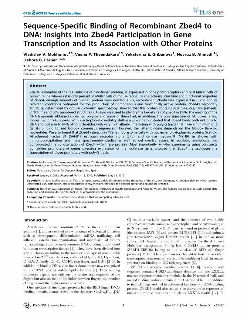
minimum around 220–222 nm (Fig. 2), which are indicative of a–
helix conformation [21]. For quantitative estimation of the
fractional content of secondary structures in Zbed4 [a-helices, b-
strands, turns and random coil (unordered)] this spectrum was
analyzed using the three programs in CDPro software (http://
lamar.colostate.edu/sreeram/CDPro/main.html). CDPro is a
series of programs for protein analysis that contains three popular
CD calculation programs, CONTINLL, SELCON3, and
CDSSTR [22]. These programs are based on a locally linearized
ridge regression method (CONTINLL), and the self-consistent
method using the singular-value decomposition algorithm
(CDSSTR and SELCON3). The three programs use the reference
set of proteins SMP56, which includes the CD spectra of 13
membrane and 43 soluble proteins. The curve estimated for
Zbed4 by CONTINLL using SMP56 as the reference set is also
shown in Figure 2. Both experimental and calculated curves
correlate very well and are essentially superimposable. Practically
the same curves were obtained using CDSSTR (not shown), and
SELCON3 (not shown).
Table S2 represents the estimated fractional content of
secondary structure for Zbed4 at pH 7.3. It shows that the three
programs, CONTINLL, SELCON3 and CDSSTR gave similar
results.
The normalized root-mean-square deviation (nrmsd) value was
used to judge the goodness of the fit between the experimental
spectrum and the curve calculated from the crystallographic data
in SMP56.
The nrmsd was calculated as: nmrsd~
ffiffiffiffiffiffiffiffiffiffiffiffiffiffiffiffiffiffiffiffiffiffiffiffiffiffiffiffiffiffiffiffiffiffiffiffiffiffiffiffiffiffiffiPN
(DEexptl{ DEcalc)2
PN
(DEexptl)2
vuuuut
where DEexptl and DEcalc are the experimental and calculated
dichroic molar absorption per amino acid residue, respectively, and
N is the number of data points.
The nmrst values for CONTINLL and CDSSTR are 0.018 and
0.080, respectively, which indicate excellent agreement between
estimated and calculated curves. The nmrst value for SELCON3
is relatively high, 0.349. However, the results obtained using
SELCON3 are very consistent with those of CONTINLL and
CDSSTR. Therefore, we averaged the results of the three
programs: Zbed4 is estimated to have 32.8% a-helices, 17.7%
b-strands, 20% turns and 29% unordered structures.
For predicting the theoretical secondary structure based on the
amino acid sequence of Zbed4 we selected the Garnier-Robson
algorithm [23], based on the fact that this method provided very
consistent results for all 6 human ZBED protein sequences in
terms of % a helices, b-strands, turns and unordered structures.
This algorithm is available from the FASTA Sequence Compar-
ison web resource at the University of Virginia (http://fasta.bioch.
virginia.edu/fasta_www2/fasta_wwwcgi?rm = misc1) and the
DNASTAR Lasergene software (DNASTAR, Madison, WI).
Similar results to the averaged values of the three programs in
CDPro were obtained by using the Garnier-Robson algorithm for
the prediction of theoretical secondary structure of Zbed4:38% a-
helices, 18% b-strands, 24% turns and 20% unordered structures.
The number of a-helices and b-strands in Zbed4 can be
calculated using the CDPro results. CONTINLL, CDSSTR, and
SELCON3 divide the a-helix and b-strand conformations into
‘‘regular’’ and ‘‘distorted’’ fractions. The distorted fractions of the
secondary structure elements reflect the fact that a certain number
of the end-residues of a-helices and b-strand fragments do not
have a regular hydrogen bonding. It has been shown that, on
average, four residues per a-helix and two residues per b-strand
are distorted [24]. Consequently, the number of a-helices and b-
strands can be estimated using the following formulas:
Na~Naa x H(d)
4(2) ; Nb~
Naa x S(d)
2(3) where Naa is the
total number of amino acid residues in the protein, and H(d) and
S(d) are the distorted fractions of the a-helix and b-strand
conformations, respectively.
We estimated the number of a-helical and b-strand segments in
the Zbed4 sequence by introducing into formulas 2 and 3 the
fractional content of distorted a-helical [H(d)] and b-strand [S(d)]
structures from Table S2. This gave us an estimate of 42 a-helices
and 45 b-strands distributed along the 1,168 amino acids of the
Zbed4 sequence. These results correlate well with the predicted
values obtained using the Garnier-Robson algorithm from the
DNASTAR and FASTA comparison softwares (Fig. 3A), which
indicate that Zbed4 has 49 a-helices and 51 b-strands along its
1,168 amino acid sequence. In Figure 3B, we are showing the
sequence of the four-BED Zinc- fingers of ZBED4. The BED
finger signatures are boxed.
Identification of Zbed4 Binding Sites in Nucleic Acidsusing Cyclic Amplification and Selection of Targets(CASTing)
Nucleic acid ligands with high affinity to Zbed4 were isolated by
CASTing. This method involves alternate cycles of ligand selection
from pools of random sequences and amplification of the ligand
that binds to the protein of interest [25]. We synthesized an 84-
base oligonucleotide containing a random set of 26 bases (making
this a 426-fold degenerate oligonucleotide) flanked by restriction
sites and PCR priming sequences (Table S3, #9, CASTrandom
oligonucleotide). After synthesis of the complementary strand
using PCR-specific primers (Table S3, primers CAST-F and
CAST-R), the double-stranded oligomer was mixed with the
bacterially expressed Zbed4 to permit the formation of protein-
DNA complexes that were then immunoprecipitated with protein
A and Zbed4 antibody. Unbound DNA was removed by washing,
and bound DNA was amplified by PCR. Several cycles of
immunoprecipitation and PCR amplification were performed.
Figure 2. Far-UV CD spectrum of Zbed4 (black curve) and curveobtained using the CONTINLL program (white curve). TheCONTINLL-calculated curve conforms well to the experimental spectraof Zbed4. SELCON and CDSSTR-calculated curves (not shown) wereessentially identical to that of CONTINLL.doi:10.1371/journal.pone.0035317.g002
Zbed4: Structural and Functional Characterization
PLoS ONE | www.plosone.org 3 May 2012 | Volume 7 | Issue 5 | e35317

The obtained DNA segments were cloned into pCR2.1-TOPO.
Figure 4A lists the sequences of 34 of these clones, all of them
containing poly-Gs. Some of these clones also contained the core
signature of GC-boxes (underlined). In addition, six other clones
were identified carrying only the GC-box sequence (Fig. 4B).
These results indicated that Zbed4 has high affinity for poly-Gs
and GC-boxes.
In order to determine the relative affinity of recombinant Zbed4
for the different nucleotides, we first designed degenerative 20-mer
oligonucleotides (Table S3, primers #12 to 22). These degener-
ative primers were used together with Zbed4 in an electrophoretic
mobility shift assay on agarose gels. When the gels were stained
with SYBR Gold for the detection of nucleic acids (Fig. 5A), they
showed that Zbed4 bound oligonucleotides that contained
guanylyl residues, such as poly-G, poly-R, poly-S and poly-K
Figure 3. Prediction of secondary structures for Zbed4 using the Garnier-Robson algorithm. A. Position of a-helices, b-sheets, turns andrandom amino acid sequences in the Zbed4 1,168 amino acid sequence. Predictions performed by two different programs, DNASTAR (Lasergene) andthe FASTA Sequence Comparison web resource at the University of Virginia, gave the same results. Boxes I, II, III and IV indicate the position of BEDzinc-finger motifs in Zbed4. B. The amino acid sequence of the four boxes in A show the specific signature of the BED-type zinc-fingers present inZbed4 containing the characteristic cystines and histidines (red).doi:10.1371/journal.pone.0035317.g003
Figure 4. Nucleotide sequence of DNA fragments selected by the CASTing method. dsDNA (CASTrandom oligonucleotide, Table S3, aftersynthesis of the complementary strand using PCR-specific primers CAST-F and CAST-R) was incubated with Zbed4 to form protein-DNA complexesthat were immunoprecipitated using Zbed4 antibody and protein A-Sepharose beads. Bound DNA was extracted, PCR-amplified and used for thenext round of CASTing. Three rounds of CASTing were performed. The amplified DNA fragments that interacted with Zbed4 were cloned andsequenced. A. Clones carrying poly-G tracts (bolded) and GC-box core sequences (underlined). B. Clones only carrying GC-box core sequences.doi:10.1371/journal.pone.0035317.g004
Zbed4: Structural and Functional Characterization
PLoS ONE | www.plosone.org 4 May 2012 | Volume 7 | Issue 5 | e35317

(Table S3, primers #14, 19, 20 and 21). No signal was observed
with poly-A, poly-C and poly-T, or with poly-Y, poly-W, poly-M
and poly-N (Table S3, primers #12, 13, 15, 16, 17, 18 and 22).
The same gel stained with Coomassie R-250 for the detection of
protein (Fig. 5B) showed that Zbed4 was present in all lanes except
in those containing a 20-mer primer, used as control, and the
DNA ladder.
Being that some zinc-finger proteins can bind RNA molecules
in a specific manner [7], we tested the binding ability of Zbed4 for
RNA oligonucleotides. We synthesized 20-base-long ssRNA
oligonucleotides containing 10 ribo-Gs or 10 ribo-Cs flanked by
5 Us at the 59 and 39 ends (Table S3, primers #42 and 43). Zbed4
efficiently interacted with the poly-ribo-G oligonucleotide, but no
signal was detected with the poly-ribo-C probe (Fig. 5A). These
data suggest that Zbed4 binds effectively both DNA and RNA and
this binding is sequence-specific.
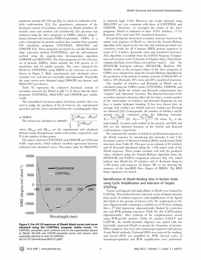
Next, we determined the minimal poly-G tract that would bind
to Zbed4. Oligonucleotides with a different number of poly-G
tracts were used (Table S3, primers #25 to 41). Zbed4-DNA
interaction was observed with probes that contained at least 5
consecutive Gs (Fig. 6A); we also used EMSA to determine the
affinity of Zbed4 for two GC-box consensus sequences,
GGGGCGGGGC or TGGGCGGAAT ([26] and Table S3,
primers #23 and 24) and found that Zbed4 formed a complex
only with GC-box1 (Fig. 6A). Figure 6B, stained for protein, shows
Zbed4 in every lane except for those containing the DNA ladder
and a 20-mer primer used as control.
Identification of Proteins from Y79 Retinoblastoma Cellsthat Interact with Zbed4
We used immunoprecipitation assays and mass spectrometry to
search for Zbed4-interacting proteins. Cell extracts were incubated
with Zbed4 antibody or pre-immune serum and the precipitated
proteins were then subjected to SDS-PAGE and stained with
SYPRO Ruby. Five protein bands that were not detected in the
separated proteins resulting from the reaction with pre-immune
serum were excised and destained. After in situ digestion with
trypsin of the proteins in each band, the resulting peptides were
subjected to nLC-MS/MS mass spectrometry. Analyses of the
data obtained using the Sequest database search algorithm from
the proteome Discoverer software, identified several proteins from
each band, some of them corresponding to peptides with very high
hits. In addition to ZBED4, nuclear SAFB1 (131 hits) and
cytoplasmic MYH9 (112 hits) were among those proteins with
highest scores.
In vitro and In vivo Confirmation of the Zbed4 and SAFB1Interaction
To analyze the in vitro interaction between Zbed4 and SAFB1
we used the membrane overlay assay. Figure 7A shows the
presence in the cell extract of endogenous Zbed4 (lane1) at
,135 kDa, corroborated by the position of purified Zbed4 run
on the same gel (lane 2). Lane 3 shows that after overlaying with
Zbed4 and washing extensively the membrane, the Zbed4
antibody recognized several bands indicating that Zbed4 had
bound other proteins of the nuclear extract. One of these
proteins with an apparent MW of 130 kDa was confirmed to be
SAFB1 by probing a Zbed4-overlayed lane with a SAFB1
antibody (lane 4). Thus, this in vitro assay provided evidence for
Zbed4 and SAFB1 interaction and confirmed the mass
spectrometry results.
To evaluate the in vivo interaction of native Zbed4 and SAFB1
proteins, we first used immuno-cytochemistry and confocal
microscopy imaging to investigate whether they co-localized in
Y79 retinoblastoma cells. These cells, which contain a thin
cytoplasm that surrounds a very large nucleus, were immuno-
stained with anti-Zbed4 2 Alexa 488 and anti-SAFB1 2 Alexa
568 fluorescent antibodies. Figure 7B shows that the two proteins
Figure 5. Relative affinity of recombinant Zbed4 for the different oligonucleotides. Zbed4 (20 mg) was incubated with 0.5 nM different 20-mer ssDNA (left panel) and ssRNA oligonucleotides (right panel) for 45 min and the whole reaction mixtures (20 ml) with the protein-DNA complexeswere subjected to EMSA on 1% agarose gels run at room temperature in HEPES buffer, pH 8.2, at 20 mA. A. Agarose gel stained using SYBR Gold fordetection of nucleic acid retardation. B. The same gel stained with Coomassie R-250 for detection of Zbed4 protein. Zbed4 (20 mg) and a singleprimer (0.5 nM) were applied separately to the gel as controls. 1 kb DNA ladder was used as a standard for nucleic acid size. As seen, Zbed4 bindsonly to DNA and RNA 20-mers that contain poly-Gs.doi:10.1371/journal.pone.0035317.g005
Zbed4: Structural and Functional Characterization
PLoS ONE | www.plosone.org 5 May 2012 | Volume 7 | Issue 5 | e35317
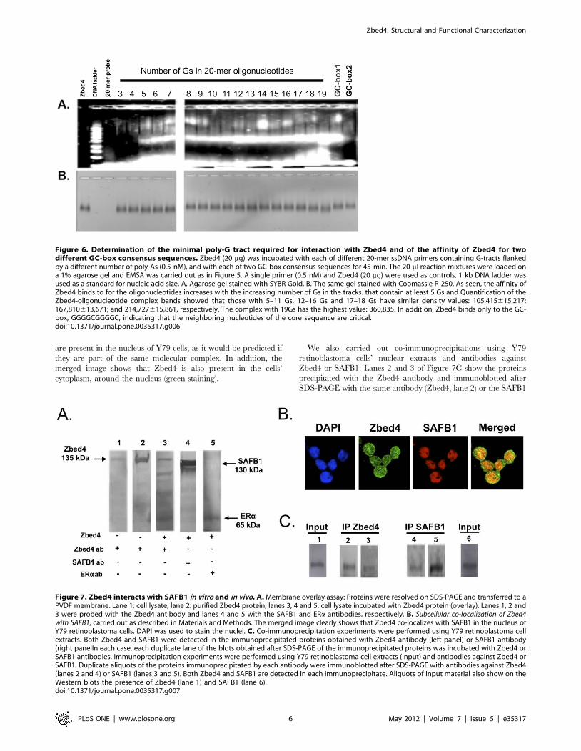
are present in the nucleus of Y79 cells, as it would be predicted if
they are part of the same molecular complex. In addition, the
merged image shows that Zbed4 is also present in the cells’
cytoplasm, around the nucleus (green staining).
We also carried out co-immunoprecipitations using Y79
retinoblastoma cells’ nuclear extracts and antibodies against
Zbed4 or SAFB1. Lanes 2 and 3 of Figure 7C show the proteins
precipitated with the Zbed4 antibody and immunoblotted after
SDS-PAGE with the same antibody (Zbed4, lane 2) or the SAFB1
Figure 6. Determination of the minimal poly-G tract required for interaction with Zbed4 and of the affinity of Zbed4 for twodifferent GC-box consensus sequences. Zbed4 (20 mg) was incubated with each of different 20-mer ssDNA primers containing G-tracts flankedby a different number of poly-As (0.5 nM), and with each of two GC-box consensus sequences for 45 min. The 20 ml reaction mixtures were loaded ona 1% agarose gel and EMSA was carried out as in Figure 5. A single primer (0.5 nM) and Zbed4 (20 mg) were used as controls. 1 kb DNA ladder wasused as a standard for nucleic acid size. A. Agarose gel stained with SYBR Gold. B. The same gel stained with Coomassie R-250. As seen, the affinity ofZbed4 binds to for the oligonucleotides increases with the increasing number of Gs in the tracks. that contain at least 5 Gs and Quantification of theZbed4-oligonucleotide complex bands showed that those with 5–11 Gs, 12–16 Gs and 17–18 Gs have similar density values: 105,415615,217;167,810613,671; and 214,727615,861, respectively. The complex with 19Gs has the highest value: 360,835. In addition, Zbed4 binds only to the GC-box, GGGGCGGGGC, indicating that the neighboring nucleotides of the core sequence are critical.doi:10.1371/journal.pone.0035317.g006
Figure 7. Zbed4 interacts with SAFB1 in vitro and in vivo. A. Membrane overlay assay: Proteins were resolved on SDS-PAGE and transferred to aPVDF membrane. Lane 1: cell lysate; lane 2: purified Zbed4 protein; lanes 3, 4 and 5: cell lysate incubated with Zbed4 protein (overlay). Lanes 1, 2 and3 were probed with the Zbed4 antibody and lanes 4 and 5 with the SAFB1 and ERa antibodies, respectively. B. Subcellular co-localization of Zbed4with SAFB1, carried out as described in Materials and Methods. The merged image clearly shows that Zbed4 co-localizes with SAFB1 in the nucleus ofY79 retinoblastoma cells. DAPI was used to stain the nuclei. C. Co-immunoprecipitation experiments were performed using Y79 retinoblastoma cellextracts. Both Zbed4 and SAFB1 were detected in the immunoprecipitated proteins obtained with Zbed4 antibody (left panel) or SAFB1 antibody(right panelIn each case, each duplicate lane of the blots obtained after SDS-PAGE of the immunoprecipitated proteins was incubated with Zbed4 orSAFB1 antibodies. Immunoprecipitation experiments were performed using Y79 retinoblastoma cell extracts (Input) and antibodies against Zbed4 orSAFB1. Duplicate aliquots of the proteins immunoprecipitated by each antibody were immunoblotted after SDS-PAGE with antibodies against Zbed4(lanes 2 and 4) or SAFB1 (lanes 3 and 5). Both Zbed4 and SAFB1 are detected in each immunoprecipitate. Aliquots of Input material also show on theWestern blots the presence of Zbed4 (lane 1) and SAFB1 (lane 6).doi:10.1371/journal.pone.0035317.g007
Zbed4: Structural and Functional Characterization
PLoS ONE | www.plosone.org 6 May 2012 | Volume 7 | Issue 5 | e35317

antibody (SAFB1, lane 3). Both Zbed4 and SAFB1 are found in
the Zbed4 immunoprecipitate. A reverse experiment using the
same nuclear extract and the SAFB1 antibody also showed the
presence of both Zbed4 and SAFB1 in the SAFB1 immunopre-
cipitate, corroborating their in vivo interaction in Y79 retinoblas-
toma cells.
Interaction and Co-localization of Zbed4 with ERa andMYH9
SAFB1 is a potent ERa co-repressor. Since Zbed4 interacts with
SAFB1, it could be part of the SAFB12ERa complex that results
in ERa transcriptional activation of several genes. However,
Zbed4’s nuclear receptor-interacting modules (LXXLL) that are
characteristic of co-activators/co-repressors of nuclear hormone
receptors, could also bind directly to ERa. We investigated this
possibility carrying out similar experiments to those described
above for Zbed4 and SAFB1. The membrane overlay assay
showed that after overlaying Zbed4 on the lane containing the
separated proteins from Y79 retinoblastoma cells and probing the
membrane with an ERa antibody, ERa was clearly detected
(Fig. 7A, lane 5). These results demonstrated that, indeed, Zbed4
and the ,65 kDa ERa were in the same overlay membrane.
Furthermore, immunocytochemistry showed that Zbed4 and ERaco-localize in both the nuclei and cytoplasm of Y79 retinoblastoma
cells (Fig. 8A, Merged image). Similar experiments also demon-
strated that Zbed4 and MYH9 co-localize to the cytoplasm of
these cells and both proteins co-immunoprecipitate after incuba-
tion with the Zbed4 or MYH9 antibodies (Figs. 8B and 8C).
Zbed4 Binding Sites in the 59-flanking Region of GenesTo verify whether Zbed4 expressed in mammalian cells was
able to interact with G-rich DNA sequences in a manner
comparable to its recombinant prokaryotic counterpart, and to
test Zbed4’s ability to activate/repress transcription in vivo, we used
stable transfected cells expressing Zbed4 and reporter plasmids
containing the promoter region of genes that have poly-G tracts in
a luciferase assay. HEK293 cells stably transfected with pZbed4eu
(Table S1) and expressing full length Zbed4, and non-transfected
HEK293 cells were used for this experiment (Fig. 9A). Consid-
ering that Zbed4 binds to poly2G tracts, we decided to test
Zbed4’s effect on sequences of four genes that contained at least 5
consecutive Gs in their 5’-flanking region: mouse vimentin (two
5G-tracts, at 2979 to 2975 and 2925 to 2921), bovine
rhodopsin (one 8G-tract, at 220 to 213), and human blue (one
6G-tract, at 28 to 23) and green (one 5G-tract, at 2123 to 2129)
opsins. The appropriate promoter region for each gene was
subcloned into the pGL2 Basic vector (Table S1). The rhodopsin
reporter plasmid [27] was kindly provided by Dr. Donald Zack
(Departments of Molecular Biology and Genetics, and Neurosci-
ence, Johns Hopkins University School of Medicine, Baltimore,
Maryland). As controls, we used the empty pGL2 Basic vector and
a region of the non-specific a-PDE promoter (which contains no
G-tracts), subcloned into the same vector.
Zbed4 expressed in the stable transfected HEK293 cells
increased the luciferase activity produced by pVim/luc, pRho/
luc, pBlue/luc and pGreen/luc above the expression level observed
with non-transfected HEK293 cells, used as control, but Zbed4
did not have an effect on the a-PDE/luc construct (Fig. 9B). The
highest increase was seen with the vimentin 5’-flanking region,
followed by the 59-flanking regions of rhodopsin, blue opsin and
green opsin. The differences in promoter-driven luciferase activity
between Zbed4 stable-transfected and non-transfected HEK293
cells were reproducible in three different experiments carried out
in triplicate, and are statistically significant (p,0.014 for vimentin,
Figure 8. Co-localization of Zbed4 with ERa and MYH9 in Y79 retinoblastoma cells, carried out as described in Materials andMethods. The immunostaining results indicate that both ERa and MYH9 co-localize with Zbed4 in these cells. The major difference is that in A co-localization is seen in the nuclei and cytoplasm for Zbed4 and ERa whereas in B it is only observed in the cytoplasm for Zbed4 and MYH9. C.Immunoprecipitation experiments were carried out as in Figure 7C with Y79 retinoblastoma cells (Input, lanes 1 and 6) but using antibodies againstZbed4 (lanes 1, 2 and 4) or MYH9 (lanes 3, 5 and 6) instead; both Zbed4 and MYH9 are detected in each immunoprecipitate.doi:10.1371/journal.pone.0035317.g008
Zbed4: Structural and Functional Characterization
PLoS ONE | www.plosone.org 7 May 2012 | Volume 7 | Issue 5 | e35317

p,0.013 for rhodopsin, p,0.013 for blue opsin and p,0.005 for
green opsin, Student’s t-test). These results suggest that Zbed4
binds in vivo to and positively regulates the transcriptional activity
of promoters containing poly-G tracts.
Discussion
In this paper we describe the purification, refolding and
expression of the mouse protein, Zbed4, and analyze some of its
structural and functional properties. As we show, Zbed4 is able to
bind DNA and RNA sequences and to trans-activate transcription
of several genes.
To obtain the recombinant protein in amounts sufficient for
further investigations, we first examined the effects of temperature
and IPTG concentration on the induction of Zbed4 synthesis in
E. coli cells. No target protein was detectable in cells that had not
been exposed to IPTG, and we found that the optimal conditions,
those that gave us the highest yield of protein, were a temperature
of 37uC and an IPTG concentration of 0.5 mM. The yield
decreased dramatically when lower temperatures were used,
independent of the IPTG concentration.
The solubility of the E. coli-expressed protein was also
investigated. After centrifugation of the whole cell lysate, Zbed4
was found only in the pellet (Fig. 1), indicating that the
recombinant protein was being expressed in the bacterial cells as
inclusion bodies. We could not extract Zbed4 from the inclusion
bodies using 8 M urea, but another chaotropic agent, 6 M
guanidine HCl, allowed us to solubilize the denatured protein,
which was then purified using cobalt affinity chromatography.
Different approaches have been used for the refolding of
denatured proteins (direct dilution [28,29], chromatographic
methods [30,31], etc.). For us, several rounds of dialysis against
refolding buffer were optimal to refold the denatured Zbed4 to its
native state and activity. Other denatured zinc finger proteins had
been successfully refolded in this way [32]. We also observed that
the efficiency of refolding largely depended on the initial protein
concentration: for Zbed4, 0.6 mg/ml worked very well. At higher
concentrations, there was aggregation during dialysis. In addition,
refolding strongly depended on the presence of Zn2+ ions, which
are necessary for the correct conformation of the zinc fingers and
for protein stabilization [33–35], and a reducing agent to reduce
non-native inter- and intramolecular disulfide bonds and to keep
the cysteines in their reduced state [36]. In our experiments we
used 10 mM ZnSO4, and 5 mMb-mercaptoethanol.
The far-UV CD spectrum of a protein generally reflects its
secondary structure content. In the case of Zbed4, it indicated that
the protein is primarily organized in an a-helix structure. Three
different programs, CONTINLL, SELCON3 and CDSSTR, used
to quantify the content of secondary structures in Zbed4 at
pH 7.3, gave results in the same range, and thus, the results were
averaged. The a-helix value obtained (32.8%) is very similar to the
a-helix content calculated for most zinc-finger proteins including
ZBED proteins [37]. Furthermore, the Garnier-Robson algorithm
for the prediction of theoretical secondary structure based on the
amino acid sequence of Zbed4 also provided similar results to
those obtained using the data of the CD spectrum: 38% a-helices,
18% b-strands, 24% turns and 20% unordered. The Garnier-
Robson program also allowed us to find out where the a-helices
and b-strands are in the Zbed4 sequence. Figure 3A shows the
Figure 9. Zbed4 mediates transcriptional activation of several genes expressed in retina. A. Immunocytochemical detection ofendogenous Zbed4 in HEK293 cells (upper image) and in HEK293 cells stably transfected with a Zbed4 expression construct (lower image). Theendogenous Zbed4 in HEK293 cells is localized mainly in the nuclei (cyan) and barely detected in the cytoplasm whereas in the HEK293 stable-transfected cells Zbed4 is seen in both, the nuclei (cyan) and cytoplasm (green). DAPI was used to stain nuclei. B. Trans-activation of promoters fromgenes expressed in retina by Zbed4. Stably transfected HEK 293 cells expressing Zbed4 and non-transfected HEK 293 cells were used in transienttransfections with luciferase reporter constructs carrying different retinal promoters. Luciferase activity was measured in the cell lysates andnormalized for each transfection system to the corresponding b-galactosidase activity for each sample. The results are expressed as fold induction byZbed4 of the mean luciferase activity of the uninduced promoter compared to that of the Vim/luc, Rho/luc, Blue/luc, Green/luc, and a-PDE/lucreporter constructs, respectively, 6 S.D. p values for each pair of Zbed4 stimulated and non-stimulated promoters are noted above the bars.doi:10.1371/journal.pone.0035317.g009
Zbed4: Structural and Functional Characterization
PLoS ONE | www.plosone.org 8 May 2012 | Volume 7 | Issue 5 | e35317
distribution of these structures as well as the turns and unordered
regions of the protein. It also shows the location of the 4 zinc-BED
fingers. In addition, in Figure 3B the BED zinc-finger specific
signature is shown. Moreover, we have generated 3D structural
models for these BED zinc-fingers (Fig. 10A). The 3D structure of
macromolecules offers insights into their molecular function at the
atomic level, and often it provides direct evidence of molecular
interactions between individual macromolecules, or between
macromolecules and small molecules. By standard sequence
similarity searches close to 60% of protein sequences can be
mapped to a 3D structure [38]. We took the same sequence
similarity approach when creating the 3D structure of all four
BED zinc-domains of Zbed4 based on the solution structure of the
zinc-finger BED domain-containing protein 1 (Zbed1, PDB ID:
2ct5), using the display application for sequence and structure
information, Chimera [39]. Our models show structural details of
the arrangement of a-helices, b-sheets and turns of each finger as
well as the potential location in them of the Zn2+ ion. In addition,
the relative distribution of these structures in all Zbed4 and Zbed1
fingers can be observed in the superimposed image of the models
(Fig. 10B), which shows that all of the BED zinc-fingers have a
very close 3D structure. To determine if Zbed4 is a sequence-
specific binding protein, we used CASTing. For these experiments,
a pool of random oligonucleotides was incubated with recombi-
nant Zbed4. Following several rounds of selection and amplifica-
tion of the DNA segments binding to Zbed4, these were cloned
and sequenced. The majority of the clones contained poly-G
tracts, indicating that these might be Zbed4’s binding sequences.
To verify that these were the specific nucleotides that interact with
Zbed4, we performed electrophoretic mobility shift assays. These
assays are usually carried out on native polyacrylamide gels
[32,40,41]. In our experiments, the complexed Zbed4-DNA was
stacked at the top of the gel, probably because of its high molecular
weight, charge or aggregation state. We were able to overcome
this difficulty using 1% agarose gel buffered by 25 mM HEPES,
pH 8.2. The high intensity of the retarded DNA band stained by
SYBR Gold confirmed a strong DNA-Zbed4 interaction (Fig. 5A).
Together, our CASTing and EMSA results demonstrated that
Zbed4 has strong affinity for poly-G sequences in double- and
single-stranded DNA. Poly-G tracts must contain 5 or more Gs in
order for Zbed4 to bind to them.
We also investigated whether Zbed4 interacts with RNA with
the use of poly-rC and poly-rG oligonucleotides in the binding
reactions. We found that only poly-rG interacted with Zbed4,
confirming once more, the high affinity of Zbed4 for poly-G, being
it either deoxy- or riboxy-nucleic acid. Since studies in the
literature have shown that zinc-finger proteins can bind not only to
single-stranded RNA but also to double-stranded RNA [42], we
can hypothesize that Zbed4 will interact with double-stranded
RNA too, and even assume that it will do the same with DNA-
RNA hybrids.
To determine whether Zbed4 also interacts with G-rich
elements such as the GC-box, we analyzed its binding to two
variants of the GC box consensus sequence, GGGGCGGGGC
and TGGGCGGAAT, both having the same core (bolded). The
GC box is one of the most common regulatory DNA elements in
the promoter region of eukaryotic genes [43]. Zbed4 only
interacted with GGGGCGGGGC, suggesting that the neighbor-
ing nucleotides of the core are crucial.
One of the goals of the present study was to search for binding
partners of Zbed4 and here we report the identification of SAFB1,
ERa and MYH9 as Zbed4-interacting proteins expressed in Y79
retinoblastoma cells.
Zbed4-SAFB1 close association was first identified by immu-
noprecipitation with Zbed4 antibody from Y79 retinoblastoma cell
extracts and mass spectrometry of the separated proteins and was
confirmed in vitro with a Zbed4 membrane overlay assay and in vivo
by the presence of both proteins in the Zbed4 and SAFB1 co-
immunoprecipitates from the same cell extracts. Moreover, Zbed4
and SAFB1 co-localized to the nuclei of Y79 retinoblastoma cells,
providing further evidence of their interaction in vivo. SAFB1 is a
large protein with multiple functions that include transcriptional
regulation, RNA splicing and RNA metabolism [44]. Recent
studies suggest its role in chromatin reorganization and demon-
strate that SAFB1 is involved in various cellular processes like cell
growth, stress response and apoptosis [45]. SAFB1 also functions
as an ERa co-repressor [44,45]. Based on our results on Zbed4
and SAFB1 interaction and the fact that co-repressors regulate
receptor activity by forming multi-protein complexes, we speculate
that both proteins may be components of one of these complexes
that could lead to chromatin remodeling, competition with co-
activators, and interference with DNA binding.
Our results also demonstrate the nuclear and cytoplasmic co-
localization of Zbed4 and ERa in Y79 retinoblastoma cells and
they confirm the close association of these proteins. Estrogen
receptors are regarded to be cytoplasmic receptors in their
unliganded state but a fraction of the receptors reside in the
nucleus [46]. ERa works through different pathways to stimulate
transcription of estrogen-responsive genes [47]. It is interesting
that Sp1 sites can also mediate estrogen induction via ERa in the
context of some promoters, but ERa does not bind to these sites
and regulation occurs through protein-protein interactions be-
tween the transcription factors and ERa. We hypothesize that
Zbed4 may function in a similar way to Sp1 on the promoter of
retinal genes, since both transcription factors have zinc-fingers and
bind to G-rich sequences. Moreover, Zbed4 may also interact
indirectly with ERa, as a co-regulator with SAFB1.
In addition to SAFB1 and ERa, we show here that Zbed4
interacts with another protein, Myosin 9 (MYH9), also known as
Myosin IIA. This protein is arranged in bundles of filaments in
the cytoplasm of non-muscle cells, where it is thought to be
responsible for vesicle formation and movement. MYH9 has
been localized to the trans-Golgi network and is probably a key
player in protein and vesicle trafficking to and from the Golgi
apparatus [48]. We speculate that in Y79 retinoblastoma cells as
well as in retina Zbed4 may be transported by the MYH9
trafficking system.
Most importantly, we have identified one of many possible
functions of Zbed4: it acts as a transcription factor. Our results
show that Zbed4, expressed in stably-transfected HEK293 cells,
was able to increase luciferase gene expression driven by different
retinal promoters containing poly-G tracts above the expression
level observed with non-transfected HEK293 cells, used as
control. The vimentin reporter plasmid was the most stimulated
by Zbed4, probably because it has two 5-G tracts compared to
the other three promoters that only have one poly-G tract.
Furthermore, as we showed in our in vitro experiments (Fig. 6),
the number of Gs in the tract is important for Zbed4 binding.
This also may occur in vivo, and could explain why the rhodopsin
promoter, with a single 8-G tract, is activated by Zbed4 more
than the blue opsin promoter that has 6 consecutive Gs, which in
turn binds Zbed4 better than the green opsin promoter with only
5Gs. As expected, the a-PDE promoter carrying no poly-G tracts
was not activated by Zbed4. The small, but statistically
significant increase in luciferase gene expression observed for
all the other promoters, might be due to the absence in HEK293
cells of additional proteins that are important for the proper
Zbed4: Structural and Functional Characterization
PLoS ONE | www.plosone.org 9 May 2012 | Volume 7 | Issue 5 | e35317

arrangement of the transcriptional complex. Supporting this
hypothesis, we had previously demonstrated that the photore-
ceptor b-PDE promoter is specifically trans-activated by Sp4, but
that this activation increases many-fold when Sp4 interacts with
other transcription factors such as Nrl, Crx and TFIIB [49,50].A similar scenario has been reported by Wu and colleagues [51]
who demonstrated that the SmNR1 protein alone is able to
activate the transcription of a reporter gene in COS-7 cells, but
when another protein already known to interact with it,
SmRXR1, is present, SmNR1’s activation increases approxi-
mately 2-fold. As for Zbed4 in the current study, if a binding
partner were present in the HEK293 cells, the increase in the
transcriptional activation of the promoters analyzed would
probably be much more substantial. In order to better
characterize Zbed4’s action as a transcription factor, future
experiments designed to determine whether SAFB1 and ERa (or
other nuclear proteins interacting with Zbed4) are present and
active in HEK293 cells will be necessary.
In conclusion, we successfully expressed a His-tagged Zbed4
fusion protein in E. coli, and developed an optimized purification
and refolding procedure for obtaining milligram amounts of
homogeneous recombinant, active protein. Initial structural
analysis revealed that the primary organization of Zbed4 has an
a-helix content that is typical for most zinc-finger proteins [37].
We showed that Zbed4 has DNA- and RNA-binding activity and
that it specifically interacts with G-rich sequences. Our experi-
ments also confirmed the nuclear co-localization and possible
association of ZBED4 with SAFB1 and ERa and the cytoplasmic
interaction of Zbed4 with MYH9 in Y79 retinoblastoma cells. A
major finding of this work is the demonstration of the ability of
Zbed4 to activate gene transcription. Future studies based on the
results presented here will establish the specific role and potential
importance of Zbed4 in retinal function.
Materials and Methods
Ethics StatementAll experiments involving mice were carried out using protocols
approved by the UCLA Animal Research Committee, and in
accordance with the ARVO Statement for Use of Animals in
Ophthalmic and Vision Research. The cell lines used were
obtained from commercial vendors: BL21(DE3)Star cells from
Invitrogen (Grand Island, NY); Y79 retinoblastoma and HEK293
cells from ATCC;
Bacterial Strains, Plasmids and OligonucleotidesBacterial strains and eukaryotic cell lines as well as all plasmids
used in this study are listed in Table S1. All oligonucleotides were
designed using the DNASTAR 5.0 software package and Amplify
1.2, in an effort to minimize non-specific DNA amplification
(Table S3).
Figure 10. A. Structure modeling for BED zinc-finger (I, II, III and IV) domains of Zbed4, based on the structure of ZBED1 (PDB ID:2ct5). The program iMol, version 0.40 and the UCSF Chimera package [39] were used to generate these models. B. Superimposed model of allpredicted BED zinc-finger structures of Zbed4 (I, II, III and IV) and ZBED1 (PDB ID: 2ct5). Each finger is shown in a different color.doi:10.1371/journal.pone.0035317.g010
Zbed4: Structural and Functional Characterization
PLoS ONE | www.plosone.org 10 May 2012 | Volume 7 | Issue 5 | e35317

Construction of the Zbed4 Plasmid for Expression inE. coli Cells
A 3555 bp fragment containing the coding region of Zbed4 and 8
extra histidines at its 3’ end was amplified by RT-PCR from mouse
retinal RNA using Pfu-Ultra DNA-polymerase (Agilent Technolo-
gies, Santa Clara, CA) and primers ZB4NheI-F and ZB4His8-
BamHI-R (Table S3). The PCR product was then purified with the
GeneElute Gel Extraction kit (Sigma-Aldrich Corporation, St Louis,
MO), digested with NheI and BamHI, and cloned into the NheI–
BamHI-restricted E. coli expression vector pET11a+. This expres-
sion plasmid was designated as pZbed4complete and its correct
sequence was confirmed on a Biosystems 3730 Capillary DNA
Analyzer (UCLA Genotyping and Sequencing Core).
Zbed4 Expression, Purification and RefoldingE. coli BL21(DE3)Star cells were transformed with pZbed4com-
plete and pLysSRARE2, plated on solid LB-buffered agar [1%
(w/v) tryptone (Difco, Franklin Lakes, NJ), 0.5% (w/v) yeast
extract (Difco), 1% (w/v) NaCl, 50 M Na2HPO4/NaH2PO4,
2.0% (w/v) agar (Difco)], pH 7.5, containing 100 mg/ml ampicil-
lin, 30 mg/ml chloramphenicol and incubated at 37uC. An
individual bacterial colony was inoculated in 100 ml of medium
with 100 mg/ml ampicillin, 30 mg/ml chloramphenicol. This pre-
culture was grown overnight at 37uC with continuous shaking at
200 rpm and then used to inoculate 1 L of buffered LB medium
(same ingredients as above, without the agar). The culture was
grown for about 2.5 h at 37uC to an OD600 0.6, and 0.5 mM
IPTG (final concentration) was added to induce the expression of
protein. Growth was continued for 6 h with shaking at 200 rpm.
Cells were then harvested: about 2.5 g of wet packed cells were
obtained per liter of culture. Cells were suspended in 20 ml of
20 mM HEPES buffer, pH 7.5, containing 0.5 M NaCl, 1%
Triton X100, 5% glycerol, 16 Complete Protease Inhibitor
Cocktail (Roche, Indianapolis, IN) and a mixture of 10 mg
DNaseI, 10 mg RNaseA and 50 mg lysozyme, and were disinte-
grated by passing through a French pressure chamber twice at
82.7 MPa. The suspension was centrifuged at 4uC for 1 h at
150,000 g. The inclusion bodies-containing pellets were resus-
pended in urea washing buffer (20 mM HEPES, pH 7.5, 0.5 M
NaCl, 8 M urea) and centrifuged using the same conditions.
Pellets were dissolved in 25 ml of Guanidine HCl buffer 1 (6 M
Gu-HCl, 20 mM HEPES, pH 7.5, 0.5 M NaCl, 5 mM imidazole,
4 mM b-mercaptoethanol) by overnight incubation at 4uC while
rotating. The mixture was centrifuged for 1 h at 150,000 g at 4uC,
and the supernatant was applied to a column with Co2+-activated
BD TALON metal affinity resin (Clontech, Mountain View, CA)
equilibrated in Gu-HCl buffer 1. The column was washed with 20
column bed volumes of Gu-HCl buffer 2 (6 M Gu-HCl, 20 mM
HEPES, pH 7.5, 0.5 M NaCl, 20 mM imidazole, 4 mM b-
mercaptoethanol). Protein was eluted with Gu-HCl elution buffer
(6 M Gu-HCl, 20 mM HEPES, pH7.5, 0.5 M NaCl, 200 mM
imidazole, 100 mM EDTA, 4 mM b-mercaptoethanol). Eluted
protein was dialyzed against refolding buffer (20 mM HEPES,
pH 7.5, 150 mM NaCl, 5 mM b-mercaptoethanol, 5% glycerol,
0.5 mM L-ascorbic acid, 0.5 mM propyl gallate, 10 mM ZnSO4)
and concentrated using a 15-ml Amicon Ultra concentrator
(Millipore, Billerica, MA).
SDS-PAGE and Western Blots50 mg protein from each fraction of the purification/refolding
steps were electrophoresed on 4–12% Bis-Tris gels (Invitrogen)
and the separated proteins were electro-transferred to PVDF
membranes, which were subsequently blocked with 3% (w/v)
casein. Blots were incubated with rabbit Penta-His antibodies
conjugated with horseradish peroxidase (1:1000 dilution, Qiagen,
Valencia, CA). Western blots were visualized with the ECL
Substrate of the Fast Western blot kit (Thermo Scientific Pierce,
Rockford, IL).
Circular Dichroism (CD) Spectroscopy of Purified Zbed4Before CD spectra analyses, Zn2+-bound Zbed4 was dialyzed
against 20 mM HEPES, pH 7.5, 150 mM NaCl, and 5%
glycerol. Far-UV CD measurements were performed at 22uCwith a JASCO J-810 spectropolarimeter (JASCO, Easton, MD)
and spectra were acquired from 16 scans between 260 to 195 nm
in 0.2 mm path length cells. A 0.4 mg/ml Zbed4 protein sample
was used. CD spectra were corrected for buffer contributions.
Ellipticity is reported as mean molar residue ellipticity (MRE) in
degree.cm2.dmol21. Protein concentration was determined using
the MicroBCA protein assay kit (Thermo Scientific).
Determination of Zbed4 Binding Sites with CASTingExperiments
Double-stranded degenerate fragments were amplified by PCR
from oligonucleotide CASTrandom (Table S3). The PCR reaction
mixture (50 ml) contained 16 ThermoPol buffer (New England
Biolabs, Ipswich, MA), 1 pmol of each primer (CAST-F, CAST-R,
Table S3), each nucleotide at a final concentration of 0.25 mM,
10% (v/v) dimethyl sulfoxide, 1 U Taq-polymerase (New England
Biolabs) and 1 pmol CASTrandom. Binding reactions were
performed by adding Zbed4 (20 mg) in refolding buffer to the
double stranded degenerate oligonucleotide (1.8 ng) and incubat-
ing at room temperature for 30 min. Then, Zbed4 antibody
(20 mg) and Protein A Sepharose beads (20 ml) were added and
incubation continued at room temperature for an additional 1 h.
The DNA-protein complexes formed were precipitated by
centrifugation at 1000 g, and the pellets were washed 6 times
with refolding buffer. Bound DNA was extracted with phenol/
chloroform and amplified by 35 cycles of 1 min at 94uC, 30 sec at
55uC, and 1 min at 72uC in the ThermoPol PCR system (New
England Biolabs). The series of steps of incubation, immunopre-
cipitation, and re-amplification, considered as one cycle of
CASTing, were repeated three times. The PCR product was
cloned into the pCR2.1-TOPO plasmid. Nucleotide sequences of
40 independent clones were determined on the Biosystems 3730
Capillary DNA Analyzer (UCLA Genotyping and Sequencing
Core).
Electrophoretic Mobility Shift Assay (EMSA)Protein–DNA binding reactions contained Zbed4 (20 mg) and
0.5 nM oligonucleotide in refolding buffer. Reaction mixtures
were incubated at room temperature for 45 min and then were
loaded on 1% agarose gels pre-electrophoresed for 1 h. Gels were
run at room temperature in 25 mM HEPES buffer, pH 8.2, at
20 mA, and were stained with the SYBR Gold nucleic acid stain
solution (Invitrogen) for 10 min in the dark. The fluorescence of
the gel bands was visualized under UV light. The agarose gels
were subsequently stained with Coomassie R-250 for detection of
Zbed4.
Cell CultureY79 retinoblastoma cells were cultured using RPMI medium
(Sigma) supplemented with 15% fetal bovine serum, and human
embryonic kidney (HEK293) cells were cultured in DMEM
medium supplemented with 13% FBS; both cell lines grew at 37uCin a humidified incubator with 95% air and 5% CO2.
Zbed4: Structural and Functional Characterization
PLoS ONE | www.plosone.org 11 May 2012 | Volume 7 | Issue 5 | e35317

HEK293 cells were used for the generation of a stable
transfected cell line expressing Zbed4 [14]. Briefly, gel-purified
Zbed4 cDNA amplified by RT-PCR from mouse mRNA was
subcloned into the eukaryotic expression vector pcDNA4/HisMax
(Invitrogen) containing the Zeocin resistance gene. Transfection
was performed with Superfect Transfection Reagent (Qiagen) and
Zeocin resistant transformants were selected 4 weeks after
transfection.
Zbed4 Immunostaining in Y79 Retinoblastoma andStably Transfected HEK293 Cells
Cultured cells (66105) were seeded on poly-D-lysine and
fibronectin-coated cover slips and allowed to adhere at least for
24 h at 37uC before immunostaining. Cells were then fixed with
ice-cold methanol at 220uC and washed with 16PBS. After
blocking with 2% BSA, 0.1% Triton X-100 in 16PBS for 40 min,
rabbit polyclonal Zbed4 antibody ([16], 1:300 dilution) was
applied to the slides and the cells were incubated for 2 h at room
temperature, followed by three washes in 16PBS, 0.1% Triton X-
100 and incubation with secondary anti-rabbit goat antibodies
labeled with Alexa-488 (Invitrogen, 1:300 dilution) for 1 h at room
temperature. After subsequent washes in 16PBS, 0.1% Triton X-
100, nuclei were stained with DAPI for 5 min at room
temperature. Slides were mounted and viewed on an Olympus
Fluoview FV 1000 Laser scanning microscope (Olympus America,
Center Valley, PA).
Co-localization of Zbed4 with SAFB1, ERa or MYH9Similar immunocytochemistry experiments were performed
using Y79 retinoblastoma cells to detect co-localization of Zbed4
with SAFB1, ERa, or MYH9 using rabbit ZBED4 (1:300), goat
SAFB1 [(Bethyl Laboratories, Montgomery, TX), 1:200], mouse
ERa [(Abcam, Cambridge, MA), 1:100] or rabbit MYH9 (Abcam,
1:400) primary antibodies along with goat anti-rabbit Alexa-488
for Zbed4 (1:1000 dilution) and donkey anti-goat Alexa-568
(1:1000 dilution), goat anti-mouse Alexa-568 (1:1000) or goat anti-
rabbit secondary antibodies (Invitrogen) for SAFB1, ERa or
MYH9, respectively.
Immunoprecipitation and Mass SpectrometryY79 cells were lysed using RIPA lysis buffer (150 mM NaCl,
1% IGEPAL CA-630, 0.5% sodium deoxycholate, 0.1% SDS,
50 mM Tris, pH 8.0, and 1 mM PMSF) and their proteins were
immunoprecipitated with the Zbed4 antibody following the
Immobilized Protein G kit protocol (Pierce). As a control, the
same reaction was carried out using pre-immune serum. The
precipitated proteins were separated on SDS-PAGE and stained
with SYPRO Ruby. Protein bands were excised and destained
for in gel trypsin digestion. The digested bands were subjected to
nLC/MS/MS mass spectrometry (MDS Sciex QSTAR XL
Quad - Time-of-Flight Mass Spectrometer, Applied Biosystems,
Carlsbad, CA) at the UCLA’s Pasarow mass spectrometry
laboratory and data were analyzed with the Sequest database
search algorithm implemented in the proteome Discoverer
software (Thermo Fisher, Waltham, MA).
Gel Overlay AssayNuclear extracts from Y79 retinoblastoma cells were subjected
to SDS-PAGE and the separated proteins were transferred to
PVDF membranes. After blocking with Odyssey blocking buffer
(LI-COR, Lincoln, NE) for 1 h at room temperature, the blots
were incubated for 2 h at 5uC in the dark with purified Zbed4
protein (5–10 mg/ml) in 10 mM Tris-HCl (pH 8), 150 mM
NaCl, and 0.05% Tween-20. The membranes were washed with
16 PBS and 0.1% Tween-20 and incubated separately with
rabbit anti-Zbed4 (1:1000), goat anti-SAFB1 (1:500), and rabbit
anti-ERa (1:100) antibodies for 1 h, washed with 16 PBS and
0.1% Tween-20, and then incubated with IRDye 800 CW goat
anti-rabbit (1:15,000) and IRDye 700 DX donkey anti-goat
(1:15,000) secondary antibodies (LI-COR) for 1 h. Membranes
were then washed and scanned on the LI-COR Odyssey Infrared
imager.
Co-immunoprecipitation of Zbed4 with SAFB1 or MYH9Proteins of cultured Y79 retinoblastoma cells immunoprecip-
itated as described above using rabbit Zbed4, goat SAFB1 or
rabbit MYH9 antibodies were separated on triplicate lanes of
4%–20% Pierce Precise Protein gels (Thermo-Scientific) by
electrophoresis, using Tris-Glycine-SDS buffer at 70 volts.
Proteins were then transferred to PVDF membranes, which were
blocked with 3% BSA in Tris Base Tween (TBST) buffer,
pH 7.5, for 1 h. Proteins precipitated with the Zbed4 antibody
were probed on the Western with SAFB1 or MYH9 primary
antibodies and those precipitated with the SAFB1 or MYH9
antibodies were probed with the Zbed4 primary antibody. The
remaining membrane with proteins of each immunoprecipitation
was incubated with the same antibody used for bringing down the
proteins. Then, anti-rabbit alkaline phosphatase antibody was
added to each of the samples followed by application of DuoLux
Chemiluminescent/Fluorescent Substrate for alkaline phospha-
tase (Vector Laboratories, Burlingame, CA). After washing with
buffer, the membranes were exposed to film (GE Healthcare,
Piscataway, NJ).
Transient Transfections and Reporter AssaysReporter plasmids containing 5’-flanking regions of the genes
encoding mouse vimentin and human blue and green opsins
were cloned into the pGL2 Basic vector (Promega, Madison, WI)
that contains the luciferase gene. These plasmids were called
pVIM/luc, pBLUE/luc and pGREEN/luc, respectively (Table
S1).
For transient transfection assays, 36106 stably-transfected
HEK293 cells expressing Zbed4 and HEK293 non-transfected
cells (used as control) were plated on 60 mm culture plates in
Dulbecco’s modified Eagle’s medium/Ham’s F-12 (Invitrogen)
supplemented with 10% fetal bovine serum 24 h prior transfec-
tion. Calcium phosphate-mediated transient transfections of
pVim/luc, pRho/luc, pBlue/luc, pGreen/luc, pa-PDE/luc and
pGL2 Basic vector as well as luciferase and b-galactosidase assays
were performed as described previously [52], except for the
addition of a glycerol shock that improved the transfection
efficiency of HEK293 cells. The glycerol shock was performed
after 5 h of exposure of the cells to the calcium phosphate-DNA
co-precipitate at 37uC under 5% CO2. For this, the cells were
washed with 16PBS and overlayed with 15% glycerol in 16PBS
for 45 sec at room temperature, washed with 16PBS and kept in
DMEM with 10% fetal bovine serum for 48 h before luciferase
and b-galactosidase assays. Triplicate plates were used for
transfection of each reporter construct. The Promega Luciferase
Assay System (Promega) was used for measuring luciferase activity
following the manufacturer’s instructions. Luciferase activity
produced by each reporter plasmid was normalized for each
transfection system, using the b-galactosidase assay. Relative
luciferase activity was calculated as luciferase light units, divided
by b-galactosidase activity x 1000.
Zbed4: Structural and Functional Characterization
PLoS ONE | www.plosone.org 12 May 2012 | Volume 7 | Issue 5 | e35317

Supporting Information
Table S1 Bacterial strain, eukaryotic cell lines and plasmids used
in these studies.
(DOCX)
Table S2 Secondary structure content for Zbed4 at pH 7.3
determined from its CD spectrum.
(DOCX)
Table S3 List of primers used throughout these studies.
(DOCX)
Acknowledgments
We are grateful to Dr. Joseph Horwitz for his encouragement and
insightful critique of our CD experiments to Lin Lin Ding for her
assistance with the CD measurements, and to Dr. Greg Madej for
structure modeling. Molecular graphics images were produced using the
UCSF Chimera package from the Resource for Biocomputing,
Visualization, and Informatics at the University of California, San
Francisco. Our thanks go also to Sarah Manske and Ekaterina I.
Mokhonova, Ph.D. for their excellent help in the preparation of the
manuscript.
Author Contributions
Conceived and designed the experiments: VVM VPT NBA DBF.
Performed the experiments: VVM VPT YEG. Analyzed the data: VVM
VPT NBA DBF. Wrote the paper: VVM VPT NBA DBF.
References
1. ‘‘Initial sequencing, analysis of the human genome’’: International Human
Genome Sequencing Consortium (2001) Nature 409: 860–921.
2. Iuchi S, Kuldell N, eds. (2005) Zinc Finger Proteins: From Atomic Contact to
Cellular Function, Georgetown, TX: Landes Biosciences and Kluwer Academ-
ic/Plenum Publishers. pp 1–276.
3. Klug A, Schwabe JW (1995) Protein Motifs 5. Zinc fingers. FASEB J 9: 597–604.
4. Mackay JP, Crossley M (1998) Zinc fingers are sticking together. Trends
Biochem Sci 23: 1–4.
5. Sanchez-Garcia I, Rabbitts TH (1994) The LIM domain: a new structural motif
found in zinc-finger-like proteins. Trends Genet 10: 315–320.
6. Matthews JM, Sunde M (2002) Zinc fingers-folds for many occasions. IUBMB
Life 54: 351–355.
7. Laity JH, Lee BM, Wright PE (2001) Zinc finger proteins: new insights intostructural and functional diversity. Curr Opin Struct Biol 11: 39–46.
8. Aravind L (2000) The BED finger, a novel DNA-binding domain in chromatin-boundary-element-binding proteins and transposases. Trends Biochem Sci 25:
421–423.
9. Lam E, Kano-Murakami Y, Gilmartin P, Niner B, Chua N (1990) A metal-
dependent DNA-binding protein interacts with a constitutive element of a light-responsive promoter. Plant Cell 2: 857–866.
10. Coupe SA, Deikman J (1997) Characterization of a DNA-binding protein thatinteracts with 5’ flanking regions of two fruit-ripening genes. Plant J 11:
1207–18.
11. Clark DV, Suleman DS, Beckenbach KA, Gilchrist EJ, Baillie DL (1995)
Molecular cloning and characterization of the dpy-20 gene of Caenorhabditis
elegans. Mol Gen Genet 247: 367–78.
12. Ohshima N, Takahashi M, Hirose FJ (2003) Identification of a human
homologue of the DREF transcription factor with a potential role in regulationof the histone H1 gene. J Biol Chem 278: 22928–38.
13. Chen T, Li M, Ding Y, Zhang LS, Xi Y, et al. (2009) Identification of zinc-fingerBED domain-containing 3 (Zbed3) as a novel Axin-interacting protein that
activates Wnt/beta-catenin signaling. J Biol Chem 284: 6683–6689.
14. Saghizadeh, M, Akhmedov NB, Yamashita CK, Gribanova Y, Theendakara V,
et al. (2009) ZBED4, a BED-type zinc-finger protein in the cones of the humanretina. Invest Ophthalmol Vis Sci 50: 3580–8.
15. Markljung E, Jiang L, Jaffe JD, Mikkelsen TS, Wallerman O, et al. (2009)
ZBED6, a novel transcription factor derived from a domesticated DNAtransposon regulates IGF2 expression and muscle growth. PLoS Biol 7:
e1000256.
16. Saghizadeh M, Gribanova Y, Akhmedov NB, Farber DB (2011) ZBED4, a cone
and Muller cell protein in human retina, has a different cellular expression inmouse. Mol Vis 17: 2011–8.
17. Mahajan MA, Murray A, Samuels HH (2002) NRC-interacting factor 1 is anovel cotransducer that interacts with and regulates the activity of the nuclear
hormone receptor coactivator NRC Mol Cell Biol 22: 6883–94.
18. Baneyx F (1999) Recombinant protein expression in Escherichia coli. Curr Opin
in Biotechnol 10: 411–421.
19. Studier FW, Rosenberg AH, Dunn JJ, Dubendorff JW (1990) Use of T7 RNA
polymerase to direct expression of cloned genes. Methods Enzymol 185: 60–89.
20. Novy R, Drott D, Yaeger K, Meirendorf R (2001) Overcoming the codon bias of
E. coli for enhanced protein expression. Innovations 12: 1–3.
21. Kallenbach NR, Liu P, Zhou H (1996) CD spectroscopy and the helix-coiltransition in peptides and polypeptides. In: Fasman GD, ed. Circular Dichroism
and the conformational analysis of biomolecules. New York: Plenum Press. pp201–260.
22. Sreerama N, Woody R (2000) Estimation of protein secondary structure fromcircular dichroism spectra: comparison of CONTINLL, SELCON, and
CDSSTR methods with an expanded reference set. Anal Biochem 287:252–260.
23. Garnier JG, Gibrat JF, Robson B (1996) GOR method for predicting protein
secondary structure from amino acid sequence. Methods Enzymol 266: 540–53.
24. Sreerama N, Venyaminov SY, Woody RW (1999) Estimation of the number of
alpha-helical and beta-strand segments in proteins using circular dichroism
spectroscopy. Protein Sci 8: 370–80.
25. Wright WE, Binder M, Funk W (1991) Cyclic Amplification and Selection of
Targets (CASTing) for the myogenin consensus binding site. Mol and Cell Biol
11: 4104–4110.
26. Jankowski JM, Dixon GH (1987) The GC box as a silencer. Biosci Rep 7:
955–63.
27. Chen S, Zack DJ (1996) Ret 4, a positive acting rhodopsin regulatory element
identified using a bovine retina in vitro transcription system. J Biol Chem 271:
28549–57.
28. Katoh S, Katoh Y (2000) Continuous refolding of lysozyme with fed-batch
addition of denatured protein solution. Process Biochem 35: 1119–1124.
29. Vallejo LF, Rinas U (2004) Optimized procedure for renaturation of
recombinant human bone morphogenetic protein-2 at high protein concentra-
tion. Biotechnol Bioeng 85: 601–609.
30. Gu Z, Su Z, Janson JC (2001) Urea gradient size-exclusion chromatography
enhanced the yield of lysozyme refolding. J Chromatogr A 918: 311–318.
31. Gu Z, Weidenhaupt M, Ivanova N, Pavlow M, Xu B, et al. (2002)
Chromatographic methods for the isolation of, and refolding of proteins from,
Escherichia coli inclusion bodies. Protein Express Purif 25: 174–179.
32. Jiang L, Zhang B, Wang G, Wang K, Xiao X (2009) Expression, purification
and characterization of rat zinc finger protein Mipu1 in Escherichia coli. Mol
Cell Biochem 328: 137–144.
33. Frankel AD, Bredt DS, Pabo CO (1987) Metal-dependent folding of a single zinc
finger from transcription factor IIIA. Proc Natl Acad Sci USA 84: 4841–4845.
34. Li W, Zhang J, Wang J, Wang W (2008) Metal-coupled folding of Cys2His2
zinc-finger. J Am Chem Soc 130: 892–900.
35. Bowers PM, Schaufler LE, Klevit RE (1999) A folding transition and novel zinc
finger accessory domain in the transcription factor ADR1. Nat Struct Biol 6:
478–85.
36. Bernardez DE, Clark E, Schwartz E, Rudolph R (1999) Inhibition of
aggregation side reactions during in vitro protein folding. Methods Enzymol
309: 217–36.
37. Ullman CG, Haris PI, Galloway DA, Emery VC, Perkins SJ (1996) Predicted
alpha-helix/beta-sheet secondary structures for the zinc-binding motifs of
human papillomavirus E7 and E6 proteins by consensus prediction averaging
and spectroscopic studies of E7. Biochem J 319: 229–239.
38. Madej T, Addess KJ, Fong JH, Geer LY, Geer RC, et al. (2012) MMDB: 3D
structures and macromolecular interactions. Nucleic Acids Res 40 (Database
issue). pp D461–4.
39. Pettersen EF, Goddard TD, Huang CC, Couch GS, Greenblatt DM (2004)
UCSF Chimera–a visualization system for exploratory research and analysis.
J Comput Chem 25: 1605–12.
40. Hellman LM, Fried MG (2007) Electrophoretic mobility shift assay (EMSA) for
detecting protein-nucleic acid interactions. Nat Protoc 2: 1849–61.
41. Harley VR, Lovell-Badge R, Goodfellow PN (1994) Definition of a consensus
DNA binding site for SRY. Nucleic Acids Res 22: 1500–1501.
42. Lu D, Searles MA, Klug A (2003) Crystal structure of a zinc-finger-RNA
complex reveals two modes of molecular recognition. Nature 426: 96–100.
43. Imataka H, Sogawa K, Yasumoto KI, Kikuchi Y, Sasano K, et al. (1992) Two
regulatory proteins that bind to the basic transcription element (BTE), a GC box
sequence in the promoter region of the rat P-4501A1 gene. EMBO J 11:
3663–71.
44. Townson SM, Kang K, Lee AV, Oesterreich S (2004) Structure-function
analysis of the estrogen receptor alpha corepressor scaffold attachment factor
B1:Identification of a potent transcriptional repression domain. J Biol Chem
279: 26074–26081.
45. Garee JP, Oesterreich S (2010) SAFB1’s multiple functions in biological control-
lots still to be done! J Cell Biol 109: 312–319.
Zbed4: Structural and Functional Characterization
PLoS ONE | www.plosone.org 13 May 2012 | Volume 7 | Issue 5 | e35317

46. Htun H, Holth LT, Walker D, Davie JR, Hager GL (1999) Direct visualization
of the human estrogen receptor alpha reveals a role for ligand in the nucleardistribution of the receptor. Mol Biol Cell 10: 471–86.
47. Kushner PJ, Webb P, Uht RM, Liu MM, Price RH Jr. (2003) Estrogen receptor
action through target genes with classical and alternative response elements.Pure Appl Chem 75: 1757–1769.
48. Togo T, Steinhardt RA (2004) Nonmuscle myosin IIA and IIB have distinctfunctions in the exocytosis-dependent process of cell membrane repair. Mol Biol
Cell 15: 688–95.
49. Lerner LE, Gribanova YE, Ji M, Knox BE, Farber DB (2001) Nrl and Spnuclear proteins mediate transcription of rod-specific cGMP-phosphodiesterase
beta-subunit gene: involvement of multiple response elements. J Biol Chem 276:
34999–5007.
50. Lerner LE, Peng GH, Gribanova YE, Chen S, Farber DB (2005) Sp4 is
expressed in retinal neurons, activates transcription of photoreceptor-specific
genes, and synergizes with Crx. J Biol Chem 280: 20642–50.
51. Wu W, Niles EG, Hirai H, LoVerde PT (2007) Identification and
characterization of a nuclear receptor subfamily I member in the Platyhelminth
Schistosoma mansoni (SmNR1). FEBS J 274: 390–405.
52. Lerner LE, Farber DB (2000) Transcriptional regulation of the cGMP
phosphodiesterase beta-subunit gene. Methods Enzymol 315: 617–35.
Zbed4: Structural and Functional Characterization
PLoS ONE | www.plosone.org 14 May 2012 | Volume 7 | Issue 5 | e35317