Ultrastructure and probable botanical affinity of the enigmatic ...
Upload
independentCategory
view
0download
0
GENOMICS, TRANSCRIPTOMICS, PROTEOMICS
Substrate-specific transcription of the enigmatic GH61 familyof the pathogenic white-rot fungus Heterobasidion irregulareduring growth on lignocellulose
Igor Yakovlev & Gustav Vaaje-Kolstad & Ari M. Hietala &
Emil Stefańczyk & Halvor Solheim &
Carl Gunnar Fossdal
Received: 10 February 2012 /Revised: 21 May 2012 /Accepted: 23 May 2012 /Published online: 21 June 2012# The Author(s) 2012. This article is published with open access at Springerlink.com
Abstract The GH61 represents the most enigmatic Glyco-side Hydrolase family (GH) regarding enzymatic activityand importance in cellulose degradation. Heterobasidionirregulare is a necrotizing pathogen and white-rot fungusthat causes enormous damages in conifer forests. The ge-nome of H. irregulare allowed identification of ten HiGH61genes. qRT-PCR analysis separate the HiGH61 membersinto two groups; one that show up regulation on lignocellu-losic substrates (HiGH61A , HiGH61B , HiGH61D,HiGH61G, HiGH61H, and HiGH61I) and a second showingeither down-regulation or constitutive expression(HiGH61C, HiGH61E , HiGH61F, and HiGH61J).HiGH61H showed up to 17,000-fold increase on spruceheartwood suggesting a pivotal role in cellulose decompo-sition during saprotrophic growth. Sequence analysis ofthese genes reveals that all GH61s except HiGH61G possessthe conserved metal-binding motif essential for activity. Thesequences also divide into groups having either an insertnear the N terminus or an insert near the second catalytichistidine, which may represent extensions of the substrate-binding surface. Three of the HiGH61s encode cellulose-binding modules (CBM1). Interestingly, HiGH61H and
HiGH61I having CBM1s are up-regulated on pure cellulose.There was a common substrate-specific induction patternsof the HiGH61s with several reference cellulolytic andhemicellulolytic GHs, this taken together with their lowtranscript levels on media lacking lignocellulose, reflectthe concerted nature of cell wall polymer degradation.
Keywords Heterobasidion spp. . Glycoside hydrolases .
GH61 .Wood degradation . Gene expression
Introduction
The Heterobasidion annosum sensu lato species complexencompasses pathogenic white-rot fungi primarily active inconifer forests that inflict massive economic and ecologicallosses due to tree mortality and wood decay (Woodward etal. 1998). The Heterobasidion complex comprised threeEurasian (H. annosum sensu stricto, Heterobasidion parvi-porum, and Heterobasidion abietinum) and two NorthAmerican (Heterobasidion occidentale and Heterobasidionirregulare) species, each with a different but overlappinghost range (Dalman et al. 2010; Niemelä and Korhonen1998; Otrosina and Garbelotto 2010). Recently, the genomeof H. irregulare (formerly H. annosum North American P-type) was sequenced and partially annotated (Olson et al.2012). The annotated gene models reveal a wide repertoireof genes encoding lignocellulose degrading enzymes andthese include several members of the family of polysaccha-ride monoxygenases that are currently classified as family61 of the glycoside hydrolases (GH61). The GH61s havebeen predicted to be responsible for the initial step in cellu-lose breakdown by white-rot fungi, disrupting the cellulosic
Electronic supplementary material The online version of this article(doi:10.1007/s00253-012-4206-x) contains supplementary material,which is available to authorized users.
I. Yakovlev :A. M. Hietala : E. Stefańczyk :H. Solheim :C. G. Fossdal (*)Norwegian Forest and Landscape Institute,P.O. Box 115, 1431 Ås, Norwaye-mail: [email protected]
G. Vaaje-KolstadDepartment of Chemistry, Biotechnology and Food Science,Norwegian University of Life Sciences,1432 Ås, Norway
Appl Microbiol Biotechnol (2012) 95:979–990DOI 10.1007/s00253-012-4206-x
structure allowing subsequent attack by traditional cellu-lases (Quinlan et al. 2011; Vaaje-Kolstad et al. 2010).
A wide variety of GHs are utilized by wood decay fungiin the degradation of cellulose in plant cell walls. So far,enzymes classified in families GH3, 5, 6, 7, 8, 15, 27, and48 in the carbohydrate active enzymes database have allbeen shown to degrade this fibrous and recalcitrant polysac-charide (Cantarel et al. 2009). Until recently, the consensusmodel for efficient enzymatic decomposition of cellulosewas based on the synergistic combination of processiveexo-acting cellobiohydrolases and non-processive endoglu-canases (Merino and Cherry 2007; Teeri 1997). A disputed,but widely claimed hypothesis for cellulolytic fungi is thatalso (unspecific) cellulose cleavage by hydroxyl radicalsplays an important role in depolymerizing this recalcitrantsubstrate (Eastwood et al. 2011; Goodell et al. 1997). Suchreactions are by some authors suggested to be mediated byFenton chemistry involving oxidative enzymes such as cel-lobiose dehydrogenases (CDHs) as electron donors (Masonet al. 2003; Zamocky et al. 2006). However, recent progressin the field has widened the view on the enzymatic factorsinvolved in the deconstruction of cellulose and how theseenzymes work in concert. In 2005, a protein (CBP21) of thecarbohydrate module 33 family (CBM33) with no apparentenzymatic activity was shown to promote the enzymatichydrolysis of chitin (Vaaje-Kolstad et al. 2005a; Vaaje-Kolstad et al. 2005b), revealing the existence of non-hydrolytic enhancing factors for recalcitrant polysaccharidesas postulated by Reese and co-workers more than 50 yearsearlier (Reese et al. 1950). The existence of similar enhanc-ing factors for cellulose was actualized upon the finding thatfungal proteins classified as family 61 GHs were structurallyvery similar to CBM33s, sharing a highly conserved metal-binding motif on the substrate-binding surface (Karkehabadiet al. 2008). A verification of the notion that CBM33s andGH61s had a similar function was revealed by a studyshowing synergy between cellulases and GH61 proteins inthe degradation of cellulose (Harris et al. 2010). Harris et al.(2010) was the first to demonstrate a functional role for theGH61 residues forming the metal-binding site by mutagen-esis, corroborating the earlier mutagenesis results by Vaaje-Kolstad on CBM33s metal ion-binding histidines (Vaaje-Kolstad et al. 2005b). Recently, a key study on CBM33sacting on chitin showed that these proteins actually aremetalloenzymes that cleave the polysaccharide chainthrough an oxidative mechanism that depends on an externalelectron donor (Vaaje-Kolstad et al. 2010). Importantly, thelatter study also showed that the efficiency of substratedegradation could be greatly improved by providing anexternal electron donor. The generality of this enzymaticmechanism for cleavage of recalcitrant polysaccharideswas recently shown for a CBM33 member (CelS2) thatwas proven active on cellulose (Forsberg et al. 2011).
Shortly after publication of the studies on CBM33 enzymes,the oxidative reaction mechanism was also shown to apply forGH61 enzymes on cellulose (Phillips et al. 2011; Quinlan etal. 2011; Westereng et al. 2011). GH61s were in additiondetermined to specifically bind copper, thereby classifyingthis group of enzymes as copper-dependent monooxygenases(Phillips et al. 2011; Quinlan et al. 2011). Interestingly, it hasalso been shown that CDHs (naturally secreted in concert withGH61) can act as the external electron donor for GH61enzymes (Langston et al. 2011; Phillips et al. 2011), indicatinga more realistic role for these enzymes than what has formerlybeen indicated in the field (Fenton chemistry) (Mason et al.2003; Zamocky et al. 2006). It should be noted that the GH61proteins were originally classified based on measurement ofvery weak endo-1,4-β-D-glucanase activity in one familymember (most likely endoglucanase activity). It is now obvi-ous that both CBM33 and GH61 enzymes are erroneouslyclassified and belong to the same group of polysaccharideoxidizing enzymes.
The main aim of the current study is to describe therelationship of GH61 members in H. irregulare and profiletheir transcriptional regulation and potential co-regulationwith other hydrolytic cell wall degrading enzymes uponfungal growth in different natural and defined lignocellulosesubstrates mimicking the feeding and infectious stages.Transcript level profiles of the GH61s of H. irregulare wereexamined during growth on Picea abies (Norway spruce)heartwood and reaction zone (xylem defense tissue) in orderto examine the predominant saprotrophic mode in this hostand fungal response to the host defense reaction. Transcriptlevels during growth on Pinus sylvestris (Scots pine) sap-wood were also examined, since sapwood is the tissuecolonized by H. irregulare in this species. Finally, abun-dance of GH61 transcripts was analyzed upon H. irregularegrowth on pine heartwood, a host tissue heavily impregnat-ed with antifungal compounds that allow only modest fun-gal growth. As controls, Hagem medium was used toexamine for the H. irregulare gene expression when feedingon simple sugars and microcrystalline spruce cellulose wasused to examine the gene expression on a cellulose substratedevoid of lignin.
Materials and methods
Samples, strain and culture conditions
The H. irregulare strain TC-32-1 subjected to genome se-quencing by the DOE Joint Genome Institute (JGI) was usedas the fungal strain in the inoculation experiment (Olson etal. 2012). The TC-32-1 strain is available from the culturecollection Norwegian Forest and Landscape Institute, P.O.Box 115, N-1431 Ås, Norway. Liquid Hagem (0.5 g
980 Appl Microbiol Biotechnol (2012) 95:979–990
NH4NO3, 0.5 g KH2PO4, 0.5 g MgSO4×7H2O, 0.038 g(200 μM) MnSO4×H2O, 0.8 mL Fe(II)Cl2×4H2O (1 %aqueous solution) and 5 g malt extract/1 L ddH2O) wasprepared to be used as a basal medium for all treatments.The pH was adjusted to 4.5 with 1 M H2SO4, and afterautoclaving, filter-sterilized thiamine HCl (0.1 mg/1 L) wasadded. To prepare the inoculum, the fungus was grown on2 % malt extract agar for 3 weeks at 21 °C in darkness.Conidiospores were collected from the cultures into liquidHagem and the spore concentration of the inoculum suspen-sion was adjusted to 300,000 conidia/mL liquid Hagem withthe aid of a light microscope and Bürker chamber. Toprepare the experimental system, either 2 g of microcrystal-line spruce cellulose powder (Sigma-Aldrich, #22182), 1 gof milled (IKA mill 10.2 impact grinding head, IKAWerke,Staufen, Germany) and gamma-sterilized heartwood or re-action zone tissue of Norway spruce (P. abies), or sapwoodor heartwood of Scots pine (P. sylvestris) was weighed in astandard 9-cm-diameter Petri dish under aseptic conditions.The wood particles used were about 0.5 mm in size. Foreach Petri dish, substrates were wetted with 9 mL of theinoculum suspension, this small volume allowing aerationof the hygroscopic cellulose and natural lignocellulose(wood) cultures. As a reference treatment, Petri dishes con-taining only 9 mL of the inoculum suspension were alsoprepared. Three replicates were prepared for each substrateand the experiment was harvested after an incubation periodof 21 days at 21 °C in darkness. The samples were stored in−80 °C until further processing.
GH61 gene annotation, peptide structure and phylogeneticanalyses
Initial search for GH61 family genes was done within theJGI Heterobasidion annosum v2.0 genome web-portal(http://genome.jgi-psf.org/Hetan2/Hetan2.home.html) bymeans of search facilities using keywords: IPR005103(InterPro Glycoside hydrolase, family 61 ID) and/orPF03443 (HMMPfam:Glyco_hydro_61 ID) as search terms.Selected gene models were confirmed using JGI H. anno-sum NCBI blast server with BLASTP search against theNational Center for Biotechnology Information (NCBI)GenBank (http://www.ncbi.nlm.nih.gov/) and the Con-served Domain Database (Marchler-Bauer et al. 2011).Detected putative homologs were characterized based onconserved domains and E values in comparison with knownproteins. All gene models containing Glyco_hydro_61pfam03443 or cl04076 motif were chosen for further study.
Signal protein sequences were predicted with SignalP 3.0(http://www.cbs.dtu.dk/services/SignalP; Dyrløv Bendtsenet al. 2004).
All selected GH61 gene models and translated proteinsequences were retrieved from the JGI Fungal Genome
Sequence databases. The amino acid sequences werealigned using ClustalW (http://align.genome.jp/) and TCof-fee (http://tcoffee.vital-it.ch/cgi-bin/Tcoffee/tcoffee_cgi/index.cgi) (Notredame et al. 2000), and obtained multiplealignments were adjusted manually with GeneDoc (http://www.nrbsc.org/gfx/genedoc/index.html). Similarities/iden-tities between selected pairs of DNA or protein sequenceswere calculated using MatGAT (Matrix Global AlignmentTool) (http://bitincka.com/ledion/matgat/).
Phylogenetic and molecular evolutionary analyses oftranslated gene sequences (full ORF) of the selected H.irregulare GH61 models were conducted using the MEGA5software (Tamura et al. 2011). The evolutionary history wasinferred using the neighbor-joining method. The bootstrapconsensus tree inferred from 500 replicates was consideredto represent the evolutionary history of the taxa analyzed.Branches corresponding to partitions reproduced in less than50 % bootstrap replicates werecollapsed. The phylogenetictree created was linearized assuming equal evolutionaryrates in all lineages. The evolutionary distances were com-puted using the Poisson correction method and are in theunits of the number of amino acid substitutions per site asthe distance measurement unit. All positions containingalignment gaps and missing data were eliminated only inpairwise sequence comparisons (Pairwise deletion option).
RNA isolation
Prior to RNA extraction from the inoculation experiment,reaction zone and heartwood samples were aliquoted into 2-ml Eppendorf tubes, and ground twice for 1 min each time atmaximum speed) using a Retsch 300 mill (Retsch GmbH,Haan, Germany) with liquid nitrogen chilled containers anda 100-mg steel bead. Other substrates were ground with apestle in a liquid nitrogen–chilled mortar. Total RNA wasextracted from 100 mg powder of the pulverized woodsamples using the MasterPure™ RNA Purification Kit (Epi-centre, Madison, WI, USA, # MCR85102) following man-ufacturer recommendation and stored at −80 °C until furtheruse. RNAwas quantified using a micro-volume spectropho-tometer NanoDrop 2000 (Thermo Scientific, Wilmington,DE, USA).
cDNA synthesis and qRT-PCR
Transcript levels of selected genes were determined formycelium growing on six different media (Hagem, cellu-lose, spruce heartwood and reaction zone, and pine heart-wood and sapwood) described above, using quantitative RT-PCR. Gene specific primers were designed using Primer3(Rozen and Skaletsky 2000) and following criteria: meltingtemperature 70 °C and product size inferior to 120 bp. Thelist of studied gene homologs and their primer sequences are
Appl Microbiol Biotechnol (2012) 95:979–990 981

shown in Table S4 in the Electronic supplementary material(ESM). Total RNA was reverse transcribed (300 ng/reac-tion) using the TaqMan Reverse Transcription kit (AppliedBiosystems, Carlsbad, CA, USA, # 8080234) in 50-μl-reaction volume. PCR amplification was performed in a 25-μl-reaction volume, using 2 μl of fourfold diluted cDNAsolution as template, 12.5 μl of ×1 SYBR Green master mixand 200 nM of each primer. RT-PCRs were performed usingthe 7500 Fast Real-time PCR System (Applied Biosystems,Carlsbad CA, USA) with standard cycling parameters. Allreactions were done in triplicate and no-template control wasrun for each primer pair. For data analysis, the arithmetic meanof two biological replicates was calculated. Target gene ex-pression was normalized to geometric mean of genes encod-ing the H. irregulare actin (HiAct), α-tubulin (HiαTub) andubiquitin-conjugating enzyme 2 (HiUbc2) (Table S4 and Fig.S1 in the ESM).
Absolute quantification was performed using 7500-systemSDS software. The qRT-PCR Data were further processed inMS Excel and analyzed using the RT2 Profiler PCRArray DataAnalysis Version 3.5 web portal from SABiosiences/Qiagen(Frederick, MD, USA) (http://pcrdataanalysis.sabiosciences.com/pcr/arrayanalysis.php) using portal defaults, to obtain p-values and correlation coefficients for each gene product(Tables S7 and S8 in the ESM).
GH61 gene amplification and sequencing
In order to confirm the predicted GH61 gene models, thecoding regions of the GH61s in H. irregulare and H. parvi-porum (a species primarily associated to Norway spruce)were PCR amplified and sequenced.
For H. irregulare RNA was obtained from colonizedcellulose medium while for H. parviporum we used RNAfrom natural infection of Norway spruce (Yakovlev et al.2008). Full-length doubled-stranded cDNAs were obtainedfrom 1 μg of RNA using the SMART–PCR cDNA Synthe-sis Kit (Clontech, Palo Alto, CA, USA). PCR amplificationof the full-length coding cDNA, from the start to the stopcodons, was performed on a GeneAmp 9600 thermocycler(Perkin-Elmer Instruments, Shelton, CT, USA) using theAdvantage 2 Polymerase Mix (Clontech). Primers weredesigned using the nucleotide sequences of selected H.irregulare GH61 gene models (Table S5 in the ESM) andused for both species. The PCR products were analyzed inparallel on a 2.0 % agarose gel stained with ethidium bro-mide. Amplified products (single and multiple clear bands)were excised from gel, purified with the Qiagen MinEluteGel Extraction Kit (Qiagen, #28606), and sequenced direct-ly from both sides using the corresponding cloning primers.The obtained sequences were assembled into contigs usingthe SecMan Pro module of DNAStar Lasergene sequenceanalysis software suite (DNASTAR Inc., Madison, WI).
Contigs for each of the Heterobasidion species were alignedwith H. irregulare gene model sequences and between spe-cies. Sequences of the H. parviporum cDNAs described inthis study are available at the NCBI database under acces-sion numbers JQ290102 to JQ290109.
Molecular modelling
A sequence alignment representing the starting point of themodeling procedure was prepared by extracting theHiGH61A and TtGH61E (PDB ID: 3EII) (Harris et al.2010) sequences from a ClustalW generated structure basedmultiple sequence alignment (MSA) generated for allHiGH61 sequences (see above) and converted to PIR formatwith “gap-only” columns removed. The alignment was usedto build the 3D molecular model of HiGH61A, runningMODELLER 9v4 (Eswar et al. 2007) and using theTtGH61E crystal-structure as structural template. The 3Dmodels of HiGH61A were selected from 20 models, basedon the value of the Modeller objective function, and furtherrefined. The energy configuration outputs of the best-rankedmodels were used as starting parameters for loop optimiza-tion, followed by energy minimization using a constrainedCα backbone. The stereochemical quality and overall G-factor of the optimized model was calculated with Procheck(Laskowski et al. 1993) and the compatibilities of themodels with overall folds were estimated with ProSA-web(Wiederstein and Sippl 2007). The root-mean-square devia-tion (RMSD) values in the Cα backbone positions betweenthe HiGH61A model and 3EII was determined using the‘super’ algorithm in PyMol (http://www.pymol.org) that wasalso used for generation of molecular graphics (DeLano2002). Analysis of the accessible molecular surface was doneusing the WHATIF software (Vriend 1990).
Results
Sequence analysis and structural modeling of HiGH61sequences of H. irregulare
In total, ten genes coding for candidate proteins of the glyco-side hydrolase family 61 (GH61) were identified (Table S1 inthe ESM). The HiGH61 family genes are relatively dispersedthroughout the H. irregulare genome and placed at scaffolds01, 03, 05 (two genes), 07, 08, 09, 10 (two genes), and 13. Thetwo genes, HiGH61G and HiGH61I, are adjacent to eachother at scaffold 5 within <4 kb genomic interval with 46 %of identity (Table S2 in the ESM).
All the analyzed HiGH61 polypeptide sequences containa 16–22 aa long secretory signal peptide at the N terminus.The deduced amino acids sequences of full-length HiGH61genes varied in length from 233 to 343 amino acids and the
982 Appl Microbiol Biotechnol (2012) 95:979–990
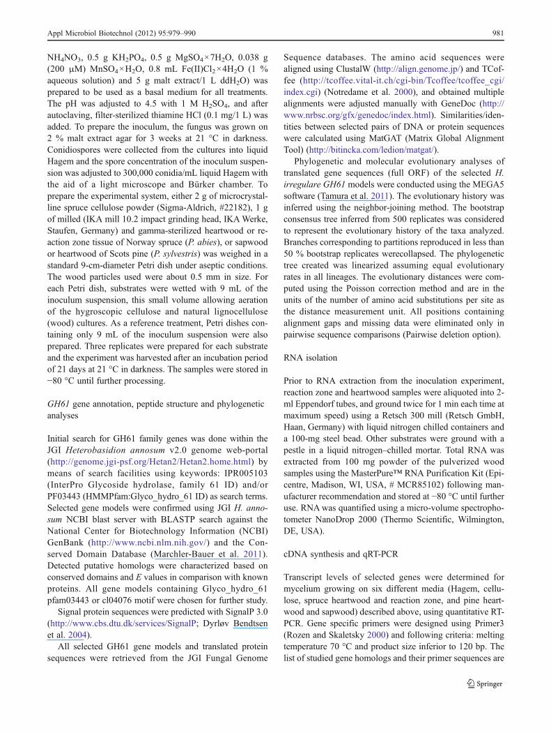
respective genomic sequences contained multiple exons(from 4 to 11) (Table S1 in the ESM). All mature HiGH61polypeptide sequences share the conserved metal ion-binding site that contain two histidines included in the Nterminus of the mature protein, except HiGH61G wherethese histidines have been replaced by arginine and gluta-mine, respectively (Fig. 1).
In addition to the conserved metal-binding motif, fourproteins (HiGH61D, HiGH61H, HiGH61I, and HiGH61J)
also possess an additional single domain at the C-terminalside of the GH6 module. For HiGH61D, HiGH61H, andHiGH61I, the extra domain was identified as a family 1carbohydrate-binding module (CBM1; also known as fungalcellulose-binding domain) (Gilkes et al. 1991), whereasHiGH61J has a C-terminal extension of ∼35 residues ofunknown function.
A MSA of the H. irregulare GH61 sequences wasobtained by aligning the sequences to a profile consisting
Fig. 1 Multiple sequence alignment of HiGH61 sequences andsequences of GH61 proteins with known 3D structures (TtGH61Efrom T. terrestris and HjGH61B from H. jecorina). Regions withinsertions/deletions are shaded yellow (region 1) and blue (region 2).Conserved residues clustered on the putative substrate-binding surface/active site are shaded red. Degree of conservation: asterisks fully
conserved, colons highly conserved, and full stops moderately con-served. Conserved residues with solvent exposed side chains (inferredby analysis of the HiGH61A 3D model by WHATIF) are marked withan “e”. CBM1 domains are indicated by a box and the conservedcysteines within these modules are colored green
Appl Microbiol Biotechnol (2012) 95:979–990 983
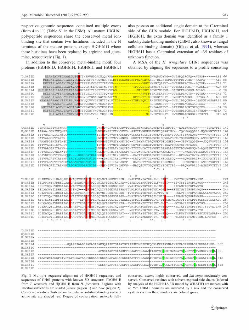
of a 3D-structural alignment of TtGH61E and HiGH61Bfrom Thielavia terrestris; PDB ID: 3EII (Harris et al. 2010)and Hypocrea jecorina; PDB ID: 2VTC (Karkehabadi et al.2008), respectively (Figs. 1 and 2). In order to aid andvisualize the interpretation of the MSA, a 3D model ofHiGH61Awas made using the MODELLER software usingthe TtGH61E structure as template (41 % sequence identityand 3 % gaps). Analysis of the HiGH61A 3D model revealsthat the majority of conserved residues of the HiGH61
isozymes are found internally in the protein where they mostlikely contribute to stabilizing the protein fold, similar towhat is observed in a study on GH61s from T. terrestris(Harris et al. 2010). Analysis of the accessible molecularsurface shows that only 11 of the 52 conserved residueshave solvent exposed side chains, whereof six are present onthe putative substrate-binding surface (Fig. 2). These in-clude His20 and His92 that are involved in the metal-binding motif and additionally Gln176, His167, Ile215,and Tyr217 that are likely to play important roles in thecatalytic mechanism/substrate binding of these proteins.Tyr217 is the only aromatic amino acid in the protein thatis solvent exposed, much like what is seen for the chitinoxidizing enzyme CBP21 (Vaaje-Kolstad et al. 2005b),where it has been shown to be important for activity andsubstrate binding (Vaaje-Kolstad et al. 2005a).
Evaluations of stereochemical parameters of the finalmodel showed no residues in the disallowed region of theRamachandran plot (analyzed by Procheck), (Laskowski etal. 1993), overall G-factor obtained0−0.2, an RMSD valueof the Cα positions between the HiGH61A model andTtGH61E of 0.18 Å (analyzed using the “super” algorithmin Pymol (DeLano 2002) and an overall goodmodel quality (Zscore0−5.12) when analyzed by the ProSA software designedfor detection errors in protein structures (Wiederstein andSippl 2007).
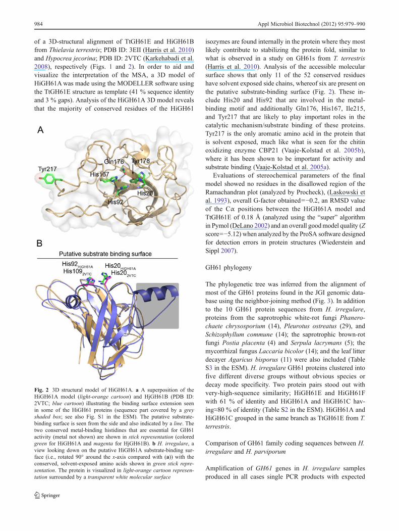
GH61 phylogeny
The phylogenetic tree was inferred from the alignment ofmost of the GH61 proteins found in the JGI genomic data-base using the neighbor-joining method (Fig. 3). In additionto the 10 GH61 protein sequences from H. irregulare,proteins from the saprotrophic white-rot fungi Phanero-chaete chrysosporium (14), Pleurotus ostreatus (29), andSchizophyllum commune (14); the saprotrophic brown-rotfungi Postia placenta (4) and Serpula lacrymans (5); themycorrhizal fungus Laccaria bicolor (14); and the leaf litterdecayer Agaricus bisporus (11) were also included (TableS3 in the ESM). H. irregulare GH61 proteins clustered intofive different diverse groups without obvious species ordecay mode specificity. Two protein pairs stood out withvery-high-sequence similarity; HiGH61E and HiGH61Fwith 61 % of identity and HiGH61A and HiGH61C hav-ing≈80 % of identity (Table S2 in the ESM). HiGH61A andHiGH61C grouped in the same branch as TtGH61E from T.terrestris.
Comparison of GH61 family coding sequences between H.irregulare and H. parviporum
Amplification of GH61 genes in H. irregulare samplesproduced in all cases single PCR products with expected
Fig. 2 3D structural model of HiGH61A. a A superposition of theHiGH61A model (light-orange cartoon) and HjGH61B (PDB ID:2VTC; blue cartoon) illustrating the binding surface extension seenin some of the HiGH61 proteins (sequence part covered by a greyshaded box; see also Fig. S1 in the ESM). The putative substrate-binding surface is seen from the side and also indicated by a line. Thetwo conserved metal-binding histidines that are essential for GH61activity (metal not shown) are shown in stick representation (coloredgreen for HiGH61A and magenta for HjGH61B). b H. irregulare, aview looking down on the putative HiGH61A substrate-binding sur-face (i.e., rotated 90° around the x-axis compared with (a)) with theconserved, solvent-exposed amino acids shown in green stick repre-sentation. The protein is visualized in light-orange cartoon represen-tation surrounded by a transparent white molecular surface
984 Appl Microbiol Biotechnol (2012) 95:979–990
product size, except for HiGH61C and HiGH61E, whichshowed faint PCR products. Amplification of GH61 genesin H. parviporum samples produced clear single PCR prod-ucts with expected product size only for HpGH61G–HpGH61J, multiple bands for HpGH61A–HpGH61F andno amplification product for HpGH61C and HpGH61E(Table S6 in the ESM). The obtained multiple bands for H.
parviporum, when bright and clear on gel, were also se-quenced. In all cases the sequences obtained from H. irreg-ulare corresponded to predicted gene model sequences. TheGH61G–GH61J gene sequences revealed clear single nu-cleotide polymorphisms between the two species, somebeing non-synonymous. The HpGH61J sequence showeda synonymous insert of 21 bp (7 aa). Most of sequenced
Fig. 3 Phylogenetic tree of GH61 family proteins (full ORF aminoacid sequences) available at the JGI fungal genomic database. Speciesabbreviation and protein ID are shown for each entry. The phylogenetictree was constructed using MEGA5 as detailed in “Materials andmethods.” The tree is drawn to scale, with branch lengths in the same
units as those of the evolutionary distances used to infer the phyloge-netic tree. Sequences from H. irregulare (Hir) are shown in red. Fungalspecies: Agabi A. bisporus, Pleos P. ostreatus, Pospl P. placenta, LacbiL. bicolor, Phchr P. chrysosporium, Serla S. lacrymans, Schco S.commune, TtGH61E from T. terrestris and HjGH61B from H. jecorina
Appl Microbiol Biotechnol (2012) 95:979–990 985
fragments excised from H. parviporum samples with multi-ple PCR products were not related to GH61 family genes orwere not found in the H. irregulare genome.
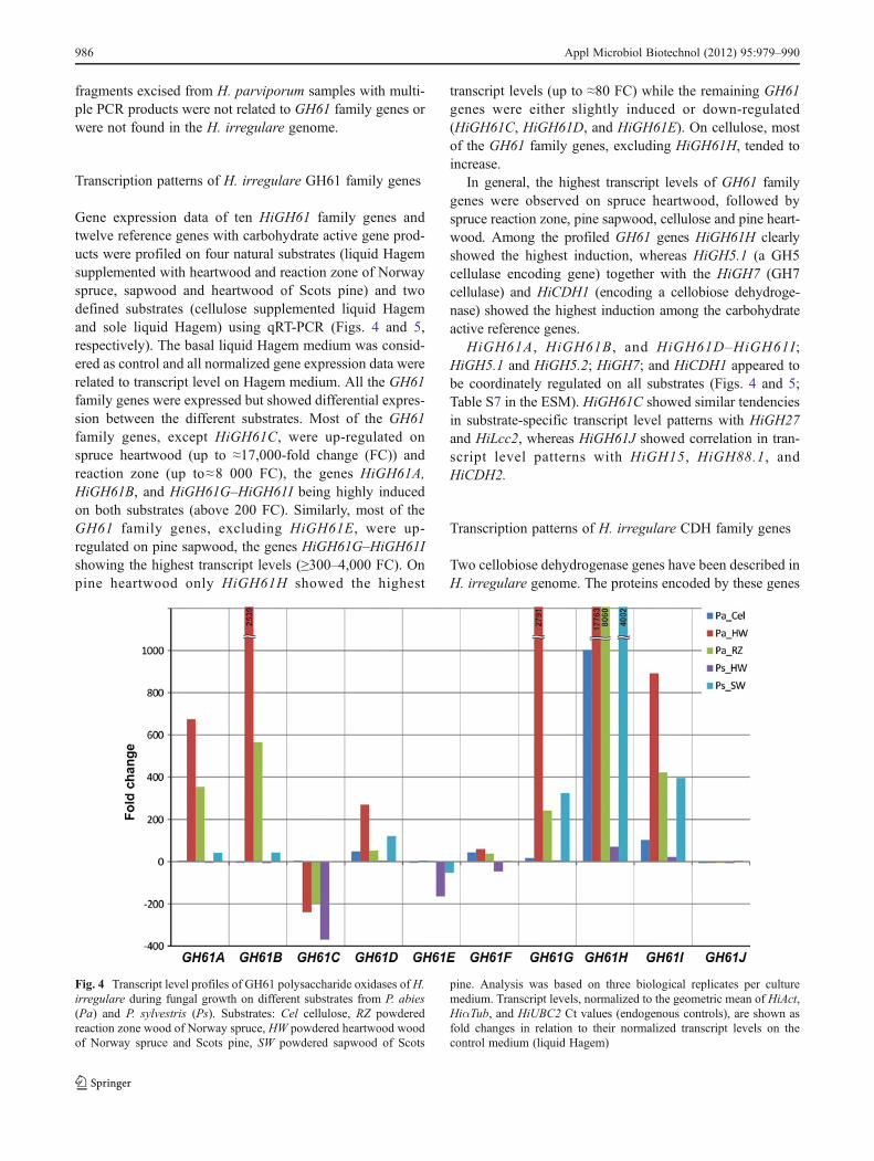
Transcription patterns of H. irregulare GH61 family genes
Gene expression data of ten HiGH61 family genes andtwelve reference genes with carbohydrate active gene prod-ucts were profiled on four natural substrates (liquid Hagemsupplemented with heartwood and reaction zone of Norwayspruce, sapwood and heartwood of Scots pine) and twodefined substrates (cellulose supplemented liquid Hagemand sole liquid Hagem) using qRT-PCR (Figs. 4 and 5,respectively). The basal liquid Hagem medium was consid-ered as control and all normalized gene expression data wererelated to transcript level on Hagem medium. All the GH61family genes were expressed but showed differential expres-sion between the different substrates. Most of the GH61family genes, except HiGH61C, were up-regulated onspruce heartwood (up to ≈17,000-fold change (FC)) andreaction zone (up to≈8 000 FC), the genes HiGH61A,HiGH61B, and HiGH61G–HiGH61I being highly inducedon both substrates (above 200 FC). Similarly, most of theGH61 family genes, excluding HiGH61E, were up-regulated on pine sapwood, the genes HiGH61G–HiGH61Ishowing the highest transcript levels (≥300–4,000 FC). Onpine heartwood only HiGH61H showed the highest
transcript levels (up to ≈80 FC) while the remaining GH61genes were either slightly induced or down-regulated(HiGH61C, HiGH61D, and HiGH61E). On cellulose, mostof the GH61 family genes, excluding HiGH61H, tended toincrease.
In general, the highest transcript levels of GH61 familygenes were observed on spruce heartwood, followed byspruce reaction zone, pine sapwood, cellulose and pine heart-wood. Among the profiled GH61 genes HiGH61H clearlyshowed the highest induction, whereas HiGH5.1 (a GH5cellulase encoding gene) together with the HiGH7 (GH7cellulase) and HiCDH1 (encoding a cellobiose dehydroge-nase) showed the highest induction among the carbohydrateactive reference genes.
HiGH61A, HiGH61B, and HiGH61D–HiGH61I;HiGH5.1 and HiGH5.2; HiGH7; and HiCDH1 appeared tobe coordinately regulated on all substrates (Figs. 4 and 5;Table S7 in the ESM). HiGH61C showed similar tendenciesin substrate-specific transcript level patterns with HiGH27and HiLcc2, whereas HiGH61J showed correlation in tran-script level patterns with HiGH15, HiGH88.1, andHiCDH2.
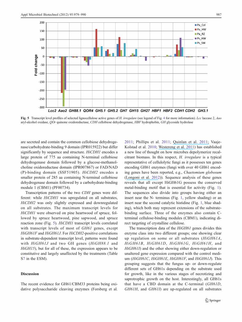
Transcription patterns of H. irregulare CDH family genes
Two cellobiose dehydrogenase genes have been described inH. irregulare genome. The proteins encoded by these genes
Fig. 4 Transcript level profiles of GH61 polysaccharide oxidases of H.irregulare during fungal growth on different substrates from P. abies(Pa) and P. sylvestris (Ps). Substrates: Cel cellulose, RZ powderedreaction zone wood of Norway spruce, HW powdered heartwood woodof Norway spruce and Scots pine, SW powdered sapwood of Scots
pine. Analysis was based on three biological replicates per culturemedium. Transcript levels, normalized to the geometric mean of HiAct,HiαTub, and HiUBC2 Ct values (endogenous controls), are shown asfold changes in relation to their normalized transcript levels on thecontrol medium (liquid Hagem)
986 Appl Microbiol Biotechnol (2012) 95:979–990
are secreted and contain the common cellobiose dehydroge-nase/carbohydrate-binding 9 domain (IPR015922) but differsignificantly by sequence and structure. HiCDH1 encodes alarge protein of 775 aa containing N-terminal cellobiosedehydrogenase domain followed by a glucose-methanol-choline oxidoreductase domain (IPR007867) or FAD/NAD(P)-binding domain (SSF51905). HiCDH2 encodes asmaller protein of 285 aa containing N-terminal cellobiosedehydrogenase domain followed by a carbohydrate-bindingmodule 1 (CBM1) (PF00734).
Transcription patterns of the two CDH genes were dif-ferent: while HiCDH1 was upregulated on all substrates,HiCDH2 was only slightly expressed and downregulatedon all substrates. The maximum transcript levels forHiCDH1 were observed on pine heartwood of spruce, fol-lowed by spruce heartwood, pine sapwood, and sprucereaction zone (Fig. 5). HiCDH1 transcript levels correlatedwith transcript levels of most of GH61 genes, exceptHiGH61F and HiGH61J. For HiCDH2-positive correlationsin substrate-dependent transcript level, patterns were foundwith HiGH61J and two GH genes (HiGH88.1 andHiGH15), but for all of these, the expression appears to beconstitutive and largely unaffected by the treatments (TableS7 in the ESM).
Discussion
The recent evidence for GH61/CBM33 proteins being oxi-dative polysaccharide cleaving enzymes (Forsberg et al.
2011; Phillips et al. 2011; Quinlan et al. 2011; Vaaje-Kolstad et al. 2010; Westereng et al. 2011) has establisheda new line of thought on how microbes depolymerize recal-citrant biomass. In this respect, H. irregulare is a typicalrepresentative of cellulolytic fungi as it possesses ten genesencoding GH61 enzymes (fungi with over 40 GH61 encod-ing genes have been reported, e.g., Chaetomium globosum(Longoni et al. 2012)). Sequence analysis of these genesreveals that all except HiGH61G possess the conservedmetal-binding motif that is essential for activity (Fig. 1).The sequences also divide into groups having either aninsert near the N- terminus (Fig. 1, yellow shading) or aninsert near the second catalytic histidine (Fig. 1, blue shad-ing), which both may represent extensions of the substrate-binding surface. Three of the enzymes also contain C-terminal cellulose-binding modules (CBM1), indicating di-rect targeting of crystalline cellulose.
The transcription data of the HiGH61 genes divides thisenzyme class into two different groups; one showing clearup regulation on some or all substrates (HiGH61A,HiGH61B , HiGH61D , HiGH61G , HiGH61H , andHiGH61I) and the other showing either down-regulation orunaltered gene expression compared with the control medi-um (HiGH61C, HiGH61E, HiGH61F, and HiGH61J). Thisgrouping suggests that the fungus up- or down-regulatedifferent sets of GH61s depending on the substrate usedfor growth, like in the various stages of necrotizing andsaprotrophic growth on the host. Interestingly, all GH61sthat have a CBD domain at the C-terminal (GH61D,GH61H, and GH61I) are up-regulated on all substrates
Fig. 5 Transcript level profiles of selected lignocellulose active genes of H. irregulare (see legend of Fig. 4 for more information). Lcc laccase 2, Aaoaryl-alcohol oxidase, QOr quinone oxidoreductase, СDH cellobiose dehydrogenase, HBF hydrophobin, GH glycoside hydrolase
Appl Microbiol Biotechnol (2012) 95:979–990 987
except heartwood of Scots pine and are the only GH61s thatshow clear up regulation in the pure cellulose substrate(Fig. 4), indicating that at least these three GH61s aredesigned to specifically target cellulose surfaces. The mosthighly induced HiGH61 (HiGH61A, HiGH61B, HiGH61Dand HiGH61G–HiGH61I) genes showed the highest ob-served transcript levels on spruce natural substrates, higheron spruce hearth wood than in spruce reaction zone woodpossibly reflecting is pivotal role in the decomposition ofcellulose during this preferred saproptrophic mode in thishost. HiGH61A, HiGH61B, HiGH61D, and HiGH61G–HiGH61I are induced on pine sapwood, while close toconstitutive levels is detected on pine heartwood (resinouswood with antifungal properties), possibly reflecting thatcolonization of the pine sapwood is the predominant growthmode in this host. The additional two GH61 genes(HIGH61C and HIGH61J) appeared to be coordinatelyexpressed with HiLcc2 (encoding a laccase), leading us tospeculate that these GH61s may play a role beyond cleavingcellulose. The common upregulated transcription patterns ofmost GH61 genes together with several of the referencecellulolytic and hemicellulolytic GHs (GH5 and GH7 cellu-lases) during growth on the natural woody substrates reflectthe co-metabolic nature of cell wall polymer degradation.
Sequence and phylogenetic analysis of the H. irregulareGH61s shows high sequence divergence (Figs. 1 and 3).Similar divergence has been shown for other ascomycetesand basidiomycetes fungi (Harris et al. 2010), indicating thatmuch of the duplication and divergence of the genes encod-ing GH61 proteins is a fairly ancient evolutionary event.This diversity has been largely maintained and in somecases apparently amplified during this evolutionary span,suggesting a significant selective pressure to retain a hetero-geneous collection of GH61-encoding genes in cellulose-degrading fungi (Harris et al. 2010). The high level of indeland single-nucleotide polymorphism observed when com-paring GH61s between the closely related H. irregulare andH. parviporum species (Table S6 in the ESM) is compatiblewith this hypothesis. Generally white-rot fungi have more ofGH61 family proteins than brown-rot fungi, and this alsoapplies for H. irregulare which possessed ten members inthe gene family, whereas the reference brown rot fungi P.placenta and S. lacrymans have four and five members,respectively (Table S3 in the ESM). Among white-rot fungi,the necrotrophic and saprotrophic H. irregulare possessesthe lower number of GH61 family genes (10) in comparisonwith saprotrophic P. chrysosporium (14), P. ostreatus (29),and S. commune (22). Notably, fungi growing on hardwoods(non-monocot angiosperm trees) generally possess moreGH61 family genes than fungal species growing mostly onsoftwoods (wood from conifers that are gymnosperms), likeH. irregulare and P. carnosa (this could be due to the morecomplex cell wall chemistry and that the lignin composition
and their links to cellulose is more complex in angiospermtrees).
Detailed analysis of the H. irregulare GH61 polypeptidesenabled identification of a region in the GH61 sequencesthat may contain insertions/deletions (Figs. 1 (yellow-col-ored part of MSA) and 2a). This region provides an exten-sion of the protein surface next to the metal-binding site(Fig. 2; also observed for HjGH61B), putatively providingmeans of substrate binding as this region also containsexposed aromatic amino acids that are known to interactwith carbohydrates. A second insertion is observed in theHiGH61H, HiGH61G, HiGH61I, and HiGH61E sequencesclose to the second conserved His in the metal-binding motif(Fig. 1 (blue-colored part of MSA)), which may also expandthe putative substrate-binding surface residues important forGH61 activity clustered at the putative reactive surface ofthe GH61s (Fig. 2b; His20, His92, His167, Gln176, Tyr178,and Tyr217—HiGH61A numbering) are conserved in allproteins except HiGH61G and HiGH61J (Fig. 2). Interest-ingly, HiGH61G has most of these residues substituted withamino acids that are likely to be incompatible with thecellulose oxidizing activity proposed for proteins in theGH61 family (His20→Arg, His92→Glu, His167→Gln,and Tyr178→Phe, HiGH61A numbering; Figs. 1 and 2b;(Vaaje-Kolstad et al. 2010) while HiGH61J shows two sub-stitutions of these conserved residues; Gln176→Glu andTyr217→Phe.
It is intriguing to note that HiGH61G is highly upregu-lated on several substrates (Fig. 4) since this protein lacksamino acids essential for the predicted GH61 oxidativeactivity (the sequence of this gene was verified by sequenc-ing of PCR products from two species, independent iso-lates). It may be that this protein has evolved a differentenzymatic function or even a non-enzymatic role comparedwith other GH61 enzymes. The putative enzymatic proper-ties of HiGH61G should be further characterized in order toreveal the functional significance of this protein.
Recent work (Langston et al. 2011) demonstrated thatcellobiose dehydrogenase can provide electrons to GH61s toform an oxidoreductive system for microbial lignocellulosedegradation. The two CDH genes of H. irregulare differsignificantly by domain structure. HiCDH1 showedsubstrate-specific transcript level profiles and appeared tobe coordinately regulated with different subgroups ofHiGH61 genes, whereas HiCDH2 appeared not to be sig-nificantly transcribed on any substrates (Fig. 5) Interesting-ly, similar results have been observed for the filamentousascomycete Neurospora crassa which also have two genesencoding CDHs, whereof only one is expressed (Phillips etal. 2011). The expressed N. crassa CDH (CDH-1; UniprotID Q7RXM0), which has been suggested to be the desig-nated electron donor for the N. crassa GH61s, also sharesthe domain structure of the H. irregulare protein HiCDH1
988 Appl Microbiol Biotechnol (2012) 95:979–990
(these genes share ∼60 % sequence identity). It is possiblethat HiCDH1 of H. irregulare can play a similar role to thatof CDH1 from N. crassa in biomass degradation. The sec-ond CDH from H. irregulare (HiCDH2) also has a similardomain structure to the second CDH from N. crassa, whichonly contains one flavin domain and a C-terminal CBM1.Interestingly, neither the HiCDH2 nor the N. crassa CDH2are expressed during fungal growth on cellulose (Fig. 5)(Phillips et al. 2011).
The generally higher transcript levels of GH61 genes onwood substrates in comparison to artificial media (MacDonaldet al. 2011) suggest involvement of these genes in degradationof wood fiber lignocelluloses. Differential regulation ofGH61family genes on different substrates also imply specificity ofdifferent GH61 polysaccharide oxidases to different types ofwood cell-wall compounds and could determine the hostsspecies specificity of a particular fungi species. The fact thatsome of the GH61 genes, such as HiGH61A and HiGH61B,showed marked difference in the transcript level betweenspruce heartwood and pine sapwood may reflect some tree-specific differences in the structure or composition of thetracheid cell wall. For natural substrates, the currentlyemployed experimental system should provide artificially ac-celerated decay conditions due to the involved pulverizing ofwood and use of inoculum high in colony forming units. Thegenerally lower transcript levels of HiGH61 genes on sprucereaction zone and pine heartwood compared with spruceheartwood and pine sapwood, respectively, are presumablyrelated to the high concentration of antifungal compounds inspruce reaction zone and pine heartwood (Woodward et al.1998). For example, in the reaction zone of Norway spruce thefungus is likely to use its ligninolytic repertoire for detoxifi-cation of host defense polyphenols as well, this resourceallocation affecting negatively the rate of cell wall degradationand exposure of cellulose to enzymatic activity (Yamada2001).
In summary, there is high diversity among the ten GH61genes present in H. irregulare. Based on the transcriptprofile patterns on the included natural and defined sub-strates, these genes could be divided into two groups. Themajor group consisted of eight genes that showed coordi-nated gene expression with the included reference cellulo-lytic and hemicellulolytic GHs and generally high up-regulation level on woody substrates in contrast to thedefined cellulose medium. This demonstrates the co-metabolic nature of lignocellulose degradation. The remain-ing two GH61s, together with starch and oligosaccharideactive reference genes, showed little regulation in compari-son with the basal Hagem medium control, and could beinvolved in degradation of more easily available non-structural carbohydrates that are particularly important dur-ing the initial exploratory colonization phase of host tissueby a microbe. These data suggest that GH61 genes
participate in degradation of both the structural and reservecarbohydrates of xylem. Further research on both GH61genes and proteins are warranted particularly on the highlyexpressed HiGH61G with a widely divergent catalytic siteand whether specific GH61s may have a role in breaking thelink between cellulose and lignin.
Acknowledgments We thank Åke Olson (Swedish University ofAgricultural Sciences) for providing the H. irregulare strain TC-32-1for study and Mari Kjos (Skogoglandskap) for valuable technicalassistance in gene amplification and sequencing. This work was sup-ported by the Norwegian Research Council (grants #315052 and135901).
Open Access This article is distributed under the terms of the Crea-tive Commons Attribution License which permits any use, distribution,and reproduction in any medium, provided the original author(s) andthe source are credited.
References
Cantarel BL, Coutinho PM, Rancurel C, Bernard T, Lombard V,Henrissat B (2009) The carbohydrate-active EnZymes database(CAZy): an expert resource for glycogenomics. Nucleic AcidsRes 37:D233–D238
Dalman K, Olson Å, Stenlid J (2010) Evolutionary history of theconifer root rot fungus Heterobasidion annosum sensu lato. MolEcol 19:4979–4993
DeLano WL (2002) The PyMOL molecular graphics system. DeLanoScientific, San Carlos, CA
Dyrløv Bendtsen J, Nielsen H, von Heijne G, Brunak S (2004) Im-proved prediction of signal peptides: signalP 3.0. J Mol Biol340:783–795
Eastwood DC, Floudas D, Binder M, Majcherczyk A, Schneider P,Aerts A, Asiegbu FO, Baker SE, Barry K, Bendiksby M,Blumentritt M, Coutinho PM, Cullen D, de Vries RP, GathmanA, Goodell B, Henrissat B, Ihrmark K, Kauserud H, Kohler A,LaButti K, Lapidus A, Lavin JL, Lee Y-H, Lindquist E, Lilly W,Lucas S, Morin E, Murat C, Oguiza JA, Park J, Pisabarro AG,Riley R, Rosling A, Salamov A, Schmidt O, Schmutz J, Skrede I,Stenlid J, Wiebenga A, Xie X, Kües U, Hibbett DS, HoffmeisterD, Högberg N, Martin F, Grigoriev IV, Watkinson SC (2011) Theplant cell wall–decomposing machinery underlies the functionaldiversity of forest fungi. Science 333:762–765
Eswar N, Webb B, Marti-Renom MA, Madhusudhan MS, Eramian D,Shen M-Y, Pieper U, Sali A (2007) Comparative protein structuremodeling using MODELLER. Current protocols in protein sci-ence. Wiley, New York
Forsberg Z, Vaaje-Kolstad G, Westereng B, Bunaes AC, Stenstrom Y,Mackenzie A, Sorlie M, Horn SJ, Eijsink VGH (2011) Cleavageof cellulose by a CBM33 protein. Protein Sci 20:1479–1483
Gilkes NR, Henrissat B, Kilburn DG, Miller RC Jr, Warren RA (1991)Domains in microbial β-1,4-glycanases: sequence conservation,function, and enzyme families. Microbiol Rev 55:303–315
Goodell B, Jellison J, Liu J, Daniel G, Paszczynski A, Fekete F,Krishnamurthy S, Jun L, Xu G (1997) Low molecular weightchelators and phenolic compounds isolated from wood decayfungi and their role in the fungal biodegradation of wood. JBiotechnol 53:133–162
Harris PV, Welner D, McFarland KC, Re E, Navarro Poulsen J-C,Brown K, Salbo R, Ding H, Vlasenko E, Merino S, Xu F, CherryJ, Larsen S, Lo Leggio L (2010) Stimulation of lignocellulosic
Appl Microbiol Biotechnol (2012) 95:979–990 989
biomass hydrolysis by proteins of glycoside hydrolase family 61:structure and function of a large, enigmatic family. Biochemistry49:3305–3316
Karkehabadi S, Hansson H, Kim S, Piens K, Mitchinson C, SandgrenM (2008) The first structure of a glycoside hydrolase family 61member, Cel61B from Hypocrea jecorina, at 1.6 Å resolution. JMol Biol 383:144–154
Langston JA, Shaghasi T, Abbate E, Xu F, Vlasenko E, Sweeney MD(2011) Oxidoreductive cellulose depolymerization by theenzymes cellobiose dehydrogenase and glycoside hydrolase 61.Appl Environ Microbiol 77:7007–7015
Laskowski RA, MacArthur MW, Moss DS, Thornton JM (1993)PROCHECK: a program to check the stereochemical quality ofprotein structures. J App Cryst 26:283–291
Longoni P, Rodolfi M, Pantaleoni L, Doria E, Concia L, PiccoAM, Cella R (2012) Degradation of cellulosic substrates by aChaetomium globosum endophytic isolate: a functional anal-ysis. Appl Environ Microbiol 78:3693–3705. doi:10.1128/AEM.00124-12
MacDonald J, Doering M, Canam T, Gong Y, Guttman DS, CampbellMM, Master ER (2011) Transcriptomic responses of thesoftwood-degrading white-rot fungus Phanerochaete carnosaduring growth on coniferous and deciduous wood. Appl EnvironMicrobiol 77:3211–3218
Marchler-Bauer A, Lu S, Anderson JB, Chitsaz F, Derbyshire MK,DeWeese-Scott C, Fong JH, Geer LY, Geer RC, Gonzales NR,Gwadz M, Hurwitz DI, Jackson JD, Ke Z, Lanczycki CJ, Lu F,Marchler GH, Mullokandov M, Omelchenko MV, Robertson CL,Song JS, Thanki N, Yamashita RA, Zhang D, Zhang N, Zheng C,Bryant SH (2011) CDD: a conserved domain database for the func-tional annotation of proteins. Nucleic Acids Res 39:D225–D229
Mason MG, Nicholls P, Wilson MT (2003) Rotting by radicals the roleof cellobiose oxidoreductase? Biochem Soc Trans 31:1335–1336
Merino S, Cherry J (2007) Progress and challenges in enzyme devel-opment for biomass utilization. In: Olsson L (ed) Biofuels.Advances in Biochemical Engineering/Biotechnology. Springer,Berlin, pp 95–120
Niemelä T, Korhonen K (1998) Taxonomy of the genus Heterobasi-dion. In: Woodward S, Stenlid О, Karjalainen К, Hüttermann A(eds) Heterobasidion annosum: biology, ecology, impact andcontrol. CAB International, London, pp 27–33
Notredame C, Higgins DG, Heringa J (2000) T-coffee: a novel methodfor fast and accurate multiple sequence alignment. J Mol Biol302:205–217
Olson Å, Aerts A, Asiegbu F, Belbahri L, Bouzid O, Broberg A, CanbäckB, Coutinho PM, Cullen D, Dalman K, Deflorio G, van DiepenLTA, Dunand C, Duplessis S, Durling M, Gonthier P, Grimwood J,Fossdal CG, Hansson D, Henrissat B, Hietala A, Himmelstrand K,Hoffmeister D, Högberg N, James TY, Karlsson M, Kohler A, KüesU, Lee Y-H, Lin Y-C, Lind M, Lindquist E, Lombard V, Lucas S,Lundén K,Morin E,Murat C, Park J, Raffaello T, Rouzé P, SalamovA, Schmutz J, Solheim H, Ståhlberg J, Vélëz H, de Vries RP,Wiebenga A, Woodward S, Yakovlev I, Garbelotto M, Martin F,Grigoriev IV, Stenlid J (2012) Insight into trade-off between wooddecay and parasitism from the genome of a fungal forest pathogen.New Phytol 194:1001–1013
Otrosina WJ, Garbelotto M (2010) Heterobasidion occidentale sp. nov.and Heterobasidion irregulare nom. nov.: A disposition of North
American Heterobasidion biological species. Fungal Biol114:16–25
Phillips CM, Beeson WT, Cate JH, Marletta MA (2011) Cellobiosedehydrogenase and acopper-dependent polysaccharide monooxy-genase potentiate cellulose degradation by Neurospora crassa.ACS Chem Biol 6:1399–1406
Quinlan RJ, Sweeney MD, Lo Leggio L, Otten H, Poulsen J-CN,Johansen KS, Krogh KBRM, Jørgensen CI, Tovborg M, Anthon-sen A, Tryfona T, Walter CP, Dupree P, Xu F, Davies GJ, WaltonPH (2011) Insights into the oxidative degradation of cellulose by acopper metalloenzyme that exploits biomass components. ProcNatl Acad Sci USA 108:15079–15084
Reese ET, Siu RGH, Levinson HS (1950) The biological degradationof soluble cellulose derivatives and its relationship to the mech-anism of cellulose hydrolysis. J Bacteriol 59:485–497
Rozen S, Skaletsky HJ (2000) Primer3 on the WWW for general usersand for biologist programmers. In: Krawetz S, Misener S (eds)Methods in molecular biology. Humana Press, Totowa, pp 365–386
Tamura K, Peterson D, Peterson N, Stecher G, Nei M, Kumar S (2011)MEGA5: molecular evolutionary genetics analysis using maxi-mum likelihood, evolutionary distance, and maximum parsimonymethods. Mol Biol Evol 28:2731–2739
Teeri TT (1997) Crystalline cellulose degradation: new insight into thefunction of cellobiohydrolases. Trends Biotechnol 15:160–167
Vaaje-Kolstad G, Horn SJ, van Aalten DMF, Synstad B, Eijsink VGH(2005a) The non-catalytic chitin-binding protein CBP21 fromSerratia marcescens is essential for chitin degradation. J BiolChem 280:28492–28497
Vaaje-Kolstad G, Houston DR, Riemen AHK, Eijsink VGH, vanAalten DMF (2005b) Crystal structure and binding properties ofthe Serratia marcescens chitin-binding protein CBP21. J BiolChem 280:11313–11319
Vaaje-Kolstad G, Westereng B, Horn SJ, Liu Z, Zhai H, Sørlie M,Eijsink VGH (2010) An oxidative enzyme boosting the enzymaticconversion of recalcitrant polysaccharides. Science 330:219–222
Vriend G (1990) WHAT IF: a molecular modeling and drug designprogram. J Mol Graphics 8:52–56
Westereng B, Ishida T, Vaaje-Kolstad G, Wu M, Eijsink VGH, IgarashiK, Samejima M, Ståhlberg J, Horn SJ, Sandgren M (2011) Theputative endoglucanase PcGH61d from Phanerochaete chryso-sporium is a metal-dependent oxidative enzyme that cleaves cel-lulose. PLoS One 6:e27807
Wiederstein M, Sippl MJ (2007) ProSA-web: interactive web servicefor the recognition of errors in three-dimensional structures ofproteins. Nucleic Acids Res 35:W407–W410
Woodward S, Stenlid J, Karjalainen R, Hüttermann A (1998) Hetero-basidion annosum. Biology, ecology, impact and control. CABInternational, Cambridge
Yakovlev IA, Hietala AM, Steffenrem A, Solheim H, Fossdal CG(2008) Identification and analysis of differentially expressed Het-erobasidion parviporum genes during natural colonization ofNorway spruce stems. Fungal Genet Biol 45:498–513
Yamada T (2001) Defense mechanisms in the sapwood of living treesagainst microbial infection. J For Res 6:127–137
ZamockyM, Ludwig R, Peterbauer C, Hallberg BM,DivneC,Nicholls P,Haltrich D (2006) Cellobiose dehydrogenase—a flavocytochromefrom wood-degrading, phytopathogenic and saprotropic fungi. CurrProtein Peptide Sci 7:255–280
990 Appl Microbiol Biotechnol (2012) 95:979–990
Copyright © 2022 FDOKUMEN