Development of diabetes mellitus in aging transgenic mice following suppression of pancreatic...
11
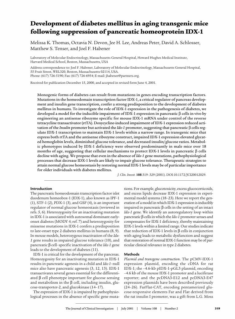
The Journal of Clinical Investigation | July 2001 | Volume 108 | Number 2 319 Introduction The pancreatic homeodomain transcription factor islet duodenum homeobox-1 (IDX-1), also known as IPF-1 (1), STF-1 (2), PDX-1 (3), and GSF (4), is an important regulator of normal glucose homeostasis (reviewed in refs. 5, 6). Heterozygosity for an inactivating mutation in IDX-1 is associated with autosomal dominant early- onset diabetes (MODY 4; ref. 7) and heterozygosity for missense mutations in IDX-1 confers a predisposition to late-onset type 2 diabetes mellitus in humans (8, 9). In mouse models, heterozygous inactivation of the Idx- 1 gene results in impaired glucose tolerance (10), and pancreatic β cell–specific inactivation of the Idx-1 gene leads to the development of diabetes (11). IDX-1 is critical for the development of the pancreas. Homozygosity for an inactivating mutation in IDX-1 results in pancreatic agenesis in a child and Idx-1–null mice also have pancreatic agenesis (3, 12, 13). IDX-1 transactivates several genes essential for the differenti- ated β cell phenotype important for glucose sensing and metabolism in the β cell, including insulin, glu- cose-transporter-2, and glucokinase (14–17). The expression of IDX-1 is impaired by pathophysio- logical processes in the absence of specific gene muta- tions. For example, glucotoxicity, excess glucocorticoids, and excess lipids decrease IDX-1 expression in experi- mental model systems (18–23). Here we report the gen- eration of a model in which IDX-1 expression is inducibly impaired in pancreatic β cells in the setting of an intact Idx-1 gene. We identify an autoregulatory loop within pancreatic β cells in which the Idx-1 promoter senses and compensates for IDX-1 deficiency, thereby maintaining IDX-1 levels within a limited range. Our studies indicate that reduction of IDX-1 levels in β cells in conjunction with aging leads to metabolic dysfunction and suggest that restoration of normal IDX-1 function may be of par- ticular clinical relevance in type 2 diabetes. Methods Plasmid and transgene construction. The pCMV-IDX-1 expression plasmid, encoding the cDNA for rat IDX-1; the –4.6-kb pIDX-1-pGL3 plasmid, encoding –4.6 kb of the mouse IDX-1 promoter and a luciferase reporter; and the pcDNA3-E12 and pcDNA3-E47 expression plasmids have been described previously (24–26). FarFlat-CAT, encoding pentamerized glu- cose-responsive elements Far and Flat derived from the rat insulin I promoter, was a gift from L.G. Moss Development of diabetes mellitus in aging transgenic mice following suppression of pancreatic homeoprotein IDX-1 Melissa K. Thomas, Octavia N. Devon, Jee H. Lee, Andreas Peter, David A. Schlosser, Matthew S. Tenser, and Joel F. Habener Laboratory of Molecular Endocrinology, Massachusetts General Hospital, Howard Hughes Medical Institute, Harvard Medical School, Boston, Massachusetts, USA Address correspondence to: Joel F. Habener, Laboratory of Molecular Endocrinology, Massachusetts General Hospital, 55 Fruit Street, WEL320, Boston, Massachusetts 02114, USA. Phone: (617) 726-5190; Fax: (617) 726-6954; E-mail: [email protected]. Received for publication December 15, 2000, and accepted in revised form June 4, 2001. Monogenic forms of diabetes can result from mutations in genes encoding transcription factors. Mutations in the homeodomain transcription factor IDX-1, a critical regulator of pancreas develop- ment and insulin gene transcription, confer a strong predisposition to the development of diabetes mellitus in humans. To investigate the role of IDX-1 expression in the pathogenesis of diabetes, we developed a model for the inducible impairment of IDX-1 expression in pancreatic β cells in vivo by engineering an antisense ribozyme specific for mouse IDX-1 mRNA under control of the reverse tetracycline transactivator (rtTA). Doxycycline-induced impairment of IDX-1 expression reduced acti- vation of the Insulin promoter but activated the Idx-1 promoter, suggesting that pancreatic β cells reg- ulate IDX-1 transcription to maintain IDX-1 levels within a narrow range. In transgenic mice that express both rtTA and the antisense ribozyme construct, impaired IDX-1 expression elevated glycat- ed hemoglobin levels, diminished glucose tolerance, and decreased insulin/glucose ratios. Metabol- ic phenotypes induced by IDX-1 deficiency were observed predominantly in male mice over 18 months of age, suggesting that cellular mechanisms to protect IDX-1 levels in pancreatic β cells decline with aging. We propose that even in the absence of Idx-1 gene mutations, pathophysiological processes that decrease IDX-1 levels are likely to impair glucose tolerance. Therapeutic strategies to attain normal glucose homeostasis by restoring normal IDX-1 levels may be of particular importance for older individuals with diabetes mellitus. J. Clin. Invest. 108:319–329 (2001). DOI:10.1172/JCI20012029.
-
Upload
independent -
Category
Documents
-
view
1 -
download
0
Transcript of Development of diabetes mellitus in aging transgenic mice following suppression of pancreatic...
The Journal of Clinical Investigation | July 2001 | Volume 108 | Number 2 319
IntroductionThe pancreatic homeodomain transcription factor isletduodenum homeobox-1 (IDX-1), also known as IPF-1(1), STF-1 (2), PDX-1 (3), and GSF (4), is an importantregulator of normal glucose homeostasis (reviewed inrefs. 5, 6). Heterozygosity for an inactivating mutationin IDX-1 is associated with autosomal dominant early-onset diabetes (MODY 4; ref. 7) and heterozygosity formissense mutations in IDX-1 confers a predispositionto late-onset type 2 diabetes mellitus in humans (8, 9).In mouse models, heterozygous inactivation of the Idx-1 gene results in impaired glucose tolerance (10), andpancreatic β cell–specific inactivation of the Idx-1 geneleads to the development of diabetes (11).
IDX-1 is critical for the development of the pancreas.Homozygosity for an inactivating mutation in IDX-1results in pancreatic agenesis in a child and Idx-1–nullmice also have pancreatic agenesis (3, 12, 13). IDX-1transactivates several genes essential for the differenti-ated β cell phenotype important for glucose sensingand metabolism in the β cell, including insulin, glu-cose-transporter-2, and glucokinase (14–17).
The expression of IDX-1 is impaired by pathophysio-logical processes in the absence of specific gene muta-
tions. For example, glucotoxicity, excess glucocorticoids,and excess lipids decrease IDX-1 expression in experi-mental model systems (18–23). Here we report the gen-eration of a model in which IDX-1 expression is induciblyimpaired in pancreatic β cells in the setting of an intactIdx-1 gene. We identify an autoregulatory loop withinpancreatic β cells in which the Idx-1 promoter senses andcompensates for IDX-1 deficiency, thereby maintainingIDX-1 levels within a limited range. Our studies indicatethat reduction of IDX-1 levels in β cells in conjunctionwith aging leads to metabolic dysfunction and suggestthat restoration of normal IDX-1 function may be of par-ticular clinical relevance in type 2 diabetes.
MethodsPlasmid and transgene construction. The pCMV-IDX-1expression plasmid, encoding the cDNA for rat IDX-1; the –4.6-kb pIDX-1-pGL3 plasmid, encoding–4.6 kb of the mouse IDX-1 promoter and a luciferasereporter; and the pcDNA3-E12 and pcDNA3-E47expression plasmids have been described previously(24–26). FarFlat-CAT, encoding pentamerized glu-cose-responsive elements Far and Flat derived fromthe rat insulin I promoter, was a gift from L.G. Moss
Development of diabetes mellitus in aging transgenic micefollowing suppression of pancreatic homeoprotein IDX-1
Melissa K. Thomas, Octavia N. Devon, Jee H. Lee, Andreas Peter, David A. Schlosser,Matthew S. Tenser, and Joel F. Habener
Laboratory of Molecular Endocrinology, Massachusetts General Hospital, Howard Hughes Medical Institute, Harvard Medical School, Boston, Massachusetts, USA
Address correspondence to: Joel F. Habener, Laboratory of Molecular Endocrinology, Massachusetts General Hospital, 55 Fruit Street, WEL320, Boston, Massachusetts 02114, USA. Phone: (617) 726-5190; Fax: (617) 726-6954; E-mail: [email protected].
Received for publication December 15, 2000, and accepted in revised form June 4, 2001.
Monogenic forms of diabetes can result from mutations in genes encoding transcription factors.Mutations in the homeodomain transcription factor IDX-1, a critical regulator of pancreas develop-ment and insulin gene transcription, confer a strong predisposition to the development of diabetesmellitus in humans. To investigate the role of IDX-1 expression in the pathogenesis of diabetes, wedeveloped a model for the inducible impairment of IDX-1 expression in pancreatic β cells in vivo byengineering an antisense ribozyme specific for mouse IDX-1 mRNA under control of the reverse tetracycline transactivator (rtTA). Doxycycline-induced impairment of IDX-1 expression reduced acti-vation of the Insulin promoter but activated the Idx-1 promoter, suggesting that pancreatic β cells reg-ulate IDX-1 transcription to maintain IDX-1 levels within a narrow range. In transgenic mice thatexpress both rtTA and the antisense ribozyme construct, impaired IDX-1 expression elevated glycat-ed hemoglobin levels, diminished glucose tolerance, and decreased insulin/glucose ratios. Metabol-ic phenotypes induced by IDX-1 deficiency were observed predominantly in male mice over 18months of age, suggesting that cellular mechanisms to protect IDX-1 levels in pancreatic β cellsdecline with aging. We propose that even in the absence of Idx-1 gene mutations, pathophysiologicalprocesses that decrease IDX-1 levels are likely to impair glucose tolerance. Therapeutic strategies toattain normal glucose homeostasis by restoring normal IDX-1 levels may be of particular importancefor older individuals with diabetes mellitus.
J. Clin. Invest. 108:319–329 (2001). DOI:10.1172/JCI20012029.
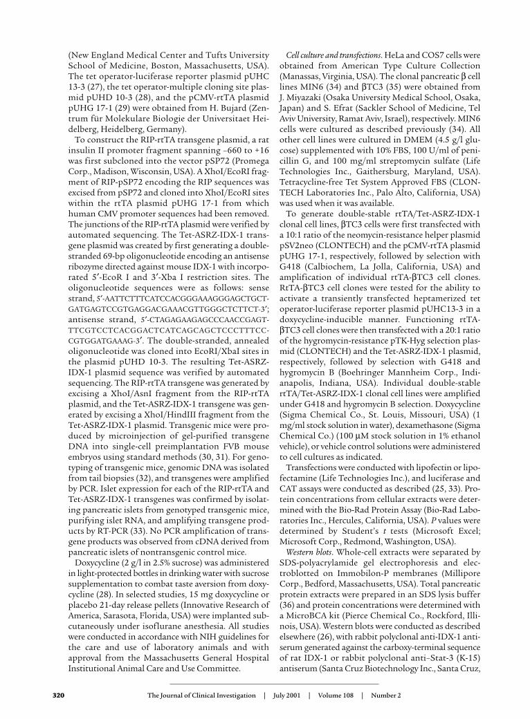
(New England Medical Center and Tufts UniversitySchool of Medicine, Boston, Massachusetts, USA).The tet operator-luciferase reporter plasmid pUHC13-3 (27), the tet operator-multiple cloning site plas-mid pUHD 10-3 (28), and the pCMV-rtTA plasmidpUHG 17-1 (29) were obtained from H. Bujard (Zen-trum für Molekulare Biologie der Universitaet Hei-delberg, Heidelberg, Germany).
To construct the RIP-rtTA transgene plasmid, a ratinsulin II promoter fragment spanning –660 to +16was first subcloned into the vector pSP72 (PromegaCorp., Madison, Wisconsin, USA). A XhoI/EcoRI frag-ment of RIP-pSP72 encoding the RIP sequences wasexcised from pSP72 and cloned into XhoI/EcoRI siteswithin the rtTA plasmid pUHG 17-1 from whichhuman CMV promoter sequences had been removed.The junctions of the RIP-rtTA plasmid were verified byautomated sequencing. The Tet-ASRZ-IDX-1 trans-gene plasmid was created by first generating a double-stranded 69-bp oligonucleotide encoding an antisenseribozyme directed against mouse IDX-1 with incorpo-rated 5′-EcoR I and 3′-Xba I restriction sites. Theoligonucleotide sequences were as follows: sensestrand, 5′-AATTCTTTCATCCACGGGAAAGGGAGCTGCT-GATGAGTCCGTGAGGACGAAACGTTGGGCTCTTCT-3′;antisense strand, 5′-CTAGAGAAGAGCCCAACCGAGT-TTCGTCCTCACGGACTCATCAGCAGCTCCCTTTCC-CGTGGATGAAAG-3′. The double-stranded, annealedoligonucleotide was cloned into EcoRI/XbaI sites inthe plasmid pUHD 10-3. The resulting Tet-ASRZ-IDX-1 plasmid sequence was verified by automatedsequencing. The RIP-rtTA transgene was generated byexcising a XhoI/AsnI fragment from the RIP-rtTAplasmid, and the Tet-ASRZ-IDX-1 transgene was gen-erated by excising a XhoI/HindIII fragment from theTet-ASRZ-IDX-1 plasmid. Transgenic mice were pro-duced by microinjection of gel-purified transgeneDNA into single-cell preimplantation FVB mouseembryos using standard methods (30, 31). For geno-typing of transgenic mice, genomic DNA was isolatedfrom tail biopsies (32), and transgenes were amplifiedby PCR. Islet expression for each of the RIP-rtTA andTet-ASRZ-IDX-1 transgenes was confirmed by isolat-ing pancreatic islets from genotyped transgenic mice,purifying islet RNA, and amplifying transgene prod-ucts by RT-PCR (33). No PCR amplification of trans-gene products was observed from cDNA derived frompancreatic islets of nontransgenic control mice.
Doxycycline (2 g/l in 2.5% sucrose) was administeredin light-protected bottles in drinking water with sucrosesupplementation to combat taste aversion from doxy-cycline (28). In selected studies, 15 mg doxycycline orplacebo 21-day release pellets (Innovative Research ofAmerica, Sarasota, Florida, USA) were implanted sub-cutaneously under isoflurane anesthesia. All studieswere conducted in accordance with NIH guidelines forthe care and use of laboratory animals and withapproval from the Massachusetts General HospitalInstitutional Animal Care and Use Committee.
Cell culture and transfections. HeLa and COS7 cells wereobtained from American Type Culture Collection(Manassas, Virginia, USA). The clonal pancreatic β celllines MIN6 (34) and βTC3 (35) were obtained from J. Miyazaki (Osaka University Medical School, Osaka,Japan) and S. Efrat (Sackler School of Medicine, TelAviv University, Ramat Aviv, Israel), respectively. MIN6cells were cultured as described previously (34). Allother cell lines were cultured in DMEM (4.5 g/l glu-cose) supplemented with 10% FBS, 100 U/ml of peni-cillin G, and 100 mg/ml streptomycin sulfate (LifeTechnologies Inc., Gaithersburg, Maryland, USA).Tetracycline-free Tet System Approved FBS (CLON-TECH Laboratories Inc., Palo Alto, California, USA)was used when it was available.
To generate double-stable rtTA/Tet-ASRZ-IDX-1clonal cell lines, βTC3 cells were first transfected witha 10:1 ratio of the neomycin-resistance helper plasmidpSV2neo (CLONTECH) and the pCMV-rtTA plasmidpUHG 17-1, respectively, followed by selection withG418 (Calbiochem, La Jolla, California, USA) andamplification of individual rtTA-βTC3 cell clones.RtTA-βTC3 cell clones were tested for the ability toactivate a transiently transfected heptamerized tetoperator-luciferase reporter plasmid pUHC13-3 in adoxycycline-inducible manner. Functioning rtTA-βTC3 cell clones were then transfected with a 20:1 ratioof the hygromycin-resistance pTK-Hyg selection plas-mid (CLONTECH) and the Tet-ASRZ-IDX-1 plasmid,respectively, followed by selection with G418 andhygromycin B (Boehringer Mannheim Corp., Indi-anapolis, Indiana, USA). Individual double-stablertTA/Tet-ASRZ-IDX-1 clonal cell lines were amplifiedunder G418 and hygromycin B selection. Doxycycline(Sigma Chemical Co., St. Louis, Missouri, USA) (1mg/ml stock solution in water), dexamethasone (SigmaChemical Co.) (100 µM stock solution in 1% ethanolvehicle), or vehicle control solutions were administeredto cell cultures as indicated.
Transfections were conducted with lipofectin or lipo-fectamine (Life Technologies Inc.), and luciferase andCAT assays were conducted as described (25, 33). Pro-tein concentrations from cellular extracts were deter-mined with the Bio-Rad Protein Assay (Bio-Rad Labo-ratories Inc., Hercules, California, USA). P values weredetermined by Student’s t tests (Microsoft Excel;Microsoft Corp., Redmond, Washington, USA).
Western blots. Whole-cell extracts were separated bySDS-polyacrylamide gel electrophoresis and elec-troblotted on Immobilon-P membranes (MilliporeCorp., Bedford, Massachusetts, USA). Total pancreaticprotein extracts were prepared in an SDS lysis buffer(36) and protein concentrations were determined witha MicroBCA kit (Pierce Chemical Co., Rockford, Illi-nois, USA). Western blots were conducted as describedelsewhere (26), with rabbit polyclonal anti-IDX-1 anti-serum generated against the carboxy-terminal sequenceof rat IDX-1 or rabbit polyclonal anti–Stat-3 (K-15)antiserum (Santa Cruz Biotechnology Inc., Santa Cruz,
320 The Journal of Clinical Investigation | July 2001 | Volume 108 | Number 2
California, USA) and visualized with enhanced chemi-luminescence using ECL Western blotting detectionreagents (Amersham Life Sciences Inc., ArlingtonHeights, Illinois, USA). Data were scanned by comput-ing densitometry (Molecular Dynamics, Sunnyvale,California, USA) and analyzed with ImageQuant soft-ware (Molecular Dynamics).
Glucose, insulin, and glycated hemoglobin measurements.Glucose levels were measured with a YSI 2300 STATglucose analyzer (Yellow Springs Instrument Co. Inc.,Yellow Springs, Ohio, USA). For glucose tolerance test-ing, mice were fasted for approximately 8 hours beforeintraperitoneal injection of 1.5 g glucose per kilogramof body weight. Plasma insulin levels were determinedin duplicate with a rat insulin ELISA kit standardizedwith mouse insulin standards (Crystal Chem Inc.,Chicago, Illinois, USA). Analyses of whole-blood sam-ples for glycated hemoglobin levels were conducted withGlyc-Affin Ghb kits (Isolab Inc., Akron, Ohio, USA).
Northern blots. Whole pancreatic RNA was preparedwith commercial nucleotide-binding columns (RNeasy;QIAGEN Inc., Valencia, California, USA), separated onformaldehyde agarose gels, and stained with ethidiumbromide before Northern blotting conducted asdescribed elsewhere (33). Blots were probed with a [32P]-radiolabeled probes directed against rat IDX-1 (5′-AGGT-TACGGCACAATCCTGCTCCGGCTCTT-3′) or β-actin (33).
ResultsA model of inducible impairment of idx-1 expression. Wedesigned a system of impairment of IDX-1 expression
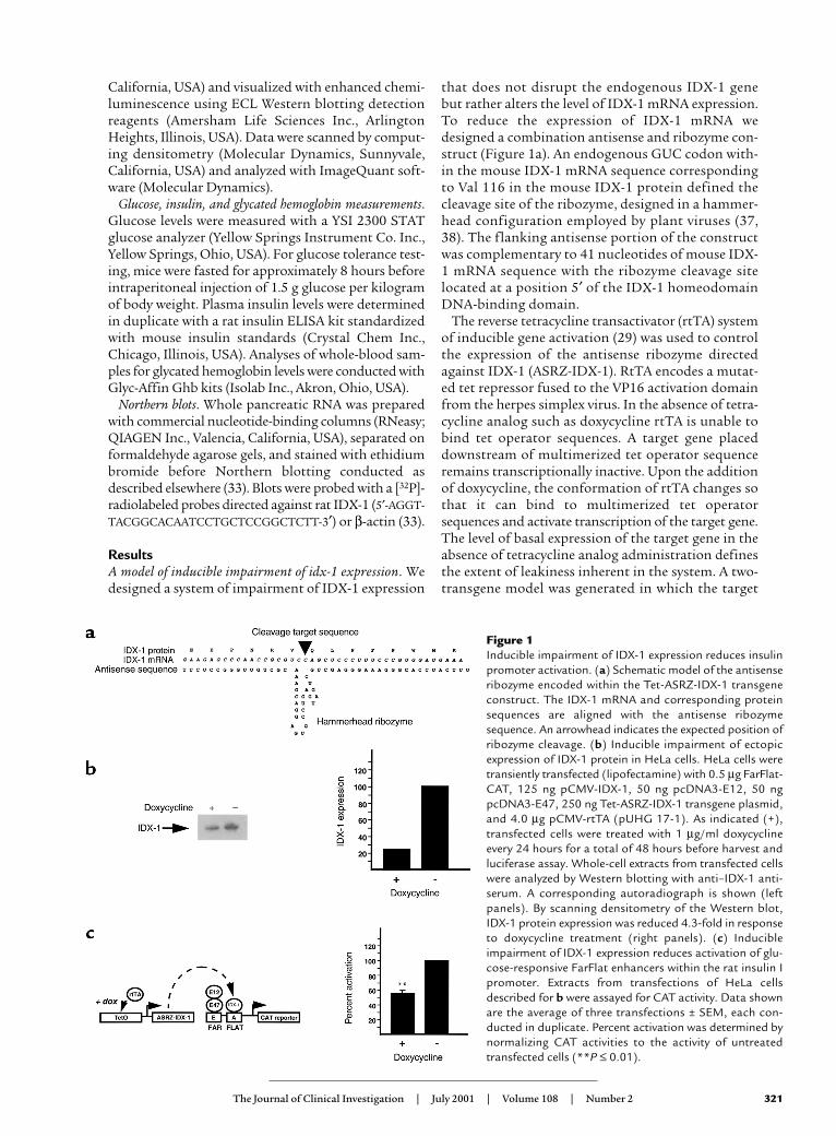
that does not disrupt the endogenous IDX-1 genebut rather alters the level of IDX-1 mRNA expression.To reduce the expression of IDX-1 mRNA wedesigned a combination antisense and ribozyme con-struct (Figure 1a). An endogenous GUC codon with-in the mouse IDX-1 mRNA sequence correspondingto Val 116 in the mouse IDX-1 protein defined thecleavage site of the ribozyme, designed in a hammer-head configuration employed by plant viruses (37,38). The flanking antisense portion of the constructwas complementary to 41 nucleotides of mouse IDX-1 mRNA sequence with the ribozyme cleavage sitelocated at a position 5′ of the IDX-1 homeodomainDNA-binding domain.
The reverse tetracycline transactivator (rtTA) systemof inducible gene activation (29) was used to controlthe expression of the antisense ribozyme directedagainst IDX-1 (ASRZ-IDX-1). RtTA encodes a mutat-ed tet repressor fused to the VP16 activation domainfrom the herpes simplex virus. In the absence of tetra-cycline analog such as doxycycline rtTA is unable tobind tet operator sequences. A target gene placeddownstream of multimerized tet operator sequenceremains transcriptionally inactive. Upon the additionof doxycycline, the conformation of rtTA changes sothat it can bind to multimerized tet operatorsequences and activate transcription of the target gene.The level of basal expression of the target gene in theabsence of tetracycline analog administration definesthe extent of leakiness inherent in the system. A two-transgene model was generated in which the target
The Journal of Clinical Investigation | July 2001 | Volume 108 | Number 2 321
Figure 1Inducible impairment of IDX-1 expression reduces insulinpromoter activation. (a) Schematic model of the antisenseribozyme encoded within the Tet-ASRZ-IDX-1 transgeneconstruct. The IDX-1 mRNA and corresponding proteinsequences are aligned with the antisense ribozymesequence. An arrowhead indicates the expected position ofribozyme cleavage. (b) Inducible impairment of ectopicexpression of IDX-1 protein in HeLa cells. HeLa cells weretransiently transfected (lipofectamine) with 0.5 µg FarFlat-CAT, 125 ng pCMV-IDX-1, 50 ng pcDNA3-E12, 50 ngpcDNA3-E47, 250 ng Tet-ASRZ-IDX-1 transgene plasmid,and 4.0 µg pCMV-rtTA (pUHG 17-1). As indicated (+),transfected cells were treated with 1 µg/ml doxycyclineevery 24 hours for a total of 48 hours before harvest andluciferase assay. Whole-cell extracts from transfected cellswere analyzed by Western blotting with anti–IDX-1 anti-serum. A corresponding autoradiograph is shown (leftpanels). By scanning densitometry of the Western blot,IDX-1 protein expression was reduced 4.3-fold in responseto doxycycline treatment (right panels). (c) Inducibleimpairment of IDX-1 expression reduces activation of glu-cose-responsive FarFlat enhancers within the rat insulin Ipromoter. Extracts from transfections of HeLa cellsdescribed for b were assayed for CAT activity. Data shownare the average of three transfections ± SEM, each con-ducted in duplicate. Percent activation was determined bynormalizing CAT activities to the activity of untreatedtransfected cells (**P ≤ 0.01).
gene is an antisense ribozyme targeted against IDX-1(Tet-ASRZ-IDX-1). In the RIP-rtTA transgene, expres-sion of rtTA was directed to pancreatic β cells with therat insulin II promoter (39). The model was designedso that the addition of a tetracycline analog such asdoxycycline would increase expression of the antisenseribozyme, decrease levels of IDX-1 mRNA, and there-by reduce IDX-1 protein levels.
The function of the RIP-rtTA transgene constructwas tested in the clonal pancreatic β cell line βTC3 (35)by cotransfection of the RIP-rtTA transgene constructand a tet-luciferase reporter plasmid pUHC 13-3 (27).Doxycycline increased the expression of the tet-luciferase reporter 15-fold, indicating that the RIP-rtTAtransgene construct was properly expressed in pancre-atic β cells and induced target gene expression inresponse to doxycycline.
Impairment of IDX-1 expression reduces activation of theinsulin promoter. The function of the Tet-ASRZ-IDX-1construct was assessed in COS7 cells in which theendogenous Idx-1 gene is not expressed. Cells weretransiently transfected with an expression plasmid forrat IDX-1, a plasmid encoding rtTA regulated by thehuman CMV promoter (pUHG 17-1) (29), and the Tet-ASRZ-IDX-1 plasmid. IDX-1 protein levels were
assessed by Western blotting of whole-cell extracts.Doxycycline markedly attenuated IDX-1 expressionindicating that induction of expression of the anti-sense ribozyme decreased IDX-1 levels.
We next reconstituted an experimental model systemin HeLa cells in which the glucose-responsive elementsFar (E) and Flat (A) of the rat insulin I promoter areknown to be activated by the synergism between IDX-1 and the basic helix-loop-helix transcription fac-tors E12 and E47 (15, 24, 40, 41). HeLa cells were tran-siently transfected with expression plasmids encodingIDX-1, E12, E47, a FarFlat-CAT reporter plasmid,pCMV-rtTA (pUHG17-1) and Tet-ASRZ-IDX-1 plas-mids. The administration of doxycycline reduced theexpression of IDX-1 protein fourfold (Figure 1b). Thisdegree of reduction in IDX-1 levels had significantfunctional consequences. Synergistic activation of theinsulin promoter glucose-responsive elements FarFlatby coexpression of IDX-1 with the basic helix-loop-helix transcription factors E12 and E47 (15, 24, 40, 41)was reduced in cells treated with doxycycline, indicat-ing that IDX-1 levels can be rate-limiting for insulingene activation (Figure 1c). These effects in transcrip-tional regulation in response to doxycycline were com-parable in magnitude to the induction of luciferase
322 The Journal of Clinical Investigation | July 2001 | Volume 108 | Number 2
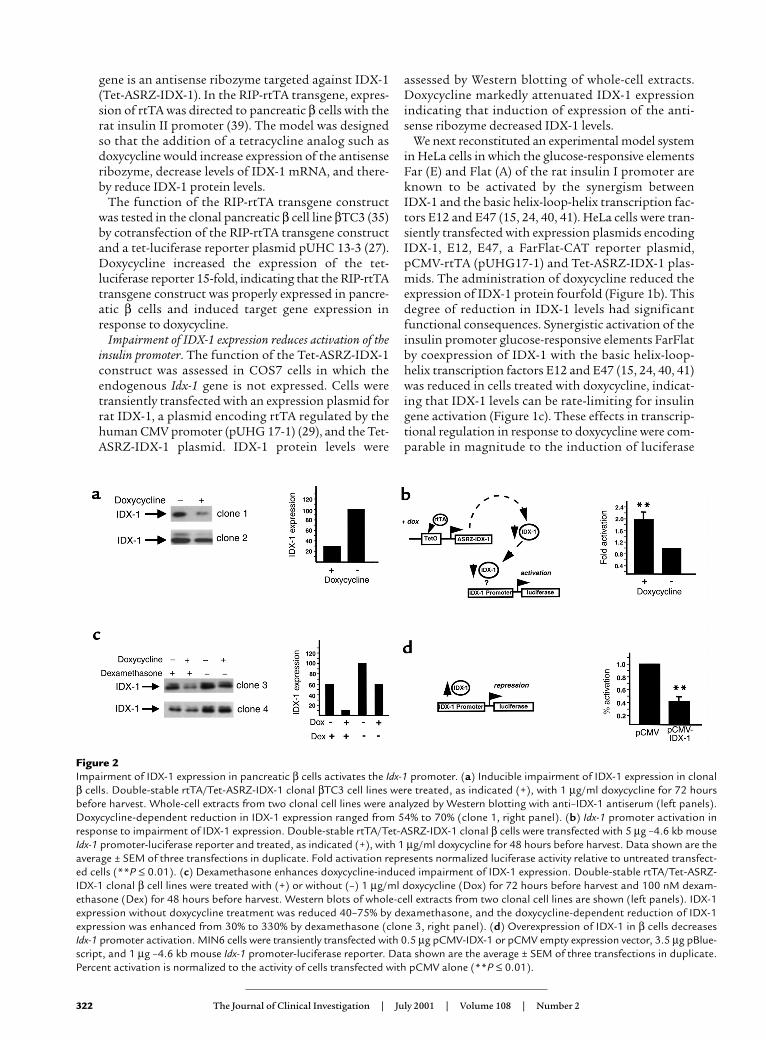
Figure 2Impairment of IDX-1 expression in pancreatic β cells activates the Idx-1 promoter. (a) Inducible impairment of IDX-1 expression in clonalβ cells. Double-stable rtTA/Tet-ASRZ-IDX-1 clonal βTC3 cell lines were treated, as indicated (+), with 1 µg/ml doxycycline for 72 hoursbefore harvest. Whole-cell extracts from two clonal cell lines were analyzed by Western blotting with anti–IDX-1 antiserum (left panels).Doxycycline-dependent reduction in IDX-1 expression ranged from 54% to 70% (clone 1, right panel). (b) Idx-1 promoter activation inresponse to impairment of IDX-1 expression. Double-stable rtTA/Tet-ASRZ-IDX-1 clonal β cells were transfected with 5 µg –4.6 kb mouseIdx-1 promoter-luciferase reporter and treated, as indicated (+), with 1 µg/ml doxycycline for 48 hours before harvest. Data shown are theaverage ± SEM of three transfections in duplicate. Fold activation represents normalized luciferase activity relative to untreated transfect-ed cells (**P ≤ 0.01). (c) Dexamethasone enhances doxycycline-induced impairment of IDX-1 expression. Double-stable rtTA/Tet-ASRZ-IDX-1 clonal β cell lines were treated with (+) or without (–) 1 µg/ml doxycycline (Dox) for 72 hours before harvest and 100 nM dexam-ethasone (Dex) for 48 hours before harvest. Western blots of whole-cell extracts from two clonal cell lines are shown (left panels). IDX-1expression without doxycycline treatment was reduced 40–75% by dexamethasone, and the doxycycline-dependent reduction of IDX-1expression was enhanced from 30% to 330% by dexamethasone (clone 3, right panel). (d) Overexpression of IDX-1 in β cells decreases Idx-1 promoter activation. MIN6 cells were transiently transfected with 0.5 µg pCMV-IDX-1 or pCMV empty expression vector, 3.5 µg pBlue-script, and 1 µg –4.6 kb mouse Idx-1 promoter-luciferase reporter. Data shown are the average ± SEM of three transfections in duplicate.Percent activation is normalized to the activity of cells transfected with pCMV alone (**P ≤ 0.01).
activity by doxycycline in HeLa cells cotransfected withpCMV-rtTA and tet-luciferase. The magnitude ofinducible target gene regulation with the rtTA systemoften is reduced in transient transfection systems com-pared with stably transfected cells (29).
Activation of the IDX-1 promoter in response to impair-ment of IDX-1 expression in pancreatic β cells. To analyzethe efficacy of the impairment of IDX-1 expression inpancreatic β cells, we developed double-stable β celllines (βTC3) in which rtTA and Tet-ASRZ-IDX-1 werestably expressed under selection with G418 andhygromycin B. We analyzed multiple individual cellclones for efficiency of impairment of IDX-1 expres-sion in response to doxycycline. For many of the dou-ble-stable clones, we observed 50–70% decreases inthe expression of IDX-1 protein after 72 hours ofexposure of the cells to doxycycline (Figure 2a). Doxy-cycline did not affect IDX-1 protein expression levelsin stable β cell lines in which rtTA alone was stablyexpressed. Further analysis of IDX-1 expression inresponse to doxycycline in selected double-stableclones over time revealed an interesting pattern. Aftera reduction in IDX-1 levels at 48–72 hours of doxycy-cline treatment, IDX-1 protein levels returned to thebasal state after 92 hours of doxycycline exposure. Weinterpreted this pattern of IDX-1 expression as anindication that compensatory mechanisms existwithin the pancreatic β cell to maintain normal IDX-1 levels. Because IDX-1 is a critical regulator ofpancreatic β cell function, we reasoned that mecha-nisms likely exist in pancreatic β cells to preserve cel-lular levels of IDX-1.
One potential mechanism to compensate fordecreases in IDX-1 mRNA expression would be toincrease IDX-1 mRNA production by activating theendogenous Idx-1 gene promoter to overcome theactivity of the antisense ribozyme construct. We test-ed this possibility by transiently transfecting double-stable rtTA/Tet-ASRZ-IDX-1 β cells with a –4.6 kbmouse Idx-1 promoter-reporter construct. This seg-ment of the mouse Idx-1 promoter contains the regu-latory sequences necessary to faithfully impart thedevelopmental and differentiated β cell expression pat-terns of mouse IDX-1 (42). Treatment of the trans-fected double-stable rtTA/Tet-ASRZ-IDX-1 β cellswith doxycycline activated the Idx-1 promoter approx-imately twofold relative to untreated transfected cells(Figure 2b). On Northern blots within 20 hours oftreatment of double-stable rtTA/Tet-ASRZ-IDX-1 βcells with doxycycline, this compensatory mechanismwas detected as an increase in IDX-1 mRNA levels of25–60% compared with those of untreated control lev-els (normalized to 18S rRNA and actin mRNA, respec-tively). The IDX-1 mRNA levels in this model systemreflect the sum of the two processes of the reductionof IDX-1 expression by the antisense ribozyme and therestoration of IDX-1 expression by activation of theIdx-1 promoter. These results indicated that the Idx-1promoter is able to sense, either through direct or indi-
rect mechanisms, the levels of IDX-1 protein in thepancreatic β cell. The Idx-1 promoter senses IDX-1deficiency and is consequently activated thereby pro-viding a mechanism to restore IDX-1 levels back to thenormal ambient concentration.
To further evaluate the mechanism of Idx-1 promot-er autoregulation, we selected double-stable rtTA/Tet-ASRZ-IDX-1 β cell clones in which minimal reduc-tions of IDX-1 protein levels in response to doxycyclineadministration were observed. The addition of dex-amethasone, an agent known to repress the activity ofthe Idx-1 promoter via an HNF 3β-mediated mecha-nism (23), to these double-stable clonal cell linesreduced basal IDX-1 protein levels by 40–75% asexpected (Figure 2c). Interestingly, repression of Idx-1promoter activation enhanced the ability of doxycy-cline to reduce IDX-1 protein expression by as muchas 300% (Figure 2c). These findings suggested that theactivity of the endogenous Idx-1 gene promoter limitsthe potency of the antisense ribozyme. The Idx-1 pro-moter likely senses both decreases and increases inIDX-1 expression levels. In transient transfections of
The Journal of Clinical Investigation | July 2001 | Volume 108 | Number 2 323
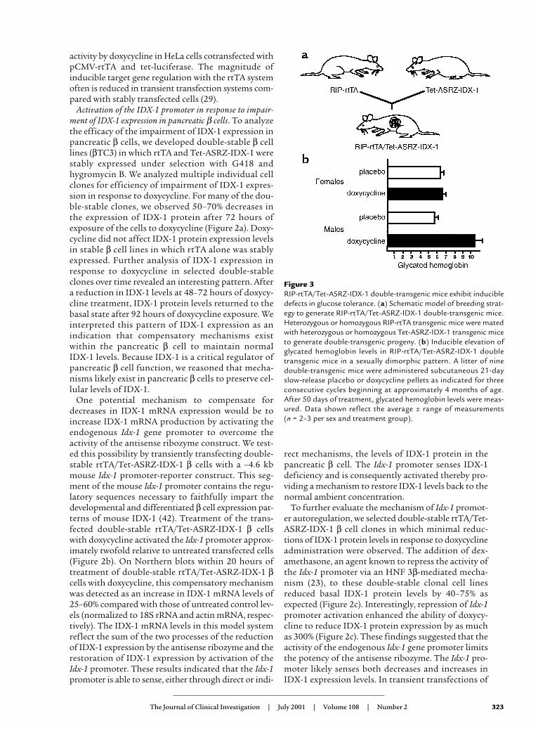
Figure 3RIP-rtTA/Tet-ASRZ-IDX-1 double-transgenic mice exhibit inducibledefects in glucose tolerance. (a) Schematic model of breeding strat-egy to generate RIP-rtTA/Tet-ASRZ-IDX-1 double-transgenic mice.Heterozygous or homozygous RIP-rtTA transgenic mice were matedwith heterozygous or homozygous Tet-ASRZ-IDX-1 transgenic miceto generate double-transgenic progeny. (b) Inducible elevation ofglycated hemoglobin levels in RIP-rtTA/Tet-ASRZ-IDX-1 doubletransgenic mice in a sexually dimorphic pattern. A litter of ninedouble-transgenic mice were administered subcutaneous 21-dayslow-release placebo or doxycycline pellets as indicated for threeconsecutive cycles beginning at approximately 4 months of age.After 50 days of treatment, glycated hemoglobin levels were meas-ured. Data shown reflect the average ± range of measurements (n = 2–3 per sex and treatment group).
the clonal pancreatic β cell line MIN6, overexpressionof an expression plasmid encoding exogenous IDX-1suppressed activation of the Idx-1 promoter-reporterconstruct compared with cells transfected with theempty expression plasmid (Figure 2d). Thus IDX-1 lev-els appear to be regulated within a narrow range inpancreatic β cells.
Inducible impairment of IDX-1 expression in transgenicmice. We generated two strains of transgenic mice, RIP-rtTA and Tet-ASRZ-IDX-1 to avoid the integration ofboth transgenes in the same locus and minimize thelevels of basal ribozyme expression in pancreatic β cells(28). Three founder lines were bred and analyzed forthe Tet-ASRZ-IDX-1 transgene, and two founder lineswere bred and analyzed for the RIP-rtTA transgene. Togenerate a mouse model of inducible impairment ofIDX-1 expression, we mated RIP-rtTA and Tet-ASRZ-IDX-1 mice and produced double-transgenic RIP-rtTA/Tet-ASRZ-IDX-1 animals (Figure 3a). The dou-ble-transgenic mice developed normally in the absenceor presence of doxycycline, unlike IDX-1 knockoutmice that were born without a pancreas and diedshortly after birth (3, 12). No differences were observedin body weight or pancreatic morphology betweendouble-transgenic and nontransgenic littermate con-trol mice. Oral administration of doxycycline insucrose vehicle for 4 weeks to double-transgenic RIP-rtTA/Tet-ASRZ-IDX-1 mice at 3-4 months of age didnot impair glucose tolerance as assessed by intraperi-toneal glucose tolerance testing.
As a paradigm for longer-term administration ofdoxycycline we subcutaneously implanted three con-secutive cycles of 21-day slow-release doxycycline (15mg) or placebo pellets (Innovative Research of America)in each animal in a litter of double-transgenic RIP-rtTA/Tet-ASRZ-IDX-1 mice that were generated frommating homozygous RIP-rtTA with homozygous Tet-ASRZ-IDX-1 mice. After day 50 of treatment with doxy-cycline, we measured serum glycated hemoglobin levelsin the 6-month-old animals. No significant differencesin glycated hemoglobin levels were noted among thethree female littermates treated with doxycycline andtheir two placebo-treated female littermates (Figure 3b).However, the two male littermates treated with doxycy-cline had substantially higher glycated hemoglobin lev-els than did their two placebo-treated male littermates(Figure 3b). These findings indicate a sexually dimor-phic phenotypic pattern in this mouse model. Sexualdimorphism is a common finding in many rodent mod-els of diabetes in which male mice have more markedabnormalities in glucose tolerance, including mousemodels of defective insulin action (43). The metabolicphenotype in our double-transgenic mouse model wasof low penetrance because six double-transgenic malemice from two subsequent litters did not develop ele-vated glycated hemoglobin levels in response to a simi-lar course of subcutaneous doxycycline administrationcompared with five placebo-treated double-transgenicmale littermate controls. Variable penetrance of dia-betes has been observed in other mouse models (43, 44)and in humans with IDX-1 mutations (7) and likelyreflects the variability in cosegregation of additionalgenes that confer susceptibility to or protection fromthe development of the disease.
Inducible diabetes mellitus with aging and IDX-1 deficiency.To obtain a phenotype of higher penetrance we treated
324 The Journal of Clinical Investigation | July 2001 | Volume 108 | Number 2
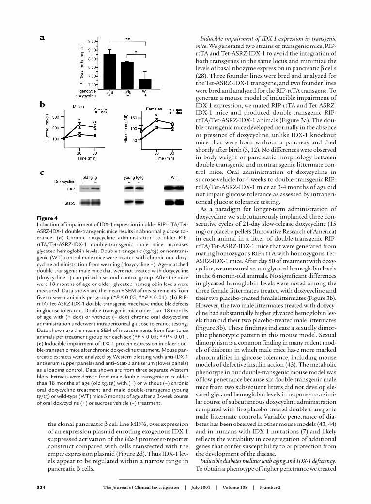
Figure 4Induction of impairment of IDX-1 expression in older RIP-rtTA/Tet-ASRZ-IDX-1 double-transgenic mice results in abnormal glucose tol-erance. (a) Chronic doxycycline administration to older RIP-rtTA/Tet-ASRZ-IDX-1 double-transgenic male mice increasesglycated hemoglobin levels. Double transgenic (tg/tg) or nontrans-genic (WT) control male mice were treated with chronic oral doxy-cycline administration from weaning (doxycycline +). Age-matcheddouble-transgenic male mice that were not treated with doxycycline(doxycycline –) comprised a second control group. After the micewere 18 months of age or older, glycated hemoglobin levels weremeasured. Data shown are the mean ± SEM of measurements fromfive to seven animals per group (*P ≤ 0.05; **P ≤ 0.01). (b) RIP-rtTA/Tet-ASRZ-IDX-1 double-transgenic mice have inducible defectsin glucose tolerance. Double-transgenic mice older than 18 monthsof age with (+ dox) or without (– dox) chronic oral doxycyclineadministration underwent intraperitoneal glucose tolerance testing.Data shown are the mean ± SEM of measurements from four to sixanimals per treatment group for each sex (*P < 0.05; **P < 0.01).(c) Inducible impairment of IDX-1 protein expression in older dou-ble-transgenic mice after chronic doxycycline treatment. Mouse pan-creatic extracts were analyzed by Western blotting with anti-IDX-1antiserum (upper panels) and anti–Stat-3 antiserum (lower panels)as a loading control. Data shown are from three separate Westernblots. Extracts were derived from male double-transgenic mice olderthan 18 months of age (old tg/tg) with (+) or without (–) chronicoral doxycycline treatment and male double-transgenic (youngtg/tg) or wild-type (WT) mice 3 months of age after a 3-week courseof oral doxycycline (+) or sucrose vehicle (–) treatment.
double-transgenic RIP-rtTA/Tet-ASRZ-IDX-1 and non-transgenic control mice from the time of weaning withchronic oral doxycycline in a sucrose vehicle. At 3months of age, no significant differences were observedin glucose tolerance between double-transgenic mice inthe presence or absence of chronic oral doxycyclineadministration, as assessed by intraperitoneal glucosetolerance tests. At 8 months of age, no significant dif-ferences were found in glucose or glycated hemoglobinlevels between double-transgenic mice in the presenceor absence of chronic oral doxycycline administration.
However, after the mice were more than 18 monthsof age, significant differences were observed in gly-cated hemoglobin levels between double-transgenicRIP-rtTA/Tet-ASRZ-IDX-1 male mice and nontrans-genic control mice after chronic doxycycline adminis-tration (Figure 4a). With chronic oral administrationof doxycycline, the double transgenic male mice (Fig-ure 4a, tg/tg) had significantly higher glycated hemo-globin levels than did nontransgenic control malemice (Figure 4a, WT). Of note, double-transgenicmale mice not treated with doxycycline had lower gly-cated hemoglobin levels than did age-matched dou-ble-transgenic male mice treated with chronic doxy-cycline, but they also had higher glycated hemoglobinlevels than did nontransgenic controls. These data areconsistent with a level of basal expression of the anti-sense ribozyme in the absence of tetracycline analogadministration (leakiness in the transcriptional regu-latory system) sufficient to induce abnormal glucosetolerance. To better characterize the phenotype indouble-transgenic mice older than the age of 18months induced by chronic oral doxycycline, we per-formed intraperitoneal glucose tolerance tests after 8-hour fasts (Figure 4b). The chronic administration ofdoxycycline to the double-transgenic male miceresulted in significantly higher glucose excursions at30 and 60 minutes relative to untreated double-trans-genic age- and sex-matched mice. The impairment inglucose tolerance in males in this double-transgenicmouse model in response to doxycycline was accom-panied by significant reductions in serum insulin/glu-cose ratios (Table 1). After treatment with doxycy-cline, variable impaired expression of IDX-1 proteinwas detected in pancreatic extracts derived from olderbut not younger double-transgenic male mice com-
pared with untreated age- and sex-matched doubletransgenic controls (Figure 4c). With chronic doxycy-cline treatment the double-transgenic female mice didnot have significant inducible elevations in glycatedhemoglobin levels. Double-transgenic female micehad lower fasting and stimulated glucose levels thandid male mice with small doxycycline-induced eleva-tions of glucose levels (Figure 4b).
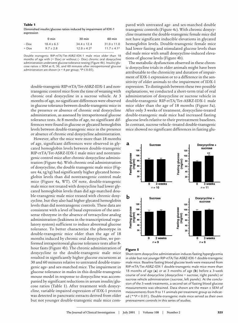
The metabolic dysfunction observed in these chron-ic doxycycline trials in older animals might have beenattributable to the chronicity and duration of impair-ment of IDX-1 expression or to a difference in the sen-sitivity of older animals to the impairment of IDX-1expression. To distinguish between these two possibleexplanations, we conducted a short-term trial of oraladministration of doxycycline or sucrose vehicle indouble-transgenic RIP-rtTA/Tet-ASRZ-IDX-1 malemice older than the age of 18 months (Figure 5a).After only 3 weeks of treatment, doxycycline-treateddouble-transgenic male mice had increased fastingglucose levels relative to their pretreatment baselines.In contrast, sucrose vehicle–treated double-transgenicmice showed no significant differences in fasting glu-
The Journal of Clinical Investigation | July 2001 | Volume 108 | Number 2 325
Figure 5Short-term doxycycline administration induces fasting hyperglycemiain older but not younger RIP-rtTA/Tet-ASRZ-IDX-1 double-transgenicmale mice. Baseline fasting blood glucose levels were measured fromRIP-rtTA/Tet-ASRZ-IDX-1 double-transgenic male mice more than18 months of age (a) or at 3 months of age (b) before a 3-weekcourse of oral doxycycline (doxycycline + sucrose, right panels) orsucrose vehicle administration (sucrose, left panels). At the conclu-sion of the 3-week treatments, a second set of fasting blood glucosemeasurements was obtained. Data shown are the mean ± SEM ofmeasurements from 9 to 11 animals per treatment group as indicat-ed (**P < 0.01). Double-transgenic male mice served as their ownpretreatment controls in this series of studies.
Table 1Diminished insulin/glucose ratios induced by impairment of IDX-1expression
0 min 30 min 60 min
– Dox 18.4 ± 6.5 34.4 ± 12.4 31.0 ± 11.0+ Dox 9.7 ± 2.8 12.0 ± 4.2A 11.7 ± 4.1A
Double transgenic RIP-rtTA/Tet-ASRZ-IDX-1 male mice older than 18months of age with (+ Dox) or without (– Dox) chronic oral doxycyclineadministration underwent glucose-tolerance testing (Figure 4b). Insulin/glu-cose ratios ± SEM at 0, 30, and 60 minutes after intraperitoneal glucoseadministration are shown (n = 4 per group; AP ≤ 0.05).
cose levels before and after treatment. Double trans-genic RIP-rtTA/Tet-ASRZ-IDX-1 female mice morethan the age of 18 months had no significant increas-es in fasting glucose levels after 3 weeks of doxycyclinetreatment. Treatment of 3-month-old double-trans-genic RIP-rtTA/Tet-ASRZ-IDX-1 male mice for 3weeks with sucrose vehicle or doxycycline did notresult in any significant differences in fasting glucoselevels before and after treatment (Figure 5b). Doxycy-cline-dependent changes in pancreatic IDX-1 proteinlevels were not detected after 3 weeks of treatment of3-month-old double transgenic (Figure 4c, youngtg/tg) male mice or nontransgenic male wild type(Figure 4c, WT) male control mice (Figure 4c). Non-transgenic wild-type 3-month-old male mice showedno significant differences in fasting glucose levelsobtained after 3 weeks of treatment with sucrose-vehi-cle or doxycycline. These data suggest that older ani-mals have increased sensitivity to the impairment ofIDX-1 expression and demonstrate that short-termreduction of IDX-1 expression in older animals is suf-ficient to impair glucose tolerance.
DiscussionDeficiency of the pancreatic homeodomain transcrip-tion factor IDX-1 impairs glucose tolerance. Inhumans, IDX-1 appears to normally function within alimited concentration range because heterozygosityfor mutations in the Idx-1 gene is sufficient to con-tribute to a metabolic phenotype (7–9). In other modelsystems, transcription factor dosage can provide a sub-stantial impact. By altering expression levels of indi-
vidual transcription factors, cell fate decisions anddevelopmental programs are initiated or terminated(45, 46). Our studies of inducible IDX-1 deficiencysuggest that even modest decrements in IDX-1 levelsin differentiated pancreatic β cells can have significantmetabolic consequences.
In contrast to previous mouse models, the Idx-1 genewas not mutated or disrupted in our studies, therebyenabling normal regulatory mechanisms in the pan-creatic β cell to compensate for decreased IDX-1 expres-sion. A variety of possible regulatory responses areavailable within the β cell to preserve IDX-1 functionin response to the reduction of IDX-1 mRNA levelsincluding increasing rates of IDX-1 protein synthesis,IDX-1 protein stability, cytoplasmic to nuclear translo-cation of IDX-1 protein, expression of substitute tran-scriptional activators (including other homeodomaintranscription factors), and Idx-1 gene transcription. Weidentified one such compensatory mechanism utilizedby pancreatic β cells in response to induction of expres-sion of the antisense ribozyme targeted against IDX-1;namely, the activation of the Idx-1 promoter. The mag-nitude of this compensatory promoter activation wasonly approximately twofold, but within the expectedrange of differences in IDX-1 expression levels betweenhumans heterozygous for Idx-1 mutations with a pre-disposition to the development of diabetes mellitusand humans without mutations. The implication ofthe observation that mutations in a single copy of theIdx-1 gene result in a metabolic phenotype is that com-pensatory mechanisms for IDX-1 deficiency rely heav-ily on two intact Idx-1 genes. Although alternate com-pensatory mechanisms that stabilize IDX-1 proteinlevels may still be operative in younger individuals, theyare insufficient to prevent the development of defectiveglucose tolerance with aging in humans heterozygousfor Idx-1 missense mutations.
We propose that IDX-1 levels regulate the activationof the Idx-1 promoter in an autoregulatory feedbackloop in pancreatic β cells. Our data suggest that theIdx-1 promoter is able to sense and respond to declin-ing IDX-1 levels with increased transcriptional acti-vation to restore levels of IDX-1 to the normal range.In the setting of excess IDX-1, as in our overexpres-sion transfection studies (Figure 2d), the Idx-1 pro-moter responds with decreased transcriptional acti-vation. Both of these proposed feedback mechanismswould serve to maintain IDX-1 protein levels in atightly regulated range in the pancreatic β cell. Mech-anisms to alter Idx-1 promoter activation may bedirectly and/or indirectly mediated by the IDX-1 pro-tein. Multiple AT-rich regions that could serve aspotential direct binding sites for IDX-1 are presentwithin the mouse and human Idx-1 promoters. TheIDX-1 protein binds to oligonucleotides encoding onesuch AT-rich region within the human Idx-1 promot-er that contributes to β cell–specific expression (47).The expression of IDX-1 in NIH-3T3 cells activates anenhancer-reporter construct that incorporates this
326 The Journal of Clinical Investigation | July 2001 | Volume 108 | Number 2
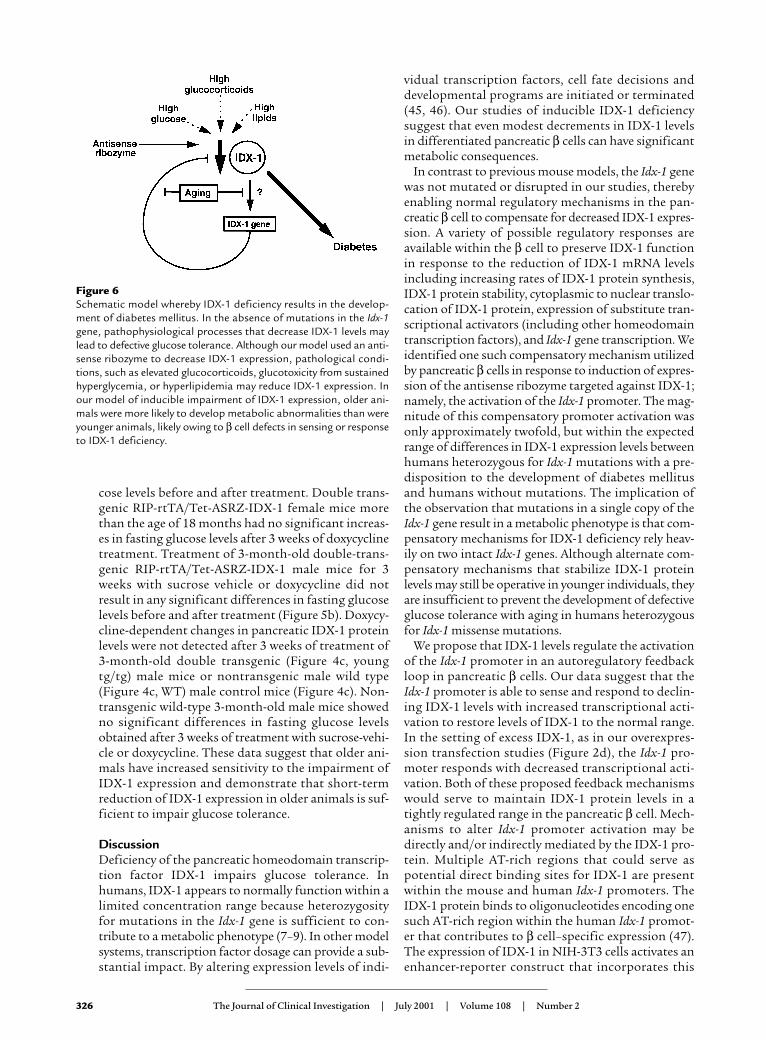
Figure 6Schematic model whereby IDX-1 deficiency results in the develop-ment of diabetes mellitus. In the absence of mutations in the Idx-1gene, pathophysiological processes that decrease IDX-1 levels maylead to defective glucose tolerance. Although our model used an anti-sense ribozyme to decrease IDX-1 expression, pathological condi-tions, such as elevated glucocorticoids, glucotoxicity from sustainedhyperglycemia, or hyperlipidemia may reduce IDX-1 expression. Inour model of inducible impairment of IDX-1 expression, older ani-mals were more likely to develop metabolic abnormalities than wereyounger animals, likely owing to β cell defects in sensing or responseto IDX-1 deficiency.
AT-rich region in synergy with HNF-3β. On the basisof these studies, a positive feedback loop was pro-posed in which IDX-1 expression activates its ownpromoter (47). Our studies suggest that in the contextof a pancreatic β cell in which IDX-1 is normallyexpressed, overexpression of IDX-1 inhibits Idx-1 pro-moter activity. It is possible that IDX-1–mediatedautoregulation may operate differently in distinct cel-lular or developmental contexts. Multiple enhancersare differentially used in pituitary development andin the autoregulation of expression of the homeopro-tein Pit-1 (48), and it is conceivable that mechanismsof IDX-1 autoregulation will be of similar complexity.
Insulin production declined in response to theimpairment of IDX-1 expression, a finding that is con-sistent with the critical importance of IDX-1 in the reg-ulation of the insulin gene. The magnitude of theinducible hyperglycemia may have been limited byextrapancreatic adjustments to decreased insulin pro-duction because increased insulin sensitivity has beenreported in humans with Idx-1 mutations (8, 49).
The pattern of phenotypic sexual dimorphismobserved in the RIP-rtTA/Tet-ASRZ-IDX-1 double-transgenic mouse model is interesting and has notbeen reported in humans with Idx-1 mutations. Apropensity for male mice to develop abnormalities inglucose tolerance more readily than female littermatesis observed in other diabetes models (43), but the mech-anisms responsible for such a pattern are unknown.Human females have lower fasting glucose levels (50)with reduced epinephrine, glucagon, and hepatic glu-cose production in response to insulin-induced hypo-glycemia (51–53). Sex-related differences in neuroen-docrine and metabolic responses to exercise also havebeen reported (54). The US Centers for Disease Controland Prevention report a steady increase in the preva-lence of diabetes in Americans over the age of 65 from1989 to 1996, with a steeper rate of increase in malescompared with females (55).
The convergence of IDX-1 deficiency, aging, and dia-betes was somewhat surprising but underscores simi-lar phenotypes among humans with IDX-1 mutationsand mouse models. The severity of the mutations inthe human Idx-1 gene correlate with the age of onset ofmetabolic dysfunction (7–9). In our mouse model,younger animals did not develop metabolic pheno-types in response to doxycycline administration, butafter 18 months of age, male mice were highly suscep-tible to impairment of IDX-1 expression. This pheno-typic pattern suggests that mechanisms used by pan-creatic β cells to compensate for reductions in IDX-1expression decline with aging (Figure 6). Of note, amouse model of conditional β cell–specific disruptionof the IDX-1 gene using the cre/lox system did notdevelop diabetes until 4 months of age (11), but theage dependence of the phenotype may have beenattributable to the time required for sufficient accu-mulation of cre expression in pancreatic β cells toexcise the targeted region of the Idx-1 gene. In contrast
to our RIP-rtTA/Tet-ASRZ-IDX-1 transgenic mousemodel, in the cre-lox model the endogenous Idx-1 geneis disrupted and would not be expected to respondwith normal compensatory mechanisms.
Increasing insulin resistance has been identified asa major component in the increased risk of the devel-opment of type 2 diabetes with aging. However, wefound no evidence of hyperinsulinemia in the age-dependent inducible development of diabetes in ourRIP-rtTA/Tet-ASRZ-IDX-1 double-transgenic mice. Infact, induction of IDX-1 deficiency resulted in dimin-ished, not increased, insulin/glucose ratios. Age-dependent reductions in pancreatic islet insulin production in rats have been reported (56). Our dataimply that defects in pancreatic β cell function, par-ticularly in the ability to regulate IDX-1 expression,develop with aging. In other contexts, age-dependentchanges in gene expression correlate with modifica-tions in oxidative stress responses, caloric intake,mitotic regulation, and chromatin silencing (57–60).
The RIP-rtTA/Tet-ASRZ-IDX-1 double-transgenicmodel system of inducible IDX-1 deficiency has someintrinsic limitations. The extent of ASRZ-IDX-1 expres-sion is directly regulated by rtTA expression, and thusindirectly limited by the activity of the rat insulin IIpromoter. Neogenesis, or the development of new pan-creatic β cells from precursor cells, is an active processin the endocrine pancreas (61). Newly developing pan-creatic β cells would not be susceptible to impairmentof IDX-1 expression until insulin promoter activationwas sufficient to promote rtTA expression, providing apotential mechanism to transiently bypass reductionof IDX-1 expression. In addition, the potency of themodel system in IDX-1 inactivation is inherently limit-ed by its design. Because IDX-1 is an important regula-tor of the activation of the insulin promoter, at somepoint a decrement in IDX-1 levels is likely to reduce ratinsulin II promoter activity resulting in decreased rtTAexpression, reducing ASRZ-IDX-1 expression and pro-viding a limit to the extent of IDX-1 deficiency that canbe induced. Furthermore, the higher glycated hemo-globin levels in double-transgenic mice in the absenceof treatment with doxycycline implies a level of basalASRZ-IDX-1 expression. This regulatory leak in ourmouse model system limits the range of doxycycline-inducible differences in IDX-1 expression.
Although mutations in the Idx-1 gene are found inonly 3–6% of individuals with type 2 diabetes (8, 9),our data suggest that abnormalities in glucose toler-ance due to IDX-1 deficiency may occur in the largerpopulation without IDX-1 mutations. Other investi-gators have identified a variety of insults that impairIDX-1 expression, including hyperglycemia, hyper-lipidemia, and excess glucocorticoids (18–23). If agingdiminishes the capacity of the pancreatic β cell torestore impaired IDX-1 expression, older individualsare likely to be at greater risk for the subsequent devel-opment of diabetes. Thus, the development of thera-peutic strategies to restore IDX-1 expression in set-
The Journal of Clinical Investigation | July 2001 | Volume 108 | Number 2 327
tings of IDX-1 deficiency may be of particular impor-tance for older adults. In this regard, activators of Idx-1 gene expression, such as glucagon-like peptide-1 (36, 62) and hedgehog signaling proteins (26), war-rant further investigation to assess their therapeuticpotential in type 2 diabetes.
AcknowledgmentsWe thank H. Bujard for rtTA regulatory system plas-mids, S. Efrat for βTC3 cells, J. Miyazaki for MIN6cells, J.L. Moss for FarFlat-CAT, and G.C. Weir forresearch support and insightful discussions. We appre-ciate the expert technical assistance of H. Hermannand K. McManus. We thank D. Nathan and the hemo-globin A1C clinical laboratory at Massachusetts Gen-eral Hospital for assistance with glycated hemoglobinassays. We are indebted to the members of the Labora-tory of Molecular Endocrinology for valuable discus-sions. We also thank T. Budde for assistance in thepreparation of this manuscript. These studies weresupported by grants DK02476 and DK58783 (M.K.Thomas) and DK30457 and DK30834 (J.F. Habener)from the NIH. M.K. Thomas is the recipient of aHoward Hughes Medical Institute Postdoctoral Fel-lowship for Physicians. J.F. Habener is an Investigatorwith the Howard Hughes Medical Institute.
1. Ohlsson, H., Karlsson, K., and Edlund, T. 1993. IPF1, a homeodomain-containing transactivator of the insulin gene. EMBO J. 12:4251–4259.
2. Leonard, J., et al. 1993. Characterization of somatostatin transactivat-ing factor-1, a novel homeobox factor that stimulates somatostatinexpression in pancreatic islet cells. Mol. Endocrinol. 7:1275–1283.
3. Offield, M.F., et al. 1996. PDX-1 is required for pancreatic outgrowthand differentiation of the rostral duodenum. Development.122:983–995.
4. Marshak, S., Totary, H., Cerasi, E., and Melloul, D. 1996. Purificationof the beta-cell glucose-sensitive factor that transactivates the insulingene differentially in normal and transformed islet cells. Proc. Natl.Acad. Sci. USA. 93:15057–15062.
5. Stoffers, D.A., Thomas, M.K., and Habener, J.F. 1997. Homeodomainprotein IDX-1: a master regulator of pancreas development and insulingene expression. Trends Endocrinol. Metab. 8:145–151.
6. Edlund, H. 1998. Transcribing pancreas. Diabetes. 47:1817–1823.7. Stoffers, D.A., Ferrer, J., Clarke, W.L., and Habener, J.F. 1997. Early-
onset type-II diabetes mellitus (MODY4) linked to IPF-1. Nat. Genet.17:138–139.
8. Hani, E.H., et al. 1999. Defective mutations in the insulin promoterfactor-1 (IPF-1) gene in late-onset type 2 diabetes mellitus. J. Clin. Invest.104:R41–R48.
9. Macfarlane, W.M., et al. 1999. Missense mutations in the insulin pro-moter factor-1 gene predispose to type 2 diabetes. J. Clin. Invest.104:R33–R39.
10. Dutta, S., Bonner-Weir, S., Montminy, M., and Wright, C. 1998. Regu-latory factor linked to late-onset diabetes? Nature. 392:560.
11. Ahlgren, U., Jonsson, J., Jonsson, L., Simu, K., and Edlund, H. 1998. β-cell-specific inactivation of the mouse Ipf1/Pdx1 gene results in loss ofthe β-cell phenotype and maturity onset diabetes. Genes Dev.12:1763–1768.
12. Jonsson, J., Carlsson, L., Edlund, T., and Edlund, H. 1994. Insulin-pro-moter-factor 1 is required for pancreas development in mice. Nature.371:606–609.
13. Stoffers, D.A., Zinkin, N.T., Stanojevic, V., Clarke, W.L., and Habener,J.F. 1997. Pancreatic agenesis attributable to a single nucleotide dele-tion in the human IPF1 coding region. Nat Genet. 15:106–110.
14. German, M.S., Moss, L.G., Wang, J., and Rutter, W.J. 1992. The insulinand islet amyloid polypeptide genes contain similar cell-specific pro-moter elements that bind identical beta-cell nuclear complexes. Mol.Cell. Biol. 12:1777–1788.
15. Peers, B., Leonard, J., Sharma, S., Teitelman, G., and Montminy, M.R.1994. Insulin expression in pancreatic islet cells relies on cooperativeinteractions between the helix loop helix factor E47 and the homeobox
factor STF-1. Mol. Endocrinol. 8:1798–1806.16. Waeber, G., Thompson, N., Nicod, P., and Bonny, C. 1996. Transcrip-
tional activation of the GLUT2 gene by the IPF-1/STF-1/IDX-1 home-obox factor. Mol. Endocrinol. 10:1327–1334.
17. Watada, H., et al. 1996. PDX-1 induces insulin and glucokinase geneexpressions in αTC1 clone 6 cells in the presence of betacellulin. Dia-betes. 45:1826–1831.
18. Olson, L.K., et al. 1995. Reduction of insulin gene transcription in HIT-T15 β cells chronically exposed to a supraphysiologic glucose concen-tration is associated with loss of STF-1 transcription factor expression.Proc. Natl. Acad. Sci. USA. 92:9127–9131.
19. Sharma, A., Olson, L.K., Robertson, R.P., and Stein, R. 1995. The reduc-tion of insulin gene transcription in HIT-T15 β cells chronicallyexposed to high glucose concentration is associated with the loss ofRIPE3b1 and STF-1 transcription factor expression. Mol. Endocrinol.9:1127–1134.
20. Zangen, D.H., et al. 1997. Reduced insulin, GLUT2, and IDX-1 in β-cells after partial pancreatectomy. Diabetes. 46:258–264.
21. Seufert, J., Weir, G.C., and Habener, J.F. 1998. Differential expressionof the insulin gene transcriptional repressor CCAAT/enhancer-bind-ing protein beta and transactivator islet duodenum homeobox-1 in ratpancreatic beta cells during the development of diabetes mellitus. J.Clin. Invest. 101:2528–2539.
22. Gremlich, S., Bonny, C., Waeber, G., and Thorens, B. 1997. Fatty acidsdecrease IDX-1 expression in rat pancreatic islets and reduce GLUT2,glucokinase, insulin, and somatostatin levels. J. Biol. Chem.272:30261–30269.
23. Sharma, S., et al. 1997. Hormonal regulation of an islet-specificenhancer in the pancreatic homeobox gene STF-1. Mol. Cell. Biol.17:2598–2604.
24. Stoffers, D.A., Stanojevic, V., and Habener, J. 1998. Insulin promoterfactor-1 gene mutation linked to early-onset type 2 diabetes mellitusdirects expression of a dominant negative isoprotein. J. Clin. Invest.102:232–241.
25. Thomas, M.K., Yao, K.M., Tenser, M.S., Wong, G.G., and Habener, J.F.1999. Bridge-1, a novel PDZ-domain coactivator of E2A-mediated reg-ulation of insulin gene transcription. Mol. Cell. Biol. 19:8492–8504.
26. Thomas, M.K., Lee, J.H., Rastalsky, N., and Habener, J.F. 2001. Hedge-hog signaling regulation of homeodomain protein IDX-1 expressionin pancreatic β cells. Endocrinology. 142:1033–1040.
27. Gossen, M., and Bujard, H. 1992. Tight control of gene expression inmammalian cells by tetracycline-responsive promoters. Proc. Natl. Acad.Sci. USA. 89:5547–5551.
28. Kistner, A., et al. 1996. Doxycycline-mediated quantitative and tissue-specific control of gene expression in transgenic mice. Proc. Natl. Acad.Sci. USA. 93:10933–10938.
29. Gossen, M., et al. 1995. Transcriptional regulation by tetracyclines inmammalian cells. Science. 268:1766–1769.
30. Gordon, J., and Ruddle, F.H. 1983. Gene transfer into mouse embryos:production of transgenic mice by pronuclear injection. Methods Enzy-mol. 89:411–433.
31. Hogan, B., Constantini, F., and Lacy, E. 1986. Manipulating the mouseembryo: a laboratory manual. Cold Spring Harbor Laboratory Press. NewYork, New York, USA. 487 pp.
32. Laird, P.W., et al. 1991. Simplified mammalian DNA isolation proce-dure. Nucleic Acids Res. 19:4293.
33. Thomas, M.K., Rastalsky, N., Lee, J., and Habener, J.F. 2000. Hedgehogsignaling regulates insulin production by pancreatic β-cells. Diabetes.49:2039–2047.
34. Miyazaki, J., et al. 1990. Establishment of a pancreatic beta cell line thatretains glucose-inducible insulin secretion: special reference to expres-sion of glucose transporter isoforms. Endocrinology. 127:126–132.
35. Efrat, S., Surana, M., and Fleischer, N. 1991. Glucose induces insulingene transcription in a murine pancreatic β-cell line. J. Biol. Chem.266:11141–11143.
36. Stoffers, D.A., et al. 2000. Insulinotropic glucagon-like peptide 1 ago-nists stimulate expression of homeodomain protein IDX-1 andincrease islet size in mouse pancreas. Diabetes. 49:741–748.
37. Haseloff, J., and Gerlach, W.L. 1988. Simple RNA enzymes with newand highly specific endoribonuclease activities. Nature. 334:585–591.
38. Scanlon, K.J., et al. 1995. Oligonucleotide-mediated modulation ofmammalian gene expression. FASEB J. 9:1288–1296.
39. Hanahan, D. 1985. Heritable formation of pancreatic β-cell tumors intransgenic mice expressing recombinant insulin/simian virus 40 onco-genes. Nature. 315:115–122.
40. German, M.S., Wang, J., Chadwick, R.B., and Rutter, W.J. 1992. Syner-gistic activation of the insulin gene by a LIM-homeodomain proteinand a basic helix-loop-helix protein: building a functional insulinminienhancer complex. Genes Dev. 6:2165–2176.
41. Ohneda, K., Mirmira, R.G., Wang, J., Johnson, J.D., and German, M.S.2000. The homeodomain of PDX-1 mediates multiple protein-protein
328 The Journal of Clinical Investigation | July 2001 | Volume 108 | Number 2
interactions in the formation of a transcriptional activation complexon the insulin promoter. Mol. Cell. Biol. 20:900–911.
42. Stoffers, D.A., Heller, R.S., Miller, C.P., and Habener, J.F. 1999. Devel-opmental expression of the homeodomain protein IDX-1 in micetransgenic for an IDX-1 promoter/lacZ transcriptional reporter.Endocrinology. 140:5374–5381.
43. Brüning, J.C., et al. 1997. Development of a novel polygenic model ofNIDDM in mice heterozygous for IR and IRS-1 null alleles. Cell.88:561–572.
44. Kido, Y., et al. 2000. Tissue-specific insulin resistance in mice withmutations in the insulin receptor, IRS-1, and IRS-2. J. Clin. Invest.105:199–205.
45. Greer, J.M., Puetz, J., Thomas, K.R., and Capecchi, M.R. 2000. Mainte-nance of functional equivalence during paralogous Hox gene evolu-tion. Nature. 403:661–665.
46. DeKoter, R.P., and Singh, H. 2000. Regulation of B lymphocyte andmacrophage development by graded expression of PU.1. Science.288:1439–1441.
47. Marshak, S., et al. 2000. Functional conservation of regulatory ele-ments in the pdx-1 gene: PDX-1 and hepatocyte nuclear factor 3β tran-scription factors mediate β-cell-specific expression. Mol. Cell. Biol.20:7583–7590.
48. DiMattia, G.E., et al. 1997. The PIT-1 gene is regulated by distinct earlyand late pituitary-specific enhancers. Dev. Biol. 182:180–90.
49. Clocquet, A.R., et al. 2000. Impaired insulin secretion and increasedinsulin sensitivity in familial maturity-onset diabetes of the young 4(insulin promoter factor 1 gene). Diabetes. 49:1856–1864.
50. Merimee, T.J., and Tyson, J.E. 1974. Stabilization of plasma glucoseduring fasting. N. Engl. J. Med. 291:1275–1278.
51. Amiel, S.A., Maran, A., Powrie, J.K., Umpleby, A.M., and Macdonald,I.A. 1993. Gender differences in counterregulation to hypoglycemia.Diabetologia. 36:460–464.
52. Davis, S.N., Cherrington, A.D., Goldstein, R.E., Jacobs, J., and Price, L.
1993. Effects of insulin on the counterregulatory response to equiva-lent hypoglycemia in normal females. Am. J. Physiol. 265:E680–E689.
53. Fanelli, C., et al. 1994. Relative roles of insulin and hypoglycaemia oninduction of neuroendocrine responses to, symptoms of, and deterio-ration of cognitive function in hypoglycaemia in male and femalehumans. Diabetologia. 37:797–807.
54. Davis, S.N., Galassetti, P., Wasserman, D.H., and Tate, D. 2000. Effectsof gender on neuroendocrine and metabolic counterregulatoryresponses to exercise in normal man. J. Clin. Endocrinol. Metab.85:224–230.
55. National Center for Chronic Disease Prevention and Health Promo-tion. 1999. Diabetes public health resource, statistics diabetes surveil-lance. Tables 2.9–2.10. http://www.cdc.gov/diabetes/statistics/survl99/chap2/contents.htm.
56. Perfetti, R., Rafizadeh, C.M., Liotta, A.S., and Egan, J.M. 1995. Age-dependent reduction in insulin secretion and insulin mRNA in isolat-ed islets from rats. Am. J. Physiol. 269:E983–E990.
57. Lee, C.-K., Klopp, R.G., Weindruch, R., and Prolla, T.A. 1999. Geneexpression profile of aging and its retardation by caloric restriction.Science. 285:1390–1393.
58. Guarente, L. 2000. Sir2 links chromatin silencing, metabolism, andaging. Genes Dev. 14:1021–1026.
59. Ly, D.H., Lockhart, D.J., Lerner, R.A., and Schultz, P.G. 2000. Mitoticmisregulation and human aging. Science. 287:2486–2491.
60. Zou, S., Meadows, S., Sharp, L., Jan, L.Y., and Jan, Y.N. 2000. Genome-wide study of aging and oxidative stress response in Drosophilamelanogaster. Proc. Natl. Acad. Sci. USA. 97:13726–13731.
61. Finegood, D.T., Scaglia, L., and Bonner-Weir, S. 1995. Dynamics of β-cell mass in the growing rat pancreas: estimation with a simple math-ematical model. Diabetes. 44:249–256.
62. Wang, X., et al. 1999. Glucagon-like peptide-1 regulates the beta celltranscription factor, PDX-1, in insulinoma cells. Endocrinology.140:4904–4907.
The Journal of Clinical Investigation | July 2001 | Volume 108 | Number 2 329