Pancreatic pseudocysts – when and how to treat?
10
REVIEW ARTICLE Pancreatic pseudocysts when and how to treat? ALEXANDER A. AGHDASSI 1 , JULIA MAYERLE 1 , MATTHIAS KRAFT 1 , ANDREAS W. SIELENKA ¨ MPER 2 , CLAUS-DIETER HEIDECKE 3 & MARKUS M. LERCH 1 1 Department of Gastroenterology, Endocrinology and Nutrition, Ernst-Moritz-Arndt Universita ¨t Greifswald, 2 Department of Anesthesiology and Intensive Care Medicine, Westfa ¨ lische Wilhelms-Universita ¨ t Mu ¨ nster and 3 Department of Surgery, Ernst- Moritz-Arndt Universita ¨t Greifswald, Germany Abstract Pancreatic pseudocysts are a well-known complication of acute or chronic pancreatitis, with a higher incidence in the latter. Currently several classification systems are in use that are based on the origin of the pseudocyst, their relation to pancreatic duct anatomy and a possible pseudocyst duct communication. Diagnosis is accomplished most often by CT scanning, by endoscopic retrograde cholangiopancreaticography (ERCP) or by ultrasound, and rapid progress in the improvement of diagnostic tools has enabled detection with high sensitivity and specificity. There are different therapeutic strategies: endoscopic transpapillary or transmural drainage, percutaneous catheter drainage, or open surgery. The feasibility of endoscopic drainage is highly dependent on the anatomy and topography of the pseudocyst, but provides high success and low complication rates. Percutaneous drainage is used for infected pseudocysts. However, its usefulness in chronic pancreatitis-associated pseudocysts is questionable. Internal drainage and pseudocyst resection are frequently used as surgical approaches with a good overall outcome, but a somewhat higher morbidity and mortality compared with endoscopic intervention. We therefore conclude that pseudocyst treatment in chronic pancreatitis can be effectively achieved by both endoscopic and surgical means. Introduction Pancreatic pseudocysts belong to a large and hetero- geneous group of cystic pancreatic lesions and repre- sent a complication of acute or chronic pancreatitis. Due to progress in sensitivity and more widespread availability of diagnostic imaging techniques, the incidence of pancreatic pseudocysts seems to be increasing steadily. The development of new inter- ventional options for the diagnosis and treatment of pancreatic pseudocysts allows for different approaches to the disease. Review criteria This review entails publications referring to the classification of pancreatic pseudocysts, epidemiology and diagnostic tools, as well as therapeutic options for pancreatic pseudocysts. Only full papers were con- sidered for the review. Based on a search in PubMed the MeSH terms ‘pancreatic pseudocysts and classification’, ‘diagnosis’ and ‘endoscopic, percuta- neous and surgical treatment’ were used, either alone or in combination. Classification From a histopathological viewpoint, pancreatic pseu- docysts can be described as fluid-filled cavities arising from the pancreas and surrounded by a wall of fibrous or inflammatory tissue, but lacking an epithelial cover [1]. The cyst can be filled with pancreatic juice containing amylase, lipase and zymogens or, if no communication with the pancreatic ducts exists, with protease-free serous fluid. Several classification systems of pancreatic pseudo- cysts have been proposed addressing either the pathogenesis of pseudocyst formation, as in the Atlanta classification, or morphological features such as pancreatic duct anatomy and communication of the pseudocyst with the ducts. The latter are less ISSN 1365-182X print/ISSN 1477-2574 online # 2006 Taylor & Francis DOI: 10.1080/13651820600748012 Correspondence: Markus M. Lerch, MD, FRCP, Professor and Chair, Department of Gastroenterology, Endocrinology and Nutrition, Ernst-Moritz-Arndt Universita ¨t Greifswald, Friedrich-Loeffler-Str. 23A, D-17487 Greifswald, Germany. Tel: /49 03834 867230. Fax: /49 03834 867234. E-mail: lerch@uni- greifswald.de HPB, 2006; 8: 432 441
-
Upload
greifswald -
Category
Documents
-
view
4 -
download
0
Transcript of Pancreatic pseudocysts – when and how to treat?

REVIEW ARTICLE
Pancreatic pseudocysts � when and how to treat?
ALEXANDER A. AGHDASSI1, JULIA MAYERLE1, MATTHIAS KRAFT1,
ANDREAS W. SIELENKAMPER2, CLAUS-DIETER HEIDECKE3 & MARKUS M. LERCH1
1Department of Gastroenterology, Endocrinology and Nutrition, Ernst-Moritz-Arndt Universitat Greifswald, 2Department of
Anesthesiology and Intensive Care Medicine, Westfalische Wilhelms-Universitat Munster and 3Department of Surgery, Ernst-
Moritz-Arndt Universitat Greifswald, Germany
AbstractPancreatic pseudocysts are a well-known complication of acute or chronic pancreatitis, with a higher incidence in the latter.Currently several classification systems are in use that are based on the origin of the pseudocyst, their relation to pancreaticduct anatomy and a possible pseudocyst�duct communication. Diagnosis is accomplished most often by CT scanning, byendoscopic retrograde cholangiopancreaticography (ERCP) or by ultrasound, and rapid progress in the improvement ofdiagnostic tools has enabled detection with high sensitivity and specificity. There are different therapeutic strategies:endoscopic transpapillary or transmural drainage, percutaneous catheter drainage, or open surgery. The feasibility ofendoscopic drainage is highly dependent on the anatomy and topography of the pseudocyst, but provides high success andlow complication rates. Percutaneous drainage is used for infected pseudocysts. However, its usefulness in chronicpancreatitis-associated pseudocysts is questionable. Internal drainage and pseudocyst resection are frequently used assurgical approaches with a good overall outcome, but a somewhat higher morbidity and mortality compared withendoscopic intervention. We therefore conclude that pseudocyst treatment in chronic pancreatitis can be effectivelyachieved by both endoscopic and surgical means.
Introduction
Pancreatic pseudocysts belong to a large and hetero-
geneous group of cystic pancreatic lesions and repre-
sent a complication of acute or chronic pancreatitis.
Due to progress in sensitivity and more widespread
availability of diagnostic imaging techniques, the
incidence of pancreatic pseudocysts seems to be
increasing steadily. The development of new inter-
ventional options for the diagnosis and treatment of
pancreatic pseudocysts allows for different approaches
to the disease.
Review criteria
This review entails publications referring to the
classification of pancreatic pseudocysts, epidemiology
and diagnostic tools, as well as therapeutic options for
pancreatic pseudocysts. Only full papers were con-
sidered for the review. Based on a search in PubMed
the MeSH terms ‘pancreatic pseudocysts and
classification’, ‘diagnosis’ and ‘endoscopic, percuta-
neous and surgical treatment’ were used, either alone
or in combination.
Classification
From a histopathological viewpoint, pancreatic pseu-
docysts can be described as fluid-filled cavities arising
from the pancreas and surrounded by a wall of fibrous
or inflammatory tissue, but lacking an epithelial cover
[1]. The cyst can be filled with pancreatic juice
containing amylase, lipase and zymogens or, if no
communication with the pancreatic ducts exists, with
protease-free serous fluid.
Several classification systems of pancreatic pseudo-
cysts have been proposed addressing either the
pathogenesis of pseudocyst formation, as in the
Atlanta classification, or morphological features such
as pancreatic duct anatomy and communication of
the pseudocyst with the ducts. The latter are less
ISSN 1365-182X print/ISSN 1477-2574 online # 2006 Taylor & Francis
DOI: 10.1080/13651820600748012
Correspondence: Markus M. Lerch, MD, FRCP, Professor and Chair, Department of Gastroenterology, Endocrinology and Nutrition, Ernst-Moritz-Arndt
Universitat Greifswald, Friedrich-Loeffler-Str. 23A, D-17487 Greifswald, Germany. Tel: �/49 03834 867230. Fax: �/49 03834 867234. E-mail: lerch@uni-
greifswald.de
HPB, 2006; 8: 432�441

frequently used. The Atlanta classification system [2]
subdivides four entities: a) acute fluid collection,
occurring early in the course of acute pancreatitis
and lacking a wall of granulomatous or fibrous tissue;
b) acute pseudocysts, a cavity surrounded by fibrous
or granulomatous tissue that is a consequence of acute
pancreatitis or trauma; c) chronic pseudocysts, arising
in chronic pancreatitis and without a preceding
episode of acute pancreatitis; and d) pancreatic
abscess, an intra-abdominal collection of pus in the
proximity of the pancreas with little or no necrosis
resulting from acute or chronic pancreatitis or trauma.
The diagnosis of an acute pseudocyst can be made if
an acute fluid collection persists for 4�6 weeks and is
enveloped by a distinct wall [3]. Another classification
system offered by D’Egidio and Schein in 1991 [4] is
also based on the underlying disease (acute, acute-on-
chronic or chronic pancreatitis), but takes the duct
anatomy (normal, diseased, strictured) and the
pseudocyst�duct communication (rare, sometimes,
always) into account.
Nealon and Walser [5] classified pancreatic pseu-
docysts according to the duct anatomy and the
presence or absence of communication with the
pseudocyst cavity. The aim of this classification
system was to propose guidelines for an appropriate
treatment of pancreatic pseudocysts.
Epidemiology
The incidence of pseudocysts in both acute and
chronic pancreatitis has been assessed in large series
of clinical studies. The relative proportion of acute
and chronic pseudocysts varies between reports and
depends on how pancreatic pseudocysts are defined
and by what means they are detected [6�8]. The
incidence of pseudocysts ranges from 5% to 16% in
acute pancreatitis [9�11], whereas in chronic pan-
creatitis the numbers are higher and incidence rates of
20�40% have been published even in cohorts where
advanced imaging techniques were not employed
[12�14].
The highest incidence of pancreatic pseudocysts
can be found in patients with chronic pancreatitis due
to alcohol abuse. In a study of 97 patients with
pseudocysts, alcohol consumption was found to be
the causative factor in 64% of patients with chronic
pancreatitis and in 26% of patients with acute
pancreatitis [15].
Other studies also revealed alcohol-related pancrea-
titis preceding pancreatic pseudocysts in about 56�78% of patients [7,16�19]. Besides this, as far as
aetiology of pancreatitis is concerned 6�36% of
pseudocysts arise in gallstone-induced pancreatitis,
3�8% in post-surgical or traumatic pancreatitis, rarely
after hyperlipidaemia-induced pancreatitis and in 6�20% no cause is found (idiopathic pancreatitis).
Diagnostic techniques
A variety of diagnostic tools including CT scanning,
transcutaneous and endoscopic ultrasound, ERCP
and cyst aspiration, chemistry and cytology are used
for the diagnostics of pancreatic pseudocysts. Accord-
ing to the Atlanta classification a pseudocyst is
characterized by presence of a defined wall of fibrous
or granulomatous tissue whereas the acute fluid
collection lacks that boundary. However, a late
pancreatic necrosis may also have a partly organized
encapsulated morphology and differentiation be-
comes more difficult [20]. On CT imaging the
capsule or wall of a pseudocyst shows evidence of
contrast enhancement. A necrosis, particularly an
infected one, can be presumed by non-enhancing
zones or a heterogeneous pancreas seen on CT.
However, the final diagnosis should correlate with
the clinical condition of the patient [21].
In conclusion, employing imaging techniques,
pseudocyst characteristics like size, location, wall
thickness and septa can be detected. However,
approximately 10% of pancreatic pseudocysts can
have ill-defined features that overlap with the char-
acteristics of cystic tumours [22,23].
Transabdominal ultrasonography
As transabdominal ultrasonography is a very inexpen-
sive and non-invasive technique it should be per-
formed as a first step in the diagnosis of pancreatic
pseudocysts. Taking into account that the gland can
only be visualized in 80% of patients and that the
technique is highly dependent on the experience of
the examiner, the diagnostic sensitivity of 88�100%
and the specificity of 92�98% are still high. Never-
theless, the negative predictive value (NPV) has been
calculated with only 9%, which makes transabdom-
inal ultrasound a poor tool to exclude small pancreatic
pseudocysts. If interventional treatment is to be
attempted, the use of a colour Doppler ultrasound,
visualizing blood vessels, greatly increases the safety of
the procedure [24].
Endoscopic ultrasound (EUS)
Since pancreatic cystic lesions are pathologically a
heterogeneous group, high-resolution EUS imaging
helps to detect the majority of cystic lesions and, for
small lesions B/2 cm in diameter, EUS appears to be
of particular high diagnostic sensitivity [25,26]. En-
doscopic ultrasound was reported to be superior to
CT regarding small lesions (B/2 cm in diameter)
because of better spatial resolution [24]. There has
been some discussion about higher sensitivity of EUS
in identification of debris within a pseudocyst [27] but
literature regarding solid material within a cyst is not
sufficient to give a final answer on that issue yet.
Whether EUS-guided fine-needle aspiration (FNA) is
Management of pancreatic pseudocysts 433

clearly helpful for distinguishing between benign or
malignant cystic lesion is not clear yet, as the success
rate and sensitivity of this technique vary greatly in
different studies. Data from 123 patients with pan-
creatic cystic lesions of unknown origin indicated that
the combination of EUS with FNA allowed for the
correct diagnosis in 97%, whereas EUS alone yielded
only 73% correct diagnoses [28]. A second study on
96 patients compared data from EUS-FNA with the
results based on surgery and histology. The sensitivity
of FNA was calculated with only 50% in patients with
a cystic pancreatic lesion [29]. The Cooperative
Pancreatic Cyst Study in 2004 reported 341 patients
with cystic lesions �/1 cm on EUS. They performed
EUS�/FNA with CEA, CA 72-4, CA 125, CA 19-9,
CA 15-3, as well as cytology. The major finding of this
large multicentre study in favour of FNA is that when
CEA is found to be �/192 ng/ml in the cystic fluid, a
malignant pancreatic lesion can be assumed with a
sensitivity of 73% and a specificity of 84% (p B/0.001)
[23].
CT scanning
There is a consensus that CT scanning is mandatory
for planning the therapy of a pancreatic pseudocyst
and CT imaging yields the highest sensitivity (82�100%) and specificity (98%, NPV: 92�94%) and an
overall accuracy of 88�94% [7,30�32]. Pseudocysts
mostly appear as round, fluid-filled cavities sur-
rounded by a dense wall. CT scans should also be
reviewed for location and thickness of wall, internal
architecture of pseudocysts, probable necrotic debris
and relation of pseudocysts to arterial vessels, as the
proximity to arteries may influence the therapeutic
strategy [22,30,33].
Endoscopic retrograde cholangiopancreaticography
Endoscopic retrograde cholangiopancreaticography
(ERCP) is of major importance regarding the man-
agement of pseudocysts not only as a diagnostic tool,
but also for endoscopic therapy. Although ERCP
provides less information regarding the size and
surrounding visceral structures than CT and ultra-
sound, it provides important information on the
anatomy of the pancreatic and biliary ductal system
and helps categorize pancreatic pseudocysts according
to the classification systems by Nealon and Walser [5]
or D’Egidio and Schein [4]. Communication of
pancreatic pseudocysts with the pancreatic duct can
be identified in 40�69% and this suggests therapy by
transpapillary drainage. It is noteworthy that in the
case of a suspected pancreatic pseudocyst with a
communication to the pancreatic duct system, anti-
biotic prophylaxis before the examination is required
to prevent secondary infection of the cystic lesion.
Studies have demonstrated that 62�80% of patients
show retrograde filling of the pseudocyst with contrast
material proving the presence of a duct�pseudocyst
communication [16,34,35]. Common bile duct stric-
ture is a frequent complication in chronic pancreatitis
with a reported incidence of 3�23% and sometimes
caused by a pancreatic pseudocyst in the head of the
organ. Nealon et al. [36] have shown that, as a result
of ERCP findings, the initially planned operative
strategy was altered in 24 of 41 patients (22 of 26
chronic pancreatitis patients). Evaluating the pancrea-
togram of 24 patients, originally classified as acute
pancreatitis, ERCP even led to a change in diagnosis
in 9 patients and patients were classified as suffering
from chronic pancreatitis instead. Even more im-
pressively, Laxson et al. [16] reported that ERCP
changed the surgical management in 8 of 25 patients
(32%).
Magnetic resonance cholangiopancreatography
The sensitivity of magnetic resonance cholangiopan-
creatography (MRCP) varies between 70% and 92%
if ERCP is used as the gold standard. The fact that
MRCP has a lower complication rate than ERCP and
is less investigator-dependent than ultrasound will
lead to its increased use as a diagnostic procedure for
chronic pancreatitis in spite of its cost and its inherent
lack of therapeutic options [37,38]. A diagnosis of
pseudocyst�pancreatic duct communication is rather
difficult, as a communication can only be identified by
MRCP if a high intensity fluid tract can be detected
between the pseudocyst and the duct. In this respect
ERCP was found to be superior to MRCP [39].
Further developments in MRI technology will cer-
tainly improve the chances of MRCP for replacing
more invasive diagnostic procedures.
Treatment
Pancreatic pseudocysts show a wide variety of clinical
presentations ranging from completely asymptomatic
lesions to multiple pseudocysts with pancreatic and
bile duct obstruction. The latter may require im-
mediate endoscopic or surgical intervention to pre-
vent secondary complications. Indications for
immediate or elective interventions are summarized
in Table I [2,40]. The management of pseudocysts
also depends on the aetiology. Cystic pancreatic
lesions, arising after an episode of acute pancreatitis,
may resolve without treatment over a period of 4�6
weeks, whereas in chronic pancreatitis spontaneous
pseudocyst resolution occurs rarely as maturation of
the cyst wall is already complete [41,42]. The prob-
ability of spontaneous resolution ranges widely from
8% to 85% [43], depending on the aetiology, the
localization and, predominantly, the size.
According to Warshaw and Rattner, a pseudocyst is
unlikely to resolve spontaneously if: a) it persists for
more than 6 weeks, b) chronic pancreatitis is evident,
c) there is a pancreatic duct anomaly (except for a
434 A.A. Aghdassi et al.

communication with the pseudocyst) or d) the pseu-
docyst is surrounded by a thick wall [43]. Studying 92
patients with chronic alcoholic pancreatitis, Gouyon
and co-workers reported a spontaneous regression
rate of 25.7%. However, pseudocysts �/4 cm and
those localized extrapancreatically were found to
represent predictive factors for persistent symptoms
and/or complications [40].
Endoscopic drainage
The aim of endoscopic treatment is to create a
connection between the pseudocyst cavity and the
gastrointestinal lumen. There are various methods for
carrying out an endoscopic drainage and it can be
accomplished by either a transpapillary or a trans-
mural approach; the latter requires access through the
stomach (cystogastrostomy) or the duodenum (cysto-
duodenostomy) [44,45]. Pseudocysts should have a
mature capsule (wall thickness �/3 mm and B/1 cm),
impress the stomach wall and have a minimum size of
5�6 cm to become eligible for endoscopic drainage
[22,27]. Proposed guidelines are shown in Table II
[22,27,42,44�47]. At the time of writing, it is still not
clear which technique should be generally favoured.
Some authors suggest that transpapillary drainage
should be preferred as the morbidity is lower com-
pared with alternative drainage methods [27,45]. The
patient should also be evaluated for pseudoaneurysms
and for portal hypertension as well as gastric varices to
decrease the risk of haemorrhage after puncture [27].
Endoscopic transpapillary drainage
If the pseudocyst communicates with the pancreatic
duct, transpapillary drainage becomes the therapy of
choice. Pancreatic duct sphincterotomy facilitates
cannulation and a guidewire is passed through the
duct directly into the pseudocyst cavity. Thereafter a
plastic stent of 5�7F (but up to 10F) in diameter is
pushed over the wire [44,45]. Transpapillary drainage
can still be considered when the proximal pancreatic
duct is obstructed by stones and strictures, but
becomes less likely when pseudocyst location is in
the tail of the pancreas [42]. If pseudocysts present
with heterogeneous content, either necrotic or filled
with debris, or an abscess is suspected, a transpapil-
lary nasocystic catheter is inserted to allow aspiration
of the pseudocysts content and rinsing of the cystic
cavity with saline. Broad-spectrum antibiotics will be
administered in case of infected pseudocysts [48].
The duration of stenting depends on the time course
of pseudocyst regression. The length of therapy varies,
with a median of 4.4 months [48]. In a study by
Catalano et al., stents were routinely exchanged every
6�8 weeks as long as pseudocysts remained unre-
solved [49].
Endoscopic transmural drainage
When the pseudocyst causes a visible impression of
the gastric or duodenal wall, transmural drainage
becomes a feasible option. Apposition of the cyst
wall towards the stomach or small intestine is ascer-
tained by CT scan or EUS and intraluminal bulging
should be obvious on upper endoscopy [45]. Once the
bulge is located, its apex can be identified for needle
puncture. Following needle puncture of the pseudo-
cyst fluid content can be aspirated (for chemical or
cytological analysis) and a guidewire is inserted, along
which an incision can be made using either a
diathermic coagulation probe [45] or a needle-knife-
papillotome [27,30]. Once access has been achieved,
either a balloon [44] or a double-pigtail catheter can
be passed into the cyst over the wire. Transmural
stents are removed after complete resolution of the
pancreatic pseudocyst, which is monitored by CT, or
preferably ultrasound, performed at 4-week intervals
after the initial endoscopic drainage [44].
Table I. Indications for therapeutic intervention of pancreatic
pseudocysts.
Complicated pancreatic pseudocysts (one criterion sufficient)
. Compression of large vessels (clinical symptoms or seen on CT
scan)
. Gastric or duodenal outlet obstruction
. Stenosis of the common bile duct due to compression
. Infected pancreatic pseudocysts
. Haemorrhage into pancreatic pseudocyst
. Pancreatico-pleural fistula
Symptomatic pancreatic pseudocyst
. Satiety
. Nausea and vomiting
. Pain
. Upper gastrointestinal bleeding (10�20%)
Asymptomatic pancreatic pseudocyst:
. Pseudocysts �/5 cm, unchanged in size and morphology for
more than 6 weeks [2]
. Diameter �/4 cm and extrapancreatic complications in patients
with chronic alcoholic pancreatitis [40]
. Suspected malignancy: median 5-year survival rate after resec-
tion 56% [57]
Table II. Prerequisites for endoscopic drainage.
Prerequisites for endoscopic drainage
. Distance of pseudocyst to the gastrointestinal wall B/1 cm
[42,44,46]
. Location of transmural approach based on maximal bulge of the
pseudocyst to the adjacent wall [46,47]
. Size �/5 cm, gut compression, single cyst, mature cyst, no
disconnected segment of pancreatic duct [22]
. Mature cyst, perform pancreatography first, prefer transpapil-
lary approach, if feasible [27]
. Check for debris within pseudocyst [27]
. Symptomatic, failure with conservative treatment, persistence
over 4 weeks or longer [45]
. Neoplasm and pseudoaneurysm have to be ruled out [42]
Management of pancreatic pseudocysts 435

Success rates for endoscopic treatment are outlined
in Table III and are taken from studies on patients
with mainly chronic pancreatitis or with pseudocysts
that persisted for �/6 weeks. Technical feasibility rates
of endoscopic transpapillary and transmural drainage
are between 92% and 100% [45,46,49]. Reasons for
failure are: no clear impression of the pseudocyst into
the lumen of the stomach or gut, failed insertion of
the drain, bleeding and gallbladder puncture. Bin-
moeller et al. and Catalano et al. could show success-
ful transpapillary endoprosthesis placement in all
cases [45,49]. Long-term success of pseudocyst
drainage is rated from 65% up to 81%, but the initial
success is likely to be higher, because recurrence of
pseudocysts occurs in up to 23%. In a series of 50
patients with initial endoscopic drainage, pseudocysts
resolved completely in 47 patients (94%) after a mean
period of 3.6 months. Recurrence of pseudocysts was
observed in 11 patients after a mean observation
period of 11 months, of whom 5 had been treated by
transpapillary and 6 by transmural drainage [45].
None of the approaches � transpapillary, transgastric
or transduodenal drainage � were predictive of
recurrence [20].
Complications are related either directly to the
procedure or can occur in relation to stents and
drains. Bleeding is one of the most serious complica-
tions in endoscopic drainage, as variceal or arterial
bleeding due to penetration of the gastric or duodenal
wall can occur, requiring sclerotherapy or emergency
surgery. Complications of transpapillary drainage are
closely related to those of ERCP and include pan-
creatitis, risk of bacteraemia or sepsis and abscess
formation. Stent-related complications imply disloca-
tion and clogging with subsequent infection. Pigtail
stents may be inferior in drainage capacity to straight
stents but the risk of migration is lower [20,49].
Endoscopic drainage seems to be an effective tool in
treating pancreatic pseudocysts, with final success
rates of �/80%. Recurrence of pseudocysts or com-
plications may require endoscopic re-treatment.
There are also encouraging data on drainage using
the Seldinger technique in pancreatic fluid collections
that can reduce the risk of bleeding and accidental
perforation [47]. In conclusion, if technically feasible,
endoscopic drainage should be the method of choice
to treat large pancreatic pseudocysts.
Percutaneous drainage
Percutaneous drainage involves either simple percu-
taneous aspiration or percutaneous catheter place-
ment, most commonly performed under CT control,
but in some cases under sonographic or fluoroscopic
guidance. It is a valuable alternative to operative
management, as maturation of the pseudocyst wall
does not have to be awaited. Further indications are
symptomatic, expanding immature cysts and patients
with infected pseudocysts [3,25]. Drainage can be
performed via a 7�12F pigtail catheter that is inserted
into the pseudocyst via needle-inserted guidewires or
alternatively by using a trocar (Figure 1). Possible
routes for percutaneous pseudocyst drainage are
transperitoneal, retroperitoneal, transgastric, transhe-
patic and transduodenal approaches [3,6,50,51].
Continuous percutaneous drainage is reported to
show a failure rate of 16%, a recurrence rate of 7%
and a complication rate of 18% [3]; however, most
series do not distinguish between acute and chronic
pseudocysts.
The use of percutaneous drainage on patients with
chronic pancreatitis is rather questionable. Nealon
and Walser predicted the outcome of percutaneous
drainage to be dependent on the pancreatic duct
anatomy: most favourable results were obtained in
patients with normal ductal anatomy or ductal stric-
ture without communication to the pseudocyst. Pa-
tients with chronic pancreatitis were found to be
ineligible [5]. D’Egidio and Schein [4] divided a total
of 78 patients into 3 groups: post-necrotic pseudo-
cysts associated with acute pancreatitis (group I);
post-necrotic pseudocysts associated with acute
Table III. Outcome of endoscopic drainage (cumulative data for transpapillary and transmural endoscopic drainage).
Reference No. of patients Ultimate
success
Complete
resolution*
Recurrence
rate$Complications%
Binmoller et al., 1995 [45] 53 (49 with chronic
pancreatitis)
43 (81%) 47 (89%) 11 (23%) 6 (11%)
Smits et al., 1995 [46] 37 (all chronic
pancreatitis)
24 (65%) 24 (65%) 3 (12.5%) 6 (16%) procedure-related
7 (19%) related to stents or drains
Barthet et al., 1995 [48] 30 (28 with chronic
pancreatitis)
23 (77%) 26 (87%) 3 (11.5%) 4 (13%)
Baron et al., 2002 [20] 64 (chronic
pseudocysts)
52 (81%) 59 (92%) 7 (12%) 11 (17%)
Catalano et al., 1995 [49] 21 (persistence of
pseudocysts �/6 weeks
in all patients)
16 (76%) 17 (81%) 1 (6%) 1 (5%)
*Based on number of patients who underwent endoscopy.
$Based on number of patients with complete resolution.
%Based on total number of patients.
436 A.A. Aghdassi et al.

pancreatitis, superimposed on chronic pancreatitis
(group II); and retention pseudocysts based on
chronic pancreatitis (group III). According to that
classification, percutaneous drainage was highly effec-
tive in patients with acute pancreatitis and to a lesser
degree in group II patients who needed prolonged
drainage. If drainage persists for longer than 6�7
weeks, it should be considered to have failed. Never-
theless, at the time when D’Egidio et al. conducted
their study, the frequent use of EUS-guided endo-
scopic drainage of pancreatic pseudocysts was not
an option and this has to be taken into account
when the results are evaluated. In patients with
retention pseudocysts after chronic pancreatitis
(group III) percutaneous drainage was not effective.
However, for infected pancreatic pseudocysts that
require immediate treatment percutaneous drainage
was shown to be applicable with success rates of 94�96% and a mean drainage time of 16.7 or 26.5 days
[50,51].
Complications include catheter-related secondary
infections, catheter occlusion, cellulitis at the site of
entry and sepsis [3].
Surgery
Despite recent developments in minimally invasive
techniques and further progress in CT- and ultra-
sound-guided therapy, surgical drainage is still a
principal method in the management of pancreatic
pseudocysts. It traditionally includes internal and
external drainage and excision. A surgical approach
can be indicated in patients with: a) complicated
pseudocysts, i.e. infected and necrotic pseudocysts; b)
pseudocysts associated with pancreatic duct stricture
and a dilated pancreatic duct; c) suspected cystic
neoplasia; d) coexistence of pseudocysts and bile
duct stenosis; and e) complications such as compres-
sion of the stomach or the duodenum, perforation
and haemorrhage due to erosion of arteries or
A C
D E F
G H
B
Figure 1. A 42-year-old man presented with an acute episode of alcoholic chronic pancreatitis. During the course of the disease he
developed an infected pancreatic pseudocyst as shown on the CT scan (A and B). To achieve rapid drainage a percutaneous CT-guided
catheter was inserted (C, plain radiograph), but this did not lead to resolution of the cyst. In a second step a pigtail catheter was inserted via
puncture of the duodenal wall (D, plain radiograph). The pigtail catheter spontaneously dislocated into the cyst. By needle knife incision
from the duodenum into the cyst we recovered the dislocated stent and endoscopically cleared and rinsed the cystic cavity every other day (E
and F). The patient was discharged without any further complications 8 days after the salvage of the pigtail catheter. Three months later
transabdominal ultrasound examination revealed a small residual cyst (G), as well as a dialated pancreatic duct (H) but no further
complication.
Management of pancreatic pseudocysts 437
pseudoaneurysms [52]. Timing of surgical interven-
tion depends on maturation of the cyst wall. In
chronic pancreatitis pseudocysts can be treated with-
out any delay under the assumption that maturation
of the cyst wall has already taken place and can thus
withstand sutures, whereas optimal timing in acute or
traumatic pseudocysts is more difficult [6,43].
Surgical internal drainage
Internal drainage is the method of choice for un-
complicated mature pseudocysts. Depending on the
topographic anatomy, pseudocystogastrostomy is
done for cysts directly adherent to the posterior wall
of the stomach. Small (B/4 cm) pseudocysts in the
head and the uncinate process of the pancreas are
eligible for pseudocystoduodenostomy and pseudo-
cystojejunostomy can be performed for all other cysts
including extremely large (�/15 cm) cysts [3,42].
There is controversy as to whether pseudocystogas-
trostomy and pseudocystoduodenostomy are equiva-
lent in their outcome: pseudocystogastrostomy has
been reported to be simple, quick and less prone to
infections, but tends to be associated with more
frequent upper gastrointestinal bleedings. Pseudocys-
tojejunostomy seems to be more popular and results
are somewhat better than for pseudocystogastrostomy
[42]. Newell et al. [53] found no significant difference
in cyst recurrence, morbidity or mortality between
cystogastrostomy and cystojejunostomy but the dura-
tion of the operation and blood loss were less after
cystogastrostomy.
Pseudocyst resection
Resection is an alternative procedure to internal
drainage for chronic pseudocysts and indications
include painful chronic pancreatitis, multiple cysts,
gastrointestinal haemorrhage from pseudoaneurysm,
common bile duct or duodenal obstruction and
technical inability to drain pseudocysts located in
the uncinate process [6]. Resection is performed
by different operation methods including partial
left-sided pancreatectomy preserving the spleen if
possible, or by partial right-sided pancreatectomy
(Whipple’s procedure, pylorus-preserving pancreato-
duodenectomy, Beger’s operation or Frey’s proce-
dure) [42].
Laparoscopic surgery
Due to continuing progress in laparoscopic techni-
ques minimally invasive surgery offers new modalities
in the treatment of pancreatic pseudocysts. Although
laparoscopic pseudocystogastrostomy and pseudocys-
tojejunostomy result in adequate internal drainage
and minimal morbidity, experience is limited and
long-term outcome of relevant studies is awaited [54].
External drainage
External drainage is indicated for immature cysts with
infected contents and for ruptured cysts. It hardly ever
applies to patients with chronic pancreatitis unless the
pancreatic cyst has developed after a superimposed
attack of necrotizing pancreatitis [3,42].
Comparison of surgical and percutaneous
treatment modalities
Both, operative and non-operative management are
effective means for the resolution of pancreatic
pseudocysts, as shown in various studies. In a work
done by Usatoff et al. [17] 112 patients with
confirmed chronic pancreatitis underwent open op-
eration, either by drainage, resection or a combination
of both. The morbidity rate was 28% and the
mortality rate was 1%. In 74% of patients pain was
relieved and pseudocyst recurrence rate was 3%.
Those data are compatible with cumulative data
showing success rates from 70% to 100%, morbidity
of 9�36% and a mortality of between 0% and 8%.
Cyst recurrence was observed in 0�30% of the
patients [42].
Compared with surgery, percutaneous cyst drai-
nage avoids a major operation, but outcome and
complication rates vary between studies. According
to Adams and Anderson pseudocysts can be managed
effectively by operation or percutaneous drainage and
no significant difference in direct complications and
subsequent operations due to complications was
evident [55]. On the other hand, percutaneous
drainage was associated with a higher failure rate
and the initial success rate was only 42% compared
with 88% after surgery. Morbidity and mortality were
increased in patients who underwent percutaneous
drainage in another study [56]. Percutaneous drai-
nage is a useful tool for immature or infected cysts
after acute pancreatitis, but it is of limited use and
treatment benefit for pseudocysts related to chronic
pancreatitis. In addition, recurrence rates are high and
fistulas may form.
So far there are no studies available that directly
compared success rates, morbidity and mortality of
endoscopic therapy versus surgical intervention.
Some studies favour the endoscopic approach as it is
less invasive and is associated mostly with a shorter
hospital stay, lower morbidity and lower mortality.
However, one has to take into account that only
selected patients can be managed endoscopically and
surgical patients tend to be more critically ill.
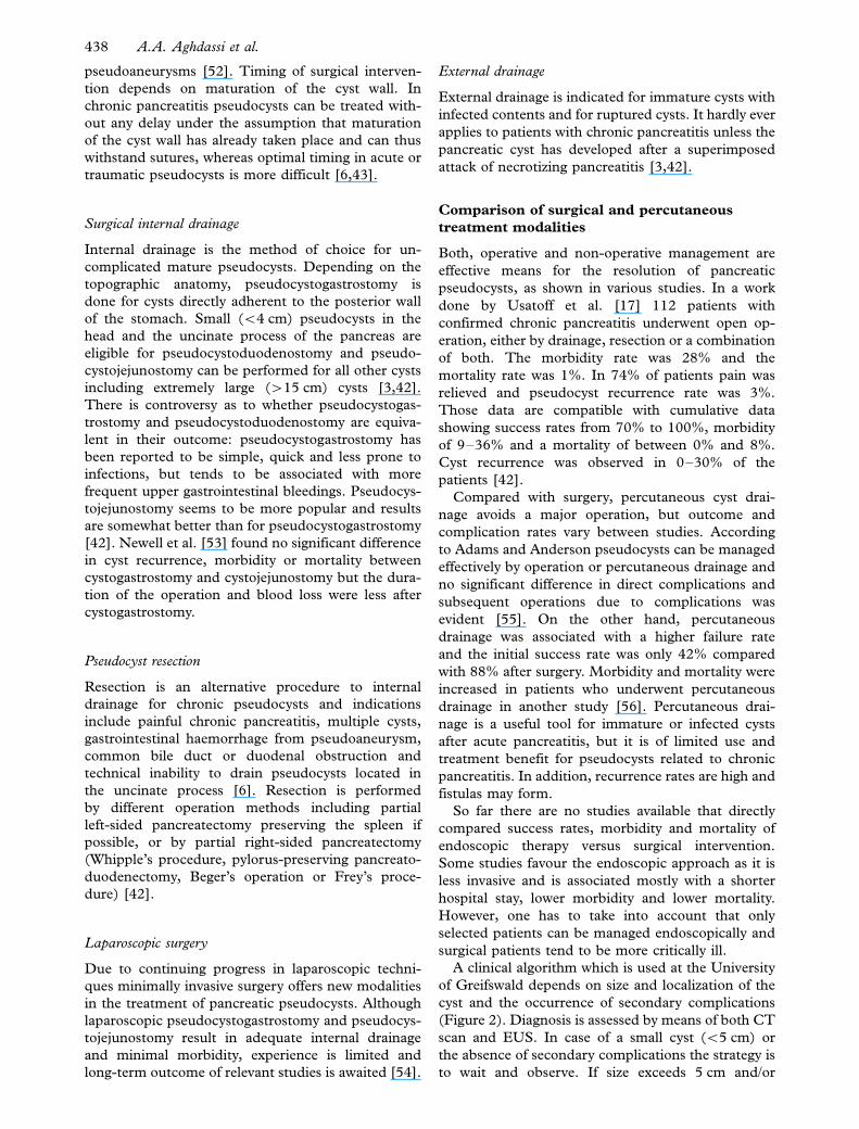
A clinical algorithm which is used at the University
of Greifswald depends on size and localization of the
cyst and the occurrence of secondary complications
(Figure 2). Diagnosis is assessed by means of both CT
scan and EUS. In case of a small cyst (B/5 cm) or
the absence of secondary complications the strategy is
to wait and observe. If size exceeds 5 cm and/or
438 A.A. Aghdassi et al.

complications occur the cyst can be treated either
surgically or endoscopically with equivalent outcome.
Conclusions
Pancreatic pseudocysts are a known complication of
acute and chronic pancreatitis. Chronic pseudocysts
over 8 weeks are less likely to resolve spontaneously
and, as the risk of complications increases with time,
treatment of large pseudocysts (�/5 cm) should not
be postponed [6]. Introduction of new and sensitive
imaging techniques permits the detection of more
pancreatic cystic lesions with better evaluation of
adjacent structures. Exact classification of pseudo-
cysts is an important factor for both the determination
of the actual number of pseudocysts and the imple-
mentation of therapeutic strategies.
Surgery is the traditional modality for treating
pancreatic pseudocysts, with high success rates and
low morbidity and mortality, and it still plays an
important role in therapy. Laparoscopic management
has been reported with very encouraging results, but
long-term follow-up has still to show equivalence to
open surgery. Endoscopic therapy is a reasonable
alternative to surgery, particularly for chronic pseu-
docysts, displaying an even lower morbidity and
mortality rate. Failure of transpapillary or transmural
drainage may make subsequent surgery necessary
[42]. Nonetheless, initial endoscopic drainage should
be considered as a valuable tool and the method of
choice in patients with chronic pancreatitis-associated
large pseudocysts [56].
References
[1] Kloppel G. Pseudocysts and other non-neoplastic cysts of the
pancreas. Semin Diagn Pathol 2000;/17:/7�15.
[2] Bradley EL 3rd. A clinically based classification system for
acute pancreatitis. Summary of the International Symposium
on Acute Pancreatitis, Atlanta, Ga, September 11 through 13,
1992. Arch Surg 1993;/128:/586�90.
[3] Pitchumoni CS, Agarwal N. Pancreatic pseudocysts. When
and how should drainage be performed? Gastroenterol Clin
North Am 1999;/28:/615�39.
[4] D’Egidio A, Schein M. Pancreatic pseudocysts: a proposed
classification and its management implications. Br J Surg
1991;/78:/981�4.
[5] Nealon WH, Walser E. Main pancreatic ductal anatomy can
direct choice of modality for treating pancreatic pseudocysts
(surgery versus percutaneous drainage). Ann Surg 2002;/235:/
751�8.
[6] Grace PA, Williamson RC. Modern management of pancrea-
tic pseudocysts. Br J Surg 1993;/80:/573�81.
[7] O’Malley VP, Cannon JP, Postier RG. Pancreatic pseudocysts:
cause, therapy, and results. Am J Surg 1985;/150:/680�2.
[8] Sankaran S, Walt SJ. The natural and unnatural history of
pancreatic pseudocysts. Br J Surg 1975;/62:/37�44.
[9] Bradley EL, Gonzalez AC, Clements JL Jr. Acute pancreatic
pseudocysts: incidence and implications. Ann Surg 1976;/184:/
734�7.
Acute pancreatitis
Pancreatic injury
Chronic pancreatitis
> 5 cm or complications < 5 cm without complications
Spontaneousresolution
Complications
[2,40] [5]
Endoscopic and surgical interventions are equivalent in the treatment of pseudocysts
Figure 2. Clinical algorithm used at the University of Greifswald for the treatment of pancreatic pseudocysts. Pancreatic pseudocysts result
from acute pancreatitis, chronic pancreatitis or pancreatic injury. The primary decision for or against treatment of pancreatic pseudocysts
depends on size and localization of the cyst and the occurrence of secondary complications. In case of a small cyst (B/5 cm) or absent
secondary complications the strategy is to wait and observe. If size exceeds 5 cm and/or complications occur the cyst can be treated either
surgically or endoscopically with equal outcome.
Management of pancreatic pseudocysts 439

[10] Maringhini A, Uomo G, Patti R, Rabitti P, Termini A,
Cavallera A, et al. Pseudocysts in acute nonalcoholic pancrea-
titis: incidence and natural history. Dig Dis Sci 1999;/44:/
1669�73.
[11] London NJ, Neoptolemos JP, Lavelle J, Bailey I, James D.
Serial computed tomography scanning in acute pancreatitis: a
prospective study. Gut 1989;/30:/397�403.
[12] Barthet M, Bugallo M, Moreira LS, Bastid C, Sastre B, Sahel
J. Management of cysts and pseudocysts complicating chronic
pancreatitis. A retrospective study of 143 patients. Gastro-
enterol Clin Biol 1993;/17:/270�6.
[13] Ammann RW, Akovbiantz A, Largiader F, Schueler G. Course
and outcome of chronic pancreatitis. Longitudinal study of a
mixed medical-surgical series of 245 patients. Gastroenterol-
ogy 1984;/86(5 Pt 1):/820�8.
[14] Elliott DW. Pancreatic pseudocysts. Surg Clin North Am
1975;/55:/339�62.
[15] Sanfey H, Aguilar M, Jones RS. Pseudocysts of the pancreas, a
review of 97 cases. Am Surg 1994;/60:/661�8.
[16] Laxson LC, Fromkes JJ, Cooperman M. Endoscopic retro-
grade cholangiopancreatography in the management of pan-
creatic pseudocysts. Am J Surg 1985;/150:/683�6.
[17] Usatoff V, Brancatisano R, Williamson RC. Operative treat-
ment of pseudocysts in patients with chronic pancreatitis. Br J
Surg 2000;/87:/1494�9.
[18] Kolars JC, Allen MO, Ansel H, Silvis SE, Vennes JA.
Pancreatic pseudocysts: clinical and endoscopic experience.
Am J Gastroenterol 1989;/84:/259�64.
[19] Walt AJ, Bouwman DL, Weaver DW, Sachs RJ. The impact of
technology on the management of pancreatic pseudocyst.
Fifth Annual Samuel Jason Mixter Lecture. Arch Surg 1990;/
125:/759�63.
[20] Baron TH, Harewood GC, Morgan DE, Yates MR. Outcome
differences after endoscopic drainage of pancreatic necrosis,
acute pancreatic pseudocysts, and chronic pancreatic pseudo-
cysts. Gastrointest Endosc 2002;/56:/7�17.
[21] Balthazar EJ, Freeny PC, vanSonnenberg E. Imaging and
intervention in acute pancreatitis. Radiology 1994;/193:/297�306.
[22] Lehman GA. Pseudocysts. Gastrointest Endosc 1999;/49(3 Pt
2):/S81�S84.
[23] Brugge WR, Lewandrowski K, Lee-Lewandrowski E, Centeno
BA, Szydlo T, Regan S, et al. Diagnosis of pancreatic cystic
neoplasms: a report of the cooperative pancreatic cyst study.
Gastroenterology 2004;/126:/1330�6.
[24] Polakow J, Ladny JR, Serwatka W, Walecki J, Puchalski Z,
Czech B. Percutaneous fine-needle pancreatic pseudocyst
puncture guided by three-dimensional sonography. Hepato-
gastroenterology 2001;/48:/1308�11.
[25] Andren-Sandberg A, Maleckas A. Pancreatic pseudocysts.
Diagnosis, treatment and results in the 2003s. A literature
study aiming at evidence based surgery. 2003;/262.
[26] Gress F, Gottlieb K, Cummings O, Sherman S, Lehman G.
Endoscopic ultrasound characteristics of mucinous cystic
neoplasms of the pancreas. Am J Gastroenterol 2000;/95:/
961�5.
[27] Chak A. Endosonographic-guided therapy of pancreatic
pseudocysts. Gastrointest Endosc 2000;52(6 Suppl):S23�S27.
[28] Frossard JL, Amouyal P, Amouyal G, Palazzo L, Amaris J,
Soldan M, et al. Performance of endosonography-guided fine
needle aspiration and biopsy in the diagnosis of pancreatic
cystic lesions. Am J Gastroenterol 2003;/98:/1516�24.
[29] Brandwein SL, Farrell JJ, Centeno BA, Brugge WR. Detection
and tumor staging of malignancy in cystic, intraductal, and
solid tumors of the pancreas by EUS. Gastrointest Endosc
2001;/53:/722�7.
[30] Hawes RH. Endoscopic management of pseudocysts. Rev
Gastroenterol Disord 2003;/3:/135�41.
[31] Kressel HY, Margulis AR, Gooding GW, Filly RA, Moss A,
Korobkin M. CT scanning and ultrasound in the evaluation of
pancreatic pseudocysts: a preliminary comparison. Radiology
1978;/126:/153�7.
[32] Lee JK, Stanley RJ, Melson GL, Sagel SS. Pancreatic imaging
by ultrasound and computed tomography: a general review.
Radiol Clin North Am 1979;/17:/105�17.
[33] Brugge WR. Approaches to the drainage of pancreatic
pseudocysts. Curr Opin Gastroenterol 2004;/20:/488�92.
[34] Rohrmann CA, Silvis SE, Vennes JA. Evaluation of the
endoscopic pancreatogram. Radiology 1974;/113:/297�304.
[35] Sugawa C, Walt AJ. Endoscopic retrograde pancreatography
in the surgery of pancreatic pseudocysts. Surgery 1979;/86:/
639�47.
[36] Nealon WH, Townsend CM Jr, Thompson JC. Preoperative
endoscopic retrograde cholangiopancreatography (ERCP) in
patients with pancreatic pseudocyst associated with resolving
acute and chronic pancreatitis. Ann Surg 1989;209:532�8;
discussion 538�40.
[37] Sica GT, Braver J, Cooney MJ, Miller FH, Chai JL, Adams
DF. Comparison of endoscopic retrograde cholangiopancrea-
tography with MR cholangiopancreatography in patients with
pancreatitis. Radiology 1999;/210:/605�10.
[38] Jensen AR, Matzen P, Malchow-Moller A, Christofferson I.
Pattern of pain, duct morphology, and pancreatic function in
chronic pancreatitis. A comparative study. Scand J Gastro-
enterol 1984;/19:/334�8.
[39] Varghese JC, Masterson A, Lee MJ. Value of MR pancreato-
graphy in the evaluation of patients with chronic pancreatitis.
Clin Radiol 2002;/57:/393�401.
[40] Gouyon B, Levy P, Ruszneiwski P, Zins M, Hammel P,
Vilgrain V, et al. Predictive factors in the outcome of
pseudocysts complicating alcoholic chronic pancreatitis. Gut
1997;/41:/821�5.
[41] Crass RA, Way LW. Acute and chronic pancreatic pseudocysts
are different. Am J Surg 1981;/142:/660�3.
[42] Rosso E, Alexakis N, Ghaneh P, Lombard M, Smart HL,
Evans J, et al. Pancreatic pseudocyst in chronic pancreatitis:
endoscopic and surgical treatment. Dig Surg 2003;/20:/397�406.
[43] Warshaw AL, Rattner DW. Timing of surgical drainage for
pancreatic pseudocyst. Clinical and chemical criteria. Ann
Surg 1985;/202:/720�4.
[44] Monkemuller KE, Kahl S, Malfertheiner P. Endoscopic
therapy of chronic pancreatitis. Dig Dis 2004;/22:/280�91.
[45] Binmoeller KF, Seifert H, Walter A, Soehendra N. Transpa-
pillary and transmural drainage of pancreatic pseudocysts.
Gastrointest Endosc 1995;/42:/219�24.
[46] Smits ME, Rauws EA, Tytgat GN, Huibregtse K. The efficacy
of endoscopic treatment of pancreatic pseudocysts. Gastro-
intest Endosc 1995;/42:/202�7.
[47] Monkemuller KE, Baron TH, Morgan DE. Transmural
drainage of pancreatic fluid collections without electrocautery
using the Seldinger technique. Gastrointest Endosc 1998;/48:/
195�200.
[48] Barthet M, Sahel J, Bodiu-Bertei C, Bernard JP. Endoscopic
transpapillary drainage of pancreatic pseudocysts. Gastrointest
Endosc 1995;/42:/208�13.
[49] Catalano MF, Geenan JE, Schmalz MJ, Johnson GK, Dean
RS, Hogan WJ. Treatment of pancreatic pseudocysts with
ductal communication by transpapillary pancreatic duct en-
doprosthesis. Gastrointest Endosc 1995;/42:/214�8.
[50] Cantasdemir M, Kara B, Kantarci F, Mihmanli I, Numan F,
Erguney S. Percutaneous drainage for treatment of infected
pancreatic pseudocysts. South Med J 2003;/96:/136�40.
[51] vanSonnenberg E, Wittich GR, Casola G, Brannigan TC,
Karnel F, Stabile BE, et al. Percutaneous drainage of infected
and noninfected pancreatic pseudocysts: experience in 101
cases. Radiology 1989;/170(3 Pt 1):/757�61.
440 A.A. Aghdassi et al.

[52] Cooperman AM. Surgical treatment of pancreatic pseudo-
cysts. Surg Clin North Am 2001;81:411�19, xii.
[53] Newell KA, Liu T, Aranha GV, Prinz RA. Are cystgastrostomy
and cystjejunostomy equivalent operations for pancreatic
pseudocysts? Surgery 1990;108:635�9; discussion 639�40.
[54] Bhattacharya D, Ammori BJ. Minimally invasive approaches
to the management of pancreatic pseudocysts: review of the
literature. Surg Laparosc Endosc Percutan Tech 2003;/13:/
141�8.
[55] Adams DB, Anderson MC. Percutaneous catheter drainage
compared with internal drainage in the management of
pancreatic pseudocyst. Ann Surg 1992;215:571�6; discussion
576�8.
[56] Heider R, Meyer AA, Galanko Ja, Behrns KE. Percutaneous
drainage of pancreatic pseudocysts is associated with a higher
failure rate than surgical treatment in unselected patients. Ann
Surg 1999;229:781�7; discussion 787�9.
[57] Ridder GJ, Maschek H, Klempnauer J. Favourable prognosis
of cystadeno- over adenocarcinoma of the pancreas after
curative resection. Eur J Surg Oncol 1996;/22:/232�6.
Management of pancreatic pseudocysts 441