NFATc1 autoregulation: a crucial step for cell-fate determination

Haruhisa Inoue1, Kayoko Tsukita1,Takuji Iwasato2,3, Yasuyuki Suzuki1,Masanori Tomioka4, Minako Tateno1,Masahiro Nagao5, Akihiro Kawata5,Takaomi C.Saido4, Masayuki Miura6,Hidemi Misawa7, Shigeyoshi Itohara2
and Ryosuke Takahashi1,8
1Laboratory for Motor System Neurodegeneration, 2Laboratory forBehavioral Genetics and 4Laboratory for Proteolytic Neuroscience,RIKEN Brain Science Institute (BSI), Saitama, 3PRESTO, JapanScience and Technology Corporation, Saitama, 5Department ofNeurology, Tokyo Metropolitan Neurological Hospital, Tokyo,6Department of Genetics, Graduate School of Pharmaceutical Sciences,University of Tokyo and 7Department of Neurology,Tokyo Metropolitan Institute for Neuroscience, Tokyo, Japan
8Corresponding authore-mail: [email protected]
Mutant copper/zinc superoxide dismutase (SOD1)-overexpressing transgenic mice, a mouse model forfamilial amyotrophic lateral sclerosis (ALS), providesan excellent resource for developing novel therapiesfor ALS. Several observations suggest that mito-chondria-dependent apoptotic signaling, includingcaspase-9 activation, may play an important role inmutant SOD1-related neurodegeneration. To elucidatethe role of caspase-9 in ALS, we examined the effectsof an inhibitor of X chromosome-linked inhibitor ofapoptosis (XIAP), a mammalian inhibitor of cas-pase-3, -7 and -9, and p35, a baculoviral broad caspaseinhibitor that does not inhibit caspase-9. Whenexpressed in spinal motor neurons of mutant SOD1mice using transgenic techniques, XIAP attenuateddisease progression without delaying onset. In con-trast, p35 delayed onset without slowing disease pro-gression. Moreover, caspase-9 was activated in spinalmotor neurons of human ALS subjects. These datastrongly suggest that caspase-9 plays a crucial rolein disease progression of ALS and constitutes apromising therapeutic target.Keywords: ALS/baculovirus p35/caspase/SOD1/XIAP
Introduction
Amyotrophic lateral sclerosis (ALS) is a neurodegenerativedisorder resulting in progressive paralysis caused by motorneuron loss in the brain, brainstem and spinal cord. It isuniversally fatal, with a mean survival of 5 years afterdisease onset (for a review see Gurney et al., 2000; Brownand Robberecht, 2001; Cleveland and Rothstein, 2001;Julien, 2001; Rowland and Shneider, 2001). Mutations inthe human superoxide dismutase-1 (SOD1) are responsible
for an autosomal dominant form of familial ALS (Rosenet al., 1993).
Accumulating evidence indicates that mutant SOD1protein activity precipitates proapoptotic effects throughits resulting abnormal function. Possible pathophysio-logoical mechanisms in familial ALS associated withSOD1 mutation are the failure to fold or degrade mutantSOD1 (for a review see Gurney et al., 2000; Brown andRobberecht, 2001; Cleveland and Rothstein, 2001; Julien,2001; Rowland and Shneider, 2001), the formation of freeradicals (for a review see Gurney et al., 2000; Brown andRobberecht, 2001; Cleveland and Rothstein, 2001; Julien,2001; Rowland and Shneider, 2001), the release of freecopper (Hayward et al., 2002; Subramaniam et al., 2002),and/or a susceptibility to disul®de reduction of mutantSOD1 (Tiwari and Hayward, 2003). The resulting effectsare subsequent axonal strangulation from neuro®lamen-tous disorganization (for a review see Cleveland andRothstein, 2001; Julien, 2001) and excitotoxic death due tomishandling of glutamate (Howland et al., 2002).However, the precise mechanisms behind mutantSOD1-mediated neurotoxicity have yet to be unravelled.
The mutant SOD1 transgenic (mSOD1-Tg) mouse, amouse model for familial ALS (Gurney et al., 1994),provides the opportunity to elucidate the pathogeneticmechanisms underpinning ALS. The importance ofapoptotic pathways in the pathogenesis of mSOD1-Tgmice is supported by the neuroprotective effects of severalfactors, including: Bcl-2 transgene (Kostic et al., 1997),the dominant-negative caspase-1 transgene (Friedlanderet al., 1997), intraventricular administration of pan-caspase-inhibitor z-VAD-FMK (Li et al., 2000) andfeeding with minocycline (Zhu et al., 2002). Caspase-1,-3, -7, -8 and -9 are also activated in the spinal motorneurons of mSOD1-Tg mice at various stages throughoutthe clinical course (Pasinelli et al., 2000; GueÂgan et al.,2001, 2002; for a review see Friedlander, 2003; GueÂganand Przedborski, 2003). Moreover, cytochrome c releasefrom mitochondria, which is also seen in mSOD1-Tg mice(GueÂgan et al., 2001), activates caspase-9 in the presenceof Apaf-1 which, in turn, activates downstream execu-tioner caspases. These observations suggest that mito-chondria-dependent apoptotic signaling, includingcaspase-9 activation, may play an important role inmotor neuronal degeneration in mSOD1-Tg mice.However, the signi®cance of caspase activation down-stream of cytochrome c release has remained unclear.
To elucidate the role of caspase-9 in ALS, we used atransgenic approach to examine the effects of two differentcaspase inhibitory proteins, XIAP and p35, on the clinicalcourse of mSOD1-Tg mice. XIAP, a mammalian protein,speci®cally inhibits caspase-3, -7 and -9; whereas p35, abaculoviral protein, inhibits a broad range of caspases, butnot -9 (for a review see Deveraux and Reed, 1999; Ekert
The crucial role of caspase-9 in the diseaseprogression of a transgenic ALS mouse model
The EMBO Journal Vol. 22 No. 24 pp. 6665±6674, 2003
ã European Molecular Biology Organization 6665

et al., 1999; Yuan and Yankner, 2000; Suzuki et al., 2001).When expressed using the same promoter, both proteinsprolonged survival; however, human XIAP slowed diseaseprogression without delaying its onset, while p35 delayeddisease onset but did not affect its progression. These datastrongly suggest that the inhibition of caspase-9 inmotor neurons contributes signi®cantly to attenuating theprogression of ALS.
Results
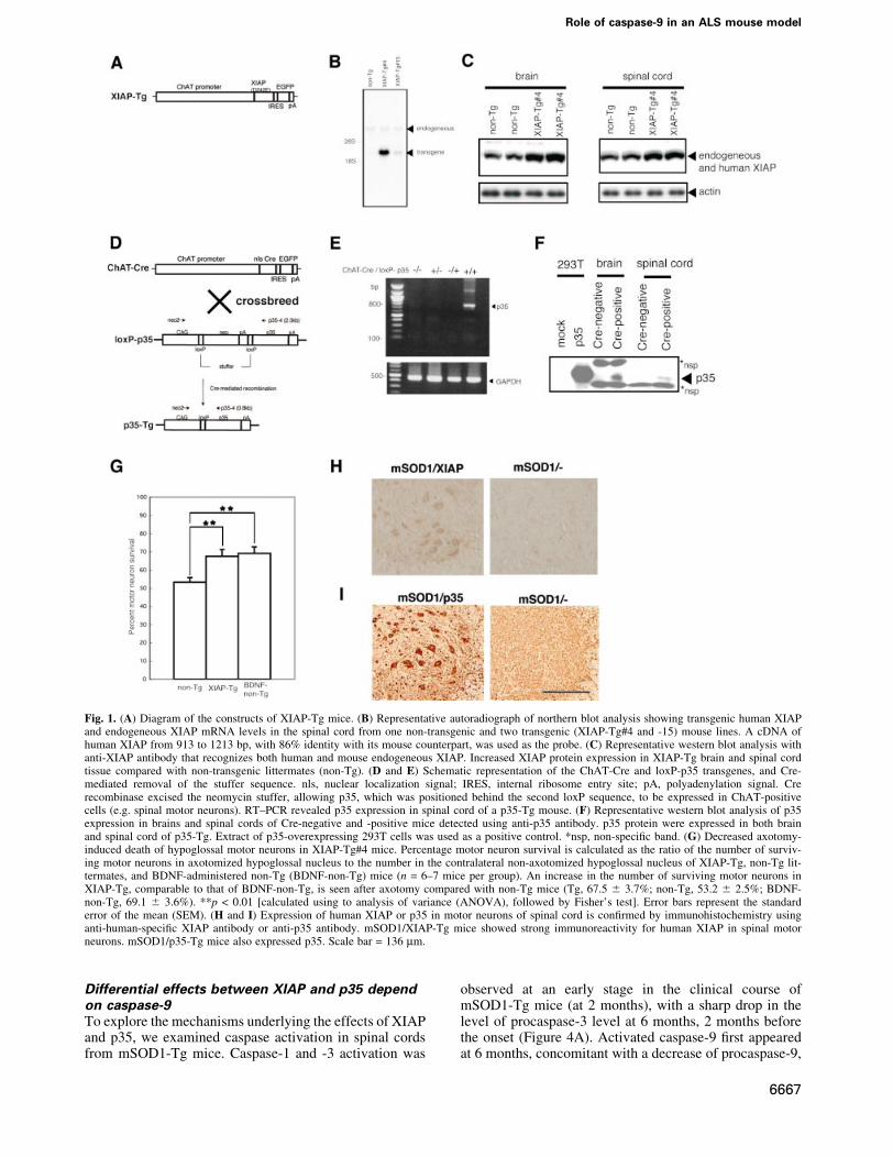
Spinal motor neurons expressing either XIAP orp35First we generated transgenic mice (XIAP-Tg) expressinghuman XIAP in spinal motor neurons, with glutamic acidsubstituted for aspartic acid in position 242 (D242E) underthe control of the murine choline acethyltransferase(ChAT) promoter (Naciff et al., 1999) (Figure 1A).Since ChAT is expressed only in matured neurons and arelatively speci®c marker protein for spinal motor neurons,we avoided developmental lethality caused by inhibitingapoptosis. The D242E mutant avoids cleavage bycaspase-3 and is more resistant to Fas- or Bax-inducedapoptosis than wild-type XIAP in vitro (Deveraux et al.,1999). Human XIAP mRNA expression was detected inthe spinal cords of XIAP-Tg lines (Figure 1B). Higherprotein expressions were detected using western blotanalysis in brain and spinal cord tissue in XIAP-Tg#4 micecompared with non-transgenic littermates (non-Tg)(Figure 1C). For our experiments we used theXIAP-Tg#4 line.
To express p35 in spinal motor neurons, transgenic(ChAT-Cre) mice expressing the P1-phage Cre recombin-ase were developed using a construct containing cDNA forCre with a nuclear localization signal and the murineChAT promoter (Figure 1D). Using CAG-CAT-Z reportermice (Sakai and Miyazaki, 1997), we found that Cre/loxPrecombination occurs in spinal motor neurons in ChAT-Cre#23 (data not shown). ChAT-Cre/loxP-p35 transgenic(p35-Tg) mice were generated by crossing ChAT-Cre#23with loxP-p35 transgenic mice; in these mice, p35 geneexpression is prevented because a stuffer sequence ¯ankedby two loxP sequences is present (Figure 1D). p35 mRNAand protein expression were detected in p35-Tg micespinal cords using RT±PCR (Figure 1E) and western blotanalysis (Figure 1F). The p35 expressed by Cre/loxPrecombination in our loxP-p35 transgenic mice reportedlyinhibits caspases and associated cell death under variouspathological conditions, including autoimmune-mediateddemyelination in vivo (Hisahara et al., 2000; Tomiokaet al., 2002; Viswanath et al., 2002).
To con®rm that XIAP in our XIAP-Tg mice functions asan inhibitor of cell death in vivo (KuÈgler et al., 2000;Perrelet et al., 2000; Crocker et al., 2003), axotomy of thehypoglossal nerve was performed on adult femaleXIAP-Tg#4 mice. Axotomy-induced cell death wassigni®cantly suppressed, indicating that expressed XIAPwas functional (Figure 1G). The effect of XIAP wassimilar to that of brain-derived neurotrophic factor(BDNF)-administration to the nerve stump (p > 0.05)(Figure 1G).
We used a line of mSOD1-Tg mice [G93A; glycinesubstituted for alanine at codon 93 of the human SOD1
protein (Gurney et al., 1994)] as our ALS model.Female XIAP-Tg (or p35-Tg) mice were crossbred withmale mSOD1-Tg mice to produce four varieties ofmice: wild type (non-Tg), wild type (XIAP- or p35-Tg),mSOD1-Tg or mSOD1/(XIAP or p35)-Tg. Beforeinvestigating the neuroprotective effects of each trans-gene on mSOD1-Tg mice spinal cord, its expression inmutant SOD1 and human XIAP double transgenic(mSOD1/XIAP-Tg) [or mutant SOD1 and p35 doubletransgenic (mSOD1/p35-Tg)] mice was con®rmed byimmunohistochemistry (Figure 1H and I). As shown inFigure 1H (or I), the product of the transgene wasexpressed in spinal motor neurons of mSOD1/XIAP-Tg(or mSOD1/p35-Tg) mice.
XIAP and p35 prolong survival of the ALS mousemodel differentlyTo determine the effects of XIAP or p35 transgenes on theonset and progression of motor neuron disease in mutantSOD1 mice, we compared the time of disease onset andlife span between mSOD1-Tg and mSOD1/XIAP-Tg ormSOD1/p35-Tg mice. Onset was determined by the loss ofmotor function, which was measured using a rota-rod test.This occurred at a mean age of 242.0 6 4.3 days inmSOD1/XIAP-Tg mice and at 236.3 6 4.0 days in controlmSOD1-Tg mice (p > 0.05) (Figure 2A). Onset of motorfunction loss was observed at a mean age of 257.1 6 5.1days in mSOD1/p35-Tg mice, compared with 235.1 6 5.1days in control mSOD1-Tg mice (p = 0.0015) (Figure 2B).Signi®cantly, mean survival time was prolonged in bothmSOD1/XIAP-Tg and mSOD1/p35-Tg mice. Mean sur-vival for mSOD1/XIAP-Tg mice was 276.6 6 4.8 days,whereas control mSOD1-Tg mice survived 258.9 6 4.1days (p < 0.001) (Figure 2A). Mean survival for mSOD1/p35-Tg mice was 281.9 6 4.8 days, compared with257.4 6 4.6 days for control mSOD1-Tg mice (p < 0.0005)(Figure 2B).
Mean disease duration, the period marked by onset anddeath, was also assessed. Mean disease duration inmSOD1/XIAP-Tg mice was 34.6 6 2.6 days, comparedwith 22.6 6 1.7 days in control mSOD1-Tg mice (p <0.0005). Mean disease duration in mSOD1/p35-Tg micewas 24.8 6 2.3 days, compared with 22.3 6 1.7 days incontrol mSOD1-Tg mice (p > 0.05). These data indicatethat XIAP signi®cantly extends survival of mSOD1-Tgmice by prolonging the duration of the disease, i.e. byameliorating disease progression. In contrast, p35 extendssurvival by delaying disease onset.
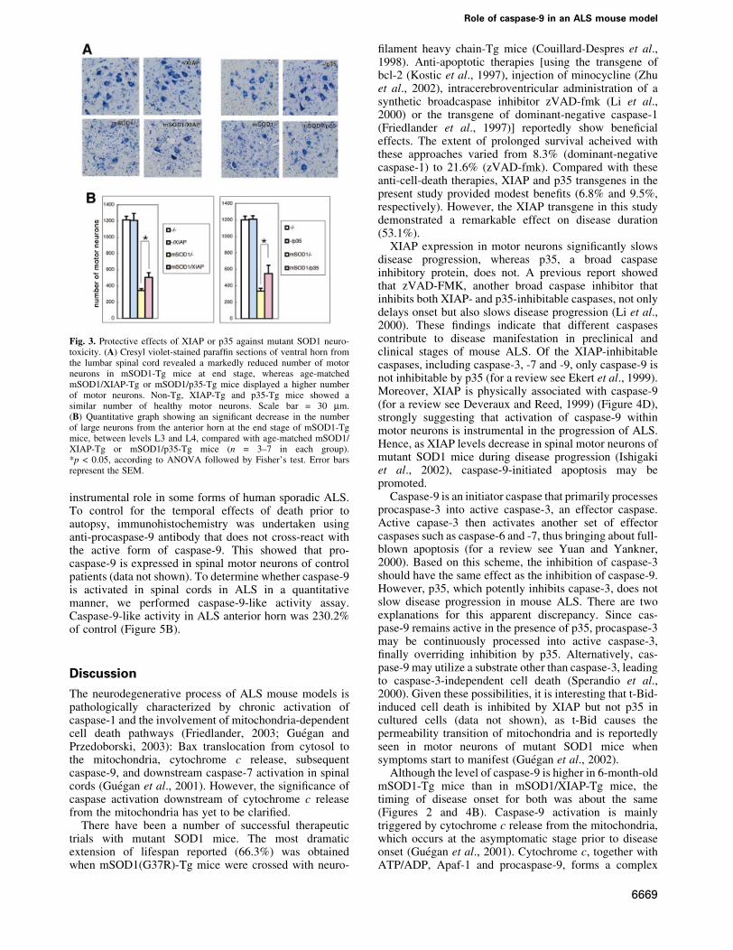
We next assessed the effects of XIAP and p35 on spinalmotor neuron death in mSOD1-Tg mice. At the end stageof mSOD1-Tg mice, ~30% of spinal motor neuronsremained in mSOD1-Tg control mice compared withage-matched wild-type, XIAP-Tg and p35-Tg mice. Incontrast, a signi®cantly larger number of motor neuronswere found in mSOD1/XIAP-Tg and mSOD1/p35-Tg thanwere found in mSOD1-Tg mice (Figure 3). As wild-type,XIAP-Tg and p35-Tg mice all have the same number ofmotor neurons, we can assume that that naturallyoccurring motoneuron death was not inhibited in theseTg mice.
H.Inoue et al.
6666
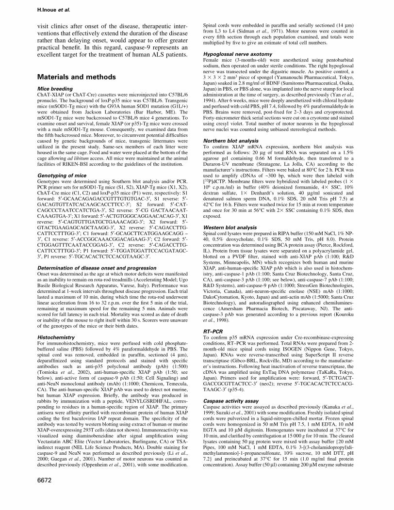
Differential effects between XIAP and p35 dependon caspase-9To explore the mechanisms underlying the effects of XIAPand p35, we examined caspase activation in spinal cordsfrom mSOD1-Tg mice. Caspase-1 and -3 activation was
observed at an early stage in the clinical course ofmSOD1-Tg mice (at 2 months), with a sharp drop in thelevel of procaspase-3 level at 6 months, 2 months beforethe onset (Figure 4A). Activated caspase-9 ®rst appearedat 6 months, concomitant with a decrease of procaspase-9,
Fig. 1. (A) Diagram of the constructs of XIAP-Tg mice. (B) Representative autoradiograph of northern blot analysis showing transgenic human XIAPand endogeneous XIAP mRNA levels in the spinal cord from one non-transgenic and two transgenic (XIAP-Tg#4 and -15) mouse lines. A cDNA ofhuman XIAP from 913 to 1213 bp, with 86% identity with its mouse counterpart, was used as the probe. (C) Representative western blot analysis withanti-XIAP antibody that recognizes both human and mouse endogeneous XIAP. Increased XIAP protein expression in XIAP-Tg brain and spinal cordtissue compared with non-transgenic littermates (non-Tg). (D and E) Schematic representation of the ChAT-Cre and loxP-p35 transgenes, and Cre-mediated removal of the stuffer sequence. nls, nuclear localization signal; IRES, internal ribosome entry site; pA, polyadenylation signal. Crerecombinase excised the neomycin stuffer, allowing p35, which was positioned behind the second loxP sequence, to be expressed in ChAT-positivecells (e.g. spinal motor neurons). RT±PCR revealed p35 expression in spinal cord of a p35-Tg mouse. (F) Representative western blot analysis of p35expression in brains and spinal cords of Cre-negative and -positive mice detected using anti-p35 antibody. p35 protein were expressed in both brainand spinal cord of p35-Tg. Extract of p35-overexpressing 293T cells was used as a positive control. *nsp, non-speci®c band. (G) Decreased axotomy-induced death of hypoglossal motor neurons in XIAP-Tg#4 mice. Percentage motor neuron survival is calculated as the ratio of the number of surviv-ing motor neurons in axotomized hypoglossal nucleus to the number in the contralateral non-axotomized hypoglossal nucleus of XIAP-Tg, non-Tg lit-termates, and BDNF-administered non-Tg (BDNF-non-Tg) mice (n = 6±7 mice per group). An increase in the number of surviving motor neurons inXIAP-Tg, comparable to that of BDNF-non-Tg, is seen after axotomy compared with non-Tg mice (Tg, 67.5 6 3.7%; non-Tg, 53.2 6 2.5%; BDNF-non-Tg, 69.1 6 3.6%). **p < 0.01 [calculated using to analysis of variance (ANOVA), followed by Fisher's test]. Error bars represent the standarderror of the mean (SEM). (H and I) Expression of human XIAP or p35 in motor neurons of spinal cord is con®rmed by immunohistochemistry usinganti-human-speci®c XIAP antibody or anti-p35 antibody. mSOD1/XIAP-Tg mice showed strong immunoreactivity for human XIAP in spinal motorneurons. mSOD1/p35-Tg mice also expressed p35. Scale bar = 136 mm.
Role of caspase-9 in an ALS mouse model
6667

and increased gradually right to the end stage (Figure 4A).Caspase-7 activation was detected only after the onset (at 8months) (Figure 4A).
Next, we examined caspase-9 activation at 6 monthsin both mSOD1/XIAP-Tg and mSOD1/p35-Tg mice(Figure 4B) because different effects were observed inXIAP and p35 during the clinical course of mSOD1-Tgmice (Figure 2). In mSOD1/p35-Tg mice's spinal cords at6 months, no caspase-9 activation was observed(Figure 4B); however, caspase-9 activation was observedin the end stage (Figure 4C). These data suggest that p35delayed caspase-9 activation by inhibiting upstreamcaspase(s) leading to caspase-9 activation, although thenature of such caspase(s) is unknown. As a sharp drop ofprocaspase-3 did not occur in mSOD1/p35-Tg mice spinalcords at 6 months, p35 may inhibit an unidenti®edupstream caspase(s) of caspase-3 other than caspase-9(Figure 4B). Caspase-9 activation, however, was observedin mSOD1/XIAP-Tg mice at 6 months, although to a lesserextent compared with that in mSOD1-Tg mice (Figure 4B).
A serial examination of caspase-9 activation in mSOD1/XIAP-Tg mice (Figure 4C) was also performed.Weakened activation of caspase-9 in mSOD1/XIAP-Tgwas observed when compared with mSOD1-Tg mice. Animmunoprecipitation assay using anti-XIAP antibodyrevealed a binding between the human XIAP and thecaspase-9 in the spinal cord lysates of mSOD1/XIAP-Tgmice (Figure 4D). Caspase-9 activation in spinal motorneurons of mSOD1-Tg mice was con®rmed by doublestaining with a neuron-speci®c antibody (NeuN) and anantibody for activated caspase-9 (Figure 4E). These data
strongly suggest that the inhibition of caspase-9 by XIAPin motor neurons attenuates disease progression.
These results are con®rmed by in vitro data (Ryan et al.,2002) showing that p35 does not inhibit caspase-9, whileXIAP does (Srinvasula et al., 2001). Ac-YVAD-MCA(caspase-1-like), Ac-DEVD-MCA (caspase-3- or -7-like)and Ac-LEHD-MCA (casapse-9-like) cleavage activities,showing the catalytic activity of these caspases, wereelevated in the lumber spinal cords of mSOD1-Tg mice,which was consistent with western blot results (Figure 4F).In mSOD1/XIAP or mSOD1/p35-Tg mice, the presence ofXIAP or p35 resulted in a signi®cant reduction of allcaspase-like activities tested (Figure 4F). Reducedcaspase-1 activity in mSOD1/XIAP-Tg mice as comparedwith that in mSOD1-Tg mice is probably due to the delayof glial response against neuronal cell death accompany-ing casapse-1 activation. On the other hand, reducedcaspase-9 activity in mSOD1/p35-Tg mice suggests thatp35 delays the activation of caspase-9, as shown inFigure 4B. p35-mediated inhibition of caspase-1,caspase-3, or another unidenti®ed caspase(s) upstream ofcaspase-9 activation may be responsible for this effect.
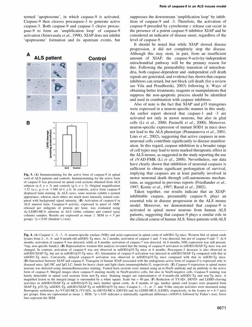
Caspase-9 is activated in ALS spinal cordsTo examine possible caspase-9-activation in humansporadic ALS, we performed immunohistochemistryusing anti-active caspase-9 antibody on post mortemhuman samples. Four of the eight ALS spinal cordsshowed obvious caspase-9 activation in the motor neuronsstudied, but this was not seen in any of the controls(Figure 5A); this suggests that caspase-9 may play an
Fig. 2. Age comparison at the end stage of disease (survival) and at the onset of motor de®cit scored by rota-rod test (onset) in mSOD1/XIAP-Tg (A)and mSOD1/p35-Tg (B) mice. (A) Probability of survival revealed an extended life span in mSOD1/XIAP-Tg (n = 20; red solid line) compared withmSOD1-Tg mice (n = 20; green solid line). The cumulative probability of onset of rota-rod de®cit was not signi®cantly changed in mSOD1/XIAP-Tg(n = 20; red dotted line) compared with mSOD1-Tg mice (n = 20; green dotted line). (B) Probability of survival showed an extended life span inmSOD1/p35-Tg (n = 17; red solid line) compared with mSOD1-Tg mice (n = 18; green solid line). Disease onset, scored by rota-rod test, was delayedin mSOD1/p35-Tg (n = 17; red dotted line) compared with mSOD1-Tg mice (n = 18; green dotted line). The data were analyzed using theKaplan±Meier life test and the log-rank test.
H.Inoue et al.
6668
instrumental role in some forms of human sporadic ALS.To control for the temporal effects of death prior toautopsy, immunohistochemistry was undertaken usinganti-procaspase-9 antibody that does not cross-react withthe active form of caspase-9. This showed that pro-caspase-9 is expressed in spinal motor neurons of controlpatients (data not shown). To determine whether caspase-9is activated in spinal cords in ALS in a quantitativemanner, we performed caspase-9-like activity assay.Caspase-9-like activity in ALS anterior horn was 230.2%of control (Figure 5B).
Discussion
The neurodegenerative process of ALS mouse models ispathologically characterized by chronic activation ofcaspase-1 and the involvement of mitochondria-dependentcell death pathways (Friedlander, 2003; GueÂgan andPrzedoborski, 2003): Bax translocation from cytosol tothe mitochondria, cytochrome c release, subsequentcaspase-9, and downstream caspase-7 activation in spinalcords (GueÂgan et al., 2001). However, the signi®cance ofcaspase activation downstream of cytochrome c releasefrom the mitochondria has yet to be clari®ed.
There have been a number of successful therapeutictrials with mutant SOD1 mice. The most dramaticextension of lifespan reported (66.3%) was obtainedwhen mSOD1(G37R)-Tg mice were crossed with neuro-
®lament heavy chain-Tg mice (Couillard-Despres et al.,1998). Anti-apoptotic therapies [using the transgene ofbcl-2 (Kostic et al., 1997), injection of minocycline (Zhuet al., 2002), intracerebroventricular administration of asynthetic broadcaspase inhibitor zVAD-fmk (Li et al.,2000) or the transgene of dominant-negative caspase-1(Friedlander et al., 1997)] reportedly show bene®cialeffects. The extent of prolonged survival acheived withthese approaches varied from 8.3% (dominant-negativecaspase-1) to 21.6% (zVAD-fmk). Compared with theseanti-cell-death therapies, XIAP and p35 transgenes in thepresent study provided modest bene®ts (6.8% and 9.5%,respectively). However, the XIAP transgene in this studydemonstrated a remarkable effect on disease duration(53.1%).
XIAP expression in motor neurons signi®cantly slowsdisease progression, whereas p35, a broad caspaseinhibitory protein, does not. A previous report showedthat zVAD-FMK, another broad caspase inhibitor thatinhibits both XIAP- and p35-inhibitable caspases, not onlydelays onset but also slows disease progression (Li et al.,2000). These ®ndings indicate that different caspasescontribute to disease manifestation in preclinical andclinical stages of mouse ALS. Of the XIAP-inhibitablecaspases, including caspase-3, -7 and -9, only caspase-9 isnot inhibitable by p35 (for a review see Ekert et al., 1999).Moreover, XIAP is physically associated with caspase-9(for a review see Deveraux and Reed, 1999) (Figure 4D),strongly suggesting that activation of caspase-9 withinmotor neurons is instrumental in the progression of ALS.Hence, as XIAP levels decrease in spinal motor neurons ofmutant SOD1 mice during disease progression (Ishigakiet al., 2002), caspase-9-initiated apoptosis may bepromoted.
Caspase-9 is an initiator caspase that primarily processesprocaspase-3 into active caspase-3, an effector caspase.Active capase-3 then activates another set of effectorcaspases such as caspase-6 and -7, thus bringing about full-blown apoptosis (for a review see Yuan and Yankner,2000). Based on this scheme, the inhibition of caspase-3should have the same effect as the inhibition of caspase-9.However, p35, which potently inhibits capase-3, does notslow disease progression in mouse ALS. There are twoexplanations for this apparent discrepancy. Since cas-pase-9 remains active in the presence of p35, procaspase-3may be continuously processed into active caspase-3,®nally overriding inhibition by p35. Alternatively, cas-pase-9 may utilize a substrate other than caspase-3, leadingto caspase-3-independent cell death (Sperandio et al.,2000). Given these possibilities, it is interesting that t-Bid-induced cell death is inhibited by XIAP but not p35 incultured cells (data not shown), as t-Bid causes thepermeability transition of mitochondria and is reportedlyseen in motor neurons of mutant SOD1 mice whensymptoms start to manifest (GueÂgan et al., 2002).
Although the level of caspase-9 is higher in 6-month-oldmSOD1-Tg mice than in mSOD1/XIAP-Tg mice, thetiming of disease onset for both was about the same(Figures 2 and 4B). Caspase-9 activation is mainlytriggered by cytochrome c release from the mitochondria,which occurs at the asymptomatic stage prior to diseaseonset (GueÂgan et al., 2001). Cytochrome c, together withATP/ADP, Apaf-1 and procaspase-9, forms a complex
Fig. 3. Protective effects of XIAP or p35 against mutant SOD1 neuro-toxicity. (A) Cresyl violet-stained paraf®n sections of ventral horn fromthe lumbar spinal cord revealed a markedly reduced number of motorneurons in mSOD1-Tg mice at end stage, whereas age-matchedmSOD1/XIAP-Tg or mSOD1/p35-Tg mice displayed a higher numberof motor neurons. Non-Tg, XIAP-Tg and p35-Tg mice showed asimilar number of healthy motor neurons. Scale bar = 30 mm.(B) Quantitative graph showing an signi®cant decrease in the numberof large neurons from the anterior horn at the end stage of mSOD1-Tgmice, between levels L3 and L4, compared with age-matched mSOD1/XIAP-Tg or mSOD1/p35-Tg mice (n = 3±7 in each group).*p < 0.05, according to ANOVA followed by Fisher's test. Error barsrepresent the SEM.
Role of caspase-9 in an ALS mouse model
6669

H.Inoue et al.
6670
termed `apoptosome', in which caspase-9 is activated.Caspase-9 then cleaves procaspase-3 to generate activecaspase-3. Both caspase-9 and caspase-3 cleave procas-pase-9 to form an `ampli®cation loop' of caspase-9activation (Srinivasula et al., 1998). XIAP does not inhibit`apoptosome' formation and its upstream events, but
suppresses the downstream `ampli®cation loop' by inhib-ition of caspase-9 and -3. Therefore, the activation ofcaspase-9 preceded by cytochrome c release can occur inthe presence of a potent caspase-9 inhibitor XIAP and beconsidered an indicator of disease onset, regardless of thelevel of caspase-9.
It should be noted that while XIAP slowed diseaseprogression, it did not completely stop the disease.Although this may stem, in part, from an insuf®cientamount of XIAP, the caspase-9-activity-independentmitochondrial pathway will be the primary reason forthis. Following the permeability transition of mitochon-dria, both caspase-dependent and -independent cell deathsignals are generated, and evidence has shown that caspaseinhibitors can retard, but not block cell death (for a reviewsee Vila and Przedborski, 2003) following it. Ways ofobtaining better treatments, reagents or manipulations thatsuppress the non-apoptotic process should be identi®edand used in combination with caspase inhibitors.
Also of note is the fact that XIAP and p35 transgeneswere expressed in a neuron-speci®c manner in this study.An earlier report showed that caspase-1 and -3 areactivated not only in motor neurons, but also in glialcells (Li et al., 2000; Pasinelli et al., 2000). Moreover,neuron-speci®c expression of mutant SOD1 in mice doesnot lead to the ALS phenotype (Pramatarova et al., 2001;Lino et al., 2002), suggesting that active caspases in non-neuronal cells contribute signi®cantly to disease manifest-ation. In this regard, caspase inhibition in a broader rangeof cell types may lead to more marked therapeutic effect inthe ALS mouse, as suggested in the study reporting the useof zVAD-FMK (Li et al., 2000). Nevertheless, our datahave clearly shown that inhibition of neuronal caspases issuf®cient to obtain signi®cant prolongation of survival,implying that caspases are at least partially involved inmotor neuronal death through cell-autonomous mechan-isms, as suggested in previous reports (Friedlander et al.,1997; Kostic et al., 1997; Raoul et al., 2002).
Taken together, our results indicate that an XIAP-inhibitable caspase, presumably caspase-9, plays anessential role in disease progression in the ALS mousemodel. Moreover, we demonstrated that caspase-9 isactivated in spinal motor neurons from human ALSpatients, suggesting that caspase-9 plays a similar role inthe clinical course of human ALS. Since patients with ALS
Fig. 5. (A) Immunostaining for the active form of caspase-9 in spinalcord of ALS patients and controls. Immunostaining for the active formof caspase-9 was processed on spinal cord sections obtained from ALSsubjects (a±f; n = 3) and controls (g±l; n = 3). Original magni®cation333 (a±c, g±i) or 3100 (d±f, j±l). In controls, active form caspase-9displayed faint staining. In ALS cases, some neurons exhibit a normalappearance, whereas most others are much more intensely stained com-pared with background signal intensity. (B) Activation of caspase-9 inALS anterior horn. Casapase-9 activity, expressed in pmol of AMCreleased per miligram of protein per hour, was evaluated usingAc-LEHD-MCA substrate in ALS (white column) and control (graycolumn) samples. Results are expressed as mean 6 SEM (n = 5 pergroup). *p < 0.05 (Student's t-test).
Fig. 4. (A) Caspase-1, -3, -7, -9, neuron-speci®c enolase (NSE) and actin expression in spinal cords of mSOD1-Tg mice. Western blot of spinal cordslysates from 2-, 4-, 6- and 8-month-old mSOD1-Tg mice. At 2 months, activation of caspase-1 and -3 was detected, but not of caspase-9 and -7. At 6months, activation of caspase-9 was detected, while at 8 months, activation of caspase-7 was detected. At 8 months, NSE expression was still present.*nsp, non-speci®c band(s). (B) Representative western blot analysis revealed that the timing of caspase-9 activation in mSOD1/XIAP-Tg mice was notchanged. In contrast, activation of caspase-9 was not observed in mSOD1/p35-Tg mice at 6 months. Procaspase-3 decrease is also observed inmSOD1/XIAP-Tg, but not in mSOD1/p35-Tg mice. (C) Attenuation of caspase-9 activation was detected in mSOD1/XIAP-Tg compared with that inmSOD1-Tg mice. Conversely, delayed caspase-9 activation was observed in mSOD1/p35-Tg mice compared with that in mSOD-Tg mice.(D) Interaction between XIAP and caspase-9. Transgene of human XIAP associated with the endogenous active form of caspase-9 is expressed only indiseased mice. IgG HC and IgG LC, bands for heavy chain and light chain immunoglobulin G, respectively. (E) Caspase-9 expression in spinal motorneurons was detected using immuno¯uorescence staining. Ventral horn sections were stained using an a-NeuN antibody and an antibody to the activeform of caspase-9. Merged images show caspase-9 staining mostly in NeuN-positive cells, but also in NeuN-negative cells. Caspase-9 staining wasbarely detectable in spinal cord sections from non-Tg mice. Staining images are representative of 6-month-old mSOD1-Tg and non-Tg mice. Amagni®ed lesion in the merged image is surrounded by a white square. Scale bar = 40 mm. (F) Reduction of YVAD-, DEVD- and LEHD-cleavingactivities in mSOD1/XIAP-Tg or mSOD1/p35-Tg mice lumber spinal cords. At 8 months of age, lumber spinal cord lysates were prepared fromXIAP-Tg, p35-Tg, mSOD1-Tg, mSOD1/XIAP-Tg or mSOD1/p35-Tg mice. Casapse-1-, -3-, or -7- and -9-like enzyme activities were measured using¯uorogenic susbstrates Ac-YVAD-MCA (YVAD), Ac-DEVD-MCA (DEVD) and Ac-LEHD-MCA (LEHD), respectively, in the lysates (n = 6±7 miceper group). Data are represented as mean 6 SEM. *p < 0.05 indicates a statistically signi®cant difference (ANOVA followed by Fisher's test). Errorbars represent the SEM.
Role of caspase-9 in an ALS mouse model
6671
visit clinics after onset of the disease, therapeutic inter-ventions that effectively extend the duration of the diseaserather than delaying onset, would appear to offer greaterpractical bene®t. In this regard, caspase-9 represents anexcellent target for the treatment of human ALS patients.
Materials and methods
Mice breedingChAT-XIAP (or ChAT-Cre) cassettes were microinjected into C57BL/6pronuclei. The background of loxP-p35 mice was C57BL/6. Transgenicmice (mSOD1-Tg mice) with the G93A human SOD1 mutation (G1L/+)were obtained from Jackson Laboratories (Bar Harbor, ME). ThemSOD1-Tg mice were backcrossed to C57BL/6 mice 4 generations. Toexamine onset and survival, female XIAP (or p35)-Tg mice were crossedwith a male mSOD1-Tg mouse. Consequently, we examined data fromthe ®fth backcrossed mice. Moreover, to circumvent potential dif®cultiescaused by genetic backgrounds of mice, transgenic littermates wereutilized in the present study. Same-sex members of each litter werehoused in the same cage. Food and water were placed at the bottom of thecage allowing ad libitum access. All mice were maintained at the animalfacilities of RIKEN-BSI according to the guidelines of the institution.
Genotyping of miceGenotypes were determined using Southern blot analysis and/or PCR.PCR primer sets for mSOD1-Tg mice (S1, S2), XIAP-Tg mice (X1, X2),ChAT-Cre mice (C1, C2) and loxP-p35 mice (P1) were, respectively: S1forward: 5¢-GCAACAGAGACCGTTTGTGTGAC-3¢, S1 reverse: 5¢-GACAGTGTTATCACAAGCACCTTCC-3¢; S2 forward: 5¢-CAT-CAGCCCTAATCCATCTGA-3¢, S2 reverse: 5¢-CG GACTAACAAT-CAAAGTGA-3¢; X1 forward: 5¢-ACTGTGGGCAGGAACACAG-3¢, X1reverse: 5¢-CAGTGTTGATGCTGAAACAGG-3¢; X2 forward: 5¢-GTACTGAAGAGCAGCTAAGG-3¢, X2 reverse: 5¢-CAGACCTTG-CATTCCTTTGG-3¢; C1 forward: 5¢-GCAGCTTCATGGAAGCAGG ±3¢, C1 reverse: 5¢-ACCGGCAAACGGACAGAAG-3¢; C2 forward: 5¢-CTGGAGTTTCAATACCGGAG-3¢, C2 reverse: 5¢-CAGACCTTG-CATTCCTTTGG-3¢; P1 forward: 5¢-TGGATGGATTCCACGATAGC-3¢, P1 reverse: 5¢-TGCACACTCTCCACGTAAGC-3¢.
Determination of disease onset and progressionOnset was determined as the age at which motor de®cits were manifestedas an inability to remain on rota-rod treadmills (Accelerating Model; UgoBasile Biological Research Apparatus, Varese, Italy). Performance wasdetermined at 1-week intervals throughout disease progression. Each triallasted a maximum of 10 min, during which time the rota-rod underwentlinear acceleration from 16 to 32 r.p.m. over the ®rst 5 min of the trial,remaining at maximum speed for the remaining 5 min. Animals werescored for fall latency in each trial. Mortality was scored as date of deathor inability of the mouse to right itself within 30 s. Scorers were unawareof the genotypes of the mice or their birth dates.
HistochemistryFor immunohistochemistry, mice were perfused with cold phosphate-buffered saline (PBS) followed by 4% paraformaldehyde in PBS. Thespinal cord was removed, embedded in paraf®n, sectioned (4 mm),deparaf®nized using standard protocols and stained with speci®cantibodies such as anti-p35 polyclonal antibody (pAb) (1:500)(Tomioka et al., 2002), anti-human-speci®c XIAP pAb (1:50; seebelow), anti-active form of caspase-9 pAb (1:50; Cell Signaling) andanti-NeuN monoclonal antibody (mAb) (1:1000; Chemicon, Temecula,CA). The anti-human-speci®c XIAP pAb was used to detect not murine,but human XIAP expression. Brie¯y, the antibody was produced inrabbits by immunization with a peptide, VENYLGSRDHFAL, corres-ponding to residues in a human-speci®c region of XIAP. The primaryantisera were af®nity puri®ed with recombinant protein of human XIAPcoding the ®rst baculovirus IAP repeat domain. The speci®city of theantibody was tested by western blotting using extract of human or murineXIAP-overexpressing 293T cells (data not shown). Immunoreactivity wasvisualized using diaminobenzidine after signal ampli®cation usingVectastatin ABC Elite (Vector Laboratories, Burlingame, CA) or TSA-indirect reagent (NEL Life Science Products, MA). Double staining forcaspase-9 and NeuN was performed as described previously (Li et al.,2000; Guegan et al., 2001). Number of motor neurons was counted asdescribed previously (Oppenheim et al., 2001), with some modi®cation.
Spinal cords were embedded in paraf®n and serially sectioned (14 mm)from L3 to L4 (Sidman et al., 1971). Motor neurons were counted inevery ®fth section through each population examined, and totals weremultiplied by ®ve to give an estimate of total cell numbers.
Hypoglossal nerve axotomyFemale mice (3-months-old) were anesthetized using pentobarbitalsodium, then operated on under sterile conditions. The right hypoglossalnerve was transected under the digastric muscle. As positive control, a3 3 3 3 2 mm3 piece of spongel (Yamanouchi Pharmaceutical, Tokyo,Japan) soaked in 2.8 mg/ml of BDNF (Sumitomo Pharmaceutical, Osaka,Japan) in PBS, or PBS alone, was implanted into the nerve stump for localadministration at the time of surgery, as described previously (Yan et al.,1994). After 6 weeks, mice were deeply anesthetized with chloral hydrateand perfused with cold PBS, pH 7.4, followed by 4% paraformaldehyde inPBS. Brains were removed, post-®xed for 2±3 days and cryoprotected.Forty-micrometer thick serial sections were cut on a cryotome and stainedusing cresyl violet. Total number of motor neurons in the hypoglossalnerve nuclei was counted using unbiased stereological methods.
Northern blot analysisTo con®rm XIAP mRNA expression, northern blot analysis wasperformed as follows: 20 mg of total RNA was separated on a 1.5%agarose gel containing 0.66 M formaldehyde, then transferred to aDuraron-UV membrane (Stratagene, La Jolla, CA) according to themanufacturer's instructions. Filters were baked at 80°C for 2 h. PCR wasused to amplify cDNAs of ~300 bp, which were then labeled with[32P]dCTP. Membrane ®lters were hybridized with labeled probes (1 3106 c.p.m./ml) in buffer (40% deionized formamide, 43 SSC, 10%dextran sulfate, 13 Denhardt's solution, 40 mg/ml sonicated anddenatured salmon sperm DNA, 0.1% SDS, 20 mM Tris pH 7.5) at42°C for 16 h. Filters were washed twice for 15 min at room temperatureand once for 30 min at 56°C with 23 SSC containing 0.1% SDS, thenexposed.
Western blot analysisSpinal cord lysates were prepared in RIPA buffer (150 mM NaCl, 1% NP-40, 0.5% deoxycholate, 0.1% SDS, 50 mM Tris, pH 8.0). Proteinconcentration was determined using BCA protein assay (Pierce, Rockford,IL). Protein from tissue lysates were separated on a polyacrylamide gel,blotted on a PVDF ®lter, stained with anti-XIAP pAb (1:100; R&DSystems, Minneapolis, MN) which recognizes both human and murineXIAP, anti-human-speci®c XIAP pAb which is also used in histochem-istry, anti-caspase-1 pAb (1:100; Santa Cruz Biotechnology, Santa Cruz,CA), anti-caspase-3 pAb (1:100; see below), anti-caspase-7 pAb (1:100;R&D Systems), anti-caspase-9 pAb (1:1000; StressGen Biotechnologies,Victoria, Canada), anti-neuron-speci®c enolase (NSE) mAb (1:1000;DakoCytomation, Kyoto, Japan) and anti-actin mAb (1:5000; Santa CruzBiotechnology), and autoradiographed using enhanced chemilumines-cence (Amersham Pharmacia Biotech, Piscataway, NJ). The anti-caspase-3 pAb was generated according to a previous report (Kourokuet al., 1998).
RT±PCRTo con®rm p35 mRNA expression under Cre-recombinase-expressingconditions, RT±PCR was performed. Total RNAs were prepared from 2-month-old mice spinal cords using ISOGEN (Nippon Gene, Tokyo,Japan). RNAs were reverse-transcribed using SuperScript II reversetranscriptase (Gibco-BRL, Rockville, MD) according to the manufactur-er's instructions. Following heat inactivation of reverse transcriptase, thecDNA was ampli®ed using ExTaq DNA polymerase (TaKaRa, Tokyo,Japan). Primers used for ampli®cation were: forward, 5¢-TCTGACT-GACCGCGTTACTCC-3¢ (neo2); reverse 5¢-TGCACACTCTCCACG-TAAGC-3¢ (p35-4).
Caspase activity assayCaspase activities were assayed as described previously (Kanuka et al.,1999; Suzuki et al., 2001) with some modi®cation. Freshly isolated spinalcords were pulverized in a liquid-nitrogen-chilled mortar. Frozen spinalcords were homogenized in 50 mM Tris pH 7.5, 1 mM EDTA, 10 mMEGTA and 10 mM digitonin. Homogenates were incubated at 37°C for10 min, and clari®ed by centrifugation at 15 000 g for 10 min. The clearedlysates containing 50 mg protein were mixed with assay buffer {20 mMPipes, 100 mM NaCl, 1 mM EDTA, 0.1% 3-[(3-cholamidopropyl)di-methylammonio]-1-propanesulfonate, 10% sucrose, 10 mM DTT, pH7.2} and preincubated at 37°C for 15 min (1.0 mg/ml ®nal proteinconcentration). Assay buffer (50 ml) containing 200 mM enzyme substrate
H.Inoue et al.
6672
Ac-YVAD-MCA, Ac-DEVD-MCA or Ac-LEHD-MCA (PeptideInstitute, Osaka, Japan) was added to the lysate. Activity was measuredcontinuously over the time indicated by the release of 7-amino-4-methylcoumarin (AMC) from each substrate as emission at 460 nm andexcitation at 355 nm using a ¯uorometer, Fluoroskan Ascent FL(Labsystems, Chicago, IL) in the kinetic mode. Activity was expressedas pmol of AMC generated per hour per miligram of the total proteinextract at 37°C.
ImmunoprecipitationWhole spinal cord lysates from 8-month-old mSOD1/XIAP-Tg mice(n = 3) and mSOD1-Tg mice (n = 3) (1 mg protein) were incubated with1 mg of control mAb or anti-XIAP mAb (Transduction Laboratories, KY)conjugated with 10 ml of protein G±Sepharose at 4°C for 3 h.Immunoprecipitates were separated on a polyacrylamide gel, transferredto PVDF membrane, and analyzed by immunoblotting using anti-human-speci®c XIAP pAb (1:50) or anti-caspase-9 pAb (1:1000; StressGenBiotechnologies).
Human samplesPatients were diagnosed as having ALS by clinical and/or neuropatho-logical criteria (Kuncl et al., 1992). None of the ALS patients had afamily history of the illness. At autopsy, spinal cords were removed andblocks of each level of spinal cord were immediately placed in 10%-buffered formalin, embedded in paraf®n, and subjected to neuropatho-logical examination, or immediately frozen, and subjected to biochemicalexamination.
For immunohistochemistry, the tissues used originated from eightspinal cord samples from patients with ALS and three from neurologicalpatients with no spinal cord pathology. Causes of death were respiratoryarrest (n = 8) in the ALS group and cerebrovascular disease (n = 3) in thecontrol group. The mean differences between the time from death toautopsy for ALS and control groups were 9.1 6 6.4 and 6.3 6 4.9 h,respectively (mean 6 SD). The mean ages for ALS and control groupswere 70.8 6 3.3 and 73.7 6 4.5 years, respectively. The clinical diagnosisof ALS was con®rmed pathologically in all eight ALS patients. In thesespinal cord specimens, severe neuronal loss was recorded in the anteriorhorn with mild to moderate gliosis, whereas no remarkable pathologicalchanges were noted in spinal cords from controls.
For caspase-9-like activity assay, the tissues used originated from ®vespinal cord samples from patients with ALS and ®ve from age-matchedcontrol individuals. Causes of death were respiratory arrest (n = 4) andpneumonia (n = 1) in the ALS group, and pneumonia (n = 2), lungcarcinoma (n = 1), lung abscess (n = 1) and heart failure (n = 1) in thecontrol group. Samples of spinal cord included only the anterior horn graymatter (mostly the group IX column) of lumber levels as describedpreviously (Martin, 1999). Frozen tissue samples from ALS (n = 5) andcontrol (n = 5) cases were homogenized and LEHD cleavage (caspase-9-like) activity was assayed as described above. The post mortem delays forthe ALS and control groups were 4.8 6 1.6 and 6.6 6 4.0 h, respectively.The mean ages for ALS and control groups were 66.0 6 13.5 and70.0 6 6.7 years, respectively.
Acknowledgements
The authors wish to thank: J.Miyazaki for CAG-CAT-Z mice; Y.Nagaokafor mouse embryo manipulation; M.Katayama for histology; T.Usami,T.Yoda and M.Shichita for technical support; M.Urushitani, C.Nakayamaand K.Shirotani for advice on experiments; H.Kanuka for the anti-caspaseantibody; H.Hayashi and T.Mizutani for human samples; J.Matsuura forinstruction in axotomy; and B.L.La Madeleine for editing the manuscript.This work was supported by research grants from RIKEN BSI, a Grant-in-Aid for Scienti®c Research on Priority Areas (Advanced Brain ScienceProject) from the Ministry of Education, Culture, Sports, Sciece andTechnology, Japan, a Grant-in-Aid for Encouragement of YoungScientists, a Grant-in-Aid for Encouragement of Young Scientists, anda Grant-in-Aid from the Nakabayashi Trust For ALS Research.
References
Brown,R.H.,Jr and Robberecht,W. (2001) Amyotrophic lateral sclerosis:pathogenesis. Semin. Neurol., 21, 131±139.
Cleveland,D.W. and Rothstein,J.D. (2001) From Charcot to Lou Gehrig:deciphering selective motor neuron death in ALS. Nat. Rev. Neurosci.,2, 806±819.
Couillard-Despres,S., Zhu,Q., Wong,P.C., Price,D.L., Cleveland,D.W.and Julien, J-P. (1998) Protective effect of neuro®lament heavy geneoverexpression in motor neuron disease induced by mutant superoxidedisumutase. Proc. Natl Acad. Sci. USA, 95, 9626±9630.
Crocker,S.J. et al. (2003) Attenuation of MPTP-induced neurotoxicityand behavioural impairment in NSE-XIAP transgenic mice.Neurobiol. Dis., 12, 150±161.
Deveraux,Q.L. and Reed,J.C. (1999) IAP family proteins-suppressors ofapoptosis. Genes Dev., 13, 239±252.
Deveraux,Q.L., Leo,E., Stennicke,H.R., Welsh,K., Salvesen,G.S. andReed,J.C. (1999) Cleavage of human inhibitor of apoptosis proteinXIAP results in fragments with distinct speci®cities for caspases.EMBO J., 18, 5242±5251.
Ekert,P.G., Silke,J. and Vaux,D.L. (1999) Caspase inhibitors. Cell DeathDiffer., 6, 1081±1086.
Friedlander,R.M. (2003) Apoptosis and caspases in neurodegenerativediseases. N. Engl. J. Med., 348, 1365±1375.
Friedlander,R.M., Brown,R.H.,Jr, Gagliardini,V., Wang,J. and Yuan,J.(1997) Inhibition of ICE slows ALS in mice. Nature, 388, 31.
GueÂgan,C. and Przedborski,S. (2003) Programmed cell death inamyotrophic lateral sclerosis. J. Clin. Invest., 111, 153±161.
GueÂgan,C., Vila,M., Rosoklija,G., Hays,A.P. and Przedborski,S. (2001)Recruitment of the mitochondrial-dependent apoptotic pathway inamyotrophic lateral sclerosis. J. Neurosci., 21, 7569±6576.
GueÂgan,C., Vila,M., Teissman,P., Chen,C., Onteniente,B., Li,M.,Friedlander,R.M. and Przedoborski,S. (2002) Instrumental activationof Bid by caspase-1 in a transgenic mouse model of ALS. Mol. Cell.Neurosci., 20, 553±562.
Gurney,M.E. et al. (1994) Motor neuron degeneration in mice thatexpress a human Cu, Zn superoxide dismutase mutation. Science, 264,1772±1775.
Gurney,M.E., Tomasselli,A.G. and Heinrikson,R.L. (2000) Stay theexecutioner's hand. Science, 288, 283±284.
Hayward,L.J., Rodriguez,J.A., Kim,J.W., Tiwari,A., Goto,J.J., Cabelli,D.E., Valentine,J.S. and Brown,R.H.,Jr (2002) Decreased metallationand activity in subsets of mutant superoxide dismutases associatedwith familial amyotrophic lateral sclerosis. J. Biol. Chem., 277,15923±15931.
Hisahara,S. et al. (2000) Targeted expression of baculovirus p35 caspaseinhibitor in oligodendrocytes protects mice against autoimmune-mediated demyelination. EMBO J., 19, 341±348.
Howland,D.S. et al. (2002) Focal loss of the glutamate transporterEAAT2 in a transgenic rat model of SOD1 mutant-mediatedamyotrophic lateral sclerosis (ALS). Proc. Natl Acad. Sci. USA, 99,1604±1609.
Ishigaki,S., Liang,Y., Yamamoto,M., Niwa,J., Ando,Y., Yoshihara,T.,Takeuchi,H., Doyu,M. and Sobue,G. (2002) X-linked inhibitor ofapoptosis protein is involved in mutant SOD1-mediated neuronaldegeneration. J. Neurochem., 82, 576±584.
Julien,J.-P.(2001) Amyotrophic lateral sclerosis: unfolding the toxicityof the misfolded. Cell, 104, 581±591.
Kanuka,H., Hisahara,S., Sawamoto,K, Shoji,S., Okano,H. and Miura,M.(1999) Proapoptotic activity of Caenorhabditis elegans CED-4 proteinin Drosophila: Implicated mechanisms for caspase activation. Proc.Natl Acad. Sci. USA, 96, 145±150.
Kostic,V., Jackson-Lewis,V., Bilbao,F., Dubois-Dauphin,M. andPrzedborski,S. (1997) Bcl-2: Prolonged life in a transgenic mousemodel of familial amyotrophic lateral sclerosis. Science, 277, 559±562.
Kouroku,Y., Urase,K., Fujita,E., Isahara,K., Ohsawa,Y., Uchiyama,Y.,Momoi,M.Y. and Momoi,T. (1998) Detection of activated Caspase-3by a cleavage site-directed antiserum during naturally occurring DRGneurons apoptosis. Biochem. Biophys. Res. Commun., 247, 780±784.
KuÈgler,S., Straten,G., Kreppel,F., Isenmann,S., Liston,P. and Bahr,M.(2000) The X-linked inhibitor of apoptosis (XIAP) prevents cell deathin axotomized CNS neurons in vivo. Cell Death Differ., 7, 815±824.
Kuncl,R.W., Crawford,T.O., Rothstein,J.D. and Drachman,D.B. (1992)Motor neuron diseases. In Asbury,A.K., McKhann,G.M. andMcDonald,W.I. (eds), Diseases of the Nervous System. ClinicalNeurobiology. WB Saunders, Philadelphia, PA, pp. 1179±1208.
Li,M. et al. (2000) Functional role of caspase-1 and caspase-3 in an ALStransgenic mouse model. Science, 288, 335±339.
Lino,M.M., Schneider,C. and Caroni,P. (2002) Accumulation of SOD1mutants in postnatal motoneurons does not cause motoneuronpathology or motor neuron disease. J. Neurosci., 22, 4825±4832.
Martin,L.J. (1999) Neuronal death in amyotrophic lateral sclerosis isapoptosis: possible contribution of a programmed cell deathmechanism. J. Neuropathol. Exp. Neurol., 58, 459±471.
Role of caspase-9 in an ALS mouse model
6673
Naciff,J.M., Behbehani,M.M., Misawa,H. and Dedman,J.R. (1999)Identi®cation and transgenic analysis of a murine promoter thattargets cholinergic neuron expression. J. Neurochem., 72, 17±28.
Oppenheim,R.W. et al. (2001) Cardiotrophin-1, a muscle-derivedcytokine, is required for the survival of subpopulations ofdeveloping motoneurons. J. Neurosci., 21, 1283±1291.
Pasinelli,P., Houseweart,M.K., Brown,R.H.,Jr and Cleveland,D.W.(2000) Caspase-1 and caspase-3 are sequentially activated in motorneuron death in Cu, Zn superoxide dismutase-mediated familialamyotrophic lateral sclerosis. Proc. Natl Acad. Sci. USA, 97, 13901±13906.
Perrelet,D. et al. (2000) IAP family proteins delay motoneuron cell deathin vivo. Eur. J. Neurosci., 12, 2059±2067.
Pramatarova,A., LaganiereJ., Roussel,J., Brisebois,K. and Rouleau,G.A.(2001) Neuron-speci®c expression of mutant superoxide dismutase 1in transgenic mice does not lead to motor impairment. J. Neurosci.,21, 3369±3374.
Raoul,C., Estevez,A.G., Nishimune,H., Cleveland,D.W.,deLapeyriere,O., Henderson,C.E., Haase,G. and Pettmann,B. (2002)Motoneuron death triggered by speci®c pathway downstream of Fas:potentiation by ALS-linked SOD1 mutations. Neuron, 35, 1067±1083.
Rosen,D.R. et al. (1993) Mutations in Cu/Zn superoxide dismutase geneare associated with familial amyotrophic lateral sclerosis. Nature, 362,59±62.
Rowland,L.P. and Shneider,N.A. (2001) Amyotrophic lateral sclerosis.N. Engl. J. Med., 344, 1688±1700.
Ryan,C.A., Stennicke,H.R., Nava,V.E., Burch,J.B.,Hardwick,J.M. andSalvesen,G.S. (2002) Inhibitor speci®city of recombinant andendogeneous caspase-9. Biochem. J., 366, 595±601.
Sakai,K. and Miyazaki,J. (1997) A transgenic mouse line that retains Crerecombinase activity in mature oocytes irrespective of the cre transgenetransmission. Biochem. Biophys. Res. Commun., 237, 318±324.
Sidman,R.L., Angevine,J.B.,Jr and Pierce,E.T. (1971) Atlas of the MouseBrain and Spinal Cord. Harvard University Press, Cambridge, MA,pp. 249±261.
Sperandio,S., Belle,I. and Bredesen,D.E. (2000) An alternative,nonapoptotic form of programmed cell death. Proc. Natl Acad. Sci.USA, 97, 14376±14381.
Srinivasula,S.M., Ahmad,M., Fernandes-Alnemri,T. and Alnemri,E.S.(1998) Autoactivation of procaspase-9 by Apaf-1-mediatedoligomerization. Mol. Cell, 7, 949±957.
Srinvasula,S.M. et al. (2001) A conserved XIAP-interaction motif incaspase-9 and Smac/DIABLO regulates caspase activity andapoptosis. Nature, 410, 112±116.
Subramaniam,J.R., Lyons,W.E., Liu,J., Bartnikas,T.B., Rothstein,J.D.,Price,D.L., Cleveland,D.W., Gitlin,J.D. and Wong,P.C. (2002) MutantSOD1 causes motor neuron disease independent of copper chaperone-mediated copper loading. Nat. Neurosci., 5, 301±307.
Suzuki,Y., Nakabayashi,Y. and Takahashi,R. (2001) Ubiquitin-protein ligaseactivity of X-linked inhibitor of apoptosis protein promotes proteasomaldegradation of caspase-3 and enhances its anti-apoptotic effect in Fas-induced cell death. Proc. Natl Acad. Sci. USA, 98, 8662±8667.
Tiwari,A. and Hayward,L.J. (2003) Familial amyotrophic lateralsclerosis mutants of copper/zinc superoxide dismutase aresusceptible to disulu®de reduction. J. Biol. Chem., 278, 5984±5992.
Tomioka,M., Shirotani,K., Iwata,N., Lee,H.J., Yang,F., Cole,G.M.,Seyama,Y. and Saido,T.C. (2002) In vivo role of caspases inexcitotoxic neuronal death: generation and analysis of transgenicmice expressing baculoviral caspase inhibitor, p35, in postnatalneurons. Mol. Brain. Res., 108, 18±32.
Vila,M.,and Przedborski,S. (2003) Targeting programmed cell death inneurodegenerative diseases. Nat. Rev. Neurosci., 4, 1±11.
Viswanath,V., Wu,Z., Fonck,C., Wei,Q., Boonplueang,R. andAndersen,J.K. (2002) Transgenic mice neurally expressingbaculoviral p35 are resistant to diverse types of induced apoptosis,including seizure-associated neurodegeneration. Proc. Natl Acad. Sci.USA, 97, 2270±2275.
Yan,Q., Matheson,C., Lopez,O.T. and Miller,J.A. (1994) The biologicalresponses of axotomized adult motoneurons to bain-derivedneurotrophic factor. J. Neurosci., 14, 5281±5291.
Yuan,J. and Yankner,B.A. (2000) Apoptosis in the nervous system.Nature, 407, 802±809.
Zhu,S. et al. (2002) Minocycline inhibits cytochrome c release and delaysprogression of amyotrophic lateral sclerosis. Nature, 417, 74±78.
Received June 24, 2003; revised October 27, 2003;accepted October 30, 2003
H.Inoue et al.
6674
Copyright © 2022 FDOKUMEN