Crucial role for CA2 inputs in the sequential organization of ...
12
Crucial role for CA2 inputs in the sequential organization of CA1 time cells supporting memory Christopher J. MacDonald a,b,1 and Susumu Tonegawa a,b,c,1 a RIKEN–MIT Laboratory for Neural Circuit Genetics, The Picower Institute for Learning and Memory, Department of Biology, Massachusetts Institute of Technology, Cambridge, MA 02139; b Department of Brain and Cognitive Sciences, Massachusetts Institute of Technology, Cambridge, MA 02139; and c HHMI, Massachusetts Institute of Technology, Cambridge, MA 02139 Contributed by Susumu Tonegawa, December 7, 2020 (sent for review October 2, 2020; reviewed by Thomas J. McHugh and Richard G. M. Morris) There is considerable evidence for hippocampal time cells that briefly activate in succession to represent the temporal structure of memories. Previous studies have shown that time cells can be disrupted while leaving place cells intact, indicating that spatial and temporal information can be coded in parallel. However, the circuits in which spatial and temporal information are coded have not been clearly identified. Here we investigated temporal and spatial coding by dorsal hippocampal CA1 (dCA1) neurons in mice trained on a classic spatial working-memory task. On each trial, the mice approached the same choice point on a maze but were trained to alternate between traversing one of two distinct spatial routes (spatial coding phase). In between trials, there was a 10-s mne- monic delay during which the mouse continuously ran in a fixed lo- cation (temporal coding phase). Using cell-type–specific optogenetic methods, we found that inhibiting dorsal CA2 (dCA2) inputs into dCA1 degraded time cell coding during the mnemonic delay and im- paired the mouse’s subsequent memory-guided choice. Conversely, inhibiting dCA2 inputs during the spatial coding phase had a negligi- ble effect on place cell activity in dCA1 and no effect on behavior. Collectively, our work demonstrates that spatial and temporal coding in dCA1 is largely segregated with respect to the dCA2–dCA1 circuit and suggests that CA2 plays a critical role in representing the flow of time in memory within the hippocampal network. hippocampus | time cells | place cells | memory | CA2 A large body of work has shown that the hippocampus (HPC) is crucial for remembering events in the context of where and when they occurred (1, 2). Since the discovery of place cells in the HPC (3, 4), there is compelling evidence supporting their role in coding cognitive maps of space that emerge and stabilize while animals explore new environments (reviewed in refs. 5 and 6). Individual place cells exhibit spatial coding; they selectively fire when an animal is in a particular location and sets of place cells can collectively tile environments to represent a distinct spatial context (7, 8). More recently, a striking example of temporal coding in the HPC has been identified in the form of time cells. When animals repeatedly experience distinct event sequences that have a consistent temporal structure, many cells in the HPC re- liably fire for brief periods at specific times during the sequence independently of space (9–11). In this way, sets of such time cells activate in succession and collectively tile intervals to represent a distinct temporal context for a specific experience. Both place and time cells share several commonalities for how repeated experi- ences are coded during spatially or temporally structured memory tasks that require an intact HPC for successful performance (10, 12, 13). Given the prevalence of spatial and temporal coding in these tasks, place and time cells are often studied as an underlying mechanism for hippocampal-dependent memories. A critical open issue is to what degree temporal information is segregated from spatial information within the HPC. There is little known about the specific hippocampal subcircuits that support temporal coding and hippocampal-dependent memory, as well as their relationship to the circuits that generate spatial coding. Specific to this issue, some studies have shown that time cells in the dorsal hippocampal subregion CA1 (dCA1) can be disrupted under conditions where dCA1 place cells remain intact (14, 15), while another study reported the opposite effect (16). Though these studies differed in several aspects, all of them employed methods to disrupt brain activity that leave open the question of whether specific inputs into dCA1 may preferentially carry temporal information. In order to investigate this issue, we focused our attention on the dCA2 subregion as a source of temporal information into the dCA1. Previous physiological work has identified a prominent time-varying signal in the dCA2 network (17–21) and additional studies have demonstrated that dCA2 inputs are the most potent excitatory drivers of dCA1 pyramidal cells in comparison to alternative excitatory inputs into dCA1 (22, 23). Moreover, temporal processing deficits have been reported in mice with a targeted disruption of the vaso- pressin 1b receptor (Avpr1b) gene (24), which is predominantly expressed in the CA2 subregion (25). We adapted a classic hippocampal-dependent spatial working- memory task (delayed spatial alternation or DSA task) for mice that required them to alternate between taking a distinct left and right spatial route (spatial coding phase) on successive trials (26, 27). In between trials, mice were required to continuously run on a treadmill for an extended temporal delay (9, 28). Therefore, to make a correct subsequent choice, the mice had to maintain trial-specific information across a mnemonic delay while their spatial location was fixed (temporal coding phase). Previous work has shown that the inclusion of a mnemonic delay that Significance The hippocampus (HPC) is critical for remembering “where” and “when” past events occurred, as though our memories are bound to a neural representation defined by space and time. In support of this idea, the HPC contains place and time cells whose firing patterns exhibit spatial and temporal coding, respectively. There is evidence that space and time are coded in parallel, but the underlying hippocampal subcircuits have not been deter- mined. We combined cell-type–specific inhibition with in vivo extracellular recordings to demonstrate that spatial and tem- poral coding is segregated in the final hippocampal output (re- gion CA1) with respect to a major input (region CA2). These data suggest a selective role for CA2 temporally organizing CA1 neural activity for a hippocampal-dependent memory. Author contributions: C.J.M. and S.T. designed research; C.J.M. performed research; S.T. contributed new reagents/analytic tools; C.J.M. and S.T. analyzed data; and C.J.M. and S.T. wrote the paper. Reviewers: T.J.M., RIKEN Center for Brain Science; and R.G.M.M., University of Edinburgh. The authors declare no competing interest. Published under the PNAS license. 1 To whom correspondence may be addressed. Email: [email protected] or cjmac@ mit.edu. This article contains supporting information online at https://www.pnas.org/lookup/suppl/ doi:10.1073/pnas.2020698118/-/DCSupplemental. Published January 11, 2021. PNAS 2021 Vol. 118 No. 3 e2020698118 https://doi.org/10.1073/pnas.2020698118 | 1 of 12 NEUROSCIENCE Downloaded by guest on February 14, 2022
-
Upload
khangminh22 -
Category
Documents
-
view
3 -
download
0
Transcript of Crucial role for CA2 inputs in the sequential organization of ...
Crucial role for CA2 inputs in the sequentialorganization of CA1 time cells supporting memoryChristopher J. MacDonalda,b,1 and Susumu Tonegawaa,b,c,1
aRIKEN–MIT Laboratory for Neural Circuit Genetics, The Picower Institute for Learning and Memory, Department of Biology, Massachusetts Institute ofTechnology, Cambridge, MA 02139; bDepartment of Brain and Cognitive Sciences, Massachusetts Institute of Technology, Cambridge, MA 02139;and cHHMI, Massachusetts Institute of Technology, Cambridge, MA 02139
Contributed by Susumu Tonegawa, December 7, 2020 (sent for review October 2, 2020; reviewed by Thomas J. McHugh and Richard G. M. Morris)
There is considerable evidence for hippocampal time cells thatbriefly activate in succession to represent the temporal structureof memories. Previous studies have shown that time cells can bedisrupted while leaving place cells intact, indicating that spatialand temporal information can be coded in parallel. However, thecircuits in which spatial and temporal information are coded havenot been clearly identified. Here we investigated temporal andspatial coding by dorsal hippocampal CA1 (dCA1) neurons in micetrained on a classic spatial working-memory task. On each trial, themice approached the same choice point on a maze but weretrained to alternate between traversing one of two distinct spatialroutes (spatial coding phase). In between trials, there was a 10-s mne-monic delay during which the mouse continuously ran in a fixed lo-cation (temporal coding phase). Using cell-type–specific optogeneticmethods, we found that inhibiting dorsal CA2 (dCA2) inputs intodCA1 degraded time cell coding during the mnemonic delay and im-paired the mouse’s subsequent memory-guided choice. Conversely,inhibiting dCA2 inputs during the spatial coding phase had a negligi-ble effect on place cell activity in dCA1 and no effect on behavior.Collectively, our work demonstrates that spatial and temporal codingin dCA1 is largely segregated with respect to the dCA2–dCA1 circuitand suggests that CA2 plays a critical role in representing the flow oftime in memory within the hippocampal network.
hippocampus | time cells | place cells | memory | CA2
Alarge body of work has shown that the hippocampus (HPC)is crucial for remembering events in the context of where
and when they occurred (1, 2). Since the discovery of place cellsin the HPC (3, 4), there is compelling evidence supporting theirrole in coding cognitive maps of space that emerge and stabilizewhile animals explore new environments (reviewed in refs. 5 and6). Individual place cells exhibit spatial coding; they selectively firewhen an animal is in a particular location and sets of place cellscan collectively tile environments to represent a distinct spatialcontext (7, 8). More recently, a striking example of temporalcoding in the HPC has been identified in the form of time cells.When animals repeatedly experience distinct event sequences thathave a consistent temporal structure, many cells in the HPC re-liably fire for brief periods at specific times during the sequenceindependently of space (9–11). In this way, sets of such time cellsactivate in succession and collectively tile intervals to represent adistinct temporal context for a specific experience. Both place andtime cells share several commonalities for how repeated experi-ences are coded during spatially or temporally structured memorytasks that require an intact HPC for successful performance (10,12, 13). Given the prevalence of spatial and temporal coding inthese tasks, place and time cells are often studied as an underlyingmechanism for hippocampal-dependent memories.A critical open issue is to what degree temporal information is
segregated from spatial information within the HPC. There islittle known about the specific hippocampal subcircuits thatsupport temporal coding and hippocampal-dependent memory,as well as their relationship to the circuits that generate spatialcoding. Specific to this issue, some studies have shown that time
cells in the dorsal hippocampal subregion CA1 (dCA1) can bedisrupted under conditions where dCA1 place cells remain intact(14, 15), while another study reported the opposite effect (16).Though these studies differed in several aspects, all of thememployed methods to disrupt brain activity that leave open thequestion of whether specific inputs into dCA1 may preferentiallycarry temporal information. In order to investigate this issue, wefocused our attention on the dCA2 subregion as a source oftemporal information into the dCA1. Previous physiologicalwork has identified a prominent time-varying signal in the dCA2network (17–21) and additional studies have demonstrated thatdCA2 inputs are the most potent excitatory drivers of dCA1pyramidal cells in comparison to alternative excitatory inputsinto dCA1 (22, 23). Moreover, temporal processing deficits havebeen reported in mice with a targeted disruption of the vaso-pressin 1b receptor (Avpr1b) gene (24), which is predominantlyexpressed in the CA2 subregion (25).We adapted a classic hippocampal-dependent spatial working-
memory task (delayed spatial alternation or DSA task) for micethat required them to alternate between taking a distinct left andright spatial route (spatial coding phase) on successive trials (26,27). In between trials, mice were required to continuously run ona treadmill for an extended temporal delay (9, 28). Therefore, tomake a correct subsequent choice, the mice had to maintaintrial-specific information across a mnemonic delay while theirspatial location was fixed (temporal coding phase). Previouswork has shown that the inclusion of a mnemonic delay that
Significance
The hippocampus (HPC) is critical for remembering “where” and“when” past events occurred, as though our memories arebound to a neural representation defined by space and time. Insupport of this idea, the HPC contains place and time cells whosefiring patterns exhibit spatial and temporal coding, respectively.There is evidence that space and time are coded in parallel, butthe underlying hippocampal subcircuits have not been deter-mined. We combined cell-type–specific inhibition with in vivoextracellular recordings to demonstrate that spatial and tem-poral coding is segregated in the final hippocampal output (re-gion CA1) with respect to a major input (region CA2). These datasuggest a selective role for CA2 temporally organizing CA1neural activity for a hippocampal-dependent memory.
Author contributions: C.J.M. and S.T. designed research; C.J.M. performed research; S.T.contributed new reagents/analytic tools; C.J.M. and S.T. analyzed data; and C.J.M. and S.T.wrote the paper.
Reviewers: T.J.M., RIKEN Center for Brain Science; and R.G.M.M., University of Edinburgh.
The authors declare no competing interest.
Published under the PNAS license.1To whom correspondence may be addressed. Email: [email protected] or [email protected].
This article contains supporting information online at https://www.pnas.org/lookup/suppl/doi:10.1073/pnas.2020698118/-/DCSupplemental.
Published January 11, 2021.
PNAS 2021 Vol. 118 No. 3 e2020698118 https://doi.org/10.1073/pnas.2020698118 | 1 of 12
NEU
ROSC
IENCE
Dow
nloa
ded
by g
uest
on
Feb
ruar
y 14
, 202
2
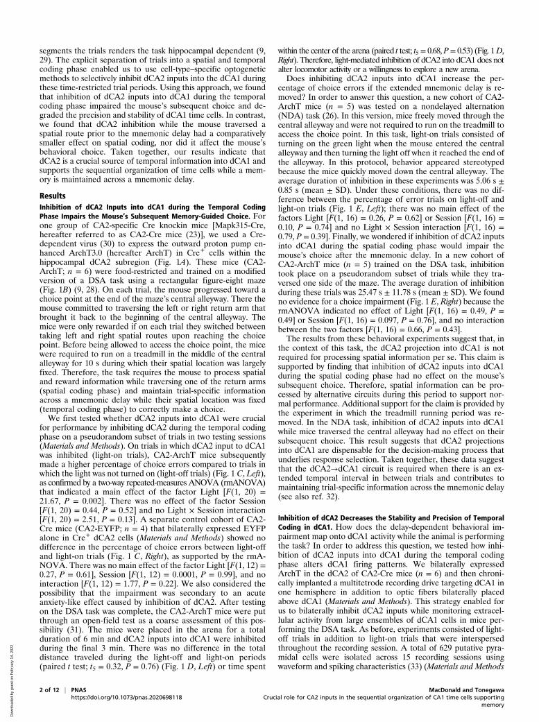
segments the trials renders the task hippocampal dependent (9,29). The explicit separation of trials into a spatial and temporalcoding phase enabled us to use cell-type–specific optogeneticmethods to selectively inhibit dCA2 inputs into the dCA1 duringthese time-restricted trial periods. Using this approach, we foundthat inhibition of dCA2 inputs into dCA1 during the temporalcoding phase impaired the mouse’s subsequent choice and de-graded the precision and stability of dCA1 time cells. In contrast,we found that dCA2 inhibition while the mouse traversed aspatial route prior to the mnemonic delay had a comparativelysmaller effect on spatial coding, nor did it affect the mouse’sbehavioral choice. Taken together, our results indicate thatdCA2 is a crucial source of temporal information into dCA1 andsupports the sequential organization of time cells while a mem-ory is maintained across a mnemonic delay.
ResultsInhibition of dCA2 Inputs into dCA1 during the Temporal CodingPhase Impairs the Mouse’s Subsequent Memory-Guided Choice. Forone group of CA2-specific Cre knockin mice [Mapk315-Cre,hereafter referred to as CA2-Cre mice (23)], we used a Cre-dependent virus (30) to express the outward proton pump en-hanced ArchT3.0 (hereafter ArchT) in Cre+ cells within thehippocampal dCA2 subregion (Fig. 1A). These mice (CA2-ArchT; n = 6) were food-restricted and trained on a modifiedversion of a DSA task using a rectangular figure-eight maze(Fig. 1B) (9, 28). On each trial, the mouse progressed toward achoice point at the end of the maze’s central alleyway. There themouse committed to traversing the left or right return arm thatbrought it back to the beginning of the central alleyway. Themice were only rewarded if on each trial they switched betweentaking left and right spatial routes upon reaching the choicepoint. Before being allowed to access the choice point, the micewere required to run on a treadmill in the middle of the centralalleyway for 10 s during which their spatial location was largelyfixed. Therefore, the task requires the mouse to process spatialand reward information while traversing one of the return arms(spatial coding phase) and maintain trial-specific informationacross a mnemonic delay while their spatial location was fixed(temporal coding phase) to correctly make a choice.We first tested whether dCA2 inputs into dCA1 were crucial
for performance by inhibiting dCA2 during the temporal codingphase on a pseudorandom subset of trials in two testing sessions(Materials and Methods). On trials in which dCA2 input to dCA1was inhibited (light-on trials), CA2-ArchT mice subsequentlymade a higher percentage of choice errors compared to trials inwhich the light was not turned on (light-off trials) (Fig. 1 C, Left),as confirmed by a two-way repeated-measures ANOVA (rmANOVA)that indicated a main effect of the factor Light [F(1, 20) =21.67, P = 0.002]. There was no effect of the factor Session[F(1, 20) = 0.44, P = 0.52] and no Light × Session interaction[F(1, 20) = 2.51, P = 0.13]. A separate control cohort of CA2-Cre mice (CA2-EYFP; n = 4) that bilaterally expressed EYFPalone in Cre+ dCA2 cells (Materials and Methods) showed nodifference in the percentage of choice errors between light-offand light-on trials (Fig. 1 C, Right), as supported by the rmA-NOVA. There was no main effect of the factor Light [F(1, 12) =0.27, P = 0.61], Session [F(1, 12) = 0.0001, P = 0.99], and nointeraction [F(1, 12) = 1.77, P = 0.22]. We also considered thepossibility that the impairment was secondary to an acuteanxiety-like effect caused by inhibition of dCA2. After testingon the DSA task was complete, the CA2-ArchT mice were putthrough an open-field test as a coarse assessment of this pos-sibility (31). The mice were placed in the arena for a totalduration of 6 min and dCA2 inputs into dCA1 were inhibitedduring the final 3 min. There was no difference in the totaldistance traveled during the light-off and light-on periods(paired t test; t5 = 0.32, P = 0.76) (Fig. 1 D, Left) or time spent
within the center of the arena (paired t test; t5= 0.68, P= 0.53) (Fig. 1D,Right). Therefore, light-mediated inhibition of dCA2 into dCA1 does notalter locomotor activity or a willingness to explore a new arena.Does inhibiting dCA2 inputs into dCA1 increase the per-
centage of choice errors if the extended mnemonic delay is re-moved? In order to answer this question, a new cohort of CA2-ArchT mice (n = 5) was tested on a nondelayed alternation(NDA) task (26). In this version, mice freely moved through thecentral alleyway and were not required to run on the treadmill toaccess the choice point. In this task, light-on trials consisted ofturning on the green light when the mouse entered the centralalleyway and then turning the light off when it reached the end ofthe alleyway. In this protocol, behavior appeared stereotypedbecause the mice quickly moved down the central alleyway. Theaverage duration of inhibition in these experiments was 5.06 s ±0.85 s (mean ± SD). Under these conditions, there was no dif-ference between the percentage of error trials on light-off andlight-on trials (Fig. 1 E, Left); there was no main effect of thefactors Light [F(1, 16) = 0.26, P = 0.62] or Session [F(1, 16) =0.10, P = 0.74] and no Light × Session interaction [F(1, 16) =0.79, P = 0.39]. Finally, we wondered if inhibition of dCA2 inputsinto dCA1 during the spatial coding phase would impair themouse’s choice after the mnemonic delay. In a new cohort ofCA2-ArchT mice (n = 5) trained on the DSA task, inhibitiontook place on a pseudorandom subset of trials while they tra-versed one side of the maze. The average duration of inhibitionduring these trials was 25.47 s ± 11.78 s (mean ± SD). We foundno evidence for a choice impairment (Fig. 1 E, Right) because thermANOVA indicated no effect of Light [F(1, 16) = 0.49, P =0.49] or Session [F(1, 16) = 0.097, P = 0.76], and no interactionbetween the two factors [F(1, 16) = 0.66, P = 0.43].The results from these behavioral experiments suggest that, in
the context of this task, the dCA2 projection into dCA1 is notrequired for processing spatial information per se. This claim issupported by finding that inhibition of dCA2 inputs into dCA1during the spatial coding phase had no effect on the mouse’ssubsequent choice. Therefore, spatial information can be pro-cessed by alternative circuits during this period to support nor-mal performance. Additional support for the claim is provided bythe experiment in which the treadmill running period was re-moved. In the NDA task, inhibition of dCA2 inputs into dCA1while mice traversed the central alleyway had no effect on theirsubsequent choice. This result suggests that dCA2 projectionsinto dCA1 are dispensable for the decision-making process thatunderlies response selection. Taken together, these data suggestthat the dCA2→dCA1 circuit is required when there is an ex-tended temporal interval in between trials and contributes tomaintaining trial-specific information across the mnemonic delay(see also ref. 32).
Inhibition of dCA2 Decreases the Stability and Precision of TemporalCoding in dCA1. How does the delay-dependent behavioral im-pairment map onto dCA1 activity while the animal is performingthe task? In order to address this question, we tested how inhi-bition of dCA2 inputs into dCA1 during the temporal codingphase alters dCA1 firing patterns. We bilaterally expressedArchT in the dCA2 of CA2-Cre mice (n = 6) and then chroni-cally implanted a multitetrode recording drive targeting dCA1 inone hemisphere in addition to optic fibers bilaterally placedabove dCA1 (Materials and Methods). This strategy enabled forus to bilaterally inhibit dCA2 inputs while monitoring extracel-lular activity from large ensembles of dCA1 cells in mice per-forming the DSA task. As before, experiments consisted of light-off trials in addition to light-on trials that were interspersedthroughout the recording session. A total of 629 putative pyra-midal cells were isolated across 15 recording sessions usingwaveform and spiking characteristics (33) (Materials and Methods
2 of 12 | PNAS MacDonald and Tonegawahttps://doi.org/10.1073/pnas.2020698118 Crucial role for CA2 inputs in the sequential organization of CA1 time cells supporting
memory
Dow
nloa
ded
by g
uest
on
Feb
ruar
y 14
, 202
2
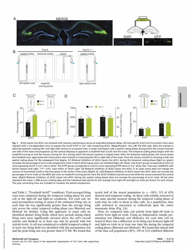
and Table 1, “Treadmill ArchT” condition). Trial-averaged firingrates were estimated during the temporal coding phase for eachcell in the light-off and light-on conditions. For each cell, weused permutation testing to assess if the estimated firing rate ineach time bin was significantly greater than the average firingrate across the entire temporal coding phase (see Materials andMethods for details). Using this approach, in many cells weidentified distinct firing fields, which were periods during whichfiring rates were significantly elevated above the cell’s overallactivity and flanked on at least one side by periods with lowactivity levels. A cell was considered to exhibit temporal coding ifat least one firing field was identified with this permutation testand the peak firing rate was greater than 0.75 Hz. We found that
nearly half of the neural population (n = ∼50%; 313 of 629)showed such temporal coding. As these cells reliably activated atthe same specific moment during the temporal coding phase ofeach trial, we refer to them as time cells. As a population, timecells activated in succession to collectively span the entiremnemonic delay (Fig. 2A).Next, we compared time cell activity from light-off trials to
activity from light-on trials. Using an independent sample per-mutation test (Materials and Methods), for each time cell wetested if there was a difference in its firing rate between light-offand light-on trials in at least one time bin during the temporalcoding phase (Materials and Methods). We found that almost halfof the time cell population (48%; 149 of 313) exhibited different
ArchT ArchT0
25
50
Per
cent
age
erro
r tria
ls
ArchT EYFP0
25
50
Per
cent
age
erro
r tria
ls
0
2500
5000
Dis
tanc
e tra
velle
d (c
m)
0
25
50
Time spent in center (s)
75light off light on7500
ArchT ArchT
delayed spatialalternation
delayed spatialalternation
light off
light on
continuous spatial alternation
delayedspatial alternation
light off
light on
**
MERGE
DAPI GFP
520 nm
choicetemporal codingspatial coding
left spatial route
Trial N
spatial coding
right spatial routetreadmill running period
Trial N + 1
A
B
C D E
Fig. 1. dCA2 inputs into dCA1 are involved with memory maintenance across an extended temporal delay. (A) CA2-specific (CA2-Cre) Cre knockin mice wereinjected with a Cre-dependent virus to express the ArchT-EYFP in Cre+ cells comprising dCA2. (Magnification: 10×.) (B) The DSA task: Mice are trained toalternate between making left- and right-side choices on successive trials. A single trial begins with a spatial coding phase during which the mouse traversesone side of the maze and progresses up the central alleyway to approach a treadmill that is built into the maze. The temporal coding phase begins with thetreadmill turning on and the mouse running for 10 s, during which the mouse’s position is largely fixed. After the temporal coding phase, the mouse leavesthe treadmill area, approaches the choice point, and commits to traversing the left or right side of the maze. Once the mouse commits to choosing a side, thespatial coding phase for the subsequent trial begins. (C) Bilateral inhibition of dCA2 inputs into dCA1 during the temporal coding phase (light on, green)increases the percentage of error trials compared to trials in which dCA2 inputs were not inhibited (light off, black). CA2-ArchT group corresponds to CA2-Cremice expressing ArchT in Cre+ cells in dCA2. The EYFP group corresponds to CA2-Cre mice expressing EYFP alone in Cre+ dCA2 cells. Two-way rmANOVA withfactors Session and Light. **P < 0.01, main effect or factor Light. (D) Bilateral inhibition of dCA2 inputs into dCA1 in an open arena does not change theamount of locomotion (Left) or the time spent in the center of the arena (Right). (E, Left) Bilateral inhibition of dCA2 inputs into dCA1 does not increase thepercentage of error trials on the NDA task with no treadmill running period. Here the dCA2 inhibition period occurred while the mouse traversed the centralstem. (Right) Bilateral inhibition of dCA2 inputs into dCA1 during the spatial coding phase does not increase the percentage of error trials. All bar plotsrepresent the mean ± SEM across 2 testing days and the individual data points for this average from light-off and light-on trials are shown for each mouse.The gray connecting lines are included to visualize the paired comparisons.
MacDonald and Tonegawa PNAS | 3 of 12Crucial role for CA2 inputs in the sequential organization of CA1 time cells supportingmemory
https://doi.org/10.1073/pnas.2020698118
NEU
ROSC
IENCE
Dow
nloa
ded
by g
uest
on
Feb
ruar
y 14
, 202
2
firing patterns between these trial types (Fig. 2B). The averagefiring rate of the time cell population was largely the same acrossthe temporal coding phase during both light-off and light-ontrials (Fig. 2C). However, at the level of individual cells we ob-served a broad range of differing activity patterns in response todCA2 inhibition. We observed a robust decrease in activityduring light-on trials in many time cells (18%; n = 57), as would
be expected by removing a major source of excitatory drive(Fig. 2 D, Upper). In many other cases (30%; n = 97 overall),time cells showed increased levels of activity during light-on trialscompared to light-off trials (Fig. 2 D, Lower). However, firingrates were often elevated because the time cell’s tuning curvebroadened, resulting in less precise temporal coding. In fact, theproportion of time cells showing elevated firing rates during
Table 1. Summary of the number of recording sessions and total cells recorded for each mouseused in the three extracellular recording experiments
Condition Mouse No. of recording sessions Total no. of cells recorded
Treadmill ArchT CA2-ArchT-01 3 123CA2-ArchT-02 3 112CA2-ArchT-03 2 113CA2-ArchT-04 2 68CA2-ArchT-05 2 84CA2-ArchT-06 3 129
Total cells 629Treadmill EYFP CA2-EYFP-01 1 51
CA2-EYFP-02 2 54CA2-EYFP-03 2 79
Total cells 184Maze ArchT CA2-ArchT-07 1 30
CA2-ArchT-08 1 30CA2-ArchT-09 3 53CA2-ArchT-10 1 22CA2-ArchT-11 1 32
Total cells 167
Firin
g ra
te (H
z)
temporal coding phasetreadmill running period
0 15
Time (s)
5
no light on effect
no temporal coding
time cells
light on effect
24%
26%50%
100 5
Firin
g ra
te (H
z)
Time (s)
0
4Light offLight on
2
0
2
4
0 5 10
0
1
2
3
0 5 10 0 5 100
10
20
0 5 10
0
2
4
6
8
0 5 100
1
2
0 5 10
0
2
4
6
8
0 5 10
0 5 10
0
5
10
15
0 5 10
0
5
10
15
0 5 10
ON
OFF
ON
OFF
Ligh
t
0
3
0
4
0
4
0
8
0
20
0
8
0 0
15
0
15
Rate
(Hz)
0 10 0 10 0 10 0 10 0 10Time (s)
6
0
8
Ligh
tRa
te (H
z)
A B C
D
Fig. 2. Inhibition of dCA2 altered the firing rates of a large proportion of dCA1 cells that exhibited temporal coding. (A) Peristimulus time histograms (PSTH)and raster plots depicting spiking activity from seven time cells that were recorded at the same time. The shaded area shown on the raster plot corresponds tothe temporal coding phase during which the mouse was running on the treadmill for 10 s. Each cell selectively fires at a particular time during the temporalcoding phase but differs according to the time coded such that they collectively activate in sequence. The black bars running along the time axis above theraster plots demarcate the cell’s firing field. (B) Proportion of total recorded CA1 cells that were classified as time cells and showed altered temporal firingpatterns (green: light-on effect) or no change (gray: no light-on effect) during trials in which dCA2 was inhibited. The remaining cells showed no evidence oftemporal coding. (C) Average firing rate across all time cells during light-off and light-on trials. Shaded areas correspond to 99% confidence intervals. (D)PSTH and raster plots depicting activity from 10 cells during the temporal coding phase and subdivided into light-off (gray) and light-on (green) trials. The toprow depicts cases in which CA2 inhibition decreased firing rates while the bottom row depicts cases in which CA2 inhibition impaired the precision oftemporal coding through the elevation of firing rates outside the temporal firing field.
4 of 12 | PNAS MacDonald and Tonegawahttps://doi.org/10.1073/pnas.2020698118 Crucial role for CA2 inputs in the sequential organization of CA1 time cells supporting
memory
Dow
nloa
ded
by g
uest
on
Feb
ruar
y 14
, 202
2
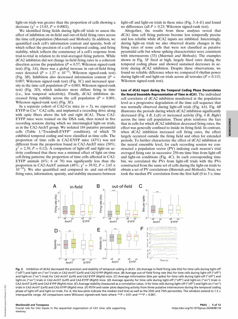
light-on trials was greater than the proportion of cells showing adecrease (χ2 = 13.83, P = 0.0002).We identified firing fields during light-off trials to assess the
effect of inhibition on in-field and out-of-field firing rates acrossthe time cell population (Materials and Methods). In addition, wequantified each cell’s information content and sparsity, both ofwhich reflect the precision of a cell’s temporal coding, and firingstability, which reflects the consistency of a cell’s response fromtrial-to-trial in relation to its trial-averaged firing pattern. WhiledCA2 inhibition did not change in-field firing rates in a coherentdirection across the population (P = 0.57; Wilcoxon signed-ranktest) (Fig. 3A), there was a global increase in out-of-field firingrates detected (P = 1.37 × 10⁻³¹; Wilcoxon signed-rank test)(Fig. 3B). Inhibition also decreased information content (P =0.007; Wilcoxon signed-rank test) (Fig. 3C) and increased spar-sity in the time cell population (P = 0.003; Wilcoxon signed-ranktest) (Fig. 3D), which indicates more diffuse firing in time(i.e., less temporal selectivity). Finally, dCA2 inhibition de-creased firing stability across the cell population (P = 0.001;Wilcoxon signed-rank test) (Fig. 3E).In a separate cohort of CA2-Cre mice (n = 3), we expressed
EYFP in Cre+ CA2 cells, and implanted a recording drive alongwith optic fibers above the left and right dCA1. These CA2-EYFP mice were trained on the DSA task, then tested in fiverecording sessions during which we intermingled light-on trials,as in the CA2-ArchT group. We isolated 184 putative pyramidalcells (Table 1,“Treadmill-EYFP” condition), of which 78exhibited temporal coding and were classified as time cells. Theproportion of time cells in CA2-EYFP mice (43%) was notdifferent from the proportion found in CA2-ArchT mice (50%;χ2 = 2.39, P = 0.12). A comparison of light-off and light-on ac-tivity confirmed that there was a minimal effect of light on timecell-firing patterns; the proportion of time cells affected in CA2-EYFP animals (6%; 6 of 78) was significantly less than theproportion in CA2-ArchT animals (48%; χ2 = 39.92, P = 2.65 ×10⁻¹⁰). We also quantified and compared in- and out-of-fieldfiring rates, information, sparsity, and stability measures between
light-off and light-on trials in these mice (Fig. 3 A–E) and foundno differences (all P > 0.23; Wilcoxon signed-rank test).Altogether, the results from these analyses reveal that
dCA1 time cell firing patterns become less temporally preciseand less reliable while dCA2 inputs are inhibited. Interestingly,during light-on trials we also observed drastic changes in thefiring rates of some cells that were not classified as putativepyramidal cells but whose spiking characteristics were consistentwith interneurons (33) (Materials and Methods). The examplesshown in Fig. 3F fired at high, largely fixed rates during thetemporal coding phase and showed sustained decreases in ac-tivity during dCA2 inhibition. Despite these observations, wefound no reliable difference when we compared θ rhythm powerduring light-off and light-on trials across all tetrodes (P = 0.115;Wilcoxon signed-rank test).
Loss of dCA2 Input during the Temporal Coding Phase Decorrelatesthe Neural Ensemble Representation of Time in dCA1. The individualcell correlates of dCA2 inhibition manifested at the populationlevel as a progressive degradation of the time cell sequence thatwas normally observed during light-off trials (Fig. 4A). Fig. 4Billustrates the periods during which dCA2 inhibition significantlydecreased (Fig. 4 B, Left) or increased activity (Fig. 4 B, Right)across the time cell population. These plots reinforce the factthat in cells for which dCA2 inhibition decreased firing rates, theeffect was generally confined to inside its firing field. In contrast,when dCA2 inhibition increased cell firing rates, the effectlargely occurred outside the firing field and often for extendedperiods. To further characterize the effect of dCA2 inhibition atthe neural ensemble level, for each recording session we con-structed a population vector (PV) indexing each neuron’s trialaveraged firing rate in successive 250-ms time bins from light-offand light-on conditions (Fig. 4C). In each corresponding timebin, we correlated the PVs from light-off trials with the PVsconstructed from the same set of cells during the light-on trials toobtain a set of PV correlations (Materials and Methods). Next, wetook the median PV correlation from the first half (0 to 5 s; time
ArchToff on
EYFP0
1
2
Firin
g ra
te (H
z)
3
off on 0
1
2 ***
Firin
g ra
te (H
z)
ArchToff on
EYFPoff on
Bits
per
spi
ke
0
2**
1
ArchToff on
EYFPoff on
Spa
rsity
0
**
0.5
1
ArchToff on
EYFPoff on
Sta
bilit
y (r
)
0
0.5
**
-0.5
1
ArchToff on
EYFPoff on
0
2
4
0
1
2
3
ON
OFF
ON
OFF
Ligh
t
0
20
0
40
Rate
(Hz)
0 10
Ligh
tRa
te (H
z)
0
2
4
ON
OFF
Ligh
t
0
20
Rate
(Hz)
Time (s)
A B C
D E F
Fig. 3. Inhibition of dCA2 decreased the precision and stability of temporal coding in dCA1. (A) Average in-field firing rate (Hz) for time cells during light-off(“off”) and light-on (“on”) trials in CA2-ArchT (Left) and CA2-EYFP (Right) mice. (B) Average out-of-field firing rate (Hz) for time cells during light-off (“off”)and light-on (“on”) trials for CA2-ArchT (Left) and CA2-EYFP (Right) mice. (C) Average information (bits per spike) for time cells during light-off (“off”) andlight-on (“on”) trials in CA2-ArchT (Left) and CA2-EYFP (Right) mice. (D) Average sparsity for time cells during light-off (“off”) and light-on (“on”) trials inCA2-ArchT (Left) and CA2-EYFP (Right) mice. (E) Average stability (measured as a correlation value, r) for time cells during light-off (“off”) and light-on (“on”)trials in CA2-ArchT (Left) and CA2-EYFP (Right) mice. (F) PSTH and raster plots depicting activity from three putative interneurons during the temporal codingphase of light-off and light-on trials. For A, the box-plots indicate the median (red line) as well as the 25th and 75th percentiles. The whiskers extend to 1.5 ×interquartile range. All comparisons were Wilcoxon signed-rank tests where **P < 0.01 and ***P < 0.001.
MacDonald and Tonegawa PNAS | 5 of 12Crucial role for CA2 inputs in the sequential organization of CA1 time cells supportingmemory
https://doi.org/10.1073/pnas.2020698118
NEU
ROSC
IENCE
Dow
nloa
ded
by g
uest
on
Feb
ruar
y 14
, 202
2
bins 1 to 20) and second half (5 to 10 s; time bins 21 to 40) of thisset. Thus, for each experiment we obtained two PV correlationvalues that measured the similarity between light-off and light-ontime cell firing patterns with respect to the first or second half ofthe temporal coding phase (hereafter referred to as the “CA2inhibition” comparison).In order to properly interpret this measure, we evaluated PV
correlations for two additional control comparisons. For onecontrol comparison, we correlated PVs between even and oddtrials from the temporal coding phase of light-off trials. Thiscorrelation provides an upper bound for the PV correlationbecause it measures the self-consistency of the ensemble patternacross trials of the same trial-type (“same trial” comparison). Forthe second control comparison, we took a resampling approachto estimate a PV correlation between time cell ensembles thatrandomly fired during the temporal coding phase of each trial(Materials and Methods). For each recording session, we ran-domly split all trials from the light-off condition into two trialsets and randomly shifted (circularly) each trial’s temporal firingpattern from each cell before computing a trial average. Then we
obtained a PV correlation from the first and second half of thetemporal coding phase, just as we did for the CA2 inhibitioncomparison. This process was repeated 999 more times and themean PV correlation of this resampled distribution for the firstand second half was taken as the PV correlation for the “ran-dom” comparison in the recording session. This comparisonyields a lower bound for the PV correlation, as it measures thesimilarity between PVs that were randomly rearranged in time.The results from these analyses are shown in Fig. 4D. A two-
way rmANOVA comparing PV correlations across Halves (“firsthalf” and “second half”) and Comparison (same trial, CA2 in-hibition, and random) revealed a main effect of Halves [F(1, 14) =96.52 P = 1.16 × 10⁻⁷], Comparison [F(1, 14) = 62.79,P = 1.15 × 10⁻⁷], and an interaction between these two factors[F(2, 28) = 34.36, P = 9.79 × 10⁻⁷]. The significant interactionterm indicated that the difference between the PV correlationin each half depended on the Comparison and prompted us tomake several specific statistical comparisons using differentcontrasts in the rmANOVA model. First, the PV correlation forCA2 inhibition was less than the same trial correlation in the first
First Half (0-5 s)
Second Half (5-10 s)
PV
cor
rela
tion
(r)
0
1
Same trial
CA2 inhibitionRandom
n.s
-0.5
0.5
***
******
*** ***
***
Light off
Time (s)100 5
Light onN
euro
ns (s
orte
d)
100 51
300
Time (s)100 5
Neu
rons
(sor
ted)
100 51
300
significant differences light on < light off
significant differences light on > light off
First Half (0-5 s)
Second Half (5-10 s)
-1
0***
∆ PV
corre
latio
n (∆
r)CA
2 in
hibi
tion
- sam
e tri
a l
-0.5correlate(r=0.81)
sliding correlationTime (s)
0 10
Ligh
t off
Ligh
t on
A B
C ED
Fig. 4. Loss of dCA2 input during the temporal coding phase decorrelates the neural ensemble representation of time in dCA1. (A) Each panel showsnormalized (range: 0 to 1) trial-averaged firing rates of time cells during the temporal coding phase of light-off (Left) and light-on (Right) trials. Cells aresorted by increasing latency to its maximum firing rate in light-off trials. (B) Each panel shows the periods during the temporal coding phase in which a cell’sfiring rate was significantly decreased in light-on trials (Left, gray segments) or significantly increased in light-on trials (Right, green segments). Cells aresorted in these panels as in A. The red bands running down the diagonal of each panel traces out the half-maximum width from the cell’s peak firing rate inlight-off trials (compare with Left in A). (C) Depiction of how the PV correlation is computed. The panels on the left-hand side show trial averaged activityfrom an ensemble of 13 time cells recorded during a single experiment and sorted as in A. The x axis corresponds to time and is segmented into 250-ms bins.Each row on the top panel shows one cell’s average activity during light-off trials while each row on the bottom panel shows the same corresponding cell’saverage activity during light-on trials. In this example, the PV from the first time bin in the light-off condition is correlated with the first time bin in the light-on condition to yield a PV correlation of 0.81. This is done for each time bin to obtain a set of 40 PV correlation values. The median PV correlation is takenfrom the first half (time bins 1 to 20) and second half (time bins 21 to 40) to obtain the first and second half PV correlation for this recording session. Thisprocess is carried out for each recording session. (D) Depiction of the average PV correlation (mean ± SEM) during the first and second half of the temporalcoding phase for the CA2 inhibition, same trial, and random comparison (see main text). The PV correlation values computed for each recording session areoverlaid on the bar plots. To assist visualization, we used gray lines to connect data points from the same recording session with respect to same trial and CA2inhibition PV correlations. (E) Illustration of the difference between the PV correlation for CA2 inhibition and the same trial comparison in the first andsecond half (mean ± SEM). The difference is more negative in the second half indicating that CA2 inhibition significantly decorrelated firing patterns more inthe second half compared to the first half. As before, gray lines connect data points obtained from the same recording session. Post hoc comparisons areshown and Bonferroni-corrected where ***P < 0.001. See main text for details.
6 of 12 | PNAS MacDonald and Tonegawahttps://doi.org/10.1073/pnas.2020698118 Crucial role for CA2 inputs in the sequential organization of CA1 time cells supporting
memory
Dow
nloa
ded
by g
uest
on
Feb
ruar
y 14
, 202
2

(P = 9.49 × 10⁻⁵; Bonferroni-corrected) and second half (P =1.22 × 10⁻⁵; Bonferroni-corrected) of the temporal coding phase.Furthermore, the PV correlation decreased from the first to secondhalf of the temporal coding phase in the same trial (P =9.76 × 10⁻⁷; Bonferroni-corrected) and CA2 inhibition com-parison (P = 3.49 × 10⁻⁵; Bonferroni-corrected). However, thedegree in which the PV correlation decreased across Halvesdepended on the CA2 inhibition or same trial comparison[two-way rmANOVA that excludes the random comparison:Halves × Comparison interaction: F(1, 14) = 15.37, P = 0.002].Specifically, the PV correlation showed a larger decrease acrossHalves for CA2 inhibition compared to the same trial compari-son (Fig. 4E) (P = 0.0009; Bonferroni-corrected). The PV cor-relation for the same trial comparison was greater than therandom comparison in each half (both P < 1.16 × 10⁻⁹; Bon-ferroni-corrected). However, while the PV correlation for theCA2 inhibition and “error” comparison was different in the firsthalf of the temporal coding phase (P = 0.004; Bonferroni-cor-rected), they were not different in the second half (P = 1;Bonferroni-corrected). Finally, we conducted this analysis on theCA2-EYFP group and confirmed a decrease in the PV correla-tion between Halves [F(1, 4) = 15.34, P = 0.017] but there was nodifference between Comparisons [F(1, 4) = 0.75, P = 0.435] andno interaction between these factors [F(1, 4) = 1.58, P = 0.28].These analyses reveal that time cell firing patterns are more
variable in the second half of the temporal coding phase undernormal conditions (light-off trials), which is consistent with adecrease in the precision of temporal coding with elapsed time(10, 34). Despite this greater variability, temporal coding ispreserved in the second half and considerably greater than whatone would expect if cells fired randomly over time. Conversely,dCA2 inhibition significantly decorrelated dCA1 temporal firingpatterns from the patterns observed under normal conditions. Inaddition, this effect was more pronounced in the second half andduring this period the PV correlation was comparable to thevalue one would obtain using temporally random firing patterns.Combined with our single-cell analyses, these data suggest thatthe loss of dCA2 input degrades temporal coding and destabi-lizes firing patterns in dCA1.
Inhibition of dCA2 Marginally Degrades Spatial Coding in dCA1.Given that dCA2 inhibition during the spatial coding phasedoes not impair the mouse’s behavior, we tested if dCA1 placecell activity remained intact during inhibition. To test this, werecorded cells from dCA1 in five mice (seven recording sessions)while inhibiting dCA2 inputs during the spatial coding phase ona pseudorandom subset of trials in which the mouse traversedthe right side of the maze. Firing rates were estimated as afunction of the mouse’s linearized position during the spatialcoding phase of light-off and light-on trials. We identified cellsexhibiting spatial coding by using the same analysis that was usedto confirm temporal coding (Materials and Methods and Table 1,“Maze ArchT” condition). Using this approach, we found over33% of the cells from which we recorded (33%; n = 55 of 167)showed robust spatial coding during right-side trials. The spatialfiring patterns in 15% (n = 8 of 55) of these place cells signifi-cantly differed between light-off and light-on trials (Fig. 5A), butthis proportion was much lower than the proportion of time cellsthat was affected by dCA2 inhibition (48%; χ2 = 19.89, P =8.19 × 10⁻⁶). There were no differences between light-off andlight-on trials regarding population averaged measures of in- andout-of-field firing rates, information, sparsity, and firing stability(all P > 0.12; Wilcoxon signed-rank test).Next, we quantified the effect of dCA2 inhibition on spatial
coding using the PV correlation analysis described earlier. In thiscase, we separated the spatial coding phase into a first and sec-ond half with respect to the mouse’s linearized position on the
right-side of the maze (Fig. 5B). A rmANOVA comparing PVcorrelations across Halves and Comparison (Fig. 5C) identified amain effect of Comparison [F(2, 12) = 364.97, P = 2.62 × 10⁻⁷]but there was no effect of Halves [F(1, 14) = 0.87; P = 0.39] andno interaction between Comparison and Halves [F(1, 14) = 0.35;P = 0.68]. Expectedly, the PV correlation for the CA2 inhibitioncomparison was different from the “error” comparison in thefirst (P = 6.84 × 10⁻⁵; Bonferroni-corrected) and second (P =3.59 × 10⁻⁷; Bonferroni-corrected) half, and this was alsothe case for the same trial Comparison (first half: P = 0.002;Bonferroni-corrected and second half: P = 2.91 × 10⁻⁷;Bonferroni-corrected). To confirm if the main effect of Com-parison was largely driven by the inclusion of the “error” com-parison in our model, we directly compared the PV correlationsfrom the same trial and CA2 inhibition comparison. This analysisconfirmed a modest but reliable difference between the same trialand CA2 inhibition PV correlations [main effect of Comparison:F(1, 6) = 19.29, P = 0.005], though there was no evidence for aneffect of Halves [F(1, 6) = 0.60, P = 0.48] or a Halves × Comparisoninteraction [F(1, 6) = 0.71, P = 0.43].Finally, we directly compared the PV correlations for the CA2
inhibition comparison from our time cell and place cell experi-ments (Fig. 5D). We used a mixed ANOVA to compare the PVcorrelation for the CA2 inhibition between Condition(between-subject factor: “place cell” or “time cell”) and Halves(within-subject factor: “first half” and “second half), and found amain effect of Condition [F(1, 20) = 15.91, P = 0.0007], Halves[F(1, 20) = 17.02, p = 0.0005], and a Halves × Condition inter-action [F(1, 2 0) = 24.02, p = 8.63 χ 10−5]. Altogether, these dataindicate that dCA2 inhibition had a substantially larger effect ontime cells compared to place cells and place cell disruption didnot worsen while dCA2 was inhibited, unlike in time cells.
DiscussionThere is a great deal of interest in defining a functional role ofthe hippocampal CA2-CA1 circuit with regard to learning andmemory (35, 36) and a growing literature indicates a majorcontribution of CA2 to processing social memories (37, 38).However, this function depends on information flow from dCA2to vCA1 rather than dCA2’s interactions with dCA1 (38, 39).Our present work combined cell-type–specific optogenetics withlarge-scale extracellular recordings of hippocampal dCA1 neu-rons as mice underwent the hippocampal-dependent DSA task.The study resulted in a discovery of a functional role of dCA2projections into dCA1 for the organization of time in ahippocampal-dependent memory.
The dCA2→dCA1 Projection Is Crucial for DSA Memory. We foundthat selective and reversible inhibition of dCA2 inputs into dCA1during the treadmill running period of the DSA task caused miceto make more errors in the subsequent choice of two alternativeouter arms within the trial (Fig. 1C). When the delay period wasreduced or the treadmill removed (NDA task), the inhibition ofdCA2 inputs during the remaining delay period or the centraltrack running period was ineffective in impairing the animal’sperformance (Fig. 1 E, Left). These results are consistent withprior work demonstrating a requirement for the HPC in DSAtasks but not in NDA tasks (29). But we substantially advancedour knowledge on the requirement of the HPC for the DSA taskby identifying not only the specific dCA2→dCA1 circuit, but alsothe specific phase of the DSA task during which it is crucial. Thelatter finding was even more specified by demonstrating thatinhibition of the dCA2→dCA1 circuit activity is largely dis-pensable during the predelay period of the outer arm runningwithin the trial (Fig. 1 E, Right). Taking these data together, wefind that a crucial factor that determines a requirement for thedCA2→dCA1 circuit to support normal performance in the DSA
MacDonald and Tonegawa PNAS | 7 of 12Crucial role for CA2 inputs in the sequential organization of CA1 time cells supportingmemory
https://doi.org/10.1073/pnas.2020698118
NEU
ROSC
IENCE
Dow
nloa
ded
by g
uest
on
Feb
ruar
y 14
, 202
2

task is whether the mouse experiences a distinct mnemonic delaybefore being allowed to make a memory-guided choice. Thisfinding suggests that the behavioral impairment is related to themouse’s failure to maintain trial-specific information across anextended time interval. A role of CA2 in temporal processingwas previously studied employing constitutive Avpr1b knockoutmice (24) in a battery of tasks to test for social and cognitivedeficits. However, the lack of a high level of spatial and temporalrestriction of the knockout prevented a precise interpretation ofthe observed behavioral impairments as the present study.
The Loss of dCA2 Input Disrupts Temporal Coding in dCA1 during theMnemonic Delay. After clarifying the nature of the behavioralimpairment, we determined how dCA2 inhibition altered tem-poral coding in the dCA1 network during the mnemonic delay.In our task, temporal coding was isolated from spatial codingduring this period because the mouse’s spatial location was fixed.Consistent with previous work, we identified a robust form oftemporal coding in the dCA1, as shown by time cells that reliablyactivated for brief periods in succession to span the entiremnemonic delay (Fig. 2A). Firing patterns were also more vari-able for cells that coded for times in the last half of this trialphase (Fig. 4D), which agrees with several previous time cellstudies that report decreasing accuracy in firing times as elapsedtime grows (10, 11, 28, 34, 40). Importantly, we found that in-hibition of dCA2 inputs during this trial phase drastically alteredtime cell firing patterns in dCA1 (Figs. 2 B and D and 4A). Theeffect of inhibition was to reduce firing rates within the mainfiring field of some cells. However, in many other cells, activitywas elevated outside of its main firing field (Fig. 4B). One
potential explanation for elevated firing rates is the presence offeed-forward inhibitory transmission between dCA2 and inter-neurons within the dCA1 (41). We observed several putativedCA1 interneurons to decrease their firing rates in response tooptogenetic dCA2 inhibition (Fig. 3F), so it is possible that theincrease in a time cell’s out-of-field firing rate reflects adisinhibitory effect.Overall, the reduction of dCA2 inputs decreased the precision
and stability of dCA1 time cell firing (Fig. 3 A–E), and degradedthe fidelity of the dCA1 time cell sequence that unfolded whileanimals were behaving normally (Fig. 4 A, D, and E). That thesechanges are associated with a behavioral deficit is analogous toprevious studies that have identified associations between theprecision and stability of dCA1 place fields and other measuresof spatial memory (42–45). Our results provide strong evidencefor a link between the temporal organization of neural activityduring a mnemonic delay and the accuracy of the mouse’seventual memory-guided choice (11, 14, 15, 46, 47). They arealso consistent with some aspects of hippocampal networkmodels of spatial alternation behavior (48, 49) in which separateinputs for spatial and temporal information are combined withinCA1, and the prefrontal cortex (PFC) interacts with hippocam-pal output to guide behavioral response selection (i.e., left orright choice) (see also refs. 50 and 51). Although we do not teaseapart these CA1-PFC circuits in the present study, based on suchprevious work we also assume that there are at least two types ofmemory supporting performance on this DSA task: One that is ahippocampal-dependent memory and the other a PFC-depen-dent “rule memory” that is acquired gradually with repeatedexperience and guides response selection. In this respect, whenoutput from the HPC is unstable and temporally imprecise, as is
Light off
10
Light on
Neu
rons
(sor
ted)
01
55
1
Linearized position (normalized)
B
C D
First Half (0-5 s)
Second Half (5-10 s)
PV
cor
rela
tion
(r)
1 Same trial
CA2 inhibitionRandom
n.s.
0
0.5
*
******
******
n.s.
First Half (0-5 s)
Second Half (5-10 s)
PV
cor
rela
tion
(r)
1*
-0.5
0
*** Spatial coding phaseTemporal coding phase
no light on effect
no spatial coding
place cells
light on effect
29%
6%
65%
10
ON
OFF
Ligh
t
0
10
Rat
e (H
z)Li
ght
Rat
e (H
z)
0
6
0
6
ON
OFF
Ligh
tR
ate
(Hz)
ON
OFF
Linearized position(Normalized)
A
Fig. 5. Inhibition of dCA2 has a marginal effect on spatial coding compared to temporal coding in dCA1. (A, Left) The proportion of cells exhibiting spatialcoding on the right-hand side of the maze as well as those whose firing patterns were significantly affected by inhibition of dCA2. (Right) Depiction of theraster plot and trial averaged firing rate of three place cells with respect to the mouse’s linearized position on the right-hand side of the maze during light-off(black) and light-on (green) trials. These cells exhibited robust spatial coding and were inhibited during light-on trials. (B) Each panel shows normalized trialaveraged firing rates of the cells identified as having spatial coding in A during light-off (Left) or light-on (Right) trials. The cells are sorted with respect to thelinearized position at which the cell fired maximally during light-off trials. (C) The average PV correlation (mean ± SEM) is shown for the cell populationidentified in A in the first and second half of the spatial coding phase for the same trial, CA2 inhibition, and random comparison (see main text). (D) The PVcorrelation (mean ± SEM) for the “CA2 inhibition” comparison in the spatial coding phase (C) is directly compared to the values obtained from the temporalcoding phase illustrated in Fig. 4D. All comparisons were Bonferroni corrected such that * = P < 0.05, *** = P < 0.001, and n.s. = P > 0.05. As in Fig. 4, gray linesconnect data points obtained from the same recording session. See main text for details.
8 of 12 | PNAS MacDonald and Tonegawahttps://doi.org/10.1073/pnas.2020698118 Crucial role for CA2 inputs in the sequential organization of CA1 time cells supporting
memory
Dow
nloa
ded
by g
uest
on
Feb
ruar
y 14
, 202
2
the case when dCA2 inputs are inhibited, the PFC cannotproperly guide response selection.
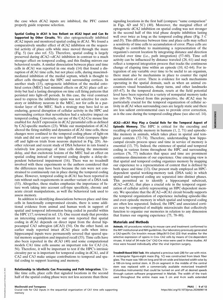
Spatial Coding in dCA1 Is less Reliant on dCA2 Input and Can BeSupported by Other Circuits. We also optogenetically inhibiteddCA2 inputs and monitored spatial coding in dCA1. We found acomparatively smaller effect of dCA2 inhibition on the sequen-tial activity of place cells while mice moved through the maze(Fig. 5) (see also ref. 52). Therefore, spatial coding is largelypreserved during dCA2→dCA1 inhibition in contrast to a muchstronger effect on temporal coding, and this finding mirrors ourbehavioral results. A similar dissociation between place and timecells in dCA1 was reported in one study (14) that found a dis-ruption of dCA1 time cells, but not place cells, during muscimol-mediated inhibition of the medial septum, which is thought toaffect cells throughout the HPC and surrounding cortices. Inanother study (15), optogenetic inhibition of the medial ento-hiral cortex (MEC) had minimal effects on dCA1 place cell ac-tivity but had a lasting disruption on time cell firing patterns thatpersisted into light-off periods. In this case, the viral constructused to express the inhibitory opsin was not selective for excit-atory or inhibitory neurons in the MEC, nor for cells in a par-ticular layer of the MEC. Such a strategy may have led to anenduring, general disruption of cellular activity in the HPC andsurrounding cortices that nevertheless had a selective impact ontemporal coding. Conversely, our use of the CA2-Cre mouse lineenabled for ArchT expression in dCA2 pyramidal cells as definedby CA2-specific markers (23). While our manipulation of dCA2altered the firing stability and dynamics of dCA1 time cells, thesechanges were confined to the temporal coding phase of light-ontrials and did not carry over into light-off trials (compare light-off and light-on trials from neurons depicted in Fig. 2D). An-other relevant and recent study of DSA behavior in rats found arelatively low percentage of time cells during the mnemonicdelay, and that excitotoxic lesions of the MEC had an effect onspatial coding instead of temporal coding despite a delay-de-pendent behavioral impairment (16). There was no treadmillinvolved with these experiments so the authors considered thatthe differences may have been because the rats were not con-strained to continuously run in place during the temporal codingphase. However, temporal coding in dCA1 has been reported inrats without such requirements in another variant of a DSA task(53). Altogether, these studies emphasize the importance of fu-ture work taking into account cell-type specificity, chronic andacute circuit manipulations, as well the behavioral task used toassess memory.In addition to identifying dissociations between place and time
cells in functionally compromised circuits, there is some addi-tional evidence from animal and human work in support ofspatial and temporal information being coded in parallel withinthe HPC (17; reviewed in ref. 13). One recent study that providesan interesting complement to our own reported that spatialcoding in dCA1 depends on direct input from the dorsal hip-pocampal CA3 subregion (dCA3) (54). On the other hand, anearlier study reported intact dCA1 place cells when intra-hippocampal inputs were permanently severed that spared spa-tial memory acquisition and impaired recall (55). Time cells havealso been reported in the dCA3 (40) and some computationalmodels CA1 time cells assume an important role for CA3 (14,56). Therefore, it will be important to test if the HPC’s intrinsiccircuitry per se is important for temporal coding in dCA1, and ifCA2 and CA3 make unique contributions to temporal and spa-tial coding to support learning and memory.
Relationship to Idiothetic Cue Processing and Path Integration. Un-like time cells, place cells that signaled locations in the secondhalf of the spatial coding phase were not less accurate than those
signaling locations in the first half (compare “same comparison”in Figs. 4D and 5C) (40). Moreover, the marginal effect ofoptogenetic dCA2 inhibition on spatial coding was not strongerin the second half of this trial phase despite inhibition lastingwell over twice as long as the temporal coding phase (Fig. 5 Cand D). This difference between time and place cells may reflecta sensitivity of time cells to accumulation of error. Place cells arethought to contribute to maintaining a representation of theorganism’s current location by integrating distance and directiontraveled over time (i.e., path integration) (57–60). Time cellactivity can be influenced by distance traveled (28, 61) and mayreflect a temporal integration process that tracks the continuouschange of elapsing time within an episode (10, 62–64). For in-tegration of any variable over time to be useful to an organism,there must also be mechanisms in place to counter the rapidaccumulation of error. There is evidence for such mechanismsoperating in the spatial domain, such as when an animal en-counters visual boundaries, sharp turns, and other landmarks(65–67). In the temporal domain, resets at the field potentiallevel have been reported in a variety of nonspatial memory taskstoo (68–70). Viewed from this perspective, the dCA2 may beparticularly crucial for the temporal organization of cellular ac-tivity in dCA1 when surrounding cues are largely static and thereare less salient environmental cues to provide error correction,as is the case during the temporal coding phase (see also ref. 14).
dCA2→dCA1 May Play a Crucial Role for the Temporal Aspect ofEpisodic Memory. The HPC is critical for the encoding andrecalling of episodic memory in humans (1, 2, 71) and episodic-like memory in animals, which takes place in spatial and tem-poral contexts (72–74). Therefore, understanding how hippo-campal circuits process space and time in service of memory isessential (13, 75). Indeed, the existence of spatial and temporalcoding in various forms throughout the HPC and surroundingcortices (76, 77) indicates that space and time are prominentcontinuous dimensions of our experience. One emerging view isthat spatial and temporal coding organizes memory by mappingour experience to a representation of context that is defined byboth space and time (13). Our present study employed an HPC-dependent spatial working-memory task (DSA task) in whichspatial and temporal coding are separated into distinct phases.This permitted us to identify a hippocampal subcircuit,dCA2→dCA1, that plays a crucial role in the temporal organi-zation of cellular activty representing an HPC-dependent mem-ory. We speculate that the dCA2→dCA1 circuit broadly supportsthe temporal organization of experience including episodic-likeand even episodic memory in which spatial and temporal codingare often less separated. Indeed, the HPC and associated corti-ces may be comprised of multiple microcircuits that collectivelyfunction to organize our memories in relation to any dimensionthat frames our ongoing experience (75, 78–80).
Materials and MethodsAnimals. All procedures relating to mouse care and treatment conformed totheMIT institutional andNIH guidelines. Our laboratory previously generateda CA2-specific Cre knockin mouse (Map3k15-Cre) (23) that enables for theselective expression of opsins in Cre+ CA2 cells by means of Cre-dependentviruses. A total of 34 male Cre+ CA2-Cre mice were used in these studies. Allmice were housed individually after the viral injection surgery.
Treadmill-Based DSA Task. We adapted a previous task (28) for use with mice.A rectangular figure-eight maze (Fig. 1C) was constructed from black fiberglass. The maze was 100-cm long and 50-cm wide and bisected width-wise bya 100-cm-long central stem. A 35-cm segment in the middle of the centralstem was replaced with a commercially available motorized treadmill(Columbus Instruments) that could be turned on and off at desired speedsthrough custom software programmed in Matlab. The width of the trackused throughout the entire maze was 5 cm and 4-cm walls extended
MacDonald and Tonegawa PNAS | 9 of 12Crucial role for CA2 inputs in the sequential organization of CA1 time cells supportingmemory
https://doi.org/10.1073/pnas.2020698118
NEU
ROSC
IENCE
Dow
nloa
ded
by g
uest
on
Feb
ruar
y 14
, 202
2
upwards from each side of the track. Additional details about the trainingprotocol and testing are found in SI Appendix, Materials and Methods.
Open-Field Test. As described previously (31), an automated video-trackingsystem (Ethovision by Noldus) was used to track the amount of time spent inthe center of an open metal chamber (Accuscan system) compared to theedges, as well as the total distance traveled across a session (31).
Surgery, Recording and Tracking Procedures, Spike Sorting, and Optogenetics.The details about these procedures are in SI Appendix,Materials and Methods.
Histological Procedures and Immunohistochemistry. For the in vivo physiologyexperiments, after the completion of experiments the mice were deeplyanesthetized, and small lesions were made near the tips of each tetrode bypassing current (30 mA for 10 s) for subsequent electrode track positionconfirmation. All mice were deeply anesthetized and transcardially perfusedwith cold phosphate-buffered saline (PBS) followed by 4% paraformalde-hyde in cold PBS. Their brains were extracted for histology using standardprocedures. Additional details are in SI Appendix, Materials and Methods.
Statistical Analyses of Spiking Activity and Local Field Potential. All analyseswere performed using Matlab. For each neuron, each spike was assigned atime in relation to the onset of the spatial or temporal coding phase in eachtrial as well as a linearized spatial coordinate in relation to mouse’s positionduring left- or right-side trials. θ-Power on each tetrode during the temporalcoding phase was estimated using multitaper spectral methods available aspart of the Chronux toolbox (81) (chronux.org). The function mtspectrumc.mwas used to compute the trial-averaged spectrum and θ-power was taken asthe average value between 6 and 12 Hz. The time-bandwidth product wasset to 3 and the number of tapers used was five. This computation was doneseparately for light-off and light-on trials and compared across all tetrodes.
To identify time cells, for each neuronwe separated light-off from light-ontrials, smoothed the trial averaged firing rates with a Gaussian kernel (SD =200 ms), and these rates were down-sampled to 1,000 bins. For light-off andlight-on trials, we used a one-sample paired permutation method to test ifthe trial-averaged firing rate in each time bin was different from the overallfiring rate across the entire temporal coding phase. We accounted formultiple comparisons by adjusting the P values with respect to a family-wiseerror rate of 0.05 (82). The details of this analysis and Matlab code are foundin Groppe et al. (83). For each cell, if at least one time bin significantlyexceeded the overall firing rate in the entire temporal coding phase usingP < 0.05 corrected for multiple comparisons, then it was considered a can-didate “time cell.” Next, we identified all time bins for which the firing rateexceeded the overall firing rate using a P < 0.05 that was not corrected formultiple comparisons, and bins that passed this criterion and less than250 ms apart from one another were merged to obtain firing fields. Can-didate time cells that had a maximum trial averaged firing rate > 0.75 Hz forlight-off or light-on trials were considered time cells. The same approach wasused to identify place cells during the spatial coding phase except that firingrates were occupancy-normalized with respect to the mouse’s linearizedposition and only data during which the mouse was moving at a speedgreater than 4 cm/s were used. Spatial bins that were less than 5 cm apartwere merged.
A two independent-sample permutation method was used to test if aneuron’s firing rate in light-off and light-on trials was different from zero.Here again, P values were adjusted according to a family-wise error rate of0.05. The details of this analysis and Matlab code can be found in Groppeet al. (83). To visualize the periods during which firing rates between con-ditions significantly differed (Fig. 4B), we took the same approach used toidentify firing fields described above.
In-Field and Out-Of-Field Firing Rates. Firing fields during the temporal orspatial coding phase were identified during light-off trials as describedabove. The firing rates inside and outside this firing field were computedseparately for each cell.
Firing Stability. In order to assess firing stability for each neuron in a givencondition, we 1) computed the trial average firing rate of first five trials andthen 2) computed the trial average of all remaining trials in the condition.Finally, 3) these firing rate vectors were correlated using Matlab’s corrfunction. The five-trial window was advanced by one trial (i.e., trials 2 to 6)and steps 1 to 3 were repeated. This process was iterated until the slidingfive-trial block reached the last trial to create a list of correlation values. Thefiring stability for the cell was taken as the median value within the list.
Information. Information (84) was computed for time or space as:
Σ[λ̂x*log2( λ̂x
λ− )*px], where λ̂x is the estimated firing rate for the neuron at
spatial or temporal bin x, λ is the mean firing rate of the neuron’s tuning curveacross all n bins of x, and px is the probability of the animal being in bin x.
PV Correlations to Measure Time Cell Ensemble Pattern Similarity betweenConditions. We adapted an analysis that is often used in studies of placecell populations (85). Regarding Fig. 4, for each recording session trials weresplit into light-off and light-on conditions and the trial-averaged firing ratefor each time cell was computed using consecutive, nonoverlapping 250-msbins. The cell population firing pattern in each condition was organized as atwo-dimensional matrix with each row indexing a neuron and each columnindexing a firing rate in a 250-ms bin during the temporal coding phase(Fig. 4B). Therefore, a PV was defined for each 250-ms time bin in a condi-tion. Using Matlab’s corr function, we correlated the PVs from the same timebin in each condition to obtain an r value. As there were 40 time bins in thetemporal coding phase, we obtained a set of 40 r values. Finally, we took themedian r value from the first (time bin 1 to 20) and second half (time bin 21to 40) to yield a PV correlation for the CA2 inhibition condition in a re-cording session. To compute a same-trial PV correlation, we correlated thePVs representing the ensemble’s trial averaged firing rates from even andodd trials from the light-off condition. To compute a random PV correlation,1) the light-off trials were randomly divided into two sets of trials, 2) thespike train from each neuron in each trial was circularly shifted by a valuethat was selected randomly for each neuron and trial, then 3) the trial av-erage was computed for each neuron using these two shifted trial sets toconstruct two PVs for each time bin in the temporal coding phase. Each PVfor a given time bin was correlated and the median value taken from eachhalf was taken as the r value. This process was repeated 999 more times andthe mean PV correlation from this resampled distribution was used tomeasure similarity in the random condition. The PV correlations were com-pared using an rmANOVA and post hoc comparisons were Bonferroni-cor-rected. All P values were Greenhouse–Geiser-corrected. The same procedurewas used to measure and compare PVs from the spatial coding phase (Fig. 5)by subdividing the linearized position into 40 spatial bins. All trial-averagedfiring rates were occupancy-normalized and only data during which themouse was moving >4 cm/s was used.
Data Availability. All study data are included in the article and supportinginformation.
ACKNOWLEDGMENTS. We thank Mike Ragion, Anthony Moffa, Carl Twiss,and Jayson Derwin for technical assistance; Quentin Ferry, Shruti Muralidhar,and Chen Sun for comments about the manuscript before submission; andthe rest of the S.T. laboratory for their support. This work was supported bythe RIKEN Center for Brain Science, the Howard Hughes Medical Institute,and the JPB Foundation (S.T.).
1. W. B. Scoville, B. Milner, Loss of recent memory after bilateral hippocampal lesions.
J. Neurol. Neurosurg. Psychiatry 20, 11–21 (1957).
2. F. Vargha-Khadem et al., Differential effects of early hippocampal pathology on
episodic and semantic memory. Science 277, 376–380 (1997).
3. J. O’Keefe, J. Dostrovsky, The hippocampus as a spatial map. Preliminary evidence
from unit activity in the freely-moving rat. Brain Res. 34, 171–175 (1971).
4. J. O’Keefe, L. Nadel, The Hippocampus as a Cognitive Map (Oxford University Press, 1978).
5. T. Hartley, C. Lever, N. Burgess, J. O’Keefe, Space in the brain: How the hippocampal for-
mation supports spatial cognition. Philos. Trans. R. Soc. Lond. B Biol. Sci. 369, 20120510 (2013).
6. G. Buzsáki, E. I. Moser, Memory, navigation and theta rhythm in the hippocampal-
entorhinal system. Nat. Neurosci. 16, 130–138 (2013).
7. T. J. Wills, C. Lever, F. Cacucci, N. Burgess, J. O’Keefe, Attractor dynamics in the
hippocampal representation of the local environment. Science 308, 873–876
(2005).
8. S. Leutgeb et al., Independent codes for spatial and episodic memory in hippocampal
neuronal ensembles. Science 309, 619–623 (2005).
9. E. Pastalkova, V. Itskov, A. Amarasingham, G. Buzsáki, Internally generated cell as-
sembly sequences in the rat hippocampus. Science 321, 1322–1327 (2008).
10 of 12 | PNAS MacDonald and Tonegawahttps://doi.org/10.1073/pnas.2020698118 Crucial role for CA2 inputs in the sequential organization of CA1 time cells supporting
memory
Dow
nloa
ded
by g
uest
on
Feb
ruar
y 14
, 202
2
10. C. J. MacDonald, K. Q. Lepage, U. T. Eden, H. Eichenbaum, Hippocampal “time cells”
bridge the gap in memory for discontiguous events. Neuron 71, 737–749 (2011).
11. C. J. MacDonald, S. Carrow, R. Place, H. Eichenbaum, Distinct hippocampal time cell
sequences represent odor memories in immobilized rats. J. Neurosci. 33, 14607–14616
(2013).
12. H. Eichenbaum, Time cells in the hippocampus: A new dimension for mapping
memories. Nat. Rev. Neurosci. 15, 732–744 (2014).
13. H. Eichenbaum, On the integration of space, time, and memory. Neuron 95,
1007–1018 (2017).
14. Y. Wang, S. Romani, B. Lustig, A. Leonardo, E. Pastalkova, Theta sequences are es-
sential for internally generated hippocampal firing fields. Nat. Neurosci. 18, 282–288
(2015).
15. N. T. M. Robinson et al., Medial entorhinal cortex selectively supports temporal
coding by hippocampal neurons. Neuron 94, 677–688.e6 (2017).
16. M. Sabariego et al., Time cells in the hippocampus are neither dependent on medial
entorhinal cortex inputs nor necessary for spatial working memory. Neuron 102,
1235–1248.e5 (2019).
17. E. A. Mankin, G. W. Diehl, F. T. Sparks, S. Leutgeb, J. K. Leutgeb, Hippocampal CA2
activity patterns change over time to a larger extent than between spatial contexts.
Neuron 85, 190–201 (2015).
18. L. Lu, K. M. Igarashi, M. P. Witter, E. I. Moser, M.-B. Moser, Topography of place maps
along the CA3-to-CA2 axis of the hippocampus. Neuron 87, 1078–1092 (2015).
19. H. Lee, C. Wang, S. S. Deshmukh, J. J. Knierim, Neural population evidence of func-
tional heterogeneity along the CA3 transverse axis: Pattern completion versus pattern
separation. Neuron 87, 1093–1105 (2015).
20. G. M. Alexander et al., Social and novel contexts modify hippocampal CA2 repre-
sentations of space. Nat. Commun. 7, 10300 (2016).
21. M. E. Wintzer, R. Boehringer, D. Polygalov, T. J. McHugh, The hippocampal CA2 en-
semble is sensitive to contextual change. J. Neurosci. 34, 3056–3066 (2014).
22. V. Chevaleyre, S. A. Siegelbaum, Strong CA2 pyramidal neuron synapses define a
powerful disynaptic cortico-hippocampal loop. Neuron 66, 560–572 (2010).
23. K. Kohara et al., Cell type-specific genetic and optogenetic tools reveal hippocampal
CA2 circuits. Nat. Neurosci. 17, 269–279 (2014).
24. L. M. DeVito et al., Vasopressin 1b receptor knock-out impairs memory for temporal
order. J. Neurosci. 29, 2676–2683 (2009).
25. W. S. Young, J. Li, S. R. Wersinger, M. Palkovits, The vasopressin 1b receptor is
prominent in the hippocampal area CA2 where it is unaffected by restraint stress or
adrenalectomy. Neuroscience 143, 1031–1039 (2006).
26. E. R. Wood, P. A. Dudchenko, R. J. Robitsek, H. Eichenbaum, Hippocampal neurons
encode information about different types of memory episodes occurring in the same
location. Neuron 27, 623–633 (2000).
27. L. M. Frank, E. N. Brown, M. Wilson, Trajectory encoding in the hippocampus and
entorhinal cortex. Neuron 27, 169–178 (2000).
28. B. J. Kraus, R. J. Robinson 2nd, J. A. White, H. Eichenbaum, M. E. Hasselmo, Hippo-
campal “time cells”: Time versus path integration. Neuron 78, 1090–1101 (2013).
29. J. A. Ainge, M. A. A. van der Meer, R. F. Langston, E. R. Wood, Exploring the role of
context-dependent hippocampal activity in spatial alternation behavior. Hippocam-
pus 17, 988–1002 (2007).
30. J. Yamamoto, J. Suh, D. Takeuchi, S. Tonegawa, Successful execution of working
memory linked to synchronized high-frequency gamma oscillations. Cell 157, 845–857
(2014).
31. S. Ramirez et al., Activating positive memory engrams suppresses depression-like
behaviour. Nature 522, 335–339 (2015).
32. H. Eichenbaum, The role of the hippocampus in navigation is memory. J. Neurophysiol.
117, 1785–1796 (2017).
33. J. Csicsvari, H. Hirase, A. Czurkó, A. Mamiya, G. Buzsáki, Oscillatory coupling of hip-
pocampal pyramidal cells and interneurons in the behaving Rat. J. Neurosci. 19,
274–287 (1999).
34. M. W. Howard et al., A unified mathematical framework for coding time, space, and
sequences in the hippocampal region. J. Neurosci. 34, 4692–4707 (2014).
35. S. M. Dudek, G. M. Alexander, S. Farris, Rediscovering area CA2: Unique properties
and functions. Nat. Rev. Neurosci. 17, 89–102 (2016).
36. S. J. Middleton, T. J. McHugh, CA2: A highly connected intrahippocampal relay. Annu.
Rev. Neurosci. 43, 55–72 (2020).
37. F. L. Hitti, S. A. Siegelbaum, The hippocampal CA2 region is essential for social
memory. Nature 508, 88–92 (2014).
38. T. Okuyama, T. Kitamura, D. S. Roy, S. Itohara, S. Tonegawa, Ventral CA1 neurons
store social memory. Science 353, 1536–1541 (2016).
39. T. Meira et al., A hippocampal circuit linking dorsal CA2 to ventral CA1 critical for
social memory dynamics. Nat. Commun. 9, 4163 (2018).
40. D. M. Salz et al., Time cells in hippocampal area CA3. J. Neurosci. 36, 7476–7484
(2016).
41. K. Nasrallah et al., Routing hippocampal information flow through parvalbumin in-
terneuron plasticity in area CA2. Cell Rep. 27, 86–98.e3 (2019).
42. C. A. Barnes, B. L. McNaughton, J. O’Keefe, Loss of place specificity in hippocampal
complex spike cells of senescent rat. Neurobiol. Aging 4, 113–119 (1983).
43. T. J. McHugh, K. I. Blum, J. Z. Tsien, S. Tonegawa, M. A. Wilson, Impaired hippocampal
representation of space in CA1-specific NMDAR1 knockout mice. Cell 87, 1339–1349
(1996).
44. C. G. Kentros, N. T. Agnihotri, S. Streater, R. D. Hawkins, E. R. Kandel, Increased at-
tention to spatial context increases both place field stability and spatial memory.
Neuron 42, 283–295 (2004).
45. A. S. Bahar, P. R. Shirvalkar, M. L. Shapiro, Memory-guided learning: CA1 and CA3
neuronal ensembles differentially encode the commonalities and differences be-
tween situations. J. Neurosci. 31, 12270–12281 (2011).
46. C. D. Harvey, P. Coen, D. W. Tank, Choice-specific sequences in parietal cortex during a
virtual-navigation decision task. Nature 484, 62–68 (2012).
47. K. Rajan, C. D. Harvey, D. W. Tank, Recurrent network models of sequence generation
and memory. Neuron 90, 128–142 (2016).
48. M. E. Hasselmo, H. Eichenbaum, Hippocampal mechanisms for the context-dependent
retrieval of episodes. Neural Netw. 18, 1172–1190 (2005).
49. Y. Katz, W. L. Kath, N. Spruston, M. E. Hasselmo, Coincidence detection of place and
temporal context in a network model of spiking hippocampal neurons. PLOS Comput.
Biol. 3, e234 (2007).
50. M. W. Jones, M. A. Wilson, Theta rhythms coordinate hippocampal-prefrontal inter-
actions in a spatial memory task. PLoS Biol. 3, e402 (2005).
51. M. W. Jones, M. A. Wilson, Phase precession of medial prefrontal cortical ac-
tivity relative to the hippocampal theta rhythm. Hippocampus 15, 867–873
(2005).
52. R. Boehringer et al., Chronic loss of CA2 transmission leads to hippocampal hyper-
excitability. Neuron 94, 642–655.e9 (2017).
53. P. R. Gill, S. J. Y. Mizumori, D. M. Smith, Hippocampal episode fields develop with
learning. Hippocampus 21, 1240–1249 (2011).
54. H. Davoudi, D. J. Foster, Acute silencing of hippocampal CA3 reveals a dominant role
in place field responses. Nat. Neurosci. 22, 337–342 (2019).
55. V. H. Brun et al., Impaired spatial representation in CA1 after lesion of direct input
from entorhinal cortex. Neuron 57, 290–302 (2008).
56. E. T. Rolls, P. Mills, The generation of time in the hippocampal memory system. Cell
Rep. 28, 1649–1658.e6 (2019).
57. B. L. McNaughton, F. P. Battaglia, O. Jensen, E. I. Moser, M.-B. Moser, Path inte-
gration and the neural basis of the ‘cognitive map’. Nat. Rev. Neurosci. 7, 663–678
(2006).
58. F. Savelli, J. J. Knierim, Origin and role of path integration in the cognitive repre-
sentations of the hippocampus: Computational insights into open questions. J. Exp.
Biol. 222(suppl. 1), jeb188912 (2019).
59. E. I. Moser, M. B. Moser, B. L. McNaughton, Spatial representation in the hippocampal
formation: A history. Nat. Neurosci. 20, 1448–1464 (2017).
60. A. Samsonovich, B. L. McNaughton, Path integration and cognitive mapping in
a continuous attractor neural network model. J. Neurosci. 17, 5900–5920
(1997).
61. C. Haimerl et al., Internal representation of hippocampal neuronal population spans a
time-distance continuum. Proc. Natl. Acad. Sci. U.S.A. 116, 7477–7482 (2019).
62. M. E. Hasselmo, Arc length coding by interference of theta frequency oscillations may
underlie context-dependent hippocampal unit data and episodic memory function.
Learn. Mem. 14, 782–794 (2007).
63. M. E. Hasselmo, How We Remember: Brain Mechanisms of Episosic Memory (The MIT
Press, 2012),.
64. V. Itskov, C. Curto, E. Pastalkova, G. Buzsáki, Cell assembly sequences arising from
spike threshold adaptation keep track of time in the hippocampus. J. Neurosci. 31,
2828–2834 (2011).
65. K. Hardcastle, S. Ganguli, L. M. Giocomo, Environmental boundaries as an error cor-
rection mechanism for grid cells. Neuron 86, 827–839 (2015).
66. R. P. Jayakumar et al., Recalibration of path integration in hippocampal place cells.
Nature 566, 533–537 (2019).
67. D. Derdikman et al., Fragmentation of grid cell maps in a multicompartment envi-
ronment. Nat. Neurosci. 12, 1325–1332 (2009).
68. D. S. Rizzuto et al., Reset of human neocortical oscillations during a working memory
task. Proc. Natl. Acad. Sci. U.S.A. 100, 7931–7936 (2003).
69. M. J. Jutras, P. Fries, E. A. Buffalo, Oscillatory activity in the monkey hippocampus
during visual exploration and memory formation. Proc. Natl. Acad. Sci. U.S.A. 110,
13144–13149 (2013).
70. M. E. Hasselmo, C. E. Stern, Theta rhythm and the encoding and retrieval of space and
time. Neuroimage 85, 656–666 (2014).
71. R. G. M. Morris et al., Elements of a neurobiological theory of the hippocampus: The
role of activity-dependent synaptic plasticity in memory. Philos. Trans. R. Soc. Lond. B
Biol. Sci. 358, 773–786 (2003).
72. N. S. Clayton, A. Dickinson, Episodic-like memory during cache recovery by scrub jays.
Nature 395, 272–274 (1998).
73. N. J. Fortin, S. P. Wright, H. Eichenbaum, Recollection-like memory retrieval in rats is
dependent on the hippocampus. Nature 431, 188–191 (2004).
74. R. G. M. Morris, Episodic-like memory in animals: Psychological criteria, neural
mechanisms and the value of episodic-like tasks to investigate animal models of
neurodegenerative disease. Philos. Trans. R. Soc. Lond. B Biol. Sci. 356, 1453–1465
(2001).
75. H. Eichenbaum, N. J. Cohen, Can we reconcile the declarative memory and spatial
navigation views on hippocampal function? Neuron 83, 764–770 (2014).
76. A. Tsao et al., Integrating time from experience in the lateral entorhinal cortex.
Nature 561, 57–62 (2018).
MacDonald and Tonegawa PNAS | 11 of 12Crucial role for CA2 inputs in the sequential organization of CA1 time cells supportingmemory
https://doi.org/10.1073/pnas.2020698118
NEU
ROSC
IENCE
Dow
nloa
ded
by g
uest
on
Feb
ruar
y 14
, 202
2
77. B. J. Kraus et al., During running in place, grid cells integrate elapsed time and dis-
tance run. Neuron 88, 578–589 (2015).
78. H. Eichenbaum, N. J. Cohen, From Conditioning to Conscious Recollection (Oxford
University Press, 2004).
79. A. D. Ekstrom, C. Ranganath, Space, time and episodic memory: The Hippocampus is
all over the cognitive map. Hippocampus 28, 680–687 (2017).
80. H. Eichenbaum, P. Dudchenko, E. Wood, M. Shapiro, H. Tanila, The hippocampus, memory,
and place cells: Is it spatial memory or a memory space? Neuron 23, 209–226 (1999).
81. H. Bokil, P. Andrews, J. E. Kulkarni, S. Mehta, P. P. Mitra, Chronux: A platform for
analyzing neural signals. J. Neurosci. Methods 192, 146–151 (2010).
82. R. C. Blair, J. J. Higgins, W. Karniski, J. D. Kromrey, A study of multivariate permu-
tation tests which may replace Hotelling’s T2 test in prescribed circumstances. Mul-
tivariate Behav. Res. 29, 141–163 (1994).
83. D. M. Groppe, T. P. Urbach, M. Kutas, Mass univariate analysis of event-related brain
potentials/fields I: A critical tutorial review. Psychophysiology 48, 1711–1725 (2011).
84. E. J. Markus, C. A. Barnes, B. L. McNaughton, V. L. Gladden, W. E. Skaggs, Spatial
information content and reliability of hippocampal CA1 neurons: Effects of visual
input. Hippocampus 4, 410–421 (1994).
85. S. Leutgeb, J. K. Leutgeb, A. Treves, M.-B. Moser, E. I. Moser, Distinct ensemble codes
in hippocampal areas CA3 and CA1. Science 305, 1295–1298 (2004).
12 of 12 | PNAS MacDonald and Tonegawahttps://doi.org/10.1073/pnas.2020698118 Crucial role for CA2 inputs in the sequential organization of CA1 time cells supporting
memory
Dow
nloa
ded
by g
uest
on
Feb
ruar
y 14
, 202
2