The C-terminal module IV of connective tissue growth factor is a novel immune modulator of the Th17...
13
The C-terminal module IV of connective tissue growth factor is a novel immune modulator of the Th17 response Raquel Rodrigues-Dı ´ez 1 , Rau ´ l R Rodrigues-Dı ´ez 1 , Sandra Rayego-Mateos 1 , Beatriz Suarez-Alvarez 1 , Carolina Lavoz 1 , Luiz Stark Aroeira 2 , Elsa Sa ´nchez-Lo ´ pez 3 , Macarena Orejudo 1 , Matilde Alique 1 , Carlos Lopez-Larrea 4 , Alberto Ortiz 5 , Jesu ´ s Egido 6 and Marta Ruiz-Ortega 1 Connective tissue growth factor (CTGF/CCN2) is a matricellular protein susceptible to proteolytic degradation. CCN2 levels have been suggested as a potential risk biomarker in several chronic diseases. In body fluids, CCN2 full-length and its degradation fragments can be found; however, their in vivo effects are far from being elucidated. CCN2 was described as a profibrotic mediator, but this concept is changing to a proinflammatory cytokine. In vitro, CCN2 full-length and its C-terminal module IV (CCN2(IV)) exert proinflammatory properties. Emerging evidence suggest that Th17 cells, and its effector cytokine IL-17A, participate in chronic inflammatory diseases. Our aim was to explore whether CCN2(IV) could regulate the Th17 response. In vitro, stimulation of human naive CD4 þ T lymphocytes with CCN2(IV) resulted in differentiation to Th17 phenotype. The in vivo effects of CCN2(IV) were studied in C57BL/6 mice. Intraperitoneal administration of recombinant CCN2(IV) did not change serum IL-17A levels, but caused an activation of the Th17 response in the kidney, characterized by interstitial infiltration of Th17 (IL17A þ /CD4 þ ) cells and upregulation of proinflammatory mediators. In CCN2(IV)-injected mice, elevated renal levels of Th17-related factors (IL-17A, IL-6, STAT3 and RORgt) were found, whereas Th1/Th2 cytokines or Treg-related factors (TGF-b and Foxp-3) were not modified. Treatment with an anti-IL-17A neutralizing antibody diminished CCN2(IV)-induced renal inflammation. Our findings unveil that the C-terminal module of CCN2 induces the Th17 differentiation of human Th17 cells and causes a renal Th17 inflammatory response. Furthermore, these data bear out that IL-17A targeting is a promising tool for chronic inflammatory diseases, including renal pathologies. Laboratory Investigation (2013) 93, 812–824; doi:10.1038/labinvest.2013.67; published online 6 May 2013 KEYWORDS: CCN2/ IL-17A; inflammation; TGF-b; Th17 cells Connective tissue growth factor (CTGF/CCN2) is a cysteine- rich secreted matricellular protein that exerts many biological functions depending on the cellular context. 1 CCN2 is a 36–38 kDa monomeric protein composed of 349 residues. This protein has a modular architecture comprising four functional modules: an amino-terminal insulin-like growth- factor-binding domain (IGFB), the cysteine-rich domain (also called von Willebrand type c domain), a thrombospondin type 1 repeat domain, and the C-terminal heparin-binding domain (Module 4) also known as a carboxy-terminal cystine knot domain (namely here CCN2(IV)). 2 Between modules 2 and 3 is the ‘hinge region’ that is cleaved by most proteases, including matrix metalloproteases (MMPs) 1, 2, 3, 7, 9, and 13, elastase, and plasmin, the latter two of which also cleave the individual modules to yield four fragments. 3 In particular, MMP2 clips CCN2 in vitro to a 10-kDa fragment and that both full-length and clipped CCN2 were recovered from fibroblast cell culture supernatants. 1 In biological fluids, CCN2 can be found in various forms: full-length molecule, fragments including N-terminal and C-terminal halves cleaved in the ‘hinge region,’ as well as the individual 10–12 kDa that corresponds to CCN2(IV). 1,4 1 Cellular Biology in Renal Diseases Laboratory, Instituto de Investigacion Sanitaria Fundacio ´ n Jime ´nez Dı ´az, Universidad Auto ´ noma Madrid, Madrid, Spain; 2 Servicio de Nefrologı ´a, Hospital Universitario La Paz- IdiPAZ, Madrid, Spain; 3 Translational Oncology Unit, Instituto de Investigaciones Biome ´ dicas ‘Alberto Sols,’ CSIC, Madrid, Spain; 4 Department of Immunology, Hospital Universitario Central de Asturias, Oviedo, Spain; 5 Dialysis, Fundacio ´n Jime ´nez Dı ´az, Madrid, Spain and 6 Renal Research Laboratory, Fundacio ´n Jime ´nez Dı ´az, Madrid, Spain Correspondence: Dr M Ruiz-Ortega, PhD, Cellular Biology in Renal Diseases Laboratory, Instituto de Investigacio ´ n Fundacio ´n Jime ´nez Dı ´az, Universidad Auto ´ noma Madrid, Avda, Reyes Cato ´ licos, 2, 28040 Madrid, Spain. E-mail:[email protected] Received 3 May 2012; revised 4 April 2013; accepted 8 April 2013 812 Laboratory Investigation | Volume 93 July 2013 | www.laboratoryinvestigation.org Laboratory Investigation (2013) 93, 812–824 & 2013 USCAP, Inc All rights reserved 0023-6837/13
-
Upload
independent -
Category
Documents
-
view
2 -
download
0
Transcript of The C-terminal module IV of connective tissue growth factor is a novel immune modulator of the Th17...

The C-terminal module IV of connective tissue growthfactor is a novel immune modulator of the Th17responseRaquel Rodrigues-Dıez1, Raul R Rodrigues-Dıez1, Sandra Rayego-Mateos1, Beatriz Suarez-Alvarez1, Carolina Lavoz1,Luiz Stark Aroeira2, Elsa Sanchez-Lopez3, Macarena Orejudo1, Matilde Alique1, Carlos Lopez-Larrea4, Alberto Ortiz5,Jesus Egido6 and Marta Ruiz-Ortega1
Connective tissue growth factor (CTGF/CCN2) is a matricellular protein susceptible to proteolytic degradation. CCN2levels have been suggested as a potential risk biomarker in several chronic diseases. In body fluids, CCN2 full-lengthand its degradation fragments can be found; however, their in vivo effects are far from being elucidated. CCN2 wasdescribed as a profibrotic mediator, but this concept is changing to a proinflammatory cytokine. In vitro, CCN2 full-lengthand its C-terminal module IV (CCN2(IV)) exert proinflammatory properties. Emerging evidence suggest that Th17 cells,and its effector cytokine IL-17A, participate in chronic inflammatory diseases. Our aim was to explore whether CCN2(IV)could regulate the Th17 response. In vitro, stimulation of human naive CD4þ T lymphocytes with CCN2(IV) resultedin differentiation to Th17 phenotype. The in vivo effects of CCN2(IV) were studied in C57BL/6 mice. Intraperitonealadministration of recombinant CCN2(IV) did not change serum IL-17A levels, but caused an activation of the Th17response in the kidney, characterized by interstitial infiltration of Th17 (IL17Aþ /CD4þ ) cells and upregulation ofproinflammatory mediators. In CCN2(IV)-injected mice, elevated renal levels of Th17-related factors (IL-17A, IL-6,STAT3 and RORgt) were found, whereas Th1/Th2 cytokines or Treg-related factors (TGF-b and Foxp-3) were not modified.Treatment with an anti-IL-17A neutralizing antibody diminished CCN2(IV)-induced renal inflammation. Our findingsunveil that the C-terminal module of CCN2 induces the Th17 differentiation of human Th17 cells and causes a renalTh17 inflammatory response. Furthermore, these data bear out that IL-17A targeting is a promising tool for chronicinflammatory diseases, including renal pathologies.Laboratory Investigation (2013) 93, 812–824; doi:10.1038/labinvest.2013.67; published online 6 May 2013
KEYWORDS: CCN2/ IL-17A; inflammation; TGF-b; Th17 cells
Connective tissue growth factor (CTGF/CCN2) is a cysteine-rich secreted matricellular protein that exerts many biologicalfunctions depending on the cellular context.1 CCN2 is a36–38 kDa monomeric protein composed of 349 residues.This protein has a modular architecture comprising fourfunctional modules: an amino-terminal insulin-like growth-factor-binding domain (IGFB), the cysteine-rich domain(also called von Willebrand type c domain), a thrombospondintype 1 repeat domain, and the C-terminal heparin-bindingdomain (Module 4) also known as a carboxy-terminal cystineknot domain (namely here CCN2(IV)).2 Between modules
2 and 3 is the ‘hinge region’ that is cleaved by most proteases,including matrix metalloproteases (MMPs) 1, 2, 3, 7, 9, and13, elastase, and plasmin, the latter two of which also cleavethe individual modules to yield four fragments.3 Inparticular, MMP2 clips CCN2 in vitro to a 10-kDafragment and that both full-length and clipped CCN2 wererecovered from fibroblast cell culture supernatants.1 Inbiological fluids, CCN2 can be found in various forms:full-length molecule, fragments including N-terminal andC-terminal halves cleaved in the ‘hinge region,’ as well as theindividual 10–12 kDa that corresponds to CCN2(IV).1,4
1Cellular Biology in Renal Diseases Laboratory, Instituto de Investigacion Sanitaria Fundacion Jimenez Dıaz, Universidad Autonoma Madrid, Madrid, Spain; 2Servicio deNefrologıa, Hospital Universitario La Paz- IdiPAZ, Madrid, Spain; 3Translational Oncology Unit, Instituto de Investigaciones Biomedicas ‘Alberto Sols,’ CSIC, Madrid, Spain;4Department of Immunology, Hospital Universitario Central de Asturias, Oviedo, Spain; 5Dialysis, Fundacion Jimenez Dıaz, Madrid, Spain and 6Renal ResearchLaboratory, Fundacion Jimenez Dıaz, Madrid, SpainCorrespondence: Dr M Ruiz-Ortega, PhD, Cellular Biology in Renal Diseases Laboratory, Instituto de Investigacion Fundacion Jimenez Dıaz, Universidad AutonomaMadrid, Avda, Reyes Catolicos, 2, 28040 Madrid, Spain. E-mail:[email protected]
Received 3 May 2012; revised 4 April 2013; accepted 8 April 2013
812 Laboratory Investigation | Volume 93 July 2013 | www.laboratoryinvestigation.org
Laboratory Investigation (2013) 93, 812–824
& 2013 USCAP, Inc All rights reserved 0023-6837/13
These variations produce a subfamily of secreted proteinswith distinct functions. However, the biological in vivo effectsof CCN2 and its fragments are still unknown. Severalindependent studies have evaluated CCN2 levels in theurine and/or serum in several chronic kidney diseases. Somegroups have found elevated levels of the full-length CCN2,5,6
or the N-terminal,6,7 or the C-terminal8–10 CCN2 fragmentsdetermined by ELISA, using antibodies that recognized eachpart of the molecule. Based on these data, CCN2 has beenproposed as a risk biomarker of human diabetic nephropathyand other forms of chronic kidney disease,2,5–10 and forcardiac dysfunction in patients exhibiting myocardial fibrosisand chronic heart failure.11
CCN2 has been found overexpressed in almost all humandisorders characterized by excessive scarring and fibrosis.2,12
The concept of CCN2 as a downstream mediator ofprofibrotic factors, including transforming growth factor-b(TGF-b) and Angiotensin II, is the chief operating paradigmin the field.13,14 Therapeutic approaches that selectively blockendogenous CCN2 activity have proven beneficial effects infibrotic-related diseases, including experimental lung, liver,vascular, and renal diseases, and some authors have suggestedthat CCN2 could be a therapeutic target for fibrosis.12–16
However, cardiac CCN2 overexpression conferredcardioprotection in Angiotensin II-infused mice and inischemia–reperfusion injury,17,18 showing that CCN2 exertsprotective effects in some pathological settings. These datasuggest that before using CCN2 blockers in humans, it isnecessary to fully understand the in vivo biological functionsof this protein and its fragments.
Although many authors have proposed that CCN2overproduction plays a major role in pathways that lead tofibrosis,12 the direct contribution of CCN2 to fibrosis in vivois still unclear. In contrast, in vitro studies support the novelconcept of CCN2 as a proinflammatory cytokine.19 CCN2is a chemotactic factor for immune cells,20 has promotedcell adhesion and migration, and has upregulatedproinflammatory factor production, including cytokines,chemokines, and adhesion molecules.19 We have previouslydemonstrated that systemic administration of CCN2(IV)elicited the recruitment of inflammatory cells in the murinekidney after 24 h, suggesting that CCN2(IV) could induceacute inflammation in vivo,21 but there are no data onsustained inflammation. Several studies have investigated thebiological functions of CCN2 modules. In vitro studies usingrecombinant proteins have shown that the full-length CCN2and its C-terminal module IV (named here CCN2(IV))increased matrix production, caused epithelial-to-mesenchymal transition of tubular epithelial cells, andinduced endothelial cell migration and proliferation,whereas the N-terminal domain was not effective in thesestudies.22–24 In human renal fibroblasts, the N-terminaldomain binds IGF-I and enhances the induction of matrixcomponents.25–27 Data from several laboratories support animportant role for CCN2(IV) in the regulation of several
process induced by other proteins, including mitosis,angiogenic activity, and heparin binding.4,28,29 These datashow the complexity of this molecule.
The classical view of the immune response regulationbased on Th1/Th2 paradigm of T helper (Th) cells hasbeen enlarged by the discovery of novel subsets, includingproinflammatory Th17 (CD4þ /IL-17Aþ ) cells andimmunosuppressive CD4þ /CD25high/Foxp3þ regulatoryT cells (Tregs).30,31 Growing evidence suggests that IL-17A,the Th17 effector cytokine, besides participating in immune-mediated diseases, is also involved in chronic inflammatorydiseases such as atherosclerosis, and hypertension.30–33
Recent studies suggest that blockade of IL-17A is a pro-mising tool for chronic human inflammatory diseases, asobserved in rheumatoid arthritis, uveitis, and psoriasis.34–36
Our aim was to explore whether the CCN2(IV) couldregulate the Th17 response and therefore contribute topersistent inflammation. First, we evaluated the direct effectof CCN2(IV) in human CD4þ T lymphocyte differentiation.Second, we investigated whether CCN2(IV) could regulateTh17 response in vivo using a model of systemic CCN2(IV)administration into mice.
MATERIALS AND METHODSHuman T-Cell IsolationHuman peripheral blood mononuclear cells (PBMCs) wereobtained from peripheral blood of healthy donors (BloodTransfusion Center, Oviedo, Spain) after written informedconsent and approval in accordance with the Declaration ofHelsinki Principles. PBMCs were isolated by Ficoll-Hypaquedensity gradient centrifugation (Lymphoprep, Oslo,Norway). Naive CD4þ T cells were subsequently isolatedfrom PBMCs by negative selection using naive CD4þ T cellisolation Kit II (Miltenyi Biotec GmbH, Bergich Gladbach,Germany) according to the manufacturer’s instructions. Thepurity of isolated cells was always 495%.
In Vitro Th17 Polarization CellNaive T-cell differentiation toward Th17 effector cells wasperformed as following. Isolated naive CD4þ T cells werecultured at a concentration of 5� 105 cells/ml in completeRPMI-1640 medium supplemented with 10% heat-inactivated fetal calf serum (FBS) and 100 U/mlstreptomycin–penicillin solution at 37 1C and 5% CO2 in48-well plates. For the experiments, cells were stimulated inTh17 cell polarizing medium containing 50 U/ml interleukin-2 (IL-2), plate-bound anti-CD3 (3 mg/ml) and anti-CD28(3 mg/ml), and neutralizing anti-IFN-g and anti-IL4 mono-clonal antibodies (5 mg/ml each, eBiosciences, San Diego, CA,USA), and different stimuli were added (1 ng/ml TGF-b,20 ng/ml IL-6, 50 ng/ml CCN2(IV); supplied by Peprotech,NJ, USA). After 2 days, cells were restimulated with Th17 cellpolarizing medium and stimuli, adding additionally 10 mg/mlIL-23 to each well. After 5 days, the Th17 phenotype wasanalyzed by flow cytometry.
CCN2 module IV regulates IL17A production
R Rodrigues-Dıez et al
www.laboratoryinvestigation.org | Laboratory Investigation | Volume 93 July 2013 813
Design of the Experimental ModelStudies were performed in adult male C57BL/6 mice (9–12weeks old, 20 g; obtained from Harlan Interfauna Iberica,S.A., Barcelona, Spain) and maintained at the local animalfacilities, with free access to food and water, normal light/dark cycles, and under special pathogen-free conditions.C57BL/6 mice received a single intraperitoneal injectionof recombinant CCN2(IV) (corresponding to human CCN2module 4, a protein of 98A.A. residues and 11 kDa;purchased from Peprotech), at the dose of 2.5 ng/g of bodyweight, dissolved in saline, as described previously.21
The recombinant CCN2(IV) present endotoxin levels ofo0.01, and the quality and purity was also confirmed byMALDI-TOF (data not shown). Mice were studied after 5,10, and 15 days (n¼ 10 mice in the 10-day group and 5 micein the other groups). A control group with same volume ofsaline injection was also studied (n¼ 10 mice). To discardthat CCN2(IV) inflammatory reaction was caused by trans-xeno differences in human/murine protein, the IgM/IgG ratiowas evaluated in mice serum (no differences between groupswere found, data not shown).
The model of unilateral ureteral obstruction (UUO) wasstudied in male C57BL/6 mice. The model was performedunder isofluorane-induced anesthesia; the left ureter wasligated with silk (4/0) at two locations and cut betweenligatures to prevent urinary tract infection (obstructedkidney), as described previously.37 A control group ofsham-operated mice was also studied (n¼ 8 mice per group).
For IL-17A neutralization studies, mice were injected with100 mg/mouse of anti-IL-17A neutralizing antibody or iso-type K1 IgG control (eBiosciences, n¼ 5 mice per group),based on previous studies,38 and starting 24 h beforeCCN2(IV) injection and every 72 h thereafter until the timeof killing at 10 days.
Mice were killed under anesthesia (Ketamine-HCl/Xyla-zine-HCl) and then the kidneys were perfused in situ withcold saline before removal. One kidney from each mouse wasfixed in buffered formalin, embedded in paraffin, and usedfor immunohistochemistry. The other kidney was snap-frozen in liquid nitrogen for gene and protein studies. Allthe procedures on animals were performed according tothe European Community and Instituto de InvestigacionSanitaria Fundacion Jimenez Dıaz Animal Research EthicalCommittee guidelines.
Flow CytometryFor analysis of intracellular cytokine production, culturedhuman CD4þ T cells were stimulated with PMA (50 ng/ml)and ionomycin (1 mg/ml; both from Sigma-Aldrich) in thepresence of monensin (eBiosciences) for 6 h. Followingsurface staining with APC-labeled anti-CD4 and PerCP–Cy5.5-labeled anti-CD3 (Biolegend, San Diego, CA, USA),cells were fixed and permeabilized using Fix and Permreagent (eBiosciences). Furthermore, intracellular cytokinestaining was performed with an anti-IL17A-PE antibody
(Biolegend). Isotype controls were used to define markerssettings. In blood samples from mice models, circulatinglevels of T lymphocytes were evaluated. Cell surface stainingwas performed using FITC-labeled anti-CD3 and PE-labeledanti-CD4 (BD Pharmingen, San Diego, CA, USA). Aftercell surface staining, factor X-linked forkhead/wingedhelix (Foxp3) was stained using Foxp3 staining kit (BDPharmingen) according to the manufacture’s instructions.Samples were acquired on a BD FACScaliburTM Cytometerusing the CellQuest Pro Software package (Becton Dickinson,Franklin Lakes, NJ, USA). The specific fluorescence intensitywas quantified as the mean fluorescence intensity (MFI)calculated by subtracting the background of isotype-matchedcontrol staining from the total fluorescence.
Renal Histology and ImmunohistochemistryParaffin-embedded sections were stained using standardhistology procedures. Immunostaining was carried out in3 mm thick tissue sections that were deparafinized and anti-gen retrieved using the PT Link system (Dako DiagnosticosS.A, Barcelona, Spain) with Sodium Citrate Buffer (10 mM)adjusted to pH 6 or pH 9 depending on the immuno-histochemical marker. Immunohistochemical staining wasperformed using the Dako Autostainer (Dako). Briefly,endogenous peroxidase was blocked and then sections wereincubated for 30–20 min at room temperature with the fol-lowing primary antibodies: mouse anti-CD4 (Dako, IS649)and rabbit anti-CD3 (Dako, IS503) ready to use, rat antiF4/80 (1/5000; Serotec, MCAP497), and rabbit anti-IL-17A(1:250; Abcam: ab9565). After washing, slides were treatedwith the EnVisiont DuoFLEX Doublestain System using3,30-diaminobenzidine as cromogen. For F4/80 staining, arabbit anti-rat IgG antibody was used as linker beforeEnVision treatment. Sections were counterstained withCarazzi’s hematoxylin. The total number of positive stainedcells was quantified in five randomly chosen fields (� 200)using Image-Pro Plus software. Data are expressed as positivestained area vs total analyzed area. Samples from each animalwere examined in a blind manner.
Double immunofluorescence staining for CD4 and IL-17Awas performed in OCT (Optimal Cutting Temperature,Sakura) embedded frozen sections. Before fixation (PFA 4%10 min at room temperature), slides were incubated for 1 h atroom temperature with 4% of bovine serum albumin (BSA)and serum in 1� phosphate-buffered saline (PBS) to elim-inate nonspecific protein binding sites. Primary antibodyrabbit anti-IL-17A (1:250; Abcam: ab9565) was incubatedovernight at 4 1C. After washing, slides were treated with thecorresponding anti-IgG Alexa488-conjugated secondaryantibody (1:300). To determinate T-cell colocalization, anti-body to CD4 was incubated overnight followed by anAlexa633-conjugated secondary antibody (1:300). Nucleiwere contrasted with DAPI. Negative controls were incubatedwith a nonspecific immunoglobulin of the same isotype asthe primary antibody and without primary antibody.
CCN2 module IV regulates IL17A production
R Rodrigues-Dıez et al
814 Laboratory Investigation | Volume 93 July 2013 | www.laboratoryinvestigation.org

Protein StudiesKidney extracts were lysed in lysis buffer (50 mM Tris-HCl,pH 7.4, 150 mM NaCl, 2 mM EDTA, 2 mM EGTA, 0.2%Triton X-100, 0.3% NP-40, 100 mM phenylmethylsulphonyl-fluoride, 1 mM dithiothreitol, 100 mM Na3VO4, and 1 mMprotease-inhibitor cocktail (Sigma)). Nuclear proteins wereisolated as described previously.21 Protein concentration wasdetermined by the BCA method (Pierce). Tissue proteinextracts (30 mg/lane) were separated on 8–12% polyacryl-amide-SDS gels under reducing conditions. Samples werethen transferred onto PVDF membranes (Bio-Rad, Alco-bendas, Spain), blocked with 5% nonfat dry milk, in TBSwith 0.05% Tween-20, and incubated overnight at 4 1C withthe primary antibodies, subsequently incubated with perox-idase-conjugated IgG (Amersham), and developed by ECLchemiluminiscence (GE Healthcare, Buckinghamshire, UK).Autoradiographs were scanned using the Gel Doct EZ im-ager and analyzed with the Image Lab 3.0 software (Bio-Rad).
The primary antibodies used were: Affinity Purifiedanti-mouse RORgt (1:1000; eBiosciences: 14-6981), AffinityPurified anti-mouse/human/rat Foxp3 (1:1000; eBiosciences;14-4774), rabbit anti-phospho-Stat3 (Tyr705, 1:1000; CellSignaling; #9131), and p65 NF-kB subunit (C-20, 1:1000;Santa Cruz; sc-372). The efficacy of protein loading andtransfer to membranes was assessed by incubation withmouse anti-GAPDH antibody (1:5000; Chemicon, MAB374).In renal protein extracts, cytokine levels were analyzed byELISA (100 ng/ml). Interferon-g (IFN-g), IL-4, IL-10, IL-17A,and TGF-b protein levels were assayed by an ELISA kitfrom eBiosciences; IL-6, monocyte chemotactic protein-1(MCP-1), and RANTES (Regulated upon Activation, NormalT-cell Expressed, and Secreted) from BD Bioscience.Cytokine levels were quantified by comparison with astandard curve and data were expressed as fold change overthe mean of value of control mice levels.
Gene Expression StudiesTotal RNA was isolated from renal samples with Trizol(Invitrogen, Groningen, The Netherlands). cDNA wassynthesized using the High capacity cDNA Archive Kit(Applied Biosystems, Foster City, CA, USA) using 2 mgof total RNA primed with random hexamer primers,following the manufacturer’s instructions. Multiplex RT-PCRwas performed using fluorogenic (FAM) primers designedby Assay-on-Demandt gene expression products (AppliedBiosystems): MCP-1: Mm00441242_m1, RANTES:Mm01302428_m1, monocyte chemotactic protein-1 receptor 2(CCR2): Mm-99999051-gH, CXCL1 (GRO-a): Mm04207460_m1, a-smooth muscle actin (a-SMA): Mm01546133_m1,Fibronectin: Mm01256734_m1, Type I Pro-Collagen:Mm00483888_m1, Vimentin: Mm00449208_m5. Datawere normalized to eukaryotic ribosomic 18s rRNA(4310893E) (VIC). IL-17A gene expression (sense 50-GGACTCTCCACCGCAATGA-30 and antisense 50-GACCAGGATCTCTTGCTGGA-30) was detected using SYBR Green Master Mix
(Applied Biosystems) and normalized with eukaryotic ribo-somic 18s (sense 50-CAGCTTTGCAACCATACTCCC-30, andantisense 50-CCGTCGTAGTTCCGACCATAA-30). The mRNAcopy numbers were calculated for each sample by theinstrument software using Ct value (‘arithmetic fit point ana-lysis for the lightcycler’). Results were expressed in copynumbers, calculated relative to control mice, after normali-zation against 18s.
Statistical AnalysisStatistical analysis was done using the SPSS statisticalsoftware (version 11.0, Chicago, IL, USA). AfterKolmogorov–Smirnov test, which determined the nonnormalsample distribution of the data, differences between groupswere assessed by Mann–Whitney U-test. Differences wereconsidered significant when Po0.05.
RESULTSCCN2(IV) Induced Th17 Differentiation of Human NaiveCD4þT CellsDepending on cytokine environment, human naive CD4þ
T cells can differentiate into different Th effector subsets.IL-6 is an indispensable factor that generates Th17 cells fromnaive T cells31 through activation of the retinoic acid-related orphan receptor-gt (RORgt) and signal transducerand activator of transcription 3 (STAT3).39,40 In vitrostimulation of isolated human naive CD4þ T cells, in Th17cell polarizing conditions as described in the Materials andMethods section, with IL-6 or CCN2(IV) for 5 days induceda significant increase in the number of IL-17Aþ-producingCD4þ T cells (Th17 cells, determined by flow cytometry)compared with unstimulated cells in the same culturedconditions (Figures 1a and b). In CCN2(IV)-treated CD4þ Tcells, elevated levels of phosphorylated STAT-3 were found(Figure 1c), showing that CCN2(IV) regulates this molecularmechanism involved in Th17 differentiation.
CCN2(IV) In Vivo Administration in Mice Did NotModulate Circulating IL-17A LevelsIn murine models of inflammatory diseases, includingmultiple sclerosis, inflammatory bowel disease, and arthritis,serum IL-17A levels are elevated, and the Th17 cell popula-tion is expanded and plays a pathogenic role;30 therefore, wefirst evaluated whether systemic administration of CCN2(IV)in mice could regulate circulating IL-17A levels. However, inCCN2(IV)-injected mice, plasma IL-17A levels were notmodified at any time point studied (5, 10, and 15 days;Table 1), and remained at similar levels to control mice.Moreover, the total number of circulating CD4þ cells werenot changed between groups (evaluated flow cytometry, datanot shown). Our data suggest that CCN2(IV) in vivo did notinduce an expansion of systemic murine Th17 cells, the maincells responsible for circulating IL-17A levels, at least in theconditions of our study.
CCN2 module IV regulates IL17A production
R Rodrigues-Dıez et al
www.laboratoryinvestigation.org | Laboratory Investigation | Volume 93 July 2013 815

CCN2(IV) Induced an Activation of the Th17 Response inthe KidneyNext, we evaluated whether CCN2(IV) could modulate theTh17 response at tissue levels. CCN2(IV) in vivo induced aninflammatory response in the murine kidney after 24 h,21 butthere were no data at longer times. The involvement of aspecific Th response activation can be determined by theevaluation of Th hallmark cytokines (Th1, Th2, Th17, andTreg cell-related cytokines: IFN-g, IL-4, IL-17A, and TGF-b1/IL-10, respectively). We have previously observed that after24 h, CCN2(IV) increased renal levels of IFN-g and IL-4,downregulated IL-10, and did not change IL-17A renallevels.21 After 5 days, a slight upregulation of renal levels of all
Figure 1 CCN2(IV) in vitro induced Th17 differentiation of human naive CD4þ T cells. Naive CD4þ T cells were isolated from PBMCs. 5� 105 cells/ml
were incubated in Th17 cell polarizing medium (containing IL-2, anti-CD3, anti-CD28, neutralizing anti-IFN-g, and anti-IL4) in the presence or absence of
different stimuli (20 ng/ml IL-6 or 50 ng/ml CCN2(IV)) for 5 days. Intracellular IL-17A staining was evaluated by flow cytometry and expressed as mean
fluorescence intensity (MFI) calculated by subtracting the background of isotype-matched control staining from the total fluorescence. (a) Dot plots
show a representative experiment of the induced IL-17A expression on gated CD4þ T cells after stimulation with IL-6 or CCN2(IV) cytokines, and
without stimulation. (b) The percentage of CD4þ T cells expressing IL-17A cells (mean±s.e.m. of six experiments done) is shown. (c) Activation of
STAT-3 was evaluated by levels of phosphorylated STAT3 in cellular extracts. A representative western blot experiment and data as mean±s.e.m. as
fold change over control of four experiments. *Po0.05 vs control.
Table 1 IL-17A serum levels in CCN2(IV)-injected mice
group IL-17A serum levels P-value vs control
Control 32.5±8.4
CCN2(IV), 5 days 30.5±2.0 1
CCN2(IV), 10 days 26.7±4.1 0.905
CCN2(IV), 15 days 34.5±7.2 0.841
C57BL/6 mice received a single i.p. injection of 2.5 ng/g body weightrecombinant CCN2(IV) and were killed after 5, 10, or 15 days (n¼ 5, 10, and5 mice per group, respectively) compared with control group (i.p. vehicle(saline), n¼ 10). Data are expressed in pg/ml as mean±s.e.m. of 5 to10 mice per group.
CCN2 module IV regulates IL17A production
R Rodrigues-Dıez et al
816 Laboratory Investigation | Volume 93 July 2013 | www.laboratoryinvestigation.org

cytokines was observed in CCN2(IV)-injected mice(Figure 2a). Importantly, only IL-17A remained elevated at10 and 15 days of CCN2(IV) administration (Figure 2a), as
confirmed by real-time PCR (Figure 2b). These findings showthat CCN2(IV) promotes a dynamic Th differentiation processin the kidney. Moreover, positive IL-17A staining was found inglomerular and tubulointerstitial areas of CCN2(IV)-injectedmice after 10 days (immunohistochemistry), whereas no IL-17Aexpression was observed in control mice kidneys (Figure 2c).Our data clearly demonstrated that CCN2(IV) in vivo increasedrenal production of IL-17A, whereas Th1/Th2/Treg-relatedcytokines were not changed.
In the present study, at all time points evaluated (5, 10, and15 days), CCN2(IV)-treated mice had a significant increase inthe number of F4/80þ cells (that mainly recognizes mono-cytes/macrophages) and CD3þ and CD4þ T lymphocytes,compared with controls (Figures 3a and b). To detectTh17 cells, double immunostaining using antibodies againstIL-17A and CD4 was performed (Figure 3c). After 10 daysof CCN2(IV) administration, IL-17Aþ /CD4þ cells wereobserved, demonstrating the presence of Th17 cells inthe kidney in response to CCN2(IV). However, someIL17Aþ /CD4� cells were also found, suggesting that othercells besides Th17, probably tubular epithelial cells asdescribed previously,41 can produce IL-17A.
The regulation of inflammatory cell infiltration in thekidney is controlled by the local production of chemokinessuch as MCP-1 and RANTES, the main factors involvedin the recruitment of macrophages and T lymphocytes,respectively.42 Among the intracellular signaling systemsinvolved in the pathogenesis of renal inflammation, theactivation of the NF-kB pathway has special interest.43 InCCN2(IV)-treated mice, elevated renal mRNA and proteinlevels of MCP-1 and RANTES were found at all time pointsstudied (real-time PCR and ELISA; Figures 4a and b).Moreover, in renal nuclear protein extracts of CCN2(IV)-treated mice, elevated levels of p65 NF-kB subunit wereobserved as compared with controls (Figure 4c), demon-strating renal NF-kB activation.
In contrast, CCN2(IV) administration in mice did notinduce any significant change in renal function or inductionof proteinuria as compared with controls (data not shown).The evaluation of profibrotic-related genes showed a tran-sient upregulation of a-SMA (a marker of activated fibro-blasts) and some extracellular matrix proteins, includingfibronectin and procollagen, after 5 days of CCN2(IV)injection, which were downregulated thereafter (Figure 5).Moreover, there were no changes in renal morphology andcollagen content (determined by Masson Trichrome, notshown). Our experimental data show that CCN2(IV) in vivoinduced a sustained inflammatory response in the kidney,characterized by local production of IL-17A, but not fibrosisor kidney dysfunction.
IL-17A Blockade Inhibited CCN2(IV)-Induced RenalInflammatory ResponseTo identify the role of IL-17A in the inflammatory responsecaused by CCN2(IV) at 10 days, a neutralizing IL-17A
Figure 2 CCN2(IV) in vivo upregulated the Th17 hallmark cytokine IL-17A
in the kidney, whereas it did not change renal levels of Th1, Th2, or
Treg-related cytokines. C57BL/6 mice received a single i.p. injection of
2.5 ng/g body weight recombinant CCN2(IV) or vehicle (saline) and were
killed after 5, 10, or 15 days (n¼ 5, 10, and 5 mice per group,
respectively) compared with control group (vehicle; n¼ 10). (a) Renal
protein levels of IL-17A, IFN-g, IL-4, IL-10, and TGF-b (Th17, Th1, Th2,
Treg, and hallmark cytokines) analyzed by ELISA and expressed as fold
change over control of mean±s.e.m. (10 to 5 animals per group).
*Po0.05 vs control. (b) Total kidney RNA was isolated and IL-17A mRNA
expression was evaluated by real-time PCR. Results are expressed as
fold change over control of mean±s.e.m. (5 to 10 animals per group).
*Po0.05 vs control. (c) Immunolocalization of IL-17A in the kidney of
CCN2(IV)-treated mice. (a) IL-17A immunostaining in control and
CCN2(IV)-treated mice, studied after 10 days, as a representative image
of 5 mice per group, analyzed in independent experiments. Arrows mark
positive IL-17A-expressing cells (� 400 magnification).
CCN2 module IV regulates IL17A production
R Rodrigues-Dıez et al
www.laboratoryinvestigation.org | Laboratory Investigation | Volume 93 July 2013 817
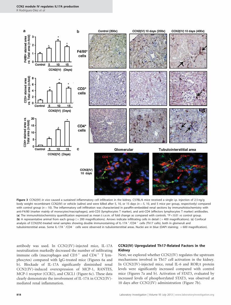
antibody was used. In CCN2(IV)-injected mice, IL-17Aneutralization markedly decreased the number of infiltratingimmune cells (macrophages and CD3þ and CD4þ T lym-phocytes) compared with IgG-treated mice (Figures 6a andb). Blockade of IL-17A significantly diminished renalCCN2(IV)-induced overexpression of MCP-1, RANTES,MCP-1 receptor (CCR2), and CXCL1 (Figure 6c). These dataclearly demonstrate the involvement of IL-17A in CCN2(IV)-mediated renal inflammation.
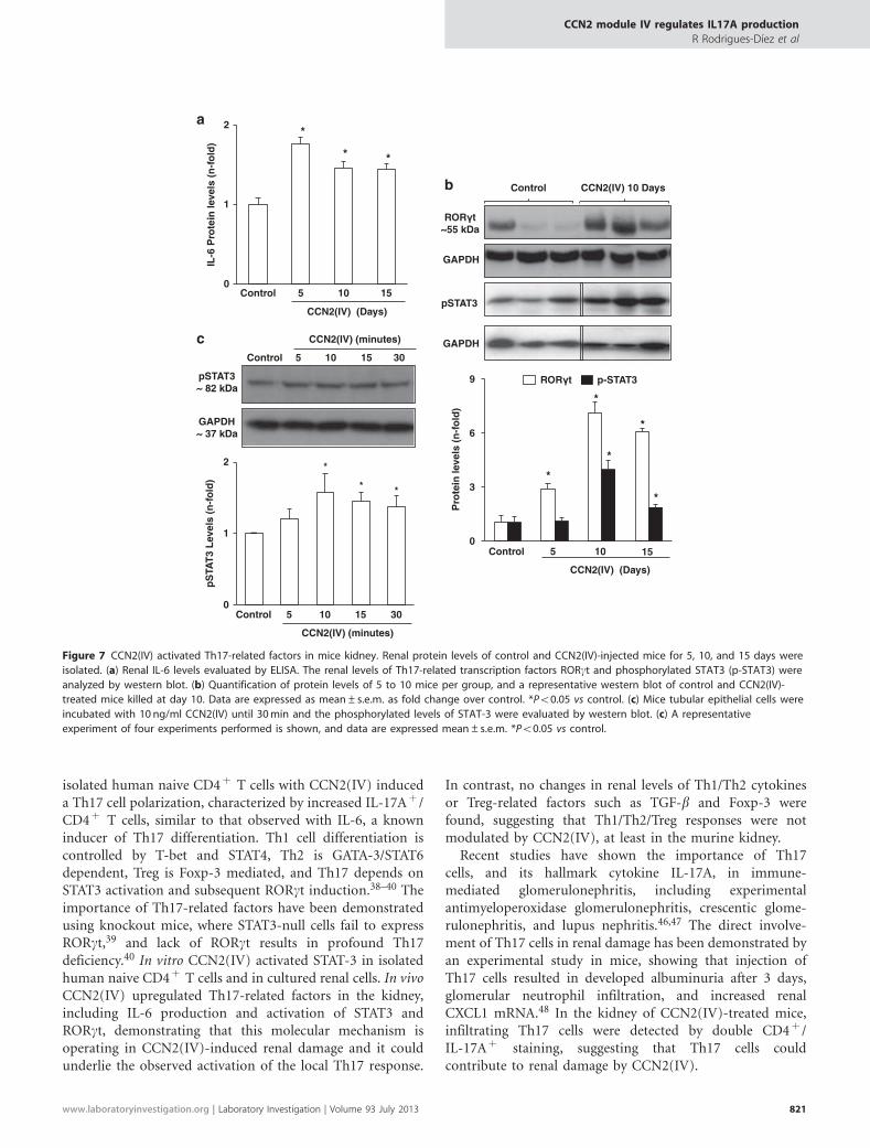
CCN2(IV) Upregulated Th17-Related Factors in theKidneyNext, we explored whether CCN2(IV) regulates the upstreammechanisms involved in Th17 cell activation in the kidney.In CCN2(IV)-injected mice, renal IL-6 and RORgt proteinlevels were significantly increased compared with controlmice (Figures 7a and b). Activation of STAT3, evaluated byincreased levels of phosphorylated STAT3, was observed at10 days after CCN2(IV) administration (Figure 7b).
Figure 3 CCN2(IV) in vivo caused a sustained inflammatory cell infiltration in the kidney. C57BL/6 mice received a single i.p. injection of 2.5 ng/g
body weight recombinant CCN2(IV) or vehicle (saline) and were killed after 5, 10, or 15 days (n¼ 5, 10, and 5 mice per group, respectively) compared
with control group (n¼ 10). The inflammatory cell infiltration was characterized in paraffin-embedded renal sections by immunohistochemistry with
anti-F4/80 (marker mainly of monocytes/macrophages), anti-CD3 (lymphocytes T marker), and anti-CD4 (effectors lymphocytes T marker) antibodies.
(a) The immunohistochemistry quantification expressed as mean±s.e.m. of fold change as compared with controls. *Po0.01 vs control group.
(b) A representative animal from each group (� 200 magnifications). Arrows indicate infiltrating cells in detail (� 400 magnifications). (c) Confocal
analysis of CCN2(IV)-treated renal samples showing double immunostaining of IL-17Aþ /CD4þ cells (Th17 cells), both in glomeruli and
tubulointerstitial areas. Some IL-17Aþ /CD4� cells were observed in tubulointerstitial areas. Nuclei are in blue (DAPI staining; � 600 magnification).
CCN2 module IV regulates IL17A production
R Rodrigues-Dıez et al
818 Laboratory Investigation | Volume 93 July 2013 | www.laboratoryinvestigation.org

We have further evaluated whether CCN2(IV) couldregulate Th17-related factors in cultured renal cells.Previous studies have demonstrated that CCN2(IV) increasedIL-16 production in renal cells.21 In cultured renal cells,incubation with CCN2(IV) increased STAT3 phos-phorylation, with a maximal response after 10 min ofstimulation (Figure 7c).
CCN2(IV) did not Regulate Treg-Related TranscriptionFactors in the KidneyTh17/Treg differentiation from naive CD4þ cells is a processclosely regulated that depends on cytokine environmentand activation of specific transcription factors. Treg celldifferentiation is regulated through the activation ofFoxp3.44 In CCN2(IV)-injected mice, Foxp3 renal levels werenot elevated compared with controls at all times studied(Figure 8a). Moreover, frequency of the CD4þ /Foxp3þ Tregcirculating cells, assessed by flow cytometry, were not modi-fied in response to CCN2(IV) administration (Figure 8b).
Elevated Renal Levels of IL-17A in NonimmuneExperimental Models of Renal Damage Characterized byCCN2 OverexpressionPrevious studies have shown the involvement of Th17 cells innonimmune inflammatory experimental renal injury, such asthe model of UUO.45 In this model we have previouslydescribed elevated renal levels of CCN2 in the obstructedkidneys.37 Now, we have observed that obstructed kidneysalso presented elevated levels of IL-17A as compared withcontrols (Figure 9), showing an association between CCN2and IL-17A upregulation in experimental renal damage.
DISCUSSIONOur in vitro and in vivo studies support the novel concept ofCCN2(IV) as a proinflammatory cytokine that is able tomodulate the Th17 immune response. Our findings expandthe known profibrogenic CCN2 signaling pathways to a truecytokine with an active role in sustained renal inflammation.
Our studies carried out on human CD4þ T lymphocytesshowed that CCN2(IV) drives Th17 differentiation.The differentiation of naive CD4þ T cells into differentTh effector subsets is a process closely regulated by specificcytokines and transcription factors.30,31,39,40 Stimulation of
Figure 4 CCN2(IV) increased proinflammatory chemokine expression and
activated NF-kB in the kidney. In renal samples from control and
CCN2(IV)-injected mice for 5, 10, and 15 days, gene expression of MCP-1
and RANTES was evaluated by real-time PCR (a) and protein levels by
ELISA (b). Activation of renal NF-kB was evaluated determining the
nuclear levels of p65 NF-kB subunit by western blot (c). Data are
expressed as n-fold increase over control as mean±s.e.m. of 5–10
animals per group. *Po0.05 vs control.
Figure 5 CCN2(IV) induced a transient upregulation of matrix-related
proteins in the kidney. In renal samples from control and CCN2(IV)-
injected C57BL/6 mice for 5, 10, and 15 days, gene expression of
fibronectin, type I collagen, a-SMA, and vimentin was evaluated by real-
time PCR. Data are expressed as n-fold increase over control as
mean±s.e.m. of 5–10 animals per group. *Po0.05 vs control.
CCN2 module IV regulates IL17A production
R Rodrigues-Dıez et al
www.laboratoryinvestigation.org | Laboratory Investigation | Volume 93 July 2013 819

Figure 6 IL-17A blockade inhibited CCN2(IV)-induced renal inflammatory responses. To block IL-17A responses, mice were treated with an IL-17A
neutralizing antibody or its corresponding IgG isotype as control (n¼ 5 mice per group) 24 h before CCN2(IV)) injection and every 72 h thereafter until
the time of killing at day 10. IL-17A neutralization diminished renal inflammatory cell infiltration in CCN2(IV)-injected mice. (a) Quantification of
F4/80-, CD3-, or CD4-positive cells, expressed as mean±s.e.m. fold change of positive staining vs total area over control. (b) Representative
immunohistochemistry from each group (� 200 magnification). (c) IL-17A neutralization decreased CCN2(IV)-induced renal chemokine expression.
IL-6, MCP-1, RANTES, CCR2, and CXCL1 mRNA expression as mean±s.e.m. analyzed by real-time PCR. *Po0.05 vs control; #Po0.05 vs CCN2(IV)-IgG.
CCN2 module IV regulates IL17A production
R Rodrigues-Dıez et al
820 Laboratory Investigation | Volume 93 July 2013 | www.laboratoryinvestigation.org
isolated human naive CD4þ T cells with CCN2(IV) induceda Th17 cell polarization, characterized by increased IL-17Aþ /CD4þ T cells, similar to that observed with IL-6, a knowninducer of Th17 differentiation. Th1 cell differentiation iscontrolled by T-bet and STAT4, Th2 is GATA-3/STAT6dependent, Treg is Foxp-3 mediated, and Th17 depends onSTAT3 activation and subsequent RORgt induction.38–40 Theimportance of Th17-related factors have been demonstratedusing knockout mice, where STAT3-null cells fail to expressRORgt,39 and lack of RORgt results in profound Th17deficiency.40 In vitro CCN2(IV) activated STAT-3 in isolatedhuman naive CD4þ T cells and in cultured renal cells. In vivoCCN2(IV) upregulated Th17-related factors in the kidney,including IL-6 production and activation of STAT3 andRORgt, demonstrating that this molecular mechanism isoperating in CCN2(IV)-induced renal damage and it couldunderlie the observed activation of the local Th17 response.
In contrast, no changes in renal levels of Th1/Th2 cytokinesor Treg-related factors such as TGF-b and Foxp-3 werefound, suggesting that Th1/Th2/Treg responses were notmodulated by CCN2(IV), at least in the murine kidney.
Recent studies have shown the importance of Th17cells, and its hallmark cytokine IL-17A, in immune-mediated glomerulonephritis, including experimentalantimyeloperoxidase glomerulonephritis, crescentic glome-rulonephritis, and lupus nephritis.46,47 The direct involve-ment of Th17 cells in renal damage has been demonstrated byan experimental study in mice, showing that injection ofTh17 cells resulted in developed albuminuria after 3 days,glomerular neutrophil infiltration, and increased renalCXCL1 mRNA.48 In the kidney of CCN2(IV)-treated mice,infiltrating Th17 cells were detected by double CD4þ /IL-17Aþ staining, suggesting that Th17 cells couldcontribute to renal damage by CCN2(IV).
IL-6
Pro
tein
leve
ls (
n-f
old
)
0
1
2
Control
CCN2(IV) (Days)
5 10 15
*
* *
Control
CCN2(IV) (minutes)
5 10 15 30
pSTAT3~ 82 kDa
GAPDH~ 37 kDa
0
1
2 *
* *
pS
TAT
3 L
evel
s (n
-fo
ld)
Control
CCN2(IV) (minutes)
5 10 15 30
GAPDH
pSTAT3
Control CCN2(IV) 10 Days
ROR t~55 kDa
GAPDH
Pro
tein
leve
ls (
n-f
old
)
Control
CCN2(IV) (Days)
5 10 150
3
6
9
*
*
*
*
*
ROR t p-STAT3
a
c
b
Figure 7 CCN2(IV) activated Th17-related factors in mice kidney. Renal protein levels of control and CCN2(IV)-injected mice for 5, 10, and 15 days were
isolated. (a) Renal IL-6 levels evaluated by ELISA. The renal levels of Th17-related transcription factors RORgt and phosphorylated STAT3 (p-STAT3) were
analyzed by western blot. (b) Quantification of protein levels of 5 to 10 mice per group, and a representative western blot of control and CCN2(IV)-
treated mice killed at day 10. Data are expressed as mean±s.e.m. as fold change over control. *Po0.05 vs control. (c) Mice tubular epithelial cells were
incubated with 10 ng/ml CCN2(IV) until 30 min and the phosphorylated levels of STAT-3 were evaluated by western blot. (c) A representative
experiment of four experiments performed is shown, and data are expressed mean±s.e.m. *Po0.05 vs control.
CCN2 module IV regulates IL17A production
R Rodrigues-Dıez et al
www.laboratoryinvestigation.org | Laboratory Investigation | Volume 93 July 2013 821

Besides Th17 cells, IL-17A can be produced by other cells,including CD8þ T cells, gd T cells, natural killer cells, neu-trophils, and some resident cells.49 In the model of ischemia–reperfusion, neutrophils, but not Th17 cells, are the mainsources of IL-17A and contribute to renal injury by naturalkiller T activation and IL-12/IFN-g production.50 Inglomeruli of patients with lupus nephritis, double-negative(CD4, CD8) T cells are a major source of IL-17A,51 and inrenal allograft rejection, positive staining for IL-17A has beendetected in tubular cells,41 as we have observed here inCCN2(IV)-treated mice, suggesting that renal cells couldproduce this cytokine and contribute to extend the damage.
The biological actions of IL-17A are predominantlyproinflammatory.49 There are several pathways by which this
cytokine might affect local inflammation. In tubular cells,mesangial cells, vascular smooth muscle cells, endothelialcells, and fibroblasts, IL-17A induces the release of cytokinesand chemokines such as IL-16, MCP-1, RANTES, CXCL1,and CXCL8 that recruit different leukocyte subsets leading totarget organ injury.52 IL-17A can also directly promotemonocyte chemotaxis.53 Our in vitro and in vivo data clearlyshow that CCN2(IV) induces IL-17A production. InCCN2(IV)-injected mice, renal overexpression of IL-17Awas associated with the presence of inflammatory infiltratingcells and upregulation of chemokines that were reduced inIL-17A neutralization experiments, clearly demonstratingthe involvement of IL-17A in CCN2(IV)-mediated renalinflammation. Studies in knockout mice have shown that theabsence of IL-17A resulted in less glomerular crescentformation and improved renal function in experimentalnephrotoxic nephritis.54 Several IL-17A blockade strategies,including neutralizing antibodies or receptor gene targeting,ameliorated several experimental nonimmune diseases.32–36
Preliminary data in rheumatoid arthritis and uveitis,34
and more recently, data from two phase 2 clinical trials, inwhich patients with psoriasis were treated with specifictargeting antibodies for IL-17A or its receptor,35,36
have shown beneficial effects in humans. CCN2 has beeninvolved in several chronic diseases, including skin disorders,cardiac hypertrophy, atherosclerosis, pulmonary hyper-tension, and lung and liver diseases,12 where IL-17A isa key player.32,33,55–58 In the nonimmune murine modelof UUO, we have observed elevated renal levels of IL-17Aand infiltrating inflammatory cells associated with renalCCN2 overexpression. Although future studies are needed,our data showing that IL-17A blockade amelioratedCCN2-induced renal inflammation support the concept of
Figure 8 CCN2(IV) did not regulate Treg-related transcription factors.
Foxp3 renal levels were analyzed by western blot. (a) Data as ratio of
renal Foxp3/GAPDH protein as mean±s.e.m. of 10 mice per group and
a representative western blot experiment. Treg (CD4þFoxp3þ ) cells were
analyzed in plasma by flow cytometry. (b) The percentage of CD4þ
T cells expressing Foxp3 cells (mean±s.e.m. of 10 mice per group).
*Po0.05 vs control.
Figure 9 Upregulation of renal levels of IL-17A in the model of unilateral
ureteral obstruction in mice. IL-17A renal levels were evaluated by ELISA
and expressed as data as mean±s.e.m. of 6 animals per group. *Po0.05
vs control mice.
CCN2 module IV regulates IL17A production
R Rodrigues-Dıez et al
822 Laboratory Investigation | Volume 93 July 2013 | www.laboratoryinvestigation.org

IL-17A-neutralizing antibody as a promising tool for chronicinflammatory diseases, including chronic kidney diseases.
CCN2 overexpression has been described in humanfibrotic diseases, and it is produced temporally and spatiallyin close proximity to fibrotic areas.2,12 Antagonists of CCN2have proven effective in blocking profibrogenic CCN2signaling pathways in vitro and have yielded promising datawith respect to preventing experimental fibrosis.14 Regardingrenal diseases, knockdown of CCN2 gene expression withantisense gene transfer into rat kidney amelioratestubulointerstitial fibrosis in obstructive nephropathy14 andin experimental mice diabetes in C57BL/6 mice.16 CCN2is known to function downstream of TGF-b, drivingextracellular matrix accumulation and fibrosis.2,12 Indeed,in cultured renal cells, CCN2 blockade diminished matrixproteins synthesis induced by TGF-b and Angiotensin II.59
Although initial studies in fibroblasts, and other culturedcells, showed that recombinant CCN2, both the C-terminaland the full molecule, induce extracellular matrixproduction,2,7,14 several in vivo studies have shown thatCCN2 alone is not sufficient to cause ongoing fibroticchanges. In a model of a skin fibrosis, injection of CCN2alone did not induce fibrosis and only the simultaneousapplication of TGF-b1 caused persistent fibrosis.60 CCN2overexpression in rat lungs only caused a temporal inductionof procollagen gene expression and transient matrix accumu-lation, but it is not sufficient to induce progressive fibrosis.61
Podocyte-specific CCN2-transgenic mice (in C57BL/6 back-ground) exhibited no glomerular abnormalities, proteinuria,or matrix accumulation, although they were more susceptibleto streptozotocin-induced diabetes.62 Our in vivo datashowed a similar response in the kidney. SystemicCCN2(IV) administration into C57BL/6 mice increasedprofibrotic genes renal expression at day 5, and decreasingthereafter, and no collagen accumulation was observed.
Our findings expand the known profibrogenic CCN2signaling pathways to a true cytokine with an activerole in sustained renal inflammation mediated by Th17 cells.Different strategies for blocking CCN2 activity haveproven beneficial effects in fibrotic-related pathologies,including renal diseases,12 whereas CCN2 overexpressioninduces cardioprotection.17,18 Our results set out that IL17Aneutralization block inflammatory events caused by theC-terminal module of CCN2 in the kidney, and suggest thatIL-17A targeting could be a better therapeutic option thanCCN2 blockers for human use in inflammatory diseases.
ACKNOWLEDGEMENTS
We thank Ma Mar Gonzalez Garcia-Parreno for her technical
help with confocal microscopy, and Susana Carrasco for her help in
immunohistochemical procedures. Grant numbers and sources of support
are as follows: grants from the Instituto de Salud Carlos III (ISCIII-RETIC
RD06/0016 y RD0012/0021, REDINREN, PI081564, PI11/01854, PI12/02587,
PI10/00072, PI12/00204 and PS09/00447), Comunidad de Madrid (S2010/
BMD-2321), Sociedad Espanola de Nefrologıa, Fundacion Lilly, Research
Institute Queen Sophia (FRIAT), Programa Intensificacion Actividad
Investigadora (ISCIII/Agencia Laın-Entralgo/CM) to AO, RR-D, ES-L, CL, and
MA, who are fellows of ISCIII.
DISCLOSURE/CONFLICT OF INTEREST
The authors declare no conflict of interest.
1. Winter P, Leoni P, Abraham D. Connective tissue growth factor:structure-function relationships of a mosaic, multifunctional protein.Growth Factors 2008;26:80–91.
2. Phanish MK, Winn SK, Dockrell ME. Connective tissue growth factor-(CTGF, CCN2)-a marker mediator and therapeutic target for renalfibrosis. Nephron Exp Nephrol 2010;114:e83–e92.
3. Hashimoto G, Inoki I, Fujii Y, et al. Matrix metalloproteinasescleave connective tissue growth factor and reactivate angiogenicactivity of vascular endothelial growth factor 165. J Biol Chem2002;277:36288–36295.
4. Brigstock DR, Steffen CL, Kim GY, et al. Purification andcharacterization of novel heparin-binding growth factors in uterinesecretory fluids. Identification as heparin-regulated Mr 10,000 forms ofconnective tissue growth factor. J Biol Chem 1997;272:20275–20282.
5. O’Seaghdha CM, Hwang SJ, Bhavsar NA, et al. Lower urinaryconnective tissue growth factor levels and incident CKD stage 3 inthe general population. Am J Kidney Dis 2011;57:841–849.
6. Nguyen TQ, Tarnow L, Andersen S, et al. Urinary connective tissuegrowth factor excretion correlates with clinical markers of renaldisease in a large population of type 1 diabetic patients with diabeticnephropathy. Diabetes Care 2006;29:83–88.
7. Slagman MC, Nguyen TQ, Waanders F, et al. Clin Effects ofantiproteinuric intervention on elevated connective tissue growthfactor (CTGF/CCN2) plasma and urine levels in nondiabetic nephro-pathy. J Am Soc Nephrol 2011;6:1845–1850.
8. Riser BL, Cortes P, DeNichilo M, et al. Urinary CCN2 (CTGF) as a possiblepredictor of diabetic nephropathy: preliminary report. Kidney Int2003;64:451–458.
9. Tam FW, Riser BL, Meeran K, et al. Urinary monocyte chemoattractantprotein-1 (MCP-1) and connective tissue growth factor (CCN2) asprognostic markers for progression of diabetic nephropathy. Cytokine2009;47:37–42.
10. Bao J, Tu Z, Wang J, et al. A novel accurate rapid ELISA for detectionof urinary connective tissue growth factor, a biomarker of chronicallograft nephropathy. Transplant Proc 2008;40:2361–2364.
11. Koitabashi N, Arai M, Niwano K, et al. Plasma connective tissue growthfactor is a novel potential biomarker of cardiac dysfunction in patientswith chronic heart failure. Eur J Heart Fail 2008;10:373–379.
12. Leask A, Parapuram SK, Shi-Wen X, et al. Connective tissue growthfactor (CTGF, CCN2) gene regulation: a potent clinical bio-marker offibroproliferative disease? J Cell Commun Signal 2009;3:89–94.
13. Ruiz-Ortega M, Rodrıguez-Vita J, Sanchez-Lopez E, et al. TGF-betasignaling in vascular fibrosis. Cardiovasc Res 2007;74:196–206.
14. Yokoi H, Mukoyama M, Nagae T, et al. Reduction in connective tissuegrowth factor by antisense treatment ameliorates renaltubulointerstitial fibrosis. J Am Soc Nephrol 2004;15:1430–1440.
15. Okada H, Kikuta T, Kobayashi T, et al. Connective tissue growth factorexpressed in tubular epithelium plays a pivotal role in renalfibrogenesis. J Am Soc Nephrol 2005;16:133–143.
16. Guha M, Xu ZG, Tung D, et al. Specific down-regulation of connectivetissue growth factor attenuates progression of nephropathy in mousemodels of type 1 and type 2 diabetes. FASEB J 2007;21:3355–3368.
17. Panek AN, Posch MG, Alenina N, et al. Connective tissue growth factoroverexpression in cardiomyocytes promotes cardiac hypertrophy andprotection against pressure overload. PLoS One 2009;4:e6743.
18. Ahmed MS, Gravning J, Martinov VN, et al. Mechanisms of novelcardioprotective functions of CCN2/CTGF in myocardial ischemia-reperfusion injury. Am J Physiol Heart Circ Physiol 2011;300:H1291–H1302.
19. Kular L, Pakradouni J, Kitabgi P, et al. The CCN family: a new class ofinflammation modulators? Biochimie 2011;93:377–388.
20. Cicha I, Yilmaz A, Klein M, et al. Connective tissue growth factor isoverexpressed in complicated atherosclerotic plaques and inducesmononuclear cell chemotaxis in vitro. Arterioscler Thromb Vasc Biol2005;25:1008–1013.
CCN2 module IV regulates IL17A production
R Rodrigues-Dıez et al
www.laboratoryinvestigation.org | Laboratory Investigation | Volume 93 July 2013 823
21. Sanchez-Lopez E, Rayego S, Rodrigues-Dıez R, et al. CTGF promotesinflammatory cell infiltration of the renal interstitium by activatingNF-kappaB. J Am Soc Nephrol 2009;20:1513–1526.
22. Liu BC, Zhang JD, Zhang XL, et al. Role of connective tissue growthfactor (CTGF) module 4 in regulating epithelial mesenchymaltransition in HK-2 cells. Clin Chim Acta 2006;373:144–150.
23. Markiewicz M, Nakerakanti SS, Kapanadze B, et al. Connective tissuegrowth factor (CTGF/CCN2) mediates angiogenic effect of S1P inhuman dermal microvascular endothelial cells. Microcirculation2011;18:1–11.
24. Sanchez-Lopez E, Rodriguez-Vita J, Cartier C, et al. Inhibitory effect ofinterleukin-1beta on angiotensin II-induced connective tissue growthfactor and type IV collagen production in cultured mesangial cells. AmJ Physiol Renal Physiol 2008;294:F149–F160.
25. Wang X, McLennan SV, Allen TJ, et al. Regulation of pro-inflammatoryand pro-fibrotic factors by CCN2/CTGF in H9c2 cardiomyocytes. J CellCommun Signal 2010;4:15–23.
26. Kim HS, Nagalla SR, Oh Y, et al. Identification of a family of low-affinityinsulin-like growth factor binding proteins (IGFBPs): characterizationof connective tissue growth factor as a member of the IGFBPsuperfamily. Proc Natl Acad Sci USA 1997;94:12981–12986.
27. Lam S, van der Geest RN, Verhagen NA, et al. Connective tissue growthfactor and IGF-I are produced by human renal fibroblasts andcooperate in the induction of collagen production by high glucose.Diabetes 2003;52:2975–2983.
28. Chen Y, Abraham DJ, Shi-Wen X, et al. CCN2 (connective tissue growthfactor) promotes fibroblast adhesion to fibronectin. Mol Biol Cell2004;15:5635–5646.
29. Chang CC, Shih JY, Jeng YM, et al. Connective tissue growth factor andits role in lung adenocarcinoma invasion and metastasis. J Natl CancerInst 2004;96:364–375.
30. Bettelli E, Korn T, Oukka M, et al. Induction and effector functions ofT(H)17 cells. Nature 2008;453:1051–1057.
31. Harrington LE, Hatton RD, Mangan PR, et al. Interleukin 17-producingCD4þ effector T cells develop via a lineage distinct from the T helpertype 1 and 2 lineages. Nat Immunol 2005;6:1123–1132.
32. Erbel C, Chen L, Bea F, et al. Inhibition of IL-17A attenuatesatherosclerotic lesion development in apoE-deficient mice.J Immunol 2009;183:8167–8175.
33. Smith E, Prasad KM, Butcher M, et al. Blockade of interleukin-17Aresults in reduced atherosclerosis in apolipoprotein E-deficient mice.Circulation 2010;121:1746–1755.
34. Hueber W, Patel DD, Dryja T, et al. Effects of AIN457, a fully humanantibody to interleukin-17A, on psoriasis, rheumatoid arthritis, anduveitis. Sci Transl Med 2010;252ra72.
35. Leonardi C, Matheson R, Zachariae C, et al. Anti-interleukin-17monoclonal antibody ixekizumab in chronic plaque psoriasis. N EnglJ Med 2012;366:1190–1199.
36. Papp KA, Leonardi C, Menter A, et al. Brodalumab, an anti-interleukin-17-receptor antibody for psoriasis. N Engl J Med 2012;366:1181–1189.
37. Esteban V, Lorenzo O, Ruperez M, et al. Angiotensin II, via AT1 and AT2receptors and NF-kappaB pathway, regulates the inflammatoryresponse in unilateral ureteral obstruction. J Am Soc Nephrol2004;15:1514–1529.
38. Das J, Ren G, Zhang L, et al. Transforming growth factor beta isdispensable for the molecular orchestration of Th17 cell differen-tiation. J Exp Med 2009;206:2407–2416.
39. Mathur AN, Chang HC, Zisoulis DG, et al. Stat3 and Stat4 direct deve-lopment of IL-17-secreting Th cells. J Immunol 2007;178:4901–4907.
40. Ivanov II, McKenzie BS, Zhou L, et al. The orphan nuclear receptorRORgammat directs the differentiation program of proinflammatoryIL-17þ T helper cells. Cell 2006;126:1121–1133.
41. Loverre A, Tataranni T, Castellano G, et al. IL-17 expression by tubularepithelial cells in renal transplant recipients with acute antibody-mediated rejection. Am J Transplant 2011;11:1248–1259.
42. Chung AC, Lan HY. Chemokines in renal injury. J Am Soc Nephrol2011;22:802–809.
43. Sanz AB, Sanchez-Nino MD, Ramos AM, et al. NF-kappaB in renalinflammation. J Am Soc Nephrol 2010;21:1254–1262.
44. Hori S, Nomura T, Sakaguchi S. Control of regulatory T celldevelopment by the transcription factor Foxp3. Science2003;299:1057–1061.
45. Dong X, Bachman LA, Miller MN, et al. Dendritic cells facilitateaccumulation of IL-17 T cells in the kidney following acute renalobstruction. Kidney Int 2008;74:1294–1309.
46. Turner JE, Paust HJ, Steinmetz OM, et al. The Th17 immune responsein renal inflammation. Kidney Int 2010;77:1070–1075.
47. Kitching AR, Holdsworth SR. The emergence of Th17 cells as effectorsof renal injury. J Am Soc Nephrol 2011;22:235–238.
48. Summers SA, Steinmetz OM, Li M, et al. Th1 and Th17 cells induceproliferative glomerulonephritis. J Am Soc Nephrol 2009;20:2518–2524.
49. Gaffen SL. Structure and signalling in the IL-17 receptor family.Nat Rev Immunol 2009;9:556–567.
50. Li L, Huang L, Vergis AL, et al. IL-17 produced by neutrophils regulatesIFN-gamma-mediated neutrophil migration in mouse kidneyischemia-reperfusion injury. J Clin Invest 2010;120:331–342.
51. Crispin JC, Oukka M, Bayliss G, et al. Expanded double negative T cellsin patients with systemic lupus erythematosus produce IL-17 andinfiltrate the kidneys. J Immunol 2008;181:8761–8766.
52. Van Kooten C, Boonstra JG, Paape ME, et al. Interleukin-17 activateshuman renal epithelial cells in vitro and is expressed during renalallograft rejection. J Am Soc Nephrol 1998;9:1526–1534.
53. Shahrara S, Pickens SR, Dorfleutner A, et al. IL-17 induces mono-cyte migration in rheumatoid arthritis. J Immunol 2009;182:3884–3891.
54. Paust HJ, Turner JE, Steinmetz OM, et al. The IL-23/Th17 axiscontributes to renal injury in experimental glomerulonephritis. J AmSoc Nephrol 2009;5:969–979.
55. Guttman-Yassky E, Nograles KE, Krueger JG. Contrasting patho-genesis of atopic dermatitis and psoriasis—part II: immune cell sub-sets and therapeutic concepts. J Allergy Clin Immunol 2011;127:1420–1432.
56. Nembrini C, Marsland BJ, Kopf M. IL-17-producing T cells in lungimmunity and inflammation. J Allergy Clin Immunol 2009;123:986–994.
57. Booth AJ, Bishop DK. TGF-b, IL-6, IL-17 and CTGF direct multiplepathologies of chronic cardiac allograft rejection. Immunotherapy2010;2:511–520.
58. Hammerich L, Heymann F, Tacke F. Role of IL-17 and Th17 cells in liverdiseases. Clin Dev Immunol 2011;2011:345803.
59. Ruperez M, Lorenzo O, Blanco-Colio LM, et al. Connective tissuegrowth factor is a mediator of angiotensin II-induced fibrosis.Circulation 2003;108:1499–1505.
60. Mori T, Kawara S, Shinozaki M, et al. Role and interaction of connectivetissue growth factor with transforming growth factor-beta inpersistent fibrosis: a mouse fibrosis model. J Cell Physiol1999;181:153–159.
61. Bonniaud P, Margetts PJ, Kolb M, et al. Adenoviral gene transfer ofconnective tissue growth factor in the lung induces transient fibrosis.Am J Respir Crit Care Med 2003;168:770–778.
62. Yokoi H, Mukoyama M, Mori K, et al. Overexpression of connectivetissue growth factor in podocytes worsens diabetic nephropathy inmice. Kidney Int 2008;73:446–455.
CCN2 module IV regulates IL17A production
R Rodrigues-Dıez et al
824 Laboratory Investigation | Volume 93 July 2013 | www.laboratoryinvestigation.org