Netrin-1: A Modulator of Macrophage Driven Acute and ... - MDPI
24
Citation: Ziegon, L.; Schlegel, M. Netrin-1: A Modulator of Macrophage Driven Acute and Chronic Inflammation. Int. J. Mol. Sci. 2022, 23, 275. https://doi.org/ 10.3390/ijms23010275 Academic Editor: Janine M. van Gils Received: 5 December 2021 Accepted: 23 December 2021 Published: 27 December 2021 Publisher’s Note: MDPI stays neutral with regard to jurisdictional claims in published maps and institutional affil- iations. Copyright: © 2021 by the authors. Licensee MDPI, Basel, Switzerland. This article is an open access article distributed under the terms and conditions of the Creative Commons Attribution (CC BY) license (https:// creativecommons.org/licenses/by/ 4.0/). International Journal of Molecular Sciences Review Netrin-1: A Modulator of Macrophage Driven Acute and Chronic Inflammation Laura Ziegon and Martin Schlegel * Department of Anesthesiology and Intensive Care Medicine, Klinikum Rechts der Isar, Technical University Munich, 81675 Munich, Germany; [email protected] * Correspondence: [email protected] Abstract: Netrins belong to the family of laminin-like secreted proteins, which guide axonal migra- tion and neuronal growth in the developing central nervous system. Over the last 20 years, it has been established that netrin-1 acts as a chemoattractive or chemorepulsive cue in diverse biological processes far beyond neuronal development. Netrin-1 has been shown to play a central role in cell adhesion, cell migration, proliferation, and cell survival in neuronal and non-neuronal tissue. In this context, netrin-1 was found to orchestrate organogenesis, angiogenesis, tumorigenesis, and inflammation. In inflammation, as in neuronal development, netrin-1 plays a dichotomous role directing the migration of leukocytes, especially monocytes in the inflamed tissue. Monocyte- derived macrophages have long been known for a similar dual role in inflammation. In response to pathogen-induced acute injury, monocytes are rapidly recruited to damaged tissue as the first line of immune defense to phagocyte pathogens, present antigens to initiate the adaptive immune response, and promote wound healing in the resolution phase. On the other hand, dysregulated macrophages with impaired phagocytosis and egress capacity accumulate in chronic inflammation sites and foster the maintenance—and even the progression—of chronic inflammation. In this review article, we will highlight the dichotomous roles of netrin-1 and its impact on acute and chronic inflammation. Keywords: netrin-1; neuronal guidance protein; chronic inflammation; acute inflammation 1. Netrin-1 Acts as a Chemoattractant and Chemorepellent Guidance Cue The name netrin originates from the Sanskrit word netr, which means “the one who guides.” This reflects the fact that these laminin-like secreted neuronal guidance proteins were initially described as guidance cues in the developing central nervous system, where they act either as chemoattractive or as chemorepulsive cues. The netrin family consists of netrin-1, netrin-2, and netrin-3, and more distantly related netrin-4, netrin-G1, and G2 [1,2], which all belong to the superfamily of laminins [3]. Netrins are characterized by a preserved structure containing N-terminal domain laminin homologous domains, followed by three epidermal growth factor (EGF)-like repeats (LE) and a small, less well conserved, positively charged domain C-terminal [4,5]. In the early 1990s, netrin-1 was first described in the developing central nervous system [6,7]. Netrin-1 was shown to coordinate cell migration and axonal growth as a chemoattractant or chemorepellent, depending on the receptors expressed by the target cells. The chemorepulsion mediated by netrin-1 is facilitated by an Unc5 homodimer within close proximity and an Unc5/DCC heterodimer at longer ranges [8]. DCC has been further shown to signal netrin-1 dimerization with neogenin, an- other neuronal guidance receptor [6,8]. Moreover, it is suggested that the composition of the ligand receptor complex not only results in unique signaling mechanisms, but the different architectures of the ligand–receptor complex can also result in diverse signaling responses [6]. In addition to this complexity, it was recently shown that Int. J. Mol. Sci. 2022, 23, 275. https://doi.org/10.3390/ijms23010275 https://www.mdpi.com/journal/ijms
-
Upload
khangminh22 -
Category
Documents
-
view
2 -
download
0
Transcript of Netrin-1: A Modulator of Macrophage Driven Acute and ... - MDPI
�����������������
Citation: Ziegon, L.; Schlegel, M.
Netrin-1: A Modulator of
Macrophage Driven Acute and
Chronic Inflammation. Int. J. Mol. Sci.
2022, 23, 275. https://doi.org/
10.3390/ijms23010275
Academic Editor: Janine M. van Gils
Received: 5 December 2021
Accepted: 23 December 2021
Published: 27 December 2021
Publisher’s Note: MDPI stays neutral
with regard to jurisdictional claims in
published maps and institutional affil-
iations.
Copyright: © 2021 by the authors.
Licensee MDPI, Basel, Switzerland.
This article is an open access article
distributed under the terms and
conditions of the Creative Commons
Attribution (CC BY) license (https://
creativecommons.org/licenses/by/
4.0/).
International Journal of
Molecular Sciences
Review
Netrin-1: A Modulator of Macrophage Driven Acute andChronic InflammationLaura Ziegon and Martin Schlegel *
Department of Anesthesiology and Intensive Care Medicine, Klinikum Rechts der Isar,Technical University Munich, 81675 Munich, Germany; [email protected]* Correspondence: [email protected]
Abstract: Netrins belong to the family of laminin-like secreted proteins, which guide axonal migra-tion and neuronal growth in the developing central nervous system. Over the last 20 years, it hasbeen established that netrin-1 acts as a chemoattractive or chemorepulsive cue in diverse biologicalprocesses far beyond neuronal development. Netrin-1 has been shown to play a central role in celladhesion, cell migration, proliferation, and cell survival in neuronal and non-neuronal tissue. Inthis context, netrin-1 was found to orchestrate organogenesis, angiogenesis, tumorigenesis, andinflammation. In inflammation, as in neuronal development, netrin-1 plays a dichotomous roledirecting the migration of leukocytes, especially monocytes in the inflamed tissue. Monocyte-derived macrophages have long been known for a similar dual role in inflammation. In responseto pathogen-induced acute injury, monocytes are rapidly recruited to damaged tissue as the firstline of immune defense to phagocyte pathogens, present antigens to initiate the adaptive immuneresponse, and promote wound healing in the resolution phase. On the other hand, dysregulatedmacrophages with impaired phagocytosis and egress capacity accumulate in chronic inflammationsites and foster the maintenance—and even the progression—of chronic inflammation. In thisreview article, we will highlight the dichotomous roles of netrin-1 and its impact on acute andchronic inflammation.
Keywords: netrin-1; neuronal guidance protein; chronic inflammation; acute inflammation
1. Netrin-1 Acts as a Chemoattractant and Chemorepellent Guidance Cue
The name netrin originates from the Sanskrit word netr, which means “the one whoguides.” This reflects the fact that these laminin-like secreted neuronal guidance proteinswere initially described as guidance cues in the developing central nervous system, wherethey act either as chemoattractive or as chemorepulsive cues. The netrin family consists ofnetrin-1, netrin-2, and netrin-3, and more distantly related netrin-4, netrin-G1, and G2 [1,2],which all belong to the superfamily of laminins [3]. Netrins are characterized by a preservedstructure containing N-terminal domain laminin homologous domains, followed by threeepidermal growth factor (EGF)-like repeats (LE) and a small, less well conserved, positivelycharged domain C-terminal [4,5].
In the early 1990s, netrin-1 was first described in the developing central nervoussystem [6,7]. Netrin-1 was shown to coordinate cell migration and axonal growth as achemoattractant or chemorepellent, depending on the receptors expressed by the targetcells. The chemorepulsion mediated by netrin-1 is facilitated by an Unc5 homodimer withinclose proximity and an Unc5/DCC heterodimer at longer ranges [8].
DCC has been further shown to signal netrin-1 dimerization with neogenin, an-other neuronal guidance receptor [6,8]. Moreover, it is suggested that the compositionof the ligand receptor complex not only results in unique signaling mechanisms, butthe different architectures of the ligand–receptor complex can also result in diversesignaling responses [6]. In addition to this complexity, it was recently shown that
Int. J. Mol. Sci. 2022, 23, 275. https://doi.org/10.3390/ijms23010275 https://www.mdpi.com/journal/ijms

Int. J. Mol. Sci. 2022, 23, 275 2 of 24
the post-transcriptional splicing of netrin-1 can significantly impact its biologicalfunctions [7].
Netrin-1 and its receptor DCC play an important role in different cellular processessuch as cell adhesion, mortality, proliferation, cell survival, tissue organization, and can-cer [9].
Moreover, the netrin-1 receptors Unc5b and DCC are so-called dependence recep-tors. These dependence receptors induce a caspase-triggered apoptosis in the absenceof their ligand netrin-1 [9–11]. The importance of netrin-1 was shown in human tu-mors, such as brain tumors, neuroblastomas, prostate cancer, pancreatic cancer, andcolorectal cancer. In these tumors, the gain of netrin-1 or the deletion of Unc5b andDCC prevents tumor cells from undergoing apoptosis and thereby promotes tumorgrowth and metastases [12].
Netrin-1 is also involved in the purinergic pathway, as it binds to the adenosinereceptor Adora2b (A2BAR) [13–15]. By binding to A2BAR, netrin-1 increases intracellularcAMP levels, which can enhance the endothelial barrier function [13] and macrophagepolarization [16]. However, it is of importance that the overall consequences of elevatedcAMP levels are opposing in different cell types and organs [17,18]. Moreover, netrin-1 wasshown to mediate epithelial cell adhesion and migration through the integrins α6β4 andα3β1 [19].
In 2005, Ly et al. [20] provided the earliest evidence that, outside the central nervoussystem, netrin-1 can also modulate the migration of granulocytes, monocytes, and leuko-cytes in and outside the inflamed tissue. Later it was shown that netrin-1 further promotesmacrophage differentiation to an alternative activated phenotype [21].
Over the 30 years since its discovery, it has been well established that netrin-1is involved in diverse biological processes far beyond the neuronal axon guidance.In this context, netrin-1 was found to play an important role in organogenesis [22],angiogenesis [23], tumorigenesis [11,24], promoting epithelium repair after injury [25],and inflammation. Given the numerous receptors and dichotomous signaling of netrin-1, it is not surprising that netrin-1 plays divergent roles in various settings of acute andchronic inflammation.
2. Acute Inflammation
Acute inflammation is a vital early physiological response to infection, trauma, injury,and a variety of other immunological challenges. The acute inflammatory response beginswith an influx of neutrophils into the infected tissue to fight the infection and to create aninflammatory milieu by the release of granules and proinflammatory cytokines [26]. Subse-quently, monocytes are attracted into this inflammatory milieu and support the neutrophilschallenging the invading pathogen [27]. This initial phase of acute inflammation needs tobe contained as the pathogen is cleared. In the resolution phase, anti-inflammatory and pro-resolving programs are initiated to control the acute inflammatory response and activelyattain homeostasis. The impairment or absence of resolution leads to an overwhelminguncontrolled inflammatory response, resulting in chronic inflammation, persistent organdysfunction, and even death [27]. In contrast to the numerous positive guidance cuescontrolling the influx of leukocytes into the inflamed tissue [26], negative guidance cueslimiting this influx remained unrevealed for a long time [27]. Ongoing research has shownthat netrin-1 can actively attenuate the acute inflammatory response and accelerate pro-resolving mechanisms, which cease the inflammation and help the inflamed tissue to returnto homeostasis [27,28] (Figure 1 and Table 1).

Int. J. Mol. Sci. 2022, 23, 275 3 of 24
Figure 1. Netrin-1 in acute inflammation. Acute inflammation is defined by a rapid influx ofneutrophils into the inflamed tissue to combat the intruding pathogen. This is followed by a monocyterecruitment to extend the rapid immune response. In many different models, it has been shown thatnetrin-1 can very effectively mitigate the influx of neutrophils and monocytes and thereby dampenthe acute inflammatory response. The acute, initial phase is followed by a resolving phase, in whichneutrophils undergo apoptosis and are taken up by monocytes. The efferocytosis in turn promotesthe differentiation of pro-inflammatory into anti-inflammatory monocytes. This phenotype switchand the monocyte egress in acute inflammation is facilitated by netrin-1.
Table 1. Murine studies on netrin-1 and its receptors in acute inflammation.
Acute Inflammation
Model Receptor Netrin-1 Expression Outcome Ref.
Lung Inflammation
S. aureus pneumonia inmice Unc5b ⇓ Netrin-1 expression is attenuated in S.
aureus septicemia. [20]
LPS and ventilatorinduced lung injury in
miceAdora2b ⇓
Netrin-1 expression is attenuated byinflammation, and netrin-1 limitsneutrophil influx into the lung.
[29]
LPS induced lunginjury in pigs not defined
Intravenous and inhalative netrin-1mitigated pulmonary inflammation
and lung damage.[30]
Mouse model with LIRI Adora2b ⇓ Positive correlation between netrin-1expression and Treg cell population. [31]
An ALI model wasestablished byintratracheal
instillation of LPS inC57BL/J mice
Adora2b ⇓
Receptor binding has the potential toenhance ENaC-dependent alveolar
fluid clearance by supplementation ofnetrin-1.
[32]
Hypobarichypoxia-induced lung
injury in miceUNC5HB ⇓ Pretreatment of netrin-1 dampen ALI
and inhibits neutrophil migration. [33]
Int. J. Mol. Sci. 2022, 23, 275 4 of 24
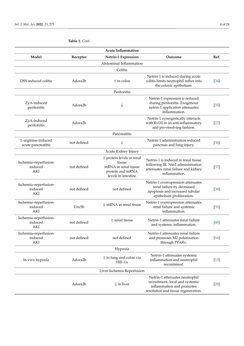
Table 1. Cont.
Acute Inflammation
Model Receptor Netrin-1 Expression Outcome Ref.
Abdominal Inflammation
Colitis
DSS-induced colitis Adora2b ⇑ in colonNetrin-1 is induced during acute
colitis limits neutrophil influx intothe colonic epithelium.
[34]
Peritonitis
ZyA-inducedperitonitis Adora2b ⇓
Netrin-1 expression is reducedduring peritonitis. Exogenousnetrin-1 application attenuates
inflammation.
[35]
ZyA-inducedperitonitis Adora2b
Netrin-1 synergistically interactswith RvD2 in an anti-inflammatory
and pro-resolving fashion.[27]
Pancreatitis
L-arginine-inducedacute pancreatitis not defined ⇓ Netrin-1 administration reduced
pancreas and lung injury. [36]
Acute Kidney Injury
Ischemia–reperfusion-induced
AKInot defined
⇑ protein levels in renaltissue
mRNA in renal tissueprotein and mRNAlevels in intestine
Netrin-1 is induced in renal tissuefollowing IR. Ntn1 administrationattenuates renal failure and kidney
inflammation.
[37]
Ischemia–reperfusion-induced
AKInot defined not defined
Netrin-1 overexpression attenuatesrenal failure by decreased
apoptosis and increased tubularepithelium proliferation.
[38]
Ischemia–reperfusion-induced
AKIUnc5b ⇓ mRNA in renal tissue Netrin-1 overexpression attenuates
renal failure and systemicinflammation.
[39]
Ischemia–reperfusion-induced
AKInot defined ⇑ renal tissue Netrin-1 attenuates renal failure
and systemic inflammation. [40]
Ischemia–reperfusion-induced
AKInot defined not defined
Netrin-1 attenuates renal failureand promotes M2 polarization
through PPARγ.[16]
Hypoxia
In vivo hypoxia Adora2b ⇑ in lung and colon viaHIF-1α
Netrin-1 attenuates systemicinflammation and neutrophil
recruitment.[13]
Liver-Ischemia Reperfusion
Adora2b ⇓ in liver
Netrin-1 attenuates neutrophilrecruitment, local and systemic
inflammation and promotesresolution and tissue regeneration.
[28]

Int. J. Mol. Sci. 2022, 23, 275 5 of 24
Table 1. Cont.
Acute Inflammation
Model Receptor Netrin-1 Expression Outcome Ref.
Myocardial Infarct
Normothermicischemia reperfusion
Langendorff perfusionDCC not defined
Netrin-1 pre- and postconditioningdecrease infarct size and attenuatemyocardiocyte apoptosis through
ERK1/2 and NO induction.
[41–43]
Heterotopic cardiactransplant with 8 h of
cold ischemia⇓
Netrin-1 protects from reperfusioninjury by limiting leukocyte influx,cardiomyocyte apoptosis, and M2macrophage polarization through
PPARg.
[44]
Chronic LAD coronaryligation not defined not defined
Intracardial application ofnetrin-1-transduced mesenchymal
stem cells attenuated infarctionsize and prevented cardiachypertrophic remodeling.
[45]
Normothermicischemia reperfusion
Langendorff perfusionnot defined not defined
Netrin-1 preconditioningattenuates infarcts size by
preventing superoxide andNADPH oxidase production,
preventing mitochondrialdysfunction.
[46]
2.1. Acute Lung Injury
Acute lung injury (ALI) and the acute respiratory distress syndrome (ARDS) show,despite the progress of modern intensive care medicine, an incidence of up to 86.2 cases per100,000 persons per year [47] and still a very high mortality (34.9–46.1%) [48] due to the lackof specific therapeutic options. The acute phase injury results from alveolar-endothelialinjury and the formation of pulmonary edema [49,50]. ALI is driven by a neutrophil andmonocyte influx into the alveoli. Several studies have shown that the netrin-1 level isdecreased in ALI and inversely correlates with the cytokine levels and leukocyte infiltrationinto the lung [20,29,30]. The systemic intraperitoneal administration of netrin-1 mitigateslymphocyte trafficking into the inflamed lung, dampens the systemic inflammation, anddiminishes the resulting lung injury [29,30]. The beneficial netrin-1 effects in ALI aremediated partly by Unc5b [20] as well as by A2BAR [29].
Mirakaj et al. [20] identified that the netrin-1 repression upon inflammation is medi-ated by NF-κB binding to a putative netrin-1 promoter region 69 bp upstream of its firstcodon [29]. Moreover, in tumorigenesis [51], by activation through free fatty acids [52], andhypoxemia [13] it was shown that netrin-1 is induced via direct binding of NF-κB subunitp65 (RelA) to a netrin-1 promoter [33]. However, the mechanisms by which netrin-1 isactivated or repressed in other inflammatory conditions need further study.
In ALI the A2BAR binding of netrin-1 enhances cAMP, which consequently increasesepithelial sodium channel-mediated alveolar fluid clearance [32]. This effect was reduced bythe specific inhibitor PSB1115 of A2BAR. Lung ischemia reperfusion injury (LIRI) leads toan increase of T cells and macrophages and a decrease of netrin-1 and Treg cells in vitro andin vivo. The positive correlation between netrin-1 and the upregulated Treg cell population,which reduces LIRI, is also mediated by the A2BAR receptor [31].
Both the netrin-1 receptor A2BAR and the UNC5B receptor affect ALI. Ko et al. [33]established a hypobaric hypoxia-induced lung injury model that showed a decrease innetrin-1 and its receptor UNC5HB. This decrease was prevented by pre-treatment withnetrin-1.

Int. J. Mol. Sci. 2022, 23, 275 6 of 24
In a sepsis rat model, levels of netrin-1 and its receptor UNC5B were downregulated.The transfection of pcDNA-netrin-1 increased netrin-1 and UNC5B expression, whichresulted in a prevention of lung injury caused by sepsis [53].
In another study, Berg et al. [54] investigated the role of netrin-1 during endotoxin-induced lung injury and found the importance of hypoxia-inducible factor HIF-1α for theinduction of myeloid-derived netrin-1, which led to a decrease in the NK cell number, ex-amined by the deletion of netrin-1 in the myeloid compartment. These findings emphasizethe regulation of lung inflammation by myeloid-derived netrin-1 through the modulationof NK cell infiltration.
2.2. Acute Abdominal Inflammation
Acute abdominal inflammation includes a broad spectrum and a variety of verydivergent organs, causing inflammation in the abdominal cavity. Despite the wide varietyof abdominal inflammations, the prophylactic and therapeutic application of netrin-1dampens the inflammation and improves the outcome. However, netrin-1 expression inthe abdomen was found to be both induced or repressed, depending on the stimulus andthe organ.
2.2.1. Inflammatory Bowel Diseases
Inflammatory bowel diseases (IBDs) are a class of heterogeneous inflammatory condi-tions of the small intestine and the colon. The two major forms of IBD are Crohn’s disease(CD) and ulcerative colitis (UC) [55]. Both are characterized by relapsing and remittingepisodes of acute inflammation in mucosal lining and are characterized by intestinal bar-rier dysfunction and neutrophil influx. A significant expansion of tissue-resident andbone-marrow-derived macrophages in the lamina propria, in CD, and in the muscularismucosa have been reported [56,57]. The local imbalance of pro- and anti-inflammatorycytokines [58] leads to an increase in both M1-like proinflammatory macrophages [59] aswell as CD163+ M2-like pro-healing macrophages [60,61].
In acute experimental colitis, a model for inflammatory bowel disease (IBD),netrin-1 is induced in the mucosal lining of the whole colon. Aherne et al. [34] foundthat intrinsic netrin-1 released by the intestinal epithelium, as well as supplementednetrin-1, limits the neutrophil, but not the monocyte, influx into the mucosa, therebyattenuating mucosal inflammation, while the epithelial barrier function remains un-changed [34,62]. Mice partially deficient in netrin-1 displayed an aggravated inflam-matory response. Those beneficial effects of netrin-1 application vanished in micedeficient in the adenosine receptor Adora2b (A2BAR) [34]. This indicates that thepurinergic signaling pathway contributes to the favorable effects of netrin-1 in inflam-matory bowel disease. However, the effects of netrin-1 on the mucosal macrophageexpansion and differentiation have yet to be studied.
IBD confers an increased risk of colorectal cancer. The transformation of colorectalcancer that could arise from IBD with a higher risk is mediated by the NF-κB signalingpathway [11,63]. Paradisi et al. [63] showed a selected upregulation of netrin-1 and itsreceptors DCC and UNC5H in IBD patients and concluded that this upregulation is causalfor the development of colorectal cancer.
2.2.2. Acute Pancreatitis
Acute pancreatitis is an acute inflammation of the pancreas induced by diverse etio-logical factors (such as ethanol, duct obstruction, trauma, metabolic disorder, and manymore) [64].
The premature activation of digestive enzymes (e.g., trypsin, elastase, and lipase)induces an autodigestion of the pancreas causing an overwhelming local and systemicinflammatory response. This systemic inflammatory response in severe acute pancreatitis isassociated with a 10–30% mortality. The acute response is mainly driven by an infiltration

Int. J. Mol. Sci. 2022, 23, 275 7 of 24
of neutrophils and monocytes into the pancreas, as well as by an activation of Kupffer cells,peritoneal and alveolar macrophages (as reviewed in [65,66]).
In acute pancreatitis, netrin-1 protein levels are increased within the pancreas [36].Prophylactic treatment with intravenously administered netrin-1 diminishes the systemicinflammatory cytokines and the neutrophil degranulation in the pancreas as well as in thelung [36]. As macrophages are key players in acute pancreatitis, we suggest an effect ofnetrin-1 on macrophage dynamics and polarization. However, the receptors, the signalingpathways mediating netrin-1′s beneficial effects, and the effect of netrin-1 on macrophagesin acute pancreatitis are still undetermined.
2.2.3. Acute Peritonitis
Acute peritonitis is a final common path of a variety of intra-abdominal causes com-monly associated with a perforation of the abdominal wall. Especially in intensive careunits (ICUs), acute peritonitis is a relevant problem due to its high incidence [67], highmortality rate [68,69], and limited therapeutic options. Acute peritonitis is characterized bythe typical sequence of acute inflammation, starting with edema and followed by an influxof neutrophils and monocytes.
In a model of Zymosan-A-induced peritonitis, netrin-1 levels decrease in theperitoneal cavity during the inflammatory response [35]. The therapeutic intravenousreconstitution of netrin-1 diminishes the acute inflammatory response by limiting theneutrophil influx into the peritoneum. These protective netrin-1 effects are blunted inA2BAR KO-mice. In a subsequent study, Mirakaj et al. [27] showed that netrin-1 notonly has an anti-inflammatory effect but furthermore that it can promote the resolutionof acute inflammation and act synergistically with the specialized pro-resolving lipidmediator resolvin D2 (RvD2). Netrin-1 accelerates the return to homeostasis by limitingthe influx of neutrophils into the inflamed tissue, enhancing the synthesis of pro-resolving lipid mediators and stimulating monocyte efferocytosis [27]. This studyfurther focused on the vagal nerve stimulating netrin-1 expression at homeostasisand in inflammation. This was the earliest evidence that the neuronal inflammatoryreflex can actively modulate the expression of netrin-1 and that netrin-1 can effectivelypromote resolution of acute inflammation.
2.3. Hypoxia and Ischemia–Reperfusion Injury
Hypoxia can be both a cause and a result of acute inflammation. That hypoxia caninduce inflammation was first noticed by the finding that patients with high altitudeillness show increased levels of proinflammatory cytokines, as well as pulmonary andcerebral vascular leakages [70]. These observations were supported by animal experimentsin which short-term exposure to hypoxia induced similar patterns of proinflammatorycytokines, vascular leakage, and leukocyte trafficking into the hypoxic organs [13,71]. Inacute inflammation, however, hypoxia occurs due to an increased oxygen demand in theinflamed tissue and often is aggravated by a reduced oxygen supply. This hypoxic milieufurther worsens the local inflammation. The activation of hypoxia inducible factor (Hif)-1αand Hif2α promotes macrophage migration, phagocytosis, and cytokine production andfosters the local inflammatory environment [72,73].
The relationship and the pathophysiological interactions between hypoxia and in-flammation have been discussed in detail in recent reviews [74,75]. Often at this primaryhypoxic injury, the hypoxic induction of inflammation is followed by a secondary reperfu-sion injury aggravating the hypoxic injury [76]. Reperfusion injuries are clinically criticalin a variety of conditions such as hemorrhage, sepsis, revascularized myocardial infarction,and organ transplantations.

Int. J. Mol. Sci. 2022, 23, 275 8 of 24
2.3.1. Hypoxia
As discussed earlier, hypoxia is a potent inductor for the expression of netrin-1 [13,77].Rosenberger et al. [13] showed that this netrin-1 induction in hypoxia is promoted byHIF-1α binding to a hypoxia responsive element (HRE) on the netrin-1 promoter region tocontrol the hypoxic inflammation. This study also showed that netrin-1 is efficiently able toattenuate the inflammation and neutrophil influx elicited by hypoxia in the lung, colon,and kidneys [13].
In this hypoxia study, netrin-1 binding to Adora2b but not to Unc5b increased in-tracellular cAMP levels. These increased cAMP levels enhanced the endothelial barrierfunction [13]. Furthermore, increased intracellular cAMP levels in macrophages have beenshown to induce a macrophage phenotype switch from proinflammatory M1 macrophagesto pro-resolving M2-like macrophages [78]. In line with these findings, kidneys and spleenoverexpressing netrin-1 show a phenotypic shift from M1 macrophages to a M2-like pheno-type by the induction of PPARγ pathways [16]. A2BAR-deficient mice, on the other hand,showed an impaired polarization towards M2 macrophages in inflammation [79]. Takentogether, even though netrin-1 induces intracellular cAMP levels, the overall consequencesare opposing in different cell types and organs, depending on the target mediating thecAMP effects [17,18].
Jin et al. [80] demonstrated the importance of netrin-1 in tumor development underhypoxia, which is a microenvironment condition of non-small cell lung cancer by thedetection of an epithelial-to-mesenchymal transition, which is mediated by netrin-1 throughthe phosphoinositide 3 kinase/AKT pathway.
2.3.2. Acute Ischemic Kidney Injury
Ischemic renal failure is a classical acute inflammation driven by leukocyte influx intothe kidney, vascular leakage, proinflammatory cytokine release, and apoptosis [81]. Severalstudies have shown that activated proinflammatory macrophages accumulate within thefirst 24 h after kidney I/R. Depletion of the macrophages prior to the kidney ischemialeads to a diminished kidney injury. However, a depletion of macrophages later during theresolution phase impaired the tissue regeneration. Netrin-1 is highly induced in tubularepithelial cells [38,39] and urinary excretion [38–40] following kidney IR.
Netrin-1 regulates immune cell migration, cytokine production, like IL-6, IL-1β,and TNF-α, and macrophage polarization [82]. UNC5B, which was detected on immunecell surfaces, is expressed in proximal tubular epithelial cells and vascular endothelialcells [82]. Neogenin is expressed in all segments of the nephron in the basolateralsurface [82]. Anti-inflammatory effects in acute kidney injury (AKI) are mediated viathe netrin-1 receptor UNC5B by the downregulation of inflammatory cytokines [40,82].Netrin-1 was upregulated at the translational level in tubular epithelial cells 3 h afterreperfusion in I/R injury. A shift from endothelial cells that show lower netrin-1 levelsafter hypoxia to epithelial cells and a downregulation of netrin-1 in peritubular cellswas detected [82].
The increased protein levels in the renal tubular epithelium are induced by an ac-tivation of ERK and AKT pathways [83]. Despite the abundant netrin-1 expression inthe tubular epithelial cells following kidney IR, the additional systemic administrationof netrin-1 [37,39,40] and the renal overexpression [38] of netrin-1 attenuate systemic in-flammation, neutrophil and monocyte influx, and renal failure. The renal overexpressionof netrin-1 prevents ischemia–reperfusion-induced apoptosis and even induces tubularepithelial proliferation, in addition to limiting the leukocyte migration and the acute in-flammatory response [39]. In kidney IR netrin-1, beneficial effects can be reversed by afunctional blockade of a Unc5b [39]. The deletion of UNC5B reduced netrin-1-mediatedprotective effects and exacerbated AKI [82,84]. The signaling pathway through NF-κBinhibits COX-2 and PGE2 expression, which mediates stimulation of immune cell function,chemotaxis, and chemokine expression [82]. Netrin-1 regulates macrophage polarizationto the M2 phenotype, which produces anti-inflammatory molecules such as IL-10 or TGF-
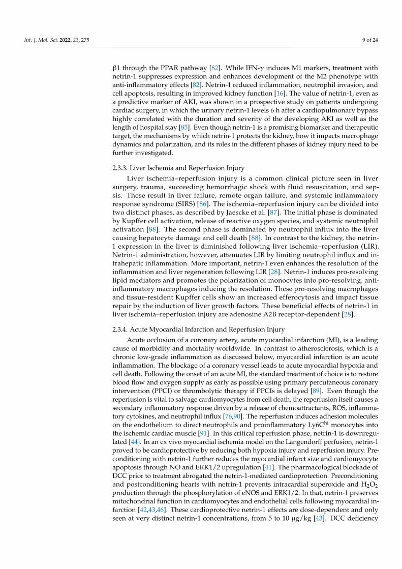
Int. J. Mol. Sci. 2022, 23, 275 9 of 24
β1 through the PPAR pathway [82]. While IFN-γ induces M1 markers, treatment withnetrin-1 suppresses expression and enhances development of the M2 phenotype withanti-inflammatory effects [82]. Netrin-1 reduced inflammation, neutrophil invasion, andcell apoptosis, resulting in improved kidney function [16]. The value of netrin-1, even asa predictive marker of AKI, was shown in a prospective study on patients undergoingcardiac surgery, in which the urinary netrin-1 levels 6 h after a cardiopulmonary bypasshighly correlated with the duration and severity of the developing AKI as well as thelength of hospital stay [85]. Even though netrin-1 is a promising biomarker and therapeutictarget, the mechanisms by which netrin-1 protects the kidney, how it impacts macrophagedynamics and polarization, and its roles in the different phases of kidney injury need to befurther investigated.
2.3.3. Liver Ischemia and Reperfusion Injury
Liver ischemia–reperfusion injury is a common clinical picture seen in liversurgery, trauma, succeeding hemorrhagic shock with fluid resuscitation, and sep-sis. These result in liver failure, remote organ failure, and systemic inflammatoryresponse syndrome (SIRS) [86]. The ischemia–reperfusion injury can be divided intotwo distinct phases, as described by Jaescke et al. [87]. The initial phase is dominatedby Kupffer cell activation, release of reactive oxygen species, and systemic neutrophilactivation [88]. The second phase is dominated by neutrophil influx into the livercausing hepatocyte damage and cell death [88]. In contrast to the kidney, the netrin-1 expression in the liver is diminished following liver ischemia–reperfusion (LIR).Netrin-1 administration, however, attenuates LIR by limiting neutrophil influx and in-trahepatic inflammation. More important, netrin-1 even enhances the resolution of theinflammation and liver regeneration following LIR [28]. Netrin-1 induces pro-resolvinglipid mediators and promotes the polarization of monocytes into pro-resolving, anti-inflammatory macrophages inducing the resolution. These pro-resolving macrophagesand tissue-resident Kupffer cells show an increased efferocytosis and impact tissuerepair by the induction of liver growth factors. These beneficial effects of netrin-1 inliver ischemia–reperfusion injury are adenosine A2B receptor-dependent [28].
2.3.4. Acute Myocardial Infarction and Reperfusion Injury
Acute occlusion of a coronary artery, acute myocardial infarction (MI), is a leadingcause of morbidity and mortality worldwide. In contrast to atherosclerosis, which is achronic low-grade inflammation as discussed below, myocardial infarction is an acuteinflammation. The blockage of a coronary vessel leads to acute myocardial hypoxia andcell death. Following the onset of an acute MI, the standard treatment of choice is to restoreblood flow and oxygen supply as early as possible using primary percutaneous coronaryintervention (PPCI) or thrombolytic therapy if PPCIs is delayed [89]. Even though thereperfusion is vital to salvage cardiomyocytes from cell death, the reperfusion itself causes asecondary inflammatory response driven by a release of chemoattractants, ROS, inflamma-tory cytokines, and neutrophil influx [76,90]. The reperfusion induces adhesion moleculeson the endothelium to direct neutrophils and proinflammatory Ly6Chi monocytes intothe ischemic cardiac muscle [91]. In this critical reperfusion phase, netrin-1 is downregu-lated [44]. In an ex vivo myocardial ischemia model on the Langendorff perfusion, netrin-1proved to be cardioprotective by reducing both hypoxia injury and reperfusion injury. Pre-conditioning with netrin-1 further reduces the myocardial infarct size and cardiomyocyteapoptosis through NO and ERK1/2 upregulation [41]. The pharmacological blockade ofDCC prior to treatment abrogated the netrin-1-mediated cardioprotection. Preconditioningand postconditioning hearts with netrin-1 prevents intracardial superoxide and H2O2production through the phosphorylation of eNOS and ERK1/2. In that, netrin-1 preservesmitochondrial function in cardiomyocytes and endothelial cells following myocardial in-farction [42,43,46]. These cardioprotective netrin-1 effects are dose-dependent and onlyseen at very distinct netrin-1 concentrations, from 5 to 10 µg/kg [43]. DCC deficiency
Int. J. Mol. Sci. 2022, 23, 275 10 of 24
in mice or the pharmacological blockade of ERK1/2 and NO activation abolished thecardioprotective netrin-1 effects [42]. Daliang et al. [92] found a decrease of serum netrin-1,myocardial netrin-1, and DCC expression in an AMI model, but an increase after aerobicexercise. This could reduce myocardial fibrosis, and indicates a role for netrin-1 and itsreceptor DCC during AMI.
In a murine heart transplant model, netrin-1 administration before the reperfusionameliorated the reperfusion injury. Netrin-1 improved the myocardial function, decreasedcardiomyocyte apoptosis, and decreased neutrophil and monocyte influx into the heart [44].Further, netrin-1 induces the polarization of alternative activated M2 macrophages (AAM)in injured hearts [44] by the activation of the PPARγ pathways. While the netrin-1 releasingcells were not studied in those studies mentioned, it was shown by Ke et al. [45] that thecardioprotective effects of netrin-1 can be achieved by the intramyocardial injection ofnetrin-1-transfected mesenchymal stem cells (MSCs) after coronary artery ligation. Theapplication of netrin-1 MSCs reduces the infarct size and prevents hypertrophic cardiacremodeling by the promotion of NO liberation and neoangiogenesis [45]. Altogether, thesestudies suggest that netrin-1 can mitigate the acute inflammatory response and acceleratewound healing following acute MI. However, the source of netrin-1 in healthy and hypoxicmyocardial tissue remains an open question.
Patients with acute coronary syndrome (ACS) showed a high netrin-1 level on arrivalat the emergency room, which decreased after angiography in patients with TIMI 3 flow(thrombolysis in MI; complete perfusion) [93]. High serum netrin-1 levels were foundin elderly females with ACS and correlated with a bad outcome. Reasons for this couldinclude more severe ACS and induced netrin-1 expression by hypoxia [94].
Li et al. [14] showed not only elevated netrin-1 in the blood of AMI patients, but alsoincreased infarct size in mice with the deletion of netrin-1 in the myeloid compartment(Ntn1loxP/loxP Lyz2 Cre+ mice). The cardioprotection of PMN-dependent netrin-1, whichis mainly synthesized by neutrophils, is mediated by the migration of neutrophils via anautocrine loop through myeloid adenosine A2b signaling [14]. These findings confirma functional role of netrin-1 interaction with ADORA2B in attenuating hypoxia-driveninflammation in AMI [14,34].
3. Chronic Low-Grade Inflammation
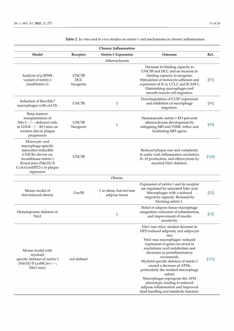
In contrast to acute inflammation, chronic inflammation is a long-lasting, non-resolvingadaptive immune response chiefly maintained by macrophages and lymphocytes. It ischaracterized by the simultaneous presence of active inflammation, tissue destruction, andtissue repair. Chronic inflammation can result from the host’s impairment of resolutionprograms following an acute inflammation. In these cases, the host defense is unable toeffectively overcome the pathogen and therefore impairs the macrophage egress or apopto-sis. Chronic inflammation can also develop as an independent low-grade inflammatorycondition from the start, as in arthritis, chronic lung disease, obesity, and atherosclerosis.These ongoing chronic inflammatory processes involve tissue proliferation, ineffectiveapoptosis, fibrosis, and necrosis, which can result in deteriorated organ function or organfailure. Chronic inflammation is further linked to the development of a variety of can-cers, such as bladder, cervical, gallbladder, liver, gastric, intestinal, and esophageal cancer(reviewed in [95]). In colorectal cancer, for example, the upregulation of netrin-1 and itsreceptor UNC5B correlates with the number of cancer-associated fibroblasts (CAFs). Theinhibition of netrin-1 in colon cancer inhibits the increase of CAFs, resulting in decreasedcancer stemness and plasticity [96]. Various murine models addressing the role and theexpression of netrin-1 and its different receptors in chronic inflammation are summarizedin Table 2.
Int. J. Mol. Sci. 2022, 23, 275 11 of 24
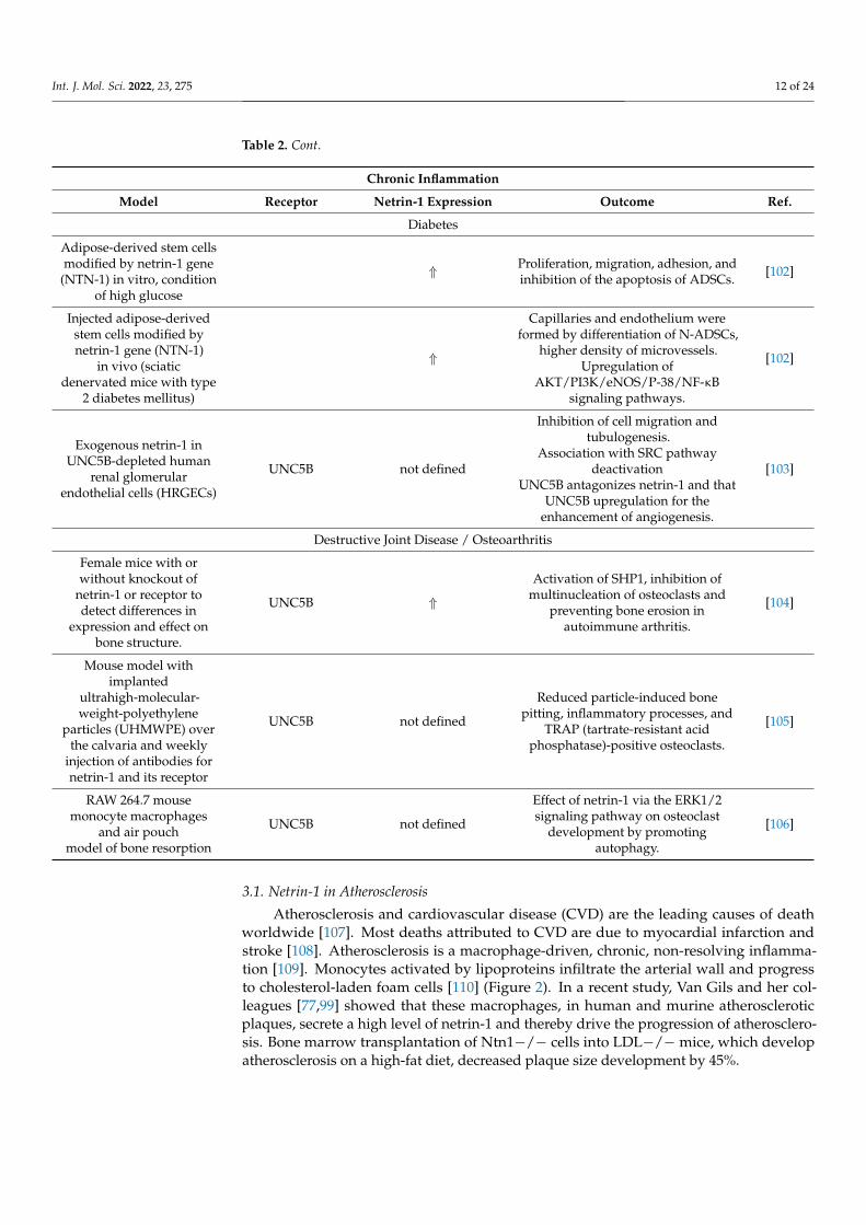
Table 2. In vitro and in vivo studies on netrin-1 and mechanisms in chronic inflammation.
Chronic Inflammation
Model Receptor Netrin-1 Expression Outcome Ref.
Atherosclerosis
Analysis of p.R590Lvariant of netrin-1
(mutNetrin-1)
UNC5BDCC
Neogenin
Decrease in binding capacity toUNC5B and DCC and an increase in
binding capacity to neogenin.Stimulation of monocyte adhesion andexpression of IL-6, CCL2, and ICAM-1.
Diminishing macrophages andsmooth muscle cell migration.
[97]
Induction of Raw264.7macrophages with oxLDL UNC5B ⇑
Downregulation of CCR7 expressionand inhibition of macrophage
migration.[98]
Bone marrowtransplantation of
Ntn-1−/−-deficient cellsin LDLR−/− KO mice on
western diet in plaqueprogression
UNC5BNeogenin ⇑
Hematopoetic netrin-1 KO preventsatherosclerosis development by
mitigating MØ and VSMC influx andfacilitating MØ egress
[99]
Monocyte- andmacrophage-specifictamoxifen-inducibleCX3CR1-driven cre
recombinase netrin-1floxed mice (Ntn1fl/fl
Cx3cr1creERT2+) in plaqueregression
UNC5B
Reduced plaque size and complexityin aortic wall, inflammation resolution,IL-10 production, and efferocytosis by
myeloid Ntn1 deletion.
[100]
Obesity
Mouse model ofdiet-induced obesity Unc5B ⇑ in obese, but not lean
adipose tissue
Expression of netrin-1 and its receptorare regulated by saturated fatty acid.
Macrophages with a reducedmigratory capacity. Restored by
blocking netrin-1.
[52]
Hematopoietic deletion ofNtn1 ⇓
Relief of adipose tissue macrophageemigration, reduction of inflammation,
and improvement of insulinsensitivity.
[52]
Mouse model withmyeloid-
specific deletion of netrin-1(Ntn1fl/fl LysMCre+/−;
Ntn1 mac)
not defined
Ntn1 mac mice: modest decrease inHFD-induced adiposity and adipocyte
size.Ntn1 mac macrophages: reducedexpression of genes involved in
arachidonic acid metabolism anddecreases in proinflammatory
eicosanoids.Myeloid-specific deletion of netrin-1
caused a decrease of ATMs,particularly the resident macrophage
subset.Macrophages reprogram the ATM
phenotype, leading to reducedadipose inflammation and improved
lipid handling and metabolic function.
[101]
Int. J. Mol. Sci. 2022, 23, 275 12 of 24
Table 2. Cont.
Chronic Inflammation
Model Receptor Netrin-1 Expression Outcome Ref.
Diabetes
Adipose-derived stem cellsmodified by netrin-1 gene
(NTN-1) in vitro, conditionof high glucose
⇑ Proliferation, migration, adhesion, andinhibition of the apoptosis of ADSCs. [102]
Injected adipose-derivedstem cells modified bynetrin-1 gene (NTN-1)
in vivo (sciaticdenervated mice with type
2 diabetes mellitus)
⇑
Capillaries and endothelium wereformed by differentiation of N-ADSCs,
higher density of microvessels.Upregulation of
AKT/PI3K/eNOS/P-38/NF-κBsignaling pathways.
[102]
Exogenous netrin-1 inUNC5B-depleted human
renal glomerularendothelial cells (HRGECs)
UNC5B not defined
Inhibition of cell migration andtubulogenesis.
Association with SRC pathwaydeactivation
UNC5B antagonizes netrin-1 and thatUNC5B upregulation for the
enhancement of angiogenesis.
[103]
Destructive Joint Disease / Osteoarthritis
Female mice with orwithout knockout of
netrin-1 or receptor todetect differences in
expression and effect onbone structure.
UNC5B ⇑
Activation of SHP1, inhibition ofmultinucleation of osteoclasts and
preventing bone erosion inautoimmune arthritis.
[104]
Mouse model withimplanted
ultrahigh-molecular-weight-polyethylene
particles (UHMWPE) overthe calvaria and weekly
injection of antibodies fornetrin-1 and its receptor
UNC5B not defined
Reduced particle-induced bonepitting, inflammatory processes, and
TRAP (tartrate-resistant acidphosphatase)-positive osteoclasts.
[105]
RAW 264.7 mousemonocyte macrophages
and air pouchmodel of bone resorption
UNC5B not defined
Effect of netrin-1 via the ERK1/2signaling pathway on osteoclast
development by promotingautophagy.
[106]
3.1. Netrin-1 in Atherosclerosis
Atherosclerosis and cardiovascular disease (CVD) are the leading causes of deathworldwide [107]. Most deaths attributed to CVD are due to myocardial infarction andstroke [108]. Atherosclerosis is a macrophage-driven, chronic, non-resolving inflamma-tion [109]. Monocytes activated by lipoproteins infiltrate the arterial wall and progressto cholesterol-laden foam cells [110] (Figure 2). In a recent study, Van Gils and her col-leagues [77,99] showed that these macrophages, in human and murine atheroscleroticplaques, secrete a high level of netrin-1 and thereby drive the progression of atherosclero-sis. Bone marrow transplantation of Ntn1−/− cells into LDL−/−mice, which developatherosclerosis on a high-fat diet, decreased plaque size development by 45%.

Int. J. Mol. Sci. 2022, 23, 275 13 of 24
Figure 2. Netrin-1 in atherosclerosis. Subendothelial lipoprotein accumulation incite a proinflamma-tory activation of the endothelial cells and promote monocyte influx into the developing atheroscle-rotic plaque. As lipoproteins undergo various modifications, such as oxidation and hydrolysis, theyare taken up by monocytes and form proinflammatory foam cells. In this inflammatory milieuchronically, activated macrophages secrete netrin-1, which recruit further monocytes and retainmacrophages in the tissue until they undergo apoptosis and form the detrimental necrotic core.Netrin-1 further mitigates pro-resolving macrophage differentiation and efferocytosis. Moreover,netrin-1-secreting macrophages advance the plaque further by recruiting smooth muscle cells into thedeveloping plaque, which can themselves become macrophages and promote the ongoing progressionof the plaque.
Netrin-1 produced by macrophages plays a proatherogenic role: first, it preventsmacrophage migration and the egress from the plaque; second, it promotes lesion progres-sion while enhancing the chemoattraction of smooth muscle cells and their recruitmentinto the intima [21,111,112].
The expression of netrin-1 and Unc5b in the atherosclerotic plaque are induced byhypoxia [77]. This hypoxic induction is dependent on HIF-1α, which itself is induced byhypoxic stressors such as oxLDL [113]. In the progression of the disease, these inflammatorycholesterol-laden foam cells persist in the plaque and contribute to local and systemicinflammation [111,114].
The continued monocyte recruitment, the expansion of lipid-laden macrophages inthe lesion [115], and the impaired egress lead to the formation of large necrotic cores anda thinning of the fibrous cap [116]. Taken together, all these mechanisms contribute to adestabilization of the plaque, resulting in myocardial infarction [117]. It was shown thatthe deletion of netrin-1 in myeloid cells reduces atherosclerosis plaque size [111].
On the other hand, netrin-1 induced by TNF-α and produced by endothelial cellsinhibits the entry of monocytes into the plaque by reducing the expression of adhesionmolecules as well as the chemotaxis of leukocytes by the suppression of cytokine production,toll-like receptor (TLR) 4, and the NF-κB signaling pathway [21,111,112]. Stimulation withTNF-α in vitro showed a reduction in the expression of VCAM-1, ICAM-1, and E-selectinwhen cells were co-stimulated with netrin-1. They inhibit the expression of IL-6 and COX-2 [21], among other things. The downregulation of ICAM-1, IL-6, and MCP-1 that canalso be induced by the UNC5B blockade was also observed in atherosclerosis patients [21].These different effects may depend on the stage of atherosclerosis.
The importance and benefits of controlling systemic inflammation in atherosclerosiswas shown in the CANTOS trial, a prospective patient study [118]. The anti-inflammatorytreatment with an anti-IL-1β monoclonal antibody resulted in a decreased incidence

Int. J. Mol. Sci. 2022, 23, 275 14 of 24
of cardiovascular events and deaths without altering the cholesterol levels [118]. Un-der pro-atherogenic conditions, netrin-1 expression by endothelial cells is downregu-lated and thereby facilitates increased monocyte and macrophage influx into the arterialwall [119,120]. However, the plaque progression, which is driven by an on-going influxand accumulation of macrophages in the plaque, is not a one-way road. Fisher and Ran-dolph [121] showed that plaque regression can be induced by facilitating macrophageegress from the arterial wall in a CCR7-dependent manner [122]. Netrin-1 blocks thisdirected macrophage migration towards the CCR7 ligands CCL-19 and CCL-21 and to-wards MCP-1 [99]. Netrin-1 disrupts the Rac1 signaling pathway, the actin cytoskeletonreorganization, and the cell polarization, thereby mitigating macrophage egress from theplaque [99]. These netrin-1-mediated chemotactic effects are inhibited by the blockade ofUnc5b.
In this context, Yang et al. [98] examined the expression of netrin-1 and its receptorUNC5B at the mRNA and protein level in Raw264.7 macrophages in response to ox-LDL.The data showed a downregulation of CCD7 that can bind the chemokines CCL19 andCCL21, an upregulation of UNC5B in an NF-κB-dependent manner, which inhibited foamcell migration [98]. However, more studies are needed to understand these mechanismsmore thoroughly.
In atherosclerosis, the bone-marrow transplantation of Ntn-1−/−-deficient cells inLDLR−/−mice leads to a significant increase in macrophage egress from the plaque [99].In contrast to its effects on macrophages, netrin-1 recruits smooth muscle cells (SMC) intothe plaque and promotes plaque growth [99]. The chemoattraction of SMC by netrin-1 ismediated through neogenin and not through Unc5b, which is responsible for the impairedegress of cholesterol-laden macrophages from the plaque. The restored macrophage migra-tory capacity in hematopoietic netrin-1-deficient mice led to a decrease in macrophages, Tcells, and necrotic core area in the plaque [99]. Overall, the netrin-1 deletion in hematopoi-etic cells promotes a significant reduction in plaque size and complexity. Altogether thisstudy demonstrates for the first time that netrin-1 secreted by plaque macrophages activelypromotes the progression of atherosclerosis in an Unc5b-dependent manner.
Netrin-1, however, not only promotes the onset and propagation of atherosclerosis,but also hinders its regression. In established atherosclerosis, cholesterol lowering alone isinsufficient to promote plaque regression, as monocyte-specific netrin-1 impairs the reorga-nization of the plaque’s immune cell landscape and hinders monocyte egress [100]. We wereable to show that lipid lowering in established atherosclerosis and silencing macrophage-specific netrin-1 by a tamoxifen-inducible knock-out reduced the infiltration of intimalsmooth muscle cells, macrophage accumulation, and survival in plaques, while markersof inflammation such as IL-10 and efferocytosis were simultaneously increased [100]. Theatherosclerotic plaque burden in the thoracic aorta decreased by 50% after the deletion ofmonocyte-specific netrin-1, while it remained unchanged in littermate controls despite aneffective lipid lowering. Therefore, targeting netrin-1 could be a promising approach alsofor already advanced atherosclerotic disease.
Bruikman et al. [123] focused on netrin-1 levels in patients and found a negativecorrelation between netrin-1 and arterial wall inflammation, subclinical atherosclerosis,and plaque volume. However, there was no significant difference in netrin-1 plasmaconcentrations between patients with stable vs. unstable plaques.
In another study, Bruikman and her colleagues [97] found a genomic variant of netrin-1leading to a familiar premature atherosclerosis. The change in an arginine to leucine aminoacid at position 1769 (NTN1 c.1769G > T; p.Arg590Leu) led to fewer anti-inflammatoryeffects on endothelial cells, and a strong blockage in macrophage emigration.
All these results indicate the important role of netrin-1 and its receptors in the processof atherosclerosis. However, given the dual function of netrin-1 in atherosclerosis, moreinsight is needed to determine at which stage of atherosclerosis netrin-1 supplementationor netrin-1 blocking substances will ameliorate atherosclerosis.
Int. J. Mol. Sci. 2022, 23, 275 15 of 24
3.2. Aortic Abdominal Aneurysma
Aortic abdominal aneurysms (AAAs) are a potentially life-threatening dilatation ofthe abdominal aorta. An AAA is regarded as a long-term consequence of cardiovasculardisease, even though its molecular mechanisms are not fully understood. More recentstudies have revealed chronic inflammatory processes in the aortic vessel wall, includingextracellular matrix (ECM) degeneration by matrix metalloproteinase (MMP) activationand oxidative stress.
A recent study identified, in damaged areas of thoracic aortic aneurysms, an upreg-ulation of neuronal guidance proteins, including netrin-1 and its receptors UNC5B [124].In line with these findings, Hadi et al. [125] describe how macrophage-derived netrin-1promotes AAA progression through the release of MMP-3 from vascular smooth muscles bybinding its receptor neogenin-1 [125]. Therefore, netrin-1 might be an important player inmediating the crossing between inflammatory processes and the erosion of the extracellularmatrix in AAAs in both mice and humans [125].
3.3. Obesity
The rates of obesity, particularly excessive visceral adiposity and its comorbidities,insulin resistance type 2 diabetes (T2D), and cardiovascular diseases, have increased dra-matically over the past 25 years throughout the world [126,127] and are major threats toglobal health [128–130]. Chronic low-grade inflammation, mediated by the innate andadaptive immune system, has emerged as the key pathogenic link between obesity and itscomorbidities [131].
It is now generally accepted that chronic low-grade inflammation in obesity is dominatedby macrophages infiltrating the expanded adipose tissue in obese individuals [132–135]. Theseadipose tissue macrophages (ATMs) produce mainly proinflammatory “M1” cytokines suchas TNF-α [136], interleukin (IL)-1β [137], and CCL2 [138], contributing to the persistenceof local and systemic inflammation and increasing insulin resistance (IR) [52]. Recently,Ramkhelawon and her colleagues [52] showed that, in obesity, netrin-1 plays a pivotalrole in the retention of ATMs in visceral adipose tissue (VAT) and promotes not onlysystemic inflammation but also metabolic dysfunction. This study showed that netrin-1and its receptor Unc5b are highly upregulated in ATMs from obese compared to lean miceand humans. Other netrin-1 receptors, however, such as Neo1 and DCC, are expressedsimilarly in lean and obese individuals [52]. Free fatty acids, such as palmitate, can inducethe expression of netrin-1 and Unc5b in BMDM directly in a NF-κB-dependent manner.Moreover, netrin-1 was shown to be induced by a paracrine release of cytokines, such asTNF-α and IL-6, but not IL-4, from adipocytes [52]. Murine 3T3-L1 adipocytes treatedwith free fatty acid stimulated BMDMs and conditioned media highly induces Ntn-1 andUnc5b expression. The blockade of TNF-α and IL-6 abrogates this netrin-1 and Unc5binduction [52].
In obesity, netrin-1 hinders ATM egress from the VAT, protects ATM from apoptosis,and induces an inflammatory M1-like phenotype. Netrin-1 further mediates chemosta-sis in an Unc5b-dependent manner and thereby actively fuels inflammation in the VAT.Moreover, netrin-1 impedes the migration of ATM egress from the VAT, as it mitigatesmigration towards CCL-19 [99]. This impairs the resolution of the ongoing chronic in-flammation. WT mice were reconstituted with Ntn1−/− bone-marrow cells and fed ahigh-fat diet (HFD) to study the impact hematopoietic netrin-1 in vivo [101]. The netrin-1deficiency did not protect the mice from gaining weight and becoming obese. However,the adipose tissue proved to be less inflamed, as fewer macrophages were found in theVAT. Bead-labeling studies showed that netrin-1 did not impact the recruitment of mono-cytes into the VAT but mitigated the egress from the VAT and thereby led to the increasedATM accumulation [101]. Functionally, the decreased local inflammation in mice withhematopoietic netrin-1 deficiency resulted in an improved glucose and insulin tolerance.We were able to show that macrophage-specific netrin-1 affects the metabolism in HFD-fedmice and leads to increased weight gain compared to Ntn1-deficient mice. Ntn1 further

Int. J. Mol. Sci. 2022, 23, 275 16 of 24
promotes macrophage differentiation, impairs lipid handling and migration, and promotesproinflammatory mediators [101].
Sanders et al. [139] showed in mouse experiments that expression of the netrin-1receptors Unc5b and DCC is altered in the arcuate nucleus in fetuses developing in obesemothers [139] and that these alterations impact developmental pathways important forfetal development. These results may explain the defective neuropeptide Y innervation ofthe paraventricular nucleus of obese mothers [139].
A recent clinical study showed that circulating netrin-1 levels positively correlatedwith fasting glucose, the HbA1c level, and the insulin resistance index. This suggests thatnetrin-1 may be a sensitive indicator for early type 2 diabetes [140]. Together, these studiesindicate that netrin-1 is a key player in obesity-induced inflammation and functionallyimpacts glucose tolerance.
3.4. Rheumatoid Arthritis and Osteolysis
Netrin-1 expression by osteoblasts and synovial fibroblasts is enhanced by IL-17. Bind-ing its receptor UNC5B on the osteoclasts’ surfaces, netrin-1 initiates the activation of SHP1.This results in a blockade of the multinucleation, but not in the differentiation, of osteo-clasts. Hence, netrin-1 prevents bone erosion in autoimmune arthritis and age-related bonedestruction [104]. As netrin-1 is highly expressed in osteolysis, its blockade mitigates osteol-ysis by altering osteoclast differentiation [105]. In contrast, Mediero et al. [105] showed thatanti-netrin-1 and anti-UNC5B antibodies increase bone density and reduce inflammatoryinfiltrates and particle-induced bone pitting. Netrin-1 also mediates osteoclastogenesisby autophagy in vitro and in vivo [106]. The netrin-1 blockade has been shown to reduceK/Bxn-induced arthritis [141].
Zhu et al. [142] found that osteoclast-secreted netrin-1 induces sensory nerve growthresponsible for pain in osteoarthritis. The inhibition of netrin-1 or its receptor DCC wasable to reduce the pain. Taken together, netrin-1 seems to differentially regulate osteoclasto-genesis in different settings, and its therapeutic potential still needs to be determined.
3.5. Diabetes
A recent study found netrin-1 plasma levels to be decreased in patients with newlydiagnosed diabetes mellitus type 2, and even suggest that netrin-1 should be assessed forits potential in early diabetes screening [143].
A major clinical challenge in diabetes is the diabetic peripheral neurovascular dis-eases (DPNVs), for which no effective treatment is currently available. In this frameworkZhang et al. [102] showed that netrin-1 might mitigate DPNVs. In their study, trans-planted adipose-derived stem cells, which have potential for differentiation and tissuerestoration, were exposed to netrin-1 differentiated into endothelial cells and formedsmall capillaries [102]. Netrin-1 not only promotes neuronal migration but also regulatesthe survival, adhesion, migration, proliferation, and differentiation of endothelial cells.A negative effect on the proliferation and apoptosis of stem cells was shown in previousstudies because of hypoglycemia. In this recent study [102], the important role of netrin-1 in proliferation, migration, adhesion, and apoptosis was shown under high-glucoseconditions in vivo and in vitro. The combined signaling pathway AKT/PI3K/eNOS/P-38/NK-κB and the expression of many growth factors, such as VEGF, TNF-α, EGF, andnetrin-1, were upregulated [102]. These results suggest that netrin-1 might be a potentialtarget for DPNVs.
On the other hand, Toque et al. [144] showed that netrin-1, not only reduces the highglucose induced by vascular endothelial dysfunction and limits the impairment of the nitricoxide synthase, but also suppresses inflammatory and apoptotic processes.
Other studies have characterized the protective effect of netrin-1 on the apoptosis of β-cells [145], in diabetic retinopathy [146], diabetic nephropathy [147], and diabetes-associatedmyocardial infarction [45]. Goa et al. [148] reported a preventive role of netrin-1 againstdiabetes progression via dual action on islet insulin secretion and inflammation. Since

Int. J. Mol. Sci. 2022, 23, 275 17 of 24
diabetes and its co-morbidities are associated with hyperactive inflammation, netrin-1 couldbe a promising therapeutic candidate that reduces hypoglycemia while also amelioratinginflammation for diabetic individuals.
Jiao et al. [103] showed that UNC5B is upregulated in diabetic kidney diseases (DKDs)and promoted by high glucose levels. Netrin-1 and its receptor UN5B are protective duringDKDs because of the inhibition of netrin-1 by binding on UNC5B, which eliminates thenegative effects of netrin-1 on tubulogenesis.
The upregulation of the receptor UNC5B was examined in retinal microvessels, whichenhances vascular integrity and is protective for adaptation to diabetes [7]. Elevatedlevels of UNC5B are associated with chronic inflammation such as atherosclerosis obesity,oxygen-induced retinopathy, and retinal vessel sprout [7].
Miloudi et al. [7] showed that metaloproteinase-9 cleaves netrin-1 into fragments.These fragments of truncated netrin-1 promote diabetic retinopathy, exacerbate retinal andmacular edema. In contrast, the full netrin-1 protein maintains blood–brain barrier functionand mitigates the development of diabetic retinopathy [7].
3.6. Chronic Inflammation of Lungs
Patients with idiopathic lung fibrosis showed high expression levels of netrin-1. There-fore, netrin-1, which interacts with its receptor DCC, promotes lung fibrosis with histopatho-logical changes and remodels adrenergic nerves [149].
Kerget et al. [150] found that patients with an acute exacerbation of COPD showed ahigher expression of netrin-1 than a control group. More studies are necessary to evaluatethese findings.
4. Discussion
Even though much has been learned over the last two decades since netrin-1 was firstdescribed outside the central nervous system, many new questions have emerged.
Collectively, most studies show that the regulation of netrin-1 is critically involvedin acute (Figure 1) and chronic inflammation (Figure 2). The expression of netrin-1 and itsvarious receptors are differentially regulated depending on the tissue, the stimulus, thesource, and the target cell (Tables 1 and 2). Although the effects of netrin-1 in acute andchronic inflammation seem to be opposed, most studies show that netrin-1 can effectivelychange the kinetics and functions of the leukocytes in the setting of inflammation.
In chronic inflammation, adipose tissue macrophages and plaque macrophagessecrete netrin-1, which actively hinders the egress of macrophages out of the inflamed tis-sue. Changing the local proinflammatory environment, however, is key for overcomingthe chronic ongoing inflammation. Specifically targeting these persistent proinflamma-tory macrophages through the secretion of netrin-1 and, in this context, its main receptorUnc5b is a very promising target in order to beneficially modify the local environment,overcome the ongoing inflammation, promote resolution, and restore homeostasis. Inchronic inflammation, cells of the adaptive immune system play a key role. After beingchallenged by antigen-presenting cells such as monocytes, macrophages, and dendriticcells, B- and T-cells are activated and recruited into the inflamed tissue. Understandinghow netrin-1 either directly or indirectly, through the polarization of monocytes, affectsthe adaptive immune response in chronic inflammation will be key to further delineatingthe effects by which netrin-1 promotes and sustains chronic inflammation.
In acute inflammation, the systemic application has been shown to mainly mitigatethe acute inflammatory response and mitigate leukocyte influx into the inflamed tissuepredominantly involving the purinergic pathway through the adenosine A2B receptor. Inthe early phase of acute inflammation, involving primarily the innate immune system,netrin-1 is further able to promote the infiltrating monocytes towards an anti-inflammatoryand pro-resolving phenotype. However, all the models of acute inflammation that havebeen studied do not involve any exogenous pathogens, which must be controlled. Thesemodels all have an impaired restraint of the innate immune system in common, in which

Int. J. Mol. Sci. 2022, 23, 275 18 of 24
netrin-1 effectively mitigates the onset of the inflammation and in the latter promotes theresolution. However, it will be important to understand whether, in the case of an under-lying pathogen, this extenuated immune response is still able to overcome the invadingpathogen or whether it contributes to fatal outcomes.
The great challenge in the field remains understanding how netrin-1 is regulatedunder various conditions. Further, the downstream mechanisms and pathways of itsvarious receptors need further study to understand how netrin-1 regulates the organ-specific activation and differentiation of tissue-resident and hematopoietic leukocytesunder distinct acute and chronic inflammatory conditions.
Author Contributions: Writing—review and editing, L.Z. and M.S.; visualization, M.S.; fundingacquisition, M.S. All authors have read and agreed to the published version of the manuscript.
Funding: This research received no external funding.
Institutional Review Board Statement: Not applicable.
Conflicts of Interest: The authors have no conflict of interest to declare.
References1. Barallobre, M.J.; Pascual, M.; Del Rio, J.A.; Soriano, E. The Netrin family of guidance factors: Emphasis on Netrin-1 signalling.
Brain Res. Rev. 2005, 49, 22–47. [CrossRef]2. Cirulli, V.; Yebra, M. Netrins: Beyond the brain. Nat. Rev. Mol. Cell Biol. 2007, 8, 296–306. [CrossRef]3. Yurchenco, P.D.; Wadsworth, W.G. Assembly and tissue functions of early embryonic laminins and netrins. Curr. Opin. Cell Biol.
2004, 572–579. [CrossRef] [PubMed]4. Serafini, T.; Kennedy, T.E.; Galko, M.J.; Mirzayan, C.; Jessell, T.M.; Tessier-Lavigne, M. The netrins define a family of axon
outgrowth-promoting proteins homologous to C. elegans UNC-6. Cell 1994, 78, 409–424. [CrossRef]5. Bruikman, C.S.; Zhang, H.; Kemper, A.M.; van Gils, J.M. Netrin Family: Role for Protein Isoforms in Cancer. J. Nucleic Acids 2019,
2019, 3947123. [CrossRef] [PubMed]6. Xu, K.; Wu, Z.; Renier, N.; Antipenko, A.; Tzvetkova-Robev, D.; Xu, Y.; Minchenko, M.; Nardi-Dei, V.; Rajashankar, K.R.; Himanen,
J.; et al. Neural migration. Structures of netrin-1 bound to two receptors provide insight into its axon guidance mechanism.Science 2014, 344, 1275–1279. [CrossRef] [PubMed]
7. Miloudi, K.; Binet, F.; Wilson, A.; Cerani, A.; Oubaha, M.; Menard, C.; Henriques, S.; Mawambo, G.; Dejda, A.; Nguyen, P.T.; et al.Truncated netrin-1 contributes to pathological vascular permeability in diabetic retinopathy. J. Clin. Investig. 2016, 126, 3006–3022.[CrossRef]
8. Kruger, R.P.; Lee, J.; Li, W.; Guan, K.L. Mapping netrin receptor binding reveals domains of Unc5 regulating its tyrosinephosphorylation. J. Neurosci. 2004, 24, 10826–10834. [CrossRef]
9. Finci, L.; Zhang, Y.; Meijers, R.; Wang, J.-H. Signaling mechanism of the netrin-1 receptor DCC in axon guidance. Prog. Biophys.Mol. Biol. 2015, 118, 153–160. [CrossRef]
10. Mehlen, P.; Rabizadeh, S.; Snipas, S.J.; Assa-Munt, N.; Salvesen, G.S.; Bredesen, D.E. The DCC gene product induces apoptosis bya mechanism requiring receptor proteolysis. Nature 1998, 395, 801–804. [CrossRef]
11. Paradisi, A.; Mehlen, P. Netrin-1, a missing link between chronic inflammation and tumor progression. Cell Cycle 2010, 9,1253–1262. [CrossRef] [PubMed]
12. Arakawa, H. Netrin-1 and its receptors in tumorigenesis. Nat. Rev. Cancer 2004, 4, 978–987. [CrossRef]13. Rosenberger, P.; Schwab, J.M.; Mirakaj, V.; Masekowsky, E.; Mager, A.; Morote-Garcia, J.C.; Unertl, K.; Eltzschig, H.K. Hypoxia-
inducible factor-dependent induction of netrin-1 dampens inflammation caused by hypoxia. Nat. Immunol. 2009, 10, 195–202.[CrossRef] [PubMed]
14. Li, J.; Conrad, C.; Mills, T.W.; Berg, N.K.; Kim, B.; Ruan, W.; Lee, J.W.; Zhang, X.; Yuan, X.; Eltzschig, H.K. PMN-derived netrin-1attenuates cardiac ischemia-reperfusion injury via myeloid ADORA2B signaling. J. Exp. Med. 2021, 218. [CrossRef] [PubMed]
15. Corset, V.; Nguyen-Ba-Charvet, K.T.; Forcet, C.; Moyse, E.; Chedotal, A.; Mehlen, P. Netrin-1-mediated axon outgrowth andcAMP production requires interactionwith adenosine A2b receptor. Nature 2000, 407, 747–750. [CrossRef]
16. Ranganathan, P.V.; Jayakumar, C.; Ramesh, G. Netrin-1-treated macrophages protect the kidney against ischemia-reperfusioninjury and suppress inflammation by inducing M2 polarization. Am. J. Physiol. Renal Physiol. 2013, 304, F948–F957. [CrossRef][PubMed]
17. Gloerich, M.; Bos, J.L. Epac: Defining a new mechanism for cAMP action. Annu. Rev. Pharmacol. Toxicol. 2010, 50, 355–375.[CrossRef]
18. Lorenowicz, M.J.; Fernandez-Borja, M.; Hordijk, P.L. cAMP signaling in leukocyte transendothelial migration. Arterioscler. Thromb.Vasc. Biol. 2007, 27, 1014–1022. [CrossRef] [PubMed]
Int. J. Mol. Sci. 2022, 23, 275 19 of 24
19. Yebra, M.; Montgomery, A.M.; Diaferia, G.R.; Kaido, T.; Silletti, S.; Perez, B.; Just, M.L.; Hildbrand, S.; Hurford, R.; Florkiewicz,E.; et al. Recognition of the neural chemoattractant Netrin-1 by integrins alpha6beta4 and alpha3beta1 regulates epithelial celladhesion and migration. Dev. Cell 2003, 5, 695–707. [CrossRef]
20. Ly, N.P.; Komatsuzaki, K.; Fraser, I.P.; Tseng, A.A.; Prodhan, P.; Moore, K.J.; Kinane, T.B. Netrin-1-inhibits-leukocyte-migrationin vitro and in vivo. Proc. Natl. Acad. Sci. USA 2005, 102, 14729–14734. [CrossRef]
21. Claro, V.; Ferro, A. Netrin-1: Focus on its role in cardiovascular physiology and atherosclerosis. JRSM Cardiovasc. Dis. 2020, 9,2048004020959574. [CrossRef]
22. Bradford, D.; Cole, S.J.; Cooper, H.M. Netrin-1: Diversity in development. Int. J. Biochem. Cell Biol. 2009, 41, 487–493. [CrossRef]23. Castets, M.; Mehlen, P. Netrin-1 role in angiogenesis: To be or not to be a pro-angiogenic factor? Cell Cycle 2010, 9, 1466–1471.
[CrossRef]24. Mazelin, L.; Bernet, A.; Bonod-Bidaud, C.; Pays, L.; Arnaud, S.; Gespach, C.; Bredesen, D.E.; Scoazec, J.Y.; Mehlen, P. Netrin-1
controls colorectal tumorigenesis by regulating apoptosis. Nature 2004, 431, 80–84. [CrossRef]25. Han, Y.; Jiang, N.; Su, T.; Yang, Q.-C.; Yan, C.-C.; Ye, L.; Yuan, Q.; Zhu, P.-W.; Li, W.; Liu, Z.-G.; et al. Netrin-1 promotes epithelium
repair in corneal injury. Int. J. Ophthalmol. 2020, 13, 206–212. [CrossRef] [PubMed]26. Baggiolini, M. Chemokines and leukocyte traffic. Nature 1998, 392, 565–568. [CrossRef] [PubMed]27. Mirakaj, V.; Dalli, J.; Granja, T.; Rosenberger, P.; Serhan, C.N. Vagus nerve controls resolution and pro-resolving mediators of
inflammation. J. Exp. Med. 2014, 211, 1037–1048. [CrossRef] [PubMed]28. Schlegel, M.; Kohler, D.; Korner, A.; Granja, T.; Straub, A.; Giera, M.; Mirakaj, V. The neuroimmune guidance cue netrin-1 controls
resolution programs and promotes liver regeneration. Hepatology 2016, 63, 1689–1705. [CrossRef] [PubMed]29. Mirakaj, V.; Thix, C.A.; Laucher, S.; Mielke, C.; Morote-Garcia, J.C.; Schmit, M.A.; Henes, J.; Unertl, K.E.; Köhler, D.; Rosenberger, P.
Netrin-1 dampens pulmonary inflammation during acute lung injury. Am. J. Respir. Crit. Care Med. 2010, 181, 815–824. [CrossRef]30. Mutz, C.; Mirakaj, V.; Vagts, D.A.; Westermann, P.; Waibler, K.; König, K.; Iber, T.; Nöldge-Schomburg, G.; Rosenberger, P. The
neuronal guidance protein netrin-1 reduces alveolar inflammation in a porcine model of acute lung injury. Crit. Care 2010, 14,R189. [CrossRef]
31. Chen, Z.; Chen, Y.; Zhou, J.; Li, Y.; Gong, C.; Wang, X. Netrin-1 reduces lung ischemia-reperfusion injury by increasing theproportion of regulatory T cells. J. Int. Med. Res. 2020, 48, 300060520926415. [CrossRef]
32. He, J.; Zhao, Y.; Deng, W.; Wang, D. Netrin-1 promotes epithelial sodium channel-mediated alveolar fluid clearance via activationof the adenosine 2B receptor in lipopolysaccharide-induced acute lung injury. Respiration 2014, 87, 394–407. [CrossRef] [PubMed]
33. Ko, C.-L.; Lin, J.-A.; Chen, K.-Y.; Hsu, A.-C.; Wu, S.-Y.; Tai, Y.-T.; Lin, K.-H.; Chung, W.-C.; Li, M.-H. Netrin-1 Dampens HypobaricHypoxia-Induced Lung Injury in Mice. High Alt. Med. Biol. 2019, 20, 293–302. [CrossRef] [PubMed]
34. Aherne, C.M.; Collins, C.B.; Masterson, J.C.; Tizzano, M.; Boyle, T.A.; Westrich, J.A.; Parnes, J.A.; Furuta, G.T.; Rivera-Nieves, J.;Eltzschig, H.K. Neuronal guidance molecule netrin-1 attenuates inflammatory cell trafficking during acute experimental colitis.Gut 2012, 61, 695–705. [CrossRef]
35. Mirakaj, V.; Gatidou, D.; Pötzsch, C.; König, K.; Rosenberger, P. Netrin-1 signaling dampens inflammatory peritonitis. J. Immunol.2011, 186, 549–555. [CrossRef] [PubMed]
36. Chen, J.; Cai, Q.-P.; Shen, P.-J.; Yan, R.-L.; Wang, C.-M.; Yang, D.-J.; Fu, H.-B.; Chen, X.-Y. Netrin-1 protects against L-Arginine-induced acute pancreatitis in mice. PLoS ONE 2012, 7, e46201. [CrossRef]
37. Wang, W.; Reeves, W.B.; Ramesh, G. Netrin-1 and kidney injury. I. Netrin-1 protects against ischemia-reperfusion injury of thekidney. Am. J. Physiol. Renal Physiol. 2008, 294, F739–F747. [CrossRef]
38. Wang, W.; Reeves, W.B.; Pays, L.; Mehlen, P.; Ramesh, G. Netrin-1 overexpression protects kidney from ischemia reperfusioninjury by suppressing apoptosis. Am. J. Pathol. 2009, 175, 1010–1018. [CrossRef]
39. Tadagavadi, R.K.; Wang, W.; Ramesh, G. Netrin-1 regulates Th1/Th2/Th17 cytokine production and inflammation throughUNC5B receptor and protects kidney against ischemia-reperfusion injury. J. Immunol. 2010, 185, 3750–3758. [CrossRef]
40. Grenz, A.; Dalton, J.H.; Bauerle, J.D.; Badulak, A.; Ridyard, D.; Gandjeva, A.; Aherne, C.M.; Brodsky, K.S.; Kim, J.-H.; Tuder, R.M.;et al. Partial netrin-1 deficiency aggravates acute kidney injury. PLoS ONE 2011, 6, e14812. [CrossRef]
41. Zhang, J.; Cai, H. Netrin-1 prevents ischemia/reperfusion-induced myocardial infarction via a DCC/ERK1/2/eNOSs1177/NO/DCC feed-forward mechanism. J. Mol. Cell Cardiol. 2010, 48, 1060–1070. [CrossRef] [PubMed]
42. Bouhidel, J.O.; Wang, P.; Li, Q.; Cai, H. Pharmacological postconditioning treatment of myocardial infarction with netrin-1. Front.Biosci. 2014, 19, 566–570. [CrossRef] [PubMed]
43. Bouhidel, J.O.; Wang, P.; Siu, K.L.; Li, H.; Youn, J.Y.; Cai, H. Netrin-1 improves post-injury cardiac function in vivo via DCC/NO-dependent preservation of mitochondrial integrity, while attenuating autophagy. Biochim. Biophys. Acta 2015, 1852, 277–289.[CrossRef] [PubMed]
44. Mao, X.; Xing, H.; Mao, A.; Jiang, H.; Cheng, L.; Liu, Y.; Quan, X.; Li, L. Netrin-1 attenuates cardiac ischemia reperfusion injuryand generates alternatively activated macrophages. Inflammation 2014, 37, 573–580. [CrossRef]
45. Ke, T.; Wu, Y.; Li, L.; Liu, Y.; Yao, X.; Zhang, J.; Kong, D.; Li, C. Netrin-1 ameliorates myocardial infarction-induced myocardialinjury: Mechanisms of action in rats and diabetic mice. Hum. Gene Ther. 2014, 25, 787–797. [CrossRef]
46. Siu, K.L.; Lotz, C.; Ping, P.; Cai, H. Netrin-1 abrogates ischemia/reperfusion-induced cardiac mitochondrial dysfunction via nitricoxide-dependent attenuation of NOX4 activation and recoupling of NOS. J. Mol. Cell. Cardiol. 2015, 78, 174–185. [CrossRef]
Int. J. Mol. Sci. 2022, 23, 275 20 of 24
47. Rubenfeld, G.D.; Caldwell, E.; Peabody, E.; Weaver, J.; Martin, D.P.; Neff, M.; Stern, E.J.; Hudson, L.D. Incidence and outcomes ofacute lung injury. N. Engl. J. Med. 2005, 353, 1685–1693. [CrossRef]
48. Bellani, G.; Laffey, J.G.; Pham, T.; Fan, E.; Brochard, L.; Esteban, A.; Gattinoni, L.; van Haren, F.; Larsson, A.; McAuley, D.F.; et al.Epidemiology, Patterns of Care, and Mortality for Patients With Acute Respiratory Distress Syndrome in Intensive Care Units in50 Countries. JAMA 2016, 315, 788–800. [CrossRef]
49. Ware, L.B.; Matthay, M.A. The acute respiratory distress syndrome. N. Engl. J. Med. 2000, 342, 1334–1349. [CrossRef]50. Matthay, M.A.; Ware, L.B.; Zimmerman, G.A. The acute respiratory distress syndrome. J. Clin. Investig. 2012, 122, 2731–2740.
[CrossRef]51. Paradisi, A.; Maisse, C.; Bernet, A.; Coissieux, M.M.; Maccarrone, M.; Scoazec, J.Y.; Mehlen, P. NF-kappaB regulates netrin-1
expression and affects the conditional tumor suppressive activity of the netrin-1 receptors. Gastroenterology 2008, 135, 1248–1257.[CrossRef]
52. Ramkhelawon, B.; Hennessy, E.J.; Ménager, M.; Ray, T.D.; Sheedy, F.J.; Hutchison, S.; Wanschel, A.; Oldebeken, S.; Geoffrion, M.;Spiro, W.; et al. Netrin-1 promotes adipose tissue macrophage retention and insulin resistance in obesity. Nat. Med. 2014, 20,377–384. [CrossRef]
53. Liu, J.; Du, J.; Cheng, X.; Zhang, X.; Li, Y.; Fu, X.; Chen, X. Effect of Netrin-1 Anti-Inflammatory Factor on Acute Lung Injury inSepsis Rats. Med. Sci. Monit. 2019, 25, 7928–7935. [CrossRef]
54. Berg, N.K.; Li, J.; Kim, B.; Mills, T.; Pei, G.; Zhao, Z.; Li, X.; Zhang, X.; Ruan, W.; Eltzschig, H.K.; et al. Hypoxia-induciblefactor-dependent induction of myeloid-derived netrin-1 attenuates natural killer cell infiltration during endotoxin-induced lunginjury. FASEB J. 2021, 35, e21334. [CrossRef] [PubMed]
55. Kaser, A.; Zeissig, S.; Blumberg, R.S. Inflammatory bowel disease. Annu. Rev. Immunol. 2010, 28, 573–621. [CrossRef] [PubMed]56. Xavier, R.J.; Podolsky, D.K. Unravelling the pathogenesis of inflammatory bowel disease. Nature 2007, 448, 427–434. [CrossRef]57. Kuhl, A.A.; Erben, U.; Kredel, L.I.; Siegmund, B. Diversity of Intestinal Macrophages in Inflammatory Bowel Diseases. Front.
Immunol. 2015, 6, 613. [CrossRef] [PubMed]58. Neurath, M.F. Cytokines in inflammatory bowel disease. Nat. Rev. Immunol. 2014, 14, 329–342. [CrossRef]59. Lissner, D.; Schumann, M.; Batra, A.; Kredel, L.I.; Kuhl, A.A.; Erben, U.; May, C.; Schulzke, J.D.; Siegmund, B. Monocyte and
M1 Macrophage-induced Barrier Defect Contributes to Chronic Intestinal Inflammation in IBD. Inflamm. Bowel Dis. 2015, 21,1297–1305. [CrossRef]
60. Demetter, P.; de Vos, M.; van Huysse, J.A.; Baeten, D.; Ferdinande, L.; Peeters, H.; Mielants, H.; Veys, E.M.; de Keyser, F.; Cuvelier,C.A. Colon mucosa of patients both with spondyloarthritis and Crohn’s disease is enriched with macrophages expressing thescavenger receptor CD163. Ann. Rheum. Dis. 2005, 64, 321–324. [CrossRef]
61. Franze, E.; Caruso, R.; Stolfi, C.; Sarra, M.; Cupi, M.L.; Caprioli, F.; Monteleone, I.; Zorzi, F.; de Nitto, D.; Colantoni, A.; et al.Lesional accumulation of CD163-expressing cells in the gut of patients with inflammatory bowel disease. PLoS ONE 2013, 8,e69839. [CrossRef] [PubMed]
62. Aherne, C.M.; Collins, C.B.; Eltzschig, H.K. Netrin-1 guides inflammatory cell migration to control mucosal immune responsesduring intestinal inflammation. Tissue Barriers 2013, 1, e24957. [CrossRef] [PubMed]
63. Paradisi, A.; Maisse, C.; Coissieux, M.-M.; Gadot, N.; Lépinasse, F.; Delloye-Bourgeois, C.; Delcros, J.-G.; Svrcek, M.; Neufert, C.;Fléjou, J.-F.; et al. Netrin-1 up-regulation in inflammatory bowel diseases is required for colorectal cancer progression. Proc. Natl.Acad. Sci. USA. 2009, 106, 17146–17151. [CrossRef] [PubMed]
64. Whitcomb, D.C. Clinical practice. Acute pancreatitis. N. Engl. J. Med. 2006, 354, 2142–2150. [CrossRef]65. Shrivastava, P.; Bhatia, M. Essential role of monocytes and macrophages in the progression of acute pancreatitis. World J.
Gastroenterol. 2010, 16, 3995–4002. [CrossRef]66. Gea-Sorlí, S.; Closa, D. Role of macrophages in the progression of acute pancreatitis. World J. Gastrointest. Pharmacol. Ther. 2010, 1,
107–111. [CrossRef]67. Karlsson, S.; Varpula, M.; Ruokonen, E.; Pettila, V.; Parviainen, I.; Ala-Kokko, T.I.; Kolho, E.; Rintala, E.M. Incidence, treatment,
and outcome of severe sepsis in ICU-treated adults in Finland: The Finnsepsis study. Intensive Care Med. 2007, 33, 435–443.[CrossRef]
68. Pieracci, F.M.; Barie, P.S. Management of severe sepsis of abdominal origin. Scand. J. Surg. 2007, 96, 184–196. [CrossRef]69. Tridente, A.; Clarke, G.M.; Walden, A.; McKechnie, S.; Hutton, P.; Mills, G.H.; Gordon, A.C.; Holloway, P.A.; Chiche, J.D.; Bion, J.;
et al. Patients with faecal peritonitis admitted to European intensive care units: An epidemiological survey of the GenOSeptcohort. Intensive Care Med. 2014, 40, 202–210. [CrossRef]
70. Hackett, P.H.; Roach, R.C. High-altitude illness. N. Engl. J. Med. 2001, 345, 107–114. [CrossRef]71. Eckle, T.; Faigle, M.; Grenz, A.; Laucher, S.; Thompson, L.F.; Eltzschig, H.K. A2B adenosine receptor dampens hypoxia-induced
vascular leak. Blood 2008, 111, 2024–2035. [CrossRef]72. Imtiyaz, H.Z.; Williams, E.P.; Hickey, M.M.; Patel, S.A.; Durham, A.C.; Yuan, L.J.; Hammond, R.; Gimotty, P.A.; Keith, B.; Simon,
M.C. Hypoxia-inducible factor 2alpha regulates macrophage function in mouse models of acute and tumor inflammation. J. Clin.Investig. 2010, 120, 2699–2714. [CrossRef]
73. Anand, R.J.; Gribar, S.C.; Li, J.; Kohler, J.W.; Branca, M.F.; Dubowski, T.; Sodhi, C.P.; Hackam, D.J. Hypoxia causes an increase inphagocytosis by macrophages in a HIF-1alpha-dependent manner. J. Leukoc. Biol. 2007, 82, 1257–1265. [CrossRef]
Int. J. Mol. Sci. 2022, 23, 275 21 of 24
74. Eltzschig, H.K.; Carmeliet, P. Hypoxia and Inflammation. N. Engl. J. Med. 2011, 364, 656–665. [CrossRef]75. Taylor, C.T.; Doherty, G.; Fallon, P.G.; Cummins, E.P. Hypoxia-dependent regulation of inflammatory pathways in immune cells.
J. Clin. Investig. 2016, 126, 3716–3724. [CrossRef] [PubMed]76. Yellon, D.M.; Hausenloy, D.J. Myocardial reperfusion injury. N. Engl. J. Med. 2007, 357, 1121–1135. [CrossRef] [PubMed]77. Ramkhelawon, B.; Yang, Y.; van Gils, J.M.; Hewing, B.; Rayner, K.J.; Parathath, S.; Guo, L.; Oldebeken, S.; Feig, J.L.; Fisher,
E.A.; et al. Hypoxia induces netrin-1 and Unc5b in atherosclerotic plaques: Mechanism for macrophage retention and survival.Arterioscler. Thromb. Vasc. Biol. 2013, 33, 1180–1188. [CrossRef]
78. Bystrom, J.; Evans, I.; Newson, J.; Stables, M.; Toor, I.; van Rooijen, N.; Crawford, M.; Colville-Nash, P.; Farrow, S.; Gilroy, D.W.Resolution-phase macrophages possess a unique inflammatory phenotype that is controlled by cAMP. Blood 2008, 112, 4117–4127.[CrossRef] [PubMed]
79. Patel, N.; Wu, W.; Mishra, P.K.; Chen, F.; Millman, A.; Csoka, B.; Koscso, B.; Eltzschig, H.K.; Hasko, G.; Gause, W.C. A2B adenosinereceptor induces protective antihelminth type 2 immune responses. Cell Host Microbe 2014, 15, 339–350. [CrossRef] [PubMed]
80. Jin, X.; Luan, H.; Chai, H.; Yan, L.; Zhang, J.; Wang, Q.; Cao, L. Netrin-1 interference potentiates epithelial-to-mesenchymaltransition through the PI3K/AKT pathway under the hypoxic microenvironment conditions of non-small cell lung cancer. Int. J.Oncol. 2019, 54, 1457–1465. [CrossRef] [PubMed]
81. Bellomo, R.; Kellum, J.A.; Ronco, C. Acute kidney injury. Lancet 2012, 380, 756–766. [CrossRef]82. Ranganathan, P.; Mohamed, R.; Jayakumar, C.; Ramesh, G. Guidance cue netrin-1 and the regulation of inflammation in acute
and chronic kidney disease. Mediat. Inflamm. 2014, 2014, 525891. [CrossRef] [PubMed]83. Jayakumar, C.; Mohamed, R.; Ranganathan, P.V.; Ramesh, G. Intracellular kinases mediate increased translation and secretion of
netrin-1 from renal tubular epithelial cells. PLoS ONE 2011, 6, e26776. [CrossRef] [PubMed]84. Ranganathan, P.; Jayakumar, C.; Navankasattusas, S.; Li, D.Y.; Kim, I.; Ramesh, G. UNC5B receptor deletion exacerbates tissue
injury in response to AKI. J. Am. Soc. Nephrol. 2014, 25, 239–249. [CrossRef]85. Ramesh, G.; Krawczeski, C.D.; Woo, J.G.; Wang, Y.; Devarajan, P. Urinary netrin-1 is an early predictive biomarker of acute kidney
injury after cardiac surgery. Clin. J. Am. Soc. Nephrol. 2010, 5, 395–401. [CrossRef]86. Barbosa Evora, P.R.; Celotto, A.C.; Sumarelli Albuquerque, A.A.; Martinez Évora, P. Circulatory Shock, Ischemia-Reperfusion
Injury, Systemic Inflammatory Response Syndrome (SIRS), and Multiple Organ Failure. In Vasoplegic Endothelial Dysfunction:Circulatory Shock and Methylene Blue; Barbosa Evora, P.R., Celotto, A.C., Sumarelli Albuquerque, A.A., Martinez Évora, P., Eds.;Springer International Publishing: Cham, Switzerland, 2021; pp. 29–34, ISBN 978-3-030-74096-2.
87. Jaeschke, H. Molecular mechanisms of hepatic ischemia-reperfusion injury and preconditioning. Am. J. Physiol. Gastrointest. LiverPhysiol. 2003, 284, G15–G26. [CrossRef]
88. Lentsch, A.B.; Kato, A.; Yoshidome, H.; McMasters, K.M.; Edwards, M.J. Inflammatory mechanisms and therapeutic strategies forwarm hepatic ischemia/reperfusion injury. Hepatology 2000, 32, 169–173. [CrossRef]
89. Ibanez, B.; James, S.; Agewall, S.; Antunes, M.J.; Bucciarelli-Ducci, C.; Bueno, H.; Caforio, A.L.P.; Crea, F.; Goudevenos, J.A.;Halvorsen, S.; et al. 2017 ESC Guidelines for the management of acute myocardial infarction in patients presenting with ST-segment elevation: The Task Force for the management of acute myocardial infarction in patients presenting with ST-segmentelevation of the European Society of Cardiology (ESC). Eur. Heart J. 2017. [CrossRef]
90. Hausenloy, D.J.; Yellon, D.M. Myocardial ischemia-reperfusion injury: A neglected therapeutic target. J. Clin. Investig. 2013, 123,92–100. [CrossRef]
91. Nahrendorf, M.; Swirski, F.K.; Aikawa, E.; Stangenberg, L.; Wurdinger, T.; Figueiredo, J.L.; Libby, P.; Weissleder, R.; Pittet, M.J.The healing myocardium sequentially mobilizes two monocyte subsets with divergent and complementary functions. J. Exp. Med.2007, 204, 3037–3047. [CrossRef]
92. Daliang, Z.; Lifang, Y.; Hong, F.; Lingling, Z.; Lin, W.; Dapeng, L.; Tianshu, Z.; Weimin, L. Netrin-1 plays a role in the effect ofmoderate exercise on myocardial fibrosis in rats. PLoS ONE 2019, 14, e0199802. [CrossRef]
93. Mutlu, H.; Akilli, N.; Cander, B.; Köylü, Ö.; Gul, M.; Köylü, R. Effect of Serum Netrin-1 Levels on Diagnosis and Prognosis inPatients Admitted to the Emergency Service for Acute Coronary Syndrome. Cureus 2020, 12, e7741. [CrossRef] [PubMed]
94. Leocádio, P.; Menta, P.; Dias, M.; Fraga, J.; Goulart, A.; Santos, I.; Lotufo, P.; Bensenor, I.; Alvarez-Leite, J. Níveis Elevados deNetrina-1 e IL-1β em Mulheres Idosas com SCA: Pior Prognóstico no Acompanhamento de Dois Anos. Arq. Bras. Cardiol. 2020,114, 507–514. [CrossRef]
95. Grivennikov, S.I.; Greten, F.R.; Karin, M. Immunity, inflammation, and cancer. Cell 2010, 140, 883–899. [CrossRef] [PubMed]96. Sung, P.-J.; Rama, N.; Imbach, J.; Fiore, S.; Ducarouge, B.; Neves, D.; Chen, H.-W.; Bernard, D.; Yang, P.-C.; Bernet, A.; et al.
Cancer-Associated Fibroblasts Produce Netrin-1 to Control Cancer Cell Plasticity. Cancer Res. 2019, 79, 3651–3661. [CrossRef][PubMed]
97. Bruikman, C.S.; Vreeken, D.; Zhang, H.; van Gils, M.J.; Peter, J.; van Zonneveld, A.J.; Hovingh, G.K.; van Gils, J.M. Theidentification and function of a Netrin-1 mutation in a pedigree with premature atherosclerosis. Atherosclerosis 2020, 301, 84–92.[CrossRef] [PubMed]
98. Yang, X.; Zhang, J.; Chen, L.; Yuan, Z.; Qin, X.; Wu, Q.; Shen, D.; He, H.; Yu, C. The role of UNC5b in ox-LDL inhibiting migrationof RAW264.7 macrophages and the involvement of CCR7. Biochem. Biophys. Res. Commun. 2018, 505, 637–643. [CrossRef]
Int. J. Mol. Sci. 2022, 23, 275 22 of 24
99. van Gils, J.M.; Derby, M.C.; Fernandes, L.R.; Ramkhelawon, B.; Ray, T.D.; Rayner, K.J.; Parathath, S.; Distel, E.; Feig, J.L.;Alvarez-Leite, J.I.; et al. The neuroimmune guidance cue netrin-1 promotes atherosclerosis by inhibiting the emigration ofmacrophages from plaques. Nat. Immunol. 2012, 13, 136–143. [CrossRef]
100. Schlegel, M.; Sharma, M.; Brown, E.J.; Newman, A.A.C.; Cyr, Y.; Afonso, M.S.; Corr, E.M.; Koelwyn, G.J.; van Solingen, C.;Guzman, J.; et al. Silencing Myeloid Netrin-1 Induces Inflammation Resolution and Plaque Regression. Circ. Res. 2021, 129,530–546. [CrossRef]
101. Sharma, M.; Schlegel, M.; Brown, E.J.; Sansbury, B.E.; Weinstock, A.; Afonso, M.S.; Corr, E.M.; van Solingen, C.; Shanley, L.C.;Peled, D.; et al. Netrin-1 Alters Adipose Tissue Macrophage Fate and Function in Obesity. Immunometabolism 2019, 1. [CrossRef]
102. Zhang, X.; Qin, J.; Wang, X.; Guo, X.; Liu, J.; Wang, X.; Wu, X.; Lu, X.; Li, W.; Liu, X. Netrin-1 improves adipose-derived stem cellproliferation, migration, and treatment effect in type 2 diabetic mice with sciatic denervation. Stem Cell Res. Ther. 2018, 9, 285.[CrossRef]
103. Jiao, X.; Zhang, D.; Hong, Q.; Yan, L.; Han, Q.; Shao, F.; Cai, G.; Chen, X.; Zhu, H. Netrin-1 works with UNC5B to regulateangiogenesis in diabetic kidney disease. Front. Med. 2020, 14, 293–304. [CrossRef]
104. Maruyama, K.; Kawasaki, T.; Hamaguchi, M.; Hashimoto, M.; Furu, M.; Ito, H.; Fujii, T.; Takemura, N.; Karuppuchamy, T.; Kondo,T.; et al. Bone-protective Functions of Netrin 1 Protein. J. Biol. Chem. 2016, 291, 23854–23868. [CrossRef]
105. Mediero, A.; Ramkhelawon, B.; Wilder, T.; Purdue, P.E.; Goldring, S.R.; Dewan, M.Z.; Loomis, C.; Moore, K.J.; Cronstein, B.N.Netrin-1 is highly expressed and required in inflammatory infiltrates in wear particle-induced osteolysis. Ann. Rheum. Dis. 2016,75, 1706–1713. [CrossRef]
106. Wang, L.; Gao, Z.; Zhang, J.; Huo, Y.; Xu, Q.; Qiu, Y. Netrin-1 regulates ERK1/2 signaling pathway and autophagy activation inwear particle-induced osteoclastogenesis. Cell Biol. Int. 2021, 45, 612–622. [CrossRef] [PubMed]
107. Roth, G.A.; Huffman, M.D.; Moran, A.E.; Feigin, V.; Mensah, G.A.; Naghavi, M.; Murray, C.J. Global and regional patterns incardiovascular mortality from 1990 to 2013. Circulation 2015, 132, 1667–1678. [CrossRef] [PubMed]
108. Benjamin, E.J.; Blaha, M.J.; Chiuve, S.E.; Cushman, M.; Das, S.R.; Deo, R.; de Ferranti, S.D.; Floyd, J.; Fornage, M.; Gillespie, C.;et al. Heart Disease and Stroke Statistics-2017 Update: A Report From the American Heart Association. Circulation 2017, 135,e146–e603. [CrossRef]
109. Kasikara, C.; Doran, A.C.; Cai, B.; Tabas, I. The role of non-resolving inflammation in atherosclerosis. J. Clin. Investig. 2018, 128,2713–2723. [CrossRef] [PubMed]
110. Moore, K.J.; Sheedy, F.J.; Fisher, E.A. Macrophages in atherosclerosis: A dynamic balance. Nat. Rev. Immunol. 2013, 13, 709–721.[CrossRef] [PubMed]
111. Kang, H.; Li, X.; Xiong, K.; Song, Z.; Tian, J.; Wen, Y.; Sun, A.; Deng, X. The Entry and Egress of Monocytes in Atherosclerosis: ABiochemical and Biomechanical Driven Process. Cardiovasc. Ther. 2021, 2021, 6642927. [CrossRef] [PubMed]
112. Bruikman, C.S.; van Gils, J.M. Netrin-1 in coronary artery disease (CAD): Mechanism of action and potential as a therapeutictarget. Expert Opin. Ther. Targets 2019, 23, 729–731. [CrossRef]
113. Shatrov, V.A.; Sumbayev, V.V.; Zhou, J.; Brüne, B. Oxidized low-density lipoprotein (oxLDL) triggers hypoxia-inducible factor-1alpha (HIF-1alpha) accumulation via redox-dependent mechanisms. Blood 2003, 101, 4847–4849. [CrossRef]
114. Steinberg, D. Low density lipoprotein oxidation and its pathobiological significance. J. Biol. Chem. 1997, 272, 20963–20966.[CrossRef]
115. Rosenfeld, M.E. Macrophage proliferation in atherosclerosis: An historical perspective. Arterioscler. Thromb. Vasc. Biol. 2014, 34,e21–e22. [CrossRef]
116. Nadkarni, S.K.; Bilenca, A.; Bouma, B.E.; Tearney, G.J. Measurement of fibrous cap thickness in atherosclerotic plaques byspatiotemporal analysis of laser speckle images. J. Biomed. Opt. 2006, 11, 21006. [CrossRef]
117. Moore, K.J.; Tabas, I. Macrophages in the pathogenesis of atherosclerosis. Cell 2011, 145, 341–355. [CrossRef]118. Ridker, P.M.; Everett, B.M.; Thuren, T.; MacFadyen, J.G.; Chang, W.H.; Ballantyne, C.; Fonseca, F.; Nicolau, J.; Koenig, W.; Anker,
S.D.; et al. Antiinflammatory Therapy with Canakinumab for Atherosclerotic Disease. N. Engl. J. Med. 2017, 377, 1119–1131.[CrossRef] [PubMed]
119. van Gils, J.M.; Ramkhelawon, B.; Fernandes, L.; Stewart, M.C.; Guo, L.; Seibert, T.; Menezes, G.B.; Cara, D.C.; Chow, C.; Kinane,T.B.; et al. Endothelial expression of guidance cues in vessel wall homeostasis dysregulation under proatherosclerotic conditions.Arterioscler. Thromb. Vasc. Biol. 2013, 33, 911–919. [CrossRef] [PubMed]
120. Passacquale, G.; Phinikaridou, A.; Warboys, C.; Cooper, M.; Lavin, B.; Alfieri, A.; Andia, M.E.; Botnar, R.M.; Ferro, A. Aspirin-induced histone acetylation in endothelial cells enhances synthesis of the secreted isoform of netrin-1 thus inhibiting monocytevascular infiltration. Br. J. Pharmacol. 2015, 172, 3548–3564. [CrossRef] [PubMed]
121. Llodra, J.; Angeli, V.; Liu, J.; Trogan, E.; Fisher, E.A.; Randolph, G.J. Emigration of monocyte-derived cells from atheroscleroticlesions characterizes regressive, but not progressive, plaques. Proc. Natl. Acad. Sci. USA 2004, 101, 11779–11784. [CrossRef][PubMed]
122. Trogan, E.; Feig, J.E.; Dogan, S.; Rothblat, G.H.; Angeli, V.; Tacke, F.; Randolph, G.J.; Fisher, E.A. Gene expression changes in foamcells and the role of chemokine receptor CCR7 during atherosclerosis regression in ApoE-deficient mice. Proc. Natl. Acad. Sci.USA 2006, 103, 3781–3786. [CrossRef] [PubMed]

Int. J. Mol. Sci. 2022, 23, 275 23 of 24
123. Bruikman, C.S.; Vreeken, D.; Hoogeveen, R.M.; Bom, M.J.; Danad, I.; Pinto-Sietsma, S.-J.; van Zonneveld, A.J.; Knaapen, P.;Hovingh, G.K.; Stroes, E.S.G.; et al. Netrin-1 and the Grade of Atherosclerosis Are Inversely Correlated in Humans. Arterioscler.Thromb. Vasc. Biol. 2020, 40, 462–472. [CrossRef] [PubMed]
124. Alebrahim, D.; Nayak, M.; Ward, A.; Ursomanno, P.; Shams, R.; Corsica, A.; Sleiman, R.; Fils, K.H.; Silvestro, M.; Boytard, L.; et al.Mapping Semaphorins and Netrins in the Pathogenesis of Human Thoracic Aortic Aneurysms. Int. J. Mol. Sci. 2019, 20, 2100.[CrossRef]
125. Hadi, T.; Boytard, L.; Silvestro, M.; Alebrahim, D.; Jacob, S.; Feinstein, J.; Barone, K.; Spiro, W.; Hutchison, S.; Simon, R.; et al.Macrophage-derived netrin-1 promotes abdominal aortic aneurysm formation by activating MMP3 in vascular smooth musclecells. Nat. Commun. 2018, 9, 5022. [CrossRef] [PubMed]
126. Finucane, M.M.; Stevens, G.A.; Cowan, M.J.; Danaei, G.; Lin, J.K.; Paciorek, C.J.; Singh, G.M.; Gutierrez, H.R.; Lu, Y.; Bahalim,A.N.; et al. National, regional, and global trends in body-mass index since 1980: Systematic analysis of health examinationsurveys and epidemiological studies with 960 country-years and 9.1 million participants. Lancet 2011, 377, 557–567. [CrossRef]
127. Ogden, C.L.; Yanovski, S.Z.; Carroll, M.D.; Flegal, K.M. The epidemiology of obesity. Gastroenterology 2007, 132, 2087–2102.[CrossRef] [PubMed]
128. Berrington de Gonzalez, A.; Hartge, P.; Cerhan, J.R.; Flint, A.J.; Hannan, L.; MacInnis, R.J.; Moore, S.C.; Tobias, G.S.; Anton-Culver,H.; Freeman, L.B.; et al. Body-mass index and mortality among 1.46 million white adults. N. Engl. J. Med. 2010, 363, 2211–2219.[CrossRef] [PubMed]
129. Flegal, K.M.; Graubard, B.I.; Williamson, D.F.; Gail, M.H. Cause-specific excess deaths associated with underweight, overweight,and obesity. JAMA 2007, 298, 2028–2037. [CrossRef]
130. Zheng, W.; McLerran, D.F.; Rolland, B.; Zhang, X.; Inoue, M.; Matsuo, K.; He, J.; Gupta, P.C.; Ramadas, K.; Tsugane, S.; et al.Association between body-mass index and risk of death in more than 1 million Asians. N. Engl. J. Med. 2011, 364, 719–729.[CrossRef]
131. Hotamisligil, G.S. Inflammation and metabolic disorders. Nature 2006, 444, 860–867. [CrossRef]132. Olefsky, J.M.; Glass, C.K. Macrophages, inflammation, and insulin resistance. Annu. Rev. Physiol. 2010, 72, 219–246. [CrossRef]
[PubMed]133. Weisberg, S.P.; McCann, D.; Desai, M.; Rosenbaum, M.; Leibel, R.L.; Ferrante, A.W., Jr. Obesity is associated with macrophage
accumulation in adipose tissue. J. Clin. Investig. 2003, 112, 1796–1808. [CrossRef]134. Xu, H.; Barnes, G.T.; Yang, Q.; Tan, G.; Yang, D.; Chou, C.J.; Sole, J.; Nichols, A.; Ross, J.S.; Tartaglia, L.A.; et al. Chronic
inflammation in fat plays a crucial role in the development of obesity-related insulin resistance. J. Clin. Investig. 2003, 112,1821–1830. [CrossRef] [PubMed]
135. Shoelson, S.E.; Lee, J.; Goldfine, A.B. Inflammation and insulin resistance. J. Clin. Investig. 2006, 116, 1793–1801. [CrossRef]136. Hotamisligil, G.S.; Shargill, N.S.; Spiegelman, B.M. Adipose expression of tumor necrosis factor-alpha: Direct role in obesity-linked
insulin resistance. Science 1993, 259, 87–91. [CrossRef] [PubMed]137. Vandanmagsar, B.; Youm, Y.H.; Ravussin, A.; Galgani, J.E.; Stadler, K.; Mynatt, R.L.; Ravussin, E.; Stephens, J.M.; Dixit, V.D. The
NLRP3 inflammasome instigates obesity-induced inflammation and insulin resistance. Nat. Med. 2011, 17, 179–188. [CrossRef]138. Weisberg, S.P.; Hunter, D.; Huber, R.; Lemieux, J.; Slaymaker, S.; Vaddi, K.; Charo, I.; Leibel, R.L.; Ferrante, A.W., Jr. CCR2
modulates inflammatory and metabolic effects of high-fat feeding. J. Clin. Investig. 2006, 116, 115–124. [CrossRef]139. Sanders, T.R.; Kim, D.W.; Glendining, K.A.; Jasoni, C.L. Maternal obesity and IL-6 lead to aberrant developmental gene expression
and deregulated neurite growth in the fetal arcuate nucleus. Endocrinology 2014, 155, 2566–2577. [CrossRef]140. Yim, J.; Kim, G.; Lee, B.-W.; Kang, E.S.; Cha, B.-S.; Kim, J.-H.; Cho, J.W.; Lee, S.-G.; Lee, Y.-H. Relationship Between Circulating
Netrin-1 Concentration, Impaired Fasting Glucose, and Newly Diagnosed Type 2 Diabetes. Front. Endocrinol. 2018, 9, 691.[CrossRef]
141. Mediero, A.; Wilder, T.; Ramkhelawon, B.; Moore, K.J.; Cronstein, B.N. Netrin-1 and its receptor Unc5b are novel targets for thetreatment of inflammatory arthritis. FASEB J. 2016, 30, 3835–3844. [CrossRef]
142. Zhu, S.; Zhu, J.; Zhen, G.; Hu, Y.; An, S.; Li, Y.; Zheng, Q.; Chen, Z.; Yang, Y.; Wan, M.; et al. Subchondral bone osteoclasts inducesensory innervation and osteoarthritis pain. J. Clin. Investig. 2019, 129, 1076–1093. [CrossRef] [PubMed]
143. Liu, C.; Ke, X.; Wang, Y.; Feng, X.; Li, Q.; Zhang, Y.; Zhu, J.; Li, Q. The level of netrin-1 is decreased in newly diagnosed type 2diabetes mellitus patients. BMC Endocr. Disord. 2016, 16, 33. [CrossRef] [PubMed]
144. Toque, H.A.; Fernandez-Flores, A.; Mohamed, R.; Caldwell, R.B.; Ramesh, G.; Caldwell, R.W. Netrin-1 is a novel regulator ofvascular endothelial function in diabetes. PLoS ONE 2017, 12, e0186734. [CrossRef]
145. Yang, Y.H.C.; Szabat, M.; Bragagnini, C.; Kott, K.; Helgason, C.D.; Hoffman, B.G.; Johnson, J.D. Paracrine signalling loops in adulthuman and mouse pancreatic islets: Netrins modulate beta cell apoptosis signalling via dependence receptors. Diabetologia 2011,54, 828–842. [CrossRef] [PubMed]
146. Binet, F.; Mawambo, G.; Sitaras, N.; Tetreault, N.; Lapalme, E.; Favret, S.; Cerani, A.; Leboeuf, D.; Tremblay, S.; Rezende, F.; et al.Neuronal ER stress impedes myeloid-cell-induced vascular regeneration through IRE1α degradation of netrin-1. Cell Metab. 2013,17, 353–371. [CrossRef] [PubMed]
147. Tak, E.; Ridyard, D.; Badulak, A.; Giebler, A.; Shabeka, U.; Werner, T.; Clambey, E.; Moldovan, R.; Zimmerman, M.A.; Eltzschig,H.K.; et al. Protective role for netrin-1 during diabetic nephropathy. J. Mol. Med. 2013, 91, 1071–1080. [CrossRef]

Int. J. Mol. Sci. 2022, 23, 275 24 of 24
148. Gao, S.; Zhang, X.; Qin, Y.; Xu, S.; Zhang, J.; Wang, Z.; Wang, W.; Kong, D.; Li, C. Dual actions of Netrin-1 on islet insulin secretionand immune modulation. Clin. Sci. 2016, 130, 1901–1911. [CrossRef]
149. Gao, R.; Peng, X.; Perry, C.; Sun, H.; Ntokou, A.; Ryu, C.; Gomez, J.L.; Reeves, B.C.; Walia, A.; Kaminski, N.; et al. Macrophage-derived netrin-1 drives adrenergic nerve-associated lung fibrosis. J. Clin. Investig. 2021, 131. [CrossRef] [PubMed]
150. Kerget, B.; Afsin, D.E.; Kerget, F.; Koçak, A.O.; Aksakal, A.; Kızıltunç, A.; Araz, Ö.; Yılmazel Uçar, E.; Akgün, M. KOAH akutalevlenme hastalarında plasma netrin-1 düzeyinin inflamatuar denge ve komorbiditeler üzerine etkisi. Tuberk. Toraks 2021, 69,30–38. [CrossRef]