AT1 blockade during lactation as a model of chronic nephropathy: mechanisms of renal injury
Upload
independentCategory
view
0download
0
The Angiotensin II AT1 Receptor Structure-Activity Correlationsin the Light of Rhodopsin Structure
LAERTE OLIVEIRA, CLAUDIO M. COSTA-NETO, CLOVIS R. NAKAIE, SHIRLEY SCHREIER,SUMA I. SHIMUTA, AND ANTONIO C. M. PAIVA
Department of Biophysics, Escola Paulista de Medicina, Federal University of Sao Paulo; Department of
Biochemistry and Immunology, Ribeirao Preto Medical School, University of Sao Paulo, Ribeirao Preto; and
Department of Biochemistry, Institute of Chemistry, University of Sao Paulo, Sao Paulo, Brazil
I. Introduction 566A. The Renin-angiotensin system 566B. Scope of this review 567
II. Angiotensin II Structure-Activity Relationships 567A. Receptor binding and activation 567B. Antagonism by ANG II analogs 568C. Desensitization and tachyphylaxis 568
III. AT1 Receptor Structure-Activity Correlations 569A. Receptor structure 569B. Receptor activation 573C. Mechanisms following receptor activation 578D. Overview of the AT1 receptor 580
IV. Final Remarks 583A. Active rhodopsin (AGPCR) structures as a result of probabilistic analyses 583B. The extracellular locus as a site for cross-talking mechanisms 584C. The AT2 receptor is a natural constitutively activated form of AGPCR 584D. The cluster of polar residues: the sodium site 584
V. Summary 584
Oliveira L, Costa-Neto CM, Nakaie CR, Schreier S, Shimuta SI, Paiva ACM. The Angiotensin II AT1 Recep-tor Structure-Activity Correlations in the Light of Rhodopsin Structure. Physiol Rev 87: 565–592, 2007;10.1152/physrev.00040.2005.—The most prevalent physiological effects of ANG II, the main product of the renin-angiotensin system, are mediated by the AT1 receptor, a rhodopsin-like AGPCR. Numerous studies of the cardio-vascular effects of synthetic peptide analogs allowed a detailed mapping of ANG II’s structural requirements forreceptor binding and activation, which were complemented by site-directed mutagenesis studies on the AT1 receptorto investigate the role of its structure in ligand binding, signal transduction, phosphorylation, binding to arrestins,internalization, desensitization, tachyphylaxis, and other properties. The knowledge of the high-resolution structureof rhodopsin allowed homology modeling of the AT1 receptor. The models thus built and mutagenesis data indicatethat physiological (agonist binding) or constitutive (mutated receptor) activation may involve different degrees ofexpansion of the receptor’s central cavity. Residues in ANG II structure seem to control these conformationalchanges and to dictate the type of cytosolic event elicited during the activation. 1) Agonist aromatic residues (Phe8
and Tyr4) favor the coupling to G protein, and 2) absence of these residues can favor a mechanism leading directlyto receptor internalization via phosphorylation by specific kinases of the receptor’s COOH-terminal Ser and Thrresidues, arrestin binding, and clathrin-dependent coated-pit vesicles. On the other hand, the NH2-terminal residuesof the agonists ANG II and [Sar1]-ANG II were found to bind by two distinct modes to the AT1 receptor extracellularsite flanked by the COOH-terminal segments of the EC-3 loop and the NH2-terminal domain. Since the [Sar1]-ligandis the most potent molecule to trigger tachyphylaxis in AT1 receptors, it was suggested that its corresponding bindingmode might be associated with this special condition of receptors.
Physiol Rev 87: 565–592, 2007;10.1152/physrev.00040.2005.
www.prv.org 5650031-9333/07 $18.00 Copyright © 2007 the American Physiological Society
I. INTRODUCTION
A. The Renin-Angiotensin System
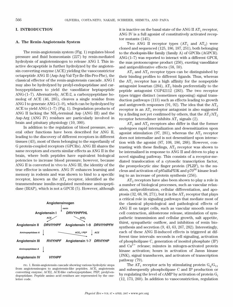
The renin-angiotensin system (Fig. 1) regulates bloodpressure and fluid homeostasis (227) by renin-mediatedhydrolysis of angiotensinogen to release ANG I. This in-active decapeptide is further hydrolyzed by the angioten-sin converting enzyme (ACE) yielding the vasoconstrictoroctapeptide ANG II (Asp-Arg-Val-Tyr-Ile-His-Pro-Phe), theclassical effector of the renin-angiotensin cascade. ANG Imay also be hydrolyzed by prolyl-endopeptidase and car-boxypeptidases to yield the vasodilator heptapeptideANG-(1–7). Alternatively, ACE-2, a carboxypeptidase ho-molog of ACE (46, 285), cleaves a single residue fromANG I to generate ANG-(1–9), which can be hydrolyzed byACE to yield ANG-(1–7) (Fig. 1). Degradation products ofANG II lacking the NH2-terminal Asp (ANG III) and theAsp-Arg (ANG IV) residues are particularly involved inbrain and pituitary physiology (10, 300).
In addition to the regulation of blood pressure, sev-eral other functions have been described for ANG II,leading to the discovery of different receptors in differenttissues (43), most of them belonging to the superfamily ofG protein-coupled receptors (GPCRs). ANG III shares thesame receptors and exerts similar effects as ANG II in thebrain, where both peptides have equivalent biologicalpotencies to increase blood pressure; however, becauseANG II is converted in vivo to ANG III, the identity of thetrue effector is unknown. ANG IV enhances learning andmemory in rodents and was shown to bind to a specificreceptor, known as the AT4 receptor, identified as thetransmembrane insulin-regulated membrane aminopepti-dase (IRAP), which is not a GPCR (5). However, although
it is inactive on the basal state of the ANG II AT1 receptor,ANG IV is a full agonist of constitutively activated recep-tor mutants (145).
Two ANG II receptor types (AT1 and AT2) werecloned and sequenced (125, 186, 187, 251), both belongingto the rhodopsin-like family (family A) of GPCRs (AGPCR).ANG-(1–7) was reported to interact with a different GPCR,the mas protooncogene product (250), exerting vasodilatorand antiproliferative effects (58, 59).
AT1 and AT2 receptor types can be distinguished bytheir binding profiles to different ligands. Thus, whereasthe AT1 receptor has a high affinity for the nonpeptideantagonist losartan (284), AT2 binds preferentially to thepeptide antagonist CGP42112 (283). The two receptortypes trigger distinct (sometimes opposing) signal trans-duction pathways (113) such as effects leading to growthand antigrowth responses (91, 92). The idea that the AT2
receptor is an AT1 receptor antagonist is also suggestedby a finding not yet confirmed by others, that the AT1/AT2
receptor heterodimer inhibits AT1 signals (2).AT1 and AT2 receptors also differ in that the former
undergoes rapid internalization and desensitization uponagonist stimulation (97, 281), whereas the AT2 receptordoes not internalize and is not desensitized upon interac-tion with the agonist (87, 108, 186, 288). However, con-trasting with these findings, AT2 receptor was shown tobe internalized in response to ANG II and driven to play anovel signaling pathway. This consists of a receptor-me-diated translocation of a cytosolic transcription factor,the promyelocytic zinc finger protein (PLZF), to the nu-cleus and activation of p85alfaPI3K and p70S6 kinase lead-ing to an increase of protein synthesis (256).
AT2 receptors have also been shown to play a role ina number of biological processes, such as vascular relax-ation, antiproliferation, cellular differentiation, and apo-ptosis (32, 68, 98, 271), but it is the AT1 receptor that playsa critical role in signaling pathways that mediate most ofthe classical physiological and pathological effects ofANG II on target cells, such as vascular smooth musclecell contraction, aldosterone release, stimulation of sym-pathetic transmission and cellular growth, salt appetite,thirst, sympathetic outflow, and inhibition of renin bio-synthesis and secretion (9, 43, 63, 267, 282). Interestingly,each of these ANG II-induced effects is triggered at dif-ferent time intervals: seconds in cell signaling, activationof phospholipase C, generation of inositol phosphate (IP)and Ca2� release; minutes in mitogen-activated proteinkinase activation; hours in activation of Janus kinase(JNK), signal transducers, and activators of transcriptionpathway (79).
The AT1 receptor acts by stimulating protein Gq/G11
and subsequently phospholipase C and IP production orby regulating the level of cAMP by activation of protein Gi
(12, 173, 260). In addition to vasoconstriction, regulation
FIG. 1. Renin-angiotensin cascade showing various hydrolytic stepsfrom angiotensinogen to angiotensin-like peptides. ACE, angiotensinconverting enzyme; ACE2, ACE-like carboxypeptidase; PEP, prolyl-en-dopeptidase. Peptide amino acid residues are represented by the one-letter code.
566 OLIVEIRA, COSTA-NETO, NAKAIE, SCHREIER, SHIMUTA, AND PAIVA
Physiol Rev • VOL 87 • APRIL 2007 • www.prv.org
of renal tubular electrolyte handling, aldosterone release,and facilitation of adrenergic release, the AT1 receptor isone of the most potent stimulators of hypertrophic re-modeling of the vascular walls, through ANG II-mediatedgrowth-promoting signals. It has been suggested that theintracellular signaling mechanisms by which the AT1 re-ceptor exerts hypertrophic and/or hyperplastic effects ontargets such as vascular smooth muscle cells are closelyassociated with receptor and nonreceptor tyrosine ki-nases (49). Furthermore, it has been demonstrated thatG12/G13 can induce vascular smooth muscle cell contrac-tion through a Rho/Rho-kinase (ROCK)-mediated path-way (71). These authors provided evidence that receptorAT1 couples with Gq and G12 chains to efficiently inducecell contraction via dual regulation of the myosin lightchain (MLC) phosphorylation: the Ca2�-dependent stim-ulation of MLC kinase and the Ca2�-independent Rho/ROCK-mediated inhibition of myosin phosphatase.
ANG II-stimulated activation of some mitogen-acti-vated protein kinases (MAPKs) has been reported (74,76), with calcium channels being essential in this mecha-nism. In fact, activation of the extracellular signal-regu-lated kinase (ERK1/ERK2), tyrosine kinase 2, and Janus-activated kinase 2 (Jak2), as well as phosphorylation ofsignal transducer and activator of transcription (STAT1
and STAT3), were shown to be inhibited by the calciumchannel blocker azelnidipine (152).
Superoxide generation has been found to be medi-ated by ANG II and to play an important role in vascularsmooth muscle cell growth, contraction/relaxation, andinflammation. It was shown that ANG II-induced oxidantactivity is increased and generation of reactive oxygenspecies is enhanced in conditions associated with vascu-lar damage such as in hypertension, ischemia-reperfusioninjury, atherosclerosis, and diabetes (30, 49).
AT1 receptors desensitize following agonist stimula-tion due to sequestration and endocytosis (281). Differ-ently from this desensitization, acute loss of responseupon repeated treatments with ANG II is also observed insmooth muscle cells, characterizing the event of tachy-phylaxis (130, 216, 270).
Homodimerization has been described for AT1 recep-tors linked to the action of factor XIIIA transglutaminase(3) or as a natural phenomenon occurring in wild-typereceptors (233). However, experiments demonstrating oli-gomerization (or at least cross-talking) in AT1 receptorsconsisted of coexpression of a wild-type and a nonfunc-tional mutant (85, 183). Heterodimerization of AT1 recep-tor with AT2 (2) and bradykinin B2 (1) receptors, andother AGPCRs such �2-adrenergic receptors (19), hasbeen claimed to occur. Interestingly, it appears that thedimerization of AT2 receptors might be regulated by di-sulfide bond exchange (179).
B. Scope of This Review
The ANG II AT1 receptor is responsible for virtuallyall of the known peripheral actions of ANG II. It has beenmost extensively studied by site-directed mutagenesisaiming at the elucidation of ligand binding, signal trans-duction, phosphorylation, binding to arrestins, internal-ization, desensitization, tachyphylaxis, and other proper-ties. The main scope of this review is to correlate the AT1
receptor basic functions (ANG II binding, receptor acti-vation, signal transduction across the transmembranestructure, and some events at the cytosolic ends of thereceptor) with the data available about AGPCR se-quences, rhodopsin structure, and mechanism of receptoraction. Presentation and discussion of related events suchas G protein coupling, arrestin binding, desensitization,and tachyphylaxis shall not be fully comprehensive butlimited to features considered necessary to enrich themain subject. Other events such as oligo(di)merization ofAGPCRs, for which no relationship with receptor activa-tion has been completely confirmed, shall not be dis-cussed.
II. ANGIOTENSIN II STRUCTURE-ACTIVITY
RELATIONSHIPS
A. Receptor Binding and Activation
Long before cloning of the receptors was achieved,numerous studies of the cardiovascular effects of syn-thetic peptide analogs allowed a detailed mapping of ANGII’s structural requirements for receptor binding and ac-tivation (130, 131, 212, 213, 215). These structural require-ments can be assumed to apply to the AT1 receptor, sincethe biological responses that were analyzed in those earlyreports were later found to be mediated by that receptortype.
Analyses of the effects of different ANG II analogsbearing single or multiple modifications indicated that anelectrostatic interaction between the ANG II COOH-ter-minal carboxylate group and the receptor is an importantfactor for high-affinity binding (100) and that the peptide’sArg2, Tyr4, and His6 side chains are also important forhigh-affinity binding (31, 178, 239, 240). On the other hand,replacements of Tyr4 and mainly Phe8 by aliphatic resi-dues were shown to generate competitive antagonists,thus attesting to the importance of these aromatic resi-dues for receptor activation (70, 247). The Val3, Ile5, andPro7 residues were found to be neither crucial for bindingnor for activation. Asp1 was not found to be relevant,since analogs with replacements in this position, as wellas des-Asp1-ANG II (ANG III), still retain significant activ-ity. On the other hand, the introduction of a sarcosine
ANG II AT1 RECEPTOR STRUCTURE AND ACTIVITY 567
Physiol Rev • VOL 87 • APRIL 2007 • www.prv.org
(Sar) residue in position 1 increased the potencies ofagonist and antagonist analogs (26, 81, 211, 238, 239).Pharmacological (158) and mutagenesis (249) studies onthe AT1 receptor indicated that [Sar1]-ANG II has a bind-ing mode different from that of the natural agonist, asdiscussed in section III, B4 and D1. Also, the presence ofSar in position 1 seems to induce a reaccommodation ofother residues involved in binding, such as Arg2 (249).
ANG II was shown to be in a random conformation inaqueous solution, various conformers coexisting in equi-librium, which would be displaced towards the “activeone” upon binding to the receptor (217). One of theseconformers could be that of a horseshoe-shaped structureof ANG II described in a complex of the peptide withantibody (67). Other studies were published in whichdifferent conformations were proposed for ANG II orsynthetic peptide analogs in aqueous or other media (57,163, 194, 236), but no rational information was obtainedabout the “active conformation” that binds to the recep-tor. Other approaches were based on constraints intro-duced in the ANG II structure so as to freeze certainconformations of the peptide, thus allowing their biolog-ical activities to be checked (122, 192, 306). However,despite the great deal of effort, no conclusive resultsleading to knowledge about the active conformation ofANG II could be obtained using these approaches.
B. Antagonism by ANG II Analogs
As expected from the structural determinants forsignaling described above, one of the first reported antag-onists for ANG II was [Phe4,Tyr8]-ANG II (164), and in thefollowing years, other antagonists were reported, mostlybearing modifications in the Phe8 position and usuallyalso with sarcosine in place of Asp1. A milestone in suchstudies was the discovery of the analog saralasin([Sar1,Ala8]-ANG II) as the first high-affinity antagonist(222).
During the first years of investigations on this sub-ject, development of antagonists with modifications at theTyr4 position was less explored, possibly due to the lowerpotency observed in the first analogs with modificationsin this position (246). Later on, sarmesin, a potent antag-onist with hydroxymethylated Tyr4 ([Sar1,Tyr(Me)4]-ANGII), was described (252). Combined modifications on bothTyr4 and Phe8 were shown to be nonadditive, generatingantagonists of lower potencies (70, 247).
C. Desensitization and Tachyphylaxis
It has been demonstrated in whole animals, isolatedtissues, and cultured cells that the AT1 receptor may beinactivated when left in prolonged contact with the ago-nist (96, 130, 184, 216, 218, 226, 270, 281). This loss of
response has been linked to two mechanisms, tachyphy-laxis and desensitization, which were first recognized bythe experimental protocols used to elicit them. Tachyphy-laxis is triggered by cycles of stimulation (agonist addi-tion and washing) that have to be repeated twice, thrice,and more times to abolish the response (216, 270). De-sensitization is promoted by prolonged contact of recep-tor with a same dose of agonist.
Differently from desensitization, tachyphylaxisseems to act at the level of agonist-receptor interactionand is dependent on the NH2-terminal ammonium groupand the Arg2 guanidinium group of ANG II. [Succinyl1]-ANG II does not induce and [Sar1]-ANG II induces anenhanced tachyphylactic phenomenon (211), with thiseffect being explained by the increased protonation of thesarcosine secondary amino group at physiological pH(214, 216). The importance of the Arg2 guanidinium groupfor tachyphylaxis triggering has also been supported bythe fact that [Lys2]-ANG II and even [Sar1,Lys2]-ANG IIcannot induce tachyphylaxis (169, 211). Also differingfrom desensitization, tachyphylaxis induction depends onthe aromatic ring at position 4 of ANG II, but someapparently conflicting aspects need to be analyzed. Thephenolic hydroxyl of Tyr4 is vital to elicit in vitro tachy-phylaxis, a property that is lost with [Phe4]-ANG II (185,262). However, paradoxically, this ANG II analog is ableto trigger tachyphylaxis in vivo (286), a condition that wasnot described for wild-type ANG II.
Thus tachyphylaxis is possibly due to conformationalchanges on the ANG II-AT1 receptor complex (126, 216,263, 264, 270), a supposition compatible with findingsshowing that this phenomenon can be prevented or re-versed by the AT1 receptor antagonist losartan (241).
Desensitization has been shown to be correlated withphosphorylation of Ser and Thr residues in the cytosolicdomains of AT1 receptor (104, 281). Therefore, rapid de-sensitization involves primarily phosphorylation (27) ofthe receptor through two distinct types of Ser/Thr proteinkinases: the second messenger-activated kinases, proteinkinase A or C (75, 127, 245, 275), and the second messen-ger-independent G protein-coupled receptor kinases(110).
The installation of both tachyphylaxis and desensiti-zation in AT1 receptors leads to decays of IP formationand calcium influx (4, 126, 264), but the most flagrantdifference between these two states is that only tachyphy-laxis is accompanied by reduction of sodium influx (4,264).
AT1 receptor sequestration and internalization havebeen involved in ANG II desensitization in smooth musclecells (6, 75, 76, 289). However, no results could be ob-tained from a series of studies supporting this correlation(23, 97, 102, 202, 280). Hence, sequestration does notappear to play a major role in desensitization but mayinstead be involved in the resensitization process (79).
568 OLIVEIRA, COSTA-NETO, NAKAIE, SCHREIER, SHIMUTA, AND PAIVA
Physiol Rev • VOL 87 • APRIL 2007 • www.prv.org
III. AT1 RECEPTOR STRUCTURE-ACTIVITY
CORRELATIONS
A. Receptor Structure
1. Sequences of AGPCRs
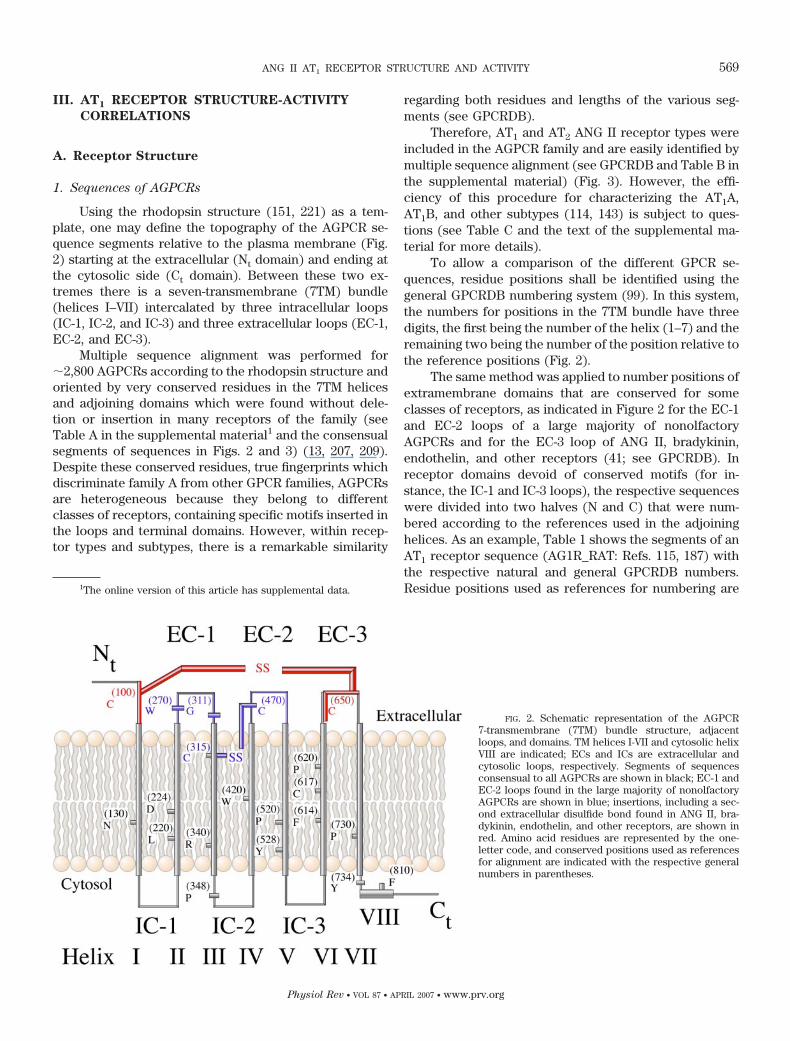
Using the rhodopsin structure (151, 221) as a tem-plate, one may define the topography of the AGPCR se-quence segments relative to the plasma membrane (Fig.2) starting at the extracellular (Nt domain) and ending atthe cytosolic side (Ct domain). Between these two ex-tremes there is a seven-transmembrane (7TM) bundle(helices I–VII) intercalated by three intracellular loops(IC-1, IC-2, and IC-3) and three extracellular loops (EC-1,EC-2, and EC-3).
Multiple sequence alignment was performed for�2,800 AGPCRs according to the rhodopsin structure andoriented by very conserved residues in the 7TM helicesand adjoining domains which were found without dele-tion or insertion in many receptors of the family (seeTable A in the supplemental material1 and the consensualsegments of sequences in Figs. 2 and 3) (13, 207, 209).Despite these conserved residues, true fingerprints whichdiscriminate family A from other GPCR families, AGPCRsare heterogeneous because they belong to differentclasses of receptors, containing specific motifs inserted inthe loops and terminal domains. However, within recep-tor types and subtypes, there is a remarkable similarity
regarding both residues and lengths of the various seg-ments (see GPCRDB).
Therefore, AT1 and AT2 ANG II receptor types wereincluded in the AGPCR family and are easily identified bymultiple sequence alignment (see GPCRDB and Table B inthe supplemental material) (Fig. 3). However, the effi-ciency of this procedure for characterizing the AT1A,AT1B, and other subtypes (114, 143) is subject to ques-tions (see Table C and the text of the supplemental ma-terial for more details).
To allow a comparison of the different GPCR se-quences, residue positions shall be identified using thegeneral GPCRDB numbering system (99). In this system,the numbers for positions in the 7TM bundle have threedigits, the first being the number of the helix (1–7) and theremaining two being the number of the position relative tothe reference positions (Fig. 2).
The same method was applied to number positions ofextramembrane domains that are conserved for someclasses of receptors, as indicated in Figure 2 for the EC-1and EC-2 loops of a large majority of nonolfactoryAGPCRs and for the EC-3 loop of ANG II, bradykinin,endothelin, and other receptors (41; see GPCRDB). Inreceptor domains devoid of conserved motifs (for in-stance, the IC-1 and IC-3 loops), the respective sequenceswere divided into two halves (N and C) that were num-bered according to the references used in the adjoininghelices. As an example, Table 1 shows the segments of anAT1 receptor sequence (AG1R_RAT: Refs. 115, 187) withthe respective natural and general GPCRDB numbers.Residue positions used as references for numbering are1The online version of this article has supplemental data.
FIG. 2. Schematic representation of the AGPCR7-transmembrane (7TM) bundle structure, adjacentloops, and domains. TM helices I-VII and cytosolic helixVIII are indicated; ECs and ICs are extracellular andcytosolic loops, respectively. Segments of sequencesconsensual to all AGPCRs are shown in black; EC-1 andEC-2 loops found in the large majority of nonolfactoryAGPCRs are shown in blue; insertions, including a sec-ond extracellular disulfide bond found in ANG II, bra-dykinin, endothelin, and other receptors, are shown inred. Amino acid residues are represented by the one-letter code, and conserved positions used as referencesfor alignment are indicated with the respective generalnumbers in parentheses.
ANG II AT1 RECEPTOR STRUCTURE AND ACTIVITY 569
Physiol Rev • VOL 87 • APRIL 2007 • www.prv.org
indicated. In the text, for every position of the AT1 recep-tor (or of other classes of AGPCRs), the numbering sys-tem is indicated as shown in Table 1: first the naturalnumber (1–3 digits) followed by superscript A (AT1 re-ceptor), superscript R (rhodopsin), and superscript G
(other AGPCRs), and then the respective generalGPCRDB numbers (3 digits) in parentheses.
2. Rhodopsin structure
The ground or inactive structure of bovine rhodopsin(151, 221) consists of a 7TM bundle surrounding an ellip-soidal central cavity whose major axis is aligned withhelices I-III and V-VII (Fig. 4). The broader side of thiscavity is flanked by helices II-III and VI-VII, and the nar-rower sides are closed by helices I and V. The cavity isaccessible from the intracellular side despite the exis-tence of interactions between the cytosolic ends of heli-ces II, III, and V-VII. The extracellular side is closed by theEC-2 loop hairpin that lies inside the central cavity paral-lel to the membrane surface, making interactions with
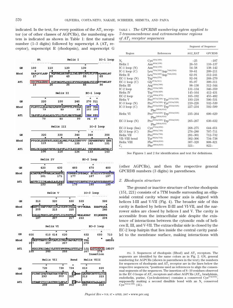
FIG. 3. Sequences of rhodopsin (Rhod) and AT1 receptors. Thesegments are identified by the same colors as in Fig. 2. GN, generalnumbering for AGPCRs (shown in parentheses in the text); the numbersfor sequences of rhodopsin and AT1 receptor are in the lines below therespective sequences; *positions used as references to align the consen-sual segments of the sequences. The insertion of 8–10 residues observedin the EC-3 loops of AT1 receptors and other AGPCRs (AT2, bradykinin,endothelin, purine, Cys-leukotriene) contains a conserved Cys274(650),supposedly making a second disulfide bond with an Nt conservedCys18AA(100) (41).
TABLE 1. The GPCRDB numbering sytem applied to
7-transmembrane and extramembrane regions
of AT1 receptor sequences
Region References
Segment of Sequence
AG2_RAT GPCRDB
Nt Cys18A(100) –25 –107Helix I Asn46A(130) 26–53 110–137IC-1 loop (N) Asn46A(130) 54–58 138–142IC-1 loop (C) Leu70A(220)Asp74A(224) 59–61 209–211Helix II Leu70A(220)Asp74A(224) 62–91 212–241EC-1 loop (N) Trp94A(270) 92–94 268–270EC-1 loop (C) Gly97A(311) 95–97 309–311Helix III Arg126A(340) 98–130 312–344IC-2 loop Pro133A(348) 131–134 346–359Helix IV Trp153A(420) 145–164 412–431EC-2 loop Cys100A(470) 165–192 455–482Helix V Pro207A(520) Tyr215A(528) 193–218 506–531IC-3 loop (N) Pro207A(520) Tyr215A(528) 219–226 532–539IC-3 loop (C) Pro255A(620) Trp253A(618)
Phe249A(614)227–234 592–599
Helix VI Pro255A(620) Trp253A(618)
Phe249A(614)235–264 600–629
EC-3 loop (N) Pro255A(620) Trp253A(618)
Phe249A(614)265–267 630–632
EC-3 loop (Ins) Cys274A(650) 268–275 644–651EC-3 loop (C) Pro299A(730) 276–280 707–711Helix VII Pro299A(730) 281–301 712–732VII–VIII bend Tyr302A(734) 302–306 734–738Helix VIII Phe309A(810) 307–320 808–821Ct Phe309A(810) 321– 822–
See Figures 1 and 2 for identification and text for definitions.
570 OLIVEIRA, COSTA-NETO, NAKAIE, SCHREIER, SHIMUTA, AND PAIVA
Physiol Rev • VOL 87 • APRIL 2007 • www.prv.org

side chains of helices. A remarkable interaction in rho-dopsin is a disulfide bridge [Cys110R(315)-Cys187R(470)] be-tween EC-2 and the top of helix III (Figs. 2 and 3), whichis found in a majority of AGPCRs. Following the end ofhelix VII, the main chain forms a 90° bend and then acytosolic helix (helix VIII) running parallel to the mem-brane. This helix, whose relative position was claimed tobe due to crystal contacts, is tied to helix VII, to theVII-VIII bend, and to helices I and II by side chain inter-actions such as that involving Tyr306R(734) and Phe313R(810)
(151) (Figs. 2 and 3).
3. Patterns of residue conservation
Patterns of residue conservation have previouslybeen determined (209) on residue positions of AGPCRaligned sequences (see supplemental material Table A).For each position of the alignment, values of entropy (E)and variability (V) were determined and distributed alongan E-V plot (Fig. 5; see legend of this figure for definitionsof E and V). Over a broad range of values, it was possibleto separate the positions of the sequence alignment ac-cording to levels of residue conservation by groupingthem into boxes 11, 12, 22, 23, and 33, according todifferent ranges of E and V values.
When the residue positions of EV boxes weremapped in the rhodopsin structure, an oriented distribu-tion was observed (Fig. 6): 1) the most conserved posi-tions of boxes 11, 12, and 22 are at the cytosolic half of the7TM bundle central cavity; 2) the positions of boxes 12,22, and 23 with intermediate E and V values are at themiddle of the 7TM bundle central cavity; 3) variable po-sitions of box 23 are in the extracellular half of the centralcavity and on the external wall of the 7TM bundle struc-ture forming an interface with the membrane bilayer; and
4) the most variable positions of box 33 are at the extra-cellular and cytosolic limits of the 7TM bundle.
Applied to well-known protein families, such as glo-bins, ras-like proteins, and serine-proteases (208), EV
analysis allowed us to relate function to the structure ofthese molecules: 1) the more conserved positions aremostly at accessible regions of the structures forming acommon main site, heme site in globins, nucleotide site inras-like chains, and catalytic site in serine-proteases; 2)the more variable positions are mostly at the surface ofthe structures forming modulator sites, specific for eachprotein type and subtype; and 3) the positions of boxes12, 22, and 23 are in the core of the proteins involved instructure-stabilizing or signal-transduction roles.
This structure-function map obtained from knownproteins may be extrapolated to AGPCRs in general, whatallowed us to assume that the conserved positions ofthese receptors (EV boxes 11 and 12 in Fig. 5) form acommon main site and the variable positions (boxes 23and 33 in Fig. 5) form modulator sites. The main site is atthe cytosolic side of the 7TM bundle central cavity andmay be related to a common cluster of conserved polarresidues consisting of Asn(130), Asp(224), Asp(339), Arg(340),Asn(729), and Tyr(734) (see Fig. 3) which includes a part ofa sodium allosteric site for some AGPCR classes (191)(see sect. IIID2). The most important modulator site (the
FIG. 5. Entropy/variability plot calculated for residue positions inmultiple sequence alignment of �2,800 AGPCRs. Variability (V) is thenumber of different residues (1–20) found at each position. Entropy (E)(0.0–3.0) is given by the equation E � �¥(i � 1–20)fi(lnfi), where fi isthe fraction of each amino acid found at each position. In the numbersdesignating boxes 11, 12, 22, 23, and 33, the first and second digitsexpress increasing ranges of V and E values, respectively. Thus 11 and33 are boxes containing highly conserved and highly variable residuepositions, respectively.
FIG. 4. Longitudinal (A) and cross-section (B) views of rhodopsin’s7TM bundle structure. The helix pairs II and III (gold) and VI and VII(green) form the broader walls of the ellipsoidal central cavity. HelicesI and V (red) form the other sides of the cavity. Helix IV is shown in lightblue, and retinal is shown in magenta.
ANG II AT1 RECEPTOR STRUCTURE AND ACTIVITY 571
Physiol Rev • VOL 87 • APRIL 2007 • www.prv.org

agonist site) is at the extracellular side of the 7TM bundlecentral cavity (29, 205) (Fig. 6) and corresponds to theretinal site in rhodopsin structure (221). Positions withintermediate E and V values in the alignment of allAGPCR sequences are in the core of the 7TM structure,between the two sites. Thus, in the course of receptoractivation, a signal may be transmitted from one site tothe other through positions with intermediate EV values(209).
Additional experimental evidence suggests the exis-tence of other modulator sites (variable positions) inother regions of the AGPCR structure, such as the COOH-terminal end of helix III [including the Tyr(339) residue ofthe DRY motif], the cytosolic ends of helices V and VI andadjacent sequences of the loop between these helices,helix VIII, and the VII-VIII bend (see references in supple-mental material Table D).
4. The AT1 receptor
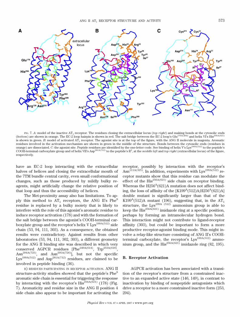
Despite the low level of residue identity (�20% inrelation to rhodopsin), AT1 receptors can be modeled(203) in homology to rhodopsin structure (151, 221), atleast at the level of sequence segments containing con-served residues (Fig. 7). Details and the rationale support-
ing this procedure are described in the supplemental ma-terial.
A model of inactive AT1 receptor was built by homol-ogy to available inactive rhodopsin structures (Fig. 7A). Amodel of the activated receptor (Fig. 7B) was obtained byan expansion of the 7TM bundle, allowing the docking ofone molecule of the peptide. For these modeling proce-dures, the alignment of the sequences in the 7TM bundleand extramembrane domains to the rhodopsin structurewas made using the scheme shown in Figure 3 (249).
A) BINDING-RELATED RESIDUES. Binding assays using ra-diolabeled ANG II or peptide analogs, and cloned AT1
receptors heterologously expressed in cultured cells, al-lowed identification of the following binding-related resi-dues (Fig. 7, B and C): 1) the ANG II COOH-terminalcarboxylate group is neutralized by the positive charge ofthe receptor’s helix V Lys199A(512) (94, 111, 183, 195, 302,303); 2) the peptide’s Tyr4 side chain binds at the recep-tor’s Asn111A(325) (55); 3) the receptor’s Ser105A(319) is re-quired for ANG II binding (78); 4) ANG II’s His6 and Phe8
side chains are in proximity to the receptor’s His256A(621)
and Phe259A(624) side chains (82, 195, 196); 5) the peptide’sArg2 guanidinium group interacts either with the recep-tor’s Asp281A(712) (53) or Asp278A(709) side chain (94) {thisdiscrepancy seems to be due to the fact that differentligands (ANG II or [Sar1,Leu8]-ANG II) were used in thetwo studies, as discussed in sect. IIID1}; 6) the NH2-termi-nal half of the EC-2 loop, including residues Val179A(469)
(94) and His183A(473) (53, 129, 303), is involved in ANG IIbinding; 7) Lys101A(316), located at the extracellular end ofhelix III, is important for binding (94, 183); 8) other resi-dues involved in agonist binding are the Nt domainArg23A(105) (249), His24A(106), Tyr26A(108), and Ile27A(109) (94)as well as the EC-1 loop Thr88A(238) (42) and Tyr92A(268)
(42, 94).Besides site-mutation studies, structures of AGPCRs
have been analyzed by two indirect procedures: the sub-stituted-cysteine accessibility method (16, 120, 176, 181)and the methionine proximity assay (36, 154). In thesemethods, residues are replaced by Cys and Met, and theirrelative positions in the three-dimensional structure ofthe receptors are defined as a function of kinetic param-eters determined with Cys-specific reactants and bymeans of photoaffinity labeling, using (in the case of theAT1 receptor) ANG II analogs with benzophenone resi-dues in position 8 (36).
The relative location (inside or outside the structure)of residues along the sequences of helices was estimatedin many AGPCRs from cysteine accessibility results (16).Also, movements of helices upon AT1 receptor activation,as denoted by change in accessibility of strategicallyplaced Cys residues, was studied by Miura and Karnik(176) and Miura et al. (181). A drawback linked to the Cysaccessibility method is that this residue is revealed bybulky reagents. Because rhodopsin structures (151, 221)
FIG. 6. Structure of rhodopsin 7TM bundle showing residue posi-tions identified according to degree of residue conservation. Within thelimits indicated for the membrane bilayer, only internal positions (point-ing to the structure central cavity) are shown. Dark blue, variableinternal positions (EV boxes 23 and 33); orange, internal positions withintermediate values of residue variation (EV boxes 12 and 22); red, veryconserved internal positions (EV box 11); green, variable extramem-brane positions; light blue, very variable extramembrane positions.
572 OLIVEIRA, COSTA-NETO, NAKAIE, SCHREIER, SHIMUTA, AND PAIVA
Physiol Rev • VOL 87 • APRIL 2007 • www.prv.org
have an EC-2 loop interacting with the extracellularhalves of helices and closing the extracellular mouth ofthe 7TM bundle central cavity, even small conformationalchanges, such as those produced by mildly bulky re-agents, might artificially change the relative position ofthat loop and thus the accessibility of helices.
The Met-proximity assay also has limitations. To ap-ply this method to AT1 receptors, the ANG II’s Phe8
residue is replaced by a bulky moiety that is likely tointerfere with the role of this agonist’s aromatic residue toinduce receptor activation (178) and with the formation ofthe salt bridge between the agonist’s COOH-terminal car-boxylate group and the receptor’s helix V Lys199A(512) sidechain (53, 94, 111, 303). As a consequence, the obtainedresults were contradictory. Against results from otherlaboratories (53, 94, 111, 302, 303), a different geometryfor the ANG II binding site was described in which veryconserved AGPCR residues [Phe249A(614), Trp253A(618),Asn294A(725), and Asn295A(726)], but not the specificLys199A(512) and Asp281A(712) residues, are claimed to beinvolved in peptide binding (36).
B) RESIDUES PARTICIPATING IN RECEPTOR ACTIVATION. ANG IIstructure-activity studies showed that the peptide’s Phe8
aromatic side chain is essential for triggering the responseby interacting with the receptor’s His256A(621) (178) (Fig.7). Aromaticity and residue size in the ANG II position 4side chain also appear to be important for activating the
receptor, possibly by interaction with the receptor’sAsn111A(325). In addition, experiments with Lys199A(512) re-ceptor mutants show that this residue can modulate theeffect of the His256A(621) side chain on receptor binding.Whereas the H256A(621)A mutation does not affect bind-ing, the loss of affinity of the [K199A(512)A;H256A(621)A]double mutant is significantly larger than that of theK199A(512)A mutant (196), suggesting that, in the AT1
structure, the Lys199A (512) ammonium group is able tokeep the His256A(621) imidazole ring at a specific position,perhaps by forming an intramolecular hydrogen bond.This interaction might not contribute to ligand-receptoraffinity (303), but could be important to form a moreproductive receptor-agonist binding mode. This might in-volve a relay-like structure consisting of ANG II’s COOH-terminal carboxylate, the receptor’s Lys199A(512) ammo-nium group, and the His256A(621) imidazole ring (82, 195).
B. Receptor Activation
AGPCR activation has been associated with a transi-tion of the receptor’s structure from a constrained inac-tive to an expanded active state (146, 149), in contrast toinactivation by binding of nonpeptide antagonists whichdrive a receptor to a more constrained inactive form (253,294).
FIG. 7. A: model of the inactive AT1 receptor. The residues closing the extracellular locus (top right) and making bonds at the cytosolic ends(bottom) are shown in orange. The EC-2 loop hairpin is shown in red. The salt bridge between the EC-2 loop’s Glu176A(464) and helix VI’s His256A(621)
is shown in green. B: model of activated AT1 receptor. The agonist site is at the top of the figure, with the ANG II molecule in magenta. Aromaticresidues involved in the activation mechanism are shown in green in the middle of the structure. Bonds between the cytosolic ends (residues inorange) are dissociated. C: the agonist site. Peptide residues are identified by the one-letter code. See binding of helix V’s Lys199A(512) to the peptide’sCOOH-terminal carboxylate group and of helix VII’s Asp281A(712) to the peptide’s R2, at the middle left and top right (extracellular locus) of the figure,respectively.
ANG II AT1 RECEPTOR STRUCTURE AND ACTIVITY 573
Physiol Rev • VOL 87 • APRIL 2007 • www.prv.org

Physiological receptor activation is triggered by ago-nist binding (297), whereas constitutive receptor activa-tion occurs in the absence of agonistic stimulus by sidechain mutations and other modifications of the AGPCRstructure (225).
1. Activation of rhodopsin
Activation of rhodopsin is seen as an expansion ofthe structure. Figure 8A shows that the more likely site ofthe 7TM bundle structure which may allow this expan-sion, without the breakage of covalent bonds, is thatbetween two blocks of structure: a (helices I–V) and b(helices VI and VII). Inside the block a helices, helix V alsomoves in relation to helix III during activation (21, 224).Functionally, ground state rhodopsin has a compressed7TM bundle structure with an 11-cis-retinal moiety linkedto helix VII’s Lys296R(723) by a Schiff bond (151, 221, 254)(Fig. 8B, 1). Light absorption causes retinal isomerizationto an all-trans-form (296), followed by slight modifica-tions of the rhodopsin structure, probably limited to theretinal pocket (150, 254). In a late stage, this effect canspread over the structure giving rise to an equilibriumbetween metarhodopsins I (MI) and II (MII), the latterbeing the fully active form (112) that triggers transducincoupling and other cytosolic events (168, 244). The acti-vation of rhodopsin can be approached by following thestructural features relative to three states.
1) The first is the inactive state, containing importantinteractions such as those involving the retinal Schiff base
and helix III’s Glu113R(318) (Fig. 8B, 1); the EC-2 loop’sGlu181R(464) and helix VI’s Tyr268R(621) (151) (Fig. 8B, 2);the helix III’s Arg135R(340) and helix VI’s Glu247R(600) (Fig.8B, 3); and the VII-VIII bend’s Tyr306R(734) and helix VIII’sPhe313R(810) (Fig. 8B, 4).
2) The second state is the MI form, in which thebridge between the Schiff base and Glu113R(318) (Fig. 8B,1) is ruptured and the counterion function exerted by thisresidue is transferred to Glu181R(464) (243) (Fig. 8C, 5).These modifications lead to the release of the Tyr268R(621)
residue and consequently of the extracellular third ofhelix VI.
3) The third state is the MII form, produced by all-trans-retinal contacts with aromatic side chains in themiddle of the 7TM bundle: helix VI’s Trp265R(618) andTyr268R(621), and helix III’s Trp126R(331) (137, 157) (Fig. 8C,6). As a consequence of change in these residues’ posi-tions, bonds between cytosolic ends of rhodopsin areruptured (Fig. 8C, 7 and 8) leading to expansion (170) andactivation of the receptor.
2. Physiological activation of AGPCRs and
AT1 receptors
The high variability of residues in the AGPCR agonistbinding sites (Fig. 6) reflects the heterogeneity of ligandspecificities. In general, the locations and dimensions ofthe sites may differ according to size of the ligands. Forsmall molecules with retinal-like dimensions, such as bio-amines, prostanoids, phospholipids, purines, and small
FIG. 8. The 7TM bundle of rhodopsin structure (151) is divided into two blocks: a (helices I–V) and b (TM helices VI and VII, and the cytosolichelix VIII), shown in blue and light blue, respectively. The retinal and the hairpin in EC-2 loop are shown in magenta and red, respectively, whereasresidues involved in specific bonds [LysR(723), GluR(318),GluR(464), TyrR(621), ArgR(340) and GluR(600)] are shown in orange. Aromatic residues placedat the middle of the 7TM bundle are shown in green. A: cross-section of the structure showing blocks a and b. B: inactive rhodopsin.Retinal-helix-VII’s Lys(723) Schiff base makes a bond with helix III’s Glu(318) (1). Number 2 indicates hydrogen bond between EC-2 loop’s Glu(464)
and helix VI’s Tyr(621). At the bottom of the figure, two bonds are shown between helix III’s Arg(340) and helix VI’s Glu(600) (3) and between thearomatic rings of the VII-VIII bend’s Tyr(734) and helix VIII’s Phe(810) (4). C: a model mimicking active rhodopsin (MII form) displaying theall-trans-retinal with the Schiff base making a bond (5) with the EC-2 loop’s hairpin Glu(464). The all-trans-retinal is in contact (6) with the clusterof aromatic residues. The bonds between cytosolic ends Arg(340)-Glu(600) (7) and Tyr(734)-Phe(810) (8), present in the inactive structure, are nowbroken.
574 OLIVEIRA, COSTA-NETO, NAKAIE, SCHREIER, SHIMUTA, AND PAIVA
Physiol Rev • VOL 87 • APRIL 2007 • www.prv.org
peptides, the binding sites overlap entirely the rhodopsinretinal pocket (29, 205). For larger ligands, e.g., peptides(43, 69, 94, 116), melanocortin (159, 210), and glycopro-tein hormones (52, 153, 293), the receptor-agonist inter-action also involves positions in the extracellular seg-ments of the receptor structure.
A) AGONIST BINDING AND ACTIVATION. ANG II binding to theAT1 receptor, leading to its activation, is likely to be atwo-step process as suggested by studies of Le et al. (145)and Feng et al. (56). The first step involves binding ofthe peptide’s COOH-terminal segment (Tyr4-Ile5-His6-Pro7-Phe8) at the retinal-like locus inside the 7TM bundlecentral cavity, oriented by a salt bridge between the pep-tide’s COOH-terminal carboxylate and the receptor’sLys199A(512) side chain (Fig. 7C, left). This binding, respon-sible for the basal agonistic activity, would lead to expan-sion of the 7TM bundle structure thus enlarging the spacebetween the EC-2 loop’s hairpin turn and helices I, II, VI,and VII (Fig. 7C, right). For the interaction of its NH2
terminus (Asp1-Arg2) with AT1 receptor (second step ofbinding), the ANG II molecule should bend at the level ofVal3-Tyr4 so that the Arg2’s guanidinium group would beat binding distance from the receptor’s Asp278A(709) andAsp281A(712) side chains (Fig. 7B, top). By completing itsinteraction at the extracellular space between the EC-1,EC-3 loops, and the Nt domain, the peptide would openthis space, favoring the separation of blocks a and binitiated at the 7TM bundle.
AGPCR activation due to agonist binding is to becompared with the mechanism triggered by retinalisomerization in rhodopsin, leading to MI formation (Fig.8). Agonist binding could exert pressure on the receptor’sretinal-like locus, pushing aside the 7TM bundle helices.The possibility of a role of aromatic residues on triggeringAGPCR activation was raised by the higher density ofthese residues in the extracellular half of the receptor’s7TM bundle, flanking the limits of the agonist site (118,209). As this site comprises variable positions (Fig. 6),activation in different AGPCRs may not be like that de-scribed above for rhodopsin, namely, breakage of thebond between EC-2 loop’s Glu181R(464) and helix VI’sTyr268R(621) (Fig. 8), being specific for receptor classes andperformed by different residues at different locations ofthat site.
In AT1 receptor activation, a role of aromatic sidechains at the receptor and at the agonist in pushing thestructure towards an expanded form is observed. Forinstance, ANG II’s Phe8 and Tyr4 (and His6) aromatic sidechains are involved in receptor activation (70, 178, 247)through interactions with helix V’s Phe204A(517), helix VI’sPhe249A(614), Trp253A(618), His256A(621) and Phe259A(624), andhelix VII’s Tyr292A(723) side chains (82, 175, 178, 195, 196)(Fig. 7B).
Interestingly, the AT1 receptor, like rhodopsin, has anEC-2 loop hairpin’s residue, Glu173A(464), which in the
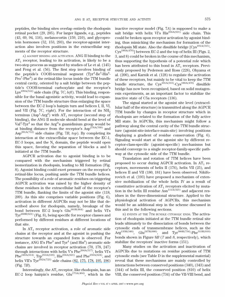
inactive receptor model (Fig. 7A) is supposed to make asalt bridge with helix VI’s His256A(621) side chain. Thiscould be broken upon receptor activation by agonist bind-ing, thus mimicking the mechanism for formation of therhodopsin MI state. Also the disulfide bridge [Cys101A(315)-Cys180A(470)] between EC-2 and the top of helix III (Figs. 2,3, and 8) could be broken in the course of this mechanism,thus supporting the hypothesis of a potential role whichhas been attributed to this bond in AT1 receptors. Previ-ously proposed by Pederson and Ross (228), Ohyama etal. (200), and Karnik et al. (128) to regulate the activationof these receptors, but mainly to be vital to keep the 7TMbundle structure, the Cys101A(315)-Cys180A(470) disulfidebridge has now been recognized, based on solid mutagen-esis experiments, as an important factor to stabilize theinactive state of C5a receptors (136).
The signal started at the agonist site level (extracel-lular half of the structure) is transmitted along the AGPCR7TM bundle by changes in receptor structure which inrhodopsin are related to the formation of the fully activeMII state. In AGPCRs, this mechanism might follow apathway along the central cavity of the 7TM bundle struc-ture (agonist-site-interface-main-site) involving positionsdisplaying a gradient of residue conservation (Fig. 6).Signaling would start at the agonist site by different re-ceptor-class-specific (agonist-specific) mechanisms butshould converge to a single receptor-family-specific path-way at the cytosolic side of the 7TM bundle.
Translation and rotation of 7TM helices have beenproposed to occur during AGPCR activation. In AT1 re-ceptors, movements of helix II (176), helix VII (25), andhelices II and VII (180, 181) have been observed. Nikifo-rovich et al. (193) have proposed a mechanism of exten-sive mobilization of the whole helix IV as a result ofconstitutive activation of AT1 receptors elicited by muta-tion in the helix III residue Asn111A(325) and adjacent res-idues in the three-dimensional structure. Applied to thephysiological activation of AGPCRs, this mechanismwould be an additional step in the scheme discussed inthis and in the following sections.
B) EVENTS AT THE 7TM BUNDLE CYTOSOLIC ENDS. The activa-tion of rhodopsin initiated at the 7TM bundle retinal siteleads ultimately to the dissociation of bonds between thecytosolic ends of transmembrane helices, such as theArg135R(340), Glu247R(600), and Tyr306R(734)-Phe313R(810)
bonds shown in Figure 8B (7 and 8, respectively), whichstabilize the receptors’ inactive forms (151).
Many studies on the activation and inactivation ofAGPCRs due to mutations on residue positions of 7TMcytosolic ends (see Table D in the supplemental material)reveal that these mechanisms are mainly controlled byinteractions between conserved positions (340), (343) and(344) of helix III, the conserved position (810) of helixVIII, the conserved position (734) of the VII-VIII bend, and
ANG II AT1 RECEPTOR STRUCTURE AND ACTIVITY 575
Physiol Rev • VOL 87 • APRIL 2007 • www.prv.org
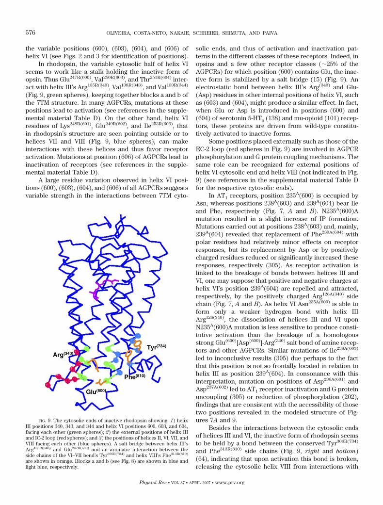
the variable positions (600), (603), (604), and (606) ofhelix VI (see Figs. 2 and 3 for identification of positions).
In rhodopsin, the variable cytosolic half of helix VIseems to work like a stalk holding the inactive form ofopsin. Thus Glu247R(600), Val250R(603), and Thr251R(604) inter-act with helix III’s Arg135R(340), Val138R(343), and Val139R(344)
(Fig. 9, green spheres), keeping together blocks a and b ofthe 7TM structure. In many AGPCRs, mutations at thesepositions lead to activation (see references in the supple-mental material Table D). On the other hand, helix VIresidues of Lys248R(601), Glu249R(602), and Ile253R(606), thatin rhodopsin’s structure are seen pointing outside or tohelices VII and VIII (Fig. 9, blue spheres), can makeinteractions with these helices and thus favor receptoractivation. Mutations at position (606) of AGPCRs lead toinactivation of receptors (see references in the supple-mental material Table D).
A large residue variation observed in helix VI posi-tions (600), (603), (604), and (606) of all AGPCRs suggestsvariable strength in the interactions between 7TM cyto-
solic ends, and thus of activation and inactivation pat-terns in the different classes of these receptors. Indeed, inopsins and a few other receptor classes (�25% of theAGPCRs) for which position (600) contains Glu, the inac-tive form is stabilized by a salt bridge (15) (Fig. 9). Anelectrostatic bond between helix III’s Arg(340) and Glu-(Asp) residues in other internal positions of helix VI, suchas (603) and (604), might produce a similar effect. In fact,when Glu or Asp is introduced in positions (600) and(604) of serotonin 5-HT6 (138) and mu-opioid (101) recep-tors, these proteins are driven from wild-type constitu-tively activated to inactive forms.
Some positions placed externally such as those of theEC-2 loop (red spheres in Fig. 9) are involved in AGPCRphosphorylation and G protein coupling mechanisms. Thesame role can be recognized for external positions ofhelix VI cytosolic end and helix VIII (not indicated in Fig.9) (see references in the supplemental material Table Dfor the respective cytosolic ends).
In AT1 receptors, position 235A(600) is occupied byAsn, whereas positions 238A(603) and 239A(604) bear Ileand Phe, respectively (Fig. 7, A and B). N235A(600)Amutation resulted in a slight increase of IP formation.Mutations carried out at positions 238A(603) and, mainly,239A(604) revealed that replacement of Phe239A(604) withpolar residues had relatively minor effects on receptorresponses, but its replacement by Asp or by positivelycharged residues reduced or significantly increased theseresponses, respectively (305). As receptor activation islinked to the breakage of bonds between helices III andVI, one may suppose that positive and negative charges athelix VI’s position 239A(604) are repelled and attracted,respectively, by the positively charged Arg126A(340) sidechain (Fig. 7, A and B). As helix VI Asn235A(600) is able toform only a weaker hydrogen bond with helix IIIArg126(340), the dissociation of helices III and VI uponN235A(600)A mutation is less sensitive to produce consti-tutive activation than the breakage of a homologousstrong Glu(600)[Asp(600)]-Arg(340) salt bond of amine recep-tors and other AGPCRs. Similar mutations of Ile238A(603)
led to inconclusive results (305) due perhaps to the factthat this position is not so frontally located in relation tohelix III as position 239A(604). In consonance with thisinterpretation, mutation on positions of Asp236A(601) andAsp237A(602) led to AT1 receptor inactivation and G proteinuncoupling (305) or reduction of phosphorylation (202),findings that are consistent with the accessibility of thosetwo positions revealed in the modeled structure of Fig-ures 7A and 9.
Besides the interactions between the cytosolic endsof helices III and VI, the inactive form of rhodopsin seemsto be held by a bond between the conserved Tyr306R(734)
and Phe313R(810) side chains (Fig. 9, right and bottom)(64), indicating that upon activation this bond is broken,releasing the cytosolic helix VIII from interactions with
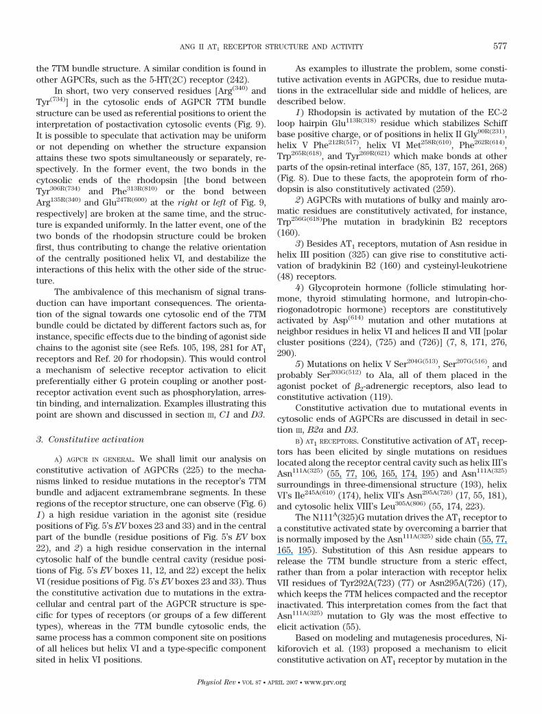
FIG. 9. The cytosolic ends of inactive rhodopsin showing: 1) helixIII positions 340, 343, and 344 and helix VI positions 600, 603, and 604,facing each other (green spheres); 2) the external positions of helix IIIand IC-2 loop (red spheres); and 3) the positions of helices II, VI, VII, andVIII facing each other (blue spheres). A salt bridge between helix III’sArg135R(340) and Glu247R(600) and an aromatic interaction between theside chains of the VI–VII bend’s Tyr306R(734) and helix VIII’s Phe313R(810)
are shown in orange. Blocks a and b (see Fig. 8) are shown in blue andlight blue, respectively.
576 OLIVEIRA, COSTA-NETO, NAKAIE, SCHREIER, SHIMUTA, AND PAIVA
Physiol Rev • VOL 87 • APRIL 2007 • www.prv.org
the 7TM bundle structure. A similar condition is found inother AGPCRs, such as the 5-HT(2C) receptor (242).
In short, two very conserved residues [Arg(340) andTyr(734)] in the cytosolic ends of AGPCR 7TM bundlestructure can be used as referential positions to orient theinterpretation of postactivation cytosolic events (Fig. 9).It is possible to speculate that activation may be uniformor not depending on whether the structure expansionattains these two spots simultaneously or separately, re-spectively. In the former event, the two bonds in thecytosolic ends of the rhodopsin [the bond betweenTyr306R(734) and Phe313R(810) or the bond betweenArg135R(340) and Glu247R(600) at the right or left of Fig. 9,respectively] are broken at the same time, and the struc-ture is expanded uniformly. In the latter event, one of thetwo bonds of the rhodopsin structure could be brokenfirst, thus contributing to change the relative orientationof the centrally positioned helix VI, and destabilize theinteractions of this helix with the other side of the struc-ture.
The ambivalence of this mechanism of signal trans-duction can have important consequences. The orienta-tion of the signal towards one cytosolic end of the 7TMbundle could be dictated by different factors such as, forinstance, specific effects due to the binding of agonist sidechains to the agonist site (see Refs. 105, 198, 281 for AT1
receptors and Ref. 20 for rhodopsin). This would controla mechanism of selective receptor activation to elicitpreferentially either G protein coupling or another post-receptor activation event such as phosphorylation, arres-tin binding, and internalization. Examples illustrating thispoint are shown and discussed in section III, C1 and D3.
3. Constitutive activation
A) AGPCR IN GENERAL. We shall limit our analysis onconstitutive activation of AGPCRs (225) to the mecha-nisms linked to residue mutations in the receptor’s 7TMbundle and adjacent extramembrane segments. In theseregions of the receptor structure, one can observe (Fig. 6)1) a high residue variation in the agonist site (residuepositions of Fig. 5’s EV boxes 23 and 33) and in the centralpart of the bundle (residue positions of Fig. 5’s EV box22), and 2) a high residue conservation in the internalcytosolic half of the bundle central cavity (residue posi-tions of Fig. 5’s EV boxes 11, 12, and 22) except the helixVI (residue positions of Fig. 5’s EV boxes 23 and 33). Thusthe constitutive activation due to mutations in the extra-cellular and central part of the AGPCR structure is spe-cific for types of receptors (or groups of a few differenttypes), whereas in the 7TM bundle cytosolic ends, thesame process has a common component site on positionsof all helices but helix VI and a type-specific componentsited in helix VI positions.
As examples to illustrate the problem, some consti-tutive activation events in AGPCRs, due to residue muta-tions in the extracellular side and middle of helices, aredescribed below.
1) Rhodopsin is activated by mutation of the EC-2loop hairpin Glu113R(318) residue which stabilizes Schiffbase positive charge, or of positions in helix II Gly90R(231),helix V Phe212R(517), helix VI Met258R(610), Phe262R(614),Trp265R(618), and Tyr269R(621) which make bonds at otherparts of the opsin-retinal interface (85, 137, 157, 261, 268)(Fig. 8). Due to these facts, the apoprotein form of rho-dopsin is also constitutively activated (259).
2) AGPCRs with mutations of bulky and mainly aro-matic residues are constitutively activated, for instance,Trp256G(618)Phe mutation in bradykinin B2 receptors(160).
3) Besides AT1 receptors, mutation of Asn residue inhelix III position (325) can give rise to constitutive acti-vation of bradykinin B2 (160) and cysteinyl-leukotriene(48) receptors.
4) Glycoprotein hormone (follicle stimulating hor-mone, thyroid stimulating hormone, and lutropin-cho-riogonadotropic hormone) receptors are constitutivelyactivated by Asp(614) mutation and other mutations atneighbor residues in helix VI and helices II and VII [polarcluster positions (224), (725) and (726)] (7, 8, 171, 276,290).
5) Mutations on helix V Ser204G(513), Ser207G(516), andprobably Ser203G(512) to Ala, all of them placed in theagonist pocket of �2-adrenergic receptors, also lead toconstitutive activation (119).
Constitutive activation due to mutational events incytosolic ends of AGPCRs are discussed in detail in sec-tion III, B2a and D3.
B) AT1 RECEPTORS. Constitutive activation of AT1 recep-tors has been elicited by single mutations on residueslocated along the receptor central cavity such as helix III’sAsn111A(325) (55, 77, 106, 165, 174, 195) and Asn111A(325)
surroundings in three-dimensional structure (193), helixVI’s Ile245A(610) (174), helix VII’s Asn295A(726) (17, 55, 181),and cytosolic helix VIII’s Leu305A(806) (55, 174, 223).
The N111A(325)G mutation drives the AT1 receptor toa constitutive activated state by overcoming a barrier thatis normally imposed by the Asn111A(325) side chain (55, 77,165, 195). Substitution of this Asn residue appears torelease the 7TM bundle structure from a steric effect,rather than from a polar interaction with receptor helixVII residues of Tyr292A(723) (77) or Asn295A(726) (17),which keeps the 7TM helices compacted and the receptorinactivated. This interpretation comes from the fact thatAsn111A(325) mutation to Gly was the most effective toelicit activation (55).
Based on modeling and mutagenesis procedures, Ni-kiforovich et al. (193) proposed a mechanism to elicitconstitutive activation on AT1 receptor by mutation in the
ANG II AT1 RECEPTOR STRUCTURE AND ACTIVITY 577
Physiol Rev • VOL 87 • APRIL 2007 • www.prv.org
position of Asn111A(325) and surroundings in both helicesIII and IV. This mechanism would consist of a chain ofconformational changes along the transmembrane helixIII from Leu112A(326) to Tyr113A(327) to Phe117A(331), whichcan be propagated to helix IV residues Ile152A(419) andMet155A(422) leading to an expressive movement of thishelix as a whole and modifications in the structure of theIC-2 loop.
A screening of a randomly mutated cDNA library ofAT1 receptors (223) allowed the identification of the fol-lowing 7TM mutations linked to constitutive activation ofthese receptors: F77A(227)Y, N111A(325)S, L112A(326)H,L112A(326)F, L118A(332)H, I193A(506)K, L195A(508)P,and L305A(806)Q. Except for positions (506), (508), and(806), the other positions are in or close to the interfacebetween blocks a and b of the structure (Fig. 8A) and thusmight contribute to the association of these receptor sub-domains on the inactivated form of receptors.
Constitutive activation of AT1 receptors has alsobeen associated with mutations on residues Leu262A(627)
and Leu265A(630) at the extracellular third of helix VI whichincreased the receptor-mediated adenylate cyclase activ-ity (40, 41). Residue mutations in the cytosolic ends ofAT1 receptors have led to constitutive activation as de-scribed, for instance, by Gaborik et al. (66) for the DRYmotif Asp125A(339) (see the supplemental material Table Dfor many other occurrences).
4. AT1 receptor inhibition
Specific mutagenesis studies (41, 53, 94) showed thatcompetitive antagonistic peptides, such as [Sar1,Leu8]-ANG II, bind AT1 receptors in the same two-step fashionobserved with the natural ligand. A minor difference isthat [Sar1]-ANG II requires both the EC-3 loop’sAsp278A(709) and Asp281A(712) side chains for binding,whereas ANG II is more dependent on binding toAsp281A(712) and to the Nt domain’s Arg23A(105). The impli-cation of this difference is further discussed with moredetails in section IIID1.
AT1 receptor inhibition by losartan is surmountablesince it can be reversed by ANG II and follows a typicalcompetitive mechanism in which only EC50 values areincreased. In contrast, inhibition by losartan’s carboxy-lated metabolite EXP3174 is insurmountable and charac-terized by increased EC50 values and decrease of maxi-mum effect (291). Other carboxylate-containing deriva-tives, such as irbesartan (34) and candesartan (182), arealso insurmountable inhibitors (274, 291). It has beenproposed that this type of inhibition is dictated by a freecarboxylate connected to the nonpeptide compound’s im-idazole ring that would interact with the receptor’sLys199A(512) (Fig. 8) leading to insurmountable inhibition(61, 188, 197, 292, 294, 295). However, the insurmountableinhibition is also regulated by the receptor’s Gln257A(622)
residue at the extracellular third of helix VI (274), whichallows for a different binding mode of inhibitors to thereceptor agonist site.
Whereas AT1 receptor residues such as Asp281A(712),at the EC-3 loop, are not required for losartan binding(129), mutagenesis screening showed that binding in-volves many residues along the 7TM bundle structure (17,78, 107, 121, 129, 183, 195, 197, 230, 231, 253). Afterbinding at positions in the extracellular halves of helicesV and VI (retinal-like locus), losartan seems to follow abinding pathway different from that of ANG II (Fig. 7).Instead of bending to fit the receptor’s extracellular locus,the antagonist molecule is extended along the 7TM bun-dle central cavity, between helices III, VI, and VII, proba-bly contacting residues in the middle of the 7TM bundleposition 111A(325) in helix III, positions 294A(725) and295A(726) in helix VII, and attaining the limits of theprotein/cytosol interface [positions 300A(731) and301A(732) in helix VII].
Interestingly, this type of nonpeptide binding modeto AT1 receptors seems to correspond to the bindingmodes to the same receptors of ANG II-like moleculescontaining bulky side chains such as benzophenone resi-dues instead of Phe at position 8 (36), or to the model ofANG II docking to the receptor obtained from moleculardynamics simulations monitored by energy-minimizationcriteria (14).
Perhaps due to its special binding mode, losartan canalso block the constitutive activation produced by muta-tions in the middle of the AT1 receptor’s helices III, VI,and VII, and thus it may be considered an inverse agonist(174) which can prevent the installation of the receptor’stachyphylactic state (241). New findings, however, haveshown that only insurmountable nonpeptide AT1 receptorinhibitors such as EXP3174 and candesartan, but notlosartan, are in fact strong inverse agonists that neutralizethe constitutive activation caused by N111A(325)G,N295A(726)S, and L305A(806)Q mutations (56).
C. Mechanisms Following Receptor Activation
1. Signal transduction
AGPCR activation propagates to the intracellularside of the receptor causing dissociation of interactionsbetween the cytosolic ends of helices I-III and V-VIII.Considering the high residue conservation in the cytosolichalf of the 7TM bundle central cavity (Fig. 6), this mech-anism is likely to happen in many AGPCRs, includingANG II receptors. Due to the expansion of the structure,some cytosolic ends of the receptor become accessiblefor coupling to cytosolic proteins (170), triggering recep-tor postactivation events such as G protein coupling,phosphorylation, arrestin binding, internalization, andbinding to other factors.
578 OLIVEIRA, COSTA-NETO, NAKAIE, SCHREIER, SHIMUTA, AND PAIVA
Physiol Rev • VOL 87 • APRIL 2007 • www.prv.org
The various states assumed by AGPCRs to elicit dif-ferent postactivation events seem to be interdependentand interconvertible (12, 54, 124, 140, 167, 229, 280, 298)and require the simultaneous participation of all receptorcytosolic ends (189, 304).
The cytosolic ends of AGPCR structure can then becoupled to G protein, phosphorylated by specific kinases,bound to arrestin or be internalized via dynamin- andclathrin-dependent coated-pit vesicles (80, 148). In AT1
receptors, the pathway of G protein coupling is indepen-dent of that of arrestins and occurs only in response toANG II aromatic (Tyr4 and Phe8) and Arg2 side chains (45,54, 97, 173). Arrestin pathway leads to autonomous cyto-solic events (257, 258). In the case of rhodopsin, it hasalso been shown that the structure of the agonist canremotely regulate the selection of cytosolic events. Infact, modifications in the structure of retinal can turn thismolecule into a partial agonist that is able to give rise toa shorter and less intense activated state of this opsin(20).
The two most known events following activation ofAGPCRs, namely, G protein coupling and arrestin binding,are discussed in section III, C2 and C3. However, onlymolecular features of these events, which can be relatedto the mechanism of receptor activation, were selectedfor this discussion. In this context, oligomerization ofAGPCRs shall not be the focus, since it is yet not clearwhether this event is systematically due to previous ago-nist binding and is related to receptor activation (28, 117,233, 277).
2. G protein activation
G protein activation mediated by activated GPCRscauses GDP release from the G� chain nucleotide site,GTP association, and consequent dissociation of G�chains from the G�� complex (37, 142, 272). Kinetic anal-yses in which rhodopsin’s mutants were used to activatewild-type transducin heterotrimers showed at least twocategories of results (50, 51, 162). First, mutations in theDRY motif of helix III and deletion in the EC-2 loopimpaired initial coupling of rhodopsin to the G���-GDPcomplex. Second, deletions in the IC-3 loop and in thecytosolic end of helix VI, replacement of the sequence inthe IC-2 loop and mutations in NH2-terminal positions ofhelix VIII, impaired GDP dissociation.
For nonopsin AGPCRs, the regions involved in Gprotein coupling are the same as those described forrhodopsin: Asp-Arg of the DRY motif of helix III, the IC-2loop middle, the IC-3 loop and beginning of helix VI, andthe cytosolic helix VIII (see the supplemental materialTable D).
An important problem about receptor-G protein cou-pling is whether to analyze it by a chemical equilibrium orby a kinetic approach. According to chemical equilibrium
criteria, the interaction between biological molecules isdriven by specific binding sites, and no G�-specific sites inthe structure of receptors have been described. Neverthe-less, G protein coupling to GPCRs occurs in practice, andtwo other approaches for this interaction should be con-sidered.
1) No direct contact exists, and G protein coupling isdriven by external environmental factors (88, 167) or byspecial adaptors providing specific connection betweenthe different receptors and G� chain types. The existenceof a long list of little characterized proteins with theability to bind G protein chains and GPCRs (24, 65, 190,235) favors this possibility.
2) G protein coupling to GPCRs can be interpreted bykinetic criteria according to mechanisms found in intrin-sically unfolded protein structures (47). Being loose ordissociated from the main folds, these structures canadapt themselves to interactions with different structuresand thus play roles such as that observed in the case ofthe activation of viral transcription factor (123). Thismechanism is supported by the fact that the process ispromiscuous (86, 88, 95, 139, 287), occurs at plasma mem-brane compartments, and can be associated with rafts(141, 265). Also, it has been linked to loosely foldedperipheral regions of the G protein and GPCR structureswhich are not integrated in the basic folds, such as thecytosolic ends of AGPCRs and the NH2-terminal andCOOH-terminal tails of G� and G� chains. All these struc-tures have to be anchored to and clustered at a sameregion of the membrane, thus forming a compartment, toelicit function (37, 142, 272). G protein coupling is transi-tory, since it requires fast molecular contacts aiming at aneasier dissociation of GDP from G� chain nucleotide sitesfollowed by GTP association. Thus the receptor is imme-diately released and may be reutilized in a new cycle, andG�� chain complexes are released for specific functions.
Some models for rhodopsin coupling to transducinproposed along the last 10 years (11, 35, 62, 89, 133–135,204, 206) are subject to questions since, by ignoring thepromiscuity of the G protein-receptor interaction, theywere proposed as if all criteria for chemical equilibriumwere fully satisfied.
3. Arrestin binding
Arrestins are adaptors able to perform biphasic bind-ing to activated AGPCRs and to clathrin (148), thus pro-viding conditions for internalization of these receptors viathe classical mechanism of coated-pit vesicles (60).
Among the four types of arrestins known, two (1 and4) are in visual rod and cone cells and regulate opsins.Arrestins 2 and 3 (or �-arrestins 1 and 2) are present inmany other cells and regulate other GPCRs (80, 148).Arrestin structures consist of two lobes with seven sand-wiched �-strands connected by interactions of internal
ANG II AT1 RECEPTOR STRUCTURE AND ACTIVITY 579
Physiol Rev • VOL 87 • APRIL 2007 • www.prv.org
polar or hydrophobic residue side chains. Interactionsinvolving the arrestin COOH- and NH2-terminal segmentsare the key elements that keep the protein structureclosed under an inactive form (72, 83, 93, 220).
Due to this structural feature, binding of arrestins toAGPCRs is biphasic (73, 219, 234, 237). The first stepfollows protein kinase-mediated phosphorylation (155) ofSer and Thr residues at the ends of the receptor’s COOH-terminal domain (172, 219). In interacting with the soincorporated phosphate groups, arrestins can have someof their internal interactions broken, thus allowing theexpansion of their structures and high-affinity binding atreceptor regions whose conformation was changed byagonist-mediated activation (44, 80, 132, 147, 219, 234,237). Under special conditions, the first step of arrestinbinding to AGPCR phosphate groups can be omitted, asfor instance when a different splicing form of this protein(p44) is expressed (219, 220, 237).
In consonance with the previously discussed mecha-nism of AGPCR activation (see sect. IIIB), this arrestinbinding mechanism can emphasize a point already dis-cussed above for G protein coupling (see sect. IIIC2).Arrestin coupling to receptors has also to be analyzed bykinetic approaches. In fact, for G proteins, only a fewarrestin types are known with the ability to couple manydifferent classes of receptors. In addition, coupling ofarrestins to receptor would have a main role of transito-rily exposing areas of these proteins for more stablebinding to clathrin and formation of coated-pit vesicles(60).
D. Overview of the AT1 Receptor
1. Activation in the 7TM bundle
Upon activation, disruption of bonds between helicesreleases the AGPCR’s 7TM bundle from constraints, lead-ing to expansion of the structure. Physiological activa-tion, due to binding of full agonists, causes widespreadreceptor expansion, whereas constitutive activation, dueto residue mutations at different sites of the 7TM bundle,leads to different degrees of expansion. This assumptionis supported by the finding that ANG IV [ANG-(3O8)],which has a negligible effect on the wild-type AT1 recep-tor, is also, as well as ANG II, a bad agonist for theD281A(712)A mutant (145), indicating that the interactionof ANG II’s Arg2 with the receptor’s Asp281A(712) plays animportant role in the general binding. Also, [Sar1,Ile4,Ile8]-ANG II, which is inactive on the wild-type receptor, elic-ited a normal response by the constitutively activeN111A(325)G mutant (56, 181), indicating that ANG II’saromatic residues Tyr4 and Phe8 induce receptor activa-tion mimicking the N111A(325) mutation’s effects (56).
The interaction between ANG II’s Arg2 and the EC-3loop’s Asp281A(712) is important since, in the absence of
any of the two partners, the agonist-receptor complex canonly be formed at higher EC50 values. This finding couldbe explained by assuming that the extracellular locus ofinactive AT1 receptor is closed by bonds between resi-dues of the EC-3 loop [Asp278A(709) and Asp281A(712)] andthe Nt domain [Arg23A(105) and His24A(106)] (Fig. 8). Therole of the Arg2-Asp281A(712) bond would be to break theseinteractions, thus driving the opening of the extracellularlocus, a mechanism that is absent when ANG IV is theligand or the D281A(712)A mutant is the receptor.
The expansion resulting from the N111A(325)G mu-tation seems not to be restricted to the 7TM bundle butcan attain the extracellular locus where it drives theseparation of the EC-3 loop from the Nt domain. Underthis condition, the activation of the N111A(325)G mutantdoes not require the Arg2-Asp281A(712) interaction betweenthe peptide and receptor. This is confirmed by the factthat ANG IV can activate a double N111A(325)G;D281A(712)A mutant of AT1 receptor (145).
Additional features of the AT1 receptor extracellularlocus, such as the specific roles of the Asp278A(709) andAsp281A(712) residues in agonist binding, can be recognizedwhen ANG II analogs with modifications in position 1 areused as ligands. Both receptor’s Asp residues are involvedin the [Sar1]-ANG II binding, but only Asp281A(712) is im-portant in the ANG II binding (53, 94). It was also reportedthat the receptor’s Arg23A(105) residue is involved in thebinding of ANG II’s NH2-terminal end (probably of theAsp1 side chain) but not when [Sar1]-ANG II is the ligand(249). To explain these data, a scheme was built in whichtwo states of the AT1 receptor extracellular locus wereconsidered (Fig. 10). In state 1, the ANG II’s Asp1 and Arg2
bind to the receptor’s Arg23A(105) and Asp281A(512), respec-tively. In state 2, the peptide’s NH2-terminal segment isshifted towards the EC-3 loop so that the NH2-terminalammonium group now binds to the receptor’s Asp278A(709)
while the Arg2 side chain remains bound to the receptor’sAsp281A(512) (structure I in Fig. 10). When the ligand is[Sar1]-ANG II, state 2 is favored by the positively chargedSar1 moving away from the receptor’s Nt domain andbinding to the EC-3 loop’s Asp278A(709) (249). As [Sar1]-ANG II is the most potent ligand to produce the tachy-phylactic state in AT1 receptors (211), it has been specu-lated that this functional event is associated with confor-mational state 2 of these receptors.
In summary, the first and second steps of ANG IIactivation (Fig. 8) may mimic the constitutive activationdue to the N111A(325)G mutation (55, 77, 106, 165, 195).The activation is propagated to the middle of the 7TMstructure and to its cytosolic half, where it could changethe arrangement of polar interactions, thus facilitating theseparations of blocks a and b (Fig. 8A). This effect wouldbe mimicked by mutations at these residues, especiallythe N295A(726)S mutation in helix VII. In a final step, theactivation reaches the cytosolic ends of the 7TM bundle,
580 OLIVEIRA, COSTA-NETO, NAKAIE, SCHREIER, SHIMUTA, AND PAIVA
Physiol Rev • VOL 87 • APRIL 2007 • www.prv.org

resulting in dissociation of these structures. The muta-tions at Tyr302A(734) and Phe309A(810), disrupting the bondbetween them (see Ref. 242 for serotonin 2c receptors),and at Leu305A(806), reported to cause constitutive activa-tion (56, 174, 223), may also be interpreted as facilitatingthe dissociation of the 7TM cytosolic ends.
Interestingly, AT2 receptors that were previously de-scribed as constitutively activated under a wild-type form(179), were shown to have binding ability independent ofANG II’s aromatic or Arg2 side chains. It appears thatthese receptors could be compared with Asn111A(325) mu-tants of the AT1 receptor. In addition, AT2 receptors havedifferent residues at the extracellular locus’s EC-3 loopand Nt domains so as to allow the hypothesis that this sitecould also be opened in agonist-free receptor forms.
2. The role of the cluster of polar residues:
the sodium site
AGPCRs have a cluster of conserved polar residues(mostly belonging to EV boxes 11 and 12 of Fig. 5) in thecytosolic half of the 7TM bundle central cavity consistingof the following residues: Asn(130), Asp(224), Ser(329),Asp(339), Asn(Ser)(725), Asn(Ser)(726), and Asn(729). Theyare involved in signal transduction of practically allAGPCRs (see mutation data in GPCRDB and tiny GRAP).
Some AGPCRs, such as opioid (232), �2-adrenergic(156), dopamine D2 (269), neurotensin (166), and adeno-sine (18) receptors, are regulated by sodium whose actionleads to decrease of receptor affinity for agonists withoutinterfering with antagonist binding (156). These effectsmay be linked to the cation binding site located in themiddle and in the cytosolic sides of the 7TM bundlecentral cavity shown in models built for dopamine D2
receptor (191), but which can be applied to otherAGPCRs, considering the conservation of the involvedresidues. These residues [Asn(130), Asp(224), Ser(329),Asn(Ser)(725), Asn(Ser)(726) and Asn(729)] are also found inthe list of AGPCR polar cluster residues, which allows usto suggest that the effects due to sodium ions and theeffects linked to AGPCR polar cluster are interrelated.
Sodium ions do not alter substantially the affinity ofagonist binding to AT1 receptors (23), but strong cation-mediated inhibitory effects on both constitutive and phys-iological activations of these receptors have been re-ported (56). As these receptors have all residues of thepolar cluster and consequently of the sodium site, it isplausible to make an association between the cation-mediated inhibitory effects on AT1 receptors and an AT1
facsimile of the site of Neve et al. (191) built for dopamineD2 receptors.
The roles of the AGPCR polar cluster residues, in-cluding those of the sodium site, should be crucial as longas their replacements in many classes of receptors canlead to so extreme conditions as signal abolition. If this isso, how do you explain the fact that some natural replace-ments of these residues in other receptors are not accom-panied by function impairment? Many examples showingthese discrepancies are found in GPCRDB and tiny GRAPmutation data, and some of them deserve to be shownhere for illustration.
1) The first experiments on the AT1 receptor helix IIshowed that D74A(224)N mutation produced drastic de-cay of IP formation (23), while D74A(224)Y andD74A(224)H mutations had little effect on phosphorylaseactivation but impaired largely the receptor internaliza-tion (102). Paradoxically, the position of Asp(224) is occu-pied by Asn in many functional AGPCRs such as opsins(rhodopsins and visual blue or violet pigments) and re-ceptors for chemokine, phospholipids, and other ligands.
2) N295A(726)S mutation in AT1 receptors usuallyleads to constitutive activation (56, 181), whereasN294A(725)A mutation at the adjacent position of thesame receptor impaired receptor activation (107). Asnand Ser are frequently replaced by each one in positions(725) and (726) of AGPCRs and are both absent in func-tionally active opsins and olfactory receptors. Ala is foundin position (725) of many opsins and in position (726) ofsome purinergic receptors.
3) S115A(329)A mutation in AT1 receptors (183) andS(329)A mutation in �1-adrenergic receptors (33) were
FIG. 10. Two states (1 and 2) of the AT1 receptor extracellular locus(see Fig. 8, top right). Upon binding ANG II, the receptor first attainsstate 1, in which the Nt’s Arg23A(105) and the EC-3 loop’s Asp281A(712)
residues contact the peptide’s D1 and R2, respectively. In a second step,the complex relaxes to state 2, in which the receptor’s Nt domain isdissociated from the peptide’s D1, which is left to contact the peptide’sNH2-terminal NH3
� group or the receptor’s EC-3 loop Asp278A(709) residue(condition I of state 2). Upon binding [Sar1]-ANG II, the receptor issupposed to go directly to state 2 (condition II) as the Sar1 group movesaway from the receptor’s Nt domain towards the EC-3 loop’s Asp278A(709)
residue.
ANG II AT1 RECEPTOR STRUCTURE AND ACTIVITY 581
Physiol Rev • VOL 87 • APRIL 2007 • www.prv.org

accompanied by IP formation decay without impairmentof agonist binding. Paradoxically, Ser(329) which is foundin many AGPCRs (amine and many peptide receptors) isreplaced in many functionally active receptors by Gly orGlu (interleukin-8 and chemokine receptors), Thr (endo-thelin receptors), Ala (opsins), Asp or Glu (olfactory re-ceptors).
A straightforward way to circumvent these conflict-ing events should go through some assumptions. Themechanism of activation of AGPCRs can basically bedefined as an expansion of the 7TM bundle structure asdescribed in section IIIB (Fig. 8). There are all along the7TM bundle structure many structural motifs that canmodulate the extent of AGPCR activation. The cluster ofpolar residues in the middle and in the cytosolic sides ofthe structure is one of these motifs. In the cluster, thepolar residues are interacting with each other giving riseto certain configurations that can favor the active or theinactive forms of AGPCRs. In this context, the modulationby sodium ions is a special condition for some AGPCRs inwhich the polar cluster acquired a configuration withappropriate residues located in a such manner that theformation of a cation site and modulation by specificbinding were allowed. In this case, sodium ions havemostly been found to inhibit agonist binding in manyAGPCRs as well as IP formation in AT1 receptors (seeabove).
It was shown in AT1 receptors that mutations inresidues of the polar cluster, including residues of thesodium site, have effects on receptor function that aremore inhibited by the cation. For instance, the effects dueto the mutation in the position of Asn111A(325) of AT1
receptors is much less cation sensitive than mutations inpositions 295A(726) and 305A(806) of the same receptors(56).
In the absence of sodium, and therefore under acti-vating conditions, the organization of the polar residuesforming the binding site and its neighborhood should beso as to facilitate the separation of blocks a and b and theexpansion of the 7TM bundle structure (Fig. 8). Thisshould be a special arrangement which, in the structure ofthe AT1 receptor, depends on the Asn295A(726) side chain.Sodium binding at this site might bring the polar residuestogether, thus inhibiting receptor activation. Mutations ofAsn295A(726) to other polar residues, such as Ser, shouldlead to other loose arrangements of the sodium site resi-dues allowing easier expansion and larger activationwhen the cation is absent.
The existence of different configurations for the clus-ter of polar residues in AGPCRs is embodied by modelsdescribing the activation of different receptor classes. Forinstance, a special arrangement of hydrogen bonds hasbeen considered in 5-HT2A receptors involving internalpolar residues of helix II Asp(224) and helix VII Asn(729).Whereas an Asp-Asn mutation in helix II eliminated cou-
pling, interchanging the residues at the two positions by asecond mutation of Asn-Asp in helix VII restored receptorfunction. These data suggest that these residues are adja-cent in space and interact with each other (255). As AT1
receptor has both Asp74A(224) and helix VII Asn298A(729)
residues, a same arrangement of polar residues may bepostulated to exist in the cytosolic side of 7TM bundlecentral cavity of these receptors. Even the mechanism ofAsn111A(325) binding to Tyr292A(723) postulated by Grob-lewski et al. (76) might be involved in this context.
In glycoprotein thyroid stimulating hormone recep-tors, the helix VI contains a conserved Asp instead of theprevalent residue of Phe in the position (614), which issupposed to make special arrangements in the AGPCRcommon cluster of polar residues (290).
3. Events at the cytosolic domains
The classic pathway of cytosolic events triggered byagonist-mediated activation of AGPCRs consists of G pro-tein coupling and effector activation, followed by phos-phorylation of the COOH-terminal Ser and Thr residues,arrestin binding, and internalization (80, 148). In AT1 re-ceptors, peptide binding leads to activation of Gq/11 pro-teins and subsequently of phospholipase C, or to activa-tion of protein Gi followed by regulation of cAMP levels(12, 173, 260).
Ohtsu et al. (199) provided evidence that Rho/ROCKactivation through PYK2 and protein kinase C-� is specif-ically required for ANG II-induced migration of vascularsmooth muscle cells (VSMC) via JNK activation. Thus, inaddition to the cell migration, activation of Rho/ROCKrepresents one of the key signal transduction pathwaysoriginating from the VSMC AT1 receptor that mediatespathophysiological functions of ANG II such as contrac-tion mediated by G12/G13 (71), atherogenic gene expres-sion mediated by Rho-kinase, MEK/ERK pathways (273),and cardiovascular hypertrophy involving Rho-kinase andNAD(P)H oxidase system (90, 301).
A pathway leading to internalization is also observed(105, 198, 266, 279, 281). However, the assumption thatthe receptor state linked to G protein coupling is a pre-requisite step for phosphorylation-arrestin-binding-inter-nalization is subject to question (97). In fact, it appearsthat postactivation cytosolic events in AGPCRs do notfollow a simple rule but are regulated by an intricatescheme, and the AT1 receptor might be used as a model toanalyze the events in AGPCR’s 7TM bundle structure inview of the large number of available experimental data.
1) The pathway leading to AT1 receptor endocytosis(38) seems to start with phosphorylation of Ct domain’sSer and Thr residues (104) and to be regulated by otherreceptor regions such as helix VIII’s Leu316(817) andTyr319(820) (278) and the cytosolic end of helix VI[Asp263(601)-Asp264(602)] (202).
582 OLIVEIRA, COSTA-NETO, NAKAIE, SCHREIER, SHIMUTA, AND PAIVA
Physiol Rev • VOL 87 • APRIL 2007 • www.prv.org
2) To allow G protein coupling, many residues on theAT1 receptor cytosolic end structure are required, such asArg126(340) in helix III (54, 66, 201, 260, 299), Ile130(344) andMet134(349) in the IC-2 loop (66, 260), Tyr215(528) in helix V(103), Leu222(535) in the IC-3 loop (109, 144, 201),Tyr292(723) in helix VII (161), and Tyr312(813), Phe313(814),and Leu314(815) in helix VIII (248).
3) The pathway leading from receptor activation to Gprotein coupling is initiated by ANG II binding, whereasanalogs devoid of aromatic rings (such as [Sar1,Ile4,Ile8]-ANG II) lead to the internalization pathway (56, 97, 105,181, 280, 298).
4) Constitutively activated N111(325)A(G), I245(610)T,and L305(806)Q receptor mutants have been found locatedin intracellular vesicles, differing from wild-type receptorsthat are in the plasma membranes (174). Furthermore, theN111(325)G mutant induced basal IP production that wasreverted by the inverse agonist EXP3174 but not by losar-tan (181).
5) When stimulated by ANG II or [Sar1,Ile4,Ile8]-ANGII, the N111(325)G mutant induces maximum IP re-sponses and is internalized without increase of phosphor-ylation (280).
The above results suggest a mechanism to select oneof the two main AT1 receptor postactivation pathways. Gprotein coupling would occur when a larger expansion ofthe AT1 bundle is caused by agonist binding, exposingmany residues in the cytosolic ends of this structure. Thiscondition is likely to occur by binding of agonists con-taining aromatic side chains, and it could involve a largeseparation of the ends of helical columns due to simulta-neous breaking of bonds linking helix III to helix VI andthe VII-VIII bend to helix VIII as shown in Figure 9.
On the other hand, a phosphorylation-arrestin-inter-nalization pathway may require only accessibility of thereceptor’s COOH-terminal domain for protein kinase ac-tion and consequent initial binding of arrestins or otherfactors. Nevertheless, this explanation contradicts find-ings that constitutively activated AT1 receptors [such asN111A(325)G mutants] can induce both pathways, G pro-tein coupling and internalization, by a mechanism that isindependent of phosphorylation (280).
4. Conclusions
Irrespective of the amino acid residues found in themiddle of the 7TM central cavity (conserved positions inFig. 6) or even in the agonist site (variable positions inFig. 6), practically all AGPCRs can be physiologicallyactivated by a general mechanism compatible with that ofAT1 receptors proposed by Feng and co-workers (54, 56):an initial two-step ANG II binding to the inactive receptorretinal-like locus and adjacent extracellular loops leadingto a large structural rearrangement consistent with theAsn111A(325) mutation effect, followed by breaking of in-
terhelix bonds leading to expansion of the 7TM bundleand separation of its cytosolic ends.
The existence of a cluster of polar residues in themiddle of the 7TM bundle, forming or not a sodium bind-ing site, could have been randomly developed duringevolution in some receptors that made early contact withions, and it should not be recognized as a rule in the basicgeneral mechanism of AGPCR signal transduction.
Two main pathways of cytosolic events have beencommonly observed. The first one involves an obligatoryG protein coupling step and is selected by the binding ofANG II aromatic and/or NH2-terminal residues. A secondpathway that may be selected by binding of ANG analogsdevoid of aromatic or NH2-terminal residues leads di-rectly to phosphorylation, arrestin binding, coated pitformation, and internalization steps without a previous Gprotein coupling event. The NH2-terminal residues of theagonists ANG II and [Sar1]-ANG II seem to bind by twodistinct modes to the AT1 receptor extracellular site (Fig.7). As the [Sar1]-ligand is the most potent molecule totrigger tachyphylaxis in AT1 receptors, it was suggestedthat its corresponding binding mode might be associatedwith this special condition of receptors.
IV. FINAL REMARKS
Among other aspects of AGPCR function, the follow-ing themes are or may be supposed to become targets forcurrent or future investigations.
A. Active Rhodopsin (AGPCR) Structures as a
Result of Probabilistic Analyses
Following the determination of the inactive rhodop-sin structure there was an expectation that the structureof activated rhodopsin and the entire functioning cycle ofthis protein would also be determined. However, high-resolution structures of activated rhodopsin could not beobtained by crystallographic procedures, perhaps be-cause this form of protein is not sufficiently stable (254).This suggests that the study of rhodopsin and otherGPCRs requires a probabilistic approach in which acti-vated receptors behave as unstable or fluctuating struc-tures. Thus activation should not enable them to stronglybind but rather to destabilize stable cytosolic proteinssuch as GDP-G��� chain complexes and inactive forms ofarrestins. This would allow the onset of other mecha-nisms such as GTP binding at an empty G� nucleotidesite, kinase-mediated phosphorylation of receptor and ar-restin binding to the receptor and clathrin, first steps ofknown series of chain reactions occurring at the cytosol.The nonexistence of specific binding sites to mediatecoupling of each GPCR to cytosolic proteins such as G�
ANG II AT1 RECEPTOR STRUCTURE AND ACTIVITY 583
Physiol Rev • VOL 87 • APRIL 2007 • www.prv.org

chains and arrestins provides corroboration for the prob-abilistic assumption.
B. The Extracellular Locus as a Site for
Cross-Talking Mechanisms
An extracellular locus in AT1 receptors (Fig. 7) waspostulated as a result of the finding that the receptor’sArg23A(105) residue contacts ANG II Asp1 (249), thus rais-ing the possibility of an agonist-mediated mechanism di-rected to the extracellular medium. The second disulfidebond linking two walls of the extracellular locus of theAT1 receptor (the distal segments of EC-3 loop and Nt
domain) also reinforces the same hypothesis, since in AT2
receptors the homologous disulfide bond was found to bea regulator of dimerization (179). Interestingly, in AT1
receptors, the same bond was suggested to be involved ina mechanism leading to stabilization of the extracellularsite structure (39).
C. The AT2 Receptor Is a Natural Constitutively
Activated Form of AGPCR
Although AT2 receptor sequences are similar to thoseof the AT1 receptor (see the supplementary material TableB), the functions of these types of ANG II receptors arequite different. The AT2 receptor seems to be constitu-tively activated to produce apoptosis (177) and, in thisrespect, may be considered as an Asn111A(325) mutant ofthe AT1 receptor (175). An inspection of the sequences ofthe AT1 and AT2 receptors shows some subtle differencesthat might be responsible for the dissimilar behavior ofthese proteins.
1) The His256A(621) residue present at the extracellu-lar third of the AT1 receptor’s helix VI, which might bebound to the EC-2 loop favoring the inactive form of thereceptor, is displaced to position (622) in the AT2 recep-tor, where it cannot play a similar role.
2) An Arg residue is found at the cytosolic COOH-terminal tail [position (600)] of the AT2 receptor’s helix VIwhich, as discussed above, would favor constitutive acti-vation.
3) Asp(709) is absent in the EC-3 loop of the AT2
receptor, and an Asp(106) is found instead of the His24A(106)
at this position of the AT1 receptor Nt domain. Thesedifferences suggest that the extracellular locus in the AT2
receptor is likely to be opened in agonist-free forms,favoring constitutive activation.
D. The Cluster of Polar Residues: The Sodium Site
The fact is that all the mechanisms related to thecluster of polar residues and sodium site are still nebu-
lous. Systematic and exhaustive research is needed. Allpolar residues of the internal polar cluster of AGPCRs(AT1 receptors included) have to be mutated and assayedat different concentrations of sodium to test the regula-tion of these ions (or of other ions) on effects producedby the mutations. The first steps in this direction werealready given by Feng et al. (56) who revealed that AT1
receptor N111A(325)G and N295A(726)S or L305A(806)Qmutations were less and more sensitive to sodium ions,respectively, a condition which may a priori be inter-preted as a function of the proximity of these residues tothe ion binding site.
V. SUMMARY
The most prevalent physiological effects of ANG II,the main product of the renin-angiotensin system, aremediated by the AT1 receptor, a rhodopsin-like AGPCR.Numerous studies of the cardiovascular effects of syn-thetic peptide analogs allowed a detailed mapping of ANGII’s structural requirements for receptor binding and ac-tivation, which were complemented by site-directed mu-tagenesis studies on the AT1 receptor to investigate therole of its structure in ligand binding, signal transduction,phosphorylation, binding to arrestins, internalization, de-sensitization, tachyphylaxis, and other properties. Theknowledge of the high-resolution structure of rhodopsinallowed homology modeling of the AT1 receptor. Themodels thus built and mutagenesis data indicate thatphysiological (agonist binding) or constitutive (mutatedreceptor) activation may involve different degrees of ex-pansion of the receptor’s central cavity. Residues in ANGII structure seem to control these conformational changesand to dictate the type of cytosolic event elicited duringthe activation: 1) agonist aromatic residues (Phe8 andTyr4) favor the coupling to G protein, and 2) absence ofthese residues can favor a mechanism leading directly toreceptor internalization via phosphorylation by specifickinases of the receptor’s COOH-terminal Ser and Thrresidues, arrestin binding, and clathrin-dependent coated-pit vesicles. On the other hand, the NH2-terminal residuesof the agonists ANG II and [Sar1]-ANG II were found tobind by two distinct modes to the AT1 receptor extracel-lular site flanked by the COOH-terminal segments of theEC-3 loop and the NH2-terminal domain. As the [Sar1]-ligand is the most potent molecule to trigger tachyphy-laxis in AT1 receptors, it was suggested that its corre-sponding binding mode might be associated with thisspecial condition of receptors.
ACKNOWLEDGMENTS
For additional information, see the following databanks: 1)GPCRDB: http://www.gpcr.org/7tm (99); 2) tinyGRAP: http://tinygrap.uit.no (22); and 3) GenBank: http://www.ncbi.nlm.nih.gov/entrez/.
584 OLIVEIRA, COSTA-NETO, NAKAIE, SCHREIER, SHIMUTA, AND PAIVA
Physiol Rev • VOL 87 • APRIL 2007 • www.prv.org
Address for reprint requests and other correspondence: L.Oliveira, Dept. of Biophysics, Federal University of Sao Paulo,Rua Botucatu, 862, 04023–062 Sao Paulo, SP, Brazil (e-mail:[email protected]).
GRANTS
The authors’ research is supported by the Sao Paulo StateResearch Foundation (FAPESP) and by the Brazilian NationalCouncil for Scientific and Technological Research (CNPq).
REFERENCES
1. AbdAlla S, Lother H, Quitterer U. AT1-receptor heterodimersshow enhanced G-protein activation and altered receptor seques-tration. Nature 407: 94–98, 2000.
2. AbdAlla S, Lother H, Abdel-Tawab AM, Quitterer U. The an-giotensin II AT2 receptor is an AT1 receptor antagonist. J Biol
Chem 276: 39721–39726, 2001.3. AbdAlla S, Lother H, Langer A, el Faramawy Y, Quitterer U.
Factor XIIIA transglutaminase crosslinks AT1 receptor dimers ofmonocytes at the onset of atherosclerosis. Cell 119: 343–354, 2004.
4. Aboulafia J, Oshiro ME, Feres T, Shimuta SI, Paiva ACM.
Angiotensin II desensitization and Ca2� and Na� fluxes in vascularsmooth muscle cells. Pflugers Arch 415: 230–234, 1989.
5. Albiston AL, McDowall SG, Matsacos D, Sim P, Clune E,
Mustafa T, Lee J, Mendelsohn FA, Simpson RJ, Connolly LM,
Chai SY. Evidence that the angiotensin IV [AT(4)] receptor is theenzyme insulin-regulated aminopeptidase. J Biol Chem 276: 48623–48626, 2001.
6. Anderson KM, Murahashi T, Dostal DE, Peach MJ. Morpho-logical and biochemical analysis of angiotensin II internalization incultured rat aortic smooth muscle cells. Am J Physiol Cell Physiol
264: C179–C188, 1993.7. Angelova K, Fanelli F, Puett D. A model for constitutive lutropin
receptor activation based on molecular simulation and engineeredmutations in transmembrane helices 6 and 7. J Biol Chem 277:32202–32213, 2002.
8. Angelova K, Narayan P, Simon JP, Puett D. Functional role oftransmembrane helix 7 in the activation of the heptahelical lutropinreceptor. Mol Endocrinol 14: 459–471, 2000.
9. Antunes-Rodrigues J, de Castro M, Elias LL, Valenca MM,
McCann SM. Neuroendocrine control of body fluid metabolism.Physiol Rev 84: 169–208, 2004.
10. Ardaillou R. Angiotensin II receptors. J Am Soc Nephrol 11: S30–S39, 1999.
11. Arimoto R, Kisselev OG, Makara GM, Marshall GR. Rhodopsin-transducin interface: studies with conformationally constrainedpeptides. Biophys J 81: 3285–3293, 2001.
12. Auger-Messier M, Clement M, Lanctot PM, Leclerc PC, Leduc
R, Escher E, Guillemette G. The constitutively active N111G-AT1receptor for angiotensin II maintains a high affinity conformationdespite being uncoupled from its cognate G protein Gq/11alpha.Endocrinology 144: 5277–5284, 2003.
13. Baldwin JM, Schertler GF, Unger VM. An alpha-carbon templatefor the transmembrane helices in the rhodopsin family of G-pro-tein-coupled receptors. J Mol Biol 272: 144–164, 1997.
14. Baleanu-Gogonea C, Karnik S. Model of the whole rat AT(1)receptor and the ligand-binding site. J Mol Mod 1–13, 2006.
15. Ballesteros JA, Jensen AD, Liapakis G, Rasmussen SG, Shi L,
Gether U, Javitch JA. Activation of the beta 2-adrenergic recep-tor involves disruption of an ionic lock between the cytoplasmicends of transmembrane segments 3 and 6. J Biol Chem 276: 29171–29177, 2001.
16. Ballesteros JA, Shi L, Javitch JA. Structural mimicry in Gprotein-coupled receptors: implications of the high-resolutionstructure of rhodopsin for structure-function analysis of rhodopsin-like receptors. Mol Pharmacol 60: 1–19, 2001.
17. Balmforth AJ, Lee AJ, Warburton P, Donnelly D, Ball SG. Theconformational change responsible for AT1 receptor activation is
dependent upon two juxtaposed asparagine residues on transmem-brane helices III and VII. J Biol Chem 272: 4245–4251, 1997.
18. Barbhaiya H, McClain R, Ijzerman A, Rivkees SA. Site-directedmutagenesis of the human A1 adenosine receptor: influences ofacidic and hydroxy residues in the first four transmembrane do-mains on ligand binding. Mol Pharmacol 50: 1635–1642, 1996.
19. Barki-Harrington L, Luttrell LM, Rockman HA. Dual inhibitionof beta-adrenergic and angiotensin II receptors by a single antag-onist: a functional role for receptor-receptor interaction in vivo.Circulation 108: 1611–1618, 2003.
20. Bartl FJ, Fritze O, Ritter E, Herrmann R, Kuksa V, Palczewski
K, Hofmann KP, Ernst OP. Partial agonism in a G protein-coupled receptor: role of the retinal ring structure in rhodopsinactivation. J Biol Chem 280: 34259–34267, 2005.
21. Beck M, Sakmar TP, Siebert F. Spectroscopic evidence forinteraction between transmembrane helices 3 and 5 in rhodopsin.Biochemistry 37: 7630–7639, 1998.
22. Beukers MW, Kristiansen K, Ijzerman AP, Edvardsen O. Ti-nyGRAP database: a bioinformatics tool to mine G protein-coupledreceptor mutant data. Trends Pharmacol Sci 20: 475–477, 1999.
23. Bihoreau C, Monnot C, Davies E, Teutsch B, Bernstein KE,
Corvol P, Clauser E. Mutation of Asp74 of the rat angiotensin IIreceptor confers changes in antagonist affinities and abolishesG-protein coupling. Proc Natl Acad Sci USA 90: 5133–5137, 1993.
24. Bockaert J, Roussignol G, Becamel C, Gavarini S, Joubert L,
Dumuis A, Fagni L, Marin P. GPCR-interacting proteins (GIPs):nature and functions. Biochem Soc Trans 32: 851–855, 2004.
25. Boucard AA, Roy M, Beaulieu ME, Lavigne P, Escher E, Guil-
lemette G, Leduc R. Constitutive activation of the angiotensin IItype 1 receptor alters the spatial proximity of transmembrane 7 tothe ligand-binding pocket. J Biol Chem 278: 36628–36636, 2003.
26. Bovy PR, O’Neal JM, Olins GM, Patton DR, McMahon EG,
Palomo M, Koepke JP, Salles KS, Trapani AJ, Smits GJ.
Structure-activity relationships for the carboxy-terminus truncatedanalogues of angiotension II, a new class of angiotensin II antago-nists. J Med Chem 33: 1477–1482, 1990.
27. Brock TA, Rittenhouse SE, Powers CW, Ekstein LS, Gim-
brone MA Jr, Alexander RW. Phorbol ester and 1-oleoyl-2-acetyl-glycerol inhibit angiotensin activation of phospholipase C in cul-tured vascular smooth muscle cells. J Biol Chem 260: 14158–14162,1985.
28. Bulenger S, Marullo S, Bouvier M. Emerging role of homo- andheterodimerization in G-protein coupled receptor biosynthesis andmaturation. Trends Pharmacol Sci 26: 131–137, 2005.
29. Bywater RP. Location and nature of the residues important forligand recognition in G-protein coupled receptors. J Mol Recognit
18: 60–72, 2005.30. Cai H, Griendling KK, Harrison DG. The vascular NAD(P)H
oxidases as therapeutic targets in cardiovascular diseases. Trends
Pharmacol Sci 24: 471–478, 2003.31. Capponi AM, Catt KJ. Angiotensin II receptors in adrenal cortex
and uterus. Binding and activation properties of angiotensin ana-logues. J Biol Chem 254: 5120–5127, 1979.
32. Carey RM. Cardiovascular and renal regulation by the angiotensintype 2 receptor. The AT2 receptor comes of age. Hypertension 45:840–844, 2005.
33. Cavalli A, Fanelli F, Taddei C, De Benedetti PG, Cotecchia S.
Amino acids of the alpha1B-adrenergic receptor involved in agonistbinding: differences in docking catecholamines to receptor sub-types. FEBS Lett 399: 9–13, 1996.
34. Cazaubon C, Gougat J, Bousquet F, Guiraudou P, Gayraud R,
Lacour C, Roccon A, Galindo G, Barthelemy G, Gautret B.
Pharmacological characterization of SR 47436, a new nonpeptideAT1 subtype angiotensin II receptor antagonist. J Pharmacol Exp
Ther 265: 826–834, 1993.35. Ciarkowski J, Witt M, Slusarz R. A hypothesis for GPCR acti-
vation. J Mol Mod 11: 407–415, 2005.36. Clement M, Martin SS, Beaulieu ME, Chamberland C,
Lavigne P, Leduc R, Guillemette G, Escher E. Determining theenvironment of the ligand binding pocket of the human angiotensinII type I (hAT1) receptor using the methionine proximity assay.J Biol Chem 280: 27121–27129, 2005.
ANG II AT1 RECEPTOR STRUCTURE AND ACTIVITY 585
Physiol Rev • VOL 87 • APRIL 2007 • www.prv.org
37. Coleman DE, Lee E, Mixon MB, Linder ME, Berghuis AM,
Gilman AG, Sprang SR. Crystallization and preliminary crystal-lographic studies of Gi alpha 1 and mutants of Gi alpha 1 in the GTPand GDP-bound states. J Mol Biol 238: 630–634, 1994.
38. Conchon S, Monnot C, Teutsch B, Corvol P, Clauser E. Inter-nalization of the rat AT1a and AT1b receptors: pharmacologicaland functional requirements. FEBS Lett 349: 365–370, 1994.
39. Correa SA, Graciela C, Pignatari GC, Ferro ES, Pacheco NAS,
Costa-Netto CM, Pesquero JB, Oliveira L, Paiva ACM,
Shimuta SI. Role of the Cys18-Cys274 disulfide bond and of thethird extracellular loop in the constitutive activation and internal-ization of angiotensin II type 1 receptor. Regul Pept 134: 132–140,2006.
40. Correa SA, Pacheco NA, Costa-Neto CM, Oliveira L, Pesquero
JB, Han SW, Paiva AC, Shimuta SI. Angiotensin II AT(1) recep-tor mutants expressed in CHO cells caused morphological changeand inhibition of cell growth. Regul Pept 131:18–22, 2005.
41. Correa SA, Zalcberg H, Han SW, Oliveira L, Costa-Neto CM,
Paiva AC, Shimuta SI. Aliphatic amino acids in helix VI of theAT(1) receptor play a relevant role in agonist binding and activity.Regul Pept 106: 33–38, 2002.
42. Costa-Neto CM, Miyakawa AA, Oliveira L, Hjorth SA,
Schwartz TW, Paiva ACM. Mutational analysis of the interactionof N- and C-terminal ends of angiotensin II with the AT1 receptor.Br J Pharmacol 130: 1263–1268, 2000.
43. De Gasparo M, Catt KJ, Inagami T, Wright JW, Unger T.
International Union of Pharmacology. XXIII. The angiotensin IIreceptors. Pharmacol Rev 52: 415–472, 2000.
44. DeGraff JL, Gurevich VV, Benovic JL. The third intracellularloop of alpha 2-adrenergic receptors determines subtype specificityof arrestin interaction. J Biol Chem 277: 43247–43252, 2002.
45. Doan TN, Ali MS, Bernstein KE. Tyrosine kinase activation bythe angiotensin II receptor in the absence of calcium signaling.J Biol Chem 276: 20954–20958, 2001.
46. Donoghue M, Hsieh F, Baronas E, Godbout K, Gosselin M,
Stagliano N, Donovan M, Woolf B, Robinson K, Jeyaseelan R,
Breitbart RE, Acton S. A novel angiotensin-converting enzyme-related carboxypeptidase (ACE 2) converts angiotensin I to angio-tensin (1–9). Circ Res 87: 1–9, 2000.
47. Dunker AK, Brown CJ, Lawson JD, Iakoucheva LM, Ob-
radovic Z. Intrinsic disorder and protein function. Biochemistry
41: 6573–6582, 2002.48. Dupre DJ, Le Gouill C, Gingras D, Rola-Pleszczynski M,
Stankova J. Inverse agonist activity of selected ligands of thecysteinyl-leukotriene receptor 1. J Pharmacol Exp Ther 309: 102–108, 2004.
49. Eguchi S, Inagami T. Signal transduction of angiotensin II type 1receptor through receptor tyrosine kinase. Regul Pept 91: 13–20,2000.
50. Ernst OP, Hofmann KP, Sakmar TP. Characterization of rhodop-sin mutants that bind transducin but fail to induce GTP nucleotideuptake. Classification of mutant pigments by fluorescence, nucle-otide release, flash-induced light-scattering assays. J Biol Chem
270: 10580–10586, 1995.51. Ernst OP, Meyer CK, Marin EP, Henklein P, Fu WY, Sakmar
TP, Hofmann KP. Mutation of the fourth cytoplasmic loop ofrhodopsin affects binding of transducin and peptides derived fromthe carboxyl-terminal sequences of transducin alpha and gammasubunits. J Biol Chem 275: 1937–1943, 2000.
52. Fan QR, Hendrickson WA. Structure of human follicle-stimulat-ing hormone in complex with its receptor. Nature 433: 269–277,2005.
53. Feng YH, Noda K, Saad Y, Liu XP, Husain A, Karnik SS. Thedocking of Arg2 of angiotensin II with Asp281 of AT1 receptor isessential for full agonism. J Biol Chem 270: 12846–12850, 1995.
54. Feng YH, Ding Y, Ren S, Zhou L, Xu C, Karnik SS. Unconven-tional homologous internalization of the angiotensin II type-1 re-ceptor induced by G-protein-independent signals. Hypertension 46:419–425, 2005.
55. Feng YH, Miura S, Husain A, Karnik SS. Mechanism of consti-tutive activation of the AT1 receptor: influence of the size of theagonist switch binding residue Asn(111). Biochemistry 37: 15791–15798, 1998.
56. Feng YH, Zhou L, Qiu R, Zeng R. Single mutations at Asn295 andLeu305 in the cytoplasmic half of transmembrane �-helix domain 7of the AT1 receptor induce promiscuous agonist specificity forangiotensin II fragments: a pseudo-constitutive activity. Mol Phar-
macol 68: 347–355, 2005.57. Fermandjian S, Fromageot P, Tistchenko AM, Leicknam JP,
Lutz M. Angiotensin II conformations. Infrared and raman studies.Eur J Biochem 28: 174–182, 1972.
58. Ferrario CM, Averill DB, Brosnihan KB, Chappell MC, Iskan-
dar SS, Dean RH, Diz DI. Vasopeptidase inhibition and Ang-(1O7) in the spontaneously hypertensive rat. Kidney Int 62: 1349–1357, 2002.
59. Ferrario CM, Chappell MC, Tallant EA, Brosnihan KB, Diz DI.
Counterregulatory actions of angiotensin-(1O7). Hypertension 30:535–541, 1997.
60. Fessart D, Simaan M, Laporte SA. c-Src regulates clathrinadapter protein 2 interaction with beta-arrestin and the angiotensinII type 1 receptor during clathrin-mediated internalization. Mol
Endocrinol 19: 491–503, 2005.61. Fierens FL, Vanderheyden PM, Gaborik Z, Minh TL, Backer
JP, Hunyady L, Ijzerman A, Vauquelin G. Lys(199) mutation ofthe human angiotensin type 1 receptor differentially affects thebinding of surmountable and insurmountable non-peptide antago-nists. J Renin Angiotensin Aldosterone Syst 1: 283–288, 2000.
62. Filipek S, Krzysko KA, Fotiadis D, Liang Y, Saperstein DA,
Engel A, Palczewski K. A concept for G protein activation by Gprotein-coupled receptor dimers: the transducin/rhodopsin inter-face. Photochem Photobiol Sci 3: 628–638, 2004.
63. Fitzsimons JT. Angiotensin, thirst, sodium appetite. Physiol Rev
78: 583–686, 1998.64. Fritze O, Filipek S, Kuksa V, Palczewski K, Hofmann KP,
Ernst OP. Role of the conserved NPxxY(x)5,6F motif in the rho-dopsin ground state and during activation. Proc Natl Acad Sci USA
100: 2290–2295, 2003.65. Fu Y, Zhong H, Nanamori M, Mortensen RM, Huang X, Lan K,
Neubig RR. RGS-insensitive G-protein mutations to study the roleof endogenous RGS proteins. Methods Enzymol 389: 229–243,2004.
66. Gaborik Z, Jagadeesh G, Zhang M, Spat A, Catt KJ, Hunyady
L. The role of a conserved region of the second intracellular loopin AT1 angiotensin receptor activation and signaling. Endocrinol-
ogy 144: 2220–2228, 2003.67. Garcia KC, Ronco PM, Verroust PJ, Brunger AT, Amzel LM.
Three-dimensional structure of an angiotensin II-Fab complex at 3A: hormone recognition by an anti-idiotypic antibody. Science 257:502–507, 1992.
68. Gendron L, Payet MD, Gallo-Payet N. The angiotensin type 2receptor of angiotensin II and neuronal differentiation: from obser-vations to mechanisms. J Mol Endocrinol 31: 359–372, 2003.
69. Gether U. Uncovering molecular mechanisms involved in activa-tion of G protein-coupled receptors. Endocr Rev 21: 90–113, 2000.
70. Goghari MH, Franklin KJ, Moore GJ. Structure-activity relation-ships for the competitive angiotensin antagonist [sarcosine,O-methyltyrosine4]angiotensin II (sarmesin). J Med Chem 29: 1121–1124, 1986.
71. Gohla A, Schultz G, Offermanns S. Role for G12/G13 in agonist-induced vascular smooth muscle cell contraction. Circ Res 87:221–227, 2000.
72. Granzin J, Wilden U, Choe HW, Labahn J, Krafft B, Buldt G.
X-ray crystal structure of arrestin from bovine rod outer segments.Nature 391: 918–921, 1998.
73. Gray JA, Compton-Toth BA, Roth BL. Identification of twoserine residues essential for agonist-induced 5-HT2A receptor de-sensitization. Biochemistry 42: 10853–10862, 2003.
74. Griendling KK, Lassegue B, Murphy TJ, Alexander RW. An-giotensin II receptor pharmacology. Adv Pharmacol 28: 269–306,1994.
75. Griendling KK, Berk BC, Socorro L, Tsuda T, Delafontaine P,
Alexander RW. Secondary signalling mechanisms in angiotensinII-stimulated vascular smooth muscle cells. Clin Exp Pharmacol
Physiol 15: 102–112, 1988.
586 OLIVEIRA, COSTA-NETO, NAKAIE, SCHREIER, SHIMUTA, AND PAIVA
Physiol Rev • VOL 87 • APRIL 2007 • www.prv.org
76. Griendling KK, Ushio-Fukai M, Lasseg B, Alexander RW. An-giotensin II signaling vascular smoth muscle. New concepts. Hy-
pertension 29: 366–373, 1997.77. Groblewski T, Maigret B, Larguier R, Lombard C, Bonnafous
JC, Marie J. Mutation of Asn111 in the third transmembranedomain of the AT1A angiotensin II receptor induces its constitutiveactivation. J Biol Chem 272: 1822–1826, 1997.
78. Groblewski T, Maigret B, Nouet S, Larguier R, Lombard C,
Bonnafous JC, Marie J. Amino acids of the third transmembranedomain of the AT1A angiotensin II receptor are involved in thedifferential recognition of peptide and nonpeptide ligands. Bio-
chem Biophys Res Commun 209: 153–160, 1995.79. Guo DF, Sun YL, Hamet P, Inagami T. The angiotensin II type 1
receptor and receptor-associated proteins. Cell Res 11: 165–180,2001.
80. Gurevich VV, Gurevich EV. The molecular acrobatics of arrestinactivation. Trends Pharmacol Sci 25: 105–111, 2004.
81. Hall MM, Khosla MC, Khairallah PA, Bumpus FM. Angiotensinanalogs: the influence of sarcosine substituted in position 1. J Phar-
macol Exp Ther 188: 222–228, 1974.82. Han HM, Shimuta SI, Kanashiro CA, Oliveira L, Han SW,
Paiva AC. Residues Val254, His256, Phe259 of the angiotensin IIAT1 receptor are not involved in ligand binding but participate insignal transduction. Mol Endocrinol 12: 810–814, 1998.
83. Han M, Gurevich VV, Vishnivetskiy SA, Sigler PB, Schubert C.
Crystal structure of beta-arrestin at 1.9 A: possible mechanism ofreceptor binding and membrane translocation. Structure 9: 869–880, 2001.
84. Han M, Smith SO, Sakmar TP. Constitutive activation of opsin bymutation of methionine 257 on transmembrane helix 6. Biochem-
istry 37: 8253–8261, 1998.85. Hansen JL, Theilade J, Haunso S, Sheikh SP. Oligomerization
of wild type and nonfunctional mutant angiotensin II type I recep-tors inhibits galphaq protein signaling but not ERK activation.J Biol Chem 279: 24108–24115, 2004.
86. Hazari A, Lowes V, Chan JH, Wong CS, Ho MK, Wong YH.
Replacement of the alpha5 helix of Galpha16 with Galphas-specificsequences enhances promiscuity of Galpha16 toward Gs-coupledreceptors. Cell Signal 16: 51–62, 2004.
87. Hein L, Meinel L, Pratt RE, Dzau VJ, Kobilka BK. Intracellulartrafficking of angiotensin II and its AT1 and AT2 receptors: evi-dence for selective sorting of receptor and ligand. Mol Endocrinol
11: 1266–1277, 1997.88. Hermans E. Biochemical and pharmacological control of the mul-
tiplicity of coupling at G-protein-coupled receptors. Pharmacol
Ther 99: 25–44, 2003.89. Herrmann R, Heck M, Henklein P, Henklein P, Kleuss C,
Hofmann KP, Ernst OP. Sequence of interactions in receptor-Gprotein coupling. J Biol Chem 279: 24283–24290, 2004.
90. Higashi M, Shimokawa H, Hattori T, Hiroki J, Mukai Y,
Morikawa K, Ichiki T, Takahashi S, Takeshita A. Long-terminhibition of Rho-kinase suppresses angiotensin II-induced cardio-vascular hypertrophy in rats in vivo: effect on endothelial NAD(P)Hoxidase system. Circ Res 93: 767–775, 2003.
91. Higuchi W, Fuse I, Hattori A, Aizawa Y. Mutations of the plate-let thromboxane A2 (TXA2) receptor in patients characterized bythe absence of TXA2-induced platelet aggregation despite normalTXA2 binding activity. Thromb Haemostasis 82: 1528–1531, 1999.
92. Hiroi Y, Hiroi J, Kudoh S, Yazaki Y, Nagai R, Komuro I. Twodistinct mechanisms of angiotensin II-induced negative regulationof the mitogen-activated protein kinases in cultured cardiac myo-cytes. Hypertens Res 24: 385–394, 2001.
93. Hirsch JA, Schubert C, Gurevich VV, Sigler PB. The 2.8 Acrystal structure of visual arrestin: a model for arrestin’s regulation.Cell 97: 257–269, 1999.
94. Hjorth SA, Schambye HT, Greenlee WJ, Schwartz TW. Identi-fication of peptide binding residues in the extracellular domains ofthe AT1 receptor. J Biol Chem 269: 30953–30959, 1994.
95. Ho MK, Chan JH, Wong CS, Wong YH. Identification of a stretchof six divergent amino acids on the alpha5 helix of Galpha16 as amajor determinant of the promiscuity and efficiency of receptorcoupling. Biochem J 380: 361–369, 2004.
96. Hollenberg NK, Barbero LR, Hinrichs KP. Angiotensin tachy-phylaxis and vascular angiotensinase activity. Experientia 27:1032–1034, 1971.
97. Holloway AC, Qian H, Pipolo L, Ziogas J, Miura S, Karnik S,
Southwell BR, Lew MJ, Thomas WG. Side-chain substitutionswithin angiotensin II reveal different requirements for signaling,internalization, phosphorylation of type 1A angiotensin receptors.Mol Pharmacol 61: 768–777, 2002.
98. Horiuchi M. Functional aspects of angiotensin type 2 receptor.Adv Exp Med Biol 396: 217–224, 1996.
99. Horn F, Bettler E, Oliveira L, Campagne F, Cohen FE, Vriend
G. GPCRDB information system for G protein-coupled receptors.Nucleic Acids Res 31: 294–297, 2003.
100. Hsieh KH, Marshall GR. Role of the C-terminal carboxylate inangiotensin II activity: alcohol, ketone, ester analogues of angio-tensin II. J Med Chem 29: 1968–1971, 1986.
101. Huang P, Visiers I, Weinstein H, Liu-Chen LY. The local envi-ronment at the cytoplasmic end of TM6 of the mu opioid receptordiffers from those of rhodopsin and monoamine receptors: intro-duction of an ionic lock between the cytoplasmic ends of helices 3and 6 by a L6 30(275)E mutation inactivates the mu opioid receptorand reduces the constitutive activity of its T6 34(279)K mutant.Biochemistry 41: 11972–11980, 2002.
102. Hunyady L, Baukal AJ, Balla T, Catt KJ. Independence of typeI angiotensin II receptor endocytosis from G protein coupling andsignal transduction. J Biol Chem 269: 24798–247804, 1994.
103. Hunyady L, Bor M, Balla T, Catt KJ. Critical role of a conservedintramembrane tyrosine residue in angiotensin II receptor activa-tion. J Biol Chem 270: 9702–9705, 1995.
104. Hunyady L, Bor M, Balla T, Catt KJ. Identification of a cyto-plasmic Ser-Thr-Leu motif that determines agonist-induced inter-nalization of the AT1 angiotensin receptor. J Biol Chem 269:31378–31382, 1994.
105. Hunyady L, Catt KJ, Clark AJ, Gaborik Z. Mechanisms andfunctions of AT(1) angiotensin receptor internalization. Regul Pept
91: 29–44, 2000.106. Hunyady L, Vauquelin G, Vanderheyden P. Agonist induction
and conformational selection during activation of a G-protein-cou-pled receptor. Trends Pharmacol Sci 24: 81–86, 2003.
107. Hunyady L, Ji H, Jagadeesh G, Zhang M, Gaborik Z, Mihalik B,
Catt KJ. Dependence of AT1 angiotensin receptor function onadjacent asparagine residues in the seventh transmembrane helix.Mol Pharmacol 54: 427–434, 1998.
108. Hunyady L. Molecular mechanism of angiotensin II receptor in-ternalization. J Am Soc Nephrol 11: S47–S56, 1999.
109. Hunyady L, Zhang M, Jagadeesh G, Bor M, Balla T, Catt KJ.
Dependence of agonist activation on a conserved apolar residue inthe third intracellular loop of the AT1 angiotensin receptor. Proc
Natl Acad Sci USA 93: 10040–10045, 1996.110. Inglese J, Freedman NJ, Koch WJ, Lefkowitz RJ. Structure and
mechanism of the G protein-coupled receptor kinases. J Biol Chem
268: 23735–23738, 1993.111. Inoue Y, Nakamura N, Inagami T. A review of mutagenesis
studies of angiotensin II type 1 receptor, the three-dimensionalreceptor model in search of the agonist and antagonist binding siteand the hypothesis of a receptor activation mechanism. J Hyper-
tens 15: 703–714, 1997.112. Ishiguro M, Oyama Y, Hirano T. Structural models of the pho-
tointermediates in the rhodopsin photocascade, lumirhodopsin,metarhodopsin I, metarhodopsin II. Chembiochem 5: 298–310,2004.
113. Ishii K, Takekoshi K, Shibuya S, Kawakami Y, Isobe K, Nakai
T. Angiotensin subtype-2 receptor (AT2) negatively regulates sub-type-1 receptor (AT1) in signal transduction pathways in culturedporcine adrenal medullary chromaffin cells. J Hypertens 19: 1991–1999, 2001.
114. Iwai N, Inagami T. Identification of two subtypes in the rat type Iangiotensin II receptor. FEBS Lett 298: 257–260, 1992.
115. Iwai N, Yamano Y, Chaki S, Konishi F, Bardhan S, Tibbetts C,
Sasaki K, Hasegawa M, Matsuda Y, Inagami T. Rat angiotensinII receptor: cDNA sequence and regulation of the gene expression.Biochem Biophys Res Commun 177: 299–304, 1991.
ANG II AT1 RECEPTOR STRUCTURE AND ACTIVITY 587
Physiol Rev • VOL 87 • APRIL 2007 • www.prv.org
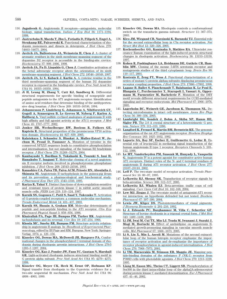
116. Jagadeesh G. Angiotensin II receptors—antagonists, molecularbiology, signal transduction. Indian J Exp Biol 36: 1171–1194,1998.
117. Jastrzebska B, Maeda T, Zhu L, Fotiadis D, Filipek S, Engel A,
Stenkamp RE, Palczewski K. Functional characterization of rho-dopsin monomers and dimers in detergents. J Biol Chem 279:54663–54675, 2004.
118. Javitch JA, Ballesteros JA, Weinstein H, Chen J. A cluster ofaromatic residues in the sixth membrane-spanning segment of thedopamine D2 receptor is accessible in the binding-site crevice.Biochemistry 37: 998–1006, 1998.
119. Javitch JA, Fu D, Liapakis G, Chen J. Constitutive activation ofthe beta2 adrenergic receptor alters the orientation of its sixthmembrane-spanning segment. J Biol Chem 272: 18546–18549, 1997.
120. Javitch JA, Li X, Kaback J, Karlin A. A cysteine residue in thethird membrane-spanning segment of the human D2 dopaminereceptor is exposed in the binding-site crevice. Proc Natl Acad Sci
USA 91: 10355–10359, 1994.121. Ji H, Leung M, Zhang Y, Catt KJ, Sandberg K. Differential
structural requirements for specific binding of nonpeptide andpeptide antagonists to the AT1 angiotensin receptor. Identificationof amino acid residues that determine binding of the antihyperten-sive drug losartan. J Biol Chem 269: 16533–16536, 1994.
122. Johannesson P, Lindeberg G, Johansson A, Nikiforovich GV,
Gogoll A, Synnergren B, Le Greves M, Nyberg F, Karlen A,
Hallberg A. Vinyl sulfide cyclized analogues of angiotensin II withhigh affinity and full agonist activity at the AT(1) receptor. J Med
Chem 45: 1767–1777, 2002.123. Jonker HR, Wechselberger RW, Boelens R, Folkers GE,
Kaptein R. Structural properties of the promiscuous VP16 activa-tion domain. Biochemistry 44: 827–839, 2005.
124. Kalatskaya I, Schussler S, Blaukat A, Muller-Esterl W, Jo-
chum M, Proud D, Faussner A. Mutation of tyrosine in theconserved NPXXY sequence leads to constitutive phosphorylationand internalization, but not signaling, of the human B2 bradykininreceptor. J Biol Chem 279: 31268–31276, 2004.
125. Kambayashi Y, Bardhan S, Takahashi K, Tsuzuki S, Inui H,
Hamakubo T, Inagami T. Molecular cloning of a novel angioten-sin II receptor isoform involved in phosphotyrosine phosphataseinhibition. J Biol Chem 268: 24543–24546, 1993.
126. Kanashiro CA, Paiva TB, Paiva ACM, Prioste RN, Aboulafia J,
Shimuta SI. Angiotensin II tachyphylaxis in the guinea-pig ileumand its prevention: a pharmacological and biochemical study.J Pharmacol Exp Ther 275: 1543–1550, 1995.
127. Kariya K, Takai Y. Distinct functions of down-regulation-sensitiveand -resistant types of protein kinase C in rabbit aortic smoothmuscle cells. FEBS Lett 219: 119–124, 1987.
128. Karnik SS, Gogonea C, Patil S, Saad Y, Takezako T. Activationof G-protein-coupled receptors: a common molecular mechanism.Trends Endocrinol Metab 14: 431–437, 2003.
129. Karnik SS, Husain A, Graham RM. Molecular determinants ofpeptide and non-peptide binding to the AT1 receptor. Clin Exp
Pharmacol Physiol Suppl 3: S58–S56, 1996.130. Khairallah PA, Page IH, Bumpus FM, Turker RK. Angiotensin
tachyphylaxis and its reversal. Circ Res 19: 247–254, 1966.131. Khosla MC, Smeby RR, Bumpus FM. Structure activity relation-
ship in angiotensin II analogs. In: Handbook of Experimental Phar-
macology, edited by IH Page and RM. Bumpus. New York: Springer-Verlag, 1974, p. 126–156.
132. Kisselev OG, Downs MA, McDowell JH, Hargrave PA. Confor-mational changes in the phosphorylated C-terminal domain of rho-dopsin during rhodopsin arrestin interactions. J Biol Chem 279:51203–5 1207, 2004.
133. Kisselev OG, Kao J, Ponder JW, Fann YC, Gautam N, Marshall
GR. Light-activated rhodopsin induces structural binding motif inG protein alpha subunit. Proc Natl Acad Sci USA 95: 4270–4275,1998.
134. Kisselev OG, Meyer CK, Heck M, Ernst OP, Hofmann KP.
Signal transfer from rhodopsin to the G-protein: evidence for atwo-site sequential fit mechanism. Proc Natl Acad Sci USA 96:4898–4903, 1999.
135. Kisselev OG, Downs MA. Rhodopsin controls a conformationalswitch on the transducin gamma subunit. Structure 11: 367–373,2003.
136. Klco JM, Wiegand CB, Narzinski K, Baranski TJ. Essential rolefor the second extracellular loop in C5a receptor activation. Nat
Struct Mol Biol 12: 320–326, 2005.137. Kochendoerfer GG, Kaminaka S, Mathies RA. Ultraviolet res-
onance Raman examination of the light-induced protein structuralchanges in rhodopsin activation. Biochemistry 36: 13153–13159,1997.
138. Kohen R, Fashingbauer LA, Heidmann DE, Guthrie CR, Ham-
blin MW. Cloning of the mouse 5-HT6 serotonin receptor andmutagenesis studies of the third cytoplasmic loop. Brain Res 90:110–117, 2001.
139. Kostenis E, Zeng FY, Wess J. Functional characterization of aseries of mutant G protein alphaq subunits displaying promiscuousreceptor coupling properties. J Biol Chem 273: 17886–17892, 1998.
140. Lagane B, Ballet S, Planchenault T, Balabanian K, Le Poul E,
Blanpain C, Percherancier Y, Staropoli I, Vassart G, Opper-
mann M, Parmentier M, Bachelerie F. Mutation of the DRYmotif reveals different structural requirements for CCR5-mediatedsignaling and receptor endocytosis. Mol Pharmacol 67: 1966–1976,2005.
141. Lagerholm BC, Weinreb GE, Jacobson K, Thompson NL. De-tecting microdomains in intact cell membranes. Annu Rev Phys
Chem 56: 309–336, 2005.142. Lambright DG, Sondek J, Bohm A, Skiba NP, Hamm HE,
Sigler PB. The 2.0 A crystal structure of a heterotrimeric G pro-tein. Nature 379: 311–319, 1996.
143. Langford K, Frenzel K, Martin BM, Bernstein KE. The genomicorganization of the rat AT1 angiotensin receptor. Biochem Biophys
Res Commun 183: 1025–1032, 1992.144. Laporte SA, Roy SF, Escher E, Guillemette G, Leduc R. Es-
sential role of leucine222 in mediating signal transduction of thehuman angiotensin II type 1 receptor. Receptors Channels 5: 103–112, 1998.
145. Le MT, Vanderheyden PM, Szaszak M, Hunyady L, Vauquelin
G. Angiotensin IV is a potent agonist for constitutive active humanAT1 receptors. Distinct roles of the N- and C-terminal residues ofangiotensin II during AT1 receptor activation. J Biol Chem 277:23107–23110, 2002.
146. Leff P. The two-state model of receptor activation. Trends Phar-
macol Sci 16: 89–97, 1995.147. Lefkowitz RJ, Shenoy SK. Transduction of receptor signals by
beta-arrestins. Science 308: 512–517, 2005.148. Lefkowitz RJ, Whalen EJ. Beta-arrestins: traffic cops of cell
signaling. Curr Opin Cell Biol 16: 162–168, 2004.149. Lew MJ, Ziogas J. The two-state model of antagonist-AT1 recep-
tor interaction: an hypothesis defended but not tested. Biochem
Pharmacol 67: 397–399, 2004.150. Lewis JW, Kliger DS. Photointermediates of visual pigments.
J Bioenerg Biomembr 4: 201–210, 1992.151. Li J, Edwards PC, Burghammer M, Villa C, Schertler GF.
Structure of bovine rhodopsin in a trigonal crystal form. J Mol Biol
343: 1409–1438, 2004.152. Li JM, Iwai M, Cui TX, Min LJ, Tsuda M, Iwanami J, Suzuki J,
Mogi M, Horiuchi M. Effect of azelnidipine on angiotensin II-mediated growth-promoting signaling in vascular smooth musclecells. Mol Pharmacol 67: 1666–1673, 2005.
153. Li S, Liu X, Min L, Ascoli M. Mutations of the second extracel-lular loop of the human lutropin receptor emphasize the impor-tance of receptor activation and de-emphasize the importance ofreceptor phosphorylation in agonist-induced internalization. J Biol
Chem 276: 7968–7973, 2001.154. Li YM, Marnerakis M, Stimson ER, Maggio JE. Mapping pep-
tide-binding domains of the substance P (NK-1) receptor fromP388D cells with photolabile agonists. J Biol Chem 270: 1213–1220,1995.
155. Liang M, Eason MG, Theiss CT, Liggett SB. Phosphorylation ofSer360 in the third intracellular loop of the alpha2A-adrenoceptorduring protein kinase C-mediated desensitization. Eur J Pharmacol
437: 41–46, 2002.
588 OLIVEIRA, COSTA-NETO, NAKAIE, SCHREIER, SHIMUTA, AND PAIVA
Physiol Rev • VOL 87 • APRIL 2007 • www.prv.org
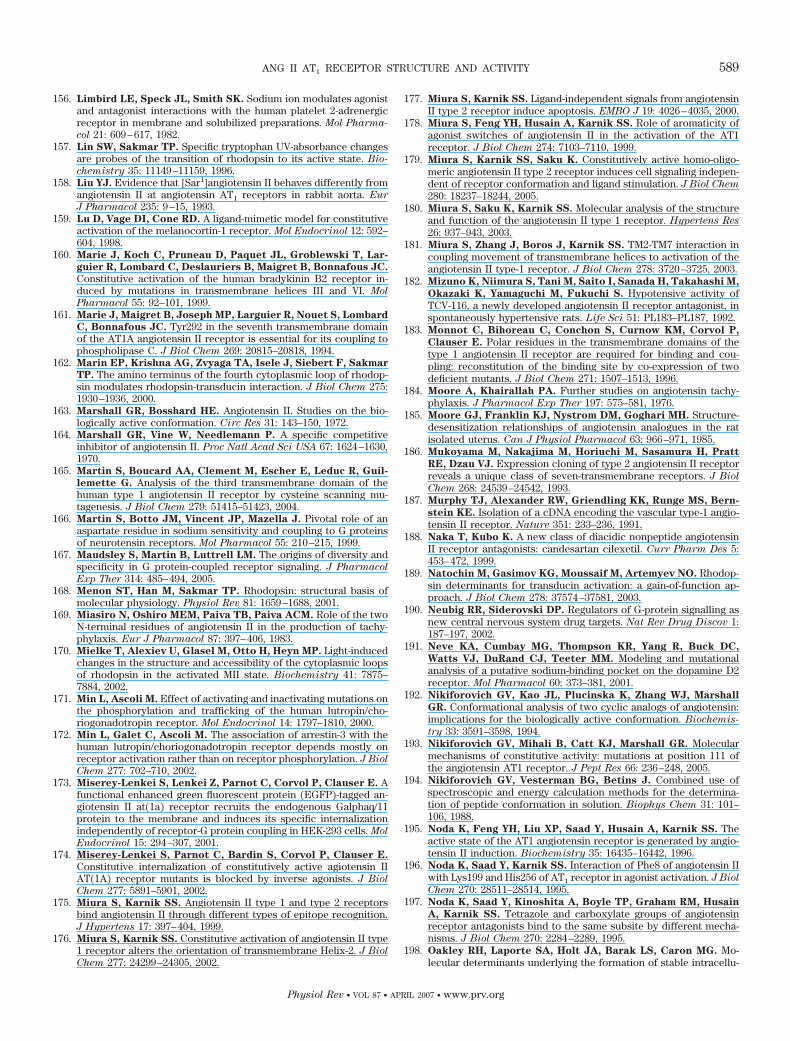
156. Limbird LE, Speck JL, Smith SK. Sodium ion modulates agonistand antagonist interactions with the human platelet 2-adrenergicreceptor in membrane and solubilized preparations. Mol Pharma-
col 21: 609–617, 1982.157. Lin SW, Sakmar TP. Specific tryptophan UV-absorbance changes
are probes of the transition of rhodopsin to its active state. Bio-
chemistry 35: 11149–11159, 1996.158. Liu YJ. Evidence that [Sar1]angiotensin II behaves differently from
angiotensin II at angiotensin AT1 receptors in rabbit aorta. Eur
J Pharmacol 235: 9–15, 1993.159. Lu D, Vage DI, Cone RD. A ligand-mimetic model for constitutive
activation of the melanocortin-1 receptor. Mol Endocrinol 12: 592–604, 1998.
160. Marie J, Koch C, Pruneau D, Paquet JL, Groblewski T, Lar-
guier R, Lombard C, Deslauriers B, Maigret B, Bonnafous JC.
Constitutive activation of the human bradykinin B2 receptor in-duced by mutations in transmembrane helices III and VI. Mol
Pharmacol 55: 92–101, 1999.161. Marie J, Maigret B, Joseph MP, Larguier R, Nouet S, Lombard
C, Bonnafous JC. Tyr292 in the seventh transmembrane domainof the AT1A angiotensin II receptor is essential for its coupling tophospholipase C. J Biol Chem 269: 20815–20818, 1994.
162. Marin EP, Krishna AG, Zvyaga TA, Isele J, Siebert F, Sakmar
TP. The amino terminus of the fourth cytoplasmic loop of rhodop-sin modulates rhodopsin-transducin interaction. J Biol Chem 275:1930–1936, 2000.
163. Marshall GR, Bosshard HE. Angiotensin II. Studies on the bio-logically active conformation. Circ Res 31: 143–150, 1972.
164. Marshall GR, Vine W, Needlemann P. A specific competitiveinhibitor of angiotensin II. Proc Natl Acad Sci USA 67: 1624–1630,1970.
165. Martin S, Boucard AA, Clement M, Escher E, Leduc R, Guil-
lemette G. Analysis of the third transmembrane domain of thehuman type 1 angiotensin II receptor by cysteine scanning mu-tagenesis. J Biol Chem 279: 51415–51423, 2004.
166. Martin S, Botto JM, Vincent JP, Mazella J. Pivotal role of anaspartate residue in sodium sensitivity and coupling to G proteinsof neurotensin receptors. Mol Pharmacol 55: 210–215, 1999.
167. Maudsley S, Martin B, Luttrell LM. The origins of diversity andspecificity in G protein-coupled receptor signaling. J Pharmacol
Exp Ther 314: 485–494, 2005.168. Menon ST, Han M, Sakmar TP. Rhodopsin: structural basis of
molecular physiology. Physiol Rev 81: 1659–1688, 2001.169. Miasiro N, Oshiro MEM, Paiva TB, Paiva ACM. Role of the two
N-terminal residues of angiotensin II in the production of tachy-phylaxis. Eur J Pharmacol 87: 397–406, 1983.
170. Mielke T, Alexiev U, Glasel M, Otto H, Heyn MP. Light-inducedchanges in the structure and accessibility of the cytoplasmic loopsof rhodopsin in the activated MII state. Biochemistry 41: 7875–7884, 2002.
171. Min L, Ascoli M. Effect of activating and inactivating mutations onthe phosphorylation and trafficking of the human lutropin/cho-riogonadotropin receptor. Mol Endocrinol 14: 1797–1810, 2000.
172. Min L, Galet C, Ascoli M. The association of arrestin-3 with thehuman lutropin/choriogonadotropin receptor depends mostly onreceptor activation rather than on receptor phosphorylation. J Biol
Chem 277: 702–710, 2002.173. Miserey-Lenkei S, Lenkei Z, Parnot C, Corvol P, Clauser E. A
functional enhanced green fluorescent protein (EGFP)-tagged an-giotensin II at(1a) receptor recruits the endogenous Galphaq/11protein to the membrane and induces its specific internalizationindependently of receptor-G protein coupling in HEK-293 cells. Mol
Endocrinol 15: 294–307, 2001.174. Miserey-Lenkei S, Parnot C, Bardin S, Corvol P, Clauser E.
Constitutive internalization of constitutively active agiotensin IIAT(1A) receptor mutants is blocked by inverse agonists. J Biol
Chem 277: 5891–5901, 2002.175. Miura S, Karnik SS. Angiotensin II type 1 and type 2 receptors
bind angiotensin II through different types of epitope recognition.J Hypertens 17: 397–404, 1999.
176. Miura S, Karnik SS. Constitutive activation of angiotensin II type1 receptor alters the orientation of transmembrane Helix-2. J Biol
Chem 277: 24299–24305, 2002.
177. Miura S, Karnik SS. Ligand-independent signals from angiotensinII type 2 receptor induce apoptosis. EMBO J 19: 4026–4035, 2000.
178. Miura S, Feng YH, Husain A, Karnik SS. Role of aromaticity ofagonist switches of angiotensin II in the activation of the AT1receptor. J Biol Chem 274: 7103–7110, 1999.
179. Miura S, Karnik SS, Saku K. Constitutively active homo-oligo-meric angiotensin II type 2 receptor induces cell signaling indepen-dent of receptor conformation and ligand stimulation. J Biol Chem
280: 18237–18244, 2005.180. Miura S, Saku K, Karnik SS. Molecular analysis of the structure
and function of the angiotensin II type 1 receptor. Hypertens Res
26: 937–943, 2003.181. Miura S, Zhang J, Boros J, Karnik SS. TM2-TM7 interaction in
coupling movement of transmembrane helices to activation of theangiotensin II type-1 receptor. J Biol Chem 278: 3720–3725, 2003.
182. Mizuno K, Niimura S, Tani M, Saito I, Sanada H, Takahashi M,
Okazaki K, Yamaguchi M, Fukuchi S. Hypotensive activity ofTCV-116, a newly developed angiotensin II receptor antagonist, inspontaneously hypertensive rats. Life Sci 51: PL183–PL187, 1992.
183. Monnot C, Bihoreau C, Conchon S, Curnow KM, Corvol P,
Clauser E. Polar residues in the transmembrane domains of thetype 1 angiotensin II receptor are required for binding and cou-pling: reconstitution of the binding site by co-expression of twodeficient mutants. J Biol Chem 271: 1507–1513, 1996.
184. Moore A, Khairallah PA. Further studies on angiotensin tachy-phylaxis. J Pharmacol Exp Ther 197: 575–581, 1976.
185. Moore GJ, Franklin KJ, Nystrom DM, Goghari MH. Structure-desensitization relationships of angiotensin analogues in the ratisolated uterus. Can J Physiol Pharmacol 63: 966–971, 1985.
186. Mukoyama M, Nakajima M, Horiuchi M, Sasamura H, Pratt
RE, Dzau VJ. Expression cloning of type 2 angiotensin II receptorreveals a unique class of seven-transmembrane receptors. J Biol
Chem 268: 24539–24542, 1993.187. Murphy TJ, Alexander RW, Griendling KK, Runge MS, Bern-
stein KE. Isolation of a cDNA encoding the vascular type-1 angio-tensin II receptor. Nature 351: 233–236, 1991.
188. Naka T, Kubo K. A new class of diacidic nonpeptide angiotensinII receptor antagonists: candesartan cilexetil. Curr Pharm Des 5:453–472, 1999.
189. Natochin M, Gasimov KG, Moussaif M, Artemyev NO. Rhodop-sin determinants for transducin activation: a gain-of-function ap-proach. J Biol Chem 278: 37574–37581, 2003.
190. Neubig RR, Siderovski DP. Regulators of G-protein signalling asnew central nervous system drug targets. Nat Rev Drug Discov 1:187–197, 2002.
191. Neve KA, Cumbay MG, Thompson KR, Yang R, Buck DC,
Watts VJ, DuRand CJ, Teeter MM. Modeling and mutationalanalysis of a putative sodium-binding pocket on the dopamine D2receptor. Mol Pharmacol 60: 373–381, 2001.
192. Nikiforovich GV, Kao JL, Plucinska K, Zhang WJ, Marshall
GR. Conformational analysis of two cyclic analogs of angiotensin:implications for the biologically active conformation. Biochemis-
try 33: 3591–3598, 1994.193. Nikiforovich GV, Mihali B, Catt KJ, Marshall GR. Molecular
mechanisms of constitutive activity: mutations at position 111 ofthe angiotensin AT1 receptor. J Pept Res 66: 236–248, 2005.
194. Nikiforovich GV, Vesterman BG, Betins J. Combined use ofspectroscopic and energy calculation methods for the determina-tion of peptide conformation in solution. Biophys Chem 31: 101–106, 1988.
195. Noda K, Feng YH, Liu XP, Saad Y, Husain A, Karnik SS. Theactive state of the AT1 angiotensin receptor is generated by angio-tensin II induction. Biochemistry 35: 16435–16442, 1996.
196. Noda K, Saad Y, Karnik SS. Interaction of Phe8 of angiotensin IIwith Lys199 and His256 of AT1 receptor in agonist activation. J Biol
Chem 270: 28511–28514, 1995.197. Noda K, Saad Y, Kinoshita A, Boyle TP, Graham RM, Husain
A, Karnik SS. Tetrazole and carboxylate groups of angiotensinreceptor antagonists bind to the same subsite by different mecha-nisms. J Biol Chem 270: 2284–2289, 1995.
198. Oakley RH, Laporte SA, Holt JA, Barak LS, Caron MG. Mo-lecular determinants underlying the formation of stable intracellu-
ANG II AT1 RECEPTOR STRUCTURE AND ACTIVITY 589
Physiol Rev • VOL 87 • APRIL 2007 • www.prv.org
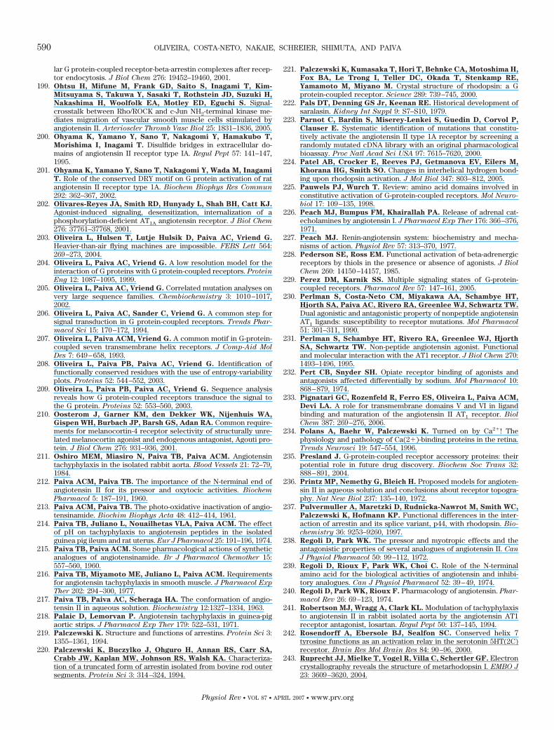
lar G protein-coupled receptor-beta-arrestin complexes after recep-tor endocytosis. J Biol Chem 276: 19452–19460, 2001.
199. Ohtsu H, Mifune M, Frank GD, Saito S, Inagami T, Kim-
Mitsuyama S, Takuwa Y, Sasaki T, Rothstein JD, Suzuki H,
Nakashima H, Woolfolk EA, Motley ED, Eguchi S. Signal-crosstalk between Rho/ROCK and c-Jun NH2-terminal kinase me-diates migration of vascular smooth muscle cells stimulated byangiotensin II. Arterioscler Thromb Vasc Biol 25: 1831–1836, 2005.
200. Ohyama K, Yamano Y, Sano T, Nakagomi Y, Hamakubo T,
Morishima I, Inagami T. Disulfide bridges in extracellular do-mains of angiotensin II receptor type IA. Regul Pept 57: 141–147,1995.
201. Ohyama K, Yamano Y, Sano T, Nakagomi Y, Wada M, Inagami
T. Role of the conserved DRY motif on G protein activation of ratangiotensin II receptor type 1A. Biochem Biophys Res Commun
292: 362–367, 2002.202. Olivares-Reyes JA, Smith RD, Hunyady L, Shah BH, Catt KJ.
Agonist-induced signaling, desensitization, internalization of aphosphorylation-deficient AT1A angiotensin receptor. J Biol Chem
276: 37761–37768, 2001.203. Oliveira L, Hulsen T, Lutje Hulsik D, Paiva AC, Vriend G.
Heavier-than-air flying machines are impossible. FEBS Lett 564:269–273, 2004.
204. Oliveira L, Paiva AC, Vriend G. A low resolution model for theinteraction of G proteins with G protein-coupled receptors. Protein
Eng 12: 1087–1095, 1999.205. Oliveira L, Paiva AC, Vriend G. Correlated mutation analyses on
very large sequence families. Chembiochemistry 3: 1010–1017,2002.
206. Oliveira L, Paiva AC, Sander C, Vriend G. A common step forsignal transduction in G protein-coupled receptors. Trends Phar-
macol Sci 15: 170–172, 1994.207. Oliveira L, Paiva ACM, Vriend G. A common motif in G-protein-
coupled seven transmembrane helix receptors. J Comp-Aid Mol
Des 7: 649–658, 1993.208. Oliveira L, Paiva PB, Paiva AC, Vriend G. Identification of
functionally conserved residues with the use of entropy-variabilityplots. Proteins 52: 544–552, 2003.
209. Oliveira L, Paiva PB, Paiva AC, Vriend G. Sequence analysisreveals how G protein-coupled receptors transduce the signal tothe G protein. Proteins 52: 553–560, 2003.
210. Oosterom J, Garner KM, den Dekker WK, Nijenhuis WA,
Gispen WH, Burbach JP, Barsh GS, Adan RA. Common require-ments for melanocortin-4 receptor selectivity of structurally unre-lated melanocortin agonist and endogenous antagonist, Agouti pro-tein. J Biol Chem 276: 931–936, 2001.
211. Oshiro MEM, Miasiro N, Paiva TB, Paiva ACM. Angiotensintachyphylaxis in the isolated rabbit aorta. Blood Vessels 21: 72–79,1984.
212. Paiva ACM, Paiva TB. The importance of the N-terminal end ofangiotensin II for its pressor and oxytocic activities. Biochem
Pharmacol 5: 187–191, 1960.213. Paiva ACM, Paiva TB. The photo-oxidative inactivation of angio-
tensinamide. Biochim Biophys Acta 48: 412–414, 1961.214. Paiva TB, Juliano L, Nouailhetas VLA, Paiva ACM. The effect
of pH on tachyphylaxis to angiotensin peptides in the isolatedguinea pig ileum and rat uterus. Eur J Pharmacol 25: 191–196, 1974.
215. Paiva TB, Paiva ACM. Some pharmacological actions of syntheticanalogues of angiotensinamide. Br J Pharmacol Chemother 15:557–560, 1960.
216. Paiva TB, Miyamoto ME, Juliano L, Paiva ACM. Requirementsfor angiotensin tachyphylaxis in smooth muscle. J Pharmacol Exp
Ther 202: 294–300, 1977.217. Paiva TB, Paiva AC, Scheraga HA. The conformation of angio-
tensin II in aqueous solution. Biochemistry 12:1327–1334, 1963.218. Palaic D, Lemorvan P. Angiotensin tachyphylaxis in guinea-pig
aortic strips. J Pharmacol Exp Ther 179: 522–531, 1971.219. Palczewski K. Structure and functions of arrestins. Protein Sci 3:
1355–1361, 1994.220. Palczewski K, Buczylko J, Ohguro H, Annan RS, Carr SA,
Crabb JW, Kaplan MW, Johnson RS, Walsh KA. Characteriza-tion of a truncated form of arrestin isolated from bovine rod outersegments. Protein Sci 3: 314–324, 1994.
221. Palczewski K, Kumasaka T, Hori T, Behnke CA, Motoshima H,
Fox BA, Le Trong I, Teller DC, Okada T, Stenkamp RE,
Yamamoto M, Miyano M. Crystal structure of rhodopsin: a Gprotein-coupled receptor. Science 289: 739–745, 2000.
222. Pals DT, Denning GS Jr, Keenan RE. Historical development ofsaralasin. Kidney Int Suppl 9: S7–S10, 1979.
223. Parnot C, Bardin S, Miserey-Lenkei S, Guedin D, Corvol P,
Clauser E. Systematic identification of mutations that constitu-tively activate the angiotensin II type 1A receptor by screening arandomly mutated cDNA library with an original pharmacologicalbioassay. Proc Natl Acad Sci USA 97: 7615–7620, 2000.
224. Patel AB, Crocker E, Reeves PJ, Getmanova EV, Eilers M,
Khorana HG, Smith SO. Changes in interhelical hydrogen bond-ing upon rhodopsin activation. J Mol Biol 347: 803–812, 2005.
225. Pauwels PJ, Wurch T. Review: amino acid domains involved inconstitutive activation of G-protein-coupled receptors. Mol Neuro-
biol 17: 109–135, 1998.226. Peach MJ, Bumpus FM, Khairallah PA. Release of adrenal cat-
echolamines by angiotensin I. J Pharmacol Exp Ther 176: 366–376,1971.
227. Peach MJ. Renin-angiotensin system: biochemistry and mecha-nisms of action. Physiol Rev 57: 313–370, 1977.
228. Pederson SE, Ross EM. Functional activation of beta-adrenergicreceptors by thiols in the presence or absence of agonists. J Biol
Chem 260: 14150–14157, 1985.229. Perez DM, Karnik SS. Multiple signaling states of G-protein-
coupled receptors. Pharmacol Rev 57: 147–161, 2005.230. Perlman S, Costa-Neto CM, Miyakawa AA, Schambye HT,
Hjorth SA, Paiva AC, Rivero RA, Greenlee WJ, Schwartz TW.
Dual agonistic and antagonistic property of nonpeptide angiotensinAT1 ligands: susceptibility to receptor mutations. Mol Pharmacol
51: 301–311, 1990.231. Perlman S, Schambye HT, Rivero RA, Greenlee WJ, Hjorth
SA, Schwartz TW. Non-peptide angiotensin agonist. Functionaland molecular interaction with the AT1 receptor. J Biol Chem 270:1493–1496, 1995.
232. Pert CB, Snyder SH. Opiate receptor binding of agonists andantagonists affected differentially by sodium. Mol Pharmacol 10:868–879, 1974.
233. Pignatari GC, Rozenfeld R, Ferro ES, Oliveira L, Paiva ACM,
Devi LA. A role for transmembrane domains V and VI in ligandbinding and maturation of the angiotensin II AT1 receptor. Biol
Chem 387: 269–276, 2006.234. Polans A, Baehr W, Palczewski K. Turned on by Ca2�! The
physiology and pathology of Ca(2�)-binding proteins in the retina.Trends Neurosci 19: 547–554, 1996.
235. Presland J. G-protein-coupled receptor accessory proteins: theirpotential role in future drug discovery. Biochem Soc Trans 32:888–891, 2004.
236. Printz MP, Nemethy G, Bleich H. Proposed models for angioten-sin II in aqueous solution and conclusions about receptor topogra-phy. Nat New Biol 237: 135–140, 1972.
237. Pulvermuller A, Maretzki D, Rudnicka-Nawrot M, Smith WC,
Palczewski K, Hofmann KP. Functional differences in the inter-action of arrestin and its splice variant, p44, with rhodopsin. Bio-
chemistry 36: 9253–9260, 1997.238. Regoli D, Park WK. The pressor and myotropic effects and the
antagonistic properties of several analogues of angiotensin II. Can
J Physiol Pharmacol 50: 99–112, 1972.239. Regoli D, Rioux F, Park WK, Choi C. Role of the N-terminal
amino acid for the biological activities of angiotensin and inhibi-tory analogues. Can J Physiol Pharmacol 52: 39–49, 1974.
240. Regoli D, Park WK, Rioux F. Pharmacology of angiotensin. Phar-
macol Rev 26: 69–123, 1974.241. Robertson MJ, Wragg A, Clark KL. Modulation of tachyphylaxis
to angiotensin II in rabbit isolated aorta by the angiotensin AT1receptor antagonist, losartan. Regul Pept 50: 137–145, 1994.
242. Rosendorff A, Ebersole BJ, Sealfon SC. Conserved helix 7tyrosine functions as an activation relay in the serotonin 5HT(2C)receptor. Brain Res Mol Brain Res 84: 90–96, 2000.
243. Ruprecht JJ, Mielke T, Vogel R, Villa C, Schertler GF. Electroncrystallography reveals the structure of metarhodopsin I. EMBO J
23: 3609–3620, 2004.
590 OLIVEIRA, COSTA-NETO, NAKAIE, SCHREIER, SHIMUTA, AND PAIVA
Physiol Rev • VOL 87 • APRIL 2007 • www.prv.org
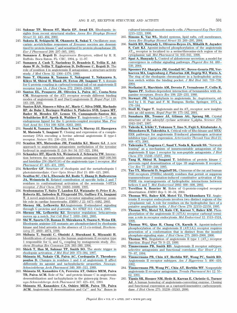
244. Sakmar TP, Menon ST, Marin EP, Awad ES. Rhodopsin: in-sights from recent structural studies. Annu Rev Biophys Biomol
Struct 31: 443–484, 2002.245. Sakuta H, Sekiguchi M, Okamoto K, Sakai Y. Oscillatory mus-
carinic acetylcholine responses of Xenopus oocytes are desensi-tized by protein kinase C and sensitized by protein phosphatase 2B.Eur J Pharmacol 208: 297–305, 1991.
246. Samanen J, Regoli D. Angiotensin Receptors, edited by R. R.Ruffolo. Boca Raton, FL: CRC, 1994, p. 11–97.
247. Samanen J, Cash T, Narindray D, Brandeis E, Yellin E, Ad-
dams W Jr, Yellin T, Eggleston D, DeBrosse C, Regoli D. Therole of position 4 in angiotensin II antagonism: a structure-activitystudy. J Med Chem 32: 1366–1370, 1989.
248. Sano T, Ohyama K, Yamano Y, Nakagomi Y, Nakazawa S,
Kikyo M, Shirai H, Blank JS, Exton JH, Inagami T. A domainfor G protein coupling in carboxyl-terminal tail of rat angiotensin IIreceptor type 1A. J Biol Chem 272: 23631–23636, 1997.
249. Santos EL, Pesquero JB, Oliveira L, Paiva AC, Costa-Neto
CM. Mutagenesis of the AT1 receptor reveals different bindingmodes of angiotensin II and [Sar1]-angiotensin II. Regul Pept 119:183–188, 2004.
250. Santos RAS, Simoes e Silva AC, Maric C, Silva DMR, Machado
RP, de Buhr I, Heringer-Walther S, Pinheiro SVB, Lopes MT,
Bader M, Mendes EP, Lemos VS, Campagnole-Santos MJ,
Schultheiss H-P, Speth R, Walther T. Angiotensin-(1O7) is anendogenous ligand for the G protein-coupled receptor Mas. Proc
Natl Acad Sci USA 100: 8258–8263, 2003.251. Sasaki K, Yamano Y, Bardhan S, Iwai N, Murray JJ, Hasegawa
M, Matsuda Y, Inagami T. Cloning and expression of a comple-mentary DNA encoding a bovine adrenal angiotensin II type-1receptor. Nature 351: 230–233, 1991.
252. Scanlon MN, Matsoukas JM, Franklin KJ, Moore GJ. A newapproach to angiotensin antagonists: methylation of the tyrosinehydroxyl in angiotensin II. Life Sci 34: 317–321, 1984.
253. Schambye HT, Hjorth SA, Weinstock J, Schwartz TW. Interac-tion between the nonpeptide angiotensin antagonist SKF-108,566and histidine 256 (HisVI:16) of the angiotensin type 1 receptor. Mol
Pharmacol 47: 425–431, 1995.254. Schertler GF. Structure of rhodopsin and the metarhodopsin I
photointermediate. Curr Opin Struct Biol 15: 408–415, 2005.255. Sealfon SC, Chi L, Ebersole BJ, Rodic V, Zhang D, Ballesteros
JA, Weinstein H. Related contribution of specific helix 2 and 7residues to conformational activation of the serotonin 5-HT2Areceptor. J Biol Chem 270: 16683–16688, 1995.
256. Senbonmatsu T, Saito T, Landon EJ, Watanabe O, Price E Jr,
Roberts RL, Imboden H, Fitzgerald TG, Gaffney FA, Inagami
T. A novel angiotensin II type 2 receptor signaling pathway: possi-ble role in cardiac hypertrophy. EMBO J 22: 6471–6482, 2003.
257. Shenoy SK, Lefkowitz RJ.Angiotensin II-stimulated signalingthrough G proteins and �-arrestin. Sci STKE 311: Cm14, 2005.
258. Shenoy SK, Lefkowitz RJ. Receptor regulation: beta-arrestinmoves up a notch. Nat Cell Biol 7: 1059–1061, 2005.
259. Shi W, Sports CD, Raman D, Shirakawa S, Osawa S, Weiss ER.
Rhodopsin arginine-135 mutants are phosphorylated by rhodopsinkinase and bind arrestin in the absence of 11-cis-retinal. Biochem-
istry 37: 4869–4874, 1998.260. Shibata T, Suzuki C, Ohnishi J, Murakami K, Miyazaki H.
Identification of regions in the human angiotensin II receptor type1 responsible for Gi and Gq coupling by mutagenesis study. Bio-
chem Biophys Res Commun 218: 383–389, 1996.261. Shieh T, Han M, Sakmar TP, Smith SO. The steric trigger in
rhodopsin activation. J Mol Biol 269: 373–384, 1997.262. Shimuta SI, Nakaie CR, Paiva AC, Cordopatis P, Theodoro-
poulos D. Changes in residues 1 and 4 of angiotensin II affectdifferently its agonist and tachyphylactic properties. Naunyn-
Schmiedebergs Arch Pharmacol 340: 309–313, 1989.263. Shimuta SI, Kanashiro CA, Ferreira AT, Oshiro MEM, Paiva
TB, Paiva ACM. Role of Na� and protein kinase C in angiotensindesensibilization and tachyphylaxis in the guinea-pig ileum. Nau-
nyn-Schmiedebergs Arch Pharmacol 347: 425–431, 1993.264. Shimuta SI, Kanashiro CA, Oshiro MEM, Paiva TB, Paiva
ACM. Angiotensin II desensitization and Ca2� and Na� fluxes in
cultured intestinal smooth muscle cells. J Pharmacol Exp Ther 253:1215–1221, 1990.
265. Simons K, Vaz WL. Model systems, lipid rafts, cell membranes.Annu Rev Biophys Biomol Struct 33: 269–295, 2004.
266. Smith RD, Hunyady L, Olivares-Reyes JA, Mihalik B, Jayadev
S, Catt KJ. Agonist-induced phosphorylation of the angiotensinAT1a receptor is localized to a serine/threonine-rich region of itscytoplasmic tail. Mol Pharmacol 54: 935–941, 1998.
267. Spat A, Hunyady L. Control of aldosterone secretion: a model forconvergence in cellular signaling pathways. Physiol Rev 84: 489–539, 2004.
268. Spooner PJ, Sharples JM, Goodall SC, Bovee-Geurts PH, Ver-
hoeven MA, Lugtenburg J, Pistorius AM, Degrip WJ, Watts A.
The ring of the rhodopsin chromophore in a hydrophobic activa-tion switch within the binding pocket. J Mol Biol 343: 719–730,2004.
269. Stefanini E, Marchisio AM, Devoto P, Vernaleone F, Collu R,
Spano PF. Sodium-dependent interaction of benzamides with do-pamine receptors. Brain Res 198: 229–233, 1980.
270. Stewart JM. Tachyphylaxis to angiotensin. In: Angiotensin, ed-ited by I. H. Page and F. M. Bumpus. Berlin: Springer, 1974, p.170–184.
271. Stoll M, Unger T. Angiotensin and its AT2 receptor: new insightsinto an old system. Regul Pept 99: 175–182, 2001.
272. Sunahara RK, Tesmer JJ, Gilman AG, Sprang SR. Crystalstructure of the adenylyl cyclase activator Gsalpha. Science 278:1943–1947, 1997.
273. Takeda K, Ichiki T, Tokunou T, Iino N, Fujii S, Kitabatake A,
Shimokawa H, Takeshita A. Critical role of Rho-kinase and MEK/ERK pathways for angiotensin II-induced plasminogen activatorinhibitor type-1 gene expression. Arterioscler Thromb Vasc Biol 21:868–873, 2001.
274. Takezako T, Gogonea C, Saad Y, Noda K, Karnik SS. “Networkleaning” as a mechanism of insurmountable antagonism of theangiotensin II type 1 receptor by non-peptide antagonists. J Biol
Chem 279: 15248–15257, 2004.275. Tang H, Shirai H, Inagami T. Inhibition of protein kinase C
prevents rapid desensitization of type 1B angiotensin II receptor.Circ Res 77: 239–248, 1995.
276. Tao YX, Mizrachi D, Segaloff DL. Chimeras of the rat and humanFSH receptors (FSHRs) identify residues that permit or suppresstransmembrane 6 mutation-induced constitutive activation of theFSHR via rearrangements of hydrophobic interactions betweenhelices 6 and 7. Mol Endocrinol 2002: 600–608, 2002.
277. Terrillon S, Bouvier M. Roles of G-protein-coupled receptordimerization. EMBO Rep 5: 30–34, 2004.
278. Thomas WG, Baker KM, Motel TJ, Thekkumkara TJ. Angio-tensin II receptor endocytosis involves two distinct regions of thecytoplasmic tail. A role for residues on the hydrophobic face of aputative amphipathic helix. J Biol Chem 270: 22153–22159, 1995.
279. Thomas WG, Motel TJ, Kule CE, Karoor V, Baker KM. Phos-phorylation of the angiotensin II (AT1A) receptor carboxyl termi-nus: a role in receptor endocytosis. Mol Endocrinol 12: 1513–1524,1998.
280. Thomas WG, Qian H, Chang CS, Karnik S. Agonist-inducedphosphorylation of the angiotensin II (AT(1A)) receptor requiresgeneration of a conformation that is distinct from the inositolphosphate-signaling state. J Biol Chem 275: 2893–2900, 2000.
281. Thomas WG. Regulation of angiotensin II type 1 (AT1) receptorfunction. Regul Pept 79: 9–23, 1999.
282. Timmermans PB, Smith RD. Angiotensin II receptor subtypes:selective antagonists and functional correlates. Eur Heart J 15:79–87, 1994.
283. Timmermans PB, Chiu AT, Herblin WF, Wong PC, Smith RD.
Angiotensin II receptor subtypes. Am J Hypertens 5: 406–410,1992.
284. Timmermans PB, Wong PC, Chiu AT, Herblin WF. Nonpeptideangiotensin II receptor antagonists. Trends Pharmacol Sci 12: 55–62, 1991.
285. Tipnis SR, Hooper NM, Hyde R, Karran E, Christie G, Turner
AJ. A human homolog of angiotensin-converting enzyme. Cloningand functional expression as a captopril-insensitive carboxypepti-dase. J Biol Chem 275: 33238–33243, 2000.
ANG II AT1 RECEPTOR STRUCTURE AND ACTIVITY 591
Physiol Rev • VOL 87 • APRIL 2007 • www.prv.org
286. Tofovic SP, Jackson EK. Tachyphylaxis to angiotensin II and[Phe4]-angiotensin II in the rat mesentery in vivo. J Pharmacol Exp
Ther 276: 13–20, 1996.287. Tucek S, Michal P, Vlachova V. Modelling the consequences of
receptor-G-protein promiscuity. Trends Pharmacol Sci 23: 171–176, 2002.
288. Turu G, Szidonya L, Gaborik Z, Buday L, Spat A, Clark AJ,
Hunyady L. Differential beta-arrestin binding of AT1 and AT2
angiotensin receptors. FEBS Lett 580: 41–45, 2006.289. Ullian ME, Linas SL. Role of receptor cycling in the regulation of
angiotensin II surface receptor number and angiotensin II uptake inrat vascular smooth muscle cells. J Clin Invest 84: 840–846, 1989.
290. Urizar E, Claeysen S, Deupi X, Govaerts C, Costagliola S,
Vassart G, Pardo L. An activation switch in the rhodopsin familyof G protein coupled receptors: the thyrotropin receptor. J Biol
Chem 280: 17135–17141, 2005.291. Vanderheyden PM, Fierens FL, Vauquelin G. Angiotensin II
type 1 receptor antagonists. Why do some of them produce insur-mountable inhibition? Biochem Pharmacol 60: 1557–1563, 2000.
292. Vanderheyden PM, Fierens FL, De Backer JP, Fraeyman N,
Vauquelin G. Distinction between surmountable and insurmount-able selective AT1 receptor antagonists by use of CHO-K1 cellsexpressing human angiotensin II AT1 receptors. Br J Pharmacol
126: 1057–1065, 1999.293. Vassart G, Pardo L, Costagliola S. A molecular dissection of the
glycoprotein hormone receptors. Trends Biochem Sci 29: 119–126,2004.
294. Vauquelin G, Fierens F, Verheijen I, Vanderheyden P. Insur-mountable AT1 receptor antagonism: the need for different antag-onist binding states of the receptor. Trends Pharmacol Sci 22:333–334, 2001.
295. Verheijen I, Vanderheyden PM, De Backer JP, Vauquelin G.
AT1 receptor antagonists. Curr Med Chem Cardiovasc Hematol
Agents 2: 69–77, 2004.296. Wald G. Molecular basis of visual excitation. Science 162: 230–239,
1968.297. Watson S, Arkinstall S. The G-Protein Linked Receptor Fact
Book, edited by S. Watson and S. Arkinstall. London: Academic,1994.
298. Wei H, Ahn S, Shenoy SK, Karnik SS, Hunyady L, Luttrell LM,
Lefkowitz RJ. Independent beta-arrestin 2 and G protein-medi-ated pathways for angiotensin II activation of extracellular signal-regulated kinases 1 and 2. Proc Natl Acad Sci USA 100: 10782–10787, 2003.
299. Wilbanks AM, Laporte SA, Bohn LM, Barak LS, Caron MG.
Apparent loss-of-function mutant GPCRs revealed as constitutivelydesensitized receptors. Biochemistry 41: 11981–11989, 2002.
300. Wright JW, Harding JW. Important roles for angiotensin III andIV in the brain renin-angiotensin system. Brain Res Rev 25: 96–124,1997.
301. Yamakawa T, Tanaka S, Numaguchi K, Yamakawa Y, Motley
ED, Ichihara S, Inagami T. Involvement of Rho-kinase in angio-tensin II-induced hypertrophy of rat vascular smooth muscle cells.Hypertension 35: 313–318, 2000.
302. Yamano Y, Ohyama K, Chaki S, Guo DF, Inagami T. Identifica-tion of amino acid residues of rat angiotensin II receptor for ligandbinding by site directed mutagenesis. Biochem Biophys Res Com-
mun 187: 1426–1431, 1992.303. Yamano Y, Ohyama K, Kikyo M, Sano T, Nakagomi Y, Inoue Y,
Nakamura N, Morishima I, Guo DF, Hamakubo T, Inagami T.
Mutagenesis and the molecular modeling of the rat angiotensin IIreceptor (AT1). J Biol Chem 270: 14024–14030, 1995.
304. Yu J, Prado GN, Taylor L, Piserchio A, Gupta A, Mierke DF,
Polgar P. Global chimeric exchanges within the intracellular faceof the bradykinin B2 receptor with corresponding angiotensin IItype Ia receptor regions: generation of fully functional hybridsshowing characteristic signaling of the AT1a receptor. J Cell Bio-
chem 85: 809–819, 2002.305. Zhang M, Zhao X, Chen HC, Catt KJ, Hunyady L. Activation of
the AT1 angiotensin receptor is dependent on adjacent apolarresidues in the carboxyl terminus of the third cytoplasmic loop.J Biol Chem 275: 15782–15788, 2000.
306. Zhang WJ, Nikiforovich GV, Perodin J, Richard DE, Escher E,
Marshall GR. Novel cyclic analogs of angiotensin II with cycliza-tion between positions 5 and 7: conformational and biologicalimplications. J Med Chem 39: 2738–2744, 1996.
592 OLIVEIRA, COSTA-NETO, NAKAIE, SCHREIER, SHIMUTA, AND PAIVA
Physiol Rev • VOL 87 • APRIL 2007 • www.prv.org
Copyright © 2022 FDOKUMEN