Mechanism of Spectral Tuning Going from Retinal in Vacuo to Bovine Rhodopsin and its Mutants:...
21
Mechanism of Spectral Tuning Going from Retinal in Vacuo to Bovine Rhodopsin and its Mutants: Multireference ab initio Quantum Mechanics/Molecular Mechanics Studies Ahmet Altun a,b , Shozo Yokoyama b , and Keiji Morokuma a,c aCherry L. Emerson Center for Scientific Computation and Department of Chemistry, Emory University, Atlanta, GA 30322, USA bDepartment of Biology, Rollins Research Center, Emory University, Atlanta, GA 30322, USA cFukui Institute for Fundamental Chemistry, Kyoto University, 34-4 Takano Nishihiraki-cho, Sakyo, Kyoto 606-8103, Japan Abstract We have investigated photoabsorption spectra of bovine rhodopsin and its mutants (E122Q and E113Q) by hybrid quantum mechanical/molecular mechanical (QM/MM) calculations as well as retinal in vacuo by pure QM calculations, employing multireference (MR) ab initio and TD-B3LYP methods. The sophisticated MR-SORCI+Q and MRCISD+Q methods extrapolated with respect to adopted approximations can reproduce the experimental absorption maxima of retinal very well. The relatively inexpensive MR-DDCI2+Q method gives absorption maxima blue-shifted by ca. 65 nm from experimental values; however, this error is systematic and thus MR-DDCI2+Q can be used to estimate spectral shifts. In MR calculations, the ground state energy of retinal at B3LYP geometry is significantly lower than that at CASSCF geometry. Therefore, B3LYP geometry is more reliable than CASSCF geometry, which has blue-shift error as large as 100 nm in the gas phase. The effect of ground state geometry on the excitation energies is less critical in the polarizing field of protein environments. At the B3LYP geometry, there is no significant charge transfer upon vertical excitation to the S 1 excited state either from Glu113 to retinal or from Schiff-base terminal to β-ionone ring through the polyene chain. All-trans to 11-cis isomerization of retinal in the gas phase has no influence on the calculated S 1 absorbing state, in agreement with experiment. The shoulder of the experimental absorption spectrum of retinal in vacuo at the S 1 absorbing band appears to be the second electronic transition (S 2 ) in our calculations, contrary to previous tentative assignment to vibrational state of S 1 or to the S 1 band of a retinal isomer. Keywords Spectral Tuning; Retinal; Multireference ab initio methods; QM/MM calculations; Electronic Excitation Correspondence to: Keiji Morokuma. To whom correspondence should be addressed. K.M.: e-mail, E-mail: [email protected]; phone, +1 (404) 727-2180; fax, +1 (404) 727-7412. email: [email protected] Supporting Information Available. All calculated vertical excitation energies along with their oscillator strengths. This information is available free of charge via the Internet at http://pubs.acs.org. NIH Public Access Author Manuscript J Phys Chem B. Author manuscript; available in PMC 2009 December 25. Published in final edited form as: J Phys Chem B. 2008 December 25; 112(51): 16883–16890. NIH-PA Author Manuscript NIH-PA Author Manuscript NIH-PA Author Manuscript
-
Upload
independent -
Category
Documents
-
view
3 -
download
0
Transcript of Mechanism of Spectral Tuning Going from Retinal in Vacuo to Bovine Rhodopsin and its Mutants:...
Mechanism of Spectral Tuning Going from Retinal in Vacuo toBovine Rhodopsin and its Mutants: Multireference ab initioQuantum Mechanics/Molecular Mechanics Studies
Ahmet Altuna,b, Shozo Yokoyamab, and Keiji Morokumaa,c
aCherry L. Emerson Center for Scientific Computation and Department of Chemistry, Emory University,Atlanta, GA 30322, USA
bDepartment of Biology, Rollins Research Center, Emory University, Atlanta, GA 30322, USA
cFukui Institute for Fundamental Chemistry, Kyoto University, 34-4 Takano Nishihiraki-cho, Sakyo, Kyoto606-8103, Japan
AbstractWe have investigated photoabsorption spectra of bovine rhodopsin and its mutants (E122Q andE113Q) by hybrid quantum mechanical/molecular mechanical (QM/MM) calculations as well asretinal in vacuo by pure QM calculations, employing multireference (MR) ab initio and TD-B3LYPmethods. The sophisticated MR-SORCI+Q and MRCISD+Q methods extrapolated with respect toadopted approximations can reproduce the experimental absorption maxima of retinal very well. Therelatively inexpensive MR-DDCI2+Q method gives absorption maxima blue-shifted by ca. 65 nmfrom experimental values; however, this error is systematic and thus MR-DDCI2+Q can be used toestimate spectral shifts. In MR calculations, the ground state energy of retinal at B3LYP geometryis significantly lower than that at CASSCF geometry. Therefore, B3LYP geometry is more reliablethan CASSCF geometry, which has blue-shift error as large as 100 nm in the gas phase. The effectof ground state geometry on the excitation energies is less critical in the polarizing field of proteinenvironments. At the B3LYP geometry, there is no significant charge transfer upon vertical excitationto the S1 excited state either from Glu113 to retinal or from Schiff-base terminal to β-ionone ringthrough the polyene chain. All-trans to 11-cis isomerization of retinal in the gas phase has no influenceon the calculated S1 absorbing state, in agreement with experiment. The shoulder of the experimentalabsorption spectrum of retinal in vacuo at the S1 absorbing band appears to be the second electronictransition (S2) in our calculations, contrary to previous tentative assignment to vibrational state ofS1 or to the S1 band of a retinal isomer.
KeywordsSpectral Tuning; Retinal; Multireference ab initio methods; QM/MM calculations; ElectronicExcitation
Correspondence to: Keiji Morokuma.To whom correspondence should be addressed. K.M.: e-mail, E-mail: [email protected]; phone, +1 (404) 727-2180; fax, +1 (404)727-7412.email: [email protected] Information Available. All calculated vertical excitation energies along with their oscillator strengths. This information isavailable free of charge via the Internet at http://pubs.acs.org.
NIH Public AccessAuthor ManuscriptJ Phys Chem B. Author manuscript; available in PMC 2009 December 25.
Published in final edited form as:J Phys Chem B. 2008 December 25; 112(51): 16883–16890.
NIH
-PA Author Manuscript
NIH
-PA Author Manuscript
NIH
-PA Author Manuscript
I. IntroductionRhodopsin (Rh) is responsible for the dim-light vision in vertebrate eyes and belongs to theG-protein-coupled receptor family.1,2 It is activated by the light-induced 11-cis to all-transisomerization of protonated Schiff-base retinal (PSBR). While the retinal in vacuo absorbslight at 610 nm,3 the retinal in bovine Rh absorbs at 500 nm.1,2 Despite extensive mutagenesisand computational analyses in the last two decades (see ref 2 and references therein), themolecular basis of spectral tuning, e.g., how environments of retinal modulate its absorptionmaximum in the UV-visible spectrum, remains unclear.
In general, computation of the absorption maxima for retinal proteins is a very challengingproblem. Our recent ONIOM quantum mechanical/molecular mechanical (QM/MM)calculations on bovine Rh, employing density functional theory (DFT), show that the effectsof amino acids other than Glu113 on the first (S1) vertical excitation energy of retinal tend tocancel to each other.5 Analogously, the protein environment of retinal and Glu113 representedwith several point charge models contributes up to ±20 nm at CASPT2/ANO level.6 However,the protein environment around retinal and Glu113 red-shifts the S1 excitation energy by 110nm at (aug-MCQDPT2/cc-pVDZ)/EFP level, where EFP is an effective fragment potentialmethod for calculating QM/MM interaction.7 Glu113 blue-shifts the S1 excitation energy inthe gas phase by ca. 60, 150 and 210 nm at TD-B3LYP/6-31G*,4 CASPT2/(ANO or 6-31G*)6,8 and aug-MCQDPT2/cc-pVDZ7 levels, respectively. Although this varying Glu113 effectis well captured at those levels where its atoms are represented with point charges rather thanincluded in the QM model system, SACCI/D95(d) calculations find a significant blue-shift (ca.100 nm) charge-transfer contribution of Glu113.9
The computed gas-phase excitation energies of all-trans and 11-cis retinals also vary quite alot.2,5 For example, the errors in the S1 excitation energy of all-trans retinal (experiment:3,4610 nm) are -100/-50 nm (TD-B3LYP/6-31G* at B3LYP and complete active space self-consistent field [CASSCF] geometries, respectively),10,11 -65 nm (CASPT2/6-31G* atCASSCF geometry),12 -10/-40 nm (OM2-MRCI at B3LYP and CASSCF geometries,respectively),11 25/-20 nm (spectroscopy-oriented configuration interaction [SORCI]/6-31G*at B3LYP and CASSCF geometries, respectively)11 and more than 200 nm (SACCI/D95(d)at B3LYP geometry).14 However, CASPT2 calculations with a large ANO basis set at MP2geometry (606 nm)6,15 reproduce the experiment (610 nm).
In our previous study,5 we investigated absorption spectra of bovine rhodopsin and its mutantsby time-dependent (TD)-DFT QM/MM calculations in ONIOM scheme and reproduced theexperimental absorption maxima within 10 nm. In this study, we employ high-levelmultireference (MR) ab initio QM/MM calculations on retinal to pinpoint origins of differencesamong various calculations and to provide computational strategies for accurately calculatingits absorption spectra in the gas phase as well as in the protein environments of bovinerhodopsin and its mutants.
II. Computational DetailsFor bovine Rh and its mutants (E122Q and E113Q), ONIOM(B3LYP/6-31G*:AMBER)-optimized structures with electronic embedding (EE) are used in this study (see ref 5). Allsingle-point ab initio QM/MM calculations on these structures were carried out with ORCA2.6.19 program package16 using an EE scheme,17 in which fixed MM point charges areincluded in the one-electron QM Hamiltonian and the QM/MM electrostatic interactions areevaluated from the QM electrostatic potential and the MM atomic charges. Although this EEscheme17 is slightly different from the ONIOM-EE,5,18 they result in identical excitationenergies at given Rh geometries in our test calculations. Therefore, ONIOM-EE geometriescan be used for QM/MM excitation energy calculations with the ORCA program package that
Altun et al. Page 2
J Phys Chem B. Author manuscript; available in PMC 2009 December 25.
NIH
-PA Author Manuscript
NIH
-PA Author Manuscript
NIH
-PA Author Manuscript
has many MR methods implemented. No cutoffs were introduced for the nonbonding MM andQM/MM interactions. Hydrogen link atoms19 were used at the QM/MM boundary along witha charge shift model.20
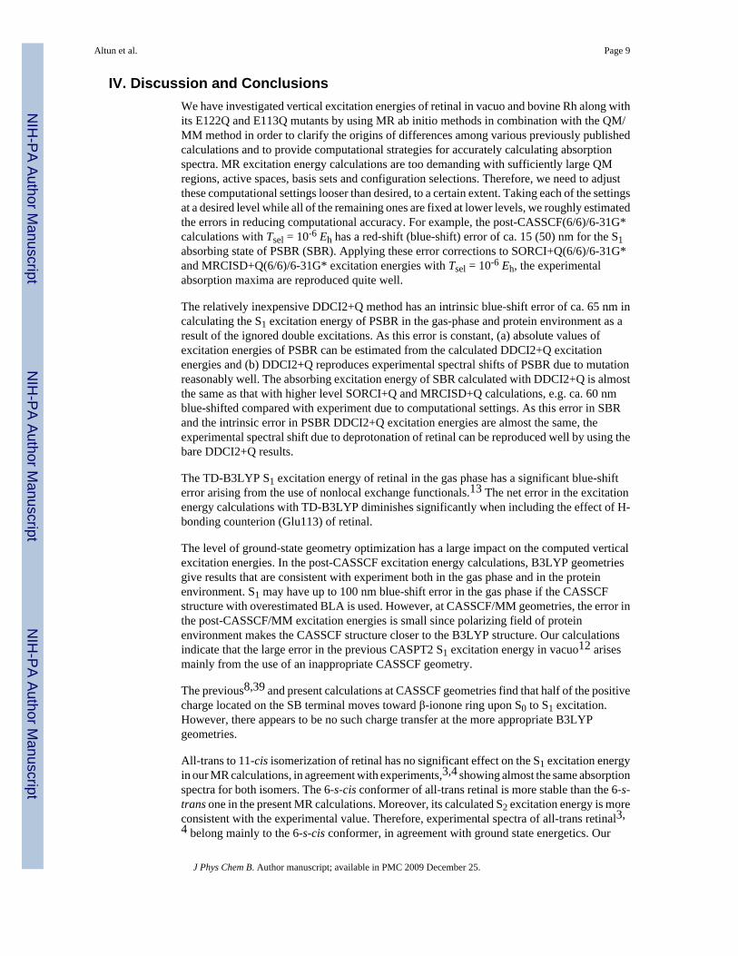
We employed two QM regions shown in Figure 1: (i) R1, including the full retinal along withcovalently bound N/NH moiety of Lys296 for deprotonated/protonated Schiff-base (SB)linkage, which results in total of 50/51 atoms. We do not extend this QM region to Cε atom ofLys296 since it red-shifts the S1 excitation energy only 10 nm in TD-DFT5 and CASPT212studies. (ii) R2, consisting of R1 region plus full Glu113, which results in total of 68 atomswhen either retinal or Glu113 side chain is protonated. The gas-phase QM calculations on thebare R1 and R2 regions at the QM/MM geometries will be called as QM-none. Unless statedotherwise, our calculations were performed for PSBR modeled as R1 and using 6-31G* basisset.21 In order to assess the basis set dependence of computed excitation energies, we also usedVDZP (known also as SV(P)),22 VTZP,22 TZVP,23 double-ξ {BNANO-DZP: (33s12p3d) →[3s2p1d] for N, (32s12p3d) → [3s2p1d] for C and (4s1p) → [2s1p] for H} and triple-ζ{BNANO-TZ2P: (44s18p6d) → [4s3p2d] for N, (43s18p6d) → [4s3p2d] for C and (5s2p) →[3s2p] for H} type ANO basis sets from the ORCA library.16,24 Auxiliary basis sets used tospeed-up the ab initio calculations are SV/C and TZV/C for double-ζ and triple-ζ quality basissets, respectively.25 The calculated excitation energies are not affected by the use of auxiliarybasis sets for the current system.
We applied CASSCF26 theory for obtaining zero-order wave function of all MR ab initiocalculations. CASSCF captures a major part of static correlation by mixing the active-spaceconfigurations at the full configuration interaction (CI) level. The largest 6-root CASSCF(12/12) calculations include all valance π-electrons of the polyene chain of R1 from C5 to theSB terminal. For the extensive post-CASSCF calculations at SORCI and MRCISD levels, wehad to use 3-root CASSCF(6/6). For R2 with Glu113 in the QM region, we performed 6-rootCASSCF(12/12) calculations including π-electrons of Glu113 in the active space.
Subsequent post-CASSCF MR calculations on the CASSCF reference wave function wereperformed at several CI levels. As cost-effective post-CASSCF methods, we applied differencededicated CI (DDCI) methods DDCI2 and DDCI3.27 These methods use a CI expansion aimedat improving energy difference between individual states rather than their absolute correlationenergies. Whereas the DDCI2 method includes double excitations which involve either twoparticles in the inactive empty orbitals or two holes in the inactive occupied orbitals in additionto all single excitations, the DDCI3 method also includes out of the active-space configurationswith two holes and one particle or two particles and one hole.27-29 The effects of excitationshigher than doubles (i.e., disconnected quadruple excitations) were estimated with the aposteriori MR Davidson +Q term, which corrects the energy but not the expansion coefficientsof the correlated wavefunction.30 To assess the effect of size-consistency error in DDCI typemethods more accurately, we applied a slightly modified version of MR averaged coupled pairfunctional ACPF/230 procedure to DDCI2 excitations, leading to the size-consistent DDACPF/2a-2 method implemented in ORCA.
We also applied the SORCI method,28 which performs a DDCI3 calculation in a truncatedbasis of approximate average natural orbitals (AANOs) obtained from a DDCI2 calculation.In the DDCI3 step of SORCI, AANOs with an occupation number less than a threshold Tnatare rejected while those with an occupation number larger than 2.0-Tnat are frozen.
Finally, we also applied the most straightforward MRCISD approach including all single anddouble excitations32,33 to benchmark the validity of results obtained with the lower levelDDCI2, DDCI3 and SORCI methods. For accounting size-consistency error, +Q correctionwas also calculated for SORCI and MRCISD results.
Altun et al. Page 3
J Phys Chem B. Author manuscript; available in PMC 2009 December 25.
NIH
-PA Author Manuscript
NIH
-PA Author Manuscript
NIH
-PA Author Manuscript
To enhance computational efficiency, the ORCA thresholds Tnat (used only for SORCI),Tpre and Tsel were set to 10-6, 10-4 and 10-6 Eh, respectively.11,28,13 The present DDCI2 andSORCI calculations on retinal in vacuo and the previous SORCI calculations on model retinalsystems (see Supporting Information of ref 13) show that looser ORCA thresholds of Tsel andTnat result in the significant loss of accuracy. Improved virtual orbitals were not used. Coreorbitals with energies of less than -4 Eh were frozen, and no virtual orbitals were neglected. Inall perturbative treatments, a level shift of 0.4 Eh was applied in order to avoid intruder stateproblems.
III. ResultsA. Protonated Schiff-Base Retinal (PSBR) in Vacuo
In this section, we mainly discuss the calculated first (S1) and second (S2) singlet verticalexcitation energies of PSBR.
Basis Set Dependence—The improvement of the 6-31G* basis set to aug-cc-pVTZ red-shifts the S1 vertical energy of PSBR by only 10 nm in DFT calculations.5 As the correlatedab initio calculations converge to the basis set limit slower than DFT calculations, it is necessaryto investigate the basis set effect on the excitation energies of PSBR for MR ab initio treatments.Indeed, for a small model of PSBR (without β-ionone ring), the S1 and S2 vertical energieswere found to red-shift by ca. 20 nm when 6-31G* basis set is improved to aug-cc-pVTZ atthe SORCI level (see Supporting Information of ref 13). To assess the basis set effect furtheron the full all-trans retinal, we performed 6-root DDCI2+Q(12/12) calculations at the gas-phaseB3LYP/6-31G* geometry of all-trans-6-s-trans retinal, as shown in Table 1. When the double-ζ quality basis sets were improved to TZVP, the S1 excitation energy red-shifts by ca. 20 nm.However, with the largest basis set considered (BNANO-TZ2P), it comes back and gives only5 nm blue-shifted result relative to the 6-31G*. This ANO basis set gave slightly betteragreement with experiment than the aug-cc-pVTZ basis set in previous test calculations.24 Asto the S2 excitation energy, 6-31G* and VDZP basis sets give ca. 20 nm blue-shift comparedwith the larger basis sets, which is similar to the results on model systems.13 In the following,we will use the cost-effective 6-31G* basis set that contains ca. 5 (20) nm blue-shift error forthe S1 (S2) excitation energy of full PSBR.
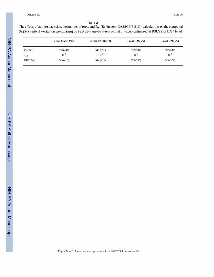
The Effects of Several Computational Settings—We have investigated the effects ofseveral computational settings (approximations) at the DDCI2+Q/6-31G* level on theexcitation energies of PSB all-trans-6-s-trans retinal, as shown in Table 2. The number of rootshas no significant effect on the computed excitation energies. Reducing CASSCF(12/12) toCASSCF(6/6) introduces a large blue-shift error of ca. 160 (60) nm in the CASSCF S1 (S2)vertical excitation energy. CI calculations substantially reduces the error due to small activespace; the DDCI2+Q S1 (S2) excitation energy error due to the reduction of the active spaceis ca. 15 (30) nm to red (blue). Tightening Tsel from 10-6 Eh to 10-7 Eh blue-shifts the S1 (S2)DDCI2+Q excitation energy by ca. 15 (5) nm whereas tightening Tpre = 10-4 has no notableeffect, a situation similar to SORCI calculations on model systems.13 In an overall assessment,CI calculations with Tsel = 10-6 using CASSCF(6/6) wave function have a net error of ca. +15(-55) nm in S1 (S2) excitation energy, arising from Tsel [+15 (-5) nm], active space size [+15(-30) nm], basis set [-5 (-20) nm] and the use of NH2 terminal rather than NHCH3 [-10 (0)nm11].
Accuracy of Several CI Levels—Experimental absorption spectra of PSBR (both all-transand 11-cis conformations) have absorption bands peaking at 610 nm and ca. 390 nm.3,4 Thesebands were assigned previously to S1 and S2 transitions, respectively.3,4 The band at 610 nmhas a shoulder at around 540 nm.3,4 This shoulder was tentatively assigned either to the S1
Altun et al. Page 4
J Phys Chem B. Author manuscript; available in PMC 2009 December 25.
NIH
-PA Author Manuscript
NIH
-PA Author Manuscript
NIH
-PA Author Manuscript
state of an all-trans retinal isomer or to a vibrational state of the electronic S1 state.3,4 Theresults of several CI calculations are given in Table 3. From the error estimates in the previoussection, one should subtract 15 nm from S1 and add 55 nm to S2 excitation energies given inTable 3 when CI results using CASSCF(6/6)/6-31G* are compared with experiment. Themagnitude of +Q correction for the S1 (S2) excitation energy is sizable and depends on theapplied CI level: -26 (-33) nm, 42 (0) nm, 47 (5) nm and -73 (-280) nm at DDCI2, DDCI3,SORCI and MRCISD levels, respectively. Therefore, we consider the results only with Qcorrection.
As already found in the previous section, CI calculations improve the poor performance ofsmall CASSCF(6/6) and make the results comparable to the CI results based on CASSCF(12/12). Our lowest level post-CASSCF calculation (DDCI2+Q) corrects the TD-B3LYP S1excitation energy by ca. 50 nm. DDCI2+Q and size-consistent DDACPF/2a-2 results agreewithin 20 nm. DDCI3+Q, SORCI+Q and MRCISD+Q methods improve DDCI2+Q energiessignificantly, making the S1 excitation energies agree with experiment within ±20 nm(including 15 nm correction). Natural orbital iteration at SORCI+Q level further improves theS1 DDCI3+Q excitation energy toward experiment by ca. 15 nm.
As to the S2 excitation energy, when the DDCI3+Q, SORCI+Q and MRCISD+Q S2 excitationenergies based on CASSCF(6/6) are corrected with our error estimate of 55 nm, we obtainvalues (513-546 nm) that are close to the 540 nm shoulder of S1 band rather than to the 390nm absorption band assigned to S2 in the experimental spectrum.3,4 At the B3LYP-optimizedgeometry, the calculated oscillator strengths f of S1 and S2 excitation for the 6-s-trans/6-s-cis conformation are around 1.8/1.8 and 0.03/0.14, respectively. As these oscillator strengthsare significantly different than zero, we assign the shoulder of the S1 band to the S2 band ofground-state 6-s-cis conformation (see Table 4 for the results on this species), rather than avibrational splitting or S1 of a local minimum. The difference between the S1 and third (S3)excitation energies in DDCI2+Q(12/12)/6-31G* (not listed in Table 3, see SupportingInformation) is 237 nm; which agrees reasonably well with the difference (225 nm) in theexperimental absorption maxima of 610 and 385 nm bands of all-trans retinal. Therefore, weassign the band peaking at 385 nm in the experimental spectrum as the S3 band, contrary tothe previous assigned S2.3,4 The appearance of three absorption bands in the absorptionspectrum of PSB all-trans retinal in vacuo is in agreement with two-photon spectroscopy ofPSB all-trans retinal in the protein environment of bacteriorhodopsin (568 nm, 488 nm and410 nm).34
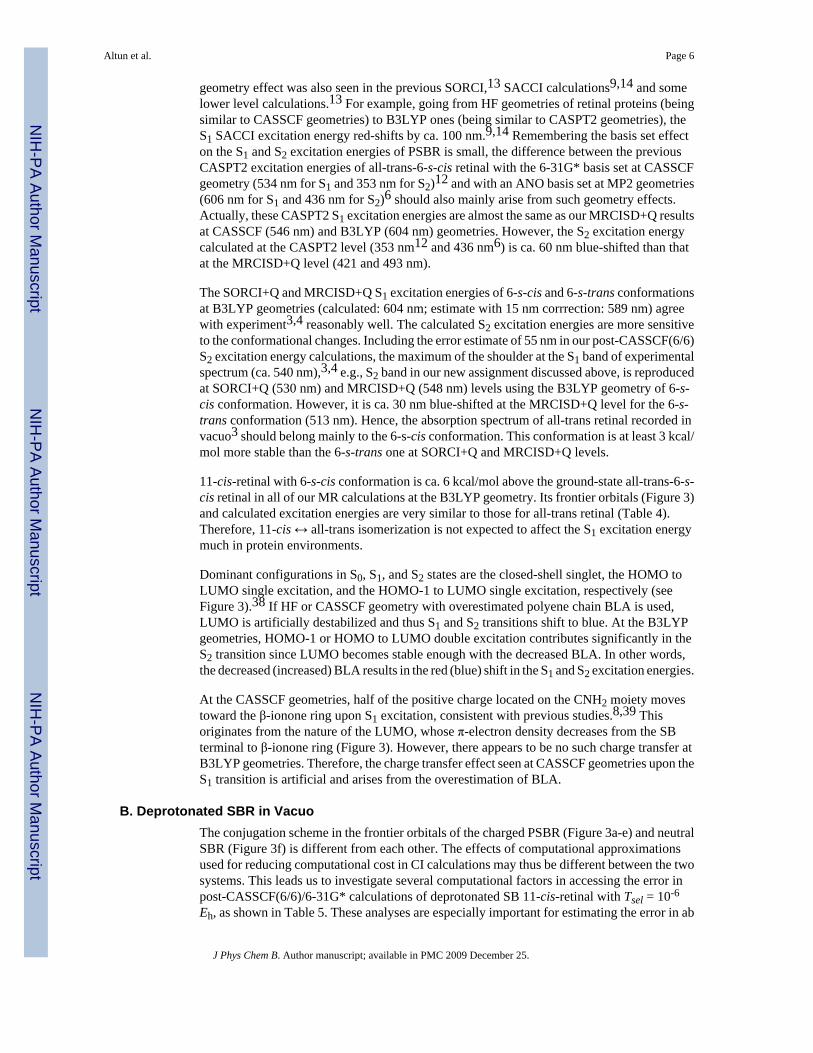
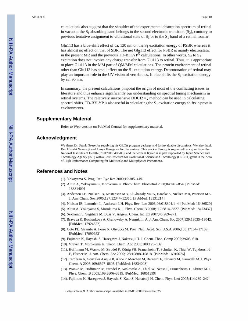
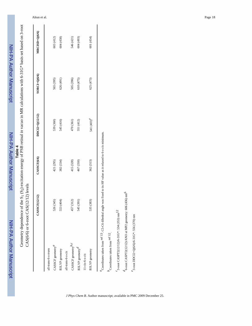
Geometry Effect at Several CI Levels—As shown in Figure 2, CASSCF single anddouble bonds are on average 0.04 Å longer and 0.03 Å shorter than B3LYP distances,respectively. In the SORCI+Q and MRCISD+Q calculations, the B3LYP geometry of PSB all-trans retinal (both 6-s-trans and 6-s-cis) is at least 8 kcal/mol more stable than the CASSCFgeometry. Therefore, the B3LYP ground-state geometries (incorporating both static anddynamical correlations) are more appropriate for retinal systems than CASSCF ones(incorporating only static correlation and unbalanced). B3LYP, MP2 and CASPT2 geometrieshave been shown to be almost the same on small PSBR models, whereas HF (no electroncorrelation) and CASSCF (incorporating only static correlation) methods overestimate bondlength alternation (BLA) of the polyene chain.5,35-37 As discussed earlier, the clear BLApattern of PSBR in rhodopsins is the result of the polarizing field of protein environments.5Therefore, it is not expected to have a significant BLA in gas-phase retinal systems.
CASSCF and B3LYP geometries are both used extensively in literature for retinal systems.However, going from CASSCF geometries to B3LYP ones in the present post-CASSCFcalculations, the S1 and S2 excitation energies improve up to 100 nm and reach to the range ofexperimental value at SORCI+Q and MRCISD+Q levels, as shown in Table 4. An analogous
Altun et al. Page 5
J Phys Chem B. Author manuscript; available in PMC 2009 December 25.
NIH
-PA Author Manuscript
NIH
-PA Author Manuscript
NIH
-PA Author Manuscript
geometry effect was also seen in the previous SORCI,13 SACCI calculations9,14 and somelower level calculations.13 For example, going from HF geometries of retinal proteins (beingsimilar to CASSCF geometries) to B3LYP ones (being similar to CASPT2 geometries), theS1 SACCI excitation energy red-shifts by ca. 100 nm.9,14 Remembering the basis set effecton the S1 and S2 excitation energies of PSBR is small, the difference between the previousCASPT2 excitation energies of all-trans-6-s-cis retinal with the 6-31G* basis set at CASSCFgeometry (534 nm for S1 and 353 nm for S2)12 and with an ANO basis set at MP2 geometries(606 nm for S1 and 436 nm for S2)6 should also mainly arise from such geometry effects.Actually, these CASPT2 S1 excitation energies are almost the same as our MRCISD+Q resultsat CASSCF (546 nm) and B3LYP (604 nm) geometries. However, the S2 excitation energycalculated at the CASPT2 level (353 nm12 and 436 nm6) is ca. 60 nm blue-shifted than thatat the MRCISD+Q level (421 and 493 nm).
The SORCI+Q and MRCISD+Q S1 excitation energies of 6-s-cis and 6-s-trans conformationsat B3LYP geometries (calculated: 604 nm; estimate with 15 nm corrrection: 589 nm) agreewith experiment3,4 reasonably well. The calculated S2 excitation energies are more sensitiveto the conformational changes. Including the error estimate of 55 nm in our post-CASSCF(6/6)S2 excitation energy calculations, the maximum of the shoulder at the S1 band of experimentalspectrum (ca. 540 nm),3,4 e.g., S2 band in our new assignment discussed above, is reproducedat SORCI+Q (530 nm) and MRCISD+Q (548 nm) levels using the B3LYP geometry of 6-s-cis conformation. However, it is ca. 30 nm blue-shifted at the MRCISD+Q level for the 6-s-trans conformation (513 nm). Hence, the absorption spectrum of all-trans retinal recorded invacuo3 should belong mainly to the 6-s-cis conformation. This conformation is at least 3 kcal/mol more stable than the 6-s-trans one at SORCI+Q and MRCISD+Q levels.
11-cis-retinal with 6-s-cis conformation is ca. 6 kcal/mol above the ground-state all-trans-6-s-cis retinal in all of our MR calculations at the B3LYP geometry. Its frontier orbitals (Figure 3)and calculated excitation energies are very similar to those for all-trans retinal (Table 4).Therefore, 11-cis ↔ all-trans isomerization is not expected to affect the S1 excitation energymuch in protein environments.
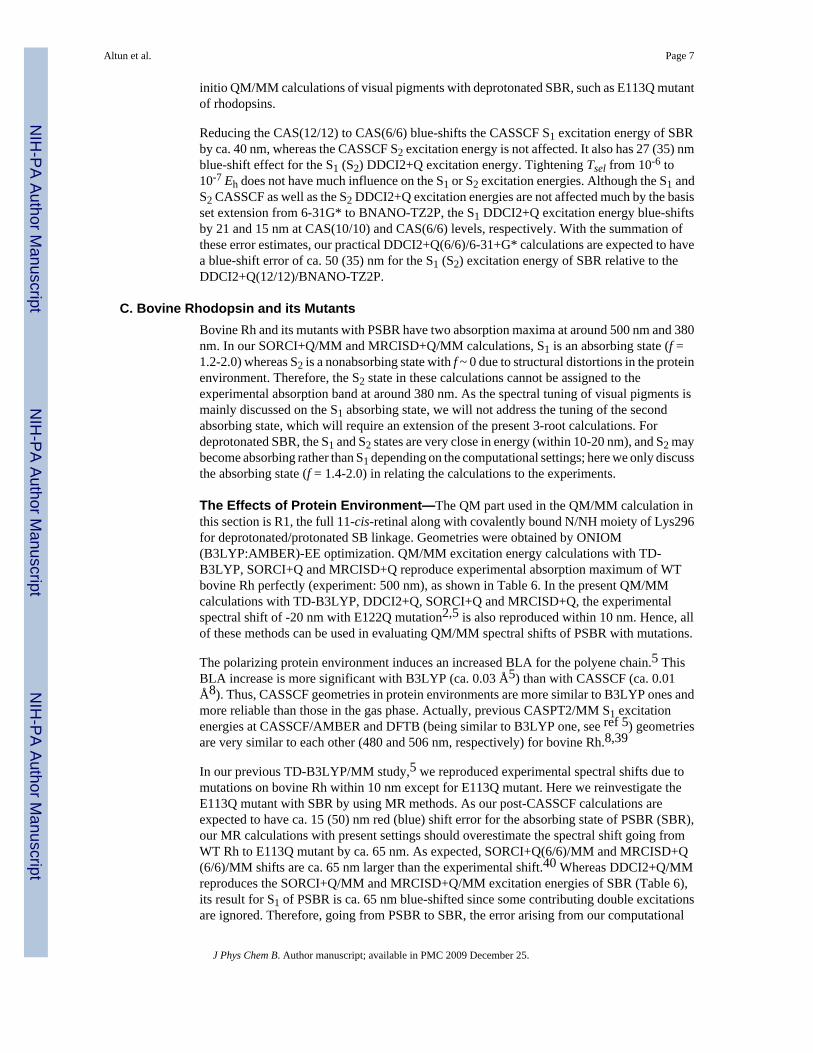
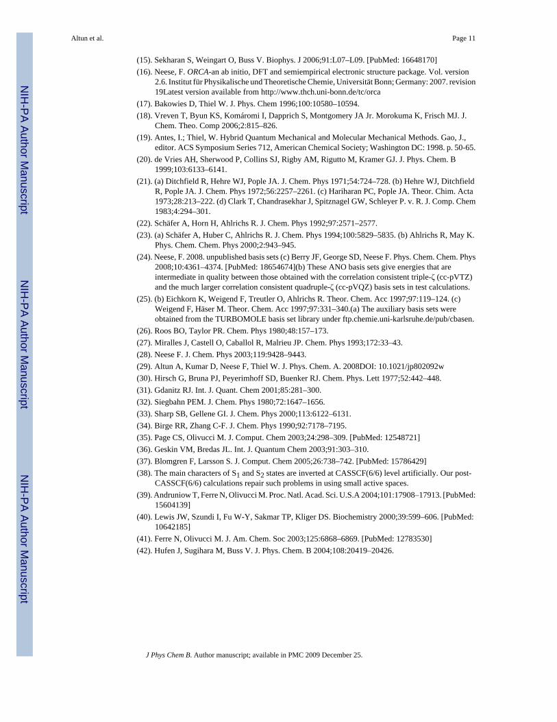
Dominant configurations in S0, S1, and S2 states are the closed-shell singlet, the HOMO toLUMO single excitation, and the HOMO-1 to LUMO single excitation, respectively (seeFigure 3).38 If HF or CASSCF geometry with overestimated polyene chain BLA is used,LUMO is artificially destabilized and thus S1 and S2 transitions shift to blue. At the B3LYPgeometries, HOMO-1 or HOMO to LUMO double excitation contributes significantly in theS2 transition since LUMO becomes stable enough with the decreased BLA. In other words,the decreased (increased) BLA results in the red (blue) shift in the S1 and S2 excitation energies.
At the CASSCF geometries, half of the positive charge located on the CNH2 moiety movestoward the β-ionone ring upon S1 excitation, consistent with previous studies.8,39 Thisoriginates from the nature of the LUMO, whose π-electron density decreases from the SBterminal to β-ionone ring (Figure 3). However, there appears to be no such charge transfer atB3LYP geometries. Therefore, the charge transfer effect seen at CASSCF geometries upon theS1 transition is artificial and arises from the overestimation of BLA.
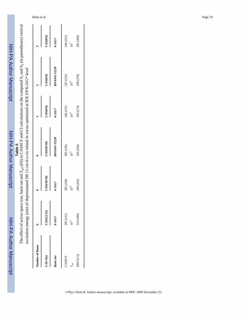
B. Deprotonated SBR in VacuoThe conjugation scheme in the frontier orbitals of the charged PSBR (Figure 3a-e) and neutralSBR (Figure 3f) is different from each other. The effects of computational approximationsused for reducing computational cost in CI calculations may thus be different between the twosystems. This leads us to investigate several computational factors in accessing the error inpost-CASSCF(6/6)/6-31G* calculations of deprotonated SB 11-cis-retinal with Tsel = 10-6
Eh, as shown in Table 5. These analyses are especially important for estimating the error in ab
Altun et al. Page 6
J Phys Chem B. Author manuscript; available in PMC 2009 December 25.
NIH
-PA Author Manuscript
NIH
-PA Author Manuscript
NIH
-PA Author Manuscript
initio QM/MM calculations of visual pigments with deprotonated SBR, such as E113Q mutantof rhodopsins.
Reducing the CAS(12/12) to CAS(6/6) blue-shifts the CASSCF S1 excitation energy of SBRby ca. 40 nm, whereas the CASSCF S2 excitation energy is not affected. It also has 27 (35) nmblue-shift effect for the S1 (S2) DDCI2+Q excitation energy. Tightening Tsel from 10-6 to10-7 Eh does not have much influence on the S1 or S2 excitation energies. Although the S1 andS2 CASSCF as well as the S2 DDCI2+Q excitation energies are not affected much by the basisset extension from 6-31G* to BNANO-TZ2P, the S1 DDCI2+Q excitation energy blue-shiftsby 21 and 15 nm at CAS(10/10) and CAS(6/6) levels, respectively. With the summation ofthese error estimates, our practical DDCI2+Q(6/6)/6-31+G* calculations are expected to havea blue-shift error of ca. 50 (35) nm for the S1 (S2) excitation energy of SBR relative to theDDCI2+Q(12/12)/BNANO-TZ2P.
C. Bovine Rhodopsin and its MutantsBovine Rh and its mutants with PSBR have two absorption maxima at around 500 nm and 380nm. In our SORCI+Q/MM and MRCISD+Q/MM calculations, S1 is an absorbing state (f =1.2-2.0) whereas S2 is a nonabsorbing state with f ~ 0 due to structural distortions in the proteinenvironment. Therefore, the S2 state in these calculations cannot be assigned to theexperimental absorption band at around 380 nm. As the spectral tuning of visual pigments ismainly discussed on the S1 absorbing state, we will not address the tuning of the secondabsorbing state, which will require an extension of the present 3-root calculations. Fordeprotonated SBR, the S1 and S2 states are very close in energy (within 10-20 nm), and S2 maybecome absorbing rather than S1 depending on the computational settings; here we only discussthe absorbing state (f = 1.4-2.0) in relating the calculations to the experiments.
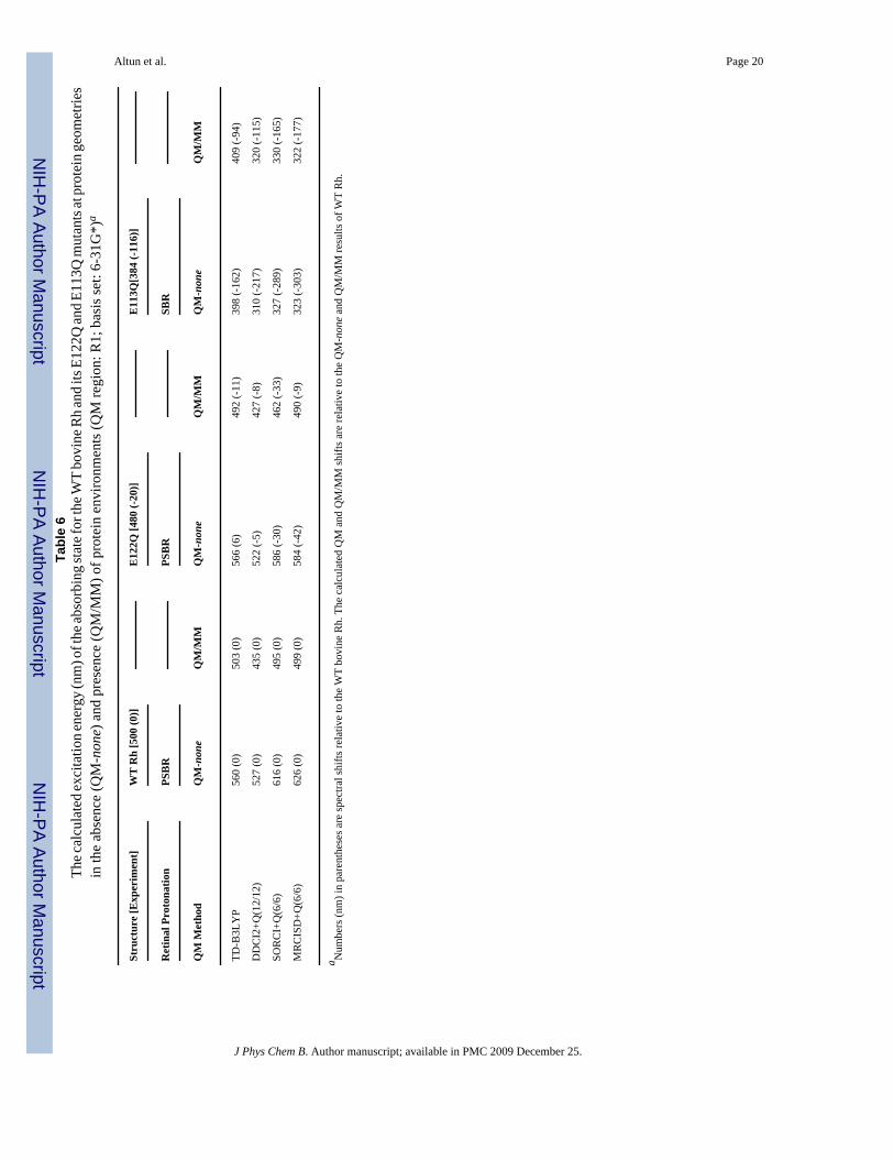
The Effects of Protein Environment—The QM part used in the QM/MM calculation inthis section is R1, the full 11-cis-retinal along with covalently bound N/NH moiety of Lys296for deprotonated/protonated SB linkage. Geometries were obtained by ONIOM(B3LYP:AMBER)-EE optimization. QM/MM excitation energy calculations with TD-B3LYP, SORCI+Q and MRCISD+Q reproduce experimental absorption maximum of WTbovine Rh perfectly (experiment: 500 nm), as shown in Table 6. In the present QM/MMcalculations with TD-B3LYP, DDCI2+Q, SORCI+Q and MRCISD+Q, the experimentalspectral shift of -20 nm with E122Q mutation2,5 is also reproduced within 10 nm. Hence, allof these methods can be used in evaluating QM/MM spectral shifts of PSBR with mutations.
The polarizing protein environment induces an increased BLA for the polyene chain.5 ThisBLA increase is more significant with B3LYP (ca. 0.03 Å5) than with CASSCF (ca. 0.01Å8). Thus, CASSCF geometries in protein environments are more similar to B3LYP ones andmore reliable than those in the gas phase. Actually, previous CASPT2/MM S1 excitationenergies at CASSCF/AMBER and DFTB (being similar to B3LYP one, see ref 5) geometriesare very similar to each other (480 and 506 nm, respectively) for bovine Rh.8,39
In our previous TD-B3LYP/MM study,5 we reproduced experimental spectral shifts due tomutations on bovine Rh within 10 nm except for E113Q mutant. Here we reinvestigate theE113Q mutant with SBR by using MR methods. As our post-CASSCF calculations areexpected to have ca. 15 (50) nm red (blue) shift error for the absorbing state of PSBR (SBR),our MR calculations with present settings should overestimate the spectral shift going fromWT Rh to E113Q mutant by ca. 65 nm. As expected, SORCI+Q(6/6)/MM and MRCISD+Q(6/6)/MM shifts are ca. 65 nm larger than the experimental shift.40 Whereas DDCI2+Q/MMreproduces the SORCI+Q/MM and MRCISD+Q/MM excitation energies of SBR (Table 6),its result for S1 of PSBR is ca. 65 nm blue-shifted since some contributing double excitationsare ignored. Therefore, going from PSBR to SBR, the error arising from our computational
Altun et al. Page 7
J Phys Chem B. Author manuscript; available in PMC 2009 December 25.
NIH
-PA Author Manuscript
NIH
-PA Author Manuscript
NIH
-PA Author Manuscript
settings (ca. 50 nm red shift with CAS(12/12)) and the intrinsic error of DDCI2 for S1 of PSBR(ca. 65 nm blue shift) tend to cancel to each other. Hence, DDCI2+Q(12/12)/MM reproducesthe experimental shift due to error cancellation without applying any correction.
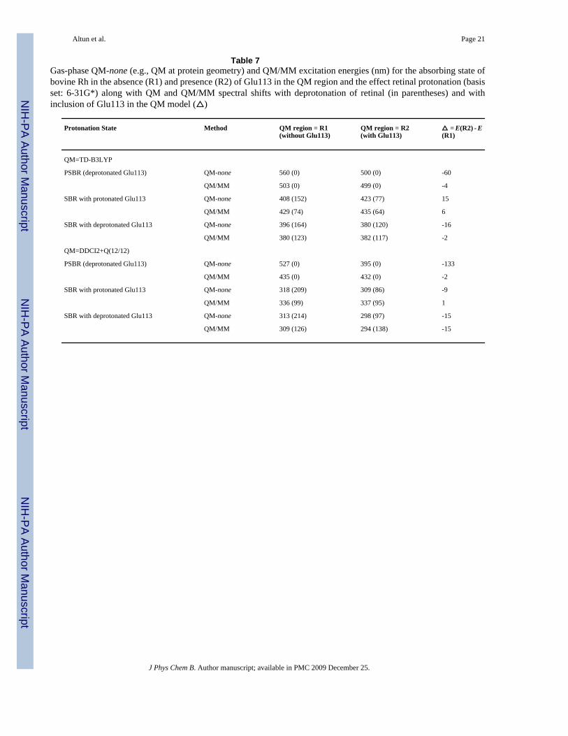
The Effects of Glu113 Counterion and Retinal Protonation—DDCI2+Q reproducesthe experimental shifts reasonably well with the current settings as a result of error cancellation.Therefore, we will assess the effect of Glu113 counterion on the absorbing state of 11-cis-retinal with 6-root DDCI2+Q(12/12)/6-31G* calculations, as shown in Table 7.
When Glu113 is included in the gas-phase QM model of PSBR at the protein geometry, theS1 excitation energy blue-shifts at DDCI2+Q level by ca. 130 nm. This shift agrees withprevious CASPT2 results within 30 nm.6,8,39,41,42 The remaining protein environment red-shifts the S1 excitation energy by ca. 35 nm, similar to a previous CASPT2/ANO study atDFTB geometry.6 This shift is significantly larger (ca. 100 nm) in CASPT2 studies at CASSCFgas phase geometry,8,39 which should not be used because of overestimated BLA. Thecalculated S1 excitation energy of PSBR at TD-B3LYP/MM and DDCI2+Q/MM levels doesnot depend on the presence or absence of Glu113 in the QM region (R1 vs R2). Hence, Glu113contributes to the S0 and S1 states equally and the Glu113 point-charge model can be safelyused in calculating the S1 excitation energy of retinal.
Although previous SACCI/AMBER9 and aug-MCQDPT2/EFP7 calculations reproduce theexperimental absorption maximum of bovine Rh, their energy contributions are somewhatdifferent to each other and from the other studies. First, concerning the SACCI/AMBER result,inclusion of Glu113 in the QM region, e.g. R2, is necessary to reproduce experimental S1absorption maximum. It blue-shifts the SACCI/AMBER S1 excitation energy by ca. 100 nm.9 However, such a charge transfer effect from Glu113 to retinal has not been found in thepresent ab initio MR-QM/MM and the previous TD-B3LYP/MM calculations. Furthermore,the SACCI S1 excitation energy with R1 has a large error of more than 200 nm in the gas phase.The source of errors in the SACCI and SACCI/MM excitation energies with R1 remains apuzzle.9,13 Next, concerning the aug-MCQDPT2/EFP7 result, the protein environment aroundretinal and Glu113 (an R2-like QM region) red-shifts S1 excitation energy (QM-none = 388nm vs. QM/MM = 515 nm) more than 100 nm.7 However, all previous calculations with R2(TD-B3LYP,5 CASPT26 and the present DDCI2+Q) find a small contribution from the proteinenvironment. It seems that this discrepancy comes from significant blue-shift error in the gas-phase aug-MCQDPT2 result.7
TD-B3LYP has a significant error of 110 nm for the S1 excited state of protonated R1 in thegas phase (Table 3) as a result of the use of nonlocal exchange functionals.13 The gas phaseTD-B3LYP S1 excitation energy of protonated R1 at the protein geometry has a smaller errorof ca. 60 nm, i.e. the difference between TD-B3LYP gas-phase spectral shifts relating to R1with the corresponding DDCI2+Q shifts in Table 7. The inclusion of the effect of Glu113 fixesthe error in TD-B3LYP calculations significantly. Hence, TD-B3LYP can be useful forestimating excitation energies in protein environments both with R1 and R2 QM regions aswell as in the gas phase with R2 (but not with R1). This means that the conclusions of theprevious TD-B3LYP/AMBER study remain true although the gas phase results with R1 areblue-shifted.5
Deprotonation of retinal blue-shifts the absorption maximum by ca. 90 nm (Table 7). Theinclusion of Glu113 in the QM region and/or of the protein environment does not affect theSBR excitation energies much in both TD-B3LYP and DDCI2+Q calculations (less than 20nm, see ref 5 for detailed discussions). When the interaction between SBR and its counterionis weakened, the absorbing state blue-shifts, as seen from the comparison of the results withthe protonated and deprotonated Glu113.
Altun et al. Page 8
J Phys Chem B. Author manuscript; available in PMC 2009 December 25.
NIH
-PA Author Manuscript
NIH
-PA Author Manuscript
NIH
-PA Author Manuscript
IV. Discussion and ConclusionsWe have investigated vertical excitation energies of retinal in vacuo and bovine Rh along withits E122Q and E113Q mutants by using MR ab initio methods in combination with the QM/MM method in order to clarify the origins of differences among various previously publishedcalculations and to provide computational strategies for accurately calculating absorptionspectra. MR excitation energy calculations are too demanding with sufficiently large QMregions, active spaces, basis sets and configuration selections. Therefore, we need to adjustthese computational settings looser than desired, to a certain extent. Taking each of the settingsat a desired level while all of the remaining ones are fixed at lower levels, we roughly estimatedthe errors in reducing computational accuracy. For example, the post-CASSCF(6/6)/6-31G*calculations with Tsel = 10-6 Eh has a red-shift (blue-shift) error of ca. 15 (50) nm for the S1absorbing state of PSBR (SBR). Applying these error corrections to SORCI+Q(6/6)/6-31G*and MRCISD+Q(6/6)/6-31G* excitation energies with Tsel = 10-6 Eh, the experimentalabsorption maxima are reproduced quite well.
The relatively inexpensive DDCI2+Q method has an intrinsic blue-shift error of ca. 65 nm incalculating the S1 excitation energy of PSBR in the gas-phase and protein environment as aresult of the ignored double excitations. As this error is constant, (a) absolute values ofexcitation energies of PSBR can be estimated from the calculated DDCI2+Q excitationenergies and (b) DDCI2+Q reproduces experimental spectral shifts of PSBR due to mutationreasonably well. The absorbing excitation energy of SBR calculated with DDCI2+Q is almostthe same as that with higher level SORCI+Q and MRCISD+Q calculations, e.g. ca. 60 nmblue-shifted compared with experiment due to computational settings. As this error in SBRand the intrinsic error in PSBR DDCI2+Q excitation energies are almost the same, theexperimental spectral shift due to deprotonation of retinal can be reproduced well by using thebare DDCI2+Q results.
The TD-B3LYP S1 excitation energy of retinal in the gas phase has a significant blue-shifterror arising from the use of nonlocal exchange functionals.13 The net error in the excitationenergy calculations with TD-B3LYP diminishes significantly when including the effect of H-bonding counterion (Glu113) of retinal.
The level of ground-state geometry optimization has a large impact on the computed verticalexcitation energies. In the post-CASSCF excitation energy calculations, B3LYP geometriesgive results that are consistent with experiment both in the gas phase and in the proteinenvironment. S1 may have up to 100 nm blue-shift error in the gas phase if the CASSCFstructure with overestimated BLA is used. However, at CASSCF/MM geometries, the error inthe post-CASSCF/MM excitation energies is small since polarizing field of proteinenvironment makes the CASSCF structure closer to the B3LYP structure. Our calculationsindicate that the large error in the previous CASPT2 S1 excitation energy in vacuo12 arisesmainly from the use of an inappropriate CASSCF geometry.
The previous8,39 and present calculations at CASSCF geometries find that half of the positivecharge located on the SB terminal moves toward β-ionone ring upon S0 to S1 excitation.However, there appears to be no such charge transfer at the more appropriate B3LYPgeometries.
All-trans to 11-cis isomerization of retinal has no significant effect on the S1 excitation energyin our MR calculations, in agreement with experiments,3,4 showing almost the same absorptionspectra for both isomers. The 6-s-cis conformer of all-trans retinal is more stable than the 6-s-trans one in the present MR calculations. Moreover, its calculated S2 excitation energy is moreconsistent with the experimental value. Therefore, experimental spectra of all-trans retinal3,4 belong mainly to the 6-s-cis conformer, in agreement with ground state energetics. Our
Altun et al. Page 9
J Phys Chem B. Author manuscript; available in PMC 2009 December 25.
NIH
-PA Author Manuscript
NIH
-PA Author Manuscript
NIH
-PA Author Manuscript
calculations also suggest that the shoulder of the experimental absorption spectrum of retinalin vacuo at the S1 absorbing band belongs to the second electronic transition (S2), contrary toprevious tentative assignment to vibrational state of S1 or to the S1 band of a retinal isomar.
Glu113 has a blue-shift effect of ca. 130 nm on the S1 excitation energy of PSBR whereas ithas almost no effect on that of SBR. The net Glu113 effect for PSBR is mainly electrostaticin the present MR and the previous TD-B3LYP5 calculations. In other words, S0 to S1excitation does not involve any charge transfer from Glu113 to retinal. Thus, it is appropriateto place Glu113 in the MM part of QM/MM calculations. The protein environment of retinalother than Glu113 has small effect on the S1 excitation energy. Deprotonation of retinal mayplay an important role in the UV vision of vertebrates. It blue-shifts the S1 excitation energyby ca. 90 nm.
In summary, the present calculations pinpoint the origin of most of the conflicting issues inliterature and thus enhance significantly our understanding on spectral tuning mechanism inretinal systems. The relatively inexpensive DDCI2+Q method can be used in calculatingspectral shifts. TD-B3LYP is also useful in calculating the S1 excitation energy shifts in proteinenvironments.
Supplementary MaterialRefer to Web version on PubMed Central for supplementary material.
AcknowledgmentWe thank Dr. Frank Neese for supplying his ORCA program package and for invaluable discussions. We also thankDrs. Hiroshi Nakatsuji and Jun-ya Hasegawa for discussions. This work at Emory is supported by a grant from theNational Institutes of Health (R01EY016400-03), and the work at Kyoto is in part supported by Japan Science andTechnology Agency (JST) with a Core Research for Evolutional Science and Technology (CREST) grant in the Areaof High Performance Computing for Multiscale and Multiphysics Phenomena.
References and Notes(1). Yokoyama S. Prog. Ret. Eye Res 2000;19:385–419.(2). Altun A, Yokoyama S, Morokuma K. PhotoChem. PhotoBiol 2008;84:845–854. [PubMed:
18331400](3). Andersen LH, Nielsen IB, Kristensen MB, El Ghazaly MOA, Haacke S, Nielsen MB, Petersen MA.
J. Am. Chem. Soc 2005;127:12347–12350. [PubMed: 16131214](4). Nielsen IB, Lammich L, Andersen LH. Phys. Rev. Lett 2006;96:018304/1–4. [PubMed: 16486529](5). Altun A, Yokoyama S, Morokuma K. J. Phys. Chem. B 2008;112:6814–6827. [PubMed: 18473437](6). Sekharan S, Sugihara M, Buss V. Angew. Chem. Int. Ed 2007;46:269–271.(7). Bravaya K, Bochenkova A, Granowsky A, Nemukhin A. J. Am. Chem. Soc 2007;129:13035–13042.
[PubMed: 17924622](8). Coto PB, Strambi A, Ferre N, Olivucci M. Proc. Natl. Acad. Sci. U.S.A 2006;103:17154–17159.
[PubMed: 17090682](9). Fujimoto K, Hayashi S, Hasegawa J, Nakatsuji H. J. Chem. Theo. Comp 2007;3:605–618.(10). Vreven T, Morokuma K. Theor. Chem. Acc 2003;109:125–132.(11). Hoffmann M, Wanko M, Strodel P, König PH, Frauenheim T, Schulten K, Thiel W, Tajkhorshid
E, Elstner M. J. Am. Chem. Soc 2006;128:10808–10818. [PubMed: 16910676](12). Cembran A, Gonzalez-Luque R, Altoe P, Merchan M, Bernardi F, Olivucci M, Garavelli M. J. Phys.
Chem. A 2005;109:6597–6605. [PubMed: 16834008](13). Wanko M, Hoffmann M, Strodel P, Koslowski A, Thiel W, Neese F, Frauenheim T, Elstner M. J.
Phys. Chem. B 2005;109:3606–3615. [PubMed: 16851399](14). Fujimoto K, Hasegawa J, Hayashi S, Kato S, Nakatsuji H. Chem. Phys. Lett 2005;414:239–242.
Altun et al. Page 10
J Phys Chem B. Author manuscript; available in PMC 2009 December 25.
NIH
-PA Author Manuscript
NIH
-PA Author Manuscript
NIH
-PA Author Manuscript
(15). Sekharan S, Weingart O, Buss V. Biophys. J 2006;91:L07–L09. [PubMed: 16648170](16). Neese, F. ORCA-an ab initio, DFT and semiempirical electronic structure package. Vol. version
2.6. Institut für Physikalische und Theoretische Chemie, Universität Bonn; Germany: 2007. revision19Latest version available from http://www.thch.uni-bonn.de/tc/orca
(17). Bakowies D, Thiel W. J. Phys. Chem 1996;100:10580–10594.(18). Vreven T, Byun KS, Komáromi I, Dapprich S, Montgomery JA Jr. Morokuma K, Frisch MJ. J.
Chem. Theo. Comp 2006;2:815–826.(19). Antes, I.; Thiel, W. Hybrid Quantum Mechanical and Molecular Mechanical Methods. Gao, J.,
editor. ACS Symposium Series 712, American Chemical Society; Washington DC: 1998. p. 50-65.(20). de Vries AH, Sherwood P, Collins SJ, Rigby AM, Rigutto M, Kramer GJ. J. Phys. Chem. B
1999;103:6133–6141.(21). (a) Ditchfield R, Hehre WJ, Pople JA. J. Chem. Phys 1971;54:724–728. (b) Hehre WJ, Ditchfield
R, Pople JA. J. Chem. Phys 1972;56:2257–2261. (c) Hariharan PC, Pople JA. Theor. Chim. Acta1973;28:213–222. (d) Clark T, Chandrasekhar J, Spitznagel GW, Schleyer P. v. R. J. Comp. Chem1983;4:294–301.
(22). Schäfer A, Horn H, Ahlrichs R. J. Chem. Phys 1992;97:2571–2577.(23). (a) Schäfer A, Huber C, Ahlrichs R. J. Chem. Phys 1994;100:5829–5835. (b) Ahlrichs R, May K.
Phys. Chem. Chem. Phys 2000;2:943–945.(24). Neese, F. 2008. unpublished basis sets (c) Berry JF, George SD, Neese F. Phys. Chem. Chem. Phys
2008;10:4361–4374. [PubMed: 18654674](b) These ANO basis sets give energies that areintermediate in quality between those obtained with the correlation consistent triple-ζ (cc-pVTZ)and the much larger correlation consistent quadruple-ζ (cc-pVQZ) basis sets in test calculations.
(25). (b) Eichkorn K, Weigend F, Treutler O, Ahlrichs R. Theor. Chem. Acc 1997;97:119–124. (c)Weigend F, Häser M. Theor. Chem. Acc 1997;97:331–340.(a) The auxiliary basis sets wereobtained from the TURBOMOLE basis set library under ftp.chemie.uni-karlsruhe.de/pub/cbasen.
(26). Roos BO, Taylor PR. Chem. Phys 1980;48:157–173.(27). Miralles J, Castell O, Caballol R, Malrieu JP. Chem. Phys 1993;172:33–43.(28). Neese F. J. Chem. Phys 2003;119:9428–9443.(29). Altun A, Kumar D, Neese F, Thiel W. J. Phys. Chem. A. 2008DOI: 10.1021/jp802092w(30). Hirsch G, Bruna PJ, Peyerimhoff SD, Buenker RJ. Chem. Phys. Lett 1977;52:442–448.(31). Gdanitz RJ. Int. J. Quant. Chem 2001;85:281–300.(32). Siegbahn PEM. J. Chem. Phys 1980;72:1647–1656.(33). Sharp SB, Gellene GI. J. Chem. Phys 2000;113:6122–6131.(34). Birge RR, Zhang C-F. J. Chem. Phys 1990;92:7178–7195.(35). Page CS, Olivucci M. J. Comput. Chem 2003;24:298–309. [PubMed: 12548721](36). Geskin VM, Bredas JL. Int. J. Quantum Chem 2003;91:303–310.(37). Blomgren F, Larsson S. J. Comput. Chem 2005;26:738–742. [PubMed: 15786429](38). The main characters of S1 and S2 states are inverted at CASSCF(6/6) level artificially. Our post-
CASSCF(6/6) calculations repair such problems in using small active spaces.(39). Andruniow T, Ferre N, Olivucci M. Proc. Natl. Acad. Sci. U.S.A 2004;101:17908–17913. [PubMed:
15604139](40). Lewis JW, Szundi I, Fu W-Y, Sakmar TP, Kliger DS. Biochemistry 2000;39:599–606. [PubMed:
10642185](41). Ferre N, Olivucci M. J. Am. Chem. Soc 2003;125:6868–6869. [PubMed: 12783530](42). Hufen J, Sugihara M, Buss V. J. Phys. Chem. B 2004;108:20419–20426.
Altun et al. Page 11
J Phys Chem B. Author manuscript; available in PMC 2009 December 25.
NIH
-PA Author Manuscript
NIH
-PA Author Manuscript
NIH
-PA Author Manuscript
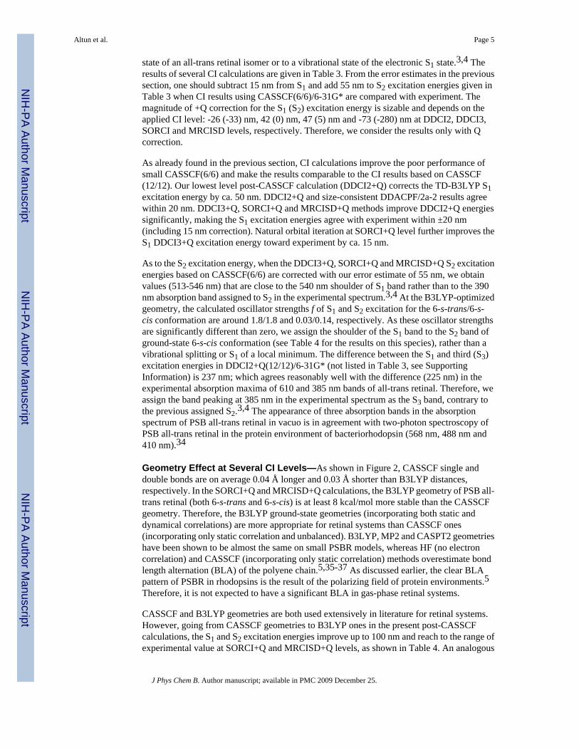
Figure 1.QM models and atom numbering of PSBR. SBR corresponds to removing the hydrogen circledor transferring it to Glu113 carboxylic oxygen.
Altun et al. Page 12
J Phys Chem B. Author manuscript; available in PMC 2009 December 25.
NIH
-PA Author Manuscript
NIH
-PA Author Manuscript
NIH
-PA Author Manuscript
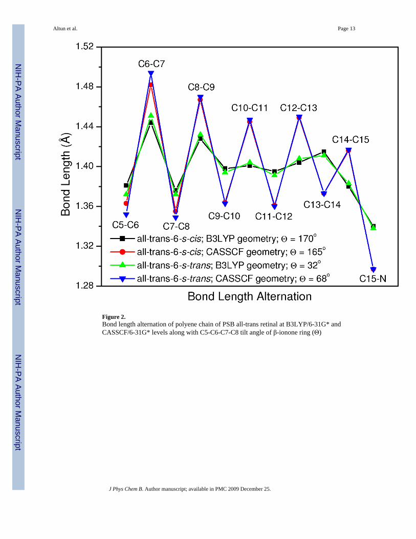
Figure 2.Bond length alternation of polyene chain of PSB all-trans retinal at B3LYP/6-31G* andCASSCF/6-31G* levels along with C5-C6-C7-C8 tilt angle of β-ionone ring (Θ)
Altun et al. Page 13
J Phys Chem B. Author manuscript; available in PMC 2009 December 25.
NIH
-PA Author Manuscript
NIH
-PA Author Manuscript
NIH
-PA Author Manuscript

Figure 3.The key 6-root CASSCF(12/12) orbitals of retinal in vacuo.
Altun et al. Page 14
J Phys Chem B. Author manuscript; available in PMC 2009 December 25.
NIH
-PA Author Manuscript
NIH
-PA Author Manuscript
NIH
-PA Author Manuscript

NIH
-PA Author Manuscript
NIH
-PA Author Manuscript
NIH
-PA Author Manuscript
Altun et al. Page 15
Table 1Basis set dependence of the S1 (S2 in parentheses) vertical excitation energy (nm) of PSB all-trans-6-s-trans retinal invacuo at the 6-root CASSCF(12/12) and DDCI2+Q(12/12) level at the B3LYP/6-31G*-optimized geometry
Basis Set Basis Set Size CASSCF(12/12) DDCI2+Q(12/12)
6-31G* 354 553 (404) 545 (416)
VDZP [or SV(P)] 444 566 (405) 555 (420)
BNANO-DZP 444 559 (405) 555 (437)
TZVP 579 572 (406) 570 (438)
VTZP 600 572 (406) 563 (441)
BNANO-TZ2P 753 552 (404) 550 (437)
J Phys Chem B. Author manuscript; available in PMC 2009 December 25.
NIH
-PA Author Manuscript
NIH
-PA Author Manuscript
NIH
-PA Author Manuscript
Altun et al. Page 16
Table 2The effects of active space size, the number of roots and Tsel (Eh) in post-CASSCF/6-31G* calculations on the computedS1 (S2) vertical excitation energy (nm) of PSB all-trans-6-s-trans retinal in vacuo optimized at B3LYP/6-31G* level
6-root CAS(12/12) 3-root CAS(12/12) 3-root CAS(6/6) 3-root CAS(6/6)
CASSCF 553 (404) 548 (391) 382 (334) 382 (334)
Tsel 10-6 10-6 10-6 10-7
DDCI2+Q 545 (416) 544 (412) 559 (385) 545 (379)
J Phys Chem B. Author manuscript; available in PMC 2009 December 25.

NIH
-PA Author Manuscript
NIH
-PA Author Manuscript
NIH
-PA Author Manuscript
Altun et al. Page 17
Table 3S1 and S2 vertical excitation energies (nm) for PSB all-trans-6-s-trans retinal in vacuo at various CI/6-31G* levelsalong with CASSCF and TD-B3LYP results at the B3LYP/6-31G*-optimized geometry
Method S1 S2
TD-B3LYP 501 380
CASSCF(12/12)a 553 404
CASSCF(6/6) 382 334
DDCI2+Q(12/12)a 545 416
DDCI2+Q(6/6) 559 385
DDACPF/2a-2(12/12)a 527 394
DDCI3+Q(6/6) 640 474
SORCI+Q(6/6) 626 491
MRCISD+Q(6/6) 604 458
a6-root CAS(12/12) results are given for comparison.
J Phys Chem B. Author manuscript; available in PMC 2009 December 25.
NIH
-PA Author Manuscript
NIH
-PA Author Manuscript
NIH
-PA Author Manuscript
Altun et al. Page 18Ta
ble
4G
eom
etry
dep
ende
nce
of th
e S 1
(S2)
exc
itatio
n en
ergy
of P
SB re
tinal
in v
acuo
in M
R c
alcu
latio
ns w
ith 6
-31G
* ba
sis s
et b
ased
on
3-ro
otC
AS(
6/6)
or 6
-roo
t CA
S(12
/12)
leve
ls
CA
SSC
F(12
/12)
CA
SSC
F(6/
6)D
DC
I2+Q
(12/
12)
SOR
CI+
Q(6
/6)
MR
CIS
D+Q
(6/6
)
all-t
rans
-6-s
-tran
s
CA
SSC
F ge
omet
rya
526
(345
)42
1 (2
95)
539
(360
)56
5 (3
95)
603
(412
)
B3L
YP
geom
etry
553
(404
)38
2 (3
34)
545
(416
)62
6 (4
91)
604
(458
)
all-t
rans
-6-s
-cis
CA
SSC
F ge
omet
ryb,
c45
7 (3
12)
415
(328
)47
0 (3
61)
505
(396
)54
6 (4
21)
B3L
YP
geom
etry
d54
5 (3
91)
467
(350
)55
1 (4
12)
610
(475
)60
4 (4
93)
11-c
is-6
-s-c
is
B3L
YP
geom
etry
535
(383
)36
2 (3
13)
541
(403
)e62
5 (4
73)
601
(454
)
a Coo
rdin
ates
take
n fr
om re
f 13 .
C5-
C6
dihe
dral
ang
le w
as fi
xed
to it
s HF
valu
e as
it re
laxe
d to
6-s
-cis
min
imum
.
b Coo
rdin
ates
take
n fr
om re
f 12 .
c 3-ro
ot C
ASP
T2(1
2/12
)/6-3
1G*:
534
(353
) nm
12
d 6-ro
ot C
ASP
T2(1
2/12
)/AN
O a
t MP2
geo
met
ry: 6
06 (4
36) n
m6
e 3-ro
ot D
DC
I2+Q
(6/6
)/6-3
1G*:
556
(370
) nm
J Phys Chem B. Author manuscript; available in PMC 2009 December 25.
NIH
-PA Author Manuscript
NIH
-PA Author Manuscript
NIH
-PA Author Manuscript
Altun et al. Page 19Ta
ble
5Th
e eff
ect o
f act
ive s
pace
size
, bas
is se
t and
Tse
l (Eh
) in
CA
SSC
F an
d C
I cal
cula
tions
on
the c
ompu
ted
S 1 an
d S 2
(in
pare
nthe
ses)
ver
tical
exci
tatio
n en
ergy
(nm
) of d
epro
tona
ted
SB 1
1-ci
s-6-
s-ci
s ret
inal
in v
acuo
opt
imiz
ed a
t B3L
YP/
6-31
G*
leve
l
Num
ber
of R
oots
66
63
33
CA
S Si
zeC
AS(
12/1
2)C
AS(
10/1
0)C
AS(
10/1
0)C
AS(
6/6)
CA
S(6/
6)C
AS(
6/6)
Bas
is S
et6-
31G
*6-
31G
*B
NA
NO
-TZ
2P6-
31G
*B
NA
NO
-TZ
2P6-
31G
*
CA
SSC
F28
5 (2
31)
285
(230
)28
5 (2
30)
246
(231
)24
7 (2
35)
246
(231
)
T sel
10-6
10-6
10-6
10-6
10-6
10-7
DD
CI2
+Q31
0 (3
09)
299
(292
)32
0 (2
94)
283
(274
)29
8 (2
78)
282
(269
)
J Phys Chem B. Author manuscript; available in PMC 2009 December 25.
NIH
-PA Author Manuscript
NIH
-PA Author Manuscript
NIH
-PA Author Manuscript
Altun et al. Page 20Ta
ble
6Th
e cal
cula
ted
exci
tatio
n en
ergy
(nm
) of t
he ab
sorb
ing
stat
e for
the W
T bo
vine
Rh
and
its E
122Q
and
E113
Q m
utan
ts at
pro
tein
geo
met
ries
in th
e ab
senc
e (Q
M-n
one)
and
pre
senc
e (Q
M/M
M) o
f pro
tein
env
ironm
ents
(QM
regi
on: R
1; b
asis
set:
6-31
G*)
a
Stru
ctur
e [E
xper
imen
t]W
T R
h [5
00 (0
)]E
122Q
[480
(-20
)]E
113Q
[384
(-11
6)]
Ret
inal
Pro
tona
tion
PSB
RPS
BR
SBR
QM
Met
hod
QM
-non
eQ
M/M
MQ
M-n
one
QM
/MM
QM
-non
eQ
M/M
M
TD-B
3LY
P56
0 (0
)50
3 (0
)56
6 (6
)49
2 (-
11)
398
(-16
2)40
9 (-
94)
DD
CI2
+Q(1
2/12
)52
7 (0
)43
5 (0
)52
2 (-
5)42
7 (-
8)31
0 (-
217)
320
(-11
5)
SOR
CI+
Q(6
/6)
616
(0)
495
(0)
586
(-30
)46
2 (-
33)
327
(-28
9)33
0 (-
165)
MR
CIS
D+Q
(6/6
)62
6 (0
)49
9 (0
)58
4 (-
42)
490
(-9)
323
(-30
3)32
2 (-
177)
a Num
bers
(nm
) in
pare
nthe
ses a
re sp
ectra
l shi
fts re
lativ
e to
the
WT
bovi
ne R
h. T
he c
alcu
late
d Q
M a
nd Q
M/M
M sh
ifts a
re re
lativ
e to
the
QM
-non
e an
d Q
M/M
M re
sults
of W
T R
h.
J Phys Chem B. Author manuscript; available in PMC 2009 December 25.
NIH
-PA Author Manuscript
NIH
-PA Author Manuscript
NIH
-PA Author Manuscript
Altun et al. Page 21
Table 7Gas-phase QM-none (e.g., QM at protein geometry) and QM/MM excitation energies (nm) for the absorbing state ofbovine Rh in the absence (R1) and presence (R2) of Glu113 in the QM region and the effect retinal protonation (basisset: 6-31G*) along with QM and QM/MM spectral shifts with deprotonation of retinal (in parentheses) and withinclusion of Glu113 in the QM model (△)
Protonation State Method QM region = R1(without Glu113)
QM region = R2(with Glu113)
△ = E(R2) - E(R1)
QM=TD-B3LYP
PSBR (deprotonated Glu113) QM-none 560 (0) 500 (0) -60
QM/MM 503 (0) 499 (0) -4
SBR with protonated Glu113 QM-none 408 (152) 423 (77) 15
QM/MM 429 (74) 435 (64) 6
SBR with deprotonated Glu113 QM-none 396 (164) 380 (120) -16
QM/MM 380 (123) 382 (117) -2
QM=DDCI2+Q(12/12)
PSBR (deprotonated Glu113) QM-none 527 (0) 395 (0) -133
QM/MM 435 (0) 432 (0) -2
SBR with protonated Glu113 QM-none 318 (209) 309 (86) -9
QM/MM 336 (99) 337 (95) 1
SBR with deprotonated Glu113 QM-none 313 (214) 298 (97) -15
QM/MM 309 (126) 294 (138) -15
J Phys Chem B. Author manuscript; available in PMC 2009 December 25.