The acute pulmonary inflammatory response to the graded severity of smoke inhalation injury*
20
The acute pulmonary inflammatory response to the graded severity of smoke inhalation injury Joslyn M. Albright, MD, Christopher S. Davis, MD, MPH, Melanie D. Bird, PhD, Luis Ramirez, BS, Hajwa Kim, MS, MA § , Ellen L. Burnham, MD, MS € , Richard L. Gamelli, MD, FACS, and Elizabeth J. Kovacs, PhD Burn and Shock Trauma Institute, Department of Surgery, Loyola University Medical Center, Maywood, IL § Center for Clinical and Translational Science, University of Illinois at Chicago, Chicago, IL € University of Colorado Denver School of Medicine, Department of Medicine, Division of Pulmonary Sciences and Critical Care Medicine, Aurora, Colorado Abstract Objective—To determine if the graded severity of smoke inhalation is reflected by the acute pulmonary inflammatory response to injury. Design—In a prospective observational study we assessed the bronchoalveolar lavage fluid (BALF) for both leukocyte differential and concentration of 28 cytokines, chemokines, and growth factors. Results were then compared to the graded severity of inhalation injury as determined by Abbreviated Injury Score criteria (0: None, 1: Mild, 2: Moderate, 3: Severe, 4: Massive). Setting—All patients were enrolled at a single tertiary burn center. Patients—The BALF was obtained from 60 patients within 14 hours of burn injury who underwent bronchoscopy for suspected smoke inhalation. Interventions—None. Measurements and Main Results—Those who presented with worse grades of inhalation injury had higher plasma levels of carboxyhemoglobin and enhanced airway neutrophilia. Patients with the most severe inhalation injuries also had a greater requirement for tracheostomy, longer time on the ventilator, and a prolonged stay in the intensive care unit. Of the 28 inflammatory mediators assessed in the BALF, 21 were at their highest in those with the worst inhalation injury scores (Grades 3 and 4), the greatest of which was interleukin (IL)-8 (92,940 pg/ml, Grade 4). When compared in terms of low inhalation injury (Grades 1–2) versus high inhalation injury (Grades 3–4), we found significant differences between groups for IL-4, IL-6, IL-9, IL-15, interferon-γ, granulocyte-macrophage colony-stimulating factor, and monocyte chemotactic protein-1 (p<0.05 for all). Conclusions—These data reveal that the degree of inhalation injury: 1) has basic and profound effects on burn patient morbidity; 2) evokes complex changes of multiple alveolar inflammatory proteins; and 3) is a determinant of the pulmonary inflammatory response to smoke inhalation. Accordingly, future investigations should consider inhalation injury to be a graded phenomenon. Corresponding Author: Elizabeth J. Kovacs, PhD, Professor and Vice Chair of Research, Department of Surgery, Director of Research, Burn & Shock Trauma Institute, Director, Alcohol Research Program, Loyola University Medical Center, Stritch School of Medicine, Building 110, Room 4232, 2160 South First Avenue, Maywood, IL 60153, Office: 708-327-2477, Fax: 708-327-2813, Lab: 708-327-2438, [email protected]. NIH Public Access Author Manuscript Crit Care Med. Author manuscript; available in PMC 2013 April 1. Published in final edited form as: Crit Care Med. 2012 April ; 40(4): 1113–1121. doi:10.1097/CCM.0b013e3182374a67. NIH-PA Author Manuscript NIH-PA Author Manuscript NIH-PA Author Manuscript
-
Upload
independent -
Category
Documents
-
view
4 -
download
0
Transcript of The acute pulmonary inflammatory response to the graded severity of smoke inhalation injury*
The acute pulmonary inflammatory response to the gradedseverity of smoke inhalation injury
Joslyn M. Albright, MD, Christopher S. Davis, MD, MPH, Melanie D. Bird, PhD, LuisRamirez, BS, Hajwa Kim, MS, MA§, Ellen L. Burnham, MD, MS€, Richard L. Gamelli, MD,FACS, and Elizabeth J. Kovacs, PhDBurn and Shock Trauma Institute, Department of Surgery, Loyola University Medical Center,Maywood, IL§Center for Clinical and Translational Science, University of Illinois at Chicago, Chicago, IL€University of Colorado Denver School of Medicine, Department of Medicine, Division ofPulmonary Sciences and Critical Care Medicine, Aurora, Colorado
AbstractObjective—To determine if the graded severity of smoke inhalation is reflected by the acutepulmonary inflammatory response to injury.
Design—In a prospective observational study we assessed the bronchoalveolar lavage fluid(BALF) for both leukocyte differential and concentration of 28 cytokines, chemokines, and growthfactors. Results were then compared to the graded severity of inhalation injury as determined byAbbreviated Injury Score criteria (0: None, 1: Mild, 2: Moderate, 3: Severe, 4: Massive).
Setting—All patients were enrolled at a single tertiary burn center.
Patients—The BALF was obtained from 60 patients within 14 hours of burn injury whounderwent bronchoscopy for suspected smoke inhalation.
Interventions—None.
Measurements and Main Results—Those who presented with worse grades of inhalationinjury had higher plasma levels of carboxyhemoglobin and enhanced airway neutrophilia. Patientswith the most severe inhalation injuries also had a greater requirement for tracheostomy, longertime on the ventilator, and a prolonged stay in the intensive care unit. Of the 28 inflammatorymediators assessed in the BALF, 21 were at their highest in those with the worst inhalation injuryscores (Grades 3 and 4), the greatest of which was interleukin (IL)-8 (92,940 pg/ml, Grade 4).When compared in terms of low inhalation injury (Grades 1–2) versus high inhalation injury(Grades 3–4), we found significant differences between groups for IL-4, IL-6, IL-9, IL-15,interferon-γ, granulocyte-macrophage colony-stimulating factor, and monocyte chemotacticprotein-1 (p<0.05 for all).
Conclusions—These data reveal that the degree of inhalation injury: 1) has basic and profoundeffects on burn patient morbidity; 2) evokes complex changes of multiple alveolar inflammatoryproteins; and 3) is a determinant of the pulmonary inflammatory response to smoke inhalation.Accordingly, future investigations should consider inhalation injury to be a graded phenomenon.
Corresponding Author: Elizabeth J. Kovacs, PhD, Professor and Vice Chair of Research, Department of Surgery, Director ofResearch, Burn & Shock Trauma Institute, Director, Alcohol Research Program, Loyola University Medical Center, Stritch School ofMedicine, Building 110, Room 4232, 2160 South First Avenue, Maywood, IL 60153, Office: 708-327-2477, Fax: 708-327-2813, Lab:708-327-2438, [email protected].
NIH Public AccessAuthor ManuscriptCrit Care Med. Author manuscript; available in PMC 2013 April 1.
Published in final edited form as:Crit Care Med. 2012 April ; 40(4): 1113–1121. doi:10.1097/CCM.0b013e3182374a67.
NIH
-PA Author Manuscript
NIH
-PA Author Manuscript
NIH
-PA Author Manuscript
KeywordsBurn; Inhalation Injury; Inflammation; Cytokine; Chemokine; Growth Factor
IntroductionIn the United States, 40,000 hospital admissions for burn injury occur each year (1).Approximately 6–20% of fire victims have a concurrent inhalation injury, which is achemical insult rather than a thermal tracheobronchiolitis (2, 3). Acutely, those with aninhalation injury have more pronounced arterial hypotension and require more extensivefluid resuscitation, and the diffuse epithelial sloughing, bronchorrhea, and plugging of sub-segmental bronchi characteristic of inhalation injury can result in subsequent pneumoniarates of 20–50% (4–8). Moreover, the combination of pulmonary and dermal injury maydouble a patient’s mortality (8). Prior investigations in patients with smoke inhalation haverevealed a temporal increase in pro-inflammatory cytokine levels in the airway (9), coupledwith alveolar leukocytes that are “primed” for an enhanced response to stimulation withlipopolysaccharide (10). However, these early studies did not examine inflammatorymediators in the context of severity of inhalation injury. As such, we hypothesized that pro-inflammatory mediators at the alveolar level would increase as the degree of inhalationinjury increased, highlighting the importance of classifying inhalation injury as a gradedphenomenon.
Materials and MethodsFrom January 2007 to April 2010, bronchoalveolar lavage fluid (BALF) was collected from60 patients admitted to the Burn Intensive Care Unit in whom inhalation injury wassuspected by history and/or physical findings, such as injury within a confined space or sootat the nares or tracheal secretions. Patients were excluded from the study for the following:age less than 18 years, malignancy, immunosuppressive medications or known autoimmuneor chronic inflammatory diseases. Clinical variables and outcomes collected were: age,gender, race/ethnicity, % total body surface area (% TBSA) burn, inhalation injury grade,partial pressure of oxygen in arterial blood to fraction of inspired oxygen (P:F) ratio at thetime of bronchoscopy, initial 24 and 72 hour fluid requirements, incidence of pneumonia,incidence of sepsis, ICU and hospital length of stay, and mortality. Pneumonia was definedas any BAL culture of >100,000 colony forming units (CFUs), and sepsis defined ashemodynamic instability in combination with a documented source of infection (blood,urine, BAL, or wound). All samples were collected before any aerosolized pulmonarymedications had been administered. Six non-smoking, non-intubated, non-hospitalizedhealthy adult volunteers (free of pulmonary, cardiac, infectious, and allergic disease) werealso included as a control group for comparison. This study was approved by the LoyolaUniversity Medical Center Institutional Review Board (LU#107372).
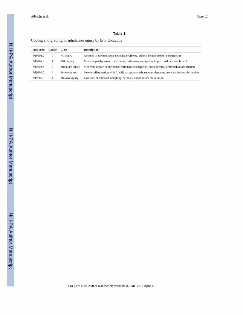
Inhalation injury scoring systemThe degree of inhalation injury was determined using a standardized bronchoscopic scoringsystem based on Abbreviated Injury Score (AIS) criteria as we have previously published(11). The severity of inhalation injury was graded into five categories (0, 1, 2, 3, and 4) with0 being the absence of visible inhalation injury (Table 1).
Bronchoalveolar lavageBronchoscopy and BAL were performed by a standardized protocol within 14 hours in allburn-injured patients (12). That which was not required for routine clinical analysis was
Albright et al. Page 2
Crit Care Med. Author manuscript; available in PMC 2013 April 1.
NIH
-PA Author Manuscript
NIH
-PA Author Manuscript
NIH
-PA Author Manuscript

pooled for research use. In control subjects, the bronchoscope was directed into asubsegment of the right middle lobe and wedged; the first 50 mL aliquot of saline wasinstilled, and the aspirate discarded. Subsequent 50 mL aliquots were instilled into the samesubsegment, and immediately aspirated with gentle hand aspiration into sterile syringes.These were immediately transferred into sterile 50 mL conical tubes, placed on ice, andtransported to the laboratory for additional processing. There were no immediatecomplications to bronchoscopy in either the patient or control groups.
Sample processingBALF samples were strained through sterile 100 micron nylon cell strainers (BDBiosciences, Bedford, MA). The raw sample was centrifuged at 1200 RPM for 5 minutes toseparate the supernatant from the cellular component. The supernatant was then aliquotedand frozen at −80°. Cytokine concentrations in BALF were measured by Bio-Rad MultiplexAssays (Hercules, CA) according to manufacturer protocol. All samples were assayed induplicate and the results analyzed using the Bio-Plex manager software, version 5.0.
Statistical analysisPatient demographics, outcomes, and inflammatory mediator concentrations were assessedfor normality and parametric or non-parametric tests applied where appropriate. Correlationswere performed with Spearman’s rank correlation coefficient, and the r value reported.Continuous variables of parametric tests are reported as mean with standard deviation, andnon-parametric tests reported as median with 25th and 75th percentiles. Otherwisedichotomous variables are reported as a number and percent. Statistical analyses werecalculated with SAS Version 9.1 (SAS Institute Inc., Cary, NC) and corresponding graphscreated with GraphPad Prism 5 for Windows (GraphPad Software, La Jolla, CA). Adifference between observed variables was considered statistically significant when p <0.05.
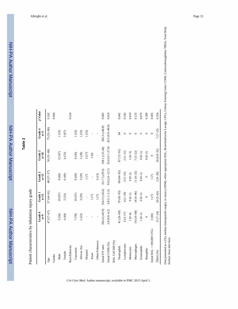
ResultsPatient characteristics by inhalation injury grade
As shown in Table 2, patients with each grade of inhalation injury were similar in terms ofage, gender, race/ethnicity, and % TBSA burn across grades of inhalation injury. Thepercent of plasma COHb measured nearest the time of injury was predictably higher amongpatients with higher grades of inhalation injury. Likewise, the BALF white blood cell(WBC) distribution changed across grades of inhalation injury with a significant shift from amacrophage predominant population to one dominated by neutrophils. Though the initial P:F ratio was lower in those with a Grade 4 injury no significant difference was reached.
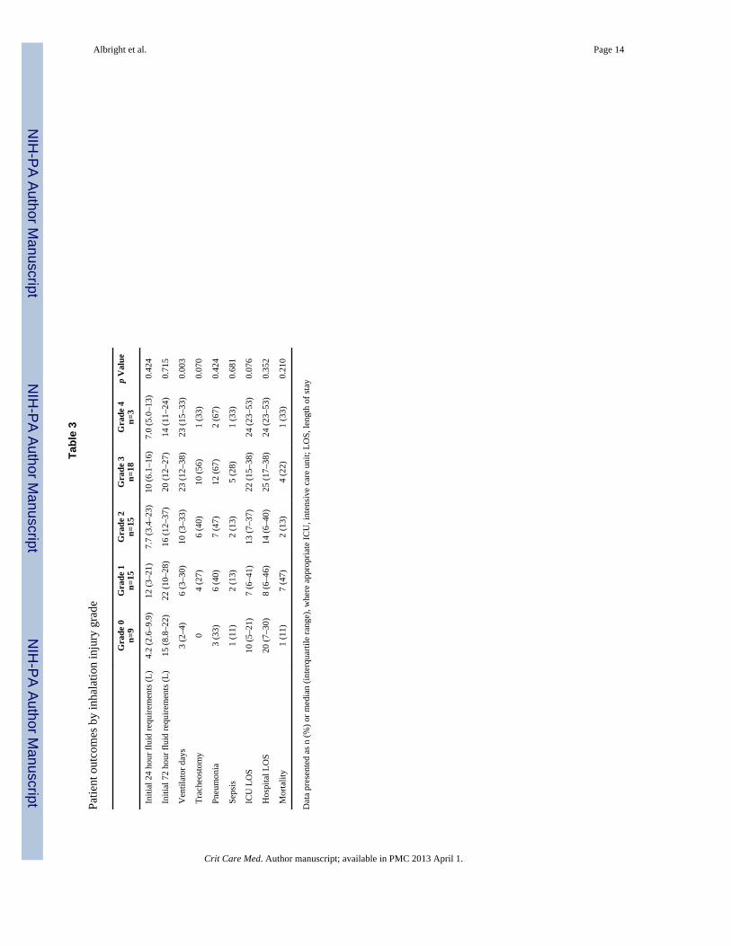
Patient outcomes by inhalation injury gradeAs shown in Table 3, the degree of inhalation injury had neither a significant effect onsubsequent pneumonia, sepsis, hospital length of stay, or mortality, nor did it appear to be arisk factor for mortality when adjusting for the effects of age and % TBSA. However, thosewith a more severe inhalation injury required greater ventilator days and a trend toward agreater rate of tracheostomy and ICU length of stay. When comparing those withoutinhalation injury (Grade 0) to those with any inhalation injury as a single group (Grades 1–4) initial 24-hour fluid requirements were greater (median 4.2 Liters (L) vs 8.8 L,respectively; p=0.036), as were the number of ventilator days (median 3 vs 15; p<0.001) andtracheostomies (0 vs 51%; p=0.021).
Albright et al. Page 3
Crit Care Med. Author manuscript; available in PMC 2013 April 1.
NIH
-PA Author Manuscript
NIH
-PA Author Manuscript
NIH
-PA Author Manuscript
Inflammatory mediators by inhalation injury gradeInflammatory mediators with statistical significance and/or the potential for biologicalrelevance are shown in Figure 1. Across grades of inhalation injury (and excluding controlsfor statistical comparison), the overall median value differed significantly (p < 0.05) forcomplement component 5a (C5a), IL-6, IL-15, interferon (IFN)-γ, monocyte chemotacticprotein-1 (MCP-1), regulated on activation, normal T expressed and secreted (RANTES),tumor necrosis factor (TNF)-α, and vascular endothelial growth factor (VEGF). IL-7 andplatelet derived growth factor (PDGF) were not graphically represented though had similardifferences between medians (p < 0.05). Of all the mediators tested, only VEGFdemonstrated a pattern of decreasing concentrations across the grades of inhalation injury.No protein demonstrated a linear relationship to the grade of inhalation injury.
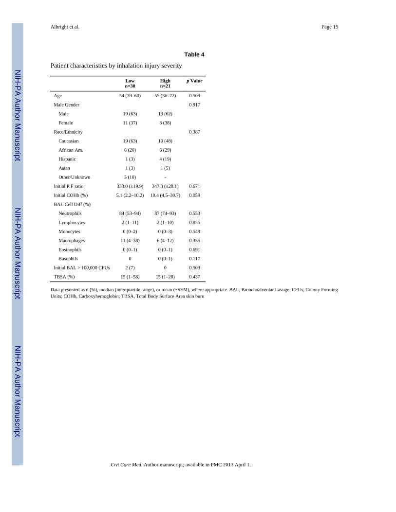
Patient characteristics when grouped by inhalation injury severityIn order to minimize clinician bias during inhalation injury grading, and to better assess theeffects of inhalation injury severity, patients were grouped and compared according to lowinhalation injury severity (Grades 1 and 2) and high inhalation injury severity (Grades 3 and4). Patient characteristics by group are reported in Table 4. Most importantly there were nodifferences in age, gender, race/ethnicity, and % TBSA; moreover, the groups did not differby initial P:F ratio, BALF WBC differential, or initial BALF culture for microorganisms. Onthe contrary, those with a high compared to low inhalation injury severity appeared to have agreater % COHb (median 10.4% vs 5.1%), though this difference only approached statisticalsignificance (p=0.059).
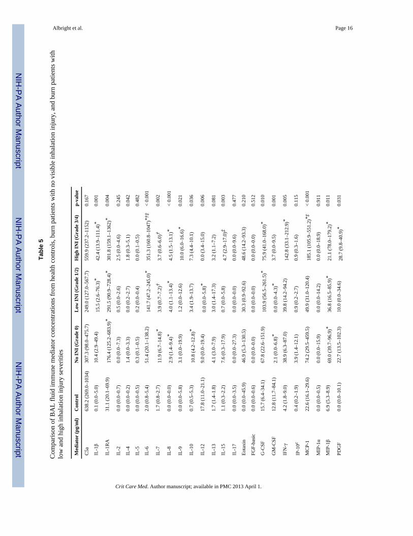
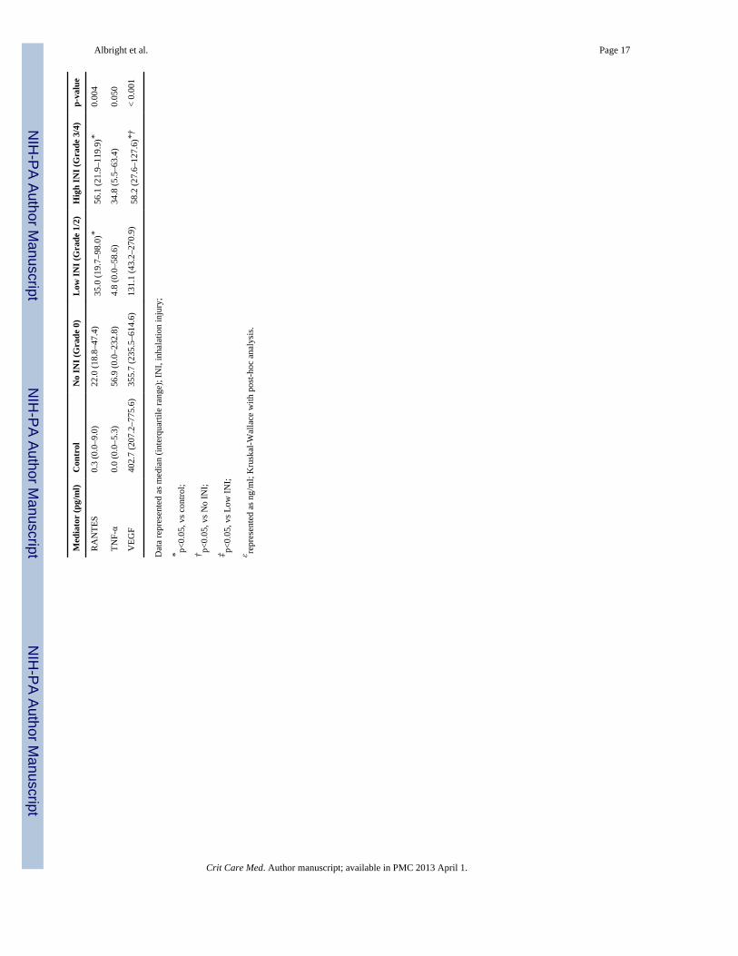
Table 5 demonstrates the comparison of BAL fluid immune mediator concentrations notonly of those with low and high inhalation injury severities, but also of subjects withoutvisible inhalation injury (Grade 0), and controls. Compared to controls, IL-1RA, IL-7, IL-8,IL-10, and MIP-1β were elevated in Grade 0 patients, while the concentration of GM-CSFwas lower. With the addition of inhalation injuries, IL-1β, IL-1RA, IL-6, IL-8, IL-9, G-CSF,IFN-γ, MCP-1, MIP-1β, PDGF, and RANTES were increased over controls and/or Grade 0patients, while IL-7 and VEGF tended to be lower.
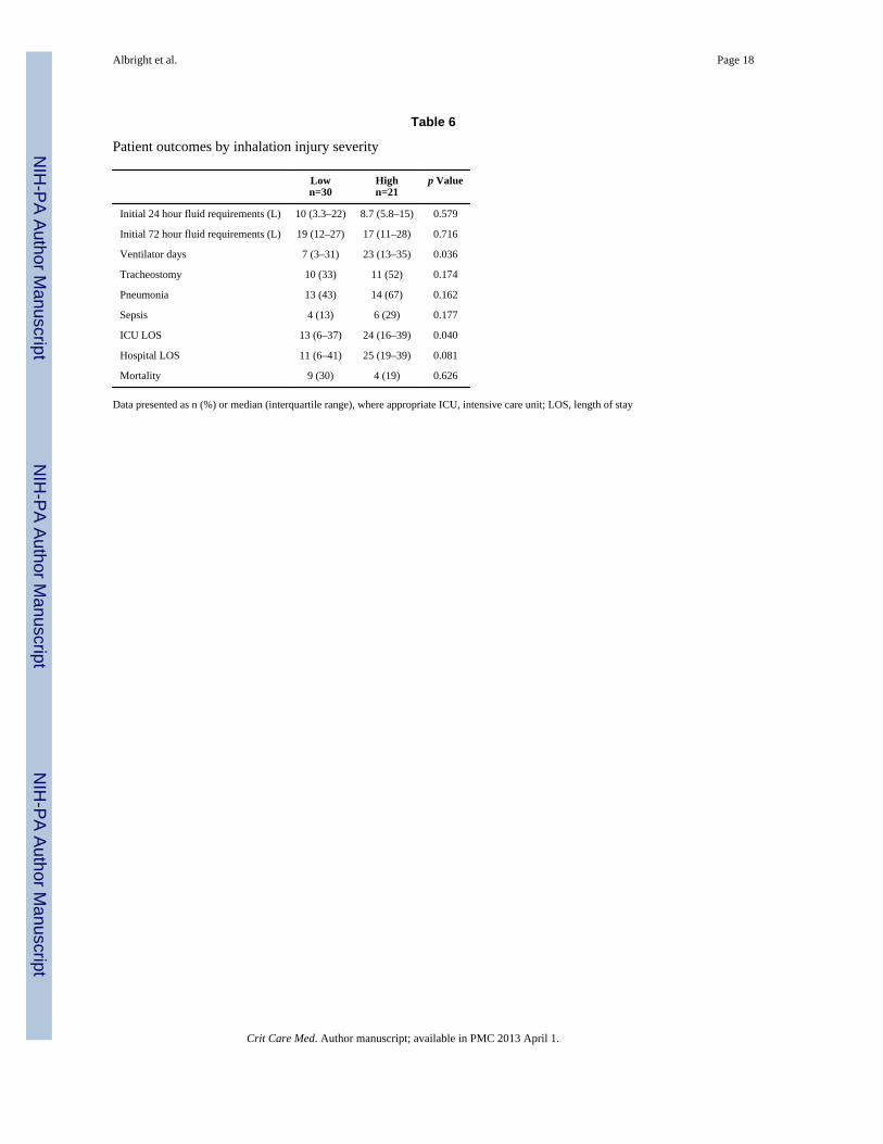
Patient outcomes when grouped by inhalation injury severityAs with worsening inhalation injury grades, the group with the highest inhalation injuryseverity required significantly more ventilator days (p=0.036) and ICU days (p=0.040)compared to those with a more mild inhalation injury severity (Table 6). Though the severityof inhalation injury had no effect on either the resuscitation fluid requirements or mortality,patients with the worst inhalation injury severity also tended to have a greater percentagewho required a tracheostomy (33% vs. 52%), percentage who developed pneumonia orsepsis (43% vs. 67% and 13% vs. 29%), and a longer hospital length of stay (11 vs. 25days).
Inflammatory mediators when grouped by inhalation injury severityIn contrast to the comparison of patient characteristics, numerous differences were foundbetween the inflammatory mediator profile of those with low vs. high inhalation injuryseverities (Figure 2), and the respective differences are as follows: IL-4 (median 0.2 vs. 1.8pg/ml; p=0.048), IL-6 (median 141.7 vs. 351.3 pg/ml; p=0.001), IL-9 (median 1.2 vs. 10.0pg/ml; p=0.015), IL-15 (median 0.7 vs. 4.7 pg/ml; p=0.001), granulocyte-macrophagecolony-stimulating factor (GM-CSF) (median 4.3 vs. 9.5; p=0.040), IFN-γ (median 39.8 vs.142.8 pg/ml; p=0.048), and MCP-1 (median 49.9 vs. 185.1; p=0.001).
Albright et al. Page 4
Crit Care Med. Author manuscript; available in PMC 2013 April 1.
NIH
-PA Author Manuscript
NIH
-PA Author Manuscript
NIH
-PA Author Manuscript
Correlations with Presenting Characteristics and OutcomesGiven that carbon monoxide (CO) has known immune modulating effects (namely anti-inflammatory), we assessed for a correlation between the initial % COHb and BALFimmune mediator concentrations. We found differences that were only suggestive ofsignificance (p<0.10) between % COHb and the BALF concentrations of C5a, IL-1β, IL-8,FGF-basic, IFN-γ, MCP-1, MIP-1β, and PDGF. Furthermore, the % COHb did not correlatewith initial P:F ratio (r: −0.184; p=0.187).
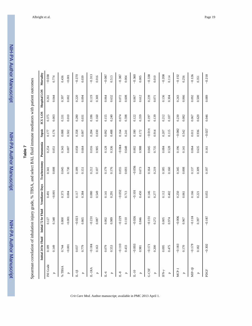
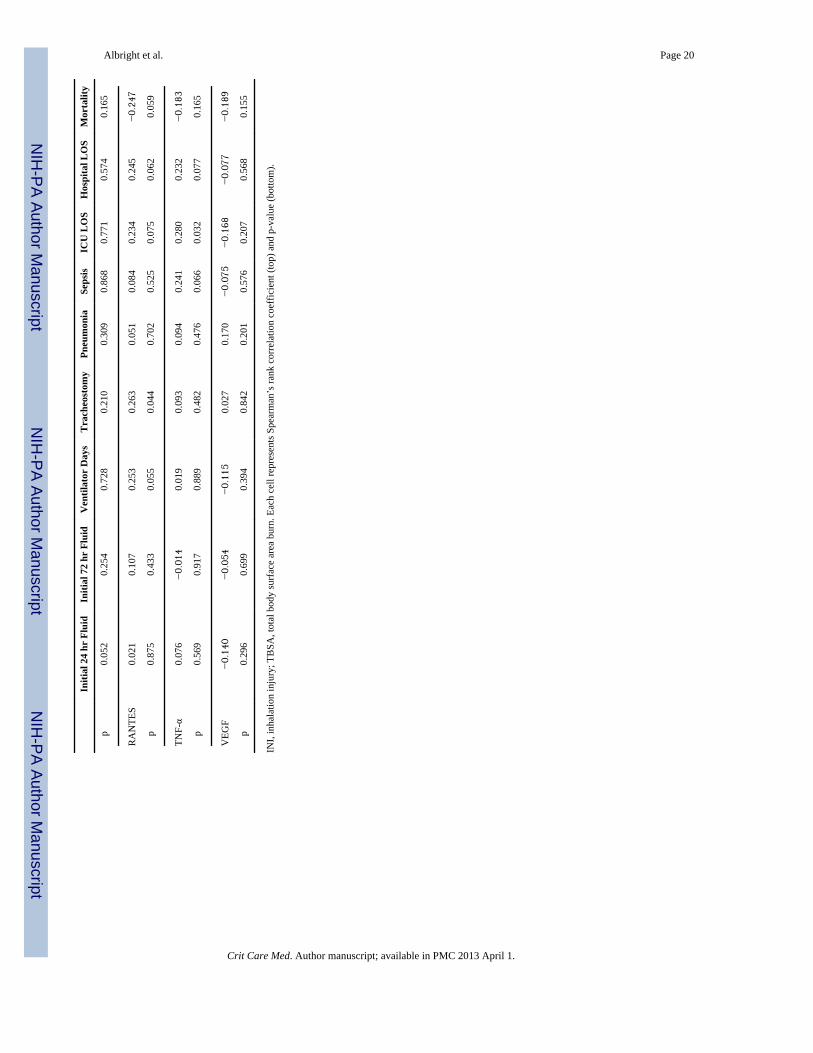
Correlations with outcomes were also assessed for inhalation injury grade, % TBSA, and theconcentration of 13 immune mediators measured in the BALF selected for eitherconcentrations found at physiologically relevant levels or those that have been previouslystudied in burn care (Table 7). Inhalation injury grade correlated with frequency oftracheostomy, number of ventilator days, ICU LOS, hospital LOS (p<0.05 for all) and nearlythe frequency of pneumonia (p=0.051). Likewise, % TBSA correlated with fluidresuscitation requirements, pneumonia, ventilator days, ICU LOS, hospital LOS, andmortality (p<0.05 for all). No BALF mediator concentration correlated with resuscitationrequirements. However, we did find the following differences: IL-1β correlated with sepsisand ICU LOS, and inversely with mortality; IL-1RA correlated with sepsis and inverselywith mortality; IL-8 correlated inversely with mortality; IL-10 correlated inversely withmortality; G-CSF correlated inversely with mortality; MCP-1 correlated with the frequencyof tracheostomy; RANTES correlated with the frequency of tracheostomy; and TNF-αcorrelated with ICU LOS (p<0.05 for all).
DiscussionIt is well-established that patients suffering smoke inhalation injury in addition to dermalinjury have poorer outcomes than those with skin burns alone (4–8, 13, 14). Animalexperiments suggest that these effects are modulated by an augmented inflammatoryresponse (15–18), and in humans, smoke inhalation injury seems to evoke enhancedpulmonary leukocyte activity and pronounced alveolar neutrophilia (9, 10). Though theseinitial reports focused on the early time-course of the pulmonary inflammatory response toinhalation injury, none addressed the magnitude of inflammatory response as it relates to thedegree of inhalation injury. The present study of human burn victims not only reaffirms theeffects of smoke inhalation on many burn patient outcomes, but is the first to demonstratethat the severity of inhalation injury may play a role in the overall pulmonary inflammatoryresponse.
One such feature of the burn patient with smoke inhalation that we have re-demonstrated isthe association of elevated COHb levels in patients with higher grades of inhalation injury.In addition to the potential for cerebrotoxicity, CO has been shown in a large animal modelof combined burn and smoke inhalation injury to be associated with an increased pulmonaryshunt fraction (18, 19). Although this feature could have profound effects on the burnedpatient through hypoxemic pulmonary vasoconstriction and ventilation/perfusionmismatching, we failed to note any substantial differences in the initial P:F ratio over therange of inhalation injury severities. Moreover, the anti-inflammatory effects of CO havebeen previously noted in the setting of burn and smoke inhalation injury, sepsis, and trauma(20–24). One might theorize that CO, if present at higher levels in patients with greaterinhalation injury severities, would mute the inflammatory response to worsening injury.That we still see a greater immune response with worse grades of inhalation injury suggestsa limited effect of CO on reducing the pulmonary inflammatory response in these patients.
Another finding of our study was a shift in the BALF WBC differential to one of neutrophilpredominance, confirming earlier findings observed in smoke inhalation by Wright and
Albright et al. Page 5
Crit Care Med. Author manuscript; available in PMC 2013 April 1.
NIH
-PA Author Manuscript
NIH
-PA Author Manuscript
NIH
-PA Author Manuscript
Murphy, and Riyami et al. (10, 25). In fact, in patients with a Grade 4 inhalation injury, 94%of the leukocyte population was neutrophils. This contrasts sharply with the normalpulmonary leukocyte distribution, which is upwards of 95% macrophages (26). In light ofprevious studies on burn and inhalation injury, we speculate that a robust production ofchemotactic factors (such as IL-8), in addition to the extensive and narrow pulmonaryvascular bed, are contributory to this phenomenon (9, 27, 28). Regardless of the mechanism,the preponderance of neutrophils might contribute to later immune dysfunction, bacterialovergrowth, and pneumonia (14, 29).
Though differences in many initial patient characteristics fell short of statistical significancewhen contrasting low and high severities of inhalation injury, certain outcomes acrossinhalation injury grades persisted with this method of comparison. For instance, therequirement for tracheostomy, the number of ventilator days, and the ICU length of staytended to be increased with worse grades of inhalation injury. Likewise, when placingemphasis on the difference between inhalation injury severities, those with the worst injurieshad more tracheostomies, ventilator days, pneumonia, sepsis, days in the ICU, and overallhospital length of stay. Perhaps of greatest importance is that patients in our study with aninhalation injury of any kind had a mortality of 27% as compared to only 11% for thosewithout, which is similar to findings by Shirani et al. in 1987, who also showed thatinhalation injury in addition to burns increased mortality by approximately 20% (30).Finally, the inverse correlation of numerous pulmonary immune mediator concentrationswith mortality deserves mention. On close inspection of these patients, Davis et al found thatnot only were many BALF immune mediator concentrations lower in those who wouldsuccumb to their injuries, but that pulmonary leukocyte production of these mediators inculture, particularly after stimulation with lipopolysaccharide, was blunted (31).
Although many of our results substantiate already held tenets of burn and inhalation injury,our having demonstrated an enhanced pulmonary inflammatory response to worse degreesof inhalation injury is a new contribution. Previous studies directed at delineating thealveolar response to inhalation injury have shown that leukocytes recruited to the alveolarspace have been primed for further stimulus, and that alterations of the inflammatorymediator profile of the lung may correlate with pulmonary physiologic dysfunction and laterpneumonia (9, 27). Our results indicate that many inflammatory mediators present in theBALF of those with an inhalation injury are significantly altered, even as early as within 14hours of injury. Of these, we have identified IL-1β, IL-1rα, IL-6, IL-8, and IFN-γ as bestrepresenting physiologically relevant levels and among the most important in regards to lunginjury. Indeed, each of these immune mediators has been extensively studied in both humansand animal models of burn injury, lending further credibility for their importance andpotential for therapeutic intervention (10, 32–47).
We concede that our study has certain limitations. First, there is an inherent selection bias inthat only those suspected of an inhalation injury were candidates for enrollment. Therefore,much of the burn patient population outside of this category were not included forcomparison, which may or may not have had significant effects on specific findings, those inparticular being resuscitative fluid requirements and mortality. Second, the precision ofBALF for measuring pulmonary inflammatory mediators is subject not only to variousdegrees of dilution but iatrogenic trauma to the airway which may result in contaminationfrom blood and other cellular debris. Furthermore, at present, there is no standarddenominator to correct for these possible confounders. Nonetheless, adjustment for totalprotein content in the BAL fluid had little impact on the results of our study and the overalldiscussion would have remained unchanged. Third, we were unable to account for thepossibility that aspiration of oral, pharyngeal, or gastroduodenal contents may have had anyeffect on the inflammatory response that we observed. Finally, bronchoscopic grading of
Albright et al. Page 6
Crit Care Med. Author manuscript; available in PMC 2013 April 1.
NIH
-PA Author Manuscript
NIH
-PA Author Manuscript
NIH
-PA Author Manuscript
inhalation injury grade is inherently subject to inter-rater differences, even though oursystem is based on Abbreviated Injury Score criteria. To minimize such effects, we alsodemonstrated our findings according to low and high severities of inhalation injury in amanner that we feel allowed for better clarity into the primary inflammatory response toinhalation injury.
ConclusionsOur study, emphasizing smoke inhalation injury in the burn-injured patient, demonstratesseveral correlates with worse grades of inhalation injury. Specifically, we have found thatworse inhalation injury is associated with: 1) a greater degree of alveolar neutrophilia; 2)elevated plasma COHb levels; 3) a greater likelihood of tracheostomy, prolonged ventilatorrequirements, and longer length of stay in both the ICU and the hospital; and 4) enhancedpulmonary inflammatory mediator production. These data, we hope, will serve the purposeof better understanding the biological mechanisms behind smoke inhalation injury, and webelieve that future investigations should consider inhalation injury to be a gradedphenomenon.
AcknowledgmentsFunded by the National Institutes of Health [T32 GM008750 (RLG), T32 AA013527 (EJK), R01 AA012034 (EJK),P30 AA019373 (EJK)], the International Association of Fire Fighters (JMA), and the Dr. Ralph and Marian C. FalkMedical Research Trust (EJK)
We kindly thank Jürgen Peters, MD, of the Klinik für Anästhesiologie und Intensivmedizin, UniversitätsklinikumEssen, Essen, Germany for his insight and critical review during the manuscript drafting process. Additionally, thiswork could not have been accomplished without the dedicated assistance of nursing and support staff in the BurnIntensive Care Unit at Loyola University Medical Center.
References1. Burn Incidence Fact Sheet. 2007. Retrieved August 22, 2010, from the American Burn Association:
http://ameriburn.org2. Latenser BA, Miller SF, Bessey PQ, Browning SM, Caruso DM, Gomez M, Jeng JC, Krichbaum
JA, Lentz CW, Saffle JR, Schurr MJ, Greenhalgh DG, Kagan RJ. National Burn Repository 2006Report Dataset Version 3.0. J Burn Care Res. 2007 Jul 24. Publish Ahead of Print.
3. Saffle JR, Davis B, Williams P. Recent outcomes in the treatment of burn injury in the UnitedStates: a report from the American Burn Association Patient Registry. J Burn Care Rehabil. 1995;16(3 Pt 1):219–32. discussion 288–9. [PubMed: 7673300]
4. Navar PD, Saffle JR, Warden GD. Effect of inhalation injury on fluid resuscitation requirementsafter thermal injury. Am J Surg. 1985; 150(6):716–20. [PubMed: 4073365]
5. Dai NT, Chen TM, Cheng TY, Chen SL, Chen SG, Chou GH, Chou TD, Wang HJ. The comparisonof early fluid therapy in extensive flame burns between inhalation and noninhalation injuries. Burns.1998; 24(7):671–5. [PubMed: 9882069]
6. Cancio LC, Chávez S, Alvarado-Ortega M, Barillo DJ, Walker SC, McManus AT, Goodwin CW.Predicting increased fluid requirements during the resuscitation of thermally injured patients. JTrauma. 2004; 56(2):404–13. discussion 413–4. [PubMed: 14960986]
7. Edelman DA, White MT, Tyburski JG, Wilson RF. Factors affecting prognosis of inhalation injury.J Burn Care Res. 2006; 27(6):848–53. [PubMed: 17091081]
8. Edelman DA, Khan N, Kempf K, White MT. Pneumonia after inhalation injury. J Burn Care Res.2007; 28(2):241–6. [PubMed: 17351439]
9. Kurzius-Spencer M, Foster K, Littau S, Richey KJ, Clark BM, Sherrill D, Goodman RB, Boitano S,Burgess JL. Tracheobronchial markers of lung injury in smoke inhalation victims. J Burn Care Res.2008; 29(2):311–8. [PubMed: 18354287]
Albright et al. Page 7
Crit Care Med. Author manuscript; available in PMC 2013 April 1.
NIH
-PA Author Manuscript
NIH
-PA Author Manuscript
NIH
-PA Author Manuscript
10. Wright MJ, Murphy JT. Smoke inhalation enhances early alveolar leukocyte responsiveness toendotoxin. J Trauma. 2005; 59(1):64–70. [PubMed: 16096540]
11. Endorf FW, Gamelli RL. Inhalation injury, pulmonary perturbations, and fluid resuscitation. JBurn Care Res. 2007; 28(1):80–3. [PubMed: 17211205]
12. Wahl WL, Ahrns KS, Brandt MM, Rowe SA, Hemmila MR, Arbabi S. Bronchoalveolar lavage indiagnosis of ventilator-associated pneumonia in patients with burns. J Burn Care Rehabil. 2005;26(1):57–61. [PubMed: 15640736]
13. Clark WR. Smoke inhalation: diagnosis and treatment. World J Surg. 1992; 16(1):24–9. [PubMed:1290262]
14. Enkhbaatar P, Traber DL. Pathophysiology of acute lung injury in combined burn and smokeinhalation injury. Clin Sci (Lond). 2004; 107:137–43. [PubMed: 15151496]
15. Soejima K, Schmalstieg FC, Sakurai H, Traber LD, Traber DL. Pathophysiological analysis ofcombined burn and smoke inhalation injuries in sheep. Am J Physiol Lung Cell Mol Physiol.2001; 280(6):L1233–41. [PubMed: 11350803]
16. Lange M, Hamahata A, Traber DL, Esechie A, Jonkam C, Bansal K, Nakano Y, Traber LD,Enkhbaatar P. A murine model of sepsis following smoke inhalation injury. Biochem Biophys ResCommun. 2010; 391(3):1555–60. [PubMed: 20036639]
17. Mizutani A, Enkhbaatar P, Esechie A, Traber LD, Cox RA, Hawkins HK, Deyo DJ, Murakami K,Noguchi T, Traber DL. Pulmonary changes in a mouse model of combined burn and smokeinhalation-induced injury. J Appl Physiol. 2008; 105(2):678–84. [PubMed: 18436699]
18. Westphal M, Cox RA, Traber LD, Morita N, Enkhbaatar P, Schmalstieg FC, Hawkins HK,Maybauer DM, Maybauer MO, Murakami K, Burke AS, Wesphal-Varghese BB, Rudloff HE,Salsbury JR, Jodoin JM, Lee S, Traber DL. Combined burn and smoke inhalation injury impairsovine hypoxic pulmonary vasoconstriction. Crit Care Med. 2006; 34(5):1428–36. [PubMed:16540966]
19. Westphal M, Morita N, Enkhbaatar P, Murakami K, Traber L, Traber DL. Acute effects ofcombined burn and smoke inhalation injury on carboxyhemoglobin formation, tissue oxygenation,and cardiac performance. Biochem Biophys Res Commun. 2004; 317(3):945–9. [PubMed:15081431]
20. Lange M, Cox RA, Enkhbaatar P, Whorton EB, Nakano Y, Hamahata A, Jonkam C, Esechie A,von Borzyskowski S, Traber LD, Traber DL. Predictive role of arterial carboxyhemoglobinconcentrations in ovine burn and smoke inhalation-induced lung injury. Exp Lung Res. 2011;37(4):239–45. [PubMed: 21309735]
21. Mitchell LA, Channell MM, Royer CM, Ryter SW, Choi AM, McDonald JD. Evaluation ofinhaled carbon monoxide as an anti-inflammatory therapy in a nonhuman primate model of lunginflammation. Am J Physiol Lung Cell Mol Physiol. 2010; 299(6):L891–7. [PubMed: 20729385]
22. Kanagawa F, Takahashi T, Inoue K, Shimizu H, Omori E, Morimatsu H, Maeda S, Katayama H,Nakao A, Morita K. Protective effect of carbon monoxide inhalation on lung injury afterhemorrhagic shock/resuscitation in rats. J Trauma. 2010; 69(1):185–94. [PubMed: 20622590]
23. Goebel U, Siepe M, Mecklenburg A, Doenst T, Beyersdorf F, Loop T, Schlensak C. Reducedpulmonary inflammatory response during cardiopulmonary bypass: effects of combinedpulmonary perfusion and carbon monoxide inhalation. Eur J Cardiothorac Surg. 2008; 34(6):1165–72. [PubMed: 18829339]
24. Zhou H, Qian H, Liu J, Zhu D, Ding W, Pan P, Jin D, Wang J, Li W. Protection against lung graftinjury from brain-dead donors with carbon monoxide, biliverdin, or both. J Heart Lung Transplant.2011; 30(4):460–6. [PubMed: 21216159]
25. Riyami BMS, Tree R, Kinsella J, Clark CJ, Reid WH, Campbell D, Gemmell CG. Changes inalveolar macrophage, monocyte, and neutrophil cell profiles after smoke inhalation injury. J ClinPathol. 1990; 43(1):43–5. [PubMed: 2312750]
26. Ettensohn DB, Jankowski MJ, Duncan PG, Lalor PA. Bronchoalveolar lavage in the normalvolunteer subject. I. Technical aspects and intersubject variability. Chest. 1988; 94(2):275–80.[PubMed: 3396403]
Albright et al. Page 8
Crit Care Med. Author manuscript; available in PMC 2013 April 1.
NIH
-PA Author Manuscript
NIH
-PA Author Manuscript
NIH
-PA Author Manuscript
27. Rodriguez JL, Miller CG, Garner WL, Till GO, Guerrero P, Moore NP, Corridore M, NormolleDP, Smith DJ, Remick DG. Correlation of the local and systemic cytokine response with clinicaloutcome following thermal injury. J Trauma. 1993; 34(5):684–95. [PubMed: 8497003]
28. Murakami K, Traber DL. Pathophysiological basis of smoke inhalation injury. News PhysiologSci. 2003; 18:125–29.
29. Traber DL, Linares HA, Herndon DN, Prien T. The pathophysiology of inhalation injury -- areview. Burns Incl Therm Inj. 1988; 14(5):357–64. [PubMed: 3067821]
30. Shirani KZ, Pruitt BA, Mason AD. The influence of inhalation injury and pneumonia on burnmortality. Ann Surg. 1987; 205:82–7. [PubMed: 3800465]
31. Davis CS, Albright JM, Carter SR, Ramirez L, Kim H, Gamelli RL, Kovacs EJ. Early pulmonaryimmune hyporesponsiveness is associated with mortality after burn and smoke inhalation injury. JBurn Care Res. in press.
32. Dinarello CA. The interleukin-1 family: 10 years of discovery. FASEB J. 1994; 8(15):1314–25.[PubMed: 8001745]
33. Chen LW, Chang WJ, Wang JS, Hsu CM. Interleukin-1 mediates thermal injury-induced lungdamage through C-Jun NH2-terminal kinase signaling. Crit Care Med. 2007; 35(4):1113–22.[PubMed: 17334237]
34. Fleischmann RM, Tesser J, Schiff MH, Schechtman J, Burmester GR, Bennett R, Modafferi D,Zhou L, Bell D, Appleton B. Ann Rheum Dis. 2006; 65(8):1006–12. [PubMed: 16396977]
35. Endo S, Inada K, Yamada Y, Kasai T, Takakuwa T, Nakae H, Kamei Y, Shimamura T, Suzuki T,Taniguchi S, Yoshida M. Plasma levels of interleukin-1 receptor antagonist (IL-1ra) and severityof illness in patients with burns. J Med. 1996; 27(1–2):57–71. [PubMed: 8863178]
36. Vindenes HA, Ulvestad E, Bjerknes R. Concentrations of cytokines in plasma of patients withlarge burns: their relation to time after injury, burn size, inflammatory variables, infection, andoutcome. Eur J Surg. 1998; 164(9):647–56. [PubMed: 9728783]
37. Steensberg A, Fischer CP, Keller C, Moller K, Pedersen BK. IL-6 enhances plasma IL-1ra, Il-10,and cortisol in humans. Am J Physiol Endocrinol Metab. 2003; 285(2):E433–7. [PubMed:12857678]
38. Mizel SB. The interleukins. FASEB J. 1989; 3(12):2379–88. [PubMed: 2676681]39. Molloy RG, Mannick JA, Rodrick ML. Cytokines, sepsis and immunomodulation. Br J Surg. 1993;
80(3):289–97. [PubMed: 8472134]40. Kaplanski G, Farnarier C, Kaplanski S, Porat R, Shapiro L, Bongrand P, Dinarello CA.
Interleukin-1 induces interleukin-8 secretion from endothelial cells by a juxtacrine mechanism.Blood. 1994; 84(12):4242–8. [PubMed: 7994038]
41. Polk HC Jr, Cheadle WG, Livingston DH, et al. A randomized prospective clinical trial todetermine the efficacy of interferon-gamma in severely injured patients. Am J Surg. 1992; 63:191–96. [PubMed: 1739172]
42. Dries DJ, Jurkovich J, Maier RV, et al. Effect of interferon gamma on infection-related death inpatients with severe injuries. Arch Surg. 1994; 129:1031–42. [PubMed: 7944932]
43. Mooney DP, Gamelli RL. Sepsis following thermal injury. Compr Ther. 1989; 15:22–29.[PubMed: 2676330]
44. Hershman MJ, Appel SH, Wellhausen SR, Sonnenfeld G, Polk HC Jr. Interferon-gamma treatmentincreases HLA-DR expression on monocytes in severely injured patients. Clin Exp Immunol.1989; 77(1):67–70. [PubMed: 2504520]
45. Suzuki F, Pollard RB. Alterations of interferon production in a mouse model of thermal injury. JImmunol. 1982; 129:1806–1810. [PubMed: 6181145]
46. Wasserman D, Ioannovich JD, Hinzmann RD, Deichsel G, Steinmann GG. Interferon-gamma inthe prevention of severe burn-related infections: a European phase III multicenter trial. The SevereBurns Study Group. Crit Care Med. 1998; 26(3):434–9. [PubMed: 9504568]
47. Nakos G, Malamou-Mitsi VD, Lachana A, Karassavoglou A, Kitsiouli E, Agnandi N, Lekka ME.Immunoparalysis in patients with severe trauma and the effect of inhaled interferon-gamma. CritCare Med. 2002; 30(7):1488–94. [PubMed: 12130967]
Albright et al. Page 9
Crit Care Med. Author manuscript; available in PMC 2013 April 1.
NIH
-PA Author Manuscript
NIH
-PA Author Manuscript
NIH
-PA Author Manuscript

Figure 1.Inflammatory mediator concentrations in the BAL fluid of controls (C) and burn patientswith inhalation injury graded 0–4 by bronchoscopic criteria (0 = no injury; 1 = mild injury; 2= moderate injury; 3 = severe injury; 4 = massive injury). Statistical comparison by Kruskal-Wallace and post-hoc analysis do not include controls, which are included for visualcomparison. *p < 0.05 between groups.
Albright et al. Page 10
Crit Care Med. Author manuscript; available in PMC 2013 April 1.
NIH
-PA Author Manuscript
NIH
-PA Author Manuscript
NIH
-PA Author Manuscript

Figure 2.Inflammatory mediator concentrations in the BAL fluid of burn patients with low inhalationinjury severity (Grades 1–2) vs high inhalation injury severity (Grades 3–4). Differencesconsidered statistically significant when p < 0.05.
Albright et al. Page 11
Crit Care Med. Author manuscript; available in PMC 2013 April 1.
NIH
-PA Author Manuscript
NIH
-PA Author Manuscript
NIH
-PA Author Manuscript
NIH
-PA Author Manuscript
NIH
-PA Author Manuscript
NIH
-PA Author Manuscript
Albright et al. Page 12
Table 1
Coding and grading of inhalation injury by bronchoscopy
AIS code Grade Class Description
919201.2 0 No injury Absence of carbonaceous deposits, erythema, edema, bronchorrhea or obstruction
919202.3 1 Mild injury Minor or patchy areas of erythema, carbonaceous deposits in proximal or distal bronchi
919204.4 2 Moderate injury Moderate degree of erythema, carbonaceous deposits, bronchorrhea, or bronchial obstruction
919206.5 3 Severe injury Severe inflammation with friability, copious carbonaceous deposits, bronchorrhea or obstruction
919208.6 4 Massive injury Evidence of mucosal sloughing, necrosis, endoluminal obliteration
Crit Care Med. Author manuscript; available in PMC 2013 April 1.
NIH
-PA Author Manuscript
NIH
-PA Author Manuscript
NIH
-PA Author Manuscript
Albright et al. Page 13
Tabl
e 2
Patie
nt c
hara
cter
istic
s by
inha
latio
n in
jury
gra
de
Gra
de 0
n=9
Gra
de 1
n=15
Gra
de 2
n=15
Gra
de 3
n=18
Gra
de 4
n=3
p V
alue
Age
47 (2
7–67
)57
(44–
61)
48 (3
7–57
)50
(31–
68)
75 (5
5–80
)0.
162
Gen
der
0.94
4
M
ale
5 (5
6)10
(67)
9 (6
0)12
(67)
1 (3
3)
Fe
mal
e4
(44)
5 (3
3)6
(40)
6 (3
3)2
(67)
Rac
e/Et
hnic
ity0.
524
C
auca
sian
7 (7
8)10
(67)
9 (6
0)9
(50)
1 (3
3)
A
fric
an A
m.
2 (2
2)3
(20)
3 (2
0)5
(28)
1 (3
3)
H
ispa
nic
--
1 (7
)3
(17)
1 (3
3)
A
sian
-1
(7)
-1
(6)
-
O
ther
/Unk
now
n-
1 (7
)2
(13)
--
Initi
al P
:F ra
tio35
6.4
(±45
.9)
354.
3 (±
26.6
)31
1.7
(±29
.5)
358.
1 (±
31.4
4)28
2.3
(±48
.8)
0.68
5
Initi
al C
OH
b (%
)2.
8 (0
.8–4
.2)
3.8
(1.1
–5.5
)9.
9 (2
.6–1
3.7)
10.4
(4.7
–27.
9)20
.5 (0
.9–4
0.0)
0.01
4
BA
L C
ell D
iff (%
)
N
eutro
phils
41 (2
6–69
)79
(45–
93)
84 (6
4–95
)87
(72–
91)
940.
042
Ly
mph
ocyt
es6
(3–1
7)4
(1–1
0)2
(1–1
5)2
(1–1
1)0
0.24
2
M
onoc
ytes
1 (0
–4)
0 (0
–1)
0 (0
–2)
1 (0
–3)
00.
619
M
acro
phag
es54
(10–
68)
14 (4
–46)
11 (4
–32)
7 (3
–12)
60.
133
Eo
sino
phils
1 (0
–1)
0 (0
–1)
0 (0
–1)
0 (0
–1)
00.
679
B
asop
hils
00
00
(0–2
)0
0.28
9
Initi
al B
AL
> 10
0,00
0 C
FUs
3 (6
0)1
(7)
1 (7
)0
00.
005
TBSA
(%)
15 (7
–24)
20 (5
–65)
2 (0
–39)
18 (0
.8–3
5)7
(7–1
5)0.
354
Dat
a pr
esen
ted
as n
(%),
med
ian
(inte
rqua
rtile
rang
e), o
r mea
n (±
SEM
), w
here
app
ropr
iate
BA
L, B
ronc
hoal
veol
ar L
avag
e; C
FUs,
Col
ony
Form
ing
Uni
ts; C
OH
b, C
arbo
xyhe
mog
lobi
n; T
BSA
, Tot
al B
ody
Surf
ace
Are
a sk
in b
urn
Crit Care Med. Author manuscript; available in PMC 2013 April 1.
NIH
-PA Author Manuscript
NIH
-PA Author Manuscript
NIH
-PA Author Manuscript
Albright et al. Page 14
Tabl
e 3
Patie
nt o
utco
mes
by
inha
latio
n in
jury
gra
de
Gra
de 0
n=9
Gra
de 1
n=15
Gra
de 2
n=15
Gra
de 3
n=18
Gra
de 4
n=3
p V
alue
Initi
al 2
4 ho
ur fl
uid
requ
irem
ents
(L)
4.2
(2.6
–9.9
)12
(3–2
1)7.
7 (3
.4–2
3)10
(6.1
–16)
7.0
(5.0
–13)
0.42
4
Initi
al 7
2 ho
ur fl
uid
requ
irem
ents
(L)
15 (8
.8–2
2)22
(10–
28)
16 (1
2–37
)20
(12–
27)
14 (1
1–24
)0.
715
Ven
tilat
or d
ays
3 (2
–4)
6 (3
–30)
10 (3
–33)
23 (1
2–38
)23
(15–
33)
0.00
3
Trac
heos
tom
y0
4 (2
7)6
(40)
10 (5
6)1
(33)
0.07
0
Pneu
mon
ia3
(33)
6 (4
0)7
(47)
12 (6
7)2
(67)
0.42
4
Seps
is1
(11)
2 (1
3)2
(13)
5 (2
8)1
(33)
0.68
1
ICU
LO
S10
(5–2
1)7
(6–4
1)13
(7–3
7)22
(15–
38)
24 (2
3–53
)0.
076
Hos
pita
l LO
S20
(7–3
0)8
(6–4
6)14
(6–4
0)25
(17–
38)
24 (2
3–53
)0.
352
Mor
talit
y1
(11)
7 (4
7)2
(13)
4 (2
2)1
(33)
0.21
0
Dat
a pr
esen
ted
as n
(%) o
r med
ian
(inte
rqua
rtile
rang
e), w
here
app
ropr
iate
ICU
, int
ensi
ve c
are
unit;
LO
S, le
ngth
of s
tay
Crit Care Med. Author manuscript; available in PMC 2013 April 1.
NIH
-PA Author Manuscript
NIH
-PA Author Manuscript
NIH
-PA Author Manuscript
Albright et al. Page 15
Table 4
Patient characteristics by inhalation injury severity
Lown=30
Highn=21
p Value
Age 54 (39–60) 55 (36–72) 0.509
Male Gender 0.917
Male 19 (63) 13 (62)
Female 11 (37) 8 (38)
Race/Ethnicity 0.387
Caucasian 19 (63) 10 (48)
African Am. 6 (20) 6 (29)
Hispanic 1 (3) 4 (19)
Asian 1 (3) 1 (5)
Other/Unknown 3 (10) -
Initial P:F ratio 333.0 (±19.9) 347.3 (±28.1) 0.671
Initial COHb (%) 5.1 (2.2–10.2) 10.4 (4.5–30.7) 0.059
BAL Cell Diff (%)
Neutrophils 84 (53–94) 87 (74–93) 0.553
Lymphocytes 2 (1–11) 2 (1–10) 0.855
Monocytes 0 (0–2) 0 (0–3) 0.549
Macrophages 11 (4–38) 6 (4–12) 0.355
Eosinophils 0 (0–1) 0 (0–1) 0.691
Basophils 0 0 (0–1) 0.117
Initial BAL > 100,000 CFUs 2 (7) 0 0.503
TBSA (%) 15 (1–58) 15 (1–28) 0.437
Data presented as n (%), median (interquartile range), or mean (±SEM), where appropriate. BAL, Bronchoalveolar Lavage; CFUs, Colony FormingUnits; COHb, Carboxyhemoglobin; TBSA, Total Body Surface Area skin burn
Crit Care Med. Author manuscript; available in PMC 2013 April 1.
NIH
-PA Author Manuscript
NIH
-PA Author Manuscript
NIH
-PA Author Manuscript
Albright et al. Page 16
Tabl
e 5
Com
paris
on o
f BA
L flu
id im
mun
e m
edia
tor c
once
ntra
tions
from
hea
lth c
ontro
ls, b
urn
patie
nts w
ith n
o vi
sibl
e in
hala
tion
inju
ry, a
nd b
urn
patie
nts w
ithlo
w a
nd h
igh
inha
latio
n in
jury
seve
ritie
s
Med
iato
r (p
g/m
l)C
ontr
olN
o IN
I (G
rade
0)
Low
INI (
Gra
de 1
/2)
Hig
h IN
I (G
rade
3/4
)p-
valu
e
C5a
638.
2 (2
69.0
–103
4)30
7.3
(98.
0–47
5.7)
249.
0 (1
27.0
–567
.7)
559.
9 (2
37.2
–115
2)0.
167
IL-1β
0.1
(0.0
–5.0
)10
.4 (2
.9–4
9.4)
15.5
(2.6
–76.
3)*
42.4
(13.
9–11
1.4)
*0.
001
IL-1
RA
31.1
(20.
1–69
.9)
176.
4 (1
25.2
–683
.9)*
291.
5 (9
0.9–
728.
4)*
301.
8 (1
59.1
–136
2)*
0.00
4
IL-2
0.0
(0.0
–0.7
)0.
0 (0
.0–7
.3)
0.5
(0.0
–2.6
)2.
5 (0
.0–4
.6)
0.24
5
IL-4
0.0
(0.0
–0.2
)1.
4 (0
.0–3
.3)
0.0
(0.2
–2.7
)1.
8 (0
.3–5
.1)
0.04
2
IL-5
0.0
(0.0
–0.5
)0.
3 (0
.1–0
.5)
0.2
(0.0
–0.4
)0.
0 (0
.1–0
.5)
0.48
2
IL-6
2.0
(0.8
–5.4
)51
.4 (2
0.1–
138.
2)14
1.7
(47.
2–24
5.0)
*35
1.3
(160
.8–1
047)
*†‡
< 0.
001
IL-7
1.7
(0.8
–2.7
)11
.9 (6
.7–1
4.8)
*3.
9 (0
.7–7
.2)†
3.7
(0.6
–6.0
)†0.
002
IL-8
0.0
(0.0
–0.0
)2.
9 (1
.4–9
.4)*
4.0
(1.1
–13.
4)*
4.5
(1.5
–13.
1)*
< 0.
001
IL-9
0.0
(0.0
–5.8
)3.
1 (0
.0–1
9.9)
1.2
(0.0
–12.
6)10
.0 (6
.0–1
6.6)
*0.
021
IL-1
00.
7 (0
.5–5
.3)
10.8
(4.2
–12.
8)*
3.4
(1.9
–13.
7)7.
3 (4
.4–1
0.1)
0.03
6
IL-1
217
.8 (1
1.0–
21.1
)9.
0 (0
.0–1
9.4)
0.0
(0.0
–5.8
)*0.
0 (3
.4–1
5.0)
0.00
6
IL-1
31.
7 (1
.4–1
.8)
4.1
(3.0
–7.9
)3.
0 (1
.4–1
7.3)
3.2
(1.1
–7.2
)0.
081
IL-1
51.
1 (0
.3–2
.2)
7.6
(0.3
–17.
9)0.
7 (0
.0–5
.8)
4.7
(2.9
–17.
0)‡
0.00
3
IL-1
70.
0 (0
.0–3
.5)
0.0
(0.0
–27.
3)0.
0 (0
.0–0
.0)
0.0
(0.0
–9.6
)0.
477
Eota
xin
0.0
(0.0
–45.
9)46
.9 (5
.3–1
30.5
)30
.3 (0
.9–9
2.6)
48.6
(14.
2–93
.3)
0.21
0
FGF-
basi
c0.
0 (0
.0–0
.6)
0.0
(0.0
–0.0
)0.
0 (0
.0–0
.0)
0.0
(0.0
–0.0
)0.
512
G-C
SF15
.7 (6
.4–3
4.1)
67.8
(22.
6–15
1.9)
103.
9 (5
6.5–
261.
5)*
75.9
(41.
0–16
8.0)
*0.
010
GM
-CSF
12.8
(11.
7–84
.1)
2.1
(0.0
–6.8
)*0.
0 (0
.0–4
.3)*
3.7
(0.0
–9.5
)0.
001
IFN
-γ4.
2 (1
.8–9
.0)
38.9
(6.3
–87.
0)39
.8 (1
4.2–
94.2
)14
2.8
(33.
1–21
2.9)
*0.
005
IP-1
0ε0.
4 (0
.2–1
.9)
3.9
(1.4
–12.
1)0.
9 (0
.2–2
.7)
0.9
(0.3
–1.6
)0.
115
MC
P-1
22.6
(16.
1–29
.6)
74.2
(29.
5–45
9.5)
49.9
(31.
0–12
0.4)
185.
1 (1
05.9
–551
.2)*
‡<
0.00
1
MIP
-1α
0.0
(0.0
–0.5
)0.
0 (0
.0–1
5.9)
0.0
(0.0
–14.
2)0.
0 (0
.0–1
8.9)
0.91
1
MIP
-1β
6.9
(5.3
–8.9
)69
.0 (3
9.7–
96.9
)*36
.8 (1
6.5–
85.9
)*21
.1 (7
8.0–
179.
2)*
0.01
1
PDG
F0.
0 (0
.0–1
0.1)
22.7
(13.
5–10
2.3)
10.0
(0.0
–34.
6)28
.7 (9
.8–4
0.9)
*0.
031
Crit Care Med. Author manuscript; available in PMC 2013 April 1.
NIH
-PA Author Manuscript
NIH
-PA Author Manuscript
NIH
-PA Author Manuscript
Albright et al. Page 17
Med
iato
r (p
g/m
l)C
ontr
olN
o IN
I (G
rade
0)
Low
INI (
Gra
de 1
/2)
Hig
h IN
I (G
rade
3/4
)p-
valu
e
RA
NTE
S0.
3 (0
.0–9
.0)
22.0
(18.
8–47
.4)
35.0
(19.
7–98
.0)*
56.1
(21.
9–11
9.9)
*0.
004
TNF-α
0.0
(0.0
–5.3
)56
.9 (0
.0–2
32.8
)4.
8 (0
.0–5
8.6)
34.8
(5.5
–63.
4)0.
050
VEG
F40
2.7
(207
.2–7
75.6
)35
5.7
(235
.5–6
14.6
)13
1.1
(43.
2–27
0.9)
58.2
(27.
6–12
7.6)
*†<
0.00
1
Dat
a re
pres
ente
d as
med
ian
(inte
rqua
rtile
rang
e); I
NI,
inha
latio
n in
jury
;
* p<0.
05, v
s con
trol;
† p<0.
05, v
s No
INI;
‡ p<0.
05, v
s Low
INI;
ε repr
esen
ted
as n
g/m
l; K
rusk
al-W
alla
ce w
ith p
ost-h
oc a
naly
sis.
Crit Care Med. Author manuscript; available in PMC 2013 April 1.
NIH
-PA Author Manuscript
NIH
-PA Author Manuscript
NIH
-PA Author Manuscript
Albright et al. Page 18
Table 6
Patient outcomes by inhalation injury severity
Lown=30
Highn=21
p Value
Initial 24 hour fluid requirements (L) 10 (3.3–22) 8.7 (5.8–15) 0.579
Initial 72 hour fluid requirements (L) 19 (12–27) 17 (11–28) 0.716
Ventilator days 7 (3–31) 23 (13–35) 0.036
Tracheostomy 10 (33) 11 (52) 0.174
Pneumonia 13 (43) 14 (67) 0.162
Sepsis 4 (13) 6 (29) 0.177
ICU LOS 13 (6–37) 24 (16–39) 0.040
Hospital LOS 11 (6–41) 25 (19–39) 0.081
Mortality 9 (30) 4 (19) 0.626
Data presented as n (%) or median (interquartile range), where appropriate ICU, intensive care unit; LOS, length of stay
Crit Care Med. Author manuscript; available in PMC 2013 April 1.
NIH
-PA Author Manuscript
NIH
-PA Author Manuscript
NIH
-PA Author Manuscript
Albright et al. Page 19
Tabl
e 7
Spea
rman
cor
rela
tion
of in
hala
tion
inju
ry g
rade
, % T
BSA
, and
sele
ct B
AL
fluid
imm
une
med
iato
rs w
ith p
atie
nt o
utco
mes
Initi
al 2
4 hr
Flu
idIn
itial
72
hr F
luid
Ven
tilat
or D
ays
Tra
cheo
stom
yPn
eum
onia
Seps
isIC
U L
OS
Hos
pita
l LO
SM
orta
lity
INI G
rade
0.18
90.
127
0.49
10.
341
0.25
40.
177
0.37
50.
261
−0.038
p
0.14
90.
348
<0.0
010.
008
0.05
10.
176
0.00
30.
044
0.77
4
% T
BSA
0.74
40.
800
0.37
30.
045
0.34
30.
088
0.33
10.
397
0.43
6
p
<0.0
01<0
.001
0.00
40.
730
0.00
70.
502
0.01
00.
002
<0.0
01
IL-1β
0.03
7−0.023
0.11
70.
189
0.05
80.
358
0.28
00.
220
−0.270
p
0.77
90.
865
0.38
40.
151
0.66
40.
007
0.03
10.
094
0.03
9
IL-1
RA
−0.184
−0.231
0.08
00.
212
0.01
60.
284
0.18
60.
119
−0.313
p
0.16
30.
087
0.54
90.
107
0.90
50.
030
0.16
00.
369
0.01
6
IL-6
0.07
90.
002
0.14
10.
179
0.12
80.
092
0.15
50.
064
−0.087
p
0.55
30.
989
0.29
10.
176
0.33
60.
488
0.24
00.
632
0.51
3
IL-8
−0.110
−0.229
−0.052
0.05
5−0.064
0.16
40.
074
0.07
2−0.387
p
0.43
30.
110
0.71
30.
693
0.64
80.
241
0.59
80.
608
0.00
4
IL-1
0−0.003
−0.026
−0.101
−0.056
0.00
20.
180
0.12
20.
067
−0.360
p
0.98
10.
846
0.45
00.
673
0.98
80.
172
0.35
90.
612
0.00
5
G-C
SF−0.171
−0.151
0.14
60.
164
0.04
5−0.014
0.19
70.
239
−0.338
p
0.20
00.
272
0.27
70.
219
0.73
50.
914
0.13
90.
071
0.01
0
IFN
-γ0.
095
0.00
50.
112
0.18
50.
084
0.20
70.
212
0.13
6−0.208
p
0.47
50.
974
0.40
20.
160
0.52
80.
115
0.10
70.
304
0.11
4
MC
P-1
−0.183
−0.006
0.25
00.
345
0.19
6−0.082
0.23
00.
243
−0.152
p
0.17
00.
967
0.06
10.
008
0.14
10.
542
0.08
20.
066
0.25
6
MIP
-1β
−0.179
−0.118
0.16
60.
137
0.06
40.
011
0.06
70.
092
−0.126
p
0.18
20.
397
0.22
10.
309
0.63
50.
936
0.62
00.
500
0.35
1
PDG
F−0.302
−0.187
0.05
50.
197
0.16
1−0.027
0.04
60.
089
−0.218
Crit Care Med. Author manuscript; available in PMC 2013 April 1.
NIH
-PA Author Manuscript
NIH
-PA Author Manuscript
NIH
-PA Author Manuscript
Albright et al. Page 20
Initi
al 2
4 hr
Flu
idIn
itial
72
hr F
luid
Ven
tilat
or D
ays
Tra
cheo
stom
yPn
eum
onia
Seps
isIC
U L
OS
Hos
pita
l LO
SM
orta
lity
p
0.05
20.
254
0.72
80.
210
0.30
90.
868
0.77
10.
574
0.16
5
RA
NTE
S0.
021
0.10
70.
253
0.26
30.
051
0.08
40.
234
0.24
5−0.247
p
0.87
50.
433
0.05
50.
044
0.70
20.
525
0.07
50.
062
0.05
9
TNF-α
0.07
6−0.014
0.01
90.
093
0.09
40.
241
0.28
00.
232
−0.183
p
0.56
90.
917
0.88
90.
482
0.47
60.
066
0.03
20.
077
0.16
5
VEG
F−0.140
−0.054
−0.115
0.02
70.
170
−0.075
−0.168
−0.077
−0.189
p
0.29
60.
699
0.39
40.
842
0.20
10.
576
0.20
70.
568
0.15
5
INI,
inha
latio
n in
jury
; TB
SA, t
otal
bod
y su
rfac
e ar
ea b
urn.
Eac
h ce
ll re
pres
ents
Spe
arm
an’s
rank
cor
rela
tion
coef
ficie
nt (t
op) a
nd p
-val
ue (b
otto
m).
Crit Care Med. Author manuscript; available in PMC 2013 April 1.