The 90K Protein Increases Major Histocompatibility Complex Class I Expression and Is Regulated by...
9
The 90K Protein Increases Major Histocompatibility Complex Class I Expression and Is Regulated by Hormones, -Interferon, and Double-Strand Polynucleotides ANTONINO GRASSADONIA, NICOLA TINARI, BRUNO FIORENTINO, KOICHI SUZUKI, MINORU NAKAZATO, MICHELE DE TURSI, CESIDIO GIULIANI, GIORGIO NAPOLITANO, DINAH S. SINGER, STEFANO IACOBELLI, AND LEONARD D. KOHN Cell Regulation Section (A.G., B.F., K.S., M.N., C.G., G.N., L.D.K.), Metabolic Diseases Branch, National Institute of Diabetes and Digestive and Kidney Diseases, National Institutes of Health, Bethesda, Maryland 20892; Section of Medical Oncology (A.G., N.T., B.F., M.D.T., S.I.), Department of Oncology and Neurosciences and Section of Endocrinology (C.G., G.N.), Department of Medicine and Science of Aging, Universita ` degli Studi G. D’Annunzio, Faculty of Medicine and Surgery, 66100 Chieti, Italy; Experimental Immunology Branch (D.S.S.), National Cancer Institute, National Institutes of Health, Bethesda, Maryland 20892; and Edison Biotechnology Institute (L.D.K.), Department of Biomedical Sciences, Ohio University College of Osteopathic Medicine, Athens, Ohio 45701 Here we report the cloning of the rat 90K, a homolog of the mouse cyclophilin C-associated protein/mouse adherent mac- rophage and human 90K. The protein is constitutively ex- pressed by FRTL-5 thyrocytes, and its levels are modulated by TSH, insulin/IGF-I, and -interferon. Transfection of the cells with 90K cDNA or exposure to purified 90K resulted in a sig- nificant increase of the expression of major histocompatibil- ity complex class I but not class II antigens. An increased expression of 90K was obtained after viral infection or intro- duction into the cells of fragments of viral, bacterial, or mam- malian double-strand polynucleotides. The increase was sequence independent, not CpG mediated, and associated with the expression of molecules characterizing antigen- presenting-cell phenotype. The present data along with re- sults from previous studies suggest that 90K plays an impor- tant role in the maintenance of an appropriate level of immune response. (Endocrinology 145: 4728 – 4736, 2004) A LARGE OLIGOMERIC human protein composed of approximately 90-kDa subunits, designated 90K/ Mac-2BP, has been originally identified as a tumor-associ- ated antigen (1–3) and as a ligand of galectin-3 (formerly Mac-2) (4, 5). Characterization of 90K by cDNA cloning and sequencing revealed a multidomain organization of 567 amino acid residues in the mature protein. Notably, a region in the N-terminal portion of the protein shows a high degree of homology with members of the macrophage scavenger receptor cysteine-rich (SRCR) domain family (4, 5). A mouse homolog of human 90K with a 69% sequence identity has been independently cloned as cyclophilin C-associated protein (CyCAP) (6) and mouse adherent macrophage (MAMA) (7). The function of 90K is not well defined yet. Like other members of the SRCR family (8), the protein might be in- volved in host defense. In vitro, tumor-derived 90K induces production of IL-1, IL-6, and other cytokines by blood mono- cytes and stimulates natural killer cell and lymphokine- activated killer cell activity (5, 9). Elevated expression levels of the protein have been observed in tissues and serum of patients with different types of cancer (3, 10 –13) or infected by viruses (14 –16) and proven to be of prognostic value. Ex- pression of 90K in normal tissues has been less well established, and there is little knowledge of its biological role in normal cells or how its synthesis or secretion might be regulated. The FRTL-5 rat thyroid cells are a continuously cultured line that have no attributes of tumor cells and exhibit normal TSH/insulin/IGF-I-regulated growth and function (17, 18). They represent, therefore, a reasonable model to evaluate the role of 90K in normally functioning cells with a differentiated phenotype. Here, we report the cloning of rat 90K and demonstrate that its expression is modulated by -interferon (-IFN) and hormones controlling growth and function of FRTL-5 cells. Additionally, we show that rat 90K increases major histo- compatibility complex (MHC) class I and that both 90K and MHC class I appear to be coordinately increased by intro- ducing double-strand (ds) polynucleotides into the cells. The data support a role of 90K in immune self defense mecha- nisms of nonimmune cells. Materials and Methods Materials Highly purified bovine TSH was obtained from the hormone distri- bution program of the National Institute of Diabetes and Digestive and Abbreviations: CIITA, Class II transactovator; CyCAP, cyclophilin C-associated protein; ds, double-strand; GAPDH, glyceraldehyde-3 phosphate dehydrogenase; HLA, human leukocyte antigen; IFN, inter- feron; IPTG, isopropyl--d-thiogalactopyranoside; LMP, low-molecu- lar-mass polypeptide; MAMA, mouse adherent macrophage; MHC, major histocompatibility complex; ODN, oligodeoxynucleotide; SRCR, scavenger receptor cysteine-rich; s, phosphorothioate; ss, single-strand; TAP, transporter of antigen peptide. Endocrinology is published monthly by The Endocrine Society (http:// www.endo-society.org), the foremost professional society serving the endocrine community. 0013-7227/04/$15.00/0 Endocrinology 145(10):4728 – 4736 Printed in U.S.A. Copyright © 2004 by The Endocrine Society doi: 10.1210/en.2004-0506 4728
-
Upload
independent -
Category
Documents
-
view
3 -
download
0
Transcript of The 90K Protein Increases Major Histocompatibility Complex Class I Expression and Is Regulated by...
The 90K Protein Increases Major HistocompatibilityComplex Class I Expression and Is Regulated byHormones, �-Interferon, and Double-StrandPolynucleotides
ANTONINO GRASSADONIA, NICOLA TINARI, BRUNO FIORENTINO, KOICHI SUZUKI,MINORU NAKAZATO, MICHELE DE TURSI, CESIDIO GIULIANI, GIORGIO NAPOLITANO,DINAH S. SINGER, STEFANO IACOBELLI, AND LEONARD D. KOHN
Cell Regulation Section (A.G., B.F., K.S., M.N., C.G., G.N., L.D.K.), Metabolic Diseases Branch, National Institute ofDiabetes and Digestive and Kidney Diseases, National Institutes of Health, Bethesda, Maryland 20892; Section of MedicalOncology (A.G., N.T., B.F., M.D.T., S.I.), Department of Oncology and Neurosciences and Section of Endocrinology (C.G.,G.N.), Department of Medicine and Science of Aging, Universita degli Studi G. D’Annunzio, Faculty of Medicine andSurgery, 66100 Chieti, Italy; Experimental Immunology Branch (D.S.S.), National Cancer Institute, National Institutes ofHealth, Bethesda, Maryland 20892; and Edison Biotechnology Institute (L.D.K.), Department of Biomedical Sciences, OhioUniversity College of Osteopathic Medicine, Athens, Ohio 45701
Here we report the cloning of the rat 90K, a homolog of themouse cyclophilin C-associated protein/mouse adherent mac-rophage and human 90K. The protein is constitutively ex-pressed by FRTL-5 thyrocytes, and its levels are modulated byTSH, insulin/IGF-I, and �-interferon. Transfection of the cellswith 90K cDNA or exposure to purified 90K resulted in a sig-nificant increase of the expression of major histocompatibil-ity complex class I but not class II antigens. An increasedexpression of 90K was obtained after viral infection or intro-
duction into the cells of fragments of viral, bacterial, or mam-malian double-strand polynucleotides. The increase wassequence independent, not CpG mediated, and associatedwith the expression of molecules characterizing antigen-presenting-cell phenotype. The present data along with re-sults from previous studies suggest that 90K plays an impor-tant role in the maintenance of an appropriate level ofimmune response. (Endocrinology 145: 4728–4736, 2004)
A LARGE OLIGOMERIC human protein composed ofapproximately 90-kDa subunits, designated 90K/
Mac-2BP, has been originally identified as a tumor-associ-ated antigen (1–3) and as a ligand of galectin-3 (formerlyMac-2) (4, 5). Characterization of 90K by cDNA cloning andsequencing revealed a multidomain organization of 567amino acid residues in the mature protein. Notably, a regionin the N-terminal portion of the protein shows a high degreeof homology with members of the macrophage scavengerreceptor cysteine-rich (SRCR) domain family (4, 5). A mousehomolog of human 90K with a 69% sequence identity hasbeen independently cloned as cyclophilin C-associated protein(CyCAP) (6) and mouse adherent macrophage (MAMA) (7).
The function of 90K is not well defined yet. Like othermembers of the SRCR family (8), the protein might be in-volved in host defense. In vitro, tumor-derived 90K inducesproduction of IL-1, IL-6, and other cytokines by blood mono-
cytes and stimulates natural killer cell and lymphokine-activated killer cell activity (5, 9). Elevated expression levelsof the protein have been observed in tissues and serum ofpatients with different types of cancer (3, 10–13) or infectedby viruses (14–16) and proven to be of prognostic value. Ex-pression of 90K in normal tissues has been less well established,and there is little knowledge of its biological role in normal cellsor how its synthesis or secretion might be regulated.
The FRTL-5 rat thyroid cells are a continuously culturedline that have no attributes of tumor cells and exhibit normalTSH/insulin/IGF-I-regulated growth and function (17, 18).They represent, therefore, a reasonable model to evaluate therole of 90K in normally functioning cells with a differentiatedphenotype.
Here, we report the cloning of rat 90K and demonstratethat its expression is modulated by �-interferon (�-IFN) andhormones controlling growth and function of FRTL-5 cells.Additionally, we show that rat 90K increases major histo-compatibility complex (MHC) class I and that both 90K andMHC class I appear to be coordinately increased by intro-ducing double-strand (ds) polynucleotides into the cells. Thedata support a role of 90K in immune self defense mecha-nisms of nonimmune cells.
Materials and MethodsMaterials
Highly purified bovine TSH was obtained from the hormone distri-bution program of the National Institute of Diabetes and Digestive and
Abbreviations: CIITA, Class II transactovator; CyCAP, cyclophilinC-associated protein; ds, double-strand; GAPDH, glyceraldehyde-3phosphate dehydrogenase; HLA, human leukocyte antigen; IFN, inter-feron; IPTG, isopropyl-�-d-thiogalactopyranoside; LMP, low-molecu-lar-mass polypeptide; MAMA, mouse adherent macrophage; MHC,major histocompatibility complex; ODN, oligodeoxynucleotide; SRCR,scavenger receptor cysteine-rich; s, phosphorothioate; ss, single-strand;TAP, transporter of antigen peptide.Endocrinology is published monthly by The Endocrine Society (http://www.endo-society.org), the foremost professional society serving theendocrine community.
0013-7227/04/$15.00/0 Endocrinology 145(10):4728–4736Printed in U.S.A. Copyright © 2004 by The Endocrine Society
doi: 10.1210/en.2004-0506
4728
Kidney Diseases, National Institutes of Health (NIDDK-bTSH; 30U/mg). Rat �-IFN was from Amgen (Thousand Oaks, CA); recombinantIGF-I was from the Fujisawa Pharmaceutical Co. (Osaka, Japan).[�-32P]dCTP (3000 Ci/mmol), [32P]UTP (3000 Ci/mmol), and [35S]me-thionine were from DuPont/NEN Life Science Products (Boston, MA).Synthetic polynucleotides were from Pharmacia Biotech (Piscataway,NJ), salmon sperm DNA from Stratagene (La Jolla, CA), calf thymusDNA from Sigma Chemical Co. (St. Louis, MO), and pcDNA3 andpRc/RSV plasmids from Invitrogen (San Diego, CA). Genomic DNAwas purified using Wizard Genomic DNA purification Kit (Promega,Madison, WI). The source of other materials was Sigma unless otherwisenoted.
Cells
Rat FRTL-5 thyroid cells (Interthyr Research Foundation, Baltimore,MD) were a fresh subclone (F1) with all properties previously described(19–21). Cells were grown in Coon’s modified F12 medium supple-mented with 5% heat-treated, mycoplasma-free calf serum (Life Tech-nologies, Inc., Grand Island, NY), 1 mm nonessential amino acids (LifeTechnologies), and a six-hormone mixture (6H medium) including bo-vine TSH (1 � 10–10 m), insulin (10 �g/ml), hydrocortisone (0.4 ng/ml),transferrin (5 �g/ml), glycyl-l-histidyl-l-lysine acetate (10 ng/ml), andsomatostatin (10 ng/ml) (27, 28). Complete medium containing no TSHis referred to as 5H medium. In some experiments, cells were maintainedin medium with no TSH, insulin, or hydrocortisone (3H medium) andthen stimulated with the three hormones, separately or in combination.Unless otherwise noted, cells were stimulated with 100 U/ml rat �-IFN,1 � 10–10 m TSH (or 5 �m forskolin), or 10 �g/ml insulin (or 100 ng/mlIGF-I).
Library screening, DNA sequencing, and sequence analysis
To isolate rat 90K cDNA, a previously described �gt11 rat cDNAlibrary, constructed using FRTL-5 cell poly(A�) RNA (22), was screenedby plaque hybridization with 32P-labeled human 90K cDNA. Hybrid-ization was performed at 68 C; washes were done at room temperatureand at 37 C. DNA fragments from the screening were subcloned intopGEM7zf(�) (Promega) and sequenced, using the dideoxynucleotidechain termination method (23). Sequence alignments and comparisonswere performed by using Gene Works (IntelliGenetics, Inc., MountainView, CA).
Protein production in Escherichia coli and FRTL-5 cells
Recombinant protein was produced by using the pET system (No-vagen, Madison, WI). The rat 90K cDNA was subcloned into the EcoRIsite of the pET-30(�) expression vector. The protein was produced in E.coli BL21 (DE3) after a 4-h stimulation with 1 mm isopropyl-�-d-thio-galactopyranoside (IPTG) and purified according to the manufacturer’sprocedures under reducing conditions.
Rat 90K protein was also purified from the culture medium of FRTL-5cells. To increase the concentration of the protein in the medium, cellswere transfected with a pcDNA3 expression vector (Invitrogen) con-taining the 90K cDNA insert in its EcoRI site (pcDNA3/90K). The vectorwas subjected to restriction enzyme analysis to identify and isolateplasmids with 90K incorporated in a sense or antisense direction. Theprotein was purified by high-pressure gel permeation chromatographyusing a 7.5 � 300-mm TSK-4000 SW column (Tosohaas, Montgomery-ville, PA). A Gilson HPLC system (Gilson, Inc., Middletown, WI) pump-ing prefiltered, degassed PBS (pH 7.5) at 0.5 ml/min, was used to resolve0.2-ml samples; detection was by dual-wavelength 280/256 monitoring.
Peptide synthesis and antibody production
A 17-amino-acid peptide (amino acids 530–546), identified as poten-tially immunogenic with the aid of the GeneWorks software, was syn-thesized (Genemed Biotechnologies, San Francisco, CA) and used toimmunize rabbits after conjugation with keyhole limpet hemocyanin(24). The antiserum was purified by affinity chomatography usingCNBr-activated Sepharose (Sigma) coupled with the immunizingpeptide.
Western blotting
Transformed bacteria or FRTL-5 cells were directly lysed in Laemmlibuffer (25) and protein content determined with the BCA protein assay(Pierce Chemical Co., Rockford, IL). Proteins (30 �g) were separated bySDS-gel electrophoresis under reducing conditions on precast 8% Tris-glycine gels (Novex, San Diego, CA). Proteins were transferred to ni-trocellulose membranes according to standard procedures (26) and pro-cessed for immunoblotting using the polyclonal anti-90K followed by ahorseradish peroxidase-conjugated donkey antirabbit IgG (AmershamLife Science, Cleveland, OH). The nitrocellulose membrane was devel-oped using an enhanced chemiluminescence kit (Amersham).
Immunofluorescence
FRTL-5 cells were plated at a density of 1 � 105 cells per well onPermanox chamber slides (Nalge Nunc International, Rochester, NY)and grown in 6H medium for 2–3 d before shifting to 5H medium for6 d. Cells were rinsed in PBS, fixed in 3.5% formaldehyde in PBS for 1 h,and permeabilized with methanol at –20 C for 5 min. After a 1-h incu-bation in blocking solution (5% BSA/0.05% Tween 20 in PBS), cells wereincubated overnight at 4 C with polyclonal anti-90K antibody in 1%BSA/0.05% Tween 20 in PBS. After washing with PBS/0.05% Tween 20,cells were incubated for 1 h with an ALEXA-594-conjugated donkeyantirabbit antibody (Molecular Probes Inc., Eugene, OR). Images weretaken using a confocal laser-scanning microscope (LSM-510 META,Zeiss) equipped with a �100 objective.
In vivo labeling and immunoprecipitation
FRTL-5 cells were grown overnight in methionine-free Coon’s mod-ified F-12 medium (0.5 ml/well) containing 1% dialyzed calf serum (LifeTechnologies) in the presence of [35S]methionine (50 �Ci/ml). Spentmedium was precleared with rabbit preimmune serum and incubatedwith the polyclonal anti-90K antibody followed by protein A-Sepharose(Sigma) The immunoprecipitate was separated by SDS-PAGE and pro-teins detected by autoradiography.
Northern blot analysis
Total cellular RNA was isolated using RNeasy Mini Kits (QIAGEN,Valencia, CA). Northern blot analysis was performed as described (19,27) using nitrocellulose membranes (Nytran Plus, Schleicher & Schuell,Keene, NH). Filters were sequentially hybridized with probes for rat90K, MHC class I, MHC class II, rat class II transactivator (CIITA),proteasomal subunit low-molecular-mass polypeptide 2 (LMP2), trans-porters of antigen peptide (TAP)-1, invariant chain, human leukocyteantigen (HLA)-DMB, B7.1, and glyceraldehyde-3 phosphate dehydro-genase (GAPDH). Labeling of probes and hybridization (1.0 � 106 cpm/ml) were performed as described (19, 27). The rat 90K cDNA was thefull-length clone described herein; all other probes have been describedelsewhere (19).
Nucleic acid transfections
Plasmid DNAs were purified using EndoFree Plasmid Maxi Kits(QIAGEN). Methylation was by treatment with SssI methylase (NewEngland BioLabs, Beverly, MA) at 37 C for 2 h; methylation of CpGmotifs was confirmed by resistance to BstUI (New England BioLabs).DNase I (Promega) digestion was performed at 37 C for 30 min and wasfollowed by phenol-chloroform extraction and ethanol precipitation.Digestion was confirmed by agarose gel electrophoresis. For transfec-tion, 5 �g of DNA was mixed with 30 �l of Lipofectamine Plus reagent(GIBCO BRL, Gaithersburg, MD) and 750 �l of serum-free medium, thenincubated at room temperature for 15 min. Cells were washed withserum-free medium before the addition of DNA. After 3 h, medium wasreplaced with serum-containing medium.
Flow cytometry
One-hundred microliters of single-cell suspensions (106 cells) wereplaced in a 96-well flat-bottomed plate and incubated on ice for 30 minwith 100 �l of fluorescein isothiocyanate-labeled antirat MHC class Imonoclonal antibody (Serotec, Raleigh, NC). FACS analysis was per-
Grassadonia et al. • 90K in Normal Thyrocytes Endocrinology, October 2004, 145(10):4728–4736 4729
formed using a FACSort instrument and CellQuest software (BectonDickinson, San Jose, CA).
Statistical analysis
All experiments were done in triplicate. Differences among meanexpression levels of three or more groups were assessed by one-wayANOVA.
ResultsCloning of rat 90K
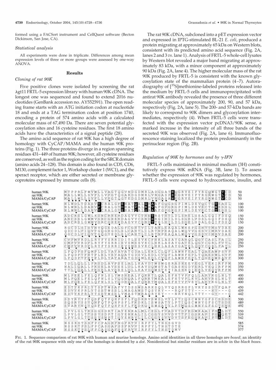
Five positive clones were isolated by screening the rat�gt11 FRTL-5 expression library with human 90K cDNA. Thelongest one was sequenced and found to extend 2016 nu-cleotides (GenBank accession no. AY552591). The open read-ing frame starts with an ATG initiation codon at nucleotide18 and ends at a TAG termination codon at position 1740,encoding a protein of 574 amino acids with a calculatedmolecular mass of 67,490 Da. There are seven potential gly-cosylation sites and 16 cysteine residues. The first 18 aminoacids have the characteristics of a signal peptide (28).
The amino acid sequence of rat 90K has a high degree ofhomology with CyCAP/MAMA and the human 90K pro-teins (Fig. 1). The three proteins diverge in a region spanningresidues 431–449 of human 90K; however, all cysteine residuesare conserved, as well as the region coding for the SRCR domain(amino acids 24–128). This domain is also found in CD5, CD6,M130, complement factor 1, Workshop cluster 1 (WC1), and thesperact receptor, which are either secreted or membrane gly-coproteins expressed by immune cells (8).
The rat 90K cDNA, subcloned into a pET expression vectorand expressed in IPTG-stimulated BL-21 E. coli, produced aprotein migrating at approximately 65 kDa on Western blots,consistent with its predicted amino acid sequence (Fig. 2A,lanes 2 and 3 vs. lane 1). Analysis of FRTL-5 whole-cell lysatesby Western blot revealed a major band migrating at approx-imately 83 kDa, with a minor component at approximately90 kDa (Fig. 2A, lane 4). The higher molecular mass of the rat90K produced by FRTL-5 is consistent with the known gly-cosylation state of the mammalian protein (4–7). Autora-diography of [35S]methionine-labeled proteins released intothe medium by FRTL-5 cells and immunoprecipitated withantirat 90K antibody revealed the presence of three differentmolecular species of approximately 200, 90, and 57 kDa,respectively (Fig. 2A, lane 5). The 200- and 57-kDa bands arelikely to correspond to 90K dimers and glycosylation inter-mediates, respectively (4). When FRTL-5 cells were trans-fected with the expression vector pcDNA3/90K sense, amarked increase in the intensity of all three bands of thesecreted 90K was observed (Fig. 2A, lane 6). Immunofluo-rescence staining localized the protein predominantly in theperinuclear region (Fig. 2B).
Regulation of 90K by hormones and by �-IFN
FRTL-5 cells maintained in minimal medium (3H) consti-tutively express 90K mRNA (Fig. 3B, lane 1). To assesswhether the expression of 90K was regulated by hormones,FRTL-5 cells were exposed to hydrocortisone, insulin, and
FIG. 1. Sequence comparison of rat 90K with human and murine homologs. Amino acid identities in all three homologs are boxed; an identityof the rat 90K sequence with only one of the homologs is denoted by a dot. Nonidentical but similar residues are in white in the black boxes.
4730 Endocrinology, October 2004, 145(10):4728–4736 Grassadonia et al. • 90K in Normal Thyrocytes

TSH, separately or in combination. Hydrocortisone signifi-cantly decreased 90K mRNA levels (Fig. 3A, bottom; 3B, lane2), with a maximal effect after 24 h. Insulin or TSH increased90K mRNA levels (Fig. 3A, top; 3B, lanes 3 and 4), althoughtheir effect differed as a function of time; insulin caused aprogressive increase that maximized after 24 h, whereas theTSH-induced increase was maximal at 6 h and plateaued orslightly decreased thereafter. Time-course experiments werethen performed on 3H cells using TSH and insulin in com-bination. Surprisingly, a decrease in 90K RNA/GAPDH ra-tios was observed, with a half-maximal decrease at 12 h andmaximal decrease at 24 h lasting up to 48 h (data not shown).Therefore, 24- or 48-h time points were chosen to evaluate theaction of the combined hormones in subsequent experi-ments. The presence of hydrocortisone attenuated the effectof insulin (Fig. 3B, lane 5 vs. 3) but not that of TSH (Fig. 3B,lane 6 vs. 4). The simultaneous presence of insulin and TSHsignificantly decreased 90K mRNA levels, independentlyfrom the presence of hydrocortisone (Fig. 3B, lanes 7 and 8).Similar results were obtained by substituting TSH with for-skolin (Fig. 3B, lane 9) and insulin with IGF-I (data notshown).
We previously showed that IFN is able to increase human90K both in vitro and in vivo (29–31). Exposure of FRTL-5 cellscultured in 3H medium to �-IFN resulted in a significantincrease of 90K mRNA level (P � 0.01) (Fig. 4A, lane 2 vs. 1).The �-IFN effect was not influenced by TSH, insulin, hydro-cortisone, hydrocortisone plus insulin, or hydrocortisoneplus TSH (Fig. 4A, lanes 3–7); however, it was significantlydecreased by the combination of TSH and insulin (P � 0.01)(Fig. 4A, lanes 8 and 9 vs. 2–7). This last result was repro-duced in run-on experiments (Fig. 4B), suggesting an effectat the transcriptional level.
Effect of rat 90K on MHC class I antigens
Human 90K has been found to increase the expression ofMHC class I antigens in human breast cancer cells (32). InpcDNA3/90K-transfected FRTL-5 cells, Northern blot anal-ysis revealed a clear increase of MHC class I (Fig. 5A). Asimilar increase of MHC class I, but not of MHC class II,antigen mRNA was obtained when parental FRTL-5 cellswere exposed to rat 90K purified from the culture fluid of thesame cells (P � 0.01) (Fig. 5B). The effect was evident at theprotein level as well (Fig. 5C).
Effect of viral infection and ds polynucleotides on rat 90K
TSH/cAMP and insulin/IGF-I are known to down-regu-late MHC class I antigen in FRTL-5 cells (33–35) (Fig. 6A). Asthese same hormones decrease rat 90K, it was worthwhile toassess whether other experimental conditions known tomodulate MHC class I antigens (viral infection, transfectionwith ds bacterial DNA, cell genomic DNA, etc.) (19) wereeffective in influencing rat 90K level as well. Infection ofFRTL-5 cells with herpes simplex coordinately increased 90Kand class I mRNA levels within 16 h (Fig. 6B). Similar resultswere observed when ds bacterial DNA, salmon sperm DNA,calf thymus DNA, or FRTL-5 cell genomic DNA was intro-duced into the cytoplasm of the cells (Fig. 6C, lanes 2–5).Additionally, transfection of the cells with herpes or foamyvirus dsDNA, pcDNA3, or pRc/RSV (Fig. 6C, lanes 7–10),but not with single-strand (ss) herpesvirus DNA (Fig. 6C,lane 6) increased 90K and MHC class I mRNA. Similar resultswere obtained using different transfection procedures, i.e.electroporation or diethylaminoethyl dextran (data notshown). The increase in rat 90K mRNA was DNase but notmethylase sensitive (Fig. 7A, lanes 5 and 8) as for MHC classI antigens (19). The ss oligodeoxynucleotides (ODNs) having
FIG. 2. Rat 90K expression in transformed BL-21 bacteria and in FRTL-5 cells. A, 30 �g of BL-21 bacteria lysates (lanes 1 and 2), recombinantpurified protein (lane 3), or FRTL-5 cell lysates (lane 4) were separated by SDS-PAGE and Western blots performed with the rabbit antibodyraised against a synthetic peptide encompassing residues 530–546 of the deduced amino acid sequence of rat 90K. Proteins were detected byenhanced chemiluminescence. Recombinant protein was present in bacteria lysates after, but not before, induction with IPTG (lane 2 vs. lane1). Lane 3 depicts the recombinant product of approximately 65 kDa after purification on a His-tagged column, and lane 4 shows the 83- and90-kDa components in the whole-cell lysates of FRTL-5 maintained in 5H medium. To verify whether rat 90K was secreted, FRTL-5 cells weremetabolically labeled overnight with [35S]methionine and medium incubated with the anti-90K antibody followed by protein-A Sepharose. Theimmunoprecipitate was separated by SDS-PAGE, and autoradiography revealed three bands at 55, 90, and 200 kDa, respectively (lane 5). Thesebands were clearly increased when FRTL-5 cells were transfected with the 90K expression vector pcDNA3/90K sense (lane 6). B, FRTL-5 cells,grown in chamber slides and maintained in 5H medium, were fixed, permeabilized, and incubated with the antirat 90K antibody, followed byan ALEXA-594-conjugated donkey antirabbit antibody. The red fluorescence shows a perinuclear localization. Control staining with preimmuneserum showed no fluorescence signal (data not shown).
Grassadonia et al. • 90K in Normal Thyrocytes Endocrinology, October 2004, 145(10):4728–4736 4731

one or more CpG motifs (CpG-1; CpG-2) or non-CpG controlcounterparts had no effect on 90K mRNA levels (Fig. 7A,lanes 9–12), whereas the expression was increased by ds-phosphorothioate (s)ODNs, but not by ss sODNs (Fig. 7A,lanes 13–16). Furthermore, as seen for MHC class I (19),dsDNA copolymers (Fig. 7B, lanes 9–12) or duplexes (Fig. 7B,lanes 6–8) increased 90K expression, whereas ss polymersdid not (Fig. 7B, lanes 3–5). The ds oligonucleotides as shortas 35 bp were effective in increasing 90K mRNA levels (Fig.7C). The effect of dsDNA on 90K mRNA appeared to be as
good or better then that of �-IFN at maximal concentrationof each (Fig. 7D). 90K gene expression was also increased bydsRNA but not by ssRNA (data not shown). Finally, theincrease of 90K expression after transfection with dsDNAwas additive to that of �-IFN (Fig. 7E), but it was mecha-nistically different because it was not paralleled by the in-crease of CIITA (Fig. 7F) nor attenuated by TSH plus insulin(Fig. 8).
The ds-polynucleotide effect on rat 90K was associatedwith that on gene products characterizing the antigen-presenting-cell phenotype, such as proteasomal subunitLMP2, TAP molecules, MHC class II, invariant chain, HLA-DMB proteins, and the costimulatory molecule B7.1 (Fig. 9).
Discussion
Most of the available information on the function of themammalian 90K proteins is derived from studies on cancercells, whereas little is known about the role and the regula-tion of these proteins in normal cells. To address these issues,
FIG. 3. Hormonal modulation of rat 90K mRNA levels. A, FRTL-5cells were maintained in 3H medium and then stimulated with TSHor insulin (top), or hydrocortisone (bottom). At the time noted, totalRNA was isolated and subjected (10 �g/lane) to Northern analysisusing 32P-labeled rat 90K cDNA and GAPDH probes. The 90K datawere normalized to GAPDH mRNA levels on the same blots. The90K/GAPDH ratio was compared with the 3H control; results are themean � SD of three different experiments on three batches of cells. B,FRTL-5 cells were maintained in 3H medium and then stimulatedwith TSH, insulin (INS), or hydrocortisone (CORT) individually or incombination. On lane 9, forskolin (FSK, 5 �M) was used instead ofTSH. After 24 h, total RNA was isolated and subjected to Northernanalysis. Representative Northern data are presented at the top.Quantitative results from three different experiments on threebatches of cells are presented on the bottom, expressed as the mean �SD after 90K/GAPDH ratios were calculated. Data in 3H medium (lane1) were set at unity and other results expressed relative to these. *,Significant decrease in 90K mRNA levels (P � 0.05); **, significantincrease (P � 0.01).
FIG. 4. Effect of hormones on the ability of �-IFN to increase 90KmRNA levels. A, 32P-labeled rat 90K cDNA and GAPDH probes wereused in Northern analyses to characterize mRNA levels in FRTL-5cells maintained in 3H medium and stimulated with �-IFN, TSH,hydrocortisone (CORT), or insulin (INS) for 24 h. Total RNA wasisolated and subjected (10 �g/lane) to Northern analysis. Represen-tative Northern data are presented at the top of the figure. Resultsfrom three different experiments on three batches of cells are pre-sented on the bottom as the mean � SD after 90K/GAPDH ratios werecalculated and the value of 3H set at unity. *, Significant increase (P �0.01) in 90K mRNA levels; **, significant decrease in the effect of�-IFN (P � 0.01). B, FRTL-5 cells maintained in 3H medium weretreated with insulin (10 �g/ml) plus �-IFN, �-IFN plus TSH, or �-IFNplus 5 �M forskolin (FSK) for 24 h. Nuclei were isolated and incubatedwith [32P]UTP before mRNA was purified, denatured, and hybridizedto an excess of the noted unlabeled cDNA probes.
4732 Endocrinology, October 2004, 145(10):4728–4736 Grassadonia et al. • 90K in Normal Thyrocytes

we used rat FRTL-5 thyrocytes, a model of normal, hormon-ally responsive cells. Several novel observations emerged.
First, rat 90K expression was modulated by TSH andinsulin/IGF-I, which are both required for the growth andfunction of normal thyrocytes (36, 37), and by �-IFN. Inter-estingly, when TSH and insulin were used in combination,a marked decrease of the constitutive and �-IFN-induced 90Kexpression levels were seen, whereas when these same hor-mones were used individually an increased expression of theprotein was noticed. The reason for this differential behaviorof 90K when hormones are used individually or in combi-
nation is not clear, although it very likely results from theactivation of different pathways. The mechanisms respon-sible for 90K hormonal regulation are currently being inves-tigated at the promoter level. Moreover, studies on the ex-pression of 90K in thyrocytes of normal vs. hypothyroid miceare in progress to evaluate the physiological relevance of thisphenomenon.
Second, the expression levels of MHC class I antigensincreased when FRTL-5 cells were engineered to overexpressrat 90K or exposed to the protein purified from the culturefluid of the same cells, even in the presence of TSH and
FIG. 6. Coordinate regulation of MHC class I and 90K mRNAs by TSH plus insulin, by herpes simplex infection, or by dsDNA transfected intoFRTL-5 cells. A, FRTL-5 cells were maintained in 5H medium, then maintained 48 h in the same medium with TSH. RNA was isolated andNorthern analyses performed using 10 �g RNA and radiolabeled 90K, MHC class I, and GAPDH probes. B, FRTL-5 cells that had been grownto 80% confluency in 6H medium were infected with herpes simplex virus (HSV-2). RNA was isolated at the times noted and Northern analysesperformed using radiolabeled 90K, MHC class I, and GAPDH probes. C, FRTL-5 cells, grown to 80% confluency in 6H medium, were transfectedwith 5 �g each of the noted dsDNAs (lanes 2–5), with ss or ds herpes simplex virus DNA fragments (lanes 6 and 7, respectively), with a 54-bpds oligonucleotide from Foamy virus (lane 8), or with two plasmid DNAs, pcDNA3, and pRc/RSV (lanes 9 and 10). Total RNA was preparedand Northern analysis performed 48 h after transfection using MHC class I, 90K, or GAPDH probes, as described above. Lipofectamine treatmentalone served as a control of the transfection procedure (Mock).
FIG. 5. 90K increases MHC class I mRNA expression in FRTL-5 cells. A, FRTL-5 cells were grown in 6H medium to 80% confluency andtransfected with pcDNA3 containing the 90K cDNA in a sense (pcDNA3/90K sense) or antisense (pcDNA3/90K antisense) direction. After 24 h,the levels of 90K, MHC class I, and GAPDH mRNAs were measured by Northern analysis as described earlier. B, Fresh FRTL-5 cells maintainedin 5H medium were incubated with 10 �g/ml purified 90K for 24 h, and MHC class I and II levels were measured by Northern analysis. Arepresentative Northern blot is presented on the top; results from three different experiments on three batches of cells are presented on thebottom as the mean � SD after 90K/GAPDH ratios were calculated and the value with no 90K set at unity. Densitometry indicated that MHCclass I increase by 90� was statistically significant (P � 0.01). C, FACS analysis for measuring MHC class I expression on FRTL-5 cells treatedas in B.
Grassadonia et al. • 90K in Normal Thyrocytes Endocrinology, October 2004, 145(10):4728–4736 4733

insulin, which are known to down-regulate MHC class Iantigens. These data, which duplicate those obtained withhuman 90K (32), suggest that these proteins may behave asan autocrine or paracrine/exocrine factor able to up-regulateMHC class I antigens. Studies are in progress to clarify themechanisms underlying the 90K effect on MHC class I andto verify whether this phenomenon is specific for thyrocytesor can apply also to other normal endocrine cells, e.g. �-cells.
The functional meaning of these findings could be ex-plained in light of the mechanism regulating immune sur-veillance in the thyroid gland. In this tissue, TSH and insulinstimulate cell proliferation and function, increasing de novosynthesis of proteins (17, 18, 36, 37). Peptides derived fromthese self-proteins would complex with MHC class I anti-gens, increasing the cell surface density of these complexes,possibly leading to a break of self-tolerance (38). To avoidthis, down-regulation of MHC class I antigens by TSH andinsulin is required (38–41). As these same hormones de-crease the expression of 90K, which is able to induce MHCclass I antigens, it can be argued that the decrease of this
molecule is aimed at down-regulating the immune response,thus preserving self-tolerance. The need for a coordinateexpression of 90K and MHC class I is further supported bythe finding that both these immune-related proteins are up-regulated by �-IFN and that this induction is counteracted byTSH/cAMP (33, 34).
Last, and perhaps most intriguing, the level of rat 90Kexpression is increased after virus infection or the introduc-tion of DNAs into the cells. In all respects, this phenomenonreproduced what is seen for MHC class I (19). In fact, 90Kexpression does not seem to be mediated by CIITA or sup-pressed by TSH/insulin/IGF-I, as observed in cells exposedto �-IFN, supporting the hypothesis that ds polynucleotidesand �-IFN act through additive although independent mech-anisms, in contrast with previous suggestions (42). Thepresent findings add new insights to the role of 90K inthe immune defense against viruses and possibly cancer. Theintroduction of DNA into the cells experimentally repro-duces the migration of self genomic or mitochondrial DNAinto the cytoplasm, phenomena frequently observed in in-
FIG. 7. Properties of the nucleic acid needed to induce 90K expression in FRTL-5 cells. Transfection and Northern analysis using rat 90K andGAPDH probes were performed 48 h after treatment, exactly as described in Fig. 6C. A, FRTL-5 cells were transfected with 10 �g intact,methylated or DNase-treated plasmid, pcDNA3 or pRc/RSV (lanes 3–8) or 10 �g each of the ss-CpG oligodeoxynucleotides (ODNs) or controlODNs (CpG oligos; lanes 9–12) or ss- or ds-phosphorothioate ODNs (s-oligos; lanes 13–16). Lane 1 contains RNA from nontreated cells and lane2 from cells subjected to the transfection procedure only (Mock). B, Ten micrograms of the noted synthetic polymer nucleotides and their duplexeswere used to transfect cells (lanes 3–12). C, Cells were transfected with 5 �g of dsDNA fragments from 1004 to 24 bp in length (lanes 2–10)or with indicated amount of a 35-bp dsODNs (lanes 12–15) as described above. RNA from cells subjected to a mock transfection with lipofectaminealone is in lane 1; RNA from cells treated with 100 U/ml �-IFN is in lane 11. These serve as negative and positive controls, respectively. D,The 90K mRNA levels were compared after either transfecting cells with different amounts of dsDNA or incubating them with different amountsof �-IFN as noted. RNA was isolated 48 h after transfection or treatment and Northern analyses performed as above. E, 90K mRNA levels werecompared 48 h after transfecting cells with 10 �g dsDNA (pcDNA3), incubating them with 1000 U/ml �-IFN, or subjecting them to bothtreatments. A representative Northern analysis is presented at the top; the mean � SD of the calculated 90K/GAPDH ratios from four separateexperiments using different batches of cells is presented at the bottom of the panel. The control value is set at unity. *, Significant increase(P � 0.01) in 90K RNA levels induced by �-IFN or dsDNA; **, significant increase in the individual effect of �-IFN or dsDNA (P � 0.05). F,90K and CIITA mRNA levels were compared 48 h after transfecting cells with 10 �g dsDNA (pcDNA3) or incubating them with 100 U/ml �-IFN.
4734 Endocrinology, October 2004, 145(10):4728–4736 Grassadonia et al. • 90K in Normal Thyrocytes

jured tissues (43) and in tumor cells (44, 45). These sameprocedures have been found to induce MHC class I antigens(19). This induction is highly desirable because the presen-tation of peptides by MHC antigens is required for the de-velopment of an effective immune response against cellsinfected by viruses or altered by malignant transformation(46). Interestingly, these pathogenic events are associatedwith an increased expression of 90K, which in turn promotesan additional MHC class I response. Therefore, it can beargued that the enhanced expression of 90K may be part ofa mechanism aimed at triggering immune effectors againstcells infected by viruses or altered after an oncogenic insult.This hypothesis is further supported by the finding that theincrease of 90K and MHC class I is accompanied by enhancedexpression of MHC class II, the costimulatory molecule B7–1,and other genes or gene products known to be important forantigen presentation, including LMP2, TAP, invariant chain,and HLA-DMB proteins (47, 48).
Some of the features of 90K, including the increased levelsfound in the tumors and after virus infection or interferontreatment of the cell, may resemble those of heat shock pro-teins. However, unlike these proteins, heating the cells up to42 C does not result in an increased production or release of90K (data not shown).
In sum, we have shown that 90K, constitutively expressedby FRTL-5 thyrocytes, increases MHC class I antigens andthat the expression of these proteins is coordinately regulatedby hormones. We suggest that 90K may participate in mech-anisms ensuring an appropriate level of immune response inall instances. The decrease of this protein in cells proliferatingunder physiological conditions may contribute to the main-tenance of the immune response within the limits of self-tolerance. On the other hand, the increase of 90K that follows
viral infection or conditions resembling self DNA leakageinto the cytoplasm could be part of the host defense mech-anisms facilitating the elimination of infected or tumor-transformed cells.
Acknowledgments
Received April 19, 2004. Accepted June 23, 2004.Address all correspondence and requests for reprints to: Stefano
Iacobelli, M.D., Dipartimento di Oncologia e Neuroscienze, Sezione diOncologia Medica, SEBI, via dei Vestini, 66100 Chieti, Italy. E-mail:[email protected].
This work was supported in part by grants from the AssociazioneItaliana per la Ricerca sul Cancro (AIRC) and Ministero dell’Istruzione,Universita e Ricerca (MIUR).
The sequence of rat 90K has been submitted to the GenBank databaseunder accession no. AY552591.
References
1. Iacobelli S, Arno E, D’Orazio A, Coletti G 1986 Detection of antigens rec-ognized by a novel monoclonal antibody in tissue and serum from patientswith breast cancer. Cancer Res 46:3005–3010
2. Iacobelli S, Arno E, Sismondi P, Natoli C, Gentiloni N, Scambia G, Giai M,Cortese P, Panici PB, Mancuso S 1988 Measurement of a breast cancer asso-ciated antigen detected by monoclonal antibody SP-2 in sera of cancer patients.Breast Cancer Res Treat 11:19–30
3. Iacobelli S, Sismondi P, Giai M, D’Egidio M, Tinari N, Di Stefano P, NatoliC 1994 Prognostic value of a novel circulating serum 90K antigen in breastcancer. Br J Cancer 69:172–176
4. Koths K, Taylor E, Halenbeck R, Casipit C, Wang A 1993 Cloning andcharacterization of a human Mac-2-binding protein, a new member of the
FIG. 8. TSH plus insulin decreases 90K mRNA levels induced by�-IFN, but not by dsDNA. 32P-labeled rat 90K cDNA and GAPDHprobes were used in Northern analyses to characterize mRNA levelsin FRTL-5 cells maintained in 5H with 5% serum, then shifted to freshmedium with �-IFN (100 U/ml) and with or without TSH (1 � 10–10
M) for 48 h. Alternatively, cells were transfected with 10 �g/ml dsDNAand then placed in medium with or without TSH for 48 h. Total RNAwas isolated and subjected (10 �g/lane) to Northern analysisas described above. Results from three different experiments onthree batches of cells are presented on the bottom as the mean � SDafter 90K/GAPDH ratios were calculated and the value of 5H setat unity. *, Significant increase (P � 0.01) in 90K RNA levels inducedby �-IFN or dsDNA; **, significant decrease in the effect of �-IFN(P � 0.01). FIG. 9. The dsDNA increased 90K and MHC class I in FRTL-5 cells
is associated with conversion of the cells to a potential antigen-presenting cell. Changes in 90K, MHC class I, and GAPDH mRNAlevels in cells transfected with dsDNA are compared with changes ingenes important for the antigen-presenting-cell activity of a cell: theproteasome-processing protein (LMP2); the transporter of antigenpeptide (TAP1); MHC class II; invariant chain (Ii); HLA-DMB, andthe costimulatory molecule B7.1. Total RNA was isolated and sub-jected to Northern blot analysis as described above.
Grassadonia et al. • 90K in Normal Thyrocytes Endocrinology, October 2004, 145(10):4728–4736 4735

superfamily defined by the macrophage scavenger receptor cysteine-rich do-main. J Biol Chem 268:14245–14249
5. Ullrich A, Sures I, D’Egidio M, Jallal B, Powell TJ, Herbst R, Dreps A, AzamM, Rubinstein M, Natoli C, Shawver LK, Schlessinger J, Iacobelli S 1994 Thesecreted tumor-associated antigen 90K is a potent immune stimulator. J BiolChem 269:18401–18407
6. Friedman J, Trahey M, Weissman I 1993 Cloning and characterization ofcyclophilin C-associated protein: a candidate natural cellular ligand for cy-clophilin C. Proc Natl Acad Sci USA 90:6815–6819
7. Chicheportiche Y, Vassalli P 1994 Cloning and expression of a mouse mac-rophage cDNA coding for a membrane glycoprotein of the scavenger receptorcysteine-rich domain family. J Biol Chem 269:5512–5517
8. Resnick D, Pearson A, Krieger M 1994 The SRCR superfamily: a familyreminiscent of the Ig superfamily. Trends Biochem Sci 19:5–8
9. Powell TJ, Schreck R, McCall M, Hui T, Rice A, App H, Ullrich A, ShawverLK 1995 A tumor-derived protein which provides T-cell costimulation throughaccessory cell activation. J Immunother Emphasis Tumor Immunol 17:209–221
10. Rea A, Calmieri G, Tinari N, Natoli C, Tagliaferro P, Morabito A, Grassa-donia A, Bianco AR, Iacobelli S 1994 90K is a serum marker of poor prognosisin non-Hodgkin’s lymphoma patients. Oncol Rep 1:723–725
11. Zeimet AG, Natoli C, Herold M, Fuchs D, Windbichler G, Daxenbichler G,Iacobelli S, Dapunt O, Marth C 1996 Circulating immunostimulatory protein90K and soluble interleukin-2-receptor in human ovarian cancer. Int J Cancer68:34–38
12. Fornarini B, D’Ambrosio C, Natoli C, Tinari N, Silingardi V, Iacobelli S 2000Adhesion to 90K (Mac-2 BP) as a mechanism for lymphoma drug resistancein vivo. Blood 96:3282–3285
13. Marchetti A, Tinari N, Buttitta F, Chella A, Angeletti CA, Sacco R, MucilliF 2002 Expression of 90K (Mac-2 BP) correlates with distant metastasis andpredicts survival in stage I non-small cell lung cancer patients. Cancer Res62:2535–2539
14. Natoli C, Dianzani F, Mazzotta F, Balocchini E, Pierrotti P, Antonelli G,Iacobelli S 1993 90K protein: a new predictor marker of disease progressionin human immunodeficiency virus infection. J Acquir Immune Defic Syndr6:370–375
15. Briggs NC, Natoli C, Tinari N, D’Egidio M, Goedert JJ, Iacobelli S 1993 A90-kDa protein serum marker for the prediction of progression to AIDS in acohort of HIV-1� homosexual men. AIDS Res Hum Retroviruses 9:811–816
16. Artini M, Natoli C, Tinari N, Costanzo A, Marinelli R, Balsano C, Porcari P,Angelucci D, D’Egidio M, Levrero M, Iacobelli S 1996 Elevated serum levelsof 90K/MAC-2 BP predict unresponsiveness to �-interferon therapy in chronicHCV hepatitis patients. J Hepatol 25:212–217
17. Ambesi-Impiombato FS, Perrild H 1989 FRTL-5 today. Int Congress Series818. Amsterdam: Elsevier
18. Ekholm R, Kohn LD, Wollman S 1989 Control of the thyroid: regulation ofits normal growth and function. New York: Plenum Press
19. Suzuki K, Mori A, Ishii KJ, Saito J, Singer DS, Klinman DM, Krause PR,Kohn LD 1999 Activation of target-tissue immune-recognition molecules bydouble-stranded polynucleotides. Proc Natl Acad Sci USA 96:2285–2290
20. Ambesi-Impiombato FS 1986 Fast growing thyroid cell strain. US Patent 4,608,341
21. Kohn LD, Valente WA, Grollman EF, Aloj SM, Vitti P 1986 Clinical deter-mination and/or quantification of thyrotropin and a variety of thyroid stim-ulatory or inhibitory factors performed in vitro with an improved cell lineFRTL-5. US Patent 4,609,622
22. Akamizu T, Ikuyama S, Saji M, Kosugi S, Kozak C, McBride OW, Kohn LD1990 Cloning, chromosomal assignment, and regulation of the rat thyrotropinreceptor: expression of the gene is regulated by thyrotropin, agents that in-crease cAMP levels, and thyroid autoantibodies. Proc Natl Acad Sci USA87:5677–5681
23. Sanger F, Nicklen S, Coulson AR 1977 DNA sequencing with chain-terminating inhibitors. Proc Natl Acad Sci USA 74:5463–5467
24. Green N, Alexander H, Olson A, Alexander S, Shinnick TM, Sutcliffe JG,Lerner RA 1982 Immunogenic structure of the influenza virus hemagglutinin.Cell 28:477–487
25. Laemmli UK 1970 Cleavage of structural proteins during the assembly of thehead of bacteriophage T4. Nature 227:680–685
26. Towbin H, Staehelin T, Gordon J 1979 Electrophoretic transfer of proteinsfrom polyacrylamide gels to nitrocellulose sheets: procedure and some ap-plications. Proc Natl Acad Sci USA 76:4350–4354
27. Suzuki K, Lavaroni S, Mori A, Okajima F, Kimura S, Katoh R, Kawaoi A,Kohn LD 1998 Thyroid transcription factor 1 is calcium modulated and co-ordinately regulates genes involved in calcium homeostasis in C cells. Mol CellBiol 18:7410–7422
28. Dangott LJ, Jordan JE, Bellet RA, Garbers DL 1989 Cloning of the mRNA forthe protein that crosslinks to the egg peptide speract. Proc Natl Acad Sci USA86:2128–2132
29. Iacobelli S, Scambia G, Natoli C, Panici PB, Baiocchi G, Perrone L, MancusoS 1988 Recombinant human leukocyte interferon-�2b stimulates the synthesisand release of a 90K tumor-associated antigen in human breast cancer cells. IntJ Cancer 42:182–184
30. Natoli C, Garufi C, Tinari N, D’Egidio M, Lesti G, Gaspari LA, Visini R,Iacobelli S 1993 Dynamic test with recombinant interferon-�-2b: effect on 90Kand other tumour-associated antigens in cancer patients without evidence ofdisease. Br J Cancer 67:564–567
31. Marth C, Dreps A, Natoli C, Zeimet AG, Lang T, Widschwendter M,Daxenbichler G, Ullrich A, Iacobelli S 1994 Effects of type-I and -II interferonson 90K antigen expression in ovarian carcinoma cells. Int J Cancer 59:808–813
32. Natoli C, Iacobelli S, Kohn LD 1996 The immune stimulatory protein 90Kincreases major histocompatibility complex class I expression in a humanbreast cancer cell line. Biochem Biophys Res Commun 225:617–620
33. Saji M, Moriarty J, Ban T, Kohn LD, Singer DS 1992 Hormonal regulation ofmajor histocompatibility complex class I genes in rat thyroid FRTL-5 cells:thyroid-stimulating hormone induces a cAMP-mediated decrease in class Iexpression. Proc Natl Acad Sci USA 89:1944–1948
34. Saji M, Moriarty J, Ban T, Singer DS, Kohn LD 1992 Major histocompatibilitycomplex class I gene expression in rat thyroid cells is regulated by hormones,methimazole, and iodide as well as interferon. J Clin Endocrinol Metab 75:871–878
35. Saji M, Shong M, Napolitano G, Palmer LA, Taniguchi SI, Ohmori M, OhtaM, Suzuki K, Kirshner SL, Giuliani C, Singer DS, Kohn LD 1997 Regulationof major histocompatibility complex class I gene expression in thyroid cells.Role of the cAMP response element-like sequence. J Biol Chem 272:20096–20107
36. Tramontano D, Cushing GW, Moses AC, Ingbar SH 1986 Insulin-like growthfactor-I stimulates the growth of rat thyroid cells in culture and synergizes thestimulation of DNA synthesis induced by TSH and Graves’-IgG. Endocrinol-ogy 119:940–942
37. Tramontano D, Moses AC, Picone R, Ingbar SH 1987 Characterization andregulations of the receptor for insulin-like growth factor-I in the FRTL-5 ratthyroid follicular cell line. Endocrinology 120:785–790
38. Singer DS, Mozes E, Kirshner S, Kohn LD 1997 Role of MHC class I moleculesin autoimmune disease. Crit Rev Immunol 17:463–468
39. Singer DS, Maguire JE 1990 Regulation of the expression of class I MHC genes.Crit Rev Immunol 10:235–257
40. Kohn LD, Napolitano G, Singer DS, Moltini M, Scorza R, Shimojo N, KohnoY, Mozes E, Nakazato M, Ulianich L, Chung HK, Matoba H, Saunier B,Suzuki K, Schuppert F, Saji M 2000 Graves’ disease: a host defense mecha-nism gone awry. Int Rev Immunol 19:633–664
41. Kohn LD, Giuliani C, Montani V, Napolitano G, Ohmori M, Ohta M, SajiM, Schuppert F, Shong M, Suzuki K, Taniguchi, SI, Yano K, Singer DS 1995Antireceptor immunity. In: Rayner D, Champion B, eds. Thyroid immunity.Austin/Georgetown, TX: RG Landes Biomedical; 115–170
42. Brakebusch C, Jallal B, Fusco O, Iacobelli S, Ullrich A 1997 Expression of the90K immunostimulator gene is controlled by a promoter with unique features.J Biol Chem 272:3674–3682
43. Moffett CW, Paden CM 1994 Microglia in the rat neurohypophysis increaseexpression of class I major histocompatibility antigens following central ner-vous system injury. J Neuroimmunol 50:139–151
44. Solage A, Laskov R 1975 Characterization of cytoplasmic DNA of mouse-myeloma cells. Eur J Biochem 60:23–33
45. Hegger R, Abken H 1995 The short DNA sequences in the cytoplasm of Ehrlichascites tumor cells are tightly associated with proteins. Physiol Chem PhysMed NMR 27:321–328
46. Foss FM 2002 Immunologic mechanisms of antitumor activity. Semin Oncol29:5–11
47. York IA, Rock KL 1996 Antigen processing and presentation by the class Imajor histocompatibility complex. Annu Rev Immunol 14:369–396
48. Pieters J 1997 MHC class II restricted antigen presentation. Curr Opin Im-munol 9:89–96
Endocrinology is published monthly by The Endocrine Society (http://www.endo-society.org), the foremost professional society serving theendocrine community.
4736 Endocrinology, October 2004, 145(10):4728–4736 Grassadonia et al. • 90K in Normal Thyrocytes