Tesi_Pau_Brugada.pdf - TDX (Tesis Doctorals en Xarxa)
204
C. Claravall, 1-3 | 08022 Barcelona | Tel. 93 602 22 00 | Fax 93 602 22 49 | [email protected] | www.url.edu C.I.F. G: 59069740 Universitat Ramon Llull Fundació Rgtre. Fund. Generalitat de Catalunya núm. 472 (28-02-90) Boosting intravenous administration of therapeutic viral vectors using an oligopeptide-modified poly(β-aminoester)s-based coating technology Pau Brugada Vilà http://hdl.handle.net/10803/664008 ADVERTIMENT. L'accés als continguts d'aquesta tesi doctoral i la seva utilització ha de respectar els drets de la persona autora. Pot ser utilitzada per a consulta o estudi personal, així com en activitats o materials d'investigació i docència en els termes establerts a l'art. 32 del Text Refós de la Llei de Propietat Intel·lectual (RDL 1/1996). Per altres utilitzacions es requereix l'autorització prèvia i expressa de la persona autora. En qualsevol cas, en la utilització dels seus continguts caldrà indicar de forma clara el nom i cognoms de la persona autora i el títol de la tesi doctoral. No s'autoritza la seva reproducció o altres formes d'explotació efectuades amb finalitats de lucre ni la seva comunicació pública des d'un lloc aliè al servei TDX. Tampoc s'autoritza la presentació del seu contingut en una finestra o marc aliè a TDX (framing). Aquesta reserva de drets afecta tant als continguts de la tesi com als seus resums i índexs. ADVERTENCIA. El acceso a los contenidos de esta tesis doctoral y su utilización debe respetar los derechos de la persona autora. Puede ser utilizada para consulta o estudio personal, así como en actividades o materiales de investigación y docencia en los términos establecidos en el art. 32 del Texto Refundido de la Ley de Propiedad Intelectual (RDL 1/1996). Para otros usos se requiere la autorización previa y expresa de la persona autora. En cualquier caso, en la utilización de sus contenidos se deberá indicar de forma clara el nombre y apellidos de la persona autora y el título de la tesis doctoral. No se autoriza su reproducción u otras formas de explotación efectuadas con fines lucrativos ni su comunicación pública desde un sitio ajeno al servicio TDR. Tampoco se autoriza la presentación de su contenido en una ventana o marco ajeno a TDR (framing). Esta reserva de derechos afecta tanto al contenido de la tesis como a sus resúmenes e índices. WARNING. The access to the contents of this doctoral thesis and its use must respect the rights of the author. It can be used for reference or private study, as well as research and learning activities or materials in the terms established by the 32nd article of the Spanish Consolidated Copyright Act (RDL 1/1996). Express and previous authorization of the author is required for any other uses. In any case, when using its content, full name of the author and title of the thesis must be clearly indicated. Reproduction or other forms of for profit use or public communication from outside TDX service is not allowed. Presentation of its content in a window or frame external to TDX (framing) is not authorized either. These rights affect both the content of the thesis and its abstracts and indexes.
-
Upload
khangminh22 -
Category
Documents
-
view
1 -
download
0
Transcript of Tesi_Pau_Brugada.pdf - TDX (Tesis Doctorals en Xarxa)
C. Claravall, 1-3 | 08022 Barcelona | Tel. 93 602 22 00 | Fax 93 602 22 49 | [email protected] | www.url.edu
C.I.
F. G
: 590
6974
0 U
nive
rsita
t Ram
on L
lull
Fund
ació
R
gtre
. Fun
d. G
ener
alita
t de
Cat
alun
ya n
úm. 4
72 (2
8-02
-90)
Boosting intravenous administration of therapeutic viral vectors using an oligopeptide-modified poly(β-aminoester)s-based coating technology
Pau Brugada Vilà
http://hdl.handle.net/10803/664008
ADVERTIMENT. L'accés als continguts d'aquesta tesi doctoral i la seva utilització ha de respectar els drets de la persona autora. Pot ser utilitzada per a consulta o estudi personal, així com en activitats o materials d'investigació i docència en els termes establerts a l'art. 32 del Text Refós de la Llei de Propietat Intel·lectual (RDL 1/1996). Per altres utilitzacions es requereix l'autorització prèvia i expressa de la persona autora. En qualsevol cas, en la utilització dels seus continguts caldrà indicar de forma clara el nom i cognoms de la persona autora i el títol de la tesi doctoral. No s'autoritza la seva reproducció o altres formes d'explotació efectuades amb finalitats de lucre ni la seva comunicació pública des d'un lloc aliè al servei TDX. Tampoc s'autoritza la presentació del seu contingut en una finestra o marc aliè a TDX (framing). Aquesta reserva de drets afecta tant als continguts de la tesi com als seus resums i índexs.
ADVERTENCIA. El acceso a los contenidos de esta tesis doctoral y su utilización debe respetar los derechos de la persona autora. Puede ser utilizada para consulta o estudio personal, así como en actividades o materiales de investigación y docencia en los términos establecidos en el art. 32 del Texto Refundido de la Ley de Propiedad Intelectual (RDL 1/1996). Para otros usos se requiere la autorización previa y expresa de la persona autora. En cualquier caso, en la utilización de sus contenidos se deberá indicar de forma clara el nombre y apellidos de la persona autora y el título de la tesis doctoral. No se autoriza su reproducción u otras formas de explotación efectuadas con fines lucrativos ni su comunicación pública desde un sitio ajeno al servicio TDR. Tampoco se autoriza la presentación de su contenido en una ventana o marco ajeno a TDR (framing). Esta reserva de derechos afecta tanto al contenido de la tesis como a sus resúmenes e índices.
WARNING. The access to the contents of this doctoral thesis and its use must respect the rights of the author. It can be used for reference or private study, as well as research and learning activities or materials in the terms established by the 32nd article of the Spanish Consolidated Copyright Act (RDL 1/1996). Express and previous authorization of the author is required for any other uses. In any case, when using its content, full name of the author and title of the thesis must be clearly indicated. Reproduction or other forms of for profit use or public communication from outside TDX service is not allowed. Presentation of its content in a window or frame external to TDX (framing) is not authorized either. These rights affect both the content of the thesis and its abstracts and indexes.

C. Claravall, 1-3 | 08022 Barcelona | Tel. 93 602 22 00 | Fax 93 602 22 49 | [email protected] | www.url.edu
C.I.
F. G
: 590
6974
0 U
nive
rsita
t Ram
on L
lull
Fund
ació
R
gtre
. Fun
d. G
ener
alita
t de
Cat
alun
ya n
úm. 4
72 (2
8-02
-90)
DOCTORAL THESIS
Title Boosting intravenous administration of therapeutic viral vectors using an oligopeptide-modified poly(b-amino ester)s-based coating technology
Presented by Pau Brugada Vilà
Centre IQS School of Engineering
Department Bioengineering
Directed by Dr. Salvador Borrós Gómez Dra. Anna Cascante Cirera Dra. Cristina Fillat Fonts

This page left blank intentionally

A la meva família

This page left blank intentionally

“Ir y venir, seguir y guiar, dar y tener, entrar y salir de fase.
Amar la trama más que el desenlace.”
Jorge Drexler

This page left blank intentionally

6
Acknowledgments Finalment ha arribat el moment de posar-me davant d’aquestes últimes fulles en blanc de
la tesi. Tinc una barreja de sensacions. Satisfacció i alegria però també nostàlgia. Són molts
anys, moltes experiències viscudes, molt aprenentatge, moltes victòries i derrotes, molta gent
amb qui he compartit i gaudit i de qui m’emporto records molt intensos i especials.
Primer de tot m’agradaria agrair molt afectuosament al meu director i directores de tesi, en
Dr. Salvador Borrós, la Dra. Anna Cascante i la Dra. Cristina Fillat el seu suport i confiança durant
aquests anys.
Salvador, gràcies per ser com ets. M’has donat moltíssim i sempre que ho he necessitat
m’has arrencat un somriure i m’has aixecat els ànims. Hi ha algunes converses que sempre
recordaré. Que si a mi se’m dona bé que em tirin a la selva amb un matxet. Que si sempre has
volgut una casa amb bosquet. Que si PEG o no PEG. Les coses importants solen rimar! Les
reunions improvisades els divendres a última hora quan ja no quedava gairebé ningú, que potser
començaven seriosament però que sempre les acabàvem estirant tot caminant fins al “hall” junts
parlant de ves a saber què. Gràcies per haver cregut en mi. Gràcies per haver valorat les meves
idees i per haver-me donat tantes oportunitats. Gràcies per la teva amistat. Una frase que també
m’has dit moltes vegades és “Pau, tu tens moltes virtuts”. Sempre he sabut que aquesta frase
amaga alguna cosa. Estic segur que gràcies a tu he crescut i he superat algunes de les meves
debilitats. Moltíssimes gràcies per tot!
Anna, sense tu no hagués arribat fins aquí. M’has ajudat a trobar la direcció adequada en
moments crítics. Sempre que ho he necessitat m’has escoltat i aconsellat i has fet tot el possible
per tal d’obrir-me portes i per fer que els projectes tiressin endavant a pas ferm. Hem passat
moltes coses junts, des dels models in vitro de BBB fins als experiments in vivo d’eficàcia. Molts
anys de reunions i d’esforç que m’han fet créixer i aprendre moltíssim. Hem aconseguit fites
realment increïbles! Algun dia les recordarem junts i ens en farem creus. Gràcies per tot, de tot
cor!
Gràcies Cristina per haver-me obert les portes al teu grup i haver-me acollit com un més
durant aquests anys. M’he sentit molt afortunat de poder aprendre de tu, ja sigui en els seminaris,
anant de congrés, en reunions al teu despatx o durant les converses a l’hora de dinar. Per tu
sempre és un bon moment per compartir alguna experiència que ens pugui ser útil o per acabar
de discutir algun dubte o resultat. M’has ensenyat molt però sobre tot m’has entès molt. Et desitjo
molta sort i t’agraeixo moltíssim la teva confiança!
I am also very grateful to Dr. Florian Kreppel from the Witten/Herdecke University. It was a
real pleasure to stay with and learn from you and your group members. You are excellent

7
scientists, hard workers and also very nice and fun people. I felt at home and enjoyed a lot these
days in Germany. I wish you lots of success in the future!
Un dels pilars fonamentals d’aquesta tesi es sustenta sobre Sagetis-Biotech S.L. Amb ells
he compartit objectius i desitjos i han sigut el meu dia a dia durant tot aquest temps. Sense ells
jo molt probablement avui no estaria escrivint aquests agraïments. Gràcies per tot l’esforç
Eduard, Xavier, Salvador i Anna. Sou uns lluitadors i lluitadores i estic segur que algun dia rebreu
la recompensa que us mereixeu. Gràcies també a tota la gent amb qui hi he coincidit. Irene has
sigut una gran companya, vals molt i et desitjo el millor. Elena no canviïs mai, tu puedes con
todo. Cristina Fornaguera estic segur que aconseguiràs tot el que et proposis. Les llargues hores
a l’estabulari m’ho van demostrar, Cristina Castells ets un sol de persona i tenir-te a prop ha sigut
un regal, Miguel Angel yo sin ti no puedo hacer coatings ni puedo hacer nada. ¡Gracias por todo
y mucha suerte! Pri vas ser un gran exemple al principi de la meva tesi i et considero un germà.
Seijin te adoro y te deseo mucha suerte. Mariana tu vas demostrar que treballar incansablement
sense perdre el somriure és possible. Ingrid tu ets el millor que m’emporto d’aquesta etapa. Equip
Sagetis, junts hem compartit històries de tots colors. Hem suat, hem rigut, hem plorat i hem
somiat. Sempre us portaré al cor i us desitjo tota la sort del món a tots i a totes!
Un altre puntal de la meva tesi es recolza sobre el grup de GEMAT i deu ni do si n’hi ha per
recolzar. Desde els més veterans fins als TFMs. Començaré per dalt i això vol dir començar per
un grande Victor Ramos. Gràcies Victor, en tu sempre he trobat un equilibri perfecte entre el
treballar i el divertir-se treballant. Ets un pou de coneixements i sempre estàs disposat a
compartir-los! A les meves estimades Núria i Marina del Lab Vell, només dir-vos que sou
precioses i que trobar-vos i compartir alguna conversa sempre m’alegrarà el dia. A la Majo per
tota l’alegria contagiada i per algun soparet compartit. A en Robert, què dir d’aquest tros d’home?
Una joia. Algun dia tocaràs la tecla, aquella tecla que molts pocs troben. N’estic segur. A en Joan,
lo puto crack de IQS, només dir-te que saps que t’estimo i que ens seguirem veient. Gràcies per
totes les converses i ajuda i molta sort als dos en la vostra aventura a Tractivus! A en Pere que
hem passat una llarga història junts a IQS, dir-te que no tens límits Peri. Ets un “tremendu” i
arribaràs allà a on tu vulguis. M’ha encantat compartir aquests anys amb tu!. A l’Anna Mas, la
jefa. Quan en xixo se’n cansi t’hi poses tu i segur que tot anirà bé. Molta sort futura Dra. Mas.
També vull agrair a tots els demés GEMATs i no GEMATs de IQS amb els que he compartit
algunes històries especials. A la Mire i a l’Alba per ser tant divertides i bona gent. A la Núria amb
la qual compartim una bona amiga i els pBAEs. A en Pol per les converses apassionants que
hem tingut. A en Tito per les tardes de braves i birres. A en German per la teva energia espontània
i bona fe. A la Sara Bardají pels moments divertits que hem compartit. A en Mario que amaga un
gran cor. A la gent de teixits, la Mire, la Patri, la Cris, la Lourdes... Arribar i trobar-vos a vosaltres
va ser un gran estímul. Per tots els moments viscuts, fora i dintre del laboratori, moltes gràcies a
totes i a tots! Jo per GEMAT, GEMATO!
8
Per últim vull agrair especialment a la Laura i a la Sara. Les meves TFMs. Vaig aprendre
molt treballant amb vosaltres. Laura molts d’ànims amb el teu doctorat, estic segur que faràs una
gran feina! Gràcies per tractar-me amb confiança i valorar-me.
A la meva gent del CEK, què dir-vos? El que semblava una petita col·laboració es va acabar
convertint en la meva primera casa. He passat uns anys inoblidables amb vosaltres. L’ambient
de treball ha sigut immillorable i he aprés moltíssimes coses. Maria, no em cansaré de donar-te
les gràcies! Sense tu no sé què n’hagués fet de tants animalons. M’ho has ensenyat tot. Giullia
segueix lluitant amb el bon humor que et caracteritza, ha sigut un gust tenir-te a prop. Estela
sempre recordarem alguna aventura imprevista, les converses científiques i la companyia. Jero
amb tu la ciència està en bones mans. La pau i ordre que transmets son molt valuosos. Sabri
gràcies per tenir sempre paciència i no perdre mai el somriure. A l’Eneko per convertir la ciència
en una tasca estimulant i innovadora. A l’Anna per ajudar-me i compartir sempre un somriure i
finalment a en Xevi, per rebre’m amb els braços oberts i fer-me sentir acollit i com a casa des de
el primer dia. Espero que algun dia ens tornem a trobar tots junts. Mentrestant, cuidaré aquests
records. Moltes gràcies per tot!
No em puc deixar els meus estimats màsters: Estela, Alex, Jorge, Jenny, Aitor, Pere, Anna,
Su i Miquel. Amb ells comença tot. Em sento molt afortunat d’haver compartit aquest temps amb
vosaltres. Espero que no deixem mai de trobar moments per retrobar-nos i compartir rialles i
vivències. Som una gran “promo” de Bioenginyers!
Per últim vull agrair als amics de fora dels grups de recerca que m’han donat suport, m’han
animat i m’han aguantat. A tu Dani perquè sense tu em faltaria un tros. A en Bermu per haver-
me acompanyat sempre i haver-me ajudat a créixer. A la Jess per ser una súper doctora i una
gran amiga. A l’Uri per creure en la meva faceta artística i per l’amistat. A en Vic per haver
compartit moments molt especials dins i fora dels escenaris. A l’Eduard per les quedades
revitalitzants. A l’Andreu per fer-me sentir sempre capaç.
Per últim vull agrair al meu pare i a la meva mare que sempre han estat al meu costat i que
han treballat molt per oferir-me totes les oportunitats que he tingut. Sou un gran exemple i us
estimo moltíssim. Gràcies per donar-m’ho tot! Aquesta tesi va per vosaltres. A la meva germana,
que sempre m’ha donat força i que m’ha permès compartir i superar moltes de les meves pors.
A l’Ingrid que la duc a tot arreu a dins del cor. Quina sort he tingut de passar per IQS quan tu
rondaves per allà. T’estimo mori. A les petites de la família, la Júlia i l’Erin, que segur que faran
grans coses en el futur. Als meus avis, Esteve, Ramon, Nita i Pepita. L’impuls que li vau donar al
vostre net l’ha fet arribar fins aquí. A tota la meva estimada família, als que hi son i als que no,
us dedico aquesta tesi!
Pau Brugada Vilà, 3 de Setembre de 2018

This page left blank intentionally

Abstract
10
Abstract
Boosting intravenous administration of therapeutic viral vectors using an oligopeptide-modified poly(ß-amino ester)s-based coating technology
The use of viruses as therapeutic agents has become a reality for the treatment of inherited
genetic diseases and cancer in the recent years. The regulatory approval, commercialization and
clinical use of some virus-based products is the best demonstration of their applicability. However,
their potential is far from being completely exploited. Their inherent promiscuity to infect non-
target cells, their intrinsic immunogenicity and the high seroprevalence within the population pose
serious risks regarding their safety and minimize their efficacy when systemically administered.
Several strategies have been attempted in order to circumvent these drawbacks, from genetic
engineering of viral genomes to the development of complex delivering procedures. The
generation of hybrid viral/non-viral vectors using novel biomaterials aiming to hinder viral capsids
and enhance virus accumulation in target sites is one among these strategies.
Recently, newly developed poly(β-amino ester)s (pBAEs) have emerged as an interesting
choice as non-viral gene delivery vectors. Terminal modified pBAEs with oligopeptides (OM-
pBAEs) have been used to produced cell-specific vectors and slightly hydrophobic backbones
have improved their in vivo applicability. Here, we present their use as coating agents to boost
the intravenous administration of non-enveloped viral vectors such as adeno-associated viruses
(AAV) and adenoviruses (Ad). We demonstrate that cationic OM-pBAEs efficiently interact with
viral capsids serving as an electrostatic anchorage to physically modify them. Furthermore, we
demonstrate that targeting moieties and shielding polymers such as polyethylene glycol can be
incorporated into the coating structure modifying the viruses’ natural tropisms. In addition, we
have developed an OM-pBAEs-based coating technology able to improve pharmacokinetics,
safety and efficacy of intravenously administered Ads. Furthermore, this work culminates with the
production of SAG101, a combination of the coating technology with the AdNuPARE1A, resulting
in a coated oncolytic adenovirus with great potential for the treatment of pancreatic ductal
adenocarcinoma (PDAC). Finally, we have explored the use of combined genetic and chemical
viral capsid modifications as a minimally invasive radiolabelling tool for biodistribution studies of
electrostatically coated Ads.
In conclusion, this thesis demonstrates that coating of viral vectors with OM-pBAEs is a
valuable tool to improve intravenous administration of non-enveloped viruses boosting their
efficacy and safety for specific therapeutic applications.

This page left blank intentionally
Resumen
12
Resumen
Boosting intravenous administration of therapeutic viral vectors using an oligopeptide-modified poly(ß-amino ester)s based virus coating technology
En los últimos años, el uso terapéutico de virus para el tratamiento de enfermedades
genéticas y cáncer se ha convertido en una realidad. La mejor demostración es la aprobación
regulatoria, la comercialización y el uso clínico de algunos productos basados en virus. No
obstante, todavía no se ha logrado desplegar todo su potencial. La inherente promiscuidad de
los virus para infectar células indeseadas, su alta immunogenicidad y la alta seroprevalencia
entre la población ha limitado su uso a tratamientos administrados localmente para así garantizar
la seguridad y eficacia de dichos tratamientos. Se han explorado muchas estrategias para
solucionar estos inconvenientes, yendo desde la ingeniería genética de los genomas virales al
desarrollo de complejos procedimientos de administración. Entre estas estrategias se encuentran
los vectores híbridos formados por componentes virales y no virales usando biomateriales
avanzados que enmascaran las partículas virales y mejoran la acumulación de las mismas en
las zonas deseadas después de su administración sistémica.
Recientemente, los poly(β-amino ester)s (pBAEs) han despuntado como vectores no virales
para aplicaciones en terapia génica. Su potencial se ha incrementado generando vectores con
especificidad celular incorporando oligopeptidos (OM-pBAEs) en su estructura y mejorando la
estabilidad de los complejos DNA/pBAEs para aplicaciones in vivo. En el presente trabajo
descubrimos su potencial para mejorar la adminsitracion intravenosa de virus sin envoltura, como
los virus adeno-associados (AAV) y los adenovirus (Ad). Hemos demostrado que los OM-pBAEs
catiónicos interaccionan con las capsides virales y sirven para recubrir las partículas virales.
Además, hemos demostrado que es posible incorporar en el recubrimiento componentes que
dirijan la infección a células concretas, así como incorporar polímeros que eviten interacciones
indeseadas modificando el tropismo natural de los virus. Como consecuencia, se ha desarrollado
una tecnología de recubrimiento específica para mejorar la farmacocinética, la seguridad y la
eficacia de adenovirus administrados por vía intravenosa. Este trabajo ha culminado en la
producción del SAG101, un adenovirus oncolítico recubierto, con gran potencial para el
tratamiento del adenocarcinoma ductal de páncreas (PDAC). Por último, hemos explorado la
biodistribución física de los adenovirus recubiertos a través del marcaje radiactivo mediante la
modificación genético-químicas de los adenovirus.
Con esta tesis se demuestra que el recubrimiento de virus con OM-pBAEs es una
herramienta con enorme potencial para mejorar la seguridad y eficacia de los agentes
terapéuticos víricos administrados por vía intravenosa.

This page left blank intentionally
Resum
14
Resum
Boosting intravenous administration of therapeutic viral vectors using an oligopeptide-modified poly(ß-amino ester)s based virus coating technology
En els últims anys, l’ús terapèutic de virus pel tractament de malalties genètiques i càncer
ha esdevingut una realitat. La millor demostració és l’aprovació regulatoria, la comercialització i
l’ús clínic d’alguns productes basats en virus. No obstant, encara no s’ha aconseguit desplegar
tot el seu potencial. La inherent promiscuïtat dels virus per infectar cèl·lules indesitjades, la seva
alta immunogenicitat i la alta seroprevalença de la població ha limitat el seu ús a tractaments
administrats localment per garantir la seva seguretat i eficàcia. S’han explorat moltes estratègies
per tal de solucionar aquests inconvenients, des de la enginyeria genètica dels genomes virals
fins al desenvolupament de complexos procediments d’administració. Entre aquestes estratègies
s’hi troben els vectors híbrids formats per components virals i biomaterials avançats que
emmascaren les partícules virals I milloren la seva acumulació en zones desitjades després de
l’administració sistèmica.
Recentment, els poly(β-amino ester)s (pBAEs) han despuntat com a vectors no virals per
aplicacions en teràpia gènica. El nostre grup n’ha incrementat el potencial generant vectors amb
especificitat cel·lular incorporant oligopèptids (OM-pBAEs) a la seva estructura i millorant
l’estabilitat dels complexes DNA/pBAEs per aplicacions in vivo. En el present treball descrivim el
seu potencial millorant l’administració intravenosa de virus sense embolcall, com els virus adeno-
associats (AAV) i els adenovirus (Ad). Hem demostrat que els OM-pBAEs catiònics interaccionen
amb les càpsides virals i serveixen per recobrir les partícules. A més a més, hem demostrat que
es possible incorporar en el recobriment components que dirigeixin la infecció a cèl·lules
concretes, així com incorporar polímers que evitin interaccions indesitjades modificant el tropisme
natural dels virus. Com a conseqüència, s’ha desenvolupat una tecnologia de recobriment
específica per millorar la farmacocinètica, la seguretat i l’eficàcia d’adenovirus administrats per
via intravenosa. Aquest treball ha culminat en la producció del SAG101, un adenovirus oncolític
recobert, amb gran potencial pel tractament de l’adenocarcinoma ductal de pàncreas (PDAC).
Per últim, hem explorat la biodistribució física dels adenovirus recoberts a través del marcatge
radioactiu mitjançant modificacions genètico-químiques dels virus.
Amb aquesta tesi demostrem que el recobriment de virus amb OM-pBAEs es una eina amb
un gran potencial per millorar la seguretat i eficàcia dels agents terapèutics vírics administrats
per via intravenosa.

This page left blank intentionally
Table of Contents
16
Table of contents
Acknowledgments .......................................................................................................... 6
Abstract ........................................................................................................................ 10
Resumen ...................................................................................................................... 12
Resum .......................................................................................................................... 14
Table of contents .......................................................................................................... 16
Index of Figures ............................................................................................................ 20
Index of Tables ............................................................................................................. 24
List of Abbreviations ..................................................................................................... 26
Chapter I. Introduction: Viruses as therapeutic agents .................................... 281.1 Introduction ....................................................................................................... 301.2 Content of this Dissertation ............................................................................... 411.3 References ....................................................................................................... 42
Chapter II. Brain-targeting adeno-associated viral vectors using OM-pBAEs: Proof of concept………. ............................................................................................. 542.1 Introduction ....................................................................................................... 562.2 Materials and methods ..................................................................................... 60
2.2.1 Materials ........................................................................................................ 60
2.2.2 Synthesis of oligopeptide end-modified C32 pBAEs ..................................... 60
2.2.3 Coating of AAVs with OM-pBAEs .................................................................. 61
2.2.4 DLS physicochemical characterization of coated AAV samples ................... 61
2.2.5 Electron microscopy characterization ............................................................ 61
2.2.6 Mouse primary astrocytes isolation and culture ............................................ 62
2.2.7 In vitro BBB models ....................................................................................... 63
2.2.8 Transendothelial electrical resistance (TEER) measurements ..................... 63
2.2.9 Permeability of Lucifer Yellow ....................................................................... 63
2.2.10 Immunocytochemistry ................................................................................... 64
2.2.11Fluorescent peptides BBB crossing experiments .......................................... 64

Table of Contents
17
2.2.12 In vitro transduction assay to study viral tropism ........................................... 65
2.2.13Bioluminiscence assay of protein extracts .................................................... 65
2.2.14qPCR biodistribution analysis ........................................................................ 66
2.3 Results and discussion ..................................................................................... 672.3.1 Arginine modified OM-pBAEs interact with AAV viral particles modifying their
surface charge and size ........................................................................................... 67
2.3.2 BBB in vitro modelling as a tool to discover brain-penetrating peptides ....... 69
2.3.3 pBAE-based AAV-coating for viral retargeting strategies .............................. 72
2.4 Concluding remarks .......................................................................................... 772.5 References ....................................................................................................... 79
Chapter III. Engineering adenoviral particles using OM-pBAEs ........................ 843.1 Introduction ....................................................................................................... 863.2 Materials and Methods ..................................................................................... 90
3.2.1 Materials ........................................................................................................ 90
3.2.2 Synthesis of oligopeptide end-modified C32, C6, and C6PEG pBAEs ......... 90
3.2.3 Adenovirus vectors ........................................................................................ 94
3.2.4 Coating of Ad with OM-pBAEs ...................................................................... 95
3.2.5 Biophysical characterization of pBAE-coated Ad .......................................... 97
3.2.6 Biological in vitro characterization ................................................................. 99
3.2.7 Biological in vivo characterization ............................................................... 100
3.2.8 Molecular modelling Hexon trimer ............................................................... 102
3.2.9 Statistical analysis ....................................................................................... 102
3.3 Results and discussion ................................................................................... 1033.3.1 Biophysical characterization of OM-pBAEs-coated Ad5 particles ............... 106
3.3.2 OM-pBAEs coated Ad5 maintains its infectivity and slightly protects viral
particles to Nabs-mediated neutralization .............................................................. 111
3.3.3 Biodistribution and blood circulation time of systemically administered OM-
pBAE-coated Ad5 depends on pBAEs backbone hydrophobicity .......................... 114

Table of Contents
18
3.3.4 Inclusion of poly(ethylene glycol) into C6CR3 structure increases transduction
in the presence of Nabs and solves aggregation of OM-pBAEs-coated viral particles
118
3.3.5 CPEG coating formulation promotes liver detargeting and improves circulation
time of intravenously administered Ad vectors ....................................................... 125
3.3.6 CPEG coating improves the tumor-to-liver ratio of intravenously administered
Ad5 vectors ............................................................................................................. 127
3.3.7 CPEG coating reduces the production of NAbs against intravenously
administered Ad vectors in vivo .............................................................................. 129
3.4 Concluding remarks ........................................................................................ 1313.5 References ..................................................................................................... 133
Chapter IV. SAG101: Systemic virotherapy for the treatment of Pancreatic Ductal Adenocarcinoma… ....................................................................................... 1424.1 Introduction ..................................................................................................... 1444.2 Materials and Methods ................................................................................... 148
4.2.1 Materials ...................................................................................................... 148
4.2.2 Adenovirus vectors ...................................................................................... 148
4.2.3 Preparation of SAG101 (CPEG-coated AdNuPARE1A) ............................. 149
4.2.4 Dose-response analyses ............................................................................. 149
4.2.5 RAW264.7 cytokine release assays ............................................................ 149
4.2.6 Neutralizing sera generation in vivo ............................................................ 149
4.2.7 Antitumor activity in passively immunized mice in vivo ............................... 149
4.2.8 In vivo toxicity study in mice ........................................................................ 150
4.2.9 Statistical analysis ....................................................................................... 151
4.3 Results and discussion ................................................................................... 1524.3.1 In vitro infectivity and cytotoxicity assessment of SAG101 ......................... 152
4.3.2 In vitro cytokine release studies .................................................................. 154
4.3.3 In vivo toxicity profile of SAG101 ................................................................. 156
4.3.4 In vivo efficacy studies in passively immunized tumor-bearing mice .......... 160
Table of Contents
19
4.4 Concluding remarks ........................................................................................ 1644.5 References ..................................................................................................... 165
Chapter V. Biodistribution study of OM-pBAE-coated adenoviral vectors by genetic-chemical radiolabelling .............................................................................. 1725.1 Introduction ..................................................................................................... 1745.2 Materials and methods ................................................................................... 177
5.2.1 Materials ...................................................................................................... 177
5.2.2 Synthesis of C6CR3Y .................................................................................. 177
5.2.3 Generation of cysteine-bearing adenovirus by homologous recombination and
positive-negative selection in bacteria (AdZ system) ............................................. 177
5.2.4 Polymerase chain reaction (PCR) analysis ................................................. 179
5.2.5 Enzymatic restriction analysis ..................................................................... 180
5.2.6 Adenovirus vector production, purification and chemical modification ........ 181
5.2.7 SDS-PAGE analysis .................................................................................... 182
5.2.8 AdGL-DFO radioactive labelling with Zr-89 ................................................. 182
5.2.9 C6CR3Y radioactive labelling with I124 ........................................................ 183
5.2.10Biodistribution studies ................................................................................. 183
5.3 Results and discussion ................................................................................... 1855.3.1 Recombineering approach to produce cysteine-bearing AdGL-HVR1-Cys 185
5.3.2 Chemical modification of AdGL-HVR1-Cys and radiolabelling of AdGL-
DFO……. ................................................................................................................ 187
5.3.3 Biodistribution of CPEG-coated AdGL using radiolabelled polymers .......... 188
5.4 Concluding remarks ........................................................................................ 1915.5 References ..................................................................................................... 192
Chapter VI. Conclusions ...................................................................................... 196
List of Publications and Presentations ....................................................................... 202
Index of Figures
20
Index of Figures
Figure I-1. Global distribution of deaths associated with 2009 pandemic influenza A
H1N1 during the first year of virus circulation by country. ............................................ 31
Figure I-2. Timeline highlighting important milestones of gene therapy from 1928 to the
present. ........................................................................................................................ 33
Figure I-3. Indications addressed and Vectors used by gene therapy clinical trials. .... 34
Figure I-4. Reported Interactions of Ad5 with Blood Components In Vivo. .................. 36
Figure I-5. Engineering capsid surfaces of Ad vectors. ................................................ 38
Figure I-6. Poly (β-amino ester)s (pBAEs) chemical structure. .................................... 39
Figure II-1. Backbone of C32 pBAE ............................................................................. 60
Figure II-2. Structure of C32CR3 OM-pBAE ................................................................ 61
Figure II-1. Z-potential determination of C32CR3-coated AAV viral particles. ............ 67
Figure II-2. Transmission electron microscopy characterization of pBAE-coated AAVs.
...................................................................................................................................... 68
Figure II-3. In Vitro BBB model characterization. ......................................................... 70
Figure II-4. BBB crossing assay of candidate peptides. ............................................... 72
Figure II-5. Biophysical characterization of pBAE-coated AAVs by dynamic light
scattering (DLS). .......................................................................................................... 72
Figure II-6. Luciferase activity quantification of HEK293 and BBMVECs cells infected
with naked and C32CR3-10%C12 coated AAV2/5. ..................................................... 73
Figure II-7. In vivo biodistribution of brain-targeted coated-AAVs. ............................... 74
Figure II-8. Luciferase activity quantification of organ homogenates. .......................... 75
Figure II-9. AAVs genomes biodistribution. .................................................................. 76
Figure III-2. Structure of C32CR3 OM-pBAE. .............................................................. 90
Figure III-3. Backbone of pBAE C6. ............................................................................. 91
Figure III-4. Structure of C6CR3 OM-pBAE. ................................................................ 92
Figure III-5. Structure of the intermediate product pBAE C6-PEG. .............................. 93
Figure III-6. Structure of C6PEGCR3. .......................................................................... 94
Figure III-7. Coating of Ads with OM-pBAEs for in vitro uses. ...................................... 96
Index of Figures
21
Figure III-8. Coating of Ads with OM-pBAEs for in vivo uses. ...................................... 97
Figure III-9. Electrostatic map of hexon trimer generated in silico by molecular modelling
approach. ................................................................................................................... 104
Figure III-10. Z-potential determination of viral particles coated with increasing pol/VP
ratios of C32CR3 and C6CR3. ................................................................................... 107
Figure III-11. Comparison of C32CR3 and C6CR3 coatings by transmission electron
microscopy. ................................................................................................................ 109
Figure III-12. Characterization of multi-layered coating by TEM. ............................... 110
Figure III-13. Cytotoxicity of different polymers assessed in PANC-1 cells using a
colorimetric MTT-based cell viability assay. ............................................................... 112
Figure III-14. Infectivity of C32CR3 and C6CR3-coated AdTL at different pol/VP ratios in
the presence or absence of NAbs. ............................................................................. 113
Figure III-15. Infectivity of AdTL using different OM-pBAE coating formulations and
approaches. ................................................................................................................ 114
Figure III-16. Blood persistence study of C32CR3 and C6CR3-coated AdTL. ........... 115
Figure III-17. In vivo transductional biodistribution of pBAEs-coated AdTL determined by
bioluminescence imaging. .......................................................................................... 116
Figure III-18. Study of liver tropism of pBAEs-coated AdTL and coating stability. ..... 117
Figure III-19. Infectivity of AdTL coated with formulations containing different ratios of
C6CR3 and C6PEGCR3 in the presence of NAbs. .................................................... 119
Figure III-20. DLS size characterization of Naked Ad, CPEGAd and C6CR3Ad. ....... 120
Figure III-21. Biophysical characterization of CPEG-coated Ads by Nanoparticle Tracking
Analysis (NTA) and DLS. ........................................................................................... 121
Figure III-22. Addition of C6PEGCR3 polymer neutralizes the positive Z-potential of
C6CR3-coated Ad. ..................................................................................................... 121
Figure III-23. TEM micrographs of PTA stained CPEGAd complexes. ...................... 122
Figure III-24. Morphology of CPEGAd complexes studied by TEM tomography. ...... 123
Figure III-25. Stochastic Optical Reconstruction Microscopy (STORM) characterization
of C6CR3Ad and CPEGAd complexes. ..................................................................... 124
Figure III-26. Blood persistence comparison of CPEGAd and C6CR3Ad formulations.
.................................................................................................................................... 125

Index of Figures
22
Figure III-27. Biodistribution study of CPEG-coated AdTL. ........................................ 126
Figure III-28. Biodistribution profile of CPEGAd quantified in vitro by luciferase activity
assays of organ homogenates. .................................................................................. 127
Figure III-29. Tumor-to-liver ratio study of C6CR3Ad and CPEGAd formulations. ..... 128
Figure III-30. Correlations between luciferase activity and tumor mass for each coating
condition. .................................................................................................................... 129
Figure III-31. De novo generation of neutralizing antibodies for naked, C6CR3 and
CPEG-coated Ads in vivo. .......................................................................................... 130
Figure IV-1. Histologic changes and major molecular alterations that occur during the
development of pancreatic adenocarcinoma. ............................................................. 145
Figure IV-2. Oncolytic virotherapy conceptual representation. ................................... 146
Figure IV-3. Schematic representation of SAG101, a hybrid OM-pBAE/AdNuPARmE1A
oncolytic virus for the treatment of pancreatic ductal adenocarcinoma (PDAC). ....... 147
Figure IV-4. Functional titer determination of AdNuPARE1A (AdNu) and SAG101. .. 153
Figure IV-5. IC50 determination of AdNuPARE1A (AdNu) and SAG101 in PANC-1, MIA
PaCa-2 and A549 tumoral cells. ................................................................................. 153
Figure IV-6. Cytokine quantification of infected RAW264.7 48h after infection. ......... 155
Figure IV-9. General toxicity profile of AdNuPARmE1A and SAG101 after systemic
administration in immunocomptent mice. ................................................................... 156
Figure IV-11. Hematologic study by cell counting in response to AdNuPARE1A and
SAG101 intravenous injection. ................................................................................... 158
Figure IV-12. Platelet cell counts variation study in response to AdNuPARE1A and
SAG101 intravenous injection. ................................................................................... 159
Figure IV-13. In vivo cytokine release study in immunocompetent Balb/C mice in
response to AdNuPARE1A and SAG101 intravenous injection. ................................ 160
Figure IV-7. In vitro neutralization assay to determine ND50 of sera injected to passively
immunize tumor-bearing immunodeficient mice. ........................................................ 161
Figure IV-8. Efficacy studies of SAG101 and AdNuPARmE1A in passively immunized
tumor-bearing mice. ................................................................................................... 162
Figure V-1. Diagram summarizing the purification and chemical modification of cysteine-
bearing viruses. .......................................................................................................... 182
Index of Figures
23
Figure V-2. Generation of AdGL-HVR1-Cys by recombineering approach. ............... 186
Figure V-3. Purification and chemical modification of AdGL-HVR1-Cys virus. .......... 187
Figure V-4. SPECT-CT in vivo imaging of I124 radiolabelled CPEGAd (coated adenovirus)
and CPEG (free polymeric component) treated animals. ........................................... 189
Figure V-5. Ex vivo radioactivity biodistribution profile. .............................................. 190

Index of Tables
24
Index of Tables
Table III-1. Ad size determination by different nanoparticle characterization techniques.
.................................................................................................................................... 106
Table III-2. Size determination by DLS of Naked Ad, C32CR3 and C6CR3-coated Ad
.................................................................................................................................... 108
Table III-3. Relation between each experimental polymer concentration in the culture
well and its equivalent viral dose calculated as MOI and VP/cell. .............................. 111
Table IV-1. IC50 of AdNuPARE1A and SAG101 in VP/cell ......................................... 154
Table IV-2. Tumor growth inhibition (%) of antitumor efficacy experiments. .............. 162
Table V-1. Characterization of radiolabelled AdGL-DFO ........................................... 188

25
This page left blank intentionally
List of Abbreviations
26
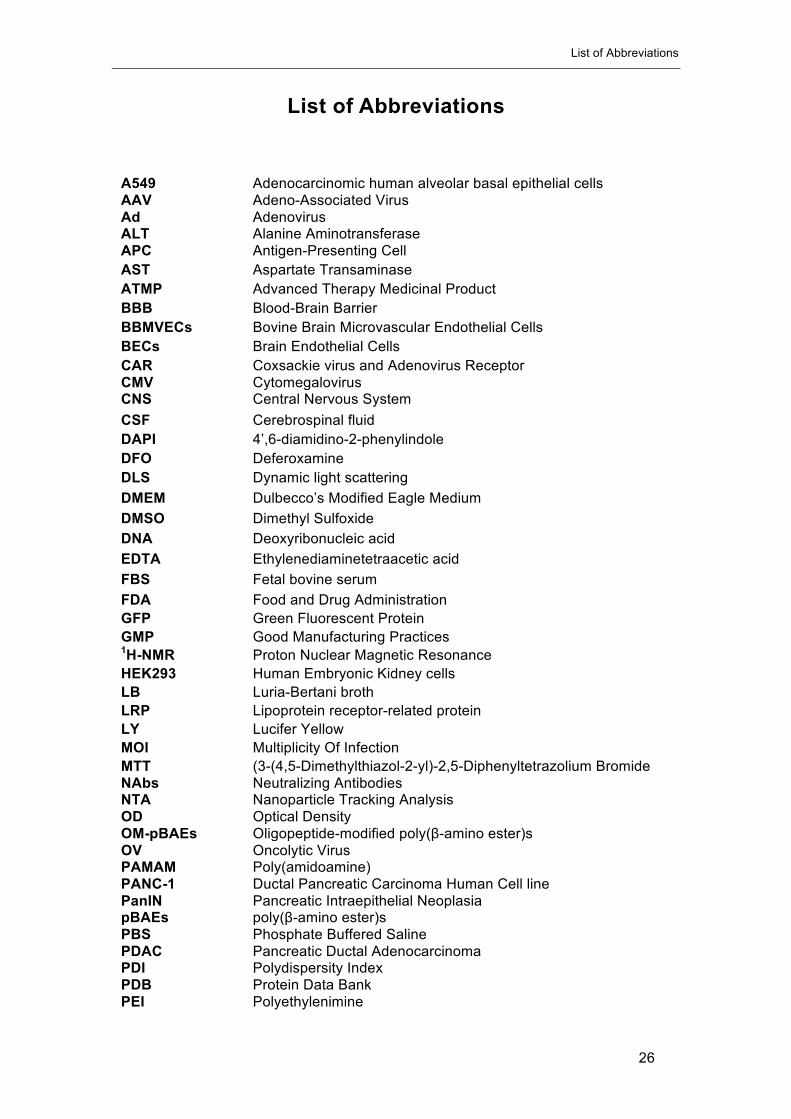
List of Abbreviations
A549 Adenocarcinomic human alveolar basal epithelial cells AAV Adeno-Associated Virus Ad Adenovirus ALT Alanine Aminotransferase APC Antigen-Presenting Cell AST Aspartate Transaminase ATMP Advanced Therapy Medicinal Product BBB Blood-Brain Barrier BBMVECs Bovine Brain Microvascular Endothelial Cells BECs Brain Endothelial Cells CAR Coxsackie virus and Adenovirus Receptor CMV Cytomegalovirus CNS Central Nervous System CSF Cerebrospinal fluid DAPI 4’,6-diamidino-2-phenylindole DFO Deferoxamine DLS Dynamic light scattering DMEM Dulbecco’s Modified Eagle Medium DMSO Dimethyl Sulfoxide DNA Deoxyribonucleic acid EDTA Ethylenediaminetetraacetic acid FBS Fetal bovine serum FDA Food and Drug Administration GFP Green Fluorescent Protein GMP Good Manufacturing Practices 1H-NMR Proton Nuclear Magnetic Resonance HEK293 Human Embryonic Kidney cells LB Luria-Bertani broth LRP Lipoprotein receptor-related protein LY Lucifer Yellow MOI Multiplicity Of Infection MTT (3-(4,5-Dimethylthiazol-2-yl)-2,5-Diphenyltetrazolium Bromide NAbs Neutralizing Antibodies NTA Nanoparticle Tracking Analysis OD Optical Density OM-pBAEs Oligopeptide-modified poly(β-amino ester)s OV Oncolytic Virus PAMAM Poly(amidoamine) PANC-1 Ductal Pancreatic Carcinoma Human Cell line PanIN Pancreatic Intraepithelial Neoplasia pBAEs poly(β-amino ester)s PBS Phosphate Buffered Saline PDAC Pancreatic Ductal Adenocarcinoma PDI Polydispersity Index PDB Protein Data Bank PEI Polyethylenimine
List of Abbreviations
27
PEG Polyethylene glycol PET Positron Emission Tomography PFU Plaque-Forming Unit PTA Phosphotungstic Acid HPMA (2-hydroxypropyl)methacrylamide) HVR Hypervariable Region PFA Paraformaldehyde RLU Relative Light Unit RT-qPCR Real Time quantitative Polymerase Chain Reaction RCAs Rat Cortical Astrocytes SDS Sodium Dodecyl Sulfate SEM Scanning Electron Microscopy SPECT Single-Photon Emission Computed Tomography STORM Stochastic Optical Reconstruction Microscopy TCEP Tris(2-carboxyethyl)phosphine hydrochloride TEM Transmission Electron Microscopy TEER Trans-endothelial Electrical Resistance TRPS Tunable Resistive Pulse Sensing VP Viral Particle

Chapter I. Introduction: Viruses as therapeutic agents

This page left blank intentionally
Chapter I – Introduction: Viruses as therapeutic agents
30
Introduction: Viruses as therapeutic agents
This chapter summarizes the knowledge evolution about the use of
viruses as therapeutic agents, from the origin of this idea to the current status.
Several milestones have been reached using viruses as carriers to deliver
therapeutic nucleic acids for gene therapy applications and in the oncolytic
virotherapy field. However, their intravenous administration is still a hurdle
limiting their efficacy due to the lack of targeting strategies, instability in the
blood stream and safety risks. In this thesis, our interest is focused on the
development of a polymer-based coating technology able to overcome current
delivery limitations.
1.1 Introduction
Viruses have always been related to something dangerous and threatening for the human
health and there are plenty of reasons that support this view. For instance, several epidemic and
pandemic episodes of Influenza (Flu) have challenged humanity throughout history. Claims have
been made for epidemics supposed to be due to Influenza since Greek and Roman times from
430 BC, but without clear conviction and evidences [1]. Interestingly, references to influenza can
be found in both scientific and lay publications since 1700 AC [2]. However, this identification
becomes more difficult as one goes back further in time.
The most dramatic and documented pandemic episode was the 1918 Spanish flu pandemic.
It is estimated that 500 million people were infected around the world resulting in the death of 50
to 100 million individuals. This is considered the deadliest natural disaster in human history [3].
The 1957 Asian flu was the second major Influenza pandemic episode to occur in the 20th century
[4], causing two million deaths worldwide, followed by the Hong Kong flu pandemic of 1968 with
an estimation of one to four million deaths [5]. The most recent 2009 pandemic flu, also known
as Swine flu, was globally spread (Figure I-1) and experts, including the World Health
Organization (WHO) estimated around 285,500 deaths [6].

Chapter I – Introduction: Viruses as therapeutic agents
31
Figure I-1. Global distribution of deaths associated with 2009 pandemic influenza A H1N1 during the first year of virus circulation by country. (Adapted from Lancet Infectious Diseases 2012; 12:687-95)
Unfortunately, Influenza is not the only virus posing a significant public health threat. The
Human Immunodeficiency Virus (HIV) causing Acquired Immunodeficiency Syndrome (AIDS) is
also considered a pandemic that has infected at least 60 million people and caused more than 25
million deaths [7]. The nature of its transmission, which is mainly by sexual interaction, has truly
placed HIV in the focus of the public opinion and debate.
Moreover, the recent Ebola epidemic outbreak in West Africa (2013-2016) evidenced that
resource-poor regions tend to suffer disproportionate morbidity from viral diseases because of
poor sanitation and limited access to health services [8]. From December 2013 to May 2016,
Ebola infected 28,616 people causing 11,310 deaths. Ebola has also threatened first world
countries when infected citizens from USA, Spain, Italy and UK residing in West Africa were
evacuated to their home countries. Then, a global alarm was triggered, and political and health
care institutions were challenged.
This was just a short list of examples aiming to explain the origin of the social fear to viruses
and, taking all this into account, their bad reputation seems justified. However, nowadays we are
also aware of their kind face and we know that viruses have actually helped us during evolution.
Our very long evolutionary history in a virus-rich environment has driven human adaptation
to such infections from the cellular level by domestication of retroviral genes and by teaching our
hyper reactive immune system. A clear example of cellular adaptation is that our genome contains
traces of viral DNA integrated from viral infections in our ancestors. About 8 percent of the human
genome consists of retroviral DNA sequences that have been inserted into the human germline,
where some of their functions have been adopted to serve essential functions for their host’s
survival and development [9]. For instance, expressed proteins from such endogenous
retroviruses can bind to and block cellular receptors that might otherwise be used by exogenous,
pathogenic retroviruses [10]. Another example is the endogenous retroviral envelope proteins
Chapter I – Introduction: Viruses as therapeutic agents
32
which are responsible for fusion of trophoblast cells into the structures of the mammalian placenta
that mediate nutrient and gas exchange between maternal and foetal systems [11]. The exact
role of viruses on human evolution is hardly trackable but its importance seems increasingly
undeniable.
Accordingly, apart from being a threat to human health, viruses have also offered very
valuable services to humanity during evolution and in 1966, Edward Tatum hypothesized that
these services could go a step further. On the basis of the ability of viruses to transfer genes,
Tatum visualized a future where viruses would be engineered to carry therapeutic genes to serve
as delivery vehicles to target organs or tissues with defective gene expression. The feasibility of
transferring genetic material to treat genetic diseases was demonstrated by successive
achievements and discoveries during the early days of genetic engineering. Cloned genes were
used to correct genetic defects or mutations in mammalian cells and transfection techniques were
combined with selection systems for cultured cells and recombinant DNA technology [4][12]. The
early identification of genes responsible for several Mendelian disorders [13,14], followed by
advances in human genetics boosted by the Human Genome Project [15], consolidated the idea
that DNA could be used as a medicine to treat human diseases.
The term “gene therapy” was born and viruses were engaged to play a key role thanks to
their ability to deliver their viral genome inside living cells [16]. However, the momentum was
drastically crashed in 1999 with the tragic death of Jesse Gelsinger during a clinical assay.
Gelsinger's immune system responded immediately after the administration of a very high dose
of Adenovirus and he died four days later because of multi organ failure [17]. After that, it was not
until 2003, that GendicineTM was approved in China, as the first gene therapy product for clinical
use [18,19]. GendicineTM is a replication-incompetent recombinant adenovirus expressing the
wild-type p53 tumor suppressor gene recommended to treat head and neck squamous cell
carcinoma [20]. Later, in 2012, an adeno-associated virus serotype 1 (AAV1) carrying a copy of
the human lipoprotein lipase (LPL) gene was approved in Europe for the treatment of lipoprotein
lipase deficiency (LPLD) under the commercial name GlyberaTM [21]. However, the rarity of this
disease (prevalence-worldwide 1-2 per million) and the expensive costs of this medicine ($1
million per treatment in 2015) forced the abandon of its commercialization. More recently, T-
VECTM was approved in 2015 as the first commercial oncolytic virus for the treatment of
melanoma [22]. It is based on the local administration of a replication-competent herpes virus
expressing an immune system stimulation factor [23]. The achievement of this milestone has
opened a window of opportunity for the development of other strategies based on oncolytic
viruses, and stimulated research and investment. Another successful case is StrimvelisTM, which
became the first ex-vivo stem cell gene therapy product approved by the European Medicines
Agency (EMA) in 2016. It was indicated for the treatment of patients with a very rare disease
called ADA-SCID (Severe Combined Immunodeficiency due to Adenosine Deaminase
deficiency), a rare disorder caused by the absence of an essential protein called adenosine
deaminase (ADA), which is required for the production of lymphocytes [24]. Finally, an AAV-

Chapter I – Introduction: Viruses as therapeutic agents
33
based gene therapy product commercially called LuxturnaTM was recently approved by the FDA
for the treatment of Leber’s congenital amaurosis, a rare inherited eye disease that appears at
birth or in the first few months of life [25]. Figure I-2 represents a timeline highlighting the main
milestones of gene therapy from 1928 to the present.
Figure I-2. Timeline highlighting important milestones of gene therapy from 1928 to the present. (Adapted from
The Journal of Gene Medicine).
To date, more than 2500 gene therapy clinical trials have been conducted worldwide
showing remarkable therapeutic benefits. The treatment of cancer diseases is, with a 65% of all
trials performed, the main area of application. Viruses are present in approximately 70% of trials
as delivery vectors (Figure I-3), being the most commonly used: Adenovirus (20,49%), Retrovirus
(17,9 %) and Adeno-associated virus (7,64%) (Figure I-3).
Generally, gene therapy is classified depending on the approach used to treat the disease:
• Gene supplementation or correction: Supplementation of a defective gene by gene
replacement or mutation correction in order to obtain a functional gene.
• Gene augmentation: Introduction of a novel gene into the cell, aiming to produce a
novel function that is not present or increase expression of specific genes. Suicide
gene therapy for cancer treatment included.
• Gene silencing: Delivery of interfering RNA (RNAi) to induce post-transcriptional
gene silencing by specific degradation of messenger RNA (mRNA).
• Oncolytic virotherapy: Use of viruses to induce lysis of cancerous cells by their self-
amplification mechanism.

Chapter I – Introduction: Viruses as therapeutic agents
34
Figure I-3. Indications addressed and Vectors used by gene therapy clinical trials. Adapted from
www.abedia.com/wiley (The Journal of Gene Medicine, 2017)
Adenoviral vectors have shown efficient transduction in vitro, a good ability to infect dividing
and non-dividing cells (in vitro and in vivo) and the possibility of inserting big expression cassettes,
as well as producing high titers following good manufacturing practices (GMP) [26]. For these
reasons, Adenoviruses (Ad) represent the most used viral vectors in gene therapy clinical trials.
Furthermore, Ad have been also extensively studied for their oncolytic potential. In particular,
tumor-selective replicative Ad, via deletion of certain genes, such as E1B, dispensable for virus
replication in tumor cells, but necessary to complete their life cycle in normal cells, and via
inclusion of tumor-specific promoters controlling viral replication, have been developed showing
remarkable safety and efficiency profiles [27,28]. Moreover, replication-competent viruses
expressing immune system stimulating factors have been developed and tested alone and in
combination with immune modulating agents such as check-point inhibitors and promising results
have been reported [29–31].
On the other hand, adeno-associated viruses (AAV) are also among the most frequently
used viral vectors for gene therapy. AAV-mediated transgene expression persists during years in
comparison with replication-defective Ad vectors with reported transgene expression life-time of
Indications
Total=2597
65.00% Cancer diseases6.93% Cardiovascular diseases1.93% Gene marking2.16% Healthy volunteers7.01% Infectious diseases0.58% Inflammatory diseases11.05% Monogenic diseases1.81% Neurological diseases
1.31% Ocular diseases2.23% Others
Vectors used
20.49% Adenovirus17.90% Retrovirus16.55% Naked/Plasmid DNA7.64% Adeno-associated virus7.34% Lentivirus6.55% Vaccinia virus4.38% Lipofection4.01% Poxvirus3.48% Herpes simplex virus8.35% Other vectors3.30% Unknown
Chapter I – Introduction: Viruses as therapeutic agents
35
1 to 3 weeks [32,33]. The ability of AAVs to promote a stable expression of therapeutic genes has
made them one of the best candidates to treat inherited diseases where long-term expression is
desired. Accordingly, the main indications for AAV-based gene therapy are monogenic inherited
diseases in which the gene product is non-functional or missing. Several recombinant AAV-based
candidates are being clinically tested for the treatment of inherited blindness, cystic fibrosis,
haemophilia B, hereditary cardiac diseases, and muscular dystrophies among others [34].
Moreover, AAV seem to be the preferred vectors to be used as delivery vehicles for in vivo
CRISPR/Cas9 genome editing approaches [35]. Another interesting feature of wild-type AAV is
their lack of pathogenicity. The discovery of twelve human serotypes of AAV (AAV serotype 1
[AAV-1] to AAV-12) and more than 100 serotypes from nonhuman primates with different tropisms
upon systemic administration have increased AAV's potential as delivery vehicles for gene
therapy applications [36].
Despite the encouraging results in pre-clinical and clinical studies, further improvements of
viral-based therapies are required to fully maximise their therapeutic efficacy. The main limiting
factor for the efficient use of viruses as therapeutic agents is the inability to effectively deliver viral
particles to target tissues, organs or tumors. Although in some cases this issue has been
addressed by local administration of viruses, sometimes it is strictly necessary to perform
systemic administration in order to spread the virus throughout the body. For instance, in the case
of muscular dystrophies a bodywide correction must be achieved to fully change the disease
phenotype [36]. In the treatment of cancer, given its metastatic nature and the inaccessibility of
some tumor sites, intravenous viral administration is also the most attractive route of anti-cancer
viral agents with potential to treat both primary and metastases [37].
Several barriers have been identified as factors limiting systemic delivery of viruses. Figure
I-4 summarizes the main limiting factors affecting efficacy of systemically delivered adenovirus-
based therapies. Several undesired interactions upon systemic administration of viral particles
affect the accumulation of viral particles to target cells, necessary in both gene transfer
approaches and oncolytic estrategies. Non-specific binding to serum factors such as pre-immune
immunoglobulin M [38], complement [39], anti-viral cytokines [40] and macrophages result in a
rapid neutralization and clearance of a virus by the reticuloendothelial system with its associated
toxicity consequences due to the massive production of cytokines [41]. Moreover, the existence
of pre-existing immunity to the virus is very common when using human viruses and these
circulating neutralizing antibodies (NAbs) in blood serum severely hamper systemic delivery
[42,43]. Even in the case of no previous exposure to the virus, antibodies can be generated after
the first injection, limiting the efficacy of subsequent doses [44]. Finally, non-specific binding of
adenoviruses to blood cells has been demonstrated [45], and in the case of tumors, their high
interstitial fluid pressure disfavors extravasation of virions from the tumor vasculature [46].
Thus, instability of therapeutic virus particles in the hostile environment of the human blood
stream seems to be one of the main factors culminating in a number of unsuccessful clinical

Chapter I – Introduction: Viruses as therapeutic agents
36
studies [47]. Therefore, during the development of viral-based therapies, attention must be
focused on features of the delivery process, including circulation kinetics, by-passing first hepatic
clearance, and improvement of extravasation in order to achieve a sufficient viral infection of
target cells upon systemic administration in vivo with minimal toxic effects [48].
Figure I-4. Reported Interactions of Ad5 with Blood Components In Vivo. 1. Ad5 binding to CAR-expressing
erythrocytes (species-specific expression of CAR) can cause trapping of virus in the circulation. In the presence of
antibody and complement, Ad5 can bind human erythrocytes via CR-1 [49,50]. 2. Opsonization of Ad5 with natural IgM
and/or complement promotes KC uptake via complement receptor-3 (CR-3) or Fc Receptor [41]. 3. Ad interactions with
T-cells [51]. 4. FX binding to the Ad5 hexon promotes hepatocyte entry through HSPGs [52]. 5. FIX/C4BP binding to the
fiber knob has been proposed to mediate hepatocyte entry via HSPGs or LRP, and has been suggested to direct KC
uptake [53]. 6. Ad binding to platelets has been shown to enhance uptake by KCs [54]. Von Willebrand factor (vWF) and

Chapter I – Introduction: Viruses as therapeutic agents
37
P-selectin have been associated with the formation of activated platelet-leukocyte aggregates which are cleared by
scavenging macrophages [55]. Adapted from “Tropism-Modification Strategies for Targeted Gene Delivery Using
Adenoviral Vectors” [56]
Many strategies have been studied in order to overcome hurdles for a successful systemic
delivery of viral-based therapeutics seeking the reduction of immunogenicity, the avoidance of
undesired interactions and the generation of specifically targeted viral vectors. The main
approaches can be classified in the following groups, although in some cases different strategies
have been combined:
• Cell carriers: Ex vivo infected blood and stem cells are used as vehicles to deliver
viral particles to target cells [42,57–59],
• Genetic modification of viral genomes: Replication-incompetent and
conditionally-replicative viral vectors have been produced by depleting or modifying
genes necessaries for viral replication [28,60–70]. Chimeric vectors combining
genomes from different virus serotypes have been explored to produce vectors with
engineered tropisms [71–74]. Inclusion of point mutations [53,75,76], targeting
peptides [77–83], and albumin-binding domains [84] in viral capsid structure have
demonstrated promising potential to avoid interaction with blood coagulation factors,
re-target viral tropism and by-pass neutralization.
• Covalent chemical modification of viral capsids: Reactive polymers have been
used to coat viral particles through covalent-bond formation with amino groups on
capsid surface [85–88] or with genetically included cysteine residues [76,89], aiming
to avoid the previously described undesired interactions. Moreover, viral particles
surfaces can be easily decorated with targeting moieties linked to polymers [90,91].
• Non-covalent modification of virions: Electrostatically-coated viral particles can
be prepared by using new generation biomaterials [26]. Cationic polymers allow to
coat viral particles without forming covalent bonds with their structure [76–79].
These coatings are less stable but fully preserve the infectivity of viral vectors.
Artificial lipidic envelopes have been also explored [95–97].
Figure I-5 summarizes and represents the main strategies based on the engineering of
adenovirus capsid surfaces by genetic and chemical approaches.
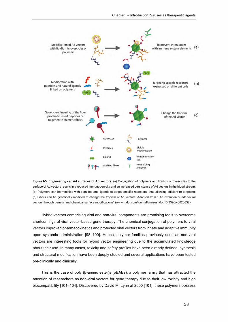
Chapter I – Introduction: Viruses as therapeutic agents
38
Figure I-5. Engineering capsid surfaces of Ad vectors. (a) Conjugation of polymers and lipidic microvescicles to the
surface of Ad vectors results in a reduced immunogenicity and an increased persistence of Ad vectors in the blood stream;
(b) Polymers can be modified with peptides and ligands to target specific receptors, thus allowing efficient re-targeting;
(c) Fibers can be genetically modified to change the tropism of Ad vectors. Adapted from “The evolution of adenoviral
vectors through genetic and chemical surface modifications” (www.mdpi.com/journal/viruses; doi:10.3390/v6020832).
Hybrid vectors comprising viral and non-viral components are promising tools to overcome
shortcomings of viral vector-based gene therapy. The chemical conjugation of polymers to viral
vectors improved pharmacokinetics and protected viral vectors from innate and adaptive immunity
upon systemic administration [98–100]. Hence, polymer families previously used as non-viral
vectors are interesting tools for hybrid vector engineering due to the accumulated knowledge
about their use. In many cases, toxicity and safety profiles have been already defined, synthesis
and structural modification have been deeply studied and several applications have been tested
pre-clinically and clinically.
This is the case of poly (β-amino ester)s (pBAEs), a polymer family that has attracted the
attention of researchers as non-viral vectors for gene therapy due to their low toxicity and high
biocompatibility [101–104]. Discovered by David M. Lynn at 2000 [101], these polymers possess
Chapter I – Introduction: Viruses as therapeutic agents
39
interesting characteristics, such as simple synthesis and easy chemical modifications, as shown
in Figure I-6.
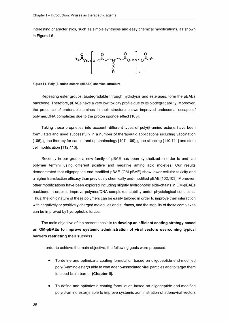
Figure I-6. Poly (β-amino ester)s (pBAEs) chemical structure.
Repeating ester groups, biodegradable through hydrolysis and esterases, form the pBAEs
backbone. Therefore, pBAEs have a very low toxicity profile due to its biodegradability. Moreover,
the presence of protonable amines in their structure allows improved endosomal escape of
polymer/DNA complexes due to the proton sponge effect [105].
Taking these proprieties into account, different types of poly(β-amino ester)s have been
formulated and used successfully in a number of therapeutic applications including vaccination
[106], gene therapy for cancer and ophthalmology [107–109], gene silencing [110,111] and stem
cell modification [112,113].
Recently in our group, a new family of pBAE has been synthetized in order to end-cap
polymer termini using different positive and negative amino acid moieties. Our results
demonstrated that oligopeptide end-modified pBAE (OM-pBAE) show lower cellular toxicity and
a higher transfection efficacy than previously chemically end-modified pBAE [102,103]. Moreover,
other modifications have been explored including slightly hydrophobic side-chains in OM-pBAEs
backbone in order to improve polymer/DNA complexes stability under physiological conditions.
Thus, the ionic nature of these polymers can be easily tailored in order to improve their interaction
with negatively or positively charged molecules and surfaces, and the stability of those complexes
can be improved by hydrophobic forces.
The main objective of the present thesis is to develop an efficient coating strategy based on OM-pBAEs to improve systemic administration of viral vectors overcoming typical barriers restricting their success.
In order to achieve the main objective, the following goals were proposed:��
• To define and optimize a coating formulation based on oligopeptide end-modified
poly(β-amino ester)s able to coat adeno-associated viral particles and to target them
to blood-brain barrier (Chapter II).
• To define and optimize a coating formulation based on oligopeptide end-modified
poly(β-amino ester)s able to improve systemic administration of adenoviral vectors

Chapter I – Introduction: Viruses as therapeutic agents
40
in terms of biodistribution, pharmacokinetics, by-pass of neutralizing antibodies and
immune system activation (Chapter III).
• To determine the effect of the previously developed polymeric coating technology
on the safety and efficacy of an oncolytic virus for the treatment of pancreatic ductal
adenocarcinoma (Chapter IV).
• To study biodistribution of pBAE-coated adenoviral particles by a genetic-chemical
radioactive labelling of adenoviral vectors (Chapter V).
Chapter I – Introduction: Viruses as therapeutic agents
41
1.2 Content of this Dissertation
Advances in viral genomics and genetic engineering have led to the development of a variety
of viral vectors with great potential in gene therapy and immunotherapy fields. In order to
completely unlock this potential, systemic delivery of viruses needs to become feasible. In this
work, the use of oligopeptide end-modified poly(β-amino ester) polymers as coating agents to
protect and boost virotherapeutics efficacy after systemic administration has been explored.
In chapter II, the ability of OM-pBAEs to efficiently coat AAV viral particles has been
investigated. The inclusion of peptic targeting moieties into the coating structure have been
studied in order to promote targeted AAV2/5-mediated gene delivery across the blood brain
barrier (BBB). Through a close collaboration with Sagetis-Biotech, BBB in vitro models were
established and used as a screening platform to discover LRP-1 targeting peptides. The resulting
candidate was chemically introduced into the OM-pBAE structure and the capability of targeted
OM-pBAE-coated AAV2/5 particles to transduce BBB endothelial cells was assessed both in vitro
and in vivo.
Adenoviruses are one of the most studied viruses to be used as gene delivery vectors. This
virus is non-integrative and can be easily modified to be replication-defective and even
conditionally-replicating, which is the basis of oncolytic virotherapy. Chapter III goes a step
forward in the development and characterization of the coating technology, and deeply studies
the effect of OM-pBAEs shielding on systemic administration of adenoviral therapeutic agents. A
non-replicative reporter adenovirus was used as a vector to determine how the coating affected
the virus behavior in terms of infectivity, biodistribution and interaction with the immune system.
Chapter IV applies all the knowledge gathered in previous chapters to improve the efficacy
and the safety profile of a therapeutic oncolytic adenovirus AdNuPARmE1A, a pancreatic cancer
specific conditionally-replicative oncolytic adenovirus previously developed in the Cancer and
Gene Therapy group at Institut d’Investigacions Biomèdiques Pi i Sunyer (IDIBAPS), led by Dra.
Cristina Fillat.
The last part of this thesis is centred to accurately determine the biodistribution of coated Ad
particles shortly after systemic administration. To fulfill this purpose, a reporter non-replicative Ad
was genetically modified to include exposed cysteine residues on its capsid surface. This allowed
to covalently attach a radioisotope chelating agent to the viral capsid and follow the radioactive
label to study biodistribution (Chapter V). This work was done in collaboration with Dr. Florian
Kreppel from Witten/Herdecke University and Dr. Jordi Llop from Biomagune.

Chapter I – Introduction: Viruses as therapeutic agents
42
1.3 References
[1] C.W. Potter, R. Jennings, A definition for influenza pandemics based on historial
records, J. Infect. 63 (2011) 252–259. doi:10.1016/j.jinf.2011.04.013.
[2] H. A., Handbook of geographical and historical pathology, London New Sydenham Soc.
(1883).
[3] N.P.A.S. Johnson, J. Mueller, Updating the accounts: global mortality of the 1918-1920
“Spanish” influenza pandemic., Bull. Hist. Med. 76 (2002) 105–115.
[4] C. Viboud, L. Simonsen, R. Fuentes, J. Flores, M.A. Miller, G. Chowell, Global mortality
impact of the 1957-1959 influenza pandemic, J. Infect. Dis. 212 (2016) 738–745.
doi:10.1093/infdis/jiv534.
[5] C. Viboud, R.F. Grais, B.A.P. Lafont, M.A. Miller, L. Simonsen, Multinational Impact of
the 1968 Hong Kong Influenza Pandemic: Evidence for a Smoldering Pandemic, J.
Infect. Dis. 192 (2005) 233–248. doi:10.1086/431150.
[6] F.S. Dawood, A.D. Iuliano, C. Reed, M.I. Meltzer, D.K. Shay, P.Y. Cheng, D.
Bandaranayake, R.F. Breiman, W.A. Brooks, P. Buchy, D.R. Feikin, K.B. Fowler, A.
Gordon, N.T. Hien, P. Horby, Q.S. Huang, M.A. Katz, A. Krishnan, R. Lal, J.M.
Montgomery, K. Mølbak, R. Pebody, A.M. Presanis, H. Razuri, A. Steens, Y.O. Tinoco,
J. Wallinga, H. Yu, S. Vong, J. Bresee, M.A. Widdowson, Estimated global mortality
associated with the first 12 months of 2009 pandemic influenza A H1N1 virus circulation:
A modelling study, Lancet Infect. Dis. 12 (2012) 687–695. doi:10.1016/S1473-
3099(12)70121-4.
[7] M.H. Merson, J. O’Malley, D. Serwadda, C. Apisuk, The history and challenge of HIV
prevention., Lancet (London, England). 372 (2008) 475–488. doi:10.1016/S0140-
6736(08)60884-3.
[8] M.G. Dixon, I.J. Schafer, Ebola viral disease outbreak--West Africa, 2014., MMWR.
Morb. Mortal. Wkly. Rep. 63 (2014) 548–551.
[9] D.J. Griffiths, Endogenous retroviruses in the human genome sequence., Genome Biol.
2 (2001) reviews1017.1-1017.5. doi:10.1186/gb-2001-2-6-reviews1017.
[10] R. Malfavon-Borja, C. Feschotte, Fighting Fire with Fire: Endogenous Retrovirus
Envelopes as Restriction Factors, J. Virol. 89 (2015) 4047–4050. doi:10.1128/JVI.03653-
14.
[11] S. Mi, X. Lee, X. Li, G.M. Veldman, H. Finnerty, L. Racie, E. LaVallie, X.Y. Tang, P.
Edouard, S. Howes, J.C.J. Keith, J.M. McCoy, Syncytin is a captive retroviral envelope
Chapter I – Introduction: Viruses as therapeutic agents
43
protein involved in human placental morphogenesis., Nature. 403 (2000) 785–789.
doi:10.1038/35001608.
[12] T. Friedmann, A brief history of gene therapy., Nat. Genet. 2 (1992) 93–98.
doi:10.1038/ng1092-93.
[13] J.M. Rommens, M.C. Iannuzzi, B. Kerem, M.L. Drumm, G. Melmer, M. Dean, R.
Rozmahel, J.L. Cole, D. Kennedy, N. Hidaka, Identification of the cystic fibrosis gene:
chromosome walking and jumping., Science. 245 (1989) 1059–1065.
[14] A novel gene containing a trinucleotide repeat that is expanded and unstable on
Huntington’s disease chromosomes. The Huntington’s Disease Collaborative Research
Group., Cell. 72 (1993) 971–983.
[15] E.D. Green, J.D. Watson, F.S. Collins, Human Genome Project: Twenty-five years of big
biology., Nature. 526 (2015) 29–31. doi:10.1038/526029a.
[16] T. Wirth, N. Parker, S. Ylä-Herttuala, History of gene therapy, Gene. 525 (2013) 162–
169. doi:10.1016/j.gene.2013.03.137.
[17] S.G. Stolberg, The biotech death of Jesse Gelsinger, N.Y.Times Mag. (1999).
[18] J.M. Wilson, Gendicine: the first commercial gene therapy product., Hum. Gene Ther. 16
(2005) 1014–1015. doi:10.1089/hum.2005.16.1014.
[19] Z. Peng, Current status of gendicine in China: recombinant human Ad-p53 agent for
treatment of cancers., Hum. Gene Ther. 16 (2005) 1016–1027.
doi:10.1089/hum.2005.16.1016.
[20] S. Pearson, H. Jia, K. Kandachi, China approves first gene therapy, Nat. Biotechnol. 22
(2004) 3. http://dx.doi.org/10.1038/nbt0104-3.
[21] S. Yla-Herttuala, Endgame: glybera finally recommended for approval as the first gene
therapy drug in the European union., Mol. Ther. 20 (2012) 1831–1832.
doi:10.1038/mt.2012.194.
[22] E. Dolgin, Oncolytic viruses get a boost with first FDA-approval recommendation, Nat.
Rev. Drug Discov. 14 (2015) 369. http://dx.doi.org/10.1038/nrd4643.
[23] F.J. Kohlhapp, A. Zloza, H.L. Kaufman, Talimogene laherparepvec (T-VEC) as cancer
immunotherapy., Drugs Today (Barc). 51 (2015) 549–558.
doi:10.1358/dot.2015.51.9.2383044.
[24] J. Hoggatt, Gene Therapy for “Bubble Boy” Disease., Cell. 166 (2016) 263.
Chapter I – Introduction: Viruses as therapeutic agents
44
doi:10.1016/j.cell.2016.06.049.
[25] S. Russell, J. Bennett, J.A. Wellman, D.C. Chung, Z.-F. Yu, A. Tillman, J. Wittes, J.
Pappas, O. Elci, S. McCague, D. Cross, K.A. Marshall, J. Walshire, T.L. Kehoe, H.
Reichert, M. Davis, L. Raffini, L.A. George, F.P. Hudson, L. Dingfield, X. Zhu, J.A. Haller,
E.H. Sohn, V.B. Mahajan, W. Pfeifer, M. Weckmann, C. Johnson, D. Gewaily, A. Drack,
E. Stone, K. Wachtel, F. Simonelli, B.P. Leroy, J.F. Wright, K.A. High, A.M. Maguire,
Efficacy and safety of voretigene neparvovec (AAV2-hRPE65v2) in patients with RPE65-
mediated inherited retinal dystrophy: a randomised, controlled, open-label, phase 3 trial.,
Lancet (London, England). 390 (2017) 849–860. doi:10.1016/S0140-6736(17)31868-8.
[26] C. Capasso, M. Garofalo, M. Hirvinen, V. Cerullo, The evolution of adenoviral vectors
through genetic and chemical surface modifications, Viruses. 6 (2014) 832–855.
doi:10.3390/v6020832.
[27] J.J. Short, D.T. Curiel, Oncolytic adenoviruses targeted to cancer stem cells., Mol.
Cancer Ther. 8 (2009) 2096–2102. doi:10.1158/1535-7163.MCT-09-0367.
[28] A. Mato-Berciano, G. Raimondi, M.V. Maliandi, R. Alemany, L. Montoliu, C. Fillat, A
NOTCH-sensitive uPAR-regulated oncolytic adenovirus effectively suppresses
pancreatic tumor growth and triggers synergistic anticancer effects with gemcitabine and
nab-paclitaxel, Oncotarget. 8 (2017) 22700–22715.
doi:https://dx.doi.org/10.18632/oncotarget.15169.
[29] C.E. Engeland, C. Grossardt, R. Veinalde, S. Bossow, D. Lutz, J.K. Kaufmann, I.
Shevchenko, V. Umansky, D.M. Nettelbeck, W. Weichert, D. Jäger, C. von Kalle, G.
Ungerechts, CTLA-4 and PD-L1 Checkpoint Blockade Enhances Oncolytic Measles
Virus Therapy, Mol. Ther. 22 (2014) 1949–1959. doi:10.1038/mt.2014.160.
[30] B.A. Jonas, Combination of an oncolytic virus with PD-L1 blockade keeps cancer in
check., Sci. Transl. Med. 9 (2017). doi:10.1126/scitranslmed.aan2781.
[31] O. Hamid, B. Hoffner, E. Gasal, J. Hong, R.D. Carvajal, Oncolytic immunotherapy:
unlocking the potential of viruses to help target cancer, Cancer Immunol. Immunother.
(2017) 1–16. doi:10.1007/s00262-017-2025-8.
[32] K. Stieger, G. Le Meur, F. Lasne, M. Weber, J.-Y. Deschamps, D. Nivard, A. Mendes-
Madeira, N. Provost, L. Martin, P. Moullier, F. Rolling, Long-term doxycycline-regulated
transgene expression in the retina of nonhuman primates following subretinal injection of
recombinant AAV vectors., Mol. Ther. 13 (2006) 967–975.
doi:10.1016/j.ymthe.2005.12.001.
[33] G. Vassalli, H. Büeler, J. Dudler, L.K. Von Segesser, L. Kappenberger, Adeno-
Chapter I – Introduction: Viruses as therapeutic agents
45
associated virus (AAV) vectors achieve prolonged transgene expression in mouse
myocardium and arteries in vivo: A comparative study with adenovirus vectors, Int. J.
Cardiol. 90 (2003) 229–238. doi:10.1016/S0167-5273(02)00554-5.
[34] M.F. Naso, B. Tomkowicz, W.L. Perry, W.R. Strohl, Adeno-Associated Virus (AAV) as a
Vector for Gene Therapy, BioDrugs. 31 (2017) 317–334. doi:10.1007/s40259-017-0234-
5.
[35] C.E. Nelson, C.H. Hakim, D.G. Ousterout, P.I. Thakore, E.A. Moreb, R.M. Castellanos
Rivera, S. Madhavan, X. Pan, F.A. Ran, W.X. Yan, A. Asokan, F. Zhang, D. Duan, C.A.
Gersbach, In vivo genome editing improves muscle function in a mouse model of
Duchenne muscular dystrophy., Science. 351 (2016) 403–407.
doi:10.1126/science.aad5143.
[36] M. Lim, Systemic delivery of adeno-associated viral vectors, 123 (2016) 413–423.
doi:10.1007/s11060-015-1747-8.The.
[37] A. Marchini, E.M. Scott, J. Rommelaere, Overcoming barriers in oncolytic virotherapy
with HDAC inhibitors and immune checkpoint blockade, Viruses. 8 (2016) 1–22.
doi:10.3390/v8010009.
[38] M.R. Patel, R.A. Kratzke, Oncolytic virus therapy for cancer: the first wave of
translational clinical trials., Transl. Res. 161 (2013) 355–364.
doi:10.1016/j.trsl.2012.12.010.
[39] K. Ikeda, H. Wakimoto, T. Ichikawa, S. Jhung, F.H. Hochberg, D.N. Louis, E.A. Chiocca,
Complement depletion facilitates the infection of multiple brain tumors by an
intravascular, replication-conditional herpes simplex virus mutant., J. Virol. 74 (2000)
4765–4775.
[40] H. Wakimoto, G. Fulci, E. Tyminski, E.A. Chiocca, Altered expression of antiviral
cytokine mRNAs associated with cyclophosphamide’s enhancement of viral oncolysis.,
Gene Ther. 11 (2004) 214–223. doi:10.1038/sj.gt.3302143.
[41] Z. Xu, J. Tian, J.S. Smith, A.P. Byrnes, Clearance of adenovirus by Kupffer cells is
mediated by scavenger receptors, natural antibodies, and complement., J. Virol. 82
(2008) 11705–11713. doi:10.1128/JVI.01320-08.
[42] I.D. Iankov, B. Blechacz, C. Liu, J.D. Schmeckpeper, J.E. Tarara, M.J. Federspiel, N.
Caplice, S.J. Russell, Infected cell carriers: a new strategy for systemic delivery of
oncolytic measles viruses in cancer virotherapy., Mol. Ther. 15 (2007) 114–122.
doi:10.1038/sj.mt.6300020.
Chapter I – Introduction: Viruses as therapeutic agents
46
[43] Y. Chen, D.C. Yu, D. Charlton, D.R. Henderson, Pre-existent adenovirus antibody
inhibits systemic toxicity and antitumor activity of CN706 in the nude mouse LNCaP
xenograft model: implications and proposals for human therapy., Hum. Gene Ther. 11
(2000) 1553–1567. doi:10.1089/10430340050083289.
[44] K.-W. Peng, R. Myers, A. Greenslade, E. Mader, S. Greiner, M.J. Federspiel, A.
Dispenzieri, S.J. Russell, Using clinically approved cyclophosphamide regimens to
control the humoral immune response to oncolytic viruses., Gene Ther. 20 (2013) 255–
261. doi:10.1038/gt.2012.31.
[45] M. Lyons, D. Onion, N.K. Green, K. Aslan, R. Rajaratnam, M. Bazan-Peregrino, S.
Phipps, S. Hale, V. Mautner, L.W. Seymour, K.D. Fisher, Adenovirus type 5 interactions
with human blood cells may compromise systemic delivery., Mol. Ther. 14 (2006) 118–
128. doi:10.1016/j.ymthe.2006.01.003.
[46] E. Smith, J. Breznik, B.D. Lichty, Strategies to enhance viral penetration of solid tumors.,
Hum. Gene Ther. 22 (2011) 1053–1060. doi:10.1089/hum.2010.227.
[47] L.W. Seymour, K.D. Fisher, Oncolytic viruses: finally delivering, Br. J. Cancer. 114
(2016) 357–61. doi:10.1038/bjc.2015.481.
[48] K. Fisher, Striking out at disseminated metastases: the systemic delivery of oncolytic
viruses., Curr. Opin. Mol. Ther. 8 (2006) 301–313.
[49] R.C. Carlisle, Y. Di, A.M. Cerny, A.F.-P. Sonnen, R.B. Sim, N.K. Green, V. Subr, K.
Ulbrich, R.J.C. Gilbert, K.D. Fisher, R.W. Finberg, L.W. Seymour, Human erythrocytes
bind and inactivate type 5 adenovirus by presenting Coxsackie virus-adenovirus receptor
and complement receptor 1, Blood. 113 (2009) 1909–1918. doi:10.1182/blood-2008-09-
178459.
[50] E. Seiradake, D. Henaff, H. Wodrich, O. Billet, M. Perreau, C. Hippert, F. Mennechet, G.
Schoehn, H. Lortat-Jacob, H. Dreja, S. Ibanes, V. Kalatzis, J.P. Wang, R.W. Finberg, S.
Cusack, E.J. Kremer, The cell adhesion molecule “CAR” and sialic acid on human
erythrocytes influence adenovirus in vivo biodistribution., PLoS Pathog. 5 (2009)
e1000277. doi:10.1371/journal.ppat.1000277.
[51] D. Onion, L.J. Crompton, D.W. Milligan, P.A.H. Moss, S.P. Lee, V. Mautner, The CD4+
T-cell response to adenovirus is focused against conserved residues within the hexon
protein., J. Gen. Virol. 88 (2007) 2417–2425. doi:10.1099/vir.0.82867-0.
[52] S.N. Waddington, J.H. McVey, D. Bhella, A.L. Parker, K. Barker, H. Atoda, R. Pink,
S.M.K. Buckley, J.A. Greig, L. Denby, J. Custers, T. Morita, I.M.B. Francischetti, R.Q.
Monteiro, D.H. Barouch, N. van Rooijen, C. Napoli, M.J.E. Havenga, S.A. Nicklin, A.H.
Chapter I – Introduction: Viruses as therapeutic agents
47
Baker, Adenovirus Serotype 5 Hexon Mediates Liver Gene Transfer, Cell. 132 (2008)
397–409. doi:10.1016/j.cell.2008.01.016.
[53] D.M. Shayakhmetov, A. Gaggar, S. Ni, Z.Y. Li, A. Lieber, Adenovirus binding to blood
factors results in liver cell infection and hepatotoxicity, J Virol. 79 (2005) 7478–7491.
doi:10.1128/JVI.79.12.7478-7491.2005.
[54] D. Stone, Y. Liu, D. Shayakhmetov, Z.-Y. Li, S. Ni, A. Lieber, Adenovirus-platelet
interaction in blood causes virus sequestration to the reticuloendothelial system of the
liver., J. Virol. 81 (2007) 4866–4871. doi:10.1128/JVI.02819-06.
[55] M. Othman, A. Labelle, I. Mazzetti, H.S. Elbatarny, D. Lillicrap, Adenovirus-induced
thrombocytopenia: the role of von Willebrand factor and P-selectin in mediating
accelerated platelet clearance., Blood. 109 (2007) 2832–2839. doi:10.1182/blood-2006-
06-032524.
[56] L. Coughlan, R. Alba, A.L. Parker, A.C. Bradshaw, I.A. McNeish, S.A. Nicklin, A.H.
Baker, Tropism-modification strategies for targeted gene delivery using adenoviral
vectors, Viruses. 2 (2010) 2290–2355. doi:10.3390/v2102290.
[57] A. Munguia, T. Ota, T. Miest, S.J. Russell, Cell carriers to deliver oncolytic viruses to
sites of myeloma tumor growth, Gene Ther. 15 (2008) 797.
http://dx.doi.org/10.1038/gt.2008.45.
[58] D.G. Roy, J.C. Bell, Cell carriers for oncolytic viruses: current challenges and future
directions, Oncolytic Virotherapy. 2 (2013) 47–56. doi:10.2147/OV.S36623.
[59] C. Willmon, K. Harrington, T. Kottke, R. Prestwich, A. Melcher, R. Vile, Cell Carriers for
Oncolytic Viruses: Fed Ex for Cancer Therapy, Mol. Ther. J. Am. Soc. Gene Ther. 17
(2009) 1667–1676. doi:10.1038/mt.2009.194.
[60] W.T. Steegenga, A. Shvarts, N. Riteco, J.L. Bos, A.G. Jochemsen, Distinct regulation of
p53 and p73 activity by adenovirus E1A, E1B, and E4orf6 proteins., Mol. Cell. Biol. 19
(1999) 3885–3894.
[61] M. Zamanian, N.B. La Thangue, Adenovirus E1a prevents the retinoblastoma gene
product from repressing the activity of a cellular transcription factor., EMBO J. 11 (1992)
2603–2610.
[62] J. Fu, L. Li, M. Bouvier, Adenovirus E3-19K proteins of different serotypes and
subgroups have similar, yet distinct, immunomodulatory functions toward major
histocompatibility class I molecules., J. Biol. Chem. 286 (2011) 17631–17639.
doi:10.1074/jbc.M110.212050.
Chapter I – Introduction: Viruses as therapeutic agents
48
[63] D.B. Schowalter, J.C. Tubb, M. Liu, C.B. Wilson, M.A. Kay, Heterologous expression of
adenovirus E3-gp19K in an E1a-deleted adenovirus vector inhibits MHC I expression in
vitro, but does not prolong transgene expression in vivo., Gene Ther. 4 (1997) 351–360.
doi:10.1038/sj.gt.3300398.
[64] P. Flomenberg, E. Gutierrez, K.T. Hogan, Identification of class I MHC regions which
bind to the adenovirus E3-19k protein., Mol. Immunol. 31 (1994) 1277–1284.
[65] S.K. Tripathy, H.B. Black, E. Goldwasser, J.M. Leiden, Immune responses to transgene-
encoded proteins limit the stability of gene expression after injection of replication-
defective adenovirus vectors., Nat. Med. 2 (1996) 545–550.
[66] D. Wohlleber, H. Kashkar, K. Gartner, M.K. Frings, M. Odenthal, S. Hegenbarth, C.
Borner, B. Arnold, G. Hammerling, B. Nieswandt, N. van Rooijen, A. Limmer, K.
Cederbrant, M. Heikenwalder, M. Pasparakis, U. Protzer, H.-P. Dienes, C. Kurts, M.
Kronke, P.A. Knolle, TNF-induced target cell killing by CTL activated through cross-
presentation., Cell Rep. 2 (2012) 478–487. doi:10.1016/j.celrep.2012.08.001.
[67] J.M. Janssen, J. Liu, J. Skokan, M.A.F. V Goncalves, A.A.F. de Vries, Development of
an AdEasy-based system to produce first- and second-generation adenoviral vectors
with tropism for CAR- or CD46-positive cells., J. Gene Med. 15 (2013) 1–11.
doi:10.1002/jgm.2687.
[68] N. Brunetti-Pierri, P. Ng, Helper-dependent adenoviral vectors for liver-directed gene
therapy., Hum. Mol. Genet. 20 (2011) R7-13. doi:10.1093/hmg/ddr143.
[69] G. Toietta, L. Pastore, V. Cerullo, M. Finegold, A.L. Beaudet, B. Lee, Generation of
helper-dependent adenoviral vectors by homologous recombination., Mol. Ther. 5 (2002)
204–210. doi:10.1006/mthe.2002.0532.
[70] D.J. Palmer, P. Ng, Characterization of helper-dependent adenoviral vectors., Cold
Spring Harb. Protoc. 2011 (2011) 867–870. doi:10.1101/pdb.prot5628.
[71] K.H. Kim, M.J. Ryan, J.E. Estep, B.M. Miniard, T.L. Rudge, J.O. Peggins, T.L. Broadt, M.
Wang, M.A. Preuss, G.P. Siegal, A. Hemminki, R.D. Harris, R. Aurigemma, D.T. Curiel,
R.D. Alvarez, A new generation of serotype chimeric infectivity-enhanced conditionally
replicative adenovirals: the safety profile of ad5/3-Delta24 in advance of a phase I
clinical trial in ovarian cancer patients., Hum. Gene Ther. 22 (2011) 821–828.
doi:10.1089/hum.2010.180.
[72] M. Murakami, H. Ugai, N. Belousova, A. Pereboev, P. Dent, P.B. Fisher, M. Everts, D.T.
Curiel, Chimeric adenoviral vectors incorporating a fiber of human adenovirus 3
efficiently mediate gene transfer into prostate cancer cells., Prostate. 70 (2010) 362–
Chapter I – Introduction: Viruses as therapeutic agents
49
376. doi:10.1002/pros.21070.
[73] R. van de Ven, J.J. Lindenberg, D. Oosterhoff, M.P. van den Tol, R.A. Rosalia, M.
Murakami, M. Everts, G.L. Scheffer, R.J. Scheper, T.D. de Gruijl, D.T. Curiel, Selective
transduction of mature DC in human skin and lymph nodes by CD80/CD86-targeted
fiber-modified adenovirus-5/3., J. Immunother. 32 (2009) 895–906.
doi:10.1097/CJI.0b013e3181b56deb.
[74] A. Koski, E. Karli, A. Kipar, S. Escutenaire, A. Kanerva, A. Hemminki, Mutation of the
fiber shaft heparan sulphate binding site of a 5/3 chimeric adenovirus reduces liver
tropism., PLoS One. 8 (2013) e60032. doi:10.1371/journal.pone.0060032.
[75] R. Alba, A.C. Bradshaw, A.L. Parker, D. Bhella, S.N. Waddington, A. Stuart, N. Van
Rooijen, J. Custers, J. Goudsmit, D.H. Barouch, J.H. Mcvey, A.H. Baker, S.A. Nicklin,
Identification of coagulation factor ( F ) X binding sites on the adenovirus serotype 5
hexon : effect of mutagenesis on FX interactions and gene transfer Identification of
coagulation factor ( F ) X binding sites on the adenovirus serotype 5 hexon : effe, Gene
Ther. (2011) 965–971. doi:10.1182/blood-2009-03-208835.
[76] L. Krutzke, J.M. Prill, T. Engler, C.Q. Schmidt, Z. Xu, A.P. Byrnes, T. Simmet, F.
Kreppel, Substitution of blood coagulation factor X-binding to Ad5 by position-speci fi c
PEGylation : Preventing vector clearance and preserving infectivity, 235 (2016) 379–392.
doi:10.1016/j.jconrel.2016.06.022.
[77] N. Belousova, G. Mikheeva, J. Gelovani, V. Krasnykh, Modification of adenovirus capsid
with a designed protein ligand yields a gene vector targeted to a major molecular marker
of cancer., J. Virol. 82 (2008) 630–637. doi:10.1128/JVI.01896-07.
[78] L. Coughlan, S. Vallath, A. Saha, M. Flak, I.A. McNeish, G. Vassaux, J.F. Marshall, I.R.
Hart, G.J. Thomas, In vivo retargeting of adenovirus type 5 to alphavbeta6 integrin
results in reduced hepatotoxicity and improved tumor uptake following systemic
delivery., J. Virol. 83 (2009) 6416–6428. doi:10.1128/JVI.00445-09.
[79] S. Pesonen, I. Diaconu, V. Cerullo, S. Escutenaire, M. Raki, L. Kangasniemi, P.
Nokisalmi, G. Dotti, K. Guse, L. Laasonen, K. Partanen, E. Karli, E. Haavisto, M.
Oksanen, A. Karioja-Kallio, P. Hannuksela, S.-L. Holm, S. Kauppinen, T. Joensuu, A.
Kanerva, A. Hemminki, Integrin targeted oncolytic adenoviruses Ad5-D24-RGD and
Ad5-RGD-D24-GMCSF for treatment of patients with advanced chemotherapy refractory
solid tumors., Int. J. Cancer. 130 (2012) 1937–1947. doi:10.1002/ijc.26216.
[80] J.J. Rojas, M. Gimenez-Alejandre, R. Gil-Hoyos, M. Cascallo, R. Alemany, Improved
systemic antitumor therapy with oncolytic adenoviruses by replacing the fiber shaft
HSG-binding domain with RGD., Gene Ther. 19 (2012) 453–457.
Chapter I – Introduction: Viruses as therapeutic agents
50
doi:10.1038/gt.2011.106.
[81] H. Matsui, F. Sakurai, K. Katayama, S. Kurachi, K. Tashiro, K. Sugio, K. Kawabata, H.
Mizuguchi, Enhanced transduction efficiency of fiber-substituted adenovirus vectors by
the incorporation of RGD peptides in two distinct regions of the adenovirus serotype 35
fiber knob., Virus Res. 155 (2011) 48–54. doi:10.1016/j.virusres.2010.08.021.
[82] H. Wu, T. Han, N. Belousova, V. Krasnykh, E. Kashentseva, I. Dmitriev, M. Kataram,
P.J. Mahasreshti, D.T. Curiel, Identification of sites in adenovirus hexon for foreign
peptide incorporation., J. Virol. 79 (2005) 3382–3390. doi:10.1128/JVI.79.6.3382-
3390.2005.
[83] B. Di, Q. Mao, J. Zhao, X. Li, D. Wang, H. Xia, A rapid generation of adenovirus vector
with a genetic modification in hexon protein., J. Biotechnol. 157 (2012) 373–378.
doi:10.1016/j.jbiotec.2011.12.022.
[84] L.A. Rojas, G.N. Condezo, R.M. Oli??, C.A. Fajardo, M. Arias-Badia, C. San Mart??n, R.
Alemany, Albumin-binding adenoviruses circumvent pre-existing neutralizing antibodies
upon systemic delivery, J. Control. Release. 237 (2016) 78–88.
doi:10.1016/j.jconrel.2016.07.004.
[85] U. Wattendorf, H.P. Merkle, PEGylation as a tool for the biomedical engineering of
surface modified microparticles, J. Pharm. Sci. 97 (2008) 4655–4669.
doi:https://doi.org/10.1002/jps.21350.
[86] C.R. O’Riordan, A. Lachapelle, C. Delgado, V. Parkes, S.C. Wadsworth, A.E. Smith,
G.E. Francis, PEGylation of adenovirus with retention of infectivity and protection from
neutralizing antibody in vitro and in vivo, Hum. Gene Ther. 10 (1999) 1349–1358. doi:Doi
10.1089/10430349950018021.
[87] P. Wonganan, M.A. Croyle, PEGylated adenoviruses: From mice to monkeys, 2010.
doi:10.3390/v2020468.
[88] V. Subr, L. Kostka, T. Selby-Milic, K. Fisher, K. Ulbrich, L.W. Seymour, R.C. Carlisle,
Coating of adenovirus type 5 with polymers containing quaternary amines prevents
binding to blood components, J. Control. Release. 135 (2009) 152–158.
doi:10.1016/j.jconrel.2008.12.009.
[89] F. Kreppel, J. Gackowski, E. Schmidt, S. Kochanek, Combined Genetic and Chemical
Capsid Modifications Enable Flexible and Efficient De- and Retargeting of Adenovirus
Vectors, Mol. Ther. 12 (2005) 107–117. doi:10.1016/j.ymthe.2005.03.006.
[90] K.D. Fisher, Y. Stallwood, N.K. Green, K. Ulbrich, V. Mautner, L.W. Seymour, Polymer-

Chapter I – Introduction: Viruses as therapeutic agents
51
coated adenovirus permits efficient retargeting and evades neutralising antibodies.,
Gene Ther. 8 (2001) 341–348. doi:10.1038/sj.gt.3301389.
[91] O.J. Kwon, E. Kang, J.W. Choi, S.W. Kim, C.O. Yun, Therapeutic targeting of chitosan-
PEG-folate-complexed oncolytic adenovirus for active and systemic cancer gene
therapy, J. Control. Release. 169 (2013) 257–265. doi:10.1016/j.jconrel.2013.03.030.
[92] P.H. Kim, J. Kim, T. Il Kim, H.Y. Nam, J.W. Yockman, M. Kim, S.W. Kim, C.O. Yun,
Bioreducible polymer-conjugated oncolytic adenovirus for hepatoma-specific therapy via
systemic administration, Biomaterials. 32 (2011) 9328–9342.
doi:10.1016/j.biomaterials.2011.08.066.
[93] J.F. Vega, E. Vicente-Alique, R. Núñez-Ramírez, Y. Wang, J. Martínez-Salazar,
Evidences of changes in surface electrostatic charge distribution during stabilization of
HPV16 virus-like particles, PLoS One. 11 (2016) 1–17.
doi:10.1371/journal.pone.0149009.
[94] B. Michen, T. Graule, Isoelectric points of viruses, J. Appl. Microbiol. 109 (2010) 388–
397. doi:10.1111/j.1365-2672.2010.04663.x.
[95] R. Singh, K.T. Al-Jamal, L. Lacerda, K. Kostarelos, Nanoengineering artificial lipid
envelopes around adenovirus by self-assembly., ACS Nano. 2 (2008) 1040–1050.
doi:10.1021/nn8000565.
[96] K. Toyoda, H. Ooboshi, Y. Chu, a Fasbender, B.L. Davidson, M.J. Welsh, D.D. Heistad,
Cationic polymer and lipids enhance adenovirus-mediated gene transfer to rabbit carotid
artery., Stroke. 29 (1998) 2181–2188. doi:10.1161/01.STR.29.10.2181.
[97] S.-S. Park, S.-K. Park, J.-H. Lim, Y.H. Choi, W.-J. Kim, S.-K. Moon, Preparation of a
novel adenovirus formulation with artificial envelope of multilayer polymer-coatings:
Therapeutic effect on metastatic ovarian cancer, Oncol. Rep. 25 (2011) 223–230.
doi:10.3892/or.
[98] J.-W. Choi, Y.S. Lee, C.-O. Yun, S.W. Kim, Polymeric oncolytic adenovirus for cancer
gene therapy., J. Control. Release. 219 (2015) 181–91.
doi:10.1016/j.jconrel.2015.10.009.
[99] F. Kreppel, S. Kochanek, Modification of adenovirus gene transfer vectors with synthetic
polymers: a scientific review and technical guide., Mol. Ther. 16 (2008) 16–29.
doi:10.1038/sj.mt.6300321.
[100] R. Carlisle, J. Choi, M. Bazan-Peregrino, R. Laga, V. Subr, L. Kostka, K. Ulbrich, C.-C.
Coussios, L.W. Seymour, Enhanced tumor uptake and penetration of virotherapy using

Chapter I – Introduction: Viruses as therapeutic agents
52
polymer stealthing and focused ultrasound., J. Natl. Cancer Inst. 105 (2013) 1701–10.
doi:10.1093/jnci/djt305.
[101] D.M. Lynn, R. Langer, Degradable Poly(β-amino esters): Synthesis, Characterization,
and Self-Assembly with Plasmid DNA, J. Am. Chem. Soc. 122 (2000) 10761–10768.
doi:10.1021/ja0015388.
[102] P. Dosta, N. Segovia, A. Cascante, V. Ramos, S. Borrós, Surface charge tunability as a
powerful strategy to control electrostatic interaction for high efficiency silencing, using
tailored oligopeptide-modified poly(beta-amino ester)s (PBAEs), Acta Biomater. 20
(2015) 82–93. doi:http://dx.doi.org/10.1016/j.actbio.2015.03.029.
[103] N. Segovia, P. Dosta, A. Cascante, V. Ramos, S. Borrós, Oligopeptide-terminated
poly(β-amino ester)s for highly efficient gene delivery and intracellular localization, Acta
Biomater. 10 (2014) 2147–2158. doi:10.1016/j.actbio.2013.12.054.
[104] G.T. Zugates, N.C. Tedford, A. Zumbuehl, S. Jhunjhunwala, C.S. Kang, L.G. Griffith,
D.A. Lauffenburger, R. Langer, D.G. Anderson, Gene delivery properties of end-modified
poly(beta-amino ester)s., Bioconjug. Chem. 18 (2007) 1887–1896.
doi:10.1021/bc7002082.
[105] J.K. M.S. Kim, D.S. Lee, H. Park, Modulation of Poly ( β -amino ester ) pH-Sensitive
Polymers by Molecular Weight Control, 13 147–151 (2005).
[106] S.R. Little, D.M. Lynn, Q. Ge, D.G. Anderson, S. V Puram, J. Chen, H.N. Eisen, R.
Langer, Poly-beta amino ester-containing microparticles enhance the activity of nonviral
genetic vaccines., Proc. Natl. Acad. Sci. U. S. A. 101 (2004) 9534–9539.
doi:10.1073/pnas.0403549101.
[107] Y.-H. Huang, G.T. Zugates, W. Peng, D. Holtz, C. Dunton, J.J. Green, N. Hossain, M.R.
Chernick, R.F.J. Padera, R. Langer, D.G. Anderson, J.A. Sawicki, Nanoparticle-
delivered suicide gene therapy effectively reduces ovarian tumor burden in mice.,
Cancer Res. 69 (2009) 6184–6191. doi:10.1158/0008-5472.CAN-09-0061.
[108] C.D. Kamat, R.B. Shmueli, N. Connis, C.M. Rudin, J.J. Green, C.L. Hann, Poly(beta-
amino ester) nanoparticle delivery of TP53 has activity against small cell lung cancer in
vitro and in vivo., Mol. Cancer Ther. 12 (2013) 405–415. doi:10.1158/1535-7163.MCT-
12-0956.
[109] J.C. Sunshine, S.B. Sunshine, I. Bhutto, J.T. Handa, J.J. Green, Poly(beta-amino ester)-
nanoparticle mediated transfection of retinal pigment epithelial cells in vitro and in vivo.,
PLoS One. 7 (2012) e37543. doi:10.1371/journal.pone.0037543.
Chapter I – Introduction: Viruses as therapeutic agents
53
[110] S.Y. Tzeng, B.P. Hung, W.L. Grayson, J.J. Green, Cystamine-terminated poly(beta-
amino ester)s for siRNA delivery to human mesenchymal stem cells and enhancement
of osteogenic differentiation., Biomaterials. 33 (2012) 8142–8151.
doi:10.1016/j.biomaterials.2012.07.036.
[111] D. Jere, C.-X. Xu, R. Arote, C.-H. Yun, M.-H. Cho, C.-S. Cho, Poly(beta-amino ester) as
a carrier for si/shRNA delivery in lung cancer cells., Biomaterials. 29 (2008) 2535–2547.
doi:10.1016/j.biomaterials.2008.02.018.
[112] J.J. Green, B.Y. Zhou, M.M. Mitalipova, C. Beard, R. Langer, R. Jaenisch, D.G.
Anderson, Nanoparticles for gene transfer to human embryonic stem cell colonies.,
Nano Lett. 8 (2008) 3126–3130. doi:10.1021/nl8012665.
[113] R. Núñez-Toldrà, P. Dosta, S. Montori, V. Ramos, M. Atari, S. Borrós, Improvement of
osteogenesis in dental pulp pluripotent-like stem cells by oligopeptide-modified poly(β-
amino ester)s, Acta Biomater. 53 (2017) 152–164. doi:10.1016/j.actbio.2017.01.077.
Chapter II. Brain-targeting adeno-associated viral vectors using OM-pBAEs: Proof of concept
Part of this work was originally published as:
C. Fornaguera, M.Á. Lázaro, P. Brugada-Vilà, I. Porcar, I. Morera, M. Guerra-Rebollo, C.
Garrido, N. Rubio, J. Blanco, A. Cascante, S. Borrós, “Application of an assay Cascade
methodology for a deep preclinical characterization of polymeric nanoparticles as a treatment for
gliomas”, Drug Deliv. 25 (2018) 472–483. doi:10.1080/10717544.2018.1436099.

This page left blank intentionally

Chapter II – Brain-targeting adeno-associated viral vectors using OM-pBAEs: Proof of concept
56
Brain-targeting adeno-associated viral vectors using OM-pBAEs: Proof of concept
This chapter assesses the suitability of using oligopeptide-modified
poly(b-amino ester)s (OM-pBAEs) as coating agents to modify adeno-
associated viral capsid surfaces. Results indicate that the generation of hybrid
OM-pBAEs/AAV vectors is a promising tool to improve systemic delivery of
AAVs. Liver detargeting and tropism tailoring have been achieved in vivo by
coating AAV particles with OM-pBAEs containing an active brain-targeting
peptide tested in in vitro blood-brain barrier (BBB) modelling crossing assays.
2.1 Introduction
Adeno-associated virus (AAV) vectors are currently among the most frequently used vectors
for gene therapy. Their lack of pathogenicity, persistence of transgene expression, and the many
described serotypes have increased AAV’s potential as a delivery vehicle for gene therapy
applications [1].
AAVs were discovered in 1965 by Atchison et al. as a 25nm contaminant particle of
adenovirus preparations. They also demonstrated that AAVs were nonautonomous, as particle
production also required adenovirus coinfection [2]. This dependence on helper viruses to achieve
productive infection placed AAV in the genus of Dependovirus. Their unique life cycle consists in
two main stages, a lytic stage and a lysogenic stage. The lytic stage is only activated in the
presence of helper virus (adenovirus or herpesvirus). Helper virus gene products are necessary
to successfully replicate AAVs genome. In the absence of helper viruses AAV replication is limited
and the AAV genome is integrated into host cell genome specific site in chromosome 19, a unique
characteristic among mammalian DNA viruses [3]. The small AAV genome and concerns about
Rep gene products on the expression of host cell genes led to the construction of AAV vectors
that do not encode Rep and that lack the cis-active integration efficiency elements (IEE), which
are required for frequent site-specific integration. In the engineered genome of AAV vectors the
inverter terminal repeat (ITRs) sequences are kept because they are the cis signals necessary to
promote packaging. Thus, current recombinant AAV (rAAV) vectors remain as extrachromosomal
elements after infection [4].
As introduced in chapter I, the ability of AAVs to promote a persistent gene expression has
made them ideal candidate vectors to treat inherited diseases where long-term expression is
desired. Several recombinant AAV-based candidates are being clinically tested for the treatment
of inherited blindness, cystic fibrosis, haemophilia B, hereditary cardiac diseases, and muscular
dystrophies among others [5]. As a clinically available product, we want to highlight the AAV-
Chapter II – Brain-targeting adeno-associated viral vectors using OM-pBAEs: Proof of concept
57
based therapy voretigene neparvovec (LuxturnaTM), which was FDA-approved on December
2017 for the treatment of Leber’s congenital amaurosis [6]. However, despite the encouraging
results in pre-clinical and clinical studies, there are limiting factors restricting the full release of
AAV-based therapies potential.
As in any gene therapy strategy, the therapeutic efficacy of AAV vectors in vivo is mainly
dependent on achieving an efficient delivery to specific tissues, organs or tumors [1]. AAV
particles must encounter the target site massively in order to transduce sufficient number of cells
to produce a significant therapeutic effect. Although in some cases this issue has been addressed
by local administration of viruses, sometimes it is strictly necessary to perform systemic
administration in order to spread the virus throughout the body [7]. The nature of inherited
diseases produces body wide or organ wide defects that must be corrected globally. The
intravenous administration of AAV vectors can virtually distribute viral particle to every single
irrigated part of the body in contrast with local administrations where viral particles remains
accumulated in the injection site. For instance, in the case of muscular dystrophies a bodywide
correction must be achieved to fully change the disease phenotype [6]. In the treatment of cancer,
given its metastatic nature and the inaccessibility of some tumors, intravenous viral administration
is also the most attractive route of anti-cancer viral agents with potential to treat both primary and
metastases [8,9]. However, several hurdles must be overcome in order to efficiently deliver AAV
vectors via intravenous administration.
When administered intravenously, AAV particles interact with blood factors and cells, and
are sequestered by the liver. Pre-existing immunity against AAVs is very common within the
human population due to widespread exposure to numerous AAV variants and serotypes [10].
The presence of anti-capsid neutralizing antibodies in the patient’s sera seriously affects efficacy
of IV injected AAV by inducing a rapid immune response against viral particles and consequently,
promoting a rapid clearance of the injected viral dose. Moreover, all naturally occurring AAV
serotypes and variants studied to date exhibit a ubiquitous propensity to be sequestered by the
liver, also affecting the bioavailability of circulating viral particles.
Moreover, when the target tissue to be treated is a privileged organ such as the brain, which
is protected by the blood-brain barrier (BBB), the intravascular delivery of AAVs is even more
difficult to achieve. The limited access to brain structures through brain vasculature transvasation
limits the therapeutic efficacy of recombinant AAVs as a treatment for central nervous system
(CNS) disorders [11,12]. Despite that AAV-based gene therapy have demonstrated to be a
promising therapeutic approach to treat central nervous system (CNS) disorder thanks to its ability
to provide a durable therapeutic protein via a single administration [13], the therapeutic efficacy
of I.V. injected AAVs is still far from the efficacy achieved by locally administered treatments.
Nowadays, it is well known that the brain works within a strictly-controlled environment,
much more stable than the general internal environment of the organism as a whole. Thanks to
one of the most sophisticated and branched vascularization in the body, brain's vast energetic
Chapter II – Brain-targeting adeno-associated viral vectors using OM-pBAEs: Proof of concept
58
demands can be satisfied while its unique environment conditions are maintained. The
mechanisms that establish and protect this unique environment are collectively called the “blood-
brain barrier” (BBB). BBB selectively regulates the transport of compounds in and out the brain
thus, limiting drug delivery to the central nervous system. Its physiology regulates the passage of
metabolically important molecules (O2, CO2, hormones), while restricting the entrance of
microscopic particles (e.g., bacteria, viruses) and large or hydrophilic molecules into the
cerebrospinal fluid (CSF). This is the main reason why many CNS diseases do not currently have
effective pharmacological treatment [14].
Several strategies have been developed to deliver genes to the CNS [15]. Direct injection of
AAVs into the brain parenchyma has demonstrated major successes in terms of preclinical and
clinical efficacy and safety responses for the treatment of Parkinson’s disease and lysosomal
storage diseases (LSDs) [16]. On the other hand, cerebrospinal fluid (CSF)-based delivery, by
intracerebroventricular or cisternal or lumbar intrathecal administration, have been also explored
by many research groups [17]. However, these delivering strategies are aggressive, require
surgical procedures and can compromise the BBB integrity.
Taking advantage of the endogenous BBB transporters, nanoparticle-based molecular
carriers have been developed and demonstrated to be the smoothest and most promising
strategy to conduct drugs into the brain. Receptor-mediated transcytosis permits the entrance of
biomolecules into the brain following a transcellular pathway. Thus, if a synthetic drug-carrier is
able to interact with receptors on brain endothelium, drug delivery into the brain can be successful
without damaging the BBB integrity [18].
Following this strategy, our lab developed polymer-based nanoparticles loaded with
Paclitaxel able to cross the BBB thanks to a unique peptide surface decoration. Nanoparticles
were decorated with a low-density lipoprotein receptor-related protein (LRP) interacting peptide
that allowed transcytosis of synthetic carriers into the brain and increased the brain concentration
of Paclitaxel after intravenous administration [19]. LRP is highly expressed on BBB and mediates
the transcytosis of multiple ligands across it. Moreover, human glioma cells over-express LRP on
the cell membrane. This makes LRP an interesting target for drug delivery to CNS and more
specifically, to glioma [20].
Physicochemical engineering of AAVs viral capsid surfaces is an interesting choice to avoid
undesired interactions. Improved pharmacokinetics of intravenously administered vectors have
been achieved by capsid surface engineering with polymers. Moreover, this approach allowed to
develop retargeted hybrid vectors able to interact with specific receptors in order to tailor viral
tropism [21].
As introduced in chapter I, our group has worked intensively in the development of non-viral
gene delivery vectors using synthetic polymers such as pBAEs. These polymers have
demonstrated to be able to form complexes with nucleic acids forming discrete nanoparticles [22].

Chapter II – Brain-targeting adeno-associated viral vectors using OM-pBAEs: Proof of concept
59
Arginine-modified pBAEs, with a sidechain pKa of 12.48, are among the best performing
formulations when introduced into pBAEs structure in terms of complexing and packaging nucleic
acids [22,23].
The degradability and easy synthesis of pBAEs stimulated their study as a non-viral gene
delivery vectors. Anderson et al. synthetized a large family of 2350 structurally unique pBAEs
using high-throughput combinatorial chemistry [24,25]. Results showed that polymer structure
have a direct influence to RNAi or DNA binding ability and delivery efficiency, finding that C32
polymer structure was one of the best formulations [26]. In addition, targeting moieties can be
easily incorporated in the polymer backbone using different conjugation or polymerization
techniques [27–29]. AAV2 has a net surface charge density (zeta potential) ranging from -5 to -
10 mV according to the literature [30]. This property allowed to efficiently coat AAVs viral particles
with cationic polymers such as polyethyleneimine (PEI) in order to develop AAV vectors with
enhanced transduction profiles and capacity to evade neutralization by NAbs [30,31].
In the present work, the use of pBAEs have been explored to produce hybrid non-viral/viral
vectors aiming to shield AAV vectors from being sequestered by liver while increasing the
interaction with BBB transcytosis receptors such as LRP1 by including targeting ligands in the
artificial polymeric envelope. The suitability of using pBAEs as a capsid surface engineering tool
for non-enveloped viral vectors will be studied extensively throughout the thesis. The first steps
were carried out using rAAV as a proof of concept with the specific objective of improving brain
transduction. Furthermore, previous work done regarding LRP1-targeting ligand discovery has
been also included as part of this work.
Therefore, the main objective of this chapter is to study the suitability of using OM-pBAEs
as capsid surface engineering tools to improve brain transduction of intravenously administered
AAV vectors.
In order to achieve this objective, the following tasks were proposed:
• Coating assessment of AAV viral particles using OM-pBAEs
• Screening of brain penetrating peptides using BBB in vitro modelling
• Inclusion of a targeting ligand into OM-pBAEs-based AAV coating and in vitro targeting
study
• In vivo brain targeting assessment
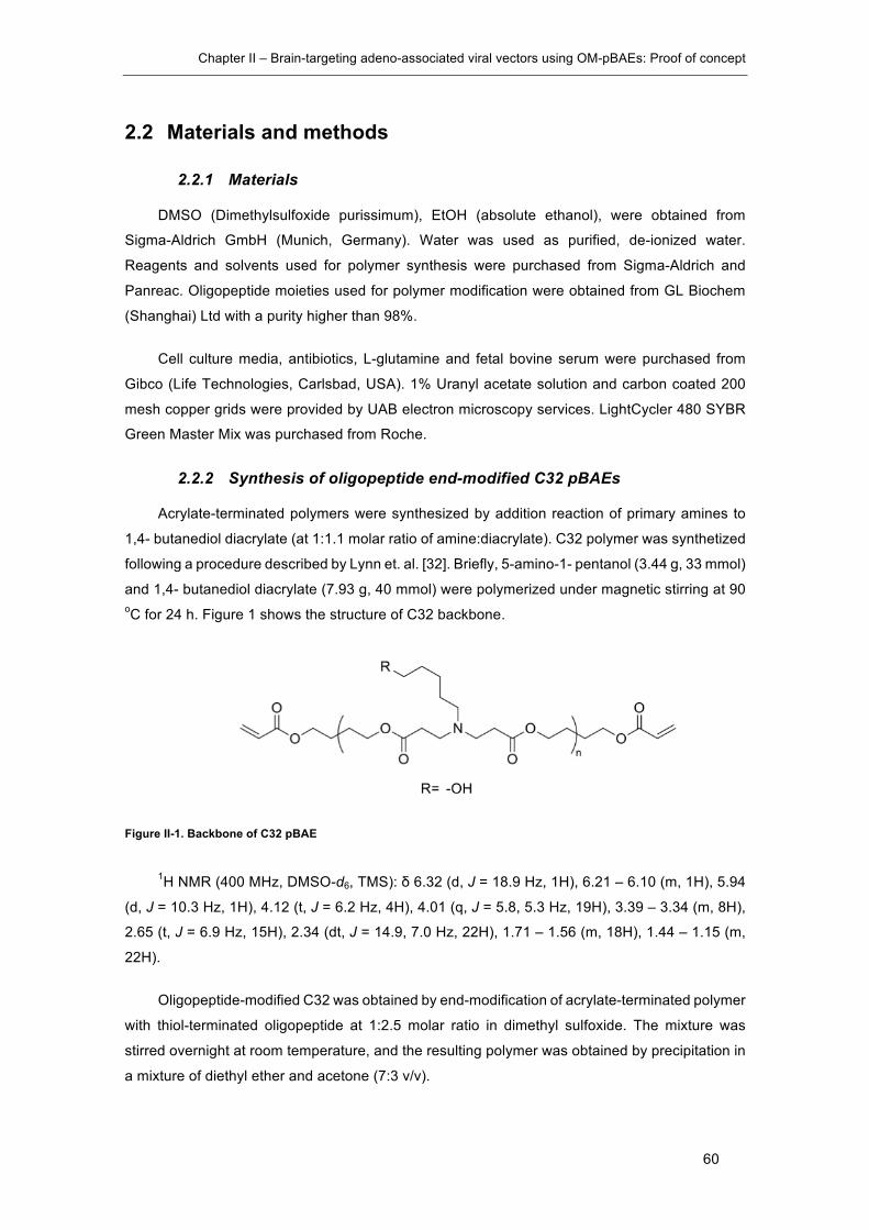
Chapter II – Brain-targeting adeno-associated viral vectors using OM-pBAEs: Proof of concept
60
2.2 Materials and methods
2.2.1 Materials
DMSO (Dimethylsulfoxide purissimum), EtOH (absolute ethanol), were obtained from
Sigma-Aldrich GmbH (Munich, Germany). Water was used as purified, de-ionized water.
Reagents and solvents used for polymer synthesis were purchased from Sigma-Aldrich and
Panreac. Oligopeptide moieties used for polymer modification were obtained from GL Biochem
(Shanghai) Ltd with a purity higher than 98%.
Cell culture media, antibiotics, L-glutamine and fetal bovine serum were purchased from
Gibco (Life Technologies, Carlsbad, USA). 1% Uranyl acetate solution and carbon coated 200
mesh copper grids were provided by UAB electron microscopy services. LightCycler 480 SYBR
Green Master Mix was purchased from Roche.
2.2.2 Synthesis of oligopeptide end-modified C32 pBAEs
Acrylate-terminated polymers were synthesized by addition reaction of primary amines to
1,4- butanediol diacrylate (at 1:1.1 molar ratio of amine:diacrylate). C32 polymer was synthetized
following a procedure described by Lynn et. al. [32]. Briefly, 5-amino-1- pentanol (3.44 g, 33 mmol)
and 1,4- butanediol diacrylate (7.93 g, 40 mmol) were polymerized under magnetic stirring at 90 oC for 24 h. Figure 1 shows the structure of C32 backbone.
Figure II-1. Backbone of C32 pBAE
1H NMR (400 MHz, DMSO-d6, TMS): δ 6.32 (d, J = 18.9 Hz, 1H), 6.21 – 6.10 (m, 1H), 5.94
(d, J = 10.3 Hz, 1H), 4.12 (t, J = 6.2 Hz, 4H), 4.01 (q, J = 5.8, 5.3 Hz, 19H), 3.39 – 3.34 (m, 8H),
2.65 (t, J = 6.9 Hz, 15H), 2.34 (dt, J = 14.9, 7.0 Hz, 22H), 1.71 – 1.56 (m, 18H), 1.44 – 1.15 (m,
22H).
Oligopeptide-modified C32 was obtained by end-modification of acrylate-terminated polymer
with thiol-terminated oligopeptide at 1:2.5 molar ratio in dimethyl sulfoxide. The mixture was
stirred overnight at room temperature, and the resulting polymer was obtained by precipitation in
a mixture of diethyl ether and acetone (7:3 v/v).

Chapter II – Brain-targeting adeno-associated viral vectors using OM-pBAEs: Proof of concept
61
C32 backbone was modified with cysteine-arginine tetrapeptide (CRRR) and SEQ12
peptide. The different oligopeptide modifications were confirmed by 1H-RMN. Figure 2 shows the
structure of C32 end-modified with peptides.
Figure II-2. Structure of C32CR3 OM-pBAE
2.2.3 Coating of AAVs with OM-pBAEs
AAV2/5 samples were coated by mixing a diluted virus sample solution (D-VS) with a
polymer solution (PS) in a 1:1 mixing ratio.
The concentration of the D-VS was 2x10⁹ GC/µl into a final 50 µl volume. Then total genome
copies were calculated and multiplied by the desired µg polymer/GC ratio in order to calculate the
total polymer amount needed. Taking into account the polymer stock concentration (100 mg/ml)
the polymer stock volume needed was calculated and diluted with PBS to 50 µl volume in order
to prepare PS. Finally, PS and D-VS were mixed by adding PS over VS and pipetting up and
down slowly at least 10 times. Samples were incubated 30’ at RT to enable the electrostatic
interactions and freshly used for testing assays.
2.2.4 DLS physicochemical characterization of coated AAV samples
Size and surface charge were determined by Dynamic Light Scaterring (DLS) (Malvern
Instruments Ltd, United Kingdom, 4-mW laser). Coated viral preparations were prepared as
previously described. 500µl of 1x10⁸ GC/µl virus preprations were used to determine the Z-
potential and sizer of viral preparations. Size was calculated using the number mean particle size
correction, where the size of the main population of particles is estimated avoiding the
interference of large particles, such as dust or aggregates.
2.2.5 Electron microscopy characterization
2.2.5.1 Negative staining transmission electron microscopy (TEM)
Five microliters from viral solutions (coated and non-coated), at a concentration of 1× 10¹¹
gc/ml in sterile mQ water, were adsorbed onto 300-mesh copper formvar-coated grids (150-
200nm) for ten seconds. Then, excess specimen was withdrawn and the grids were counter-
stained with 1% uranyl acetate for two minutes. After drying, samples were observed with a Jeol
Chapter II – Brain-targeting adeno-associated viral vectors using OM-pBAEs: Proof of concept
62
1400 transmission electron microscope, operating at 80kV and equipped with a CCD Gatan
ES1000W Erlangshen camera.
2.2.5.2 Scanning electron microscopy (SEM)
Five microliters from viral solutions (coated and non-coated), at a concentration of 1 × 10¹¹
gc/ml in sterile mQ water, were deposited onto a silicon substrate, and excess of material was
then removed. Samples were air-dried and observed with a Zeiss Merlin operating at 2 kV. Images
were acquired with a high-resolution in-lens secondary electron detector.
2.2.6 Mouse primary astrocytes isolation and culture
Pups no older than 5 days old were used. After decapitation, all heads were dip in 70%
ethanol. Using scalpel and forceps, brains were removed and putted in cold DMEM B containing
petri dish. With the help of a magnifying glass, cerebral cortices were isolated (removing
everything apart from cortex, even hippocampus) and meninges were also removed. Cortex from
6 pups were putted in a Falcon with 14,5 ml cold DMEM B and broken up with a 5 ml pipette.
After doing it with all cortex, 0,5 ml of trypsin 0,5% were added to each falcon. After 25 min at
37ºC, tempered DMEM A was added to a total volume of 50 ml. Then, cell suspension was
centrifuged 8 min at 2000rpm, supernatant was discarded and the pellet was resuspended in
10ml DMEM A.
The resulting cell suspension was homogenized several times with 10 ml pipette, filtered
through a 100 µm mesh and rinsed with additional 10 ml of DMEM A. It was filtered again through
a 45µm mesh and rinsed with 10 ml of DMEM. From each Falcon (Total volume of 30 ml), cells
were seeded in 3x 250 ml non-coated flask with Vent cap and cultured 3 days at 37ºC 5% CO2
without moving the flask. At day three, the media was changed and cells were further cultured
three additional days. Then, the media was replaced with new fresh media and flasks were
shaken O/N at 80 rpm at room temperature. Next day, cells were washed with PBS and rinsed
with fresh DMEM A. After reaching confluence (normally 3-5 additional days), cells were washed
with PBS and trypsinized (5ml trypsin). Trypsinization was stopped by adding 10ml of DMEM A,
cell suspension was centrifuged 5 min at 1000 rpm and the pellet was resuspended in 10 ml
DMEM. Cells were seeded in 8 flasks and left in the incubator. After 2-3 days (depending on
confluence), cells were frozen in Cryostor TM CS10, a commercial freezing medium specifically
designed for sensitive cells cryopreservation.
Reactives and solutions: DMEM A corresponds to DMEM supplemented with 10% (v/v) fetal
bovine serum (FBS), 2mmM l-glutamine, 100 U/ml penicillin (Labclinics), 0.1 mg/ml streptomycin,
10mM Hepes (Labclinics). DMEM B (DMEM A with 15mM Hepes). 70% EtOH solution. Trypsin-
EDTA 0,05%/0.02% (Invitrogen).

Chapter II – Brain-targeting adeno-associated viral vectors using OM-pBAEs: Proof of concept
63
2.2.7 In vitro BBB models
2.2.7.1 Monoculture
For the monoculture in vitro BBB model, 150.000 BBMVECs per insert were seeded on
Transwell insert pre-coated with commercial Attachment Factor Solution (AFS) containing rat tail
collagen and fibronectin. The cells were grown to confluency for 6 days in 10% FBS, 2mmM l-
glutamine, 100 U/ml penicillin, 0.1 mg/ml streptomycin in DMEM.
2.2.7.2 Co-culture
For the co-culture in vitro BBB model of BBMVECs with RCAs, astrocytes were seeded
on the bottom of PDL-coated 6-well plates for three days in AM-a medium. At the same time,
150.000 BBMVECs per insert were seeded on Transwell insert pre-coated with commercial
Attachment Factor Solution (AFS;Sigma) containing rat tail collagen and fibronectin in other 6-
well plates. The cells were grown to confluency for 3 days in 10% FBS, 2mmM l-glutamine, 100
U/ml penicillin, 0.1 mg/ml streptomycin in DMEM. Then, the BBMVECs containing Transwell
inserts were transferred to the 6-well plates in which RCAs reached confluency changing the
medium to complete DMEM and further cultured for 3 days.
2.2.8 Transendothelial electrical resistance (TEER) measurements
The tightness of the BBB was determined by measuring the TEER of the cellular barrier
formed by BBMVECs. TEER measurements of the Transwell inserts were collected using EVOM
resistance meter (World Precision Instruments). Three measures were done for each Transwell
at different positions and the mean values were calculated. The TEER of coated microporus
membranes without BBMVECs was subtracted and reported as Ω/cm2.
2.2.9 Permeability of Lucifer Yellow
For transport assays, the inserts were transferred to a new 6-well plate containing
DMEM/F12 medium at the abluminal side (2ml/well). DMEM/F12 containing 20µM of LY is placed
in the upper compartment. Every 15 min after addition of LY, the filter is transferred to another
well in order to minimize the possible passage from the abluminal side to the luminal side.
Incubations are performed at 37ºC and 5% of CO2. Transport studies are performed in the same
way with cell cultures and with membranes coated with collagen and fibronectin only.
And aliquot from each time point and T0 solution is taken and the amount of LY is
calculated using a multiwell plate reader fluorimeter.
Permeability calculations were done as described by Sifkinger-Birnboim et al. []. The
clearance principle is used to obtain a concentration-independent transport parameter. The
increment of cleared volume between successive sampling events is calculated by dividing the
amount of solute transport during the time interval by the donor chamber concentration. The total
Chapter II – Brain-targeting adeno-associated viral vectors using OM-pBAEs: Proof of concept
64
cleared volume at each time point is calculated by accumulating the incremental cleared volumes
up to the given time point:
Clearance(ml)=X/Cd
Equation 1. Clearance calculation equation. X is the amount of LY in the receptor chamber and Cd is the donor
chamber concentration at each time-point.
The clearance volume increases linearly with time during the 60-min experiment. The
average volume cleared is plotted vs. time, and the slope calculated by linear regression analysis.
The slope of the clearance curves for the culture is named PSt, where PS is the permeability x
surface area (in ml per min). The slope of the clearance curve with the blank control filter coated
only with collagen is named PSf.
The PS value for the endothelial monolayer (PSe) is calculated following equation 2.
1/PSe=1/PSt-1/PSf
Equation 2. Permeability calculation equation. PSe values are devided by surface area of the porous membrane to
generate the endothelial permeability coefficient (Pe, in cm per min
2.2.10 Immunocytochemistry
For immunostaining of BBMVECs tight junction proteins, cells were washed 3x with
DPBS, fixed in 10% formalin for 20min, washed 3x with DPBS and permeabilized with 0.1% Triton
X-100 in DPBS for 10 min. Different BBMVECs coming from established BBB models were then
incubated with rabbit anti-occludin, anti-claudin-5 and anti-ZO-1 monoclonal antibodies
(Invitrogen) in PBS/0.1% Triton-X 100/1% BSA overnight at room temperature. Cells were then
washed 6x for 5min in PBS/0.1% Triton-X 100 and incubated with Alexa Fluor 488 goat anti-rabbit
secondary antibody (1:500) for 1h at room temperature. For nucleus, Vectashield© commercial
mounting medium containing DAPI was used.
2.2.11 Fluorescent peptides BBB crossing experiments
Knowing the molecular weight (MW) of the peptides and the MW of the fluorescent label,
calculations were done to determine which concentration of peptide correlates with 10 µg/ml of
fluorophore. Each testing peptide solution was prepared in DMEM/F12 media to this final
fluorophore concentration taking into account that each condition will be studied in triplicates.
Also, a fluorescently-labeled scrambled peptide sample was used as a negative control.
Then, a new 24-well plate was filled with 0,8ml of media in each well. Inserts were moved
into this new plate and 0,4ml of the prepared solutions are added into each insert (Fig.1). An
800µl aliquot of each solution was collected (t=0 up) and stored at 4ºC protected from light

Chapter II – Brain-targeting adeno-associated viral vectors using OM-pBAEs: Proof of concept
65
exposure as well as media (t=0 down). After 60 min incubation at 37ºC and 5% of CO2, up and
down solutions were collected (t=60 up and t=60 down).
Using a black 96-well microplate, all samples (t=0 up, t=0 down, t=60 up and t=60 down) for
each condition (Testing peptides, LY and negative control) were plated as well as standard curves
of each condition. Then, measures at LY’s, peptide’s and scramble peptide’s fluorophore Ex/Em
wavelengths were done using a multi-well plate reader.
Once measures were done, percentage of crossing was calculated doing a mass balance
for each condition (Eq.6).
67 % = 9:;<=>:?@ABC< DB@=C?E, <60 + 9:;<=>:?@ABC< JKEB@=C?E, <60
9:;<=>:?@ABC<(EB@, <0)
Equation 3. Mass balance equation for BBB crossing assays.
2.2.12 In vitro transduction assay to study viral tropism
HEK293 and BBMVECs cells were used to study the viral tropism in vitro. 15000 cells/well
were seeded in 96 multi-well plates. The day after, cells were infected with 50000 genome
copies/cell using naked and coated AAV2/5 preparations in six replicates for each condition. 5
days after infection, luciferase activity was quantified using the Renilla luciferase assay system
(Promega; E2820). All wells were washed with PBS and 50 µl of lysis buffer was added into each
well. Multi-well plates were then freeze and thawed 3 times in and centrifuged 3 min at 10000g to
discard cell debris. Samples were collected and renilla luciferase activity was determined
following the suppliers’ protocol using white multi-well plates. To avoid lysis buffer interference
with BCA reagent, protein extracts were cleaned up by CompatAbleTM (Pierce; 23215) reagent
and subsequently quantified by BCA (Pierce; 23225).
2.2.13 Bioluminiscence assay of protein extracts
All frozen organs were grinded with a mortar at very low temperature. Then, each organ
homogenate was split in two parts, (i) one for the protein extraction and (ii) the other one for DNA
extraction.
Renilla luciferase assay system (Promega; E2820) was used to determine luciferase activity
over each tissue. Samples were freeze and thawed 3 times in the presence of lysis buffer and
centrifuged 3 min at 10000g to discard cell debris. Renilla luciferase activity was determined
following the suppliers’ protocol. To avoid lysis buffer interference with BCA reagent, protein
extracts were cleaned up by CompatAbleTM (Pierce; 23215) reagent and subsequently quantified
by BCA (Pierce; 23225).
Chapter II – Brain-targeting adeno-associated viral vectors using OM-pBAEs: Proof of concept
66
2.2.14 qPCR biodistribution analysis
Genomic DNA isolation was performed using NucleoSpin® tissue (MachereySNagel;
740952.50) and DNA concentration was determined with a NanoDrop 1000 Spectrophotometer
(Thermoscientific).
A standard curve was set using the plasmid pGL4.75[hRluc/CMV], which carries the renilla
luciferase gene, in order to correlate Ct values of the standard curve with genome copies Serial
dilutions from 1x10e7 GC/μl to 1e1 GC/ul were prepared and 100 ng of total genomic DNA was
added to each standard to build a more realistic scenario in comparison with samples coming
from tissue DNA extraction. Primers used were designed to be specific for the renilla luciferase
gene with the following sequences:
Sequence (5’ -3’)
RLuc Forward GGGCGAGAAAATGGTGCTTG
Reverse GCCCTTCTCCTTGAATGGCT
In order to convert genome copy numbers to DNA mass the following formula (Equation 4)
was used:
@ = CL660 M
@AE6,02L1023
@AE:PBE:Q/@AE
Equation 4. m is the mass of one viral genome copy and n is the size of the genome in base pairs (bp)
In order to calculate the final GC/cell result the following formula (Equation 5) was used:
R =?S:CA@:PA;=:Q
K;MTUJL6,5;MTUJ
P:EE
Equation 5. “X” units are genome copies / cell. “a” is the number of genome copies obtained by extrapolation of sample
Ct with the standard curve. “b” is the quantity of DNA added in the PCR reaction and 6,5 pg/cell is the weight of one mice
genome of 6x10⁹ bp/cell.

Chapter II – Brain-targeting adeno-associated viral vectors using OM-pBAEs: Proof of concept
67
2.3 Results and discussion
In the present chapter, the applicability of OM-pBAEs as a coating agent for AAV vectors
was explored. As previously mentioned, the main objective was to develop a technology based
on OM-pBAEs able to produce polymeric artificial envelopes around AAV virions in order to
change their tropism aiming to improve brain transduction. This study was carried out in the frame
of a collaboration with a big pharmaceutical company as a proof of concept for the application
feasibility. This company provided the AAV2/5 reporter vector expressing the renilla luciferase
gene used in this work.
2.3.1 Arginine modified OM-pBAEs interact with AAV viral particles
modifying their surface charge and size
The first step was to study the ability of arginine modified C32 pBAE (C32CR3) to interact
with AAV viral particles. This study was carried out by dynamic light scattering (DLS) in order to
characterize the physicochemical properties of formed complexes after incubating viral particles
with different ratios of C32CR3 per viral genome copy (ug polymer/GC) and by exploring the
morphology of the resulting complexes by transmission electron microscopy (TEM) and scanning
electron microscopy (SEM) techniques.
As observed in figure II-1-A, only in the presence of viral particles, the Z-potential increased
while increasing the polymer/GC ratio indicating that the viral particles were acting as a core. In
particular, Z- potential increased up to +20 mV when viruses were coated with 1x10⁻⁹ ug
polymer/GC of C32CR3; then a plateau was observed for higher ratios. This observation indicated
that this polymer ratio was enough to positivize all available viral capsids surface area in the
samples and allowed us to determine the optimal amount of C32CR3 needed to fully coat AAV
viral particles, which was set as 1x10⁻⁹ µg polymer/GC.
When analysing the same polymer concentrations but using water instead of viral solution,
almost neutral results were obtained, indicating that positive surfaces were only formed in the
presence of viruses.
Figure II-1. Z-potential determination of C32CR3-coated AAV viral particles. AAV particles are coated with increasing
µg polymer per genome copy (GC). Results are shown as mean and standard deviation of triplicates (A). DLS size
1×10
-9
2×10
-9
3×10
-9
4×10
-9
5×10
-9
-10
0
10
20
30
µg polymer / GC
Z-Po
tent
ial (
mV)
C32CR3-AAVC32CR3
Naked AAV C32CR3-AAV0
10
20
30
40
50
Num
ber m
ean
(d.n
m)
**

Chapter II – Brain-targeting adeno-associated viral vectors using OM-pBAEs: Proof of concept
68
characterization of naked AAV and C32CR3-AAVs coated at 1x10⁻⁹ g polymer/GC ratio. Z-Average. Sizes were
determined using the number correction tool (B).
The size characterization of naked AAVs and 1x10⁻⁹ µg C32CR3/GC coated AAVs showed
a 12 nm increase in size between naked and coated preparations (Naked 22 ± 3 nm; C32CR3-
AAV 35,5 ± 4,5 nm). It is important to note that size was calculated using the number mean
correction option where only the most abundant population in the sample is considered (Figure
II-1-B). Otherwise, the obtained size values were far from the expected size of an AAV viral
particle (25 nm aprox.).
Next, naked AAV and coated-AAV preparations were analysed by TEM and SEM techniques
in order to further characterize the morphology and size of the resulting complexes. As seen in
figure II-2-A, naked AAVs showed their typical icosahedral shape with a size of 26 ± 0,2 nm. In
contrast, when imaging coated AAVs by TEM (figure II-2-D and E), clustered viral particles were
observed. However, when analysing individual particles their size was 31±0,4 nm accordingly
with the DLS size determination. Interestingly, samples analysed by SEM showed the presence
of two populations. As indicated with black and white arrows in figure II-2-F, grey and white
particles were observed. We hypothesized that non-coated AAV particles were present in the
preparation, however further studies are needed to confirm this hypothesis.
Figure II-2. Transmission electron microscopy characterization of pBAE-coated AAVs. TEM imaging of naked AAVs
(300.000x) (A); TEM of polymeric component without viral particles (300.000x) (B); SEM images of polymeric component
without viral particles (C); TEM of 1x10⁻⁹ ug C32CR3/GC coated AAV (200.000x) (D); TEM of 1x10⁻⁹ ug C32CR3/GC
coated AAV (300.000x) (E); SEM of 1x10⁻⁹ ug C32CR3/GC coated AAV (F).
This study demonstrated the ability of C32CR3 to interact with AAV particles changing their
surface charge and size. C32CR3 works as an electrostatic anchorage which covers capsid

Chapter II – Brain-targeting adeno-associated viral vectors using OM-pBAEs: Proof of concept
69
surfaces. In order to change the tropism of AAV vectors, OM-pBAEs were used to expose
targeting moieties on capsid surfaces aiming to tailor viral tropism. In the following section,
different brain-targeting peptides candidates were tested in vitro using BBB models in order to
define the best candidate to be used as a brain-targeting moiety.
2.3.2 BBB in vitro modelling as a tool to discover brain-penetrating
peptides
In vitro models construct artificial environments with cultured cells in order to mimic in vivo
structures. These models are a valuable precursor to animals use due to lower cost, time and
ethical constraints. In the specific BBB case, unlike animal studies, in vitro models enable
controlled, repeatable, and non-invasive tests: permeability assays, resistance measurements,
and microscopy. Thus, the development of valid in vitro BBB models can facilitate the overall drug
development process by acting as a precursor, or even replacement, for animal studies.
The advantages related to any in vitro BBB model include lower compound requirement, the
ability to assay compounds directly in physiological buffer, greater throughput relative to in vivo
models, ability to assess transport mechanisms, the identification of early signs of cell toxicity,
and, generally, lower cost [33,34]. The validity of an in vitro BBB model is dependent on how well
it reproduces the key physiological and biological characteristics found in vivo. The key features
of the BBB include: (i) the primary structure, consisting of strongly expressed tight junction
between brain endothelial cells (BECs) which directly control compound permeability; (ii) co-
culture of BECs with astrocytes, which plays an important role in modulating barrier function
through cell-cell signaling; (iii) selective permeability from the constituted structure to dissolved
compounds; (iv) maintenance of high electrical resistance representing the maturity of the
structure.
2.3.2.1 Establishment and characterization of in vitro BBB models
In the present work, the co-culture effect of BEC with astrocytes was studied in order to
establish reliable in vitro BBB models in terms of their in vivo resemblance. The main objective
was to analyse the effect of different culturing condition on BBB-related characteristics
development and maintenance and to define a culturing method able to produce reliable and
cost/time effective BBB in vitro models to be used as a brain-penetrating peptide screening
platform.
A monoculture of low passaged bovine brain microvascular endothelial cells (BBMVECs)
grown on a collagen coated transwell insert (Figure II-3-A) was compared with a co-culture of
BBMVECs with rat cortical astrocytes (RCAs) seeded at the bottom of a multi-well plate (Figure
II-3-B). Both models were characterized in terms of trans-endothelial electrical resistance (TEER)
and permeability (Pe) of a water-soluble molecular tracer called Lucifer Yellow (LY). These
parameters are widely used to assess the barrier integrity in vitro. TEER value explains the

Chapter II – Brain-targeting adeno-associated viral vectors using OM-pBAEs: Proof of concept
70
resistance to the passage of small ions through the endothelial cell layer and is an accurate and
sensitive measure of BBB integrity. A decrease in TEER implies an increase in permeability and
a loss of barrier function. LY is a fluorescent dye that cannot be taken up by endothelial cells,
neither by active nor by facilitated transport [35]; thus, the study of its permeability coefficient (Pe)
together with the determination of TEER are used as markers to study the BBB integrity and
quality in vitro.
Figure II-3. In Vitro BBB model characterization. Schematic representation of studied in vitro BBB models. BBMVECs
cultured alone in the monoculture model (A), BBMVECs co-cultured with astrocytes grown on the bottom of the multiwall
plate (B), Trans-endothelial electrical resistance (TEER) and permeability of Lucifer yellow (Pe) of monoculture and co-
culture BBB in vitro models. Results are shown as mean and standard deviation of 6 replicates (C). Tight junction protein
expression pattern of in vitro BBB models. BBMVECs were fixed and stained with specific monoclonal antibodies directed
respectively against ZO-1, claudin-5 and occludin. Nuclei were counterstained with DAPI (D).
As shown in figure II-3-C, the presence of glial cells in the co-culture model induced
significantly higher TEER values when compared to monoculture model (268±42 vs. 133±20 Ω x
cm2 (mean ± SEM), p<0.0001, n=20, Student t-test). The obtained TEER results comparing

Chapter II – Brain-targeting adeno-associated viral vectors using OM-pBAEs: Proof of concept
71
monoculture and co-culture reaffirm the fact that astrocytes induce a tightening of the BBB due
to astrocytes-released soluble factors [36].
In order to further assess the quality of the established in vitro BBB models, BBMVECs
permeability to LY (457Da, 10μM) was determined for each culturing condition (n=6 for co-culture
model and n=6 for mono-culture model). As shown in figure II-3-C, the presence of glial cells in
the co-culture model showed a tendency towards lowering LY Pe values when compared to
monoculture model (0.899±0.08 vs. 1.269±0.67 cm/min-1 (mean ± SEM)).
The close relation between TEER and Pe let us to interpret these differences in the same
way we interpreted TEER values. The reconstruction of the in vivo-like environment improves the
tightening of BBB, yielding higher TEER values and restricting paracellular permeability of
hydrophilic compounds, such as Lucifer Yellow.
In order to further characterize the established in vitro BBB models, the expression of tight
junction proteins was studied by immunostainning. As shown in figure II-3-D, the expression
pattern of ZO-1, Claudin-5 and Occludin was consistent with the hypothesis that the in vivo
mimicking degree increases the barrier tightening through and improved expression of tight
junction proteins. ZO-1 expression was observed to be independent on the presence of glial cells.
Claudin-5 and occludin showed an increased expression in co-cultured models.
2.3.2.2 Screening of BBB penetrating peptides
To date, only a small number of peptides are known to cross the BBB. However, it is
estimated that there are several hundred thousand of peptides which can virtually cross the BBB,
including many neurotoxic molecules, which could serve as BBB shuttles [37]. Hence, testing new
molecules in in vitro BBB models and also in in vivo models, is a promising strategy to achieve
drug delivery to the brain.
One of the most promising examples of BBB shuttles is ANG1005. It consists of a 19
aminoacid long peptide vector known as Angiopep-2 that is directly conjugated to three paclitaxel
molecules. ANG1005 effectively transports across the BBB with approximately a 100-fold higher
transport rate compared to a free paclitaxel [38]. ANG1005 interacts with low-density lipoprotein
receptor-related protein 1 (LRP) which is expressed by BBB endothelial cells and is upregulated
in some cancers.
Taking as a reference the Angiopep-2 peptide, transports experiments were done in order
to determine the crossing levels of candidate peptic sequences with potentiality to interact with
LRP-1. These peptides were designed previously by Sagetis-Biotech S.L. in collaboration with
Intelligent Pharma S.L. by in silico modelling LRP-1 receptor and the interaction of peptides
known to interact with this receptor. Alternative peptic sequences were designed with similar 3D
conformations and interaction capacity by in silico docking assays. Two of those peptides (SEQ12
and SEQR) were screened using BBB in vitro models and results are presented below.

Chapter II – Brain-targeting adeno-associated viral vectors using OM-pBAEs: Proof of concept
72
As discussed in section 1.2.11, the calculations were done comparing the passage across
an insert without cells (taking this value as the 100% of crossing), with the passage across inserts
containing an established BBB co-culture model.
Figure II-4. BBB crossing assay of candidate peptides. Crossing percentages of ANG, SEQ12, SEQR and
SCRAMBLED peptide sequences and Lucifer yellow (LY) through co-culture in vitro BBB models. Results are shown as
mean and standard deviation of triplicates.
As shown in figure II-4, SEQ12 achieved the same level of crossing of Angiopep (4,28%
±0.64 vs. 4,39%±0.54 respectively). This result confirms SEQ12 as a good candidate to be used
a BBB shuttle. However, SEQR crossed less than the scrambled peptide with a result comparable
to the passage of LY (SEQR= 1.6%± 0.42; SCRAMBL= 2.23%±0.39; LY= 1.34%± 0.12).
2.3.3 pBAE-based AAV-coating for viral retargeting strategies
Once the best performing targeting peptide was defined, C32-pBAEs end-capped with
SEQ12 peptide were produced and called C32C12. This new pBAE was mixed with C32CR3 at
10% w/w ratio and used to coat AAV samples.
As previously shown with C32CR3, 1x10⁻⁹ µg C32CR3-10%C12/GC ratio also positivized
viral particles suspensions in the same way as achieved for C32CR3 (Figure II-5).
Figure II-5. Biophysical characterization of pBAE-coated AAVs by dynamic light scattering (DLS). Z-potential
determination of viral particles coated with increasing µg polymer /GC ratios of C32CR3-10%C12, C32CR3 and AAVs.
Results are shown as mean and standard deviation of triplicates (A). DLS size characterization of naked AAV and
ANGSEQ12
SEQR
SCRMBL LY0
2
4
6
% o
f cro
ssin
g
1×10
-9
2×10
-9
3×10
-9
4×10
-9
5×10
-9
-10
0
10
20
30
µg polymer / GC
Z-Po
tent
ial (
mV)
C32CR3-C12-AAVC32CR3-AAVC32CR3
Z-Ave
rage
Main pea
k
Number mea
n0
50
100200
220
240
260
d. n
m
Naked AAVCoated AAV
**

Chapter II – Brain-targeting adeno-associated viral vectors using OM-pBAEs: Proof of concept
73
C32CR3-10%C12-AAVs coated at 1x10⁻⁹ g polymer /GC ratio. Z-Average. Sizes were determined using DLS and
represented as mean and standard deviation of triplicates (B). *p < 0.05, **p < 0.01, ***p < 0.001
Accordingly, size determination by DLS characterization demonstrated the ability of the
C32CR3-10%C12 targeted formulation to increase the size of viral particles.
2.3.3.1 C32CR3-10%C12 coated AAVs improves the transduction of BBMVECs in vitro
In order to study the targeting effect in vitro, transduction experiments were carried out
infecting HEK293 cells and BBMVECs cells with naked and coated formulations. Cells were
infected at MOI 50.000 and luciferase activity was quantified 5 days after infection.
As seen in figure II-6, the infectivity of naked AAV was clearly higher for HEK293 with 1,68
± 0,03 RLUs/ug Prot in comparison with 0,48 ± 0,04 RLUs/ug Prot obtained when infecting
BBMVECs. Coated formulation improved the infectivity in the HEK293 reaching 2,13 ± 0,04
RLUs/ug Prot. However, this improvement was much more significant for BBMVECs, which are
cells with a high LRP-1 expression pattern [39], and reached 2,44 RLUs/ug Prot. This result
suggested that the presence of entangled SEQ12 containing C32 chains into the artificial coating
promoted infection of brain vascular endothelial cells.
Figure II-6. Luciferase activity quantification of HEK293 and BBMVECs cells infected with naked and C32CR3-10%C12 coated AAV2/5. Cells were infected at MOI 50.000 and luciferase activity was quantified 5 days after infection.
Results are presented as mean and standard deviation of six replicates.*p < 0.05, **p < 0.01, ***p < 0.001, ****p<0.0001
2.3.3.2 In vivo biodistribution of C32CR3-10%C12 coated AAVs
The main goal of the present section was to perform a preliminary assessment of the
biodistribution profile of C32CR3-10%C12 coated AAV2/5 by bioluminescence quantification of
protein extracts of organ homogenates (Brain, Liver, Lungs, Intestines, Kidneys and Heart) and
by determination of viral genome copy numbers in different organs.
In order to do so, two BALB/c mice per group were injected with 4,8x10¹¹ GC/animal with
naked AAV2/5 and C32CR3-10%C12 coated AAV2/5. At 26-day post injection, the renilla
HEK293
BBMVECs0
1
2
3
RLU
s / u
g Pr
ot
MOI 0Naked AAVCoated AAV
****

Chapter II – Brain-targeting adeno-associated viral vectors using OM-pBAEs: Proof of concept
74
luciferase substrate (ViviRen TM) was injected and luciferase images were taken using a
Hamamatsu CCD camera mounted in a dark box.
Figure II-7. In vivo biodistribution of brain-targeted coated-AAVs. Bioluminescence imaging of BALB/c mice I.V.
injected with 4,8x10¹¹ GC/Animal of naked AAV2/5 and C32CR3-10%C12-coated AAV2/5 26 days post injection. Organ
images were also taken ex vivo immediately before sacrifice. Brain, heart, lungs, liver, kidney and intestines are disposed
from top to bottom respectively. All images were obtained by capturing bioluminiscence during 5 minuts.
The comparison between coated and naked viruses gave some interesting insights about
the behaviour of coated viruses. As observed in figure II-7, the coating of the viruses modified the
virus tropism in vivo. At day 26 post-injection a strong signal in the liver was observed for the
naked condition, while the coated virus presented less specificity for this organ.
In order to further study the biodistribution profile, luciferase activity was quantified from
protein extracts of each organ. Measurment of luciferase activity in tissue homogenates is a very
reliable method to quantify differences of protein expression in different tissues, especially when
the target organ is the brain, where the BBB can affect the transport of luciferase substrate.
Although these was a preliminary study and more animals would be needed to achieve
significant results, it seemed clear that the BBB targeted polymer changed the biodistribution of
AAV2/5 viral particles in mice, increasing the level of functional gene delivery to the brain and
reducing liver transduction. As seen in figure II-8, the brain was the only organ where luciferase
activity was improved in contrast with liver, lungs, kidney and heart, when administering coated
viruses.
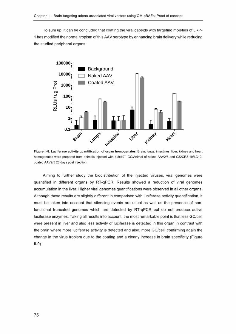
Chapter II – Brain-targeting adeno-associated viral vectors using OM-pBAEs: Proof of concept
75
To sum up, it can be concluded that coating the viral capsids with targeting moieties of LRP-
1 has modified the normal tropism of this AAV serotype by enhancing brain delivery while reducing
the studied peripheral organs.
Figure II-8. Luciferase activity quantification of organ homogenates. Brain, lungs, intestines, liver, kidney and heart
homogenates were prepared from animals injected with 4,8x10¹¹ GC/Animal of naked AAV2/5 and C32CR3-10%C12-
coated AAV2/5 26 days post injection.
Aiming to further study the biodistribution of the injected viruses, viral genomes were
quantified in different organs by RT-qPCR. Results showed a reduction of viral genomes
accumulation in the liver. Higher viral genomes quantifications were observed in all other organs.
Although these results are slightly different in comparison with luciferase activity quantification, it
must be taken into account that silencing events are usual as well as the presence of non-
functional truncated genomes which are detected by RT-qPCR but do not produce active
luciferase enzymes. Taking all results into account, the most remarkable point is that less GC/cell
were present in liver and also less activity of luciferase is detected in this organ in contrast with
the brain where more luciferase activity is detected and also, more GC/cell, confirming again the
change in the virus tropism due to the coating and a clearly increase in brain specificity (Figure
II-9).
Brain
Lungs
Intestin
eLive
r
KidneyHea
rt0.1
1
10
100
1000
10000
100000
RLU
s / u
g Pr
ot
BackgroundNaked AAVCoated AAV

Chapter II – Brain-targeting adeno-associated viral vectors using OM-pBAEs: Proof of concept
76
Figure II-9. AAVs genomes biodistribution. AAV genomes were quantified by RT-qPCR in DNA samples extracted
from brain, lungs, intestines, liver, kidney and heart homogenates prepared from animals injected with 4,8x10¹¹ GC/Animal
of naked AAV2/5 and C32CR3-10%C12-coated AAV2/5 26 days post injection. A standard curve was set using the
plasmid pGL4.75[hRluc/CMV], which carries the renilla luciferase gene, in order to correlate Ct values with genome copies.
Brain
Lungs
Intestin
eLive
r
KidneyHea
rt0.001
0.010
0.100
1
AAV
geno
mes
/ ce
ll Naked AAVCoated AAV
Chapter II – Brain-targeting adeno-associated viral vectors using OM-pBAEs: Proof of concept
77
2.4 Concluding remarks
In this chapter, the feasibility of using OM-pBAEs as coating agents aiming to retarget non-
enveloped viral vectors has been studied with AAV vectors. All the work presented in this chapter
was carried out as a proof of concept in order to assess the applicability of OM-pBAEs to be used
as a brain-targeting coating technology for AAV2/5 vectors.
Results show that C32CR3 efficiently interact with AAV virions producing hybrid viral/non-
viral gene delivery vectors with enhanced infectivity in vitro. The electrostatic interaction of
C32CR3 with viral capsids surfaces positivizes their net negative surface charge and induces an
increase of size observed by DLS and TEM. However, some degree of clustering has been
observed by both techniques indicating that further improvements in the technology or coating
process are needed in order to produce single coated viral particle suspensions.
As explained in the introduction of this chapter, brain transduction efficiency after
intravenous administration of AAV vectors is still far from achieving therapeutic levels. Aiming to
improve brain targeting of OM-pBAEs coated AAVs, a BBB targeting ligand have been included
into pBAEs structure. This ligand was discovered by screening different in silico designed
peptides, with potentiality to interact with LRP-1, in in vitro BBB models in order to test their ability
to cross the BBB.
BBB models based on either monocultured BBMVECs or BBMVECs co-cultured with
astrocytes were established and characterized in terms of TEER and Lucifer yellow permeability,
as well as for their tight junction proteins expression by immunostaining. Results have shown that
the co-culture of BBMVECs with astrocytes clearly improves the formation of in vivo resembling
characteristics. After determining the optimal culturing conditions to produce reliable in vitro BBB
models, fluorescently-labelled candidate peptides were screened in vitro for their BBB crossing
ability. SEQ12 peptide demonstrated to be as efficient as Angiopep in terms of BBB crossing in
vitro. This peptide has already been used to decorate paclitaxel-loaded nanoparticles for the
treatment of gliomas, demonstrating its potentiality in a previously published work [19].
In order to produce brain-targeted pBAE-based coating for AAV vectors, polymer C32C12
was produced by end-capping C32-backboned pBAEs with SEQ12. The inclusion of 10% w/w
C32C12 in the C32CR3 formulation maintained the ability of pBAEs to interact with viral capsids
changing their Z-potential and size. As demonstrated by in vitro transduction assays, the addition
of this targeting moiety improved the transduction of BBMVECs while maintaining the infectivity
on low LRP-1-expressing cells such as HEK293.
Finally, a pilot in vivo biodistribution study has shown that C32CR3-10%C12 coated AAVs
reduces the natural liver tropism of AAV2/5 observed as a clear decrease in luciferase activity
and genome copies accumulation in this organ after intravenous administration. Moreover, an
increase in luciferase activity and genome copies accumulation in the brain also indicated that
Chapter II – Brain-targeting adeno-associated viral vectors using OM-pBAEs: Proof of concept
78
the active targeting strategy based on OM-pBAEs coating can improve the therapeutic efficacy of
systemically administered AAV2/5 to treat central nervous system disorders.
Taking into account the results obtained in this chapter, we conclude that OM-pBAEs are
good candidates to be used as coating agents for non-enveloped viral vectors. However, the
small size of the AAV vectors affecting their physicochemical characterization and the
impossibility to produce those vectors in-house limited the possibility to continue with this branch
of study.
In the following chapters, the use of OM-pBAEs as coating agents for viral vectors was
deeply studied in order to adapt it to adenoviral vectors in collaboration with the Cancer and Gene
Therapy group at Institut d’Investigacions Biomèdiques Pi i Sunyer (IDIBAPS) led by Dra. Fillat.
The availability of facilities and expertise on producing adenoviral vectors as well as the
availability of previously developed therapeutic adenoviruses for the treatment of pancreatic
cancer facilitated the successful development of this research. This project was carried out as a
collaboration between Grup d’Enginyeria de Masterials (GEMAT, IQS), Cancer and Gene
Therapy group at Institut d’Investigacions Biomèdiques Pi i Sunyer (IDIBAPS) and Sagetis-
Biotech S.L. and was funded by a Ministerio de Ciencia, Innovación y Universidades (Gobierno
de España) through a Retos-Colaboración 2015 project.
Chapter II – Brain-targeting adeno-associated viral vectors using OM-pBAEs: Proof of concept
79
2.5 References
[1] M. Lim, Systemic delivery of adeno-associated viral vectors, 123 (2016) 413–423.
doi:10.1007/s11060-015-1747-8.The.
[2] R.W. ATCHISON, B.C. CASTO, W.M. HAMMON, ADENOVIRUS-ASSOCIATED
DEFECTIVE VIRUS PARTICLES., Science. 149 (1965) 754–756.
[3] K.I. Berns, R.M. Linden, The cryptic life style of adenoassociated virus, BioEssays. 17
(1995) 237–245. doi:10.1002/bies.950170310.
[4] B.C. Schnepp, R.L. Jensen, C. Chen, P.R. Johnson, K.R. Clark, Characterization of
Adeno-Associated Virus Genomes Isolated from Human Tissues, 79 (2005) 14793–
14803. doi:10.1128/JVI.79.23.14793.
[5] M.F. Naso, B. Tomkowicz, W.L. Perry, W.R. Strohl, Adeno-Associated Virus (AAV) as a
Vector for Gene Therapy, BioDrugs. 31 (2017) 317–334. doi:10.1007/s40259-017-0234-
5.
[6] S. Russell, J. Bennett, J.A. Wellman, D.C. Chung, Z.-F. Yu, A. Tillman, J. Wittes, J.
Pappas, O. Elci, S. McCague, D. Cross, K.A. Marshall, J. Walshire, T.L. Kehoe, H.
Reichert, M. Davis, L. Raffini, L.A. George, F.P. Hudson, L. Dingfield, X. Zhu, J.A. Haller,
E.H. Sohn, V.B. Mahajan, W. Pfeifer, M. Weckmann, C. Johnson, D. Gewaily, A. Drack,
E. Stone, K. Wachtel, F. Simonelli, B.P. Leroy, J.F. Wright, K.A. High, A.M. Maguire,
Efficacy and safety of voretigene neparvovec (AAV2-hRPE65v2) in patients with RPE65-
mediated inherited retinal dystrophy: a randomised, controlled, open-label, phase 3 trial.,
Lancet (London, England). 390 (2017) 849–860. doi:10.1016/S0140-6736(17)31868-8.
[7] C.E. Nelson, C.H. Hakim, D.G. Ousterout, P.I. Thakore, E.A. Moreb, R.M. Castellanos
Rivera, S. Madhavan, X. Pan, F.A. Ran, W.X. Yan, A. Asokan, F. Zhang, D. Duan, C.A.
Gersbach, In vivo genome editing improves muscle function in a mouse model of
Duchenne muscular dystrophy., Science. 351 (2016) 403–407.
doi:10.1126/science.aad5143.
[8] A. Marchini, E.M. Scott, J. Rommelaere, Overcoming barriers in oncolytic virotherapy
with HDAC inhibitors and immune checkpoint blockade, Viruses. 8 (2016) 1–22.
doi:10.3390/v8010009.
[9] and D.S.H. Xiwei Zheng, Cong Bi, Marissa Brooks, Adeno-Associated Virus (AAV)
Vectors in Cancer Gene Therapy, Anal Chem. 25 (2015) 368–379.
doi:10.1016/j.cogdev.2010.08.003.Personal.
[10] V. Louis Jeune, J.A. Joergensen, R.J. Hajjar, T. Weber, Pre-existing anti-adeno-
Chapter II – Brain-targeting adeno-associated viral vectors using OM-pBAEs: Proof of concept
80
associated virus antibodies as a challenge in AAV gene therapy., Hum. Gene Ther.
Methods. 24 (2013) 59–67. doi:10.1089/hgtb.2012.243.
[11] B. V Zlokovic, The blood-brain barrier in health and chronic neurodegenerative
disorders., Neuron. 57 (2008) 178–201. doi:10.1016/j.neuron.2008.01.003.
[12] N.R. Saunders, M.D. Habgood, K.M. Dziegielewska, Barrier mechanisms in the brain, I.
Adult brain, Clin Exp Pharmacol Physiol. 26 (1999) 11–19.
http://www.ncbi.nlm.nih.gov/entrez/query.fcgi?cmd=Retrieve&db=PubMed&dopt=Citation
&list_uids=10027064.
[13] S.R. Choudhury, E. Hudry, C.A. Maguire, M. Sena-Esteves, X.O. Breakefield, P. Grandi,
Viral vectors for therapy of neurologic diseases., Neuropharmacology. 120 (2017) 63–
80. doi:10.1016/j.neuropharm.2016.02.013.
[14] W.M. Pardridge, The blood-brain barrier: bottleneck in brain drug development.,
NeuroRx. 2 (2005) 3–14. doi:10.1602/neurorx.2.1.3.
[15] M. Simonato, J. Bennett, N.M. Boulis, M.G. Castro, D.J. Fink, W.F. Goins, S.J. Gray,
P.R. Lowenstein, L.H. Vandenberghe, T.J. Wilson, J.H. Wolfe, J.C. Glorioso, Progress in
gene therapy for neurological disorders, Nat. Rev. Neurol. 9 (2013) 277–291.
doi:10.1038/nrneurol.2013.56.
[16] B.L. Gurda, A. De Guilhem De Lataillade, P. Bell, Y. Zhu, H. Yu, P. Wang, J. Bagel, C.H.
Vite, T. Sikora, C. Hinderer, R. Calcedo, A.D. Yox, R.A. Steet, T. Ruane, P. O’Donnell,
G. Gao, J.M. Wilson, M. Casal, K.P. Ponder, M.E. Haskins, Evaluation of AAV-mediated
gene therapy for central nervous system disease in Canine Mucopolysaccharidosis VII,
Mol. Ther. 24 (2016) 206–216. doi:10.1038/mt.2015.189.
[17] A.K. Bevan, S. Duque, K.D. Foust, P.R. Morales, L. Braun, L. Schmelzer, C.M. Chan, M.
McCrate, L.G. Chicoine, B.D. Coley, P.N. Porensky, S.J. Kolb, J.R. Mendell, A.H.M.
Burghes, B.K. Kaspar, Systemic gene delivery in large species for targeting spinal cord,
brain, and peripheral tissues for pediatric disorders., Mol. Ther. 19 (2011) 1971–1980.
doi:10.1038/mt.2011.157.
[18] R. Gabathuler, Approaches to transport therapeutic drugs across the blood-brain barrier
to treat brain diseases., Neurobiol. Dis. 37 (2010) 48–57. doi:10.1016/j.nbd.2009.07.028.
[19] C. Fornaguera, M.Á. Lázaro, P. Brugada-Vilà, I. Porcar, I. Morera, M. Guerra-Rebollo, C.
Garrido, N. Rubio, J. Blanco, A. Cascante, S. Borrós, Application of an assay Cascade
methodology for a deep preclinical characterization of polymeric nanoparticles as a
treatment for gliomas, Drug Deliv. 25 (2018) 472–483.
doi:10.1080/10717544.2018.1436099.

Chapter II – Brain-targeting adeno-associated viral vectors using OM-pBAEs: Proof of concept
81
[20] D. Michel, C. Jean-Christophe, B. Yanick, C. Christian, N. Tran, R. Anthony, G.
Reinhard, C. Jean-Paul, B. Richard, Involvement of the low-density lipoprotein receptor-
related protein in the transcytosis of the brain delivery vector Angiopep-2, J. Neurochem.
106 (2008) 1534–1544. doi:10.1111/j.1471-4159.2008.05492.x.
[21] R.C. Carlisle, R. Benjamin, S.S. Briggs, S. Sumner-Jones, J. McIntosh, D. Gill, S. Hyde,
A. Nathwani, V. Subr, K. Ulbrich, L.W. Seymour, K.D. Fisher, Coating of adeno-
associated virus with reactive polymers can ablate virus tropism, enable retargeting and
provide resistance to neutralising antisera., J. Gene Med. 10 (2008) 400–411.
doi:10.1002/jgm.1161.
[22] N. Segovia, P. Dosta, A. Cascante, V. Ramos, S. Borrós, Oligopeptide-terminated
poly(β-amino ester)s for highly efficient gene delivery and intracellular localization, Acta
Biomater. 10 (2014) 2147–2158. doi:10.1016/j.actbio.2013.12.054.
[23] P. Dosta, N. Segovia, A. Cascante, V. Ramos, S. Borrós, Surface charge tunability as a
powerful strategy to control electrostatic interaction for high efficiency silencing, using
tailored oligopeptide-modified poly(beta-amino ester)s (PBAEs), Acta Biomater. 20
(2015) 82–93. doi:http://dx.doi.org/10.1016/j.actbio.2015.03.029.
[24] D.G. Anderson, A. Akinc, N. Hossain, R. Langer, Structure/property studies of polymeric
gene delivery using a library of poly(??-amino esters), Mol. Ther. 11 (2005) 426–434.
doi:10.1016/j.ymthe.2004.11.015.
[25] A. Akinc, D.M. Lynn, D.G. Anderson, R. Langer, Parallel synthesis and biophysical
characterization of a degradable polymer library for gene delivery., J. Am. Chem. Soc.
125 (2003) 5316–5323. doi:10.1021/ja034429c.
[26] J.J. Green, R. Langer, D.G. Anderson, A combinatorial polymer library approach yields
insight into nonviral gene delivery., Acc. Chem. Res. 41 (2008) 749–759.
doi:10.1021/ar7002336.
[27] S.Y. Tzeng, D.R. Wilson, S.K. Hansen, A. Quinones-Hinojosa, J.J. Green, Polymeric
nanoparticle-based delivery of TRAIL DNA for cancer-specific killing., Bioeng. Transl.
Med. 1 (2016) 149–159. doi:10.1002/btm2.10019.
[28] C.H. Jones, M. Chen, A. Ravikrishnan, R. Reddinger, G. Zhang, A.P. Hakansson, B. a.
Pfeifer, Mannosylated poly(beta-amino esters) for targeted antigen presenting cell
immune modulation, Biomaterials. 37 (2015) 333–344.
doi:10.1016/j.biomaterials.2014.10.037.
[29] S.Y. Tzeng, B.P. Hung, W.L. Grayson, J.J. Green, Cystamine-terminated poly(beta-
amino ester)s for siRNA delivery to human mesenchymal stem cells and enhancement
Chapter II – Brain-targeting adeno-associated viral vectors using OM-pBAEs: Proof of concept
82
of osteogenic differentiation., Biomaterials. 33 (2012) 8142–8151.
doi:10.1016/j.biomaterials.2012.07.036.
[30] W. Shao, A. Paul, S. Abbasi, P.S. Chahal, J. a. Mena, J. Montes, A. Kamen, S. Prakash,
A novel polyethyleneimine-coated adenoassociated virus-like particle formulation for
efficient siRNA delivery in breast cancer therapy: Preparation and in vitro analysis, Int. J.
Nanomedicine. 7 (2012) 1575–1586. doi:10.2147/IJN.S26891.
[31] P.Y.-J. Hsu, Y.-W. Yang, Effect of polyethylenimine on recombinant adeno-associated
virus mediated insulin gene therapy., J. Gene Med. 7 (2005) 1311–21.
doi:10.1002/jgm.779.
[32] D.M. Lynn, R. Langer, Degradable Poly(β-amino esters): Synthesis, Characterization,
and Self-Assembly with Plasmid DNA, J. Am. Chem. Soc. 122 (2000) 10761–10768.
doi:10.1021/ja0015388.
[33] R. Cecchelli, B. Dehouck, L. Descamps, L. Fenart, V. Buée-Scherrer, C. Duhem, S.
Lundquist, M. Rentfel, G. Torpier, M. Dehouck, In vitro model for evaluating drug
transport across the blood-brain barrier., Adv. Drug Deliv. Rev. 36 (1999) 165–178.
http://www.ncbi.nlm.nih.gov/pubmed/10837714.
[34] S. Lundquist, M. Renftel, The use of in vitro cell culture models for mechanistic studies
and as permeability screens for the blood-brain barrier in the pharmaceutical industry--
background and current status in the drug discovery process., Vascul. Pharmacol. 38
(2002) 355–364.
[35] P.D. Bowman, S.R. Ennis, K.E. Rarey, a L. Betz, G.W. Goldstein, Brain microvessel
endothelial cells in tissue culture: a model for study of blood-brain barrier permeability.,
Ann. Neurol. 14 (1983) 396–402. doi:10.1002/ana.410140403.
[36] N.J. Abbott, L. Ronnback, E. Hansson, Astrocyte-endothelial interactions at the blood-
brain barrier., Nat. Rev. Neurosci. 7 (2006) 41–53. doi:10.1038/nrn1824.
[37] A. Osipov, Y. Utkin, Effects of snake venom polypeptides on central nervous system.,
Cent. Nerv. Syst. Agents Med. Chem. 12 (2012) 315–328.
[38] Y. Bertrand, J.-C. Currie, J. Poirier, M. Demeule, a Abulrob, D. Fatehi, D. Stanimirovic,
H. Sartelet, J.-P. Castaigne, R. Béliveau, Influence of glioma tumour microenvironment
on the transport of ANG1005 via low-density lipoprotein receptor-related protein 1., Br. J.
Cancer. 105 (2011) 1697–707. doi:10.1038/bjc.2011.427.
[39] F. Gosselet, P. Candela, E. Sevin, V. Berezowski, R. Cecchelli, L. Fenart,
Transcriptional profiles of receptors and transporters involved in brain cholesterol

Chapter II – Brain-targeting adeno-associated viral vectors using OM-pBAEs: Proof of concept
83
homeostasis at the blood-brain barrier: Use of an in vitro model, Brain Res. 1249 (2009)
34–42. doi:10.1016/j.brainres.2008.10.036.

Chapter III. Engineering adenoviral particles using OM-pBAEs
Patented as:
S. Borrós, C. Fillat, P. Brugada, A. Cascante, “Complexes of viral-based therapeutic agents
and modified poly(beta-amino esters)s”

This page left blank intentionally

Chapter III – Engineering adenoviral particles using OM-pBAEs
86
Engineering adenoviral particles using OM-pBAEs
This chapter studies the use of oligopeptide-modified poly(b-amino
ester)s (OM-pBAEs) to coat adenoviral capsid surfaces in order to overcome
current limitations for a successful systemic delivery of adenoviral vectors
(Ad). Taking into account key parameters known to affect blood circulation
kinetics of intravenously administered Ad vectors, an optimized OM-pBAEs-
based coating technology has been developed. Results indicate that
complexation of Ad vectors with the resulting OM-pBAEs formulation may be
a booster to improve efficacy and safety of systemically delivered Ad-based
gene therapy strategies.
3.1 Introduction
The unique mechanisms of action of oncolytic viruses (OVs), which selectively replicate in
cancerous cells causing cell lysis and inflammation, have attracted the attention of scientists
working in the field of cancer gene therapy for years. Hence, several strategies have been carried
out in order to enhance the therapeutic potential of different viruses.
One of the main concerns about oncolytic virotherapy is tumor selectivity. Transcriptional
control strategies have been achieved by deleting viral genes that are critical for replication in
normal cells but dispensable in tumor cells [1]. Alternatively, inclusion of tumor-specific promoters,
which take advantage of gene expression dysregulation caused by malignancy to trigger viral
replication, has been also developed [2,3]. Other strategies have tried to modify cellular uptake
mechanisms by genetic modification of viral ligands in order to target tumor-specific cellular
receptors and reduce promiscuity [4].
Oncolytic viruses can also be engineered by the insertion of transgene products in the viral
genome aiming to maximize therapeutic effect. For instance, oncolytic adenoviruses have been
armed with hyaluronidase to improve virus spread inside solid tumors by promoting stromal
degradation [5,6]. Moreover, armed OVs expressing immunostimulatory molecules have been
also generated in order to further increase the development of anti-tumor immune responses. This is the case of the Talimogene laherparepvec (T-VEC), which is an Herpes simplex type 1
virus armed with the granulocyte macrophage colony-stimulating factor (GM-CSF) for melanoma
treatment [7]. Recent clinical studies using armed OVs such as T-VEC, poxvirus (Pexa-vec), and
adenovirus (ONCOS-102) have demonstrated significant clinical responses with limited severe
Chapter III – Engineering adenoviral particles using OM-pBAEs
87
side effects [8]. Several viruses are currently in phase II and III clinical trials [9] and the recent
regulatory approval of T-VEC in the United States, Europe and Australia, has placed oncolytic
virotherapy in the clinic.
It is crucial to note that the approved viral-based therapies available are locally administered.
However, intratumoral administration is not always feasible, especially in the case of non-solid
cancers or even in the case of deeply confined solid tumors as pancreatic cancer. In these cases,
intravenous injection is the preferred administration route due to its potentiality to reach all
irrigated tissues and vascularized solid tumors, and even metastatic circulating cancerous cells.
Currently, adenoviral vectors (Ad) are being used in more than 25% of all gene therapy trials,
especially for the treatment of cancer [10]. Ad is a non-enveloped virus with a linear, double-
stranded DNA genome of 35 kb. In humans, there are 57 accepted serotypes classified in seven
species (Human adenovirus A to G) which are associated with different types of diseases
depending on virus tropism [11]. Ad2 and Ad5, which are related to respiratory diseases, attracted
the attention of early virological studies and nowadays their biology is very well characterized. Its
ability to efficiently transduce dividing and non-dividing cells, consistent genome stability, low level
of genome integration and its ease of large-scale production are interesting characteristics in
order to enter rigorous therapeutic development [11,12].
Despite the widespread clinical application of Ads, intravenous administration of Ad-based
therapies faces hurdles that drastically limit their efficacy and safety for in vivo application. When
Ad5 vectors are administered intravenously, the expression levels of coxsackie-adenovirus
receptor (CAR), which is the primary attachment receptor for in vivo infection [13], do not correlate
with the virus tissue distribution [14]. After intravenous application, Ad5 is rapidly sequestered by
the liver causing acute hepatotoxicity [15]. Accordingly, viral gene expression after systemic
administration is mainly found in hepatocytes [16] and its transduction is known to be driven by
the interaction of Ad5 with the blood coagulation factor X (FX) instead of CAR receptor [16,17].
Only when the reticuloendothelial system of the liver is saturated, Ad transduce other cell types
[15]. Hence, detargeting Ad tropism from hepatocytes may improve the blood circulation half-life
of Ad5 and as a consequence, increase the probability to reach and accumulate in target sites
distinct from the liver as desired for applications such as disseminated cancer treatment.
Another complex hurdle to overcome arises from the induction of a strong innate immune
response due to the interaction of Adenoviral vectors with antigen-presenting cells (APCs) such
as macrophages and dendritic cells. This interaction promotes a release of proinflammatory
cytokines/chemokines such as interleukin 6 (IL-6), tumor necrosis factor- α (TNF-α), interferon-γ
(IFN-γ), inducible protein-10, and RANTES [18–21]. An important observation to take into account
is that inactivated vector particles exhibited both clinical and in vitro a similar activation of innate
immunity to biologically active vector particles [20]. Thus, the activation of innate immunity cannot
be attributed to fully functional viral particles, but largely to the capsid proteins themselves.

Chapter III – Engineering adenoviral particles using OM-pBAEs
88
Moreover, pre-existing immunity against Ad vectors represents one of the major limitations
for the successful development of effective intravenous Ad-based therapies, even for the
treatment of liver-related diseases. A previous exposition to a specific Ad serotype results in the
presence of circulating capsid-directed antibodies that will neutralize a large fraction of virions
after intravenous injection, resulting in a drastic infectivity reduction [22]. The adaptive immune
response also limits the repeated vector delivery, which is often required for a successful
therapeutic effect. Approximately 45-75% of people from Europe (Italy, UK, Netherlands, and
Belgium) have pre-existing immunity to Ad5 and seroprevalence can rise up to 90% in regions
such as Brazil, sub-Saharan Africa and India [23–26], being markedly correlated with age,
probably due to natural infections [23,27]. This further complicates their application for the
treatment of diseases with a high median age of diagnosis such as pancreatic cancer [28].
Finally, Ad vectors interact with several cellular and non-cellular blood components after
intravenous administration, besides the macrophages/dendritic cells and the complement system.
For example, systemically administered Ad vectors interact with platelets, leading to
thrombocytopenia [29] and interaction between Ad5 and human erythrocytes has been also
described [30].
All these interactions together are considered to be the major sink affecting the infectivity
and the blood residence time of systemically administered Ad vectors.
In order to overcome all the hurdles previously mentioned, chemical modification of Ad
vectors with synthetic polymers has been suggested as a promising strategy since 1999, when
Ad vectors were covalently PEGylated for the very first time [31]. In the case of liposomes,
nanoparticles and proteins, PEGylation has demonstrated to increase their circulation lifetime,
decrease the interaction with antibodies, and decrease uptake by the reticuloendothelial system
[32–34]. However, this approach significantly reduced infectivity of adenoviruses due to the bulk
modification of capsid surface amino groups affecting critical components for viral cell
internalization, such as the fiber and penton base, when using amino reactive polymers [35].
Another synthetic polymer used to modify the Ad capsid is the poly-N-(2-
hydroxypropyl)methacrylamide (HPMA), a multivalent polymer able to attach over the virus
surface at multiple sites per polymer molecule, in contrast to monovalent PEG molecules which
are linked to the virus capsid surface through a single reactive site. In addition, HPMA coating
reaction can be controlled to avoid complete bond formation, leaving free reactive sites available
to attach other moieties such as targeting ligands [36].
In order to restrict the chemical modification to specific sites on the viral capsid, genetically
site-directed chemical modifications have been also explored. For instance, cysteine inclusion in
specific capsid locations by point mutation allowed to specifically modify cysteine exposed
residues without affecting other exposed amino acids. In this context, maleimide-activated PEG
molecules have been used to form bioreducible bonds between PEG and cysteine-bearing
adenoviruses. This technique allowed to finely control chemical capsid surface modification
Chapter III – Engineering adenoviral particles using OM-pBAEs
89
serving as a tool to precisely modify Ad virions. Interestingly, Ad vectors unable to form Ad-FX
complexes have been developed rendering vectors FX-independence with greatly improved
properties for systemic gene therapy [37]. This approach is also a valuable tool to study
biofunctional roles of capsid interacting sites and can be used to produce shielded vectors able
to by-pass Nabs neutralization without affecting their infectivity [38].
Alternatively, engineering of Ad surfaces using ionically interacting polymers have also
demonstrated to be a promising biomaterials science branch to improve the systemic delivery of
virotherapeutic agents. Cationic polymers, such as poly(L-lysine), have been used in combination
with PEG to ionically coat Ad vectors showing low toxicity, biodegradable properties and evasion
of Nabs neutralization while maintaining viral infectivity [39,40]. HPMA copolymer has been also
modified with oligolysine in order to produce an ionically interacting coating agent demonstrating
its ability to mediate CAR – cells transduction and protection against antibody neutralization [41].
This hybrid viral/non-viral approach also allows to easily include active targeting moieties on the
artificial envelope. This is the case of EGFR-targeted PAMAM dendrimers which promote liver
detargeting an increase tumor tropism when tested in orthotopic lung tumor models in vivo [42].
Similarly, a cleavable PEGylated β-cyclodextrin-polyethyleneimine conjugate (CDPCP) has been
also developed including a matrix metalloproteinase substrate sequence into the conjugate
allowing PEG cleavage at the tumor site, simultaneously reducing liver biodistribution and
increasing transgene expression in tumors [43].
Taking into account the previously observed ability of OM-pBAEs to engineer AAVs particle
surfaces, in this chapter, we have explored the use of these polymers to coat Ad 5 viral particles.
Slightly hydrophobic C6-backboned OM-pBAEs were included in the study and the available
polymeric toolkit was even been expanded by producing a new C6-backboned polymer containing
PEG in its structure. As a result, we have developed an optimal coating formulation able to protect
intravenously injected virions from non-desired interactions while maintaining, and even
improving, its biofunctionality and therapeutic potential.
Therefore, the main objective of this chapter is to develop a OM-pBAE-based Ad coating
technology able to improve the therapeutic potential of intravenously injected Ad vectors.
In order to achieve this objective, the following tasks were proposed:
• Synthesis and screening by biophysical and biofunctional characterization of different
OM-pBAEs-based Ad coating formulations.
• Study of pharmacokinetics and biodistribution of newly designed OM-pBAEs-coated Ad
in vivo.
• Determination of benefits in terms of blood circulation, biodistribution and adaptive
immune system activation of newly developed OM-pBAEs-coated Ad.

Chapter III – Engineering adenoviral particles using OM-pBAEs
90
3.2 Materials and Methods
3.2.1 Materials
DMSO (Dimethylsulfoxide purissimum), EtOH (absolute ethanol), were obtained from
Sigma-Aldrich GmbH (Munich, Germany). Water was used as purified, de-ionized water.
Reagents and solvents used for polymer synthesis were purchased from Sigma-Aldrich and
Panreac. Oligopeptide moieties used for polymer modification (H-Cys-Arg-Arg-Arg-NH2 and H-
Cys-Asp-Asp-Asp-NH2) were obtained from GL Biochem (Shanghai) Ltd with a purity higher than
98%.
Cell culture 5x lysis buffer and D-luciferin sodium salt were purchased from Promega
(Mannheim, Germany). Cell culture media, antibiotics, L-glutamine and fetal bovine serum were
purchased from Gibco (Life Technologies, Carlsbad, USA). 1% Phosphotungstic acid solution
and carbon coated 200 mesh copper grids were provided by CCIT UB and UAB electron
microscopy services. QIAamp DNA mini kit was bought from Qiagen (Hilden, Germany).
LightCycler 480 SYBR Green Master Mix was purchased from Roche.
3.2.2 Synthesis of oligopeptide end-modified C32, C6, and C6PEG pBAEs
3.2.2.1 Synthesis of C32CR3 and C32CD3
Acrylate-terminated C32 polymers were synthesized by following the same procedure as
described in chapter 2 section 1.2.2.
Oligopeptide-modified C32 was obtained by end-modification of acrylate-terminated polymer
with thiol-terminated oligopeptide at 1:2.5 molar ratio in dimethyl sulfoxide. The mixture was
stirred overnight at room temperature, and the resulting polymer was obtained by precipitation in
a mixture of diethyl ether and acetone (7:3 v/v).
C32 backbone was modified with arginine (CRRR) and aspartic acid (CDDD). The different
oligopeptide modifications were confirmed by 1H-RMN. Figure 2 shows the structure of C32 end-
modified with peptides.
Figure III-2. Structure of C32CR3 OM-pBAE.
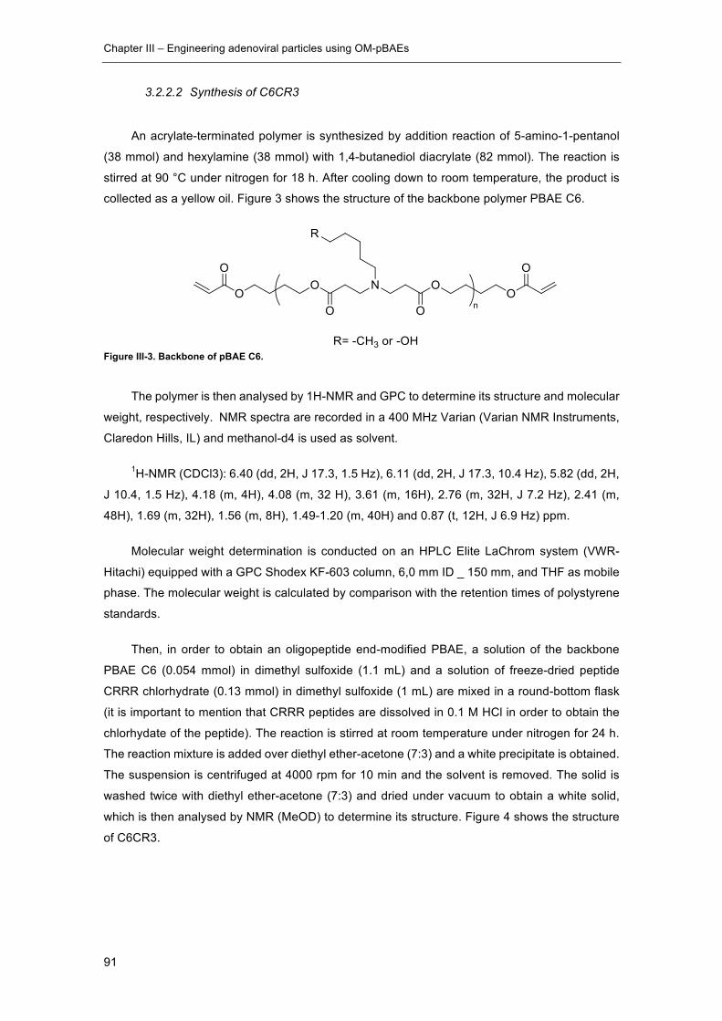
Chapter III – Engineering adenoviral particles using OM-pBAEs
91
3.2.2.2 Synthesis of C6CR3
An acrylate-terminated polymer is synthesized by addition reaction of 5-amino-1-pentanol
(38 mmol) and hexylamine (38 mmol) with 1,4-butanediol diacrylate (82 mmol). The reaction is
stirred at 90 °C under nitrogen for 18 h. After cooling down to room temperature, the product is
collected as a yellow oil. Figure 3 shows the structure of the backbone polymer PBAE C6.
Figure III-3. Backbone of pBAE C6.
The polymer is then analysed by 1H-NMR and GPC to determine its structure and molecular
weight, respectively.�NMR spectra are recorded in a 400 MHz Varian (Varian NMR Instruments,
Claredon Hills, IL) and methanol-d4 is used as solvent.
1H-NMR (CDCl3): 6.40 (dd, 2H, J 17.3, 1.5 Hz), 6.11 (dd, 2H, J 17.3, 10.4 Hz), 5.82 (dd, 2H,
J 10.4, 1.5 Hz), 4.18 (m, 4H), 4.08 (m, 32 H), 3.61 (m, 16H), 2.76 (m, 32H, J 7.2 Hz), 2.41 (m,
48H), 1.69 (m, 32H), 1.56 (m, 8H), 1.49-1.20 (m, 40H) and 0.87 (t, 12H, J 6.9 Hz) ppm.
Molecular weight determination is conducted on an HPLC Elite LaChrom system (VWR-
Hitachi) equipped with a GPC Shodex KF-603 column, 6,0 mm ID _ 150 mm, and THF as mobile
phase. The molecular weight is calculated by comparison with the retention times of polystyrene
standards.
Then, in order to obtain an oligopeptide end-modified PBAE, a solution of the backbone
PBAE C6 (0.054 mmol) in dimethyl sulfoxide (1.1 mL) and a solution of freeze-dried peptide
CRRR chlorhydrate (0.13 mmol) in dimethyl sulfoxide (1 mL) are mixed in a round-bottom flask
(it is important to mention that CRRR peptides are dissolved in 0.1 M HCl in order to obtain the
chlorhydate of the peptide). The reaction is stirred at room temperature under nitrogen for 24 h.
The reaction mixture is added over diethyl ether-acetone (7:3) and a white precipitate is obtained.
The suspension is centrifuged at 4000 rpm for 10 min and the solvent is removed. The solid is
washed twice with diethyl ether-acetone (7:3) and dried under vacuum to obtain a white solid,
which is then analysed by NMR (MeOD) to determine its structure. Figure 4 shows the structure
of C6CR3.

Chapter III – Engineering adenoviral particles using OM-pBAEs
92
Figure III-4. Structure of C6CR3 OM-pBAE.
3.2.2.3 Synthesis of C6PEGCR3
Step1: Synthesis of MeO-PEG-COOH
Trietylamine (1.25 mmol) is added to a solution of MeO-PEG (Mw = 2000, 2.5 mmol) and
succinic anhydride (2.75 mmol) in dichloromethane (5 mL). The reaction mixture is stirred at room
temperature for 4 h and washed with 1 M HCl (1 ml) twice. The organic phase is washed with
brine twice and dried over MgSO4. The solid is filtered off and the solvent is evaporated under
vacuum to obtain a white solid, whose structure is analysed by NMR (CDCl3).
Step 2: Synthesis of N-boc 5-amino-1-pentanol
A solution of di-tert-butyl dicarbonate (5.1 mmol) in dichloromethane (5 mL) is added to a
solution of 5-amino-1-pentanol (5.1 mmol) and triethylamine (5.6 mmol) in dichloromethane (16
mL). The mixture is stirred at room temperature for 1 h and then washed with 0.5 M HCl (1 ml)
three times. The organic phase is dried over MgSO4. The solid is filtered off and the solvent is
evaporated under vacuum to obtain a white solid, which is analyzed by NMR (CDCl3) to check its
structure.
Step 3: Synthesis of MeO-PEG-NHBoc
Dicyclohexylcarbodiimide (0.74 mmol) and N,N’-dimethylaminopyridine (0.074 mmol) are
added to a solution of MeO-PEG-COOH (0.49 mmol) in dichloromethane (14 mL). After 5 min, a
solution of N-boc 5-amino-1-pentanol (0.49 mmol) in dichloromethane (1 mL) is added to the
mixture. The reaction mixture is stirred at room temperature for 6 h and then the solid is filtered
off. The solvent is evaporated under vacuum and the residue is washed with diethyl ether (5 mL)
three times. The product is dried under vacuum to obtain a white solid, which is analysed by NMR
(CDCl3).
Step 4: Synthesis of MeO-PEG-NH2
Trifluoroacetic acid (1.2 mL) is added to a solution of MeO-PEG-NHBoc (0.21 mmol) in
dichloromethane (3 mL) at 0 °C. The reaction mixture is stirred at 0 °C for 10 min and then it is
stirred at room temperature for 2 h. The solvent is reduced under vacuum and the residue is

Chapter III – Engineering adenoviral particles using OM-pBAEs
93
washed with diethyl ether (5 mL) twice. The product is dissolved in dichloromethane (8 mL) and
washed with 0.5 M NaOH (1 ml) twice. The organic phase is washed with brine and dried over
MgSO4. The solid is filtered off and the solvent is evaporated under vacuum to obtain a white
solid, which is analysed by NMR (CDCl3).
Step 5: Synthesis of pBAE C6PEG
5-Amino-1-pentanol (0.41 mmol), hexylamine (0.41 mmol) and MeO-PEG-NH2 (0.14 mmol)
are mixed in dichloromethane (2 mL) and the solvent is reduced under vacuum. To the residue is
added 1,4-butanediol diacrylate (1 mmol) and the reaction mixture is stirred at 90 °C under
nitrogen for 18 h. After cooling down to room temperature, the product is collected as a yellow
solid, which is analysed by NMR (CDCl3). Figure 5 shows the structure of the intermediate product
pBAE C6PEG.
Figure III-5. Structure of the intermediate product pBAE C6-PEG.1H NMR (400 MHz, Chloroform-d) δ 6.39
(dd, J = 17.3, 1.4 Hz, 2H), 6.11 (dd, J = 17.3, 10.4 Hz, 2H), 5.82 (d, J = 10.4 Hz, 2H), 4.23 (m,
2H), 4.17 (m, 4H), 4.14-3.98 (m, 32H), 3.63 (m, 218H), 3.37 (s, 3H), 2.75 (m, 32 H), 2.63 (m, 4H),
2.43 (m, 48H), 1.8-1.63 (m, 36 H), 1.55 (m, 12H), 1.48-1.16 (m, 50H), 0.87 (t, J = 6.9 Hz, 12H).
Step 6: Synthesis of C6PEGCR3
To obtain the chlorhydrate of the peptide, 15 mL of 0.1 M HCl were added to peptide CRRR
(150 mg) and the solution was freeze-dried.
A solution of PBAE C6-PEG (0.022 mmol) in dimethyl sulfoxide (1.2 mL) is mixed in a round-
bottom flask with a solution of peptide CRRR chlorhydrate (0.054 mmol) in dimethyl sulfoxide (1.1
mL), (it is important to mention that CRRR peptides are dissolved in 0.1 M HCl in order to obtain
the chlorhydate of the peptide). The reaction is stirred at room temperature under nitrogen for 20
h and then the mixture is added over diethyl ether-acetone (7:3) to obtain a white precipitate. The
suspension is then centrifuged at 4000 rpm for 10 min and the solvent is removed. The solid is
washed twice with diethyl ether-acetone (7:3) and dried under vacuum to obtain a white solid,
which is analysed by NMR (MeOD) to check its structure. Figure 6 shows the structure of polymer
C6PEGCR3.
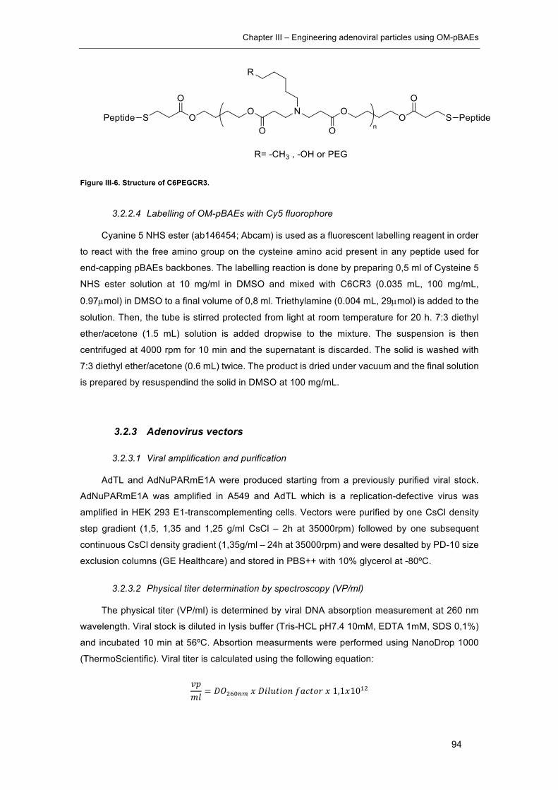
Chapter III – Engineering adenoviral particles using OM-pBAEs
94
Figure III-6. Structure of C6PEGCR3.
3.2.2.4 Labelling of OM-pBAEs with Cy5 fluorophore
Cyanine 5 NHS ester (ab146454; Abcam) is used as a fluorescent labelling reagent in order
to react with the free amino group on the cysteine amino acid present in any peptide used for
end-capping pBAEs backbones. The labelling reaction is done by preparing 0,5 ml of Cysteine 5
NHS ester solution at 10 mg/ml in DMSO and mixed with C6CR3 (0.035 mL, 100 mg/mL,
0.97µmol) in DMSO to a final volume of 0,8 ml. Triethylamine (0.004 mL, 29µmol) is added to the
solution. Then, the tube is stirred protected from light at room temperature for 20 h. 7:3 diethyl
ether/acetone (1.5 mL) solution is added dropwise to the mixture. The suspension is then
centrifuged at 4000 rpm for 10 min and the supernatant is discarded. The solid is washed with
7:3 diethyl ether/acetone (0.6 mL) twice. The product is dried under vacuum and the final solution
is prepared by resuspendind the solid in DMSO at 100 mg/mL.
3.2.3 Adenovirus vectors
3.2.3.1 Viral amplification and purification
AdTL and AdNuPARmE1A were produced starting from a previously purified viral stock.
AdNuPARmE1A was amplified in A549 and AdTL which is a replication-defective virus was
amplified in HEK 293 E1-transcomplementing cells. Vectors were purified by one CsCl density
step gradient (1,5, 1,35 and 1,25 g/ml CsCl – 2h at 35000rpm) followed by one subsequent
continuous CsCl density gradient (1,35g/ml – 24h at 35000rpm) and were desalted by PD-10 size
exclusion columns (GE Healthcare) and stored in PBS++ with 10% glycerol at -80ºC.
3.2.3.2 Physical titer determination by spectroscopy (VP/ml)
The physical titer (VP/ml) is determined by viral DNA absorption measurement at 260 nm
wavelength. Viral stock is diluted in lysis buffer (Tris-HCL pH7.4 10mM, EDTA 1mM, SDS 0,1%)
and incubated 10 min at 56ºC. Absortion measurments were performed using NanoDrop 1000
(ThermoScientific). Viral titer is calculated using the following equation:
W;@E
= TXYZ[\]LT=EB<=AC^?P<A_L1,1L10`Y
Chapter III – Engineering adenoviral particles using OM-pBAEs
95
3.2.3.3 Preparation of fluorescently-labeled Ad viral particles
In order to prepare the fluorescently-labelled AdNuPARE1A virus, DyLight 550 Microscale
Antibody labeling Kit (ThermoFischer) was used. The labelling reaction was carried out by
preparing 100 ul of AdNuPARE1A in PBS with 10 µl of borate buffer (0,67M) at 1,3x10¹² VP/ml.
The viral solution was then mixed with the labelling reagent provided as a powder in an Eppendorf
tube and incubated during 1 h at RT. Labelled virus was then washed 8 times using Microcon
Centrifugal Filters 10K (Merck) with PBS++ in order to remove the excess of free fluorophore.
Finally, labelled-virus sample was titrated in order to determine the recovery rate after labelling
reaction.
3.2.4 Coating of Ad with OM-pBAEs
3.2.4.1 Preparation of OM-pBAEs-coated Ad samples for in vitro testing
Coated viral samples for in vitro assays were prepared by diluting 1/50 the virus stock in
PBS (VS) (Generally, an aliquot of 2 ul in 98 ul of PBS). Then the volume of polymer stock needed
taking into account an specific pol:VP ratio was calculated following equation 1.
@97JaQ =b9ℎ<1000
∗ bQ<APeBE ∗ ;AE: b9
RBE97JaQ<APe =@97JaQ ∗ 3287,52 M
@AE ∗ 1:46,023:23
Equation 1. VPht refers to the physical titer in VP/ml and pol:VP refers to the ratio of polymer molecules per viral
particle.
The calculated volume of polymer stock is diluted with PBS to a final volume of 100 ul (PS).
Finally, PS and VS were mixed by adding PS over VS and pipetting up and down slowly at least
10 times. Samples were incubated 30’ at RT to enable the electrostatic interactions and freshly
used for testing assays.

Chapter III – Engineering adenoviral particles using OM-pBAEs
96
Figure III-7. Coating of Ads with OM-pBAEs for in vitro uses.
3.2.4.2 Preparation of pBAEs-coated Ad samples for in vivo testing
Coated Ad samples for in vivo testing were prepared in 1 ml batches. Taking into account
the dose of injection (For instance, 1x10¹⁰ VP/animal in 100 µl bolus) the desired final
concentration for a 1 ml of coated batch is 1x10¹¹ VP/ml, this means that 1x10¹¹ VPs will be
coated per batch. The total volume of pBAE stock solution needed to coat 1 ml batch is calculated
following equation 2.
@97JaQ = jA<?Eb9Q ∗ ;AE: b9
RBE97JaQ<APe =@97JaQ ∗ 3287,52 M
@AE ∗ 1L10k
6,023:23
Equation 2. mPBAEs means molecules of pBAEs. Total VPs means the total number of VPs that needs to be coated for
a 1 ml preparation. Pol:VP means the number of molecules of polymer used to coat a single VP. All in vivo studies are
carried out using a 4x10⁶ pol:VP.
Then, the polymer solution (Ps) is prepared by mixing 0,9% saline with the calculated volume
of pBAEs stock to a final volume of 200 ul. The virus stock volume needed to have the desired
dose in VPs/ml is calculated using the physical titter and virus stock solution is mixed with 0,9%
saline to a final volume of 200ul to prepare the virus solution (Vs). Ps and Vs are mixed by adding
Ps over Vs and pippeting up and down slowly at least 10 times. The sample is incubated 30 min
at RT to enable the electrostatic interaction between polymer and viral capsids. Then, 600 ul of
0,9% saline is added to reach the 1 ml batch volume and mixed slowly.

Chapter III – Engineering adenoviral particles using OM-pBAEs
97
Figure III-8. Coating of Ads with OM-pBAEs for in vivo uses.
3.2.5 Biophysical characterization of pBAE-coated Ad
3.2.5.1 DLS: Zeta potential and particle size measurement
DLS illuminates the sample by a laser beam and detects the fluctuations of the scattered
light at a known scattering angle θ by a fast photon detector. DLS studies the diffracted light of
particles as a whole and has limitations discerning polydispersity of samples containing different
populations because DLS do not track single particles. DLS offers an option called number mean
value to focus the size calculation to the more abundant specie in the sample, helping to avoid
interference of contamination or minor events.
Size and surface charge were determined by Dynamic Light Scaterring (DLS) (Malvern
Instruments Ltd, United Kingdom, 4-mW laser). Coated viral preparations were prepared as
previously described. 100 µl of coated Ad samples were diluted with 900 µl of PBS 1x for
hydrodynamic size and Z-potential analysis.
3.2.5.2 TRPS size characterization
TRPS is a technique that allows single particle characterization by monitoring current flow
through an aperture, combined with the use of tunable nanopore technology. Particles crossing
a pore are detected one at a time as a transient change in the ionic current flow. As blockade
magnitude is proportional to particle size, accurate particle sizing can be achieved after calibration
with a known standard. In the present study, the qNano technology (iZon) was used following
manufacturers protocols and guidance.

Chapter III – Engineering adenoviral particles using OM-pBAEs
98
3.2.5.3 NTA size characterization
NTA uses an ultramicroscope and laser illumination to visualize the movement of
nanoparticles under Brownian motion. The light scattered by the particles is captured using a
camera over multiple frame. Then, the software tracks the motion of each particle from frame to
frame. Particle movement rate is related to a sphere equivalent hydrodynamic radius as
calculated through the Stokes-Einstein equation. The technique calculates particle size on a
particle-by particle basis allowing to resolve different size populations within the same sample.
3.2.5.4 Transmission electron microscopy (TEM)
A 10 μL drop of freshly prepared coated Ad samples was put on a sheet of parafilm. A UV
light activated carbon coated 200 mesh copper grids was placed on top of the drop (filmed side
down) for 1 and washed 3 times by putting the grids over MQ water drops during 1 min each.
The, the grid was incubated with a drop of a 1% phosphotungstic acid solution (PTA) for 1,5 min
and drained with a piece of filter paper. Grids were Put inside a petri dish with Whatman paper
and leave it dry during 2 h at least. Grids were imaged using JEM 1400, JEM 2011 and JEOL
1010 electronic microscopes depending on availability.
3.2.5.5 Stochastic optical reconstruction microscopy (STORM)
The fundamental principle behind stochastic optical reconstruction microscopy and related
methodology is that the activated state of a photoswitchable molecule must lead to the
consecutive emission of sufficient photons to enable precise localization before it enters a dark
state or becomes deactivated by photobleaching. After capturing the images with a digital camera,
the point-spread functions of the individual molecules are localized with high precision based on
the photon output before the probes spontaneously photobleach or switch to a dark state.
In order to obtain STORM images of viral samples, they were physically adsorbed onto the
surface of a flow chamber assembled from a glass slide and a coverslip (24mm x 24 mm,
thickness 0.15 mm) separated by double-sided tape. Unbound particles were removed by
washing excess sample with PBS. Finally, STORM buffer was fluxed into the chamber before
imaging. Images were acquired using NIS-Elements software in Nikon Eclipse Ti microscope.
Cy5-labelled polymer was imaged with a 647nm laser (160 mW), taking 21.000 frames per image.
DyLight 550 labelled viruses were imaged with a 561nm laser (80mV), taking 20.000 frames per
image.
Fluorescence was collected by means of a Nikon 100x, 1.49 NA oil immersion objective and
passed through a quad-band pass dichroic filter (97335 Nikon). Images were acquired onto a
256x256 pixel region (pixel size 0.16µm) of a Hamamatsu 19 ORCA- Flash 4.0 camera at 10 ms
integration time. The image reconstruction was performed in the STORM module of the NIS
Chapter III – Engineering adenoviral particles using OM-pBAEs
99
element Nikon software. Intensity threshold for both channels was settled at 250 and trace length
values at minimum 1 and maximum 5.
3.2.6 Biological in vitro characterization
3.2.6.1 Cell culture
All cell lines used in this study are adherent and grow forming a monolayer. Cells were
cultured at 37ºC in 5% CO2 in DMEM (Dulbecco’s Modified Eagle Medium) supplemented with
10% fetal bovine serum, 100 U/ml penicilin, 100 ug/ml streptomycin and 2mM L-Glutamine.
PANC-1 is a human cell line derived from a pancreatic ductal adenocarcinoma and was
purchased from ATCC. A549 is a human cell line derived from a lung carcinoma. This cell line
was kindly provided by Dr. Ramon Alemany (ICO-IDIBELL, Barcelona). It is used to amplify
recombinant virus due to its high viral production efficiency. HEK293 is a human cell line derived
from kidney embryonic tissue. It is a E1A transcomplementing cell line used to produce non-
replicative recombinant adenoviruses. This cell line was also provided by Dr. Alemany.
3.2.6.2 Cell viability assay (MTT)
The influence on cell metabolism of DMSO, C32CR3 and C6CR3 polymers was evaluated
using a MTT colorimetric assay. 10 μL of MTT [3-(4,5-dimethylthiazol-2- yl)-2,5-
diphenyltetrazolium bromide, 5mg/mL] were added to the cells, 48 h after transduction and
incubated for 2 h at 37 °C. The purple formazan crystals were dissolved in 100 μL/well dimethyl
sulfoxide (DMSO). Absorbance was measured at 590 nm with background correction at 630 nm
using a microplate reader. The relative cell viability (in %) was calculated by [A] sample /[A] control
x 100.
3.2.6.3 Transduction studies
Transduction studies were done using the AdTL replication-defective virus expressing GFP
and Luciferase under CMV promoter as early genes. AdTL was tested naked and coated with
different coating formulations. Viral samples were diluted with DMEM supplemented with 10%
FBS in order to reach the desired experimental concentration. For NAbs pre-incubated conditions
the culture media was also supplemented with Aanti-Ad5 polyclonal antibody (ab6982; Abcam)
at 10.000-fold dilution for MOI 10 experiments and 100.000-fold dilution for MOI 50 infections.
Samples were incubated at RT during 30’ before infection. Then, 15000 PANC-1 cells/well in a
96 multi-well plate were infected at MOI 10 and 50. 4 hours post-infection, wells were washed
and fresh culture medium was added into each well. 48h after infection, cells were tripsinized and
analyzed using Attune NxT Flow cytometer in order to determine the % of GFP+ cells.
Chapter III – Engineering adenoviral particles using OM-pBAEs
100
3.2.7 Biological in vivo characterization
Animal procedures met the guidelines of European Community Directive 86/609/EEC and
were approved by the Comité de Experimentación Animal (CEEA) of the UB (Universitat de
Barcelona).
3.2.7.1 Animals
Animals were housed in plastic cages in controlled environmental conditions of humidity
(60%), temperature (22ºC ± 2ºC) and light with food and water ad libitum. C57Bl/6J and BALB/c
Nu/Nu mice were used for the experimentation of this chapter. C57Bl/6J were used for
biodistribution and neutralizing serums production studies. BALB/c Athymic Nu/Nu, as an
immunodeficient strain, allow the progression of human tumors and were used to produce tumor-
bearing mice.
3.2.7.2 Generation of tumor-bearing mice by subcutaneous implantation
BALB/c Nu/Nu mice were injected subcutaneously in each flank using a 29G needle. 1x10⁶
PANC-1 cells per tumor were injected. Cells were prepared in Matrigel Matrix Basement
Membrane HC (Corning) by 1:1 mixing cells in DMEM without antibiotics and supplements and
Matrigel to a final volume of 100 µl.
Tumor progression was analyzed by measuring tumor’s volume using a digital caliper.
Biodistribution studies were started when tumors reached 100 mm3. The large and the small
diameter are measured and the tumor volume calculated using the following formula:
jB@AB_WAEB@: = (T ∗ >Y ∗ p)
6
3.2.7.3 Blood circulation time
Seven-week-old C57BL/6J male mice were injected intravenously with naked AdTL and
AdTL coated with different OM-pBAE formulations with a total dose of 1×10¹⁰ VP/Animal. At 2
min and 10 min post-administration, blood samples were collected from the saphenous vein using
EDTA treated capillaries. Blood DNA Isolation Mini Kit (Product # 46300, 46380) NORGEN
BIOTEK CORP.was used to extract DNA from 50 µl whole blood samples following the
manufacturer protocol. A RT-qPCR of the samples was performed using hexon specific primers
Hexo01= 5’-GCCGCAGTGGTCTTACATGCACATC-3 and Hexo02= 5-
CAGCACGCCGCGGATGTCAAAG-3.
Chapter III – Engineering adenoviral particles using OM-pBAEs
101
3.2.7.4 Bioluminiscence in vivo imaging
In vivo luciferase expression was visualized and quantified in living animals using an in vivo
bioluminescent imaging system (Hamamatsu Photonics). Briefly, the substrate firefly D-Luciferin
(PerkinElmer, Inc) was i.p. administered (16 mg/kg) and 10 min later animals were anesthetized
with a mixture of isofluorane and oxygen preparation. Mice were introduced into the capturing
cage coupled to an inhaled anesthesia system, and images were captured and analyzed using
Wasabi software (Hamamatsu Photonics).
3.2.7.5 Luciferase assay of organs and tumors
Different organs and tumors were mechanically homogenized in a cold potter with liquid
nitrogen to obtain a fine powder. Powder was mixed with lysis buffer (Cell culture Lysis reagent,
Promega) and incubated 15 min at 25ºC. Samples were centrifuged for 10 min, 16000g at 4ºC
and supernatants were collected. Luciferase activity was quantified using the Luciferase Assay
System Kit (Promega). 10 µl of samples was mixed with 30 µl of luciferin in a white 96-well plate
and photon emission was measured in a Synergy HT luminometer (BioTek). Light emission was
normalized to total protein levels. Protein concentration was determined with BCA protein assay
(Pierce Biotechnology).
3.2.7.6 In vitro neutralization assay of serums produced in vivo
C57BL/6J naïve mice were immunized by injecting two 1x10¹⁰ VP/animal doses of naked
Ad, CPEGAd and C6CR3Ad formulations intravenously at day 0 and day 14. At day 21 after the
first injection, blood was collected by intracardiac puncture and serums were obtained and heat
inactivated.
5x10⁵ PFU/ml solution of AdTL was prepared and 50 µl of this solution was added to 96-well
white plate wells. Then, the serum sample was 10-fold diluted with PBS and 50 µl were added to
wells of the first column already containing the virus solution by triplicate. After that, samples were
serial diluted by transferring 50 µl to the consecutive columns leaving a column without serum
and another without virus nor serum as positive and negative controls. After 30 min at RT, 100.000
HEK293 cells in 100 µl were added to each well and were maintained at 37ºC and 5%CO2 for
24h. Finally, luciferase activity was quantified by changing the culture media for a 1,58 mg/ml D-
Luciferin solution in PBS and by measuring bioluminescence emission using a multiplate reader.
The ND50 values were calculated by determining the dilution which 50% neutralized the signal
from the positive transduction control without serum.
Chapter III – Engineering adenoviral particles using OM-pBAEs
102
3.2.8 Molecular modelling Hexon trimer
Protein data bank (PDB) structure of the adenovirus type 5 hexon protein (PDB code: 1P30)
was used and symmetrically assembled in order to generate the trimeric structure. The PDB file
was converted into a PQR file using PDB2PQR software which include protonation, missing
heavy (non-hydrogen) atoms and charge parameters into the model at pH 7.4. Finally, continuum
electrostatics equations were solved using APBS software and graphically represented using
VMD software.
3.2.9 Statistical analysis
Statistical analyses were carried out with Graph-Pad Prism (GraphPad Software). All error
bars reported are SD unless otherwise indicated. Pairwise comparisons were performed using
one-way Student t-tests and Turkey’s multiple comparison test. Differences between groups were
considered significant at P values below 0.05 (* p < 0.05, ** p < 0.01, *** p < 0.001, ****p < 0.0001).
Chapter III – Engineering adenoviral particles using OM-pBAEs
103
3.3 Results and discussion
Efficient intravenously delivery of oncolytic adenoviruses to tumors in the human body is the
main historical hurdle limiting the use of adenoviruses as anti-cancer agents. As previously
introduced in chapter I, pre-existing humoral immunity and liver sequestration drastically affects
the efficacy of viral agents used in virotherapy, especially upon systemic administration. In the
case of adenovirus, pre-existing humoral immunity is even more difficult to circumvent in
comparison with other viruses because of the high seroprevalence of anti-Ad5 neutralizing
antibodies (Nabs).
In chapter II, we observed the ability of OM-pBAEs to electrostatically coat AAVs and modify
their natural tropism. However, limitations regarding the quality of the physicochemical
characterization of such small particles (10-25 nm) hindered the optimization of the technology
and the associated quality controls.
In this chapter, Adenovirus type 5 have been used as a bioactive agent to be coated. Despite
the negative net charge of adenoviral particles in physiological conditions, Ad viral capsid surface
charge is complexly structured. In order to closely look and understand this complex pattern, an
in silico electrostatics study of the major component of Ad capsid, the hexon protein trimer, has
been done by a molecular modelling approach (Figure III-9). Briefly, the protein databank (PDB)
structure of the adenovirus type 5 hexon protein (PDB code: 1P30) was used and symmetrically
assembled in order to generate the trimeric structure. The PDB file was converted into a PQR file
using PDB2PQR software which include protonation, missing heavy (non-hydrogen) atoms and
charge parameters into the model at pH 7.4. Finally, continuum electrostatics equations were
solved using APBS software and graphically represented using VMD software.
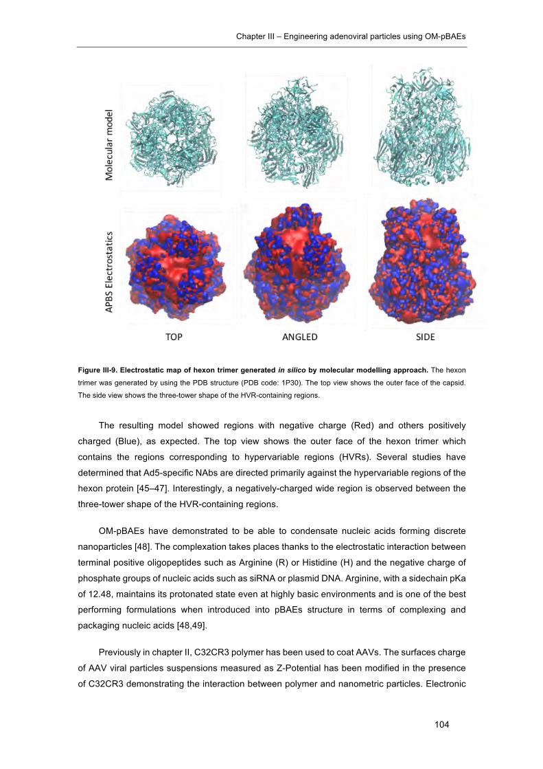
Chapter III – Engineering adenoviral particles using OM-pBAEs
104
Figure III-9. Electrostatic map of hexon trimer generated in silico by molecular modelling approach. The hexon
trimer was generated by using the PDB structure (PDB code: 1P30). The top view shows the outer face of the capsid.
The side view shows the three-tower shape of the HVR-containing regions.
The resulting model showed regions with negative charge (Red) and others positively
charged (Blue), as expected. The top view shows the outer face of the hexon trimer which
contains the regions corresponding to hypervariable regions (HVRs). Several studies have
determined that Ad5-specific NAbs are directed primarily against the hypervariable regions of the
hexon protein [45–47]. Interestingly, a negatively-charged wide region is observed between the
three-tower shape of the HVR-containing regions.
OM-pBAEs have demonstrated to be able to condensate nucleic acids forming discrete
nanoparticles [48]. The complexation takes places thanks to the electrostatic interaction between
terminal positive oligopeptides such as Arginine (R) or Histidine (H) and the negative charge of
phosphate groups of nucleic acids such as siRNA or plasmid DNA. Arginine, with a sidechain pKa
of 12.48, maintains its protonated state even at highly basic environments and is one of the best
performing formulations when introduced into pBAEs structure in terms of complexing and
packaging nucleic acids [48,49].
Previously in chapter II, C32CR3 polymer has been used to coat AAVs. The surfaces charge
of AAV viral particles suspensions measured as Z-Potential has been modified in the presence
of C32CR3 demonstrating the interaction between polymer and nanometric particles. Electronic
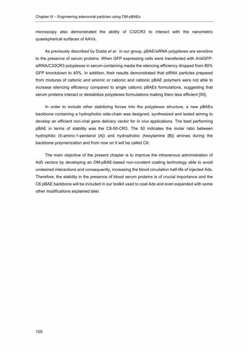
Chapter III – Engineering adenoviral particles using OM-pBAEs
105
microscopy also demonstrated the ability of C32CR3 to interact with the nanometric
quasispherical surfaces of AAVs.
As previously described by Dosta et al. in our group, pBAE/siRNA polyplexes are sensitive
to the presence of serum proteins. When GFP expressing cells were transfected with AntiGFP-
siRNA/C32CR3 polyplexes in serum-containing media the silencing efficiency dropped from 80%
GFP knockdown to 40%. In addition, their results demonstrated that siRNA particles prepared
from mixtures of cationic and anionic or cationic and cationic pBAE polymers were not able to
increase silencing efficiency compared to single cationic pBAEs formulations, suggesting that
serum proteins interact or destabilize polyplexes formulations making them less efficient [50].
In order to include other stabilizing forces into the polyplexes structure, a new pBAEs
backbone containing a hydrophobic side-chain was designed, synthesized and tested aiming to
develop an efficient non-viral gene delivery vector for in vivo applications. The best performing
pBAE in terms of stability was the C6-50-CR3. The 50 indicates the molar ratio between
hydrophilic (5-amino-1-pentanol [A]) and hydrophobic (hexylamine [B]) amines during the
backbone polymerization and from now on it will be called C6.
The main objective of the present chapter is to improve the intravenous administration of
Ad5 vectors by developing an OM-pBAE-based non-covalent coating technology able to avoid
undesired interactions and consequently, increasing the blood circulation half-life of injected Ads.
Therefore, the stability in the presence of blood serum proteins is of crucial importance and the
C6 pBAE backbone will be included in our toolkit used to coat Ads and even expanded with some
other modifications explained later.
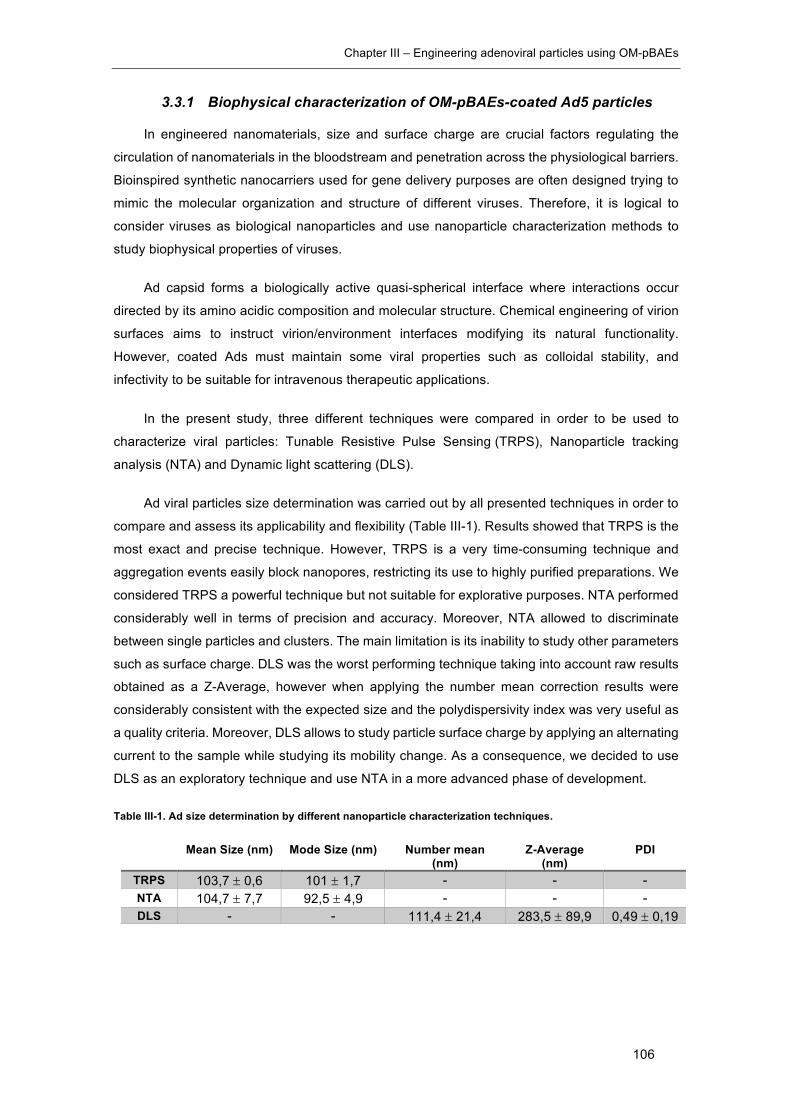
Chapter III – Engineering adenoviral particles using OM-pBAEs
106
3.3.1 Biophysical characterization of OM-pBAEs-coated Ad5 particles
In engineered nanomaterials, size and surface charge are crucial factors regulating the
circulation of nanomaterials in the bloodstream and penetration across the physiological barriers.
Bioinspired synthetic nanocarriers used for gene delivery purposes are often designed trying to
mimic the molecular organization and structure of different viruses. Therefore, it is logical to
consider viruses as biological nanoparticles and use nanoparticle characterization methods to
study biophysical properties of viruses.
Ad capsid forms a biologically active quasi-spherical interface where interactions occur
directed by its amino acidic composition and molecular structure. Chemical engineering of virion
surfaces aims to instruct virion/environment interfaces modifying its natural functionality.
However, coated Ads must maintain some viral properties such as colloidal stability, and
infectivity to be suitable for intravenous therapeutic applications.
In the present study, three different techniques were compared in order to be used to
characterize viral particles: Tunable Resistive Pulse Sensing (TRPS), Nanoparticle tracking
analysis (NTA) and Dynamic light scattering (DLS).
Ad viral particles size determination was carried out by all presented techniques in order to
compare and assess its applicability and flexibility (Table III-1). Results showed that TRPS is the
most exact and precise technique. However, TRPS is a very time-consuming technique and
aggregation events easily block nanopores, restricting its use to highly purified preparations. We
considered TRPS a powerful technique but not suitable for explorative purposes. NTA performed
considerably well in terms of precision and accuracy. Moreover, NTA allowed to discriminate
between single particles and clusters. The main limitation is its inability to study other parameters
such as surface charge. DLS was the worst performing technique taking into account raw results
obtained as a Z-Average, however when applying the number mean correction results were
considerably consistent with the expected size and the polydispersivity index was very useful as
a quality criteria. Moreover, DLS allows to study particle surface charge by applying an alternating
current to the sample while studying its mobility change. As a consequence, we decided to use
DLS as an exploratory technique and use NTA in a more advanced phase of development.
Table III-1. Ad size determination by different nanoparticle characterization techniques.
Mean Size (nm) Mode Size (nm) Number mean (nm)
Z-Average (nm)
PDI
TRPS 103,7 ± 0,6 101 ± 1,7 - - - NTA 104,7 ± 7,7 92,5 ± 4,9 - - - DLS - - 111,4 ± 21,4 283,5 ± 89,9 0,49 ± 0,19

Chapter III – Engineering adenoviral particles using OM-pBAEs
107
3.3.1.1 C32CR3 and C6CR3 interacts with Ad particles changing their physicochemical
properties
In order to study the coating formation on viral particles surfaces, Z-potential analysis was
carried out by DLS. Samples coated with increasing amounts of C32CR3 and C6CR3 polymer
molecules per VP (pol/VP ratio) were analyzed. Similar results were obtained with both polymers
(Figure III-10) in terms of Z-potential. This parameter increased until reaching 1x10⁶ molecules
of pol/VP; then a plateau started for all the remaining conditions. This indicated that all available
viral capsids surface area in the samples were positivized due to the polymer interaction and
allowed us to determine the minimum pol/VP ratio needed to fully coat Ad particles.
Figure III-10. Z-potential determination of viral particles coated with increasing pol/VP ratios of C32CR3 and C6CR3. Results are shown as mean and standard deviation of triplicates.
Taking into account the ionic nature of polymer and viral capsids interaction, an excess of
polymer in the solution may help to maintain complexes stable in solution. Therefore, a ratio of
4x10⁶ pol/VP was chosen as a coating conditions and complexes sizes were then studied.
Size characterization by DLS indicated that both polymers affected the size distribution of
viral particles in solution (Table III-2). The average hydrodynamic diameter of the complexes
formed with C32CR3 was 700,9 nm with a polydispersity (PDI) of 1. In the case of C6CR3, size
increased up to 374,1 nm with the same PDI. PDI explains the degree of uniformity of a
distribution. In DLS, the native distribution is the intensity distribution which indicates how much
light is scattered from various size bins or intervals. PDI increases from 0 to 1 in correlation with
the distribution of scattered light of different sizes. The resulting PDI values obtained for both
coated samples showed a negative effect over sample aggregation and size distribution. The
presence of clustered particles could be the main reason negatively affecting PDI values of coated
samples. Our quality threshold in terms of PDI was the value obtained for naked viral particles,
in this case 0,6. Hence, further improvement was needed to meet this criterion.
1×10
6
2×10
6
3×10
6
4×10
6
5×10
6
-20
-10
0
10
20
Pol:VP
Z-Po
tent
ial
(mV) C6CR3Ad
C32CR3Ad
Chapter III – Engineering adenoviral particles using OM-pBAEs
108
Table III-2. Size determination by DLS of Naked Ad, C32CR3 and C6CR3-coated Ad
Interestingly, C6CR3 polymer produced smaller complexes in comparison with C32CR3.
Although PDI values were very high in both cases, the hydrophobic backbone of C6CR3 could
modify the polymer-virus interaction improving its behaviour in terms of complexes size
distribution.
3.3.1.2 Transmission electron microscopy (TEM) imaging: comparison of C32CR3 and
C6CR3 coated Ad
In order to further study the morphology and size distribution of the resulting complexes,
TEM imaging was performed. TEM is capable of imaging at a significant higher resolution than
light microscopes and has been widely used in virology to solve viral capsids structures. The
smaller wavelength of electron beams allows to capture fine details of viral capsids and is suitable
to analyse capsid surface modifications due to the presence of polymeric coatings.
The structure of naked Ad, C32CR3 and C6CR3-coated Ad samples were analyzed by TEM
using phosphotungstic acid (PTA) as a contrasting agent (Figure III-11). Naked Ad samples
showed the typical adenoviral capsid structure with its icosahedral shape (Figure III-11-A). In
contrast, when coated with C32CR3, capsids were covered with a thin layer of polymer and
clustered viral particles were observed (Figure III-11-B). In the case of C6CR3, more contrasted
complexes were observed complicating the identification of viral particles. Their appearance
suggested a tighter interaction maybe due to the exclusion of water molecules over the
viral/polymer interface (Figure III-11-C). It is important to note that when preparing samples for
TEM imaging, samples are washed, stained and dried. The drying process can favour the
formation of artefacts, especially in the case of samples containing ionically interacting polymers.
Number mean PDI Z-Pot
Naked Ad 108,5 ± 15,3 0,6 ± 0,16 -11,75 ± 2,5 C32CR3Ad 700,9 ± 89 1 13,9 ± 4,4 C6CR3Ad 374,1 ± 38,6 1 15,3 ± 0,35

Chapter III – Engineering adenoviral particles using OM-pBAEs
109
Figure III-11. Comparison of C32CR3 and C6CR3 coatings by transmission electron microscopy. TEM micrographs
of PTA stained naked Ad (A), C32CR3-coated Ad (B) and C6CR3-coated Ad (C).
The main observation was the ability of both polymers to interact with capsid surfaces and
to form complexes. The presence of large aggregates in both cases was consistent with the DLS
characterization, highlighting the need to further improve the polymeric formulation in order to
produce single coated viral samples.
3.3.1.3 Transmission electron microscopy (TEM) imaging: The multilayering coating
approach
As described before by other authors, a layer-by-layer deposition of cationic and anionic
polymers produces thicker and heavier artificial envelopes on viral particles and has
demonstrated to improve their infectivity and efficacy in the presence of neutralizing antibodies in
comparison to single layer coated and naked Ads [51].
Taking advantage of the easy end-capping modification of OM-pBAEs with differently
charged peptides, anionic and cationic polymers were produced by modifying C32-backboned
pBAEs with arginine (CR3) and asparatic acid (CD3) tripeptides. These polymers were used as
a multi-layering Ad coating agent and were analysed by TEM.
Ad samples were coated with a layer-by-layer approach using C32CR3 and C32CD3. Five
layers of 4x10⁶ pol/VP were consecutively deposited by waiting 30 min between layers. The order
of the layers deposition was Ad-R-D-R-D-R. Also, another sample was prepared by using the
same total amount of polymer as used for the layer-by-layer approach but anionic and cationic
polymers were previously mixed and deposited as a single layer. Finally, a single layer C32CR3
sample was also analyzed as a control.
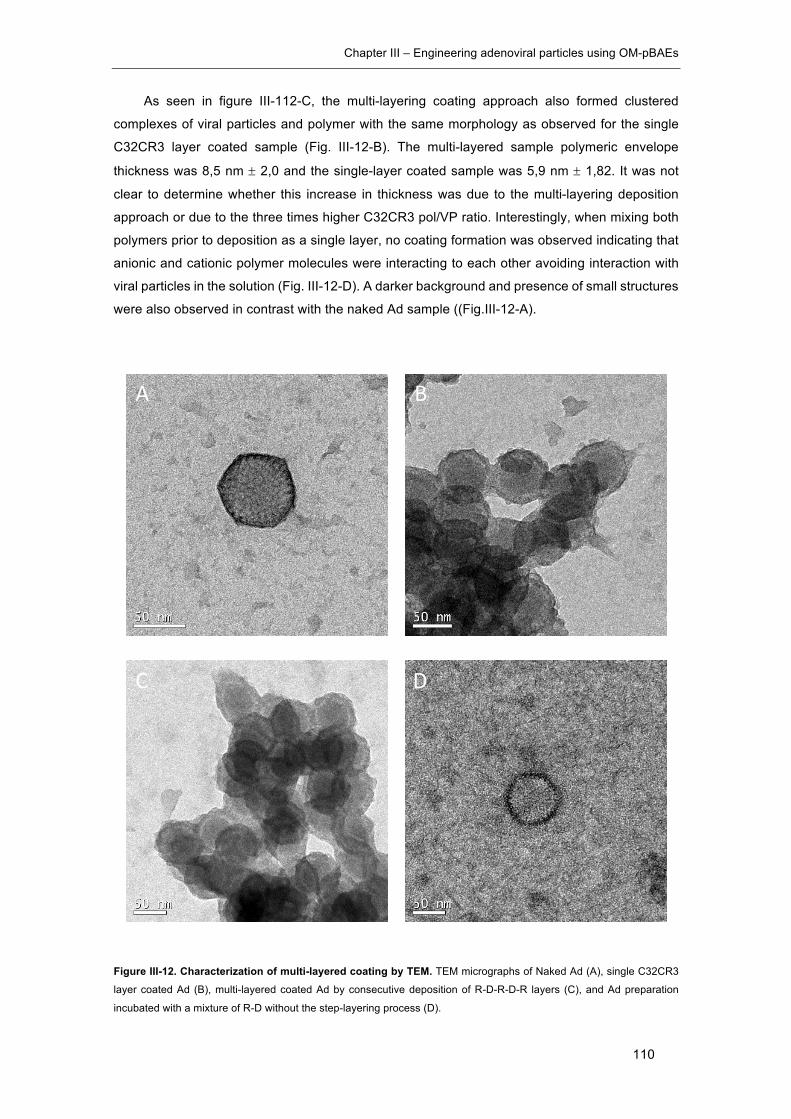
Chapter III – Engineering adenoviral particles using OM-pBAEs
110
As seen in figure III-112-C, the multi-layering coating approach also formed clustered
complexes of viral particles and polymer with the same morphology as observed for the single
C32CR3 layer coated sample (Fig. III-12-B). The multi-layered sample polymeric envelope
thickness was 8,5 nm ± 2,0 and the single-layer coated sample was 5,9 nm ± 1,82. It was not
clear to determine whether this increase in thickness was due to the multi-layering deposition
approach or due to the three times higher C32CR3 pol/VP ratio. Interestingly, when mixing both
polymers prior to deposition as a single layer, no coating formation was observed indicating that
anionic and cationic polymer molecules were interacting to each other avoiding interaction with
viral particles in the solution (Fig. III-12-D). A darker background and presence of small structures
were also observed in contrast with the naked Ad sample ((Fig.III-12-A).
Figure III-12. Characterization of multi-layered coating by TEM. TEM micrographs of Naked Ad (A), single C32CR3
layer coated Ad (B), multi-layered coated Ad by consecutive deposition of R-D-R-D-R layers (C), and Ad preparation
incubated with a mixture of R-D without the step-layering process (D).
A B
C D
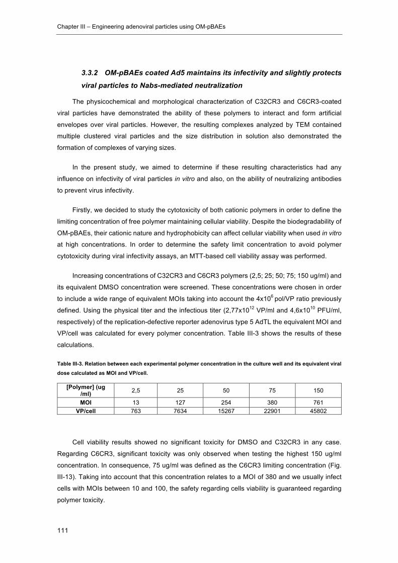
Chapter III – Engineering adenoviral particles using OM-pBAEs
111
3.3.2 OM-pBAEs coated Ad5 maintains its infectivity and slightly protects
viral particles to Nabs-mediated neutralization
The physicochemical and morphological characterization of C32CR3 and C6CR3-coated
viral particles have demonstrated the ability of these polymers to interact and form artificial
envelopes over viral particles. However, the resulting complexes analyzed by TEM contained
multiple clustered viral particles and the size distribution in solution also demonstrated the
formation of complexes of varying sizes.
In the present study, we aimed to determine if these resulting characteristics had any
influence on infectivity of viral particles in vitro and also, on the ability of neutralizing antibodies
to prevent virus infectivity.
Firstly, we decided to study the cytotoxicity of both cationic polymers in order to define the
limiting concentration of free polymer maintaining cellular viability. Despite the biodegradability of
OM-pBAEs, their cationic nature and hydrophobicity can affect cellular viability when used in vitro
at high concentrations. In order to determine the safety limit concentration to avoid polymer
cytotoxicity during viral infectivity assays, an MTT-based cell viability assay was performed.
Increasing concentrations of C32CR3 and C6CR3 polymers (2,5; 25; 50; 75; 150 ug/ml) and
its equivalent DMSO concentration were screened. These concentrations were chosen in order
to include a wide range of equivalent MOIs taking into account the 4x10⁶ pol/VP ratio previously
defined. Using the physical titer and the infectious titer (2,77x10¹² VP/ml and 4,6x10¹⁰ PFU/ml,
respectively) of the replication-defective reporter adenovirus type 5 AdTL the equivalent MOI and
VP/cell was calculated for every polymer concentration. Table III-3 shows the results of these
calculations.
Table III-3. Relation between each experimental polymer concentration in the culture well and its equivalent viral dose calculated as MOI and VP/cell.
[Polymer] (ug /ml) 2,5 25 50 75 150
MOI 13 127 254 380 761 VP/cell 763 7634 15267 22901 45802
Cell viability results showed no significant toxicity for DMSO and C32CR3 in any case.
Regarding C6CR3, significant toxicity was only observed when testing the highest 150 ug/ml
concentration. In consequence, 75 ug/ml was defined as the C6CR3 limiting concentration (Fig.
III-13). Taking into account that this concentration relates to a MOI of 380 and we usually infect
cells with MOIs between 10 and 100, the safety regarding cells viability is guaranteed regarding
polymer toxicity.

Chapter III – Engineering adenoviral particles using OM-pBAEs
112
Figure III-13. Cytotoxicity of different polymers assessed in PANC-1 cells using a colorimetric MTT-based cell viability assay. 15000 cells/well were incubated for 4 h in serum-free medium in the presence of polymer concentrations
ranging from 2,5 to 150 ug/ml (2,5, 25, 50, 75, 150 ug/ml). The medium was replaced with fresh medium without polymer,
and the cultures were incubated another 24h.*p < 0.05, **p < 0.01, ***p < 0.001
After this toxicity assessment, the effect of pBAEs coating on viral infection in the presence
or absence of NAbs was studied following GFP expression using flow cytometry. AdTL expresses
the GFP and Luciferase genes under the CMV promoter, and viral transduction can be easily
studied by counting GFP+ cells.
PANC-1 cells were infected at MOI 10 with C32CR3 and C6CR3-coated AdTL with
increasing pol/VP ratios. These conditions were replicated by incubating samples 30 min at RT
with 10000-fold dilution of the commercial ab6982 Anti-Ad5 polyclonal antibody (Abcam). The
percentage of GFP positive cells for each condition was quantified 3 days after infection by flow
cytometry.
A seen in figure III-14, a slight improvement on the infectivity of C32CR3Ad was observed
from 2x10⁶ to 1x10⁷ pol/VP ratios. However, this effect was not observed when samples were pre-
incubated with Nabs. In contrast, C6CR3-coated condition performed better when samples were
pre-incubated with Nabs, especially when pol/VP ratio was higher than 1x10⁶.
DMSO C32CR3 C6CR3
0
50
100
% V
iabi
lity
**

Chapter III – Engineering adenoviral particles using OM-pBAEs
113
Figure III-14. Infectivity of C32CR3 and C6CR3-coated AdTL at different pol/VP ratios in the presence or absence of NAbs. GFP positive PANC-1 cells were quantified by flow cytometry. Cells were infected with C32CR3 and C6CR3-
coated AdTL at increasing pol/VP ratios with (+) and without pre-incubation with NAbs at MOI 10. Their fluorescence was
determined after 48 h by flow cytometry. Results are shown as mean and standard deviation of triplicates.
The presence of polycationic polymers in the culture media can improve virus interaction
with cells by modifying the negative charge of cell membranes [52]. In order to determine if the
improvement of the viral transduction was dependent to an interaction between polymer
molecules and viral particles or between polymer molecules and cells, the components were
separated by incubating cells with polymer prior to infection with naked viral particles and were
compared with normally coated viral preparations. The multilayer coating strategy was also tested
with a five-layered sample prepared as explained before.
As seen in figure III-15-A, infectivity of C32CR3Ad and C6CR3Ad was only improved when
viral particles were pre-coated before infection. In contrast, same percentages of GFP + cells
were obtained for multilayer coated Ad (MC Ad) and for cells incubated with the multilayer
polymeric component before infection with naked Ad indicating that the effect was independent
on polymer interaction with viral particles.
Similar results were obtained when infecting in the presence of Nabs (Figure III-15-B).
However, cells pre-incubated with the polymeric component of both single layer conditions prior
to viral infection showed a reduced infectivity in the presence of NAbs. A small improvement on
the infectivity of C6CR3Ad was observed in presence of Nabs as previously observed in figure
III-14.
Taking into account infectivity results regarding the multi-coating strategy, TEM
characterization and the fact that a multi-layered coating adds complexity on technology
development and regulation, this strategy was abandoned.
105 106 10702468
203040506070
pol:VP
%G
PF+
cells
C32CR3AdC32CR3Ad +C6CR3AdC6CR3Ad +
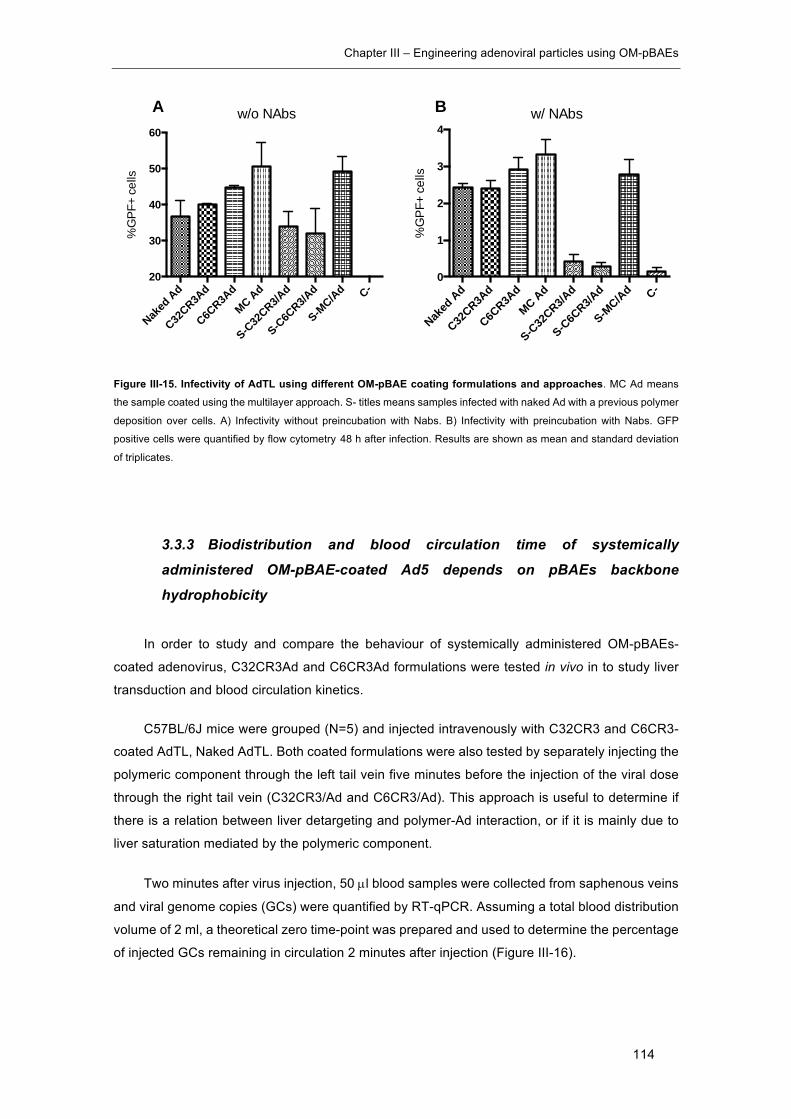
Chapter III – Engineering adenoviral particles using OM-pBAEs
114
Figure III-15. Infectivity of AdTL using different OM-pBAE coating formulations and approaches. MC Ad means
the sample coated using the multilayer approach. S- titles means samples infected with naked Ad with a previous polymer
deposition over cells. A) Infectivity without preincubation with Nabs. B) Infectivity with preincubation with Nabs. GFP
positive cells were quantified by flow cytometry 48 h after infection. Results are shown as mean and standard deviation
of triplicates.
3.3.3 Biodistribution and blood circulation time of systemically
administered OM-pBAE-coated Ad5 depends on pBAEs backbone
hydrophobicity
In order to study and compare the behaviour of systemically administered OM-pBAEs-
coated adenovirus, C32CR3Ad and C6CR3Ad formulations were tested in vivo in to study liver
transduction and blood circulation kinetics.
C57BL/6J mice were grouped (N=5) and injected intravenously with C32CR3 and C6CR3-
coated AdTL, Naked AdTL. Both coated formulations were also tested by separately injecting the
polymeric component through the left tail vein five minutes before the injection of the viral dose
through the right tail vein (C32CR3/Ad and C6CR3/Ad). This approach is useful to determine if
there is a relation between liver detargeting and polymer-Ad interaction, or if it is mainly due to
liver saturation mediated by the polymeric component.
Two minutes after virus injection, 50 µl blood samples were collected from saphenous veins
and viral genome copies (GCs) were quantified by RT-qPCR. Assuming a total blood distribution
volume of 2 ml, a theoretical zero time-point was prepared and used to determine the percentage
of injected GCs remaining in circulation 2 minutes after injection (Figure III-16).
Naked
Ad
C32CR3A
d
C6CR3A
d
MC Ad
S-C32
CR3/Ad
S-C6C
R3/Ad
S-MC/Ad C-
20
30
40
50
60%
GPF
+ ce
llsw/o NAbs
Naked
Ad
C32CR3A
d
C6CR3A
d
MC Ad
S-C32
CR3/Ad
S-C6C
R3/Ad
S-MC/Ad C-
0
1
2
3
4
%G
PF+
cells
w/ NAbsA B
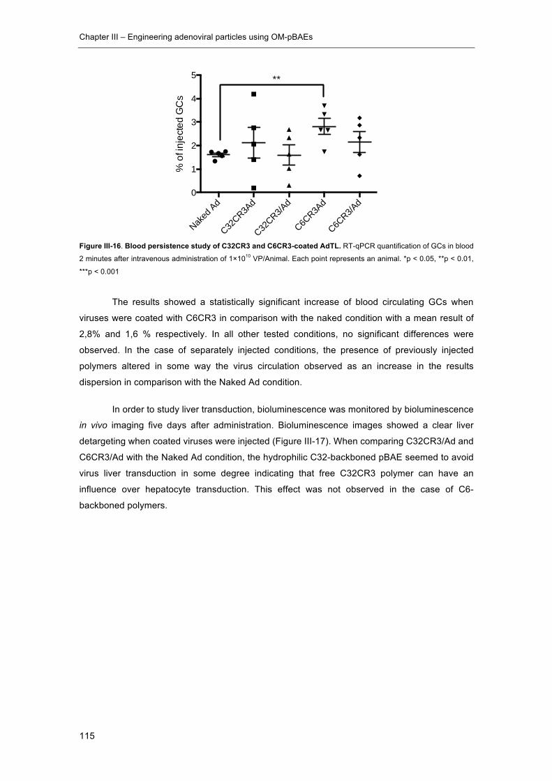
Chapter III – Engineering adenoviral particles using OM-pBAEs
115
Figure III-16. Blood persistence study of C32CR3 and C6CR3-coated AdTL. RT-qPCR quantification of GCs in blood
2 minutes after intravenous administration of 1×10¹⁰ VP/Animal. Each point represents an animal. *p < 0.05, **p < 0.01,
***p < 0.001
The results showed a statistically significant increase of blood circulating GCs when
viruses were coated with C6CR3 in comparison with the naked condition with a mean result of
2,8% and 1,6 % respectively. In all other tested conditions, no significant differences were
observed. In the case of separately injected conditions, the presence of previously injected
polymers altered in some way the virus circulation observed as an increase in the results
dispersion in comparison with the Naked Ad condition.
In order to study liver transduction, bioluminescence was monitored by bioluminescence
in vivo imaging five days after administration. Bioluminescence images showed a clear liver
detargeting when coated viruses were injected (Figure III-17). When comparing C32CR3/Ad and
C6CR3/Ad with the Naked Ad condition, the hydrophilic C32-backboned pBAE seemed to avoid
virus liver transduction in some degree indicating that free C32CR3 polymer can have an
influence over hepatocyte transduction. This effect was not observed in the case of C6-
backboned polymers.
Naked
Ad
C32CR3A
d
C32CR3/A
d
C6CR3A
d
C6CR3/A
d0
1
2
3
4
5
% o
f inj
ecte
d G
Cs
**

Chapter III – Engineering adenoviral particles using OM-pBAEs
116
Figure III-17. In vivo transductional biodistribution of pBAEs-coated AdTL determined by bioluminescence imaging. Bioluminescence imaging of C57BL/6J mice 5 days after 1x10¹⁰ AdTL VP/animal I.V. injection. Naked Ad
represents a single injection of AdTL, C32CR3Ad and C6CR3Ad groups have been injected with previously coated AdTL
samples. C32CR3/Ad and C6CR3/Ad animals were injected with the according amount of polymer in relation to the viral
injected dose. 5 minutes after the polymer injection, the viral vector was injected through the non-injected tail vein. The
total injected sample volume was 200 µl in all cases. The separately injected groups were administered by injecting a 100
µl bolus containing the polymer through the right tail-vein and another 100 µl bolus containing the viral vector through the
left tail-vein.
Liver transduction was also studied by a quantitative in vitro bioluminescence assay by
analysing luciferase activity of liver protein extracts (Figure III-18). The results were in accordance
with the bioluminescence in vivo images, indicating that C6CR3Ad formulation mediates liver
detargeting due to the virus/polymer interaction.
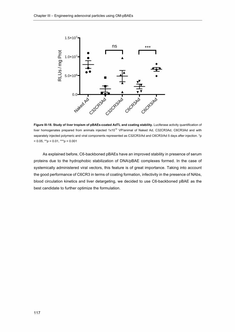
Chapter III – Engineering adenoviral particles using OM-pBAEs
117
Figure III-18. Study of liver tropism of pBAEs-coated AdTL and coating stability. Luciferase activity quantification of
liver homogenates prepared from animals injected 1x10¹⁰ VP/animal of Naked Ad, C32CR3Ad, C6CR3Ad and with
separately injected polymeric and viral components represented as C32CR3/Ad and C6CR3/Ad 5 days after injection. *p
< 0.05, **p < 0.01, ***p < 0.001
As explained before, C6-backboned pBAEs have an improved stability in presence of serum
proteins due to the hydrophobic stabilization of DNA/pBAE complexes formed. In the case of
systemically administered viral vectors, this feature is of great importance. Taking into account
the good performance of C6CR3 in terms of coating formation, infectivity in the presence of NAbs,
blood circulation kinetics and liver detargeting, we decided to use C6-backboned pBAE as the
best candidate to further optimize the formulation.
Naked
Ad
C32CR3A
d
C32CR3/A
d
C6CR3A
d
C6CR3/A
d0.0
5.0×10⁶
1.0×10⁷
1.5×10⁷
RLU
s / m
g Pr
ot
***ns
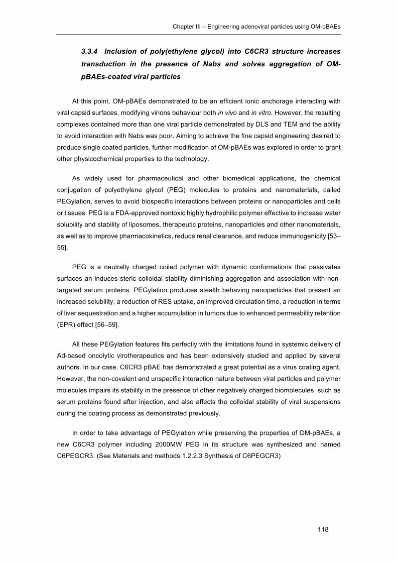
Chapter III – Engineering adenoviral particles using OM-pBAEs
118
3.3.4 Inclusion of poly(ethylene glycol) into C6CR3 structure increases
transduction in the presence of Nabs and solves aggregation of OM-
pBAEs-coated viral particles
At this point, OM-pBAEs demonstrated to be an efficient ionic anchorage interacting with
viral capsid surfaces, modifying virions behaviour both in vivo and in vitro. However, the resulting
complexes contained more than one viral particle demonstrated by DLS and TEM and the ability
to avoid interaction with Nabs was poor. Aiming to achieve the fine capsid engineering desired to
produce single coated particles, further modification of OM-pBAEs was explored in order to grant
other physicochemical properties to the technology.
As widely used for pharmaceutical and other biomedical applications, the chemical
conjugation of polyethylene glycol (PEG) molecules to proteins and nanomaterials, called
PEGylation, serves to avoid biospecific interactions between proteins or nanoparticles and cells
or tissues. PEG is a FDA-approved nontoxic highly hydrophilic polymer effective to increase water
solubility and stability of liposomes, therapeutic proteins, nanoparticles and other nanomaterials,
as well as to improve pharmacokinetics, reduce renal clearance, and reduce immunogenicity [53–
55].
PEG is a neutrally charged coiled polymer with dynamic conformations that passivates
surfaces an induces steric colloidal stability diminishing aggregation and association with non-
targeted serum proteins. PEGylation produces stealth behaving nanoparticles that present an
increased solubility, a reduction of RES uptake, an improved circulation time, a reduction in terms
of liver sequestration and a higher accumulation in tumors due to enhanced permeability retention
(EPR) effect [56–59].
All these PEGylation features fits perfectly with the limitations found in systemic delivery of
Ad-based oncolytic virotherapeutics and has been extensively studied and applied by several
authors. In our case, C6CR3 pBAE has demonstrated a great potential as a virus coating agent.
However, the non-covalent and unspecific interaction nature between viral particles and polymer
molecules impairs its stability in the presence of other negatively charged biomolecules, such as
serum proteins found after injection, and also affects the colloidal stability of viral suspensions
during the coating process as demonstrated previously.
In order to take advantage of PEGylation while preserving the properties of OM-pBAEs, a
new C6CR3 polymer including 2000MW PEG in its structure was synthesized and named
C6PEGCR3. (See Materials and methods 1.2.2.3 Synthesis of C6PEGCR3)

Chapter III – Engineering adenoviral particles using OM-pBAEs
119
3.3.4.1 A formulation comprising 65:35 w/w of C6CR3 and C6PEGCR3 improves
transduction of coated Ad in the presence Nabs.
NAbs needs to physically interact with capsid surfaces to neutralize viral infectivity. The
presence of PEG molecules over coated Ad particles can be a steric barrier impeding such
interactions thanks to its dynamic conformation and mobility. A specific antibody aiming to interact
with capsid surfaces, acts as a witness observing the molecular quality and shielding capacity of
a specific coating agent. In order to determine the optimal amount of C6PEGCR3 polymer needed
to efficiently avoid neutralization of coated Ad in the presence of Nabs, different
C6CR3:C6PEGCR3 w/w ratios were tested in vitro by neutralization assays while maintaining the
total 4x10⁶ pol/VP ratio.
After preliminary experimental conditions optimization, infection of 15000 PANC-1 cells/well
with MOI 50 in the presence of a 100000-fold dilution of Ab6982 were considered to be optimal
because the transduction level of Naked Ad and neutralized Naked Ad (Naked Ad +) was offering
a comfortable dynamic range to explore the improvement between these values (Naked Ad: 76,77
± 2,85 % GFP + cells, Naked Ad +: 54,92 ± 3,45 % GFP + cells).
Figure III-19. Infectivity of AdTL coated with formulations containing different ratios of C6CR3 and C6PEGCR3 in the presence of NAbs. Conditions marked with (+) means preincubation with NAbs prior to infection. Turkey’s multiple
comparison test was performed using graphpad *p < 0.05, **p < 0.01, ***p < 0.001, ****p < 0.0001
As observed in figure III-19, the addition of C6PEGCR3 polymer in the formulation improved
the infectivity of NAbs pre-incubated samples. More specifically, the best performing formulation
was the 65:35-coated Ad condition where 71,15 ± 2,19 % GFP positive cells were infected using
a NAbs pre-incubated coated-viral sample.
Nak
ed A
d
C6C
R3A
d
75:2
5 Ad
65:3
5 Ad
55:4
5 Ad
+ N
aked
Ad
+ C
6CR
3Ad
+ 75
:25
Ad
+ 65
:35
Ad
+ 55
:45
Ad
40
50
60
70
80
90
%G
PF+
cells ns
*
******
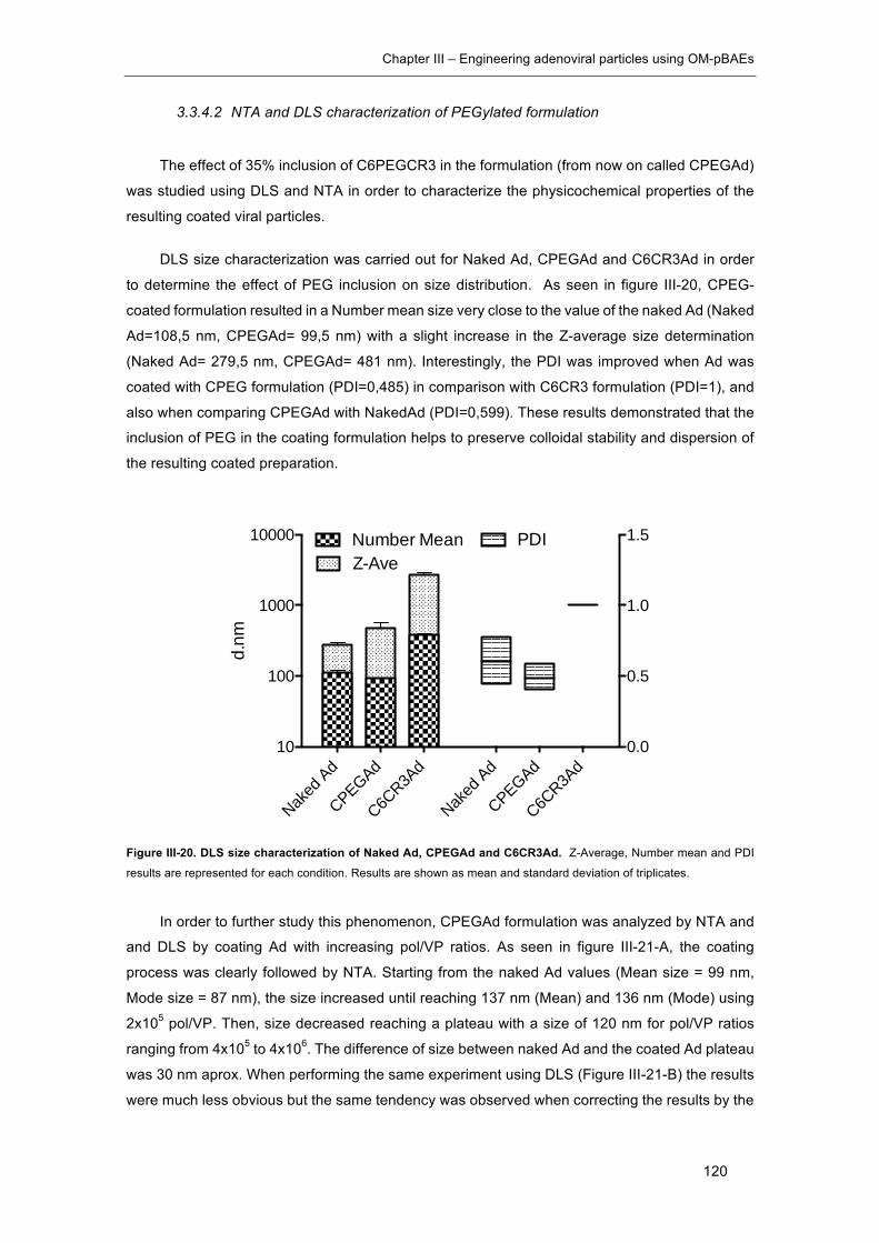
Chapter III – Engineering adenoviral particles using OM-pBAEs
120
3.3.4.2 NTA and DLS characterization of PEGylated formulation
The effect of 35% inclusion of C6PEGCR3 in the formulation (from now on called CPEGAd)
was studied using DLS and NTA in order to characterize the physicochemical properties of the
resulting coated viral particles.
DLS size characterization was carried out for Naked Ad, CPEGAd and C6CR3Ad in order
to determine the effect of PEG inclusion on size distribution. As seen in figure III-20, CPEG-
coated formulation resulted in a Number mean size very close to the value of the naked Ad (Naked
Ad=108,5 nm, CPEGAd= 99,5 nm) with a slight increase in the Z-average size determination
(Naked Ad= 279,5 nm, CPEGAd= 481 nm). Interestingly, the PDI was improved when Ad was
coated with CPEG formulation (PDI=0,485) in comparison with C6CR3 formulation (PDI=1), and
also when comparing CPEGAd with NakedAd (PDI=0,599). These results demonstrated that the
inclusion of PEG in the coating formulation helps to preserve colloidal stability and dispersion of
the resulting coated preparation.
Figure III-20. DLS size characterization of Naked Ad, CPEGAd and C6CR3Ad. Z-Average, Number mean and PDI
results are represented for each condition. Results are shown as mean and standard deviation of triplicates.
In order to further study this phenomenon, CPEGAd formulation was analyzed by NTA and
and DLS by coating Ad with increasing pol/VP ratios. As seen in figure III-21-A, the coating
process was clearly followed by NTA. Starting from the naked Ad values (Mean size = 99 nm,
Mode size = 87 nm), the size increased until reaching 137 nm (Mean) and 136 nm (Mode) using
2x10⁵ pol/VP. Then, size decreased reaching a plateau with a size of 120 nm for pol/VP ratios
ranging from 4x10⁵ to 4x10⁶. The difference of size between naked Ad and the coated Ad plateau
was 30 nm aprox. When performing the same experiment using DLS (Figure III-21-B) the results
were much less obvious but the same tendency was observed when correcting the results by the
Naked
Ad
CPEGAd
C6CR3A
d
Naked
Ad
CPEGAd
C6CR3A
d 10
100
1000
10000
0.0
0.5
1.0
1.5Number MeanZ-Ave
PDI
d.nm

Chapter III – Engineering adenoviral particles using OM-pBAEs
121
number mean. Although the difference between naked Ad and coated plateau was not significant,
the same maximum pic was observed around 2x10⁵ pol/VP.
Figure III-21. Biophysical characterization of CPEG-coated Ads by Nanoparticle Tracking Analysis (NTA) and DLS. NTA (A) and DLS (B) analysis of CPEG-coated Ads with increasing pol/VP ratios.
The presence of PEG covering coated viral particles modifies the resulting surface charge,
neutralizing the cationic nature of C6CR3 coating. As seen in figure III-22-A, this formulation
produces almost neutral Z-potentials, even at high pol/VP ratios. The shielding effect was also
confirmed by comparing Ad preparations coated with C6CR3, CPEG and Naked Ad with a
constant 4x10⁶ pol/VP ratio (Figure III-22-B).
Figure III-22. Addition of C6PEGCR3 polymer neutralizes the positive Z-potential of C6CR3-coated Ad. A) Z-
potential characterization of CPEGAd coated with increasing pol/VP ratios. B) Z-potential comparative study of Naked Ad,
CPEGAd and C6CR3Ad coated with 4x10⁶ pol/VP ratio.
3.3.4.3 CPEG formulation produces single coated viral particles
Aiming to precisely determine the morphology of CPEG-coated Ad samples, TEM
characterization was carried out. In contrast with previously observed formulations (Fig. III-11 and
III-12), no formation of big aggregates was observed for CPEGAd samples (Fig. III-23-A). Single
and non-aggregated coated viral particles were observed with a clear artificial envelope coating
every single viral particle in the preparation (Fig. III-23-B).
01×
106
2×10
6
3×10
6
4×10
6
5×10
680
100
120
140
160 Mean Size NTA Mode Size NTA
Ratio Pol:VP
d.nm
01×
106
2×10
6
3×10
6
4×10
6
0
200
400
600
Number meanZ-Average
Ratio Pol:VP
d.nm
1×10
6
2×10
6
3×10
6
4×10
6
5×10
6
-15
-10
-5
0
5
10
Ratio Pol:VP
Z-Po
tent
ial
(mV)
Naked Ad CPEGAd C6CR3Ad-20
-10
0
10
20
ZP (m
V)
A B
A B
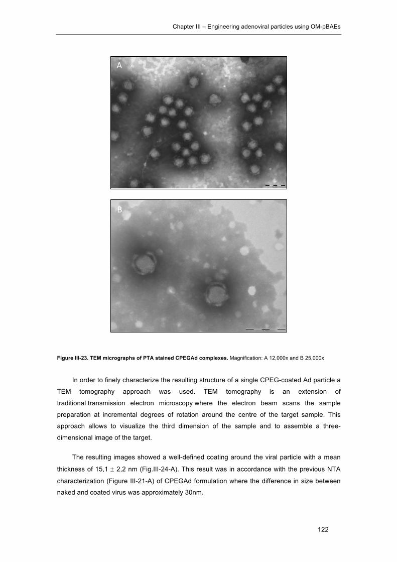
Chapter III – Engineering adenoviral particles using OM-pBAEs
122
Figure III-23. TEM micrographs of PTA stained CPEGAd complexes. Magnification: A 12,000x and B 25,000x
In order to finely characterize the resulting structure of a single CPEG-coated Ad particle a
TEM tomography approach was used. TEM tomography is an extension of
traditional transmission electron microscopy where the electron beam scans the sample
preparation at incremental degrees of rotation around the centre of the target sample. This
approach allows to visualize the third dimension of the sample and to assemble a three-
dimensional image of the target.
The resulting images showed a well-defined coating around the viral particle with a mean
thickness of 15,1 ± 2,2 nm (Fig.III-24-A). This result was in accordance with the previous NTA
characterization (Figure III-21-A) of CPEGAd formulation where the difference in size between
naked and coated virus was approximately 30nm.
A
B
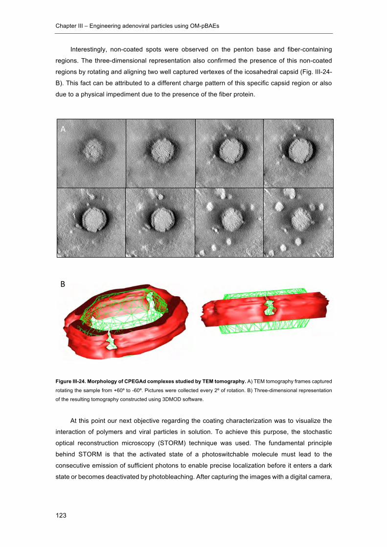
Chapter III – Engineering adenoviral particles using OM-pBAEs
123
Interestingly, non-coated spots were observed on the penton base and fiber-containing
regions. The three-dimensional representation also confirmed the presence of this non-coated
regions by rotating and aligning two well captured vertexes of the icosahedral capsid (Fig. III-24-
B). This fact can be attributed to a different charge pattern of this specific capsid region or also
due to a physical impediment due to the presence of the fiber protein.
Figure III-24. Morphology of CPEGAd complexes studied by TEM tomography. A) TEM tomography frames captured
rotating the sample from +60º to -60º. Pictures were collected every 2º of rotation. B) Three-dimensional representation
of the resulting tomography constructed using 3DMOD software.
At this point our next objective regarding the coating characterization was to visualize the
interaction of polymers and viral particles in solution. To achieve this purpose, the stochastic
optical reconstruction microscopy (STORM) technique was used. The fundamental principle
behind STORM is that the activated state of a photoswitchable molecule must lead to the
consecutive emission of sufficient photons to enable precise localization before it enters a dark
state or becomes deactivated by photobleaching. After capturing the images with a digital camera,
A
B
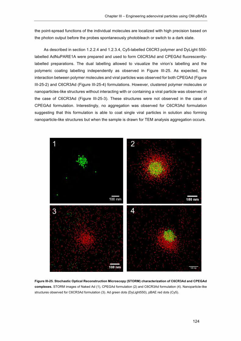
Chapter III – Engineering adenoviral particles using OM-pBAEs
124
the point-spread functions of the individual molecules are localized with high precision based on
the photon output before the probes spontaneously photobleach or switch to a dark state.
As described in section 1.2.2.4 and 1.2.3.4, Cy5-labelled C6CR3 polymer and DyLight 550-
labelled AdNuPARE1A were prepared and used to form C6CR3Ad and CPEGAd fluorescently-
labelled preparations. The dual labelling allowed to visualize the virion’s labelling and the
polymeric coating labelling independently as observed in Figure III-25. As expected, the
interaction between polymer molecules and viral particles was observed for both CPEGAd (Figure
III-25-2) and C6CR3Ad (Figure III-25-4) formulations. However, clustered polymer molecules or
nanoparticles-like structures without interacting with or containing a viral particle was observed in
the case of C6CR3Ad (Figure III-25-3). These structures were not observed in the case of
CPEGAd formulation. Interestingly, no aggregation was observed for C6CR3Ad formulation
suggesting that this formulation is able to coat single viral particles in solution also forming
nanoparticle-like structures but when the sample is drawn for TEM analysis aggregation occurs.
Figure III-25. Stochastic Optical Reconstruction Microscopy (STORM) characterization of C6CR3Ad and CPEGAd complexes. STORM images of Naked Ad (1), CPEGAd formulation (2) and C6CR3Ad formulation (4). Nanoparticle-like
structures observed for C6CR3Ad formulation (3). Ad green dots (DyLight550), pBAE red dots (Cy5).
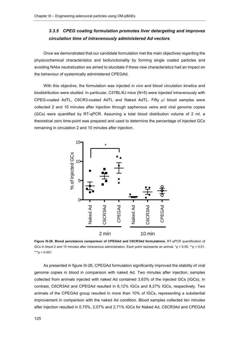
Chapter III – Engineering adenoviral particles using OM-pBAEs
125
3.3.5 CPEG coating formulation promotes liver detargeting and improves
circulation time of intravenously administered Ad vectors
Once we demonstrated that our candidate formulation met the main objectives regarding the
physicochemical characteristics and biofunctionality by forming single coated particles and
avoiding NAbs neutralization we aimed to elucidate if these new characteristics had an impact on
the behaviour of systemically administered CPEGAd.
With this objective, the formulation was injected in vivo and blood circulation kinetics and
biodistribution were studied. In particular, C57BL/6J mice (N=5) were injected intravenously with
CPEG-coated AdTL, C6CR3-coated AdTL and Naked AdTL. Fifty µl blood samples were
collected 2 and 10 minutes after injection through saphenous veins and viral genome copies
(GCs) were quantified by RT-qPCR. Assuming a total blood distribution volume of 2 ml, a
theoretical zero time-point was prepared and used to determine the percentage of injected GCs
remaining in circulation 2 and 10 minutes after injection.
Figure III-26. Blood persistence comparison of CPEGAd and C6CR3Ad formulations. RT-qPCR quantification of
GCs in blood 2 and 10 minutes after intravenous administration. Each point represents an animal. *p < 0.05, **p < 0.01,
***p < 0.001
As presented in figure III-26, CPEGAd formulation significantly improved the stability of viral
genome copies in blood in comparison with naked Ad. Two minutes after injection, samples
collected from animals injected with naked Ad contained 3,63% of the injected GCs (IGCs). In
contrast, C6CR3Ad and CPEGAd resulted in 6,12% IGCs and 8,27% IGCs, respectively. Two
animals of the CPEGAd group resulted in more than 10% of IGCs, representing a substantial
improvement in comparison with the naked Ad condition. Blood samples collected ten minutes
after injection resulted in 0,75%, 2,07% and 2,71% IGCs for Naked Ad, C6CR3Ad and CPEGAd
Nak
ed A
d
C6C
R3A
d
CPE
GAd
Nak
ed A
d
C6C
R3A
d
CPE
GAd
0
5
10
15
% o
f inj
ecte
d G
Cs
2 min 10 min
*
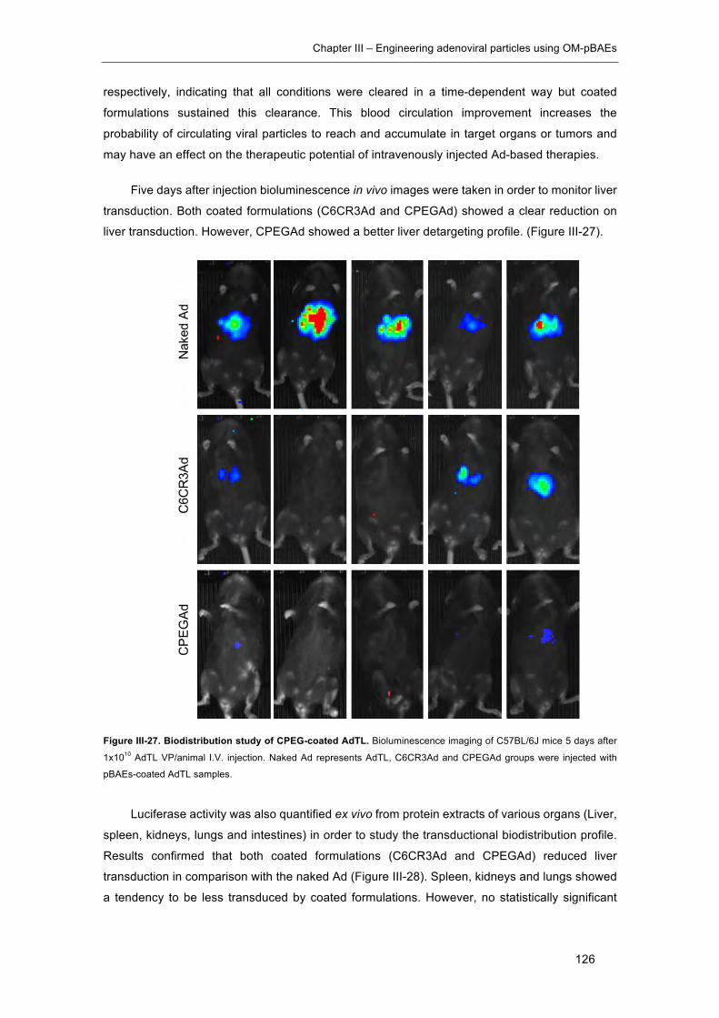
Chapter III – Engineering adenoviral particles using OM-pBAEs
126
respectively, indicating that all conditions were cleared in a time-dependent way but coated
formulations sustained this clearance. This blood circulation improvement increases the
probability of circulating viral particles to reach and accumulate in target organs or tumors and
may have an effect on the therapeutic potential of intravenously injected Ad-based therapies.
Five days after injection bioluminescence in vivo images were taken in order to monitor liver
transduction. Both coated formulations (C6CR3Ad and CPEGAd) showed a clear reduction on
liver transduction. However, CPEGAd showed a better liver detargeting profile. (Figure III-27).
Figure III-27. Biodistribution study of CPEG-coated AdTL. Bioluminescence imaging of C57BL/6J mice 5 days after
1x10¹⁰ AdTL VP/animal I.V. injection. Naked Ad represents AdTL, C6CR3Ad and CPEGAd groups were injected with
pBAEs-coated AdTL samples.
Luciferase activity was also quantified ex vivo from protein extracts of various organs (Liver,
spleen, kidneys, lungs and intestines) in order to study the transductional biodistribution profile.
Results confirmed that both coated formulations (C6CR3Ad and CPEGAd) reduced liver
transduction in comparison with the naked Ad (Figure III-28). Spleen, kidneys and lungs showed
a tendency to be less transduced by coated formulations. However, no statistically significant
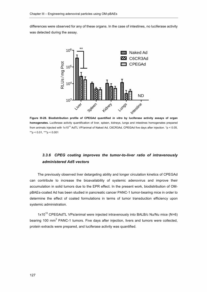
Chapter III – Engineering adenoviral particles using OM-pBAEs
127
differences were observed for any of these organs. In the case of intestines, no luciferase activity
was detected during the assay.
Figure III-28. Biodistribution profile of CPEGAd quantified in vitro by luciferase activity assays of organ homogenates. Luciferase activity quantification of liver, spleen, kidneys, lungs and intestines homogenates prepared
from animals injected with 1x10¹⁰ AdTL VP/animal of Naked Ad, C6CR3Ad, CPEGAd five days after injection. *p < 0.05,
**p < 0.01, ***p < 0.001
3.3.6 CPEG coating improves the tumor-to-liver ratio of intravenously
administered Ad5 vectors
The previously observed liver detargeting ability and longer circulation kinetics of CPEGAd
can contribute to increase the bioavailability of systemic adenovirus and improve their
accumulation in solid tumors due to the EPR effect. In the present work, biodistribution of OM-
pBAEs-coated Ad has been studied in pancreatic cancer PANC-1 tumor-bearing mice in order to
determine the effect of coated formulations in terms of tumor transduction efficiency upon
systemic administration.
1x10¹⁰ CPEGAdTL VPs/animal were injected intravenously into BALB/c Nu/Nu mice (N=6)
bearing 100 mm3 PANC-1 tumors. Five days after injection, livers and tumors were collected,
protein extracts were prepared, and luciferase activity was quantified.
Liver
Spleen
Kidney
Lung
s
Intes
tine
103
104
105
106Naked AdC6CR3AdCPEGAd
ND
**
RLU
s / m
g Pr
ot
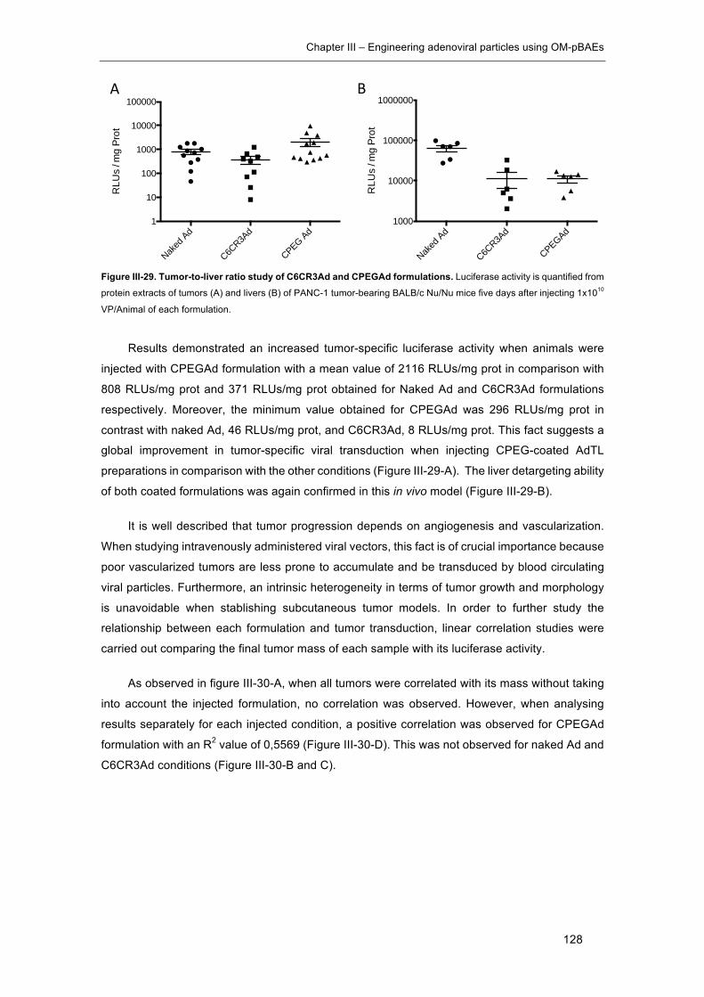
Chapter III – Engineering adenoviral particles using OM-pBAEs
128
Figure III-29. Tumor-to-liver ratio study of C6CR3Ad and CPEGAd formulations. Luciferase activity is quantified from
protein extracts of tumors (A) and livers (B) of PANC-1 tumor-bearing BALB/c Nu/Nu mice five days after injecting 1x10¹⁰
VP/Animal of each formulation.
Results demonstrated an increased tumor-specific luciferase activity when animals were
injected with CPEGAd formulation with a mean value of 2116 RLUs/mg prot in comparison with
808 RLUs/mg prot and 371 RLUs/mg prot obtained for Naked Ad and C6CR3Ad formulations
respectively. Moreover, the minimum value obtained for CPEGAd was 296 RLUs/mg prot in
contrast with naked Ad, 46 RLUs/mg prot, and C6CR3Ad, 8 RLUs/mg prot. This fact suggests a
global improvement in tumor-specific viral transduction when injecting CPEG-coated AdTL
preparations in comparison with the other conditions (Figure III-29-A). The liver detargeting ability
of both coated formulations was again confirmed in this in vivo model (Figure III-29-B).
It is well described that tumor progression depends on angiogenesis and vascularization.
When studying intravenously administered viral vectors, this fact is of crucial importance because
poor vascularized tumors are less prone to accumulate and be transduced by blood circulating
viral particles. Furthermore, an intrinsic heterogeneity in terms of tumor growth and morphology
is unavoidable when stablishing subcutaneous tumor models. In order to further study the
relationship between each formulation and tumor transduction, linear correlation studies were
carried out comparing the final tumor mass of each sample with its luciferase activity.
As observed in figure III-30-A, when all tumors were correlated with its mass without taking
into account the injected formulation, no correlation was observed. However, when analysing
results separately for each injected condition, a positive correlation was observed for CPEGAd
formulation with an R2 value of 0,5569 (Figure III-30-D). This was not observed for naked Ad and
C6CR3Ad conditions (Figure III-30-B and C).
Naked
Ad
C6CR3A
d
CPEG Ad1
10
100
1000
10000
100000R
LUs
/ mg
Prot
Naked
Ad
C6CR3A
d
CPEGAd1000
10000
100000
1000000
RLU
s / m
g Pr
ot
BA
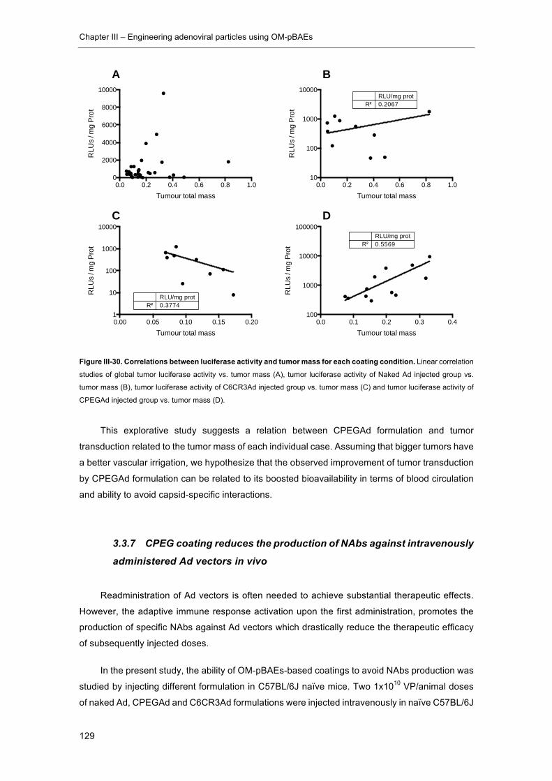
Chapter III – Engineering adenoviral particles using OM-pBAEs
129
Figure III-30. Correlations between luciferase activity and tumor mass for each coating condition. Linear correlation
studies of global tumor luciferase activity vs. tumor mass (A), tumor luciferase activity of Naked Ad injected group vs.
tumor mass (B), tumor luciferase activity of C6CR3Ad injected group vs. tumor mass (C) and tumor luciferase activity of
CPEGAd injected group vs. tumor mass (D).
This explorative study suggests a relation between CPEGAd formulation and tumor
transduction related to the tumor mass of each individual case. Assuming that bigger tumors have
a better vascular irrigation, we hypothesize that the observed improvement of tumor transduction
by CPEGAd formulation can be related to its boosted bioavailability in terms of blood circulation
and ability to avoid capsid-specific interactions.
3.3.7 CPEG coating reduces the production of NAbs against intravenously
administered Ad vectors in vivo
Readministration of Ad vectors is often needed to achieve substantial therapeutic effects.
However, the adaptive immune response activation upon the first administration, promotes the
production of specific NAbs against Ad vectors which drastically reduce the therapeutic efficacy
of subsequently injected doses.
In the present study, the ability of OM-pBAEs-based coatings to avoid NAbs production was
studied by injecting different formulation in C57BL/6J naïve mice. Two 1x10¹⁰ VP/animal doses
of naked Ad, CPEGAd and C6CR3Ad formulations were injected intravenously in naïve C57BL/6J
0.0 0.2 0.4 0.6 0.8 1.00
2000
4000
6000
8000
10000
Tumour total mass
RLU
s / m
g Pr
ot
0.00 0.05 0.10 0.15 0.201
10
100
1000
10000
R²RLU/mg prot0.3774
Tumour total mass
RLU
s / m
g Pr
ot
0.0 0.2 0.4 0.6 0.8 1.010
100
1000
10000
R²RLU/mg prot0.2067
Tumour total mass
RLU
s / m
g Pr
ot
0.0 0.1 0.2 0.3 0.4100
1000
10000
100000
R²RLU/mg prot0.5569
Tumour total mass
RLU
s / m
g Pr
ot
A B
C D

Chapter III – Engineering adenoviral particles using OM-pBAEs
130
mice at day 1 and day 14. At day 21 after the first injection, blood was collected by intracardiac
puncture and sera were obtained and heat inactivated. Collected serums were used to perform
in vitro neutralization assays in order to determine the serum dilution needed to achieve a 50%
neutralization (ND50) of the transduction level obtained on the control condition without serum.
The detailed protocol is available at section 1.2.7.6.
As seen in figure III-31, CPEGAd formulation produced serums with significantly lower
neutralization capacities. The average ND50 obtained for the Naked Ad condition was 8424 in
comparison with CPEGAd which was 2718 and 5280 for the C6CR3Ad condition. These results
showed that CPEG-coated Ad substantially avoided the activation of NAbs production in vivo
suggesting that CPEG-coated Ad formulations can improve the efficacy of Ad vectors when under
readministering procedures.
Figure III-31. De novo generation of neutralizing antibodies for naked, C6CR3 and CPEG-coated Ads in vivo. In
vitro neutralization assay of serums collected from naïve C57BL/6J mice after intravenous administration of two doses of
Naked Ad, CPEGAd and C6CR3Ad (1x10¹⁰ VP/animal were administered at day 1 and day 14 and serums were collected
at day 21 post injection). *p < 0.05, **p < 0.01, ***p < 0.001
Naked
Ad
CPEGAd
C6CR3A
d 0
5000
10000
15000
ND
50
**
Chapter III – Engineering adenoviral particles using OM-pBAEs
131
3.4 Concluding remarks
Previously in chapter II, OM-pBAEs have been used as coating agents to envelope AAV
vectors aiming to improve systemic administration of such vectors. We demonstrated the ability
of OM-pBAEs-based coatings to interact with AAV viral particles and change the AAV viral tropism
in vivo. A clear liver detargeting effect was observed when intravenously administered in vivo.
Moreover, the easy synthesis of OM-pBAEs allowed to incorporate a custom peptide-based brain
targeting moiety. The effect of this active targeting was clearly observed in vitro, however it was
not as effective in vivo.
In the present chapter, we aimed to use the same approach for Adenoviral vectors exploring
other formulations and chemical modifications and deeply characterizing the resulting polymer/Ad
hybrid vectors in terms of their physicochemical properties and in vivo behaviour after systemic
administration.
Previously in our group, it was demonstrated that slightly hydrophobic C6-backboned pBAEs
produced more stable complexes under physiological conditions and in the presence of serum
proteins, thus we decided to include the C6-polymers in our studies. The interaction of C32CR3
and C6CR3 polymers with viral particles has been confirmed by DLS studying the relation
between Z-potential and the ratio pol/VP needed to fully coat viral particles. However, both
polymers induced aggregation of viral particles but interestingly, C6CR3 produced smaller
complexes with an increased but modest ability to transduce cells in the presence of NAbs.
A multilayering coating approach was also explored but no substantial improvement was
observed in terms of in vitro aggregation and in vitro transduction. Taking into account that this
coating approach also adds complexity in a hypothetic product regulatory and industrial
production, the multilayering coating approach was abandoned.
The improved blood circulation time and liver detargeting profile of C6CR3Ad complexes
observed in vivo made us opt for C6-backboned OM-pBAEs to proceed with technology
optimization. The inclusion of polyethilenglycol was studied aiming to improve transduction in the
presence of NAbs and to solve aggregation of coated viral particles. A 35% w/w mixture ratio of
C6CR3 and C6PEGCR3 was the best performing formulation in terms of transduction in the
presence of NAbs, and after biophysical characterization and microscopy assessment it was also
demonstrated the ability of CPEGAd formulation to produce single coated particle with neutral Z-
potentials and in the nanometric size range.
In vivo studies also demonstrated that CPEGAd complexation increases the blood
circulation time in comparison with C6CR3-coated Ads and even improves the liver detargeting
profile observed for the C6CR3Ad formulation. When studying the biodistribution in tumor-bearing
mice, increased tumor-specific luciferase activity was observed in animals treated with CPEGAd
Chapter III – Engineering adenoviral particles using OM-pBAEs
132
formulation suggesting that the improved bioavailability of circulating Ad promoted by CPEG
envelopes, expands the accumulation of viral particles into solid tumors.
Finally, it was also demonstrated that CPEG-coated Ad avoids activation of NAbs production
in vivo, indicating that the artificial envelope hinders viral particles to be detected by the immune
system.
Then, we can conclude that the newly developed CPEG coating formulation is suitable to
improve the systemic delivery of Ad vectors, specially for cancer treatment purposes. Therefore,
in the next chapter, the CPEG formulation has been used to coat a therapeutic oncolytic
adenovirus and their efficacy and safety profile have been deeply studied.
Chapter III – Engineering adenoviral particles using OM-pBAEs
133
3.5 References
[1] J. Fueyo, C. Gomez-Manzano, R. Alemany, P.S.Y. Lee, T.J. McDonnell, P.
Mitlianga, Y.-X. Shi, V.A. Levin, W.K.A. Yung, A.P. Kyritsis, A mutant oncolytic
adenovirus targeting the Rb pathway produces anti-glioma effect in vivo,
Oncogene. 19 (2000) 2. http://dx.doi.org/10.1038/sj.onc.1203251.
[2] M. Huch, A. Gros, A. José, J.R. González, R. Alemany, C. Fillat, Urokinase-type
plasminogen activator receptor transcriptionally controlled adenoviruses
eradicate pancreatic tumors and liver metastasis in mouse models., Neoplasia.
11 (2009) 518–28, 4 p following 528. doi:http://dx.doi.org/10.1593/neo.81674.
[3] A. Mato-Berciano, G. Raimondi, M.V. Maliandi, R. Alemany, L. Montoliu, C.
Fillat, A NOTCH-sensitive uPAR-regulated oncolytic adenovirus effectively
suppresses pancreatic tumor growth and triggers synergistic anticancer effects
with gemcitabine and nab-paclitaxel, Oncotarget. 8 (2017) 22700–22715.
doi:https://dx.doi.org/10.18632/oncotarget.15169.
[4] H. Dehari, Y. Ito, T. Nakamura, M. Kobune, K. Sasaki, N. Yonekura, G. Kohama,
H. Hamada, Enhanced antitumor effect of RGD fiber-modified adenovirus for
gene therapy of oral cancer, Cancer Gene Ther. 10 (2002) 75.
http://dx.doi.org/10.1038/sj.cgt.7700529.
[5] A. Rodríguez-García, M. Giménez-Alejandre, J.J. Rojas, R. Moreno, M. Bazan-
Peregrino, M. Cascalló, R. Alemany, Safety and Efficacy of VCN-01, an
Oncolytic Adenovirus Combining Fiber HSG-Binding Domain Replacement with
RGD and Hyaluronidase Expression, Clin. Cancer Res. 21 (2015) 1406 LP-
1418. http://clincancerres.aacrjournals.org/content/21/6/1406.abstract.
[6] S. Guedan, J.J. Rojas, A. Gros, E. Mercade, M. Cascallo, R. Alemany,
Hyaluronidase expression by an oncolytic adenovirus enhances its intratumoral
spread and suppresses tumor growth., Mol. Ther. 18 (2010) 1275–1283.
doi:10.1038/mt.2010.79.
[7] E. Dolgin, Oncolytic viruses get a boost with first FDA-approval
recommendation, Nat. Rev. Drug Discov. 14 (2015) 369.
http://dx.doi.org/10.1038/nrd4643.
[8] J. Pol, A. Buqué, F. Aranda, N. Bloy, I. Cremer, A. Eggermont, P. Erbs, J.
Fucikova, J. Galon, J.-M. Limacher, X. Preville, C. Sautès-Fridman, R. Spisek, L.
Chapter III – Engineering adenoviral particles using OM-pBAEs
134
Zitvogel, G. Kroemer, L. Galluzzi, Trial Watch—Oncolytic viruses and cancer
therapy, Oncoimmunology. 5 (2016) e1117740.
doi:10.1080/2162402X.2015.1117740.
[9] H.L. Kaufman, F.J. Kohlhapp, A. Zloza, Oncolytic viruses: a new class of
immunotherapy drugs, Nat. Rev. Drug Discov. 14 (2015) 642.
http://dx.doi.org/10.1038/nrd4663.
[10] C.S. Lee, E.S. Bishop, R. Zhang, X. Yu, E.M. Farina, S. Yan, C. Zhao, Z. Zeng,
Y. Shu, X. Wu, J. Lei, Y. Li, W. Zhang, C. Yang, K. Wu, Y. Wu, S. Ho, A.
Athiviraham, M.J. Lee, J.M. Wolf, R.R. Reid, T.-C. He, Adenovirus-mediated
gene delivery: Potential applications for gene and cell-based therapies in the
new era of personalized medicine, Genes Dis. 4 (2017) 43–63.
doi:https://doi.org/10.1016/j.gendis.2017.04.001.
[11] R.G. Crystal, Adenovirus: The First Effective In Vivo Gene Delivery Vector, Hum.
Gene Ther. 25 (2014) 3–11. doi:10.1089/hum.2013.2527.
[12] B. Breyer, W. Jiang, H. Cheng, L. Zhou, R. Paul, T.F. and T.-C. He, Adenoviral
Vector-Mediated Gene Transfer for Human Gene Therapy, Curr. Gene Ther. 1
(2001) 149–162. doi:http://dx.doi.org/10.2174/1566523013348689.
[13] P.W. Roelvink, A. Lizonova, J.G.M. Lee, Y. Li, J.M. Bergelson, R.W. Finberg,
D.E. Brough, I. Kovesdi, T.J. Wickham, The Coxsackievirus-Adenovirus
Receptor Protein Can Function as a Cellular Attachment Protein for Adenovirus
Serotypes from Subgroups A, C, D, E, and F, J. Virol. 72 (1998) 7909–7915.
http://www.ncbi.nlm.nih.gov/pmc/articles/PMC110119/.
[14] H. Fechner, A. Haack, H. Wang, X. Wang, K. Eizema, M. Pauschinger, R.G.
Schoemaker, R. Van Veghel, A.B. Houtsmuller, H.P. Schultheiss, J.M.J. Lamers,
W. Poller, Expression of Coxsackie adenovirus receptor and alpha(v)-integrin
does not correlate with aconovector targeting in vivo indicating anatomical vector
barriers, Gene Ther. 6 (1999) 1520–1535. doi:10.1038/sj.gt.3301030.
[15] H.G. Transfer, A. Lieber, M.A. Kay, Resea r c h Re port s Method for Multiple
Portal Vein Infusions in Mice : Quantitation of Adenovirus-Mediated, 20 (1996).
[16] D.M. Shayakhmetov, A. Gaggar, S. Ni, Z.Y. Li, A. Lieber, Adenovirus binding to
blood factors results in liver cell infection and hepatotoxicity, J Virol. 79 (2005)
7478–7491. doi:10.1128/JVI.79.12.7478-7491.2005.
Chapter III – Engineering adenoviral particles using OM-pBAEs
135
[17] R. Alba, A.C. Bradshaw, L. Coughlan, L. Denby, R.A. McDonald, S.N.
Waddington, S.M.K. Buckley, J.A. Greig, A.L. Parker, A.M. Miller, H. Wang, A.
Lieber, N. van Rooijen, J.H. McVey, S.A. Nicklin, A.H. Baker, Biodistribution and
retargeting of FX-binding ablated adenovirus serotype 5 vectors, Blood. 116
(2010) 2656–2664. doi:10.1182/blood-2009-12-260026.
[18] D. a Muruve, M.J. Barnes, I.E. Stillman, T. a Libermann, Adenoviral gene
therapy leads to rapid induction of multiple chemokines and acute neutrophil-
dependent hepatic injury in vivo., Hum. Gene Ther. 10 (1999) 965–76.
doi:10.1089/10430349950018364.
[19] A. Lieber, C.Y. He, L. Meuse, D. Schowalter, I. Kirillova, B. Winther, M.A. Kay,
The role of Kupffer cell activation and viral gene expression in early liver toxicity
after infusion of recombinant adenovirus vectors., J. Virol. 71 (1997) 8798–8807.
http://www.ncbi.nlm.nih.gov/pmc/articles/PMC192346/.
[20] M.A. Schnell, Y. Zhang, J. Tazelaar, G.P. Gao, Q.C. Yu, R. Qian, S.J. Chen,
A.N. Varnavski, C. LeClair, S.E. Raper, J.M. Wilson, Activation of innate
immunity in nonhuman primates following intraportal administration of adenoviral
vectors, Mol. Ther. 3 (2001) 708–722. doi:10.1006/mthe.2001.0330.
[21] Y. Zhang, N. Chirmule, G.P. Gao, R. Qian, M. Croyle, B. Joshi, J. Tazelaar, J.M.
Wilson, Acute cytokine response to systemic adenoviral vectors in mice is
mediated by dendritic cells and macrophages, Mol. Ther. 3 (2001) 697–707.
doi:10.1006/mthe.2001.0329.
[22] Y. Yang, K. Greenough, J.M. Wilson, Transient immune blockade prevents
formation of neutralizing antibody to recombinant adenovirus and allows
repeated gene transfer to mouse liver, Gene Ther. 3 (1996) 412—420.
http://europepmc.org/abstract/MED/9156802.
[23] M.B. Appaiahgari, R.M. Pandey, S. Vrati, Seroprevalence of neutralizing
antibodies to adenovirus type 5 among children in India: Implications for
recombinant adenovirus-based vaccines, Clin. Vaccine Immunol. 14 (2007)
1053–1055. doi:10.1128/CVI.00173-07.
[24] R. Pilankatta, T. Chawla, N. Khanna, S. Swaminathan, The prevalence of
antibodies to adenovirus serotype 5 in an adult Indian population and
implications for adenovirus vector vaccines., J. Med. Virol. 82 (2010) 407–414.

Chapter III – Engineering adenoviral particles using OM-pBAEs
136
doi:10.1002/jmv.21721.
[25] K.E. Stephenson, J. Hural, S.P. Buchbinder, F. Sinangil, D.H. Barouch,
Preexisting adenovirus seropositivity is not associated with increased HIV-1
acquisition in three HIV-1 vaccine efficacy trials, J. Infect. Dis. 205 (2012) 1806–
1810. doi:10.1093/infdis/jis285.
[26] L. Holterman, R. Vogels, R. van der Vlugt, M. Sieuwerts, J. Grimbergen, J.
Kaspers, E. Geelen, E. van der Helm, A. Lemckert, G. Gillissen, S. Verhaagh, J.
Custers, D. Zuijdgeest, B. Berkhout, M. Bakker, P. Quax, J. Goudsmit, M.
Havenga, Novel Replication-Incompetent Vector Derived from Adenovirus Type
11 (Ad11) for Vaccination and Gene Therapy: Low Seroprevalence and Non-
Cross-Reactivity with Ad5, J. Virol. 78 (2004) 13207–13215.
doi:10.1128/JVI.78.23.13207-13215.2004.
[27] A.R. Thorner, R. Vogels, J. Kaspers, G.J. Weverling, L. Holterman, A.A.C.
Lemckert, A. Dilraj, L.M. McNally, P.M. Jeena, S. Jepsen, P. Abbink, A. Nanda,
P.E. Swanson, A.T. Bates, K.L. O’Brien, M.J.E. Havenga, J. Goudsmit, D.H.
Barouch, Age dependence of adenovirus-specific neutralizing antibody titers in
individuals from sub-Saharan Africa, J. Clin. Microbiol. 44 (2006) 3781–3783.
doi:10.1128/JCM.01249-06.
[28] A. Stark, G. Eibl, Pancreatic Ductal Adenocarcinoma, Pancreapedia. 10 (2015)
1–9. doi:10.3998/panc.2015.14.
[29] D.M. Appledorn, A. McBride, S. Seregin, J.M. Scott, N. Schuldt, A. Kiang, S.
Godbehere, A. Amalfitano, Complex interactions with several arms of the
complement system dictate innate and humoral immunity to adenoviral vectors,
Gene Ther. 15 (2008) 1606–1617. doi:10.1038/gt.2008.114.
[30] R.C. Carlisle, Y. Di, A.M. Cerny, A.F.-P. Sonnen, R.B. Sim, N.K. Green, V. Subr,
K. Ulbrich, R.J.C. Gilbert, K.D. Fisher, R.W. Finberg, L.W. Seymour, Human
erythrocytes bind and inactivate type 5 adenovirus by presenting Coxsackie
virus-adenovirus receptor and complement receptor 1, Blood. 113 (2009) 1909–
1918. doi:10.1182/blood-2008-09-178459.
[31] C.R. O’Riordan, A. Lachapelle, C. Delgado, V. Parkes, S.C. Wadsworth, A.E.
Smith, G.E. Francis, PEGylation of adenovirus with retention of infectivity and
protection from neutralizing antibody in vitro and in vivo, Hum. Gene Ther. 10
Chapter III – Engineering adenoviral particles using OM-pBAEs
137
(1999) 1349–1358. doi:Doi 10.1089/10430349950018021.
[32] S. Walsh, A. Shah, J. Mond, Improved Pharmacokinetics and Reduced Antibody
Reactivity of Lysostaphin Conjugated to Polyethylene Glycol, Antimicrob. Agents
Chemother. 47 (2003) 554–558. doi:10.1128/AAC.47.2.554-558.2003.
[33] M.L. Immordino, F. Dosio, L. Cattel, Stealth liposomes: review of the basic
science, rationale, and clinical applications, existing and potential, Int. J.
Nanomedicine. 1 (2006) 297–315.
http://www.ncbi.nlm.nih.gov/pmc/articles/PMC2426795/.
[34] P.P. Di Mauro, S. Borrós, Development of High Drug Loaded and Customizing
Novel Nanoparticles for Modulated and Controlled Release of Paclitaxel, Pharm.
Res. 31 (2014) 3461–3477. doi:10.1007/s11095-014-1434-z.
[35] X.L. Yao, Y. Yoshioka, G.X. Ruan, Y.Z. Chen, H. Mizuguchi, Y. Mukai, N.
Okada, J.Q. Gao, S. Nakagawa, Optimization and internalization mechanisms of
PEGylated adenovirus vector with targeting peptide for cancer gene therapy,
Biomacromolecules. 13 (2012) 2402–2409. doi:10.1021/bm300665u.
[36] K.D. Fisher, Y. Stallwood, N.K. Green, K. Ulbrich, V. Mautner, L.W. Seymour,
Polymer-coated adenovirus permits efficient retargeting and evades neutralising
antibodies., Gene Ther. 8 (2001) 341–348. doi:10.1038/sj.gt.3301389.
[37] L. Krutzke, J.M. Prill, T. Engler, C.Q. Schmidt, Z. Xu, A.P. Byrnes, T. Simmet, F.
Kreppel, Substitution of blood coagulation factor X-binding to Ad5 by position-
speci fi c PEGylation : Preventing vector clearance and preserving infectivity,
235 (2016) 379–392. doi:10.1016/j.jconrel.2016.06.022.
[38] J.M. Prill, S. Espenlaub, U. Samen, T. Engler, E. Schmidt, F. Vetrini, A.
Rosewell, N. Grove, D. Palmer, P. Ng, S. Kochanek, F. Kreppel, Modifications of
adenovirus hexon allow for either hepatocyte detargeting or targeting with
potential evasion from Kupffer cells, Mol Ther. 19 (2011) 83–92.
doi:10.1038/mt.2010.229.
[39] C.H. Ahn, S.Y. Chae, Y.H. Bae, S.W. Kim, Synthesis of biodegradable multi-
block copolymers of poly(L-lysine) and poly(ethylene glycol) as a non-viral gene
carrier, J. Control. Release. 97 (2004) 567–574.
doi:10.1016/j.jconrel.2004.04.002.
Chapter III – Engineering adenoviral particles using OM-pBAEs
138
[40] Q. Zeng, J. Han, D. Zhao, T. Gong, Z. Zhang, X. Sun, Protection of adenovirus
from neutralizing antibody by cationic PEG derivative ionically linked to
adenovirus, Int. J. Nanomedicine. 7 (2012) 985–997. doi:10.2147/IJN.S27526.
[41] C.K. Wang, L.W. Chan, R.N. Johnson, D.S.H. Chu, J. Shi, J.G. Schellinger, A.
Lieber, S.H. Pun, The transduction of Coxsackie and Adenovirus Receptor-
negative cells and protection against neutralizing antibodies by HPMA-co-
oligolysine copolymer-coated adenovirus Chung-Huei, 32 (2012) 9536–9545.
doi:10.1016/j.biomaterials.2011.08.069.The.
[42] A. Yoon, D. Kasala, Y. Li, J. Hong, W. Lee, S. Jung, C. Yun, Antitumor effect
and safety pro fi le of systemically delivered oncolytic adenovirus complexed
with EGFR-targeted PAMAM-based dendrimer in orthotopic lung tumor model, J.
Control. Release. (2016). doi:10.1016/j.jconrel.2016.02.046.
[43] G. Fan, M. Fan, Q. Wang, J. Jiang, Y. Wan, T. Gong, Z. Zhang, X. Sun, Bio-
inspired polymer envelopes around adenoviral vectors to reduce immunogenicity
and improve in vivo kinetics, Acta Biomater. 30 (2016) 94–105.
doi:10.1016/j.actbio.2015.11.005.
[44] D.M. Lynn, R. Langer, Degradable Poly(β-amino esters): Synthesis,
Characterization, and Self-Assembly with Plasmid DNA, J. Am. Chem. Soc. 122
(2000) 10761–10768. doi:10.1021/ja0015388.
[45] J. Ma, M.R. Duffy, L. Deng, R.S. Dakin, T. Uil, J. Custers, S.M. Kelly, J.H.
McVey, S.A. Nicklin, A.H. Baker, Manipulating Adenovirus Hexon Hypervariable
Loops Dictates Immune Neutralisation and Coagulation Factor X-dependent Cell
Interaction In Vitro and In Vivo, PLoS Pathog. 11 (2015) 1–22.
doi:10.1371/journal.ppat.1004673.
[46] S.L. Pichla-Gollon, M. Drinker, X. Zhou, F. Xue, J.J. Rux, G.-P. Gao, J.M.
Wilson, H.C.J. Ertl, R.M. Burnett, J.M. Bergelson, Structure-Based Identification
of a Major Neutralizing Site in an Adenovirus Hexon, J. Virol. 81 (2007) 1680–
1689. doi:10.1128/JVI.02023-06.
[47] S.M. Sumida, D.M. Truitt, A.A.C. Lemckert, R. Vogels, J.H.H. V Custers, M.M.
Addo, S. Lockman, T. Peter, F.W. Peyerl, M.G. Kishko, S.S. Jackson, D.A.
Gorgone, M.A. Lifton, M. Essex, B.D. Walker, J. Goudsmit, M.J.E. Havenga,
D.H. Barouch, Neutralizing Antibodies to Adenovirus Serotype 5 Vaccine
Chapter III – Engineering adenoviral particles using OM-pBAEs
139
Vectors Are Directed Primarily against the Adenovirus Hexon Protein, J.
Immunol. 174 (2005) 7179 LP-7185.
http://www.jimmunol.org/content/174/11/7179.abstract.
[48] N. Segovia, P. Dosta, A. Cascante, V. Ramos, S. Borrós, Oligopeptide-
terminated poly(β-amino ester)s for highly efficient gene delivery and
intracellular localization, Acta Biomater. 10 (2014) 2147–2158.
doi:10.1016/j.actbio.2013.12.054.
[49] P. Dosta, N. Segovia, A. Cascante, V. Ramos, S. Borrós, Surface charge
tunability as a powerful strategy to control electrostatic interaction for high
efficiency silencing, using tailored oligopeptide-modified poly(beta-amino ester)s
(PBAEs), Acta Biomater. 20 (2015) 82–93.
doi:http://dx.doi.org/10.1016/j.actbio.2015.03.029.
[50] R. Núñez-Toldrà, P. Dosta, S. Montori, V. Ramos, M. Atari, S. Borrós,
Improvement of osteogenesis in dental pulp pluripotent-like stem cells by
oligopeptide-modified poly(β-amino ester)s, Acta Biomater. 53 (2017) 152–164.
doi:10.1016/j.actbio.2017.01.077.
[51] S.-S. Park, S.-K. Park, J.-H. Lim, Y.H. Choi, W.-J. Kim, S.-K. Moon, Preparation
of a novel adenovirus formulation with artificial envelope of multilayer polymer-
coatings: Therapeutic effect on metastatic ovarian cancer, Oncol. Rep. 25
(2011) 223–230. doi:10.3892/or.
[52] H.E. Davis, M. Rosinski, J.R. Morgan, M.L. Yarmush, Charged polymers
modulate retrovirus transduction via membrane charge neutralization and virus
aggregation., Biophys. J. 86 (2004) 1234–1242. doi:10.1016/S0006-
3495(04)74197-1.
[53] J. Milton Harris, R.B. Chess, Effect of pegylation on pharmaceuticals, Nat. Rev.
Drug Discov. 2 (2003) 214–221. doi:10.1038/nrd1033.
[54] U. Wattendorf, H.P. Merkle, PEGylation as a tool for the biomedical engineering
of surface modified microparticles, J. Pharm. Sci. 97 (2008) 4655–4669.
doi:https://doi.org/10.1002/jps.21350.
[55] M.J. Roberts, M.D. Bentley, J.M. Harris, Chemistry for peptide and protein
PEGylation.pdf, Adv. Drug Deliv. Rev. 54 (2002) 459–476. doi:10.1016/S0169-
409X(02)00022-4.
Chapter III – Engineering adenoviral particles using OM-pBAEs
140
[56] L.E. Van Vlerken, T.K. Vyas, M.M. Amiji, Poly(ethylene glycol)-modified
nanocarriers for tumor-targeted and intracellular delivery, Pharm. Res. 24 (2007)
1405–1414. doi:10.1007/s11095-007-9284-6.
[57] A.G. Kanaras, F.S. Kamounah, K. Schaumburg, C.J. Kiely, M. Brust,
Thioalkylated tetraethylene glycol: a new ligand for water soluble monolayer
protected gold clusters., Chem. Commun. (Camb). (2002) 2294–2295.
doi:10.1039/b207838b.
[58] G.S. Kwon, Polymeric micelles for delivery of poorly water-soluble compounds.,
Crit. Rev. Ther. Drug Carrier Syst. 20 (2003) 357–403.
[59] R. Gref, Y. Minamitake, M.T. Peracchia, V. Trubetskoy, V. Torchilin, R. Langer,
Biodegradable long-circulating polymeric nanospheres., Science. 263 (1994)
1600–1603.
[60] L.P. Ganesan, S. Mohanty, J. Kim, K.R. Clark, J.M. Robinson, C.L. Anderson,
Rapid and efficient clearance of blood-borne virus by liver sinusoidal
endothelium, PLoS Pathog. 7 (2011). doi:10.1371/journal.ppat.1002281.
[61] S.N. Waddington, J.H. McVey, D. Bhella, A.L. Parker, K. Barker, H. Atoda, R.
Pink, S.M.K. Buckley, J.A. Greig, L. Denby, J. Custers, T. Morita, I.M.B.
Francischetti, R.Q. Monteiro, D.H. Barouch, N. van Rooijen, C. Napoli, M.J.E.
Havenga, S.A. Nicklin, A.H. Baker, Adenovirus Serotype 5 Hexon Mediates Liver
Gene Transfer, Cell. 132 (2008) 397–409. doi:10.1016/j.cell.2008.01.016.

This page left blank intentionally
Chapter IV. SAG101: Systemic virotherapy for the treatment of Pancreatic Ductal Adenocarcinoma
Patented as:
S. Borrós, C. Fillat, P. Brugada, A. Cascante, “Complexes of viral-based therapeutic agents
and modified poly(beta-amino esters)s”

This page left blank intentionally

Chapter IV – SAG101: Systemic virotherapy for the treatment of Pancreatic Ductal Adenocarcinoma
144
SAG101: Systemic oncolytic virotherapy as a treatment for Pancreatic Ductal Adenocarcinoma
In this chapter, the CPEG adenoviral coating technology has been
applied to AdNuPARmE1A, a therapeutic oncolytic adenovirus specifically
designed to treat pancreatic ductal adenocarcinoma (PDAC). The resulting
product is called SAG101 and results of this chapter regarding efficacy and
safety have demonstrated that CPEG-coating improves the therapeutic
potential of systemically administered oncolytic adenovirus for the treatment
of solid tumors such as pancreatic cancer.
4.1 Introduction
Pancreatic cancer is one of the deadliest cancers, and the seventh most frequent in Europe.
In the European Union, 11.6 men per 100,000 and 8.1 women per 100,000 are diagnosed with
pancreatic cancer each year and this cancer causes annually an equal death toll to 70,000 men
and women [1]. Each year in Spain 4,000 new cases are recorded, of which only 5% survives five
years after diagnosis [2]. The American Cancer Society estimated in a report that by 2015
approximately 48,960 people would be diagnosed with pancreatic cancer and 40,560 would die
from it in USA [3].
The pancreas is an organ located behind the stomach in the abdomen, which produces
digestive enzymes and hormones such as insulin and glucagon. It consists of two different types
of tissue, with different functions: the exocrine pancreas (which secretes enzymes into the
digestive tract that contribute to break down fats and proteins) and the endocrine pancreas (which
secretes glucagon and insulin into the bloodstream to control concentrations of sugar in the
blood). In more than 80% of cases, pancreatic cancer appears in the exocrine pancreas. About
75% of all cancers of the exocrine pancreas occur in the head or neck of the pancreas, 15 to 20%
in the body and 5 to 10% in the tail of the pancreas. The most common pancreatic tumor,
occurring in more than 90% of cases, corresponds to pancreatic ductal adenocarcinoma (PDAC).
PDAC represents the second malignant gastrointestinal tumor in frequency and the fourth
leading cause of cancer death in adults. It is considered one of the most lethal human cancers
and one of the most difficult to treat [4].
While only 10% of cases of pancreatic cancer have a hereditary origin, i.e. transmittable
from parent to offspring, most cases, as in PDAC, are due to genetic alterations accumulated
throughout life, which transforms healthy cells into invasive malignant ones [5]. A sequential
model describing the carcinogenesis of PDAC based on the developed genetic and histological
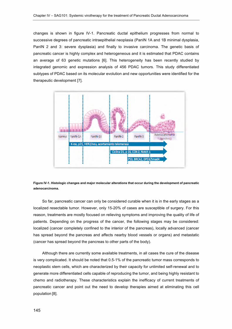
Chapter IV – SAG101: Systemic virotherapy for the treatment of Pancreatic Ductal Adenocarcinoma
145
changes is shown in figure IV-1. Pancreatic ductal epithelium progresses from normal to
successive degrees of pancreatic intraepithelial neoplasia (PanIN 1A and 1B minimal dysplasia,
PanIN 2 and 3: severe dysplasia) and finally to invasive carcinoma. The genetic basis of
pancreatic cancer is highly complex and heterogeneous and it is estimated that PDAC contains
an average of 63 genetic mutations [6]. This heterogeneity has been recently studied by
integrated genomic and expression analysis of 456 PDAC tumors. This study differentiated
subtypes of PDAC based on its molecular evolution and new opportunities were identified for the
therapeutic development [7].
Figure IV-1. Histologic changes and major molecular alterations that occur during the development of pancreatic adenocarcinoma.
So far, pancreatic cancer can only be considered curable when it is in the early stages as a
localized resectable tumor. However, only 15-20% of cases are susceptible of surgery. For this
reason, treatments are mostly focused on relieving symptoms and improving the quality of life of
patients. Depending on the progress of the cancer, the following stages may be considered:
localized (cancer completely confined to the interior of the pancreas), locally advanced (cancer
has spread beyond the pancreas and affects nearby blood vessels or organs) and metastatic
(cancer has spread beyond the pancreas to other parts of the body).
Although there are currently some available treatments, in all cases the cure of the disease
is very complicated. It should be noted that 0.5-1% of the pancreatic tumor mass corresponds to
neoplastic stem cells, which are characterized by their capacity for unlimited self-renewal and to
generate more differentiated cells capable of reproducing the tumor, and being highly resistant to
chemo and radiotherapy. These characteristics explain the inefficacy of current treatments of
pancreatic cancer and point out the need to develop therapies aimed at eliminating this cell
population [8].
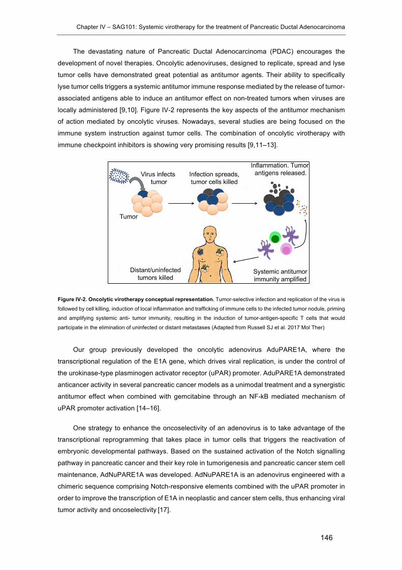
Chapter IV – SAG101: Systemic virotherapy for the treatment of Pancreatic Ductal Adenocarcinoma
146
The devastating nature of Pancreatic Ductal Adenocarcinoma (PDAC) encourages the
development of novel therapies. Oncolytic adenoviruses, designed to replicate, spread and lyse
tumor cells have demonstrated great potential as antitumor agents. Their ability to specifically
lyse tumor cells triggers a systemic antitumor immune response mediated by the release of tumor-
associated antigens able to induce an antitumor effect on non-treated tumors when viruses are
locally administered [9,10]. Figure IV-2 represents the key aspects of the antitumor mechanism
of action mediated by oncolytic viruses. Nowadays, several studies are being focused on the
immune system instruction against tumor cells. The combination of oncolytic virotherapy with
immune checkpoint inhibitors is showing very promising results [9,11–13].
Figure IV-2. Oncolytic virotherapy conceptual representation. Tumor-selective infection and replication of the virus is
followed by cell killing, induction of local inflammation and trafficking of immune cells to the infected tumor nodule, priming
and amplifying systemic anti- tumor immunity, resulting in the induction of tumor-antigen-specific T cells that would
participate in the elimination of uninfected or distant metastases (Adapted from Russell SJ et al. 2017 Mol Ther)
Our group previously developed the oncolytic adenovirus AduPARE1A, where the
transcriptional regulation of the E1A gene, which drives viral replication, is under the control of
the urokinase-type plasminogen activator receptor (uPAR) promoter. AduPARE1A demonstrated
anticancer activity in several pancreatic cancer models as a unimodal treatment and a synergistic
antitumor effect when combined with gemcitabine through an NF-kB mediated mechanism of
uPAR promoter activation [14–16].
One strategy to enhance the oncoselectivity of an adenovirus is to take advantage of the
transcriptional reprogramming that takes place in tumor cells that triggers the reactivation of
embryonic developmental pathways. Based on the sustained activation of the Notch signalling
pathway in pancreatic cancer and their key role in tumorigenesis and pancreatic cancer stem cell
maintenance, AdNuPARE1A was developed. AdNuPARE1A is an adenovirus engineered with a
chimeric sequence comprising Notch-responsive elements combined with the uPAR promoter in
order to improve the transcription of E1A in neoplastic and cancer stem cells, thus enhancing viral
tumor activity and oncoselectivity [17].

Chapter IV – SAG101: Systemic virotherapy for the treatment of Pancreatic Ductal Adenocarcinoma
147
However, the use of viruses as therapeutic agents is mainly restricted by inherent safety
risks [18]. Moreover, viral vectors�are often susceptible to neutralization by serum antibodies and
complement [19–22]. Furthermore, inefficient tumor targeting after systemic administration also
limits their efficacy in contrast with locally administered viruses.
As previously demonstrated in chapter III, CPEG-coated Ad vectors presented an improved
in vitro transduction profile in the presence of NAbs, liver transduction in vivo was minimized while
enhancing the circulation time of intravenously injected Ads, and tumor transduction was
consequently improved. All these results suggested that the CPEG-coating technology could
boost the efficacy of intravenously administered viral vectors designed to treat cancer.
In the present chapter, the combination of the CPEG OM-pBAEs-based formulation
developed in chapter III with the AdNuPARmE1A oncolytic virus has been studied. This hybrid
OM-PBAEs/Ad vector is called SAG101 and is currently under further development by Sagetis-
Biotech S.L. SAG-101 has recently achieved the classification of advanced therapeutic medicinal
product (ATMP) and the designation of orphan drug (ODD) for the treatment of pancreatic cancer
by the EMA.
Figure IV-3. Schematic representation of SAG101, a hybrid OM-pBAE/AdNuPARmE1A oncolytic virus for the treatment of pancreatic ductal adenocarcinoma (PDAC).
Therefore, the main objective of the present chapter is to study the efficacy and safety profile
of SAG101 in comparison with its naked counterpart, AdNuPARmE1A.
In order to achieve this objective, the following tasks were proposed:
• Study the in vitro functionality of SAG101 in terms of infectivity and cytotoxicity
• Determine the toxicity profile of systemically administered SAG101 in comparison with
naked AdNuPARmE1A in immunocompetent mice
• Study the antitumor efficacy of SAG101 in a pre-immune context in vivo
Chapter IV – SAG101: Systemic virotherapy for the treatment of Pancreatic Ductal Adenocarcinoma
148
4.2 Materials and Methods
4.2.1 Materials
DMSO (Dimethylsulfoxide purissimum), EtOH (absolute ethanol), MetOH (Methanol) were
obtained from Sigma-Aldrich GmbH (Munich, Germany). Water was used as purified, de-ionized
water. Reagents and solvents used for polymer synthesis were purchased from Sigma-Aldrich
and Panreac. Oligopeptide moieties used for polymer modification (H-Cys-Arg-Arg-Arg-NH2 and
H-Cys-Asp-Asp-Asp-NH2) were obtained from GL Biochem (Shanghai) Ltd with a purity higher
than 98%. MeO-PEG (Mw = 2000) was purchased from Iris Biotech GmbH.
Cell culture media, antibiotics, L-glutamine and fetal bovine serum were purchased from
Gibco (Life Technologies, Carlsbad, USA).
4.2.2 Adenovirus vectors
4.2.2.1 Virus amplification and purification
AdNuPARmE1A was produced starting from a previously purified viral stock.
AdNuPARmE1A was amplified in A549 and purified by one CsCl density step gradient (1,5, 1,35
and 1,25 g/ml CsCl – 2h at 35000rpm) followed by one subsequent continuous CsCl density
gradient (1,35g/ml – 24h at 35000rpm) and were desalted by PD-10 size exclusion columns (GE
Healthcare) and stored in PBS++ with 10% glycerol at -80ºC.
4.2.2.2 Physical titer determination by spectroscopy (VP/ml)
The physical titer (vp/ml) is determined by viral DNA absorption measurement at 260 nm
wavelength following the same procedure as explained in 3.2.3.2.
4.2.2.3 Titration by hexon staining (pfu/ml)
Viral titration by hexon staining determines the number of infective particles, and it is based
on the number of hexon-positive cells of an infected monolayer after immunostaining against the
viral hexon protein. Briefly, 50.000 HEK-293 cells per well in a 96-well plate are infected with 100
µl of a viral serial dilution bank (104-1012) in triplicate and incubated for 20h at 37ºC. Cells are
fixed with cold MeOH 20 min at -20ºC, washed three times 5 min with PBS++ 1%BSA, and
incubated for 1h at 37ºC with mouse anti-hexon hybridoma (dilution 1/3). Then, cells were washed
and incubated with the secondary antibody anti-mouse Alexa Fluor 488 (Invitrogen) (dilution
1/400), 1 h at RT. After 3 washes with PBS++ 1%BSA, positive cells are counted under
fluorescence microscope. The titer of virus is then calculated according the following formula:
;^B@E
= CLb=_BQ>=EB<=AC0,1@E

Chapter IV – SAG101: Systemic virotherapy for the treatment of Pancreatic Ductal Adenocarcinoma
149
4.2.3 Preparation of SAG101 (CPEG-coated AdNuPARE1A)
In order to prepare SAG101 samples, same procedure as explained in chapter III section
1.2.4 is used. However, in this case, ratio pol:VP is fixed at 4x10⁶ and the polymer used is a 65:35
w/w mixture of C32CR3 and C6PEGCR3. See section 1.2.4.1 for preparation of SAG101 for in
vitro testing and section 1.2.4.2 for preparation of SAG101 for in vivo studies.
4.2.4 Dose-response analyses
Ten thousand PANC-1, MIA PaCa-2 and A549 cells were treated with naked
AdNuPARmE1A, CPEG and CPEGAdNuPARmE1A (SAG101). Three days later, cell viability was
measured and quantified by a colorimetric assay system based on the tretrazolium salt 3-(4,5-
dimethylthiazol-2-yl)-2,5-diphenyl tetrazolium bromide (MTT; Roche Molecular Biochemicals), in
accordance with the manufacturer’s instructions. Results were expressed as the percent
absorbance determined in treated wells relative to that in untreated wells. IC50 values were
estimated from dose-response curves by standard non-linear regression GraphPad.
4.2.5 RAW264.7 cytokine release assays
RAW264.7 macrophages were seeded in a 6-well-plate at 1x10⁶ cells/well in supplemented
DMEM. After 24 h, culture medium was replaced with 1 mL of fresh serum-free medium containing
1000 VP/cell of naked Ad, SAG101, the polymeric component without viral particles and the
corresponding residual volume of DMSO as found in SAG101 as a control. After 6 h, 500 µl of
10% FBS-containing medium was added to each well and 48 h later, culture media were collected
from each well avoiding cell collection. The concentration of GM-CSF, IFN-γ, IL1β, IL2, IL6, IL10,
IP10 and TNF-α was quantified using a microbead-based ELISA kit (Mouse Cytokine 10-Plex
Panel; Invitrogen) on the Luminex™ 200™.
4.2.6 Neutralizing sera generation in vivo
C57BL/6J naïve mice were immunized by injecting two doses of 3x10¹⁰ VP/animal of naked
Ad intravenously at day 0 and day 14. At day 21 after the first injection, blood was collected by
intracardiac puncture and sera were obtained, heat inactivated and pooled. The neutralizing
capacity of the collected sera was quantified in vitro following the same procedure as explained
in 3.2.7.6.
4.2.7 Antitumor activity in passively immunized mice in vivo
To assess the efficacy of systemically administered viral preparations, subcutaneous PANC-
1 or Mia PaCa-2 tumors were established by injecting 2x10⁶ cells into the flanks of 7-week-old
male Balb/C nu/nu mice. Cells were prepared in Matrigel Matrix Basement Membrane HC
(Corning) by 1:1 mixing cells in DMEM without supplements and Matrigel to a final volume of 100
µl.

Chapter IV – SAG101: Systemic virotherapy for the treatment of Pancreatic Ductal Adenocarcinoma
150
Once tumors reached 100 mm3, mice were randomized (n=8 per group) and two groups
were passively immunized by injecting intraperitoneally 200 µl of neutralizing serum. The day
after, animals were treated with a single intravenous injection of vehicle (Saline) or 4x10¹⁰ VP of
AdNuPARmE1A or SAG101 in a total volume of 100 µl via tail vein injection. These doses were
used for passively immunized and non-immunized groups.
Tumor progression was analyzed by measuring tumor’s volume using a digital caliper three
times per week. The large and the small diameter were measured and the tumor volume was
calculated using the following formula:
jB@AB_WAEB@: = (T ∗ >Y ∗ p)
6
4.2.8 In vivo toxicity study in mice
Blank, (Saline), vehicle (Saline, 0,9% Glycerol, 6,2% DMSO) or 5x10¹⁰ VP/animal or
7,5x10¹⁰ VP/animal of AdNuPARmE1A or SAG101 were injected intravenously into the tail vein
in 7-week-old immunocompetent BALB/C male mice in a final volume of 100 µl (n=3-7). Animals
were weighted and examined daily for clinical signs of toxicity. At 1, 3 and 5 days after the
administration of the virus, blood aliquots were collected via tail vein for platelet count. Also, at 6
h and 72h after injection blood samples were collected for cytokine determination. Seven days
after injection, mice were killed and blood and serum samples were collected by intracardiac
puncture for transaminases activity quantification and hematologic studies.
4.2.8.1 Hepatotoxicity (ALT, AST transaminase activity analysis)
Anesthetized animals were punctured intracardiacally using a 26G needle in order to obtain
large blood volumes. After obtaining the blood, animals were sacrificed by cervical dislocation.
Blood samples were left 30 min at room temperature in order to induce blood clotting. Then, blood
samples were centrifuged (3500rpm-15min) and blood serums were obtained and stored at -
80ºC.
Blood sera were analyzed for aspartate aminotransferase (AST) and alanine
aminotransferase (ALT) activity. These biochemical markers are closely related to hepatic
damage when detected in blood. The analyses were conducted at the clinical biochemistry
service of Hospital Clínic de Barcelona.
4.2.8.2 Hematologic study
Blood samples for platelet cell count were collected using heparinized blood collection
capillaries and platelets were manually counted using a hematocytometer diluting 25 µl of blood
with lysis buffer (1% ammoinium oxalate solution) to induce lysis of red blood cells (RBC).
Chapter IV – SAG101: Systemic virotherapy for the treatment of Pancreatic Ductal Adenocarcinoma
151
Hemograms were performed with whole blood collected by intracardiac puncture in EDTA
tubes at the Centre de Diagnostic Biomèdic (CDB) of Hospital Clínic de Barcelona. Half of the
collected blood was used for hematologic studies and the other half was used to prepare sera for
biochemical studies.
4.2.8.3 In vivo cytokine release studies
Serum samples collected 6h and 72h post injection were used to determine cytokine
concentrations. GM-CSF, IFN-γ, IL1β, IL2, IL6, IL10, IP10 and TNF-α was quantified using a
microbead-based ELISA kit (Mouse Cytokine 10-Plex Panel; Invitrogen) on the Luminex™ 200™
following the manufacturer recommended procedures.
4.2.9 Statistical analysis
Statistical analyses were carried out with Graph-Pad Prism (GraphPad Software). All error
bars reported are SEM unless otherwise indicated. Pairwise comparisons were performed using
one-way Student t-tests. Differences between groups were considered significant at P values
below 0.05 (* p < 0.05, ** p < 0.01, *** p < 0.001). Tumor progression data was compared between
conditions using linear mixed effect in R v2.14.1 using the lme4 package.
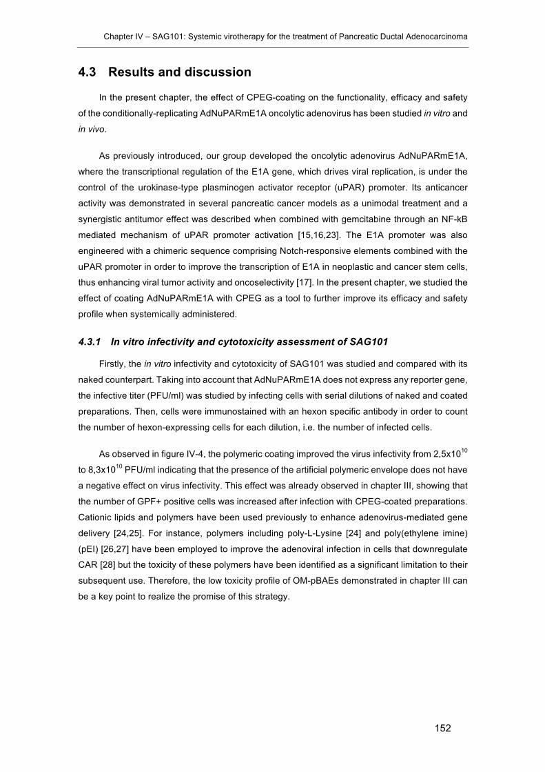
Chapter IV – SAG101: Systemic virotherapy for the treatment of Pancreatic Ductal Adenocarcinoma
152
4.3 Results and discussion
In the present chapter, the effect of CPEG-coating on the functionality, efficacy and safety
of the conditionally-replicating AdNuPARmE1A oncolytic adenovirus has been studied in vitro and
in vivo.
As previously introduced, our group developed the oncolytic adenovirus AdNuPARmE1A,
where the transcriptional regulation of the E1A gene, which drives viral replication, is under the
control of the urokinase-type plasminogen activator receptor (uPAR) promoter. Its anticancer
activity was demonstrated in several pancreatic cancer models as a unimodal treatment and a
synergistic antitumor effect was described when combined with gemcitabine through an NF-kB
mediated mechanism of uPAR promoter activation [15,16,23]. The E1A promoter was also
engineered with a chimeric sequence comprising Notch-responsive elements combined with the
uPAR promoter in order to improve the transcription of E1A in neoplastic and cancer stem cells,
thus enhancing viral tumor activity and oncoselectivity [17]. In the present chapter, we studied the
effect of coating AdNuPARmE1A with CPEG as a tool to further improve its efficacy and safety
profile when systemically administered.
4.3.1 In vitro infectivity and cytotoxicity assessment of SAG101
Firstly, the in vitro infectivity and cytotoxicity of SAG101 was studied and compared with its
naked counterpart. Taking into account that AdNuPARmE1A does not express any reporter gene,
the infective titer (PFU/ml) was studied by infecting cells with serial dilutions of naked and coated
preparations. Then, cells were immunostained with an hexon specific antibody in order to count
the number of hexon-expressing cells for each dilution, i.e. the number of infected cells.
As observed in figure IV-4, the polymeric coating improved the virus infectivity from 2,5x10¹⁰
to 8,3x10¹⁰ PFU/ml indicating that the presence of the artificial polymeric envelope does not have
a negative effect on virus infectivity. This effect was already observed in chapter III, showing that
the number of GPF+ positive cells was increased after infection with CPEG-coated preparations.
Cationic lipids and polymers have been used previously to enhance adenovirus-mediated gene
delivery [24,25]. For instance, polymers including poly-L-Lysine [24] and poly(ethylene imine)
(pEI) [26,27] have been employed to improve the adenoviral infection in cells that downregulate
CAR [28] but the toxicity of these polymers have been identified as a significant limitation to their
subsequent use. Therefore, the low toxicity profile of OM-pBAEs demonstrated in chapter III can
be a key point to realize the promise of this strategy.
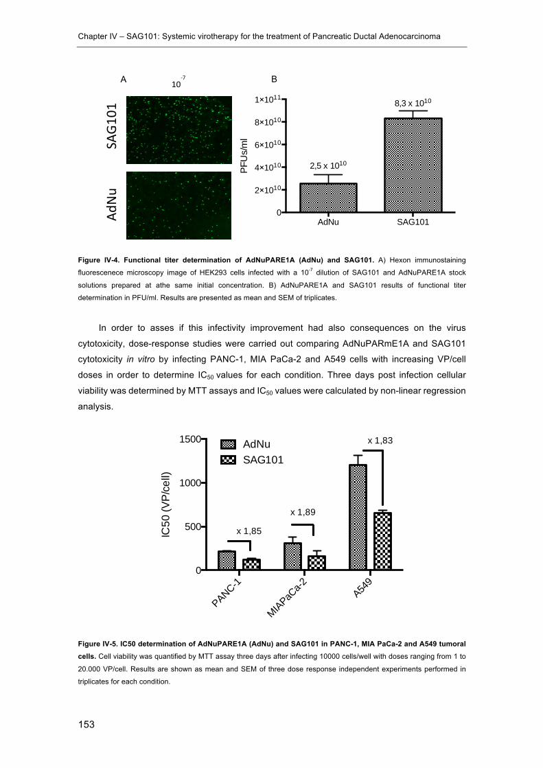
Chapter IV – SAG101: Systemic virotherapy for the treatment of Pancreatic Ductal Adenocarcinoma
153
Figure IV-4. Functional titer determination of AdNuPARE1A (AdNu) and SAG101. A) Hexon immunostaining
fluorescenece microscopy image of HEK293 cells infected with a 10-7 dilution of SAG101 and AdNuPARE1A stock
solutions prepared at athe same initial concentration. B) AdNuPARE1A and SAG101 results of functional titer
determination in PFU/ml. Results are presented as mean and SEM of triplicates.
In order to asses if this infectivity improvement had also consequences on the virus
cytotoxicity, dose-response studies were carried out comparing AdNuPARmE1A and SAG101
cytotoxicity in vitro by infecting PANC-1, MIA PaCa-2 and A549 cells with increasing VP/cell
doses in order to determine IC50 values for each condition. Three days post infection cellular
viability was determined by MTT assays and IC50 values were calculated by non-linear regression
analysis.
Figure IV-5. IC50 determination of AdNuPARE1A (AdNu) and SAG101 in PANC-1, MIA PaCa-2 and A549 tumoral cells. Cell viability was quantified by MTT assay three days after infecting 10000 cells/well with doses ranging from 1 to
20.000 VP/cell. Results are shown as mean and SEM of three dose response independent experiments performed in
triplicates for each condition.
AdNu SAG1010
2×10¹⁰
4×10¹⁰
6×10¹⁰
8×10¹⁰
1×10¹¹
PFU
s/m
l
2,5 x 10¹⁰
8,3 x 10¹⁰
PANC-1
MIAPaCa-2 A54
90
500
1000
1500 AdNuSAG101
x 1,85
x 1,89
x 1,83
IC50
(VP/
cell)
10-7
SAG1
01
AdNu
A B
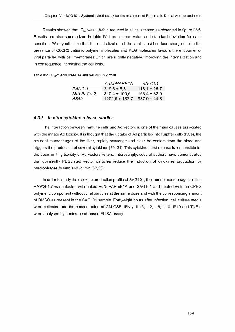
Chapter IV – SAG101: Systemic virotherapy for the treatment of Pancreatic Ductal Adenocarcinoma
154
Results showed that IC50 was 1,8-fold reduced in all cells tested as observed in figure IV-5.
Results are also summarized in table IV-1 as a mean value and standard deviation for each
condition. We hypothesize that the neutralization of the viral capsid surface charge due to the
presence of C6CR3 cationic polymer molecules and PEG molecules favours the encounter of
viral particles with cell membranes which are slightly negative, improving the internalization and
in consequence increasing the cell lysis.
Table IV-1. IC50 of AdNuPARE1A and SAG101 in VP/cell
AdNuPARE1A SAG101 PANC-1 219,6 ± 5,3 118,1 ± 25,7 MIA PaCa-2 310,4 ± 100,6 163,4 ± 82,9 A549 1202,5 ± 157,7 657,9 ± 44,5
4.3.2 In vitro cytokine release studies
The interaction between immune cells and Ad vectors is one of the main causes associated
with the innate Ad toxicity. It is thought that the uptake of Ad particles into Kupffer cells (KCs), the
resident macrophages of the liver, rapidly scavenge and clear Ad vectors from the blood and
triggers the production of several cytokines [29–31]. This cytokine burst release is responsible for
the dose-limiting toxicity of Ad vectors in vivo. Interestingly, several authors have demonstrated
that covalently PEGylated vector particles reduce the induction of cytokines production by
macrophages in vitro and in vivo [32,33].
In order to study the cytokine production profile of SAG101, the murine macrophage cell line
RAW264.7 was infected with naked AdNuPARmE1A and SAG101 and treated with the CPEG
polymeric component without viral particles at the same dose and with the corresponding amount
of DMSO as present in the SAG101 sample. Forty-eight hours after infection, cell culture media
were collected and the concentration of GM-CSF, IFN-γ, IL1β, IL2, IL6, IL10, IP10 and TNF-α
were analysed by a microbead-based ELISA assay.
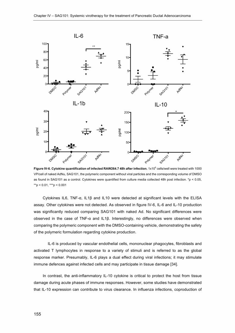
Chapter IV – SAG101: Systemic virotherapy for the treatment of Pancreatic Ductal Adenocarcinoma
155
Figure IV-6. Cytokine quantification of infected RAW264.7 48h after infection. 1x10⁶ cells/well were treated with 1000
VP/cell of naked AdNu, SAG101, the polymeric component without viral particles and the corresponding volume of DMSO
as found in SAG101 as a control. Cytokines were quantified from culture media collected 48h post infection. *p < 0.05,
**p < 0.01, ***p < 0.001
Cytokines IL6, TNF-α, IL1β and IL10 were detected at significant levels with the ELISA
assay. Other cytokines were not detected. As observed in figure IV-6, IL-6 and IL-10 production
was significantly reduced comparing SAG101 with naked Ad. No significant differences were
observed in the case of TNF-α and IL1β. Interestingly, no differences were observed when
comparing the polymeric component with the DMSO-containing vehicle, demonstrating the safety
of the polymeric formulation regarding cytokine production.
IL-6 is produced by vascular endothelial cells, mononuclear phagocytes, fibroblasts and
activated T lymphocytes in response to a variety of stimuli and is referred to as the global
response marker. Presumably, IL-6 plays a dual effect during viral infections; it may stimulate
immune defences against infected cells and may participate in tissue damage [34].
In contrast, the anti-inflammatory IL-10 cytokine is critical to protect the host from tissue
damage during acute phases of immune responses. However, some studies have demonstrated
that IL-10 expression can contribute to virus clearance. In influenza infections, coproduction of
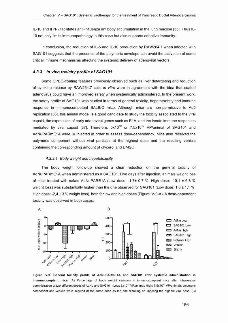
Chapter IV – SAG101: Systemic virotherapy for the treatment of Pancreatic Ductal Adenocarcinoma
156
IL-10 and IFN-γ facilitates anti-influenza antibody accumulation in the lung mucosa [35]. Thus IL-
10 not only limits immunopathology in this case but also supports adaptive immunity.
In conclusion, the reduction of IL-6 and IL-10 production by RAW264.7 when infected with
SAG101 suggests that the presence of the polymeric envelope can avoid the activation of some
critical immune mechanisms affecting the systemic delivery of adenoviral vectors.
4.3.3 In vivo toxicity profile of SAG101
Some CPEG-coating features previously observed such as liver detargeting and reduction
of cytokine release by RAW264.7 cells in vitro were in agreement with the idea that coated
adenovirus could have an improved safety when systemically administered. In the present work,
the safety profile of SAG101 was studied in terms of general toxicity, hepatotoxicity and immune
response in immunocompetent BALB/C mice. Although mice are non-permissive to Ad5
replication [36], this animal model is a good candidate to study the toxicity associated to the viral
capsid, the expression of early adenoviral genes such as E1A, and the innate immune responses
mediated by viral capsid [37]. Therefore, 5x10¹⁰ or 7,5x10¹⁰ VP/animal of SAG101 and
AdNuPARmE1A were IV injected in order to assess dose-dependency. Mice also received the
polymeric component without viral particles at the highest dose and the resulting vehicle
containing the corresponding amount of glycerol and DMSO.
4.3.3.1 Body weight and hepatotoxicity
The body weight follow-up showed a clear reduction on the general toxicity of
AdNuPARmE1A when administered as a SAG101. Five days after injection, animals weight loss
of mice treated with naked AdNuPARE1A (Low dose: -1,7± 0,7 %; High dose: -10,1 ± 6,8 %
weight loss) was substantially higher than the one observed for SAG101 (Low dose: 1,6 ± 1,1 %;
High dose: -2,4 ± 3 % weight loss), both for low and high doses (Figure IV-9-A). A dose-dependent
toxicity was observed in both cases.
Figure IV-9. General toxicity profile of AdNuPARmE1A and SAG101 after systemic administration in immunocomptent mice. (A) Percentage of body weight variation in immunocomptent mice after intravenous
administration of two different doses of AdNu and SAG101 (Low: 5x10¹⁰ VP/animal; High: 7,5x10¹⁰ VP/animal); polymeric
component and vehicle were injected at the same dose as the one resulting on injecting the highest viral dose. (B)
AdNu L
ow
SAG101 L
ow
AdNu H
igh
SAG101 H
igh
Polymer
High
Vehicle
Blank
-15
-10
-5
0
5
% o
f bod
y w
eigh
t at d
ay 5
ASTALT
0
100
200
300
400
500AdNu LowSAG101 LowAdNu HighSAG101 HighPolymer HighVehicle
U/L
Blank
* **
A B
Chapter IV – SAG101: Systemic virotherapy for the treatment of Pancreatic Ductal Adenocarcinoma
157
Assessment of hepatotoxicity by the determination of AST, ALT in the serum of treated mice 7 days post-administration.
Results are expressed as the mean and SEM of n=8 animals/group, or n = 3 in the control groups *p < 0.05, **p < 0.01.
The level of serum alanine aminotransferase (ALT) and aspartate aminotransferase (AST)
activity reflects damage to hepatocytes and is considered to be a highly sensitive and fairly
specific preclinical and clinical biomarker of hepatotoxicity [38]. Covalent and ionic linkage of
polymers on adenovirus surfaces have demonstrated to reduce the liver toxicity observed as
decreased transaminases levels in blood [39–41] after intravenous administration. As observed
in figure IV-9-B, this effect was also observed for SAG101-injected mice showing lower levels of
both markers with statistically significant improvements for the high dose seven days after
treatment. These results were in accordance with the liver detargeting effect observed in chapter
III.
4.3.3.2 Hematologic study: Cells counting
White blood cells counting showed a reduced number of monocytes and neutrophils (Figure
IV-11-B and D) in blood when animals were treated with SAG101. Both cell types are related to
the induction of innate immune responses and in the case of monocytes, they play a crucial role
as antigen-presenting cells to T-cells stimulating the adaptive immune response and triggering
NAbs production. This observation was in accordance with the reduction of the neutralizing
capacity of sera collected from animals injected with CPEGAd in comparison with those injected
with naked Ad, as described in chapter III.

Chapter IV – SAG101: Systemic virotherapy for the treatment of Pancreatic Ductal Adenocarcinoma
158
Figure IV-11. Hematologic study by cell counting in response to AdNuPARE1A and SAG101 intravenous injection. White blood cells in Balb/C peripheral blood counts at seven days post intravenous administration of 5x10¹⁰ (Low) or
7,5x10¹⁰ (High) viral particles of AdNuPARE1A (AdNu) and SAG101. (A) Lymphocytes, (B) Monocytes, (C) Eosinophils
and (D) Neutrophils cell counts. Mean values with SEM are depicted.
It is important to note that the automated cell counter is very sensitive to blood clotting.
Despite blood samples were collected in EDTA tubes and freshly analysed, the cell counter was
not able to analyse several samples. Virus treated samples starting with N=5 reached the
minimum three samples needed to calculate the standard deviation. However, this minimum was
not achieved with other groups starting with N=3, such as polymer (N=0), vehicle (N=2) and blank
(N=1).
Finally, in order to study the behaviour of SAG101 in terms of thrombocytopenia induction,
a condition related to intravenous administration of Ad characterized by abnormally low levels of
thrombocytes (platelets) [42], blood samples were collected at days 1, 3 and 5 post injection and
platelets were manually counted.
Platelet cell counts were lower in comparison with the blank condition for all virus treated
conditions one day after injection. However, the low dose of SAG101 showed a faster recovery
rate reaching normal platelet levels three days after injection.
Lymphocytes
AdNu L
ow
SAG101 L
ow
AdNu H
igh
SAG101 H
igh
Polymer
Vehicle
Blank
0
2
4
6
8
10
x106 ce
lls/m
l
Eosinophils
AdNu L
ow
SAG101 L
ow
AdNu H
igh
SAG101 H
igh
Polymer
Vehicle
Blank
0.0
0.2
0.4
0.6
0.8
1.0
x106 ce
lls/m
l
Monocytes
AdNu L
ow
SAG101 L
ow
AdNu H
igh
SAG101 H
igh
Polymer
Vehicle
Blank
0.0
0.2
0.4
0.6
x106 ce
lls/m
l
Neutrophils
AdNu L
ow
SAG101 L
ow
AdNu H
igh
SAG101 H
igh
Polymer
Vehicle
Blank
0
1
2
3
4
x106 ce
lls/m
l
A B
C D
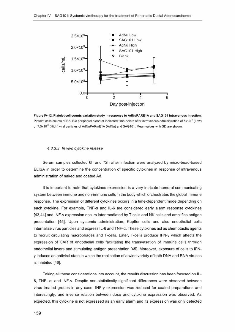
Chapter IV – SAG101: Systemic virotherapy for the treatment of Pancreatic Ductal Adenocarcinoma
159
Figure IV-12. Platelet cell counts variation study in response to AdNuPARE1A and SAG101 intravenous injection. Platelet cells counts of BALB/c peripheral blood at indicated time-points after intravenous administration of 5x10¹⁰ (Low)
or 7,5x10¹⁰ (High) viral particles of AdNuPARmE1A (AdNu) and SAG101. Mean values with SD are shown.
4.3.3.3 In vivo cytokine release
Serum samples collected 6h and 72h after infection were analyzed by micro-bead-based
ELISA in order to determine the concentration of specific cytokines in response of intravenous
administration of naked and coated Ad.
It is important to note that cytokines expression is a very intricate humoral communicating
system between immune and non-immune cells in the body which orchestrates the global immune
response. The expression of different cytokines occurs in a time-dependent mode depending on
each cytokine. For example, TNF-α and IL-6 are considered early alarm response cytokines
[43,44] and INF-γ expression occurs later mediated by T cells and NK cells and amplifies antigen
presentation [45]. Upon systemic administration, Kupffer cells and also endothelial cells
internalize virus particles and express IL-6 and TNF-α. These cytokines act as chemotactic agents
to recruit circulating macrophages and T-cells. Later, T-cells produce IFN-γ which affects the
expression of CAR of endothelial cells facilitating the transvasation of immune cells through
endothelial layers and stimulating antigen presentation [45]. Moreover, exposure of cells to IFN-
γ induces an antiviral state in which the replication of a wide variety of both DNA and RNA viruses
is inhibited [46].
Taking all these considerations into account, the results discussion has been focused on IL-
6, TNF- α, and INF-γ. Despite non-statistically significant differences were observed between
virus treated groups in any case, INF-γ expression was reduced for coated preparations and
interestingly, and inverse relation between dose and cytokine expression was observed. As
expected, this cytokine is not expressed as an early alarm and its expression was only detected
0 2 4 60.0
5.0×10⁸
1.0×10⁹
1.5×10⁹
2.0×10⁹
2.5×10⁹ AdNu LowSAG101 LowAdNu HighSAG101 High
Day post-injection
cells
/mL Blank
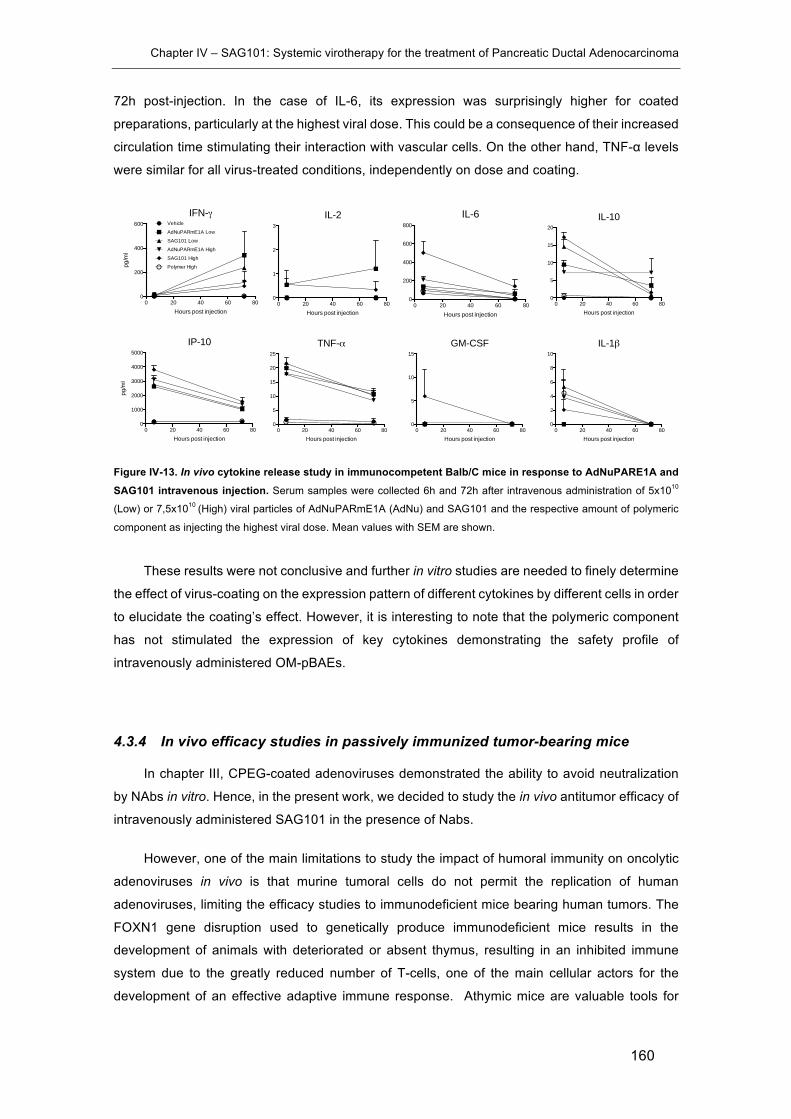
Chapter IV – SAG101: Systemic virotherapy for the treatment of Pancreatic Ductal Adenocarcinoma
160
72h post-injection. In the case of IL-6, its expression was surprisingly higher for coated
preparations, particularly at the highest viral dose. This could be a consequence of their increased
circulation time stimulating their interaction with vascular cells. On the other hand, TNF-α levels
were similar for all virus-treated conditions, independently on dose and coating.
Figure IV-13. In vivo cytokine release study in immunocompetent Balb/C mice in response to AdNuPARE1A and SAG101 intravenous injection. Serum samples were collected 6h and 72h after intravenous administration of 5x10¹⁰
(Low) or 7,5x10¹⁰ (High) viral particles of AdNuPARmE1A (AdNu) and SAG101 and the respective amount of polymeric
component as injecting the highest viral dose. Mean values with SEM are shown.
These results were not conclusive and further in vitro studies are needed to finely determine
the effect of virus-coating on the expression pattern of different cytokines by different cells in order
to elucidate the coating’s effect. However, it is interesting to note that the polymeric component
has not stimulated the expression of key cytokines demonstrating the safety profile of
intravenously administered OM-pBAEs.
4.3.4 In vivo efficacy studies in passively immunized tumor-bearing mice
In chapter III, CPEG-coated adenoviruses demonstrated the ability to avoid neutralization
by NAbs in vitro. Hence, in the present work, we decided to study the in vivo antitumor efficacy of
intravenously administered SAG101 in the presence of Nabs.
However, one of the main limitations to study the impact of humoral immunity on oncolytic
adenoviruses in vivo is that murine tumoral cells do not permit the replication of human
adenoviruses, limiting the efficacy studies to immunodeficient mice bearing human tumors. The
FOXN1 gene disruption used to genetically produce immunodeficient mice results in the
development of animals with deteriorated or absent thymus, resulting in an inhibited immune
system due to the greatly reduced number of T-cells, one of the main cellular actors for the
development of an effective adaptive immune response. Athymic mice are valuable tools for
IFN-γ
0 20 40 60 800
200
400
600 Vehicle
AdNuPARmE1A Low
SAG101 Low
AdNuPARmE1A High
SAG101 High
Polymer High
Hours post injection
pg/m
l
IP-10
0 20 40 60 800
1000
2000
3000
4000
5000
Hours post injection
pg/m
l
IL-2
0 20 40 60 800
1
2
3
Hours post injection
TNF-α
0 20 40 60 800
5
10
15
20
25
Hours post injection
IL-6
0 20 40 60 800
200
400
600
800
Hours post injection
GM-CSF
0 20 40 60 800
5
10
15
Hours post injection
IL-10
0 20 40 60 800
5
10
15
20
Hours post injection
IL-1β
0 20 40 60 800
2
4
6
8
10
Hours post injection

Chapter IV – SAG101: Systemic virotherapy for the treatment of Pancreatic Ductal Adenocarcinoma
161
cancer research because they can receive human tumor grafts without producing a rejecting
response. However, in the context of human oncolytic adenovirus research this represents an
important limitation taking into account the importance of the virus-induced anti-tumoral immune
response.
Thus, in order to be able to study the efficacy of SAG101 in vivo in the presence of NAbs
antibodies, athymic nude mice implanted with human pancreatic cancer subcutaneous xenografts
were passively immunized with sera previously generated in immunocompetent C57BL6 mice the
day before injecting naked and coated AdNuPARmE1A. The feasibility and ability of transferred
sera to neutralize antitumor activity in vivo was previously demonstrated by other authors [47]
and was also observed in our experimentation.
Neutralizing sera were generated in C57BL6J as previously explained in section 4.2.5. Their
neutralization capacity was determined in vitro by a dose-response neutralization assay (ND50).
As observed in figure IV-7, sera used to passively immunize animals for both efficacy experiments
(PANC-1 and Mia PaCa-2 tumor-bearing mice) showed the same ND50 value of 2560.
Figure IV-7. In vitro neutralization assay to determine ND50 of sera injected to passively immunize tumor-bearing immunodeficient mice. Each serum was analysed in replicate and results are presented as MIA PaCa-2 Rep and PANC-
1 Rep.
Accordingly, SAG101 efficacy after systemic administration was compared with that of
AdNuPARmE1A in naïve or pre-immune mice, following the experimental schedule summarized
in figure IV-8-A. Tumor growth was monitored during more than 40 days post injection in both
cases (figure IV-8-B, C).
1:10
1:20
1:40
1:801:1
601:3
201:6
401:1
2801:2
5601:5
120
0
50
100
PANC-1
MIA PaCa-2
PANC-1 Rep
MIA PaCa-2 Rep
STND Ref
Dilution factor
% o
f neu
traliz
atio
n
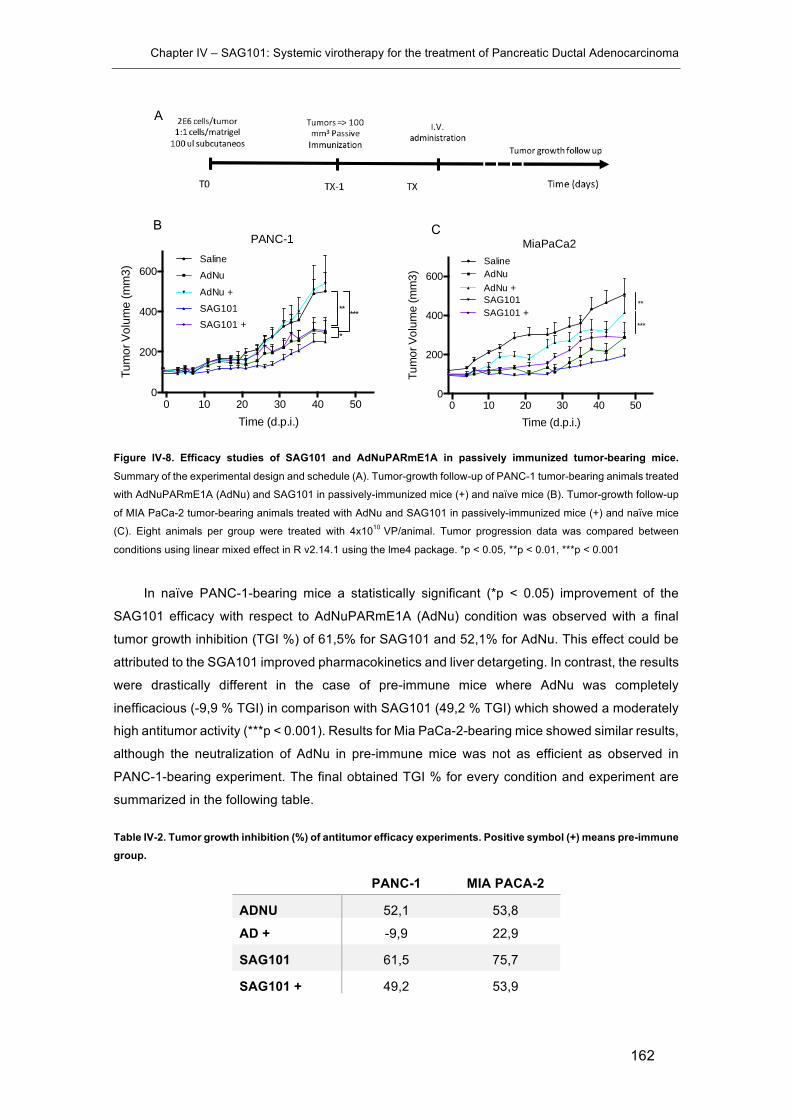
Chapter IV – SAG101: Systemic virotherapy for the treatment of Pancreatic Ductal Adenocarcinoma
162
Figure IV-8. Efficacy studies of SAG101 and AdNuPARmE1A in passively immunized tumor-bearing mice. Summary of the experimental design and schedule (A). Tumor-growth follow-up of PANC-1 tumor-bearing animals treated
with AdNuPARmE1A (AdNu) and SAG101 in passively-immunized mice (+) and naïve mice (B). Tumor-growth follow-up
of MIA PaCa-2 tumor-bearing animals treated with AdNu and SAG101 in passively-immunized mice (+) and naïve mice
(C). Eight animals per group were treated with 4x10¹⁰ VP/animal. Tumor progression data was compared between
conditions using linear mixed effect in R v2.14.1 using the lme4 package. *p < 0.05, **p < 0.01, ***p < 0.001
In naïve PANC-1-bearing mice a statistically significant (*p < 0.05) improvement of the
SAG101 efficacy with respect to AdNuPARmE1A (AdNu) condition was observed with a final
tumor growth inhibition (TGI %) of 61,5% for SAG101 and 52,1% for AdNu. This effect could be
attributed to the SGA101 improved pharmacokinetics and liver detargeting. In contrast, the results
were drastically different in the case of pre-immune mice where AdNu was completely
inefficacious (-9,9 % TGI) in comparison with SAG101 (49,2 % TGI) which showed a moderately
high antitumor activity (***p < 0.001). Results for Mia PaCa-2-bearing mice showed similar results,
although the neutralization of AdNu in pre-immune mice was not as efficient as observed in
PANC-1-bearing experiment. The final obtained TGI % for every condition and experiment are
summarized in the following table.
Table IV-2. Tumor growth inhibition (%) of antitumor efficacy experiments. Positive symbol (+) means pre-immune group.
PANC-1 MIA PACA-2
ADNU 52,1 53,8
AD + -9,9 22,9
SAG101 61,5 75,7
SAG101 + 49,2 53,9
0 10 20 30 40 500
200
400
600SalineAdNu
SAG101AdNu +
SAG101 +** ***
*
PANC-1
Time (d.p.i.)
Tum
or V
olum
e (m
m3)
0 10 20 30 40 500
200
400
600
Time (d.p.i.)Tu
mor
Vol
ume
(mm
3)
SalineAdNuAdNu +SAG101SAG101 +
MiaPaCa2
***
**
B C
A

Chapter IV – SAG101: Systemic virotherapy for the treatment of Pancreatic Ductal Adenocarcinoma
163
These results indicated that SAG101 avoids neutralization mediated by NAbs in vivo and is
also more efficacious in naïve tumor-bearing mice in comparison with naked AdNuPARmE1A.
Other authors have demonstrated the ability of polymer surface-modified adenoviruses to
enhance its efficacy or gene transfer in vivo thanks to their improved pharmacokinetics and the
subsequent enhance in tumor accumulation in naïve tumor-bearing mice [48,49]. However, the
ability of artificially coated adenoviruses to circumvent NAbs neutralization is usually studied in
vitro due to the previously mentioned animal model limitations [50–53]. In the present study, the
ability of CPEG-coated AdNuPARmE1A to control tumor progression in the presence of NAbs
was demonstrated in vivo proving its increased therapeutic potential taking into account the wide
spread pre-immunity found in humans.
Chapter IV – SAG101: Systemic virotherapy for the treatment of Pancreatic Ductal Adenocarcinoma
164
4.4 Concluding remarks
In the previous chapter the efforts were focused to develop a coating technology specifically
adapted to adenoviral vectors based on pBAEs. In this chapter, the resulting formulation has been
used to study the efficacy and safety implications of using this technology combined with the
therapeutic anti-pancreatic ductal adenocarcinoma oncolytic adenovirus AdNuPARmE1A as a
new therapeutic product called SAG101.
Firstly, the biofunctionality of the resulting SAG101 has been studied in vitro, demonstrating
that the polymeric coating preserves the virus infectivity and cytotoxicity and even improves these
properties. Moreover, we have also observed a reduction in the release of cytokines by the
RAW264.7 macrophages cell line, after their infection with SAG101 in comparison with
AdNuPARmE1A, demonstrating the ability of coated viral particles to reduce the activation of the
innate immune response.
Moreover, the toxicity profile of SAG101 has been studied in vivo in immunocompetent
BAlb/c mice. Interestingly, animals body weight follow-up and transaminases activity
quantification have shown a dose-dependent toxicity profile, clearly improved when viral particles
were coated with CPEG. Moreover, hematologic studies by white blood cells counting have
shown that SAG101 did not induce the proliferation of monocytes and neutrophils at the studied
experimental conditions, and platelet cells counting has not shown remarkable differences
regarding the induction of thrombocytopenia, since similar results were observed for all tested
formulations. Finally, cytokine levels were determined at 6h and 72h post infection in order to
study the activation of the innate immune response. No significant changes have been observed
between conditions, however a tendency on enhancing the expression of IL-6 and reducing IFN-
γ was observed for SAG101.
Then, the efficacy of SAG101 has been studied in passively immunized mice bearing
pancreatic cancer tumors. These studies have demonstrated a clear improvement on the anti-
tumoral efficacy of pBAEs-coated viruses, especially in the presence of Nabs. Hence, these
results demonstrate the ability of SAG101 to avoid neutralization in vivo and represent a crucial
improvement to circumvent critical hurldes to widen the use of oncolytic Ad via intravenous
administration. These results demonstrated that the CPEG-coated AdNuPARmE1A, also called
SAG101, has an improved efficacy and safety profile in vivo. Hence, the presence of the polymeric
coating may exploit the potential of oncolytic adenovirus and is a promising technology to be
further studied at other levels of development such as clinically.
Chapter IV – SAG101: Systemic virotherapy for the treatment of Pancreatic Ductal Adenocarcinoma
165
4.5 References
[1] M. Ducreux, A.S. Cuhna, C. Caramella, A. Hollebecque, P. Burtin, D. Goéré, T.
Seufferlein, K. Haustermans, J.L. Van Laethem, T. Conroy, D. Arnold, Cancer of
the pancreas: ESMO Clinical Practice Guidelines for diagnosis, treatment and
follow-up, Ann. Oncol. 26 (2015) v56–v68. doi:10.1093/annonc/mdv295.
[2] M. Hidalgo, D.D. Von Hoff, Translational therapeutic opportunities in ductal
adenocarcinoma of the pancreas, Clin. Cancer Res. 18 (2012) 4249–4256.
doi:10.1158/1078-0432.CCR-12-1327.
[3] American Cancer Society, Cancer Facts & Figures 2015, Cancer Facts Fig.
2015. (2015) 1–9. doi:10.1097/01.NNR.0000289503.22414.79.
[4] R. Faroux, Tumores malignos del páncreas exocrino, Colloids Surfaces A
Physicochem. Eng. Asp. 19 (2015) 1–5. doi:10.1016/S1636-5410(15)69784-7.
[5] R.H. Hruban, M. Goggins, J. Parsons, S.E. Kern, Progression Model for
Pancreatic Cancer Progression Model for Pancreatic Cancer 1, 6 (2000) 2969–
2972.
[6] S. Jones, X. Zhang, D.W. Parsons, J.C.-H. Lin, R.J. Leary, P. Angenendt, P.
Mankoo, H. Carter, H. Kamiyama, A. Jimeno, S.-M. Hong, B. Fu, M.-T. Lin, E.S.
Calhoun, M. Kamiyama, K. Walter, T. Nikolskaya, Y. Nikolsky, J. Hartigan, D.R.
Smith, M. Hidalgo, S.D. Leach, A.P. Klein, E.M. Jaffee, M. Goggins, A. Maitra,
C. Iacobuzio-Donahue, J.R. Eshleman, S.E. Kern, R.H. Hruban, R. Karchin, N.
Papadopoulos, G. Parmigiani, B. Vogelstein, V.E. Velculescu, K.W. Kinzler,
Core signaling pathways in human pancreatic cancers revealed by global
genomic analyses., Science. 321 (2008) 1801–1806.
doi:10.1126/science.1164368.
[7] P. Bailey, D.K. Chang, K. Nones, A.L. Johns, A.-M. Patch, M.-C. Gingras, D.K.
Miller, A.N. Christ, T.J.C. Bruxner, M.C. Quinn, C. Nourse, L.C. Murtaugh, I.
Harliwong, S. Idrisoglu, S. Manning, E. Nourbakhsh, S. Wani, L. Fink, O.
Holmes, V. Chin, M.J. Anderson, S. Kazakoff, C. Leonard, F. Newell, N.
Waddell, S. Wood, Q. Xu, P.J. Wilson, N. Cloonan, K.S. Kassahn, D. Taylor, K.
Quek, A. Robertson, L. Pantano, L. Mincarelli, L.N. Sanchez, L. Evers, J. Wu, M.
Pinese, M.J. Cowley, M.D. Jones, E.K. Colvin, A.M. Nagrial, E.S. Humphrey,
L.A. Chantrill, A. Mawson, J. Humphris, A. Chou, M. Pajic, C.J. Scarlett, A. V

Chapter IV – SAG101: Systemic virotherapy for the treatment of Pancreatic Ductal Adenocarcinoma
166
Pinho, M. Giry-Laterriere, I. Rooman, J.S. Samra, J.G. Kench, J.A. Lovell, N.D.
Merrett, C.W. Toon, K. Epari, N.Q. Nguyen, A. Barbour, N. Zeps, K. Moran-
Jones, N.B. Jamieson, J.S. Graham, F. Duthie, K. Oien, J. Hair, R. Grützmann,
A. Maitra, C.A. Iacobuzio-Donahue, C.L. Wolfgang, R.A. Morgan, R.T. Lawlor, V.
Corbo, C. Bassi, B. Rusev, P. Capelli, R. Salvia, G. Tortora, D. Mukhopadhyay,
G.M. Petersen, A.P.C.G. Initiative, D.M. Munzy, W.E. Fisher, S.A. Karim, J.R.
Eshleman, R.H. Hruban, C. Pilarsky, J.P. Morton, O.J. Sansom, A. Scarpa, E.A.
Musgrove, U.-M.H. Bailey, O. Hofmann, R.L. Sutherland, D.A. Wheeler, A.J. Gill,
R.A. Gibbs, J. V Pearson, N. Waddell, A. V Biankin, S.M. Grimmond, Genomic
analyses identify molecular subtypes of pancreatic cancer, Nature. 531 (2016)
47. http://dx.doi.org/10.1038/nature16965.
[8] I. Ischenko, H. Seeliger, A. Kleespies, M.K. Angele, M.E. Eichhorn, K.-W. Jauch,
C.J. Bruns, Pancreatic cancer stem cells: new understanding of tumorigenesis,
clinical implications., Langenbeck’s Arch. Surg. 395 (2010) 1–10.
doi:10.1007/s00423-009-0502-z.
[9] L. Fend, T. Yamazaki, C. Remy, C. Fahrner, M. Gantzer, V. Nourtier, X. Préville,
E. Quéméneur, O. Kepp, J. Adam, A. Marabelle, J.M. Pitt, G. Kroemer, L.
Zitvogel, Immune checkpoint blockade, immunogenic chemotherapy or IFN-α
blockade boost the local and abscopal effects of oncolytic virotherapy, Cancer
Res. 77 (2017) 4146–4157. doi:10.1158/0008-5472.CAN-16-2165.
[10] J.M. Ricca, A. Oseledchyk, T. Walther, C. Liu, L. Mangarin, T. Merghoub, J.D.
Wolchok, D. Zamarin, Pre-existing Immunity to Oncolytic Virus Potentiates Its
Immunotherapeutic Efficacy, Mol. Ther. 26 (2018) 1008–1019.
doi:https://doi.org/10.1016/j.ymthe.2018.01.019.
[11] B.A. Jonas, Combination of an oncolytic virus with PD-L1 blockade keeps
cancer in check., Sci. Transl. Med. 9 (2017). doi:10.1126/scitranslmed.aan2781.
[12] C.E. Engeland, C. Grossardt, R. Veinalde, S. Bossow, D. Lutz, J.K. Kaufmann, I.
Shevchenko, V. Umansky, D.M. Nettelbeck, W. Weichert, D. Jäger, C. von Kalle,
G. Ungerechts, CTLA-4 and PD-L1 Checkpoint Blockade Enhances Oncolytic
Measles Virus Therapy, Mol. Ther. 22 (2014) 1949–1959.
doi:10.1038/mt.2014.160.
[13] D. Zamarin, J.M. Ricca, S. Sadekova, A. Oseledchyk, Y. Yu, W.M.
Blumenschein, J. Wong, M. Gigoux, T. Merghoub, J.D. Wolchok, PD-L1 in tumor

Chapter IV – SAG101: Systemic virotherapy for the treatment of Pancreatic Ductal Adenocarcinoma
167
microenvironment mediates resistance to oncolytic immunotherapy., J. Clin.
Invest. 128 (2018) 1413–1428. doi:10.1172/JCI98047.
[14] M. Huch, A. Gros, A. José, J.R. González, R. Alemany, C. Fillat, Urokinase-type
plasminogen activator receptor transcriptionally controlled adenoviruses
eradicate pancreatic tumors and liver metastasis in mouse models., Neoplasia.
11 (2009) 518–28, 4 p following 528. doi:http://dx.doi.org/10.1593/neo.81674.
[15] L. Sobrevals, A. Mato-Berciano, N. Urtasun, A. Mazo, C. Fillat, UPAR-controlled
oncolytic adenoviruses eliminate cancer stem cells in human pancreatic tumors,
Stem Cell Res. 12 (2014) 1–10. doi:10.1016/j.scr.2013.09.008.
[16] M.V. Maliandi, A. Mato-Berciano, L. Sobrevals, G. Roué, A. José, C. Fillat,
AduPARE1A and gemcitabine combined treatment trigger synergistic antitumor
effects in pancreatic cancer through NF-κB mediated uPAR activation, Mol.
Cancer. 14 (2015) 146. doi:10.1186/s12943-015-0413-2.
[17] A. Mato-Berciano, G. Raimondi, M.V. Maliandi, R. Alemany, L. Montoliu, C.
Fillat, A NOTCH-sensitive uPAR-regulated oncolytic adenovirus effectively
suppresses pancreatic tumor growth and triggers synergistic anticancer effects
with gemcitabine and nab-paclitaxel, Oncotarget. 8 (2017) 22700–22715.
doi:https://dx.doi.org/10.18632/oncotarget.15169.
[18] A. Kamen, O. Henry, Development and optimization of an adenovirus production
process., J. Gene Med. 6 Suppl 1 (2004) S184-92. doi:10.1002/jgm.503.
[19] J. Ma, M.R. Duffy, L. Deng, R.S. Dakin, T. Uil, J. Custers, S.M. Kelly, J.H.
McVey, S.A. Nicklin, A.H. Baker, Manipulating Adenovirus Hexon Hypervariable
Loops Dictates Immune Neutralisation and Coagulation Factor X-dependent Cell
Interaction In Vitro and In Vivo, PLoS Pathog. 11 (2015) 1–22.
doi:10.1371/journal.ppat.1004673.
[20] M.B. Appaiahgari, R.M. Pandey, S. Vrati, Seroprevalence of neutralizing
antibodies to adenovirus type 5 among children in India: Implications for
recombinant adenovirus-based vaccines, Clin. Vaccine Immunol. 14 (2007)
1053–1055. doi:10.1128/CVI.00173-07.
[21] K. Ikeda, H. Wakimoto, T. Ichikawa, S. Jhung, F.H. Hochberg, D.N. Louis, E.A.
Chiocca, Complement depletion facilitates the infection of multiple brain tumors
Chapter IV – SAG101: Systemic virotherapy for the treatment of Pancreatic Ductal Adenocarcinoma
168
by an intravascular, replication-conditional herpes simplex virus mutant., J. Virol.
74 (2000) 4765–4775.
[22] P.J. Klasse, Neutralization of Virus Infectivity by Antibodies : Old Problems in
New Perspectives, 2014 (2014).
[23] M. Huch, A. Gros, A. José, J.R. González, R. Alemany, C. Fillat, Urokinase-Type
Plasminogen Activator Receptor Transcriptionally Controlled Adenoviruses
Eradicate Pancreatic Tumors and Liver Metastasis in Mouse Models, Neoplasia.
11 (2009) 518–528. http://www.ncbi.nlm.nih.gov/pmc/articles/PMC2685441/.
[24] A. Fasbender, J. Zabner, M. Chillón, T.O. Moninger, A.P. Puga, B.L. Davidson,
M.J. Welsh, Complexes of adenovirus with polycationic polymers and cationic
lipids increase the efficiency of gene transfer in vitro and in vivo, J. Biol. Chem.
272 (1997) 6479–6489. doi:10.1074/jbc.272.10.6479.
[25] C. Meunier-Durmort, R. Picart, T. Ragot, M. Perricaudet, B. Hainque, C. Forest,
Mechanism of adenovirus improvement of cationic liposome-mediated gene
transfer., Biochim. Biophys. Acta. 1330 (1997) 8–16.
[26] E. Dodds, T.A. Piper, S.J. Murphy, G. Dickson, Cationic lipids and polymers are
able to enhance adenoviral infection of cultured mouse myotubes., J.
Neurochem. 72 (1999) 2105–2112.
[27] C. Meunier-Durmort, H. Grimal, L.M. Sachs, B.A. Demeneix, C. Forest,
Adenovirus enhancement of polyethylenimine-mediated transfer of regulated
genes in differentiated cells., Gene Ther. 4 (1997) 808–814.
doi:10.1038/sj.gt.3300450.
[28] H.G. van der Poel, B. Molenaar, V.W. van Beusechem, H.J. Haisma, R.
Rodriguez, D.T. Curiel, W.R. Gerritsen, Epidermal growth factor receptor
targeting of replication competent adenovirus enhances cytotoxicity in bladder
cancer., J. Urol. 168 (2002) 266–272.
[29] A. Lieber, C.Y. He, L. Meuse, C. Himeda, C. Wilson, M.A. Kay, Inhibition of NF-
kappaB activation in combination with bcl-2 expression allows for persistence of
first-generation adenovirus vectors in the mouse liver., J. Virol. 72 (1998) 9267–
9277.
[30] A. Lieber, C.Y. He, L. Meuse, D. Schowalter, I. Kirillova, B. Winther, M.A. Kay,
Chapter IV – SAG101: Systemic virotherapy for the treatment of Pancreatic Ductal Adenocarcinoma
169
The role of Kupffer cell activation and viral gene expression in early liver toxicity
after infusion of recombinant adenovirus vectors., J. Virol. 71 (1997) 8798–8807.
http://www.ncbi.nlm.nih.gov/pmc/articles/PMC192346/.
[31] D. a Muruve, M.J. Barnes, I.E. Stillman, T. a Libermann, Adenoviral gene
therapy leads to rapid induction of multiple chemokines and acute neutrophil-
dependent hepatic injury in vivo., Hum. Gene Ther. 10 (1999) 965–76.
doi:10.1089/10430349950018364.
[32] M. a Croyle, Q.C. Yu, J.M. Wilson, Development of a rapid method for the
PEGylation of adenoviruses with enhanced transduction and improved stability
under harsh storage conditions., Hum. Gene Ther. 11 (2000) 1713–1722.
doi:10.1089/10430340050111368.
[33] H. Mok, D.J. Palmer, P. Ng, M.A. Barry, Evaluation of polyethylene glycol
modification of first-generation and helper-dependent adenoviral vectors to
reduce innate immune responses, Mol. Ther. 11 (2005) 66–79.
doi:10.1016/j.ymthe.2004.09.015.
[34] O. Devergne, M. Peuchmaur, M. Humbert, E. Navratil, M.B. Leger-Ravet, M.C.
Crevon, M.A. Petit, P. Galanaud, D. Emilie, In vivo expression of IL-1 beta and
IL-6 genes during viral infections in human, Eur. Cytokine Netw. 2 (1991) 183—
194. http://europepmc.org/abstract/MED/1654144.
[35] D. Verhoeven, S. Perry, Differential mucosal IL-10-induced immunoregulation of
innate immune responses occurs in influenza infected infants/toddlers and
adults., Immunol. Cell Biol. 95 (2017) 252–260. doi:10.1038/icb.2016.91.
[36] C. Jogler, D. Hoffmann, D. Theegarten, T. Grunwald, K. Uberla, O. Wildner,
Replication properties of human adenovirus in vivo and in cultures of primary
cells from different animal species., J. Virol. 80 (2006) 3549–3558.
doi:10.1128/JVI.80.7.3549-3558.2006.
[37] D.A. Muruve, The innate immune response to adenovirus vectors., Hum. Gene
Ther. 15 (2004) 1157–1166. doi:10.1089/hum.2004.15.1157.
[38] J. Ozer, M. Ratner, M. Shaw, W. Bailey, S. Schomaker, The current state of
serum biomarkers of hepatotoxicity., Toxicology. 245 (2008) 194–205.
doi:10.1016/j.tox.2007.11.021.
Chapter IV – SAG101: Systemic virotherapy for the treatment of Pancreatic Ductal Adenocarcinoma
170
[39] N.K. Green, C.W. Herbert, S.J. Hale, a B. Hale, V. Mautner, R. Harkins, T.
Hermiston, K. Ulbrich, K.D. Fisher, L.W. Seymour, Extended plasma circulation
time and decreased toxicity of polymer-coated adenovirus., Gene Ther. 11
(2004) 1256–1263. doi:10.1038/sj.gt.3302295.
[40] M.A. Croyle, H.T. Le, K.D. Linse, V. Cerullo, G. Toietta, A. Beaudet, L. Pastore,
PEGylated helper-dependent adenoviral vectors: highly efficient vectors with an
enhanced safety profile., Gene Ther. 12 (2005) 579–587.
doi:10.1038/sj.gt.3302441.
[41] O.J. Kwon, E. Kang, J.W. Choi, S.W. Kim, C.O. Yun, Therapeutic targeting of
chitosan-PEG-folate-complexed oncolytic adenovirus for active and systemic
cancer gene therapy, J. Control. Release. 169 (2013) 257–265.
doi:10.1016/j.jconrel.2013.03.030.
[42] S.E. Hofherr, H. Mok, F.C. Gushiken, J. a Lopez, M. a Barry, Polyethylene glycol
modification of adenovirus reduces platelet activation, endothelial cell activation,
and thrombocytopenia., Hum. Gene Ther. 18 (2007) 837–848.
doi:10.1089/hum.2007.0051.
[43] P. Zhang, W.R. Summer, G.J. Bagby, S. Nelson, Innate immunity and
pulmonary host defense., Immunol. Rev. 173 (2000) 39–51.
[44] A.S. Mistchenko, R.A. Diez, A.L. Mariani, J. Robaldo, A.F. Maffey, G. Bayley-
Bustamante, S. Grinstein, Cytokines in adenoviral disease in children:
association of interleukin-6, interleukin-8, and tumor necrosis factor alpha levels
with clinical outcome., J. Pediatr. 124 (1994) 714–720.
[45] S.M. Gregory, S.A. Nazir, J.P. Metcalf, Implications of the innate immune
response to adenovirus and adenoviral vectors, Future Virol. 6 (2011) 357–374.
doi:10.2217/fvl.11.6.
[46] P. Lengyel, Biochemistry of interferons and their actions., Annu. Rev. Biochem.
51 (1982) 251–282. doi:10.1146/annurev.bi.51.070182.001343.
[47] L.A. Rojas, G.N. Condezo, R.M. Oli??, C.A. Fajardo, M. Arias-Badia, C. San
Mart??n, R. Alemany, Albumin-binding adenoviruses circumvent pre-existing
neutralizing antibodies upon systemic delivery, J. Control. Release. 237 (2016)
78–88. doi:10.1016/j.jconrel.2016.07.004.

Chapter IV – SAG101: Systemic virotherapy for the treatment of Pancreatic Ductal Adenocarcinoma
171
[48] P.H. Kim, J. Kim, T. Il Kim, H.Y. Nam, J.W. Yockman, M. Kim, S.W. Kim, C.O.
Yun, Bioreducible polymer-conjugated oncolytic adenovirus for hepatoma-
specific therapy via systemic administration, Biomaterials. 32 (2011) 9328–9342.
doi:10.1016/j.biomaterials.2011.08.066.
[49] X.L. Yao, Y. Yoshioka, G.X. Ruan, Y.Z. Chen, H. Mizuguchi, Y. Mukai, N.
Okada, J.Q. Gao, S. Nakagawa, Optimization and internalization mechanisms of
PEGylated adenovirus vector with targeting peptide for cancer gene therapy,
Biomacromolecules. 13 (2012) 2402–2409. doi:10.1021/bm300665u.
[50] Q. Zeng, J. Han, D. Zhao, T. Gong, Z. Zhang, X. Sun, Protection of adenovirus
from neutralizing antibody by cationic PEG derivative ionically linked to
adenovirus, Int. J. Nanomedicine. 7 (2012) 985–997. doi:10.2147/IJN.S27526.
[51] K.D. Fisher, Y. Stallwood, N.K. Green, K. Ulbrich, V. Mautner, L.W. Seymour,
Polymer-coated adenovirus permits efficient retargeting and evades neutralising
antibodies., Gene Ther. 8 (2001) 341–348. doi:10.1038/sj.gt.3301389.
[52] C.R. O’Riordan, A. Lachapelle, C. Delgado, V. Parkes, S.C. Wadsworth, A.E.
Smith, G.E. Francis, PEGylation of adenovirus with retention of infectivity and
protection from neutralizing antibody in vitro and in vivo, Hum. Gene Ther. 10
(1999) 1349–1358. doi:Doi 10.1089/10430349950018021.
[53] H.E. Davis, M. Rosinski, J.R. Morgan, M.L. Yarmush, Charged polymers
modulate retrovirus transduction via membrane charge neutralization and virus
aggregation., Biophys. J. 86 (2004) 1234–1242. doi:10.1016/S0006-
3495(04)74197-1.

Chapter V. Biodistribution study of OM-pBAE-coated adenoviral vectors by genetic-chemical
radiolabelling

This page left blank intentionally

Chapter V – Biodistribution study of OM-pBAE-coated adenoviral vectors by genetic-chemical
radiolabelling
174
Biodistribution study of OM-pBAE-coated adenoviral vectors by genetic-chemical radiolabelling
In the present chapter, a new method for the radiolabelling of adenoviral
particles has been developed based on the genetic-chemical covalent linkage
of metal chelating agents to adenoviral capsids. Genetically directed inclusion
of cysteines on hyper variable loop regions of the hexon protein allowed to
chemically modify viral capsids with metal chelating agents able to bind
radioisotopes in position-specific reactive sites. This technology is a valuable
tool to study the physical biodistribution, pharmacokinetics and clearance of
intravenously administered adenovirus due to its minimal invasiveness in
terms of virus structure modification.
5.1 Introduction
As demonstrated in previous chapters, OM-pBAEs-coated viral vectors can be designed in
order to improve their systemic administration in terms of efficacy and safety. OM-pBAEs coatings
can be carefully designed to avoid unspecific interactions while improving the transduction of
specific target organs and tissues, including tumors. The presence of the polymeric coating
significantly reduced liver tropism of viral vectors while improving the efficacy of intravenously
administered oncolytic viruses in a pre-immunized in vivo context. However, the physical
biodistribution, metabolism and clearance of viral particles that have not transduced cells upon
systemic administration needs to be determined in order to completely understand the in vivo
behavior of coated and naked viral particles upon systemic administration.
Current biodistribution tracking procedures for systemically administered Ad vectors in vivo
are limited to invasive sample collection techniques such as biopsies. This sampling techniques
are error prone, cannot be repetitively performed, and do not give a body wide view of the
pharmacokinetics. Ad biodistribution tracking has been approached by several imaging
techniques, mainly by using reporter genes such as green fluorescent protein (GFP) [1],
somatostatin receptor type 2 (SSTR-2) [2–4], sodium iodide symporter (NIS)[5,6], firefly luciferase
[7,8], and herpes simplex virus thymidine kinase (HSV-TK) [9,10], as well as soluble marker
peptides such as human carcinoembryonic antigen (hCEA) [11] and b-human chorionic
gonadotropin (b-hCG) [12].
Although the use of such reporter genes and soluble markers is very useful for assessing
gene delivery and expression, it is not suitable to monitor physical biodistribution, since it requires
viral infection and subsequent cellular expression of the reporter gene to allow noninvasive

Chapter V – Biodistribution study of OM-pBAE-coated adenoviral vectors by genetic-chemical
radiolabelling
175
imaging. Thus, signal emission is restricted to infected cells expressing the reporter gene, which
does not represent the physical distribution of the virus itself.
Inclusion of tags as a structural part of viral capsids has demonstrated to be a good
alternative in order to track the biodistribution of viral particles upon systemic administration
avoiding the dependence on viral infection and cell metabolism. For instance, methallothionein,
which is a metal-binding protein able to bind radioisotopes such as 99mTc, has been genetically
fused to pIX adenoviral protein and used as a radiolabelling strategy [13]. This approach allowed
to follow physical biodistribution of radiolabelled virions by PET/SPECT techniques.
In the present chapter, we have developed a novel minimally invasive Ad radiolabelling
approach in collaboration with Dr. Florian Kreppel (Witten/Herdecke University, Germany) and
Dr. Jordi Llop (biomaGUNE, Donostia, Spain) based on a genetic-chemical modification strategy.
Kreppel et al. have demonstrated that genetically included cysteines on the adenoviral
capsids can be used as chemical reactive sites to promote position-specific attachment of
shielding or targeting moieties [14–16]. In contrast with other approaches, this technique allows
to chemically modify viral capsids in a rational way, preserving virus infectivity and minimally
affecting the virion’s structure.
In the present work, the Kreppel’s technology has been used to produce cysteine-bearing
adenoviruses in order to be modified with deferoxamine, a metal chelating agent able to retain
Zirconium radioactive isotopes (Zr-89) in its structure. The radioactive labelling and biodistribution
studies using the deferoxamine-modified adenoviruses are currently under development in
collaboration with Dr. Jordi Llop at biomaGUNE (Donostia, Spain).
In the frame of this project, a preliminary study was performed by including radiolabelled
C6CR3Y in the CPEG formulation. This C6 pBAE is an YCRRR end-modified peptide which can
be easily labelled with iodine isotopes such as I124. The presence of tyrosine residues in the
terminal peptides allows to radiolabel the polymer with I124 radioisotope simply by incubating the
polymer with the radioisotope in the presence of Iodo-Gen which is an oxidant that converts I124
iodide to I124 iodonium, which then reacts with an electron-rich amino acid residue such as
tyrosine. This approach allowed to study the biodistribution of C6CR3Y-I124 polymer when injected
as part of coated-Ads or when injected as free polymer and can be a valuable tool to explore
dual-labelling approaches in order to study the in vivo stability of pBAEs/Ads complexes.
Therefore, the main objective of this chapter is to produce radioactively-labelled Ad vectors
using a combined genetic and chemical approach in order to study the physical biodistribution of
coated viral particles.
In order to achieve the main objective, the following tasks were proposed:
Chapter V – Biodistribution study of OM-pBAE-coated adenoviral vectors by genetic-chemical
radiolabelling
176
• Produce the genetically modified virus containing the D151C point mutation.
• Study the chemical reactivity of cysteine-bearing adenovirus.
• Produce deferoxamine chemically modified adenovirus.
• Study its radiolabelling capacity.
• Explore the physical biodistribution of radiolabelled CPEG-coated Ad vectors in vivo.
Chapter V – Biodistribution study of OM-pBAE-coated adenoviral vectors by genetic-chemical
radiolabelling
177
5.2 Materials and methods
5.2.1 Materials
DMSO (Dimethylsulfoxide purissimum), EtOH (absolute ethanol), were obtained from
Sigma-Aldrich GmbH (Munich, Germany). Water was used as purified, de-ionized water.
Reagents and solvents used for polymer synthesis were purchased from Sigma-Aldrich and
Panreac. Oligopeptide moiety used for polymer modification (H-Tyr-Cys-Arg-Arg-Arg-NH2) was
obtained from GL Biochem (Shanghai) Ltd with a purity higher than 98%.
Cell culture media, antibiotics, L-glutamine and fetal bovine serum were purchased from
Gibco (Life Technologies, Carlsbad, USA. Deferoxamine-maleimide bifunctional chelator was
purchased from Macrocyclics (Texas, USA) and polyethyleneglycol-maleimide was purchased
from Iris Biotech GmbH (Germany). Primers for validating the recombination were purchased from
Sigma-Aldrich and G-block was purchased from Integrated DNA technologies (IDT, Illinois, USA).
G-block sequence:
CGCCCTGGCTCCCAAGGGTGCCCCAAATCCTTGCGAATGGGATGAAGCTGCTACTGCTCT
TGAAATAAACCTAGAAGAAGAGGACTGTGACAACGAAGACGAAGTAGACGAGCAAGCTGA
GCAGCAAAAAACTCACGTATTTGGGCAGGCGCCTTATTCTGGTATAAATA
Validation primers:
Fw: 5’-CTGGACATGGCTTCCACGTA-3’
Rv: 5’-CGACACCTATTTGAATACCCTCC-3’
5.2.2 Synthesis of C6CR3Y
C6 pBAEs containing a tyrosine in each terminal peptic sequence were synthesized as
explained in section 3.2.2. The only change in the synthesis procedure is that the end-capping
reaction was carried out using the peptide YCRRR.
5.2.3 Generation of cysteine-bearing adenovirus by homologous
recombination and positive-negative selection in bacteria (AdZ system)
The pAdZ system (adenovirus with zero cloning steps) was developed by Dr. Richard
Stanton [17] and adapted by Dr. Ramon Alemany [18]. This system uses the SW102 E. coli strain
which carries a Bacterial Artificial Chromosome (BAC) containing the Ad5 vector genome in a
single copy vector. The system allows homologous recombination because SW102 bacteria
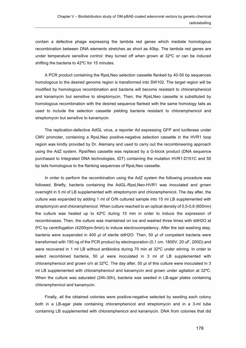
Chapter V – Biodistribution study of OM-pBAE-coated adenoviral vectors by genetic-chemical
radiolabelling
178
contain a defective phage expressing the lambda red genes which mediate homologous
recombination between DNA elements stretches as short as 40bp. The lambda red genes are
under temperature sensitive control: they turned off when grown at 32ºC or can be induced
shifting the bacteria to 42ºC for 15 minutes.
A PCR product containing the RpsLNeo selection cassette flanked by 40-50 bp sequences
homologous to the desired genome region is transformed into SW102. The target region will be
modified by homologous recombination and bacteria will become resistant to chloramphenicol
and kanamycin but sensitive to streptomycin. Then, the RpsLNeo cassette is substituted by
homologous recombination with the desired sequence flanked with the same homology tails as
used to include the selection cassette yielding bacteria resistant to chloramphenicol and
streptomycin but sensitive to kanamycin.
The replication-defective AdGL virus, a reporter Ad expressing GFP and luciferase under
CMV promoter, containing a RpsLNeo positive-negative selection cassette in the HVR1 loop
region was kindly provided by Dr. Alemany and used to carry out the recombineering approach
using the AdZ system. Rpsl/Neo cassette was replaced by a G-block product (DNA sequence
purchased to Integrated DNA technologies, IDT) containing the mutation HVR1:D151C and 50
bp tails homologous to the flanking sequences of RpsLNeo cassette.
In order to perform the recombination using the AdZ system the following procedure was
followed. Briefly, bacteria containing the AdGL-RpsLNeo-HVR1 was inoculated and grown
overnight in 5 ml of LB supplemented with streptomycin and chloramphenicol. The day after, the
culture was expanded by adding 1 ml of O/N cultured sample into 15 ml LB supplemented with
streptomycin and chloramphenicol. When culture reached to an optical density of 0,5-0,6 (600nm)
the culture was heated up to 42ºC during 15 min in order to induce the expression of
recombinases. Then, the culture was maintained on ice and washed three times with ddH2O at
0ºC by centrifugation (4200rpm-5min) to induce electrocompetency. After the last washing step,
bacteria were suspended in 400 µl of sterile ddH2O. Then, 50 µl of competent bacteria were
transformed with 150 ng of the PCR product by electroporation (0,1 cm, 1800V, 20 uF, 200Ω) and
were recovered in 1 ml LB without antibiotics during 70 min at 32ºC under stirring. In order to
select recombined bacteria, 50 µl were inoculated in 3 ml of LB supplemented with
chloramphenicol and grown o/n at 32ºC. The day after, 50 µl of this culture were inoculated in 3
ml LB supplemented with chloramphenicol and kanamycin and grown under agitation at 32ºC.
When the culture was saturated (24h-30h), bacteria was seeded in LB-agar plates containing
chloramphenicol and kanamycin.
Finally, all the obtained colonies were positive-negative selected by seeding each colony
both in a LB-agar plate containing chloramphenicol and streptomycin and in a 3-ml tube
containing LB supplemented with chloramphenicol and kanamycin. DNA from colonies that did
Chapter V – Biodistribution study of OM-pBAE-coated adenoviral vectors by genetic-chemical
radiolabelling
179
not grow in the plate but grew in the tube was extracted and DNA was tested by PCR, enzymatic
restriction and sequencing.
Antibiotic working concentrations:
Cloramphenicol: 12,5 ug/ml
Kanamycin: 15 ug/ml
Streptomycin: 5 mg/ml
5.2.4 Polymerase chain reaction (PCR) analysis
PCR allows to specifically amplify DNA fragments thanks to the DNA-polymerase enzymatic
activity. In this work, PCR have been used in order to validate the recombination by testing the
absence of RpslNeo cassettes in their specific region in the AdGL-RpslNeo-HVR1 genomes. By
using RpslNeo cassette flanking primers (Sequence available in section 1.2.1), plasmidic DNA
extracted from positive clones obtained after recombination was analysed in order to confirm the
recombination with the G-block containing the HVR1:D151C point mutation.
The PCR reaction were conducted mixing the following components:
PCR reaction
Sample DNA 100 ng
Magnesium Buffer 10x 2 µL
Primers (Fw and reverse) 10 µM 0,5 µL
dNTPs 1,25 mM 3,2 µL
TAQ polymerase 5U/µL 0,125 µL
Nuclease-free H2O Adjust to 20 µL

Chapter V – Biodistribution study of OM-pBAE-coated adenoviral vectors by genetic-chemical
radiolabelling
180
The DreamTaqTM Hot Start DNA Polymerase (ThermoFisher Scientific) was used to amplify
samples for analytical purposes. When PCR products were prepared for DNA sequencing, a high-
fidelity DNA-polymerase was used (Expand High Fidelity PCR System, Roche).
The PCR reaction consists in three phases which are consecutively repeated in order to
amplify the PCR product. These phases are the DNA denaturation, primers hybridization and
elongation and are controlled by automatic temperature cycling using a thermocycler. The
elongation time (te) depends on the target size (1min/DNA Kb) and the hybridization temperature
(Th) depnds on the primers sequences.
Thermocycling protocol
Initial denaturation 95ºC – 5min
30-40 cycles:
Denaturation 95ªC-30s
Hybridization Th – 30s
Elongation 72ºC-te
Final elongation 72ªC – 5min
5.2.5 Enzymatic restriction analysis
In order to perform the enzymatic restriction analysis, SphI restriction enzyme was selected
based on the theoretical band pattern obtained after in silico restriction using the UGENE
software. This restriction enzyme produced distinct band patterns depending on the presence of
RpslNeo cassette allowing to distinguish between recombined and non-recombined samples.
In order to perform the enzymatic restriction, 10 µl DNA samples extracted from different
clones were mixed with 1 µl of SphI restriction enzyme, 3 µl of each corresponding commercial
buffer (NEBbuffer 2.1) and were adjusted to 30 µl by adding 16 µl of deionized ultrapure water.
Samples were incubated o/n at 37ºC and analysed by agarose gel electrophoresis

Chapter V – Biodistribution study of OM-pBAE-coated adenoviral vectors by genetic-chemical
radiolabelling
181
5.2.6 Adenovirus vector production, purification and chemical
modification
AdGL-HVR1-Cys clone was expanded in 150 ml LB-cloramphenicol-kanamycin and DNA
was extracted. 5 ug were transfected into a confluent p50 plate of HEK293 cells which
transcomplement E1 using SuperFect Reagent (Qiagen). AdZ plasmids codify for the Sce-I
endonuclease under a eukaryotic promoter which is expressed in mammalian cells and linearize
the adenoviral genome intracellularly. When a complete cytopathic effect was observed due to
the virus production, cells and media were collected and homogenized by three freeze and
thawing cycles. The resulting homogenate was used to further amplify the virus by infecting
confluent p150 HEK293 plates. Cells were harvested when complete cytophatic effect was
observed and pelleted by centrifugation in 50 ml falcon tubes (1000 RPM-10 min)
Vector purification was carried out under the guidance of Dr. Kreppel in Witten (Germany).
Cysteine-bearing adenoviruses are prone to aggregation due to oxidation of cysteines. In order
to avoid aggregation, the cell lysis and purification of non-chemically modified viral particles must
be done in a reducing environment. This is achieved by including (tris(2-carboxyethyl)phosphine)
(TCEP) as a reducing agent in the lysis buffer and in the CsCl gradient buffers. All buffers were
also argonized in order to avoid oxidation.
Briefly, 10x p150 petri dishes cell pellets pooled in one tube were resuspended in 3 ml lysis
buffer (50 mM HEPES, 150 mM NaCl, 10 mM TCEP) and freeze and thawed three times. Lysates
were centrifuged 2 times at 5000xg during 10 min in order to clean up the lysate. 1,27 g/ml (5 ml)
and 1,41 g/ml (3 ml) CsCl discontinuous gradients were prepared in basal buffer with TCEP (50
mM HEPES, 150 mM NaCl, 0,1 mM TCEP) carefully pouring each layer (1,41g/ml, 1,27 g/ml and
the lysate). After equilibrating the tubes by weight, an ultracentrifugation was run (176000xG, 2
h). Virus bands were collected in argonized eppendorf tubes and physical titer determined by viral
DNA absorption measurement at 260 nm wavelength following the same procedure as explained
in 3.2.3.2. Next, the amount of maleimide-activated compound (20 kDa maleimide-activated linear
polymer PEG (MeO-PEG-mal; IRIS BiotechMarktredwitz,Germany) and Deferoxamine-
maleimide bifunctional chelator (B-772; Macrocyclics)) needed to reach a 30-fold molar excess
was calculated and weighted in a dry atmosphere in excess (thaw maleimide-activated
compounds inside a dissecator with silica gel in order to avoid hydrolysis in the presence of
water). The mal-activated compound was dissolved in 40 µl of basal buffer (50mM HEPES, 150
mM NaCl without TCEP) and the corresponding calculated volume was added to 800 µl virus
stock. The coupling reaction took 45 min at room temperature under agitation. After incubation,
virus solution was diluted to 4 ml with basal buffer and purified again by discontinuous gradients
without TCEP. After collecting the virus band, virus was diluted to 2,5 ml with basal buffer and
desalted by PD10 desalting columns. 10 % glycerol was added to the resulting sample and
chemically modified virus was ready to be characterized and stored at -80ºC. A diagram
Chapter V – Biodistribution study of OM-pBAE-coated adenoviral vectors by genetic-chemical
radiolabelling
182
summarizing the purification and chemical modification of cysteine-bearing viruses is presented
in figure V-1.
Figure V-1. Diagram summarizing the purification and chemical modification of cysteine-bearing viruses.
5.2.7 SDS-PAGE analysis
1×10¹⁰ vector particles were mixed with SDS-loading buffer, heated for 5 min at 70 °C and
loaded on an 8% separation/5% stacking SDS gel, running at 80 V. Silver staining of separated
vector proteins was done as previously described [19].
5.2.8 AdGL-DFO radioactive labelling with Zr-89
Radiolabelling of viruses was performed by incubation of the viruses with a solution oxalic
acid (1 M) containing Zr-89 (2 µCi). Briefly, 50 µl of oxalic acid (1 M) containing Zr-89 (about 2
µCi), 22,4 µl sodium carbonate (2 M), 227,5 µl HEPES buffer (0,5 M) and 200 µl of AdGL-DFO
stock were mixed together in this order in a final 500 µl volume with pH 7. The mixture was
incubated at room temperature (25°C, RT). After incubation, the crude material was purified by
centrifugal filtration using a centrifuge (Nahita 2507/100, Auxilab S.L., Navarra, Spain) and
Millipore filters (100 kDa cut-off, Amicon® Ultra 0.5 ml UFC510096, Molsheim, France). After
centrifugation, the resulting precipitate was washed three times with MQ-water to remove
unreacted Zr-89 species. Thereafter, the amount of radioactivity in the pellet, the supernatant and
the washings were determined in a 2470 WIZARD2 Automatic Gamma Counter. Radiolabelling
efficiency (RE, expressed in percentage) was calculated using the following equation:
R.E. % = Afilter / (Afilter + Afiltrates) * 100
Where Afilter is the amount of radioactivity in the filter, and Afiltrates is the amount of radioactivity
in the filtrates, including washings. Finally, VPs in the filter were suspended in 200 µL of saline
solution (0.9% NaCl, Braun Medical S.A., Catalonia, Spain), recovered in a syringe and the
Chapter V – Biodistribution study of OM-pBAE-coated adenoviral vectors by genetic-chemical
radiolabelling
183
amount of radioactivity was measured in a dose calibrator. Radiochemical yield was calculated
using the following equation:
R.Y. % = Arf / At * 100
Where Arf is the amount of radioactivity in the resuspended fraction, and At is the starting
amount of radioactivity (time corrected).
In vitro radiochemical stability of the labelled VPs was assessed by means of the
centrifugation technique. The radiolabelled VPs solution was divided into 2 different fractions, one
mixed with MQ-water, the other one with a solution of DFO (final concentration 1mM) -, which
acted as a high interacting competitor towards Zr-89. The aliquots were kept at 37 °C for 24 hours
using a digital block heater. The samples were then filtered and radioactivity in the filter
(supernatant) and the filtrate (pellet) measured in a 2470 WIZARD2 Automatic Gamma Counter.
Finally, the radiochemical integrity was calculated as the percentage of radioactivity in the
supernatant with respect to the total amount of radioactivity (pellet + supernatant).
5.2.9 C6CR3Y radioactive labelling with I124
The radiolabelling of C6CRY was carried out by mixing 10 µl of phosphate buffer 0,5%,
pH=7,4 with 10 µl of C6CR3Y at 1 ug/ul in MQ-water containing the I124 radioisotope in Iodo-Gen
coated tubes. This mixture was incubated during 1h at room temperature. The resultant labelled
polymer was characterized by HPLC in order to determine the radiolabelling efficiency.
5.2.10 Biodistribution studies
BALB/c nude mice bearing MKN95 human gastric adenocarcinoma tumors (N=1) were
injected with 4x10¹⁰ SAG101 containing 0,77% I124-C6CR3Y in its formulation or with free CPEG
polymeric component also containing 0,77% I124-C6CR3Y. The total injected radioactive dose
was 120 uCi in both cases.
SPECT-CT images were acquired using the eXplore speCZT CT preclinical imaging system
from General Electric (GE Healthcare, USA). The system combines SPECT and CT, allowing co-
registration of the SPECT and CT datasets without additional post-processing. Once the animals
were positioned within the field of view of the system, data was collected in an energy window of
84-102 keV in static mode. The mice were under isofluorane anaesthesia duringthe whole-body
SPECT-CT scans acquired at 0,13, 0,63, 6, 10, 22, 48 hours post injection using for 30, 60, or
120 minutes (40sec/step, 80sec/step, or 160sec/step, respectively). As a general rule, the image
acquisition period was dependent on the counts detected by the collimator of the scanner, thus,
longer imaging times were needed as the gamma emissions decreased. After each SPECT scan,
CT acquisitions were performed to provide anatomical information of each animal.

Chapter V – Biodistribution study of OM-pBAE-coated adenoviral vectors by genetic-chemical
radiolabelling
184
Ex vivo radioactivity quantification was carried out 48h after injection. Lungs, heart, kidneys,
spleen, liver, intestines, stomach, brain, blood, plasma and tumors radioactivity was measured
with a dose calibrator. Each measure was normalized by the weight of each sample and
presented as the percentage of the injected dose per gram (%ID/g).
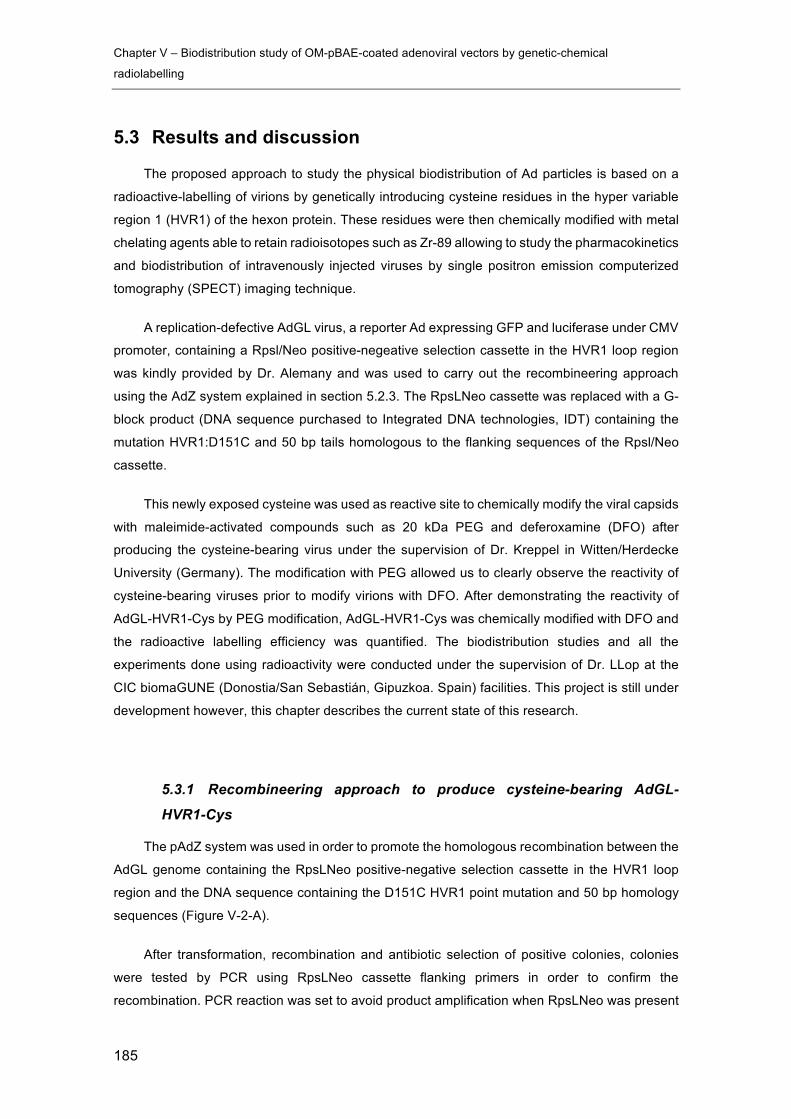
Chapter V – Biodistribution study of OM-pBAE-coated adenoviral vectors by genetic-chemical
radiolabelling
185
5.3 Results and discussion
The proposed approach to study the physical biodistribution of Ad particles is based on a
radioactive-labelling of virions by genetically introducing cysteine residues in the hyper variable
region 1 (HVR1) of the hexon protein. These residues were then chemically modified with metal
chelating agents able to retain radioisotopes such as Zr-89 allowing to study the pharmacokinetics
and biodistribution of intravenously injected viruses by single positron emission computerized
tomography (SPECT) imaging technique.
A replication-defective AdGL virus, a reporter Ad expressing GFP and luciferase under CMV
promoter, containing a Rpsl/Neo positive-negeative selection cassette in the HVR1 loop region
was kindly provided by Dr. Alemany and was used to carry out the recombineering approach
using the AdZ system explained in section 5.2.3. The RpsLNeo cassette was replaced with a G-
block product (DNA sequence purchased to Integrated DNA technologies, IDT) containing the
mutation HVR1:D151C and 50 bp tails homologous to the flanking sequences of the Rpsl/Neo
cassette.
This newly exposed cysteine was used as reactive site to chemically modify the viral capsids
with maleimide-activated compounds such as 20 kDa PEG and deferoxamine (DFO) after
producing the cysteine-bearing virus under the supervision of Dr. Kreppel in Witten/Herdecke
University (Germany). The modification with PEG allowed us to clearly observe the reactivity of
cysteine-bearing viruses prior to modify virions with DFO. After demonstrating the reactivity of
AdGL-HVR1-Cys by PEG modification, AdGL-HVR1-Cys was chemically modified with DFO and
the radioactive labelling efficiency was quantified. The biodistribution studies and all the
experiments done using radioactivity were conducted under the supervision of Dr. LLop at the
CIC biomaGUNE (Donostia/San Sebastián, Gipuzkoa. Spain) facilities. This project is still under
development however, this chapter describes the current state of this research.
5.3.1 Recombineering approach to produce cysteine-bearing AdGL-
HVR1-Cys
The pAdZ system was used in order to promote the homologous recombination between the
AdGL genome containing the RpsLNeo positive-negative selection cassette in the HVR1 loop
region and the DNA sequence containing the D151C HVR1 point mutation and 50 bp homology
sequences (Figure V-2-A).
After transformation, recombination and antibiotic selection of positive colonies, colonies
were tested by PCR using RpsLNeo cassette flanking primers in order to confirm the
recombination. PCR reaction was set to avoid product amplification when RpsLNeo was present
Chapter V – Biodistribution study of OM-pBAE-coated adenoviral vectors by genetic-chemical
radiolabelling
186
in the plasmid by setting the elongation time to 20 seconds. This time was selected taking into
account the extension time of Taq DNA polymerase (1Kb per minute) and the length of
recombined and non-recombined sequences between primers. The region contained between
primers in recombined plasmids (without the RpsLNeo cassette) was 170 bp long and 20 seconds
were enough to obtain the PCR product. In the case of non-recombined plasmids (RpsLNeo
cassete is present) the sequence between primers was 1,5 Kb long and the PCR product was
not obtained (Figure V-2-B).
Three of those positive colonies were selected (11, 15, and 17) and were analysed by
enzymatic restriction in order to further assess that the proper recombination took place. The
SphI restriction enzyme was selected as it produces a clearly differentiated band pattern between
recombined and non-recombined BACs generating a 7Kb band in recombined samples, not
observed in the non-recombined control. As observed in figure V-2-C, the expected band pattern
was observed for colonies 15 and 17. Samples 15 and 17 were analyzed by DNA sequencing in
the region surrounding the point mutation to verify the proper modification of the DNA sequence
(Figure V-2-D). Both clones included the D151C point mutation. The clone number 15 was finally
selected in order to move forward. The virus was amplified in HEK293 E1A-transcomplementing
cells as explained in section 5.2.4.
Figure V-2. Generation of AdGL-HVR1-Cys by recombineering approach. A) Schematic representation of the genetic
engineering approach used to modify the AdGL genome in order to include the D151C point mutation. B) PCR products
analysis by agarose gel electrophoresis of eight clones. C) Enzymatic restriction analysis of three selected clones using
the SphI restriction enzyme. D) DNA sequencing of clone number 15 confirming the D151C genetic modification (Codon
change: GAT à TGT).

Chapter V – Biodistribution study of OM-pBAE-coated adenoviral vectors by genetic-chemical
radiolabelling
187
5.3.2 Chemical modification of AdGL-HVR1-Cys and radiolabelling of
AdGL-DFO
One of the main crucial points to take into account when producing cysteine-bearing viruses
is that they are prone to aggregation due to the formation of disulphide bonds between viral
particles. Viral particles are efficiently assembled intracellularly however, when producer cells are
lysed and viral particles are released into the media several considerations must be taken into
account in order to avoid the aggregation and loss of the virus production. In order to efficiently
purify and chemically modify cysteine-bearing viruses, a specific methodology developed by Dr.
Kreppel was followed (See section 5.2.4 to see the detailed protocol). Figure V-3-A summarizes
the viral titers obtained for PEG-modified and non-modified virus productions.
In order to proof the surface reactivity of cysteine-bearing AdGL-HVR1-Cys, the viral
production was modified with 20K maleimide-activated PEG. The high molecular weight of this
PEG allows to easily see a change on the molecular weight of the hexon protein by SDS-PAGE
analysis. As seen in figure V-3-B, a band shift was observed in the case of 20K-PEG modified
AdGL in comparison with the non-modified virus (unPEG) demonstrating that the genetic inclusion
of cysteines granted a new surface reactivity to the viral capsids.
Figure V-3. Purification and chemical modification of AdGL-HVR1-Cys virus. A) Obtained physical titers determined
by 260 nm optical density measurements of non-modified and 20K-PEG modified viruses after the purification and
modification process. B) SDS-PAGE analysis of 20K-PEG modified and unmodified AdGL-HVR1-Cys.
After demonstrating the reactivity of AdGL-HVR1-Cys virus, another virus batch was
produced and used for the modification with maleimide-DFO. AdGL-DFO viruses were
radiolabelled following the protocol in section 5.2.5 and the radiochemical yield and stability of
the radioactive labelling were determined. The radiochemical yield describes the amount of
radioactivity retained by the viral particles after the incubation with Zr-89 and the subsequent
Chapter V – Biodistribution study of OM-pBAE-coated adenoviral vectors by genetic-chemical
radiolabelling
188
purification of viral particles. The radiolabelled VPs solution was divided into two different fractions
in order to study the radioactive labelling stability. Stability studies were carried out after 22h in
MQ-water at r.t. and after 24h in 1mM DFO-NCS at 37ºC which acted as a high interacting
competitor towards Zr-89. As observed in table V-1, the radiolabelling yield obtained was 50,6%
and radioactivity was maintained stable in both tested conditions. These results demonstrated
that AdGL-DFO was able to efficiently retain Zr-89 radioisotopes producing stably radiolabelled
viral suspensions suitable for in vivo biodistribution studies.
Table V-1. Characterization of radiolabelled AdGL-DFO
Radiochemical yield (Recovered in
MQ water)
Stability (MQ-water at r.t.)
Stability (1mM DFO-NCS at 37ºC)
AdGL-HVR1-DFO 50,6 % 92 % 91,6 %
The biodistribution studies using radiolabelled AdGL-DFO coated with the CPEG formulation
are currently under progress at CIC biomaGUNE and results are not still available to be included
in this thesis. However, we have performed preliminary studies to determine the biodistribution of
SAG101 containing I124 radiolabelled C6CR3Y polymer in its formulation as a proof of concept.
5.3.3 Biodistribution of CPEG-coated AdGL using radiolabelled polymers
The aim of this experiment was to test the feasibility of using SPECT-CT imaging to study
the biodistribution of radioactively labelled coated adenoviruses. At this point, the radiolabelled
AdGL-DFO-Z89 was still unavailable and we decided to use C6 pBAEs end-modified with YCRRR
in order to add a radiolabelled component in the coating composition. The presence of tyrosine
residues in the terminal peptides allowed to radiolabel the polymer with I124 radioisotope simply
by incubating the polymer with the radioisotope in Iodo-Gen coated tubes. Iodo-gen is an oxidant
that converts I124 iodide to I124 iodonium, which then reacts with an electron-rich amino acid
residue such as tyrosine [20]. By labelling the polymer we were unable to study the biodistribution
of viral particles, however the differences observed between free polymer and coated viruses
could indirectly proof the stability of the CPEGAd complex in vivo.
The experiment was done by injecting two BALB/c nude mice bearing MKN95 tumors with
a 4x10¹⁰ VP dose of CPEG-coated AdNuPARmE1A containing 0,77% I124-C6CR3Y in its

Chapter V – Biodistribution study of OM-pBAE-coated adenoviral vectors by genetic-chemical
radiolabelling
189
formulation and with the radiolabelled polymeric CPEG free polymer. As observed in figure V-4,
the radioactivity signal of CPEGAd showed a more spread biodistribution profile and persisted
longer before being accumulated in the liver. This observation was in accordance with the longer
circulation time of CPEG-coated adenovirus observed in chapter 3. Six hours post injection the
signal was detected in several different organs in contrast with the CPEG free polymer which was
only observed in the liver and in the bladder at this time point. Although the number of animals
was not enough to consistently reach conclusions, the biodistribution pattern suggested that the
interaction of the radiolabelled polymer with viral particles modified the biodistribution of C6CR3Y.
Figure V-4. SPECT-CT in vivo imaging of I124 radiolabelled CPEGAd (coated adenovirus) and CPEG (free polymeric component) treated animals. Images were acquired SPECT-CT images were acquired using the eXplore speCZT CT
preclinical imaging system at 0,13, 0,63, 6, 10, 22, 48 hours post injection.
These results were further confirmed by ex vivo radioactivity quantification. Lungs, heart,
kidneys, spleen, liver, intestine, stomach, brain, blood, plasma and tumors were collected 48h
after injection and radioactivity was quantified. Each sample was weighted and results were
presented as the percentage of the initial total injected radioactivity normalized by the weight of
each sample (%ID/g). As observed in figure V-5, higher radioactivity levels were observed for
lungs, spleen, intestines, stomach, blood, plasma and tumors when the animal was injected with
CPEGAd. Interestingly, lower radioactivity levels were determined in kidneys and liver.
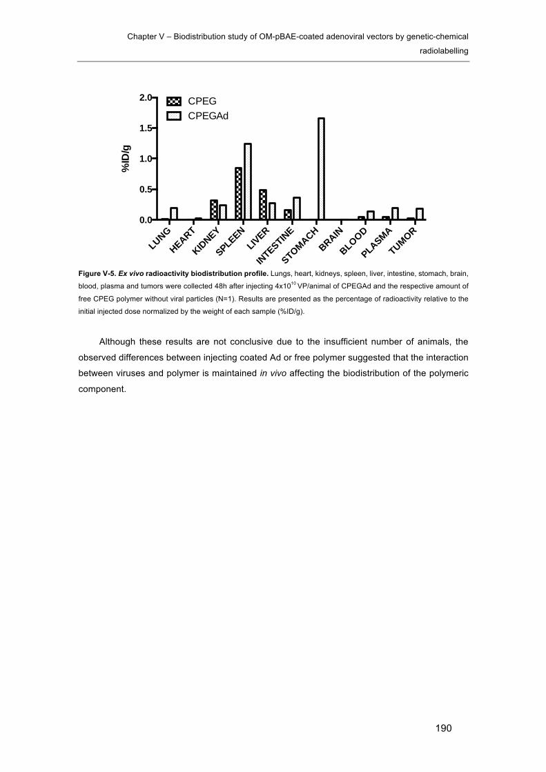
Chapter V – Biodistribution study of OM-pBAE-coated adenoviral vectors by genetic-chemical
radiolabelling
190
Figure V-5. Ex vivo radioactivity biodistribution profile. Lungs, heart, kidneys, spleen, liver, intestine, stomach, brain,
blood, plasma and tumors were collected 48h after injecting 4x10¹⁰ VP/animal of CPEGAd and the respective amount of
free CPEG polymer without viral particles (N=1). Results are presented as the percentage of radioactivity relative to the
initial injected dose normalized by the weight of each sample (%ID/g).
Although these results are not conclusive due to the insufficient number of animals, the
observed differences between injecting coated Ad or free polymer suggested that the interaction
between viruses and polymer is maintained in vivo affecting the biodistribution of the polymeric
component.
LUNG
HEART
KIDNEY
SPLEENLIVER
INTESTINE
STOMACHBRAIN
BLOOD
PLASMA
TUMOR0.0
0.5
1.0
1.5
2.0
%ID
/g
CPEGCPEGAd

Chapter V – Biodistribution study of OM-pBAE-coated adenoviral vectors by genetic-chemical
radiolabelling
191
5.4 Concluding remarks
Results from previous chapters demonstrated the ability of pBAEs/viral vectors to improve
the systemic administration of viral vectors in terms of pharmacokinetics, biodistribution, efficacy
and safety. An improved transduction of tumors was demonstrated upon systemic administration
in chapter 3 resulting in a higher efficacy of pBAEs-coated AdNuPARmE1A in comparison with
its naked counterpart, specially in the presence of NAbs. In contrast, the liver transduction was
reduced when injecting coated vectors with clear consequences on the hepatotoxicity of
intravenously injected vectors. In the present chapter, we moved a step further in terms of
studying the effect of pBAEs-coating on the physical biodistribution of intravenously injected
adenoviral vectors by developing a novel adenovirus radiolabelling strategy.
The genetic engineering approach to include the D151C point mutation in the HVR1
genomes region have been successfully achieved using the AdZ system. Then, AdGL-Cys has
been produced and purified at high titer and its new capsid surface reactivity has been
demonstrated by modifying the viral particles with 20K-PEG-maleimide. The SDS-PAGE analysis
of PEG-modified AdGL showed a clear hexon band-shift due to the presence of covalently-linked
PEG molecules increasing its molecular weight.
AdGL-Cys has been chemically modified with deferoxamine (DFO) and radiolabelled with
Zr-89 achieving a radiolabelling yield of 50,6%. Its ability to remain stably labelled has been
demonstrated by competition assays with free DFO. After 24h of incubation with 1mM DFO at
37ºC, the virus retained 91,6% of the initial radioactivity indicating its suitability to be used in vivo.
Due to the current state of this project, which is still under development, no biodistribution
results using the AdGL-DFO-Zr89 were available to be included in this work. However, the pilot
biodistribution study using radiolabelled pBAEs showed differences on the biodistribution of the
polymeric component when injecting CPEGAd coated viruses suggesting the stability of these
complexes in vivo.
The resulting technology is a valuable tool to study the physical biodistribution and clearance
of intravenously injected adenoviruses. Moreover, this approach opens the door to develop new
versions of the pBAEs-coating technology able to react with genetically included cysteine
residues producing bioresponsive covalent coatings.
Chapter V – Biodistribution study of OM-pBAE-coated adenoviral vectors by genetic-chemical
radiolabelling
192
5.5 References
[1] R. Chen, J.J. Parry, W.J. Akers, M.Y. Berezin, I.M. El Naqa, W.B. Edwards, B.E. Rogers,
Multimodality Imaging of Gene Transfer with a Receptor-Based Reporter Gene, J Nucl
Med. 51 (2010) 1456–1463. doi:10.2967/jnumed.109.063586.Multimodality.
[2] S.P. Singh, In Vivo Functional and Anatomic Imaging for Assessment of in Vivo Gene
Transfer, Mol. Imaging. 252 (2009) 763–771.
[3] B.E. Rogers, J.J. Parry, R. Andrews, P. Cordopatis, B.A. Nock, T. Maina, MicroPET
imaging of gene transfer with a somatostatin receptor-based reporter gene and Tc-94m-
demotate 1, J. Nucl. Med. 46 (2005) 1889–1897.
[4] K.H. Kim, I. Dmitriev, J.P. O’Malley, M. Wang, S. Saddekni, Z. You, M.A. Preuss, R.D.
Harris, R. Aurigemma, G.P. Siegal, K.R. Zinn, D.T. Curiel, R.D. Alvarez, A phase I
clinical trial of Ad5.SSTR/TK.RGD, a novel infectivity-enhanced bicistronic adenovirus, in
patients with recurrent gynecologic cancer, Clin. Cancer Res. 18 (2012) 3440–3451.
doi:10.1158/1078-0432.CCR-11-2852.
[5] A. Merron, P. Baril, P. Martin-Duque, A. De La Vieja, L. Tran, A. Briat, K.J. Harrington,
I.A. McNeish, G. Vassaux, Assessment of the Na/I symporter as a reporter gene to
visualize oncolytic adenovirus propagation in peritoneal tumours, Eur. J. Nucl. Med. Mol.
Imaging. 37 (2010) 1377–1385. doi:10.1007/s00259-009-1379-3.
[6] K.N. Barton, H. Stricker, S.L. Brown, M. Elshaikh, I. Aref, M. Lu, J. Pegg, Y. Zhang, K.C.
Karvelis, F. Siddiqui, J.H. Kim, S.O. Freytag, B. Movsas, Phase I study of noninvasive
imaging of adenovirus-mediated gene expression in the human prostate, Mol. Ther. 16
(2008) 1761–1769. doi:10.1038/mt.2008.172.
[7] J. Davydova, T. Gavrikova, E.J. Brown, X. Luo, D.T. Curiel, S.M. Vickers, M. Yamamoto,
In vivo bioimaging tracks conditionally replicative adenoviral replication and provides an
early indication of viral antitumor efficacy, Cancer Sci. 101 (2010) 474–481.
doi:10.1111/j.1349-7006.2009.01407.x.
[8] Y. Il Kim, B.C. Ahn, J.A. Ronald, R. Katzenberg, A. Singh, R. Paulmurugan, S. Ray, S.S.
Gambhir, L. V. Hofmann, Intratumoral versus intravenous gene therapy using a
transcriptionally targeted viral vector in an orthotopic hepatocellular carcinoma rat model,
J. Vasc. Interv. Radiol. 23 (2012) 704–711. doi:10.1016/j.jvir.2012.01.053.
[9] D. Abate-Daga, N. Andreu, J. Camacho-Sánchez, R. Alemany, R. Herance, O. Millán, C.
Fillat, Oncolytic adenoviruses armed with thymidine kinase can be traced by pet imaging
and show potent antitumoural effects by ganciclovir dosing, PLoS One. 6 (2011).

Chapter V – Biodistribution study of OM-pBAE-coated adenoviral vectors by genetic-chemical
radiolabelling
193
doi:10.1371/journal.pone.0026142.
[10] F. Pouliot, B.D.W. Karanikolas, M. Johnson, M. Sato, S.J. Priceman, D. Stout, J. Sohn,
N. Satyamurthy, J.B. deKernion, L. Wu, In Vivo Imaging of Intraprostatic-Specific Gene
Transcription by PET, J. Nucl. Med. 52 (2011) 784–791.
doi:10.2967/jnumed.110.084582.
[11] A. Kanerva, K.R. Zinn, K.W. Peng, T. Ranki, L. Kangasniemi, T.R. Chaudhuri, R.A.
Desmond, M. Wang, K. Takayama, T. Hakkarainen, H. Alfthan, U.H. Stenman, D.T.
Curiel, A. Hemminki, Noninvasive dual modality in vivo monitoring of the persistence and
potency of a tumor targeted conditionally replicating adenovirus, Gene Ther. 12 (2005)
87–94. doi:10.1038/sj.gt.3302387.
[12] P.W. Zoltick, J.M. Wilson, A quantitative nonimmunogenic transgene product for
evaluating vectors in nonhuman primates, Mol. Ther. 2 (2000) 657–659.
doi:10.1006/mthe.2000.0204.
[13] L. Liu, B.E. Rogers, N. Aladyshkina, B. Cheng, S.J. Lokitz, D.T. Curiel, J.M. Mathis,
Construction and radiolabeling of adenovirus variants that incorporate human
metallothionein into protein IX for analysis of biodistribution, Mol. Imaging. 13 (2014) 1–
12. doi:10.2310/7290.2014.00022.
[14] S. Espenlaub, S. Corjon, T. Engler, C. Fella, M. Ogris, E. Wagner, S. Kochanek, F.
Kreppel, Capsomer-specific fluorescent labeling of adenoviral vector particles allows for
detailed analysis of intracellular particle trafficking and the performance of bioresponsive
bonds for vector capsid modifications., Hum. Gene Ther. 21 (2010) 1155–1167.
doi:10.1089/hum.2009.171.
[15] L. Krutzke, J.M. Prill, T. Engler, C.Q. Schmidt, Z. Xu, A.P. Byrnes, T. Simmet, F.
Kreppel, Substitution of blood coagulation factor X-binding to Ad5 by position-specific
PEGylation: Preventing vector clearance and preserving infectivity., J. Control. Release.
235 (2016) 379–392. doi:10.1016/j.jconrel.2016.06.022.
[16] L. Krutzke, J.M. Prill, T. Engler, C.Q. Schmidt, Z. Xu, A.P. Byrnes, T. Simmet, F.
Kreppel, Substitution of blood coagulation factor X-binding to Ad5 by position-speci fi c
PEGylation : Preventing vector clearance and preserving infectivity, 235 (2016) 379–392.
doi:10.1016/j.jconrel.2016.06.022.
[17] R.J. Stanton, B.P. McSharry, M. Armstrong, P. Tomasec, G.W.G. Wilkinson, Re-
engineering adenovirus vector systems to enable high-throughput analyses of gene
function., Biotechniques. 45 (2008) 659-662-668. doi:10.2144/000112993.
Chapter V – Biodistribution study of OM-pBAE-coated adenoviral vectors by genetic-chemical
radiolabelling
194
[18] C. Puig-Saus, A. Gros, R. Alemany, M. Cascallo, Adenovirus i-leader truncation
bioselected against cancer-associated fibroblasts to overcome tumor stromal barriers.,
Mol. Ther. 20 (2012) 54–62. doi:10.1038/mt.2011.159.
[19] H. Blum, H. Beier, H.J. Gross, Improved silver staining of plant proteins, RNA and DNA
in polyacrylamide gels, Electrophoresis. 8 (1987) 93–99. doi:10.1002/elps.1150080203.
[20] J. Nairne, P.B. Iveson, A. Meijer, Chapter Five - Imaging in Drug Development, in: G.
Lawton, D.R.B.T.-P. in M.C. Witty (Eds.), Elsevier, 2015: pp. 231–280.
doi:https://doi.org/10.1016/bs.pmch.2014.10.002.

This page left blank intentionally

Chapter VI. Conclusions

This page left blank intentionally
Chapter VI - Conclusions
198
Conclusions
Hybrid vectors based on non-enveloped viruses coated with oligopeptide-modified poly(β-
amino ester)s with improved in vivo applicability by intravenous administration have been
developed and described in this thesis.
Firstly, pBAEs-coated adeno-associated viruses have been successfully produced and
targeted to brain endothelial cells by including LRP-1 targeting peptides into pBAEs structure.
• Biophysical analysis showed that cationic arginine-modified pBAEs (C32CR3) are
able to interact with AAV viral particles. The electrostatic interaction of C32CR3 with
viral capsids surfaces positivizes their net negative surface charge producing coated
viral particles observed by DLS and TEM. Optimal coating conditions for AAVs have
been determined.
• In silico designed brain-targeting peptides predicted to interact with the LRP-1
receptor have been tested in vitro by blood-brain barrier (BBB) crossing assays
using in vitro BBB models. The results obtained demonstrated that SEQ12 peptide
has a BBB crossing ability similar to Angiopep-2, the targeting peptide used in the
ANG1005, an experimental chemotherapy drug currently in advanced phases of
clinical development.
• The inclusion of 10% SEQ12 end-capped C32 pBAEs into the coating formulation
improved the infectivity of AAVs against brain vascular endothelial cells in vitro and
showed increased transduction of brain when intravenously administered in mice
while decreasing liver transduction.
Secondly, OM-pBAEs-based coating technology have been adapted to Ad5 vectors and
have been further developed in order to produce single coated viral particles suspensions able to
improve pharmacokinetics of intravenously administered Ad5 vectors while avoiding neutralizing
antibodies.
• The optimal coating conditions for Ad5 vectors have been determined by biophysical
characterization with DLS and TEM and by in vitro neutralization assays. The slightly
hydrophobic C6CR3 demonstrated to protect virion’s neutralization in vitro while
improving infectivity in a higher degree in comparison with C32CR3.

Chapter VI - Conclusions
199
• Intravenously injected C6CR3-coated Ad5 showed a significantly higher circulation
time in vivo and a more stable behaviour in terms of liver detargeting in comparison
with C32CR3-coated Ad5.
• The inclusion of 35% w/w of 2K-PEG-modyfied C6CR3 into the coating formulation
demonstrated to highly avoid NAbs neutralization in vitro while solving the clustering
of viral particles during the coating process. Fine biophysical characterization of the
resulting complexes with DLS, TEM and STORM showed the ability of the resulting
formulation to coat single viral particles with a 15 nm thick pBAEs coating not
achieved with any other formulation tested. The STORM analysis using
fluorescently-labelled Ad5 and C6CR3 demonstrated the interaction between
polymer molecules and viral particles in aqueous solution at the molecular level.
The presence of 35% PEG-modified C6CR3 polymer in the formulation also avoids
the formation of polymeric nanoparticles.
• When tested in vivo, the resulting formulation containing 65% C6CR3 and 35%
C6PEGCR3 have demonstrated to significantly improve the blood circulation time
of virions in comparison with the 100% C6CR3 formulation. Moreover, the PEG-
containing formulation have shown to reduce the AdTL liver transduction while
increasing its tumor tropism in comparison with the non-PEGylated formulation and
with naked AdTL. It has been also demonstrated that CPEG-coated Ad5 is less
immunogenic than C6CR3-coated and naked Ad5 regarding their ability to induce
the production of neutralizing antibodies when administered intravenously.
The resulting Ad5 coating formulation has demonstrated to improve the therapeutic potential
of the systemically administered oncolytic adenovirus AdNuPARmE1A for the treatment of
pancreatic ductal adenocarcinoma.
• The pBAE-coated AdNuPARmE1A (SAG101) has an improved infectivity and
cytotoxicity in vitro in comparison with their naked counterpart and is less prone to
induce an innate immune response reducing the release of IL-6 and IL-10 when
presented to RAW264.7 murine macrophages in comparison with AdNuPARmE1A.
• SAG101 showed a safe toxicity profile in vivo. The mice weight loss and
hepatotoxicity upon intravenous administration in immunocompetent mice were
minimized in comparison with naked AdNuPARmE1A. Hematologic studies by cell
counting suggested a reduction on the ability of the AdNuPARmE1A to induce
proliferation of monocytes and neutrophils when coated-AdNuPARmE1A is injected
in vivo. The release of cytokines in vivo has been also studied without showing
conclusive differences between coated and naked viruses, however the polymeric
component has not induced cytokine production in vivo indicating the safety profile
of OM-pBAEs.
Chapter VI - Conclusions
200
• The efficacy of SAG101 in the presence of circulating NAbs in vivo has been tested
in passively immunized tumor-bearing immunodeficient mice by intravenous
administration showing a significant control on the progression of PANC-1 and MIA
PaCa-2 tumors even when animals were passively immunized. These results are in
clear contrast with the naked AdNuPARmE1A performance, since it is inefficacious
in the presence of NAbs in vivo.
Finally, a novel radiolabelling approach based on a genetic-chemical modification of Ad5
viruses has been developed in order to be used as a tool to study physical biodistribution and
pharmacokinetics of intravenously administered naked and coated adenoviruses.
• Inclusion of cysteine residues in the hexon HVR1 loop by genetically engineering
Ad5 genomes and the subsequent production of the viral stock under reducing
conditions allowed to produce cysteine-bearing AdGL reporter vector at high titers
avoiding its aggregation.
• The resulting AdGL-HVR1-Cys has been successfully chemically modified with
maleimide-activated PEG and deferoxamine demonstrating that the genetic
inclusion of cysteines granted a new chemical reactivity to AdGL.
• Deferoxamine-modified AdGL demonstrated their ability to retain radioisotopes in its
structure in the presence of free deferoxamine as a high affinity competitor. This
result demonstrated that deferoxamine-modified AdGL is able to be stably
radiolabelled.
• As a proof of concept, C6 pBAEs end-capped with YCRRR peptides have
demonstrated to be a good strategy to be used as a tool to radiolabel the polymeric
component of pBAEs/Ad hybrid vectors with I124 radioisotopes. The biodistribution
study by SPECT-CT showed a longer polymer circulation time when combined with
viral particles suggesting the stability of the interaction between the polymer and the
virus in vivo after intravenous administration.

This page left blank intentional
202
List of Publications and Presentations
Publications and Patents
Fornaguera C, Lázaro MÁ, Brugada-Vilà P, Porcar I, Morera I, Guerra-Rebollo M, Garrido
C, Rubio N, Blanco J, Cascante A, Borrós S, “Application of an assay Cascade methodology for a Deep preclinical characterization of polymeric nanoparticles as a treatment for gliomas” Drug Deliv, Nov 2018.
S. Borrós, C. Fillat, P. Brugada, A. Cascante, “Complexes of viral-based therapeutic agents and modified poly(beta-amino esters)s” P070656WO, filed May 2018
P. Brugada-Vilà et al. “Boosting intravenous administration of therapeutic viral vectors using an oligopeptide-modified poly(b-amino ester)s-based coating technology” (Manuscript in preparation)
Seminars and Courses
Salvador Borrós, Anna Cascante, Pau Brugada, Nathaly Segovia, Gemma Molins, Pere
Dosta, Victor Ramos. Novel peptide-modified poly(ß-amino ester)s for highly efficient gene delivery intracelular localization and specific BBB targeting. Roche - Nature Biotechnology
Symposium 2014. September 2014. Roche, Switzerland. Poster presentation.
5th Course on the BBB in Drug Development. Principles, Transport Kinetics, Diseases &
Methods. October 2014, Uppsala, Sweden
Pau Brugada, Anna Cascante, Víctor Ramos, Crisitina Fillat, Salvador Borrós Layer-by-layer (LbL) deposition of ionic polymers onto viral particles: A step forward in the development of safe, efficient and targeted oncolytic virotherapy. 8th Biennial Congress of
the Spanish Society for Gene and Cell Therapy, November 2015, San Sebastian, Spain. Poster
presentation
PBAE-coated AdNuPARmE1A: overcoming oncolytic virotherapy hurdles, 2nd
AdenoNet Meeting: Spanish Adenovirus Network, October 2017, Barcelona, Spain. Oral
presentation
Brugada-Vilà P, Rovira-Rigau M, Castells-Sala C, Cascante A, Borrós S and Fillat C. PBAE-coated AdNuPARmE1A: overcoming oncolytic virotherapy hurdles, 25th European society
of gene & cell therapy congress, October 2017, Berlin, Germany. Poster presentation