Mediterranean forests and health - TDX (Tesis Doctorals en ...
Upload
khangminh22Category
view
0download
0

UNIVERSIDAD DE MURCIAFACULTAD DE VETERINARIA
Functionality of lactoferrin andgalactooligosaccharides in infant formulas
Esmat Aly Alsayed Aly
Directors:
Gaspar Ros Berruezo
Carmen Frontela-Saseta
Murcia, 2015


Functionality of lactoferrin and GOS in infant formulas Dedication
Dedication
To all my family: the spirit of my father, my
mother, all my brothers and sisters. Especially
to my lovely wife Aliaa, my daughter Rofida and
my brother Tarek. To everyone who loves the
justice, peace, unity, dignity and liberty.


Functionality of lactoferrin and GOS in infant formulas Acknowledgement
Acknowledgement
First and before all, I would like to thank Allah who granted me theability to perform this work and helped me to pass safety through all thedifficulties, I thought impossible to overcome.
I would like to take this opportunity to express my sincere thanks,deepest gratitude and appreciation to my supervisors, Professors Gaspar RosBerruezo and Carmen Frontela-Saseta, from Murcia University, Departmentof Food Technology, Nutrition and Food Science. This thesis would nothave been possible without their advice, support and encouragement.
Faithful thanks also extended to Professors Carmen Martinez Garciaand Mº-Jesus Periago for their help, personal and scientific compartmentwith me throughout the present investigation. Also deepest appreciation formy best friends Rubén Lopéz-Nicolas for helping me in this work by manyways as well as for Patricia Peso-Echarri, Carlos A. Gonzales, CarolsGomez-Gallego, Inmaculada Navarro, Victoria Gomez, Rocio Gonzales,Javier Garcia, Guillermo Dominic, Marina and Javier Santaella, GemaNieto, Amparo Lopez, Lorena Fernandez, Gala, Elvira Samper, NievesHidalgo and Sergio Bravo. Special and deepest thanks for Antonia TovarMadrid (la madre del mundo) for her valuable and continual help.
Special thanks to my teachers and friends that affected my life bytheir extreme support and encouragement to be the person I am: ProfessorsIbrahim A. Abd El-Gawad, Abouelsamh M. Mehriz, Saeb A. Hafez, AhmedM. Abdelsalam, Farag A. Saleh, Hany A. Fahmy, Elsayed Farahat, AbeerHaroun, Hanna Sayed, Mona H. Ahmed, Naglaa Hassanien, Ayman Diab,Hala Thabet, Esraa A. Mohamed, Hanan Fawzy, Mohsen Ahmed, HassanAbdelhalim, Wafaa Salama, Mahmoud Mailam and many others I cannotcount them. Special thanks to my brother Gamal and my close friendHossam M. Ibrahim for his continuous help. Deepest and faithful thanks formy lovely wife Aliaa for continuous help and support.
Also deepest and faithful thanks for MAEC-AECID (Ministerio deAsuntos Exteriors y de Cooperación- Agencia Española de CooperaciónInternacional para el Desarrollo) for financial support (grant nº0000595994) and Hero Baby Co. (Alcantarilla, Murcia, Spain) for providingthe infant formula.


Functionality of lactoferrin and GOS in infant formulas Abstract
ABSTRACT
The consumption of milk and dairy products is included as an
important element in a healthy and balanced diet. Human milk is the most
appropriate choice for newborns and provides all the energy and nutrients
needed to ensure proper growth and development. The pattern of the
exclusive breastfeeding during the first six months of life is very important
to provide the newborn with some immunomodulatory factors and bioactive
compounds that are naturally found in human milk and, therefore, it is
recommended that breastfeeding continue one or even two years over the
course of the introduction of some complementary foods.
Therefore, breastfeeding pattern is critically important for infant
health in the early stage of life where it has been demonstrated that breast-
fed infants suffer fewer gastrointestinal disorders and respiratory
contaminations rather than formula-fed infants. It is scientifically accepted
that control of these changes earlier by the nutritional factors may decrease
or prevent the extension of these diseases to the adult life.
Moreover, researchers, health and breastfeeding organizations are
trying to discover the precise substances in human milk that seem to supply
physiological benefits beyond its normal nutritional value which contribute
in delay, treatment or prevent some diseases. Thus, these functional
ingredients hold a great promise for future trends in human nutrition.
Additionally, the relationship between milk consumption and human health
requires a deeper understanding to uncover the protective role of some
bioactive compounds which naturally present in human milk.
These functional ingredients of human milk, particularly human milk
oligosaccharides (HMOs), participate in the promotion of the growth and
activity of beneficial bacteria such as Bifidobacteria and Lactobacilli.
HMOs are characterized by its diversity and distinct structure. Although
many attempts have been carried out to supplement the infant formulas with
different prebiotics and non-digestible oligosaccharides, it could not obtain
a similar structure and diversity of that of HMOs. Additionally, the presence
of several proteins such as immunoglobulin (IgG), lysozyme, casein,

Functionality of lactoferrin and GOS in infant formulas Abstract
lactoferrin (Lf), haptocorrin and α-lactalbumin, may improve the defense of
breast-fed infants against infection where they are relatively resistant in the
gastrointestinal tract. It was found that Lf is characterized by it’s largely
ability to remain in intact form and be only partially cleaved by
gastrointestinal enzymes resulting many bioactive peptides which are
positively correlated with the promotion of infant health. Other functional
activities were discovered for human Lf including: antibacterial, anti-
inflammatory, immunomodulatory activities, and recently, anti-cancer
activity.
Infant formulas, as milk substitute, play an indispensable role
especially after 4-6 months of infant life where human milk is no longer
sufficient to meet all necessary nutritional requirements. Although infant
formulas should be similar to mature human milk in terms of its
macronutrients and micronutrients, normally it do not have the functional
ingredients that are found in human milk, nor do they have the same protein
composition and the diversity of oligosaccharides as human milk. So it is
critically important adding these ingredients to infant formulas. This
evolution of infant formulas manufacturing allow to exert more
functionalities in a large group of infants who cannot feed human milk as a
primary source and for several physiological as well as social reasons.
For mimicking the structure of human milk, GOS (as prebiotic) and
recombinant human Lf (rhLf) are strongly added to infant formulas to obtain
similar functionalities for what human milk has.
Thus, the present study is aimed to explore the functionality of rhLf,
rhLf hydrolysate and GOS whether alone or added in infant formulas. The
present study is divided into four experiments:
1th
experiment was to in vitro evaluate of the role of Lf and/or GOS on
iron bioavailability as expressed as ferritin formation by Caco-2 cells
2nd
experiment was to in vitro evaluate of the preventive effect of rhLf
and rhLf hydrolysate on LPS-induced inflammation using co-culture
gut inflammation model

Functionality of lactoferrin and GOS in infant formulas Abstract
3rd
experiment aimed to assess the prebiotic activity of Lf and/or GOS
using batch culture fermentation system.
4rd
experiment aimed to in vitro evaluate of rhLf stability and the
identification of the generated bioactive peptides as well as
determination of long chain fatty acids (LCFAs) profiles before and
after in vitro simulated gastrointestinal digestion
The obtained findings of the first study on the role of rhLf and/or
GOS on Fe bioavailability measured as ferritin formation in Caco-2 model
revealed that the addition of rhLf and/or GOS to infant formulas resulted in
improvement of iron solubility percentage which in turn may promote iron
bioavailability by using the formed ferritin by Caco-2 cells as criteria. It was
found that the solely addition of GOS or rhLf improved the iron solubility
percentage but non-significant differences were observed between these
groups as compared with control group (without any added ingredient). It
was also observed that the higher solubility percentage of iron was resulted
by a combination of 0.15% rhLf + 10 % GOS (96.13%) followed by 0.15%
rhLf + 5% GOS (94.13%), then 0.10% rhLf + 10% GOS (90.01%) and these
obtained values significantly differed (P<0.05) as compared with the other
treatments.
Regarding with iron bioavailability which was measured by the
ferritin levels formed by Caco-2 cells after its exposure to the conditioned
digests, the findings showed that the highest value of ferritin was found to
the formula which contains 0.15 % rhLf + 5% GOS (45.83) followed by
which contains 0.20 % rhLf + 5 % GOS (45.61), 0.20 % rhLf + 3.3 % GOS
(43.50), 0.20 % rhLf + 10 % GOS (43.37). These data significantly differed
respecting with the rest of treatments. Although ferritin expression is
translationally regulated by intracellular iron concentration and its formation
by intestinal cells occurs in response to Fe that has been taken up, the
presented findings showed that iron solubility is not considered the only
determinant factor of ferritin formation by the cultures. Thus, it is possible
that another mechanism may participate in ferritin formation rather than
mineral solubility. In this manner, many published studies revealed that
divalent metal transporter 1 (DMT1) plays a key role in iron bioavailability.

Functionality of lactoferrin and GOS in infant formulas Abstract
The findings of the second study on the effect of rhLf and rhLf
hydrolysate on LPS-induced inflammation demonstrated a preventive effect
of rhLf and rhLf hydrolysate. This preventive effect of rhLf and rhLf
hydrolysate occurred in a dose-dependent manner and rhLf hydrolysate was
more effective than rhLf in prevent the LPS-induced disruption of the cell
monolayer leading to decrease the cell permeability and reverse the barrier
dysfunction. Findings demonstrated that 2 mg/mL of rhLf hydrolysate
caused the major inhibition in TEER, IL-8 and ROS production by the
inflamed cells. At the same time, nitric oxide (NO) production by the
inflamed cultures did not change after treatment with rhLf or rhLf
hydrolysate. It has been demonstrated that rhLf and rhLf hydrolysate can
modulate the inflammatory response and oxidative stress in intestinal cells
exposed to bacterial endotoxins such as LPS, thus rhLf and rhLf hydrolysate
is considered a prominent factors for delay and treatment of the
inflammation process. Many previous findings reported that rhLf and rhLf
hydrolysate can exert its anti-inflammatory activity through its ability to
modulate the production of the inflammatory cytokines as well as via TNF-
α inhibition and cytokines modulation. Our findings proposed the
importance of rhLf and rhLf hydrolysate where play a critical role in the
inflammation treatment and the later findings related with the role of rhLf
hydrolysate might start several ideas related with the ability to incorporate it
as a new additive in infant formulas to improve its functionality to be close
or near to what human milk has.
Respecting with the third study, the prebiotic activity of rhLf and/or
GOS showed that rhLf and/or GOS increased the production of acetic acid
and total short chain fatty acids (SCFAs) but no significant changes were
observed and, as previously reported, acetic acid was the major fermentation
end product. Propionic acid was moderately increased while butyric acid
was moderately decreased after 24 h of incubation with the tested
ingredients. The low level of butyric acid is related with the low numbers of
the butyrate-producing bacterial groups found in human feces such as
Clostridium, Enterobacteriaceae and Faecalibacterium prausnitzii

Functionality of lactoferrin and GOS in infant formulas Abstract
(Fpra655). Thus, rhLf and/or GOS are being able to inhibit the growth and
activity of some pathogenic bacteria.
Respecting with the minor SCFAs (isobutyric, isovaleric, n-valeric,
isocaproic, n-caproic and heptanoic acids), high variability was reported in
its concentration among the different treatments as compared with control
group and also among the time of incubation (at 10 and 24 h) with the
exception of isobutyric acid which found to be increased at 24 h in all
treatment as well as control group. The differences between the flora
patterns predominant in the three fecal inoculums may participate in this
variability of minor SCFAs concentration.
In the batch culture fermentation system, which was used in this
study, pH is one of the most important factors which influences on the
growth and/or activity of intestinal microflora, particularly Bifidobacteria
and Lactobacilli and subsequence on the produced SCFAs. The obtained pH
values in the present study decreased with the time of fermentation. This
decrease in the values of pH is able to induce changes in the gut flora
pattern and prevents overgrowth by pH-sensitive pathogenic bacteria like
Enterobacteriacae and Clostridia. Therefore, several benefits were obtained
by using rhLf and GOS related with its functionality as bifidogenic factors.
Regarding with the fourth study on rhLf stability against in vitro
simulated digestion, it was found that rhLf is more stable than human Lf.
rhLf treated with pepsin or trypsin seems to be completely degraded and
only some small bands were observed by SDS-PAGE analysis. Previous
results reported that pectin and soluble soy polysaccharides improved Lf
stability and, in the same sense, the present results proposed that the
presence of GOS may protect rhLf from the digestive enzymes leading to
increase its stability against in vitro digestion. Therefore, several studies are
needed to discover this hidden character of some prebiotics. In general, Lf
stability may be affected by several factors which must be taken into
account such as pH, the used enzymes, maturity of the digestive system, the
incubation period and the presence of some material such as phospholipid or
prebiotics. The use of HPLC technique for determination of the
chromatographic analysis of rhLf-derived peptides confirmed the obtained

Functionality of lactoferrin and GOS in infant formulas Abstract
results of SDS-PAGE analysis where it was found a similar peptidic pattern
for rhLf hydrolyzed by trypsin and pepsin. Trypsin has a higher ability to
generate fragments detectable by this method than pepsin. It seems that
pepsin had the ability to cleavage the protein at various sites of amino acids
but trypsin cleaved rhLf at two positions which are arginine (R) and lysine
(K). These rhLf-derived peptides might possess many functional activities
as the intact protein or even are more active. Respecting with the effect of in
vitro digestion on long chain fatty acids (LCFAs) profile, the most
prominent finding which reflects the importance of breastfeeding pattern
rather than bottle feeding pattern concerning with its positive role in
supporting the visual and cognitive development in newborns and infants
where most of free fatty acids in human milk increased as affected by in
vitro digestion as compared with infant formula fatty acids which
disappeared after in vitro digestion.
Overall, the results obtained from this study highlight and confirm
the functional activities of rhLf, rhLf hydrolysate and GOS whether as
infant formulas additives or alone. Likewise, through the presented findings
it was demonstrated that these functional ingredients may be behave as
human milk ingredients in the improvement of iron bioavailability, prevent
LPS-induced intestinal inflammation and decrease the growth and/or the
activity of pathogenic bacteria. As well as, it was found that rhLf is more
stable than hLf and more bioactive peptides were generated by trypsin as
compared with pepsin. However, similar peptidic pattern was observed for
rhLf whether treated with pepsin or trypsin. In vitro digestion might
increase LCFAs released in human milk rather than infant formula. The
presented findings has a great importance at industrial level, these results
may open a more specific field of research with the food industry to
improve the formulation of infant formulas in order to obtain a better
metabolism and development in infants. Taken together, supplementation of
infant formulas with rhLf, rhLf hydrolysate and GOS may participate in
improvement its functionality which reflects on the bottle-fed infant’s
health.

Functionality of lactoferrin and GOS in infant formulas Resumen
RESUMEN
El consumo de leche y lácteos se incluye como elemento importante
en una dieta sana y equilibrada. La leche humana es la elección más
adecuada para los recién nacidos y proporciona toda la energía y nutrientes
necesarios para garantizar un adecuado crecimiento y desarrollo. El patrón
de la lactancia materna exclusiva durante los primeros seis meses de vida es
muy importante para proporcionar los recién nacidos con algunos factores
inmunomoduladores y compuestos bioactivos que se encuentran
naturalmente en la leche materna y, por tanto, se recomienda que la
lactancia materna se prolongue uno o incluso dos años a lo largo de la
introducción de algunos alimentos complementarios.
Se ha demostrado que los recién nacidos que toman leche materna
sufren menos trastornos gastrointestinales y respiratorios que los bebés
alimentados con fórmulas infantiles. Está científicamente aceptado que el
control que unos adecuados patrones nutricionales durante los primeros
meses de vida del niño pueden disminuir o prevenir la extensión de ciertas
enfermedades en la vida adulta.
Además, los investigadores y las organizaciones de la salud están
tratando de descubrir qué componentes de la leche humana son los que, de
un modo específico, suministran beneficios fisiológicos más allá de su valor
nutricional normal y que contribuyen en la demora, al tratamiento o a
prevenir algunas enfermedades. Así, la investigación en estos ingredientes
funcionales es realmente clave para diseñar las futuras tendencias en la
nutrición humana en las primeras etapas de la vida.
Los ingredientes funcionales de la leche materna, particularmente los
oligosacáridos (HMOs), participan en la promoción del crecimiento y la
actividad de las bacterias beneficiosas del intestino como Bifidobacterias y
Lactobacilos. Las HMO se caracterizan por su diversidad y distinta
estructura y a pesar de que la industria alimentaria adiciona prebióticos y
oligosacáridos no digeribles en las diferentes fórmulas para bebés que
elabora no se ha podido conseguir, por el momento, una estructura similar a
la presente en la leche humana. Además, la presencia de distintas proteínas
Functionality of lactoferrin and GOS in infant formulas Resumen
como inmunoglobulinas (IgG), lisozima, caseína, lactoferrina (Lf),
haptocorrina y α-lactoalbúmina, puede mejorar la defensa de los lactantes
contra infecciones que son relativamente resistentes en el tracto
gastrointestinal. En este sentido, la Lf se caracteriza por permanecer, gran
parte de ella, en forma intacta tras la digestión gastrointestinal y sólo es
parcialmente hidrolizada a péptidos bioactivos que se correlacionan
positivamente con la promoción de la salud. Otras actividades funcionales
que son atribuidas a la Lf humana son: antibacteriana, anti-inflamatoria,
actividad inmunomoduladora, y recientemente, actividad anti-tumoral.
Las fórmulas infantiles desempeñan un papel indispensable sobre
todo después de 4 a 6 meses de vida de un recién nacido ya que la leche
materna ya no es suficiente para satisfacer todas las necesidades
nutricionales a esta edad. Aunque las fórmulas infantiles deben ser similares
a la leche materna madura en términos de sus macronutrientes y
micronutrientes, por lo general no tienen, ni cualitativa ni cuantitativamente,
los ingredientes funcionales que se encuentran en la leche humana, ni tienen
la misma composición de proteínas y la diversidad de oligosacáridos de la
leche materna. Por lo tanto, es muy importante agregar estos ingredientes de
las fórmulas infantiles. La evolución en la composición las fórmulas
infantiles con mejoras en sus procesos de fabricación permiten ejercer más
efectos positivos en un grupo grande de los recién nacidos que no pueden
ser alimentados con leche materna como fuente primaria por distintas
causas.
Por lo tanto, el objetivo del presente estudio ha sido evaluar la
funcionalidad de lactoferrina humana recombinante (rhLf), Lf hidrolizada
(LfH) y GOS en las fórmulas infantiles.
A este fin, el estudio se dividió en cuatro experimentos:
1er
experimento, para evaluar in vitro la función de rhLf y/o GOS en la
biodisponibilidad de hierro expresado como ferritina formada por las
células Caco-2
2nd
experimento, para evaluar in vitro del efecto antiinflamatorio de
rhLf y el hidrolizado de rhLf en inflamación inducida por LPS
mediante co-cultivo celular de un modelo intestinal

Functionality of lactoferrin and GOS in infant formulas Resumen
3rd
experimento, encaminado a evaluar la actividad prebiótica de rhLf
y/o GOS mediante fermentación de los ingredientes en muestras de
heces de lactantes.
4rd
experimento, evaluar in vitro de la estabilidad de rhLf y el perfil de
los ácidos grasos de cadena larga (LCFAs) antes y después de la
digestión gastrointestinal simulada in vitro, así como la identificación
de los péptidos bioactivos generados.
Los resultados del primer estudio sobre el papel de la rhLf y/o GOS
sobre la biodisponibilidad del Fe a través de la formación de ferritina en las
células Caco-2 reveló que la adición de rhLf y/o GOS a las fórmulas
infantiles mejora el porcentaje de la solubilidad del hierro que, a su vez,
puede promover la biodisponibilidad del hierro mediante la formación de
ferritina por la línea celular Caco-2. Se observó que la adición, de manera
individual, de la rhLf o GOS mejora el porcentaje de la solubilidad del
hierro, pero no se observaron diferencias significativas entre estos grupos en
comparación con el grupo control. También se observó que el mayor
porcentaje de hierro soluble se obtuvo a partir de los ensayos con una
combinación de 0.15 % rhLf + 10 % GOS (96.13 %), seguido por 0.15 %
rhLf + 5% GOS (94.13 %) y de 0.10 % rhLf + 10% GOS (90.01 %) y estos
valores obtenidos difieren significativamente (P<0.05) en comparación con
el resto tratamientos.
En cuanto a biodisponibilidad del hierro medido en función de los
niveles de ferritina sintetizados por las células Caco-2 después de su
exposición a los digeridos previamente acondicionados, los hallazgos
mostraron que el mayor valor de ferritina se encontró en las células
expuestas a la fórmula que contiene 0.15 % + rhLf 5% GOS (45.83),
seguida por que contiene 0.20 % rhLf + 5 % GOS (45.61), 0.20 % rhLf +
3.3 % GOS (43.50), 0.20 % rhLf + 10 % GOS (43.37). Estos datos difieren
significativamente con el resto de los tratamientos. Aunque la expresión de
ferritina se forma translacional regulada por la concentración del hierro
intracelular y su formación por las células intestinales se produce en
respuesta a Fe que ha sido captado, los resultados de este trabajo
demostraron que la solubilidad del hierro no es el único factor determinante

Functionality of lactoferrin and GOS in infant formulas Resumen
de la formación de ferritina por las células. Por lo tanto, es posible que otro
mecanismo pueda participar en la formación de ferritina en las células
intestinales. De esta manera, muchos estudios publicados han revelado que
transportadores de metales divalentes (como DMT1) desempeñan un papel
clave en biodisponibilidad de hierro.
Los resultados del segundo estudio sobre los efectos de la
lactoferrina intacta e hidrolizada sobre la inflamación inducida por LPS
bacteriano demostraron un efecto protector de rhLf y el hidrolizado de rhLf.
el hidrolizado de rhLf fue más capaz que rhLf en proteger frente la rotura de
la monocapa celular inducida por LPS y que conduce a disminuir la
permeabilidad celular y a una disfunción de la barrera. Los resultados
demostraron que 2 mg/mL del hidrolizado de rhLf causó una gran
disminución en los parámetros de inflamación inducida por la LPS (TEER,
y la producción de IL-8 y de ROS por células inflamadas). Al mismo
tiempo, la producción del óxido nítrico (NO) por las células inflamadas no
cambió después del tratamiento con rhLf o el hidrolizado de rhLf. Se ha
demostrado que rhLf y el hidrolizado de rhLf puede modular la respuesta
inflamatoria y el estrés oxidativo en las células intestinales expuestas a las
endotoxinas bacterianas como LPS, por lo tanto, rhLf y el hidrolizado de
rhLf son considerados un importante y destacado factor para el tratamiento
del proceso inflamatorio. Muchos de los hallazgos encontrados dicen que
rhLf y el hidrolizado de rhLf puede ejercer su actividad anti-inflamatoria a
través de su capacidad de modular la producción de las citoquinas
inflamatorias, así como mediante el TNF-α y nuestros resultados apuntan en
el mismo sentido. Nuestros hallazgos confirman la importancia de rhLf y el
hidrolizado de rhLf en ejercer un papel crítico en el tratamiento de
inflamación; y los datos relacionados con el papel del hidrolizado de rhLf
por lo que su consideración para ser incluida como ingrediente de las
fórmulas infantiles es elevada, tiene solidez y debe ser tenida en cuenta.
Respecto al tercer estudio sobre la actividad prebiótica de rhLf y/o
GOS), se encontró que rhLf y/o GOS aumentaron la producción del ácido
acético y los ácidos grasos de cadena corta (SCFAs), pero no sin poder
destacar cambios importantes. El ácido propiónico fue moderadamente

Functionality of lactoferrin and GOS in infant formulas Resumen
aumentado, mientras que ácido butírico disminuiyó después de 24 horas de
incubación con los ingredientes ensayados en el presente estudio. Los bajos
niveles del ácido butírico pueden estar relacionados con un bajo número de
los grupos bacterianos productores del ácido butírico que se encuentran en
las heces humanas, como el Clostridium, Enterobacterias y
Faecalibacterium prausnitzii (Fpra655). Por lo tanto, rhLf y/o el GOS
pueden estar inhibiendo el crecimiento y la actividad de algunas de estas
bacterias patógenas.
Respecto a la menor producción de SCFAs (isobutírico, isovalérico,
n-valérico, isocaproico, caproico y n-heptanoico), se observó una gran
variabilidad en sus concentraciones en los diferentes tratamientos, en
comparación con el grupo control y también para los distintos tiempos de
incubación (a las 10 y 24 h), con la excepción del ácido isobutírico que se
observó en concentraciones más elevadas a las 24 h, en todos los
tratamientos, así como en el grupo control. Las diferencias entre la flora
predominante en las heces fecales de los distintos donantes es,
probablemente, la responsable de esta variabilidad en la concentración de
SCFAs.
El pH es uno de los factores más importantes que influyen al
crecimiento y/o la actividad de la microflora intestinal, especialmente sobre
las Bifidobacterias y Lactobacilos y la producción de SCFAs. Los valores
de pH obtenidos en el presente estudio disminuyeron durante el tiempo de
fermentación. Este pH es capaz de modular los cambios de flora intestinal y
evitar el sobrecrecimiento de bacterias patógenas sensibles al pH como
Enterobacteriacae y Clostridium. Por lo tanto, varios beneficios se
obtuvieron utilizando rhLf y GOS relacionados con su función como
factores bifidogénicos.
En cuanto al cuarto estudio, que trata de la estabilidad de la rhLf
frente a la digestión simulada in vitro, se encontró que rhLf es más estable
que la Lf humana. rhLf tratados con pepsina y tripsina son completamente
degradados y sólo algunas pequeñas bandas fueron observados por SDS-
PAGE. Resultados de otros autores indican que la pectina y polisacáridos
solubles de soja mejoran la estabilidad de rhLf, y en el mismo sentido, los

Functionality of lactoferrin and GOS in infant formulas Resumen
resultados obtenidos podrían indicar que la presencia de GOS puede
proteger la rhLf de las enzimas digestivas contribuyendo a aumentar su
estabilidad contra la digestión in vitro. En general, la estabilidad de la Lf
puede verse afectada por varios factores tales como el pH, las enzimas
utilizadas, la madurez del sistema digestivo, el período de incubación y la
presencia de fosfolípidos o prebióticos. Por lo tanto, más estudios son
necesarios para descubrir nuevas propiedades de algunos prebióticos.
El uso de la técnica de HPLC para la determinación y análisis
cromatográfico de Lf-péptidos derivados confirmó los resultados obtenidos
en el análisis de SDS-PAGE observándose un perfil peptídico similar para
rhLf hidrolizada por tripsina y pepsina. La tripsina tiene una mayor
capacidad de generar fragmentos detectables mediante este método que la
pepsina. Parece ser que la pepsina tiene capacidad de desdoblamiento de la
proteína en diferentes lugares de aminoácidos pero la tripsina escinde la
rhLf en dos posiciones que son la arginina (R) y lisina (K). Estos péptidos
derivados podrían tener muchas actividades funcionales, como la proteína
intacta, o incluso son más activas.
Respecto al efecto de la digestión in vitro sobre el perfil de los
ácidos grasos de cadena larga (LCFAs), se refleja la importancia de la
lactancia materna en lugar de patrón de alimentación con fórmula infantil
por su papel positivo en el desarrollo visual y el desarrollo cognitivo en los
niños y en los recién nacidos donde la mayoría de los ácidos grasos libres en
la leche humana aumenta no son afectados por la digestión in vitro en
comparación con los ácidos grasos de las formulas infantiles que
desaparecieron tras la digestión in vitro.
En general, los resultados obtenidos en este estudio ponen de relieve
y confirman las actividades funcionales de rhLf, el hidrolizado de rhLf y
GOS como aditivos alimentarios a las fórmulas infantiles, de modo conjunto
o individual. Del mismo modo, a través de la presentación de los resultados
se demostró que estos ingredientes funcionales se pueden comportar como
ingredientes de la leche materna para, entre otros efectos más cococidos,
mejorar la biodisponibilidad del hierro, evitar inflamación intestinal
inducida por LPS y disminuir el crecimiento y/o la actividad de las bacterias

Functionality of lactoferrin and GOS in infant formulas Resumen
patógenas. Tal y como se ha demostrado, rhLf es más estable que Lf
humana y muchos péptidos bioactivos son generados por la tripsina en
comparación con la pepsina. Sin embargo, un similar patrón peptídico se
observó para rhLf tratada con pepsina o con tripsina. La digestión in vitro
podría aumentar LCFAs liberados en la leche materna al compararlo con
fórmula infantil.
Los resultados de esta tesis tienen una gran relevancia al nivel
industrial y pueden abrir un campo de colaboración con la industria de la
alimentación para mejorar la formulación de las fórmulas infantiles con el
fin de obtener un mejor metabolismo y desarrollo de los bebés.
Consideradas en su conjunto, la administración de rhLf, el hidrolizado de
rhLf y GOS a las fórmulas infantiles puede contribuir a mejorar la
funcionalidad de las mismas en comparación a los niños alimentados con las
formulas infantiles convencionales.


Functionality of lactoferrin and GOS in infant formulas List of abbreviations
List of abbreviations
AAP American Academy of Pediatrics
ALA α-Linolenic acid
Apo-Lf apo-lactoferrin
ARA Arachidonic acid
B cells Type of lymphocyte in the humoral immunity of
the adaptive immune system, they mature in bone marrow
bLf Bovine lactoferrin
BSA Bovine serum albumin
Caco-2 Human Caucasian colon adenocarcinoma
CD14 Cluster of differentiation 14, gene
CpG
CpG are regions of DNA where
a cytosine nucleotide occurs next to a guanine nucleotide in
the linear sequence of bases along its length. CpG Also " is
shorthand for "—C—phosphate—G—", that is, cytosine
and guanine separated by only one phosphate
DCFH-DA 2, 7-dichlorofluorescein diacetate
DCT1 Divalent cation transporter 1
Dcytb Duodenal cytochrome b
DHA Docosahexaenoic acid
DMEM Dulbecco's Modified Eagle's Medium
DMSO Di-Methyl sulfoxide
DMT1 Divalent metal transporter 1
DNA Deoxyribonucleic acid
DP Degrees of polymerization
DPA Docosapentaenoic acid
e.g. exempli gratia, means "for example"
ECACC European Collection of Cell Culture
EDTA Ethylenediamine tetra-acetic acid
EFSA European Food Safety Authority
ELISA Enzyme-Linked Immunosorbent Assay
EPA Eicosapentaenoic acid
ESPGHAN European Society for Paediatric Gastroenterology,
Hepatology and Nutrition
ETE Eicosatrienoic acid
FAME Fatty acid methyl ester
FAO Food and Agriculture Organization
FAO/ILSI Food and Agriculture Organization of the United Nations
and International Life Sciences Institute
FDA Food and Drug Administration
Fe Iron
Fe2+ Ferrous iron
Fe3+ Ferric iron

Functionality of lactoferrin and GOS in infant formulas List of abbreviations
FID flame-ionization detector
FIF First infant formulas
FOS Fructooligosaccharides
Fuc Fucose
Gal Galactose
GC Gas chromatography
GID Gastrointestinal digestion
Glc Glucose
GOS Galactooligosaccharides
GPC L-α-glycerophosphocholine
HCP1 Hem carrier protein 1
HEPES 4-(2-hydroxyethyl)-1-piperazineethanesulfonic acid
HFE hereditary human hemochromathosis protein
hLf Human lactoferrin
HMOs Human milk oligosaccharides
holo-Lf holo-lactoferrin
HPLC High pressure liquid chromatography
i. e. Abbreviation for id est. Latin meaning "that is,"
IDA Iron deficiency anemia
IFC International Formula Council
IFN- γ Interferon gamma
IgA Immunoglobulin A
IL Interleukin
ILSI International Life Sciences Institute
IREG-1 iron transporter iron regulated protein 1 = ferroportin 1
IUPAC International Union Of Pure and Applied Chemistry
LA Linoleic acid
LCPUFAs Long chain polyunsaturated fatty acids
lcFOS Long chain fructooligosaccharides
Lf Lactoferrin
LPS lipopolysaccharides
MEM Minimum Essential Medium
MOHP The Egyptian Ministry of Health and Population
NaFeEDTA sodium ethylenediaminetetraacetate
NARMP2 Resistance-associated macrophage protein 2
NDO Non-digestible oligosaccharides
NeuAc N-acetylneuraminic acid
NFP National Fortification Program
NHMRC National Health and Medical Research Council
NNI National Nutrition Institute
NO Nitric oxide
PBS Phosphate buffer solution
PGE2 Prostaglandin E2
PUFAs Polyunsaturated fatty acids
RDI Recommended dietary intake

Functionality of lactoferrin and GOS in infant formulas List of abbreviations
rhLf Recombinant human lactoferrin
RIPA Radio-Immunoprecipitation Assay
RNA Ribonucleic acid
ROS Reactive oxygen species
SCFAs Short chain fatty acids
scFOS Short-chain fructooligosaccharides
scGOS Short chain galactooligosaccharides
SD Standard deviation
SDS-PAGE Sodium dodecyl sulphate-Poly acrylamide gel
Electrophoresis
SLC11A2 Solute carrier family 11, number 2
T cells Type of white blood cell (T lymphocytes), they mature in
thymus
TEER Transepithelial electrical resistance
TNF-α Tumor necrosis factor-α
Tf Transferrin
TfR Transferrin receptor
TfR1 Transferrin receptor 1
TfR2 Transferrin receptor 2
UNICEF United Nations General Assembly
USDA United States Department of Agriculture
UV Ultra violet light
WHO World Health Organization

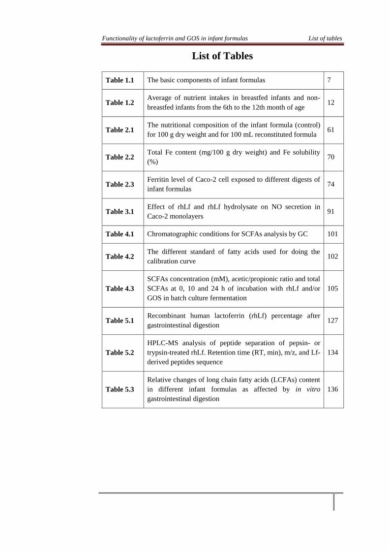
Functionality of lactoferrin and GOS in infant formulas List of tables
List of Tables
Table 1.1 The basic components of infant formulas 7
Table 1.2 Average of nutrient intakes in breastfed infants and non-
breastfed infants from the 6th to the 12th month of age 12
Table 2.1 The nutritional composition of the infant formula (control)
for 100 g dry weight and for 100 mL reconstituted formula 61
Table 2.2 Total Fe content (mg/100 g dry weight) and Fe solubility
(%) 70
Table 2.3 Ferritin level of Caco-2 cell exposed to different digests of
infant formulas 74
Table 3.1 Effect of rhLf and rhLf hydrolysate on NO secretion in
Caco-2 monolayers 91
Table 4.1 Chromatographic conditions for SCFAs analysis by GC 101
Table 4.2 The different standard of fatty acids used for doing the
calibration curve 102
Table 4.3
SCFAs concentration (mM), acetic/propionic ratio and total
SCFAs at 0, 10 and 24 h of incubation with rhLf and/or
GOS in batch culture fermentation
105
Table 5.1 Recombinant human lactoferrin (rhLf) percentage after
gastrointestinal digestion 127
Table 5.2
HPLC-MS analysis of peptide separation of pepsin- or
trypsin-treated rhLf. Retention time (RT, min), m/z, and Lf-
derived peptides sequence
134
Table 5.3
Relative changes of long chain fatty acids (LCFAs) content
in different infant formulas as affected by in vitro
gastrointestinal digestion
136


Functionality of lactoferrin and GOS in infant formulas List of figures
List of Figures
Fig. 1.1 Schematic of intestinal iron absorption 16
Fig. 1.2 Different strategies to prevent and control IDA and anemia 24
Fig. 1.3 Fig. 1.3. Protein structure of human lactoferrin 28
Fig. 2.1 Image of Caco-2 cell line in different days post-seeding by
contrast microscope observation of the test phases 65
Fig. 2.2 Image of Caco-2 cell line presented the absence of mycoplasma
in the cultured Caco-2 cell line 65
Fig. 3.1 Co-culture system constructed with Caco-2 cells and RAW
264.7 cells 85
Fig. 3.2 TEER values of Caco-2 monolayers measured at 0 and 5 h of
incubation with LPS with/without rhLf or rhLf hydrolysate 88
Fig. 3.3 Effect of rhLf and rhLf hydrolysate on IL-8 produced by Caco-
2 cells 90
Fig. 3.4 Effect of rhLf and rhLf hydrolysate on ROS production in
Caco-2 monolayers 92
Fig. 4.1 The chromatogram of the different SCFAs analyzed using the
multi-standard solution of 10 mM concentration 101
Fig. 4.2
Minor short chain fatty acids concentration produced at 10 and
24 h of incubation with rhLf and/or GOS in batch culture
fermentation
111
Fig. 4.3 pH variation at 10 and 24 h of incubation with rhLf and/or GOS
in batch culture fermentation 113
Fig. 5.1 Western Blot of rhLf- untreated and treated with pepsin or
trypsin 125
Fig. 5.2 SDS-PAGE pattern of rhLf stained with Coomassie blue 125
Fig. 5.3 Western Blot of digested human milk 126
Fig. 5.4 Western Blot of digested infant formulas without adding GOS
(A); with 3.3% of GOS (B); 5% GOS (C) and 10% GOS (D) 127
Fig. 5.5 Fractionation of rhLf-derived peptides by pepsin 133
Fig. 5.6 Fractionation of rhLf-derived peptides by trypsin 133


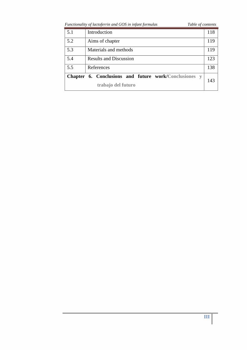
Functionality of lactoferrin and GOS in infant formulas Table of contents
I
Table of contents
Chapter 1. General Introduction 1
1.1 Overview 1
1.2 Infant feeding 3
1.2.1 The breastfeeding pattern 3
1.2.2 The bottle-feeding pattern 5
1.3 The nutritional needs during infancy 11
1.4 Iron as nutrient 14
1.4.1 The physiological importance and the presence of iron
in diet, human and cow milk 14
1.4.2 Absorption of iron 15
1.5 Dietary factors affecting iron bioavailability 17
1.6 Absorption and bioavailability: definition and applied
methods 18
1.7 The nutritional state and prevalence of iron deficiency
anemia among the Egyptian infants 19
1.8 The common strategies for prevention of iron
deficiency and anemia 23
1.9 Human milk lactoferrin and oligosaccharides as
multifunctional ingredients for infant formulas 27
1.9.1 Human milk lactoferrin 27
1.9.1.1 Lf structure and properties 27
1.9.1.2 Functional roles of Lf and its mode of action 29
1.9.1.3 Lf as a functional component in infant formulas 35
1.9.2 Human milk oligosaccharides (HMOs) 37
1.9.2.1 HMOs structure, composition and variation 37
1.9.2.2 Physiological function of HMOs in infants 38
1.9.2.3 Alternative sources of HMOs-like prebiotic in infant
formula 40
1.10 Objective of the study 41

Functionality of lactoferrin and GOS in infant formulas Table of contents
II
1.11 References 42
Chapter 2. The role of rhLf and GOS on Fe bioavailability:
Formation of ferritin by Caco-2 cell model 59
2.1 Introduction 59
2.2 Aims of chapter 59
2.3 Materials and methods 60
2.4 In vitro digestion model of infant formulas 63
2.5 Iron bioavailability in vitro by Caco-2 cell line model
(Ferritin synthesize) 66
2.6 Statistical analysis 68
2.7 Results and Discussion 69
2.8 References 77
Chapter 3. The effect of rhLf and Lf hydrolysate on LPS-
induced inflammation
81
3.1 Introduction 81
3.2 Aims of chapter 82
3.3 Materials and methods 82
3.4 Statistical analysis 87
3.5 Results and Discussion 87
3.6 References 93
Chapter 4. The prebiotic activity of rhLf and GOS. Batch
culture fermentation study
97
4.1 Introduction 97
4.2 Aims of chapter 98
4.3 Materials and methods 98
4.4 Statistical analysis 102
4.5 Results and Discussion 103
4.6 References 114
Chapter 5. Effect of in vitro gastrointestinal digestion on the
profile of rhLf and long chain fatty acids in human
milk as compared with different infant formulas
118
Functionality of lactoferrin and GOS in infant formulas Table of contents
III
5.1 Introduction 118
5.2 Aims of chapter 119
5.3 Materials and methods 119
5.4 Results and Discussion 123
5.5 References 138
Chapter 6. Conclusions and future work/Conclusiones y
trabajo del futuro 143


Functionality of lactoferrin and GOS in infant formulas Chapter 1
1
Chapter 1
General Introduction
1.1. Overview
It has been broadly acknowledged that breastfeeding is the best food
for newborn during the initial six months of life. Mother's milk gives all the
nutritive components to ordinary development and for the digestive states of
newborn children notwithstanding build the full of feeling relationship of
the mother towards the baby. It additionally contains various defensive and
immunoregulatory factors that may have a valuable impact on the
advancement of the newborn child's resistant framework. Breastfed babies
endure less gastrointestinal and respiratory contaminations, this is
particularly highlighted in the lower financial gatherings of creating nations
(Cesar et al., 1999), and there is expanding confirmation of a comparable
defensive impact of breastfeeding in created nations (Raisler et al., 1999).
Subsequently human milk is viewed as the first and the best decision for the
baby (Alles et al., 2004).
The bioactive compounds of breast milk are a large group of
different kinds of molecules (proteins, peptides, carbohydrates, ....) that are
naturally present in human milk which are added to infant formulas for
achieving the functional effects that occur in children fed with breastfeeding
(Dorca, 2008) and the interest in the presence of these bioactive substances
in human milk is reinforced by its almost total absence in infant formulas
(Gómez-Gallego et al., 2009).
Human lactoferrin (hLf) is one of the most important components of
human milk proteins constituting about 10-15%. This protein has many
positive effects on infant health where promotes the absorption of iron, has
antimicrobial, antiviral and anti-inflammatory activities, is growth and
proliferation factor of the intestinal mucosa, and favors the incorporation of
thymidine into DNA (the latter being an independent effect of iron) (Baró et
al., 2001), being also immunomodulatory and anticarcinogenic (Korhonen et
al., 1998).
Other compounds of human milk are Human Milk Oligosaccharides
(HMOs) which have been recognized as a new class of potent bioactive

Functionality of lactoferrin and GOS in infant formulas Chapter 1
2
molecules (Wu et al., 2011). HMOs are the third most abundant component
of human milk (Kunz et al. 2000), and also provides functional capacity
including prebiotic activity, anti-adhesive and immunomodulators. Various
strategies have been used to mimic the structural complexity of HMOs
(Hamosh, 1996; Oddy, 2002).
However, breastfeeding is not generally conceivable, attractive or
adequate, and is therefore necessary to create infant formulas attempting to
replace breastfeeding giving the necessary nutrients for optimal growth and
development of infants. In these cases, infant formulas play an
indispensable role in infant nutrition (Alles et al., 2004).
The creation of infant formula has developed alongside the
knowledge of breast milk composition. Nowadays, companies and research
centers is devoted to prepare these formulas focusing their efforts on
enhancing the quality of infant formulas, not only adapting the
concentration of macronutrients and micronutrients but also the composition
of bioactive compounds to make it similar to human milk (Dorca, 2008).
Regarding with infant formulas evolution and how to produce a
suitable nutritional substitute for infant and newborns, European Food
Safety Authority (EFSA, 2012) recently accepted and approved bovine
lactoferrin (bLf) as a new food ingredient. Thus, various types of infant
formulas containing Lf are available in the market of many countries such
as Japan and Spain (Mulder et al., 2008). Since bLf is truly diverse in
several perspectives when compared with hLf (Kawakami and Lönnerdal,
1991), there is significant interest in replacing the use of bLF with hLF in
products for human utilization (Conesa et al., 2010). In this way various
attempts have been made to produce recombinant human lactoferrin (rhLF)
from rice (Nandi et al., 2002; Rachmawati et al., 2005). Currently,
supplementation of infant formula with rhLf represents an attractive
application (Suzuki et al., 2003).
With the aim to mimic human milk, also prebiotic formulations,
which are now added to commercial infant formula, are mixtures of
galactooligosaccharides (GOS), fructooligosaccharides (FOS), inulin and
polydextrose. These sugars are provided in roughly the same concentration
as in human milk, but do not mimic the diversity and complexity of sugar

Functionality of lactoferrin and GOS in infant formulas Chapter 1
3
side chains exhibited in human milk (Sherman et al., 2009). In spite of the
tremendous advancement in infant formula industry, human milk is still
viewed as the best source of infant nutrition (Leung & Sauve, 2005;
American Academy of Pediatrics, AAP, 2012). Many studies needed to
assess the functionality of the bioactive components of human milk
especially lactoferrin (Lf) and oligosaccharides whether alone or added to
infant formulas.
1.2. Infant feeding
1.2.1. The breastfeeding pattern
Good nutrition is essential for the development and improvement
that happens during an infant’s first year of life. When developing infants
are fed the appropriate types and amounts of foods, their health is promoted
(United States Department of Agriculture, USDA, 2009). In such manner
and as per AAP (2012), the breastfeeding is viewed as the favored decision
of feeding for all infants and the exclusive breastfeeding for about the initial
6 months is key for an adequate health, followed by continued breastfeeding
with introducing of some complementary foods when breast milk alone is
no more adequate to meet the nutritional necessities of infants.
Consequently, it has been suggested that breastfeeding ought to be
proceeded until one or even two years old (WHO, 2001).
So breastfeeding is without a doubt the best type of feeding for
newborns and young infants and its advantage go far beyond nutritional and
anti-infective benefits (Mathew, 2004). In this respect, various studies have
shown that the breastfeeding at the first months of life can decrease
worldwide mortality diarrhea, respiratory illness, and other infectious
disease by up to 55% (Chantry et al., 2006), and this is principally because
the human milk components that are viewed as major contributors to
decrease morbidity rates in breastfed infants (Newburg, 2000a). One of
these major active components is Lf which has numerous healthy effects on
the newborns such as the antimicrobial effects which add to the protective
factor of breast milk (Story & Parish, 2008; Gifford et al., 2005; Jackson &
Nazar, 2006).
Additionally, human milk contains vital and multiple immunological
and anti-infective agents (Chirico et al., 2008). They include, among many

Functionality of lactoferrin and GOS in infant formulas Chapter 1
4
others, proteins with antimicrobial components such as secretory
immunoglobulin A (IgA), lysozyme, and Lf; the last one has immune-
modulating properties in addition to its well-known anti-infective properties.
Oligosaccharides in human milk inhibit bacterial adhesion, further
protecting against pathogens. Nucleotides and cytokines of human milk,
also assist with T-cell maturation and immune system modulation,
evidenced by, e.g., the more robust immune response that breastfed infants
exhibit after vaccination. Human milk also promotes healthful
gastrointestinal microbiota (Zivkovic et al., 2011), and can actively
stimulate development of the newborn’s host defenses to provide continued
mucosal protection after breastfeeding. Several components of human milk
such as growth factors, interleukin-10 (IL-10) and also Lf can reduce the
inflammatory response to stimuli in the newborn intestine (Petit, 2008;
Walker, 2010). Lf as a functional human milk ingredient has been
demonstrated to increase the resistance of newborns to infections and also
has many biological activities that are essential for an adequate health of
infants. Recently, Lf has taken more attention in regarding with some
healthy activities like its role in the improvement of bone health, cancer
prevention and its role as transcription factor. Lf is also able to enter a cell
and to activate the transcription of specific DNA sequences and this Lf-
DNA interaction is reported to be responsible for antiviral activity
(Adlerova et al, 2008).
The benefits of breastfeeding have been well-documented which
provides optimal nutrition and prevents common childhood diseases
(Abiona et al., 2006). The importance of breastfeeding is not only providing
essential nutrients to infants, but it has many health benefits for both
children and their mothers (Kramer & Kakuma, 2002). Breastfeeding helps
to build up a safe and full of feeling relationship between the mother and her
infant and offers numerous other positive advantages. Based on the above-
mentioned, breastfeeding should be actively promoted and supported as the
most desirable method of infant feeding.
It is scientifically accepted that the feeding pattern can influence the
composition of gut flora which differs between breast-fed infant and
formula-fed infants with a higher proportion of Bifidobacteria species in

Functionality of lactoferrin and GOS in infant formulas Chapter 1
5
breast-fed infants (Harmsen et al., 2000; Alles et al., 2004; Iacono et al.,
2005; Granier et al., 2013). Another difference was observed between the
two types of feeding which is the higher absorption of iron from human
milk as compared with feeding on cow milk or infant formula (Jovani et al.,
2001) and this might partly explained by the higher concentration of Lf in
human milk than bovine milk (Vorland, 1999). Likewise, the discovery of
Lf receptors in the enterocytes of various species and its high affinity for Lf
support this hypothesis. These Lf receptors show species and molecular
specificities depending of the animal species and this would explain the high
bioavailability of iron from human milk, as only hLf releases iron to the
enterocyte by this mechanism (Gonzalez-Chavez et al., 2009).
1.2.2. The bottle-feeding pattern
1.2.2.1. Infant formulas: concepts and types
Although breast milk is the optimal source of nutrition for infant,
infant formula and milk substitutes are considered as an appropriate
alternative for infants nutrition at the first year of life when breast milk is
not available, or the mother cannot breastfeed her baby (Alles et al., 2004),
or newborns cannot be breastfed or cannot receive human milk (WHO,
1986).
In general, the design of infant formula is based on the composition
of human milk and the current trend in infant formulas manufacturing is
looking to provide not only nutritional compounds but also similar
functional effects than human milk. The final aim of infant formula
development is not necessarily to mimic the composition of human milk in
every respect, but to achieve physiological effects as in breast fed infants
(Gómez-Gallego et al., 2009).
In the European legislation, Commission directive 2006/141/EC of
22 of December 2006 on infant formulas and follow-on formulas and
amending the directive of 1999/21/EC, are called “infant formulas or
formula 1” and defined it as "foodstuff intended for special nutritional use
during the first months of life and satisfying by themselves the nutritional
requirements of this category of persons", whereas "follow-on formula or
formula 2 ” means "foodstuffs intended for special nutritional use by

Functionality of lactoferrin and GOS in infant formulas Chapter 1
6
infants aged over four-six months and young children and constituting the
principal liquid element in a progressively diversified diet of this category
of persons" (European Commission, 2003). Table 1.1 shows the basic
composition of infant formulas 1.
1.2.2.2. The design and current trends of infant formulas composition
Infant formula manufacturers are continuously looking for
modifications on composition in an attempt to simulate human milk in
function. Nowadays, is normal that infant formulas contain some of the
functional ingredients of human milk such as prebiotic, probiotic bacteria,
polyunsaturated fatty acids, Lf and nucleotides (Joeckel & Phillips, 2009).
Infant milk formula is subjected to strict regulations for composition and
hygiene (Koletzko et al., 2005). Nowadays, Lf (Wakabayashi et al., 2006)
and GOS (Motil, 2000; Gopal & Gill 2000) are commonly added to infant
formulas. The most important compounds added to infant formulas are:
Prebiotic and probiotic
Prebiotics are defined as ¨non-digestible substances in food, such as
oligosaccharides, which can stimulate growth and activity of beneficial
bacteria in the gastrointestinal tract, they are not digested by human
gastrointestinal enzymes, hence, can enter the colon intact serving as
fermentable substrates for the colonic microbiota (Gibson & Roberfroid,
1995) preferably Bifidobacteria (Roberfroid, 2000). Human milk contains
more than one hundred different oligosaccharides structures, comprising a
total concentration of 15-23 g/L in colostrum and 8-12 g/L in transitional
and mature milk, that together with the other milk components are the major
source of prebiotic effect (Kunz et al., 2000; Euler et al., 2005).
They are also considered an important growth-promoting bifidus
factor (Kunz et al., 2000). Thus, the prebiotic oligosaccharides play a role
in enhancement of the growth and activity of probiotic bacteria and this
named “the bifidogenic effect” (Gibson & Roberfroid, 1995) which
considered one of the most important biological indicators of the
resemblance of infant formulas to human milk (Martinov et al., 2011).

Functionality of lactoferrin and GOS in infant formulas Chapter 1
7
Table 1.1. The basic components of infant formulas.
NS = not specified, According to Koletzko et al. (2005).
Components Units Minimum Maximum
Energy Kcal/100ml 60 70
Protein
Cows’ milk protein g/100 Kcal 1.8 3
Soy protein isolates g/100 Kcal 2.25 3
Hydrolyzed cows’ milk protein g/100 Kcal 1.8 3
Lipids
Total fat g/100 Kcal 4.4 6.0
Linoleic acid g/100 Kcal 0.3 1.2
-Linoleic acid g/100 Kcal 50 NS
Ratio linoleic/a-linolenic acids
Lauric + myristic acids % of fat NS 20
Trans fatty acids % of fat NS 3
Erucic acid % of fat NS 1
Carbohydrates
Total Carbohydrates g/100 Kcal 9.0 14.0
Vitamins
A µg RE/100 Kcal 60 180
D3 µg /100 Kcal 1 2.5
E mg a-TE/100 Kcal 0.5 5
K µg /100 Kcal 4 25
Thiamin µg /100 Kcal 60 300
Riboflavin µg /100 Kcal 80 400
Niacin µg /100 Kcal 300 1500
B6 µg /100 Kcal 35 175
B12 µg /100 Kcal 0.1 0.5
Pantothenic acid µg /100 Kcal 400 2000
Folic acid µg /100 Kcal 10 50
C µg /100 Kcal 10 30
Biotin µg /100 Kcal 1.5 7.5
Mineral and trace elements
Iron (formula based on cows’ milk protein
and protein hydrolysate)
mg /100 Kcal 0.3 1.3
Iron (formula based on soy protein isolate) mg /100 Kcal 0.45 2.0
Calcium mg /100 Kcal 50 140
Phosphorus (formula based on cows’ milk
protein and protein hydrolysate)
mg /100 Kcal 25 90
Phosphorus (formula based on soy protein
isolate)
mg /100 Kcal 30 100
Ratio calcium/phosphorus mg/mg 1:1 2:1
Magnesium mg /100 Kcal 5 15
Sodium mg /100 Kcal 20 60
Chloride mg /100 Kcal 50 160
Potassium mg /100 Kcal 60 160
Manganese µg /100 Kcal 1 100
Fluor µg /100 Kcal NS 100
Iodine µg /100 Kcal 10 50
Selenium µg /100 Kcal 1 9
Cupper µg /100 Kcal 35 100
Zinc mg /100 Kcal 0.5 1.5
Other substances
Choline mg /100 Kcal 7 50
Myo-inositol mg /100 Kcal 4 40
L-carnitine mg /100 Kcal 1.2 NS
Taurine mg /100 Kcal NS 12

Functionality of lactoferrin and GOS in infant formulas Chapter 1
8
As mentioned above that HMOs may serve as substrates for colonic
fermentation, it has been shown that HMOs induce an increase in the
number of Bifidobacteria of colonic flora in breast-fed infants,
accompanied with a significant reduction in the number of potentially
pathogenic bacteria (Kunz et al., 2000). Complex oligosaccharides have the
ability of inhibiting the binding of pathogens to cell surfaces because they
act as competitive receptors on the host cell surface, thereby preventing
adhesion of a number of bacterial and viral pathogens (European
Commission, 2003). Thus there are many differences in the fecal
microbiota between breast-fed infants and formula-fed infants. Harmsen et
al. (2000) used a new molecular identification and detection method to
compare the fecal flora of breast-fed and formula-fed infants and it was
reported that Bifidobacteria are dominant in breast-fed infants, while the
amounts of Bifidobacteria and Bacteriodes spp. are similar in the feces of
formula-fed infants. In this regards, Solis et al. (2010) reported that the
microbiota of formula-fed infants is more diverse and contains substantial
quantities of Bacteriodes, Enterobaceriaceae and Clostridium species.
FOS and GOS may be voluntarily added to infant formula (< 0.8
g/100 mL) in a ratio of 90% GOS: 10% FOS. The Food and Agricultural
Organization (FAO) of the United Nations supports the supplementation of
formula with prebiotics in infants aged five months and older, as these
infants will have a mature immune system and intestinal colonization
(Ackerberg et al, 2012).
Probiotics are ¨live microbial components that beneficially affect
the host by improving its intestinal microbial balance¨. Bifidobacteria are
predominant in infants fed formulas supplemented with Bifidobacteria and
was similar to that found for breast-fed infants as compared with the control.
Thus probiotic bacteria are promising component and have been used
successfully in infant formulas production (Alles et al., 2004). In general,
the aim of adding probiotics and prebiotics to preterm infant formula is to
improve growth, development and decrease infections by promoting an
intestinal microbiota resembling that of breast-fed infants (Underwood et
al., 2009).

Functionality of lactoferrin and GOS in infant formulas Chapter 1
9
Nucleotides
Nucleotides are ¨nitrogenous compounds which play and their
metabolites derivatives a key role in numerous biochemical and
physiological processes, such as energy transfer processes, they are
precursors for nucleic acid synthesis (DNA and RNA), and they are key to
the synthesis of carbohydrates, lipids and proteins¨. Human milk contains,
in free form, ribonucleotides and ribonucleosides, which account for 2-5%
of non-protein nitrogen in human milk, may contribute to excellent use of
the protein by breast-fed infants (Baró et al., 2001). In addition, human
milk contains significant amounts of related compounds: nucleosides,
purine and pyrimidine bases, nucleic acids and products derived from them
(such as uridine diphosphate galactose) (Gil & Uauy, 1995).
The concentration of free nucleotides in human milk is higher than
in bovine milk. Because bovine milk is most often used to formulate infant
milk formulas, most milk formulas are supplemented with nucleotides to
increase the concentration to a level that is similar to the concentration
found in human milk (Pickering et al., 1998).
Recently, legislation allows the addition to infant formulas and
follow-on formula, nucleotides in quantities of: 1.5 mg adenosin-5-
phosphate/100 kcal, 2.5 cytosine-5-phosphate/100 kcal, 0.5 kcal guanosina-
5-phosphate/100 mg, 1.75 mg Uridine-5-phosphate/100 kcal, 1 mg inosin-
5-phosphate/100 Kcal, until a total concentration of 5mg/100 kcal, which is
similar to the amounts of free ribonucleotides in milk (4-6 mg/100 kcal)
(European Commission, 2003). Also in this context, Koletzko et al. (2005)
reported that ESPGHAN supports the optional addition of nucleotides in
amounts not to exceed 5 mg/100 Kcal as adverse effects have been seen
with higher concentrations.
The supplementation of infant formulas with the dietary nucleotides
will results in increased the growth of probiotic bacteria in the intestinal
tract with a reduction of the pathogenic bacteria population due to the
competitive exclusion. It was reported by Gil et al. (1986) that babies fed
nucleotide-supplemented infant formula have increased ‘friendly’
Bifidobacteria counts in feces compared to infants fed standard formula
milk, but counts were still lower than found in breast-fed babies. Infant

Functionality of lactoferrin and GOS in infant formulas Chapter 1
10
studies also suggest those receiving nucleotide supplemented formula have
an improved antibody response following immunization (Schaller et al.,
2004).
Polyunsaturated fatty acids
In the last two decades, special attention has been paid to the
composition and physiological function of the lipid fraction in human milk.
Human milk fat is the major source of energy for the breast-fed infants,
contributing some 40-55% of the total energy intake. Human milk fat
contains essential nutrients which are lipid soluble vitamins and
polyunsaturated fatty acids (PUFAs), including linoleic acid of the n-6
series (C18:2 n-6) and α-linolenic acid of the n-3 series (C18:3 n-3).
Omega-3 and omega-6 fatty acids are essential fatty acids and are an
important component of human milk with a significant role in the overall
growth and development of infant (Ganapathy, 2009). The components of
human milk that may partly explain the observed differences are the
polyunsaturated fatty acids (PUFAs): docosahexaenoic acid (C22-6, n-3;
DHA) and arachidonic acid (C20-4, n-6; AA). DHA and AA are derived
mainly from their precursors, α-linolenic acid (ALA, an omega-3 fatty acid)
and linoleic acid (LA, an omega-6 fatty acid), respectively (Innis, 2008).
Concentrations of PUFAs in human milk are relatively stable during
the first year of life: DHA is equivalent to 0.5% in colostrum and 0.25% in
mature milk, which is equivalent to 7 to 8 mg/dL; AA is equivalent to 1%
in colostrum and 0.5% in mature milk, which is equivalent to 14 - 15
mg/dL (Martinez, 1992). It is well established that breastfeeding pattern is
associated with a better neurological, cognitive and behavioral outcome
than formula feeding pattern (De Jong et al., 2010), and the prolongation of
breastfeeding period was associated with a better cognitive outcome at six
years (Kramer et al., 2008), suggesting that the composition of human milk
plays a key role in the positive association between breastfeeding and
cognitive development.
Nowadays, it is generally accepted that infants should receive at
least 0.3% of both DHA and AA in infant feeding (Koletzko et al., 2008),
even though higher DHA levels in formulas have been suggested to special
group such as preterm infants (Makrides et al., 2009).
Functionality of lactoferrin and GOS in infant formulas Chapter 1
11
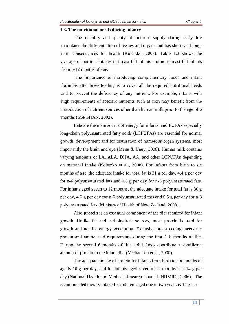
1.3. The nutritional needs during infancy
The quantity and quality of nutrient supply during early life
modulates the differentiation of tissues and organs and has short- and long-
term consequences for health (Koletzko, 2008). Table 1.2 shows the
average of nutrient intakes in breast-fed infants and non-breast-fed infants
from 6-12 months of age.
The importance of introducing complementary foods and infant
formulas after breastfeeding is to cover all the required nutritional needs
and to prevent the deficiency of any nutrient. For example, infants with
high requirements of specific nutrients such as iron may benefit from the
introduction of nutrient sources other than human milk prior to the age of 6
months (ESPGHAN, 2002).
Fats are the main source of energy for infants, and PUFAs especially
long-chain polyunsaturated fatty acids (LCPUFAs) are essential for normal
growth, development and for maturation of numerous organ systems, most
importantly the brain and eye (Mena & Uauy, 2008). Human milk contains
varying amounts of LA, ALA, DHA, AA, and other LCPUFAs depending
on maternal intake (Koletzko et al., 2008). For infants from birth to six
months of age, the adequate intake for total fat is 31 g per day, 4.4 g per day
for n-6 polyunsaturated fats and 0.5 g per day for n-3 polyunsaturated fats.
For infants aged seven to 12 months, the adequate intake for total fat is 30 g
per day, 4.6 g per day for n-6 polyunsaturated fats and 0.5 g per day for n-3
polyunsaturated fats (Ministry of Health of New Zealand, 2008).
Also protein is an essential component of the diet required for infant
growth. Unlike fat and carbohydrate sources, most protein is used for
growth and not for energy generation. Exclusive breastfeeding meets the
protein and amino acid requirements during the first 4–6 months of life.
During the second 6 months of life, solid foods contribute a significant
amount of protein to the infant diet (Michaelsen et al., 2000).
The adequate intake of protein for infants from birth to six months of
age is 10 g per day, and for infants aged seven to 12 months it is 14 g per
day (National Health and Medical Research Council, NHMRC, 2006). The
recommended dietary intake for toddlers aged one to two years is 14 g per

Functionality of lactoferrin and GOS in infant formulas Chapter 1
12
Table 1.2. Average of nutrient intakes in breastfed infants and non-
breastfed infants from the 6th to the 12th month of age.
Nutrient
Breastfed infants Non-breastfed infants
6
months
of age
9
months of
age
12
months
of age
6
months
of age
9
months
of age
12
months of
age
Energy,
macronutrients and
dietary fiber
Energy (KJ)
Protein (g)
Carbohydrate
Dietary fibers (g)
Fat (g)
3564
17
-
5
27
5364
41
-
10
39
6142
50
-
15
48
5294
41
-
6
25
6411
45
-
14
34
7641
61
-
17
55
Minerals
Ca (mg)
Zn (mg)
Fe (mg)
Mg (mg)
Si (µg)
Sodium (mg)
672
2.2
11
56
11
310
1209
3.5
32
125
16
1396
977
6
16
216
25
1600
672
3
21
128
5
8744
1209
4.1
21
209
11
1888
977
6.7
12
230
26
2315
Fat-soluble vitamins
Vitamin A (µg RE)
Vitamin D (µg)
Vitamin E (mg TE)
Vitamin K (µg)
1290
10
2
-
1731
19
4
-
1701
7
4
-
943
9
3
-
1858
10
4
-
1168
4
3
-
Water-soluble
vitamins
Thiamin (mg)
Riboflavin (mg)
Niacin (mg NE)
Vitamin B6 (mg)
Vitamin B12 (µg)
Folate (µg)
Pantothenic acid
(mg)
Biotin (µg)
Vitamin C (mg)
Choline (mg)
-
-
4
-
-
47
-
5
85
-
-
-
9
-
-
103
-
14
132
-
-
-
16
-
-
154
-
23
162
-
-
-
6
-
-
78
-
9
75
-
-
-
9
-
-
106
-
16
214
-
-
-
19
-
-
178
-
30
131
-
Adapted from Simons (1999). Note: - = not measured.

Functionality of lactoferrin and GOS in infant formulas Chapter 1
13
day, or 1.08 g per kilogram body weight (Ministry of Health of New
Zealand, 2008).
Carbohydrates can be classified to digestible and non-digestible
carbohydrates and is important to remark that human milk contains both of
them. Digestible carbohydrates are one of the main sources of dietary
energy in infancy and childhood and are essential for growth and
development (Stephen et al., 2012). The main digestible carbohydrate in
mature breast milk is lactose which provides about 40% of the energy
content (Koletzko et al., 2005), in addition to a large variety of
oligosaccharides in concentrations of approximately 5–10 g/L (Kunz et al.,
2000).
Non-digestible carbohydrates such as FOS, GOS, inulin, soy
polysaccharide, resistant starch, and gums are added to dietary products,
enteral formulas and human milk substitutes consumed by infants (Aggett et
al., 2003), considering that the adequate intake of carbohydrates for infants
from birth to six months of age is 60 g per day; and 95 g per day for infants
aged seven to 12 months (Ministry of Health of New Zealand, 2008).
Although the minerals and trace elements are very important and
play a pivotal role in infant health. The needs and role of iron for infants in
the early stage of life are only discussed in this section. Healthy term infants
are normally born with plenty of iron where they need a relatively high iron
intake because they are growing very rapidly. But after 6 months of age,
iron content of human milk is not sufficient to meet many infants’
requirements, thus requirement for dietary iron increases to an estimated
0.78 mg/day due to the stepwise depletion of endogenous stores and rapid
growth (Institute of Medicine, IOM, 2000). In this regard, where iron-
fortified complementary foods are not widely and regularly consumed by
young children, infants should routinely receive iron supplements in the first
year of life. Where the prevalence of anemia in young children (6–24
months) is 40% or more, supplementation should continue through the
second year of life (Stoltzfus & Dreyfuss, 1998).
The recommended dietary intake (RDI) for iron for an infant 7 to 12
months old is 11 mg per day. The recommended daily intake for toddlers
aged one to three years is 9 mg per day. Absorption is about 18 percent from

Functionality of lactoferrin and GOS in infant formulas Chapter 1
14
a mixed western diet including animal foods and about 10 percent from a
vegetarian diet; so vegetarian infants need higher intakes (NHMRC, 2006).
1.4. Iron as nutrient
1.4.1. The physiological importance and the presence of iron in diet,
human and cow milk
Iron is a pivotal and essential trace element for the maintenance of
the human health due to its obligate role in a number of the physiological
processes (Sharp & Srai, 2007). However, in excess, iron is potentially toxic
to cells due to its ability to catalyse the production of reactive oxygen
species (ROS) (Steele et al., 2004). Excessive iron accumulation leads to the
damage of liver, heart, pancreas and other organs. Beside systemic disorders
of iron homeostasis, local mismanagement of iron plays a role in several
disorders (Stankowski et al., 2012).
Dietary iron is present in two different forms: non-heme iron (found
in cereals, vegetables, pulses, beans, fruits as simple iron oxides or complex
iron chelates) and heme iron (mainly found in meat and meat products).
Non-heme iron is predominant in all diets forming some 90-95% (Darshan
& Anderson, 2007) and is found as Fe2+
bound to insoluble proteins,
phytates, oxalates, phosphates and carbonates, and as ferritin (Scientific
Advisory Committee on Nutrition, 2010), While heme-iron forms 5-10% of
total daily iron intake. However, the heme-iron is the most bioavailable
source of iron (20-30%) while the non-heme iron has a low bioavailability
amounting of 1-10% of the dietary load (Hallberg et al., 1989).
In human milk, iron content is low 0.2-0.4 mg/L (Domellof et al.,
2002) and is mainly bound to Lf (20-45%; Chierici & Vigi, 1994); while in
cow milk it is mainly bound to casein (24 %) (Renner et al., 1989), which
correlates well with the finding of Makino and Nishimura (1992), which
reported that 95% of hLf is in the monoferric and/or apo-lactoferrin form.

Functionality of lactoferrin and GOS in infant formulas Chapter 1
15
1.4.2. Absorption of iron
1.4.2.1. Uptake of iron into enterocytes
Iron absorption occurs mainly in duodenum and upper jejunum,
although small amounts may also be absorbed from stomach, ileum and
colon (Anderson & Vulpe, 2002). At the cellular level, iron is absorbed
through the differentiated epithelial cells (enterocytes) of the mid to upper
villus. Iron is provided to the body in various forms through the diet, but is
primarily absorbed as either inorganic (non-heme) iron or as heme iron
(Lönnerdal, 2010).
The first step is termed mucosal uptake, and iron uptake is defined
by Merit et al. (2003) as ¨the transport of dietary iron across the apical
membrane of the enterocytes into the intestinal mucosa¨. There are at least
two separate mechanisms for the uptake of heme and non-heme iron into the
enterocytes (Scientific Advisory Committee on Nutrition, 2010). The
passage of iron through the enterocyte into the circulation is depicted in Fig.
1.1.
In the case of the absorption of non-heme iron from the intestinal
lumen to the enterocytes, Fe3+
(ferric iron) is first reduced to Fe2+
(ferrous
iron), most likely by duodenal cytochrome b (Dcytb), making it available
for transport across the brush border membrane by divalent metal
transporter1 (DMT1) into the cytoplasm (Steele et al., 2004).
On the other hand, the absorption of heme iron across the apical
membrane occurs more efficiently but the mechanism is still unclear (Steele
et al., 2004; West and Oates, 2008; Le Blanc et al., 2012). The passage of
heme iron across the apical membrane is facilitated by intestinal heme
transporter named heme carrier protein 1 (HCP1) (Dunn et al., 2006).
Latunde-Dada et al. (2006) claimed that a heme receptor was identified in
piglets and human. Likewise, Dunn et al. (2007) stipulated that a receptor
for HCP1 is present in a large concentration in the duodenum. Once heme
iron has been taken up by the enterocyte, the heme molecule is degraded by
heme oxygenase to release ferrous iron as clarified by Dunn et al. (2007).
Inside the enterocyte, heme and non-heme iron enter a common transit pool,
where iron may be chelated by low molecular weight compounds or bound
to a protein ligand such as ferritin (Steele et al., 2004).

Functionality of lactoferrin and GOS in infant formulas Chapter 1
16
Fig. 1.1. Schematic of intestinal iron absorption (adapted from Rizvi
and Schoen, 2011).
1.4.2.2. Export of iron from enterocytes to blood circulation
The second stage of iron absorption is termed basolateral or serosal
transfer, where iron is transported from the enterocytes into the intestinal
capillaries across the basolateral membrane, most likely via the iron
transporter iron regulated protein 1 (IREG-1 or ferroportin 1) (Steele et al.,
2004). The ferrous iron delivered for the basolateral membrane of the
intestine is exported by ferroprotein, a metal protein transporter 1, to the
plasma. The ferrous iron is oxidized to ferric iron by ferroxidase hephaestin
(Dunn et al., 2007). The role of hephaestin in association with ferroprotein
to oxidize ferrous iron is important before it is exported and bound to
transferrin (Mackenzie & Garrick, 2005).
1.4.2.3. Transport of iron
Transferrin (Tf) is the major protein that binds and delivers iron to
tissues. Each Tf molecule can transport 2 ferrous iron molecules (Hirose,
2000). Conformation of the binding site is suitable with ferric iron in a
delicate manner (Harris, 1986). Transferrin binds to one of the transferrin
receptor (TfR) on cell membrane; TfR1 or TfR2 (Casey at al., 1988). TfR1
is expressed in all tissues except mature erythrocytes while TfR2 is
primarily expressed in the liver (Kawabata et al., 1999). Although protein

Functionality of lactoferrin and GOS in infant formulas Chapter 1
17
structures of TfR1 and TfR2 have a high degree of homology, their
functions and regulation are not the same. Expression of TfR1 is tightly
regulated by cellular iron levels through HFE (hereditary human
hemochromathosis protein) protein. However, cellular iron levels have no
effect on the regulation of TfR2. TfR2 senses the body iron status by
sensing the transferrin saturation and regulates hepcidin expression properly
(Fleming et al., 2002).
Once Fe3+
-Tf complex binds to its receptor at the cell surface, the Tf-
TfR1 complex is internalized in clathrin-coated pits that form endocytic
vesicles. Inside the cells, the internalized complex in the endosome is
acidified by a vacuolar H+-ATPase (V-ATPase) that lowers the luminal pH
to about 5.5. This acidification process induces conformational changes in
Tf-TfR1 complex with consequent release of iron (Sipe & Murphy, 1991)
which transports around the body to various tissues (Ohgami et al., 2005).
The main part of the iron carried by Tf is used by the formation of
hemoglobin (Hoppe, 2008).
1.5. Dietary factors affecting iron bioavailability
Iron chemical state is one of the most important factors that affect on
its absorption. In this context, in inorganic food compounds, iron is
normally in the oxidized form (Anderson, 2002), but absorption requires
reduction to Fe2+
, for iron enters in the mucous cell as a reduced free iron
that is more easily absorbed than ferric ion (Sgarbieri, 1987). Also Conrad
(1970) reported that iron physical and chemical form affects its absorption
that used radioactive markers and concluded that hemoglobinic iron is more
efficiently absorbed than inorganic iron. Also Anderson (2002) reported that
iron has a greater availability when present in the form of iron sulfate than
in salts such as sulfite, bisulfate, phosphate, carbonate, bicarbonate among
others. Thus it is notable that iron chemical form has an important effect on
its absorption and availability.
There are many differences in absorption of iron in food where foods
contain many dietary factors affecting on iron absorption and
bioavailability. The iron absorption enhancement agents are vitamins
(especially vitamin C or ascorbic acid) and organic acids, meat and fish,
meanwhile different compounds (mainly of vegetable origin) such as phytic

Functionality of lactoferrin and GOS in infant formulas Chapter 1
18
acid, soy protein, polyphenols, and calcium are considered as inhibitors of
iron absorption. There are also some functional components present in
different foods that are considered as iron absorption enhancement agents
like Lf and GOS which will be discussed in the section 1.9.
1.6. Absorption and bioavailability: definition and applied methods
An adequate bioavailability is dependent on an optimal digestion and
solubility, an optimal transport over the mucosal layer into the circulation,
and finally an optimal incorporation into the target organs. Although to
assess iron bioavailability all these steps, should be related any of these
steps can be used as an isolated measure to estimate bioavailability (Hoppe,
2008). Bioavailability is dependent on digestion, release from the food
matrix, absorption by intestinal cells, and transport to body cells (Etcheverry
et al., 2012). The concept of bioavailability as applied to nutrients is
critically important for understanding nutrient metabolism, homeostasis and
ultimately requirements (Krebs, 2001). While utilization is the process of
transport, cellular assimilation and conversion to biologically active form(s)
(Jackson, 1997).
Throughout the years, in vitro screening methods have been
developed and refined for determination of nutrient bioaccessibility and
bioavailability from foods. These are plentiful number of techniques
developed to assess iron absorption and bioavailability that can provide
useful information, especially when one considers the vast number of
factors that can affect nutrient absorption. There are a several of them can
be combined creating a vast number of methods with varying accuracy
when it comes to accomplish results relevant in humans. Generally, the
principally in vitro methods for measuring iron absorption and/or
bioavailability are: solubility, dialyzability and the Caco-2 models for
bioavailability (Etcheverry et al., 2012).
These in vitro methods are useful to provide knowledge on possible
interactions between nutrients and/or food components, the effects of
luminal factors (including pH and enzymes), food preparation and
processing practices, nature of the food matrix…etc., on either
micronutrient absorbability (a component of bioavailability) or on the
potential for a nutrient to be absorbed (i.e., bioaccessibility). In vitro

Functionality of lactoferrin and GOS in infant formulas Chapter 1
19
methods are less expensive, faster, and offer better controls of experimental
variables than human or animal studies (Sandberg, 2005). However, in vitro
studies cannot be substituted for in vivo studies, and should be therefore
regarded as a screening, ranking, or categorizing tool (Etcheverry et al.,
2012).
Because the in vitro digestion methods (solubility and dialyzability)
has various limitations, these methods have been improved by the
incorporation of a human colon carcinoma cell line (Caco-2) which provides
many functional and morphological properties of mature human enterocytes
(Ekmekcioglu, 2002). So this system is able to mimic and estimates the
uptake and/or transport of mineral elements, and has been used to assess
iron uptake from infant formulas (Jovaní et al., 2004). In this regard,
combining in vitro digestion with uptake in Caco-2 cells is a step forward
since it predicts both availability and uptake into the enterocyte and at times
also the absorption (Fairweather-Tait et al., 2005). Cell culture has been
used extensively as an in vitro method to assess human iron bioavailability
(Pinto et al., 1983). This cell model has been used in a wide variety of
nutritional studies, particularly in the study of mechanisms (Han et al.
1995), regulation of iron absorption (Tapia & Nuñez, 1999) and iron
bioavailability studies (Glahn et al., 1998). The application of a Caco-2 cell
model appears to be promising as a physiological means of measuring
mucosal cell iron uptake (Au & Reddy, 2000).
1.7. The nutritional state and prevalence of iron deficiency anemia
among the Egyptian infants
Anemia is defined as a state in which the level of hemoglobin or
hematocrit is below that which is expected, taking in account both age and
sex (Stoltzfus & Dreyfuss, 1998). Although anemia is the most prevalent
public health problem with serious consequences for national development,
it is ignored in most developing countries (World Bank, 1996; Bashir,
2013). In this regard, the World Bank reported that almost one third of the
world population is believed to be anemic and the WHO estimates the
number of anemic people worldwide to be about two billions (Stoltzfus,
2001a, b).

Functionality of lactoferrin and GOS in infant formulas Chapter 1
20
Iron deficiency is the main cause of anemia and it is generally
assumed that 50% of the cases of anemia are due to iron deficiency (WHO,
2001). In general, iron deficiency occurs when insufficient iron is absorbed
to meet the body’s needs. This may be due to inadequate iron intake, poor
iron absorption, increased iron need or chronic blood loss, and is well
known that prolonged iron deficiency leads to iron deficiency anemia (IDA)
(Bashir, 2013). Although the most common cause of nutritional anemia is
iron deficiency (WHO, 2002), other possible causes include deficiencies of
vitamins B-6, B-12, A, and C, folic acid, and riboflavin (Fishman et al.,
2000). These micronutrients are known to affect the synthesis of
hemoglobin either directly or indirectly by affecting the absorption and/or
mobilization of iron (Dreyfuss et al., 2000). Therefore, IDA is a major
problem in developing countries especially Egypt (Soliman et al., 2010) and
considered to be one of the most contributing factors to the global burden of
disease (WHO, 2002).
The causes of nutrition problems in Egypt are a function of many
factors: most households are food insecure because of low income, high
food prices and low local agricultural production, in addition to poor dietary
practices due to lack of awareness, and inadequate health service provision
capacities. There are also the problems of environmental pollution and food
safety challenges due to lack of enforcement of existing laws. There is an
overarching health system challenge that derives from uncoordinated and
disjointed planning of nutrition activities; often leading to sub-optimal use
of resources and impact on nutrition status (Zawilla, 2013).
Moreover, in the developing countries, the rate of exclusive
breastfeeding and complementary feeding is far from optimum, ranging
between 30-50%. In Egypt, the rate of exclusive breastfeeding is 79 % for
infants under two months of age. This figure drops to 30 % for infants who
are 4-5 months of age (El-Zanaty & Way, 2004). At first glance Egypt
appears to be doing fairly well and above the range of the rate of exclusively
breastfeeding amongst developing countries. However, the observed drop
after three months, demonstrates that Egypt is far from optimum. Thus,
there is an urgent need to improve breastfeeding practice i.e. increase the
duration of exclusive breastfeeding to ensure universal coverage of this

Functionality of lactoferrin and GOS in infant formulas Chapter 1
21
practice (The Egyptian Ministry of Health and Population, MOHP, &
UNISEF, 2012).
Also poor eating habits play a major role in the development of IDA
which is an important indicator of poor health status (World Bank, 1994).
Likewise, the Egyptian rural community has special dietary patterns depend
on their culture as drinking tea immediately after meal. In addition, the
composition of the typical Egyptian diet consumed daily can inhibit the
absorption of iron where it contains low iron-level foods, tea, coffee and a
certain type of fiber (Tatala & Svanberg, 1998; Emam et al., 2005).
Although the iron deficient intake is one of the major causes of IDA
(Aspuru et al., 2011), the poverty and ignorance still primary causal factors
of IDA (Odunayo & Oyewole, 2006) especially in the developing counties
such as Egypt.
It is worthy to note that the nutritional anemia is the most common
type of anemia in Egypt and it is mostly caused by iron deficiency or
insufficient intake of folate or vitamin B-12 where the deficiency of these
essential nutrients in children may be due to factors such as reduced
absorption during meals (e.g. due to tannin and phytate in unleavened bread
(Verster & van der Pols, 1995). Therefore, the prevalence of IDA is
relatively high especially in the rural areas (Emam et al., 2005), and almost
25-27% of the Egyptian infants and young children aged 6-59 months have
IDA (El-Beshlawy et al., 2000; Al-Buhairan & Oluboyede, 2001). The
anemia was mild in many cases, however, 11% of children had a moderate
level of anemia; and a small proportion (less than 1%) were classified as
having severe anemia. Children under age of two years were more likely to
be anemic than older children. Rural children were more likely to be anemic
than urban children (33 and 24%, respectively), and children in rural Upper
Egypt and the Frontier Governorates had the highest anemia levels (38%)
(El-Zanaty & Way, 2004). Thus and according to WHO criteria, IDA is
considered to be a moderate public health problem in Egypt (FAO &
Egyptian National Nutrition Institute, NNI, 2003).
El-Sayed et al. (1999) studied the prevalence of anemia in the Upper
Egypt area (Minia, Assiut and Sohag cities) and reported that anemia was
very highly prevalent among all Egyptian pre-schoolers (69%). Minia City

Functionality of lactoferrin and GOS in infant formulas Chapter 1
22
showed the highest trend in prevalence of anemia, followed by Sohag city
and then Assiut city. The prevalence of anemia was slightly higher in most
rural sites and was higher among girls (70%) than boys (68%). The highest
prevalence was seen in the second year of life. Severe anemia was observed
among 5.5% of pre-school children. Breastfeeding pattern, economic status,
parasitic load and the anemia state of mothers were all significantly
associated with the risk of anemia. Also the prevalence of anemia in
children is considered to be a major problem in Qena city as elsewhere in
Egypt (Moussa, 1990), but population based data, especially on children, are
limited (Ibrahim et al., 1999). Although indicators of child health have
improved, the current rates for malnutrition in children are still unacceptably
high, especially in rural Upper Egypt (WHO, 2006).
In recognition of its nutritional challenges, Egypt has developed a
10-year Food and Nutrition Policy and Strategy (2007 – 2017). In 2010,
UNICEF Egypt Country Office, together with MOHP, commissioned this
Landscape Analysis. This was timely to complement Egypt’s National Food
and Nutrition Policy and Strategy, which had been in existence for four
years. Egypt is the first country in the Middle East and North African region
to conduct this landscape analysis (MOHP & UNISEF, 2012).
The landscape analysis demonstrates that there are many views on
nutrition actions by different public and private stakeholders in Egypt.
These actions include, amongst others, development of policies and national
nutrition programs such as food fortification, food subsidies and other social
assistance, and feeding programs including school and health facility
nutrition kitchens; providing technical advice to national and sub-national
levels (by UN agencies); developing an innovative community-based
nutrition programs in rural and poor settings; supporting baby-friendly
facilities; interventions that target pregnant women, infants and children;
research, monitoring and evaluation and information dissemination
activities; targeted programs for street kids and homeless people in urban
settings; child labour and women initiatives; and various training on
nutrition. Noteworthy is that none of these activities is operating at scale to
meet the current needs in terms of addressing underlying food insecurity
issues, there are interventions to improve quality of subsidized food

Functionality of lactoferrin and GOS in infant formulas Chapter 1
23
commodities, support for improved agricultural production, and improved
management of water and sanitation services. Once again, these
interventions remain inadequate (MOHP & UNISEF, 2012).
Clearly major health benefits could be achieved by choosing
appropriate and cost effective strategies that successfully alleviate
micronutrient deficiencies in developing countries. Strategies include
supplementation to those at risk, food based strategies involving
fortification of foods and dietary diversification, and public health actions to
reduce infections and promote good health. The food supplementation refers
to the addition of a nutritious food to a simple diet (Thompson, 2007).
The WHO is revising global guidelines for controlling IDA.
Implementation of anemia control programs in developing countries
requires careful baseline epidemiologic evaluation, selection of appropriate
interventions that suit the population, and ongoing monitoring to ensure
safety and effectiveness. Fortified bread nationwide in Egypt is a goal to
compact anemia from 2012. The annual cost of fortifying bread would be
US$10.3 million, and the government will supply special mixing machinery
to bakeries. The government is also subsidizing folic acid for pregnant
women to ensure mothers get the necessary vitamins. Supplementing
children after the first year of age with fortified cow’s milk and with iron
supplements is also important to prevent anemia (Zawilla, 2013). In
addition, fortification of infant and follow-up milk-based formula remains a
valuable method for delivering iron to infant and young children that use a
significant proportion to reduce the incidence of iron deficiency anemia
(Ramakrishnan, 2001).
1.8. The common strategies for prevention of iron deficiency and IDA
The prevention and control of IDA is one of the key strategies of the
health maintaining which will contribute to reduction of maternal and child
mortality and improve health outcomes for population as a whole (Gupta et
al., 2013).The three main strategies (Fig. 1.2) that can be implemented to
overcome micronutrient malnutrition IDA are dietary diversification, food
fortification with iron and supplementation (Huma et al., 2007). Food-based
strategies, which include dietary diversification and fortification appears in

Functionality of lactoferrin and GOS in infant formulas Chapter 1
24
Fig. 1.2. Different strategies to prevent and control IDA and anemia
(WHO, 2001).
the most sustainable approaches to increase the iron and other
micronutrients status of a population (FAO/ILSI, 1997).
Dietary diversification is encouraging the consumption of
micronutrient rich foods-dark green leafy vegetables, lentils and vitamin C
rich fruits which may be available but are underutilized by the iron deficient
population (Gupta et al., 2013). Dietary modifications for reducing IDA
involve increased intake of iron rich food (Graham et al., 1992) or
consumption of ascorbic acid rich fruits and vegetables that enhance non-
heme iron absorption (Monsen, 1988). Likewise, people with poor iron
status should avoid drinking tea with meals as it is likely to inhibit non-
heme iron absorption (Nelson & Poulter, 2004).
Techniques such as soaking, germination, and fermentation promote
enzymatic hydrolysis of phytic acid in whole grain cereals and legumes by
enhancing the activity of endogenous phytase enzyme (Frontela et al.,
2009). Even use of non-enzymatic methods such as thermal processing,
soaking, and milling for reducing phytic acid content in plant-based staples
also have been successfully used (Liang et al., 2008; Schlemmer et al.,
2009).
Another approach to overcome the IDA is through supplementation
of individuals or communities at risk. This approach would be implemented

Functionality of lactoferrin and GOS in infant formulas Chapter 1
25
for the treatment of individuals with anemia or in situations where at-risk
communities of infants and young children do not have ready access to
targeted iron-fortified foods (Andres, 1999).
Also foods fortification is an effective public health intervention
strategy and it is useful to increase the intake of specific nutrient(s) that have
been identified as inadequate in the food (Clydesdale, 1991). Food
fortification strategy means the addition of one or more essential nutrients to
food for the purpose of preventing or correcting a demonstrated deficiency
of one or more nutrients in the population or specific population groups
(FAO/WHO, 1999). The first step in a fortification program is the selection
of a food that can function as a vehicle for the micronutrient. This food
vehicle must be an integral component of the diet of the general population
(Arroyave & Dary, 1996). In regard of the vehicle foods, there are two types
of fortifications strategies:
1- The fortification of staple foods, such as flour is likely to increase
iron intake and
2- The fortification of specific foods, such as infant formula, infant
cereals and most breakfast cereals, is suitable to infants and children who
have a limited capacity to eat large quantities of food. Targeted fortification
(e.g., the fortification of foods typically eaten by infants and children)
provides an excellent source of iron (USDA, 2001). The efficacy of iron
fortification strategy in the improvement of iron status depends on many
factors such as the selected vehicle, the iron compound, and the iron status
of the target population group (Hansen et al., 2005). Thus, iron fortification
of food is considered a very suitable long-term strategy when selected iron
fortificant and food vehicle is safe, acceptable, and consumed by the target
population (Huma et al., 2007).
Nowadays in Egypt, controlling micronutrient deficiencies,
especially IDA, has been one of the priorities for the Government of Egypt
(MOPH/National Nutrition Institute, NNI, 2006).
Iron/folic acid fortification program for wheat flour (82% extraction)
used for Baladi bread consumed by Egyptians started for gradual
implementation since 2008, as a long-term strategy for prevention and
control of IDA (Tawfik et al., 2014). Moreover, the Government of Egypt’s

Functionality of lactoferrin and GOS in infant formulas Chapter 1
26
National Food and Nutrition Strategy (2007–2017) report recommended that
a nationwide fortification effort must be done to reduce anemia among
Egyptian women and children (World Food Program/Cairo Demographic
Centre, 2010).
This National Fortification Program (NFP) was based on a pilot
Baladi bread fortification project conducted in 2004 and 2005 at El Fayum
governorate by NNI representing the MOHP and aimed to participate in
reducing micronutrient deficiencies, especially IDA, among the Egyptian
populations, especially vulnerable groups (Flour Fortification Initiative
Egypt, 2012). In this NFP, Baladi bread was chosen as a vehicle for iron and
folic acid fortification because it is the staple food consumed by a majority
of the poor Egyptian population and low-income groups (World Food
Program/Cairo Demographic Centre, 2010). The subsidized 82% extraction
wheat flour was fortified with iron at 30 ppm in the form of ferrous sulfate
and folic acid at 1.5 ppm. Ferrous sulfate fortificant was chosen on the basis
of previous experience in the pilot project that was implemented in one
governorate (Elhakim et al., 2012).
Clear evidence of the reduction of iron deficiency among the
Egyptian population through the consumption of wheat flour fortified with
iron and folic acid is not yet available and is pending the outcome of an end-
line survey (Elhakim et al., 2012). Regarding with the used fortificant,
WHO issued new recommendations to use sodium
ethylenediaminetetraacetate (NaFeEDTA) in addition to, or instead of,
ferrous sulfate for fortification of high-extraction flour such as that used to
make Baladi bread. The Egyptian government decided to maintain the same
fortificant (ferrous sulfate) and to judge by the outcome of the end-line
survey whether and how to proceed in the next 5 years. Depending on the
outcome, the government may decide to extend the use of ferrous sulfate for
another 5 years, use NaFeEDTA instead, or use a mix of the two (Elhakim
et al., 2012).
In addition, the Egyptian Government acknowledges that a single
intervention, such as food fortification, will not resolve the problem of iron
deficiency. It is scientifically accepted that the best results will be achieved
through a multiple intervention strategy like the one currently being

Functionality of lactoferrin and GOS in infant formulas Chapter 1
27
conducted by the Egyptian Government that includes iron supplementation
and treatment and prevention of parasitic infestations, as well as activities
that are not traditionally micronutrient interventions, such as breastfeeding
promotion, family planning, sanitation, and health education
(WHO/UNICEF, 2004).
According to Elhakim et al. (2012), a comprehensive
communications plan, incorporating an advocacy program and a consumer
education campaign, is being conducted. It disseminates messages including
the following:
Malnutrition hinders the future of our children and the development
of our country;
Nutritional deficiencies are a form of “hidden hunger” that affects
our body and health without our knowing;
Eating healthy can be done on a low budget, through a combination
of fortified foods (including Baladi bread) and natural, low-cost
foods.
1.9. Human milk Lf and oligosaccharides as multifunctional ingredients
for infant formulas
1.9.1. Human milk Lf
1.9.1.1. Lf structure and properties
It is well-known that Lf is a glycoprotein with high affinity to bind
iron and is secreted in several species, such as human (Baró et al., 2001). It
is the second most abundant protein in human milk and belongs to the
transferrin family constituting 10-15 % and acting as a first line defense
agent against infections in the body (Conneely, 2001). It is found at the
highest levels (7 g /L) in human colostrum (Rodriguez et al., 2005) and at a
lower level (1-2 g/L) in mature human milk (Nuijens et al., 1996),
meanwhile the amount of Lf is lower in bovine colostrum and mature milk
to about 1-2 and 0.01-0.1 g/L, respectively, and generally its content varies
depending on the species (Wakabayashi et al., 2006; Nuijens et al., 1996).
Structurally, bLF is 77 kDa glycoprotein and consists of a single
polypeptide chain of about 700 amino acids. Meanwhile, hLf is a
glycoprotein with a molecular weight of about 80 kDa and is a polypeptide
chain that contains of 703 amino acids distributed as follows: from amino

Functionality of lactoferrin and GOS in infant formulas Chapter 1
28
acid 1 to 332 (lobe N) and from 344 to 703 (lobe C) with a three-turn
connecting helix (residues 333-343) and that is sensitive to proteolytic
attack. Each lobe contains an iron-binding site (Fe3+
) with a high affinity,
and a glycan. N and C lobes have very similar conformations but show
slight differences in their affinity for iron (Kaim & Schwederski, 1994). The
sequence homology between hLf and bLF is about 70% and the 3 D
structure of both is very similar but not identical (Steijns et al., 2000). Fig.
1.3. showed the structure of human Lf.
Lf was originally found to be a stable protein (Kuwata et al., 2001)
and is only partly digested in newborn alimentary tract and may be absorbed
as intact Lf from the infant gut (Chatterton et al., 2004; Artym & Zimecki,
2005). The incomplete development of the digestive system of infants who
lesser than 6 months lead to its presence in infant feces where it exhibits as a
small percentage (1-6%) of holo-Lf (Britton & Koldovsky, 1987). On the
other hand, in the adults where the gastrointestinal tract reach to the
maturity status, a decrease in the gastric pH values and an increase in
enzymatic secretion are observed thereby enhancing proteolysis (Davidson
& Lönnerdal, 1987). The stability of Lf against proteolytic enzymes will be
discussed in detail in a next chapter (chapter 5).
Fig. 1.3. Protein structure of human lactoferrin (van Veen, 2008).

Functionality of lactoferrin and GOS in infant formulas Chapter 1
29
Some of the functional roles exhibited by Lf are iron-dependent,
meanwhile there are others reported to be non-dependent of iron (Farnaud &
Evans, 2003). The iron-related functions are caused by the competition for
iron ions between the protein and receptors of bacterial membranes.
Although the function roles of Lf that are non-dependent with iron
binding properties are known to depend mainly on the structural region of
amino acid residues 20-37 of the protein, the specific mechanisms still
remain unclear (Farnaud & Evans, 2003; Babina et al., 2004).
As previously well-documented that there a quite difference in
various aspects between bLf and hLf especially respecting with its lack
recognition by Lf receptors which present at surfaces of the intestinal
enterocytes of infant intestine and the need to replace the use of bLF with
hLF in products for human utilization (Conesa et al., 2010) together with the
limited availability of human milk and purified hLf (van Veen, 2008),
several attempts have been made to produce rhLf (Conesa et al., 2010).
However, some differences between hLf and rhLf were reported such as
rhLf has lower carbohydrate content than that of hLf (Nandi et al., 2002)
and rhLf from rice has the typical glycans of vegetables (Fujiyama et a.,
2004) while hLf has the typical glycan of mammals (Spick et al., 1982).
However, biochemical and biophysical analyses indicated that rhLF is
similar to native hLF where rhLF has the same isoelectric point, iron
binding capacity, pH stability, thermal stability, and antimicrobial activity
as hLf (Huang et al., 2008).
Some of the functional activities of Lf related with the aim of this
thesis were discussed as following:
1.9.1.2. Functional roles of Lf and its mode of action
1.9.1.2.1 Lf as iron-binding protein
Lf plays a key role in iron homeostasis in the newborn (Sacrino,
2007). Moreover, higher concentration of Lf in human milk than bovine
milk raised the hypothesis that it might promote iron absorption in breast-
fed infants compared with formulas-fed infants (Vorland, 1999). Likewise,
the discovery of Lf receptors in the enterocytes of various species and its
high affinity for Lf support this hypothesis. These Lf receptors show species

Functionality of lactoferrin and GOS in infant formulas Chapter 1
30
and molecular specificities depending on the animal species and this would
explain the high bioavailability of iron from human milk, as only hLf
releases iron to the enterocyte by this mechanism (Gonzalez-Chavez et al.,
2009).
It is well-known that Lf has a higher ability to bound iron and retains
this metal over a wide pH range and it starts to release the metal below pH 4
and at pH 2 the release of iron is complete allowing to the formation of the
apo-form of Lf while hLf is somewhat more resistant with release below
pH 3 (Stowell et al., 1991; Steijns et al., 2000). Lf can bind many cations
and iron is considered the main cation bound by Lf (Lönnerdal & Iyer,
1995). According to the degree of Lf saturation with iron, three forms of Lf
exist: apo-Lf (iron free), monoferric form (one ferric iron), and holo-Lf
(binds two Fe3+
ions) (Jameson et al., 1998). The apo-Lf molecule tends to
be in an open conformation, whereas the holo-Lf molecule is well-known by
its closed conformation (Kurokawa et al, 1999; Sharma et al., 1999). In this
sense, many researchers reported that holo-Lf form is more stable than apo-
Lf form against proteolysis (Gonzalez-Chavez et al., 2009, Baró et al.,
2001). So Lf ability to bind iron is playing a central role in its stability
against the enzymatic proteolysis.
Lf-iron complex is taken up by the enterocyte, probably by
endocytosis, and then release its iron at intracellular level through Lf
degradation (Sanchez et al., 1992). Iron seems to be released within the cell
where it is quickly complexed by another protein, probably ferritin, and then
apo-Lf form comes back again to mucosa surface to start a new transport
process (Sigel & Sigel, 1998). In iron absorption process enhanced by Lf, Lf
receptors of epithelial cells play a central role in uptake Lf through clathrin-
mediated endocytosis (Jiang et al., 2011).
However, the administration of non-human origin Lf involved
different pathways of iron absorption with different efficiency compared
with Lf of human origin (Jovaní et al., 2001). Although it is technically
feasible to add bLf to infant formulas, bLf does not bind consistently to hLf
receptors and has not been shown to increase iron absorption. Moreover, the
efficacy and safety of adding hLf to infant formulas has not been adequately
evaluated (Ben, 2008).

Functionality of lactoferrin and GOS in infant formulas Chapter 1
31
In this regard, there are controversial data respecting the effect of
different sources of Lf on iron absorption whether in rats (Fairweather-Tait
et al., 1986), mice (Ward et al., 2003), infants (Jovaní et al., 2001), or in
piglets (Svoboda et al., 2005). Some of these data confirmed that the use of
Lf from different sources could not be the best foods-enrichment way for
improving iron absorption in humans (Ward et al., 2003).
1.9.1.2.2 Lf as antibacterial agent
Lf has strong antimicrobial activity against wide spectrum of
microorganisms such as bacteria, fungi, yeasts and viruses (Drago, 2006).
The antibacterial activity of Lf in vitro and in vivo has been documented in
the past, for Gram-negative bacteria and Gram-positive bacteria and some
acid-alcohol resistant bacteria (Garcia-Montoya et al., 2012). Initially it was
considered that an iron-binding property is the major mechanism for its
antibacterial action. Now it is well-known that iron-independent
mechanisms are also responsible for the antibacterial action of Lf such as
direct interaction with bacteria leading to membrane destabilization,
modulation of bacteria motility, aggregation or endocytosis into host cells,
inhibition of adherence and biofilm formation (Harvard & Hancock, 2009).
In another words, the antibacterial activity of Lf is mostly due to two
mechanisms. The first is the iron chelation which makes the nutrient
unavailable for using by the microorganism thereby creating a bacteriostatic
effect. The other mechanism is the direct interaction between Lf (the
positive amino acids) and the bacterial surfaces (anionic molecules) causing
cell breakdown (bactericidal effect) (Gonzalez-Chavez et al., 2009).
However, there are some bacteria in response to iron-limited media
has the ability to produce and secrete low molecular weight high affinity
chelators, which named siderfores (Yu & Schryvers, 2002). These
compounds have a higher affinity for iron chelation than Lf, and then the
iron-siderfores complex is taken up into bacteria by siderfores-specific
receptors (Farnaud & Evans, 2003). Also other bacteria can produce specific
Lf receptors that can stimulate iron removal from the protein (Yu &
Schryvers, 2002).
Lf also exerts its antimicrobial action not just in the form of the
intact molecule but the monoferric lobes and active peptides of Lf also have

Functionality of lactoferrin and GOS in infant formulas Chapter 1
32
a role in the host defense against microbial disease (Lizzi et al., 2009).
These functional peptides are produced from Lf by the action of proteolytic
enzymes that are present in the gastrointestinal tract (Sinha et al., 2013).
Lactoferricin, multifunctional cationic peptides, is one of these
peptides that are generated by the enzymatic treatment of Lf and has a
greater antibacterial activity than the native Lf. There are two forms of
lactoferricin: human and bovine lactoferricin. Lactoferricin B consists of 25
amino acids while lactoferricin H is a 47-amino acid peptide. Lactoferricin
B is more effective as antibacterial agent than the other peptide. The
antibacterial activity of this peptide was attributed to its action of releasing
lipopolysaccharide from bacterial strains and, hence, disruption of
cytoplasmic membrane permeability after cell binding (Kang et al., 1996),
and both lactoferricin (B and H) are derived from the N-terminal region of
the N-lobe (Bellamy et al., 1992a).
Although Lf has antibacterial activity for a wide spectrum of
microorganisms, it is considered a growth promoter for other organisms and
acts as a bifidogenic factor for the growth of bifidobacteria (Kim et al.,
2005). This later effect is named as “prebiotic activity of Lf”. According to
Coppa et al. (2006) hLf supports the predominance of beneficial bacteria
which require low concentrations of iron for growth, such as Lactobacillus
and Bifidobacteria of the infant intestinal microflora. Although this
mechanism of action is not fully understood, many studies suggest that the
growth stimulatory activity of Lf may be related to the presence of Lf-
binding proteins on the surface of the bacterial membrane (Kim et al.,
2004).
1.9.1.2.3 Lf as immunomodulatory and anti-inflammatory agent
There is a growing awareness of the interaction between the food
bioactive constituents and the immune system (Calder and Fritsche, 2006).
Lf, among these bioactive constituents, is a well-known natural immune
modulator (Teraguchi et al., 2004) and may be considered a marker for
inflammation due to the fact that its level is increasing during inflammation
(Legrand et al., 2008). A variety of properties including anti-inflammatory
(Conneely, 2001), and immunomodulatory agent (Brock, 2002) have been
described so far for Lf.

Functionality of lactoferrin and GOS in infant formulas Chapter 1
33
The immunomodulatory activity is one of the very important
activities of Lf and this effect was reported on the immune system both in
vivo and in vitro (Brock, 2002), thus Lf is considered a key element in the
host defense system (Legrand et al., 2005) and might strengthen the immune
response (Teraguchi et al., 2004) as well as mediate anti-inflammatory
reactions (Zhong et al., 2003).
Although the cellular and molecular mechanisms accounting for the
immunomodulatory effects of Lf are far from being fully elucidated, both in
vitro and in vivo studies suggest the existence of multiple mechanisms that
include modulation of cytokine/chemokine production, regulation of ROS
production, and of immune cell recruitment. It is now clear that at least
some of the Lf biological activities do not merely depend on its iron-binding
capacity, but may arise from its interaction with a variety of molecules. In
this respect, the capacity of Lf to influence either negatively or positively
cytokine production relies, at least in part, on its ability to bind and
sequester both lipopolysaccharides (LPS) and its receptor CD14, as well as
CpG bacterial DNA, thus preventing the downstream activation of pro-
inflammatory pathways, septic shock and tissue damage (Britigan et al.,
2001). The membrane glycoprotein CD14 (molecular mass of 55 kDa), is
the main receptor for LPS and is expressed predominantly on the surface of
monocytes and macrophages (Jiang et al., 2011). However, Lf can also
favor the activation, differentiation, and proliferation of immune cells and
this promoting activity has been related to a direct effect of Lf on immune
cells through the recognition of specific Lf binding sites (Legrand et al.,
2006). Beside its direct effects in host defense on bacteria, fungus and
parasites, it were reported possible roles in the modulation of the immune
response and it activates the innate and acquired immunities. These effects
may resulted by the association between Lf’s positive charge and the
negatively charged molecules on the surface of various cells of the immune
system (Baker & Baker, 2005).
In general, the anti-inflammatory effects of Lf have been shown by
the inhibition of pro-inflammatory cytokine production (Kruzel et al., 2002)
and the up regulation of anti-inflammatory cytokines (Togawa et al., 2002).
On the other hand, Lf may enhance directly or indirectly the immune

Functionality of lactoferrin and GOS in infant formulas Chapter 1
34
response (in vitro and in vivo) by regulating the proliferation, differentiation
and activation of both T and B cells (Zimecki et al., 1995).
Particularly, Lf has the potency to enhance the expression of various
types of cytokines in intestinal mucosa such as IL-18, IFN-γ, IL-12,
interferons (IFNs) and IL-7 and these cytokines has a role in activation of
immune cells (Yang et al., 2009). As well as both in vitro and in vivo studies
suggest that the effect of Lf involves an inhibition of production of several
cytokines, named pro-inflammatory cytokines, including tumor necrosis
factor-α (TNF-α) and IL-1β that are key mediators of the inflammatory
response leading to death from toxic shock (Thompson et al., 1990;
Machnicki et al., 1993). Also it was reported that Lf has the capacity to
inhibit the production of LPS-induced pro-inflammatory cytokines such as
TNF-α, IL-6, and IL-1 in in vitro studies (Håversenet al., 2002). Conneely
(2001) reported that the anti-inflammatory activity of Lf has two
mechanisms through its ability in inhibition of binding of LPS to
inflammatory cells, as well as through interaction with epithelial cells at
local sites of inflammation to inhibit inflammatory cytokine production.
Cytokines are defined as soluble factors which are mostly generated by
immune cells and in turn play crucial roles in the differentiation, maturation,
and activation of various immune cells (Su et al., 2012).
The identification of receptors for Lf on the surface of myeloblasts
(Birgens et al., 1983), monocytes (Van Snick & Masson, 1976),
macrophages (Mazurier et al., 1989), and lymphocytes (Cumberbatch et al.,
2000), in addition to epithelial cells involved in local production of TNF-α
(Iyer & Lönnerdal, 1993), suggests that Lf may have a direct effect on
regulation of cytokine production by these cells via receptor mediated
signaling pathways (Conneely, 2001). However, the molecular mechanisms
that Lf mediates its anti-inflammatory effects are not well-known (Inubushi
et al., 2012).
Bovine Lf supplements have the ability to support the immune
system and influence immune cell activity (Mulder et al., 2008). The
exogenous Lf has the ability to transport as intact form from the intestine to
the blood circulation through the enterocytes by endocytosis and this it was
reported in vitro (Fischer et al., 2007) and in vivo (Hutchens et al., 1991)
Functionality of lactoferrin and GOS in infant formulas Chapter 1
35
studies. The intact Lf form can stimulate intestine-associated immune
functions and thereby enhance the immunocompetence during the postnatal
period (Kuhara et al., 2006).
It worthy to note that lactoferricin has shown to contain the
bactericidal domain (this due to the specific amino acid sequence that
contains, resiudes 18-40 of hLf) (Bellamy et al., 1992b) and has cytokine-
inhibiting activity of the protein, thus has a high affinity-binding site to LPS
(Elass-Rochard et al., 1995).
Finally, it is known that using of naturally occurring ingredient and
food bioactive components such as carotenoids, flavonoids, phenolic
compounds, fiber and oligosaccharides have many therapeutic effects and
recently Lf is considered a prominent active protein (Aly et al., 2013).
Given the emerging knowledge of the biological importance of hLf
in infant nutrition, EFSA (2012), regarded the notion of Lf supplementation
as worthy of consideration. However, clinical studies will be essential to
demonstrate the efficacy and safety of such addition. With this background,
it should be considered that Lf has enough beneficial properties on human
health to can be considered as a functional ingredient if it is added to some
foods such as infant formulas (Aly et al., 2013).
1.9.1.3. Lf as a functional component in infant formulas
Nutritional efficacy and safety are not the only challenge of the
infant nutrition research and the infant formula development. When infants
are bottle-fed should intake a food with similar properties to mother´s milk
for its optimal growth and development (Alles et al., 2004). This fact, far to
be easy to achieve, requires a deep knowledge of human milk properties and
to identify which are the responsible compounds of the beneficial effects on
the health of infant’s breastfed (Aly et al., 2013).
Attempts are in progress to supplement infant formulas with
protective and trophic factors so far unique only to human milk. The final
aim is not necessarily to mimic the composition of human milk in every
respect, but to achieve physiological effects as in breast-fed infants (Gómez-
Gallego et al., 2009). Since human milk contains a considerable amount of
Lf, special attention is paid to its functional role. Many of those functions
are directly related to its ability to bind iron, which influence on iron
Functionality of lactoferrin and GOS in infant formulas Chapter 1
36
absorption, and its bacteriostatic and antioxidant activities. Based on this,
the addition of Lf to infant formulas seems to be reasonable; nevertheless,
the supplementation of infant formulas should be discussed intensively
because there has to be a scientifically proven advantage for the infant to get
this protein by daily formula (Sawatzki, 1997). Recently, EFSA (2012)
accepted and approved bLf as a new food ingredient. Nowadays, there are
many infant formulas supplemented with Lf available in the market (Mulder
et al., 2008). From results obtained by different authors, it can be concluded
that the addition of Lf, usually bovine, to infant formulas, does not affect
iron absorption. However, given its ability to bind iron, its use in infant
formulas could be useful for protecting the gut of infants against infections
from microbial-requiring iron, its ability to reduce interelemental
interactions and especially to protect infant formulas supplemented with
iron and ascorbic acid against free radical formation.
The proposed concentration of bLF is 100 mg/100 mL for infant
formulas. It was reported that the estimated intake of bLf for infants with an
age of 0 - 6 months is approximately 200 mg per kg bodyweight and 1.2 g
bLF per day assuming that the mean intake is 1.2 litres of infant formula per
day. While the mean estimated intake of bLF by infants of 8 - 10 months of
age would amount to 1.9 g per day (EFSA, 2012).
In this context, Raiten et al. (1998) and Wakabayashi et al. (2006)
reported that it is possible to enrich infant formulas with bLf or rhLf. The
application of rhLf to infant formulas represents an attractive issue (Suzuki
et al., 2003).
In this regard, it must be taken into account that the enrichment of
infant formulas with hLf would probably lead to an improvement in their
amino acidic profile making it more similar to that of human milk (Jovaní et
al., 2001).
EFSA (2012) considered that bLf is an essential protein constituent
of cow milk and is considered a novel food ingredient. Bovine Lf is present
in the novel food ingredient mostly as non-denatured protein. It must be
noted that Lf is a normal constituent of human milk, and that the intended
consumption of bLf is within the levels of hLf consumed in breast milk by
infants; hLf is also non-denatured.
Functionality of lactoferrin and GOS in infant formulas Chapter 1
37
Currently, bLf is added as a supplement to several products in Japan,
including infant formula and yoghurt (Wakabayashi et al., 2006). Similarly,
infant formulas enriched with bLf are also available in other countries,
including Indonesia, South Korea and Spain (Conesa et al., 2010).
1.9.2. Human milk oligosaccharides (HMOs)
Traditionally, oligosaccharides are defined as polymers of
monosaccharides with degrees of polymerization (DP) between 2 and 10 (3
and 10 according to the IUB-IUPAC nomenclature) but DPs up to 20–25 are
often assimilated with them. Prebiotic oligosaccharides are non-cariogenic,
non-digestible (NDO) and low caloric compounds which stimulate the
growth and development of gastrointestinal microflora described as
probiotic bacteria. It is claimed that these bacteria belonging to
Bifidobacteria and Lactobacilli have several health-promoting effects (Zopf
& Roth, 1996; Rastall et al., 2005).
HMOs are considered the third most abundant component of human
milk (Kunz et al., 2000), and also provides functional activities (Hamosh,
1996; Oddy, 2002).
1.9.2.1. HMOs structure, composition and variation
The composition and content of milk oligosaccharides, as well as
other milk components, differs among mammalian species and also during
the course of lactation and HMOs are characterized by an enormous
structural diversity (Chaturvedi et al., 2001). In this regard, it was reported
that HMOs content and composition of breast milk has many variations
between different women and also depending on the stage of lactation. The
total amount of HMOs is highest in colostrum and decreases through
transitional to mature milk. Colostrum, the thick, yellowish fluid secreted by
the mammary gland a few days before and after parturition, contains as
much as 20-25 g/L HMO (Gabrielli et al., 2011), while HMOs
concentrations decline to 5-20 g/L of mature human milk (Bao et al., 2007;
Gabrielli et al., 2011), which still exceeds the concentration of total milk
protein. On the other hand, the concentration of oligosaccharides in milk of
the most relevant domestic mammals is smaller by a factor of 10 to 100
(Boehm & Stahl, 2003). Moreover, inter- and intrapersonal variations in

Functionality of lactoferrin and GOS in infant formulas Chapter 1
38
HMO synthesis determine the composition and relative abundance of
individual HMOs in given milk sample (Bode & Jantscher-Krenn, 2012).
HMO-like structures are also found as components of glycolipids
and glycoproteins (Newburg, 1999). So there are approximately 200 known
compositions incorporating ≥3 carbohydrate monomers via 13 possible
glycosidic linkages (Kunz et al., 2000; Niñonuevo et al., 2006), and each
one has a structurally unique and structure often determines biological
function (Bode & Jantscher-Krenn, 2012).
1.9.2.2. Physiological function of HMOs in infants
HMOs are believed to have many roles in a developing infant in
addition to putative prebiotic functions. HMOs may possess anti-adhesive
effects that reduce the binding of pathogenic bacteria to colonocytes (Lane
et al., 2010). HMOs have modulating effects on immunologic processes at
the level of gut-associated lymphoid tissue (Guarner, 2009) and may also
decrease intestinal permeability in preterm infants in a dose-related manner
in the first postnatal month (Taylor et al., 2009). Others have suggested that
HMOs are an important source of N-acetyl-neuraminic acid (NeuAc; sialic
acid), an essential monosaccharide during the period of neonate brain
development and myelination (Wang et al., 2001). Also HMOs are thought
to be the main contributors to the predominance of Bifidobacterium species
in the infant gut (Schell et al., 2002). Thus there is a broad consensus that
breast-fed infants grow and develop differently than infants with artificial
feeding (Davis, 2001). Consequently, some physiological functions HMOs
in infant related with this thesis are outlined as following:
1.9.2.2.1. HMOs as prebiotics
One of the suggested hypotheses regarding the bioactive function of
HMOs is its role as “prebiotic” (Kunz & Rudloff, 1993). Most of HMOs are
indigestible due to lack of luminal enzymes able to cleave most glycosidic
linkages in the gastrointestinal tract (Engfer et al., 2000; Gnoth et al., 2000).
However, bacteria in the colon express glycosidases and metabolize HMOs
(Sela & Mills, 2010) leading to stimulate the growth of the “beneficial”
bacteria including Bifidobacteria, genus generally predominant in the
intestinal microbiota of the breast-fed infants (Favier et al., 2003).

Functionality of lactoferrin and GOS in infant formulas Chapter 1
39
It was reported that the neonatal gastrointestinal tract undergoes
pronounced structural and functional changes in response to feeding
(Donovan, 2006). Thus, it was noted that there are different in the
composition of the microbiota between breast-fed infants and formula-fed
infants with a higher proportion of Bifidobacteria species in breast-fed
infants (Yoshioka et al., 1983).
This bifidogenic effect is likely attributed to both the protein and
carbohydrate components in human milk. For example, growth of
Bifidobacteria is promoted by Lf both in vitro (Rahman et al., 2009) and in
vivo (Roberts et al., 1992). In addition, peptides produced by in vitro
proteolytic digestion of Lf and secretory component are bifidogenic (Liepke
et al., 2002). However, Coppa et al. (2006) reported that among all of the
components, such as proteins, lactose, and nucleotides, HMOs is the only
component that has been demonstrated to play a significant role in
stimulation of the growth of specific bacteria. Also, most recent studies have
focused on HMO as the primary bifidogenic components of human milk
(Sela & Mills, 2010). Also the ability of selected Bifidobacteria to consume
prebiotic oligosaccharides from human milk is likely an essential trait
enabling these genera to be one of the most abundant colonizers of the
breast-fed infant gut (LoCascio et al., 2007, 2009).
Thus the complex array of sugars present in HMOs have evolved to
provide the newborn infant with a rich source of nutrient and also serve as
substrate for specific microbes promoting the growth of selected enteric
bacteria (Zivkovic et al., 2011), particularly Bifidobacteria and selected
lactic acid-producing bacteria, that are present in high abundance in the gut
microbiota of the exclusively breast-fed infant (Koropatkin et al., 2012).
1.9.2.2.2. HMOs as immunomoduladors
HMOs may affect the infant’s immune system indirectly by its
modulating role on the infant’s microbiota composition or intestinal
epithelial cell response (Rudloff et al., 2011; Gnoth et al., 2001). HMOs also
act as receptor analogs to inhibit the adhesion of pathogens on the epithelial
surface and this evidence is seen as a passive defense of the host (Boehm &
Stahl, 2003; Barthelson et al., 1998).

Functionality of lactoferrin and GOS in infant formulas Chapter 1
40
Presently, results from in vitro studies suggest that HMOs can also
directly modulate immune responses. HMOs may either act locally on cells
of the mucosa-associated lymphoid tissues or on a systemic level (Rudloff et
al., 2011). Because HMOs are resistant to digestion, they can pass the
intestinal wall in small amounts and reach the systemic circulation (~1% of
intake) (Rudloff et al., 2011; Eiwegger et al., 2004; Coppa et al., 2001) and
can be detected in the urine of breast-fed infants (Coppa et al., 2001).
In an in vitro study, human white blood cells -which separated from
cord blood- were incubated with fractions of neutral and acidic HMOs,
which were separated from pooled human milk (Finke et al., 2002; Geisser
et al., 2005). It was found that acidic HMOs led to a decrease of activated or
regulatory T cells (Eiwegger et al., 2004).
1.9.2.3. Alternative sources of HMOs-like prebiotic in infant formula
HMOs are structurally very complex and have a huge diversity
(Boehm & Stahl, 2003; Bode, 2006); thus, identical structures are not
available for use in infant formulas (Boehm et al., 2003). So, several
researchers suggested supplementing infant formula with oligosaccharides
similar to those found in human milk (Motil, 2000). Given that human milk
is obviously not amenable to large-scale production, there is an urgent
demand for alternative, yet functionally comparable, oligosaccharide
sources from which to obtain sufficient amounts to perform clinical studies
and examine the potential for use in infant nutrition (Gopal & Gill, 2000).
In this context, various strategies have been used to mimic the
structural complexity of HMOs; much simpler structures, including FOS
and GOS, so far have been used in dietary products (Boehm & Moro, 2008).
Bovine milk, and in particular colostrum, is also considered a source of
simple as well as complex oligosaccharides that resemble HMOs (Tao et al.,
2008, 2009; Barile et al., 2010).

Functionality of lactoferrin and GOS in infant formulas Chapter 1
41
1.10. Objectives of thesis
Given the great importance of human milk, which due to the large
number of functional components that are positively correlated with infant's
health, many studies have been carried out to identify these beneficial
effects in breast-fed infants. Based on these studies and evidences, mature
human milk is the reference. Therefore, infant formulas, as human milk
substitute, should resemble that composition. Consequently, it is necessary
to include some ingredients to infant formulas and thus it may be able to
exert its functionalities in a large group of infants who cannot feed human
milk as a primary source and for several physiological as well as social
reasons. In view of this and to examine this concept, lactoferrin and GOS, as
components of human milk, were selected to explore its health benefits
using batch culture fermentation, Caco-2 model, western blotting and co-
culture inflammation gut system.
The following points were investigated:
1. In vitro assessment of the bioavailability of iron from infant formulas
supplemented with different concentrations of rhLf and/or GOS, using
the amount of ferritin synthesized as criteria
2. In vitro evaluation of the anti-inflammatory effect of different
concentrations of rhLf and rhLf hydrolysate
3. In vitro evaluation of the prebiotic activity of rhLf and/or GOS
4. In vitro evaluation of the effect of simulated gastrointestinal digestion
on the added rhLf fractionation and long chain fatty acids (LCFAs)
profile

Functionality of lactoferrin and GOS in infant formulas Chapter 1
42
1.11. References
Abiona, T. C., Onayade, A. A., Ijadunola, K. T., Obiajunwa, P. O., Aina O. I., & Thairu, L.
N. (2006). Acceptability, feasibility and affordability of infant feeding options for
HIV-infected women: a qualitative study in south-west Nigeria. Maternal and
Child Nutrition, 2: 135-144.
Ackerberg, T. S., Labuschagne, I. L., & Lombard, M. J. (2012). The use of prebiotics and
probiotics in infant formula. South Africa Fam. Pract., 54 (4): 321-323.
Adlerova, L., Batroskova, A., & Faldyna, M. (2008). Lactoferrin: a review. Veterinari
Medicina, 53(9): 457-468.
Aggett, P. J., Agostoni, A., Axelsson, I., Edwards, C. A., Goulet, O., Hernell, O. et al.
(2003) Non-digestible carbohydrates in the diets of infants and young children: a
commentary by the ESPGHAN Committee on Nutrition. Journal of Pediatric
Gastroenterology and Nutrition 36: 329–337.
Al-Buhairan, A. M. & Oluboyede, O. A. (2001). Determination of serum iron, total iron
binding capacity and serum ferritin in Saudi adults. Annals of Saudi medicine. 21:
100-106.
Alles, M. S., Scholtens, P. A. S. M., & Bindles, J. (2004). Current trends in the composition
of infant milk formulas. Current Pediatric, 14 (1), 51-63.
Aly, E., Ros, G. & Frontela, C. (2013). Structure and functions of lactoferrin as ingredient
in infant formulas. Journal of Food Research, 2 (4): 25-36.
American Academy of Pediatrics, AAP (2012). Breastfeeding and the Use of Human Milk.
Pediatrics, 129, e827-e841.
Anderson, G. J. and Vulpe, C. D. (2002). Molecular and Cellular Iron Transport
(Templeton, D.M., ed), pp. 559-598.
Anderson, J. J. B. (2002). Minerals. In: Mahan KL, Stump SE. Krause Alimentos, nutrição
& dietoterapia. 10. ed. São Paulo: Rocca; Cap 5, p. 120-6.
Andres, N. C. (1999). Disorders of iron metabolism. N Engl J Med; 341: 1986-1995.
Anticarcinogenesis pathways activated by bovine lactoferrin in the murine small intestine.
Biochemie, 91(1), 86-101.
Arroyave, G., & Dary, O. (1996). Manual for sugar fortification with vitamin A, Part 1:
Guidelines for the development, implementation, monitoring and evaluation of a
vitamin A sugar fortification program. USA: OMNI/USAID, Arlington.
Artym, J., & Zimecki, M. (2005). The role of lactoferrin in the proper development of
newborns. Postepy Higieny i Medycyny Doswiadczalnej, 59: 421-432.
Aspuru, K., Villa, C., Bermejo, F., Herrero, P. & López, S. G. (2011). Optimal
management of iron deficiency anemia due to poor dietary intake. International
Journal of General Medicine. 4: 741–750.
Au, A. P. & Reddy, M. B. (2000). Caco-2 Cells Can Be Used to Assess Human Iron
Bioavailability from a Semipurified Meal. J. Nutr., 130 (5): 1329-1334.
Babina, S. E., Kanyshkova, T. G., Buneva, V. N., & Nevinsky, G. A. (2004). Lactoferrin is
the major deoxyribonuclease of human milk. Biochemistry (Moscow), 69(9):
1006-1015.
Baker, E. N., & Baker, H. M. (2005). Lactoferrin molecular structure, binding properties
and dynamics of lactoferrin. Cell Molecular and Life Science, 62: 2531-2539.
Bao, Y., Zhu, L. & Newburg, D. S. (2007). Simultaneous quantification of
sialyloligosaccharides from human milk by capillary electrophoresis. Anal
Biochem, 370: 206-214.
Barile, D., Marotta, M., Chu, C., Mehra, R., Grimm, R., Lebrilla, C. B. & German, J. B.
(2010). Neutral and acidic oligosaccharides in -Friesian colostrum during the first 3
days of lactation measured by high performance liquid chromatography on a
microfluidic chip and time-of-flight mass spectrometry. J Dairy Sci., 93:3940–
3949.

Functionality of lactoferrin and GOS in infant formulas Chapter 1
43
Baró, L., Jiménez, J., Martínez-Férez, A. and Boza, J. J. (2001). Bioactive compounds
derived from human milk. Ars Pharmaceutica. 42(1): 21-38.
Barthelson, R., Mobasseri, A., Zopf, D. & Simon, P. (1998). Adherence of Streptococcus
pneumoniae to respiratory epithelial cells is inhibited by sialylated
oligosaccharides. Infect Immun. 66: 1439–1444.
Bashir, R. (2013). Prevalence of iron deficiency Anaemia among adolescent girls and
impact of Health and Nutrition Education Programme in changing their dietary
behavior. Faculty of Applied Science and Technology University of Kashmir,
Srinagar, Pakistan, 2012.
Baveye, S., Elass, E., Mazurier, J., Spik, G., & Legrand, D. (1999). Lactoferrin: a
multifunctional glycoprotein involved in the modulation of the inflammatory
process. Clinical Chemistry and Laboratory Medicine, 37: 281-286.
Bellamy, W., Takase, M., Wakabayashi, H., Kawase, K. & Tomita, M. (1992a).
Antibacterial spectrum of lactoferricin B, a potent bactericidal peptide derived
from the N-terminal region of bovine lactoferrin. Journal of Applied Bacteriology,
73(6): 472-479.
Bellamy, W., Takase, M., Yamauchi, K., Wakabayashi, H., Kawase, K. & Tomita, M.
(1992b). Identification of the bactericidal domain of lactoferrin. Biochimica et
Biophysica Acta, 1121(1-2):130-136.
Ben, X-M. (2008). Nutritional management of newborn infants: Practical guidelines.
World Journal of Gastroenterology, 14(40): 6133–6139.
Birgens, H. S., Hansen, N. E., Karle, H. & Kristensen, L. O. (1983). Receptor binding of
lactoferrin by human monocytes. British Journal of Hematology, 54: 383-391.
Blais, A., Malet, A., Mikogami, T., Martin-Rouas, C., & Tomé, D. (2009). Oral bovine
lactoferrin improves bone status in ovariectomized mice. The American Journal of
Physiology-Endocrinology and Metabolism, 296, E1281-1288.
Bode L. (2006). Recent advances on structure, metabolism, and function of human milk
oligosaccharides. J. Nutr. 136:2127–2130.
Bode, L. (2012). Human milk oligosaccharides: every baby needs a sugar mama.
Glycobiology 22 (9): 1147-1162.
Bode, L. & Jantscher-Krenn, E. (2012). Structure-Function Relationships of Human Milk
Oligosaccharides, Adv. Nutr. 3: 383S–391S.
Bode, L., Rudloff, S., Kunz, C., Strobel, S., Klein, N. (2004). Human milk oligosaccharides
reduce platelet-neutrophil complex formation leading to a decrease in neutrophil
beta 2 integrin expression. J Leukoc Biol. 76:820–6.
Boehm, G., Fanaro, S., Jelinek, J., Stahl, B. & Marini, A. (2003). Prebiotic concept for
infant nutrition. Acta Paediatr., 91: 64–67.
Boehm, G. & Moro, G. (2008). Structural and functional aspects of prebiotics used in infant
nutrition. J. Nutr. 138: 1818S–1828S.
Boehm, G. & Stahl, B. (2003). Oligosaccharides. In: Mattila-Sandholm T, editor.
Functional dairy products. Cambridge: Woodhead Publishing, p. 203–243.
Britigan, B. E., Lewis, T. S., Waldschmidt, M., McCormick, M. L. & Krieg, A. M. (2001).
Lactoferrin binds CpG-containing oligonucleotides and inhibits their
immunostimulatory effects on human B cells. Journal of Immunology, 167: 2921-
2928.
Britton, J. R., & Koldovsky, O. (1987). Luminal digestion of lactoferrin in suckling and
weanling rats. The American Journal of Physiology, 253: G397-G403.
Brock, J. H. (2002). The physiology of lactoferrin. Biochemistry and Cell Biology, 80: 1-6.
Calder, P. C. & Fritsche, K. (2006). n-3 polyunsaturated fatty acids, inflammation, and
inflammatory diseases. Fatty acids as modulators of the immune response.
American Journal of Clinical Nutrition, 83 (6 Suppl): 1505S–1519S.

Functionality of lactoferrin and GOS in infant formulas Chapter 1
44
Casey, J. L., Di Jeso, B., Rao, K., Klausner, R. D. & Harford, J. B. (1988). Two genetic loci
participate in the regulation by iron of the gene for the human transferin receptor.
Proc Natl Acad Sci USA, 85:1787-1791.
Cashman, K. D. (2002). Trace elements, Nutritional Significance. In U. Roginski, H.
Fuquay & J. W. Fox (Eds.), Encyclopedia of Dairy Sciences, 3, (pp. 2059-2065).
London: Academic Press.
Cesar J.A., Victora C.G., Barros F.C., Santos I.S. & Flores J.A. (1999). Impact of
breastfeeding on admission for pneumonia during post neonatal period in Brazil:
nested case–control study. British Medical Journal 318, 1316–1320.
Chantry, C. J., Howard, C. R., & Auinger, P. (2006). Full breastfeeding duration and
associated decrease in respiratory tract infection in US children. Pediatrics, 117:
425-432.
Chatterton, D. E. W., Rasmussen, J. T., Heegaard, C. W., Sorensen, E. S., & Petersen, T. E.
(2004). In vitro digestion of novel milk protein ingredients for use in infant
formulas: Research on biological functions. Trends in Food Science Technology,
15:_373-383.
Chaturvedi, P., Warren, C. D., Altaye, M., Morrow, A. L., Ruiz-Palacios, G., et al. (2001).
Fucosylated human milk oligosaccharides vary between individuals and over the
course of lactation. Glycobiology 11: 365–372.
Chierici, R., & Vigi, V. (1994). Lactoferrin in infant formula. Acta Paediatrica, 402: 83-88.
Chirico, G., Marzoll, R., Cortinovis, S., Fonte, C., & Gasparino, A. (2008). Anti-infective
properties of human milk. Journal of Nutrition, 138: S1801-S1806.
Clydesdale, F. M. (1991). Mineral additives. In: Bauernfeind, J.C., Lachance, P.A., and
Trumbull, C.T., Eds. Nutrients Addition to Food: Nutriotional, Technological and
Regulatory Aspects. Food Nutrition Press, Inc., pp. 87–107.
Conesa, C., Calvo, M., & Sánchez, L. (2010). Recombinant human lactoferrin: A valuable
protein for pharmaceutical products and functional foods. Biotechnology
Advances, 28: 831-838.
Conneely, O. M. (2001). Anti-inflammatory activities of lactoferrin. Journal of the
American College of Nutrition, 2(5): 389S-395S.
Coppa, G. V., Pierani, P., Zampini, L., Bruni, S., Carloni, I. & Gabrielli, O. (2001).
Characterization of oligosaccharides in milk and feces of breast-fed infants by high
performance anion exchange chromatography. Adv Exp Med Biol., 501: 307–14.
Coppa, G. V., Zampini, L., Galeazzi, T., & Gabrielli, O. (2006). Prebiotics in human milk:
a review. Digestive Liver Disease, Amsterdam, 38(2): 291-294.
Cornish, J. (2004). Lactoferrin promotes bone growth. Biometals, 17, 331-335.
Cornish, J., & Naot, D. (2010). Lactoferrin as an effector molecule in the skeleton.
Biometals, 23, 425-430.
Cumberbatch, M., Dearman, R. J., Uribe-Luna, S., Headon, D. R., Ward, P. P., Conneely,
O. M. & Kimber, I. (2000). Regulation of epidermal Langerhans cell migration by
lactoferrin. Immunology, 100 (1): 21-28.
Darshan, D., & Anderson, G. J. (2007). Liver-gut axis in the regulation of iron homeostasis.
World Journal of Gastroenterology, 13: 4737-4745.
Davidson, L. A., & Lӧnnerdal, B. (1987). Persistence of human milk proteins in the breast-
fed infant. Acta Paediatrica Scandinavica, 76: 733-740.
Davis, M. K. (2001). Breastfeeding in chronic disease in childhood and adolescence.
Pediatr Clin North Am. 48: 125–141.
De Jong, C., Kikkert, H. K., Fidler, V. & Hadders-Algra, M. (2010). The Groningen
LCPUFA study: no effect of postnatal long-chain polyunsaturated fatty acids in
healthy term infants on neurological condition at 9 years. British Journal of
Nutrition, 104 (4): 566-572.

Functionality of lactoferrin and GOS in infant formulas Chapter 1
45
Domellof, M., Lonnerdal, B., Abrams, S. A., & Hernell, O. (2002). Iron absorption in
breast-fed infants: effects of age, iron status, iron supplements, and complementary
foods. The American Journal of Clinical Nutrition, 76: 198-204.
Donovan, S. M. (2006). Role of human milk components in gastrointestinal development:
current knowledge and future needs. J. Pediatr., 149: S49–S61.
Dorca, J. (2008). Ingredientes funcionales en las fórmulas infantiles. BOL PEDIATR, 48:
347-352.
Drago, S. M. E. (2006). Actividades antibacterianas de la lactoferrina. Enfermedades
Infecciosas y Microbiología, 26: 58-63.
Dreyfuss, M. L., Stoltzfus, R. J., Shrestha, J. B., Pradhan, E. K., LeClerq, S. C., et al.
(2000) Hookworms, malaria and vitamin A deficiency contribute to anemia and
iron deficiency among pregnant women in the plains of Nepal. J Nutr., 130: 2527-
2536.
Dunn, L. L., Suryo Rahmanto, Y. & Richardson, D. R. (2007). Iron uptake and metabolism
in the new millennium. Trends in Cell Biology, 17 (1): 93-100.
Dunn, L. L., Sekyere, E. O., Suryo Rahmanto, Y. & Richardson, D. R. (2006). The function
of melanotransferrin: a role in melanoma cell proliferation and tumorigenesis.
Carcinogenesis 7: 2157–2169.
EFSA (2012). Panel on Dietetic Products, Nutrition and Allergies (NDA); Scientific
Opinion on bovine lactoferrin. EFSA Journal 2012; 10(5), 26 pp.
Eiwegger, T., Stahl, B., Schmitt, J. J., Boehm, G., Gerstmayr, M., Pichler, J., Dehlink, E.,
Loibichler, C., Urbanek, R. & Sze´pfalusi, Z. (2004). Human milk derived
oligosaccharides and plant derived oligosaccharides stimulate cytokine production
of cord blood T-cells in vitro. Pediatr Res., 56: 536–540.
Ekmekcioglu, C. A. (2002). A physiological approach for preparing and conducting
intestinal bioavailability studies using experimental systems. Food Chemistry, 76:
225-230.
Elass-Rochard, Roseanu, A., Legrand, D., Trif, M., Salmon, V., Motas, C., Montreuil, J.
and Spik, G. (1995). Lactoferrin-lipopolysaccharide interaction: involvement of the
28-34 loop region of human lactoferrin in the high-affinity binding to Escherichia
coli 055B5 lipopolysaccharide. The Biochemical Journal, 15 (312): 839–845.
El-Beshlawy, A., Emara, A., Hanna, T., Moustafa, A., Ragab, L., Youssry, I. et al. (2000).
Microcytic hypochromic anemia in Egyptian children. Egyptian Journal of
Hematology, 25: 585-596.
Elhakim, N., Laillou, A., El Nakeeb, A., Yacoub, R., & Shehata, M. (2012). Fortifying
baladi bread in Egypt: Reaching more than 50 million people through the subsidy
program. Food and Nutrition Bulletin, 33 (4): S260-S271.
Ella, E. E., Ahmed, A. A., Umoh, V. J., Ogala, W. N., & Balogun, T. B. (2009). Lactoferrin
levels in human breast milk among lactating mothers with sick and healthy babies
in Kaduna state, Nigeria. International Journal of Medicine and Medical Science,
1(11), 495-500.
El-Sayed, N., Gad, A., Nofal, L., Zeid, H. A., El-Morshedy, H., El-Waseef, S. (1999).
Assessment of the prevalence and potential determinants of nutritional anaemia in
Upper Egypt. Food and Nutrition Bulletin, 20: 417-421.
El-Zanaty, F. & Way, A. A. (2004). Egypt Interim Demographic and Health Survey 2003
(2003 EIDHS). Cairo, Egypt: Ministry of Health and Population [Egypt], National
Population Council, E1-Zanaty and Associates, and ORC Macro, 179 p.
Emam, S., Mostafa, R., Wassef, O., et al. (2005). Assessment of Nutritional Status of Some
Primary School Children. Their Awareness in Slum Areas. Alexandria Journal of
Pediatrics, 19: 113-119.
Engfer, M. B., Stahl, B., Finke, B., Sawatzki, G. & Daniel, H. (2000). Human milk
oligosaccharides are resistant to enzymatic hydrolysis in the upper gastrointestinal
tract. Am J Clin Nutr. 71: 1589–1596.

Functionality of lactoferrin and GOS in infant formulas Chapter 1
46
ESPGHAN (2002). Committee on Nutrition: Aggett, P. J., Agostoni, C., Axelsson, I.,
Bresson, J. L., Goulet, O., Hernell, O., Koletzko, B., Lafeber H. L., Michaelsen, K.
F., Micheli, J. L., Rigo, J., Szajewska, H., Weaver, L. T. Iron metabolism and
needs in early childhood: do we know enough? J Pediatr Gastroenterol Nutr., 34:
337-345.
Etcheverry, P., Grusak A. M., & Fleige, L. E. (2012). Application of in vitro
bioaccessibility and bioavailability methods for calcium, carotenoids, folate, iron,
magnes, polyphenols, zinc, and vitamins B6, B 12 ,D, and E. Frontiers in
Physiology, 3, 1-22.
Euler, A., Mitchell, D., Kline, R. & Pickering, L. (2005). Prebiotic effect of fructo-
oligosaccharide supplemented term infant formula at two concentrations compared
with un-supplemented formula and human milk. J. Pediatr. Gastroenterol. Nutr. 40:
157-164.
European Commission (2003). Report of the Scientific Committee on Food on the Revision
of Essential Requirements of Infant Formulae and Follow-on Formulae. April,
2003. 213p.
Fairweather-Tait, S. J., Wright, A. G. A., & Piper, Z. (1986). The influence of human and
bovine lactoferrin on iron absorption from ferrous sulphate in rats. Proceedings of
the Nutrition Society. 45, 40A.
Fairweather-Tait, S., Lynch, S., Hotz, C., Hurrell, R., Abrahams, L., Beebe, S., Bering, S.,
Bukhave, K., Glahn, R., Hambidge, M., Hunt, J., Lonnerdal, B., Miller, D.,
Mohktar, N., Nestel, P., Reddy, M., Sandberg, A.-S., Sharp, P., Teucher, B. &
Trinidad, P. (2005). The usefulness of in vitro Models to Predict the Bioavailability
of Iron and Zinc: A Consensus Statement from the Harvest Plus Expert
Consultation. Int. J. Vit. Nutr. Res., 75: 371 – 374.
FAO/Egypt National Nutrition Institute (2003). Nutrition country profiles: Egypt. Pp. 54.
FAO/ILSI (1997). Preventing Micronutrient Malnutrition: A Guide to Food-based
Approaches. ILSI Press, Washington, D.C.
FAO/WHO (1999). Evaluation of certain food additives and contaminants (Forty-ninth
report of the Joint FAO/WHO Expert Committee on Food Additives). WHO
Technical Report Series, No. 884, p 7.
Farnaud, S., & Evans, R. W. (2003). Lactoferrin: a multifunctional protein with
antimicrobial properties. Molecular Immunology, 40: 395-405.
Favier, C. F., de Vos, W. M. & Akkermans, A. D. (2003). Development of bacterial and
bifidobacterial communities in feces of newborn babies. Anaerobe, 9: 219–229.
Finke, B., Stahl, B., Pritschet, M., Facius, D., Wolfgang, J. & Boehm, G. (2002).
Preparative continuous annular chromatography (P-CAC) enables the large-scale
fractionation of fructans. J Agric Food Chem., 50:4743–4748.
Fischer, R., Debbabi, H., Blais, A., Dubarry, M., Rautureau, M., Boyaka, P. N., & Tome,
D. (2007). Uptake of ingested bovine lactoferrin and its accumulation in adult
mouse tissues. International Immunopharmacology, 7(10): 1387-1393.
Fishman, S. M., Parul Christian, P. & West, K. P. Jr. (2000). The role of vitamins in the
prevention and control of anaemia, Public Health Nutrition: 3(2): 125-150.
Fleming, R. E., Ahmann, J. R., Migas, M. C., Waheed, A., Koeffler, H. P., Kawabata, H.,
Britton, R. S., Bacon, B. R. & Sly, W. S. (2002). Targeted mutagenesis of the
murine transferrin receptor-2 gene produces hemochromatosis. Proc Natl Acad Sci
USA, 99:10653-10658.
Flour Fortification Initiative Egypt (2012). Wheat facts. Available at:
http://www.sph.emory.edu/wheatflour/IEMT/
IEMT_files/Country_summaries/egypt.pdf.
Frontela, C., Scarino, M. L., Ferruzza, S., Ros, G. & Martínez, C. (2009). Effect of
dephytinization on bioavailability of iron, calcium and zinc from infant cereals
assessed in the Caco-2 cell model. World Journal of Gastroenterology, 28; 15(16):
1977-1984.

Functionality of lactoferrin and GOS in infant formulas Chapter 1
47
Fujita, K., Matsuda, E., Sekine, K., ligo, M., & Tsuda, H. (2004). Lactoferrin enhances Fas
expression and apoptosis in the colon mucosa of azoxymethane-treated rats.
Carcinogenesis, 25, 1961-1966.
Fujyama, K., Sakai, Y., Misaki, R., Yanagihara, I., Honda, T., Anzai, H. et al. (2004). N-
linked glycan structures of human lactoferrin by transgenic rice. Biosci.
Biotechnol. Biochem., 68: 2565-2570.
Gabrielli, O., Zampini, L., Galeazzi, T., Padella, L., Santoro, L., Peila, C., Giuliani, F.,
Bertino, E., Fabris, C. & Coppa, G. V. (2011). Preterm milk oligosaccharides
during the first month of lactation. Pediatrics, 128: 1520-1531.
Ganapathy, S. (2009). Long chain polyunsaturated fatty acids and immunity in infants.
Indian Pediatrics, 46 (17): 785-790.
Garcia-Montoya, I. A., Cendon, T. S., Arevalo-Gallegos, S., & Rascon-Cruz, Q. (2012).
Lactoferrin a multiple bioactive protein: an overview. Biochemica et Biophysica
Acta, 1820(3): 226-236.
Geisser, A., Hendrich, T., Boehm, G. & Stahl, B. (2005). Separation of lactose from human
milk oligosaccharides with simulated moving bed chromatography. J Chromatogr
A, 1092:17–23.
Gibson, G. & Roberfroid, M. (1995). Dietary modulation of the Human Colonic
Microbiota: Introducing the Concept of Prebiotics. J. Nutr. 125: 1401-1412.
Gifford, J. L., Hunter, H. N., Vogel, H. J. (2005). Lactoferricin: a lactoferrin-derived
peptide with antimicrobial, antiviral, antitumor and immunological properties. Cell
Mol Life Sci., 62: 2588–2598
Gil, A. & Uauy, R. (1995). Nucleótides and related compounds in human and bovine milks.
1995. In: Jensen, R. G. ed. Handbook in milk composition, Academic Press, New
York. 436-464.
Gil, A., Pita, M., Martinez, A., Molina, J. A. & Sanchez Medina, F. (1986) Effect of dietary
nucleotides on the plasma fatty acids in at-term neonates. Hum. Nutr. Clin. Nutr.
40:3, 185-195
Giuffre`, G., Barresi, V., Skliros, C., Barresi, G., & Tuccari, G. (2007). Immuno-expression
of lactoferrin in human sporadic renal cell carcinomas. Oncology Reports, 17,
1021-1026.
Glahn, R. P., Lai, C., Hsu, J., Thompson, J. F., Guo, M. & Van Campen, D. R. (1998)
Decreased citrate improves iron availability from infant formula: application of an
in vitro digestion/Caco-2 cell culture model. J. Nutr., 128: 257-264.
Gnoth, M. J., Kunz, C., Kinne-Saffran, E. & Rudloff, S. (2000). Human milk
oligosaccharides are minimally digested in vitro. J. Nutr., 130: 3014–3020.
Gnoth, M. J., Rudloff, S., Kunz, C. & Kinne, R. K. (2001). Investigations of the in vitro
transport of human milk oligosaccharides by a Caco-2 monolayer using a novel
high performance liquid chromatography-mass spectrometry technique. J Biol
Chem. 276: 34363–34370.
Gómez-Gallego, C., Pérez-Conesa, D., Cava, B., Periago-Castón, M. J., Ros-Berruezo, G.
(2009). Compuestos funcionales de la leche materna. Enfermería Global, 16: 1-14.
Gonzalez-Chavez, S., Arevalo-Gallegos, S., & Rascon-Cruz, Q. (2009). Lactoferrin:
structure, function and applications. International Journal of Antimicrobial Agents,
33: 301.e1-301. e8.
Gopal, P. K. & Gill, H. S. (2000). Oligosaccharides and glycoconjugates in bovine milk
and colostrum. Br. J. Nutr. 84: S69–S74.
Graham, R.D., Acher, J.S., & Hynes, S.C. (1992). Selecting Zinc-efficient cereal genotypes
for soils of low zinc status. Plant Soil, 140: 241–250.
Granier, a., Goulet , o., Hoarau, c. (2013). Fermentation products: immunological effects on
human and animal models. Pediatric Research, 74: 238–244.
Guarner F. (2009). Prebiotics, probiotics and helminths: the “natural” solution? Dig. Dis.
27: 412–417.

Functionality of lactoferrin and GOS in infant formulas Chapter 1
48
Gupta, A., Kumar, R., Salhotr, V. S., Mohan, A. & Rahi, S. (2013). Guidelines for Control
of Iron Deficiency Anaemia. National Iron+ Initiative towards Infinite Potential in
an Anemia Free India. Adolescent Division Ministry of Health and Family Welfare
Government of India, 54 p.
Hallberg, L., Brune, M., & Rossander, L. (1989). Iron absorption in man: ascorbic acid and
dose-dependent inhibition by phytate. American Journal of Clinical Nutrition, 49:
140-144.
Hamosh M. (1996). Breastfeeding: Unravelling the mysteries of mother’s milk. Medscape
Womens Health. 16:4–9.
Han, O., Failla, M., Hill, A., Morris, E. & Smith, J. Jr. (1995) Reduction of Fe(III) is
required for uptake of nonheme iron by Caco-2 cells. J. Nutr. 125: 1291– 1299.
Hansen, M., Bach, S. B., Thomsen, A. D., Tetens, I. & Sandstrom B. (2005). Long-term
intake of iron fortified whole meal rye bread appears to benefit iron status of young
women. J. Cereal Sci., 42:165–171.
Harmsen, H. J. M., Wideboer-Veloo, A. C., Grijpstra, J., Knoll, J., Degener, J. E.,
Welling, G. W. (2000). Development of 16S rRNA-based probes for the
Coriobacterium group and the Atopobium cluster and their application for
enumeration of Coriobacteriaceae in human feces from volunteers of
different age groups. Appl. Environ. Microbiol, 66: 4523-4527.
Harris, W. R. (1986). Estimation of the ferrous-transferrin binding constants based on
thermodynamic studies of nickel (II)-transferrin. J Inorg Biochem, 27:41-52.
Harvard, J., & Hancock, R. E. W. (2009). Antimicrobial properties of lactoferrin.
Biochimie, 91: 19-29.
Håversen, L., Ohlsson, B. G., Hahn-Zoric, M., Hanson, L. A. & Mattsby-Baltzer, I.
(2002). Lactoferrin down-regulates the LPS-induced cytokine production in
monocytic cells via NF-κB. Cellular Immunology, 220 (2): 83–95.
Hayashida, K., Kaneko, T., Takeuchi, T., Shimizu, H., Ando, K., & Harada, E. (2004). Oral
administration of lactoferrin inhibits inflammation and nociception in rat adjuvant-
induced arthritis. The Journal of Veterinary Medical Science, 66, 149-154.
Heaney, R. P., Abrams, S., Dawson-Hughes, B., Looker, A., Marcus, R. Matkovic, V., &
Weaver, C. (2000). Peak bone mass. Osteoporosis International, 11(12), 985-1009.
Hirose, M. (2000). The structural mechanism for iron uptake and release by transferrins.
Biosci Biotechnol Biochem, 64:1328-1336.
Hoppe, M. R. (2008). Iron absorption in man. Diet modification and fortification. Chalmers
Reproservice, Göteborg, Sweden. 67 p.
Huang, N., Bethell, D., Card, C. Cornish, J., archbank, T. M., Wyatt, D., Mabery, K.,
Playford, R. (2008). Bioactive recombinant human lactoferrin, derived from rice,
stimulates mammalian cell growth. In Vitro Cell.Dev.Biol. Animal, 44:464-471
Huma, N., Rehman, S. U., Anjum, F. M., Murtaza, M. A., & Sheikh, M. A. (2007). Food
Fortification Strategy—Preventing Iron Deficiency Anemia: A Review, Critical
Reviews in Food Science and Nutrition, 47: 259-265
Hutchens, T. W., Henry, J. F., Yip, T. T., Hachey, D. L., Schanler, R. J.,, Motil, K. J., &
Garza, C. (1991). Origin of intact lactoferrin and its DNA-binding fragments found
in the urine of human milk-fed preterm infants. Evaluation by stable isotopic
enrichment. Pediatric Research, 29(3): 243-250.
Iacono G , Merolla R., D’Amico, D., Bonci, E., Cavataio, F., Di Prima, L., Scalici, C.,
Indinnimeo, L., Averna, M. R. & Carroccio, A. (2005). Gastrointestinal symptoms
in infancy: a population-based prospective study. Dig Liver Dis., 37: 432-438.
Ibrahim, B., Sallam, S., El Tawila, S., El Gibaly, O., El Sahn, F., Lee, S. M., Mensch, B.,
Wassef, H., Bukhari, S. & Galal, O. (1999). Transition to adulthood. A national
survey of Egyptian adolescents. Cairo, Population Council, New York, Population
Council, 218 p.

Functionality of lactoferrin and GOS in infant formulas Chapter 1
49
Iigo, M., Alexander, N., Long, J., Xu, K., Fukamachi, M., Futakuchi, Takase, M., & Tsuda,
H. (2009). Anticarcinogenesis pathways activated by bovine lactoferrin in the
murine small intestine. Biochemie, 91(1), 86-101.
Innis, S. M. (2008). Dietary omega 3 fatty acids and the developing brain. Brain Res., 1237:
35–43.
Institute of Medicine (IOM) (2000). Dietary reference intakes for Vitamin A, Vitamin K,
Arsenic, Boron, Chromium, Copper, Iodine, Iron, Manganese, Molybdenum,
Nickel, Silicon, Vanadium, and Zinc. National Academy Press: Washington DC.
Inubushi, T., Kawazoe, A., Miyauchi, M., Kudo, Y., Ao, M., Ishikado, A., Makino, T. &
Takata, T. (2012). Molecular mechanisms of the inhibitory effects of bovine
lactoferrin on lipopolysaccharide-mediated osteoclastogenesis. The Journal Of
Biological Chemistry, 287 (28): 23527–23536.
Iyer, S. & Lӧnnerdal, B. (1993). Lactoferrin, lactoferrin receptors and iron metabolism.
European Journal of Clinical Nutrition, 47 (4): 232-241.
Jackson, K. M. & Nazar, A. M (2006). Breastfeeding, the Immune Response, and Long-
term Health . The Journal of the American Osteopathic Association, 106: 203-207.
Jackson, M. J. (1997). The assessment of bioavailability of micronutrients: introduction.
European Journal of Clinical Nutrition, 51: 1-2.
Jameson, G. B., Anderson, B. F., Norriss, G. E., Thomas, D. H., & Baker, E. N. (1998).
Structure of human apolctoferrin at 2.0 a resolution. Refinement and analysis of
ligand-induced conformational change. Acta Crystallographica, D54: 1319-1335.
Jiang, R., Lopez, V., Kelleher, S. L., & Lönnerdal, B. (2011). Apo- and holo-lactoferrin are
both internalized by lactoferrin receptor via clathrin-mediated endocytosis but
differentially affect ERK-signaling and cell proliferation in Caco-2 cells. Journal of
Cellular Physiology, 226(11): 3022-3031.
Joeckel, R. J. and Phillips, S. K. (2009). Overview of infant and pediatric formulas. Nutr
Clin Pract., 24(3): 356-362.
Jovaní, M., Barbera, R., & Farré, R. (2001). Lactoferrin and its possible role in iron
enrichment of infant formulas. Food Science and Technology International, 7(2):
97-103.
Jovaní, M., Viadel, B., Laparra, M., Barberá R. & Farré, R. (2004). Improvement of
analytical conditions of mineral Caco-2 cells uptake assays. Food Science and
Technonlogy International, 10: 197-201.
Kaim, W., & Schwedereski, B. (1994). Transport and storage of iron. In John Wiley &
Sons (Eds.), Bioinorganic Chemistry: Inorganic Elements in the Chemistry of Life
(pp. 162-165). England: Bookcraft (Bath) Ltd., Midsomer Norton, Somerset.
Kang, J. H., Lee, M. K., Kim, K. L., & Hahm, K. S. (1996). Structure biological activity
relationships of 11-residue highly basic peptide segment of bovine lactoferrin.
International Journal of Peptide and Protein Research, 48(4), 357-363.
Kanyshkova, T. G., Buneva, V. N., & Nevinsky, G. A. (2001). Lactotrferrin and its
biological functions. Biochemistry (Moscow), 66(1), 1-7.
Kawabata, H., Yang, R., Hirama, T., Vuong, P. T., Kawano, S., Gombart, A. F., Koeffler,
H. P. (1999). Molecular cloning of transferrin receptor 2. A new member of the
transferin receptor-like family. J Biol Chem, 274: 20826-20832.
Kawakami H & Lonnerdal B. (1991). Isolation and function of a receptor for human
lactoferrin in human fetal intestinal brush-border membranes. Am J Physiol;
261:G841–6.
Kim, W. S., Ohashi, M., Tanaka, T., Kumura, H., Kim, G. Y., Kwon, I. K., … Shimazaki,
K. (2004). Growth-promoting effects of lactoferrin on L. acidophilus and
Bifidobacterium spp. Biometals (Oxford), 17(3): 279-283.
Kim, W. S., Rahman, M. M., kumura, H., & Shimazaki, K. (2005). Comparison of growth
promoting effects on bifidobacteria spp. by bovine lactoferrin hydrolysates.
Bioscience Microflora, 24(4): 119-123.

Functionality of lactoferrin and GOS in infant formulas Chapter 1
50
Koletzko, B. (2008). Basic concepts in nutrition: Nutritional needs of children and
adolescents. e-SPEN. the European e-Journal of Clinical Nutrition and
Metabolism, 3: e179–e184.
Koletzko, B., Baker, S., Cleghorn, G., Neto, U. F., Gopalan, S., Hernell, O., et al. (2005).
Global standard for the composition of infant formula: recommendations of an
ESPGHAN coordinated international expert group. Journal of pediatric
gastroenterology and nutrition 41: 584–599.
Koletzko, B., Lien, E., Agostoni, C., Böhles, H., Campoy, C., Cetin, I., et al. (2008). The
roles of long-chain polyunsaturated fatty acids in pregnancy, lactation and infancy:
review of current knowledge and consensus recommendations. Journal of Perinatal
Medicine 36: 5–14.
Korhonen, H., Pihlanto-Leppälä, A., Rantamäki, P. and Tupasela, T. (1998). Impact of
processing on bioactive proteins and peptides. Trends Food Sci Technol. 9: 307-
319.
Koropatkin, N. M., Cameron, E. A & Martens, E. C. (2012). How glycan metabolism
shapes the human gut microbiota. Nat Rev Microbiol., 10: 323–335.
Kramer, M. S., & Kakuma, R. (2002). Optimal duration of exclusive breastfeeding.
Cochrane Database Systematic Review, 1: 1-106.
Kramer, M. S., Aboud, F., Mironova, E., Vanilovich, I., Platt, R. W., Matush, L., Igumnov,
S., Fombonne, E., Bogdanovich, N., Ducruet, T., Collet, J. P., Chalmers, B.,
Hodnett, E., Davidovsky, S., Skugarevsky, O., Trofimovich, O., Kozlova, L.,
Shapiro, S. (2008). Promotion of Breastfeeding Intervention Trial (PROBIT) Study
Group. Breastfeeding and child cognitive development: new evidence from a large
randomized trial. Arch. Gen. Psychiatry, 65: 578-584.
Krebs, N. F. (2001). Bioavailability of dietary supplements and impact of physiologic state:
infants, children and adolescents. Journal of Nutrition, 131: 1351-1354.
Kruzel, M. L., Harari, Y., Mailman, D., Actor, J. K. & Zimecki, M. (2002). Differential
effects of prophylactic, concurrent and therapeutic lactoferrin treatment on LPS-
induced inflammatory responses in mice. Clinical and Experimental Immunology,
130 (1): 25–31.
Kuhara, T., Yamauchi, K., Tamura, Y., & Okamura, H. (2006). Oral administration of
lactoferrin increases NK cell activity in mice via increased production of IL-18 and
type I IFN in the small intestine. Journal of Interferon Cytokine Research, 26(7):
489-499.
Kunz, C. & Rudloff, S. (1993). Biological functions of oligosaccharides in human milk.
Acta Pediatric, 82: 903–912.
Kunz, C., Rudloff, S., Baier, W., Klein, N. & Strobel, S. (2000). Oligosaccharides in human
milk: structural, functional, and metabolic aspects. Annual Review of Nutrition 20:
699–722.
Kurokawa, H., Dewan, J. C., Mikami, B., Sacchettini, J. C. & Hirose, M. (1999). Crystal
structure of hen apoovotransferrin. Both lobes adopt an open conformation upon
loss of iron. J. Mol. Biol. 274: 28445- 28452.
Kuwata, H., Yamauchi, K., Teraguchi, S., Ushida, Y., Shimokawa, Y., Toida, T., &
Hayasawa, H. (2001). Functional fragments of ingested lactoferrin are resistant to
proteolytic degradation in the gastrointestinal tract of adult rats. The Journal of
Nutrition, 131(8): 2121-2127.
Lane, J. A., Mehra, R. K., Carrington, S. D. & Hickey, R. M., (2010). The food glycome: a
source of protection against pathogen colonization in the gastrointestinal tract. Int.
J. Food Microbiol. 142: 1–13.
Latunde-Dada, G., Simpson, R., McKie, A. (2006). Recent advances in mammalian hem
transport. Trends in Biochemical Science, 31 (3), 182-188.
Le Blanc, S., Garrick, M. D. & Arredondo, M. (2012). Heme carrier protein 1 transports
heme and is involved in heme-Fe metabolism. Am J Physiol-Cell Ph, 302:C1780–
1785.

Functionality of lactoferrin and GOS in infant formulas Chapter 1
51
Legrand, D., Elass, E., Carpentier, M. & Mazurier, J. (2005). Lactoferrin: a modulator of
immune and inflammatory responses. Cellular and Molecular Life Science, 62:
2549-2559.
Legrand, D., Elass, E., Carpentier, M. & Mazurier, J. (2006). Interactions of lactoferrin
with cells involved in immune function, Biochemistry and Cell Biology, 84: 282-
290.
Legrand, D., Pierce, A., Elass, E., Carpentier, M., Mariller, C. & Mazurier, J. (2008).
Lactoferrin Structure and Functions. Advance in Experimental Medicine and
Biology, 606: 163–194.
Leung, A. K. & Sauve, R. S. (2005). Breast is best for babies. Journal of the National
Medical Association, 97(7):1010-1019.
Liang, J., Han, B. Z., Nout, M. J. R. & Hamer, R. J. (2008). Effects of soaking, germination
and fermentation on phytic acid, total and in vitro soluble zinc in brown rice. Food
Chem., 110: 821-828.
Liepke, C., Adermann, K., Raida, M., Mägert, H. J., Forssmann, W. G. & Zucht, H. D.
(2002). Human milk provides peptides highly stimulating the growth of
bifidobacteria. Eur J Biochem., 269: 712–718.
Lizzi, A. R., Carnicelli, V., Clarkson, M. M., Di Giulio, A., & Oratore, A. (2009).
Lactoferrin derived peptides: mechanisms of action and their perspectives as
antimicrobial and antitumoral agents. Mini-Reviews in Medicinal Chemistry, 9(6):
687-695.
LoCascio, R. G., Niñonuevo, M., Kronewitter, S., Freeman, S. L., German, J. B., et al.
(2009). A versatile and scalable strategy for glycoprofiling bifidobacterial
consumption of human milk oligosaccharides. Microb. Biotechnol., 2333–2342.
LoCascio, R. G., Niñonuevo, M. R., Freeman, S. L., Sela, D. A., Grimm, R., et al. (2007).
Glycoprofiling of bifidobacterial consumption of human milk oligosaccharides
demonstrates strain specific, preferential consumption of small chain glycans
secreted in early human lactation. J. Agric. Food Chem., 55: 8914–8919.
Lonnerdal, B. (1989). Dietary factors affecting trace elements absorption in infants. Acta
paediatrica scandinavica, S351, 109-113.
Lönnerdal, B. (2010). Alternative pathways for absorption of iron from foods. Pure Appl.
Chem., 82 (2): 429–436.
Lonnerdal, B., & Iyer, S. (1995). Lactoferrin: molecular structure and biological function.
Annual Review of Nutrition, 15: 93-110.
Lorget, F., Clough, J., Oliveira, M., Daury, M. C., Sabokbar, A., & Offord, E. (2002).
Lactoferrin reduces in vitro osteoclast differentiation and resorbing activity.
Biochemical and Biophysical Research Communications, 296, 261-266.
Losnedahl, K. J., Wang, H., Aslam, M., Zou, S., & Hurley, W. L. (1998). Antimicrobial
Factors in Milk. Illini Dairy Net Papers, University of Illinois.
Machnicki, M., Zimecki, M. & Zagulski, T. (1993). Lactoferrin regulates the release of
tumour necrosis factor alpha and interleukin 6 in vivo. International Journal of
Experimental Pathology, 74: 433-439.
Mackenzie, B. & Garrick, M. (2005). Iron imports. II Iron uptake at the apical membrane in
the intestine. American Journal of Physiology Gastrointestinal and liver
Physiology, 289: G981-G986.
Makino, Y., & Nishimura, S. (1992). High performance liquid chromatographic separation
of human apolactoferrin and monoferric and diferric lactoferrin. Journal of
Chromatography, 579: 346-349.
Makrides, M., Gibson, R. A., McPhee, A. J., Collins, C. T., Davis, P. G., Doyle, L. W.,
Simmer, K., Colditz, P. B., Morris, S., Smithers, L. G., Willson, K. & Ryan, P.
(2009). Neurodevelopmental outcomes of preterm infants fed high-dose
docosahexaenoic acid: a randomized controlled trial. JAMA. 301(2):175-82.
Martinez, M. (1992). Tissue levels of polyunsaturated fatty acids during early human
development. J. Pediatr., 120: S129–S138.

Functionality of lactoferrin and GOS in infant formulas Chapter 1
52
Martinov, O. B., Spasic, S. D., Lugonja, N. M., Gojgic-Cvijovic, G. D. J. G. D. J., &
Vrvic, M. M., (2011). Infant formula as a substrate for bifidogenesis: An in vitro
investigation. African Journal of Biotechnology, 10 (12): 2302-2307.
Mathew, J. L. (2004). Effect of maternal antibiotics on breast feeding infants. Postgraduate
Medical Journal, 80: 196-200.
Matsuda, Y., Saoo, K., Hosokawa, K., Yamakawa, K., Yokohira, M., Zeng, Y., … Imaida,
K. (2006). Post-initiation chemo-preventive effects of dietary bovine lactoferrin on
4-(methylnitrosamino)-1-(3-pyridyl)-1-butanone-induced lung tumorigenesis in
female A/J mice. Cancer Letters, 246, 41-46.
Mazurier, J., Legrand, D., Hu, W. L., Montreuil, J. & Spik, G. (1989). Expression of human
lactotransferrin receptors in phytohemagglutinin-stimulated human peripheral
blood lymphocytes. Isolation of the receptors by antiligand-affinity
chromatography. European Journal of Biochemistry, 179: 481-1989.
Mena, P. & Uauy, R, (2008). Fats. In: Pediatrics Nutrition in Practice (Berthold Koletzko,
Peter Cooper, Maria Makrides, R. Uauy, Weiping Wang, Eds). Reinhardt Druck,
Basel, Switzerland, 299 p.
Merit, S., Simpson, R., & McKie, A. (2003). Physiological and molecular biology of
dietary iron absorption. Annual Reviews of Nutrition, 23: 283-301.
Michaelsen, K. F., Weaver, L., Branca, F. & Robertson, A. (2000). Feeding and nutrition of
infants and young children: guidelines for the WHO European Region, with
emphasis on the former Soviet countries. WHO Regional Publications, European
Series, No. 87.
Ministry of Health and Population/National Nutrition Institute of the Government of Egypt.
(2006). Egypt National Food and Nutrition Strategy (2007–2017). Cairo: A
Ministry of Health Report.
Ministry of Health of New Zealand (2008). Food and Nutrition Guidelines for Healthy
Infants and Toddlers (Aged 0–2): A background paper: Wellington: Ministry of
Health. New Zealand, 246 p.
Monsen, E. (1988). Iron nutrition and absorption: Dietary factors which impact iron
bioavailability. In: Nestel, P. Ed., Proceedings: Interventions for Child Survival,
OMNI/USAID, Arlington, V. A.
Motil, K. J. (2000). Infant feeding: a critical look at infant formulas. Curr. Opin. Pediatr.,
12:469–476.
Moussa, W. A. (1990). Nutritional status in Egypt. Final report of the Health Examination
Survey. Cairo, Ministry of Health.
Mulder, A. M., Connellan, P. A., Oliver, C. J., Morris, C. A., & Stevenson, L. M. (2008).
Bovine lactoferrin supplementation supports immune and antioxidant status in
healthy human males. Nutrition Research, 28: 583-589.
Mundy, G. R. (1999). Bone remodeling. In M. J. Favus (Ed.), Primer on the Metabolic
Bone Disease and Disorders of Mineral Metabolism (pp. 30-38). (Fourth edition).
Philadelphia, Lippincot Williams & Welkins.
Nandi S, Suzuki YA, Huang J, Yalda D, Pham P, Wu L, et al. (2002). Expression of human
lactoferrin in transgenic rice grains for the application in infant formula. Plant Sci.,
163:713–722.
Naot, D., Grey, A., Reid, L. R., & Cornish, J. (2005). Lactoferrin: a novel bone growth
factor. Clinical Medicine & Research, 3(2), 93-101.
National Nutrition Institute of the Government of Egypt. Available at:
http://www.nni.org.eg/Html/Bread_Ar.html. Accessed 18 July 2012.
Nelson, M. & Poulter, J. (2004). Impact of tea drinking on iron status in the UK: a review. J
Hum Nutr Dietet, 17: 43–54
Newburg, D. S. (1999). Human milk glycoconjugates that inhibit pathogens. Curr. Med.
Chem. 6(2):117–127.

Functionality of lactoferrin and GOS in infant formulas Chapter 1
53
Newburg, D. S. (2000). Oligosaccharides in human milk and bacterial colonization. Journal
of Pediatric and Gastroenterology Nutrition, 30, S8-S17.
http://dx.doi.org/10.1097/00005176-200003002-00003.
Newburg, D. S. (2000a). Are all human milks created equal? Variation in human milk
oligosaccharides. J Pediatr Gastroenterol Nutr. 30:131–133.
NHMRC (National Health and Medical Research Council). (2006). Nutrient Reference
Values for Australia and New Zealand including Recommended Dietary Intakes.
Canberra: NHMRC, Wellington: Ministry of Health.
Niñonuevo, M. R., Park, Y., Yin, H., Zhang, J., Ward, R. E., et al. (2006). A strategy for
annotating the human milk glycome. J. Agric. Food Chem. 54: 7471–7480.
Norrby, K., Mattsby-Baltzer, I., Innocenti, M., & Tuneberg, S. (2001). Orally administered
bovine lactoferrin systemically inhibits VEGF (165)-mediated angiogenesis in the
rats. International Journal of Cancer, 91, 236-240.
Nuijens, J. H., van Berkel, P. H. C. & Schanbacher, F. L. (1996) Structure and biological
actions of lactoferrin, Journal of Mammary Gland Biology and Neoplasia, 1: 285-
295.
Oddy, W. H. (2002). The impact of breast milk on infant and child health. Breastfeed Rev.
10: 5–18.
Odunayo, S. I. & Oyewole, A. O. (2006) Risk Factors for Malnutrition among Rural
Nigerian Children. Asia Pacific Journal of Clinical Nutrition, 15: 491-495.
Ohgami, R. S., Campagna, D. R., Greer, E. L., Antiochos, B., McDonald, A., Chen, J.,
Sharp, J. J., Fujiwara, Y., Barker, J. E. & Fleming, M. D.. (2005). Identification of
a ferrireductase required for efficient transferrin-dependent iron uptake in erythroid
cells. Nat Genet, 37: 1264-1269.
Pan, Y., Rowney, M., Guo, P., & Hobman, P. (2007). Biological properties of lactoferrin:
an overview. Australian Journal of Dairy Technology, 62(1), 31-42.
Petit, A. I. (2008). Perception and knowledge on exclusive breastfeeding among women
attending antenatal and postnatal clinics. A study from Mbarara Hospital, Uganda
(pp. 27-30). Tanzania Medical Students’ Association.
Pickering, L. K., Granoff, D. M., Erickson, J. R., Masor, M. L., Cordle, C. T., Schaller, J.
P., Winship, T. R., Paule, C. L., & Hilty, M. D. (1998). Modulation of the immune
system by human milk and infant formula containing nucleotides. Pediatrics.
101:242-249.
Pinto, M., Robine-Leon, S., Appay, M.-D., Kedinger, M., Triadou, N., Dussaulx, E.,
LaCroix, B., Simon-Assmann, P., Haffen, K., Fogh, J. & Zweibaum, A. (1983).
Enterocyte-like differentiation and polarization of the human colon carcinoma cell
line Caco-2 in culture. Biol. Cell., 47: 323-330.
Rahman, M. M., Kim, W. S., Ito, T., Kumura, H. & Shimazaki, K. (2009). Growth
promotion and cell binding ability of bovine lactoferrin to Bifidobacterium
longum. Anaerobe, 15:133–137.
Raisler J, Alexander C, O’Campo P. (1999). Breast-feeding and infant illness: a dose–
response relationship? Am J Public Health 89(1):25–30.
Raiten, D. J., Talbot, J. M., & Waters, J., H. (1998). Assessment of nutrient requirement for
infant formulas. The Journal of Nutrition, 128(11S): 2059S-2294S.
Ramakrishnan, U. (2001). Functional consequences of nutritional anemia during pregnancy
and early childhood. In U Ramakrishnan (Ed.). Nutritional Anemia's. Boca Raton
FL., CRC Press. pp 43–68.
Rachmawati, D., Mori, T., Hosaka, T., Takaiwa, F., Inoue, E. & Anzai, H. (2005).
Production and characterization of recombinant human lactoferrin in transgenic
javanica rice. Breed Sci., 55:21322.
Rastall, R. A., Gibson, G. R., Gill, H. S., Guarner, F., Klaenhammer, T. R., Pot, B., Reid,
G., Rowland, I. R. & Sanders, M. E. (2005). Modulation of the microbial ecology
of the human colon by probiotics, prebiotics and synbiotics to enhance human

Functionality of lactoferrin and GOS in infant formulas Chapter 1
54
health: An overview of enabling science and potential applications, FEMS
Microbiol. Ecol. 52: 145–152.
Redel, C. A., & Shulman, R. J. (1994). Controversies in the composition of infant formulas.
Pediatric Clinics of North America, 41, 909-923.
Renner, E., Scchaafsma, G., & Scott, K. J. (1989). Micronutrients in milk. In E. Renner
(Ed.), Micronutrients in milk and milk-based products. (pp. 1-60). New Yourk:
Elsevier Applied Science.
Rizvi, S. and Schoen, R. E. (2011). Supplementation With Oral vs. Intravenous Iron for
Anemia With IBD or Gastrointestinal Bleeding: Is Oral Iron Getting a Bad Rap?.
American Journal of Gastroenterology, 106:1872–1879
Roberfroid M. (2007). Prebiotics: The concept revisited. J. Nutr., 830S–837S.
Roberfroid, M. (2000). Prebiotics and probiotics: are they functional foods. Am. J. Clin.
Nutr. 71(suppl): 1682S-1687S.
Roberts, A. K., Chierici, R., Sawatzki, G., Hill, M. J., Volpato, S. & Vigi, V. (1992).
Supplementation of an adapted formula with bovine lactoferrin: 1. Effect on the
infant faecal flora. Acta Paediatr., 81: 119–124.
Rudloff, S., Pohlentz, G., Borsch, C., Lentze, M. J. & Kunz, C. (2011). Urinary excretion of
in vivo 13C-labelled milk oligosaccharides in breastfed infants. Br J Nutr.,
107:957–963.
Rodriguez, D. A., Vázquez, L. & Ramos, G. (2005). Antimicrobial mechanisms and
potential clinical application of lactoferrin [in Spanish]. Rev Latinoam Microbiol.,
47:102–11
Sacrino, M. L. (2007). A sideways glance: Take it or leave it? The role of lactoferrin in iron
sequestration and delivery within the body. Genes Nutrition, 2: 161-162.
Sanchez, L., Calvo, M., & Brock, J. H. (1992). Biological role of lactoferrin. Archives of
disease in childhood, 67: 657- 661.
Sandberg, A. S. (2005). Methods and options in vitro dialyzability; benefits and limitations.
Int. J. Vitam. Nutr. Res., 75: 395–404.
Sawatzki, G. (1997). Lactoferrin in infant formulas. Experimental Biology and Medicine,
28: 389-397.
Schaller, J. P., Kuchan, M. J., Thomas, D. L., Cordle, C. T., Winship, T. R., Buck, R. H.,
Baggs, G. E. and Wheller, J. G. (2004). Effect of dietary ribonucleotides in infant
immune status. Part 1: Humoral responses. Pediatr Res., 56:6, 883-890
Schell, M. A., Karmirantzou, M., Snel, B., Vilanova, D., Berger, B., Pessi, G., Zwahlen, M.
C., Desiere, F., Bork, P. & other authors (2002). The genome sequence of
Bifidobacterium longum reflects its adaptation to the human gastrointestinal tract.
Proc Natl Acad Sci U S A 99: 14422–14427.
Schlemmer, U., Frølich, W., Prieto, R. M. & Grases, F. (2009). Phytate in foods and signifi
cance for humans: Food sources, intake, processing, bioavailability, protective role
and analysis. Mol Nutr Food Res., 53: S330-S375.
Scientific Advisory Committee on Nutrition (2010). Iron and health. TSO print, 374p.
Sela, D. A. & Mills D. A. (2010). Nursing our microbiota: molecular linkages between
biofidobacteria and milk oligosaccharides. Trends Microbiol. 18: 298–307.
Sgarbieri, V. C. (1987). Alimentação e nutrição-fator de saúde e desenvolvimento.
Campinas: UNICAMP; Cap 10, p. 213-42.
Sharma, A. K., Rajashankar, K. R., Yadav, M. P. & Singh, T. P. (1999). Structure of mare
apolactoferrin: the N and C lobes are in the closed form. Acta Crystallog. sect. D,
55: 1152-1157..
Sharp, P., & Srai, S. K. (2007). Molecular mechanisms involved in intestinal iron
absorption. World Journal of Gastroenterology, 21(35): 4716-4724.
Sherman, P. M., Cabana, M., Gibson, G. R., et al. (2009). Potential roles and clinical utility
of prebiotics in newborns, infants, and children: proceedings from a global
prebiotic summit meeting, J Pediatr, 155: S61–S70.

Functionality of lactoferrin and GOS in infant formulas Chapter 1
55
Sigel, A., & Sigel, H. (1988). Transferrin, the transferring receptor, and the uptake of iron
by cells. In A. Y. Sigel & H. Sigel (Eds.), Metal ions in Biological Systems (Vol.
35, pp. 586-631). New York: Marcel Dekker.
Simons, M. S. L. (1999). Feeding Practices, Nutrient Intakes and Iron Deficiency Anemia
in a Select Group of Infants from Dunedin, New Zealand. Thesis submitted for the
degree of Master of Science at the University of Otago, Dunedin New Zealand, 17
June.
Sinha, M., Kaushik, S., Kaur, P., Sharma, S., & Singh, T. P. (2013). Antimicrobial
Lactoferrin Peptides: The Hidden Players in the Protective Function of a
Multifunctional Protein. International Journal of Peptides, 3: 1-12.
Sipe, D. M. & Murphy, R. F. (1991). Binding to cellular receptors results in increased iron
release from transferrin in mildly acidic pH. J Biol Chem., 266: 8002-8007.
Soliman, G. Z., Mahfouz, M. H. & Emara, L. A. (2010). Effect of different types of oral
therapy used for the treatment of iron deficiency anemia and their effects serum on
some hormones and mineral in anemia rats. Journal of American Science, 6(6):
109-118.
Solis, G., Reyes-Gavilan, C. G., Fernandez, N., Margolles, A. & Gueimonde, M. (2010).
Establishment and development of lactic acid bacteria and bifidobacteria
microbiota in breast milk and the infant gut. Anaerobe, 16(3): 307-310.
Spike, G., Strecker, G., Fournet, B., Bouquelete, S., Montreuil, J., Dorland, L. et al. (1982).
Primary structure of the glycans from human lactotransferrin. European Journal of
Biochemistry, 121: 413-419.
Stankowski, J. N., Dawson, V. L. & Dawson, T. M. (2012). Ironing out tau’s role in
Parkinsonism. Nat Med., 18: 291-295.
Steele, T. M., Frazer, D. M. & Anderson, G. J. (2004). Systemic regulation of intestinal
iron absorption. Australian Biochemist, 35 (3): 9-12.
Steijns, J. M., & van Hooijdonk, C. M. (2000). Occurrence, structure, biochemical
properties and technological characteristics of lactoferrin. British Journal of
Nutrition, 84(1): S11-S17.
Stephen, A., Alles, M., de Graaf, C., Fleith, M., Hadjilucas, E., Isaacs, E., Maffeis, C.,
Zeinstra, G., Matthys, C., & Gil, A. (2012). The role and requirements of digestible
dietary carbohydrates in infants and toddlers. European Journal of Clinical
Nutrition, 66: 765 – 779.
Stoltzfus, R. J. (2001a). Defining Iron-Deficiency Anemia In Public Health Terms: A Tome
For Reflection. J Nutr., 131: 565S–567S.
Stoltzfus, R. J. (2001b). Iron-Deficiency Anemia: Reexamining the Nature and Magnitude
of the Public Health Problem. Summary: Implications for Research and Programs.
J. Nutr., 131: 697S–701S.
Stoltzfus, R. J. & Dreyfuss, M. L. (1998). Guidelines for the Use of Iron Supplements to
Prevent and Treat Iron Deficiency Anemia. International Nutritional Anemia
Consultative Group (INACG). International Life Sciences Institute 1126 Sixteenth
Street, N. W. Washington, D. C. ILSI PRESS, 46 p.
Story, L., & Parish, T. (2008). Breastfeeding helps prevent two major infant illnesses. The
Internet Journal of Allied Health Science and Practice, 6(3): 1-5.
Stowell, K. M., Rado, T. A., Funk, W. D., & Tweedie, J. W. (1991). Expression of cloned
human lactoferrin in baby hamster kidney cells. Journal of Biochemistry, 276: 349-
355.
Stransky, M., & Rysava, L. (2009). Nutrition as prevention and treatment of osteoporosis.
Physiological Research, 1, S7-S11.
Su, D. L., Lu, Z. M., Shen, M. N., Li, X. & Sun, L. Y. (2012). Roles of Pro- and Anti-
Inflammatory Cytokines in the Pathogenesis of SLE. Journal of Biomedicine and
Biotechnology, 1-15

Functionality of lactoferrin and GOS in infant formulas Chapter 1
56
Suzuki YA, Kelleher SL, Yalda D, Wu L, Huang J, Huang N, et al. (2003). Expression,
characterization, and biologic activity of recombinant human lactoferrin in rice. J
Pediatr Gastroenterol Nutr., 36:190–9.
Svoboda, M., Drábek, J., & Ficek, R. (2005). Effect of bovine lactoferrin on utilization of
orally administered iron in suckling piglets. Bulletin of the veterinary institute in
pulawy, 49: 471-474.
Tao, N., DePeters, E. J., Freeman, S., German, J. B., Grimm, R. & Lebrilla, C. B. (2008).
Bovine milk glycome. J. Dairy Sci., 91:3768–3778.
Tao, N., DePeters, E. J., German, J. B., Grimm, R. & Lebrilla, C. B. (2009). Variations in
bovine milk oligosaccharides during early and middle lactation stages analyzed by
high-performance liquid chromatography-chip/mass spectrometry. J Dairy
Sci.92:2991–3001.
Tapia, V. & Nuñez, M. T. (1999). Transferrin stimulates iron absorption, exocytosis and
secretion in cultured intestinal cells. Am. J. Physiol., 276: C1085-C1090.
Tatala, S., & Svanberg , U., (1998). Low dietary iron availability is a major cause of
anemia: a nutrition survey in the Lindi District of Tanzania. Am .J. clinical Nutr.,
68(1), 171-178.
Tawfik, A. A., Hanna, E. T. & Freig, S. A. (2014). Folate Status in Egypt. Journal of
Nursing and Health Science, 3 (2): 32-36.
Taylor, S. N., Basile, L. A., Ebeling, M. & Wagner, C. L. (2009). Intestinal permeability in
preterm infants by feeding type: mother’s milk versus formula. Breastfeed. Med.
4:11–15.
Teraguchi, S., Wakabayashi, H., Kuwata, H., Yamauchi, K. & Tamura, Y. (2004).
Protection against infections by oral lactoferrin: evaluation in animal models.
BioMetals, 17(3): 231–234.
The Egyptian Ministry of Health and Population and UNISEF Egypt office (2012). EGYPT
Nutrition Landscape Analysis Report, 116p.
Thompson, B. (2007). Food-based approaches for combating iron deficiency..Nutritional
Anemia. Sight and Life Press, Switzerland, pp. 22.
Thompson, A. B., Bohling, T., Payvandi, F. & Rennard, S. I. (1990). Lower respiratory
tract lactoferrin and lysozyme arise primarily in the airways and are elevated in
association with chronic bronchitis. The Journal of Laboratory and Clinical
Medicine, 115: 148-158.
Togawa, J., Nagase, H., Tanaka, K., Inamori, M., Nakajima, A., Ueno, N., Saito, T.
& Sekihara, H. (2002). Oral administration of lactoferrin reduces colitis in rats via
modulation of the immune system and correction of cytokine imbalance. Journal of
Gastroenterology and Hepatology, 17 (12):1291–1298.
Tsuda, H., Kozu, T., Iinuma, G., Ohashi, Y., Saito, Y., Saito, D., … Iigo, M. (2010).
Cancer prevention by bovine lactoferrin: from animal studies to human trial.
Biometals, 23, 399-409.
Tsuda, H., Sekine, K., Fujita, K., & Ligo, M. (2002). Cancer prevention by bovine
lactoferrin and underlying mechanism-a review of experimental and clinical
studies. Biochemistry and Cell Biology, 80, 131-136.
Underwood, M. A., Salzmand, N. H, Bennett, S. H., Barman, M., Mills, D. A., Marcobal,
A., Tancredi, D. J., Bevins, C. L. & Sherman, M. (2009). A randomized placebo-
controlled comparison of 2 prebiotic/probiotic combinations in preterm infants:
Impact on weight gain, intestinal microbiota and fecal short chain fatty acids. J
Pediatr Gastroenterol Nutr, 48:216–225.
USDA (2001). Nutrient database for standard reference: release 14. Beltsville (MD)
Nutrient Data Laboratory, Human Nutrition Research Center of the Agricultural
Research Service (ARS); Available:
www.nal.usda.gov/fnic/foodcomp/Data/SR14/sr14.html (accessed 2002 Nov 18).
USDA (2009). Infant nutrition and feeding: a guide for use in the WIC and CSF programs.
Washington. 221p.

Functionality of lactoferrin and GOS in infant formulas Chapter 1
57
Van Snick, J. L. & Masson, P. L. (1976). The binding of human lactoferrin to mouse
peritoneal cells. The Journal of Experimental Medicine, 144: 1568-1580.
Van Veen, H. (2008). Production and characterization of recombinant human lactoferrin.
Ph.D thesis, Leiden University, Netherlands, 113p.
Verster, A., & van der Pols, J. C. (1995). Anemia in the Eastern Mediterranean Region.
Eastern Mediterranean health journal, 1: 64–79.
Vorland, L. H. (1999). Lactoferrin: a multifunctional glycoprotein. Acta pathologica,
Microbiologica et Immunologica Scandinavica (APMIS), 107: 971-981.
Wakabayashi, H., Yamauchi, K., & Takase, M. (2006). Lactoferrin research, technology
and applications. International Dairy Journal, 16: 1241-1251.
Walkar, A. (2010). Breast milk as the gold standard for protective nutrients. Journal of
Pediatrics, 156(2): S3-S7.
Wang, B., Brand-Miller, J., McVeagh, P. & Petocz, P. (2001). Concentration and
distribution of sialic acid in human milk and infant formulas. Am. J. Clin. Nutr.,
74: 510–515.
Wang, W. P., Iigo, M., Sato, J., Sekine, K., Adachi, I., & Tsuda H. (2000). Activation of
intestinal mucosal immunity in tumor-bearing mice by lactoferrin. Japan Journal
of Cancer Research, 91, 1022-1027.
Wang, X. Y., Guo, H. Y., Zhang, W., Wen, P. C., Zhang, H., Guo, Z. R., & Ren, F. Z.
(2013). Effect of iron saturation level of lactoferrin on osteogenic activity in vitro
and in vivo. Journal of Dairy Science, 96(1), 33-39.
Ward, P. P., Mendoza-Meneses, M., Cunningham, G. A., & Conneely, O. M. (2003). Iron
status in mice carrying a targeted disruption of lactoferrin. Molecular and cellular
biology, 178-185.
West, A. R. & Oates, P. S. (2008). Mechanisms of heme iron absorption: Current questions
and controversies. World J Gastroenterol, 14(26): 4101-4110.
Whiting, S. J., Vatanparast, H., Baxter-Jones, A., Faulkner, R. A., Mirwald, R., & Bailey,
D. A. (2004). Factors that affect bone mineral accrual in the adolescent growth
spurt. Journal of Nutrition, 134(3S), 696S-700S.
WHO (1986). Thirty-Ninth World Health Assembly. Guidelines concerning the main health
and socioeconomic circumstances in which infants have to be fed on breast-milk
substitutes. A39/8 Add. 1. 10 April 1986.
WHO (2001). Iron deficiency anaemia: assessment, prevention and control: a guide for
programme managers. Document WHO/NHD/01.3. Geneva:
2001./publications/enWHO,(http://www.who.int/nutritionassessment_prevention_c
ontrol.pdf).
WHO (2002). The World Health Report, Reducing risks, promoting healthy life. Geneva,
World Health Organization.
WHO (2006). WHO child growth standards: length/height-for-age, weight-for-age, weight-
for-length, weight-for-height and body mass index-for-age: methods and
development. Geneva: World Health Organization.
WHO/UNICEF (2004). Focusing on anemia: Towards an integrated approach for effective
anemia control. Geneva: World Health Organization.
World Bank (1994). Enriching lives, Overcoming Vitamin and Mineral malnutrition in
developing countries. Washington DC, World Bank, 1994.
World Bank (1996). Micronutrient Interventions, HDD Flash. Washington, DC: World
Bank, 1996.
World Food Programme/Cairo Demographic Centre (2010). Analysis of consumer profiles
and behavior patterns of food subsidy recipients: an approach to targeting. Cairo:
WFP.
Wu S, Grimm R, German JB, Lebrilla CB. (2011). Annotation and structural analysis of
sialylated human milk oligosaccharides. J Proteome Res. 10:856–68.

Functionality of lactoferrin and GOS in infant formulas Chapter 1
58
Yang, D., de la Rosa, G., Tewary, P., & Oppenheim, J. J. (2009). Alarmins link neutrophils
and dendritic cells. Trends in Immunology, 30(11): 531-537.
Yoshioka, H., Iseki, K. & Fujita, K. (1983). Development and differences of intestinal flora
in the neonatal period in breast-fed and bottle-fed infants. Pediatrics, 72:317–321.
Yu, R. H., & Schryvers, A. B. (2002). Bacterial lactoferrin receptors: insight from
characterizing the Moraxella bovis receptors. Biochemistry and Cell Biology, 80:
81-90.
Zawilla, N. (2013). Iron deficiency anemia in Egypt. The Health, 4 (3): 58-59.
Zhong, Z., Wheeler, M. D., Li, X., Froh, M., Schemmer, P., Yin, M., Bunzendaul,
H., Bradford, B. & Lemasters, J. J. (2003). L-Glycine: a novel anti-inflammatory,
immunomodulatory, and cytoprotective agent. Current Opinion in Clinical
Nutrition and Metabolic Care, 6 (2): 229–240.
Zimecki, M., Mazurier, J., Spik, G. & Kapp, J. A. (1995). Human lactoferrin induces
phenotypic and functional changes in murine splenic B cells. Immunology, 86
(1):122–127.
Zivkovic, A. M., German, J. B., Lebrilla, C. B., & Mills, D. A. (2011). Human milk
glycobiome and its impact on the gastrointestinal microbiota. Proceedings of the
National Academy of Science of USA, 108(1): 4653-4658.
Zopf, D. & Roth, S. (1996). Oligosaccharide anti-infective agents, Lancet, 347: 1017–1021.

Functionality of lactoferrin and GOS in infant formulas Chapter 2
59
Chapter 2
The role of rhLf and GOS on Fe bioavailability:
Formation of ferritin by Caco-2 cell model
2.1. Introduction
Recently, it is accepted that feeding during childhood is among the
most important factors affecting immediate or short-term growth and body
composition and function. Moreover, feeding at this critical period of life
has many long term effects on different physiological and metabolic
processes that may play a key role in reducing the incidence of different
diseases (Gómez-Gallego et al., 2009).
Part of the functional components of human milk are an
overabundance of complex oligosaccharides that cannot be digested by
human but are fermented by the human gut microflora, and proteins with
antibacterial activity such as Lf (Yeung et al., 2005; González-Chávez et al.,
2009) and these ingredients have a role in enhancement the absorption of Fe
(Scholz-Ahrens et al., 2001; Legrand et al., 2008).
Therefore, the current trend for infant formulas production is that
containing the functional ingredients of human milk such as Lf and GOS
(Alles et al., 2004). HMOs are one of the functional ingredients among
many others of human milk (Niers et al., 2007). GOS has a wide spectrum
of functionalities, among them the facilitation of mineral absorption (Kaur
& Gupta, 2002).
2.2. Aims of this chapter
This study aimed to explore the functional activity of infant formulas
supplemented with different concentrations of rhLf and/or GOS on Fe
absorption throughout the determination of the synthesized ferritin by Caco-
2 cell line exposed to samples after in vitro simulated digestion.

Functionality of lactoferrin and GOS in infant formulas Chapter 2
60
2.3. Materials and methods
2.3.1. Infant formula
First infant formula (FIF) was provided by Hero Baby Co. (Alcantarilla,
Murcia, Spain). This formula was used as a base for create various formulas
by supplementation with different concentrations of rhLf (0.10, 0.15 or 0.20
g/100 mL of reconstituted infant formula) and/or different concentrations of
GOS (3.3, 5 or 10 g/100 mL of reconstituted infant formula). Thus Lf
content so obtained was similar to that of mature human milk ranging from
1 to 2 g/L (Nuijens et al., 1996). For the infant formula reconstitution, 100
mL of deionized water were mixed with 15 g of powder according to the
manufacturer’s instructions. The basic composition of the sample is reported
in Table 2.1.
2.3.2. Materials and reagents
4-(2-hydroxyethyl)-1-piperazineethanesulfonic acid (HEPES), H7006
(100 g) (Sigma Chemical Co., St. Louis, MO, USA).
D (+)-Glucose monohydrate (Merck, Darmstadt, Germany).
Di-Methylsulfoxide (DMSO) (Sigma Chemical Co., St. Louis, MO,
USA).
Enzymes and bile salts were purchased from Sigma Chemical Co.
(Sigma Chemical Co., St. Louis, MO, USA): Pepsin (Porcine: cat. No. P-
7000), Pancreatin (Porcine: cat. no. B-8756) and bile salt (Porcine: cat.
no. P-1750).
Fetal bovine serum (Sigma Chemical Co., St. Louis, MO, USA)
Glutamine (Sigma Chemical Co., St. Louis, MO, USA).
Mixture of protease/phosphatase inhibitor cocktail and EDTA
solution (100X) (Thermo Scientific, cat. no. 78440, USA)
MEM (Minimum Essential Medium, Gibco, Life Technologies, UK).
Mixture of antibiotics ″penicilin-estreptomicin״ (Sigma Chemical Co.,
St. Louis, MO, USA).
Non-essential amino acid solution (Sigma Chemical Co., St. Louis,
MO, USA).

Functionality of lactoferrin and GOS in infant formulas Chapter 2
61
Table 2.1. The nutritional composition of the infant formula (control)
for 100 g dry weight and for 100 mL reconstituted formula.
For 100 g formula
(dry weight)
For 100 mL
reconstituted formula
Energy value KJ/Kcal 2184/522 285/68
Nutrients
Proteins G 10.2 1.3
Caseins G 5.1 0.7
Whey protein G 5.1 0.7
Carbohydrates G 55.2 7.2
Lipids G 29.0 3.8
Linoleic acid Mg 4510.0 586.3
Alpha-linolenic acid Mg 430.0 55.9
Ratio 10.5 10.5
Minerals
Sodium Mg 130.0 16.9
Potassium Mg 497.0 64.6
Colure Mg 366.0 47.6
Calcium Mg 392.0 51.0
Phosphor Mg 220.0 28.6
Relation Ca/P 1.8 1.8
Magnesium Mg 42.0 5.5
Fe Mg 6.3 0.8
Zinc Mg 4.2 0.5
Cupper µg 314.0 40.8
Iodine µg 78.0 10.1
Selenium µg 7.0 0.9
Vitamins
A µg 523.0 68.0
D µg 7.8 1.0
E Mg 7.8 1.0
K µg 52.0 6.8
C Mg 52.0 6.8
B1 µg 523.0 68
B2 µg 785.0 102.0
Niacin Mg 785.0 0.7
B6 µg 78.0 10.1
Folic acid µg 2.1 0.3
Biotin µg 16.0 2.1
Pantothenic acid µg 2353.0 305.9
Others
L-Carnitine
Taurine Mg 7.8 1.0
Inositol Mg 42.0 5.5
Colin Mg 26.1 3.4

Functionality of lactoferrin and GOS in infant formulas Chapter 2
62
Recombinant human lactoferrin (rhLf). Expressed in rice, Fe
saturated, >90% (SDS-PAGE)], purchased from Sigma Chemical
Co. (cat. no. L1294, St. Louis, MO, USA).
RIPA (Radio-Immunoprecipitation Assay) lysis buffer was
purchased from Sigma Co. USA (cat. no. R0278). RIPA buffer
enables efficient cell lysis and protein solubilization while avoiding
protein degradation and interference with the proteins'
immunoreactivity and biological activity. RIPA buffer also results in
low background in immunoprecipitation and molecular pull-down
assays. Sigma's RIPA buffer formulated as follows: 150 mM NaCl,
1.0% IGEPAL® CA-630, 0.5% sodium deoxycholate, 0.1% SDS,
and 50 mM Tris, pH 8.0.
Tripan blue, CI 23850 (Merck, Darmstadt, Germany).
Tripsin-EDTA solution (0, 25 mg/mL) (Sigma St Louis, MO,
USA).
Vivinal GOS syrup (dry matter 75 % of which GOS 59 %, lactose
21 %, glucose 19 % and galactose 1 %) was provided by Hero Baby
Co. (Alcantarilla, Murcia, Spain).
2.3.3. Cleaning of laboratory materials (glassware, tubes,
porcelain)
Glassware, bottles, tubes and porcelain used throughout the in vitro
digestion experiment and throughout the determination of mineral were
immersed at least for 24 hours in 10% nitric acid to eliminate any traces of
minerals that may be trapped in the material, and then rinsed three times
thoroughly with distilled-deionized water before use.
2.3.4. Preparation of working enzymes solutions
Pepsin solution was prepared by dissolving 1.6 g of pepsin in 10 ml
of 0.1 M HCl while the pancreatic-bile extract solution was prepared by
dissolving 0.2 g of pancreatin and 1.25 g of bile extract in 50 ml of 0.1 M
NaHCO3. It is important to be taking into account that working enzyme
solutions were prepared immediately before use and Milli-Q distilled-
deionized water (Millipore Ibérica S.A., Barcelona, Spain) was used
throughout the experiments.

Functionality of lactoferrin and GOS in infant formulas Chapter 2
63
2.4. In vitro digestion of infant formulas
The different infant formulas reconstituted with deionized distilled
water according to the manufacturer instructions and they were exposed to
in vitro simulated gastrointestinal digestion using in vitro method described
by Miller et al. (1981) and modified by (Bosscher et al., 2001; Jovaní et al.,
2001) to decrease the amount of used enzymes where it found that the
gastrointestinal tract of newborns and infants is not completely mature. The
method consisted of two phases: gastric and intestinal. Prior to the gastric
stage, the pH of 4.5 g of each infant formula homogenized with 30 mL of
deionized-distilled water was lowered to pH 4 with 6 mol/L HCl. Then,
freshly prepared pepsin solution to provide 0.02 g of pepsin/g of sample was
added, and then incubated in a shaking water bath at 37ºC and 120
strokes/min for 2 h to allow pepsin digestion. The pepsin digest was then
placed in ice bath for 10 min to inactivate the pepsin enzyme. For intestinal
digestion, the pH of the gastric digests was raised to 5.0 by dropwise
addition of NaHCO3 1 mol/L. Then, a freshly prepared pancreatin-bile
extract solution to provide 0.005 g of pancreatin and 0.03 g of bile salts/g of
sample was added, and incubation was continued for 2 h at the same
conditions. To stop intestinal digestion, the sample was kept for 10 min in
an ice bath. Then, the gastrointestinal digests were centrifuged at 3500 x g
for 1 h at 4 ºC and the supernatants were kept at -20ºC until analysis.
2.4.1. Determination of Fe content in samples and their digests
Fe concentration in samples (before digestion) and in the soluble
fractions of digests was determined from the ashes obtained after
incineration of commercial slurries in muffle furnace at 525 °C for 32 h.
These ashes were dissolved by adding 2 mL of 65% HNO3 and 5 mL of
37% HCl to the porcelain crucibles, gently stirring with the application of
heating until completely evaporation. Then, the concentration of Fe of the
supernatant was measured by atomic absorption spectrophotometer (Perkin-
Elmer, Norwalk, USA). The Fe solubility % was calculated according to
Pyanert et al. (2006):
Solubility % = (soluble Fe (mg/100g)/total Fe (mg/100g) × 100

Functionality of lactoferrin and GOS in infant formulas Chapter 2
64
2.4.2. Caco-2 cell management, growth and viability
All tests performed for this work were carried out in the tissue
culture laboratory of SACE (Servicio de Apoyo a las Ciencias
Experimentales de la Universidad de Murcia). Experiments were run on
laminar flow in vertical direction type II cabins, where we obtained sterile
conditions for handling cell line. Prior to starting work with Caco-2 cell
line, the cabin was ready about 15 minutes before to get a sterile working
environment, lighting light lamp UV, and turning it off about 15 minutes
before, leaving only the fan hood to remove contaminants introduced
material work and the work area. Cabin surface was cleaned with 70%
alcohol. Only the necessary material should be introduced to the cabin
immediately. Generally, it must be take all the precautions to prevent all
using materials away from any contaminants and follow all instructions to
protect yourself and get satisfactory results before and after work.
Caco-2 cell line was obtained from the European Collection of Cell
Culture (ECACC; number 86010202, Salisbury, UK) and used in assays at
passages 15-25. Caco-2 cells are adherent cells with epithelial morphology
with the feature that when they reach the confluence stop proliferating, they
differentiate spontaneously developing monolayers of polarized cells in
which the edge stresses brush own intestinal mucosa.
MEM with red phenol was used through this experiment and changed
on alternate days and cells were incubated at 37 °C, at a partial pressure of
5% CO2 and a relative humidity of 95%. The culture was maintained until
80% confluence and then subcultured. To detach the cells from the flask
was used trypsin-EDTA solution (0.25%), the enzyme acts digesting
adherence proteins that are dependent on calcium and magnesium that is
where it intervenes EDTA sequestering free divalent cations in the joints
involved. Viability assessed by trypan blue staining became demonstrate
those cells with ruptured membranes and therefore were dead. This test was
performed each time the subculture is conducted, before freezing and after
thawing to determine the cells viability %. In all cases, cell viability was
over 85%. The Caco-2 cell line was maintained in 75 cm2 flasks until
reaching 70- 80% confluence where was the time to performs subculture. To

Functionality of lactoferrin and GOS in infant formulas Chapter 2
65
know the changes of cell growth, the culture was observed at 2, 4, 8 and 20th
day post seeding using a phase contrast microscope (Fig. 2.1). The cell line
used in this study showed to be free of mycoplasma in tests carried out (Fig.
2.2).
Fig. 2.1. Image of Caco-2 cell line in different days post-seeding by
contrast microscope observation of the test phases. A) 2
days crop, B) 4 days of culture, C) 8 days of culture, D) 20 days
post-seeding.
Fig. 2.2. Image of Caco-2 cell line presented the absence of
mycoplasma in the cultured Caco-2 cell line. A) image using
phase contrast microscope and B) image using the
fluorescence microscope.

Functionality of lactoferrin and GOS in infant formulas Chapter 2
66
The contamination of cell cultures with mycoplasmas can induce
cytogenetic effects, decreased effects on nutrient concentrations; alter the
cellular morphology, modulation of the immune response and even
disruption of cellular metabolism (Rivera et al., 2011).
Respecting with Lf toxicity on Caco-2 cell line, Yecta et al. (2010)
reported that none of the Lf concentrations tested (zero. 0.001. 0.005. 0.01.
0.05. 0.1. 0.5. 1.0. 5.0 and 10 mg/mL) were cytotoxic to Caco-2 cells, as
compared to untreated control cells. Thus, the different concentrations of
rhLf used in this study were not toxic for Caco-2 cells.
2.5. Fe bioavailability in vitro by Caco-2 cell line model (Ferritin
synthesis)
Caco-2 cell culture model is used as a good tool for determination of
mineral bioavailability in vitro especially Fe. In this study it was assessed
the effect of the supplementation of infant formula with rhLf in three
concentrations (0.10, 0.15, 0.20%) and/or GOS also in three concentrations
(3.3, 5, 10%) on Fe bioavailability. To perform these assays, cells were
seeded into flasks of 25 cm2. The seeding was 50 000 cells/cm
2, where they
were kept for up to their differentiation (21 days), the medium was changed
in alternate days.
2.5.1. Conditioning of the soluble fraction for addition to cell monolayer
In this assay, the different soluble fractions of infant formulas
obtained by in vitro digestion were used. Prior to addition of the soluble
fractions to the cell monolayer, glucose (5 mmol/L final concentration),
HEPES (50 mmol/L final concentration) were added to make the soluble
fractions similar to the culture media, and finally, the pH was adjusted to be
between 7.1-7.2 by using 0.5 mol/L NaOH (Crison pH-meter, EEC), and
finally water was added to adjust the osmolarity to 310 ± 10 mOsm/kg
(Vapor pressure osmometer 5520, Wescor, USA) according to Ekmekcioglu
(2002).
The conditioned soluble fraction was combined with an equal
volume of basal growth medium (Fe depleted-MEM). After removing the
old media from flasks and washing cell monolayers three times with suitable
volumes of PBS (37ºC), an appropriate volume of the conditioned solution,

Functionality of lactoferrin and GOS in infant formulas Chapter 2
67
previously sterilized by 0.22 µm filter, was allowed to remain on the top of
cell monolayer for 2 h at 37°C in 5% CO2 with 95% relative humidity. After
2 h of incubation, cells were washed and a new media were added. At the
same conditions, the cultures were incubated for an additional 22 h.
It is very important to note that at the day of experiment
cells were maintained in culture medium deficient in Fe. For this it was
demineralized the fetal bovine serum to eliminate a high amount of its Fe
following the procedure described by Alvarez-Hernández et al. (1991). Fifty
milliliters (50 mL) of serum fetal bovine demineralized by stirring with 15g
of Chelex-100 (Na form) for 2 h at pH 4.5. Subsequently, pH value was
increased to 7.4 with NaOH, allowed to stand overnight and the mixture was
filtered through paper filter (Whatman 1 or 2). The filtrate was sterilized by
a membrane filter of 0.22 µm pore diameter.
2.5.2. Ferritin extraction and measurement
For ferritin extraction from cell cultures, cell monolayers were
washed three times with PBS (37ºC) to remove non-specifically bound
mineral and residual medium and detached with trypsin-EDTA solution.
Cells were collected with 1 mL of deionized water at 4 ºC and homogenized
at 17000 rpm for 3 min at 4 °C (Polytron PT 2000, Kinematica AG). 50
microlitre aliquots of the sonicated Caco-2 monolayer were used in ferritin
determination (AssayMax human ferritin ELISA kit, Catalog No. EF2003-1,
Assaypro LLC, USA), where ferritin acts as the main intracellular store of
cytosolic Fe (Glahn et al., 1998).
This assay was operated according to human ferritin ELISA kit
instructions and it employs a quantitative sandwich enzyme immunoassay
technique, which measures ferritin in less than 4 hours. A polyclonal
antibody specific for ferritin has been pre-coated onto a 96-well microplate
with removable strips. Ferritin in standards and samples is sandwiched by
the immobilized antibody and biotinylated polyclonal antibody specific for
ferritin, which is recognized by a streptavidin-peroxidase conjugate. All
unbound materials were then washed away and a peroxidase enzyme
substrate is added. The color development is stopped and the intensity of the
color is measured by reading the absorbance on a microplate reader

Functionality of lactoferrin and GOS in infant formulas Chapter 2
68
(Fluostar Omega, BMG Labtech, USA) at a wavelength of 450 nm
immediately.
2.5.3. Extraction and quantification of cell proteins
The Bradford protein assay is a simple and accurate procedure for
determining the concentration of protein in solution and it is based on the
classic method of Bradford (1976). The Bradford assay is a protein
determination method that involves the binding of Coomassie Brilliant Blue
G-250 dye to proteins. When the dye binds to protein, it is converted to a
stable unprotonated blue form that can be measured easily at 595 nm using a
microplate reader (Fluostar Omega, BMG Labtech, USA). The quick start
Bradford protein assay is a compatible method with some reagents such as
acetone (10%), acetonitrile (10%), PBS, DMSO (5%), MEM, HEPES (0.1
M), and methanol (10%). To determine the total protein concentration
present in a sample of a line pattern is used bovine serum albumin (BSA) as
a protein standard.
For the extraction of protein from cells, RIPA was used, and a
mixture of protease/phosphatase inhibitor cocktail and EDTA solution
which protects proteins from degradation by endogenous proteases,
phosphatases and metalloprotease released during protein extraction and
purification. Briefly, the medium was removed by centrifugation at 450 x g
for 5 min and cells were washed 2 times with cold PBS. Then, the mixture
of RIPA lysis buffer, Halt protease/phosphatase inhibitor cocktail and
EDTA solution were added to the cell pellet, and mix or vortex briefly to
resuspend the cells completely. Incubation on ice or in a refrigerator (2–8
°C) for 5 min. Vortex briefly to resuspend and lyse residual cells. The lysate
can either be used immediately or quick frozen in liquid nitrogen or stored
at -70 °C for future use.
2.6. Statistical analysis
Results were expressed as mean ± SD of three experiments. After
testing for normality and equal variances, the mean solubility percentages of
Fe from different infant formulas were compared by one-way analysis of
variance (ANOVA) including the Duncan Multiple Range Test in the data
Functionality of lactoferrin and GOS in infant formulas Chapter 2
69
treatment to determine significant differences among means (P<0.05). A
Pearson correlation analysis was performed to investigate the possible
correlation between mineral solubility (%) and ferritin formation by Caco-2
cells. Values of P<0.05 were considered significant. All statistical analyses
were performed with the Statistical Package for the Social Sciences (version
14.0; SPSS).
2.7. Results and Discussion
Fe bioavailability of some infant formulas supplemented with some
functional ingredients of human milk such as rhLf and/or GOS was
determined. These formulas were in vitro digested, the obtained soluble
fractions were conditioned and added to the cell monolayers and finally the
ferritin content was determined espectrophotometrically by ELISA method.
Ferritin acts as the main intracellular store of cytosolic Fe, thus it was used
as indirect indicator of Fe absorption. In this regard, Glahn et al. (1998)
reported that the use of intracellular ferritin formation as an indicator of Fe
uptake by Caco-2 cells gives a highly sensitive and accurate measure of the
availability of Fe from foods, and eliminates the need for extrinsic or
intrinsic labeling of food Fe in availability assays.
Total Fe content (mg/100 g dry weight of formula) and Fe solubility
% are shown in Table 2.2. Findings indicated that control formula (without
any added ingredient) had the low concentration of total Fe. The total Fe
content of control formula was slightly lower than the data provided by the
manufacturer (∼6.3 mg/100 g dry weight). As well as formulas
supplemented with 3.3, 5 or 10% GOS had the same content of Fe. The
results revealed that there is a positive linear correlation between formula Fe
content and the increasing rhLf added where e.g. Fe content was 6.86, 7.04
and 7.36 (mg/100 g powder) for the formulas contain 0.10, 0.15 or 0.20 %
rhLf, respectively. Findings showed that all treatments had a Fe solubility %
higher than control formula. Respecting with infant formulas supplemented
with GOS (3.3, 5 or 10%), it was found that formula with 3.3 % GOS had
the highest value of Fe solubility percentage (78.65%) followed by formula

Functionality of lactoferrin and GOS in infant formulas Chapter 2
70
Table 2.2. Total Fe content (mg/100 g dry weight) and Fe solubility (%).
Values are mean ± SD of three independent experiments. Means of the same column with different superscripts are statistically different (P<0.05). Abbreviations: rhLf: recombinant human lactoferrin, GOS: galactooligosaccharides.
Treatment Total Fe Fe solubility (%)
Control 6.23 ± 0.20 66.37 ± 1.77 c
3.3 % GOS 6.23 ± 0.20 78.65 ± 8.30 abc
5 % GOS 6.23 ± 0.20 77.69 ± 4.11 abc
10 % GOS 6.23 ± 0.20 76.48 ± 7.68 bc
0.10 % rhLf 6.86 ± 0.29 83.35 ± 8.79 abc
0.10 % rhLf + 3.3 % GOS 6.86 ± 0.29 82.78 ± 5.35 abc
0.10 % rhLf + 5 % GOS 6.86 ± 0.29 84.41 ± 1.54 abc
0.10 % rhLf + 10 % GOS 6.86 ± 0.29 90.01 ± 3.51 ab
0.15 % rhLf 7.04 ± 0.10 84.97 ± 2.72 abc
0.15 % rhLf + 3.3 % GOS 7.04 ± 0.10 88.94 ± 11.53 ab
0.15 % rhLf + 5 % GOS 7.04 ± 0.10 94.13 ± 2.95 ab
0.15 % rhLf + 10 % GOS 7.04 ± 0.10 96.13 ± 5.74 a
0.20 % rhLf 7.36 ± 0.29 80.56 ± 3.66 abc
0.20 % rhLf + 3.3 % GOS 7.36 ± 0.29 78.27 ± 0.84 abc
0.20 % rhLf + 5 % GOS 7.36 ± 0.29 79.69 ± 3.20 abc
0.20 % rhLf + 10 % GOS 7.36 ± 0.29 80.71 ± 0.82 abc
Functionality of lactoferrin and GOS in infant formulas Chapter 2
71
contains 5% GOS (77.69%), then 10% GOS (76.48%); however, the
differences between these values were very low and not differ significantly
(P<0.05) (Table 2.2).
These experimental data agreed with a study reported that
supplemental inulin promotes Fe absorption in piglets (Yasuda et al., 2006)
as well as agreed with previous findings from animal studies (Lopez et al.,
2000; Santos et al., 2010). Furthermore, Scholz-Ahrens et al. (2001)
reported that prebiotics such as oligofructose, inulin, glucooligosaccharide,
and GOS promoted Fe, Ca and Mg absorption and retention. Results
obtained were in line with findings obtained by Christides and Sharp (2013)
who reported that sugars had a positive effect on non-heme Fe absorption in
Caco-2 cell line. However, they appear to conflict with findings of a study
on non-anemic humans indicating that non heme-Fe absorption does not
enhanced by using the indigestible prebiotics (Van den Heuvel et al., 1998).
Regarding with formulas contain rhLf, findings revealed that all
these formulas offered high values of Fe solubility % as compared with that
of formulas supplemented with different concentrations of GOS or control
formula (Table 2.2). The data revealed that a combination of 0.15% rhLf +
10 % GOS has provided the major positive effect on Fe solubility %
(96.13%) followed by 0.15% rhLf + 5% GOS (94.13%), then 0.10% rhLf +
10% GOS (90.01%) and these obtained values significantly differed
(P<0.05) as compared with all treatments.
The high percentage of Fe solubility obtained (ranging from 66.37 to
96.13%) was in a good agreement with those reported by Perales et al.
(2007) who found even a higher range of Fe solubility (31.9-99.1%). The
formulas composition and the type of potent promotor of Fe absorption (Lf
vs ascorbic acid) in both experiments may explained the slightly difference
in Fe solubility percentage. Interestingly, Perales et al. (2007) reported the
difficulty of comparing their results with others due to differences in
methodology, amount of enzyme, pH and time incubation in gastric
digestion and intestinal and even variations in speed centrifugation in the
case of solubility. In this sense, Bosscher et al. (2001b) conducted a study

Functionality of lactoferrin and GOS in infant formulas Chapter 2
72
on conditions similar to ours and concluded that the mineral availability
varies pH used in the gastric and intestinal digestion and incubation time.
These findings suggested that rhLf and GOS have an Fe absorption
increasing property and they are in line with those found by Kawakami et al.
(1993) that demonstrated that intact Lf could solubilizing up to a 70-fold
molar equivalent of Fe under neutral conditions in the intestine, which is
much higher than the Fe-binding property of Lf. It seems likely that the
excess Fe was electrostatically associated with Lf. In this sense, Lf may be
useful as a natural solubilizer of Fe for food products and it was suggested
that Lf, orally administered, could solubilize ferric Fe in the intestine.
Interestingly, under neutral conditions, pepsin-digested Lf retained its
capability to solubilize dietary Fe. This finding proposed that restricted
breakdown of Lf accounted for intestinal Fe solubility (Ushida et al., 2006).
Also the addition of Fe-Lf complex to complementary foods may stimulate
the Fe status (Lönnerdal & Bryant, 2006).
Likewise, Fe absorption positively influenced by the presence of
inulin and FOS (Yeung et al., 2005) and this be partly explained by the
colonic bacterial fermentation of these substances (Scholz-Ahrens et al.,
2007) which participate in a lowering of the luminal pH which in turn may
enhance mineral solubility (Yeung et al., 2005). In the same manner,
Kamasakaa et al. (1997) demonstrated that oligosaccharides could solubilize
Fe in the intestine. Thus the Fe absorption is improved by the presence of Lf
and oligosaccharides (Jovaní et al., 2003; Etcheverry et al., 2004; Uchida et
al., 2006).
Furthermore, some bioactive peptides produced during in vitro
digestion process could improve the Fe bioavailability through its high
chelating characteristics for Fe. This effect on Fe bioavailability was
demonstrated for synthetic β-casein (Bæch et al., 2003; Argyri et al., 2007)
and soy protein hydrolysate (Lv et al., 2014) as well as for certain amino
acids (Swain et al., 2002). These compounds may bind Fe, forming soluble
complexes and thus improving its bioavailability (Glahn et al., 1997;
Storcksdieck et al., 2007; Lv et al., 2014). Some published studies, using

Functionality of lactoferrin and GOS in infant formulas Chapter 2
73
Caco-2 model, revealed that milk peptides increased Fe dialyzability and Fe
uptake (Argyri et al., 2007; 2009). Recently, it was reported that the
chelating peptides have received more attention because it facilitate the
conversion of ferric Fe to ferrous which enters to the enterocytes through its
DMT1 receptors. Moreover, enterocytes may uptake the Fe-peptides
complexes through a specific peptides transporter located on the surface of
the brush border membrane and subsequently improve Fe bioavailability
(Torres-Fuentes et al., 2012).
Ferritin levels formed by Caco-2 cells exposed to soluble fractions of
formulas are shown in Table 2.3. As stated above, the increase of ferritin
formation in cells is an indicator that Fe has entered the cell because the
intracellular ferritin formation increased in response to Fe that has been
entered to the cells (Reymond et al., 1996, 1998; Cook et al., 2003; Laparra
et al.; 2008). Therefore, the intracellular ferritin formation was used as an
indicator of Fe bioavailability. Fe uptake into intracellular medium was
expressed as a ratio of ferritin and cell protein (ng ferritin/mg cell protein),
because the factors that promote multiplication of a cell such as Lf were
involved in whey (Schottstedt et al., 2005). The highest value of ferritin was
detected in the infant formula with 0.15 % rhLf + 5% GOS (45.83) followed
by the one that contained 0.20 % rhLf + 5 % GOS (45.61), 0.20 % rhLf +
3.3 % GOS (43.50), 0.20 % rhLf + 10 % GOS (43.37). These findings were
significantly (P<0.05) differed respecting with control and other treatments.
It is interesting to mention that addition of both of rhLf and GOS caused a
5-7.5-fold increase in ferritin concentration quantified in these cultures. In
the present study, Caco-2 ferritin levels ranging from 6.13 to 45.83 ng
ferritin/mg cell protein were obtained, the lowest value corresponding to the
formulas without any added ingredient and the highest to formulas
containing 0.15 % rhLf + 5% GOS. The low ferritin levels obtained in our
study, when compared to those reported by Etcheverry et al. (2004), can be
mainly explained by the differences in the formula composition. There were
also differences in the applied methodology to measure ferritin (ELISA
versus immunoradiometric).

Functionality of lactoferrin and GOS in infant formulas Chapter 2
74
Table 2.3. Ferritin level of Caco-2 cell exposed to different digests of
infant formulas.
Treatment Fe added to the
monolayer (µg)
Ferritin/cell protein
(ng/mg)
Control 16.10 ± 0.84 6.13 ± 1.40 f
3.3 % GOS 20.18 ± 2.15 12.18 ± 1.57 def
5 % GOS 20.51 ± 0.03 11.45 ± 0.60 ef
10 % GOS 19.30 ± 2.93 12.11 ± 1.37 def
0.10 % rhLf 21.13 ± 0.73 20.51 ± 3.84 cde
0.10 % rhLf + 3.3 % GOS 19.26 ± 1.75 24.32 ± 2.86 cde
0.10 % rhLf + 5 % GOS 18.43 ± 0.57 20.95 ± 0.22 cde
0.10 % rhLf + 10 % GOS 19.12 ± 0.86 32.11 ± 10.21 bc
0.15 % rhLf 24.73 ± 0.21 24.94 ± 1.89 cd
0.15 % rhLf + 3.3 % GOS 25.45 ± 3.22 33.81 ± 5.61 abc
0.15 % rhLf + 5 % GOS 26.88 ± 0.79 45.83 ± 8.91 a
0.15 % rhLf + 10 % GOS 26.88 ± 1.52 31.97 ± 3.12 bc
0.20 % rhLf 25.70 ± 1.28 31.74 ± 3.59 bc
0.20 % rhLf + 3.3 % GOS 27.67 ± 0.3 43.50 ± 4.77 ab
0.20 % rhLf + 5 % GOS 27.09 ± 1.00 45.61 ± 6.76 a
0.2 % rhLf + 10 % GOS 26.83 ± 0.20 43.37 ± 7.45 ab
Values are mean ± SD of three independent experiments. Means of the same column with different superscripts are statistically different (P<0.05). Abbreviations: rhLf: recombinant human lactoferrin, GOS: galactooligosaccharides.

Functionality of lactoferrin and GOS in infant formulas Chapter 2
75
These low levels of ferritin obtained in the presented study are in
line with those obtained by Viadel et al. (2007) who used the same
technique to measure ferritin level (ELISA method). However, the ferritin
level of our study still lower and this may be explained by the differences in
the formula composition.
The obtained results revealed that addition of GOS and/or rhLf to the
infant formulas enhanced the ferritin formation as compared to control
formula. It is important to mention that ferritin formation in cultures
exposed to formulas containing different concentration of GOS did not
correspond to the higher Fe solubility % and this it was be supported by the
findings of Laparra et al. (2008) study, which indicated that
supplementation with inulin led to a 2-fold increase in dialyzable Fe and cell
Fe content, without differences in ferritin concentration evaluated in the
cultures. Although ferritin expression is translationally regulated by
intracellular Fe concentration (Pantopoulos, 2004; Rouault, 2006) and its
formation by intestinal cells occurs in response to Fe that has been taken up
(Laparra et al., 2008), the presented findings showed that Fe solubility is not
considered the only determinant factor of ferritin formation by the cultures.
Thus, it is possible that another mechanism may participate in ferritin
formation rather than mineral solubility. In this sense, Laparra et al. (2008)
proposed that Fe solubility is needed to be absorbed; however, not all
soluble forms of Fe are available to Caco-2 cells as concluded from ferritin
formation values quantified. In this regard, Zhu et al. (2006) reported that
DMT1 may facilitate the Fe uptake via a proton-coupled mechanism.
Although the mineral solubility is not the only factor which affect on
ferritin formation, the increase in ferritin formation observed could be due
to the effect of rhLf and GOS on Fe solubilization occurred during in vitro
digestion. In this sense, in vivo study (Chierici et al., 1992) has noted an
increase in serum ferritin of infants fed formula supplemented with bLf as
compared with non-supplemented formula. Likewise, an increase in ferritin
formation was observed by Caco-2 in the presence of casein
phosphopeptides, bioactive peptides derived from milk casein by proteolytic

Functionality of lactoferrin and GOS in infant formulas Chapter 2
76
digestion (Yeung et al., 2002; Argyri et al., 2007). Moreover, Etcheverry et
al. (2004) reported that a low molecular-weight factor in human and bovine
milk whey, named Lf, enhanced Fe bioavailability by Caco-2 cells.
Similarly, it was proposed that prebiotics, e.g. inulin, FOS and GOS, had an
enhancing activity on the absorption of Fe (Yeung et al., 2005).
Furthermore, Lf plays an effective role in Fe homeostasis in newborns
(Sacrino, 2007) and this suggestion was supported by the higher content of
Lf in human milk than bovine milk (Vorland, 1999).
Despite the many and various studies have carried out to identify the
different functionalities of Lf, particularly which by promote the Fe
absorption, the role of Lf in intestinal Fe absorption is still controversial and
it is not fully understood the mechanism by which this protein may
participate in that process (Uchida et al., 2006).
For evaluating the interactions might be occurred between Fe
solubility and the formation of ferritin by the different cultures, Pearson
Correlation Test was performed where significant differences were noted. In
this sense, the presented findings are in a good agreement with those
reported by Zhu et al. (2006) where a positive linear correlation between Fe
bioavailability (expressed as ferritin level) and its intestinal solubility was
observed; thus, mineral solubility can be used to establish trends in the
bioavailability or relative bioavailability of Fe, because the chance for Fe
uptake increases with a higher solubility. Fe bioavailability can be affected
by the matrix composition of the samples and the presence of Fe enhancers
or inhibitors. Components of milk (casein and Lf) or fruit juices components
(ascorbic, citric and malic acids) and the Fe salt used for enrichment could
explain differences among samples (Perales et al., 2007).
In conclusion, the supplementation of infant formulas with both rhLf
and GOS could promote the Fe solubility almost 1.5 times, which may turn
into an improvement of Fe (bio)-availability (expressed as ferritin formation
by the cultures) 5-7.5 fold increase.

Functionality of lactoferrin and GOS in infant formulas Chapter 2
77
2.8. References
Alles, M. S., Scholtens, P. A. S. M. & Bindles, J. (2004). Current trends in the composition
of infant milk formulas. Current Pediatric, 14 (1): 51-63.
Alvarez-Hernandez, X, Nichols, G. & Glass, J (1991). Caco-2 cell line: a system for
studying Fe transport across epithelial cell monolayers. Biochem Biophys Acta,
110: 205-208.
Argyri, K., Miller, D. D., Glahn, R. P., Zhu, L. &Kapsokefalou, M. (2007). Peptides
isolated from in vitro digests of milk enhance Fe uptake by Caco-2 cells. J Agric
Food Chem, 55: 10221-10225.
Argyri, K., Tako, E., Miller, D. D., Glahn, P. R., Komaitis, M. and Kapsokefalou, M.
(2009). Milk peptides increase Fe dialyzability in water but do not affect DMT-1
expression in Caco-2 cells. J Agric Food Chem, 57: 1538-1543.
Bæch, B. S., Hansen, M., Bukhave, K., Jensen, M., Sørensen, S. S., Kristensen, L.,
Purslow, P. P., Skibsted, H. L. & Sandström, B. (2003). Non heme-Fe absorption
from a phytate-rich meal is increased by the addition of small amounts of pork
meat. Am. J. Clin. Nutr., 77: 173-179.
Bosscher, D., Lu Z, Cauwenbergh, R. V., Van Caillie-Bertrand, M., Robberecht, H. &
Deelstra, H. (2001a). A method for in vitro determination of calcium, Fe and zinc
availability from first age infant formula and human milk. Int. J. Food Sci. Nutr.,
52: 173- 182.
Bosscher, D., Van Caillie-Bertrand M, Robberecht H, Van Dyck K, Cauwenbergh RV,
Deelstra H. (2001b). In vitro availability of calcium, Fe and zinc from first-age
infant formulae and human milk. J. Pediatr. Gastroenterol. Nutr., 32: 54-58.
Bradford, M. M. (1976) A rapid and sensitive method for the quantitation of microgram
quantities of protein utilizing the principle of protein-dye binding. Anal. Biochem.
72: 248–254.
Chierici, R., Sawatzki G, Tamisari, L., Volpato, S. & Vigi, V. (1992). Supplementation of
an adapted formula with bovine lactoferrin. 2. Effects on serum Fe, ferritin and
zinc levels. Acta Paediatr., 81: 475–479.
Christides, T. & Sharp, P. (2013). Sugars increase non-heme Fe bioavailability in human
epithelial intestinal and liver cells. Plos One, 8(12): e830-831.
Cook, J. D., Flowers, C. H. & Skikne, B. S. (2003). The quantitative assessment of body
Fe. Blood, 101: 3359–3363.
Ekmekcioglu, C. A. (2002). A physiological approach for preparing and conducting
intestinal bioavailability studies using experimental systems. Food Chemistry, 76:
225-230.
Etcheverry, P., Wissler, J., Wortley, G. & Glahn, R. (2004). Caco-2 cell Fe uptake from
human milk and infant formula. Nutrition Research, 24: 573–579.
Glahn, R. P., & Van Campen, D. R. (1997). Fe uptake is enhanced in Caco-2 cell
monolayers by cysteine and reduced cysteinyl glycine. The Journal of Nutrition,
127: 642–647.
Glahn, R. P., Olivia, A. L., Yeung, A., Goldman, M. I., & Miller, D. D. (1998). Caco-2 cell
ferritin formation predicts non-radiolabeled food Fe availability in an in vitro
digestion/Caco-2 cell culture model. Journal of Nutrition, 128: 1555–1561.
Gómez-Gallego, C., Pérez Conesa, D., Bernal Cava, M. J., Periago Castón, M. J. & Ros, G.
(2009). Compuestos funcionales de la leche materna. Enfermería Global, 1-14.
Gonzalez-Chavez, S., Arevalo-Gallegos, S., & Rascon-Cruz, Q. (2009). Lactoferrin:
structure, function and applications. International Journal of Antimicrobial Agents,
33: 301-308.

Functionality of lactoferrin and GOS in infant formulas Chapter 2
78
Jovaní, M., Barbera, R. & Farre, R. (2001). Lactoferrin and its possible role in Fe
enrichment of infant formulas. Food Science and Technology International, 7: 97-
103.
Jovaní, M., Barbera, R. & Farre, R. (2003). Effect of lactoferrin addition on the
dialysability of Fe from infant formulas. J. Trace. Elem. Med. Biol., 17: 139-142.
Kamasaka, H., To-o, K., Kusaka, K., Kuriki, T., Kometani, T. & Okada, S. (1997). Effect
of Phosphoryl Oligosaccharides on Fe Solubility under Neutral Conditions,
Bioscience, Biotechnology and Biochemistry, 61: 1209-1210.
Kaur, N. & Gupta, A. K. (2002). Applications of inulin and oligofructose in health and
nutrition. Journal of Bioscience, 27 (7): 703-714.
Kawakami, H., Dosakoa, S. I. & Nakajima, I. (1993). Effect of lactoferrin on Fe solubility
under neutral conditions. Bioscience, Biotechnology, and Biochemistry, 57 (8):
1376-1377.
Laparra, J.-M., Tako, E., Glahn, R. P. & Miller, D. D. (2008). Supplemental inulin does not
enhance Fe bioavailability to Caco-2 cells from milk- or soy-based, probiotic-
containing, yogurts but incubation at 37 ºC does. Food Chemistry, 109: 122–128.
Legrand, D., Pierce, A., Elass, E., Carpentier, M., Mariller, C. & Mazurier, J. (2008).
Lactoferrin structure and functions. Advance in Experimental Medicine and
Biology, 606: 163–194.
Lönnerdal, B. & Bryant, A. (2006). Absorption of Fe from recombinant human lactoferrin
in young US Women. Am. J. Clin. Nutr., 83: 305–309.
Lopez, H. W., Coudray, C., Levrat-Verny, M. A., Feillet-Coudray, C., Demigné, C. &
Rémésy, C. (2000). Fructooligosaccharides enhance mineral apparent absorption
and counteract the deleterious effects of phytic acid on mineral homeostasis in rats.
J. Nutr. Biochem., 11: 500–508.
Lv, Y., Shuntang Guo, Elad Tako, Raymond P. & Glahn, R. P. (2014). Hydrolysis of
soybean protein improves Fe bioavailability by Caco-2 cell. Journal of Food and
Nutrition Research, 2 (4): 162-166.
Miller, D. D, Schricker, B. R., Rasmussen, R. R. & Van Campen, D. (1981). An in vitro
method for estimation of Fe availability from meals. Am. J. Clin. Nutr., 34: 2248-
2256.
Niers, L., Stasse-Wolthus, M., Rombouts, F. M. & Rijkers, G.T. (2007). Nutritional support
for the infant’s immune system. Nutr. Rev., 65: 347–360.
Nuijens, J. H., van Berkel, P. H. C. & Schanbacher, F. L. (1996). Structure and biological
actions of lactoferrin, Journal of Mammary Gland Biology and Neoplasia, 1: 285-
295.
Pantopoulos, K. (2004). Fe metabolism and the IRIE/IRP regulatory system- An update.
Annals of the New York Academy of Sciences.
Perales, S., Barbera´, R., Lagarda, M. J. & Farré, R. (2007). Availability of Fe from milk-
based formulas and fruit juices containing milk and cereals estimated by in vitro
methods (solubility, dialysability) and uptake and transport by Caco-2 cells. Food
Chemistry, 102: 1296–1303.
Pyanert, I., Armah, C., Fairweather-Tait, S., Kolsteren, P., van Camp, J. & De Henauw, S.
(2006). Fe solubility compared with in vitro digestion-Caco-2 cell culture method
for the assessment of Fe bioavailability in a processed and unprocessed
complementary food for Tanzanian infants (6 to12 months). British Journal of
Nutrition, 95(4): 721-726.
Raymond, P. G., Elizabeth, M. W., Darrell, R. V. C. & Miller, D. D. (1996). Caco-2 cell Fe
uptake from meat and casein digests parallels in vivo studies: use of a novel in
vitro method for rapid estimation of Fe bioavailability. J. Nutr., 126: 332-339.

Functionality of lactoferrin and GOS in infant formulas Chapter 2
79
Raymond, P. G., Lee, O. A., Andrew, Y., Matthew, I. G. & Miller, D. D. (1998). Caco-2
cell Ferritin formation predicts non-radiolabeled food Fe availability in an in vitro
digestion/Caco-2 cell culture model. J. Nutr., 128: 1555-1561.
Rivera. J. A., Castillo. L. V. & Sánchez, J. A. (2011). Detección de micoplasmas en
cultivos celulares. Enf. Inf. Microbiol., 31 (2): 60‐62.
Rouault, T. A. (2006). The role of Fe regulatory proteins in mammalian Fe homeostasis and
disease. Nature Chemical Biology, 2: 406–414.
Sacrino, M. L. (2007). A sideways glance: take it or leave it? the role of lactoferrin in Fe
sequestration and delivery within the body. Genes Nutrition, 2: 161-162.
Santos, E. F., Tsuboi, K. H., Araújo, M. R., Falconi, M. A., Ouwehand, A. C., Andreollo,
N. A. & Miyasaka, C. K. (2010). Ingestion of polydextrose increase the Fe
absorption in rats submitted to partial gastrectomy. Acta Cir Bras., 25(6): 518-524.
Scholz-Ahrens, K. E, Schaafsma, G., Ellen, G. H., van den Heuvel, M. & Schrezenmeir, J.
(2001). Effects of prebiotics on mineral metabolism, Am. J. Clin. Nutr., 73: 459S-
464S.
Scholz-Ahrens, K. E., Ade, P., Marten, B., Weber, P., Timm, W., Acil, Y., Gluer, C. C. &
Schrezenmeir, J. (2007). Prebiotics, probiotics, and symbiotics affect mineral
absorption, bone mineral content, and bone structure. J. Nutr., 137: 838S-846S.
Schottstedt, T., Muri, C., Morel, C., Philipona, C., Hammonand, H. M. & Blum, J. W.
(2005). Effects of feeding vitamin A and lactoferrin on epithelium of lymphoid
tissues of intestine of neonatal calves. J. Dairy Sci., 88: 1050-1061.
Storcksdieck, S., Bonsmann, G., & Hurrel, R. F. (2007). Fe-binding properties, amino acid
composition, and structure of muscle tissue peptides from in vitro digestion of
different meat sources. Journal of Food Science, 72: S19–29.
Swain, J. H., Tabatabai, L. B. & Reddy, M. B. (2002). Histidine content of low-molecular-
weight beef proteins influences non-heme Fe bioavailability in Caco-2 cells. The
Journal of Nutrition, 132: 245–251.
Torres-Fuentes, C., Alaiz, M. & Vioque, J. (2012). Fe-chelating activity of chickpea protein
hydrolysate peptides, Food Chemistry, 134 (3): 1585–1588.
Uchida, T., Oda, T., Sato, K. & Kawakami, H. (2006). Availability of lactoferrin as a
natural solubilizer of Fe for food products. Int. Dairy J., 16, 95-101.
van den Heuvel, E.G.H.M., Schaafsma, G., Muys, T. & van Dokkum, W. (1998). Non-
digestible oligosaccharides do not interfere with calcium and nonheme-Fe
absorption in young, healthy men. American Journal of Clinical Nutrition, 67:445–
451.
Viadel, B., Perales, S., Barberá, R., Lagarda, M. J. & Farré, R. (2007). Ferritin synthesis by
Caco-2 cells as an indicator of Fe bioavailability: Application to milk-based infant
formulas. Food Chemistry, 102, 925–931.
Vorland, L. H. (1999). Lactoferrin: a multifunctional glycoprotein. Acta pathologica,
Microbiologica et Immunologica Scandinavica (APMIS), 107: 971-981.
Yasuda, K., Ronecker, K. R., Miller, D., Welch, R. M. & Lei, X. G. (2006). Supplemental
dietary inulin affects the bioavailability of Fe in corn and soybean meal to young
pigs. J. Nutr. 2006, 136, 3033–3038.
Yekta, M. A., Verdonck. F., Van Den Broeck, W., Goddeeris, B. M., Cox. E. &
Vanrompay, D. (2010). Lactoferrin inhibits E. coli O157:H7 growth and
attachment to intestinal epithelial cells. Veterinarni Medicina, 55(8): 359-368.
Yeung, A. C., Glahn, R. P. & Miller, D. D. (2002). Effects of Fe source on Fe availability
from casein and casein phosphopeptides. J. Food Sci., 67: 1271-1275.
Functionality of lactoferrin and GOS in infant formulas Chapter 2
80
Yeung, C. K., Glahn, R. P., Welch, R. M. & Miller, D. D. (2005). Prebiotics and Fe
Bioavailability- Is there a Connection?. J. Food Sci., 70: 88-92.
Zhu, L., Glahn, R. P., Yeung. C. K. & Miller, D. D. (2006). Fe uptake by Caco-2 cells from
NaFeEDTA and FeSO4: Effects of ascorbic acid, pH, and a Fe (II) chelating agent.
J. Agric. Food Chem., 54: 7924-7928.

Functionality of lactoferrin and GOS in infant formulas Chapter 3
81
Chapter 3
The effect of rhLf and rhLf hydrolysate on LPS-
induced inflammation
3.1. Introduction
One of the most important factors in inducing the intestinal
inflammation is the bacterial infection (O’Byrne et al., 2000; O’Byrne and
Dalgleish, 2001). Pathogens produce some enterotoxins such as
lipopolysaccharides (LPS) whose increase the cellular permeability,
dysregulate the mucosal immune system and decrease the barrier function,
resulting in generation of an inflammatory response (Wells et al. 1996;
Robertson & Sandler 2001; McGuckin et al. 2009). Although inflammatory
response is self-balanced (Gorczyniski & Stanley, 1999) and helps to restore
homeostasis at the infected sites (Fiocchi, 2003), excessive and uncontrolled
inflammatory changes often lead to chronic diseases (Davidge et al., 2001;
Fiorucci et al., 2004). At the inflamed sites, large proportion of chemokines
(IL-8) and pro-inflammatory cytokines (TNF-α and IL-1β), are secreted by
the epithelial cells and macrophages (Arai et al., 1999). As well as the
overproduction of ROS (reactive oxygen species) at the inflammation sites
stimulates high oxidative stress (Zhang, 2010), which promotes IL-8
production in Caco-2 cells (Yamamoto et al., 2003), and other mediators of
inflammation associated with immune dysregulation (Conner & Grisham,
1996). The levels of these mediators amplify the inflammatory response
being destructive and contributing to clinical symptoms (Romier et al.,
2008).
Given the undesirable side effects of anti-inflammatory and anti-
oxidant drugs currently used (Waldner & Neurath, 2009; Chakrabarti et al.,
2014); there is a growing interest for the using of the dietary nutrients to
modulate the inflammatory process, or at least relieve its symptoms
(Romier-Crouzet et al., 2009; Sergent et al., 2010). Due to its
multifunctional properties as antibacterial, anti-inflammatory and
immunomodulatory, Lf and its hydrolysate are thought to be able to

Functionality of lactoferrin and GOS in infant formulas Chapter 3
82
suppress or decrease the development of inflammation (Håversen, et al.,
2002). This hypothesis was supported by the confirmation of its endotoxin-
neutralizing capacity (Zhang et al., 1999) and its preventive effects on
infection and inflammation (Lönnerdal, 2009). Also Lf is markedly
increased in the nidus of inflammation (Roseanu & Brock, 2006, Legrand et
al., 2008) and at infection sites (Actor et al., 2009, Ahmad et al. 2011)
showing that it plays a central role in the inflammation treatment.
Thus the addition of Lf to infant formulas or other food types or even
Lf-orally administration may plays a prominent role in inflammation
treatment. Many studies are needed to explore Lf functionality regarding
with its ability to reduce or modify the inflammation process in intestinal
models and to discover the molecular mechanisms by which participate in
diseases treatment and prevention.
3.2. Aims of this chapter
The present study aimed to explore the anti-inflammatory effect of
different concentrations of rhLf and rhLf hydrolysate (1, 1.5 or 2 mg/mL) in
vitro by using an in vitro gut inflammation model.
3.3. Materials and methods
3.3.1. Chemicals:
Lipopolysaccharides (LPS) from E.coli 0127:B8 were purchased from
Sigma Chemical Co. (Sigma, St. Louis, MO, USA). LPS was diluted in
sterile phosphate buffer solution (PBS) to give 3 mg/ml.
Recombinant human lactoferrin (rhLf). Expressed in rice, Fe
saturated, >90% (SDS-PAGE)], was purchase from Sigma Chemical
Co. (cat. no. L1294, St. Louis, MO, USA). Three different solutions of
rhLf and rhLf hydrolysate (1, 1.5 and 2 mg/mL) were prepared.
Vivinal GOS syrup (dry matter 75 % of which GOS 59 %, lactose 21
%, glucose 19 % and galactose 1 %) was provided by Hero Baby Co.
(Alcantarilla, Murcia, Spain).
3.3.2. Proteolytic digestion of rhLf
rhLf was hydrolyzed using pepsin according to the method of Bellamy
et al. (1992). Briefly, rhLf was dissolved in distilled water at a concentration

Functionality of lactoferrin and GOS in infant formulas Chapter 3
83
of 5% (w/v) and the pH was adjusted to 3.0 by addition of 1 M HCl. Pepsin
was added to a final concentration of 3% (w/w of substrate) and hydrolysis
was performed at 37°C for 4 h. The reaction was stopped by heating at 80°C
for 15 min. The solution was then cooled until 20ºC and adjusted to pH 7.00
by dropwise addition of 1 M NaOH. Then, the solution was centrifuged at 4
ºC for 15 min at 17000 x g. The pellet was discarded and the supernatant
was kept for using in the inflammation study.
3.3.3. Caco-2 and RAW 264.7 cell lines and culture conditions
The human colon adenocarcinoma Caco-2 cell line was obtained
from the European Collection of Cell Cultures (ECACC; number 86010202,
Salisbury, UK) and used in assays at passages 29-35. Murine macrophage
RAW 264.7 cell line was obtained from the European Collection of Cell
Cultures (ECACC; number TIB-71, Salisbury, UK) and used in assays at
passages 5-10. These two cell lines were cultured in Dulbecco's Modified
Eagle's Medium (DMEM, Gibco BRL Life Technologies, Paisley,
Scotland), supplemented with 10% (v/v) fetal bovine serum (heat
inactivated 60 °C, 45 min), 100 µg/mL streptomycin and 100 U/mL
penicillin, 4.5 % glutamine, 1% Pyruvate at 37 °C in a 5% CO2 humidified
atmosphere (Cell culture CO2 incubator, Thermo Scientific, USA).
For anti-inflammatory assay, Caco-2 cells were seeded onto 6
Transwell-insert plates (polycarbonate membrane chamber inserts: 24 mm
diameter, 0.4 μm pore size; Transwell, Costar Corp.) at a density of 4 × 10⁵
cells/insert and allowed to grow until the monolayer formation for 14 day.
The medium was changed every 2 day. One day before the experiment, Raw
264.7 cells were seeded at the bottom of 6-well plates at density of 8 × 10⁵
cells/well and allowed to grow in the same growth media as well as Caco-2
cells.
3.3.4. Intestinal epithelium/immune co-culture model (Caco-2/RAW
264.7 cells)
An intestinal epithelium/immune co-culture model (Caco-2/RAW
264.7) (Tanoue et al., 2008) was established to imitate inflammation events
in the gastrointestinal tract. This model system allowed co-cultivation of
Caco-2 cells (in the apical compartment) and murine RAW 264.7 cells (in

Functionality of lactoferrin and GOS in infant formulas Chapter 3
84
the basolateral compartment) in separate compartments of a Transwell
culture (the Transwell system including an 24 mm insert with a 0.4 µm pore
polycarbonate membrane, and a companion 6-well plate tissue culture
treated sterile, polystyrene plates, Corning, NY, USA) to simulate intestinal
inflammation-associated events and to study the potential anti-inflammatory
activity of rhLf and rhLf hydrolysate. For studying the anti-inflammatory
activity of rhLf and rhLf hydrolysate; 1 mL of rhLf or rhLf hydrolysate
solution (1, 1.5 or 2 mg/mL) was applied at the apical side for 5 h at two
days before the experimental day as well as the day of experiment.
At the day of experiment and after replacing the old media, the
Transwell insert Caco-2 cells had been cultured were translated into
multiple plate wells preloaded with RAW 264.7 cells as shown in Fig. 3.1.
30 µl of LPS solution (3 mg/mL) was added to the basolateral side in this
model, thus, the tested concentration is 50 µg/mL. After an additional
incubation of 5 h with LPS solution, the integrity of the monolayer was
assessed by measuring the transepithelial electrical resistance (TEER)
according to the method of Okada et al. (2000), then the culture
supernatants from the basolateral side and the apical side were collected and
stored at -80 ºC for cytokines determination by flow cytometry and NO
determination by ELISA kit. Then Transwell insert where Caco-2 cell
monolayer grow was translated a new 6-well plate, the monolayer was
cleaned by PBS and then picked up by 1 mL of PBS and scraper in an
eppendorf which was centrifuged at 1000 rpm at 6 min at ambient
temperature.
Finally, the cell pellet was stored at -80 ºC for the detection of ROS
by flow cytometry. Along the experiment, it was used negative control
(where LPS solution and the anti-inflammatory agent were not added) as
well as positive control (where LPS solution was added while the anti-
inflammatory agent was not added). The pro-/anti-inflammatory cytokines
such as: IL-8, IL-1B, IL-10, IL-6, IL-12p70 and TNF-α were measured
using flow cytometry by using BD Cytometric Bead Array (CBA) Human
Inflammatory Cytokines Kit (BD Biosciences, USA).

Functionality of lactoferrin and GOS in infant formulas Chapter 3
85
Fig. 3.1. Co-culture system constructed with Caco-2 cells and RAW
264.7 cells. Transwell inserts on which Caco-2 cells had been
cultured were inserted into 6-well plate containing RAW 264.7
cells. To imitate the gut inflammation, LPS was added to the
basolateral compartment of this co-culture system (Tanoue et al.,
2008).
3.3.5. TEER measurement
TEER is an indicator of cell confluence, monolayer integrity and the
formation of tight junctions between cells (Lu et al., 1996; Guillén Gómez,
2006) which serve as barriers to paracellular diffusion. TEER value reflects
the tightness of the junctions between epithelial cells and is expressed the
integrity of the Caco-2 monolayer (Hidalgo et al., 1989).
In the presented study, TEER value was determined immediately
after LPS application (before incubation, time = 0 h) and after incubation
(time = 5 h) and the units in which the obtained values is expressed as
Ω.cm2. TEER value was measured by using Millicell-ERS instrument
(Millipore, Eschborn, Germany).
3.3.6. Cytokine detection (Flow Cytometry)
The culture supernatants collected from the basolateral side and the
apical side and stored at -80 ºC were thawed at 4 ºC. These samples did not
expose to any type of dilution. The cytokines were determined according to
the manufacturer’s protocol (BD Cytometric Bead Array (CBA) Human
Inflammatory Cytokines Kit, BD Biosciences, USA).
To perform the assay:
1. Vortex the mixed Capture Beads and add 50 µL to all assay tubes.

Functionality of lactoferrin and GOS in infant formulas Chapter 3
86
2. Add 50 µL of the Human Inflammatory Cytokine Standard dilutions to
the control tubes.
3. Add 50 µL of each unknown sample to the appropriately labeled sample
tubes.
4. Incubate the assay tubes for 1.5 hours at room temperature, protected
from light. Note: If you have not yet performed cytometer setup, you
may wish to do so during this incubation, or during the incubation in step
8.
5. Add 1 mL of Wash Buffer to each assay tube and centrifuge at 200g for 5
minutes.
6. Carefully and consistently aspirate and discard the supernatant, leaving
approximately 100 µL of liquid in each assay tube.
7. Add 50 µL of the Human Inflammatory Cytokine PE Detection Reagent
to all assay tubes. Gently agitate the tubes to resuspend the pellet.
8. Incubate the assay tubes for 1.5 hours at room temperature, protected
from light.
9. Add 1 mL of Wash Buffer to each assay tube and centrifuge at 200g for 5
minutes.
10. Carefully aspirate and discard the supernatant from each assay tube.
11. Add 300 µL of Wash Buffer to each assay tube to resuspend the bead
pellet. Then acquire the standards from the lowest to the highest
concentration, followed by the test samples.
3.3.7. Nitric oxide determination
Nitric oxide (NO), which was present in the culture medium as
nitrite and nitrate, was assayed using the colorimetric NO assay (Thermo
Scientific, Rockford, IL, USA) according to the manufacturer’s protocol.
Total NO contributed by nitrate and nitrite was measured as nitrite
from a nitrite standard curve provided with the kit, after converting all
nitrate to nitrite. Briefly, the nitrate present in the supernatant was
enzymatically transformed into nitrite by nitrate reductase, and the total
nitrite concentration was measured photometrically at 530 nm using the
Griess reagent (solution of sulfanilamide in 2 M HCl and N-(1-napthyl)
ethylenediamine dihydrochloride in 2 M HCl). Results were expressed in

Functionality of lactoferrin and GOS in infant formulas Chapter 3
87
relative terms to the positive control (Caco-2 cells co-cultured with LPS-
stimulated RAW 264.7 in the absence of rhLf or rhLf hydrolysate). The
experiments were performed three times, with each individual treatment
being run in triplicate.
3.3.8. Intracellular accumulation of ROS
The intracellular accumulation of ROS in the Caco-2 cells was
measured using the oxidant-sensitive fluorescent probe, DCFH-DA. DCFH
converted from DCFHDA deacetylase within the cells was oxidized by a
variety of intracellular ROS to DCF, a highly fluorescent compound. After
incubation with the samples (3 h), the monolayers were washed twice with
PBS. The cells were then harvested and stained with 12.5 l M of DCFH-DA
for 20 min in darkness at room temperature. A FACSort flow cytometer (BD
Biosciences, San Jose, CA, USA) was used to analyze the intracellular ROS
production and measure the fluorescent intensities of DCFH-DA (λ ex = 488
nm and λ em = 530 nm). Approximately 10 000 counts were made for each
sample.
3.4. Statistical analysis
Results were expressed as mean ± SD of three replicates. After
testing for normality and equal variances, the mean from three experiments
were compared by analysis of variance (ANOVA) including the Duncan
Multiple Range Test in the data treatment to determine significant
differences among means (P<0.05). Values of P<0.05 were considered
significant. All statistical analyses were performed with the Statistical
Package for the Social Sciences (version 14.0; SPSS).
3.5. Results and Discussion
Many physiological effects for whey proteins have been identified
including anti-oxidant and anti-inflammatory activities (Kano et al., 2002;
Kimber et al., 2002; Ward et al., 2002; Beaulieu et al., 2007; Piccolomini et
al., 2012). In particular, Lf exerts anti-inflammatory properties in animal
models (Shimizu et al., 2006), in vitro (Håversen et al., 2002; Hirotani et al.,
2008) and humans (Conneely, 2001). Under inflammatory conditions, it is
well-known that the intestinal mucosa, in inflammatory bowel disease (IBD)
patients, produced various pro-inflammatory cytokines (MacDermot et al.,

Functionality of lactoferrin and GOS in infant formulas Chapter 3
88
1999) such as IL-8 (Strufy et al., 2005) and TNF-α (Zareie et al., 2001), and
then the newly secreted TNF-α could reduce the monolayer integrity leading
to barrier dysfunction and finally increased the monolayer permeability
(Sunaert et al., 2002) which reflected in reduction of TEER values
(Dongmei et al., 2006). A shown in Fig. 3.2, TEER value of Caco-2 cells
stimulated with LPS (positive control) highly (P<0.05) decreased (from
599.33 to 366.33 Ohm × cm2) at 5 h. This result indicated that LPS led to
the disruption of the monolayer of Caco-2 cells. Similar trends were
reported in other in vitro gut inflammation models (Satsu et al., 2006;
Tanoue et al., 2008) and in IBD patients (Suenaert et al., 2002). The present
findings revealed that rhLf and rhLf hydrolysate prevented the permeability
of the monolayers exposed to LPS and therefore LPS-induced barrier
dysfunction was reduced or prevented and this effect was correlated with
TEER values which kept higher than 500 Ω.cm2 in the inflamed Caco-2
cells. Similar results proposed that Lf is able to protect the intestinal mucosa
and improve the barrier function of intestinal cells (Hirotani et al., 2008).
Fig. 3.2. TEER values of Caco-2 monolayers measured at 0 and 5 h of
incubation with LPS with/without rhLf or rhLf hydrolysate. The
results are expressed as Ohm (resistance) × cm2 (surface area of the
monolayer) vs. Time (min). Negative control corresponded to Caco-2
cells incubated with DMEM alone. Positive control corresponded to Caco-
2 cells stimulated with 50 µg/mL LPS. Abbreviations: TEER:
Transepithelial Electric Resistance, rhLf: recombinant human lactoferrin.
Values are mean ± SD of three independent experiments.

Functionality of lactoferrin and GOS in infant formulas Chapter 3
89
In Fig. 3.3, IL-8 secreted in the co-culture system constructed with the
Caco-2 cells and the RAW 264.7 cells is shown. IL-8 is the major cytokine
measured in Caco-2 with respect the inflammatory response (Van De Walle
et al., 2010). Our findings indicated that IL-8 production was minimal at 5 h
for control cultures (3.61 ± 0.32 pg/mL) but was markedly increased in the
apical compartment of LPS-treated cultures (75.37 ± 3.24 pg/mL). As can
be seen in Fig. 3.3, a lower IL-8 production (P<0.05) was observed in the
inflamed cultures treated with rhLf and rhLf hydrolysate and this effect of
both rhLf and rhLf hydrolysate was almost dose-dependent. rhLf
hydrolysate was more effective in reducing IL-8 production. IL-8
production was highly inhibited (P<0.05) by using 2 mg/mL rhLf
hydrolysate and these cultures had the lowest value of IL-8 (10.9 ± 3.87
pg/mL) and it is worthy to mention that there is no significant difference
(P<0.05) between this value as compared with that of non-inflamed
cultures.
The obtained results confirmed previous findings reported that rhLf
(Florian et al., 2012) and native hLf (Håversen et al., 2002; Mattsby-Baltzer
et al., 1996) are able to decrease the secretion of LPS-induced IL-8. The
mechanism proposed for this anti-inflammatory effect of Lf may involves
the interference of Lf with nuclear factor (NF)-B activation (Håversen et al.,
2002) or reducing the production of TNF-α (Choe & Lee, 1999). Likewise,
the anti-inflammatory effect of Lf could partly explained by its ability to
bind with LPS (van der Velden et al., 2008). Therefore, high Lf
concentrations lead to decrease free LPS (Ward et al., 2002). Moreover, it
blocks the binding of LPS with its protein receptors (Drago-Serrano et al.,
2012). Thus, Lf ultimately modulates LPS-induced inflammation (van der
Velden et al., 2008; Puddu et al., 2010; Latorre et al., 2010; Legrand and
Mazurier, 2010).
As well as, it well-known that stimulated macrophages able to
produce NO (Stuehr & Marletta, 1985, 1987). NO concentration (µM) of the
cultures shown in Table 3.1. There is no clear trend of NO production by the
different cultures treated with rhLf or rhLf hydrolysate.

Functionality of lactoferrin and GOS in infant formulas Chapter 3
90
Fig. 3.3. Effect of rhLf and rhLf hydrolysate on IL-8 produced by Caco-
2 cells. Negative control corresponded to Caco-2 cells incubated with
DMEM alone. Positive control corresponded to Caco-2 cells stimulated
with 50 µg/mL LPS. Abbreviations: IL-8: interleukin-8, rhLf: recombinant
human lactoferrin. Values are mean ± SD of three independent
experiments. Means with different superscripts are statistically different
(P<0.05).
Non-significant differences (P<0.05) were observed for NO
production between all treated cultures as compared with negative or
positive control cultures. Interestingly, although the cultures incubated with
1 mg/mL rhLf hydrolysate showed an inhibitory effect on IL-8 secretion
after 5 h, high amount of NO (23.28 ± 7.17) secreted by these cultures as
compared to the positive control samples. These findings confirmed
previous findings found by Choe and Lee (1999) showing that Lf has no
effect on NO production showing that the inhibitory role of Lf does not
interfere the other functions of stimulated macrophages, at least NO
production. This finding leads us to conclude that rhLf function does not
occur through the interference of the macrophage activation, but possible
through the TNF-α-specific regulatory mechanism such as transcriptional
regulation of TNF-α. Likewise, the measurement of the intracellular
accumulation of ROS by FACS flow cytometry is of interest where it was
thought that ROS produced in the injured cells via the penetration of

Functionality of lactoferrin and GOS in infant formulas Chapter 3
91
neutrophils and macrophages may stimulate the inflammation process (Naik
& Dixit, 2011).
Fig. 3.4 showed the percentage of ROS produced by Caco-2 cells.
These findings revealed that non-inflamed cultures had the minimal value of
ROS (1.17 ± 0.06%) while the LPS-treated cultures highly produced ROS
(77.80 ± 4.36%). The Caco-2 cells stimulated with LPS had reduced
(P<0.05) ROS concentrations when treated with rhLf or rhLf hydrolysate
and it was found that 2 mg/mL rhLf hydrolysate is the more effective dose
in reducing ROS production. This inhibitory effect on ROS production was
more effective for rhLf hydrolysate than rhLf and these findings are in a
good agreement with previously reported findings showing that hLf and
rhLf has the ability to reduce the secretion of ROS (Mulder et al., 2008;
Kruzel et al., 2010) and this effect was in a dose-dependent manner
(Tsubota et al., 2008 Kruzel et al., 2007, 2013).
Table 3.1. Effect of rhLf and rhLf hydrolysate on NO secretion in Caco-2 monolayers.
Negative control corresponded to Caco-2 cells incubated with DMEM alone. Positive
control corresponded to Caco-2 cells stimulated with 50 µg/mL LPS. Abbreviations: NO:
nitric oxide, rhLf: recombinant human lactoferrin. Values are mean ± SD of three
independent experiments. Means with different superscripts are statistically different
(P<0.05).
Treatment NO content (µM)
Negative control 13.85 ± 5.56 ab
Positive control 14.27 ± 3.89 ab
1 mg/mL rhLf 14.77 ± 4.57 ab
1.5 mg/mL rhLf 13.51 ± 5.11 ab
2 mg/mL rhLf 11.42 ± 2.08 b
1 mg/mL rhLf hydrolysate 23.28 ± 6.17 a
1.5 mg/mL rhLf hydrolysate 14.45 ± 8.53 ab
2 mg/mL rhLf hydrolysate 12.48 ± 2.94 b

Functionality of lactoferrin and GOS in infant formulas Chapter 3
92
Fig. 3.4. Effect of rhLf and rhLf hydrolysate on ROS production in
Caco-2 monolayers. Negative control corresponded to Caco-2
cells incubated with DMEM alone. Positive control corresponded to Caco-2 cells stimulated with 50 µg/mL LPS. Abbreviations: ROS: reactive oxygen species, rhLf: recombinant human lactoferrin. Values are mean ± SD of three independent experiments. Means with different superscripts are statistically different (P<0.05).
In this sense, Kruzel et al. (2007) reported that the intracellular ROS
levels decreased up to 50% in the cultures treated with 32 µg/mL of Lf.
Moreover, ROS production decreased to the minimal levels, which reported
for non-inflamed cultures, in the presence of higher concentrations of Lf.
The obtained findings revealed that ROS production by the cultures treated
with 2 mg/mL of rhLf hydrolysate decreased up to 71.5% (~3.5 fold
decrease). The failure in reducing of ROS production may induce oxidative
stress (Kruzel et al., 2013) which promotes the pro-inflammatory cytokine
IL-8 production by caco-2 cells (Yamamoto et al., 2003).
It could be concluded that rhLf and rhLf hydrolysate exert its anti-
inflammatory activity through its ability to bind LPS and subsequently
modulate cytokine production, e.g. the reducing of IL-8 production. The
mechanism of Lf’s action is also largely dependent on its ability to influence
early responses, including the modulation of intracellular ROS production
(Kruzel et al., 2007).

Functionality of lactoferrin and GOS in infant formulas Chapter 3
93
3.6. References Actor, J. K., Hwang, S. A. & Kruzel, M. L. (2009). Lactoferrin as a natural immune
modulator. Curr. Pharm. Des., 15: 1956-1973.
Ahmad, G., Sial, G. Z., Ramadori, P., Dudas, J., Batusic, D. S. & Ramadori, G. (2011).
Changes of hepatic lactoferrin gene expression in two mouse models of the acute
phase reaction. Int. J. Biochem. Cell Biol., 43: 1822-1832.
Arai, F., Takahashi, T., Furukawa, K., Matsushima, K. & Asakura, H. (1999). Mucosal
expression of interleukin-6 and interleukin-8 messenger RNA in ulcerative colitis
and Crohn’s disease, Dig. Dis. Sci., 43: 2071-2079.
Beaulieu, J., Dupont, C. & Lemieux, P. (2007). Anti-inflammatory potential of a malleable
matrix composed of fermented whey proteins and lactic acid bacteria in an atopic
dermatitis model. Journal of Inflammation, 4(6): 1-10.
Bellamy, W., Takase, M., Yamauchi, K., Wakabayashi, H., Kawase, K. & Tomita, M.
(1992b). Identification of the bactericidal domain of lactoferrin. Biochim. Biophys.
Acta, 1121: 130-136.
Chakrabarti, S., Jahandideh, F. & Wu, J. (2014). Food-Derived Bioactive Peptides on
Inflammation and Oxidative Stress. BioMed Research International, 1-11.
Choe, Y.-H. & Lee, S. W. (1999). Effect of lactoferrin on the production of tumor necrosis
factor-α and nitric oxide. Journal of Cellular Biochemistry, 76:30-36.
Conneely, O. M. (2001). Anti-inflammatory activities of lactoferrin. J. Am. Coll. Nutr., 20:
389S–395S; discussion 396S–397S.
Conner, E. M. & Grisham, M. B. (1996). Inflammation, free radicals, and antioxidants.
Nutrition, 12: 274–277.
Davidge, S. T. (2001). Prostaglandin H synthase and vascular function, Circulation
Research, 89(8): 650–660.
Dongmei, Y., Iris, M. & Thomas, Y. M. (2006). Molecular mechanism of tumor necrosis
factor-α modulation of intestinal epithelial tight junction barrier, Am. J. Physiol.
Gastrointest. Liver Physiol., 290: 496-504.
Drago-Serrano, M. E., Garza-Amaya. M., Jesús Serrano Luna, J. S. & Campos-Rodríguez,
R. (2012). Lactoferrin-lipopolysaccharide (LPS) binding as key to antibacterial
and antiendotoxic effects. International Immunopharmacology, 12: 1–9.
Fiorucci, S., Distrutti, E., Mencarelli A., Rizzo, G., Di Lorenzo, A. R., Baldoni, M., del
Soldato, P., Morelli, A. & Wallace, J. L. (2004). Cooperation between aspirin-
triggered lipoxin and nitric oxide (NO) mediates antiadhesive properties of 2-
(acetyloxy)benzoic acid 3(nitrooxymethyl)phenyl ester (NCX-4016) (NO-aspirin)
on neutrophil-endothelial cell adherence. Journal of Pharmacology and
Experimental Therapeutics, 309(3): 1174-1182.
Florian, P. E., Moisei, M., Trif, M., Evans, R. W. & Roseanu, A. (2012). Anti-
inflammatory and anti-tumoral activit of human recombinant lactoferrin. Rom. J.
Biochem., 49 (2): 163–171.
Gorczyniski, R. M. & Stanley, J. (1999). Clinical immunology. Landes Bioscience; Austin,
TX.
Guillen-Gómez, E., (2006). Distribución tisular de transportadores de fármacos en tejidos
normales y transformados. Papel en el sistema nervioso central. Tesis doctoral.
Facultad de Biología. Universidad de Barcelona.
Håversen, L. A., Ohlsson, B. G., Hahn-Zoric, M., Hanson, L. A. & MattsbyBaltzer, I.
(2002). Lactoferrin down-regulates the LPS-induced cytokine production in
monocytic cells via NF-B. Cell Immunol., 220: 83–95.
Functionality of lactoferrin and GOS in infant formulas Chapter 3
94
Hidalgo, I. J., Raub, T. J. & Borchardt, R. T. (1989). Characterization of the human colon
carcinoma cell line (Caco-2) as a model system for intestinal epithelial
permeability, Gastroenterology, 96: 736–749.
Hirotani, Y., Ikeda, K., Kato, R., Myotoku, M., Umeda, T., Ijiri, Y. & Tanaka, K. (2008).
Protective effects of lactoferrin against intestinal mucosal damage induced by
lipopolysaccharide in human intestinal Caco-2 cells. Journal of the Pharmaceutical
Society of Japan, 128(9):1363-1368.
Kano, H., Mogami, O. & Uchida, M. (2002). Oral administration of milk fermented with
Lactobacillus delbrueckii ssp. Bulgaricus OLL1037R-1 to DBA/1 mice inhibits
secretion of pro-inflammatory cytokines. Cytotechnology, 40: 67-73.
Kimber, I., Cumberbatch, M., Dearman, R. J., Headon, D. R., Bhushan, M. & Griffiths, C.
E. (2002). Lactoferrin: influences on Langerhans cells, epidermal cytokines, and
cutaneous inflammation. Biochem. Cell. Biol., 80(1): 103-107.
Kruzel, M. L., Actor, J. K., Boldogh, I. & Zimecki, M. (2007). Lactoferrin in Health and
Disease. Postepy Hig Med Dosw., 61: 261-267.
Kruzel, M. L., Actor, J. K., Radak, Z., Bacsi, A., Saavedra-Molina, A. & Boldogh, I.,
(2010). Lactoferrin decreases LPS-induced mitochondrial dysfunction in cultured
cells and in animal endotoxemia model. Innate Immun., 16: 67–79.
Kruzel, M. L., Actor, J. K., Zimecki, M., Wisee, J., Płoszaj, P., Mirzac, S., Kruzel, M.,
Hwang, S-A., Ba, X. & Boldogh, I. (2013). Novel recombinant human lactoferrin:
Differential activation of oxidative stress related gene expression. Journal of
Biotechnology, 168: 666–675.
Latorre, D., Puddu, P., Valenti, P. & Gessani, S. (2010). Reciprocal interactions between
lactoferrin and bacterial endotoxins and their role in the regulation of the immune
response. Toxins, 2: 54–68.
Legrand, D. & Mazurier, J. (2010). A critical review of the roles of host lactoferrin in
immunity. Biometals, 23: 365–376.
Legrand, D., Pierce, A., Elass, E., Carpentier, M., Mariller, C. & Mazurier, J. (2008).
Lactoferrin structure and functions. Adv. Exp. Med. Biol., 606: 163–194.
Lönnerdal, B. (2009). Nutritional roles of lactoferrin. Curr. Opin. Clin. Nutr. Metab. Care,
12: 293-297.
Lu, S., Gough, A. W., Bobrowski, W. F. & Stewart, B. H. (1996). Transport properties are
not altered across Caco-2 cells with heightened TEER despite underlying
physiological and ultrastructural changes. Journal of Pharmaceutical Sciences, 85:
3.
MacDermott, R. P. (1999). Chemokines in the inflammatory bowel diseases, J. Clin.
Immunol., 19: 266–272.
Mattsby-Baltzer, I., Roseanu, A., Motas, C., Elverfors, J., Engberg, I. & Hanson, L. A.
(1996). Lactoferrin or a fragment thereof inhibits the endotoxin-induced
interleukin-6 response in human monocytic cells. Pediatr. Res., 40: 257-262.
McGuckin, M. A., Eri, R., Simms, L. A., Florin, T. H. & Radford-Smith, G. (2009)
Intestinal barrier dysfunction in inflammatory bowel diseases. Inflamm Bowel
Dis., 15: 100-113.
Mulder, A. M., Connellan, P. A., Oliver, C. J., Morris, C. A. & Stevenson, L. M. (2008).
Bovine lactoferrin supplementation supports immune and antioxidant status in
healthy human males. Nutrition Research, 28 (9): 583–589.
Naik, E. & Dixit, V. M. (2011). Mitochondrial reactive oxygen species drive pro-
inflamatory cytokine production. J. E. M., 3: 417–420.
O’Byrne, K. J. & Dalgleish, A. G. (2001). Chronic immune activation and inflammation as
the cause of malignancy. Br. J. Cancer, 85: 473-483.

Functionality of lactoferrin and GOS in infant formulas Chapter 3
95
O’Byrne, K. J., Dalgleish, A. G., Browning, M. J., Steward, W. P. & Harris, A. L. (2000).
The relationship between angiogenesis and the immune response in carcinogenesis
and the progression of malignant disease. Eur. J. Cancer, 36: 151-169.
Okada, T., Narai, A., Matsunaga, S., Fusetani, N. & Shimizu, M. (2000). Assessment of the
marine toxins by monitoring the integrity of human intestinal Caco-2 cell
monolayers. Toxicol. In Vitro, 14: 219–226.
Piccolomini, A. F., Iskandar, M. M., Lands, L. C. & Kubow, S. (2012). High hydrostatic
pressure pre-treatment of whey proteins enhances whey protein hydrolysate
inhibition of oxidative stress and IL-8 secretion in intestinal epithelial cells. Food
& Nutrition Research, 56: 1-10.
Puddu, P., Latorre, D., Valenti, P. & Gessani, S. (2006). Immunoregulatory role of
lactoferrin-lipopolysaccharide interactions. Biometals, 23: 387–397.
Robertson, D. J. & Sandler, R. S. (2001). Measles virus and Crohn’s disease: a critical
appraisal of the current literature. Inflamm Bowel Dis., 7: 51–57.
Romier, B., Van De Walle, J., During, A., Larondelle, Y. & Schneider, Y. J. (2008).
Modulation of signaling NF-kB activation pathway by polyphenols in human
intestinal Caco-2 cells. Br. J. Nutr., 100: 542–551.
Romier-Crouzet, B., Van De Walle, J., During, A., Joly, A. & Rousseau, C. (2009).
Inhibition of inflammatory mediators by polyphenolic plant extracts in human
intestinal Caco-2 cells. Food Chem. Toxicol., 47: 1221-1230.
Roseanu, A. & Brock, J. H. (2006). What are the structure and the biological function of
lactoferrin in human breast milk?. IUBMB Life, 58: 235–237.
Satsu, H., Ishimoto, Y., Nakano, T., Mochizuki, T., Iwanaga, T. & Shimizu, M. (2006).
Induction by activated macrophage-like THP-1 cells of apoptotic and necrotic cell
death in intestinal epithelial Caco-2 monolayers via tumor necrosis factor alpha.
Exp. Cell Res., 312: 3909-3919.
Sergent, T., Piront, N., Meurice, J., Toussaint, O. & Schneider, Y. (2010). Anti-
inflammatory effects of dietary phenolic compounds in an in vitro model of
inflamed human intestinal epithelium. Chem. Biol. Interact. 188: 659–667.
Shimizu, N., Dairiki, K., Ogawa, S. & Kaneko, T. (2006). Dietary whey protein hydrolysate
suppresses development of atopic dermatitis-like skin lesions induced by mite
antigen in NC/Nga mice. Allergol. Int., 55(2): 185-189.
Struyf, S., Gouwy, M., Dillen, C., Proost, P., Opdenakker, G. & Damme, J. V. (2005).
Chemokines synergize in the recruitment of circulating neutrophils into inflamed
tissue. European Journal of Immunology, 35: 1583–1591.
Stuehr, D. J. & Marletta, M. A. (1985). Mammalian nitrite biosynthesis: mouse
macrophages produce nitrite and nitrate in response to Escherichia coli
lipopolysaccharide. Proc. Natl. Acad. Sci., USA82: 7738.
Stuehr, D. J. & Marletta, M. A. (1987). Induction of nitrite/nitrate synthesis in murine
macrophages by BCG infection, lymphokines, or interferon-gamma. J. Immunol.,
139: 518-525.
Suenaert, P., Bulteel, V., Lemmens, L., Noman, M., Geypens, B., Assche, G.V., Geboes,
K., Ceuppens, J. L. & Rutgeerts, P. (2002). Anti-tumor necrosis factor treatment
restores the gut barrier in Crohn’s disease. American Journal of Gastroenterology.
97: 2000–2004.
Tanoue, T., Nishitani, Y., Kanazawa, K., Hashimoto, T. & Mizuno, M. (2008). In vitro
model to estimate gut inflammation using co-cultured Caco-2 and RAW 264.7
cells. Biochemical and Biophysical Research Communications, 374: 565-569.
Tsubota, A., Yoshikawa, T., Nariai, K., Mitsunaga, M., Yumoto, Y., Fukushima, K.,
Hoshina, S. & Fujise, K. (2008). Bovine lactoferrin potently inhibits liver

Functionality of lactoferrin and GOS in infant formulas Chapter 3
96
mitocondrial 8-OHdG levels and retrieves hepatic OGG1 activities in Long-Evans
Cinnamon rats. Journal of Hepatology, 48: 486-493.
Van De Walle, J., Hendrickx, A., Romier, B., Larondelle, Y. & Schneider, Y.-J. (2010).
Inflammatory parameters in Caco-2 cells: Effect of stimuli nature, concentration,
combination and cell differentiation. Toxicol in Vitro, 24: 1441-1449.
van der Velden, W. J. F. M., Blijlevens, N. M. A. & Donnelly, J. P. (2008). The potential
role of lactoferrin and derivatives in the management of infectious and
inflammatory complications of hematology patients receiving a hematopoietic
stem cell transplantation. Transpl Infect Dis., 10: 80-89.
Waldner, M. & Neurath, M. (2009). Novel cytokine-targeted therapies and intestinal
inflammation. Curr. Opin. Pharmacol. 9: 702–707.
Ward, P. P., Uribe-Luna, S. & Coonnely, O. M. (2002). Lactoferrin and host defense.
Biochem. Cell Biol., 80: 95-102.
Wells, C. L., van de Westerlo, E. M., Jechorek, R. P., Feltis, B. A., Wilkins, T. D. &
Erlandsen, S. L. (1996). Bacteroides fragilis enterotoxin modulates epithelial
permeability and bacterial internalization by HT-29 enterocytes. Gastroenterology
110: 1429-1437.
Yamamoto, K., Kushima, R., Kisaki, O., Fujiyama, Y. & Okabe, H. (2003). Combined
effect of hydrogen peroxide induced oxidative stress and IL-1 alpha on IL-8
production in CaCo-2 cells (a human colon carcinoma cell line) and normal
intestinal epithelial cells. Inflammation, 27(3):123-128.
Zareie, M., Singh, P. K., Irvine, E. J., Sherman, P. M., McKay, D. M. & Perdue, M. H.
(2001). Monocyte/macrophage activation by normal bacteria and bacterial
products: implications for altered epithelial function in Crohn’s disease, Am. J.
Pathol., 158: 1101–1109.
Zhang, G. H., Mann, D. M. & Tsai, C. M. (1999). Neutralization of endotoxin in vitro and
in vivo by a human lactoferrin-derived peptide. Infect Immun., 67: 1353-1358.
Zhang, K. (2010). Integration of ER stress, oxidative stress and the inflammatory response
in health and disease. Int. J. Clin. Exp. Med., 3: 33-40.

Functionality of lactoferrin and GOS in infant formulas Chapter 4
97
Chapter 4
The prebiotic activity of rhLf and GOS. Batch culture
fermentation study
4.1. Introduction
In the recent decades, the growing awareness of consumption of food
products containing some functional ingredients beyond to the nutritional
components due to its positive impact on consumer health. In this context,
the more demanded functional ingredients by consumers are those positively
affecting the intestinal flora (Saarela et al., 2002). Among these ingredients:
human milk oligosaccharides (HMOs) and human lactoferrin (hLf) that are
considered as prebiotics with an interesting effect on the growth of intestinal
microbiota, especially Bifidobacteria and Lactobacilli (Kim et al., 2004;
Boehm et al. 2005; Rahmarn et al., 2006; MacFarlane et al., 2008).
Many previously published studies confirmed that the presence of
these ingredients in human milk may influences the composition of the gut
microbiota leading to an increase on the Bifidobacteria proportion of breast-
fed infants as compared to those fed on infant formulas (Harmsen et al.,
2000; Alles et al., 2004; Iacono et al., 2005; Granier et al., 2013). Thus, the
addition of these functional ingredients to infant formulas is considered an
adequate and recent trend to produce milk substitute in order to highly
resemble the human milk composition (Alles et al., 2004). Since Lf has been
added as a new bioactive component in infant formulas (IFSA, 2012), many
formulas containing Lf are commercialized in the markets (Wakabayashi et
al., 2006; Mulder et al., 2008).
HMOs are resistant to digestion and reach the colon in a high
proportion, where they prevent the attachment of enteropathogens and serve
as prebiotics protecting the breast-fed infant against infections and diarrhea
by different mechanisms. HMOs stimulate the growth of Bifidobacteria and
Lactobacilli, thus decreasing the fecal pH and reducing the presence of
pathogens (Boehm et al. 2005). However, many in vitro, in vivo and

Functionality of lactoferrin and GOS in infant formulas Chapter 4
98
preclinical studies are needed to discover the functionality of human milk
components especially Lf and HMOs in the gut of infants.
4.2. Aims of this chapter
The present study aimed to evaluate the role of rhLf and GOS on the
profile of short chain fatty acids (SCFAs) and the evolution of infant fecal
bacteria using batch culture fermentation.
4.3. Materials and methods
4.3.1. Chemicals:
Recombinant human lactoferrin (rhLf). Expressed in rice, Fe
saturated, >90% (SDS-PAGE)], purchased from Sigma Chemical
Co. (cat. no. L1294, St. Louis, MO, USA).
Vivinal GOS syrup (dry matter 75 % of which GOS 59 %, lactose
21 %, glucose 19 % and galactose 1 %) was provided by Hero Baby
Co. (Alcantarilla, Murcia, Spain).
4.3.2. Proteolytic digestion of rhLf
It was used the same method described in section 3.3.2.
4.3.3. Preparation of the fecal inoculum
Fecal samples were obtained from three healthy babies (aged 2-4
months) without any known metabolic or gastrointestinal disorders and were
not taken antibiotics before fecal sample donation. Fresh fecal samples were
immediately placed in anaerobic jars and transported to the laboratory
within 2 h of collection. Fecal samples available at each time were diluted
(1:10, w: v) with phosphate buffer (PBS composition was as following: 8
g/L NaCl, 0.2g/L KCl, 1.15 g/L Na2HPO4, 0.2g/L KH2PO4, pH 7.3) and
homogenized in a stomacher for 2 min at normal speed. Subsequently, the
fecal homogenates of each donor (10 mL) were added to 90 mL of the
Minimal Basal medium (MBM) and allowed to stabilize by being kept for 4
h at 37 ºC under anaerobic conditions.

Functionality of lactoferrin and GOS in infant formulas Chapter 4
99
4.3.4. In vitro batch culture fermentations
Three independent small scale fecal batch cultures, each of them
corresponding to samples from three different babies. Briefly, the medium
Minimal Basal Medium (MBM) contained the following ingredients (g/L):
peptone water (2); yeast extract (2); NaCl (0.1); K2HPO4 (0.04); KH2PO
4
(0.04); MgSO4.7H2O (0.01); CaCl2.6H2O (0.01); NaHCO3 (2); L-Cysteine
(0.5); bile salts (0.5); Tween 80 (2 ml); 4 ml of 0.025% (w/v) Resazurin
solution. All ingredients were reconstituted with bidistilled water. The
medium was adjusted to pH 7.0 by using 1 mol/L HCl and was autoclaved
at 121 ºC for 15 min. After autoclaving, 10 µL of vitamin K1 and 1 mL of
hemin solution (50 mg/mL) per liter were added, and the two later solutions
were filter sterilized (0.20 µm pore size). First of all, 1% (w/v) lactose was
added to the media before autoclaving because it is the main sugar in milk.
The fecal slurry and autoclaved MBM were prepared at the same day and
maintained overnight under conditions of anaerobiosis at 37 °C, before use.
For each batch, MBM was distributed into different glass vessels (5
mL per vessel) along with 5, 7.5 or 10 mg of rhLf or mixture of 1% GOS
and 5, 7.5 or 10 mg rhLf. 1 % GOS was used as positive control. Likewise,
one additional tube was kept without adding any substrate and it was used as
a negative control.
The next day, ingredients assayed (rhLf and GOS) (at the different
concentrations) were added to each vessel just prior to the inoculation with
the fecal slurry (50 µL of the fecal slurry was added to each vessel). The
different treatments were incubated at 37 ºC in anaerobic conditions that
were maintained throughout the batch culture experiments. Batch culture
study was run for 24 h, which is the typical incubation time for batch system
when simulating the large intestine of monogastrics. Samples were taken at
0, 10 and 24 h of incubation and pH was measured at each time. In the
different points of sampling, approximately 1.5 mL of each tube were taken
and put into sterile vials, then centrifuged at 12000 x g for 15 min. The
pellet was immediately freezed at -80 ºC, and subsequently used for analysis
of bacterial populations by Q-PCR while the supernatant was filtered

Functionality of lactoferrin and GOS in infant formulas Chapter 4
100
through 0.2 µm filter, then freezed at -80ºC for SCFAs analysis by gas
chromatography (GC).
The different treatments used in this study are the following:
1. Negative control (without
substrate) + Fecal slurry
2. Positive control
(1 % GOS) 3. 0.10 % rhLf
4. 0.15 % rhLf 5. 0.20 % rhLf 6. 0.1 % rhLf + 1
% GOS
7. 0.15 % rhLf + 1% GOS 8. 0.20 % rhLf + 1 % GOS
4.3.5. Short chain fatty acids (SCFAs) analysis by GC
SCFAs analysis in fecal cultures, was determined by GC according
to Mateo-Anson et al. (2011) and Salazar et al. (2008). For SCFAs analysis,
1.5 mL of batch culture fermentation sample was centrifuged at 12000 rpm
for 10 min at room temperature. 100 µL of supernatant was added to 650 µL
of a mixture of formic acid (20%), methanol and 2-ethyl butyric acid
(previously prepared internal standard, 2 mg/mL in methanol) at a ratio of
1:4.5:1 (v/v/v). Then mixtures were exposed to ultrasonic sound for 5 min.
After centrifugation at 12000 rpm for 10 min, supernatants were filtered
through a 13 mm (diameter), 0.22 µm (pore size) polytetrafluoroethylene
(PTFE) filter into a GC vial and the cap applied. 2 µL of samples were
injected into GC (GC system Agilent 7890A) equipped with a flame-
ionization detector and a Nukol TM column (30 × 0.25 mm × 0.25 µm,
Supelco, USA). More details about the chromatographic conditions of the
applied analytical method are described in Table 4.1.
The different organic acids were identified according to the retention
times, and the concentrations were calculated by using calibration curve for
each fatty acid.
The chromatographic data were processed using the software ALS
Controller Utility of Agilent Technologies. Fig. 4.1 showed the
chromatogram of the different SCFAs analyzed using the multi-standard
solution of 10 mM concentration.

Functionality of lactoferrin and GOS in infant formulas Chapter 4
101
Table 4.1. Chromatographic conditions for SCFAs analysis by GC.
Instrumental parameters Conditions
Elution method Pressure injection constant
Flow Helium 25 ml/min (elution gas)
Air 400 ml/min
Hydrogen 30 ml/min
Injection method Splitless
Injection volume 2 µL
Oven temperature 80 ºC
Temperature ramp 80 ºC/5 min
5 ºC/min to 185 ºC
Injector temperature 22 ºC
Detector temperature 220 ºC
Injector pressure 58.99 kPa
Fig. 4.1. The chromatogram of the different SCFAs analyzed using the multi-standard solution of 10 mM concentration.

Functionality of lactoferrin and GOS in infant formulas Chapter 4
102
Two calibration curves were prepared, one with different
concentrations of a volatile acids standard mix (initial concentration 10 mM,
Supelco, USA) which considered as an internal standard and other with
acetic acid in order to increase its concentration respect the concentration of
the first one (Table 4.2).
Table 4.2. The different standard of fatty acids used for doing the
calibration curve.
St. 1 St. 2 St. 3 St. 4 St. 5 St. 6
Multi-standard 10 mM 5 mM 2 mM 1 mM 0.5 mM 0.25 mM
Acetic acid 200 mM 100 mM 50 mM 10 mM 5 mM 2 mM
4.3.6. Measurement of pH
pH of batch culture samples was measured immediately after taking
the aliquots to analyze the SCFAs and Q-PCR. This measurement was done
in triplicate directly by using the pH-meter (Crison, Barcelona, Spain) in the
tubes in which the bacterial growth occurred.
4.4. Statistical analysis
Results were expressed as mean ± SD of three experiments (three
donors). After testing for normality and equal variances, the mean of the
major and minor SCFAs from three experiments were compared by Two-
way analysis of variance (ANOVA) including the Duncan Multiple Range
Test in the data treatment to determine significant differences among means
(P<0.05). A Pearson Correlation Analysis was performed to investigate the
possible correlation between pH variation and SCFAs concentration using
the batch culture fermentation system. Values of P<0.05 were considered
significant. All statistical analyses were performed with the Statistical
Package for the Social Sciences (version 14.0; SPSS).

Functionality of lactoferrin and GOS in infant formulas Chapter 4
103
4.5. Results and Discussion
The findings obtained in this study regarding SCFAs production by
the fecal bacterial population are presented in Table 4.3 for the major
SCFAs (acetic, propionic, butyric acids) and in Fig. 4.2 for the minor
SCFAs (isobutyric, isovaleric, n-valeric, isocaproic, n-caproic and heptanoic
acids).
These SCFAs are organic acids with 1 - 6 carbon atoms and are one
of the main anions that resulted by the bacterial fermentation of
polysaccharides, oligosaccharides, proteins, peptides and glycoprotein
precursor in the colon (Cummings & Macfarlane, 1991). Now it is accepted
that SCFAs production has beneficial effects on human health including: the
enhancement of water transport, sodium absorption and bicarbonate
excretion. Furthermore, SCFAs functionality has expanded to include their
role as nutrients for the colonic epithelium, as modulators of colonic and
intracellular pH, and other functions related with Fe transport, and as
regulators of proliferation, differentiation, and gene expression (Hijova &
Chmelarova, 2007). Also the accumulation of SCFAs is related with the pH
decrease, which influences indirectly on the colonic microflora pattern, and
indirectly increases the minerals absorption such as calcium and iron
(Jenkins et al., 1987; Bougle et al., 2002), as well as lowering the
bioavailability of the toxic amines and thus protecting from the carcinogenic
progress (Puccio et al., 2007).
As can be seen in Table 4.3, the total SCFAs levels increased after
24 h of incubation of fecal microbiota with the tested ingredients. The
highest values of total SCFAs were detected after fermentation in the
presence of 0.10 % rhLf + 1% GOS followed by 0.15% rhLf + 1% GOS as
compared with control group (without any ingredient added). The obtained
findings revealed, as unexpected, that total SCFAs in the presence of 1%
GOS showed the lowest value after 24 h as compared with control group.
Non-significant differences (P<0.05) were observed between total SCFAs in
all treatments. Among the various detected SCFAs, acetic acid was the most
abundant, followed by similar amounts of both propionic acid and butyric

Functionality of lactoferrin and GOS in infant formulas Chapter 4
104
acid. After 24 h of fermentation, an elevated production of acetic acid was
reported and its higher value was found for 0.10 % rhLf + 1 % GOS
followed by 0.15 % rhLf + 1 % GOS as compared with control group.
Unexpectedly, in the presence of 1% GOS, the acetic acid production was
the lowest compared with control after 24 h. It is worthy to note that the
highest acetic acid value after 10 h was reported in presence of 1 % GOS as
compared with all treatments with exception of control sample. However,
non-significant (P<0.05) changes in the acetic acid production by the tested
ingredients was observed at the different points of sampling and this may be
resulted by the high differences between acetic acid produced by the three
donors.
Similar results in line with the present findings were found by Miller
and Wolin (1996) revealed that acetic acid was the major product of the
bifidobacteria pathway following fermentation of glucose purified from
cabbage by human fecal suspensions. Also Velazquez et al. (2000) found
that the incubation with glucose gave rise to more acetic acid but less
propionic and butyric acid production than other fermentable substrates. In
this sense, Rycroft et al. (2001) found that acetic acid and total SCFAs were
highly produced by lactulose and GOS fermentation as compared with other
oligosaccharides after short-term incubations. Likewise, Macfarlane et al.
(2008) reported that the colonic fermentation of GOS induced the
production of acetic acid, which correlated positively with the increase of
Bifidobacteria populations, as acetate formation is consistent with
Bifidobacteria and Lactobacilli metabolism. Similar results have been
reported showing that the increasing production of acetic acid was directly
associated with the increasing in Bifidobacteria counts (Cardelle-Cobas et
al., 2009). Many published studies remarkably reported that prebiotics are
the main responsible for the marked increasing in the counts of
Bifidobacteria, Lactobacilli and subsequently the higher production of
acetic acid and total SCFAs (Ben et al., 2004; Knol et al., 2005; Cai et al.,
2008), and these changes may be responsible for promoting the defense
functions of the host and thereby protecting the host from severe infection
(Fukuda et al., 2011, 2012).

Fu
nctio
na
lity of la
ctoferrin
and
GO
S in
infa
nt fo
rmu
las C
ha
pter 4
105
Ta
ble
4.3
. SC
FA
s c
on
ce
ntra
tion
(mM
), ace
tic/p
rop
ion
ic ra
tio a
nd
tota
l SC
FA
s a
t 0, 1
0 a
nd
24 h
of in
cu
ba
tion
with
rhL
f an
d/o
r GO
S in
b
atc
h c
ultu
re fe
rme
nta
tion
.
T
ime
(h)
Neg
ativ
e C
GO
S (1
%)
0.1
0 %
rhL
f 0
.15
% rh
Lf
0.2
0 %
rhL
f 0
.10
% rh
Lf +
1%
GO
S
0.1
5%
rhL
f +
1%
GO
S
0.2
0 %
rhL
f
+ 1
% G
OS
Ace
tic acid
0 h
3
,68 ±
1.6
9
3,6
8 ±
1.6
9
3,6
8 ±
1.6
9
3,6
8 ±
1.6
9
3,6
8 ±
1.6
9
3,6
8 ±
1.6
9
3,6
8 ±
1.6
9
3,6
8 ±
1.6
9
10
h
10.3
5 ±
3.5
1
10.0
3 ±
1.3
6
9.4
5 ±
1.9
2
9.5
4 ±
1.4
3
8.7
2 ±
1.4
6
9.4
5 ±
0.9
9
9.7
4 ±
0.7
4
9.9
1 ±
1.2
5
24
h
10.9
7 ±
3.9
7
9.2
6 ±
3.1
5
15.2
0 ±
6.0
8
11.3
2 ±
2.4
3
13.8
0 ±
7.0
1
16.0
1 ±
7.0
3
15.8
0 ±
6.1
2
15.9
2 ±
2.4
8
Pro
pio
nic a
cid
0 h
0
,24 ±
0.0
6
0,2
4 ±
0.0
6
0,2
4 ±
0.0
6
0,2
4 ±
0.0
6
0,2
4 ±
0.0
6
0,2
4 ±
0.0
6
0,2
4 ±
0.0
6
0,2
4 ±
0.0
6
10
h
0.0
6 ±
0.0
2
0.1
3 ±
0.1
4
0.4
6 ±
0.0
7
0.3
8 ±
0.0
7
0.0
9 ±
0.0
3
0.3
6 ±
0.2
8
0.2
6 ±
0.2
6
0.1
9 ±
0.1
3
24
h
0.6
2 ±
0.3
4
0.3
5 ±
0.1
4
2.0
5 ±
2.8
1
1.3
1 ±
1.4
4
0.9
0 ±
0.9
8
3.4
4 ±
5.5
1
3.2
7 ±
5.2
2
1.9
6 ±
2.7
0
Bu
tyric a
cid
0 h
0
,14 ±
0.0
9
0,1
4 ±
0.0
9
0,1
4 ±
0.0
9
0,1
4 ±
0.0
9
0,1
4 ±
0.0
9
0,1
4 ±
0.0
9
0,1
4 ±
0.0
9
0,1
4 ±
0.0
9
10
h
0.0
8 ±
0.0
1
0.0
7 ±
0.0
1
0.0
8 ±
0.0
2
0.0
8 ±
0.0
4
0.0
7 ±
0.0
1
0.0
8 ±
0.0
2
0.0
9 ±
0.0
1
0.0
9 ±
0.0
2
24
h
0.1
1 ±
0.0
1
0.0
8 ±
0.0
2
0.0
9 ±
0.0
4
0.0
9 ±
0.0
1
0.0
9 ±
0.0
3
0.1
0 ±
0.0
5
0.0
9 ±
0.0
1
0.1
2 ±
0.0
2
Ace
tic
acid
/Pro
pio
nic
0 h
1
7.9
1 c ±
13
.70
17.9
1 c ±
13
.70
17.9
1 c ±
13
.70
17.9
1 c ±
13
.70
17.9
1c ±
13.7
0
17.9
1c ±
13.7
0
17.9
1c ±
13.7
0
17.9
1c ±
13
.70
10
h
186
.07
a ± 8
8.6
8
151
.30
ab ± 3
.71
21.2
5 c ±
6.0
2
25.1
2 c ±
1.1
6
102
.4 b
c ± 3
7.1
2
38.9
2 c ±
22
.94
89.0
0 b
c ± 9
7.3
7
93.1
3b
c ± 9
4.4
0
24
h
22.5
1c
± 1
4.0
5
31.9
8 c ±
17
.40
23.9
9 c ±
21
.28
17.0
1 c ±
11
.49
34.2
8bc ±
34.7
8
31.6
4c ±
25.2
9
53.7
0 c ±
63
.69
29.0
2 c ±
25
.04
To
tal S
CF
As
0 h
4
.06 ±
1.7
3
4.0
6 ±
1.7
3
4.0
6 ±
1.7
3
4.0
6 ±
1.7
3
4.0
6 ±
1.7
3
4.0
6 ±
1.7
3
4.0
6 ±
1.7
3
4.0
6 ±
1.7
3
10
h
10.4
9 ±
3.5
2
10.2
3 ±
.14
1
9.9
8 ±
1.9
0
10.0
0 ±
1.5
3
8.8
7 ±
1.4
5
9.8
8 ±
0.7
5
10.0
5 ±
0.5
2
10.1
8 ±
1.1
5
24
h
11.7
0 ±
3.7
3
9.6
8 ±
3.0
5
17.3
3 ±
6.8
9
12.7
3 ±
2.8
3
14.7
8 ±
6.7
2
19.5
5 ±
12.5
2
19.1
6 ±
11.2
1
18.0
0 ±
4.9
3
Valu
es a
re m
ean ± S
D o
f th
ree in
de
pe
nde
nt
experim
ents
. M
eans of
the sa
me colu
mn w
ith
diffe
rent
supers
crip
ts a
re sta
tistic
ally
diffe
rent
(P<
0.0
5).
Abbre
via
tions: rh
Lf: re
com
bin
ant h
um
an la
cto
ferrin
, GO
S: g
ala
cto
olig
osaccharid
es, S
CF
As: s
hort c
ha
in fa
tty a
cid
s.

Functionality of lactoferrin and GOS in infant formulas Chapter 4
106
In the case of propionic acid, the findings revealed a moderately
increasing of its level for all the tested ingredients after 24 h of
fermentation. After 10 h of fermentation, in the group of samples containing
0.20 % rhLf showed the lowest value as compared with the rest of
treatments. While after 24 h, the highest value of propionic acid was found
in the presence of 0.10 % rhLf + 1 % GOS followed by 0.15 % rhLf + 1 %
GOS while the lowest value was observed in the presence of 1 % GOS
respecting with control group. The results revealed that there are non-
significant (P<0.05) differences between the values of propionic acid among
the different treatments. It was previously reported that propionate may
form soluble complex with iron, thereby maintaining the solubility of iron in
the lumen of the colon, as well as facilitating the transfer across endosome
membranes of the enterocytes (Bougle et al., 2002).
The contrary occurred for butyric acid, which decrease moderately
after 24 h of fermentation as compared to its content at zero time of
incubation. The highest value of butyric acid was observed at 24 h in the
presence of 0.20 % rhLf + 1 % GOS followed by control sample but it is not
significantly (P<0.05) differed with the values of different treatments.
Finally, it was observed a small variability between the other groups in their
content of butyric acid whether at 10 h or 24 h. The low concentration of
butyric acid could be related with the decrease of Clostridium and
Enterobacteriaceae numbers; the major butyrate-producing bacterial groups
found in human feces (Barcenilla et al., 2000) as well as related with the
decrease of Faecalibacterium prausnitzii (Fpra655) populations determined
by Fluorescence In Situ Hybridization (FISH) as reported in a recently study
(Rodriguez-Colinas et al., 2013). Furthermore, butyric acid production was
not observed in these mostly acidic cultures when pH values of fecal
cultures fall below 6.0 as indicated to occur by Walker et al. (2005).
Likewise, many studies revealed that acetic acid is produced at higher levels
than propionic and butyric acids (Velazquez et al., 2000; Bruck et al., 2003;
Delgado et al., 2006; Al-Tamimi et al., 2006). By contrast, Rossi et al.
(2005) revealed that SCFAs production in fecal fermentation highly affected
by the presence of FOS and inulin. Acetic and lactic acids were the major

Functionality of lactoferrin and GOS in infant formulas Chapter 4
107
fermentation end products in the presence of FOS while inulin fermentation
gave rise butyric acid level. Thus, in view of the previous reported data we
can report that the type of dietary components, its composition,
polymerization degree and intestinal flora pattern of infants and the age of
the infant may greatly influence on the produced SCFAs.
Interestingly, as a consequence of SCFAs production pattern
mentioned above, the acetic acid/propionic acid ratio highly increased at 10
h of fermentation then this ratio decreased moderately or highly depending
on the added compound where this ratio of control group achieved the
highest value at 10 h while was the lowest value at 24 h. Our findings
reported that negative control and positive control led to a significant
changes (P<0.05) in the acetic/propionic acid ratio. Respecting with this,
Delzenne & Kok (2001) reported that the decrease in acetic acid/propionic
acid ratio has been suggested to be a possible marker of the hypolipidemic
effect of prebiotics (as noted by the inhibition of cholesterol and fatty acids
biosynthesis in liver, which finally results in a decrease in lipid levels in
blood). Remarkably, the highest decrease on this ratio observed in the
present study was for the 0.15 % rhLf group.
It is well-established that the feeding pattern of infant has an effect
on the predominant microflora (type, composition and activity) in the infant
gastrointestinal tract and subsequently the gut microflora of infants who
received human milk have been reported to differ from infants on standard
infant formula (Iacono et al., 2005; Granier et al., 2013). This difference
may partly explained by the presence of HMOs. Several mechanisms can
explain these effects, such as the selective fermentation and growth of
Lactobacillus and Bifidobacteria species and the subsequent production of
SCFAs (Cummings, 1984; Roberfroid, 1993). Many published studies have
shown that acetic acid was the main fermentation end-product in infants that
received human milk while propionic and butyric acids were the
predominance metabolites in the gut of bottle-fed infants (Edwards &
Parret, 2002; Vasallo, 2010). Various studies confirmed that beneficial
effect of prebiotics added to infant formulas which strongly influenced
SCFAs pattern providing a fecal SCFAs profile that is close to that of

Functionality of lactoferrin and GOS in infant formulas Chapter 4
108
breastfed infants, with high level of acetate and lower level of propionate
and butyrate (Knol et al., 2005; Boehm & Moro, 2008).
In short, SCFA (acetate, propionate or butyrate) are the main end
products results by microbial fermentation of these indigestible
oligosaccharides and they are well-known to possess beneficial effects on
the host (Cummings, 1981; Topping & Clifton, 2001; Macfarlane &
Macfarlane, 2003). Enhanced SCFAs production and increased delivery of
these compounds in the distal colon, especially butyrate, may have a role in
preventing colon cancer and other intestinal disorders (Wong et al. 2006).
Respecting with the effect of these products of prebiotics fermentation
related with Fe absorption, it has been proposed that these acids enhanced
mineral absorption by decreasing the pH of colon contents (Coudray et al.,
1997). This decrease in pH will presumably promote the release of iron
bound to proteins, thereby increasing its bioavailability (Cook et al., 1964).
Another possible mechanism, these short-chain fatty acids may stimulate the
proliferation of epithelial cells, thereby increasing absorptive surface area in
the colon. Likewise, prebiotics or products of their fermentation may create
an environment in the colon that promotes the reduction of Fe3+
to Fe2+
(Yeung et al., 2005); and prebiotics or products of their fermentation may
stimulate the expression of iron regulatory genes, thereby increasing iron
absorption such as calbindin-D9K which positively affect Ca absorption
(Ohta et al., 1998a) and DMT1 (Gunshin et al., 1997; Zoller et al., 2001),
hemochromatosis protein (HFE) (Zhou et al., 1998), duodenal cytochrome b
(Dcytb) (McKie et al., 2001), and ferroprotein-1 (Frazer et al., 2001) which
positively influenced Fe absorption.
Respecting with the minor SCFAs, Fig. 4.2 showed the
concentration of isobutyric, isovaleric, n-valeric, isocaproic, n-caproic and
heptanoic acids. It was found great variability in the concentration of these
SCFAs among the different treatments as compared with control group and
also among the time of incubation (at 10 and 24 h). The differences between
the flora patterns predominant in the three fecal inoculums may participate
in this variability of SCFAs concentration.

Functionality of lactoferrin and GOS in infant formulas Chapter 4
109
In the case of isobutyric acid, the highest value at 24 h was found in
presence of 0.10 % rhLf + 1 % GOS (0.09 ± 0.06) followed by 0.10 % rhLf
(0.08 ± 0.02). Non-significant (P<0.05) difference was noted between the
values of isobutyric acid among the different treatments whether at 10 or 24
h of fermentation. For isovaleric acid, 1 % GOS gave rise the concentration
of this SCFA for the highest value (0.37 ± 0.04 ) at 10 h of incubation while
at 24 h, the presence of 0.10 % rhLf + 1 % GOS had the highest values
(0.39 ± 0.08) which significantly (P<0.05) differed as compared with all
treatments. Addition of 0.20 % rhLf led to a significant (P<0.05) decrease in
isovaleric content at 24 h (0.09 ± 0.01). The obtained findings indicated that
1 % GOS had the highest concentration of valeric acid at 10 h (0.23 ± 0.07),
as well as the findings indicated that high variability was found whether by
moderately increasing or decreasing for valeric acid. Non-significant
differences (P<0.05) were reported for the effect of the tested ingredients on
the production of valeric acid.
Isocaproic acid has significantly been increased and the highest
obtained value (0.40 ± 0.09) was in presence of 0.15 % rhLf at 10 h but its
content decreased after 24 h (0.24 ± 0.03). The maximum values of caproic
acid obtained at 10 h were for the groups containing 0.20 % rhLf + 1 %
GOS then 0.10 % rhLf + 1 % GOS (1.13 ± 0.18 and 1.02 ± 0.12,
respectively) while for heptanoic acid, the maximum value at 10 h was for
0.20 % rhLf group (0.82 ± 0.19).
In view of the obtained findings (Fig. 4.2), it worth noting that there
was no clear trend related with the effect of rhLf and/or GOS on the
production of minor SCFAs in fecal cultures fermentation with the
exception of isobutyric acid which found to be increased at 24 h in all
treatment as well as control group.
One of the most important factors in this topic is the pH which
influences on the growth and/or activity of intestinal microflora, particularly
Bifidobacteria and Lactobacilli and subsequence on the produced SCFAs.
More recently, Peso-Echarri (2012) indicated that decreasing of pH means
an increase in acidity due to an increased production of SCFAs by the fecal

Functionality of lactoferrin and GOS in infant formulas Chapter 4
110
microbiota and reported a negative significant relationship between the pH
and acetic acid or total SCFAs in all sampling points.
Respecting with the pH variation of our study (Fig. 4.3), pH values
decreased with the time of fermentation. At 10 h of incubation, we obtained
the lowest value of pH in presence of 1 % GOS which similar to the value
of control group whereas 0.10 % rhLf followed by 0.20 % rhLf had the
maximum values of pH. The values of pH continued to decrease at 24 h of
fermentation and the lowest value of pH was for control and 1 % GOS
groups while the major value was obtained for 0.15 % rhLf group. Recently,
the drop in pH from the ileum to the cecum due to the higher SCFAs
concentrations has two effects. First, both in vitro and animal studies
showed that lower pH values change gut microbiota composition and
secondly, it prevents overgrowth by pH-sensitive pathogenic bacteria like
Enterobacteriacae and clostridia (Duncan et al., 2009).

Functionality of lactoferrin and GOS in infant formulas Chapter 4
111

Functionality of lactoferrin and GOS in infant formulas Chapter 4
112
Fig. 4.2. Minor short chain fatty acids concentration produced at 10
and 24 h of incubation with rhLf and/or GOS in batch culture
fermentation vessels.

Functionality of lactoferrin and GOS in infant formulas Chapter 4
113
Fig. 4.3. pH variation at 10 and 24 h of incubation with rhLf and/or
GOS in batch culture fermentation vessels.

Functionality of lactoferrin and GOS in infant formulas Chapter 4
114
4.6. Reference
Alles, M. S., Scholtens, P. A. S. M. & Bindles, J. (2004). Current trends in the composition
of infant milk formulas. Current Pediatric, 14 (1): 51-63.
Al-Tamimi, M. A. H. M., Palframan, R. J., Cooper, J. M., Gibson, G. R. & Rastall, R. A.
(2006). In vitro fermentation of sugar beet arabinan and arabinooligosaccharides
by the human gut microflora. J. Appl. Microbiol., 100: 407-414.
Barcenilla, A., Pryde, S. E., Martin, J. C., Duncan, S. H., Stewart, C. S., Henderson, C. &
Flint, H. J. (2000). Phylogenetic relationships of butyrate-producing bacteria from
the human gut. Applied and Environmental Microbiology, 66: 1654e1661.
Ben, X., Zhou, X., Zhao, W., Yu, W., Pan, W., Zhang, W., Wu, S., Van, M., Beusekom, C.
M. & Schaafsma, A. (2004). Supplementation of milk formula with galacto-
oligosaccharides improves intestinal micro-flora and fermentation in term infants.
Chin Med J (Engl)., 117: 927–931.
Boehm, G., Stahl, B., Jelinek, J., Knol, J., Miniello, V. & Moro, G. E. (2005). Prebiotic
carbohydrates in human milk and formulas. Acta Paediatr, 94:18–21.
Boehm, G. & Moro, G. (2008). Structural and functional aspects of prebiotics used in infant
nutrition. The Journal of Nutrition, 138: 1818S-1828S.
Bouglé, D., Vaghefi-Vaezzadeh, N., Roland, N., Bouvard, G., Arhan, P., Bureau, F.,
Neuville, D. & Maubois, J. L. (2002). Influence of short-chain fatty acids on iron
absorption by proximal colon. Scand. J. Gastroenterol., 37: 1008–1011.
Brück, W. M., Kelleher, S. L., Gibson, G. R., Nielsen, K. E., Chatterton, D. E. & Lönnerdal
B. (2003). rRNA probes used to quantify the effects of glycomacropeptide and a-
lactalbumin supplementation on the predominant groups of intestinal bacteria of
infant rhesus monkeys challenged with enteropathogenic Escherichia coli. Journal
of Pediatric Gastroenterology and Nutrition, 37 (3): 273–280.
Cai, J. W., Luo, Y. D. & Ben, X. M. (2008). Effects of infant formula containing galacto-
oligosaccharides on the intestinal microflora in infants. Zhongguo Dang Dai
Er Ke Za Zhi, 10(5): 629-632.
Cardelle-Cobas, A., Fernández, M., Salazar, N., Martınez-Villaluenga, C., Villamiel, M.,
Ruas-Madiedo, P. & de los Reyes-Gavilan, C. G. (2009). Bifidogenic effect and
stimulation of short chain fatty acid production in human fecal slurry cultures by
oligosaccharides derived from lactose and lactulose. Journal of Dairy Research,
76: 317–325.
Cook, J. D., Brown, G. M., Valberg, L. S. (1964). The effect of achylia gastrica on iron
absorption. J. Clin. Invest., 43: 1185–1191.
Coudray, C., Bellanger, J., Castiglia-Delav, C., Remesy, C., Vermorel, M. & Rayssignuier,
Y. (1997). Effect of soluble or partly soluble dietary fibres supplementation on
absorption and balance of calcium, magnesium, iron and zinc in healthy young
men. Eur. J. Clin. Nutr., 51: 375-380.
Cummings, J. H. (1981). Short chain fatty acids in the human colon. Gut, 22: 763-779.
Cummings, J. H. (1984). Constipation, dietary fibre and the control of large bowel function.
Postgrad. Med. J., 60: 811-819.
Cummings, J. H., & MacFarlane, G. T. (1991). The control and consequences of bacterial
fermentation in the human colon. J. Appl. Bacteriol., 70: 443–459.

Functionality of lactoferrin and GOS in infant formulas Chapter 4
115
Delgado, S., Ruas-Madiedo, P., Suárez, A. & Mayo, B. (2006). Inter-individual differences
in microbial counts and biochemical-associated variables in the feces of healthy
Spanish adults. Digest. Dis. Sci., 51: 737-743.
Delzenne, N. M. & Kok, N. (2001). Effects of fructan-type prebiotics on lipid metabolism.
Am. J. Clin. Nutr., 73: 456S–458S.
Duncan, S. H., P. Louis, Thomson, J. M. & Flint, H. J. (2009). The role of pH in
determining the species composition of the human colonic microbiota. Environ.
Microbiol., 11: 2112-2122.
Edwards, C. A. & Parret, A. M. (2002). Intestinal flora during the first months of life: new
perspectives. British Journal of Nutrition, 88: S11–S18.
EFSA (2012). Panel on Dietetic Products Nutrition and Allergies. (2012). Scientific opinion
on bovine lactoferrin. EFSA Journal, 10: 1-26.
Frazer, D. M., Vulpe, C. D., McKie, A. T., Wilkins, S. J., Trinder, D., Cleghorn, G. J. &
Anderson, G. J. (2001). Cloning and gastrointestinal expression of rat hephaestin:
relationship to other iron transport proteins. Am. J. Physiol. Gastrointest. Liver
Physiol., 281: G931–G939.
Fukuda, S., Toh, H., Hase, K., Oshima, K., Nakanishi, Y., Yoshimura, K., Tobe, T., Clarke,
J. M., Topping, D. L., Suzuki, T., Taylor, T. D., Itoh, K., Kikuchi, J., Morita, H.,
Hattori, M. & Ohno, H. (2011). Bifidobacteria can protect from enteropathogenic
infection through production of acetate. Nature, 469: 543-547.
Fukuda, S., Hidehiro, Toh., Todd, D., Taylor, A., Ohno, H. & Hattori, M. (2012). Acetate-
producing bifidobacteria protect the host from enteropathogenic infection via
carbohydrate transporters. Gut Microbes, 3:5: 449-454.
Granier, A., Goulet , O. & Hoarau, C. (2013). Fermentation products: immunological
effects on human and animal models. Pediatric Research, 74: 238–244.
Gunshin, H., Mackenzie, B., Berger, U. V., Gunshin, Y., Romero, M. F., Boron, W. F.,
Nussberger, S., Gollan, J. L. & Hediger, M. A. (1997). Cloning and
characterization of a mammalian proton-coupled metal-ion transporter. Nature,
388:482–488.
Harmsen, H. J. M., Wideboer-Veloo, A. C., Grijpstra, J., Knoll, J., Degener, J. E. &
Welling, G. W. (2000). Development of 16S rRNA-based probes for the
Coriobacterium group and the Atopobium cluster and their application for
enumeration of Coriobacteriaceae in human feces from volunteers of different age
groups. Appl. Environ. Microbiol, 66: 4523-4527.
Hijova, E. & Chmelarova, A. (2007). Short chain fatty acids and colonic health. Bratisal
Lek Listy, 108 (8): 354-358.
Iacono, G., Merolla, R., D’Amico, D., Bonci, E., Cavataio, F., Di Prima, L., Scalici, C.,
Indinnimeo, L., Averna, M. R. & Carroccio, A. (2005). Gastrointestinal symptoms
in infancy: a population-based prospective study. Dig. Liver Dis., 37: 432-438.
Jenkins, D. J., Wolver, T. M., Collier, G. R., Ocana, A., Rao, A. V., Buckley, G., Lam, Y.,
Mayer, A. & Thompson, L. U. (1987). Metabolic effects of a low-glycemic-index
diet. American Journal of Clinical Nutrition, 1431S-1433S.
Kim, W. S., Ohashi, M., Tanaka, T., Kumura, H., Kim, G. Y., Kwon, I. K., Goh, J. S.
& Shimazaki, K. (2004). Growth-promoting effects of lactoferrin on L.
acidophilus and Bifidobacterium spp. Biometals, 17(3): 279-283.

Functionality of lactoferrin and GOS in infant formulas Chapter 4
116
Knol, J., Sholten, P., Kafka, C., Stemmbakkers, J., Grob, S., Helm, K., Klarczyk, M.,
Schöpfer, H., Böckler, H. M. & Wells, J. (2005). Colon microflora in infants fed
formula with galacto-oligosaccharides: More like breast-fed infants. J. Ped.
Gastroenterol. Nutr., 40: 36-42.
Macfarlane, S. & Macfarlane, G. T. (2003). Regulation of short-chain fatty acid production.
Proceedings of the Nutrition Society, 62: 67-72.
MacFarlane, G. T., Steed, H. & MacFarlane, S. (2008). Bacterial metabolism and health
related effects of galacto-oligosaccharides and other prebiotics. Journal of Applied
Microbiology, 104: 305-344.
Anson, N. M., Havenaara, R., Vaesa, W., Couliera, L., Venemaa, K., Selinheimoc,
E., Bastb, A. & Haenenb, G. R.M.M. (2011). Effect of bioprocessing of wheat
bran in whole meal wheat bread on the colonic SCFA production in vitro and
postprandial plasma concentration in men. Food chemistry, 128: 404-409.
McKie, A. T., Barrow, D., Latunde-Dada, G. O., Rolfs, A., Sager, G., Mudaly, E., Mudaly,
M., Richardson, C., Barlow, D., Bomford, A., Peters, T. J., Raja, K. B., Shirali, S.,
Hediger, M. A., Farzaneh, F. & Simpson, R. J. (2001). An iron-regulated ferric
reductase associated with the absorption of dietary iron. Science 291:1755–1759.
Miller, T. L. & Wolin, M. J. (1996). Pathways of acetate, propionate, and butyrate
formation by the human fecal microbial flora. Appl. Environ. Microbiol., 62:
1589-1592.
Mulder, A. M., Connellan, P. A., Oliver, C. J., Morris, C. A. & Stevenson, L. M. (2008).
Bovine lactoferrin supplementation supports immune and antioxidant status in
healthy human males. Nutrition Research, 28: 583-589.
Ohta, A., Motohashi, Y., Ohtsuki, M., Hirayama, M., Adachi, T. & Sakuma, K. (1998a).
Dietary fructooligosaccharides change the concentration of calbindin-D9k
differently in the mucosa of the small and large intestine of rats. J. Nutr., 128:
934–939.
Peso-Echarri, P. (2012). Ingredientes funcionales en fórmulas infantiles que afectan al
equilibrio de la microbiota intestinal del lactante y a la absorción y disponibilidad
de minerales. PhD Thesis, Faculty of veterinary, University of Murcia. 267p.
Puccio, G., Cajozzo, C., Meli, F., Rochat, F., Grathwohl, D. & Steenhout, P. (2007).
Clinical evaluation of a new starter formula for infants containing live
Bifidobacterium longum BL999 and prebiotics. Nutrition, 23: 1-8.
Rahmarn, M., Kim, W. S., Tanaka, T., Kumura, H., & Shimazaki, K. (2006). Lactoferrin
effects on the growth of bifidobacteria. Foods Food Ingredients, 211 (9): 1.
Roberfroid, M. (1993). Dietary fiber, inulin, and oligofructose: a review Comparing their
physiological effects. Crit. Rev. Food Sci. Nutr., 33: 103-148.
Rodriguez-Colinas, B., Kolida. S., Baran, M., Ballesteros, A. O., Rastall, R. A. & Plou, F.
J. (2013). Analysis of fermentation selectivity of purified galactooligosaccharides
by in vitro human fecal fermentation. Appl. Microbiol. Biotechnol., 97: 5743-
5752.
Rossi, M., Corradini, C., Zanoni, S., Amaretti, A., Nicolini, M. & Matteuzzi, D. (2005).
Fermentation of Fructooligosaccharides and Inulin by Bifidobacteria: a
Comparative Study of Pure and Fecal Cultures. Applied and Environmental
Microbiology, 6150–6158

Functionality of lactoferrin and GOS in infant formulas Chapter 4
117
Rycroft, C. E., Jones, M. R., Gibson, G. R. & Rastall, R. A. (2001). A comparative in vitro
evaluation of the fermentation properties of prebiotic oligosaccharides. J. Appl.
Microbiol., 91: 878–887.
Saarela, M., Lähteenmäki, R., Crittenden, R., Salminen, S. & Mattila-Sandholm, T., (2002).
Gut bacteria and health foods - the European perspective. International Journal of
Food Microbiology, 78: 99–117.
Salazar, N., Gueimonde, M., Hernández-Barranco, A. M., Ruas-Madiedo, P. & De los
Reyes-Gavilán, C. G. (2008). Exopolysaccharides Produced by Intestinal
Bifidobacterium Strains act as Fermentable Substrates for Human Intestinal
Bacteria. Appl. Environ. Microbiol., 74: 4737-4745.
Topping, D. L. & Clifton, P. M. (2001). Short-chain fatty acids and human colonic
function: roles of resistant starch and nonstarch polysaccharides. Physiological
Reviews, 81: 1031–1064.
Vasallo, I. (2010). Estudio del efecto nutricional y bifidogénico de una formula infantil
suplementada con nucleótidos, alfa-lactoalbúmina y ácidos grasos poliinsaturados
de cadena larga en niños recién nacidos sanos. Universidad de Murcia.
Velázquez, M., C., Davies, R., Marett, J., Slavin, L. & Feirtag. J. M. (2000). Effect of
Oligosaccharides and Fibre Substitutes on Short-chain Fatty Acid Production by
Human Faecal Microflora. Anaerobe 6: 87-92.
Wakabayashi, H., Yamauchi, K. & Takase, M. (2006). Lactoferrin research, technology and
applications. International Dairy Journal, 16: 1241-1251.
Walker, A. W., Duncan, S. H., McWilliam, E. C., Child, L., M. W. & Flint, H. J. (2005).
pH and Peptide Supply Can Radically Alter Bacterial Populations and Short-Chain
Fatty Acid Ratios within Microbial Communities from the Human Colon. Appl.
Environ. Microbiol., 71: 3692-3700.
Wong, J. M. W., Souza, d. R., Kendall, C. W. C., Emam, A. & Jenkins, D. J. A. (2006).
Colonic health: fermentation and short chain fatty acids. Journal of Clinical
Gastroenterology, 40: 235-243.
Yeung, C. K., Glahn, R. P., Welch, R. M. & Miller, D. D. (2005). Prebiotics and Iron
Bioavailability-Is There a Connection?. Journal of Food Science, 70(5): 88-92.
Zhou, X. Y., Tomatsu, S., Fleming, R. E., Parkkila, S., Waheed, A., Jiang, J., Fei, Y.,
Brunt, E. M., Ruddy, D. A., Prass, C. E., Schatzman, R. C., O’Neill, R., Britton, R.
S., Bacon, B. R. & Sly, W. S. (1998). HFE gene knockout produces mouse model
of hereditary hemochromatosis. Proc. Nat. Acad. Sci., 95:2492–4297.
Zoller, H., Koch, R. O., Theurl, I., Obrist, P., Pietrangelo, A., Montosi, G., Haile, D. J.,
Vogel, W. & Weiss, G. (2001). Expression of the duodenal iron transporters
divalent-metal transporter1 and ferroportin-1 in iron deficiency and iron overload.
Gastroenterology, 120: 1412–1419.


Functionality of lactoferrin and GOS in infant formulas Chapter 5
118
Chapter 5
Effect of in vitro gastrointestinal digestion on the
profile of rhLf and long chain fatty acids in human
milk as compared with different infant formulas
5.1. Introduction
Lactoferrin (Lf) is the second most abundant human milk protein
component (Gonzalez-Chavez et al., 2009) constituting about 10-15% (Baró
et al., 2001) and belongs to the transferrin family (Xavier et al., 2010). Since
Lf has many functional effects (e.g. enhancement of iron absorption, anti-
inflammatory and antibacterial effect,… etc) (Pan et al., 2007; Zimecki et
al., 2007; Lӧnnerdal, 2009; Zimecki et al., 2009) and is acting as first line
defense agent against infections in the body (Iigo et al., 2009), it is accepted
as functional ingredient by EFSA in infant formulas (EFSA, 2012) and are
now marketed in many countries (Mulder et al., 2008, Wakabayashi et al.,
2006).
One of the most important characteristics of Lf, which defines its
effects, is its resistance to different conditions along the gastrointestinal
digestion process, where it is slightly degraded and the produced bioactive
peptides have many functional effects (Bellamy et al., 1992). In view of
this, now the research in this topic is focused on the Lf degradation and its
bioactive peptides released during the gastrointestinal digestion (GID)
process.
There is very limited in vivo information whether Lf is digested in
the stomach or in the intestine (Britton & Koldovsky, 1989), since most of
studies about Lf stability or degradation have been tested with in vitro
digestion models using pure proteolytic enzymes (Yao et al., 2013, 2014).
Moreover, there are few studies about the resistance of Lf during the in vitro
simulated GID of human milk or infant formulas containing this protein. In
general, stability of Lf added to infant formulas exposed to in vitro digestion
is not fully understood. Thus, there was an increasing interest in improving
Lf stability in foods by using some dietary components. Regarding with this,

Functionality of lactoferrin and GOS in infant formulas Chapter 5
119
Lf was more stable by using pectin (Bengoechea et al., 2011) and soluble
soy polysaccharides in the aqueous solution (Ueno et al., 2012). In this
sense, the presence of indigestible prebiotics, e.g. GOS, which are well-
known to be quite resistant to different environments and enzymatic
digestions along the digestive tract (Hernandez-Hernandez et al., 2012), in
infant formulas may protect Lf against the proteolytic enzymes activity
leading to higher degree of stability.
To reinforce the functionality of infant formulas, the addition of long
chain fatty acids (LCFAs), such as docosahexaenoic acid (DHA) and
arachidonic acid (ARA), to infant formulas is well reported, where it have a
positive role in supporting the visual and cognitive development in
newborns and infants. Recently, the use of LCFAs as a novel food additive
in infant formulas has been supported by many scientific agencies such as
FDA, EFSA, FAO, WHO (International Formula Council, IFC, 2010).
However, there is no scientific literature about the stability of these fatty
acids along the gastrointestinal tract or its availability at intestinal level.
5.2. The aims of this chapter
The present study aimed to investigate the effect of in vitro
simulated gastrointestinal digestion (consisted in two phases: gastric and
intestinal) on the profile of rhLf and LCFAs of human milk as compared to
different infant formulas containing various concentrations of rhLf (0.1 or
0.15%) and/or GOS (3.3, 5 or 10%).
5.3. Materials and methods
5.3.1. Infant formulas and chemicals:
First infant formula (FIF): was provided by Hero Co. (Murcia,
Alcantarilla, Spain), and experimentally supplemented with different
concentrations of rhLf (0.1 and 0.15%) and/or different concentrations of
GOS (3.3, 5 and 10%) and those were used in SDS-PAGE analysis and
western blotting and LCFAs determination. To compare these infant
formulas with human milk, samples from three Egyptian mothers were
used.

Functionality of lactoferrin and GOS in infant formulas Chapter 5
120
Recombinant human Lf (rhLf) [(expressed in rice, iron saturated,
>90% (SDS-PAGE)], as well as enzymes used in this work (pepsin,
trypsin and bile salts, from porcine gastric mucosa), were purchased from
Sigma Chemical Co. (St. Louis, MO, USA).
Vivinal GOS syrup (dry matter 75 % of which GOS 59 %, lactose 21 %,
glucose 19 % and galactose 1 %) was provided by Hero Baby Co.
(Alcantarilla, Murcia, Spain).
5.3.2. In vitro digestion model of infant formulas
It was used the same method described in the above section 2.4.
5.3.3. Proteolytic digestion of rhLf
It was used the same method described in section 3.3.2. The obtained
supernatant was retained for analysis by SDS-PAGE and western blotting
(rhLf semi-quantification) and used for studying rhLf fractionation by
HPLC-Mass (see section 5.3.5).
5.3.4. SDS-PAGE analysis and western blotting
Western blotting is often used for semi-quantification of protein
levels of samples. In this study, it was used for semi-quantification of rhLf
in some infant formulas and human milk (before and after in vitro
digestion), as well as pepsin- or trypsin-treated rhLf. Samples of different
digests were separated by electrophoresis (Bio-Rad, California, USA) on 12
% SDS-PAGE. After loading standard (5 µL/lane) and samples (5µl of milk
or infant formula previously mixed with Lammeli Buffer 4X and heated
during 5 min at 95ºC), electrophoresis was started with 90 V for 20 min,
which is considered enough time until samples run the stacking gel and go
into resolving gel. Then the voltage was increased to 180 and the
electrophoresis was run for additional 60 min (until the end of resolving
gel). After gel electrophoresis, the transfer phase was carried out using
BioRad Trans-blot turbo (Bio-Rad, USA) and appropriate transfer buffer
according manufacturer instructions (the roller was used to remove any air
trapped between the blotting layers).
Thus proteins transferred electrophoretically to a nitrocellulose
membrane (0.2 μm of pore). Then the membrane was blocked with blocking
solution (2% BSA/TBST) for 1 h at least, followed by overnight incubation
at 4º C with rabbit polyclonal human lactoferrin antibody diluted in block

Functionality of lactoferrin and GOS in infant formulas Chapter 5
121
solution (1:200) (Santa Cruz Biotechnology, USA. Ref. sc-25622). The next
day and after three times 10 min washes shaking at room temperature with
Tris-Buffered Saline and Tween 20 (TBST), the membranes were incubated
with block solution for 25 min followed by incubation the membranes with
horseradish peroxidase (HRP)-conjugated secondary antibody diluted in
block solution (goat anti-rabbit IgG, 1:10000) (Santa Cruz Biotechnology,
USA. Ref. sc-2030). The membrane was washed as previously and protein
bands were detected using ECL prime and ImageQuant LAS500 (GE
Healthcare Europe, Germany) according manufacturer instructions. The
optical densities of proteins were analyzed with software ImageQuant TL
(GE Healthcare, Amersham Biosciences, Germany).
5.3.5. Chromatographic separation of rhLf-derived peptides by HPLC-
MS
The separation and analysis of the protein digests of the samples
were also performed with a HPLC-MS system consisting of an Agilent 1100
Series HPLC (Agilent Technologies, Santa Clara, CA, USA) equipped with
a μ-wellplate autosampler and a capillary pump, and connected to an
Agilent Ion Trap XCT Plus Mass Spectrometer (Agilent Technologies,
Santa Clara, CA, USA) using an electrospray (ESI) interface.
Samples (40 µl) were injected at a flow rate of 10 µl/min onto a
Waters BEH C18 HPLC column (5 µm, 150 0.5 mm) for peptide
separation and analysis, which was thermostatted at 40C. After the
injection, the column was washed for 10 min with buffer A
(water/acetonitrile/formic acid, 94.9:5:0.1) and the digested peptides were
eluted using a linear gradient 0-80% B (buffer B: water/acetonitrile/formic
acid, 10:89.9:0.1) for 150 min. The column was coupled online to an
Agilent Ion Trap XCT Plus Mass spectrometer using an electrospray
interface. UV/Vis detector was used, specifically, the absorbance at 210/280
nm was measured.
The Mass spectrometer was operated in the positive mode. The
nebulizer gas pressure was set to 15 psi, whereas the drying gas was set to a
flow of 5 l/min at a temperature of 350 C. The capillary spray voltage was
3500 V, whereas the scan speed was set to 8100 (m/z)/sec from 200-2200

Functionality of lactoferrin and GOS in infant formulas Chapter 5
122
m/z, with a target mass of 1000 m/z, and 3 spectra averaging. Smart ion
target was set to 150.000, whilst the maximum accumulation time was 20
ms. MS/MS data were collected in an automated data-dependent mode
(AutoMSn mode). The three most intense ions were sequentially fragmented
using helium collision-induced dissociation (CID) with an isolation width of
2 and relative collision energy of 35%.
Data processing was performed with DataAnalysis program for
LC/MSD Trap Version 3.3 (Bruker Daltonik, GmbH, Germany) and
Spectrum Mill MS Proteomics Workbench (Rev A.03.02.060B, Agilent
Technologies, Santa Clara, CA, USA).
Briefly, raw data were extracted under default conditions as follows:
unmodified cysteines; sequence tag length >1; [MH] +100–8000 m/z;
maximum charge +6; minimum signal-to-noise (S/N) 25; finding 12C
signals. The MS/MS search against the sequence of human Lactoferrin from
NCBInr database with the following criteria: identity search mode; digestion
with pepsin or trypsin with 5 maximum missed; peptide charge +1, +2, +3;
monoisotopic masses; peptide precursor mass tolerance 2.5 Da; product ion
mass tolerance 0.7 amu; ESI ion trap instrument; and minimum matched
peak intensity 50%.
5.3.6. LCFAs profile of digested samples
Total lipid in infant formulas and human milk (both, before and after
digestion) was extracted according to the method of Bligh & Dyer (1959)
with minor modifications. A 60 µL of sample was taken in a 15-ml screw-
top culture tube with Teflon cap and mixed with water to a total volume of 1
mL; then 2.5 mL of methanol and 1.25 mL of chloroform were added in the
same tube and vortexed. The contents of the culture tube were kept at room
temperature for 60 min with vortexing every 10 min. After 1 h, 1.25 mL of
chloroform, 1.15 of water and 100 µL of 3 M HCl were added (to ensure the
pH of the extract was acidic), vortexed and centrifuged at 1200 x g at room
temperature. The chloroform layer (bottom phase), containing lipid, was
removed using 2 Pasteur pipettes, one inserted into the other. The methanol-
water phase was extracted with an additional 1.25 mL of chloroform and
both chloroform phases were combined, dried over anhydrous Na2SO4,
Functionality of lactoferrin and GOS in infant formulas Chapter 5
123
filtered, and then transferred into a 4-mL vial. Chloroform was removed
from the vial under a stream of N2, and 3 drops of benzene were added and
vortexed. The lipid content in the vial was methylated by adding 200 µL of
NaOCH3 (0.5 M solution in methanol, Cruz-Hernandez et al., 2004 and
Muller et al., 2005). The vials were kept at room temperature for 24 min.
Then, 1 mL of 1 N methanolic sulphuric acid (2.8 mL of 96% sulphuric acid
in 100 mL methanol) was added. After vortexing, the vials were heated at
50º C for 15 min and cooled at -20º C for 3 min. Then, 1 mL of water and 1
mL hexane were added, vortexed, and centrifuged at 1200 x g for 3 min at
room temperature. The upper portion (hexane layer) containing FA methyl
ester (FAME) was transferred into an specific vial for GC analysis.
FAME analysis was performed using an Agilent 7890A GC
equipped with a HP-88 112-8867 column (60 m x 0.25 mm x 0.20 µm),
flame-ionization detector (FID) and automatic injector. Peaks were routinely
identified by comparison of retention times with FAME standards.
5.4. Results and discussion
This study was designed to examine the effect of the in vitro
simulated gastrointestinal digestion on rhLf degradation added to infant
formulas as compared to Lf of human milk. The first step of the
gastrointestinal digestion of food proteins is the gastric phase (low pH and
pepsin treatment), followed by stomach emptying and further digestion
simulating the upper part of duodenum by pancreatic and brush border
enzymes (pancreatin and bile salts), the later named intestinal digestion
phase (Inglingstad et al., 2010).
Animal origin-purified commercial enzymes (consist mainly of
pepsin, trypsin or chymotrypsin, and pancreatin) are used in the simulation
of infant gastrointestinal digestion process (David-Birm et al., 2013, Liu et
al., 2013); however, the information about the degradation of Lf by various
commercial enzymes is controversial (Gonzalez-Chavez et al., 2009, Baldi
et al., 2005). In the presented study, it was used an in vitro digestion model
employing freshly prepared pepsin solution (the gastric phase) followed by
the intestinal phase (freshly pancreatin-bile extracts solution).

Functionality of lactoferrin and GOS in infant formulas Chapter 5
124
Several factors must be taken into account when considering the
variability of results about Lf degradation. Among these factors: gastric pH,
age of the consumer (maturity of the digestive system), the transit time in
the stomach (incubation time in in vitro model) and enzyme treatments are
the most important ones (Kalantzi et al., 2006). Therefore, this topic is very
controversial and the obtained data vary depending on the conditions used.
rhLf stability in digested samples
Degradation patterns of pepsin- or trypsin-treated rhLf were checked
by Western blot (Fig. 5.1) and also SDS-PAGE pattern stained with
Coomassie blue, Fig. (5.2). Pepsin- or trypsin-treated rhLf showed
completely degradation and very small peptides were observed, whereas in
the untreated rhLf solution only one large band was observed,
corresponding to the intact protein. Regarding with human milk, a main
band of 80 kDa (corresponding to Lf) and many small peptides bands were
observed, whereas after gastrointestinal digestion, Lf from human milk was
highly degraded, only remaining around 7.3% of undigested protein and
some small peptides bands of lower molecular weight (Fig. 5.3). These
obtained findings revealed that Lf of human milk was highly sensitive to
degradation during in vitro digestion and might be partially explained by the
possibility of proteolysis occurred after samples collection and during
samples transport and storing (Fig. 5.3) which human milk samples were
frozen at -18ºC after collection in Egypt, then were transported to Spain and
then stores at -80ºC. It is known that freezing could cause protein
denaturation (Chang et al., 1996; Cao et al., 2003).
In the same manner, Rollo et al. (2014) showed that long-term
freezing significantly reduces Lf concentrations of human milk as well as
some of its biological activities could be influenced. Likewise, Lf
concentration in human milk significantly decreased over time.

Functionality of lactoferrin and GOS in infant formulas Chapter 5
125
Fig. 5.1. Western Blot of rhLf-untreated and treated with pepsin or
trypsin.
Fig. 5.2. SDS-PAGE pattern of rhLf stained with Coomassie blue. Line
1: Standard; line 2: untreated rhLf; line 3: pepsin-treated rhLf; line 4:
trypsin-treated rhLf. Abbreviation: rhLf: recombinant human lactoferrin

Functionality of lactoferrin and GOS in infant formulas Chapter 5
126
Fig. 5.3. Western Blot of digested human milk.
In the case of infant formulas, as it is shown in Table 5.1, rhLf was
poorly digested, but the stability depends on the amount of rhLf added.
When the infant formula was enriched with 0.10% of this protein, ~40.9%
of whole protein after gastrointestinal digestion process was found in intact
form, whereas in 0.15% enriched formula, the undigested protein level was
higher preserving 59 % of the initial rhLf (Fig. 5.4. a, b). As it has been
mentioned in the introduction, GOS seems to protect rhLf from the
enzymatic treatment along the gastrointestinal tract. In the presented study,
both infant formulas (supplemented with 0.10 and 0.15% of rhLf) were
enriched with three different amounts of this prebiotic substance. As
indicated in Table 5.1, infant formula supplemented with 0.10% of rhLf +
3.3% of GOS, showed that ~65.5% of this antimicrobial protein was intact
after the digestion process, while 45% and 63% is still in intact form in
formulas supplemented with 5 and 10% GOS, respectively (Fig. 5.4. c and
d).

Functionality of lactoferrin and GOS in infant formulas Chapter 5
127
Fig. 5.4. Western Blot of digested infant formulas. Formulas without
adding GOS (A); with 3.3% of GOS (B); 5% GOS (C) and 10% GOS
(D).
Table 5.1. Recombinant human lactoferrin (rhLf) intact percentage
after gastrointestinal digestion.
Treatment Intact rhLf (%)
0.10 % rhLf 40.9 ± 0.35
0.10% rhLf + 3.3 % GOS 65.5 ± 2.3
0.10% rhLf + 5 % GOS 45 ± 1.5
0.10% rhLf + 10 % GOS 63 ± 5.8
0.15% rhLf 59 ± 1.6
0.15% rhLf + 3.3% GOS 48 ± 1.9
0.15% rhLf + 5 % GOS 76 ± 4.1
0.15% rhLf + 10 % GOS 44 ± 7.4
Human milk 7.3 ± 1.8
rhLf Pepsin-treated 0.20
Trypsin-treated 0.15

Functionality of lactoferrin and GOS in infant formulas Chapter 5
128
In the digested infant formulas enriched with 0.15% rhLf, an
opposite effect was observed. The results revealed that there is a high level
of undigested rhLf (59%) while in enriched formula with 3.3, 5 or 10%
GOS, the level of intact rhLf found after being digested was 48%, 76% and
44%, respectively (Fig. 5.4. c and d).
The presented results showed that in 0.10% rhLf enriched formulas;
the addition of 3.3% and 10% of GOS was more effective preserving rhLf
from proteolysis and subsequently increase its stability than using 5% rhLf.
Meanwhile in the case of the digested formulas enriched with 0.15% rhLf, it
was found that using 5% GOS was more effective in increasing the stability
of rhLf against the digestion process.
Overall, in infant formulas the greatest stability of rhLf was observed
in samples containing 0.15% rhLf + 5% GOS while the greatest degradation
was observed in those containing 0.10% rhLf and no GOS. Regarding
human milk, we found very low level of this protein after digestion process.
However, the lowest amount was found in pepsin- or trypsin-treated rhLf,
where it was degraded completely. The presented findings showed that rhLf
added to infant formulas seemed to be highly resistant and major proportion
were detected as intact protein in digested samples after 4 h of the simulated
gastrointestinal digestion, while most of small peptides generated were not
detected (Fig. 5.3, 5.4 a, b, c and d). As well as the findings indicated that
the presence of GOS in infant formulas supplemented with rhLf could raise
its stability against simulated digestion. Depending on the amount of rhLf
added to infant formulas (0.10 or 0.15%), the presence of GOS can increase,
decrease or remain at similar concentration of this protein after digestion.
The most stable rhLf was found in infant formulas supplemented with 0.15
% of rhLf + 5 % of GOS (Table 5.1 and Fig. 5.4 c), and all these data
suggest that the protective effect of GOS to rhLf degradation during
gastrointestinal digestion may be dose-dependent and requires future
research to be confirmed. In this sense, it was found that pectin
(Bengoechea et al., 2011) and soluble soy polysaccharides (Ueno et al.,
2012) would enhance Lf stability. rhLf ability to remain stable under the
digestion conditions is extremely important, since intact rhLf could be
available to be transferred across the intestinal epithelium (Tomita et al.,

Functionality of lactoferrin and GOS in infant formulas Chapter 5
129
2009), playing its effective roles as iron transporter and immune modulator.
On the other hand, various studies inform that the partial digestion of Lf
may be beneficial for the host, since some biologically active peptides
named lactoferricin and others generated fragments play their roles, such as
antibacterial, anti-oxidant, anti-inflammatory and immunomodulatory
activities, more effectively than intact protein (Bellamy et al., 1992b,
Yamauchi et al., 1993). Thus, the partial degradation of Lf may play a dual
role; where Lf as intact protein plays many beneficial roles, and after its
partial degradation derived peptides that retains the activities of the native
protein, being in many cases even more active.
Another factor that affects Lf stability is pH, even if its variation is
slow or fast (Furlund et al., 2013). At pH above 4.0, Lf is a resistant protein
as reported Troost et al. (2001), indicating that the Lf conformation is
different to native one and pepsin is not fully active (Roberts, 2006,
Sreedhara et al., 2010), what could reduce or delay protein digestion
(Furlund et al., 2013). While at very low pH values (1.5-2.5), Lf releases its
linked iron (Mazurier & Spik, 1980), thus Lf becomes more susceptible to
degradation (Brock et al., 1976, Brines & Brock, 1983). These findings are
in accordance with the results obtained in the present work, where pH must
be adjusted at 4 before the addition of pepsin solution (starting point of
gastric phase) and at 5 before the addition of pancreatin-bile extract solution
(starting point of the intestinal phase). Therefore, the data showed in this
study revealed that rhLf was highly resistant at high intestinal pH and the
highest degradation of the protein occurred in the gastric phase when pH
decreased below 4.0. The presented findings are in a good agreement with
that reported by Britton & Koldovsky (1987); Kuwata et al. (2001) who
confirmed that Lf is not degraded in the stomach of infants because they
have a low pepsin secretion and its pH values is usually around 4, being it a
value too high for significant pepsin activity. Moreover, in infants the
secretion of pancreatic enzymes is minor, limiting the proteolytic activity in
the small intestine. It was previously reported that the oral administration of
liposomal bLf has enhanced its resistance to gastric digestion and was able
to achieve the gut and deliver the protein to all tissues via the circulation
(Yamano et al., 2010). By contrast, in adults, most of the orally ingested

Functionality of lactoferrin and GOS in infant formulas Chapter 5
130
proteins are degraded by digestive enzymes, such as pepsin or trypsin. In
this regard, Kuwata et al. (1998; 2001) reported that the presence of Lf in
the stomach of the adults confirm that major proportion of Lf is degraded in
this part of the gastrointestinal tract. In view of this, in early stages of life,
as newborns and infants, the digestive system is not fully developed
(Bosscher et al., 2001, Jovani et al., 2001) and it is thought that Lf remains
undigested preserving its bioactivity. On the other hand, in adults the
digestive system is completely mature, so proteins are completely digested
in the upper gastrointestinal tract (Brock, 2012).
These findings are in line with previous reports on in vitro digestion
(Eriksen et al., 2010) and also are well correlated with the findings of
Chatterton et al. (2004) that indicated that many milk proteins, particularly
bLF, remain stable to degradation when milk is ingested by neonates. In the
same context, studies simulating adult conditions (gastric pH was adjusted
to 2.5 before the addition of human gastric juice) showed that bLf was
completely digested (Furlund et al., 2013). However, it also was reported
that in vivo intragastric Lf digestion occurs at a pH higher than 4, suggesting
a difference between the results of in vitro and in vivo experiments (Troost
et al., 2001). One of these differences is the presence of phospholipids in the
gastrointestinal tract (in vivo studies) which probably affect to the milk
protein digestion while they are absent in in vitro digestion models using
simple commercial enzymes (Macierzanka et al., 2009).
The role of glycosylation in protecting the protein from protease
digestion allowing its continued bioactivity is well-known. Thereby, the loss
of the glycan part could make hLf more degradable by digestive enzymes,
generating small peptides such as lactoferricin (Bellamy et al., 1992a, b,
Wei et al., 2007). However, contrary to these works, other researches
highlight the presence of intact whey protein in human jejunum after milk
ingestion (Mahé et al., 1991). In this regard, Lf was highly resistant and 60–
80% was detected as intact protein, suggesting the stability of this protein
under the adult stomach conditions (Troost et al., 2001). In neonates, hLf is
only partially digested and may be absorbed in intact form in gut
(Chatterton et al., 2004). In contrast, other researcher reported that bLf is

Functionality of lactoferrin and GOS in infant formulas Chapter 5
131
totally degraded in the upper gastrointestinal tract (Kuwata et al., 1998, Yao
et al., 2014).
It is also worthy of note that along the digestive system of human
(infants and adults), the protein-linked glycans are not degraded, since the
specialized enzymes required are absent partially or totally in the upper
gastrointestinal tract, mainly in infants where the digestion system is not
completely developed. However, these enzymes are abundantly produced by
colonic bacteria leading to the most degradation of glycoproteins. The
differences in bacterial populations and species may alter degradation of
glycoproteins and therefore its functionality (Dallas et al., 2011). Stability of
Lf treated with trypsin and trypsin-like enzymes is well-known and it is
related to the iron saturation (Iyer & Lönnerdal, 1993). Hence, the level of
Lf saturation and subsequently, the iron binding, stabilizes bLf and holo-Lf
molecules (Troost et al., 2002). In this regard, holo-Lf was more resistant to
degradation than apo-Lf when were incubated with proteolytic enzymes
(Brines & Brock, 1983). In our study, rhLf was iron saturated and our
findings indicated that rhLf was more stable to degradation and more intact
rhLf was remained after 4 h of in vitro digestion. Similar results were
obtained by Spik et al. (1982) who showed that a minor degradation of Lf in
vivo in the entire digestive tract of newborns, as well as similar results were
obtained by ex vivo of gastric juice in preterm infants although some
degradation was found in the later study (Britton & Koldovsky, 1982). In
the same direction, patterns of SDS-PAGE revealed that free iron-rhLf was
totally degraded in human by in vivo studies during stomach and small
intestine phases. Therefore, the free iron-rhLf may be more susceptible to be
digested than Lf of animal origin (Troost et al., 2002). These results may
partially explain the higher degradation of Lf from human milk than infant
formula, since a major proportion (~90%) of human milk Lf is in the form
of apo-Lf (Lönnerdal, 2010), and is partially iron saturated (5-8%) (Yen et
al., 2011), thus hLf is being more sensitive to gastrointestinal enzymes.
Chromatographic separation of rhLf-derived peptides
The trypsin or pepsin digested rhLf was run in SDS-PAGE
observing that protein was completely degraded into small peptides with
low and very low molecular weights. In the case of trypsin, much more

Functionality of lactoferrin and GOS in infant formulas Chapter 5
132
peptides were generated, all of them with lower size than in pepsin
treatment. The effect of proteolytic enzymes (pepsin or trypsin) on the
integrity of rhLf and the generated peptides was determined by HPLC-MS.
The data indicated, as expected after SDS-PAGE, that rhLf treated with
pepsin or trypsin was completely degraded and many small peptides were
produced. A mass spectrum of the major peak of the UV-chromatogram is
shown in Fig. (5.5, 5.6). The findings revealed that an increase in peptides
content was observed after hydrolysis of rhLf by trypsin over pepsin (Table
5.2). However, a similar peptidic pattern was obtained for rhLf hydrolyzed
with pepsin and trypsin. As seen in Fig. 5.5; 5.6 and Table 5.2, a total of 10
and 14 chromatographic peaks were considered for peptide identification by
MS/MS analysis of pepsin-or trypsin-treated rhLf, respectively. Trypsin has
a higher ability to generate fragments detectable by this method than pepsin.
It appears from these findings that pepsin had the ability to cleavage the
protein at various sites of amino acids but trypsin cleaved rhLf at two
positions, which are arginine (R) and lysine (K). Rastogi et al. (2014) found
that hydrolysis of bLf by trypsin produced three major functional molecules
of sizes approximately 21 kDa, 38 kDa and 45 kDa with producing minor
molecular peptides with a low molecular weight, below 14 kDa. Also, in all
the three sites the residues usually attacked by trypsin are arginine and
lysine. Likewise, Legrand et al. (1986) demonstrated that the moderate
treatment of Lf with trypsin led to producing two iron-binding Lf fragments
of 30 kDa and 50 kDa. In our experiment, the rhLf-derived peptides have
molecular weight ranged between ~ 8-15 kDa. Many published studies
reported the efficacy of Lf-derived peptides produced by pepsin or trypsin
treatment. Among these well-known activities, antimicrobial activity
(Roşeanu et al., 2010; Rastogi et al., 2014), anti-inflammatory capacity
(Andrä et al., 2005) and angiotensin converting enzyme-inhibitory activity
(Lee et al., 2006; Hernandez-Ledesma et al., 2007). Therefore, many in vitro
and in vivo studies are required to clarify these activities of Lf-derived
peptides. Although the functional role of intact Lf is well-documented, the
hydrolyzed functional fragments of Lf continue to function in a similar and
sometimes more potent manner (Rastogi et al., 2014).

Functionality of lactoferrin and GOS in infant formulas Chapter 5
133
Fig. 5.5. Fractionation of rhLf-derived peptides by pepsin.
Fig. 5.6. Fractionation of rhLf-derived peptides by trypsin.

Functionality of lactoferrin and GOS in infant formulas Chapter 5
134
Table 5.2. HPLC-MS analysis of peptide separation of pepsin- or
trypsin-treated rhLf. Retention time (RT, min), m/z, and
rhLf-derived peptides sequence.
Sample Peak RT m/z Peptide sequence
Pep
sin
-tre
ate
d r
hL
f
1 15.29 1306.53 (R)LKQVLLHQQAK(F)
2 24.73 1501.24 (G)LLFNQTGSCKFDE(Y)
3 29.69 1548.82 (A)IAENRADAVTLDGGF(I)
4 41.53 1306.75 (L)LCLDGKRKPVTEARSCHLAMAPNHAVVSR
MDKVER(L)
5 46.48 1144.22 (R)IDSGLYLGSGY(F)
6 60.81 802.30 (L)YLGSGYFTAIQNLR(-)
7 61.43 501.81 (L)YLGSGYFTAIQNLR(-)
8 61.52 886.111 (A)IAENRADAVTLDGGFIYEAGLAPYK(L)
9 63.15 1537.07 (K)FQLFGSPSGQKDLL(F)
10 66.73 801.81 (A)FRCLAENAGDVAFVKDVTVLQNTDGNNMR
HG(L)
Try
psi
n-t
reate
d r
hL
f
1 15.44 533.81 (K)LRPVAAEVYGTER(Q)
2 15.47 1460.11 (K)QVLLHQQAK(F)
3 19.15 1018.4 (R)DGAGDVAFIR(E)
4 19.81 1239.17 (R)SDTSLTWNSVK(G)
5 24.56 1195.62 (K)FQLFGSPSGQK(D)
6 24.80 1150.43 (R)THYYAVAVVK(K)
7 43.49 802.58 (-)MKLVFLVLLFLGALGLCLAGRR(R)
8 45.40 1390.36 (K)GGSFQLNELQGLK(S)
9 48.84 1536.85 (K)YLGPQYVAGITNLK(K)
10 52.78 773.06 (R)DSPTQCIQAIAENR(A)
11 62.12 1129.67 (K)EDAIWNLLR(Q)
12 63.17 1129.87 (K)EDAIWNLLR(Q)
13 73.74 1045.72 (R)IDSGLYLGSGYFTAIQNLR(K)
14 152.19 1036.87 (R)ADAVTLDGGFIYEAGLAPYK(L)

Functionality of lactoferrin and GOS in infant formulas Chapter 5
135
Release of fatty acids by in vitro digestion
It was studied the effect of in vitro gastrointestinal digestion on the
profile or the evolution of polyunsaturated fatty acids in different infant
formulas as compared with human milk. Fatty acids compositions are shown
in Table 5.3.
The obtained findings revealed that in vitro digestion led to an
increase in the contents of lauric, myristic, myristoleic, palmitoleic, elaidic,
oleic and linoleic acids in the digested human milk samples. The data also
showed that oleic acid increased after digestion from 710 to 761 (µg/mL)
while the content of gamma-linolenic acid and eicosapentanoic acid in
digested human milk has been decreased. Although the effect of in vitro
digestion on profile of fatty acids content of the mature human milk, this
effect was slightly whether in the case of the increasing or decreasing.
Unlike, in first infant formulas (suitable for newborns and up to 6
months) it was oberved that the content of lauric and myristoleic acids
decreased with in vitro gastrointestinal digestion (103.6 and 4.23 µg/ml) in
comparison with their undigested formulas (355.79 and 600.09 µg/ml).
Some fatty acids of infant formulas were detected only before digestion
(palmitoleic, α-linolenic acid and gamma-linolenic acid). On the other hand,
myristic, oleic and eicosapentanoic acids were not detected whether before
or after in vitro digestion. In the presented study, it was detected a similar
trend (data not shown) of the effect of in vitro digestion on the fatty acids
profile with other formulas marketed in Egypt (e.g. Isomil 2, NAN 2 and
Aptamil 2). All fatty acids were not significantly (P<0.05) different, in all
samples. These findings reflect the importance of the breastfeeding pattern
rather than the bottle-feeding pattern respecting with its positive role in
supporting the visual and cognitive development in newborns and infants.
As far as we know, this is the first study exploring the effect of in vitro
digestion on the fat fractionation in infant formulas compared to mature
human milk. Thus, many difficulties we faced for interpreting this topic.
Hur et al. (2009) reported that the amount of free fatty acids dramatically
increased after in vitro digestion in beef patty samples.

Functionality of lactoferrin and GOS in infant formulas Chapter 5
136
Table 5.3. Relative changes of LCFAs content in different infant
formulas as affected by in vitro gastrointestinal digestion.
LCFAs (µg/ml)
FIF Human milk
Not digested Digested Not digested Digested
C12:0 Lauric acid 355,79 103.96 411,47 ± 52,65 493,09 ± 5.96
C14:0 Myristic acid ND ND 109,95 ± 7.80 123.97 ± 13.61
C14:1 Myristoleic
acid 600,09 4.23 42.59 ±7.46 52.84 ± 2.07
C16:1 Palmitoleic
acid 50,16 ND 785.98 ± 16.68 805.97 ± 84.41
C18:1 Cis-9-Oleic
acid ND ND 710.15 ± 76.54 761.47 ± 27.82
C18:2n
-6
Linoleic acid
(Cis-9, 12-
……)
7,30 ND 65.46 ± 8.51 100.09 ± 34.14
C18:3n
-6
ɤ-Linolenic
acid ɤ-
LA
663,76 ND 2024.70 ± 446.01 1872.31 ± 661.29
C20:5n
-3
Cis-5, 8, 11,
14, 17-
Eicosapentan
oic
ND ND 24.52 ± 2.52 18.92 ± 1.36
LCFAs: long chain fatty acids. FIF: First Infant formula. ND: not detected.
Based on the results of the current research it seems that the in vitro
digestion stimulates the lipid digestion of FIF and almost no free fatty acids
were not detected. The lack of free fatty acids in the digested FIF could be
due to the vegetable fats which may be more susceptible to be digested in
vitro. On the other hand, it has been identified the increase of free fatty
acids content after the digestion of human milk, partially due to the capacity
of enzymes to release digestest the structure of mature human milk. It is
well-documented that there is a difference between the lipid fraction of
human milk and infant formulas, including lipid composition, structure,
digestion and ultimately absorption, which may partially explain the
different effect of in vitro digestion on fatty acids release in both human
milk and infant formulas. Generally, dietary fat digestion in the
gastrointestinal tract into fatty acids is essential to be absorbed by the
epithelial cells. Fat digestion occurs in the stomach by gastric lipase

Functionality of lactoferrin and GOS in infant formulas Chapter 5
137
producing diacylglycerol and free fatty acids (Thomsom et al., 1989), and
continues in the gut by pancreatic lipase (Small, 1991; Carriere et al., 1993).
However, at birth the ability of lipid digestion is incomplete but rapidly
develops the capacity to digest fat, but during the first months of life quickly
develops to normal levels (Manson et al., 1999). As mentioned above, this is
the first study to evaluate the in vitro digestion of human milk and infant
formula respecting with its effect on fatty acids profile and many studies
may be required for discover this unknown-effect.
It could be concluded that rhLf is a highly stable protein against the
simulated in vitro digestion. Prebiotics might protect rhLf form the digestive
enzymes and this hidden property needs more investigation to be confirmed.
The obtained results confirmed the importance of breast-feeding pattern
related with the released LCFAs and its role in the visual and cognitive
development.
Overall, the obtained findings revealed various functionalities for
rhLf and its hydrolysate and GOS which play a prominent role in the
development of newborn and infants health.

Functionality of lactoferrin and GOS in infant formulas Chapter 5
138
5.5. References
Andrä, J., Lohner, K., Blondelle, S. E., Jerala, R., Moriyon, I., Koch, M. H., Garidel, P. &
Brandenburg, K. (2005). Enhancement of endotoxin neutralization be coupling of
a C12-alkyl chain to a lactoferricin-derived peptide. Biochem. J., 385: 135-143.
Baldi, A., Ioannis, P., Chiara, P., Eleonora, F., Roubini, C. & Vittorio, D. (2005).
Biological effects of milk proteins and their peptides with emphasis on those
related to the gastrointestinal ecosystem. J. Dairy Res., 72: 66–72.
Baró, L., Jiménez, J., Martínez-Férez, A. & Boza, J. J. (2001). Bioactive compounds
derived from human milk. Ars Pharmaceutica., 42(1): 21-38.
Bellamy, W., Takase, M., Wakabayashi, H., Kawase, K. & Tomita, M. (1992a).
Antibacterial spectrum of lactoferricin B, a potent bactericidal peptide derived
from the N-terminal region of bovine lactoferrin. J. Appl. Bacteriol., 73: 472–479.
Bellamy, W., Takase, M., Yamauchi, K., Wakabayashi, H., Kawase, K., & Tomita, M.
(1992b). Identification of the bactericidal domain of lactoferrin. Biochim. Biophys.
Acta, 1121: 130–136.
Bengoechea, C., Jones, O. G., Guerrero, A. & McClements, D. J. (2011). Formation and
characterization of lactoferrin/pectin electrostatic complexes: Impact of
composition, pH and thermal treatment. Food Hydrocoll., 25: 1227–1232.
Bligh, E. G. & Dyer, W. J. (1959). A rapid method for total lipid extraction and
purification. Can. J. Biochem. Physiol., 37: 911-917.
Bosscher, D., Lu, Z., Janssens, G., Van Caillie-Bertrand, M., Rubberiest, H., De Rycke, H.,
De Wilde, R. & Deelstra, H. (2001). In vitro availability of zinc from infant foods
with increasing phytic acid contents. British Journal of Nutrition, 86(2): 241–247.
Brines, R. D. & Brock, J. H. (1983). The effect of trypsin and chymotrypsin on the in vitro
antimicrobial and iron-binding properties of lactoferrin in human milk and bovine
colostrums: unusual resistance of human apolactoferrin to proteolytic digestion.
Biochim. Biophys. Acta, 759: 229–235.
Britton, J. R. & Koldovsky, O. (1987). Luminal digestion of lactoferrin in suckling and
weanling rats. Am. J. Physiol., 253:G397-G403.
Britton, J. R. & Koldovsky, O. (1989) Gastric luminal digestion of lactoferrin and
transferrin by preterm infants. Early Hum. Dev., 19: 127–135.
Brock J. H. (2012) Lactoferrin-50 years on. Biochem Cell Biol., 90: 245 –251.
Brock, J. H., Arzabe, F., Lampreave, F. & Pineiro, A. (1976). The effect of trypsin on
bovine transferrin and lactoferrin. Biochim. Biophys. Acta, 446: 214–225.
Cao, E., Chen, Y., Cui, Z. & Foster, P. R. (2003). Effect of freezing and thawing rates on
denaturation of proteins in aqueous solutions. Biotechnol. Bioeng., 82(6): 684-690.
Carriere, F., Barrowman, J. A., Verger, R. & Laugier, R. (1993). Secretion and contribution
to lipolysis of gastric and pancreatic lipases during a test meal in humans.
Gastroenterology, 105: 876–888.
Chang, B. S., Kendrick, B. S. & Carpenter, J. F. (1996). Surface-induced denaturation of
proteins during freezing and its inhibition by surfactants. J. Pharm. Sci., 85(12):
1325–1330.
Chatterton, D. E. W., Rasmussen, J. T., Heegaard, C. W., Sorensen, E. S. & Petersen, T. E.
(2004). In vitro digestion of novel milk protein ingredients for use in infant

Functionality of lactoferrin and GOS in infant formulas Chapter 5
139
formulas: Research on biological functions. Trends Food Sci. Technol., 15: 373–
383.
Dallas, D. C., Sela, D., Underwood, M. A. German, J. B., & Lebrilla, C. (2011). Protein-
Linked Glycan Degradation in Infants Fed Human Milk. Journal of Glycomics
Lipidomics, S1-S10.
David-Birm T., Mackie A. & Lesmes U. (2013) Impact of dietary fibers on the properties
and proteolytic digestibility of lactoferrin nano-particles. Food Hydrocoll., 31: 33–
41.
EFSA (2012). Panel on Dietetic Products, Nutrition and Allergies (NDA); Scientific
Opinion on bovine lactoferrin. EFSA Journal, 10(5): 2701. 26 pp.
Eriksen, E. K., Holm, H., Jensen, E., Aaboe, R., Devold, T. G., Jacobsen, M. & Vegarud,
G. E. (2010). Different digestion of caprine whey proteins by human and porcine
gastrointestinal enzymes. Br. J. Nutr., 104: 374–381.
Furlund, C. B., Ulleberg, E. K., Devold, T. G., Flengsrud, R., Jacobsen, M., Sekse, C.,
Holm, H. & Vegarud, G. E. (2013). Identification of lactoferrin peptides generated
by digestion with human gastrointestinal enzymes. J. Dairy Sci., 96: 75–88.
Gonzalez-Chavez, S. A., Arevalo-Gallegos, S. & Rascon-Cruz, Q. (2009) Lactoferrin:
structure, function and applications. Int. J. Antimicrob. Agents, 33: 301 e301-308.
Hernandez-Hernandez, O., Marín-Manzano, M. C., Rubio, L. A., Moreno, F., J., Sanz, M.
L. & Clemente, A. (2012). Monomer and linkage type of galacto oligosaccharides
affect their resistance to ileal digestion and prebiotic properties in rats. Journal of
Nutrition, 142: 1232–1239.
Hernandez-Ledesema, B., Quirós, A., Amigo, L. & Recio, I. (2007). Identification of
bioactive peptides after digestion of human milk and infant formula with pepsin
and pancreatin. International Dairy Journal, 17: 42-49.
Hur, B. O., Lim, S. J., Parak, G. B. & Joo, S. T. (2009). Effects of various fiber additions
on lipid digestion during in vitro digestion of beef patties. Journal of Food
Science, 74 (9): C653-C657.
IFC (2010). International Formula Council: Infant Formula Council (IFC) statement on
DHA/ARA and infant formula. Infant Feeding & Nutrition.
http://www.infantformula.org/news-room/press-releases-and-statements/infant-
formula-and-dha/ara.
Iigo, M., Alexander, N., Long, J., Xu, K., Fukamachi, M., Futakuchi, Takase, M. & Tsuda,
H. (2009). Anticarcinogenesis pathways activated by bovine lactoferrin in the
murine small intestine. Biochemie, 91(1): 86-101.
Inglingstad, R. A., Devold, T. G., Eriksen, E. K., Holm, H., Jacobsen, M., Liland, K. H.,
Rukke, E. O. & Vegarud, G. E. (2010). Comparison of the digestion of caseins and
whey proteins in equine, bovine, caprine and human milks by human
gastrointestinal enzymes. Dairy science & technology, 90: 549-563.,
Iyer, S. & Lönnerdal B. (1993). lactoferrin, lactoferrin receptors and iron metabolism.
European Journal of Clinical Nutrition, 47: 232–241.
Jovaní, M., Barbera, R. & Farré, R. (2001). Lactoferrin and its possible role in iron
enrichment of infant formulas. Food Science and Technology International, 7: 97-
103.
Kalantzi, L., Goumas, K., Kalioras, V., Abrahamsson, B., Dressman, J. B. & Reppas, C.
(2006). Characterization of the human upper gastrointestinal contents under

Functionality of lactoferrin and GOS in infant formulas Chapter 5
140
conditions simulating bioavailability/bioequivalence studies. Pharm. Res., 23:165-
176.
Kuwata, H., Yamauchi, K., Teraguchi, S., Ushida, Y., Shimokawa, Y., Toida, T. &
Hayasawa, H. (2001). Functional fragments of ingested lactoferrin are resistant to
proteolytic degradation in the gastrointestinal tract of adult rats. J. Nutr., 131:
2121–2127.
Kuwata, H., Yip, T., Tomita, M. & Hutchens, T. W. (1998). Direct evidence of the
generation in human stomach of an antimicrobial peptide domain (lactoferricin)
from ingested lactoferrin. Biochim. Biophys. Acta, 1429: 129-141.
Lee, N-Y., Cheng, J-T., Enomoto, T. & Nakamura, I. (2006). The Antihypertensive
Activity of Angiotensin-Converting Enzyme Inhibitory Peptide Containing in
Bovine Lactoferrin. Chinese Journal of Physiology, 49(2): 67-73.
Legrand, D., Mazurier, J., Aubert, J. P., Marie-Henriette, Loucheux-Lefebvre, M. H.,
Montreuil, J. & Spik, G. (1986). Evidence for interactions between the 30 kDa N-
and 50 C-terminal tryptic fragments of human lactotransferrin. Biochem. J., 236:
839-844.
Liu, W., Ye, A., Liu, W., Liu, C. & Signh, H. (2013). Stability during in vitro digestion of
lactoferrin-loaded liposomes prepared from milk fact globule membrane-derived
phospholipids. J. Dairy Sci., 96: 2061–2070.
Lönnerdal, B. (2010). Bioactive proteins in human milk: mechanisms of action. J Pediatr,
156: S26-30.
Lӧnnerdal, B. (2009) Nutritional roles of lactoferrin. Curr. Opin. Clin. Nutr. Metab. Care,
12: 293-297.
Macierzanka, A., Sancho, A. I., Mills, E. N. C., Rigby, N. M. & Mackie, A. R. (2009).
Emulsification alters simulated gastrointestinal proteolysis of β-casein and β-
lactoglobulin. Soft Matter, 5: 538–550.
Mahé, S., Messing, B., Thuillier, F. & Tome, D. (1991). Digestion of bovine milk proteins
in patients with a high jejunostomy, Ami. J. Cli. Nuir., 54:534-8.
Manson, W. G., Coward, W. A., Harding, M. & Weaver, L. T. (1999). Development of fat
digestion in infancy. Arch. Dis. Child. Fetal Neonatal Ed., 80: F183–F187.
Mazurier, J. & Spik, G. (1980) Comparative study of the iron-binding properties of human
transferrins: complete and sequential iron saturation and desaturation of the
lactotransferrin. Biochim. Biophys. Acta, 629: 399-408.
Mulder, A. M., Connellan, P. A., Oliver, C. J., Morris, C. A. & Stevenson, L. M. (2008).
Bovine lactoferrin supplementation supports immune and antioxidant status in
healthy human males. Nutrition Research, 28: 583-589.
Pan, Y., Rowney, M., Guo, P. & Hobman, P. (2007). Biological properties of lactoferrin: an
overview. Australian Journal of Dairy Technology, 62(1): 31-42.
Rastogi, N., Nagpal, N., Alam, H., Pandey, S., Gautam, L., Sinha, M., Shin, K., Manzoor,
N., Virdi, J. S., Kaur, P., Sharma, S. & Singh, T. P. (2014). Preparation and
antimicrobial action of three tryptic digested functional molecules of bovine
lactoferrin. PLoS One, 9(3): e90011: 1-12.
Roberts, N. B. (2006). Human pepsins-their multiplicity, function and role in reflux disease.
Aliment. Pharmacol. Ther., 24: 2–9.

Functionality of lactoferrin and GOS in infant formulas Chapter 5
141
Rollo, D. E., Radmacher, P. G., Turcu, R. M., Myers, S. R. & Adamkin, D. H. (2014).
Stability of lactoferrin in stored human milk. Journal of Perinatology, 1–3.
Roşeanu, A., Damian, M. & Evans, R. W. (2010). Mechanisms of the antibacterial activity
of lactoferrin and lactoferrin-derived peptides. Rom. J. Biochem., 47 (2): 203-209.
Small, D. M. (1991). The effects of glyceride structure on absorption and metabolism.
Annu. Rev. Nutr., 11: 413-434.
Spik, G., Brunet, B., Mazurier-Dehaine, C., Fontaine, G. & Montreuil, J. (1982)
Characterization and properties of the human and bovine lactotransferrins
extracted from the faeces of newborn infants. Acta Paediatr. Scand., 71: 979–985.
Sreedhara, A., Flengsrud, R., Langsrud, T., Kaul, P., Prakash, V. & Vegarud. G. E. (2010).
Structural characteristic, pH and thermal stabilities of apo and holo forms of
caprine and bovine lactoferrins. Biometals, 23:1159-1170.
Thomsom, A. B. R., Keelan, M., Garg, M. L. & Clandinin, M. T. (1989). Intestinal aspects
of lipid absorption: in review. Can. J. Physiol. Pharmacol., 67:179-191.
Tomita, M., Wakabayashi, H., Shin, K., Yamauchi, K., Yaeshima, T. & Iwatsuki, K. (2009)
Twenty-five years of research on bovine lactoferrin application. Biochimie, 91:
52–57.
Troost, F. J., Saris, W. H. M. & Brummer, R.-J. M. (2002). Orally ingested human
lactoferrin is digested and secreted in the upper gastrointestinal tract in vivo in
women with ileostomies. Journal of Nutrition, 132: 2597–2600.
Troost, F. J., Steijns, J., Saris, W. H. M. & Brummer, R.-J. M. (2001). Gastric digestion of
bovine lactoferrin in vivo in adults. Journal of Nutrition, 131(8): 2101-2104.
Ueno, H. M., Ueda, N., Morita, M., Kakehi, Y. & Kobayashi, T. (2012). Thermal stability
of the iron–lactoferrin complex in aqueous solution is improved by soluble
soybean polysaccharide. Food Biophysics, 7 (3): 183-189.
Wakabayashi, H., Yamauchi, K., & Takase, M. (2006). Lactoferrin research, technology
and applications. International Dairy Journal, 16: 1241-1251.
Wei, Z., Nishimura, T. & Yoshida, Y. (2001). Characterization of glycans in a lactoferrin
isoform, lactoferrin-a. J. Dairy Sci., 84: 2584–2590.
Xavier, P. L., Chaudhari, K., Verma, P. K., Palc, S. K. & Pradeep, T. (2010). Luminescent
quantum clusters of gold in transferrin family protein, lactoferrin exhibiting FRET.
Nanoscale, 2: 2769–2776.
Yamano, E., Miyauchi, M., Furusyo, H., Kawazoe, A., Ishikado, A., Makino, T., Tanne, K.,
Tanaka, E. & Takata, T. (2010). Inhibitory effects of orally administrated
liposomal bovine lactoferrin on the LPS-induced osteoclastogenesis. Lab. Invest.,
90: 1236–1246.
Yamauchi, K., Tomita, M., Giehl, T. & Ellison, R. (1993). Antibacterial activity of
lactoferrin and a pepsin-derived lactoferrin peptide fragment. Infect. Immun., 61:
719-728.
Yao, X., Bunt, C., Cornish, J., Quek, S. Y. & Wen, J. (2013). Improved RP-HPLC method
for determination of bovine lactoferrin and its proteolytic degradation in simulated
gastrointestinal fluids. Biomed Chromatogr., 27: 197-202.
Yao, X., Bunt, C., Cornish, J., Quek, S. Y. & Wen, J. (2014). Stability of bovine lactoferrin
in luminal extracts and mucosal homogenates from rat intestine: a prelude to oral
absorption. Chem Biol Drug Des., 1-9.

Functionality of lactoferrin and GOS in infant formulas Chapter 5
142
Yen, C. C., Shen, C. J., Hsu, W. H., Chang, Y. H., Lin, H. T., Chen, H. L. & Chen, C. M.
(2011). Lactoferrin: an iron-binding antimicrobial protein against Escherichia coli
infection. Biometals, 24: 585-594.
Zimecki, M., Artym, J. & Kocieba, M. (2009) Endogenous steroids are responsible for
lactoferrin-induced myelopoiesis in mice. Pharmacol. Rep., 61: 705-710.
Zimecki, M., Artym, J., Chodaczek, G., Kocieba, M., Kuryszko, J., Houszka, M. & Kruzel
M. L. (2007) Immunoregulatory function of lactoferrin in immunosuppressed and
autoimmune animals. Postepy. Hig. Med. Dosw., 61: 283-287.


Functionality of lactoferrin and GOS in infant formulas Chapter 6
143
Chapter 6
Conclusions and future work/Conclusiones y trabajo
del futuro
1. Beside heme and non heme specific transporters for the absorption of
iron in duodenal cells, it has been proved that rhLf has a specific site for
the absorption of ferrous and ferric forms of iron. The obtained results of
the study carried out using Caco-2 cell model confirmed the importance
of the supplementation of infant formulas with rhLf and GOS where it
led to increase ferritin formed by the cell cultures. The results confirmed
that solubility is not the only factor correlated with improvement of iron
bioavailability, therefore it is useful investigate the role of some genes
related with iron bioavailability such as DMT1.
1. Además de los transportadores específicos de hierro hemo y no-hemo
para la absorción de hierro en las células duodenales, se ha demostrado
que rhLf tiene un sitio específico para la absorción de formas ferrosos y
férricos de hierro. Los resultados obtenidos del estudio llevado a cabo
con células Caco-2 modelo confirmó la importancia de la suplementación
de las fórmulas infantiles con rhLf y GOS que llevó a aumentar ferritina
formada por los cultivos celulares. Los resultados confirmaron que la
solubilidad no es el único factor correlacionado con la mejora de la
biodisponibilidad del hierro, por lo tanto, es útil investigar el papel de
algunos genes relacionados con la biodisponibilidad del hierro como
DMT1.
2. rhLf and rhLf hydrolysate exert their anti-inflammatory activity through
its ability to bind LPS and subsequently modulate cytokine production.
The mechanism of Lf’s action is also largely dependent on its ability to
influence early responses, including the modulation of intracellular ROS
production. Between rhLf and rhLf hydrolysate, rhLf hydrolysate is more
effective and is considered a prominent therapeutic agent, which must to
be taken into account during infant formulas design.

Functionality of lactoferrin and GOS in infant formulas Chapter 6
144
2. rhLf y su hidrolizado ejercen su actividad anti- inflamatoria a través de
su capacidad para unirse a LPS y posteriormente modular la producción
de citoquinas. El mecanismo de acción de Lf también depende en gran
medida de su capacidad para influir en las respuestas tempranas,
incluyendo la modulación de la producción de ROS intracelular. La
actividad anti-inflamatoria de rhLf y del hidrolizado de rhLf, esta última
se ha mostrado más eficaz y es considerado como un destacado agente
terapéutico que se debe tener en cuenta durante el diseño de las fórmulas
infantiles.
3. The results of study carried out using batch culture fermentation system
proposed that rhLf and/or GOS influenced the fermentation end products
such as SCFAs. This fermentation process lead to a decreasing of the
pathogenic bacteria and complementary an increase of the bifidogenic
ones.
3. Los resultados del estudio llevado a cabo con cultivos por lotes
utilizando como sistema de fermentación ha mostrado que tanto la rhLf
y/o GOS influyen en la obtención de productos finales de fermentación
como los ácidos grasos de cadena corta, Este proceso fermentativo
además lleva a la disminución de bacterias patógenas y de forma
complementaria el incremento de las bifidogénicas.
4. The obtained results confirmed that rhLf is more stable than human Lf
and thus it is considered a prominent factor for infant formulas
supplementation. Although many studies are needed to discover this
characteristic, the results proposed a role for GOS in protect rhLf against
the digestive enzymes. Likewise, the results confirmed the importance of
the breastfeeding pattern concerning with the released free fatty acids by
digestion.
4. Los resultados obtenidos confirmaron que rhLf es más estable que Lf
humana, por lo que se considera un factor importante para la
suplementación de fórmulas infantiles. Aunque se necesitan estudios
complementarios para descubrir y detectar esta característica, los

Functionality of lactoferrin and GOS in infant formulas Chapter 6
145
resultados propuestos un papel de GOS a proteger rhLf contra las
enzimas digestivas. Del mismo modo, los resultados obtenidos confirman
la importancia de la lactancia materna relacionada a los ácidos grasos
liberados por la digestión.
5. Overall, the obtained findings confirm and support the urgently need to
supplementation with rhLf and/or GOS of infant formulas. The results
demonstrated the importance of the rhLf hydrolysate as a new additive
and may be in the near future new formulas contain the hydrolysate of
rhLf will be seen in the markets.
5. En conjunto, los resultados obtenidos confirman y apoyan la urgente
necesidad de suplementación con rhLf y/o GOS de las fórmulas
infantiles. Los resultados demostraron la importancia del hidrolizado de
rhLf como un nuevo aditivo y puede ser en un futuro próximo nuevas
fórmulas contienen el hidrolizado de rhLf será visto en los mercados.
Copyright © 2022 FDOKUMEN