Temporal and Spatial Control of HGC1 Expression Results in Hgc1 Localization to the Apical Cells of...
10
Published Ahead of Print 15 December 2006. 2007, 6(2):253. DOI: 10.1128/EC.00380-06. Eukaryotic Cell Hazan and Haoping Liu Allen Wang, Shelley Lane, Zhen Tian, Amir Sharon, Idit albicans Candida the Apical Cells of Hyphae in Expression Results in Hgc1 Localization to HGC1 Temporal and Spatial Control of http://ec.asm.org/content/6/2/253 Updated information and services can be found at: These include: REFERENCES http://ec.asm.org/content/6/2/253#ref-list-1 at: This article cites 57 articles, 34 of which can be accessed free CONTENT ALERTS more» articles cite this article), Receive: RSS Feeds, eTOCs, free email alerts (when new http://journals.asm.org/site/misc/reprints.xhtml Information about commercial reprint orders: http://journals.asm.org/site/subscriptions/ To subscribe to to another ASM Journal go to: on July 31, 2014 by guest http://ec.asm.org/ Downloaded from on July 31, 2014 by guest http://ec.asm.org/ Downloaded from
-
Upload
independent -
Category
Documents
-
view
1 -
download
0
Transcript of Temporal and Spatial Control of HGC1 Expression Results in Hgc1 Localization to the Apical Cells of...

Published Ahead of Print 15 December 2006. 2007, 6(2):253. DOI: 10.1128/EC.00380-06. Eukaryotic Cell
Hazan and Haoping LiuAllen Wang, Shelley Lane, Zhen Tian, Amir Sharon, Idit albicans
Candidathe Apical Cells of Hyphae in Expression Results in Hgc1 Localization to
HGC1Temporal and Spatial Control of
http://ec.asm.org/content/6/2/253Updated information and services can be found at:
These include:
REFERENCEShttp://ec.asm.org/content/6/2/253#ref-list-1at:
This article cites 57 articles, 34 of which can be accessed free
CONTENT ALERTS more»articles cite this article),
Receive: RSS Feeds, eTOCs, free email alerts (when new
http://journals.asm.org/site/misc/reprints.xhtmlInformation about commercial reprint orders: http://journals.asm.org/site/subscriptions/To subscribe to to another ASM Journal go to:
on July 31, 2014 by guesthttp://ec.asm
.org/D
ownloaded from
on July 31, 2014 by guest
http://ec.asm.org/
Dow
nloaded from

EUKARYOTIC CELL, Feb. 2007, p. 253–261 Vol. 6, No. 21535-9778/07/$08.00�0 doi:10.1128/EC.00380-06Copyright © 2007, American Society for Microbiology. All Rights Reserved.
Temporal and Spatial Control of HGC1 Expression Results in Hgc1Localization to the Apical Cells of Hyphae in Candida albicans�
Allen Wang,1 Shelley Lane,1 Zhen Tian,1 Amir Sharon,1,2 Idit Hazan,1† and Haoping Liu1*Department of Biological Chemistry, University of California, Irvine, California 92697-1700,1 and
Department of Plant Sciences, Tel Aviv University, Tel Aviv 69978, Israel2
Received 29 November 2006/Accepted 6 December 2006
The human fungal pathogen Candida albicans can undergo a morphological transition from a unicellular yeastgrowth form to a multicellular hyphal growth form. During hyphal growth, cell division is asymmetric. Only theapical cell divides, whereas subapical cells remain in G1, and cell surface growth is highly restricted to the tip of theapical cell. Hgc1, a hypha-specific, G1 cyclin-like protein, is essential for hyphal development. Here, we report, usingindirect immunofluorescence, that Hgc1 is preferentially localized to the dividing apical cells of hyphae. Hgc1protein is rapidly degraded in a cell cycle-independent manner, and the protein turnover likely occurs in both theapical and the subapical cells of hyphae. In addition to rapid protein turnover, the HGC1 transcript is alsodynamically regulated during cell cycle progression in hyphal growth. It is induced upon germ tube formation inearly G1; the transcript level is reduced during the G1/S transition and peaks again around the G2/M phase in thesubsequent cell cycles. Transcription from the HGC1 promoter is essential for its apical cell localization, as Hgc1no longer exhibits preferential apical localization when expressed under the MAL2 promoter. Using fluorescence insitu hybridization, the HGC1 transcript is detected only in the apical cells of hyphae, suggesting that HGC1 istranscribed in the apical cell. Therefore, the preferential localization of Hgc1 to the apical cells of hyphae resultsfrom the dynamic temporal and spatial control of HGC1 expression.
Asymmetric cell division and distribution of cell fate deter-minants are conserved mechanisms for generating cell diversityduring development, ranging from unicellular bacteria andyeast to multicellular plants and animals. They are the primarymechanisms used by neuronal progenitor cells to generateneuronal diversity in Drosophila (43, 55). The asymmetric dis-tribution of cell fate determinants also generates the differentfates between mother and daughter cells in the yeast Saccha-romyces cerevisiae for mating type switching in mother cells (10,47) and degradation of cell wall material at septa by daughtercells (16). The development of swarmer and stalked cells inbacterial Caulobacter crescentus depends on asymmetric ex-pression/localization of key developmental regulators andasymmetric cell division as well (19). Asymmetric cell divisionalso happens during hyphal growth in Candida albicans, one ofthe most frequently isolated opportunistic fungal pathogens ofhumans. C. albicans can undergo a switch from a unicellularyeast growth form to a multicellular hyphal growth form inresponse to various environmental signals, and the unique abil-ity of C. albicans to switch from yeast to hyphal growth isinherent to its pathogenicity (53). During hyphal growth, cellsurface growth is highly restricted to the tip of the germ tubeand of the apical cell, generating thin and long hyphal fila-ments with parallel sides along its entire length (50). Celldivision is asymmetric; only the apical cell divides, whereassubapical cells often remain in G1. Cytokinesis occurs in theabsence of cell separation, and cells remain attached after each
cell division, producing linear filaments of elongated cells withno constrictions at the septal junctions. In addition, hyphal tipcells are vacuole poor and cytosol rich compared to the rest ofthe hyphal cells (6).
Multiple signaling pathways participate in the regulation ofhyphal development and the expression of hypha-specificgenes in response to different signals (53). Among them, thecyclic AMP/protein kinase A pathway plays a major role, asmany mutants of the pathway are defective in hyphal develop-ment. A key component of the pathway is the adenylate cyclaseCdc35, which is essential for hyphal development (44). Thecyclase activity is regulated by two G proteins, Ras1 and Gpa2,in C. albicans (21, 22, 38, 41, 45). Two transcription factors,Efg1 and Flo8, function downstream of the cyclic AMP-depen-dent protein kinase A pathway and are both essential for hy-phal development and the induction of hypha-specific genes inresponse to a range of environmental signals (12, 34, 49).Another transcription factor, Tec1, whose expression requiresEfg1 (30), is also necessary for hyphal development (46).
The cell cycle has been shown to play a role in hyphal devel-opment in C. albicans. Three G1 cyclins (Ccn1, Cln3, and Hgc1)and two B-type cyclins (Clb2 and Clb4) have been characterizedfor the cell division kinase Cdc28 in C. albicans. Ccn1, similar toCln1/Cln2 of S. cerevisiae in its protein sequence and the timing ofits expression at the G1/S transition, is not required for germ tubeformation but is required for maintaining hyphal growth (35).Hgc1, homologous to Cln1/Cln2 of S. cerevisiae in its sequenceand expressed specifically in hyphae, is required for hyphal mor-phogenesis (56). Repression of the CLN3 homolog, an essentialgene in C. albicans, leads to hyphal growth under certain growthconditions (4, 13). Clb2 and Clb4, peaking at the G2/M transition,are negative regulators of polarized growth in C. albicans (9).Sol1, an inhibitor of the mitotic cyclin Cdc28 in C. albicans, can
* Corresponding author. Mailing address: Department of BiologicalChemistry, University of California, Irvine, CA 92697-1700. Phone:(949) 824-1137. Fax: (949) 824-2688. E-mail: [email protected].
† Present address: Department of Biology, Grand View College,1200 Grand View Ave., Elings Hall no. 108, Des Moines, IA 50316.
� Published ahead of print on 15 December 2006.
253
on July 31, 2014 by guesthttp://ec.asm
.org/D
ownloaded from

induce polarized growth when overexpressed (1). Repression ofthe polo-like kinase Cdc5 also induces hyphal growth (3). Cellcycle toxins, such as hydroxyurea (HU) and nocodazole (NOC),cause cell elongation as well (2, 5). Deletion of the CDC14 phos-phatase gene results in impaired true hyphal growth (15). Re-cently, repression of Cdc28 has been shown to cause cell elonga-tion and reduce the expression of several transcription factorsrequired for hyphal development, including Efg1, Tec1, andFkh2 (51).
HGC1, unlike most hypha-specific genes which encode cellwall proteins and secreted proteases, is the only hypha-specificgene found to be required for hyphal morphogenesis (56). Itsexpression in hyphae requires Cdc35, Efg1, Flo8, and Tec1 (8,12, 56). In this report, we show that Hgc1 is preferentiallylocalized to the apical cells of hyphae. Not only is the expres-sion of HGC1 hypha specific, but its expression is also cyclicduring the cell cycle. Furthermore, the HGC1 transcript isdetected only in the apical cells of hyphae, and transcriptionfrom its own promoter is essential for Hgc1 localization to theapical cells in hyphae.
MATERIALS AND METHODS
Plasmid construction. The HGC1p-myc-HGC1 plasmid was constructed byfirst cloning a 2.4-kb HGC1 PCR fragment, using primers P680 (5�CGACGCGTATGATAAATATAACTAAACCATTAACT) and P682 (5�GGGGTACCCTTTAATGAATCATATTACATAATCG), into the MluI-Kpn1 site of plasmidBP2 (31), a vector derived from BES116 (22) for genomic integration at theADE2 locus. Then, a PCR product of the 3-kb upstream region of HGC1, usingprimers P798 (5�ATAAGAATGCGGCCGCTTAATGCATCAGTTTCTACTGAATAGC) and P801 (5�GCGACGCGTGCAGCTGGGCAGATCTGCATTATTAATATATGTTTATATGTGTATATGTG), was cloned into the NotI-MluIsite, displacing the PCK1 promoter. The underlined sequences indicate MluI,PvuII, and BglII sites, respectively, and the ATG is italicized. Finally, a 13-mycPCR fragment from a modular vector designed by Longtine et al. (37) was cutwith BamHI and BfaI and cloned into the BglII-PvuII site, downstream of theATG and upstream of the MluI site.
The HGC1p-myc-HGC1(2T) plasmid was constructed by sequential replace-
ment of the two potential cyclin-dependent-kinase phosphorylation sites Thr477and Thr641 in Hgc1 with Gly by site-directed mutagenesis with fusion PCRs. Theresulting mutant, HGC1T477G, T641G, was cloned into the MluI-Kpn1 site of theplasmid HGC1p-myc-HGC1, replacing the wild-type copy, and confirmed byApaI digestion and DNA sequencing.
The MAL2p-myc-HGC1 plasmid was constructed by replacing the HGC1 pro-moter region (positions �3000 to �794 of HGC1) from the plasmid HGC1p-myc-HGC1 with a 550-bp NotI-Xba1 PCR fragment of the MAL2 promoter region. Theuse of the XbaI restriction site at position �794 upstream of the HGC1 ATG leavesan approximately 794-bp 5� untranslated region in the MAL2p-myc-HGC1 con-struct.
GALp-myc-HGC1 was constructed by cloning a myc-HGC1 PCR fragment,with primers 5�CGGGATCCGCATCAAACCAATACCCAACAC and 5�ATAAGAATGCGGCCGCTTAATGAATCATATTACATAATCGAG from theHGC1p-myc-HGC1 plasmid, into the BamHI-NotI site of the plasmid pRS316-GAL1 for expression in S. cerevisiae (33).
All subclones were confirmed by DNA sequencing.C. albicans strain construction. The C. albicans and S. cerevisiae strains used in
this study are listed in Table 1. HGC1 was deleted based on the method of Wilsonet al. (54). Primers P687 (5�GGAAATCAAAATTCATAATCAAAGTCTTGCTGAATATGATTTAGACATTTATGATATTATGGTTAATTTGATTGAAACCGTTTTCCCAGTCACGACGTT) and P688 (5�TTAATGAATCATATTACATAATCGAGTTTTAGTATAAATAGGAGAATCATTTTCACTAATAGGTGTACCACTTGTGGAATTGTGAGCGGATA) were used to amplify C. albicans HIS1and ARG4 from plasmids pGEM-HIS1 and pRS-ARG4�speI (54), respectively.The PCR products were used to transform C. albicans strain BWP17, yieldinghgc1/HGC1 heterozygous strains. The heterozygous strains were used for a secondround of transformation, yielding an hgc1 homozygous deletion strain, HLY3435(Table 1). The replacement of each HGC1 gene with a selectable marker wasdetermined by PCR with primers P689 (5�CTTCCATACAAGAAGAGTCC) andP690 (5�GGATACTTTCCAGTAGTGTA).
A C. albicans grr1 mutant was generated according to the method of Noble et al.(42). The same primers, universal primers 2 and 5, were used to amplify internaldeletion sequences of C. dubliniensis HIS1 and C. maltosa LEU2 from plasmidspSN52 and pSN40 (42), respectively. GRR1 primers 1 (5�CAGTATCAACTGACAATACTTTG) and 3 (5�cacggcgcgcctagcagcggGAGTTATGAATAATCTTGAGTTTG) and GRR1 primers 4 (5�gtcagcggccgcatccctgcGTGATAACAATTGCTGATTGAAT) and 6 (5�TATAGCACCTTCGCACTCGT) were used to amplify flankingsequences (sequences in lowercase are complementary to plasmid sequences forfusion PCR). The fusion PCR products were transformed sequentially into strainSN148 (42) to generate the grr1 deletion mutant HLY3586. The homozygous dele-tion was confirmed with upstream (5�CGTAATCAATCTCACACCGC) and down-
TABLE 1. Candida albicans and Saccharomyces cerevisiae strains
Strain Genotype Source or reference
C. albicansSC5314 Wild type 23BWP17 ura3::1 imm434/ura3::1 imm434 his1::hisG/his1::hisG arg4::hisG/arg4::hisG 54HLY3577 ura3::1 imm434/ura3::1 imm434 his1::hisG/his1::hisG arg4::hisG/arg4::hisG
ADE2/ade2::HGC1p-MYC-HGC1-URA3This study
HLY3578 ura3::1 imm434/ura3::1 imm434 his1::hisG/his1::hisG arg4::hisG/arg4::hisGADE2/ade2::MAL2p-MYC-HGC1-URA3
This study
HLY3588 ura3::1 imm434/ura3::1 imm434 his1::hisG/his1::hisG arg4::hisG/arg4::hisG ADE2/ade2::URA3 This studyHLY3435 hgc1::HIS1/hgc1::ARG4 ura3::1 imm434/ura3::1 imm434 his1::hisG/his1::hisG arg4::hisG/arg4::hisG This studyHLY3443 hgc1::HIS1/hgc1::ARG4 ura3::1 imm434/ura3::1 imm434 his1::hisG/his1::hisG arg4::hisG/arg4::hisG
ADE2/ade2::URA3This study
HLY3579 hgc1::HIS1/hgc1::ARG4 ura3::1 imm434/ura3::1 imm434 his1::hisG/his1::hisG arg4::hisG/arg4::hisGADE2/ade2::HGC1p-MYC-HGC1-2T-URA3
This study
SN148 arg4�/arg4� leu2�/leu2� his1�/his1� ura3::imm434/ura3::imm434 iro1::imm434/iro1::imm434 42HLY3586 grr1::C.d.HIS1/grr1::C.m.LEU2 arg4�/arg4� leu2�/leu2� his1�/his1� ura3::imm434/ura3::imm434
iro1::imm434/iro1::imm434This study
HLY3587 grr1::C.d.HIS1/grr1::C.m.LEU2 arg4�/arg4� leu2�/leu2� his1�/his1� ura3::imm434/ura3::imm434iro1::imm434/iro1::imm434 ADE2/ade2::MAL2p-MYC-HGC1-URA3
This study
S. cerevisiaePY1 MATa bar1 28PY288 MATa cdc34-3 bar1 28PY170 MATa cdc23-1 bar1 28HLY1377 MATa grr1::LEU2 ura3-52 leu2::hisG trp1::his G 14
254 WANG ET AL. EUKARYOT. CELL
on July 31, 2014 by guesthttp://ec.asm
.org/D
ownloaded from

stream (5�GAGCTGTTATGGACCTTGAC) primers as well as internal forward(5�TAATTCAAGCACTAATATTACCG) and reverse (5�AGACAATCCTGTTCACCACG) primers.
HLY3577 (myc-HGC1), HLY3578, and HLY3588 were obtained by trans-forming BWP17 with AscI-digested plasmids HGC1p-myc-HGC1, MAL2p-myc-HGC1, and BES116 (22), respectively.
HYL3443 was a transformant of HLY3435 with an AscI-digested BES116vector (22).
HLY3579 (myc-HGC1enh) was obtained as a transformant with enhanced andinducible hyphae by transforming HLY3435 with an AscI-digested HGC1p-myc-HGC1(2T) plasmid. Although the construct contains two point mutations atpotential cyclin-dependent-kinase phosphorylation sites of Hgc1, the mutationsdo not contribute to the enhanced filamentation phenotype of HLY3579, asother transformants of this plasmid did not exhibit an enhanced filamentationphenotype. In addition, the two mutations do not change Hgc1 stability whencloned under the MAL2 promoter (our unpublished data).
HLY3587 is a transformant of HLY3586 with AscI-digested MAL2p-myc-HGC1.
Indirect immunofluorescence. C. albicans cells were fixed by incubation in3.7% formaldehyde for 45 min. Once fixed, samples were incubated in 2.5 �g/mlZymolyase 100-T (Seikagaku, Tokyo, Japan) in 1.2 M sorbitol, pH 7.5, for 45 minto 1 h to fully digest the cell wall. Cells were then attached to poly-lysine-coated12-well slides (Erie Scientific). Slides were incubated in 9E10 antibody (Covance)diluted 1:500 in phosphate-buffered saline plus 1% Tween plus 10 mg/ml bovineserum albumin (PBST) overnight at 4°C. Slides were washed in PBST three timesand incubated for 2 h in fluorescein isothiocyanate (FITC)-labeled anti-mouseantibodies (Jackson Laboratory) diluted 1:500 in PBST. Samples were againwashed with PBST and mounted with Prolong Gold antifade reagent (Invitro-gen). A Zeiss Axioplan 2 microscope with a 100� objective and a digital camerawere used for all image acquisition. To maintain relative intensity, myc-taggedand untagged images were captured with the same exposure time on the scope,and images were processed identically with Adobe Photoshop software.
Promoter shutdown assays. C. albicans strains containing myc-Hgc1 under theregulation of the MAL2 promoter were grown in yeast extract-peptone plus 2%maltose (YPM) to early log phase to induce the expression of myc-Hgc1. Whereindicated, 50 �M nocodazole or 200 mM hydroxyurea was added until the cellswere arrested in G2/M or S phase, respectively. Glucose (2%) was added to shutoff the promoter, and aliquots were collected after the times indicated. Myc-Hgc1 protein levels were analyzed via Western blotting. Myc-Hgc1 was detectedon Western blots by using a horseradish peroxidase-conjugated anti-myc anti-body (Roche).
S. cerevisiae strains harboring a plasmid with myc-Hgc1 under the regulation ofthe GAL1 promoter were grown at 30°C to early log phase in yeast extract-peptone plus 2% raffinose, at which time the expression of myc-Hgc1 from theGAL1 promoter was induced by adding 2% galactose. After 1 h, 2% glucose wasadded, and aliquots were collected after the times indicated. myc-Hgc1 proteinlevels were analyzed via Western blotting with a horseradish peroxidase-conju-gated anti-myc antibody (Roche). In the case of temperature-sensitive mutantscdc34-3 and cdc23-1, cultures were shifted to the restrictive temperature of 37°C30 min after the addition of 2% galactose. After an additional 30 min, 2%glucose was added, aliquots were taken, and myc-Hgc1 levels were analyzed viaWestern blotting.
Northern analysis. Methods for RNA isolation and Northern blot hybridiza-tion were as previously described (30). Probes used for Northern hybridizationwere a 1.2-kb HGC1 PCR product (primers P681 [5�TTGGCGGGTTTGGTAATAGTGG] and P682 [5�GGGGTACCCTTAATGAATCATATTACATAATCG]), a CDC28 PCR product (primers P125 [5�GAAGGTGTACCTAGTACCGGC] and P126 [5�TCATACACCAACATTTGATCC]), a 0.9-kb HindIII-ClaIfragment of a CCN1 PCR product (primers P301 [5�GGGGTACCTACTATTACTCAGTCCATTCG] and P302 [5� GGGGTACCATGTATAGCTAGAAATAACACC]), and a ClaI-SalI ACT1 fragment from plasmid p1595/3.
Elutriation. C. albicans G1 cells were collected using centrifugal elutriation asdescribed previously (25). Log phase cells were collected by centrifugation froman overnight culture grown in yeast extract-peptone plus 2% raffinose medium at30°C. The cells were spun down, resuspended in water, and sonicated to resolveclumps before being loaded into a separation chamber spinning at 2,000 rpm ina Beckman JE-5.0 elutriation system. Unbudded G1 cells were collected, con-centrated by centrifugation, and released in prewarmed yeast or hyphal-inducingmedia. Aliquots were taken for Northern analysis and visualization of chitin ringsand nuclear DNA by microscopy as described by Hazan et al. (25).
Creation and labeling of double-stranded DNA probes against the HGC1transcript. Cy3-labeled double-stranded DNA probes were generated based onthe method of Dernburg et al. (18). DNA of the HGC1 coding region was
amplified via PCR in three parts by using genomic DNA from a wild-type strain.The three fragments were positions 1 to 501, 1 to 1442, and 144 to 2358 of theHGC1 open reading frame and were further digested with ApoI, MseI, and acocktail of ApoI, AluI, HinfI, and HphI, respectively, to create fragments 50 to200 bp in length. The 50- to 200-bp probes were combined and then 3� labeledwith Cy3 dCTP (Amersham Biosciences) by using the TdT terminal transferasereaction (New England Biolabs) for 1 hour.
FISH. Fluorescence in situ hybridization (FISH) on C. albicans cells was adaptedfrom the method used by Long et al. on S. cerevisiae (36). C. albicans cells were fixedby incubation in 3.7% formaldehyde for 45 min. Once fixed, samples were incubatedin 2.5 �g/ml Zymolyase 100-T (Seikagaku, Tokyo, Japan) in 1.2 M sorbitol, pH 7.5,for 45 min to 1 h to fully digest the cell wall. Cells were then attached to poly-lysine-coated 12-well slides (Erie Scientific) and dehydrated overnight in 70% ethanol.Slides containing prepared cells were removed from the 70% ethanol and werewashed with 2� SSC (1� SSC is 0.15 M NaCl plus 0.015 M sodium citrate) and 2�SSC-50% formaldehyde. The Cy3-labeled probe was boiled for 5 min to separate thedouble-stranded DNA. The probe was then hybridized to the HGC1 transcript byincubating the slides at 37°C overnight with 1 ng/ml labeled probe in 2� SSC, 50%formaldehyde, 10% dextran sulfate, and 0.02% bovine serum albumin, with 1 mg/mlcalf thymus DNA. After hybridization, the slides were washed with 2� SSC andmounted with Prolong Gold antifade reagent (Invitrogen).
RESULTS
Hgc1 shows enhanced apical cell localization in hyphae. Weidentified HGC1 as a hypha-specific gene from a genome-widetranscriptional analysis of Efg1-, Flo8-, and Tec1-regulatedgenes (12; S. Lane and H. Liu, unpublished data), as reportedby Zheng et al. (56). To gain insight into Hgc1 function andregulation, we localized Hgc1 by using indirect immunofluo-rescence. Hgc1 was tagged with 13-myc at the N terminus, andmyc-HGC1 was placed under the control of its own promoterand integrated into the ADE2 locus in a wild-type C. albicansstrain named myc-HGC1. The myc-HGC1 fusion was func-tional, as it was able to rescue the defect in hyphal morpho-genesis when transformed into an hgc1 mutant (data notshown). myc-Hgc1 localization during hyphal development wasvisualized by indirect immunofluorescence using anti-myc an-tibodies. C. albicans myc-HGC1 cells were grown in Lee’smedium at 37°C for hyphal formation. Germ tube formationwas observed after 3 h of growth in Lee’s medium, and Hgc1displayed a general staining throughout the entire cell (Fig.1A.). The general staining observed was specific to myc-Hgc1,as no background signal was detected in untagged cells underidentical treatment and exposure (Fig. 1A) or the myc-HGC1strain under yeast growth conditions (data not shown). After6 h of growth in Lee’s medium, when germ tubes had devel-oped into hyphae with two cells, myc-Hgc1 was detected pri-marily in the actively dividing apical cells but not in the sub-apical cells (Fig. 1A). To ensure that the lack of signals in thesubapical cells was not due to uneven digestion or stainingprocedure, we routinely included a control strain expressing amyc-tagged nuclear protein. This control strain persistentlyshowed nuclear signals in the apical as well as subapical cells(data not shown), verifying that the absence of detectable myc-Hgc1 in the subapical cells was not due to technical problems.Our myc-Hgc1 immunofluorescence data suggest that theHgc1 protein is primarily present in the dividing apical cellsand not in subapical G1 cells.
The apical cell localization of Hgc1 was further confirmedwith another myc-HGC1 C. albicans transformant that we ob-tained (Fig. 1B). The strain was dimorphic and grew as theyeast form at 25°C in yeast extract-peptone-dextrose (YPD)but readily switched to hyphal growth mode even in YPD at
VOL. 6, 2007 HGC1 EXPRESSION AND LOCALIZATION 255
on July 31, 2014 by guesthttp://ec.asm
.org/D
ownloaded from

30°C (data not shown). When exposed to hypha-inducing con-ditions, such as YPD plus 10% serum at 37°C, cells werehyperfilamentous, forming especially long hyphae. We there-fore named the strain myc-HGC1enh. In the myc-HGC1enhstrain, myc-Hgc1 was under the HGC1 promoter and inte-grated at the ADE2 locus, and the myc-Hgc1 was the onlysource of Hgc1 in the strain. Consistent with its phenotype,Northern analysis showed that HGC1 was highly induced inthe myc-HGC1enh cells under hyphal growth conditions, andthe transcript levels in hyphae in the myc-HGC1enh strainwere much higher than those in hyphae in the myc-HGC1strain (Fig. 1C). The myc-Hgc1 protein level was also highlyinduced in the myc-HGC1enh strain under hyphal growth con-ditions (data not shown). Taking advantage of the highly in-ducible myc-Hgc1 protein, we visualized myc-Hgc1 localizationin the myc-HGC1enh strain via indirect immunofluorescence.As expected, the immunofluorescence signal in the myc-HGC1enh cells was much stronger than that in the myc-HGC1cells and was easily detected by indirect immunofluorescence.Importantly, the myc-Hgc1 signal was strongly detected in theapical cells of hyphae and not detected in the subapical cells(Fig. 1B). This further solidifies the conclusion that Hgc1 isprimarily present in the apical cells of hyphae.
Hgc1 is rapidly degraded in both apical and subapical cells,independent of cell cycle regulation. One possible explanationfor the apical cell localization of Hgc1 in hyphae is that Hgc1 ispreferentially degraded in the G1-arrested subapical cells but sta-ble in the dividing apical cells. For example, the anaphase-pro-moting-complex (APC)-mediated ubiquitination and protein deg-radation are cell cycle dependent (20). APCCdh1 is activated atanaphase, and its activity persists to G1. A protein degraded byAPCCdh1 is likely present in the dividing apical cells only becauseit is degraded in G1-arrested subapical cells. To examine whetherthis could be a mechanism for the asymmetric localization ofHgc1 in the apical cell, we examined Hgc1 stability in C. albicans.myc-HGC1 was cloned under the regulation of the MAL2 pro-moter and transformed into the BWP17 strain. After the MAL2promoter was shut off by the addition of glucose, myc-Hgc1 wasrapidly degraded under both yeast and hyphal growth conditions(Fig. 2A and data not shown). To address the effect of the cellcycle on myc-Hgc1 degradation, we performed promoter shutoffdegradation assays on cells arrested in S phase with hydroxyureaand in the G2/M phase with nocodazole. After growth in 200 mMHU or 50 �M NOC for 3 h, �90% of the cells were arrested inS or G2/M phase, respectively, as examined by DAPI (4�,6�-di-amidino-2-phenylindole) staining/microscopy (data not shown).When arrested in S or G2/M phase, myc-Hgc1 was degraded withdynamics similar to those of cycling cells (Fig. 2A). This suggeststhat the cell cycle does not have a significant impact on Hgc1turnover, and therefore, preferential degradation in the subapicalG1 cells is not likely the mechanism for the asymmetric distribu-tion of Hgc1 between apical and subapical cells.
To determine which E2 ubiquitin-conjugating enzyme andE3 ubiquitin ligase are responsible for Hgc1 degradation, weexpressed myc-HGC1 in Saccharomyces cerevisiae under theregulation of the GAL1 promoter. After the expression wasturned off by the addition of glucose, Hgc1 was degradedrapidly in a wild-type S. cerevisiae strain (Fig. 2B). We thentransformed the pGAL1-myc-HGC1 plasmid into various E2mutants, including mms2, rad6, ubc4, ubc5, ubc7, ubc8, ubc10,
FIG. 1. Hgc1 is detected in the apical cells of hyphae via indirectimmunofluorescence. (A) myc-HGC1 (HLY3577), a strain express-ing myc-Hgc1 under the HGC1 promoter, was grown in Lee’s mediaat 37°C for hyphal induction. Cells were fixed at 3 h and 6 h afterinduction and stained for myc-Hgc1 with 9E10 mouse antibodiesand FITC-conjugated secondary antibodies. DNA was stained withDAPI. An untagged control (HLY3588) was included. (B) myc-HGC1enh (HLY3579), a strain with enhanced expression of myc-Hgc1 under the HGC1 promoter, was grown in YPD plus 10%serum at 37°C for 3 h. Cells were then fixed and treated as describedabove. (C) Northern blot analysis comparing HGC1 transcript levelsin the myc-HGC1 and myc-HGC1enh strains. RNA was extractedfrom both strains after 2 h of growth in YPD at 25°C for yeastgrowth (Y) and in YPD plus 10% serum at 37°C for hyphal growth(H). Blots were probed with ACT1 as a loading control. DIC,differential interference contrast microscopy.
256 WANG ET AL. EUKARYOT. CELL
on July 31, 2014 by guesthttp://ec.asm
.org/D
ownloaded from
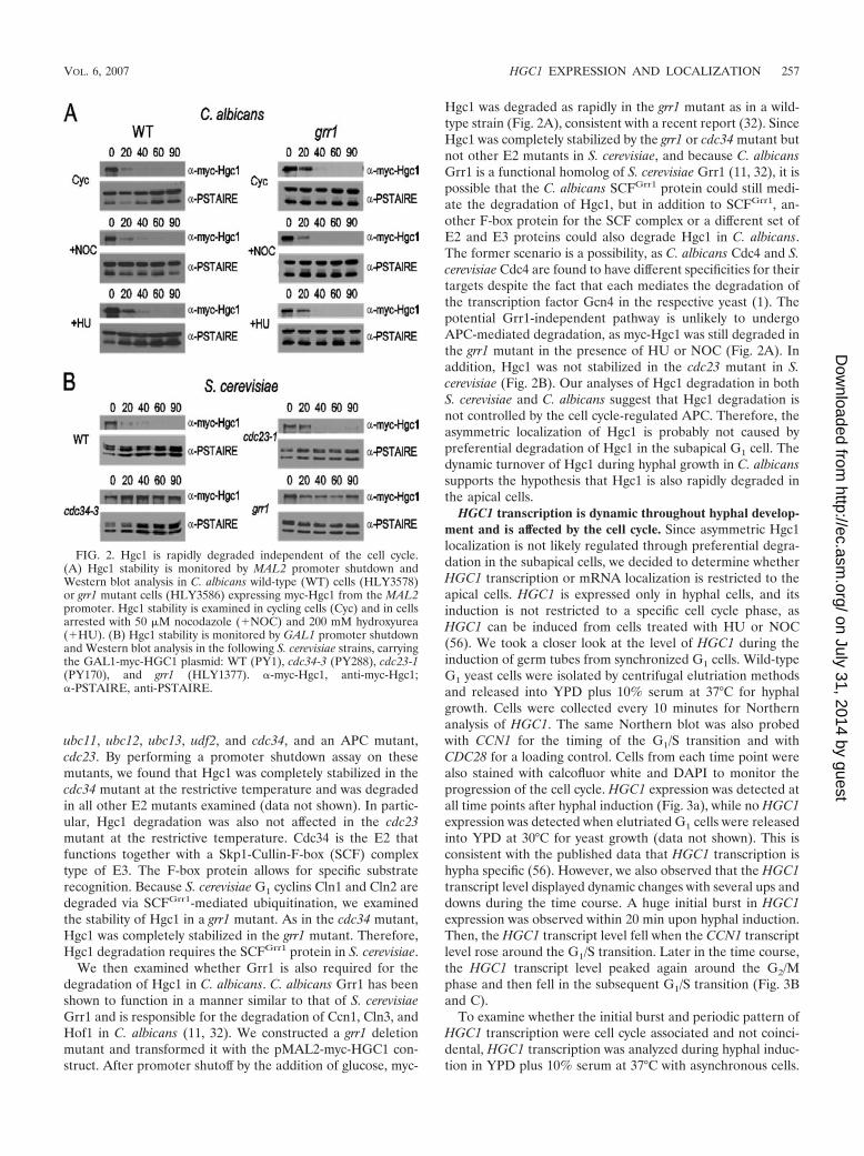
ubc11, ubc12, ubc13, udf2, and cdc34, and an APC mutant,cdc23. By performing a promoter shutdown assay on thesemutants, we found that Hgc1 was completely stabilized in thecdc34 mutant at the restrictive temperature and was degradedin all other E2 mutants examined (data not shown). In partic-ular, Hgc1 degradation was also not affected in the cdc23mutant at the restrictive temperature. Cdc34 is the E2 thatfunctions together with a Skp1-Cullin-F-box (SCF) complextype of E3. The F-box protein allows for specific substraterecognition. Because S. cerevisiae G1 cyclins Cln1 and Cln2 aredegraded via SCFGrr1-mediated ubiquitination, we examinedthe stability of Hgc1 in a grr1 mutant. As in the cdc34 mutant,Hgc1 was completely stabilized in the grr1 mutant. Therefore,Hgc1 degradation requires the SCFGrr1 protein in S. cerevisiae.
We then examined whether Grr1 is also required for thedegradation of Hgc1 in C. albicans. C. albicans Grr1 has beenshown to function in a manner similar to that of S. cerevisiaeGrr1 and is responsible for the degradation of Ccn1, Cln3, andHof1 in C. albicans (11, 32). We constructed a grr1 deletionmutant and transformed it with the pMAL2-myc-HGC1 con-struct. After promoter shutoff by the addition of glucose, myc-
Hgc1 was degraded as rapidly in the grr1 mutant as in a wild-type strain (Fig. 2A), consistent with a recent report (32). SinceHgc1 was completely stabilized by the grr1 or cdc34 mutant butnot other E2 mutants in S. cerevisiae, and because C. albicansGrr1 is a functional homolog of S. cerevisiae Grr1 (11, 32), it ispossible that the C. albicans SCFGrr1 protein could still medi-ate the degradation of Hgc1, but in addition to SCFGrr1, an-other F-box protein for the SCF complex or a different set ofE2 and E3 proteins could also degrade Hgc1 in C. albicans.The former scenario is a possibility, as C. albicans Cdc4 and S.cerevisiae Cdc4 are found to have different specificities for theirtargets despite the fact that each mediates the degradation ofthe transcription factor Gcn4 in the respective yeast (1). Thepotential Grr1-independent pathway is unlikely to undergoAPC-mediated degradation, as myc-Hgc1 was still degraded inthe grr1 mutant in the presence of HU or NOC (Fig. 2A). Inaddition, Hgc1 was not stabilized in the cdc23 mutant in S.cerevisiae (Fig. 2B). Our analyses of Hgc1 degradation in bothS. cerevisiae and C. albicans suggest that Hgc1 degradation isnot controlled by the cell cycle-regulated APC. Therefore, theasymmetric localization of Hgc1 is probably not caused bypreferential degradation of Hgc1 in the subapical G1 cell. Thedynamic turnover of Hgc1 during hyphal growth in C. albicanssupports the hypothesis that Hgc1 is also rapidly degraded inthe apical cells.
HGC1 transcription is dynamic throughout hyphal develop-ment and is affected by the cell cycle. Since asymmetric Hgc1localization is not likely regulated through preferential degra-dation in the subapical cells, we decided to determine whetherHGC1 transcription or mRNA localization is restricted to theapical cells. HGC1 is expressed only in hyphal cells, and itsinduction is not restricted to a specific cell cycle phase, asHGC1 can be induced from cells treated with HU or NOC(56). We took a closer look at the level of HGC1 during theinduction of germ tubes from synchronized G1 cells. Wild-typeG1 yeast cells were isolated by centrifugal elutriation methodsand released into YPD plus 10% serum at 37°C for hyphalgrowth. Cells were collected every 10 minutes for Northernanalysis of HGC1. The same Northern blot was also probedwith CCN1 for the timing of the G1/S transition and withCDC28 for a loading control. Cells from each time point werealso stained with calcofluor white and DAPI to monitor theprogression of the cell cycle. HGC1 expression was detected atall time points after hyphal induction (Fig. 3a), while no HGC1expression was detected when elutriated G1 cells were releasedinto YPD at 30°C for yeast growth (data not shown). This isconsistent with the published data that HGC1 transcription ishypha specific (56). However, we also observed that the HGC1transcript level displayed dynamic changes with several ups anddowns during the time course. A huge initial burst in HGC1expression was observed within 20 min upon hyphal induction.Then, the HGC1 transcript level fell when the CCN1 transcriptlevel rose around the G1/S transition. Later in the time course,the HGC1 transcript level peaked again around the G2/Mphase and then fell in the subsequent G1/S transition (Fig. 3Band C).
To examine whether the initial burst and periodic pattern ofHGC1 transcription were cell cycle associated and not coinci-dental, HGC1 transcription was analyzed during hyphal induc-tion in YPD plus 10% serum at 37°C with asynchronous cells.
FIG. 2. Hgc1 is rapidly degraded independent of the cell cycle.(A) Hgc1 stability is monitored by MAL2 promoter shutdown andWestern blot analysis in C. albicans wild-type (WT) cells (HLY3578)or grr1 mutant cells (HLY3586) expressing myc-Hgc1 from the MAL2promoter. Hgc1 stability is examined in cycling cells (Cyc) and in cellsarrested with 50 �M nocodazole (�NOC) and 200 mM hydroxyurea(�HU). (B) Hgc1 stability is monitored by GAL1 promoter shutdownand Western blot analysis in the following S. cerevisiae strains, carryingthe GAL1-myc-HGC1 plasmid: WT (PY1), cdc34-3 (PY288), cdc23-1(PY170), and grr1 (HLY1377). �-myc-Hgc1, anti-myc-Hgc1;�-PSTAIRE, anti-PSTAIRE.
VOL. 6, 2007 HGC1 EXPRESSION AND LOCALIZATION 257
on July 31, 2014 by guesthttp://ec.asm
.org/D
ownloaded from

An initial peak was observed at 15 min of induction withasynchronous cells (Fig. 3D), similar to the finding that theHGC1 transcript peaks at 10 min upon hyphal induction (8).Therefore, the initial burst in HGC1 transcription is cell cycleindependent. This is consistent with the result that cells ar-rested in the cell cycle by HU or NOC are able to induceHGC1 expression (56), but the initial burst of HGC1 transcrip-tion was strongest with synchronous G1 cells (Fig. 3A). We didnot detect any periodic pattern of HGC1 transcription duringhyphal induction with asynchronous cells, supporting the factthat the observed periodic HGC1 transcript level during hyphalinduction from synchronous G1 cells was associated with thecell cycle. Our Northern analyses, therefore, suggest that
HGC1 transcription is not only hypha specific but also associ-ated with cell cycle progress.
Enhanced apical localization of Hgc1 requires transcriptionfrom the HGC1 promoter. Because HGC1 transcription is sohighly regulated, transcription from the HGC1 promoter maybe important for the apical cell localization of Hgc1. To exam-ine this possibility, indirect immunofluorescence against myc-Hgc1 was carried out to compare the localization of myc-Hgc1under the control of the HGC1 promoter to that of myc-Hgc1under the control of the MAL2 promoter. Cells were exposedto Lee’s media with maltose as the carbon source at 37°C for6 h, so hyphae had reached the two-cell-stage of hyphal devel-opment. When expressed from the MAL2 promoter, Hgc1 was
FIG. 3. HGC1 expression is affected by the cell cycle. Wild-type (SC5314) G1 cells were collected by elutriation centrifugation and released intoYPD plus 10% serum at 37°C for hyphal growth, and cells were collected every 10 min. (A) Northern blots were probed with HGC1, CCN1, andCDC28. (B) HGC1 and CCN1 expression were quantified relative to CDC28 expression. (C) Cells were stained with calcofluor white and DAPIto determine the percentages of cells with chitin rings and dividing nuclei, respectively. (D) Northern blots of wild-type (SC5314) asynchronouscells released into YPD plus 10% serum at 37°C. Cells were collected every 15 min.
258 WANG ET AL. EUKARYOT. CELL
on July 31, 2014 by guesthttp://ec.asm
.org/D
ownloaded from

detected in both the actively dividing apical cell and the G1-arrested subapical cell with equal intensity (Fig. 4). This wasnot because Hgc1 was overexpressed from the MAL2 pro-moter, as the level of Hgc1 expression from the MAL2 pro-moter was comparable to that from the HGC1 promoter in themyc-HGC1 strain and lower than that in the myc-HGC1enhstrain (data not shown). Therefore, apical Hgc1 localization isdependent on its transcription from the HGC1 promoter. Thisdemonstrates that enhanced apical localization of Hgc1 is notregulated through differential Hgc1 degradation and suggeststhat transcription from the HGC1 promoter may be moreactive in the dividing apical cells than in the subapical cells.
Transcription is detected only in the apical cells of hyphae.The promoter swap experiment suggested that transcriptionfrom the HGC1 promoter was primarily in the apical cell. Tovalidate this observation, FISH with FITC-labeled probes wasused to directly visualize the HGC1 transcript in hyphae. Themyc-HGC1enh strain was used in this experiment because itshighly inducible HGC1 transcription gave a readily detectablesignal by FISH. Cells were grown in YPD plus 10% serum at37°C for hyphal induction, fixed, and probed for the HGC1transcript. A “dot” signal was detected near the nucleus duringgerm tube development (Fig. 5A). The signal was specific tothe HGC1 transcript, as it was not detected in the same strainin the yeast form (Fig. 5B) or in an hgc1 deletion strain underthe same experimental conditions (Fig. 5C). When hyphae had
reached the two-cell stage, the “dot” signal was detected onlyin the actively dividing apical cell, again near the nucleus, butwas not detected in the G1-arrested subapical cell. Likewise, inhyphae with three cells, a FISH signal was detected only inthe actively dividing apical cell. This provides direct evi-dence for our previous notion that transcription from theHGC1 promoter is enhanced in the apical cell.
DISCUSSION
We show in this report that the Hgc1 protein displays prefer-ential localization in the actively dividing apical cells of hyphae.Hgc1 protein is rapidly degraded, probably through SCF-medi-ated ubiquitination. The protein turnover is not under the controlof the cell cycle and occurs in both the apical and the subapicalcells of hyphae. HGC1 transcription is also dynamically regulated.Not only is HGC1 transcribed specifically in hyphae (56), but weshow here that its transcription is also regulated by the cell cycleand low during the G1/S transition. Furthermore, the HGC1 tran-script is detected, using FISH, only in the dividing apical cells andnot in the subapical cells. By replacing the HGC1 promoter withthe MAL2 promoter, we demonstrate that the dynamic temporaland spatial regulation of HGC1 transcription is critical for theasymmetric localization of Hgc1 protein to the apical cell duringhyphal development.
It has long been appreciated that the apical cells in hyphalfilaments are different from subapical cells in many aspects,and the difference in cell division is a major aspect. Only theapical cells undergo cell division; the subapical cells are ar-rested in G1 for a period of time until a lateral bud or hyphaforms. Cyclins and many cell cycle regulators, whose transcrip-tion and protein degradation are tightly regulated during thecell cycle, are likely present only in the dividing apical cells inhyphal filaments. For example, Cdc14p is detected only in thenucleus of the apical cell of the hypha, whereas no signal isdetected in any of the G1-arrested subapical cells (15). Here,we show that HGC1 transcription is not only specific to hyphalcells but also regulated during the cell cycle program. There-fore, we speculate that integration of cell cycle regulation withthe hyphal signaling pathway may give rise to the transcriptionof HGC1 in the apical cell.
Another major difference between apical and subapical cellsduring hyphal development is that much of the active growthhappens in the apical cells, and new cell wall growth is highlyrestricted to the tips of hyphae. The actin cytoskeleton is highlypolarized to the tip, where active Cdc42 and other polaritydeterminant proteins are localized (8, 17, 24, 39, 57). ActiveCdc42 and the actin cytoskeleton not only are essential forhyphal morphogenesis (7, 24, 40, 52) but also play a role inmaintaining hypha-associated transcription (8, 24). Whetheractive Cdc42 plays a role in keeping the hyphal transcriptionalprogram active in the apical cell remains to be determined.
Two molecular mechanisms for asymmetric distribution orexpression of mRNAs between mother and daughter cells havebeen elucidated in S. cerevisiae. One involves the transport ofASH1 mRNA along actin cables to the apices of daughter cells,where Ash1 protein is translated and imported into the daugh-ter nuclei to repress the expression of the HO endonucleases,preventing mating-type switching in the daughter cells (10, 47).As in S. cerevisiae, Ash1 is preferentially localized to daughter
FIG. 4. Apical-specific localization of Hgc1 is promoter dependent.myc-Hgc1 localization by indirect immunofluorescence in cells ex-pressing myc-Hgc1 from the HGC1 promoter (HLY3577) and cellsexpressing myc-Hgc1 from the MAL2 promoter (HLY3578) is shown.Both strains were grown in Lee’s medium with 2% maltose as thecarbon source for 6 h at 37°C. An untagged control (HLY3588) wasincluded. DIC, differential interference contrast microscopy.
VOL. 6, 2007 HGC1 EXPRESSION AND LOCALIZATION 259
on July 31, 2014 by guesthttp://ec.asm
.org/D
ownloaded from

cell nuclei in the yeast form of C. albicans and to the apical cellnuclei in growing hyphae (26). The fact that the HGC1 tran-script is not concentrated to the tips of hyphae indicates thatHGC1 mRNA is probably not transported on actin cables tothe apical cells. Another mechanism involves the transcriptionof a group of early-G1 genes in the daughter cells throughCbk1- and Mob2-dependent activation and localization ofAce2 to the daughter nuclei at the end of mitosis and in earlyG1 (16, 27). A functional homolog of Ace2 in C. albicans is alsolocalized to daughter yeast cells at the end of mitosis (29).However, Ace2 is localized in the nuclei of both apical andsubapical cells in hyphae (29), and whether its transcriptionalactivity is limited to the apical cell of a hypha is unclear.
Is the transcription of any other hypha-specific gene re-stricted or enriched in the apical cell? Transcription from theHWP1 promoter is likely not restricted to the apical cell, as agreen fluorescent protein under the control of the HWP1 pro-moter is expressed equally in the apical and subapical cells ofhyphae (48). Even if the transcription is specific to the apicalcell, the protein products will still be present in the subapicalcells if the mRNA and/or protein products are stable. There-fore, for cell wall proteins like Hwp1 and Rbt5, they are likelypresent throughout the hyphal walls of both apical and sub-apical cells. Since different hypha-specific genes are tran-
scribed with different tempo dynamics (8), we speculate thattheir transcripts may also differ in spatial distribution.
ACKNOWLEDGMENTS
We thank W. A. Fonzi, A. P. Mitchell, S. M. Noble, and P. Kaiser forstrains.
This work was supported by an NIH grant (GM55155) to H.L.
REFERENCES
1. Atir-Lande, A., T. Gildor, and D. Kornitzer. 2005. Role for the SCFCDC4ubiquitin ligase in Candida albicans morphogenesis. Mol. Biol. Cell 16:2772–2785.
2. Bachewich, C., A. Nantel, and M. Whiteway. 2005. Cell cycle arrest during Sor M phase generates polarized growth via distinct signals in Candida albi-cans. Mol. Microbiol. 57:942–959.
3. Bachewich, C., D. Y. Thomas, and M. Whiteway. 2003. Depletion of apolo-like kinase in Candida albicans activates cyclase-dependent hyphal-likegrowth. Mol. Biol. Cell 14:2163–2180.
4. Bachewich, C., and M. Whiteway. 2005. Cyclin Cln3p links G1 progression tohyphal and pseudohyphal development in Candida albicans. Eukaryot. Cell4:95–102.
5. Bai, C., N. Ramanan, Y. M. Wang, and Y. Wang. 2002. Spindle assemblycheckpoint component CaMad2p is indispensable for Candida albicans sur-vival and virulence in mice. Mol. Microbiol. 45:31–44.
6. Barelle, C. J., E. A. Bohula, S. J. Kron, D. Wessels, D. R. Soll, A. Schafer,A. J. Brown, and N. A. Gow. 2003. Asynchronous cell cycle and asymmetricvacuolar inheritance in true hyphae of Candida albicans. Eukaryot. Cell2:398–410.
7. Bassilana, M., J. Blyth, and R. A. Arkowitz. 2003. Cdc24, the GDP-GTPexchange factor for Cdc42, is required for invasive hyphal growth of Candidaalbicans. Eukaryot. Cell 2:9–18.
FIG. 5. HGC1 mRNA is visualized in C. albicans cells by using FISH. (A) myc-HGC1enh (HLY3579) cells were grown in YPD plus 10% serumat 37° for hyphal induction. Cells were fixed at 1, 3, and 5 h of induction during germ tube formation and when cells were entering the second andthird cell division for FISH with FITC-labeled DNA probes against the HGC1 transcript. Results are shown for control FISH experiments withmyc-HGC1enh (HLY3579) cells grown in YPD at 25°C for yeast growth (B) and hgc1 (HLY3443) cells grown in YPD plus 10% serum at 37° for1.5 h (C). DIC, differential interference contrast microscopy.
260 WANG ET AL. EUKARYOT. CELL
on July 31, 2014 by guesthttp://ec.asm
.org/D
ownloaded from

8. Bassilana, M., J. Hopkins, and R. A. Arkowitz. 2005. Regulation of theCdc42/Cdc24 GTPase module during Candida albicans hyphal growth. Eu-karyot. Cell 4:588–603.
9. Bensen, E. S., A. Clemente-Blanco, K. R. Finley, J. Correa-Bordes, and J.Berman. 2005. The mitotic cyclins Clb2p and Clb4p affect morphogenesis inCandida albicans. Mol. Biol. Cell 16:3387–3400.
10. Bobola, N., R. P. Jansen, T. H. Shin, and K. Nasmyth. 1996. Asymmetricaccumulation of Ash1p in postanaphase nuclei depends on a myosin andrestricts yeast mating-type switching to mother cells. Cell 84:699–709.
11. Butler, D. K., O. All, J. Goffena, T. Loveless, T. Wilson, and K. A. Toenjes.2006. The GRR1 gene of Candida albicans is involved in the negative controlof pseudohyphal morphogenesis. Fungal Genet. Biol. 43:573–582.
12. Cao, F., S. Lane, P. P. Raniga, Y. Lu, Z. Zhou, K. Ramon, J. Chen, and H.Liu. 2006. The Flo8 transcription factor is essential for hyphal developmentand virulence in Candida albicans. Mol. Biol. Cell 17:295–307.
13. Chapa y Lazo, B., S. Bates, and P. Sudbery. 2005. The G1 cyclin Cln3regulates morphogenesis in Candida albicans. Eukaryot. Cell 4:90–94.
14. Chou, S., L. Huang, and H. Liu. 2004. Fus3-regulated Tec1 degradationthrough SCFCdc4 determines MAPK signaling specificity during mating inyeast. Cell 119:981–990.
15. Clemente-Blanco, A., A. Gonzalez-Novo, F. Machin, D. Caballero-Lima, L.Aragon, M. Sanchez, C. R. de Aldana, J. Jimenez, and J. Correa-Bordes.2006. The Cdc14p phosphatase affects late cell-cycle events and morphogen-esis in Candida albicans. J. Cell Sci. 119:1130–1143.
16. Colman-Lerner, A., T. E. Chin, and R. Brent. 2001. Yeast Cbk1 and Mob2activate daughter-specific genetic programs to induce asymmetric cell fates.Cell 107:739–750.
17. Crampin, H., K. Finley, M. Gerami-Nejad, H. Court, C. Gale, J. Berman,and P. Sudbery. 2005. Candida albicans hyphae have a Spitzenkorper that isdistinct from the polarisome found in yeast and pseudohyphae. J. Cell Sci.118:2935–2947.
18. Dernburg, A. F., and J. W. Sedat. 1998. Mapping three-dimensional chro-mosome architecture in situ. Methods Cell Biol. 53:187–233.
19. Domian, I. J., K. C. Quon, and L. Shapiro. 1996. The control of temporaland spatial organization during the Caulobacter cell cycle. Curr. Opin.Genet. Dev. 6:538–544.
20. Fang, G., H. Yu, and M. W. Kirschner. 1999. Control of mitotic transitionsby the anaphase-promoting complex. Philos. Trans. R. Soc. Lond. B 354:1583–1590.
21. Fang, H. M., and Y. Wang. 2006. RA domain-mediated interaction of Cdc35with Ras1 is essential for increasing cellular cAMP level for Candida albicanshyphal development. Mol. Microbiol. 61:484–496.
22. Feng, Q., E. Summers, B. Guo, and G. Fink. 1999. Ras signaling is requiredfor serum-induced hyphal differentiation in Candida albicans. J. Bacteriol.181:6339–6346.
23. Fonzi, W. A., and M. Y. Irwin. 1993. Isogenic strain construction and genemapping in Candida albicans. Genetics 134:717–728.
24. Hazan, I., and H. Liu. 2002. Hyphal tip-associated localization of Cdc42 isF-actin dependent in Candida albicans. Eukaryot. Cell 1:856–864.
25. Hazan, I., M. Sepulveda-Becerra, and H. Liu. 2002. Hyphal elongation isregulated independently of cell cycle in Candida albicans. Mol. Biol. Cell13:134–145.
26. Inglis, D. O., and A. D. Johnson. 2002. Ash1 protein, an asymmetricallylocalized transcriptional regulator, controls filamentous growth and viru-lence of Candida albicans. Mol. Cell. Biol. 22:8669–8680.
27. Jorgensen, P., B. Nelson, M. D. Robinson, Y. Chen, B. Andrews, M. Tyers,and C. Boone. 2002. High-resolution genetic mapping with ordered arrays ofSaccharomyces cerevisiae deletion mutants. Genetics 162:1091–1099.
28. Kaiser, P., R. A. Sia, E. G. Bardes, D. J. Lew, and S. I. Reed. 1998. Cdc34 andthe F-box protein Met30 are required for degradation of the Cdk-inhibitorykinase Swe1. Genes Dev. 12:2587–2597.
29. Kelly, M. T., D. M. MacCallum, S. D. Clancy, F. C. Odds, A. J. Brown, andG. Butler. 2004. The Candida albicans CaACE2 gene affects morphogenesis,adherence and virulence. Mol. Microbiol. 53:969–983.
30. Lane, S., C. Birse, S. Zhou, R. Matson, and H. Liu. 2001. DNA array studiesdemonstrate convergent regulation of virulence factors by Cph1, Cph2, andEfg1 in Candida albicans. J. Biol. Chem. 276:48988–48996.
31. Lane, S., S. Zhou, T. Pan, Q. Dai, and H. Liu. 2001. The basic helix-loop-helix transcription factor Cph2 regulates hyphal development in Candidaalbicans partly via TEC1. Mol. Cell. Biol. 21:6418–6428.
32. Li, W. J., Y. M. Wang, X. D. Zheng, Q. M. Shi, T. T. Zhang, C. Bai, D. Li,J. L. Sang, and Y. Wang. 2006. The F-box protein Grr1 regulates the stabilityof Ccn1, Cln3 and Hof1 and cell morphogenesis in Candida albicans. Mol.Microbiol. 62:212–226.
33. Liu, H., J. Krizek, and A. Bretscher. 1992. Construction of a GAL1-regulatedyeast cDNA expression library and its application to the identification of geneswhose overexpression causes lethality in yeast. Genetics 132:665–673.
34. Lo, H. J., J. R. Kohler, B. DiDomenico, D. Loebenberg, A. Cacciapuoti, andG. R. Fink. 1997. Nonfilamentous C. albicans mutants are avirulent. Cell90:939–949.
35. Loeb, J. D., M. Sepulveda-Becerra, I. Hazan, and H. Liu. 1999. A G1 cyclinis necessary for maintenance of filamentous growth in Candida albicans. Mol.Cell. Biol. 19:4019–4027.
36. Long, R. M., D. J. Elliott, F. Stutz, M. Rosbash, and R. H. Singer. 1995.Spatial consequences of defective processing of specific yeast mRNAs re-vealed by fluorescent in situ hybridization. RNA 1:1071–1078.
37. Longtine, M. S., A. McKenzie III, D. J. Demarini, N. G. Shah, A. Wach, A.Brachat, P. Philippsen, and J. R. Pringle. 1998. Additional modules forversatile and economical PCR-based gene deletion and modification in Sac-charomyces cerevisiae. Yeast 14:953–961.
38. Maidan, M. M., L. De Rop, J. Serneels, S. Exler, S. Rupp, H. Tournu, J. M.Thevelein, and P. Van Dijck. 2005. The G protein-coupled receptor Gpr1and the Galpha protein Gpa2 act through the cAMP-protein kinase Apathway to induce morphogenesis in Candida albicans. Mol. Biol. Cell 16:1971–1986.
39. Martin, R., A. Walther, and J. Wendland. 2005. Ras1-induced hyphal develop-ment in Candida albicans requires the formin Bni1. Eukaryot. Cell 4:1712–1724.
40. Michel, S., S. Ushinsky, B. Klebl, E. Leberer, D. Thomas, M. Whiteway, andJ. Morschhauser. 2002. Generation of conditional lethal Candida albicansmutants by inducible deletion of essential genes. Mol. Microbiol. 46:269–280.
41. Miwa, T., Y. Takagi, M. Shinozaki, C. W. Yun, W. A. Schell, J. R. Perfect, H.Kumagai, and H. Tamaki. 2004. Gpr1, a putative G-protein-coupled recep-tor, regulates morphogenesis and hypha formation in the pathogenic fungusCandida albicans. Eukaryot. Cell 3:919–931.
42. Noble, S. M., and A. D. Johnson. 2005. Strains and strategies for large-scalegene deletion studies of the diploid human fungal pathogen Candida albi-cans. Eukaryot. Cell 4:298–309.
43. Prokopenko, S. N., and W. Chia. 2005. When timing is everything: role of cellcycle regulation in asymmetric division. Semin. Cell Dev. Biol. 16:423–437.
44. Rocha, C. R., K. Schroppel, D. Harcus, A. Marcil, D. Dignard, B. N. Taylor,D. Y. Thomas, M. Whiteway, and E. Leberer. 2001. Signaling throughadenylyl cyclase is essential for hyphal growth and virulence in the patho-genic fungus Candida albicans. Mol. Biol. Cell 12:3631–3643.
45. Sanchez-Martinez, C., and J. Perez-Martin. 2002. Gpa2, a G-protein alphasubunit required for hyphal development in Candida albicans. Eukaryot. Cell1:865–874.
46. Schweizer, A., S. Rupp, B. N. Taylor, M. Rollinghoff, and K. Schroppel. 2000.The TEA/ATTS transcription factor CaTec1p regulates hyphal developmentand virulence in Candida albicans. Mol. Microbiol. 38:435–445.
47. Sil, A., and I. Herskowitz. 1996. Identification of asymmetrically localizeddeterminant, Ash1p, required for lineage-specific transcription of the yeastHO gene. Cell 84:711–722.
48. Staab, J. F., Y. S. Bahn, and P. Sundstrom. 2003. Integrative, multifunctionalplasmids for hypha-specific or constitutive expression of green fluorescentprotein in Candida albicans. Microbiology 149:2977–2986.
49. Stoldt, V. R., A. Sonneborn, C. E. Leuker, and J. F. Ernst. 1997. Efg1p, anessential regulator of morphogenesis of the human pathogen Candida albi-cans, is a member of a conserved class of bHLH proteins regulating mor-phogenetic processes in fungi. EMBO J. 16:1982–1991.
50. Sudbery, P., N. Gow, and J. Berman. 2004. The distinct morphogenic statesof Candida albicans. Trends Microbiol. 12:317–324.
51. Umeyama, T., A. Kaneko, M. Niimi, and Y. Uehara. 2006. Repression ofCDC28 reduces the expression of the morphology-related transcription fac-tors, Efg1p, Nrg1p, Rbf1p, Rim101p, Fkh2p and Tec1p and induces cellelongation in Candida albicans. Yeast 23:537–552.
52. Ushinsky, S. C., D. Harcus, J. Ash, D. Dignard, A. Marcil, J. Morchhauser,D. Y. Thomas, M. Whiteway, and E. Leberer. 2002. CDC42 is required forpolarized growth in human pathogen Candida albicans. Eukaryot. Cell 1:95–104.
53. Whiteway, M., and U. Oberholzer. 2004. Candida morphogenesis and host-pathogen interactions. Curr. Opin. Microbiol. 7:350–357.
54. Wilson, R. B., D. Davis, and A. P. Mitchell. 1999. Rapid hypothesis testingwith Candida albicans through gene disruption with short homology regions.J. Bacteriol. 181:1868–1874.
55. Yu, F., C. T. Kuo, and Y. N. Jan. 2006. Drosophila neuroblast asymmetric celldivision: recent advances and implications for stem cell biology. Neuron51:13–20.
56. Zheng, X., and Y. Wang. 2004. Hgc1, a novel hypha-specific G1 cyclin-relatedprotein regulates Candida albicans hyphal morphogenesis. EMBO J. 23:1845–1856.
57. Zheng, X. D., Y. M. Wang, and Y. Wang. 2003. CaSPA2 is important forpolarity establishment and maintenance in Candida albicans. Mol. Micro-biol. 49:1391–1405.
VOL. 6, 2007 HGC1 EXPRESSION AND LOCALIZATION 261
on July 31, 2014 by guesthttp://ec.asm
.org/D
ownloaded from