History, Law, and Justice: Empirical Method and Conceptual Confusion in the History of Law
Upload
hms-harvardCategory
view
3download
0
DOI: 10.1167/tvst.2.4.2
Article
Considering Apical Scotomas, Confusion, and DiplopiaWhen Prescribing Prisms for Homonymous Hemianopia
Henry L. Apfelbaum1, Nicole C. Ross1,2, Alex R. Bowers1,2, and Eli Peli1,2 $
1 Schepens Eye Research Institute, Massachusetts Eye and Ear, Harvard Medical School, Boston, MA2 New England College of Optometry, Boston, MA
Correspondence: Eli Peli, SchepensEye Research Institute, 20 StanifordStreet, Boston, MA 02114-2508, USA,e-mail: [email protected]
Received: 1 October 2012Accepted: 2 April 2013Published: 29 May 2013
Keywords: low vision; rehabilita-tion; prism treatment; perimetry;visual field loss; hemianopia; trau-matic brain injury; TBI; stroke
Citation: Afelbaum HL, Ross NC,Bowers AR, Peli E. Considering apicalscotomas, confusion, and diplopiawhen prescribing prisms for hom-onymous hemianopia. Trans Vis SciTech. 2013;2(4):2, http://tvstjournal.org/doi/full/10.1167/tvst.2.4.2, doi:10.1167/tvst.2.4.2
Purpose: While prisms are commonly prescribed for homonymous hemianopia toextend or expand the visual field, they cause potentially troubling visual side effects,including nonveridical location of perceived images, diplopia, and visual confusion. Inaddition, the field behind a prism at its apex is lost to an apical scotoma equal inmagnitude to the amount of prism shift. The perceptual consequences of apicalscotomas and the other effects of various designs were examined to considerparameters and designs that can mitigate the impact of these effects.
Methods: Various configurations of sector and peripheral prisms were analyzed, invarious directions of gaze, and their visual effects were illustrated using simulatedperimetry. A novel ‘‘percept’’ diagram was developed that yielded insights into thepatient’s view through the prisms. The predictions were verified perimetrically withpatients.
Results: The diagrams distinguish between potentially beneficial field expansion viavisual confusion and the pericentrally disturbing and useless effect of diplopia, andtheir relationship to prism power and gaze direction. They also illustrate thenonexpanding substitution of field segments of some popular prism designs.
Conclusions: Yoked sector prisms have no effect at primary gaze or when gaze isdirected toward the seeing hemifield, and they introduce pericentral field loss whengaze is shifted into them. When fitted unilaterally, sector prisms also have an effectonly when the gaze is directed into the prism and may cause a pericentral scotomaand/or central diplopia. Peripheral prisms are effective at essentially all gaze angles.Since gaze is not directed into them, they avoid problematic pericentral effects. Wederive useful recommendations for prism power and position parameters, includingnovel ways of fitting prisms asymmetrically.
Translational Relevance: Clinicians will find these novel diagrams, diagrammingtechniques, and analyses valuable when prescribing prismatic aids for hemianopiaand when designing new prism devices for patients with various types of field loss.
Introduction
Homonymous hemianopia (HH), the loss of halfthe visual field in both eyes on the same side and tosimilar extent, results from postchiasmatic lesionstypically caused by stroke, tumor, or trauma.1 Hemi-anopic visual field loss reduces detection of objects inthe blind hemifield and impacts the ability to avoidobstacles while walking and driving.2–4
Spectacle-mounted prisms are the most commonlyused rehabilitation devices for HH. There are variousdesigns and methods for fitting prism spectacles. All
involve tradeoffs between the access to otherwiseinvisible areas of the visual scene and the variouspotentially troubling visual side effects the prismsintroduce. While the prism apical scotoma has beenmentioned in the literature,5–9 its functional signifi-cance has not been addressed, nor has its impact onthe residual visual field been illustrated. This paperexamines common prism configurations used for HH,and primarily analyzes the oft-ignored, but impor-tant, effect of prism apical scotomas. Other effects,such as the diplopia and visual confusion induced insome configurations and the nonveridical viewscreated by all designs are also addressed, and their
http://tvstjournal.org/doi/full/10.1167/tvst.2.4.2 TVST j 2013 j Vol. 2 j No. 4 j Article 21

impact is illustrated in a novel way. Careful fittingdesign based on the parameters and diagramsprovided here can mitigate these effects. The analyseswill also provide practitioners insights into why someconfigurations may be accepted and valued more thanothers by their patients. Novel designs emerged fromthese analyses, described in the sections on offsetsector and peripheral prism placement.
The bending of light rays by a prism creates a gapin the visible field of view between the last undeviatedray just outside the prism, at its apex, and the first raydeviated by the prism.5 The angular extent of that gapis equal to the prism power in degrees (Fig. 1A). Thatgap is referred to as the apical scotoma. The perceived(retinal) field of view is still fully covered by rays fromportions of the external field, but the view from withinthe scotoma is missing, obscured by the prism itself.Thus, this ‘‘scotoma’’ is a gap in the portion of theexternal world view that is seen, not a blind retinalarea. The retinal image is discontinuous, but has noblanks. (If a prism is replaced by an opaque occluder,visual field equal in width to the visual angle theoccluder subtends would be lost behind the occluder.With a prism, the field blocked by the prism beyondthe apical scotoma is seen at the prism apex; hence,the apical scotoma is the only loss.) The effect is
illustrated photographically by holding a prism infront of a camera lens (Fig. 1B).
All of the prism configurations to be discussedcause portions of the world view to appear innonveridical directions; the apparent location throughthe prism is displaced from the true direction (e.g., thelower portion of the lamppost in Fig. 1B).
When prisms are placed bilaterally for HH, withessentially the same position and coverage for eacheye, the prism apical scotomas induce a gap in thevisual field. When placed monocularly (or at differentrelative positions on each carrier lens), it is possiblefor the view of one eye to include field portionsmissing to the apical scotoma of the fellow eye. Theseconfigurations produce areas of visual confusion, inwhich two different views are seen at the sameapparent direction by the two eyes, and some producediplopia, wherein a given object or portion of thescene is seen in two different directions simultaneous-ly. We avoid using the term ‘‘double vision,’’ as it hasbeen ambiguously applied in the literature to includeor exclude confusion and diplopia.
If the total field area available (at a given directionof gaze) is larger than the hemifield available withoutprisms, we deem this to be true field expansion.Semitransparent mirrors have been used to providetrue expansion,10 but they are bulky, unsightly, and
Figure 1. Apical prism scotoma. (A) Schematic ray diagram of the apical scotoma of a sector prism. The apical scotoma occurs betweenthe last undeviated ray and the first light ray which is deviated by the prism (prism apex highlighted by yellow star). The angle betweenthese two light rays is equivalent to the prism power in degrees. In this example, the cat is not visible through the prism, since it islocated within the prism’s apical scotoma, indicated by the gray shading. (B) Photograph taken with a 30D prism with base to the leftcovering most of the lower left quadrant of the frame. The prism extends the camera’s field of view to the left, revealing crosswalk left ofthe lamppost, but loses field at its apex, as the lower half of the man is now missing. Had the prism extended the full height of the image,the man would be lost completely to the apical scotoma. The aperture of the camera lens creates vignetting at the prism edges.
http://tvstjournal.org/doi/full/10.1167/tvst.2.4.2 TVST j 2013 j Vol. 2 j No. 4 j Article 22
Apfelbaum et al.
add the disorientation of a reversed view to the visualconfusion. They are now rarely used and are notdiscussed further here. As will be seen below, trueexpansion is possible with prisms, but only if they arefit asymmetrically, or monocularly, so that differentportions of the field are available to each eye (albeitwith some visual confusion).
Most bilateral prism designs provide field substi-tution rather than expansion: The total area visible atany time is no greater than without prisms; the areashifted into view displaces an equal amount of theunaided view, creating an optical scotoma in thefield—the apical scotoma. We also avoid the term‘‘field enhancement’’, as it is ambiguous with respectto these important distinctions.
If prisms cover the entire field of view (as happenswith bilateral full-field yoked prisms6,9,11), fieldshifting, without expansion or substitution, occurs.Field shifting is of no value for patients with HH, asnormal fixation, head, and eye movements will negateit to bring the view straight ahead into primary gaze.The prism configurations discussed here (sector andperipheral prisms) do not span the full field horizon-tally, so the placement of the prism apices becomes acritical design element, affecting the role the apicalscotomas play in the patient’s experience, as well asthe location of regions of visual confusion anddiplopia. (The prism bases are set far into the blindside, where they are rarely in view.) Visual confusionis a necessary consequence of prismatic field expan-sion. It is more readily tolerated in the periphery thanin central vision. Confusion and diplopia are verydisturbing centrally (parafoveally), while they arenormal in the periphery.12 Prism-induced diplopiaanywhere, however, represents a waste of prismpower that could be better used for expansion (orsubstitution), as will become apparent below.
In the subsequent sections, we identify theparameters that affect optimal positioning and powerfor each of the common methods of fitting prisms,and discuss their implications in terms of fieldexpansion or substitution and troublesome perceptualeffects including apical scotomas, diplopia, and visualconfusion. Often confusion and diplopia are thoughtto coexist, but these analyses make clear howindependent these phenomena can be. The examplesare illustrated using predicted/calculated and actualvisual field diagrams from patients, and we introducea new visual field representation technique, thepercept diagram, to describe the patient’s binocularview through the prism spectacles.
Methods
Calculations used to predict Goldmann visual fielddiagrams and the associated percept diagrams foreach prism configuration, as well as to manufacturethe corresponding spectacles used, assume that thenodal point distance (NPD) between an eye’s nodalpoint and the cornea is 7.1 mm (Gullstrand’sSchematic Eye13) and the back vertex distance(BVD) between the cornea and the carrier lens is 13mm. Formulas used for these calculations areprovided below. The visual angle (A8) betweenprimary gaze and the prism apex is given by
A8 ¼ tan�1L
BVDþNPD
� �; ð1Þ
where L is the distance (mm) on the carrier lens fromthe intersection of the visual axis (in primary gaze) tothe apex. Nodal point is used when calculating visualfield angles, while center of rotation (CR, assumed tobe 13.5 mm from the cornea) is substituted for NPDwhen calculating eye rotations.
The conversion of prism power from prismdiopters (PD) to degrees (P8) is
P8 ¼ tan�1PD
100
� �: ð2Þ
A conventional Goldmann visual field diagramprovides information about which portions of theexternal visual field are detected by the visual system(in primary gaze). In this paper, we use Goldmannvisual field measurements to identify the portions ofthe external field that are seen (and unseen) whenvarious prism configurations are used and in variousdirections of gaze, not to map visual system status. Tocover the range of configurations tested (not forbetween–subject comparisons), prism spectacles wereworn by three individuals with complete HH. Thespectacles were fit according to common clinicalpractice for each of the designs.
We also introduce a synthetic variant of the Gold-mann diagram to illustrate the visual field as it wouldbe seen by the prism wearer. We call this a perceptdiagram (although, to be precise, it shows what isavailable on the retinas to be perceived, not necessarilywhat is perceived, as binocular postretinal factors suchas suppression may intercede). A percept diagramillustrates what the world would look like if the worldwere a Goldmann grid; as if the patient is inside a hugesphere marked with radial lines and concentric circlesoriginating at primary gaze. Figure 2A is the percept
http://tvstjournal.org/doi/full/10.1167/tvst.2.4.2 TVST j 2013 j Vol. 2 j No. 4 j Article 23
Apfelbaum et al.

diagram for the prism photo in Figure 1B. The perceptdiagram is a handy way to understand the nonveridicalviews that prisms introduce. The percept diagramsmake it easier to see where visual confusion isintroduced and the nonveridical perceived directionsof objects, while the Goldmann perimetry diagramsmake it easy to note field substitution or expansion,diplopia, and field-of-view losses due to apical scoto-mas.
Figure 2B gives the simulated Goldmann diagramthat corresponds to this percept diagram. It is thediagram, which would result if the camera couldrespond to Goldmann stimuli with the prism config-uration of Figure 1B interposed. The simulatedGoldmann diagram gives a clear understanding ofthe field substitution the prism provides as well as theloss of field at the prism apex. The diagrams belowillustrating the effects of various prism configurationswill, of course, show the limits of a subject’s visualfields, not the camera’s. Details of many of the
diagrams will benefit from the magnification providedby viewing this document at full screen width.
All calculated diagrams assume a pinhole pupil, sothey do not include the transition vignetting effect ofa larger pupil (exaggerated by the larger cameraaperture in Fig. 1B), nor do they include the fringes ofcolor or the other known optical distortions thatprisms induce, as these are not generally factorsconsidered when fitting prisms for HH.14,15 Thesimulated Goldmann diagrams, and those of patientswearing the prisms, are dichoptic, representingseparately what each eye sees when both are viewing.Although we have developed perimetry systems thatactually measure dichoptically,16,17 most patientdiagrams here are actually a synthesis of threeseparate diagrams, with OD, OS, and OU measuredseparately and then combined (the OU diagrams werenecessary to identify diplopia and confirm binocularscotomas). This technique provided wider plots thanare possible when wearing the goggles used in ourdichoptic perimeters. Figure 3 illustrates the diagramnotations and the distinction between binocular anddichoptic perimetry.
All procedures were conducted according to thetenets of the Declaration of Helsinki, and the protocolwas approved by the Schepens Eye Research InstituteInstitutional Review Board. Informed consent wasobtained from all subjects.
Sector Prism Spectacles for
Hemianopia
Bilateral Sector Prisms
Yoked bilateral sector prisms, up to 20D18 (andrarely 30D19–21), were one of the first designs of prismspectacles prescribed for hemianopia,22,23 and havebeen prescribed frequently since.5,7,24–26 In this
Figure 2. (A) This percept diagram represents the way aGoldmann grid would be captured by a camera with a 30Dprism interposed as in Figure 1B. The area outside the camera’sfield of view is shaded. The arrow points to the center of the fieldof view (the fixation or ‘‘gaze point’’). The arrow is not included insubsequent percept diagrams, as fixation is always in the center,although the grid cross will appear off center when diagrammingaverted gaze views. A dashed line (not part of the percept) outlinesthe prism location. Its left and lower edges are outside of thecamera’s field. The base-left prism has allowed a portion of the gridotherwise outside the camera’s field on the left to be visible to thecamera. The lines in the prism view have been blurred slightly, notjust to represent the loss of quality that occurs through prisms, butalso as a diagramming aid to differentiate the shifted andunshifted areas. The wider spacing in that area is due to its viewof more peripheral grid, not magnification. A 16.78 widerectangular patch of grid is missing at the prism apex. That isthe apical scotoma. (B) The Goldmann diagram that would result ifthe camera could respond to Goldmann stimuli with a prismsimilarly interposed. The simulated Goldmann diagram clearlyshows the effect of the apical scotoma, shaded in darker gray foremphasis. The prism has substituted the field of view to the left,with a resulting loss of pericentral view.
Figure 3. Simulated normal Goldmann fields. (A) Binocular (B)
Dichoptic (or superimposed separate OD and OS).
http://tvstjournal.org/doi/full/10.1167/tvst.2.4.2 TVST j 2013 j Vol. 2 j No. 4 j Article 24
Apfelbaum et al.
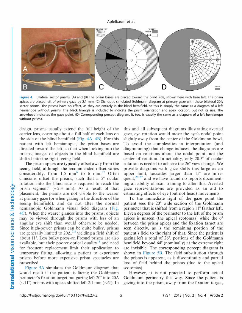
design, prisms usually extend the full height of thecarrier lens, covering about a full half of each lens onthe side of the blind hemifield (Fig. 4A, 4B). For thispatient with left hemianopia, the prism bases aredirected toward the left, so that when looking into theprisms, images of objects in the blind hemifield areshifted into the right seeing field.
The prism apices are typically offset away from theseeing field, although the recommended offset variesconsiderably, from 1.5 mm6 to 6 mm.27 Oftenclinicians offset the prisms, such that a 58 ocularrotation into the blind side is required to reach theprism segment7 (~2.3 mm). As a result of thatplacement, the prisms are not visible to the wearerat primary gaze (or when gazing in the direction of theseeing hemifield), and do not alter the normalhemianopic Goldmann visual field diagram (Fig.4C). When the wearer glances into the prisms, objectsmay be viewed through the prisms with less of anangular eye shift than would otherwise be needed.Since high-power prisms can be quite bulky, prismsare generally limited to 20D,7,8 yielding a field shift ofabout 118. Less bulky press-on Fresnel prisms are alsoavailable, but their poorer optical quality18 and needfor frequent replacement limit their application totemporary fitting, allowing a patient to experienceprisms before more expensive prism spectacles areprescribed.
Figure 5A simulates the Goldmann diagram thatwould result if the patient is facing the Goldmannperimeter’s fixation target but gazing left 208 into 20D(~118) prisms with apices shifted left 2.1 mm (~68). In
this and all subsequent diagrams illustrating avertedgaze, eye rotation would move the eye’s nodal pointslightly away from the center of the Goldmann bowl.To avoid the complexities in interpretation (anddiagramming) that change induces, the diagrams arebased on rotations about the nodal point, not thecenter of rotation. In actuality, only 20.38 of ocularrotation is needed to achieve the 268 view change. Weprovide diagrams with gaze shifts this large as anupper limit; saccades larger than 158 are infre-quent,28,29 and we have found no reports document-ing an ability of scan training to alter this. Avertedgaze representations are provided as an aid toestimating effects of eye (but not head) movements.
To the immediate right of the gaze point thepatient sees the 208 wide section of the Goldmannperimeter that is shifted from a region 118 farther left.Eleven degrees of the perimeter to the left of the prismapices is unseen (the apical scotomas) while the 68
between the prism apices and the fixation target areseen directly, as is the remaining portion of thepatient’s field to the right of that. Since the patient isgazing left a total of 268, portions of the Goldmannhemifield beyond 648 (nominally) at the extreme rightare invisible. The corresponding percept diagram isshown in Figure 5B. The field substitution throughthe prisms is apparent, as is discontinuity and partialloss of field behind the prisms (due to the apicalscotomas).
However, it is not practical to perform actualGoldmann perimetry this way. Since the patient isgazing into the prism, away from the fixation target,
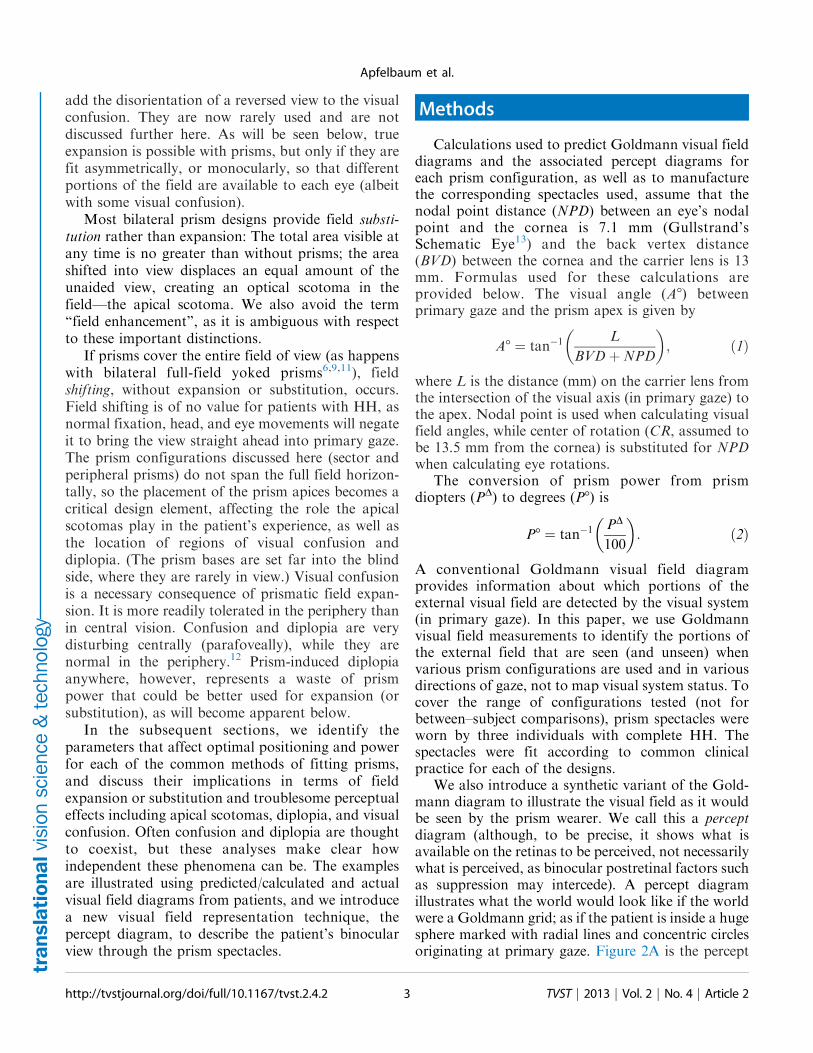
Figure 4. Bilateral sector prisms. (A) and (B) The prism bases are placed toward the blind side, shown here with base left. The prismapices are placed left of primary gaze by 2.1 mm. (C) Dichoptic simulated Goldmann diagram at primary gaze with these bilateral 20Dsector prisms. The prisms have no effect, as they are entirely in the blind hemifield, so this is simply the same as a diagram of a lefthemianope without prisms. The black triangle is included to indicate the prism orientation and apex location, but not its size. Thearrowhead indicates the gaze point. (D) Corresponding percept diagram. It, too, is exactly the same as a diagram of a left hemianopewithout prisms.
http://tvstjournal.org/doi/full/10.1167/tvst.2.4.2 TVST j 2013 j Vol. 2 j No. 4 j Article 25
Apfelbaum et al.

there is no reference point on the blank Goldmanndome to serve as a target and stabilize fixation.Instead, we had the patient turn his head (and theprism-bearing spectacles) sufficiently to the right sothat the fixation target was viewed through the prismswhen gazing 208 (in this example) into the prisms. Thecalculated Goldmann diagram in that situation isshown in Figure 5C. (It may be easier to think of thisas having the patient maintain a fixed head position,while the Goldmann perimeter is rotated until thefixation target is foveated with gaze averted 268.30 Ofcourse, it is more practical for the patient to turnwithin the chin rest.) A binocular Goldmann diagramwith the patient’s head turned in this way is shown inFigure 5D. Note that the measured apical scotomadoes not appear as large as the calculated scotoma,likely due to a combination of measurement error,vignetting, and slight misalignment of the prismapices between the eyes when the head is turned inthis direction (but see Fig. 9 for a way that ‘‘misalign-ment’’ can be an advantage).
A similar set of diagrams is given in Figure 6, withgaze shifted only 58 into the prisms (requiring 118 shiftfrom primary gaze). Since the amount of eyemovement is smaller, the apical scotoma is closer tothe center of gaze, where it may have a moredeleterious impact.
Thus, bilateral sector prisms provide no benefit atprimary gaze, but provide access to portions of theblind hemifield with less eye rotation (by the amountof prism power in degrees) than would be requiredwithout prisms. However, while gazing into theprisms, a vertical swath of visual field, also the widthof the prism shift, is missed between the shifted andunshifted views due to the apical scotomas. The fieldthus lost includes pericentral areas important for safemobility.31 The apical scotomas and large gaze shiftsneeded to achieve field substitution (but not expan-sion) likely account for the paucity of reports of long-term acceptance of this prism configuration. Someclinicians suggest that these prisms are training thepatient to become aware of the blind area ‘‘by lookingfurther into the prism or turning their head andviewing the object through the carrier portion of thespectacle,’’7 but we are not aware of any studiesdemonstrating a training effect.
In order to mitigate the effects of the apicalscotomas, we next consider two alternatives to thecommon bilateral fitting of sector prisms. The first,unilateral fitting, has been used previously.6,8,32 Thesecond, offset bilateral placement, is new and as yetuntested clinically. It arises directly from our analysesof scotoma effects presented here. Neither of theseconfigurations overcomes the lack of field expansion
Figure 5. Gazing 208 into bilateral 20D sector prisms. (A) Simulated Goldmann diagram with the prism configuration of Figure 4 and thepatient facing the fixation target, but gazing left into the prisms by 208, for a total gaze shift of 268. The arrow is rotated counterclockwiseat the gaze point into the prism to indicate the 268 angle of incidence (from perpendicular) of the gaze. A 208-wide region of field from178 into the blind hemifield is visible (68 to the prism apex plus 118 of prism shift), but the 118 just left of the prism apex is missing due tothe apical scotomas. Since the gaze is shifted left, some of the right far periphery has been lost from view. (B) The corresponding perceptdiagram shows that the patient sees 208 of field shifted from 118 farther left, but there is a discontinuity where 118 of pericentral view hasbeen lost to the apical scotomas. The triangle is also shown here to indicate the location of the prism apex. Fixation is at the lowerintersection of the triangle and the hemifield margin. (C) In order to provide a fixation reference when gazing into the prism, it isnecessary to perform Goldmann perimetry with the patient’s head turned right so that the gaze falls on the shifted view of theperimeter’s fixation target. A simulation of a field diagram taken that way is shown in (C). We have rotated the prism symbol clockwiseabout the gaze point to indicate the magnitude of head rotation to the right needed to see the fixation target with a 208 gaze into theprism (378; 68 to the prism, plus 208 into the prism, plus 118 of prism shift). The arrow rotation indicates the gaze incidence at the prism.(D) An actual Goldmann visual field diagram taken this way. This patient has overall restricted peripheral fields on the seeing side inaddition to the hemianopia. The measured apical scotoma is highlighted in dark gray.
http://tvstjournal.org/doi/full/10.1167/tvst.2.4.2 TVST j 2013 j Vol. 2 j No. 4 j Article 26
Apfelbaum et al.
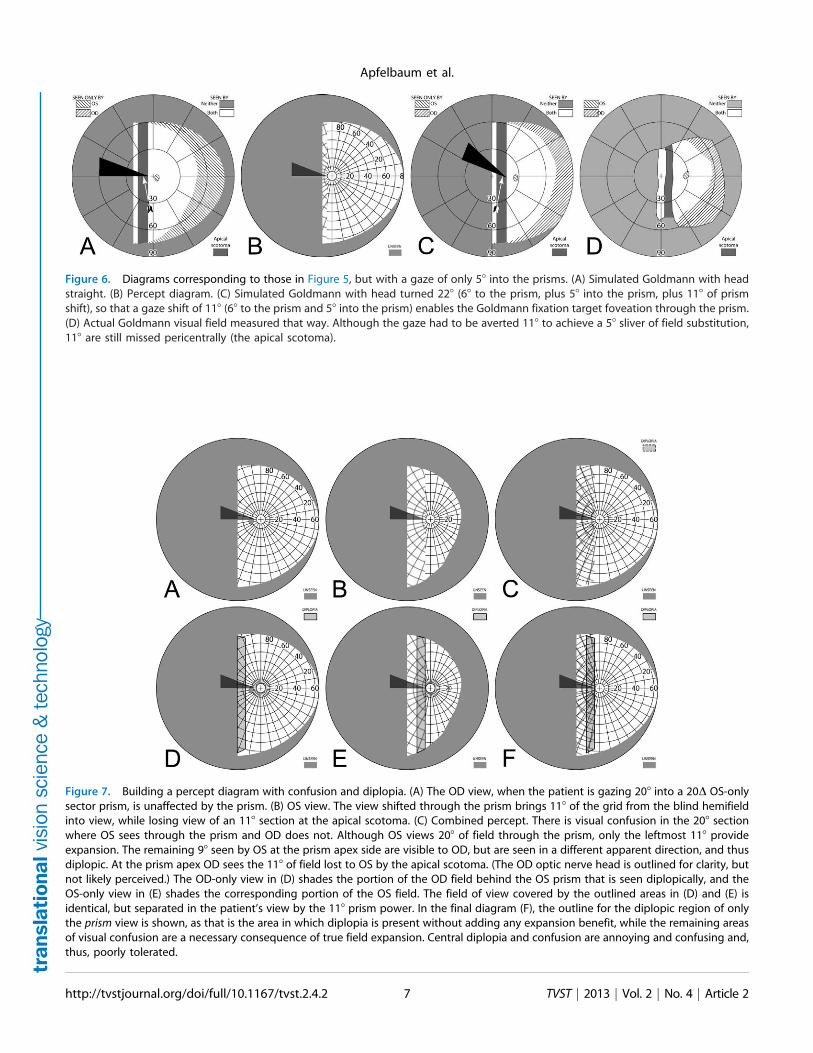
Figure 6. Diagrams corresponding to those in Figure 5, but with a gaze of only 58 into the prisms. (A) Simulated Goldmann with headstraight. (B) Percept diagram. (C) Simulated Goldmann with head turned 228 (68 to the prism, plus 58 into the prism, plus 118 of prismshift), so that a gaze shift of 118 (68 to the prism and 58 into the prism) enables the Goldmann fixation target foveation through the prism.(D) Actual Goldmann visual field measured that way. Although the gaze had to be averted 118 to achieve a 58 sliver of field substitution,118 are still missed pericentrally (the apical scotoma).
Figure 7. Building a percept diagram with confusion and diplopia. (A) The OD view, when the patient is gazing 208 into a 20D OS-onlysector prism, is unaffected by the prism. (B) OS view. The view shifted through the prism brings 118 of the grid from the blind hemifieldinto view, while losing view of an 118 section at the apical scotoma. (C) Combined percept. There is visual confusion in the 208 sectionwhere OS sees through the prism and OD does not. Although OS views 208 of field through the prism, only the leftmost 118 provideexpansion. The remaining 98 seen by OS at the prism apex side are visible to OD, but are seen in a different apparent direction, and thusdiplopic. At the prism apex OD sees the 118 of field lost to OS by the apical scotoma. (The OD optic nerve head is outlined for clarity, butnot likely perceived.) The OD-only view in (D) shades the portion of the OD field behind the OS prism that is seen diplopically, and theOS-only view in (E) shades the corresponding portion of the OS field. The field of view covered by the outlined areas in (D) and (E) isidentical, but separated in the patient’s view by the 118 prism power. In the final diagram (F), the outline for the diplopic region of onlythe prism view is shown, as that is the area in which diplopia is present without adding any expansion benefit, while the remaining areasof visual confusion are a necessary consequence of true field expansion. Central diplopia and confusion are annoying and confusing and,thus, poorly tolerated.
http://tvstjournal.org/doi/full/10.1167/tvst.2.4.2 TVST j 2013 j Vol. 2 j No. 4 j Article 27
Apfelbaum et al.

or substitution at primary gaze (or any gaze in thedirection of the sighted hemifield), and they inducecentral and pericentral visual confusion when gaze isdirected into them. Peripheral prisms, discussed later,overcome those limitations.
Unilateral Sector Prisms
If a sector prism of the type described above is fiton only one carrier lens (typically ipsilateral to thefield defect; OS for a patient with left hemianopia),the nonprism eye can see some or all of the area lostto the prism eye’s apical scotoma. The region betweenthe gaze point into the prism and the prism apex isseen unblocked by the prism by the nonprism eye, andis, thus, not lost in the binocular field to the scotoma.If the angle of gaze into the prism is less than theprism power, some scotoma will remain. If the angleof gaze into the prism is larger, there is an area ofdiplopia equal in width to the excess of the gaze anglebeyond the prism power. In this and subsequentdiscussions, we assume that the patient is orthophor-ic. Because the unilateral prism can dissociate
Figure 8. Unilateral sector prism. With a sector prism (of the type in Fig. 5) fitted on just one eye, the fellow eye sees some of or theentire region lost to the apical scotoma, albeit with visual confusion. (A–D) The calculated head straight Goldmann diagram, percept(from Fig. 7), and calculated head turned and actual Goldmann diagrams, respectively, when gazing 208 into the prism. 208 of field isshifted into view by the prism. The 118 prism scotoma region is visible to the fellow eye, but with 208 of visual confusion and 98 (208–118)of diplopia. In the Goldmann diagrams the diplopic area is crosshatched. (E–H) Corresponding diagrams when gazing just 58 into theprism (yet 118 away from primary gaze, so 118 of field at the right periphery is lost); 58 of shifted field are viewed through the prism, andthere is 58 of visual confusion, but no diplopia and only 58 of the apical scotoma has become visible to the nonprism eye.
Figure 9. Offset placement of bilateral sector prisms can avoiddiplopia. This is illustrated for a gaze 208 into 20D sector prisms.The OD prism has the conventional 68 offset from primary gaze,while the OS prism is offset the additional 118 of the prism power.Although field extent is identical, the pericentral scotoma of theconventional bilateral fitting (Fig. 5) is gone, as is the diplopic areaof unilateral fitting (Fig. 8), though central confusion remains. (A)Simulated Goldmann (with head straight). (B) The percept diagramshows that a region of visual confusion exists where OD is viewingthrough a prism and OS is not. The apex of the leftmost triangle ineach diagram identifies the location of the OS prism apex.
http://tvstjournal.org/doi/full/10.1167/tvst.2.4.2 TVST j 2013 j Vol. 2 j No. 4 j Article 28
Apfelbaum et al.

binocularity, the actual binocular field results mayvary with patient’s phoria and eye dominance. Note,in this unilateral sector design too, the prism has noeffect in primary position of gaze or when the patientgazes towards the sighted hemifield.
Figure 7 builds the percept diagram for the case inwhich the gaze angle into the prism is larger than theprism power. The fitting parameters are as above(gazing 208 into a 20D prism offset 68 from primarygaze). The fellow eye now sees the region lost to theapical scotoma of the prism eye, although there isvisual confusion in that area and a 98 wide area ofdiplopia. Confusion and diplopia around the gazepoint can make it difficult for the patient to quicklyinterpret the view, and may result in suppression;thus, eye dominance and the overall binocular statusof the patient may be important factors.
Figure 8A is the computed Goldmann visual fielddiagram analogous to Figure 5A, with the patientfacing straight at the fixation light and gaze shifted208 into the prism. Figure 8C gives the calculateddiagram analogous to Figure 5C, with the headrotated 268 right, so that the left eye is gazing 208 intothe prism when the right eye is gazing at the fixationtarget. Figure 8D gives the actual patient resultsunder that condition. As noted above, the calculateddiagrams are created as if the nodal points of botheyes are at the perimeter origin; clearly impossible toactually achieve. However, in the mobility situationswhere these diagrams are relevant, the distance to thetargets of interest are so large that the distancebetween the eyes is inconsequential.
Figures 8E through 8H are the correspondingdiagrams when gaze is shifted just 58 into the prism,where the amount of eye movement in degrees is lessthan the amount of prismatic shift. For a 58 gaze shiftinto the prism segment (total 68þ 58¼ 118 shift fromprimary gaze), there is no diplopia, but the apicalscotoma is present in the binocular view, as thenonprism eye sees only 58 of the 118 apical scotomaregion, and that part is seen with visual confusion.
Thus, the unilateral sector prism design can reduceor eliminate the apical scotoma and provide some truebinocular field expansion, not just substitution.However, the apical scotoma does appear in thebinocular view as the eye crosses the prism segment,until the amount of gaze shift is equivalent to orgreater than the prism power in degrees. Further-more, central and peripheral field confusion aroundthe gaze point do occur, and as the gaze is shiftedfarther into the prism, central and peripheral diplopiaensue. The percept diagram shows these effects. Since
this is the first example with confusion and diplopia,Figure 7 builds the diagram a step at a time, so thecomponent effects are clearer. Subsequent diagramswill only show the compounded final effect. To bestrepresent the situation the patient experiences, we donot use color to distinguish the OD and OScontributions to visual confusion. However, wherethis may be particularly hard for the reader tointerpret, we have also provided magnified detailviews with color. Nonetheless, since the perceivedlocation of prism-induced diplopia is not readilyidentified in the Goldmann diagrams, we highlight itin the percept diagrams, even though that delineation,of course, is not apparent to the patient.
The Gottlieb prism design32 differs from unilat-eral sector prisms, in that only a circular cutout ofthe sector prism is used. The prism apex is placed atthe limbus, and is, thus, offset by about 6 mm (~138
¼ tan�1[6/(CRþBVD)]), which is a larger angle than97% of natural saccades.28,29 The prism is intendedto be used during brief glances to aid in locatingobjects of interest. Its smaller size makes it lighterweight and perhaps less conspicuous (better cosmet-ics), losing some peripheral areas of a full sectorprism that are arguably less important. The mainoptical effects, however, are identical to those of aunilateral sector prism within the coverage area ofthe Gottlieb prism.
Offset Bilateral Sector Prisms
To avoid the pericentral and central diplopia thataccompanies unilateral sector prisms (when in gaze),bilateral prisms can be offset so that the apicalscotoma for each prism lies in a different portion ofthe binocular visual field. If the offset is exactly equalto the prism power, no portion of the field will becompletely lost within the apical scotomas of bothprisms simultaneously, as illustrated in Figure 9. Theextent of coverage is identical to that provided by theunilateral and conventional bilateral configurations.There is true binocular field expansion when the eyesare turned far enough to be gazing into one or bothprisms. Visual confusion is substituted for the apicalscotomas of the conventional bilateral configurationand the diplopia of unilateral fitting. There is still nofield expansion at primary gaze, making this (as yetuntested) configuration less desirable than the pe-ripheral placement of prisms described next. Thecentral confusion that remains with this design,though preferable to diplopia that does not contributeanything to field expansion, is not less annoying orbothersome than central diplopia.
http://tvstjournal.org/doi/full/10.1167/tvst.2.4.2 TVST j 2013 j Vol. 2 j No. 4 j Article 29
Apfelbaum et al.
Peripherally-Placed Prism Spectacles
for Hemianopia
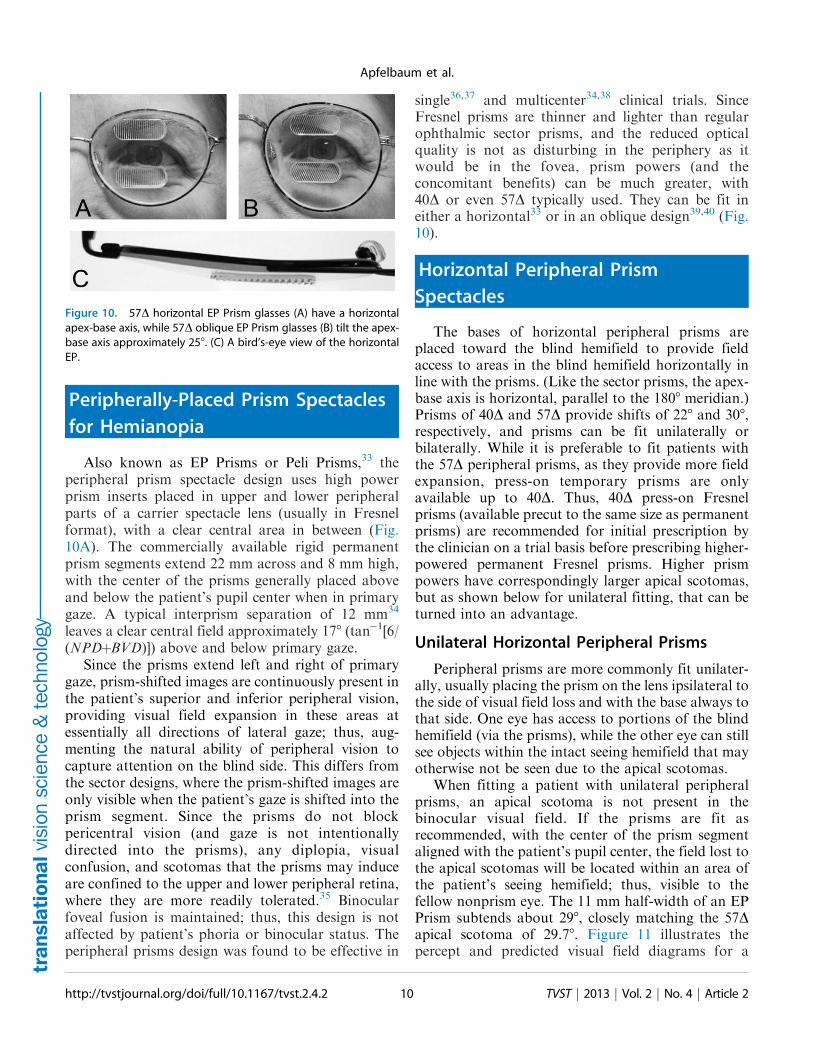
Also known as EP Prisms or Peli Prisms,33 theperipheral prism spectacle design uses high powerprism inserts placed in upper and lower peripheralparts of a carrier spectacle lens (usually in Fresnelformat), with a clear central area in between (Fig.10A). The commercially available rigid permanentprism segments extend 22 mm across and 8 mm high,with the center of the prisms generally placed aboveand below the patient’s pupil center when in primarygaze. A typical interprism separation of 12 mm34
leaves a clear central field approximately 178 (tan�1[6/(NPDþBVD)]) above and below primary gaze.
Since the prisms extend left and right of primarygaze, prism-shifted images are continuously present inthe patient’s superior and inferior peripheral vision,providing visual field expansion in these areas atessentially all directions of lateral gaze; thus, aug-menting the natural ability of peripheral vision tocapture attention on the blind side. This differs fromthe sector designs, where the prism-shifted images areonly visible when the patient’s gaze is shifted into theprism segment. Since the prisms do not blockpericentral vision (and gaze is not intentionallydirected into the prisms), any diplopia, visualconfusion, and scotomas that the prisms may induceare confined to the upper and lower peripheral retina,where they are more readily tolerated.35 Binocularfoveal fusion is maintained; thus, this design is notaffected by patient’s phoria or binocular status. Theperipheral prisms design was found to be effective in
single36,37 and multicenter34,38 clinical trials. SinceFresnel prisms are thinner and lighter than regularophthalmic sector prisms, and the reduced opticalquality is not as disturbing in the periphery as itwould be in the fovea, prism powers (and theconcomitant benefits) can be much greater, with40D or even 57D typically used. They can be fit ineither a horizontal33 or in an oblique design39,40 (Fig.10).
Horizontal Peripheral Prism
Spectacles
The bases of horizontal peripheral prisms areplaced toward the blind hemifield to provide fieldaccess to areas in the blind hemifield horizontally inline with the prisms. (Like the sector prisms, the apex-base axis is horizontal, parallel to the 1808 meridian.)Prisms of 40D and 57D provide shifts of 228 and 308,respectively, and prisms can be fit unilaterally orbilaterally. While it is preferable to fit patients withthe 57D peripheral prisms, as they provide more fieldexpansion, press-on temporary prisms are onlyavailable up to 40D. Thus, 40D press-on Fresnelprisms (available precut to the same size as permanentprisms) are recommended for initial prescription bythe clinician on a trial basis before prescribing higher-powered permanent Fresnel prisms. Higher prismpowers have correspondingly larger apical scotomas,but as shown below for unilateral fitting, that can beturned into an advantage.
Unilateral Horizontal Peripheral Prisms
Peripheral prisms are more commonly fit unilater-ally, usually placing the prism on the lens ipsilateral tothe side of visual field loss and with the base always tothat side. One eye has access to portions of the blindhemifield (via the prisms), while the other eye can stillsee objects within the intact seeing hemifield that mayotherwise not be seen due to the apical scotomas.
When fitting a patient with unilateral peripheralprisms, an apical scotoma is not present in thebinocular visual field. If the prisms are fit asrecommended, with the center of the prism segmentaligned with the patient’s pupil center, the field lost tothe apical scotomas will be located within an area ofthe patient’s seeing hemifield; thus, visible to thefellow nonprism eye. The 11 mm half-width of an EPPrism subtends about 298, closely matching the 57Dapical scotoma of 29.78. Figure 11 illustrates thepercept and predicted visual field diagrams for a
Figure 10. 57D horizontal EP Prism glasses (A) have a horizontalapex-base axis, while 57D oblique EP Prism glasses (B) tilt the apex-base axis approximately 258. (C) A bird’s-eye view of the horizontalEP.
http://tvstjournal.org/doi/full/10.1167/tvst.2.4.2 TVST j 2013 j Vol. 2 j No. 4 j Article 210
Apfelbaum et al.
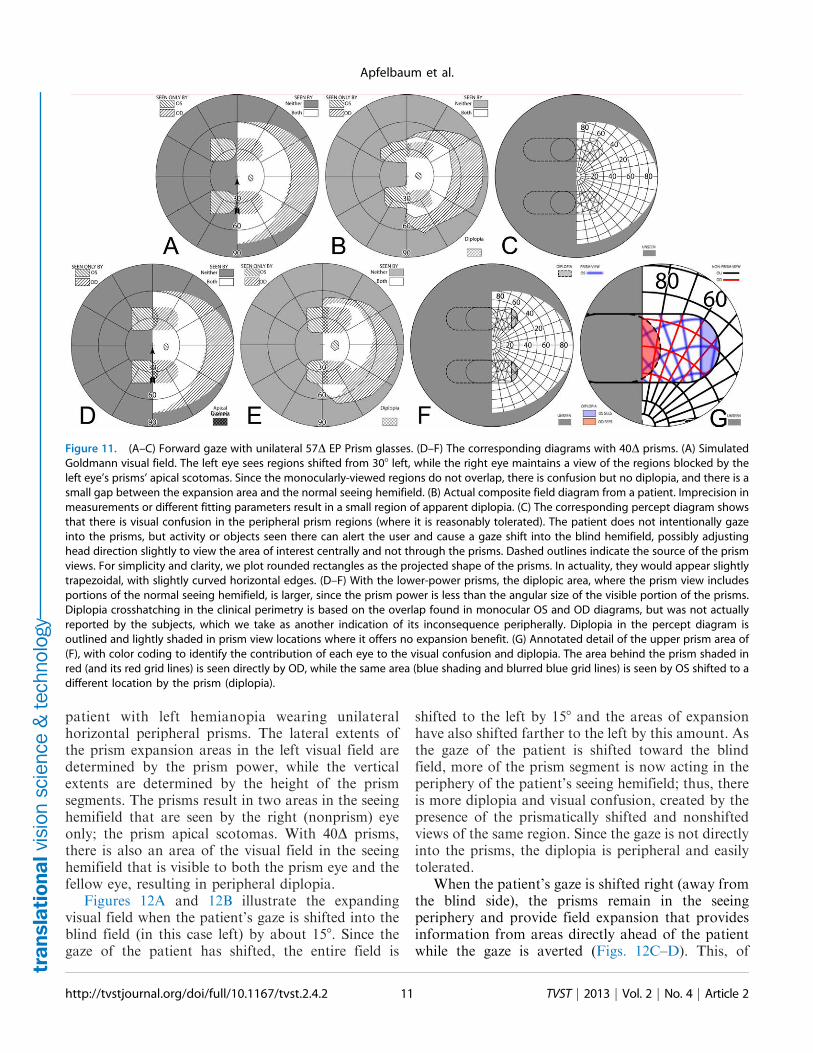
patient with left hemianopia wearing unilateralhorizontal peripheral prisms. The lateral extents ofthe prism expansion areas in the left visual field aredetermined by the prism power, while the verticalextents are determined by the height of the prismsegments. The prisms result in two areas in the seeinghemifield that are seen by the right (nonprism) eyeonly; the prism apical scotomas. With 40D prisms,there is also an area of the visual field in the seeinghemifield that is visible to both the prism eye and thefellow eye, resulting in peripheral diplopia.
Figures 12A and 12B illustrate the expandingvisual field when the patient’s gaze is shifted into theblind field (in this case left) by about 158. Since thegaze of the patient has shifted, the entire field is
shifted to the left by 158 and the areas of expansionhave also shifted farther to the left by this amount. Asthe gaze of the patient is shifted toward the blindfield, more of the prism segment is now acting in theperiphery of the patient’s seeing hemifield; thus, thereis more diplopia and visual confusion, created by thepresence of the prismatically shifted and nonshiftedviews of the same region. Since the gaze is not directlyinto the prisms, the diplopia is peripheral and easilytolerated.
When the patient’s gaze is shifted right (away fromthe blind side), the prisms remain in the seeingperiphery and provide field expansion that providesinformation from areas directly ahead of the patientwhile the gaze is averted (Figs. 12C–D). This, of
Figure 11. (A–C) Forward gaze with unilateral 57D EP Prism glasses. (D–F) The corresponding diagrams with 40D prisms. (A) SimulatedGoldmann visual field. The left eye sees regions shifted from 308 left, while the right eye maintains a view of the regions blocked by theleft eye’s prisms’ apical scotomas. Since the monocularly-viewed regions do not overlap, there is confusion but no diplopia, and there is asmall gap between the expansion area and the normal seeing hemifield. (B) Actual composite field diagram from a patient. Imprecision inmeasurements or different fitting parameters result in a small region of apparent diplopia. (C) The corresponding percept diagram showsthat there is visual confusion in the peripheral prism regions (where it is reasonably tolerated). The patient does not intentionally gazeinto the prisms, but activity or objects seen there can alert the user and cause a gaze shift into the blind hemifield, possibly adjustinghead direction slightly to view the area of interest centrally and not through the prisms. Dashed outlines indicate the source of the prismviews. For simplicity and clarity, we plot rounded rectangles as the projected shape of the prisms. In actuality, they would appear slightlytrapezoidal, with slightly curved horizontal edges. (D–F) With the lower-power prisms, the diplopic area, where the prism view includesportions of the normal seeing hemifield, is larger, since the prism power is less than the angular size of the visible portion of the prisms.Diplopia crosshatching in the clinical perimetry is based on the overlap found in monocular OS and OD diagrams, but was not actuallyreported by the subjects, which we take as another indication of its inconsequence peripherally. Diplopia in the percept diagram isoutlined and lightly shaded in prism view locations where it offers no expansion benefit. (G) Annotated detail of the upper prism area of(F), with color coding to identify the contribution of each eye to the visual confusion and diplopia. The area behind the prism shaded inred (and its red grid lines) is seen directly by OD, while the same area (blue shading and blurred blue grid lines) is seen by OS shifted to adifferent location by the prism (diplopia).
http://tvstjournal.org/doi/full/10.1167/tvst.2.4.2 TVST j 2013 j Vol. 2 j No. 4 j Article 211
Apfelbaum et al.

course, is in contrast to sector prisms, which provide
no expansion at primary gaze, let alone gazes directed
away from the blind side. Continuing to monitor the
straight ahead direction when looking to the side, as
done with normal vision, may be particularly
important during mobility.
Considering prism position size and power
Patients sometimes ask for the prisms to be shifted
farther to their blind side to provide even greater
expansion. At primary gaze, the extent of expansion is
determined (and limited by) the prism power, not the
prism edge positions. The prism base edge is almost
always in the blind hemifield, except for extreme (and
uncomfortable) gaze angles. Shifting the prism
toward the blind side simply limits the availability
of prism on the seeing side, where it is needed when
gazing away from the blind side, as shown in Figure13.
At primary gaze, if the prism power is less than theangular extent of the visible portion of the prism,diplopia will be present (cf Figs. 11A, 11D). If thepower is greater, the apical scotoma creates a gapbetween the expansion area and the seeing side atprimary gaze (as occurs with averted gaze in Fig.12C). Peripheral diplopia, while not necessarilydisturbing, represents a lost opportunity for furtherexpansion, as the diplopic view through the prismwould be more productively directed to a nonover-lapping region. Since it makes sense to optimize theprism effect at primary gaze (where the users spendmost of their time,41 Vargas-Martin F, et al. IOVS.2002;43:ARVO E-Abstract 3809), minimizing diplo-pia and any gap between the expansion area and themidline strikes the best balance, and this occurs when
Figure 12. Unilateral horizontal 57D peripheral prisms, with gaze shifted. (A) With gaze shifted left 158, more of the blind hemifield is
visible in the prisms. Since the prism shift is less than the visible width of the prisms, there is diplopia in the overlap regions
(crosshatched). (B) The corresponding percept diagram shows the visual confusion in the prism regions, combined with diplopia (outlined
and lightly shaded in the prism view location). (C–D) Unlike sector prisms, when gaze is shifted 158 away from the blind side, the prisms
still provide field expansion, providing some continued access to activity in the important region ahead of the wearer. Diagrams for 40Dprisms are included in Supplementary Figure S1. They show more diplopia during gaze to the left and a smaller gap with gaze to the
right.
Figure 13. Examples of altering the upper 40D prism position of Figure 11D. (A–C) Temporal shifts of 58, 108, and 158, respectively, openan increasing gap between the expansion area and the midline, decreasing, not increasing, the expansion. (D) A 58 nasal shift onlyincreases the amount of peripheral diplopia.
http://tvstjournal.org/doi/full/10.1167/tvst.2.4.2 TVST j 2013 j Vol. 2 j No. 4 j Article 212
Apfelbaum et al.
the prism power equals the prism half-width (whenboth are expressed in degrees).
Bilateral Horizontal Peripheral Prisms
Although peripheral prisms are typically appliedunilaterally, they can also be fitted bilaterally andyoked, all bases in the direction of the field loss. Wheneach prism center is aligned with the pupil position atprimary gaze there is no visual confusion, but apicalscotomas are present in the binocular field (Fig. 14),as was the case for sector prisms. This is also the caseif a patient with only one functional eye is fitted withperipheral prisms. Unlike sector prisms, however,objects not completely blocked vertically by theprisms can still be detected (as is the upper half ofthe man in Fig. 1B).
Offset Bilateral Horizontal Peripheral Prisms
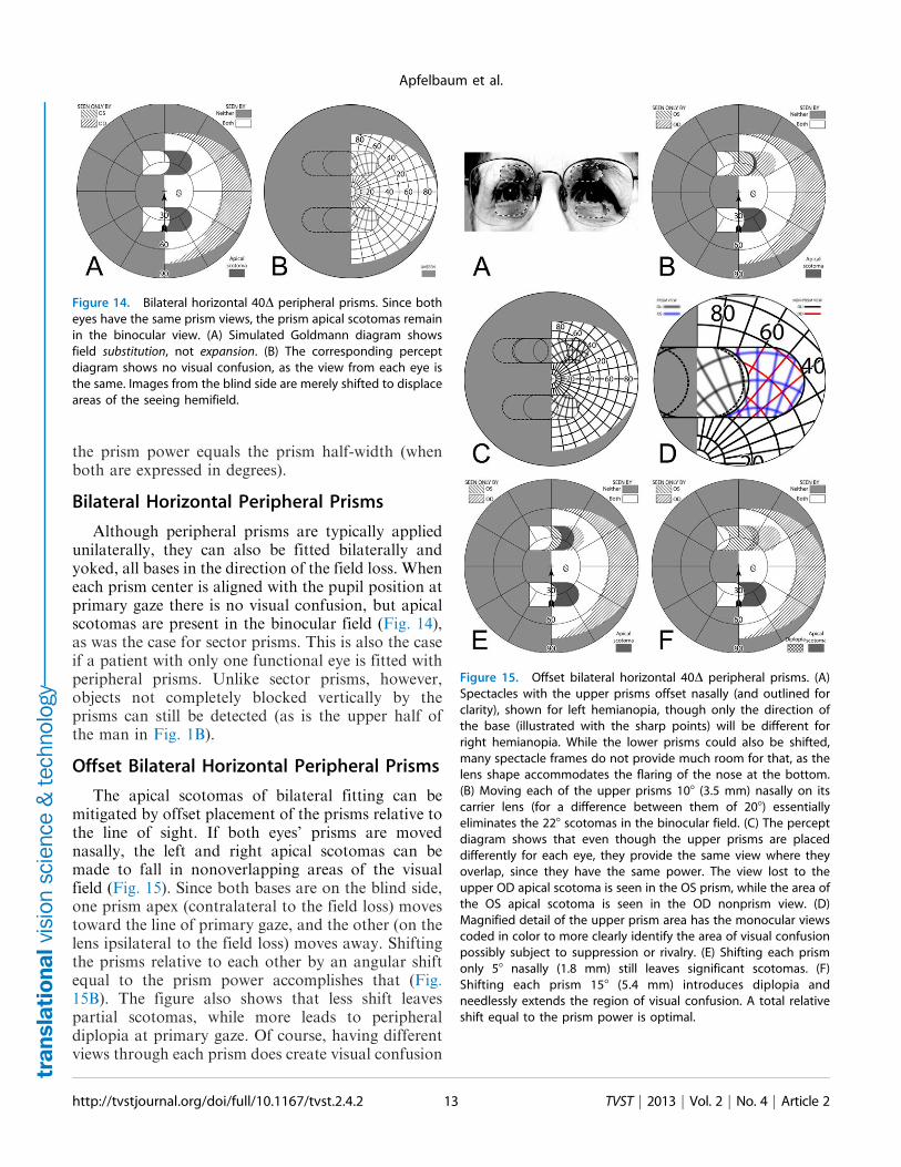
The apical scotomas of bilateral fitting can bemitigated by offset placement of the prisms relative tothe line of sight. If both eyes’ prisms are movednasally, the left and right apical scotomas can bemade to fall in nonoverlapping areas of the visualfield (Fig. 15). Since both bases are on the blind side,one prism apex (contralateral to the field loss) movestoward the line of primary gaze, and the other (on thelens ipsilateral to the field loss) moves away. Shiftingthe prisms relative to each other by an angular shiftequal to the prism power accomplishes that (Fig.15B). The figure also shows that less shift leavespartial scotomas, while more leads to peripheraldiplopia at primary gaze. Of course, having differentviews through each prism does create visual confusion
Figure 14. Bilateral horizontal 40D peripheral prisms. Since botheyes have the same prism views, the prism apical scotomas remainin the binocular view. (A) Simulated Goldmann diagram showsfield substitution, not expansion. (B) The corresponding perceptdiagram shows no visual confusion, as the view from each eye isthe same. Images from the blind side are merely shifted to displaceareas of the seeing hemifield.
Figure 15. Offset bilateral horizontal 40D peripheral prisms. (A)Spectacles with the upper prisms offset nasally (and outlined forclarity), shown for left hemianopia, though only the direction ofthe base (illustrated with the sharp points) will be different forright hemianopia. While the lower prisms could also be shifted,many spectacle frames do not provide much room for that, as thelens shape accommodates the flaring of the nose at the bottom.(B) Moving each of the upper prisms 108 (3.5 mm) nasally on itscarrier lens (for a difference between them of 208) essentiallyeliminates the 228 scotomas in the binocular field. (C) The perceptdiagram shows that even though the upper prisms are placeddifferently for each eye, they provide the same view where theyoverlap, since they have the same power. The view lost to theupper OD apical scotoma is seen in the OS prism, while the area ofthe OS apical scotoma is seen in the OD nonprism view. (D)Magnified detail of the upper prism area has the monocular viewscoded in color to more clearly identify the area of visual confusionpossibly subject to suppression or rivalry. (E) Shifting each prismonly 58 nasally (1.8 mm) still leaves significant scotomas. (F)Shifting each prism 158 (5.4 mm) introduces diplopia andneedlessly extends the region of visual confusion. A total relativeshift equal to the prism power is optimal.
http://tvstjournal.org/doi/full/10.1167/tvst.2.4.2 TVST j 2013 j Vol. 2 j No. 4 j Article 213
Apfelbaum et al.

in peripheral regions proportional to the amount ofprism offset, but, unlike unilateral placement, the trueexpansion area is seen binocularly without confusion.Portions of the seeing hemifield are now viewed withvisual confusion, but hazards that exceed the prismheight would be partially seen binocularly andwithout confusion on the seeing side. Although asyet untested clinically, this is likely to be preferableover unilateral placement and clearly avoids theapical scotomas of nonoffset bilateral placement.
Tilting the Prism Base-Apex Axis:
The Oblique Peripheral Prism Design
The field expansion provided by horizontal pe-ripheral prisms provides access to many of theobstacles, hazards, and orientation cues importantfor pedestrian mobility and common daily activities.Many states do not prohibit hemianopes fromdriving, in which case the prisms may be of help.However, as the vertical extent of a car’s windshield islimited, the area of expansion provided by horizontalperipheral prisms falls mostly outside of the visualfield typically used when driving. Moving the prismsvertically closer to primary gaze to increase the viewthrough a windshield is not an acceptable option, ashead bobbing due to car motion (that shifts theprisms into the gaze) could create the centralconfusion and diplopia that plague sector prisms.Leaving the prism location unchanged, but tilting the
prism apex-base axes, provides a solution.40 This‘‘oblique’’ design gives peripheral access to pericentralregions critical for driving, without blocking thecentral view. The apical scotomas (or visual confu-sion) with this design mostly fall in areas inside thecar, on the visor and dashboard. In an on-road studyof people with hemianopia, use of the oblique prismsimproved hazard detection on the blind side andreduced the number of interventions needed by thedriving examiner.39
An oblique prism has the same effect as thesuperposition of a vertical and horizontal prism, asillustrated in Supplementary Figure S2.
Unilateral Oblique Peripheral Prisms
An apex-base angle of about 308 provides a goodcompromise between vertical shift and the resultingloss of some horizontal expansion. An oblique prismof 57D and 308 tilt provides 268 of horizontal shift and158 vertical shift. Figure 16 diagrams the effects of thisconfiguration. Supplementary Figure S3 has diagramsillustrating the effects of the same design with gazes tothe right and left, and Supplementary Figure S4 hasthe corresponding diagrams for 40D prisms.
Thus, unilateral oblique peripheral prisms provideawareness of areas closer to the horizontal meridian,as might benefit a driver. Unlike sector prisms, theydo this without actually impinging on the pericentralregion and, thus, maintain binocular foveal fusionand avoid central confusion. With unilateral fittingthere is peripheral confusion, but apical scotomas are
Figure 16. Unilateral oblique 57D peripheral prisms, 6308 apex-base angle at 12 mm interprism separation, provide access topericentral regions. (A) Simulated Goldmann visual field diagram shows true expansion with no remaining apical scotomas. The dashedrectangle outlines the typical view area through a car’s windshield.41 When driving, the prisms’ expansion regions fall outside the directroad view, lessening any effect of visual confusion against the bland interior background. (B) The percept diagram shows the areas ofvisual confusion associated with unilateral fitting. (C) The magnified view of the upper prism area of (B) color-codes the monocularportions and shades the diplopic correspondence as in Figure 11. A portion of the small region of interocular diplopia is also seen asmonocular diplopia by OS; inconsequential, but an interesting wrinkle. (Monocular diplopia occurs when the visual angle of the effectiveprism base-apex distance is less than the prism power.) When gaze is directed to the left (Supplementary Figs. S3A, S3B) the diplopicregion increases. Although partially seen foveally, patients have not reported noticing it. (D) A measured patient visual field diagram.
http://tvstjournal.org/doi/full/10.1167/tvst.2.4.2 TVST j 2013 j Vol. 2 j No. 4 j Article 214
Apfelbaum et al.

avoided in the binocular field. With the bland designof most visors and dashboards, the outside road viewon the blind side, with its dynamic nature, is likely topredominate under this rivalrous condition.
Bilateral Oblique Peripheral Prisms
Oblique prisms can be fitted bilaterally (Fig. 17) toavoid the visual confusion associated with unilateralfitting. However, the apical scotomas are significant,and there is a small area of pericentral diplopia thatincreases as gaze is shifted toward the blind hemifield,rendering this a trade-off of questionable added value.Lower-power prisms increase the diplopia, as shownin Supplementary Figure S5. However, apical scoto-mas in the driving situation are mostly located outsidethe field of view through the windshield and, thus,may have lower impact on performance or safety. Theview through the prisms is binocular and free ofconfusion or rivalry. This yet untested design mayprove especially useful when driving.
As with the horizontal prisms, offset placementcan provide some mitigation of the apical scotomas inthe binocular field. However, since only the horizon-tal component of the prisms is offset, the vertical
component of the scotomas remains. SupplementaryFigure S6 has diagrams of the offset bilateral obliqueprisms.
Conclusions and Recommendations
Prisms are often prescribed to provide access toportions of the field of view lost to hemianopia.Failure to consider the pericentral view that is lost toprism apical scotomas can lead to deficits potentiallymore harmful than those the prisms are intended toresolve, yet reports of prism use frequently includediscussions or diagrams that omit these scoto-mas.18,21,43–45 If the fellow eye does not have ascotoma in the same location, the effect can bemitigated. Unilateral fitting, or bilateral fitting ofprisms offset so that their apical scotomas blockdifferent portions of the binocular visual field, canaccomplish that, albeit by introducing regions ofvisual confusion and possibly also creating diplopia.
Visual confusion and diplopia are particularlyobjectionable if in central or pericentral view, whilethey are well tolerated in the periphery, where in somesense they are normal. Visual confusion is the
Figure 17. Bilateral oblique 57D peripheral prisms, 6308 apex-base angle. With gaze forward (A, B) there is a bit of diplopia andsignificant peripheral apical binocular scotomas. With gaze shifted left (C, D), since the prism views include larger portions of the seeinghemifield, there is a larger area of pericentral diplopia, while areas behind the prisms are still lost to the apical scotomas. With gazeshifted right (E, F) there is some field substitution (but not expansion) without confusion or diplopia. The dashed rectangles in (A), (C),and (E) outline the typical view area through a car’s windshield. When driving, most of the peripheral scotoma is inside the car, notdetracting from the view of the road.
http://tvstjournal.org/doi/full/10.1167/tvst.2.4.2 TVST j 2013 j Vol. 2 j No. 4 j Article 215
Apfelbaum et al.

mechanism by which field expansion with prisms ismade possible. Without confusion, prisms can onlyshift the view, trading one scotomatous location withanother. Diplopia, on the other hand, has nobeneficial effect in these treatments and should beavoided or reduced when possible. Direct foveationthrough prisms reduces acuity and contrast sensitivityand introduces other disturbing and noticeable spatialand chromatic distortions. Peripherally-placed prismsare much less disturbing in these regards, and yet areeffective in attracting attention that can cause thewearer to shift gaze centrally to the alerted direction,similar to the natural role of peripheral vision.
These observations lead us to conclude thattraditional sector prisms, which provide no fieldsubstitution or expansion at primary gaze and inducecentral and pericentral scotomas when the gaze isshifted into them, are of questionable value. Theclaim that they serve to train the users to fixate intheir direction7,46 has not been supported with anydocumentation. The peripheral prisms developed inour lab avoid those shortcomings and have been metwith considerable patient acceptance. They have beenfound useful in single and multicenter clinical trials,both open label34 and randomized controlled.38,39 Theperipheral prisms provide true field expansion at mosthorizontal gaze directions, including primary. Al-though operating in the periphery, the oblique designcan provide pericentral field expansion. This effect isparticularly important for patients who are permittedto drive. The prisms access important regions of theroad view, while the confusion in unilateral fit andapical scotomas in bilateral fit fall in the blandinterior of the car and thus are less likely to bedeleterious. Although the peripheral prisms aregenerally fit unilaterally, as this is easier and lessexpensive, offset bilateral fitting provides anotheroption for dealing with the apical scotomas. Obliquebilateral fitting may prove more beneficial for drivingthan unilateral fitting.
Despite the long history of sector prisms, we foundonly three controlled sector prism trials,18,32,47 andeach had limitations. Rossi et al.18 used a parallel armtrial to evaluate bilateral 15D sector prisms against ano treatment control in patients undergoing aninpatient stroke rehabilitation program. The study(which found no advantage for the prisms in activitiesof daily living) recruited patients with either HH orspatial neglect (without HH) during the acute poststroke period, in which spontaneous recovery inneglect and visual field is common. Much of theimprovement in the treatment group occurred on
spatial neglect tests. However, the way sector prismsmay be affecting neglect is not known and beyond thescope of this paper. ‘‘Expansion’’ occurred in both thetreatment and control groups and did not includemeasurements in the seeing hemifield where the apicalscotomas lay. As the prisms were fitted 2 mm into theblind hemifield, they should have had no effect on theperimetry results; thus, the improvements recordedwere most likely due to spontaneous recovery.Gottlieb et al.32 evaluated their unilateral 18.5Dsector prisms against a control device, which ap-peared similar, but included a ‘‘plano lens withoutprism power.’’ Methodology was not well described,but all subjects tried the real prisms before the shams.They reported increased ‘‘awareness’’ of the visualfield ranging from 108 to 458 in binocular (but notmonocular) viewing with the real prisms; however, a458 increase in visual field from a 10.58 prism isphysically impossible. Szlyk et al.,47 used a counter-balanced crossover trial to evaluate the Gottliebunilateral 18.5D ophthalmic sector prisms againstsimilarly shaped and positioned press-on 20D Fresnelprisms. The aim was to evaluate the relative efficacyof the two types of prisms when combined with anintensive 3-month training program. Unsurprisingly,improvements in performance on a wide range of testswere similar for the two prism types, as they hadsimilar prismatic powers and differed only in opticalquality and cosmetics. Unfortunately, the studydesign did not permit an evaluation of the benefitsof the prisms alone (without training) relative to noprisms. The importance of including a sham controltreatment in detecting placebo effects was highlightedin our clinical trial of real versus sham peripheralprisms38 in which 26% of subjects selected the shamcontrol over no glasses or the real prisms.
The limitations in the clinical trials of sector prismsdo not strengthen or weaken our comparison withperipheral prisms, as our arguments against sectorprisms are based upon the physics of the configura-tions. Peripheral prisms were developed and refinedspecifically to address the shortcomings of sectorprisms. The theoretical benefits of the design havenow withstood verification in clinical, single andmulticenter, and randomized controlled trials, andtheir continued use by about 50% of patients after 12months is a strong result for a low vision rehabilita-tion aid.
Attention to the principles and diagrams weprovided can avoid prescription mistakes. Movingperipheral prisms temporally will not increase theireffect; rather it exacerbates the apical scotoma effects
http://tvstjournal.org/doi/full/10.1167/tvst.2.4.2 TVST j 2013 j Vol. 2 j No. 4 j Article 216
Apfelbaum et al.

and wastes much of the prism extent where it is notlikely to be used, robbing it from where it doesprovide benefit. Similarly, increasing the power ofsector prisms increases the region lost to the apicalscotomas, requiring even larger gaze shifts forpericentral views into the blind hemifield.
Our analyses and illustrations of the effects ofapical scotomas have yielded subtle insights into themany ways prisms can both aid and hinder vision.Conventional wisdom and our own intuition wereproved wrong or incomplete on numerous occasionsby this analysis and diagramming.
Acknowledgments
Supported by the National Institutes of HealthGrants EY12890 and P30EY003790.
Aspects of this study have been presented as: RossNC, et al. IOVS. 2009;50:ARVO E-Abstract 4734.
Disclosure: H.L. Apfelbaum, None; N.C. Ross,
None; A.R. Bowers, None; E. Peli, Schepens EyeResearch Institute to Chadwick Optical (P)
References
1. Zhang X, Kedar S, Lynn MJ, Newman NJ,Biousse V. Natural history of homonymoushemianopia. Neurology. 2006;66:901–905.
2. Gall C, Franke GH, Sabel BA. Vision-relatedquality of life in first stroke patients withhomonymous visual field defects. Health QualLife Outcomes. 2010;8:1–28.
3. Bowers AR, Mandel AJ, Goldstein RB, Peli E.Driving with hemianopia: 1. detection perfor-mance in a driving simulator. Invest OphthalmolVis Sci. 2009;50:5137–5147.
4. Papageorgiou E, Hardiess G, Schaeffel F, et al.Assessment of vision-related quality of life inpatients with homonymous visual field defects.Graefe’s Arch Clin Exp Ophthalmol. 2007;245:1749–1758.
5. Dickinson C. Low Vision: Principles and Practice.Oxford, England: Butterworth; 1998:189–190.
6. Cotter SA. Uses of Prism in Low Vision. In: WeissNJ, Brown WL, ed. Clinical Uses of Prism: ASpectrum of Applications. St. Louis: Mosby-YearBook, Inc.; 1995:279–300.
7. Perlin RR, Dziadul J. Fresnel prisms for fieldenhancement of patients with constricted orhemianopic visual fields. J Am Optom Assoc.1991;62:58–64.
8. Bailey IL. Prismatic treatment for field defects.Optom Monthly. 1978;69:99–107.
9. Cohen JM, Waiss B. Visual field remediation. In:RG, Cole, Rosenthal BP, eds. Remediation andManagement of Low Vision. St. Louis: Mosby,1996:1–25.
10. Goodlaw E. Rehabilitating a patient with bitem-poral hemianopia. Am J Optom Physiol Opt. 1982;59:617–619.
11. Cohen JM. An overview of enhancement tech-niques for peripheral field loss. J Am OptomAssoc. 1993;64:60–70.
12. Bishop PO. Binocular vision. In: Moses RA, ed.Adler’s Physiology of the Eye: Clinical Applica-tions. St. Louis: C.V. Mosby, 1998:575–649.
13. Fannin TE, Grosvenor T. The Correction ofAmetropia. In: Clinical Optics. 2nd ed. Boston:Butterworth-Heinemann, 1996:109–126.
14. Morgan MW. Distortions of ophthalmic prisms.Am J Optom. 1963;40:111.
15. Katz M. Visual acuity through Fresnel, refractive,and hybrid diffractive/refractive prisms. Optome-try. 2004;75:503–508.
16. Satgunam P, Apfelbaum HL, Peli E. Volumeperimetry: measurement in depth of visual fieldloss. Optom Vis Sci. 2012;89:E1353–E1363.
17. Woods RL, Apfelbaum HL, Peli E. DLP-baseddichoptic vision test system. J Biomed Optics.2010;15:1–13.
18. Rossi PW, Kheyfets S, Reding MJ. Fresnel prismsimprove visual perception in stroke patients withhomonymous hemianopia or unilateral visualneglect. Neurology. 1990;40:1597–1599.
19. Hoppe E, Perlin RR. The effectivity of Fresnelprisms for visual field enhancement. J Am OptomAssoc. 1993;64:46–53.
20. Jose RT, Smith AJ. Increasing peripheral fieldawareness with Fresnel prisms. Opt J Rev Optom.1976;113:33–37.
21. Smith JL, Weiner IG, Lucero AJ. HemianopicFresnel prisms. J Clin Neuroophthalmol. 1982;2:19–22.
22. Young CA. Homonymous hemianopsia duringpregnancy aided by reflecting prism. Arch Oph-thalmol. 1929;2:560–565.
23. Wiener A. A preliminary report regarding a deviceto be used in lateral homonymous hemianopia.Arch Opthalmol. 1925;55.
http://tvstjournal.org/doi/full/10.1167/tvst.2.4.2 TVST j 2013 j Vol. 2 j No. 4 j Article 217
Apfelbaum et al.

24. Somani S, Brent MH, Markowitz SN. Visual fieldexpansion in patients with retinitis pigmentosa.Can J Opthalmol. 2006;41:27–33.
25. Weiss NJ. An application of cemented prisms withsevere field loss. Am J Optom Arch Am AcadOptom. 1972;49:261–264.
26. Lee AG, Perez AM. Improving awareness ofperipheral visual field using sectorial prism. J AmOptom Assoc. 1999;70:624–628.
27. Bailey IL. Optometric Examination of the OlderAdult. In: Rosenbloom AAJ, ed. Rosenbloom andMorgan’s Vision and Aging. St. Louis: Butter-worth-Heinemann, 2007:133–163.
28. Bahill AT, Adler D, Stark L. Most naturallyoccurring human saccades have magnitudes of 15degrees or less. Invest Ophthalmol. 1975;14:468–469.
29. Luo G, Vargas-Martin F, Peli E. The role ofperipheral vision in saccade planning: learningfrom people with tunnel vision. J Vis. 2008;8:1–8.
30. Good GW, Fogt N, Daum KM, Mitchell GL.Dynamic visual fields of one-eyed observers.Optometry. 2005;76:285–292.
31. Lovie-Kitchin J, Mainstone J, Robinson J, BrownB. What areas of the visual field are important formobility in low vision patients? Clin Vis Sci. 1990;5:249–263.
32. Gottlieb DD, Freeman P, Williams M. Clinicalresearch and statistical analysis of a visual fieldawareness system. J Am Optom Assoc. 1992;63:581–588.
33. Peli E. Field expansion for homonymous hemi-anopia by optically-induced peripheral exotropia.Optom Vis Sci. 2000;77:453–464.
34. Bowers AR, Keeney K, Peli E. Community-basedtrial of a peripheral prism visual field expansiondevice for hemianopia. Arch Ophthalmol. 2008;126:657–664.
35. Blake R. A primer on binocular rivalry, includingcurrent controversies. Brain and Mind. 2001;2:5–38.
36. Giorgi RG, Woods RL, Peli E. Clinical andlaboratory evaluation of peripheral prism glassesfor hemianopia. Optom Vis Sci. 2009;86:492–502.
37. O’Neill EC, Connell PP, O’Connor JC, Brady J,Reid I, Logan P. Prism therapy and visualrehabilitation in homonymous visual field loss.Optom Vis Sci. 2011;88:263–268.
38. Bowers AR, Keeney K, Peli E. Randomizedcrossover clinical trial of real and sham peripheralprism glasses for hemianopia. JAMA Ophtalmol.In press.
39. Bowers AR, Tant M, Peli E. A pilot evaluation ofon-road detection performance by drivers withhemianopia using oblique peripheral prisms.Stroke Res Treat. 2012;2012:10.
40. Peli E, inventor; Peripheral field expansion device.US patent 7,374,284 B2. May 20, 2008.
41. Iorizzo DB, Riley ME, Hayhoe M, Huxlin KR.Differential impact of partial cortical blindness ongaze strategies when sitting and walking - animmersive virtual reality study. Vision Res. 2011;51:1173–1184.
42. Vargas-Martin F, Garcia-Perez MA. Visual fieldsat the wheel. Optom Vis Sci. 2005;82:675–681.
43. Cardenal MS, Hoban K, Toffel K. Novel lowvision approach to bilateral superior altitudinalfield loss. Optom Vis Sci. 2012;89:E-abstract125908.
44. Woo GC, Mandelman T. Fresnel prism therapyfor right hemianopia. Am J Optom Physiol Opt.1983;60:739–743.
45. van Waveren M, Jagle H, Besch D. Managementof strabismus with hemianopic visual field defects.Graefe’s Arch Clin Exp Ophthalmol. 2013;251:575–584.
46. Perez AM, Jose RT. The use of Fresnel andophthalmic prisms with persons with hemianopicvisual field loss. J Vis Impair Blind. 2003;97:173–176.
47. Szlyk JP, Seiple W, Stelmack J, McMahon T. Useof prisms for navigation and driving in hemi-anopic patients. Ophthalmic Physiol Opt. 2005;25:128–135.
http://tvstjournal.org/doi/full/10.1167/tvst.2.4.2 TVST j 2013 j Vol. 2 j No. 4 j Article 218
Apfelbaum et al.

Supplement to http://tvstjournal.org/doi/full/10.1167/tvst.2.4.2TVST | 2013 | Vol. 2 | No.4 | Article 2 Supplement
Supplement to tvst
Considering Apical Scotomas, Confusion, and Diplopia When Prescribing Prisms for Homonymous Hemianopia Henry L. Apfelbaum1, Nicole C. Ross1,2, Alex R. Bowers1,2, Eli Peli1,2 1 Schepens Eye Research Institute, Massachusetts Eye and Ear, Harvard Medical School, Boston, MA, USA
2 New England College of Optometry, Boston, MA, USA
Figure S1. Unilateral Horizontal 40Δ Peripheral Prisms, with gaze shifted. With the lower power the prisms show more diplopia when gazing to the left and a smaller gap when gazing to the right than the 57Δ prisms of Figure 12. (A) With gaze shifted left 15°, more of the blind hemifield is visible in the prisms. Since the prism shift is less than the visible width of the prisms, there is diplopia in the overlap regions (crosshatched). (B) The corresponding percept diagram shows the visual confusion in the prism regions, combined with diplopia (outlined and lightly shaded in prism view locations where it offers no expansion benefit). (C-D) Unlike sector prisms, when gaze is shifted 15° away from the blind side, the prisms still provide field expansion, providing some continued access to activity in the important region straight ahead of the wearer.
A B C D

Supplement to Apfelbaum et al., Considering Apical Scotomas….
2
Figure S2. An oblique 57Δ prism with power 29.7 degrees and apex-base angle 30° is equivalent to combining a vertical prism with power 29.7×sin(30) = 14.8° (26.5Δ) and a horizontal p rism with power 29.7×cos(30) = 25.7° (48.1Δ). Press -On prisms (3M, Minneapolis. MN) used in this figure are all the same shape and size and are centered (except for d) on the left lens of a normally-sighted subject, with OD patched. The solid outlines indicate the approximate prism locations, while the dashed outlines indicate the field seen in the prisms. (A) Visual field diagram with a 40Δ base left (horizontal) prism on the rear of the carrier lens, showing an apical scotoma the height of the prism and the width of the prism power (21.8°). (B) Diagram with a 15Δ base down (vertical) prism on the front of the carrier lens, showing an apical scotoma the width of the prism and the height of the prism power (8.5°). (C) Diagram with both prisms in place, showing the combined scotoma. (D) Diagram using an upper segment 57Δ oblique peripheral prism with apex-base angle 30°, showing a similar scotoma
Figure S3. Unilateral oblique 57Δ peripheral prisms provide access to pericentral regions even when gaze is shifted from primary. (A) Simulated Goldmann visual field diagram with gaze shifted 15° left. (B) The corresponding percept diagram shades the diplopic portion of the prism view that does not contribute expansion. There is peripheral (retinal), but not central, diplopia. (C-D) With gaze shifted right 15°, some pericentral expansion is still provided, with no diplopia.
A B C D
A B D C

Supplement to Apfelbaum et al., Considering Apical Scotomas….
3
Figure S4. Unilateral oblique 40Δ peripheral prisms, ±30° apex-base angle, provide access to pericentral regions. (A) Simulated Goldmann visual field diagram shows true expansion with no apical scotomas at primary gaze, but there is some peripheral diplopia. (B) The percept diagram shows the areas of visual confusion associated with unilateral fitting, and the shading indicates the (peripheral) diplopic regions that do not contribute to expansion. (C-F) Corresponding calculated diagrams with gaze shifted 15° left and right, respectively, as shown by the arrows.
C D E F
B A

Supplement to Apfelbaum et al., Considering Apical Scotomas….
4
Figure S6. In this example, 57Δ prisms are fit bilaterally, with the upper prisms offset temporally with respect to each other. Offsetting oblique prisms horizontally does not compensate for the vertical component of the apical scotomas and results in wasteful peripheral diplopia. Unilateral fitting is preferable.
Figure S5. Diagrams similar to those in Figure 17, with 40Δ rather than 57Δ prisms. Lower power results in larger areas of diplopia.
C D E F
B A
A B
Copyright © 2022 FDOKUMEN