Comparative Transcript Profiling of Candida albicans and Candida dubliniensis Identifies SFL2, a C....
15
EUKARYOTIC CELL, Feb. 2010, p. 251–265 Vol. 9, No. 2 1535-9778/10/$12.00 doi:10.1128/EC.00291-09 Copyright © 2010, American Society for Microbiology. All Rights Reserved. Comparative Transcript Profiling of Candida albicans and Candida dubliniensis Identifies SFL2,a C. albicans Gene Required for Virulence in a Reconstituted Epithelial Infection Model † Martin J. Spiering, 1 ‡ Gary P. Moran, 1 Murielle Chauvel, 2,3 Donna M. MacCallum, 4 Judy Higgins, 1 Karsten Hokamp, 5 Tim Yeomans, 1 § Christophe d’Enfert, 2,3 David C. Coleman, 1 and Derek J. Sullivan 1 * Microbiology Research Unit, Division of Oral Biosciences, Dublin Dental School and Hospital, University of Dublin, Trinity College, Dublin 2, Ireland 1 ; Institut Pasteur, Unite ´ Biologie et Pathoge ´nicite ´ Fongiques, Paris, France 2 ; INRA USC2019, Paris, France 3 ; Aberdeen Fungal Group, School of Medical Sciences, Institute of Medical Sciences, University of Aberdeen, Aberdeen AB25 2ZD, United Kingdom 4 ; and Smurfit Institute of Genetics, University of Dublin, Trinity College, Dublin 2, Ireland 5 Received 8 October 2009/Accepted 11 December 2009 Candida albicans and Candida dubliniensis are closely related species displaying differences in virulence and genome content, therefore providing potential opportunities to identify novel C. albicans virulence genes. C. albicans gene arrays were used for comparative analysis of global gene expression in the two species in reconstituted human oral epithelium (RHE). C. albicans (SC5314) showed upregulation of hypha-specific and virulence genes within 30 min postinoculation, coinciding with rapid induction of filamentation and increased RHE damage. C. dubliniensis (CD36) showed no detectable upregulation of hypha-specific genes, grew as yeast, and caused limited RHE damage. Several genes absent or highly divergent in C. dubliniensis were upregulated in C. albicans. One such gene, SFL2 (orf19.3969), encoding a putative heat shock factor, was deleted in C. albicans. sfl2 cells failed to filament under a range of hypha-inducing conditions and exhibited greatly reduced RHE damage, reversed by reintroduction of SFL2 into the sfl2 strain. Moreover, SFL2 overexpres- sion in C. albicans triggered hyphal morphogenesis. Although SFL2 deletion had no apparent effect on host survival in the murine model of systemic infection, sfl2 strain-infected kidney tissues contained only yeast cells. These results suggest a role for SFL2 in morphogenesis and an indirect role in C. albicans pathogenesis in epithelial tissues. Candida dubliniensis is the closest relative of the important opportunistic human pathogen Candida albicans (8, 21, 44). In spite of their close relationship, the two species show signifi- cant differences in virulence and epidemiology (42, 43). Al- though C. dubliniensis has historically been associated with oral infections in HIV-infected patients (33), it is generally less pathogenic than C. albicans, as judged by differences in car- riage rates and prevalence in the human host (7, 27, 28, 45) and by differences in virulence in vitro and in murine infection models (8, 12, 23, 41). However, the reasons for these virulence differences are poorly understood and are the focus of inves- tigations to determine their genetic basis. Among traits important for virulence and variable between C. albicans and C. dubliniensis are adherence to human epi- thelial tissues and production of hydrolytic enzymes, as well as resistance to antifungal agents, oxidative stress, and phagocy- tosis by cells of the host immune system (8, 12, 20, 38, 41, 47). A trait critically important for Candida virulence especially in endothelial and epithelial infection models is the ability to undergo the yeast-to-hypha transition (hyphal morphogenesis) (16, 26, 31). Importantly, hyphal morphogenesis in response to many stimuli is consistently slower in C. dubliniensis than in C. albicans (8, 41). Some specific triggers that induce hyphal mor- phogenesis in C. albicans, such as increased CO 2 /HCO 3 and growth in mammalian tissues (14, 37, 52), do not induce hy- phae in C. dubliniensis (14, 23, 41). The transcriptional regu- lator Nrg1 has been identified as a key regulator suppressing filamentation in C. dubliniensis, and it has been suggested that this suppression of hyphal growth may partially explain why C. dubliniensis is less virulent in the human host than C. albicans (23). The close phylogenetic relationship between C. albicans and C. dubliniensis may offer opportunities to identify virulence genes in C. albicans in infection models where the two species exhibit differences in virulence. A conceptually similar ap- proach, using two C. albicans strains differing in their ability to invade tissue in an organ model of mammalian infection cou- pled with microarray-based analysis of gene expression, iden- tified DFG16 as being required for pH sensing during tissue invasion (46). The approach adopted in the present study rep- resents a progression from a study by Moran et al. (22), which * Corresponding author. Mailing address: Microbiology Research Unit, Division of Oral Biosciences, Dublin Dental School and Hospi- tal, University of Dublin, Trinity College, Dublin 2, Ireland. Phone: 353 1 612 7275. Fax: 353 1 612 7295. E-mail: Derek.Sullivan@dental .tcd.ie. ‡ Present address: Center for Advanced Research in Biotechnology, UMBI, Rockville, MD 20850. § Present address: Allergy Standards, Ltd., Trinity Enterprise Cam- pus, Dublin 2, Ireland. † Supplemental material for this article may be found at http://ec .asm.org/. Published ahead of print on 18 December 2009. 251
-
Upload
independent -
Category
Documents
-
view
2 -
download
0
Transcript of Comparative Transcript Profiling of Candida albicans and Candida dubliniensis Identifies SFL2, a C....

EUKARYOTIC CELL, Feb. 2010, p. 251–265 Vol. 9, No. 21535-9778/10/$12.00 doi:10.1128/EC.00291-09Copyright © 2010, American Society for Microbiology. All Rights Reserved.
Comparative Transcript Profiling of Candida albicans andCandida dubliniensis Identifies SFL2, a C. albicans Gene
Required for Virulence in a ReconstitutedEpithelial Infection Model�†
Martin J. Spiering,1‡ Gary P. Moran,1 Murielle Chauvel,2,3 Donna M. MacCallum,4 Judy Higgins,1Karsten Hokamp,5 Tim Yeomans,1§ Christophe d’Enfert,2,3
David C. Coleman,1 and Derek J. Sullivan1*Microbiology Research Unit, Division of Oral Biosciences, Dublin Dental School and Hospital, University of Dublin, Trinity College,
Dublin 2, Ireland1; Institut Pasteur, Unite Biologie et Pathogenicite Fongiques, Paris, France2; INRA USC2019, Paris, France3;Aberdeen Fungal Group, School of Medical Sciences, Institute of Medical Sciences, University of Aberdeen, Aberdeen AB25 2ZD,
United Kingdom4; and Smurfit Institute of Genetics, University of Dublin, Trinity College, Dublin 2, Ireland5
Received 8 October 2009/Accepted 11 December 2009
Candida albicans and Candida dubliniensis are closely related species displaying differences in virulence andgenome content, therefore providing potential opportunities to identify novel C. albicans virulence genes. C.albicans gene arrays were used for comparative analysis of global gene expression in the two species inreconstituted human oral epithelium (RHE). C. albicans (SC5314) showed upregulation of hypha-specific andvirulence genes within 30 min postinoculation, coinciding with rapid induction of filamentation and increasedRHE damage. C. dubliniensis (CD36) showed no detectable upregulation of hypha-specific genes, grew as yeast,and caused limited RHE damage. Several genes absent or highly divergent in C. dubliniensis were upregulatedin C. albicans. One such gene, SFL2 (orf19.3969), encoding a putative heat shock factor, was deleted in C.albicans. ��sfl2 cells failed to filament under a range of hypha-inducing conditions and exhibited greatlyreduced RHE damage, reversed by reintroduction of SFL2 into the ��sfl2 strain. Moreover, SFL2 overexpres-sion in C. albicans triggered hyphal morphogenesis. Although SFL2 deletion had no apparent effect on hostsurvival in the murine model of systemic infection, ��sfl2 strain-infected kidney tissues contained only yeastcells. These results suggest a role for SFL2 in morphogenesis and an indirect role in C. albicans pathogenesisin epithelial tissues.
Candida dubliniensis is the closest relative of the importantopportunistic human pathogen Candida albicans (8, 21, 44). Inspite of their close relationship, the two species show signifi-cant differences in virulence and epidemiology (42, 43). Al-though C. dubliniensis has historically been associated with oralinfections in HIV-infected patients (33), it is generally lesspathogenic than C. albicans, as judged by differences in car-riage rates and prevalence in the human host (7, 27, 28, 45) andby differences in virulence in vitro and in murine infectionmodels (8, 12, 23, 41). However, the reasons for these virulencedifferences are poorly understood and are the focus of inves-tigations to determine their genetic basis.
Among traits important for virulence and variable betweenC. albicans and C. dubliniensis are adherence to human epi-thelial tissues and production of hydrolytic enzymes, as well as
resistance to antifungal agents, oxidative stress, and phagocy-tosis by cells of the host immune system (8, 12, 20, 38, 41, 47).A trait critically important for Candida virulence especially inendothelial and epithelial infection models is the ability toundergo the yeast-to-hypha transition (hyphal morphogenesis)(16, 26, 31). Importantly, hyphal morphogenesis in response tomany stimuli is consistently slower in C. dubliniensis than in C.albicans (8, 41). Some specific triggers that induce hyphal mor-phogenesis in C. albicans, such as increased CO2/HCO3
� andgrowth in mammalian tissues (14, 37, 52), do not induce hy-phae in C. dubliniensis (14, 23, 41). The transcriptional regu-lator Nrg1 has been identified as a key regulator suppressingfilamentation in C. dubliniensis, and it has been suggested thatthis suppression of hyphal growth may partially explain why C.dubliniensis is less virulent in the human host than C. albicans(23).
The close phylogenetic relationship between C. albicans andC. dubliniensis may offer opportunities to identify virulencegenes in C. albicans in infection models where the two speciesexhibit differences in virulence. A conceptually similar ap-proach, using two C. albicans strains differing in their ability toinvade tissue in an organ model of mammalian infection cou-pled with microarray-based analysis of gene expression, iden-tified DFG16 as being required for pH sensing during tissueinvasion (46). The approach adopted in the present study rep-resents a progression from a study by Moran et al. (22), which
* Corresponding author. Mailing address: Microbiology ResearchUnit, Division of Oral Biosciences, Dublin Dental School and Hospi-tal, University of Dublin, Trinity College, Dublin 2, Ireland. Phone:353 1 612 7275. Fax: 353 1 612 7295. E-mail: [email protected].
‡ Present address: Center for Advanced Research in Biotechnology,UMBI, Rockville, MD 20850.
§ Present address: Allergy Standards, Ltd., Trinity Enterprise Cam-pus, Dublin 2, Ireland.
† Supplemental material for this article may be found at http://ec.asm.org/.
� Published ahead of print on 18 December 2009.
251

analyzed the genome contents of C. albicans and C. dublinien-sis by comparative genomic hybridization to C. albicans genearrays. Although the vast majority of genes are highly con-served between the two species, approximately 200 C. albicansgenes (representing �4.0% of its genome) are absent or highlydiverged in C. dubliniensis, including known virulence genesand genes of unknown function (11, 22). Availability of thegenome sequences of C. albicans (4, 13) and C. dubliniensis(11) in database-accessible formats (2, 11) has enabled miningof the differences in the genetic repertoire of the two species.
For comparative study of C. albicans and C. dubliniensisgene expression, a suitable human infection model has to fulfillseveral criteria: chiefly ease of use, reproducibility, and suffi-cient recovery of fungal material for analysis. Reconstitutedhuman oral epithelium (RHE) represents such a model, havingbeen used extensively to study Candida virulence (reviewed inreference 36), including microarray-based transcriptional pro-filing (52). The RHE infection model permits quantitativemeasurements of virulence, and we have recently demon-strated that C. dubliniensis is far less virulent than C. albicansin the RHE model (23, 41). Here we report the results of geneexpression profiling in C. albicans and C. dubliniensis coincu-bated with RHE tissue, representing the first comparativeanalysis of global transcription in these two species in an in-fection model. We further describe the identification of one C.albicans gene, which we have named SFL2 (orf19.3969;
IPF8627.2), which is critical for filamentation in vitro and vir-ulence in the RHE model.
MATERIALS AND METHODS
Strains and culture conditions. The C. albicans and C. dubliniensis strains usedin this study are listed in Table 1. Growth media were from Oxoid (Basingstoke,Hampshire, United Kingdom), and amino acids were from Sigma-Aldrich Ire-land, Ltd. (Tallaght, Dublin, Ireland). All strains were cultured on yeast extract-peptone-dextrose (YPD) agar or in YPD broth at 37°C and 200 rpm, unlessindicated otherwise. All liquid media for fungal cell cultures for microarraystudies or in transformation experiments were filter sterilized. Hexose solutions(10� concentration) used in agar media were filter sterilized and added tomolten agar media shortly before pouring.
Chemicals and enzymes. All chemicals used were analytical grade or molec-ular biology grade and supplied by Sigma-Aldrich, Ambion (Warrington, UnitedKingdom), or Roche Diagnostics (Mannheim, Germany). Ultrapure Milli-Qwater (Millipore Ireland B.V., Cork, Ireland) was used in all experiments. PCRsfor cloning of DNA constructs were performed with Expand high-fidelity enzyme(Roche), and diagnostic PCRs were carried out with GoTaq enzyme (Promega,Madison, WI).
RHE inoculation and coincubation with Candida cells and RHE tissue damagemeasurements. Inoculation and coincubation of RHE with Candida cells wereperformed as previously described (41). In brief, Candida cells were grown insemisynchronized cultures (i.e., in 25°C and 37°C serial YPD cultures) (36) andharvested by centrifugation, the cell density was adjusted in phosphate-bufferedsaline (PBS), and RHE tissues were inoculated as described previously (41).Candida cultures on polycarbonate filter (PCF) membranes (used as RHE sup-port matrix) were initiated in the same way as the RHE cultures. Candida RHEand Candida PCF cultures and RHE-only controls were incubated in MCDB 153maintenance medium (containing 0.1% glucose; Skinethic Laboratories, Nice,
TABLE 1. Candida strains used in this study
Strain Description Parent strain Source or reference
C. albicansSC5314 Wild type 9CaMS1 �orf19.4445::SAT1/orf19.4445 SC5314 This studyCaMS1-1 �orf19.4445::FRT/orf19.4445 CaMS1 This studyCaMS16 �orf19.4445::FRT/�orf19.4445::SAT1 CaMS1-1 This studyCaMS24 �orf19.7304::SAT1/�orf19.7304::SAT1 SC5314 This studyCaMS24-2 �orf19.7304::FRT/�orf19.7304::FRT CaMS24 This studyCaMS46 �SFL2::SAT1/SFL2 SC5314 This studyCaMS48 �SFL2::SAT1/SFL2 SC5314 This studyCaMS46-2 �SFL2::FRT/SFL2 CaMS46 This studyCaMS48-2 �SFL2::FRT/SFL2 CaMS48 This studyCaMS49 �SFL2::FRT/�SFL2::SAT1 CaMS46-2 This studyCaMS49-1 �SFL2::FRT/�SFL2::FRT CaMS49 This studyCaMS50 �SFL2::FRT/�SFL2::SAT1 CaMS46-2 This studyCaMS50-1 �SFL2::FRT/�SFL2::FRT CaMS50 This studyCaMS58 �SFL2::FRT/SFL2::SAT1 CaMS49-1 This studyCaMS60 �SFL2::FRT/SFL2::SAT1 CaMS50-1 This studyCEC955 ura3�::� imm434/ura3�::� imm434 his1::hisG/HIS1
arg4::hisG/ARG4 ADH1/adh1::(ADH1p-cartTA SAT1TETp-caGFP)
BWP17 A. Firon and C. d’Enfert,unpublished
CEC1352 ura3�::� imm434/ura3�::� imm434 his1::hisG/HIS1arg4::hisG/ARG4 ADH1/adh1::(ADH1p-cartTA SAT1TETp-caGFP) RPS1/RPS1::(URA3 TETp-SFL2)
CEC955 This study
CEC1147 ura3�::� imm434/ura3�::� imm434 his1::hisG/HIS1arg4::hisG/ARG4 ADH1/adh1::(ADH1p-cartTA SAT1TETp-caGFP) RPS1/RPS1::(URA3 TETp-GFP)
CEC955 A. Firon and C. d’Enfert,unpublished
C. dubliniensisCD36 Wild type 44Wu284 Wild type 24CD36/pNIM1GFP ADH1/adh1::(ADH1p-cartTA SAT1 TETp-caGFP) CD36 This studyCD36/pNIM1SFL2 ADH1/adh1::(ADH1p-cartTA SAT1 TETp-caSFL2) CD36 This studyWu284/pNIM1GFP ADH1/adh1::(ADH1p-cartTA SAT1 TETp-caGFP) Wu284 This studyWu284/pNIM1SFL2 ADH1/adh1::(ADH1p-cartTA SAT1 TETp-caSFL2) Wu284 This study
252 SPIERING ET AL. EUKARYOT. CELL
France) in a CO2 incubator at 37°C, with 5% (vol/vol) CO2 and 100% humidity,and sampled for RNA or lactate dehydrogenase (LDH) measurements (seebelow) at regular time points (30, 90, 360, or 720 min). Activity of human LDHin RHE culture medium was measured with the CytoTox 96 nonradioactivecytotoxicity assay (Promega) (41). The LDH assay product, formazan, was mea-sured by spectrophotometry, and concentrations were determined with its ex-tinction coefficient (15,600 M�1 cm�1). Candida RHE cultures and RHE con-trols (0.5 cm2) were fixed, sectioned, stained, and examined by light microscopyas previously described (41). Cell morphology in Candida PCF cultures wasexamined by light microscopy of cell suspensions obtained by rinsing the PCFmembranes.
RNA extraction. For RNA extractions, 2 ml of a solution containing 2 parts(vol/vol) RNAlater solution (Ambion) and 1 part filter-sterilized 10% (wt/vol)saponin (Sigma-Aldrich) in PBS was added to a 4-cm2 Candida RHE or CandidaPCF culture. To recover the fungal cells, membranes were rinsed 3 or 4 timeswith the RNAlater-saponin solution, and the cell suspensions from 2 or 3 Can-dida RHE or Candida PCF cultures were transferred to a 50-ml centrifuge tube(Sarstedt, Wexford, Ireland) along with the cut filter membranes and immedi-ately frozen and stored at �20°C. For total RNA extraction, cell suspensionscontaining the filter membranes were thawed at room temperature, vortexed for
5 to 10 s, and centrifuged (3,200 � g for 5 min). The membranes were removed,the tubes were centrifuged as before, and the supernatant was removed bycareful aspiration. RNA was extracted with the RNeasy minikit (Qiagen, WestSussex, United Kingdom) with cell disruption performed in a Mikro-Dismem-brator S system (Sartorius Stedim Biotech, Gottingen, Germany) for 2 min at2,000 rpm. Candida cultures (�8 ml) used as the 0-min control (inoculum) inPBS (described above) were harvested by centrifugation as before, taken up in 2ml RNAlater solution, and stored at �20°C; RNA was extracted from Candidacell pellets as described above. Total RNA was resuspended in nuclease-freewater and DNase I treated with the DNAfree Turbo kit (Ambion), measured by260/280 spectrophotometer readings, and integrity was checked on 1.2% Tris-borate-EDTA (TBE) gels. Absence of DNA in RNA samples after DNasetreatment was routinely checked by PCR with primers qP-ACT1F and qP-ACT1R (Table 2).
Amplification and Cy labeling of RNA and hybridization to Candida genearrays. The Amino Allyl MessageAmp II aRNA amplification kit (Ambion) wasused for RNA amplification and labeling, following the manufacturer’s protocol.One microgram of total RNA was used for reverse transcription, and amino-allyl-labeled RNA (aaRNA) was linearly amplified by T7-based in vitro transcrip-tion (49). aaRNA was Cy labeled with N-hydroxysuccinimide esters of Cy3 and
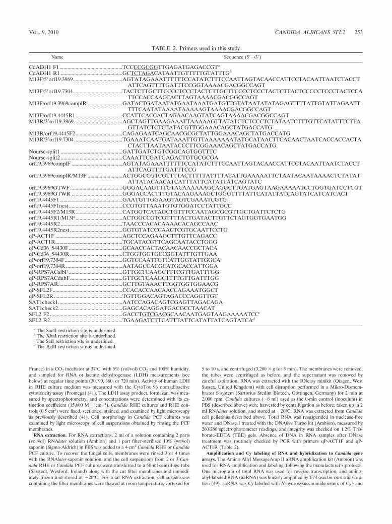
TABLE 2. Primers used in this study
Name Sequence (5�33�)
CdADH1 F1...............................................TCCCCGCGGTTGAGATGAGACCGTa
CdADH1 R1 ..............................................GCTCTAGACATAATTGTTTTTGTATTTGb
M13F/5�orf19.3969.....................................AGTATAGAAATTTTTTCCATATCTTTCCAATTAGTACAACCATTCCTACAATTAATCTACCTATTCAGTTTTGATTTCCGGTAAAACGACGGCCAGT
M13F/5�orf19.7304.....................................TACTCTTGCTTCCCCTCCCTACTCTTGCTTCCCCTCCCTACTCTTACTCCCCCTCCCTACTCCATTCCACCAACCACTTAGTAAAACGACGGCCAGT
M13F/orf19.3969complR ..........................GATACTGATAATATGAATAAATGATGTTGTATAATATATAGAGTTTTATTGTATTAGAATTTTTCAATATAAAATAAAAAGTAAAACGACGGCCAGT
M13F/orf19.4445R1...................................CCATTCACCACTAGAACAAGTATCAGTAAAACGACGGCCAGTM13R/3�orf19.3969 ....................................AGCTAGTTGAAGAAATTAAAAAGTTATATCTCTCCCTCTATAATCTTTGTTCATATTTCTTA
GTTATCTCTCTATACGTTGGAAACAGCTATGACCATGM13R/orf19.4445F2...................................CAGAGAATCAGCAACGCGCTATTGGAAACAGCTATGACCATGM13R/3�orf19.7304 ....................................TGAAATCAATGATAAATTGTTAAAAAAATATGCATAACTTCACAACTAATCACCACCACTA
CTACTTAATAATACCCTTCGGAAACAGCTATGACCATGNourse-split1 ..............................................GATTGATCTGTCGGCAGTGGTTTCNourse-split2 ..............................................CAAATTCGATGAGACTGTGCGCGAorf19.3969complF ......................................AGTATAGAAATTTTTTCCATATCTTTCCAATTAGTACAACCATTCCTACAATTAATCTACCT
ATTCAGTTTTGATTTCCGorf19.3969complR/M13F ..........................ACTGGCCGTCGTTTTACTTTTTATTTTATATTGAAAAATTCTAATACAATAAAACTCTATAT
ATTATACAACATCATTTATTCATATTATCAGTATCorf19.3969GTWF.......................................GGGACAAGTTTGTACAAAAAAGCAGGCTTGATGAGTAAGAAAAATCCTGGTGATCCTCGTorf19.3969GTWR ......................................GGGACCACTTTGTACAAGAAAGCTGGGTTTTATTCATATTATCAGTATCATCATCACTorf19.4445F1...............................................GAATGTTGGAAGTAGTCGAAATCGTGorf19.4445F1nest........................................CCGTGTTAAATGTGTGGATCCTATTGCCorf19.4445F2/M13R...................................CATGGTCATAGCTGTTTCCAATAGCGCGTTGCTGATTCTCTGorf19.4445R1/M13F...................................ACTGGCCGTCGTTTTACTGATACTTGTTCTAGTGGTGAATGGorf19.4445R2 ..............................................TAACCCACACAAAACACAGCCAACorf19.4445R2nest .......................................GGTGTATCCCAACTCGTGCAATTCCTGqP-ACT1F ..................................................AGCTCCAGAAGCTTTGTTCAGACCqP-ACT1R..................................................TGCATACGTTCAGCAATACCTGGGqP-Cd36_54430F........................................GCAACCACTACAACAACCGCTACAqP-Cd36_54430R .......................................CTGGTGGTGCCGGTATTTGTTGAAqP-orf19.7304F...........................................GGTCCAATTGTCATTGGTATTGGCAqP-orf19.7304R ..........................................AATAGCCACGCATGCACCATTGGAqP-RPS7ACalbF ........................................GTTGCTCAAGCTTTCGTTGATTTGGqP-RPS7ACdubF.......................................GTTGCTCAAGCTTTTGTTGATTTGGqP-RPS7AR................................................GCTTGTAAACTTGGTGGTGGAACGqP-SFL2F....................................................CCACACCAACAACCAGAAATGGCTqP-SFL2R ...................................................TGTTGGACAGTAGACCCAGGTTGTSAT1check1................................................AATCCAGACAGTCGAGTTAGACAGASAT1check2................................................GAGCACAGGATGACGCCTAACATSFL2 F2 ......................................................GACCTGTCGACGCAACAATGAGTAAGAAAAATCCc
SFL2 R2......................................................TGAAGATCTTCATTTATTCATATTATCAGTATCAd
a The SacII restriction site is underlined.b The XbaI restriction site is underlined.c The SalI restriction site is underlined.d The BglII restriction site is underlined.
VOL. 9, 2010 CANDIDA ALBICANS SFL2 253

Cy5 dyes (GE Healthcare, Bucks, United Kingdom) and used in hybridizationsto C. albicans gene arrays. Candida albicans 70-mer oligoarrays (NRC, Canada)representing 6,320 open reading frames (ORFs) spotted in duplicate on glassslides were used in gene expression profiling of C. albicans or C. dubliniensisRHE cultures 30 min postinoculation (p.i.) versus inoculum cultures (0 min).These experiments were carried out in 3 biological replicates (fresh RHE andCandida cultures set up on separate occasions) with dye swaps performed onbiological replicates. C. albicans-spotted cDNA gene arrays (Eurogentec, Sera-ing, Belgium) representing 6,039 ORFs were used in expression profiling ofCandida RHE cultures 90 min p.i., relative to Candida PCF cultures 90 min p.i.,performed in two biological replicates with dye swaps carried out within eachreplicate. Approximately 5 �l of Cy-labeled aaRNA (1 �g of each treatment) wasincubated at 70°C for 5 min, chilled on ice for 1 min, added to 55 �l digoxigenin(DIG) EasyHyb solution (Roche Diagnostics), and immediately loaded onto amicroarray slide in a hybridization chamber (Corning, NY). Slides were coveredwith a plastic HybriSlip (Schleicher & Schuell, Keene, NH), incubated stationaryin a hybridization oven at 42°C for 16 to 18 h, and washed in 50-ml washingsolutions at room temperature. The following washes were performed: 10 minwith 1� SSC (0.15 M NaCl plus 0.015 M sodium citrate) plus 0.2% SDS, 10 minwith 0.1� SSC plus 0.2% SDS, and 5 min with 0.1� SSC. Slides were brieflydipped in fresh 0.1� SSC and sterile Milli-Q water, dried by centrifugation at500 � g, and scanned immediately.
Microarray data analysis. Microarray slides were scanned with a GenePix4000B scanner (Axon Instruments, Sunnyvale, CA) at a resolution of 10 �m,using the auto PMT setting. Fluorescent intensity data were extracted usingGenePix Pro 6.1 software (Axon Instruments). GenePix result (gpr) files wereuploaded into ArrayPipe (http://www.pathogenomics.ca/arraypipe/) (10) for fur-ther analysis. Log2-transformed ratios of Cy5 to Cy3 intensities were calculatedfor each detected feature, effects of background subtraction and normalizationmethods were assessed with MA plots, and variation among technical replicateswere assessed by interslide ratio plots. Analysis settings giving normal distribu-tion of intensity ratios and acceptable variation among replicates were back-ground subtraction with the normexp algorithm (35) and loess normalization oneach subgrid. To identify consistently expressed genes, only those genes whoseexpression was detected in at least 2 biological replicates were included in furtheranalysis. Statistical significance of differences in log2 ratios from 0 (no change)within groups was determined with empirical Bayes (eBayes) moderated one-sample t tests (39) and between groups by two-sample Student’s t tests.
Sequence retrieval and analyses. Genomic, exon-only, and predicted proteinsequences were retrieved from the Candida Genome database (http://www.candidagenome.org/) or Candida dubliniensis GeneDB (http://www.genedb.org/genedb/cdubliniensis/) for C. albicans (Assembly 21) and C. dubliniensis, respec-tively, and used in primer design for real-time PCR and PCR-based cloning andfor BLAST searches. Real-time PCR primers were designed in SciTools at IDT(http://www.idtdna.com/SciTools/SciTools.aspx?c � US) using the default set-tings; wherever possible, the length of PCR products was restricted to 80 to 150bp. Primers were purchased from Sigma-Aldrich.
Extraction of fungal genomic DNA. Candida genomic DNA for PCR-basedcloning, diagnostic PCR, or quantitative PCR (qPCR) was extracted as describedpreviously (40) with modifications. To a cell pellet from a 2-ml overnight culturein a 1.5-ml microcentrifuge tube was added 12 acid-washed glass beads (0.7 to 1mm; Sigma-Aldrich) and 0.3 ml lysis buffer (40 mM Tris-acetate, 20 mM sodiumacetate, 1 mM EDTA, 1% SDS [pH 7.8]); the mixture was vortexed for 1 min andincubated for 30 to 45 min at 65°C, and DNA was extracted as describedpreviously (40).
RT and qPCR for quantitative analysis of gene expression. Primers used inreal-time quantitative PCR (qPCR) are listed with prefix “qP” in Table 2.Reverse transcription (RT) was performed with 250 ng of total RNA, 0.05 �Mgene-specific primer, and Superscript II reverse transcriptase (Invitrogen, Carls-bad, CA) in 10-�l volumes following the manufacturer’s protocol. Reverse tran-scription real-time qPCR (RT-qPCR) for quantification of gene expression wascarried out with Power or Fast Sybr green kits (Applied Biosystems, Foster City,CA), with 25 �l containing 0.4 �M each primer and the ABI 7700 sequencedetector or the ABI 7500 Fast real-time PCR system (Applied Biosystems). Eachreaction was carried out in duplicate. Cycling conditions were 1 hold at 95°C for10 min and 40 cycles of 95°C for 30 s, 50°C for 30 s, and 70°C for 1 min.Amplification efficiency of each gene primer set was determined with 7 serialDNA concentration steps (within 0.1 to 100 ng of genomic DNA). All primer setshad amplification efficiencies that were not significantly different from that forthe endogenous control gene, ACT1 (P 0.05; extra-sum-of-squares F test ofregression slopes in GraphPad Prism 4.0c; GraphPad Software, Inc.; http://www.graphpad.com/prism/Prism.htm).
As found in previous studies (6), ACT1 expression varied among different
growth conditions, but its variability was lower than that of PMA1 and TFB4, alsoevaluated as normalizing genes (M.J.S., unpublished observation). To accountfor the variation in ACT1 expression in the normalization of gene expression, thevariation in ACT1 expression among nine treatments (inoculum cultures, as wellas RHE and PCF cultures at four time points) was quantified with equally loadedRNA. Threshold cycle (CT) values for ACT1 obtained from two independentexperiments carried out in three technical replicates were used to calculate anadjustment factor (AF) for each treatment (TR) by the equation AFACT1(TR) �2CT ACT1(TR) � CT ACT1(median), where CT ACT1(TR) is the CT for ACT1 in the treat-ment and CT ACT1(median) is the median CT of ACT1 of the 9 treatments. AFvalues were calculated for each C. albicans and C. dubliniensis and ACT1-normalized and AF-adjusted expression (� normalized expression [NE]) of atest gene (G) in each treatment determined from the CT s for G and for ACT1by the equation NEG(TR)�2�[CT G(TR)�CT ACT1(TR)] � AFACT1(TR)
�1.Candida gene knockout constructs. Gene knockout (ko) constructs for
orf19.7304 and SFL2 (orf19.3969) were generated by a PCR-based method (50).The SAT1 flipper cassette (34) was PCR amplified from M13 forward andreverse priming sites in plasmid pSFS2A, with primers having 80 bases at their 5�ends that were identical to the 5� upstream or 3� downstream regions, respec-tively, of the targeted gene. PCR amplification was with primers M13F/5�orf19.3969 and M13R/3� orf19.3969 to replace SFL2 and M13F/5� orf19.7304 andM13R/3� orf19.7304 to replace orf19.7304 with the SAT1 flipper. All PCRs wereperformed in 50-�l reaction mixtures, using a cycling regimen of 1 hold at 95°Cfor 2 min and then 7 cycles of 95°C for 30 s, 58°C for 30 s, and 70°C for 4 minfollowed by 28 cycles of 95°C for 30 s and 68°C for 5 min. orf19.4445 was deletedby a split-marker approach. Its 5�- and 3�-flanking regions were PCR amplifiedfrom SC5314 genomic DNA with primers orf19.4445F1 and orf19.4445R1/M13Fand orf19.4445F2/M13R and orf19.4445R2, respectively. Partial, overlappingSAT1 cassettes were generated by PCR with pSFS2A and primers M13F/orf19.4445R1 and Nourse-split2 and primers Nourse-split1 and M13R/orf19.4445F2. The 5� and 3� orf19.4445 flanks were fused to the partial SAT1cassettes in a thermocycler reaction (1 cycle of 95°C for 2 min and then 10 cyclesof 95°C for 30 s and 70°C for 5 min), followed by PCR with primersorf19.4445F1nest and Nourse-split2 (orf19.4445 5�-flanking region with a 3.8-kbSAT1 fragment) or primers orf19.4445R2nest and Nourse-split2 (orf19.4445 3�-flanking region with the 0.9-kb SAT1 fragment). The amplified deletion con-structs were purified with the GeneElute PCR clean-up kit (Sigma) and concen-trated by ethanol precipitation. DNA pellets (2 to 5 �g) were taken up in 5 �lsterile ultrapure water for transformation.
Candida transformation and confirmation of transformant genotypes. Can-dida cells were transformed by electroporation as previously described (23) andselected on YPD plates with 200 �g/ml nourseothricin (Jena Bioscience GmbH,Jena, Germany). Correct integration of SAT1 into the targeted gene locus waschecked with primers SAT1check1 or SAT1check2 and primers annealing out-side the gene region targeted for knockout.
C. albicans is diploid; therefore, the two alleles of each gene were removed bysequential deletion, utilizing the reusable SAT1 flipper marker (34). The numberof alleles in heterozygous and homozygous ko and complemented strains waschecked by qPCR with genomic DNA (1 ng per 20-�l reaction) and the appli-cable “qP” primers (Table 2) annealing within the gene regions targeted fordeletion, using the same reagents and instrumentation as described for RT-qPCR. Amplification of ACT1 was used as a normalizing control for loading,and wild-type (WT) SC5314 was used as the calibrator for determination of thenumber of alleles in each transformant.
Complementation of SFL2-deleted transformants. To reintroduce SFL2 into��sfl2 strains, the SFL2 ORF was amplified from SC5314 genomic DNA by PCR(35 cycles of 95°C for 30 s and 68°C for 3 min) with primers orf19.3969complFcontaining 80 bases with 100% identity to the region immediately upstream of thedeleted SFL2 region and orf19.3969complR/M13F, containing 80 bases 100% identicalto the SFL2 3� untranscribed region (UTR) gene region—deleted in ��sfl2 strains—andM13F sequence in reverse complement orientation. The SAT1 cassette was PCR am-plified as before with primers M13F/orf19.3969complR and M13R/3� orf19.3969,containing 80 bases 100% identical to the intergenic region immediately down-stream of the deleted SFL2 3� UTR region. Both fragments were targeted to theSFL2 locus via the 80-nucleotide-long SFL2-flanking sequences; the 98-bp se-quence overlapping the SFL2 fragment with the SAT1 cassette was absent in the��sfl2 strains and permitted in vivo recombination between the two fragments.The two DNA fragments were purified as described above, mixed in equimolaramounts to give 2 to 5 �g DNA in 5 �l, and used to transform Candida cells.Transformants were selected by nourseothricin resistance, and reintroduction ofSFL2 was checked by PCR and qPCR as described above.
SFL2 overexpression in C. albicans and expression in C. dubliniensis. An SFL2overexpression vector for C. albicans was constructed using Gateway cloning tech-
254 SPIERING ET AL. EUKARYOT. CELL

nology (Invitrogen). The SFL2 ORF devoid of its stop codon was amplified from C.albicans genomic DNA using primers orf19.3969GTWF and orf19.3969GTWR anda cycling regimen of 1 hold at 95°C for 3 min and then 7 cycles of 94°C for 15 s,52°C for 15 s, and 72°C for 2.5 min followed by 26 cycles of 94°C for 15 s, 60°Cfor 15 s, and 72°C for 2.5 min. The PCR product was cloned in the pDONR207vector using the Gateway BP clonase (Invitrogen) according to the supplier’sinstructions. Using Gateway LR clonase (Invitrogen), the SFL2 ORF was thentransferred into the CIp-Op2 expression vector, a derivative of CIp10 (25) thatharbors a doxycycline-inducible promoter (30) and a Gateway cloning cassette(A. Firon and C. d’Enfert, unpublished results). A control plasmid was con-structed using the ORF for green fluorescent protein (GFP). The resulting SFL2and GFP overexpression plasmids were linearized with StuI to promote targetingat the C. albicans RPS1 locus and transformed into C. albicans strain CEC955,yielding strains CEC1352 and CEC1147, respectively.
To express CaSFL2 in C. dubliniensis strains CD36 and Wu284, the followingprocedure was used. Plasmid CdpNIM1 was produced by replacing the C. albi-cans 5� ADH1 DNA sequence in pNIM1 (30) with the orthologous C. dubliniensisADH1 sequence, obtained by PCR using the primers CdADH1 F1 and CdADH1R1 (which contain SacII and XbaI restriction enzyme sites, respectively). The 3�ADH1 sequence was left unchanged in pNIM1, because it is highly similar in C.dubliniensis. This plasmid was then digested with SalI and BglII to replace theGFP-coding sequence with the SFL2 (orf19.3969) ORF (containing SalI andBglII cutting sites on either end), PCR amplified from C. albicans SC5314 usingprimers SFL2 F2 and SFL2 R2. The resulting CdpNIM1SFL2 construct andthe GFP control, CdpNIM1, were linearized with SacII and KpnI for electropo-ration into CD36 or Wu284. Transformants were screened by PCR for thepresence of SFL2 (CD36/pNIM1SFL2 and Wu284/pNIM1SFL2) or GFP (CD36/pNIM1GFP and Wu284/pNIM1GFP), and maintained on YPD containing 100�g/ml nourseothricin.
Phenotypic analysis of gene deletion strains in vitro and in vivo. For germ tubeinduction tests, Candida cells were grown in YPD overnight at 30°C or 37°C andinoculated into 10% (vol/vol) fetal calf serum (FCS) in water (41). Germ tubeswere identified by light microscopy at a 400� magnification as filaments non-constricted at the septa. To generate microaerophilic conditions, cells were grown inYPD overnight at 30°C with shaking (200 rpm) and collected by centrifugation.Approximately 100 cells in 5 �l PBS were added to 50 �l of yeast extract–2%sucrose (YPSuc) liquid agar (2%; cooled to 50°C), spotted on 10 ml YPSuc agar,overlaid with 15 ml YPSuc agar, and grown for 24 to 48 h at 25°C or 37°C. Spiderand Pal’s agar media were prepared as described previously (1, 18), and cellswere incubated at 30°C or 37°C for 5 days. The effects of a pH shift on mor-phology were tested by using cells precultured overnight in liquid Lee’s medium(15) at pH 4.5 and 30°C, which were diluted 1:500 into fresh Lee’s medium at pH6.5 and 37°C.
C. albicans morphological responses to CO2 were tested by adding cells froman overnight YPD shaking culture (200 rpm) grown at 37°C to give a density of2 � 106 cells/ml in 25 ml yeast nitrogen base (YNB) medium (Sigma-Aldrich)with 0.1% glucose and an amino acid mixture based on the composition ofartificial saliva (51). Cells were then incubated in static cultures at ambient(0.038%) or 5% CO2 at 37°C and examined by light microscopy. To test thesensitivity of gene knockouts and the WT to specific stresses, they were grown inYPD broth at 30°C to mid-exponential phase, and 10-fold serial dilutions of thesecells spotted onto YPD plates and YPD plates supplemented with either 1 MNaCl or 5 mM H2O2. Growth was monitored after 24 and 48 h at 30°C (42°C forthe heat shock assay).
Virulence of deletion mutants in the mouse systemic model of infection wasmeasured as described previously (23). For each fungal strain, six female BALB/cmice (6 to 8 weeks old) (Harlan, United Kingdom) were inoculated intravenouslywith 1.2 � 104 to 1.4 � 104 cells/g of body weight and survival was monitored over28 days. Mice were humanely terminated when they had lost 20% body weight,showed signs of distress, or were no longer able to freely access food and water.Kidneys, spleen, and brain were removed from culled mice, and fungal burdenswere determined by homogenizing tissues in sterile saline and plating on YPDagar to determine viable cell counts. In a second experiment, for each strain, fivefemale BALB/c mice were inoculated intravenously with 2.8 � 104 to 3.2 � 104
cells/g of body weight. Mice were culled on day 3 postinfection, and kidneys,liver, lung, spleen, and brain were sampled for fungal organ burdens. In bothinfection models, half of each kidney was also fixed in formalin, to allow paraffinsections to be produced. Kidney sections (5 �m) were stained by Grocott’smethenamine silver stain and poststained with light-green SF yellowish stain, orperiodic acid Schiff stained with hematoxylin poststaining.
Microarray data accession numbers. Raw and processed microarray data havebeen submitted to GEO (http://www.ncbi.nlm.nih.gov/geo/) under accession no.GSE13318 and GSE13345.
RESULTS
Candida albicans and Candida dubliniensis show major dif-ferences in growth morphology very early during RHE infec-tion. As shown previously, C. albicans is more virulent in theRHE infection model than C. dubliniensis because C. dublini-ensis is unable to form hyphae in this model (23, 41). Theseearlier studies focused on RHE colonization �12 h postinocu-lation (p.i.) when differences in hyphal formation were mostapparent. To identify genes involved in the very early stages ofthis morphological switch in C. albicans, we conducted a timecourse study of RHE infection to determine the time points atwhich the differences in growth morphology between the twospecies first became evident. To this end, RHE tissues wereinoculated with C. albicans SC5314 or C. dubliniensis CD36,incubated in 5% CO2 at 37°C, and sampled at regular timepoints 1 to 12 h postinoculation for tissue histology and tomeasure RHE tissue damage (41). C. albicans cells had begunto form germ tubes by 1 h p.i., whereas C. dubliniensis cellsgrew in the yeast phase (Fig. 1A), in which they remained forthe whole duration of the experiment (12 h). These results
FIG. 1. Growth morphology and tissue damage by C. albicansSC5314 and C. dubliniensis CD36 on RHE. Growth on RHE 1 to 2 hp.i. (A) and RHE tissue damage (measured as LDH activity releasedfrom RHE Candida cultures) by the two species 1 to 12 h p.i and inuninoculated RHE 12 h p.i. (B). (Note that the data point for theuninoculated control is largely obscured by the value for CD36, be-cause the two values are essentially identical.) LDH data were nor-malized to the mean formazan concentration of all treatments (mean 1SE; n � 3 to 5). The difference in levels of LDH release 12 p.i. betweenSC5314 and CD36 was statistically significant (P � 0.01; Mann-Whit-ney test).
VOL. 9, 2010 CANDIDA ALBICANS SFL2 255

indicated that major differences in growth morphology be-tween C. albicans and C. dubliniensis occurred within 1 h ofRHE colonization. RHE tissue damage (measured as LDHactivity release into the RHE medium) by C. albicans started toincrease within 4 to 12 h p.i., while damage of RHE tissuesinoculated with C. dubliniensis was identical to that in theuninfected control (Fig. 1B).
C. albicans, but not C. dubliniensis, shows early upregulationof virulence genes on RHE. The above results suggested thatsignificant differences in gene expression underlying the mor-phological differences between the two species may already bepresent within �1 h of RHE colonization. Therefore, we chose30 min p.i., at which time the vast majority of cells grew as yeast(data not shown) for comparative analysis of global gene ex-pression with whole-genome microarrays. Total RNA was ex-tracted from cells of both species on RHE 30 min p.i. and fromthe 0-min reference controls (Candida cells from the same cellpools used to inoculate the RHE; see Materials and Methods).Cy-labeled amplified RNAs (aaRNAs) from C. albicans or C.dubliniensis RHE cultures 30 min p.i. were cohybridized withaaRNAs from the respective 0-min control cultures to C. albi-cans oligonucleotide microarrays (NRC, Canada). In threebiological replicates, 2,654 and 1,301 consistently expressedgenes were identified in C. albicans and C. dubliniensis, respec-tively.
To identify genes highly expressed in both species in theRHE, the following selection criteria were applied: �2-foldupregulation in RHE 30 min p.i. and statistically significant(P � 0.05; eBayes t test) difference from 1 (no change inexpression). This yielded 268 genes (10.1% of all expressedgenes) in C. albicans and 82 (6.3%) genes in C. dubliniensis,with 47 genes upregulated in both species (see Tables S1 andS2 in the supplemental material). The majority of upregulatedgenes in the two species fell into the categories of proteinsynthesis, cellular transport, membrane, amino acid metabo-lism, or mitochondrial genes (Fig. 2). The array probes con-tained C. albicans sequences; so given some sequence diver-gence between C. albicans and C. dubliniensis genes (11, 22),detection of fewer expressed genes in C. dubliniensis was ex-pected. To distinguish between lack of detection due to se-
quence divergence and that due to absence of expression in C.dubliniensis, we estimated the minimum sequence identity be-tween probe and target sequences for detection of expressionin C. dubliniensis by using the array probe sequences for allgenes (shown in Tables S1 and S2 in the supplemental mate-rial) in BLASTN searches of C. dubliniensis GeneDB (http://www.genedb.org/genedb/cdubliniensis/blast.jsp). The major-ity of genes (76%) showing consistent expression in C.dubliniensis had probe-target similarities of �90% along �68nucleotides (see Table S2 in the supplemental material), indi-cating that this level of probe-target similarity permitted reli-able detection of expression of C. dubliniensis genes with the C.albicans oligoarray.
Ribosomal protein genes were the largest group showingupregulated expression in RHE 30 min p.i.: 83 (30% of allupregulated genes) and 37 (44%) ribosomal protein geneswere �2-fold upregulated in C. albicans and C. dubliniensis,respectively (see Tables S1 and S2 in the supplemental mate-rial). RT-qPCR confirmed the expression for two ribosome-biogenesis genes in both species: RPS7A, whose dynamics inincrease of expression were very similar to those of the hy-phally regulated C. albicans gene ECE1 (Fig. 3A) and those ofNOP1 (Fig. 3B) in both species. Gene expression profiles wereconsistent with the respective morphologies of the two speciesin the RHE. Even at this early stage, C. albicans exhibitedupregulation of several hyphally regulated and virulence-asso-ciated genes, including ECE1, HWP1, HYR1, ALS3, IHD1, andRBT1 (see Table S1 in the supplemental material), encodinghypha-specific proteins or cell surface adhesins, commonly up-regulated in human tissue models (32, 46, 52). With the excep-tion of IHD1, probes for all of these genes had �90% similarityto C. dubliniensis sequences, and no upregulated expression ofany hyphally regulated genes was detectable in C. dubliniensis,with the exception of one gene, NIP7, encoding a hyphallyinduced ribosomal protein (see Table S2 in the supplementalmaterial). The C. albicans gene set was also significantly en-riched for genes encoding protein mannosyltransferases: i.e.,PMT1, PMT2, PMT4, and PMT6 were 2-fold upregulated inC. albicans, but not in C. dubliniensis (sequence similarity tothe PMT gene probes was 90%, except for being 89% for
FIG. 2. Functional categories of the genes showing significant upregulation in C. albicans and C. dubliniensis in RHE 30 min p.i. relativeto 0 min (control). Biological categories of the upregulated genes were assigned with CGD’s Gene Ontology Slim Mapper(http://www.candidagenome.org/cgi-bin/GO/goTermMapper).
256 SPIERING ET AL. EUKARYOT. CELL

PMT4 [see Table S1 in the supplemental material]). The num-bers of genes showing significant (P � 0.05) and �2-fold-downregulated expression in the RHE were 176 (6.6% of allexpressed genes) for C. albicans and 49 (1.9%) for C. dublini-ensis, with 16 genes showing significant downregulation inboth species. The majority (50%) of downregulated geneshad unknown functions, and several encoded enzymes for in-tra- and extracellular metabolite transport (ABC transportproteins as well as peptide and hexose transporters [see TablesS3 and S4 in the supplemental material]).
Besides interactions with epithelial cells in the RHE tissue,other factors in the RHE environment, including CO2 levelsand microaerophilic conditions, could affect morphology andgene expression in the Candida cells. Therefore, to control forthese conditions (29), we also grew C. albicans and C. dublini-ensis on the polycarbonate filters (PCF) used as support matrixfor the RHE. C. albicans—but not C. dubliniensis—formedhyphal elements when incubated on the polycarbonate filtersunder the same conditions and in the same growth medium asthe RHE cultures (not shown). To profile gene expression,cells of each Candida species were incubated on RHE or PCFfor 90 min. aaRNAs from RHE and PCF cultures were cohy-bridized to Eurogentec’s C. albicans cDNA array, and in twobiological replicates, consistent expression of 4,613 genes forC. albicans and 1,368 genes for C. dubliniensis was detected. Intotal, 222 (4.8%) genes in C. albicans (see Table S5 in thesupplemental material) and 122 (8.9%) genes in C. dubliniensis
(see Table S6 in the supplemental material) showed signifi-cantly (P � 0.05) and �1.5-fold upregulated expression onRHE 90 min p.i. relative to PCF 90 min p.i.
There were 78 genes that displayed upregulation on RHErelative to PCF in both species (35% and 64% of all upregu-lated in genes in C. albicans and C. dubliniensis, respectively).Among these were several heat shock protein genes (i.e.,HSP12, HSP60, HSP70, HSP78, and HSP104 [see Tables S5and S6 in the supplemental material]), suggesting similaritiesin stress response in the two species. Also upregulated in bothspecies were genes encoding enzymes for utilization of C2
compounds (i.e., fatty acid �-oxidation, glyoxylate cycle, andgluconeogenesis), such as POX1-3, ICL1, and PCK1, which wasconfirmed by RT-qPCR analysis (data not shown).
C. albicans shows early upregulation of several genes absentor divergent in C. dubliniensis. Following the initial character-ization of the C. albicans and C. dubliniensis gene sets, wefocused on uncharacterized genes that may be absent from ordivergent in C. dubliniensis (22), as increased expression ofthese genes in C. albicans in the RHE may provide clues totheir possible involvement in Candida virulence in this model.In this analysis, genes were preselected based upon �1.5-foldupregulation (P � 0.05) in C. albicans and on expression sig-nificantly different from expression in C. dubliniensis (P � 0.05;Student’s t test). In the RHE experiment 30 min p.i. versus the0-min experiment, 146 genes fulfilled these criteria (data notshown), 20 of which had unknown functions and 3 of which
FIG. 3. Expression of RPS7A, ECE1, and NOP1 and genes absent from or divergent in C. dubliniensis 0 to 12 h p.i. on RHE or PCF.(A) Expression of RPS7A in C. albicans and C. dubliniensis and ECE1 in C. albicans on RHE; (B) expression of NOP1 in C. albicans and C.dubliniensis on RHE; (C) expression of SFL2 in C. albicans and of its likely orthologue, Cd35_54430, in C. dubliniensis on RHE and PCF; and(D) expression of orf19.7304 in C. albicans on RHE and PCF. Expression of genes was measured by RT-qPCR, and all data were normalized toACT1 expression. Data are not plotted to the same scale; values are means SE (n � 2).
VOL. 9, 2010 CANDIDA ALBICANS SFL2 257

were predicted to be absent or very divergent in C. dubliniensis(Table 3). One of these genes, HYR1, encoding a hyphal cellwall protein, showed �2-fold upregulation in C. albicans inRHE. The other two, orf19.3969 (IPF8627) and orf19.7304(IPF19812), had only predicted or unknown functions, respec-tively. Because of sequence relationships with SFL1 describedbelow, we assigned orf19.3969 the name “SFL2.”
In the RHE-PCF 90-min p.i. comparison, we identified 24genes with unknown function that were predicted to be absentfrom or divergent in C. dubliniensis (10.8% of all upregulatedgenes in C. albicans) and that had �1.5-fold upregulation onthe RHE relative to PCF in C. albicans (Table 4). Among thesegenes was HYR1, also upregulated in RHE 30 min p.i. relativeto 0 min (see above), while SFL2 was not among the genesshowing differential expression in RHE relative to PCF 90 minp.i. (confirmed by RT-qPCR analysis) (Fig. 3), suggesting sim-ilar regulation of this gene under the two conditions.
RT-qPCR confirmed very early upregulation of SFL2 afterinoculation onto RHE. C. albicans SFL2 was 3-fold in-creased (P � 0.05; t test) 30 min p.i. compared to expression at0 min and declined 90 to 720 min p.i.: SFL2 expression was alsoupregulated in cells grown on PCF 30 min p.i., but tended to be
lower than expression in RHE 30 min p.i. (Fig. 3). The putativeSFL2 orthologue in C. dubliniensis, Cd36_54430, identifiedbased on both sequence homology and synteny (in the CandidaGenome Database) (2), showed no appreciable increase in ex-pression under any condition, and its expression levels appearedto be more than 1 order of magnitude lower than expression of C.albicans SFL2. Expression of orf19.7304 in C. albicans increasedrapidly within 90 min on both RHE and PCF, with a gradualdecline approximately 360 to 720 min p.i. (Fig. 3).
SFL2 is required for C. albicans filamentation in response tomultiple environmental signals. Upregulated expression of SFL2(orf19.3969) and orf19.7304 in the RHE suggested their possibleinvolvement in C. albicans virulence. BLAST alignments of thepredicted protein sequences for SFL2 with its closest C. dublini-ensis orthologue, Cd36_54430, and orf19.7304 with its closest or-thologue, Cd36_34500, indicated only 50% identity (58% similar-ity) and 68% identity (78% similarity) between them, respectively.Therefore, we focused on SFL2 and orf19.7304 for functionalanalysis by targeted gene knockout. Also included in these testswas orf19.4445, as its expression increased in C. albicans upontransfer to the RHE (not shown) and was 3-fold greater thanexpression on PCF 90 min p.i. (Table 4).
TABLE 3. C. albicans genes of unknown function or with divergent orthologues in C. dubliniensis showing significanta and �1.5-foldupregulation in C. albicans in RHE 30 min p.i.
Gene commonnameb
C. albicanssystematic
name
C. dubliniensisclosest putative
homologuesystematic name
% Sequenceidentity to
probe (length ofalignment bp�)c
Product description
Normalized expression ind:
C. albicans SC5314 C. dublinienis CD36
Foldchange
eBayes ttest Pvaluee
Foldchange
eBayes ttest Pvaluee
Student’s ttest Pvaluef
IPF16210 orf19.1782 Cd36_24080 0 Unknown function 4.79 0.0034 ND NA NAHWP1* orf19.1321 Cd36_43360 46 (64) Hyphal wall protein 4.74 0.0135 ND NA NAIPF22064 orf19.5136 Cd36_72830 93 (70) Unknown function 3.45 0.0076 ND NA NAIPF24039 orf19.3266 Cd36_25930 88 (70) Unknown function 3.07 0.0039 ND NA NAIPF22684 orf19.4590 Cd36_41920 97 (63) Unknown function 2.46 0.0008 ND NA NAHYR1* orf19.4975 Cd36_51670 0 GPIg-anchored putative
cell wall protein2.39 0.0041 ND NA NA
IPF22235 orf19.5009 Cd36_12710 89 (69) Unknown function 2.35 0.0050 ND NA NAIPF24177 orf19.3142 Cd36_46140 91 (70) Unknown function 2.19 0.0084 ND NA NAIPF17436 orf19.4600.1 Cd36_41810 96 (56) Unknown function 2.18 0.0155 �1.30 0.0551 0.0349IPF26752 orf19.556 Cd36_30580 96 (69) Unknown function 1.95 0.0011 1.10 0.3595 0.0230IPF18467 orf19.3394 Cd36_62010 90 (70) Unknown function 1.94 0.0113 ND NA NAIPF8627* orf19.3969 Cd36_54430 0 Predicted HSF-type
DNA-binding protein1.93 0.0026 ND NA NA
IPF24338 orf19.2995 Cd36_02840 87 (70) Unknown function 1.91 0.0196 ND NA NAIPF26046 orf19.1287 Cd36_53740 93 (43) Unknown function 1.90 0.0183 ND NA NAIPF25066 orf19.2259 Cd36_21320 88 (70) Unknown function 1.89 0.0395 ND NA NAIPF15822 orf19.2457 Cd36_05580 90 (62) Unknown function 1.86 0.0166 ND NA NAIPF18322 orf19.3141 Cd36_46150 95 (65) Unknown function 1.86 0.0229 ND NA NAIPF20385 orf19.6748 Cd36_87330 90 (70) Unknown function 1.60 0.0275 ND NA NAIPF19812* orf19.7304 Cd36_34500 79 (67) Unknown function 1.60 0.0112 ND NA NAIPF14519 orf19.4703 Cd36_34320 0 Unknown function 1.60 0.0252 ND NA NAIPF23019 orf19.4241 0 Unknown function 1.59 0.0177 ND NA NAIPF3432 orf19.6177 Cd36_87690 84 (70) Unknown function 1.57 0.0103 ND NA NAIPF24748 orf19.2558 Cd36_26640 0 Unknown function 1.54 0.0482 ND NA NAIPF25503 orf19.1829 Cd36_73360 0 Unknown function 1.53 0.0496 ND NA NA
a Statistically significant upregulation of expression (eBayes t test; P � 0.05) in C. albicans.b �, absent from or divergent in C. dubliniensis (22).c Percent sequence identity of 70-mer probes on C. albicans oligoarray (NRC, Canada) to C. dubliniensis orthologue sequences. Identity was determined by BLASTN.
The length of the longest alignment with the probe is given in parentheses where applicable.d Genes are ordered from highest to lowest fold changes in expression in C. albicans. ND, not detected; NA, not applicable.e Fold change � 1.f Significant difference in fold change between C. albicans and C. dubliniensis.g GPI, glycosylphosphatidylinositol.
258 SPIERING ET AL. EUKARYOT. CELL

SFL2, orf19.7304, and orf19.4445 were deleted separately inSC5314 by replacement of each gene with the SAT1 flippercassette (34). Several heterozygous (�) and homozygous (��)knockout (ko) strains were generated (Table 1), and theirphenotypes were tested in a range of filamentation-inducingconditions. Deletion of orf19.7304 and orf19.4445 had no de-tectable effects on cell morphology in 10% fetal calf serum(FCS) and embedded growth in agar (not shown) and were notinvestigated any further in this study. However, cells of the�sfl2 and ��sfl2 ko strains displayed filamentation defectsunder several conditions. When cells of the ��sfl2 ko strainswere precultured at 30°C in YPD and inoculated into 10% FCSand incubated at 37°C, they formed germ tubes at a rate similarto the wild type (WT) (data not shown). However, in theabsence of a temperature shift (37°C preculture), germ tubeformation in the ��sfl2 strains was reduced by approximately50% and 7% compared to that in the WT after incubation in10% serum for 30 min and 45 min, respectively.
SFL2 ko mutants were also impaired in hyphal formation inresponse to a pH shift. Following a shift from pH 4.5 to pH 6.5in Lee’s liquid medium, after 3 h of incubation, approximately
80% of WT cells had produced germ tubes or true hyphae,whereas �sfl2 cells exhibited only 10 to 20% germ tube formation,and ��sfl2 cells failed entirely to form hyphae in response to thispH shift (data not shown). Reintroduction of a WT SFL2 alleleinto the ��sfl2 strains restored filamentation to levels similar tothose of the heterozygous �sfl2 strains (data not shown).
We then tested morphological responses to increased CO2,which induces hyphae in C. albicans (14, 37). To detect CO2-specific morphogenic responses, we used a YNB mediumwith 0.1% glucose and supplemented with amino acids (YNB-Gluc-AA [see Materials and Methods]) in which the WTformed germ tubes only in 5% CO2 and not in ambient CO2
(Fig. 4). In YNB-Gluc-AA and 5% CO2, the �sfl2 strainformed fewer and less-elongated germ tubes than the WT, the��sfl2 strain did not form any germ tubes, and the ��sfl2strain with reintroduced SFL2 showed germ tube formationsimilar to that of the �sfl2 strain.
We next tested the morphological responses of the mutantstrains to microaerophilic conditions that induce filamentationin C. albicans (5). Cells were embedded in YPSuc agar andincubated at 25°C or 37°C; identical results were obtained at
TABLE 4. C. albicans genes of unknown function or with divergent orthologues in C. dubliniensis with significanta and �1.5-fold upregulationin C. albicans on RHE relative to expression on PCF 90 min p.i.
Gene commonnameb
C. albicanssystematic
name
C. dubliniensisclosest putative
homologuesystematic
name
% Sequenceidentity to
probe (length ofalignment bp�)c
Product description
Normalized expression ind:
C. albicansSC5314 C. dublinienis CD36
Foldchange
eBayes ttest Pvaluee
Foldchange
eBayes ttest Pvaluee
Student’s ttest Pvaluef
IPF4032 orf19.6168 Cd36_80830 75 (300) Predicted spindle pole bodycomponent
3.21 0.0000 ND NA NA
IPF8957 orf19.4894 Cd36_09640 92 (326) Unknown function 3.21 0.0000 1.45 0.0088 0.0298IPF3092 orf19.4445 Cd36_06760 85 (325) Unknown function 3.03 0.0000 ND NA NAHYR1* orf19.4975 Cd36_51670 57 (357) GPIg-anchored putative cell
wall protein2.95 0.0000 ND NA NA
IPF4696 orf19.5282 Cd36_11100 86 (381) Unknown function 2.72 0.0001 ND NA NAIPF12799 orf19.2515 Cd36_81030 89 (280) Unknown function 2.35 0.0007 ND NA NAIPF1548 orf19.951 Cd36_50410 72 (251) Unknown function 1.90 0.0042 ND NA NAPOL93* orf19.6078 0 Predicted gypsy-like reverse
transcriptase1.86 0.0091 ND NA NA
IPF7182 orf19.3439 Cd36_61560 86 (268) Unknown function 1.78 0.0082 ND NA NAIPF15784 orf19.715 Cd36_32010 83 (342) Unknown function 1.70 0.0025 ND NA NAIPF13890* orf19.4918 Cd36_44430 0 Unknown function 1.69 0.0015 ND NA NAIPF7456 orf19.2047 Cd36_15710 88 (322) Unknown function 1.67 0.0046 ND NA NAIPF12629 orf19.5552 Cd36_63350 92 (366) Unknown function 1.67 0.0057 ND NA NAIPF13017 orf19.1785 Cd36_24050 71 (259) Unknown function 1.65 0.0021 ND NA NAIPF18468 orf19.4467 Cd36_03660 93 (327) Unknown function 1.61 0.0173 ND NA NAIPF3468* orf19.4055 0 Unknown function 1.60 0.0027 ND NA NAIPF4704 orf19.5278 Cd36_11140 88 (281) Unknown function 1.60 0.0038 ND NA NAIPF10005 orf19.6196 Cd36_06620 84 (306) Unknown function 1.59 0.024 ND NA NAIPF11796 orf19.2791 Cd36_07020 90 (329) Unknown function 1.57 0.0047 ND NA NAIPF22331 orf19.5026 Cd36_12870 88 (328) Unknown function 1.57 0.0034 ND NA NAIPF13231* orf19.3877 Cd36_31750 74 (302) Predicted LPF family protein 1.56 0.0152 ND NA NAIPF11858 orf19.1277 Cd36_45480 87 (273) Unknown function 1.54 0.0041 ND NA NAIPF12435* orf19.5619 Cd36_63710 75 (290) Predicted LPF family protein 1.52 0.0100 ND NA NAIPF19617 orf19.1350 Cd36_22460 75 (279) Unknown function 1.51 0.0105 ND NA NA
a Statistically significant upregulation of expression (eBayes t test; P � 0.05) in C. albicans.b �, absent from or divergent in C. dubliniensis (22).c Percent sequence identity of cDNA probes on C. albicans gene arrays (Eurogentec) to C. dubliniensis orthologue sequences. Identity was determined by BLASTN.
The length of the longest alignment with the probe is given in parentheses where applicable.d Genes are ordered from highest to lowest fold changes in expression in C. albicans. ND, not detected; NA, not applicable.e Fold change � 1.f Significant difference in fold change between C. albicans and C. dubliniensis.g GPI, glycosylphosphatidylinositol.
VOL. 9, 2010 CANDIDA ALBICANS SFL2 259
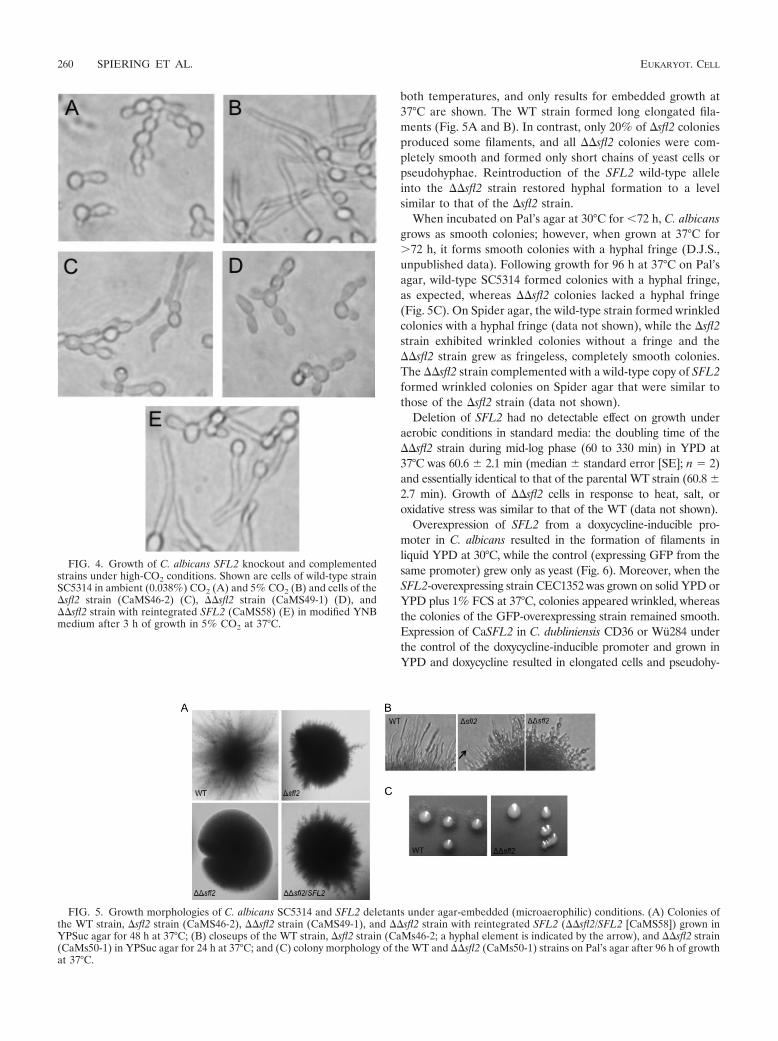
both temperatures, and only results for embedded growth at37°C are shown. The WT strain formed long elongated fila-ments (Fig. 5A and B). In contrast, only 20% of �sfl2 coloniesproduced some filaments, and all ��sfl2 colonies were com-pletely smooth and formed only short chains of yeast cells orpseudohyphae. Reintroduction of the SFL2 wild-type alleleinto the ��sfl2 strain restored hyphal formation to a levelsimilar to that of the �sfl2 strain.
When incubated on Pal’s agar at 30°C for �72 h, C. albicansgrows as smooth colonies; however, when grown at 37°C for72 h, it forms smooth colonies with a hyphal fringe (D.J.S.,unpublished data). Following growth for 96 h at 37°C on Pal’sagar, wild-type SC5314 formed colonies with a hyphal fringe,as expected, whereas ��sfl2 colonies lacked a hyphal fringe(Fig. 5C). On Spider agar, the wild-type strain formed wrinkledcolonies with a hyphal fringe (data not shown), while the �sfl2strain exhibited wrinkled colonies without a fringe and the��sfl2 strain grew as fringeless, completely smooth colonies.The ��sfl2 strain complemented with a wild-type copy of SFL2formed wrinkled colonies on Spider agar that were similar tothose of the �sfl2 strain (data not shown).
Deletion of SFL2 had no detectable effect on growth underaerobic conditions in standard media: the doubling time of the��sfl2 strain during mid-log phase (60 to 330 min) in YPD at37°C was 60.6 2.1 min (median standard error [SE]; n � 2)and essentially identical to that of the parental WT strain (60.8 2.7 min). Growth of ��sfl2 cells in response to heat, salt, oroxidative stress was similar to that of the WT (data not shown).
Overexpression of SFL2 from a doxycycline-inducible pro-moter in C. albicans resulted in the formation of filaments inliquid YPD at 30°C, while the control (expressing GFP from thesame promoter) grew only as yeast (Fig. 6). Moreover, when theSFL2-overexpressing strain CEC1352 was grown on solid YPD orYPD plus 1% FCS at 37°C, colonies appeared wrinkled, whereasthe colonies of the GFP-overexpressing strain remained smooth.Expression of CaSFL2 in C. dubliniensis CD36 or Wu284 underthe control of the doxycycline-inducible promoter and grown inYPD and doxycycline resulted in elongated cells and pseudohy-
FIG. 4. Growth of C. albicans SFL2 knockout and complementedstrains under high-CO2 conditions. Shown are cells of wild-type strainSC5314 in ambient (0.038%) CO2 (A) and 5% CO2 (B) and cells of the�sfl2 strain (CaMS46-2) (C), ��sfl2 strain (CaMS49-1) (D), and��sfl2 strain with reintegrated SFL2 (CaMS58) (E) in modified YNBmedium after 3 h of growth in 5% CO2 at 37°C.
FIG. 5. Growth morphologies of C. albicans SC5314 and SFL2 deletants under agar-embedded (microaerophilic) conditions. (A) Colonies ofthe WT strain, �sfl2 strain (CaMS46-2), ��sfl2 strain (CaMS49-1), and ��sfl2 strain with reintegrated SFL2 (��sfl2/SFL2 [CaMS58]) grown inYPSuc agar for 48 h at 37°C; (B) closeups of the WT strain, �sfl2 strain (CaMs46-2; a hyphal element is indicated by the arrow), and ��sfl2 strain(CaMs50-1) in YPSuc agar for 24 h at 37°C; and (C) colony morphology of the WT and ��sfl2 (CaMs50-1) strains on Pal’s agar after 96 h of growthat 37°C.
260 SPIERING ET AL. EUKARYOT. CELL

pha-like elements, whereas the GFP-expressing strains grewsolely as yeast cells (data not shown).
SFL2 is required for RHE tissue colonization and damageby C. albicans, but not for virulence in the mouse model ofsystemic infection. To see if SFL2 deletion affected coloniza-tion and tissue damage to the RHE, the SFL2 ko strains weretested in this model. Compared to the WT, which exhibitedextensive filamentation in the RHE, the �sfl2 strain showedonly few hyphal elements, and cells of the ��sfl2 strains werecompletely impaired in hyphal morphogenesis in the RHE;��sfl2 strains with reintegrated SFL2 showed filamentationlevels similar to those of the �sfl2 strain (Fig. 7). RHE damagemirrored the morphological phenotypes: i.e., sequential dele-tion of each SFL2 allele resulted in a gradual reduction oftissue damage: LDH release caused by �sfl2 strains was ap-proximately 40% of that of the WT, and LDH release by��sfl2 strains was about 50% of LDH release by �sfl2 strainsand significantly lower than that of the WT (P � 0.01; analysisof variance [ANOVA] and Tukey’s test), with RHE damagesimilar to that in the uninfected control. RHE damage by��sfl2 strains with reintegrated SFL2 was similar to that of the�sfl2 strains.
Finally, we tested ��sfl2 strains in in vivo mouse models ofsystemic infection, using the conventional 28-day model and a3-day infection model. In these experiments, virulence of the��sfl2 strain CaMS49-1 was very similar to that in the WT:survival times of mice inoculated with ��sfl2 or WT (SC5314)cells were 16.7 3.9 and 15.3 2.9 days (mean SE; n � 6),respectively (P 0.05; Kaplan-Meier and log rank statistics),with no significant differences (P 0.05; ANOVA) detectedfor Candida burdens in the kidney, spleen, or brain (data notshown). To examine whether changes in organ burdens wereevident at an earlier time point during infection, mice wereinfected and then sampled at 3 days postinfection. Again, therewas no significant difference in Candida burdens for the kid-ney, lung, liver, spleen, and brain (data not shown). Interest-ingly, histology of kidney sections obtained from the 3- and 28-dayinfection models revealed that the ��sfl2 strain was defectivein hypha formation, growing almost exclusively as yeast cells
FIG. 6. Overexpression of SFL2 in C. albicans. Strains CEC1352 (Table 2; indicated by TETp-SFL2), overexpressing SFL2 from a doxycycline-inducible promoter, and CEC1147 (Table 2; indicated by TETp-GFP), expressing GFP from a doxycycline-inducible promoter, were grown for 18 hin liquid YPD at 30°C or for 5 days on solid YPD or YPD plus 1% FCS at 37°C in the presence or absence of doxycycline (50 �g/ml).
FIG. 7. Growth morphology and tissue damage by C. albicans SFL2knockout and complemented strains in RHE. (A) Growth morphologyin RHE 33 h p.i. Hyphal elements present in SC5314 (WT) and the�sfl2 and ��sfl2/SFL2 strains are indicated by arrows. (B) RHE tissuedamage, measured as LDH release (means SE; n � 2 to 4) 25 h p.i.,by the WT and heterozygous (�) and homozygous (��) sfl2 strains(CaMS48-2 and CaMS49-1, respectively) and the complemented��sfl2 strain (CaMS60). One-way ANOVA detected a significant (P �0.011) effect on LDH release; Tukey’s test detected significant differ-ences in LDH release between the WT and ��sfl2 strains and theRHE-only control, indicated by asterisks (*, P � 0.05; **, P � 0.01).
VOL. 9, 2010 CANDIDA ALBICANS SFL2 261

with only a few short filaments (Fig. 8). Consistent with the invitro phenotypes, �sfl2 strain-infected renal lesions showedan intermediate phenotype, with some lesions containing longhyphae and others containing short filaments or yeasts (Fig. 8).
SFL2 encodes a putative DNA-binding heat shock factorprotein. BLASTP searches of the NCBI nonredundant (nr)protein database with the inferred Sfl2p sequence (714 aminoacids) gave a significant match with Hsr1p (E � 1e�51, 64%identity; alignment of 129 amino acids at the Sfl2p protein Nterminus), described as a heat-shock-related transcription factor(HSF) in Candida tropicalis (CAC12663), and WU-BLAST2searches of Saccharomyces cerevisiae YeastDB (at http://seq.yeastgenome.org) gave significant (E � 1e�18) matches to sev-eral HSF-type proteins, including Sfl1p (19 to 62% identity) andMga1p (25 to 28% identity). A highly conserved HSF-type DNA-binding domain (pfam00447; E � 6e�16) and an HSF-typeDNA-binding domain signature (PS00434; at amino acids 57 to81) were detected in the N-terminal region of Sfl2p, also presentat the same location (amino acids 58 to 82) in its putative C.dubliniensis orthologue, Cd36_54430p. Sfl2p shared about 24%identity and the HSF-type signature with its closest relative in C.albicans, Sfl1p (orf19.454), which is a negative regulator of hyphalmorphogenesis (3, 17).
DISCUSSION
The opportunistic pathogens C. albicans and C. dubliniensishave a close phylogenetic relationship and share several mor-
phophysiological traits. Despite this, they display large differ-ences in virulence, with C. albicans being much more patho-genic in the human host and in human infection models thanC. dubliniensis. Here, to identify novel virulence-associatedgenes in C. albicans and begin to characterize commonalitiesand differences in gene expression between the two species, wecompared global gene expression in C. albicans and C. dub-liniensis in the RHE model of the oral mucosa. Both speciesexhibited coordinated upregulation of primary metabolismgenes in RHE 30 min postinoculation, indicating conservedresponses in general metabolism in the two species. However,whereas C. albicans showed rapid filamentation and causedincreased RHE tissue damage, as well as upregulated expres-sion of several known hyphally regulated genes, C. dubliniensisgrew only as yeast cells, caused very limited RHE damage, andlacked detectable upregulation of hyphal genes. C. albicansalso displayed upregulation of several genes of unknown func-tion that were absent or significantly divergent in C. dublini-ensis. One such gene, SFL2 (orf19.3969), showed significantupregulation in C. albicans on the RHE 30 min p.i. and high(�50%) sequence divergence with its likely orthologue in C.dubliniensis (Cd36_54430). When SFL2 was deleted in C.albicans, cells were unable to form hyphae under a variety ofconditions, including growth under microaerophilic or high-CO2 conditions, in response to pH shifts, and in mouse kid-neys. Overexpression of SFL2 resulted in increased filamenta-tion in C. albicans and the production of pseudohypha-likecells in C. dubliniensis. Deletion of SFL2 in C. albicans had noeffect on mouse survival in the systemic infection model; how-ever, SFL2 deletion led to decreased RHE colonization anddamage, demonstrating a possible role for SFL2 in virulence inthis oral mucosal infection model.
The greater virulence of C. albicans and the limited viru-lence of C. dubliniensis were consistent with earlier observa-tions (12, 41) and with lower carriage and prevalence of C.dubliniensis in the oral cavities of healthy individuals (42, 43).C. albicans and C. dubliniensis displayed broadly similar pat-terns of coordinately upregulated expression of ribosomal andother primary metabolism (e.g., protein, amino acid biosynthe-sis, and mitochondrial) genes (Fig. 2; see Tables S1 and S2 inthe supplemental material), indicating high levels of metabolicactivity in both species in the RHE. So given their differencesin virulence in the RHE, it was surprising that growth initia-tion—as revealed by the temporal dynamics and magnitude ofexpression of the primary metabolism genes—appeared to beequally rapid in C. albicans and C. dubliniensis. Moreover, bothspecies exhibited very similar upregulation in the RHE of C2
utilization genes, such as genes for fatty acid �-oxidation,glyoxylate cycle, and gluconeogenesis, as well as heat shockprotein genes (see Tables S5 and S6 in the supplemental ma-terial). This pattern of gene expression in mammalian tissueshas previously been described only for C. albicans (46, 52), andupregulation of C2 utilization and stress response genes also inC. dubliniensis suggests common pathways for physiologicaladaptation to the RHE environment in the two species. Thus,we hypothesize that only a small set of physiological cues in theRHE, triggering filamentation in C. albicans while failing to doso in C. dubliniensis, may be responsible for the difference invirulence. Filamentation of C. albicans cells growing on thepolycarbonate filters used as RHE support matrix indicated
FIG. 8. Morphology of C. albicans SFL2 knockout strains in mousekidneys. Kidney sections (5 �m; 3 days postinfection) were stained withmethenamine silver and poststained with light green. The photographsshown represent two different magnifications. Panels A and B show theWT strain, panels C and D show the �sfl2 strain, and panels E and Fshow the ��sfl2 strain. Bars, 50 �m.
262 SPIERING ET AL. EUKARYOT. CELL

that hyphal morphogenesis in C. albicans was induced by thegeneral RHE growth conditions, such as levels of CO2 andcomposition of the media.
Our results indicated increased expression of many C. albi-cans virulence and hyphal genes, with lower or undetectableexpression of these genes in C. dubliniensis. The use of the C.albicans microarray could have potentially underestimated thenumber of expressed genes in C. dubliniensis, including expres-sion of several hyphal genes, where probes had lower specific-ity (�90% similarity) to the corresponding C. dubliniensis se-quences (see Table S2 in the supplemental material). While wecannot exclude the possibility that the lack of detectable ex-pression of these genes in C. dubliniensis was due to low probe-target similarity, the almost complete absence of hypha-asso-ciated gene expression in this species was consistent with itsgrowth morphology and lower virulence in the RHE model.
Hyphal morphogenesis is a pivotal process for colonizationand virulence in mucosal tissues by C. albicans (16, 26, 31) andsome C. dubliniensis mutants (23). Accordingly, the reductionin RHE damage by the ��sfl2 strains appeared to be due totheir inability to form hyphae in this model (Fig. 7), as no effectson biomass in the RHE and growth rates of the ��sfl2 strainswere observed. Key events and genes for epithelial coloniza-tion have been identified in C. albicans (29, 52), but less isknown about the factors that trigger filamentation in the RHE.CO2 at concentrations present in the oral cavity (48) inducefilamentation in C. albicans—but not in C. dubliniensis (23)—both in vitro (37) and in the RHE (14). As shown by Klengeland coworkers (14), CO2/HCO3
� directly stimulates activity ofadenylyl cyclase for cyclic AMP (cAMP) production and hy-phal morphogenesis in C. albicans. Therefore, it is noteworthythat apart from being unable to filament in the RHE, the��sfl2 strains were also impaired in CO2-induced and mi-croaerophilic (high-CO2 and low oxygen) filamentation (Fig. 4and 5). ��sfl2 strains were still capable of forming germ tubesin 10% serum, although at a slightly reduced rate when notemperature shift was applied. Along with the RHE-PCF com-parison, suggesting that SFL2 expression does not specificallyrespond to contact with the epithelial cells but rather to theenvironmental culture conditions in the RHE infection model,our results suggest that the inability of the ��sfl2 strains to col-onize and damage the RHE was due to their failure to filamentin response to the increased CO2 levels or microaerophilicconditions in the RHE. Therefore, it appears that Sfl2 does nothave a direct role in epithelial infection, but rather an indirectrole in pathogenesis in the RHE model by virtue of its effect onhypha formation in response to CO2 and microaerophilic con-ditions.
It is widely accepted that the ability to produce hyphae is animportant virulence factor in C. albicans, supported by obser-vations indicating that mutants that are defective in hyphalformation are usually less pathogenic in vitro than wild-typestrains (16, 26, 31, 52). SFL2 deletion had no detectable effecton survival in the mouse model of systemic infection. However,the finding that in the kidney tissues the ��sfl2 cells grewalmost exclusively in the yeast form was unexpected. This sug-gests that ��sfl2 cells have a capacity to infect mice and affectmouse survival similar to wild-type cells, despite the fact thatthe ��sfl2 strain does not produce hyphae in the kidney. Thepossibility that ��sfl2 cells formed hyphae only during the
early stages of infection and then reverted to the yeast formduring the later stages was not supported by our data, whichshowed that ��sfl2 strains grew only as yeast cells already atday 3 postinfection (Fig. 8). However, the possibility that ��sfl2cells might form hyphae prior to the 3-day time point cannot bediscounted. Another possible reason for the lack of reducedvirulence of ��sfl2 strains in the mouse models might be thatformation of hyphae was similar to WT C. albicans in organsother than the kidneys. The possibility that yeast cells alonecan cause disease and death in mice is intriguing; however,confirmation of this awaits further testing in infection modelexperiments in which a range of organs are examined for fun-gal morphology and burdens as well as inflammatory responsesduring an infection time course. Although ��sfl2 strains formedonly yeasts or short filaments, there was significant immunecell infiltration associated with the renal lesions (Fig. 8) (datanot shown). Since Candida virulence is correlated with theextent of immune cell infiltrate in the kidney (19), it is possiblethat the ��sfl2 strain induces damage in the host similar to theWT and, hence, is similarly virulent. As infection outcome isdetermined by early renal immune responses in this infectionmodel (19), it will be important in the future to examine thechemokine and cytokine responses to ��sfl2 and WT strains.
Similarities of the inferred Sfl2 protein sequence to heat shocktranscription factors and possession of a highly conserved heat-shock factor DNA-binding signature suggest that it may act asa transcriptional regulator. Sfl2p possesses sequence similarity,including the HSF-type DNA-binding domain, to Sfl1p, a nu-clear protein involved in regulation of microaerophilic mor-phogenesis (3, 17). However, unlike SFL2, SFL1 expression isconstant across different conditions (17) and Sfl1p suppressesC. albicans hyphal morphogenesis under several conditions,including microaerophilic growth (3). This suggests that thesetwo proteins possess opposing activities, but whether or notthey interact has yet to be determined. Given that deletion ofSFL2 abrogated hyphal morphogenesis in response to in-creased CO2 and shift to alkaline pH, Sfl2p may act down-stream of the cAMP and pH morphogenesis pathways. Furtherstudies are planned to investigate the roles of Sfl1p and Sfl2pin morphogenesis, including microarray analysis of single anddouble mutants and localization studies.
The lack of a CO2 response in C. dubliniensis (23) makes itdifficult to design experimental approaches to assign a role tothe likely SFL2 orthologue, Cd36_55430, in C. dubliniensis.While possession of a conserved HSF-type domain points tosome overlap in structure between Sfl2p and Cd36_55430p, theremainder of the sequences showed only �50% identity, anddeletion of Cd36_55430 gave no detectable effects on morpho-genesis in C. dubliniensis CD36 in microaerophilic conditions(M.J.S., unpublished results). That expression of SFL2 in C.dubliniensis triggered filamentation indicated that SFL2 acts asa hyphal inducer also in this species. It further suggested thatSFL2 and its orthologue, Cd36_55430, either have functionallydiverged or that Cd36_55430 expression is repressed in C.dubliniensis. Repression of Cd36_55430 under conditions thatinduce expression of SFL2 is indeed supported by very lowlevels of expression in the RHE of Cd36_55430 in C. dublini-ensis compared to expression of SFL2 in C. albicans (Fig. 3).
In conclusion, to our knowledge this study is the first that hascompared global gene expression in C. albicans and C. dub-
VOL. 9, 2010 CANDIDA ALBICANS SFL2 263

liniensis in a model of oral infection. Using standard conditionsfor RHE infection, we have established a baseline comparisonof gene expression in the two species in this important modeland identified SFL2 as a gene required for virulence in theRHE infection model. SFL2 was significantly upregulated dur-ing the very early stages of RHE colonization, and its deletiondrastically reduced filamentation and tissue damage of C.albicans in the RHE and also abolished filamentation in 5%CO2, in response to pH, and in microaerophilic conditions.This, along with the relationship of the predicted Sfl2 proteinto HSF-type DNA-binding transcription factors, suggests arole for Sfl2 as a regulator of filamentation and opens the wayfor detailed studies into the roles of Sfl2 in hyphal morpho-genesis and Candida virulence.
ACKNOWLEDGMENTS
We thank Jan Walker at St. James’s Hospital, Dublin, for fixationand staining of the RHE tissue sections; Joachim Morschhauser, Uni-versity of Wurzburg, for supplying plasmid pSFS2A; and Arnaud Fironfor the Gateway expression vector.
This work was supported by a grant from Science Foundation Ire-land (Programme Investigator grant no. 04/IN3/B463) to D. Sullivanand grants from the European Commission to C. d’Enfert. (GalarFungail 2 Marie Curie Research Training Network, MRTN-CT-2003-504148; FINSysB Marie Curie Initial Training Network, PITN-GA-2008-214004).
REFERENCES
1. Al Mosaid, A., D. J. Sullivan, and D. C. Coleman. 2003. Differentiation ofCandida dubliniensis from Candida albicans on Pal’s agar. J. Clin. Microbiol.41:4787–4789.
2. Arnaud, M. B., M. C. Costanzo, M. S. Skrzypek, P. Shah, G. Binkley, C.Lane, S. R. Miyasato, and G. Sherlock. 2007. Sequence resources at theCandida genome database. Nucleic Acids Res. 35:D452–D456.
3. Bauer, J., and J. Wendland. 2007. Candida albicans Sfl1 suppresses floccu-lation and filamentation. Eukaryot. Cell 6:1736–1744.
4. Braun, B. R., M. V. Hoog, C. d’Enfert, M. Martchenko, J. Dungan, A. Kuo,D. O. Inglis, M. A. Uhl, H. Hogues, M. Berriman, M. Lorenz, A. Levitin, U.Oberholzer, C. Bachewich, D. Harcus, A. Marcil, D. Dignard, T. Iouk, R.Zito, L. Frangeul, F. Tekaia, K. Rutherford, E. Wang, C. A. Munro, S. Bates,N. A. Gow, L. L. Hoyer, G. Kohler, J. Morschhauser, G. Newport, S. Znaidi,M. Raymond, B. Turcotte, G. Sherlock, M. Costanzo, J. Ihmels, J. Berman,D. Sanglard, N. Agabian, A. P. Mitchell, A. D. Johnson, M. Whiteway, andA. Nantel. 2005. A human-curated annotation of the Candida albicans ge-nome. PLoS Genet. 1:36–57.
5. Brown, D. H., A. D. Giusani, X. Chen, and C. A. Kumamoto. 1999. Filamen-tous growth of Candida albicans in response to physical environmental cuesand its regulation by the unique CZF1 gene. Mol. Microbiol. 34:651–662.
6. Delbruck, S., and J. F. Ernst. 1993. Morphogenesis-independent regulationof actin transcript levels in the pathogenic yeast Candida albicans. Mol.Microbiol. 10:859–866.
7. Dimopoulos, G., F. Ntziora, G. Rachiotis, A. Armaganidis, and M. E. Fal-agas. 2008. Candida albicans versus non-albicans intensive care unit-acquiredbloodstream infections: differences in risk factors and outcome. Anesth.Analg. 106:523–529.
8. Gilfillan, G. D., D. J. Sullivan, K. Haynes, T. Parkinson, D. C. Coleman, andN. A. R. Gow. 1998. Candida dubliniensis: phylogeny and putative virulencefactors. Microbiology 144:829–838.
9. Gillum, A. M., E. Y. H. Tsay, and D. R. Kirsch. 1984. Isolation of theCandida albicans gene for orotidine-5�-phosphate decarboxylase by comple-mentation of S. cerevisiae URA3 and Escherichia coli pyrF mutations Mol.Gen. Genet. 198:179–182.
10. Hokamp, K., F. M. Roche, M. Acab, M. E. Rousseau, B. Kuo, D. Goode, D.Aeschliman, J. Bryan, L. A. Babiuk, R. E. W. Hancock, and F. S. L. Brink-man. 2004. ArrayPipe: a flexible processing pipeline for microarray data.Nucleic Acids Res. 32:W457–W459.
11. Jackson, A. P., J. A. Gamble, T. Yeomans, G. P. Moran, D. Saunders, D.Harris, M. Aslett, J. F. Barrell, G. Butler, F. Citiulo, D. C. Coleman, P. W. J.de Groot, T. J. Goodwin, M. A. Quail, J. McQuillan, C. A. Munro, A. Pain,R. T. Poulter, M. A. Rajandream, H. Renauld, M. J. Spiering, A. Tivey,N. A. R. Gow, B. Barrell, D. J. Sullivan, and M. Berriman. 2009. Compar-ative genomics of the fungal pathogens Candida dubliniensis and C. albicans.Genome Res. 19:2231–2244.
12. Jayatilake, J., Y. H. Samaranayake, L. K. Cheung, and L. P. Samaranayake.
2006. Quantitative evaluation of tissue invasion by wild type, hyphal and SAPmutants of Candida albicans, and non-albicans Candida species in reconsti-tuted human oral epithelium. J. Oral Pathol. Med. 35:484–491.
13. Jones, T., N. A. Federspiel, H. Chibana, J. Dungan, S. Kalman, B. B. Magee,G. Newport, Y. R. Thorstenson, N. Agabian, P. T. Magee, R. W. Davis, andS. Scherer. 2004. The diploid genome sequence of Candida albicans. Proc.Natl. Acad. Sci. U. S. A. 101:7329–7334.
14. Klengel, T., W. J. Liang, J. Chaloupka, C. Ruoff, K. Schroppel, J. R. Naglik,S. E. Eckert, E. G. Mogensen, K. Haynes, M. F. Tuite, L. R. Levin, J. Buck,and F. A. Muhlschlegel. 2005. Fungal adenylyl cyclase integrates CO2 sensingwith cAMP signaling and virulence. Curr. Biol. 15:2021–2026.
15. Lee, K. L., C. C. Campbell, and H. R. Buckley. 1975. Amino-acid liquidsynthetic medium for the development of mycelial and yeast forms of Can-dida albicans. J. Med. Vet. Mycol. 13:148–153.
16. Lermann, U., and J. Morschhauser. 2008. Secreted aspartic proteases arenot required for invasion of reconstituted human epithelia by Candida albi-cans. Microbiology 154:3281–3295.
17. Li, Y., C. Su, X. Mao, F. Cao, and J. Chen. 2007. Roles of Candida albicansSfl1 in hyphal development. Eukaryot. Cell 6:2112–2121.
18. Liu, H. P., J. Kohler, and G. R. Fink. 1994. Suppression of hyphal formationin Candida albicans by mutation of a STE12 homolog. Science 266:1723–1726.
19. MacCallum, D. M., L. Castillo, A. J. P. Brown, N. A. R. Gow, and F. C. Odds.2009. Early-expressed chemokines predict kidney immunopathology in ex-perimental disseminated Candida albicans infections. PLoS One 4:e6420.
20. Martinez, M., J. L. Lopez-Ribot, W. R. Kirkpatrick, B. J. Coco, S. P. Bach-mann, and T. F. Patterson. 2002. Replacement of Candida albicans with C.dubliniensis in human immunodeficiency virus-infected patients with oropha-ryngeal candidiasis treated with fluconazole. J. Clin. Microbiol. 40:3135–3139.
21. McManus, B. A., D. C. Coleman, G. Moran, E. Pinjon, D. Diogo, M. E.Bougnoux, S. Borecka-Melkusova, H. Bujdakova, P. Murphy, C. d’Enfert,and D. J. Sullivan. 2008. Multilocus sequence typing reveals that the popu-lation structure of Candida dubliniensis is significantly less divergent thanthat of Candida albicans. J. Clin. Microbiol. 46:652–664.
22. Moran, G., C. Stokes, S. Thewes, B. Hube, D. C. Coleman, and D. Sullivan.2004. Comparative genomics using Candida albicans DNA microarrays re-veals absence and divergence of virulence-associated genes in Candida dub-liniensis. Microbiology 150:3363–3382.
23. Moran, G. P., D. M. MacCallum, M. J. Spiering, D. C. Coleman, and D. J.Sullivan. 2007. Differential regulation of the transcriptional repressor NRG1accounts for altered host-cell interactions in Candida albicans and Candidadubliniensis. Mol. Microbiol. 66:915–929.
24. Morschhauser, J., M. Ruhnke, S. Michel, and J. Hacker. 1999. Identificationof CARE-2-negative Candida albicans isolates as Candida dubliniensis. My-coses 42:29–32.
25. Murad, A. M. A., P. R. Lee, I. D. Broadbent, C. J. Barelle, and A. J. P. Brown.2000. CIp10, an efficient and convenient integrating vector for Candidaalbicans. Yeast 16:325–327.
26. Naglik, J. R., D. Moyes, J. Makwana, P. Kanzaria, E. Tsichlaki, G. Weindl,A. R. Tappuni, C. A. Rodgers, A. J. Woodman, S. J. Challacombe, M.Schaller, and B. Hube. 2008. Quantitative expression of the Candida albicanssecreted aspartyl proteinase gene family in human oral and vaginal candidi-asis. Microbiology 154:3266–3280.
27. Odds, F. C., M. F. Hanson, A. D. Davidson, M. D. Jacobsen, P. Wright, J. A.Whyte, N. A. R. Gow, and B. L. Jones. 2007. One year prospective survey ofCandida bloodstream infections in Scotland. J. Med. Microbiol. 56:1066–1075.
28. Odds, F. C., L. Van Nuffel, and G. Dams. 1998. Prevalence of Candidadubliniensis isolates in a yeast stock collection. J. Clin. Microbiol. 36:2869–2873.
29. Park, H., Y. Liu, N. Solis, J. Spotkov, J. Hamaker, J. R. Blankenship, M. R.Yeaman, A. P. Mitchell, H. Liu, and S. G. Filler. 2008. Transcriptionalresponses of Candida albicans to epithelial and endothelial cells. Eukaryot.Cell 8:1488–1510.
30. Park, Y. N., and J. Morschhauser. 2005. Tetracycline-inducible gene expres-sion and gene deletion in Candida albicans. Eukaryot. Cell 4:1328–1342.
31. Phan, Q. T., P. H. Belanger, and S. G. Filler. 2000. Role of hyphal formationin interactions of Candida albicans with endothelial cells. Infect. Immun.68:3485–3490.
32. Phan, Q. T., C. L. Myers, Y. Fu, D. C. Sheppard, M. R. Yeaman, W. H.Welch, A. S. Ibrahim, J. E. Edwards, and S. G. Filler. 2007. Als3 is a Candidaalbicans invasin that binds to cadherins and induces endocytosis by host cells.PLoS Biol. 5:543–557.
33. Pinjon, E., G. P. Moran, D. C. Coleman, and D. J. Sullivan. 2005. Azolesusceptibility and resistance in Candida dubliniensis. Biochem. Soc. Trans.33:1210–1214.
34. Reuß, O., A. Vik, R. Kolter, and J. Morschhauser. 2004. The SAT1 flipper,an optimized tool for gene disruption in Candida albicans. Gene 341:119–127.
35. Ritchie, M. E., J. Silver, A. Oshlack, M. Holmes, D. Diyagama, A. Holloway,
264 SPIERING ET AL. EUKARYOT. CELL

and G. K. Smyth. 2007. A comparison of background correction methods fortwo-colour microarrays. Bioinformatics 23:2700–2707.
36. Schaller, M., K. Zakikhany, J. R. Naglik, G. Weindl, and B. Hube. 2006.Models of oral and vaginal candidiasis based on in vitro reconstituted humanepithelia. Nat. Protoc. 1:2767–2773.
37. Sims, W. 1986. Effect of carbon dioxide on the growth and form of Candidaalbicans. J. Med. Microbiol. 22:203–208.
38. Slifkin, M. 2000. Tween 80 opacity test responses of various Candida species.J. Clin. Microbiol. 38:4626–4628.
39. Smyth, G. K. 2004. Linear models and empirical Bayes methods for assessingdifferential expression in microarray experiments. Stat. Appl. Genet. Mol.Biol. 3:Article 3.
40. Spiering, M. J., J. R. Faulkner, D.-X. Zhang, C. Machado, R. B. Grossman,and C. L. Schardl. 2008. Role of the LolP cytochrome P450 monooxygenasein loline alkaloid biosynthesis. Fungal Genet. Biol. 45:1307–1314.
41. Stokes, C., G. P. Moran, M. Spiering, G. T. Cole, D. C. Coleman, and D. J.Sullivan. 2007. Lower filamentation rates of Candida dubliniensis contributeto its lower virulence in comparison with Candida albicans. Fungal Genet.Biol. 44:920–931.
42. Sullivan, D. J., G. P. Moran, and D. C. Coleman. 2005. Candida dubliniensis:ten years on. FEMS Microbiol. Lett. 253:9–17.
43. Sullivan, D. J., G. P. Moran, E. Pinjon, A. Al-Mosaid, C. Stokes, C. Vaughan,and D. C. Coleman. 2004. Comparison of the epidemiology, drug resistancemechanisms, and virulence of Candida dubliniensis and Candida albicans.FEMS Yeast Res. 4:369–376.
44. Sullivan, D. J., T. J. Westerneng, K. A. Haynes, D. E. Bennett, and D. C.Coleman. 1995. Candida dubliniensis sp. nov.: phenotypic and molecular
characterization of a novel species associated with oral candidosis in HIV-infected individuals. Microbiology 141:1507–1521.
45. Thaweboon, S., B. Thaweboon, T. Srithavaj, and S. Choonharuangdej. 2008.Oral colonization of Candida species in patients receiving radiotherapy inthe head and neck area. Quintessence Int. 39:E52–E57.
46. Thewes, S., M. Kretschmar, H. Park, M. Schaller, S. G. Filler, and B. Hube.2007. In vivo and ex vivo comparative transcriptional profiling of invasive andnon-invasive Candida albicans isolates identifies genes associated with tissueinvasion. Mol. Microbiol. 63:1606–1628.
47. Tosello, M. E., M. S. Biasoli, A. G. Luque, H. M. Magaro, and A. R. Krapp.2007. Oxidative stress response involving induction of protective enzymes inCandida dubliniensis. Med. Mycol. 45:535–540.
48. Tsoukias, N. M., Z. Tannous, A. F. Wilson, and S. C. George. 1998. Single-exhalation profiles of NO and CO2 in humans: effect of dynamically changingflow rate. J. Appl. Physiol. 85:642–652.
49. Van Gelder, R. N., M. E. von Zastrow, A. Yool, W. C. Dement, J. D. Barchas,and J. H. Eberwine. 1990. Amplified RNA synthesized from limited quan-tities of heterogenous cDNA. Proc. Natl. Acad. Sci. U. S. A. 87:1663–1667.
50. Wilson, R. B., D. Davis, and A. P. Mitchell. 1999. Rapid hypothesis testingwith Candida albicans through gene disruption with short homology regions.J. Bacteriol. 181:1868–1874.
51. Wong, L., and C. H. Sissions. 2001. A comparison of human dental plaquemicrocosm biofilms grown in an undefined medium and a chemically definedartificial saliva. Arch. Oral Biol. 46:477–486.
52. Zakikhany, K., J. R. Naglik, A. Schmidt-Westhausen, G. Holland, M.Schaller, and B. Hube. 2007. In vivo transcript profiling of Candida albicansidentifies a gene essential for interepithelial dissemination. Cell. Microbiol.9:2938–2954.
VOL. 9, 2010 CANDIDA ALBICANS SFL2 265