pDNA loaded calcium phosphate nanoparticles: highly efficient non-viral vector for gene delivery

Synthesis and characterization of noscapine loaded magneticpolymeric nanoparticles
Mohamed O. Abdalla1,2, Ritu Aneja3, Derrick Dean4, Vijay Rangari5, Albert Russell6, JessieJaynes7, Clayton Yates1,2, and Timothy Turner1,2,*1Department of Biology, Tuskegee University2Center for Cancer Research, Tuskegee University3Department of Biology, Georgia State University4Department of Materials Science and Engineering, University of Alabama at Birmingham5Tuskegee-Center for Advanced Materials, Tuskegee University6Department of Chemistry, Tuskegee University7George Washington Carver Agricultural Experiment Station, Tuskegee University
AbstractThe delivery of noscapine therapies directly to the site of the tumor would ultimately allow higherconcentrations of the drug to be delivered, and prolong circulation time in vivo to enhance thetherapeutic outcome of this drug. Therefore, we sought to design magnetic based polymericnanoparticles for the site directed delivery of noscapine to invasive tumors. We synthesized Fe3O4nanoparticles with an average size of 10 ± 2.5 nm. These Fe3O4 NPs were used to prepare noscapineloaded magnetic polymeric nanoparticles (NMNP) with an average size of 252 ± 6.3 nm. Fouriertransform infrared (FT-IR) spectroscopy showed the encapsulation of noscapine on the surface ofthe polymer matrix. The encapsulation of the Fe3O4 NPs on the surface of the polymer was confirmedby elemental analysis. We studied the drug loading efficiency of polylactide acid (PLLA) and poly(L-lactide acid-co-gylocolide) (PLGA) polymeric systems of various molecular weights. Ourfindings revealed that the molecular weight of the polymer plays a crucial role in the capacity of thedrug loading on the polymer surface. Using a constant amount of polymer and Fe3O4 NPs, bothPLLA and PLGA at lower molecule weights showed higher loading efficiencies for the drug on theirsurfaces.
Keywordsnoscapine; magnetite; magnetic polymeric nanoparticles
1. IntroductionThe current challenges associated with systemic drug administration include evenbiodistribution of pharmaceuticals throughout the body, lack of drug specificity towards apathological site, necessity of a large dose to achieve high local concentration, non-specific
*Address correspondence Department of Biology and Center for Cancer Research, Tuskegee University, 100 Carver ResearchFoundation, Tuskegee, Alabama 36088. [email protected], Phone (334) 727-8787..PACS: 42.62.Be, 73.61.Ph, 73.61.Tm
NIH Public AccessAuthor ManuscriptJ Magn Magn Mater. Author manuscript; available in PMC 2011 January 1.
Published in final edited form as:J Magn Magn Mater. 2010 January 1; 322(2): 190–196. doi:10.1016/j.jmmm.2009.07.086.
NIH
-PA Author Manuscript
NIH
-PA Author Manuscript
NIH
-PA Author Manuscript
toxicity and adverse side effects due to high drug doses. Chemotherapy induced side effectscaused by some current chemotherapy drugs can be so severe that can lead to cessation of thetreatment plan for the patient. Therefore, development of drug-systems that could selectivelydeliver drug molecules to the tumor site, without a concurrent increase in its level in the normalhealthy cells of the tissues, is currently one of the most active areas of investigation in cancerresearch. Among the current attractive schemes of drug targeting, a promising one employsmagnetic guided nanoparticles [1;2;3;4;5;6;7]. This technology is based on the encapsulationof drug with magnetic nanoparticles or magnetic nanoparticles coupled to a polymeric carrier.The potential advantage with magnetically guided drug nanoparticles is that the strength of themagnetic field enables the nanoparticles to escape uptake by the reticular endothelial systemand multiple drug resistance pumps which are common factors for poor drug performance.Therefore, in recent years the scientific community has been seeking to exploit the intrinsicproperties of magnetic nanoparticles to obtain medical breakthroughs in both drug deliveryand diagnosis, with emphasis on applications related to cancer [8;9;10;11;12]. Nanoparticlescan act at the tissue or cellular level. The latter implies that they can be endocytosed orphagocytosed (i.e. by dendritic cells, macrophages) resulting in internalization of thenanoparticles. In this process, the nanoparticles can reach beyond the cytoplasmatic membraneand, in some cases, also beyond the nuclear membrane (i.e. transfection applications). Tumortargeting with magnetic nanoparticles may use passive or active strategies. Passive targetingoccurs as a result of extravasation of the nanoparticles at the diseased site (tumor) where themicrovasculature is hyperpermeable and leaky, a process aided by tumor-limited lymphaticdrainage. Combined, these factors lead to the selective accumulation of nanoparticles in tumortissue, a phenomenon known as enhanced permeation and retention (EPR) [10].
Another major advantage of using magnetic particles coupled to a biodegradable polymericcarrier is that the therapeutic drug concentrations are maintained for sustained periods of time.The polymeric matrix prevents drug degradation and also allows precise control over therelease kinetics of the drug from the nanoparticles. Moreover, the duration of drug levelsreleased from the nanoparticles can be easily modulated by altering formulation parameterssuch as drug: polymer ratio, or polymer molecular weight and composition [6;7;13;14;15;16;17;18;19;20]. Superparamagnetic iron oxide nanoparticles (Fe3O4 NPs) which are commonlyused to confer the magnetic property to any polymeric drug delivery systems are also used aspopular contrast agents for clinical diagnostics with magnetic resonance imaging (MRI) [21;22]. Therefore, most magnetic drug delivery systems supersede other targeted drug deliverysystems because of their dual property as a drug delivery system and an in vivo imaging system.
Noscapine, a phthalideisoquinoline antitussive alkaloid has been recently discovered for itstubulin-binding anticancer activity (Figure 1) [23;24;25]. Noscapine causes apoptosis in manycell types and has potent antitumor activity against solid murine lymphoid tumors and againsthuman breast and bladder tumors implanted in nude mice [23]. Although noscapine can inhibittumor growth and cause regression of tumor xenografts to a fair degree in preclinical micemodels, a complete obliteration of the disease has not been achieved even on increasingnoscapine dosage as high as 600 mg/kg, possibly due to saturation of its uptake by cancer cells.
We hypothesize that creating magnetically-guided noscapine nanoparticles will present a newmethod for administering this drug with high-targeting efficiency which will significantlyincrease its therapeutic index. While reports have not conclusively provided a reason for theremnant cancerous mass upon noscapine treatment, it may be attributed to the rapid eliminationof the drug and/or its widespread distribution into non targeted organs and tissues. Currently,clinical data (available on pcref.com) reveals that prostate cancer patients who consume off-label noscapine are on a regimen of about 1000 mg/day of the drug to achieve a lowering ofPSA (prostate specific antigen) levels. Although no detectable toxicity has been so far reported
Abdalla et al. Page 2
J Magn Magn Mater. Author manuscript; available in PMC 2011 January 1.
NIH
-PA Author Manuscript
NIH
-PA Author Manuscript
NIH
-PA Author Manuscript
with such enormously high doses of noscapine, consuming such large quantities of drug canpresent long-term side effects due to accumulation in healthy tissues.
Thus, this system will ensure the delivery of a concentrated dose of noscapine directly into thetumor, which will possibly eliminate the need for the patient to consume large quantities of thedrug. This system will have tremendous positive impacts, both physiological and economicalon cancer patients. Another advantage of this system is the possibility to use MRI to visualizedrug-loaded nanoparticles distribution in the tumor and its surrounding healthy tissues. Thisis very important because it leads to the next advantages such as: (i) pre-imaging of the drug-free nanoparticles distribution (a tracer dose) to enable a further good prediction of tumortargeting [26], (ii) monitoring during and after administration of the drug-loaded nanoparticlesleading to more efficacious treatment [27].
We present here the synthesis and characterization of noscapine loaded magnetic polymericnanoparticles using two polymeric systems [poly (L-lactide) and poly (DL-lactide-co-glycolide)] and also evaluate the efficiency of the drug loading on these polymers.
2. ExperimentalMaterials
Benzyl ether, 1,2-hexadecanediol, oleic acid, oleylamine, iron (III) acetylacetonate, poly (L-lactide) with variable average molecular weights [Mw = 100,000-150,000; and 55,000, labeledPLLA-HMW and PLLA-LMW, respectively], poly (DL-lactide-co-glycolide) (PLGA,lactide:glycolide 50:50) [Mw = 40,000-75,000 and 10,000-15,000, labeled PLGA-HMW andPLGA-LMW, respectively], polyvinyl alcohol (PVA) [Mw = 9,000-10,000, 80% hydrolyzed]were all purchased from Sigma Aldrich and stored as specified. All solvents used (hexane,ethanol (200 proof), dichloromethane, acetonitrile, ammonium acetate, water) were HPLCgrade and were purchased from Sigma Aldrich Co.
Synthesis of magnetiteOleic acid-stabilized Fe3O4 magnetic nanoparticles were prepared via a high temperaturereaction of iron acetylacetonate in phenyl ether in the presence of alcohol, oleic acid andoleylamine according to a reported method [28].
Synthesis of noscapine loaded magnetic nanoparticlesThe Fe3O4-containing polymeric nanoparticles were prepared by the solvent evaporation/extraction technique in o/w emulsion with PLLA or PLGA as the encapsulation material. 100mg of PLLA-HMW, 15 mg of Fe3O4 NPs and variable amounts of noscapine (2.5, 5, 7.5, 10,12.5 mg) were dissolved in 8 ml dichloromethane and vortexed for 10 min to make the organicphase. The amount of iron oxide which was used in the formulation was suggested by theliterature [11] as the optimum amount to avoid the competition between the iron oxide andnoscapine on the surface of the polymer. The organic phase was added to 12 ml of stirredaqueous solution containing 1% PVA as emulsifier. The mixture was sonicated for 120 s withan ultrasonic processor (Sonics, VCX 130 PB). The formed o/w emulsion was then stirred atroom temperature overnight with a magnetic stirrer to evaporate the organic solvent. TheNMNP were collected by centrifugation at 10,000 rpm for 10 min and washed three times withDI water. The nanoparticles were resuspended with 10 ml water and freeze-dried (Edwardsfreeze dryer, ESM 1342) for 2 days. The amount of noscapine was varied to quantify themaximum loading of the drug on the polymeric matrix. Based on the results from the aboveexperiment, the encapsulation method was repeated using only 7.5 mg of noscapine with 100mg of PLLA-LMW, PLGA-LMW, and PLGA-HMW.
Abdalla et al. Page 3
J Magn Magn Mater. Author manuscript; available in PMC 2011 January 1.
NIH
-PA Author Manuscript
NIH
-PA Author Manuscript
NIH
-PA Author Manuscript

Characterization of magnetite and the NMNPParticle size and surface properties—The synthesized Fe3O4 NPs were characterizedby X-ray diffraction (Rigaku D/MAX2000, Houston, TX). The XRD samples were preparedby dispersing the synthesized Fe3O4 NPs in hexane using an ultrasound sonicator andimmediately depositing the sample on a glass holder. After the hexane evaporated, the samplewas scanned from 2 to 80 °C. The size of the Fe3O4 NPs was estimated from their transmissionelectronic microscopy (TEM) micrographs using the National Institutes of Health (NIH)ImageJ analysis software. TEM was also used to perform elemental analysis on thesenanoparticles. The hydrodynamic size and size distribution of the Fe3O4 NPs were determinedby dynamic light scattering (DLS) method (Zetasizer nano ZS, Malvern Instruments, Malvern,United Kingdom). TheFe3O4 NPs were suspended in deionized (DI) water and sonicated for10 min to form a uniform dispersion; the DLS analysis was done at 25°C and at a 90°-detectionangle. TEM was also used to estimate the size and morphology of the prepared NMNP whileDLS was used to determine their hydrodynamic size and size distribution. Zeta potentialmeasurements (Zetasizer nano ZS, Malvern Instruments, Malvern, United Kingdom) weredone to determine the surface properties of NMNP. For particle size and surface chargemeasurements, the mean value of 5 readings was reported. Fourier transform infrared (FT-IR)spectroscopy was used to characterize the encapsulation of noscapine on the surface of theNMNP. The mid-IR spectra were recorded in the range between 650 and 4000 cm−1 from 20scans at a resolution of 4 cm−1 using a portable diamond ATR/FT-IR spectrometer(ThermoNicolet model Nexus 470 FTIR spectrometer, Waltham, MA) equipped with anattenuated total reflection (ATR) accessory). Measurements of magnetization of the Fe3O4 andNMNP were carried out with a Vibrating Sample Magnetometer (VSM, DMS 1600).
Noscapine encapsulation efficiency—The amount of noscapine incorporated in thenanoparticles produced was determined by high performance liquid chromatography (HPLC)assay. A known amount of the nanoparticles (3 mg) were placed in 5 mL of acetonitrile andleft on a shaker overnight. The solution was filtered through a 0.45 μm membrane filter beforeanalysis by HPLC assay using an Agilent 1100 HPLC system. A reverse phase C8 symmetrycolumn (Symmetry C8, 5 μm, Waters) was used as the stationary phase and the mobile phaseused was a mixture of 20 μM ammonium acetate solution and acetonitrile (65:35). The injectionvolume was 10 μL and the flow rate of the mobile phase was 1 ml/min. The column effluentwas monitored at 232 nm with a UV detector. The amount of noscapine in the nanoparticleswas determined from the calibration curve of the drug in acetonitrile. The encapsulationefficiency of the drug was calculated as the mass ratio of the amount of the drug entrapped innanoparticles to that used in the nanoparticles preparation.
3. RESULTSSynthesis of magnetite
Particle size plays a major role in the degree of magnetism of the material and thus theuniformity of response to external magnetic induction. Therefore, we utilized a synthesisprotocol that yielded magnetite nanoparticles with a narrow size distribution (within 10%) toensure that they will provide a uniform magnetic environment to efficiently direct thenanoparticles [28]. We studied the chemical structure of prepared magnetite by X-raydiffraction. Our results show that the prepared magnetite nanoparticles have six diffractionpeaks at 2θ = 29.99, 35.4, 43, 57, 62.5 (degrees) which correlate well with the characteristicspeaks of standard Fe3O4 crystal (isometric hexoctaheral crystal pattern) (Figure 2) [28]. Figure3 presents the graph of elemental analysis performed on the prepared Fe3O4 which confirmedthat the two main elements present on the sample are Fe and O. Elemental analysis detectedtraces of copper element which was attributed to the TEM copper grid and carbon element wasdetected due to hexane residues on the surface of the prepared Fe3O4.
Abdalla et al. Page 4
J Magn Magn Mater. Author manuscript; available in PMC 2011 January 1.
NIH
-PA Author Manuscript
NIH
-PA Author Manuscript
NIH
-PA Author Manuscript

The size of the prepared Fe3O4 NPs was estimated from their TEM micrographs at differentmagnifications (Figure 4 (a) and (b)). Figure 4 (a) illustrates that the particles have uniformsize of about 10 nm (estimated using National Institutes of Health (NIH) ImageJ software),which is comparable to the value reported in the literature [28]. The larger particles observedin the micrographs are simply aggregates of particles because at higher magnification the latticestructures of the separate particles can clearly be identified. The aggregations of thesenanoparticles are expected because of their very small size and magnetic property. Theestimated size and size distribution of the prepared Fe3O4 NPs was confirmed from the DLSdata (Table 1).
It is well known that Fe3O4 NPs with size less than 30 nm exhibit superparagnatism [10], whichis a desirable property in all magnetic drug delivery systems to avoid magnetic agglomerationonce the magnetic field is removed. We studied the magnetic property of the synthesizedFe3O4 NPs using a Vibrating Sample Magnetometer (VSM, DMS 1600) (see Figure 5 (a)).This curve represents a typical characteristic of superparamagnetic material with no detectedremainance or coercivity detected at room temperature. The saturation magnetization (σs) ofmagnetite was 55 which is typical value reported in the literature for this material [28]. Figure5 (b) shows that no remainance or coercivity was observed with the NMNP, which furtherconfirmed that the single-domain magnetite nanoparticles remain separated in the PLLAmatrix. The saturation magnetization value is approximately at a value of 1 for the NMNPprepared with 100 mg PLLA, 15 mg of Fe3O4 nanoparticles and 7.5 mg of noscapine.
Synthesis of noscapine loaded magnetic nanoparticles (NMNP)We prepared NMNP samples using the emulsification-evaporation method using 100 mg asthe polymeric carrier, 15 mg of the magnetite and 2.5, 5.0, 7.5, 10.0 and 12.5 mg of noscapine.The particles were labeled NMNP-2.5, NMNP-5.0, NMNP-7.5, NMNP-10, and NMNP-12.5with the suffix numbers present the amount of noscapine used in the formulation. The preparednanoparticles were characterized by TEM (Figure 5) to obtain information about themorphology and particle size of the particles). The particles were spherical in shape with anaverage size of about 250 nm. Elemental analysis obtained from the TEM showed the adsorbediron oxide nanoparticles on the surface of the polymeric carrier (data not shown).
With the aim to analyze the prepared NPs, FT-IR spectra of pure noscapine, pure PLLA andthe prepared NMNP were obtained (see Figure 8). In the fingerprint region range between 700and 1500 cm−1, noscapine’s spectrum (Figure 8 (a)) show numerous sharp bands which aremainly assigned to deformation and stretching vibrations of the alkaloid ring system.Noscapine also show strong band at 1759 which can be described at the C=O stretchingvibration, while the –C–O–C– stretching modes can be found at the 1029 cm−1 [29]. Thespectrum of PLLA (Figure 8 (b)) displays characteristic absorptions C=O stretching at 1756cm−1 and C–O stretch at 1000-1300 cm−1. Figure 8 (c) shows the spectrum of NMNP with thenoscapine’s spectrum subtracted from it. In figure 8 (c), PLLA sharp bands are observed at C–O stretch 1000-1300 cm−1 and C=O stretching at 1756 cm−1. Along with the PLLA sharpbands, figure 8 (c) shows all the characteristic bands for noscapine (noscapine characteristicbands are mostly shown in transmission bands due to subtraction between NMNP andnoscapine spectra). We observed in figure 8 (c), noscapine’s aromatic vibrational and stretchingbetween 700 and 1000 cm−1, and the –C–O–C– stretching band can be found as a small shoulderat 970-1000 cm−1. As expected, the absorptions for noscapine in NMNP are weaker since theweight percent ratio of the polymer to the drug is 14:1. The presence of the characteristic bandsfor noscapine and PLLA in the spectrum of the final nanoparticle complex (NMNP) confirmedsuccessful inclusion of noscapine in the polymer system. Figure 8 presents the elementalanalysis of the NMNP where the presence of the elements Fe and O on the surface of the NMNPconfirmed that the Fe3O4 NPs have been encapsulated on the surface of the polymer.
Abdalla et al. Page 5
J Magn Magn Mater. Author manuscript; available in PMC 2011 January 1.
NIH
-PA Author Manuscript
NIH
-PA Author Manuscript
NIH
-PA Author Manuscript

Drug loading efficiency on polymersTable 1 shows the results from the drug encapsulation procedure using 100 mg PLLA-HMW,15 mg of iron oxide nanoparticles and various amounts of noscapine. Entrapped noscapine onPLLA-HMW was measured at 1.23, 2.35, 9.5, 9.86, and 12.5% utilizing 2.5, 5, 7.5, 10, and12.5 mg of noscapine, respectively. These results showed that the compatibility of noscapinewith the 100mg of PLLA-HMW is maximal at a starting drug concentration not to exceed 7.5mg of noscapine. This conclusion was supported from our observation of free noscapineresiduals as white powder in the prepared nanoparticles when 10 and 12.5 mg of the drug wasused as starting amount of drug in the encapsulation process. Nonetheless, the 7.5 mg ofnoscapine utilized; resulted in about 0.75 mg of drug encapsulation in the PLLA-HMW matrix.Therefore, the encapsulation process was repeated using a low molecular weight PLLA whichshowed a drug loading of 19% suggesting that there is a favorite interaction between theabundant free carboxylic groups of the short chain PLLA with the methoxy groups in thenoscapine structure. The same trend was observed for the PLGA polymer as an improved drugloading efficiency was observed for the PLGA-LMW at 48% compared to 21 % for the PLGA-HMW (Table 2). This finding with the PLGA strengthened for us our suggestion about thefavorable interaction between the carboxylic groups of the short chain polymer with themethoxy groups of the noscapine.
4. ConclusionWe have successfully synthesized and characterized noscapine containing magnetic polymericnanoparticles using iron oxide nanoparticles, and two FDA approved polymeric systems-PLLA and PLGA. The average size of our prepared NMNP was about 250 nm which is wellwithin the acceptable range for the solvent evaporation/extraction technique in o/w emulsionmethod. FTIR spectra of PLLA, noscapine, and NMNP showed the encapsulation of the drugon the polymer surface while elemental analysis confirmed the presence of the Fe3O4 NPs onthe surface of the prepared NMNP. The drug loading efficiency of the drug on the twopolymeric systems was studied using HPLC and the drug entrapment was always higher in thecase of PLLA or PLGA with the lower molecular weights. Higher values of noscapineentrapment on the low molecular weight polymer were attributed to the favorable interactionbetween the noscapine methoxy groups and the higher number of free functional groups in thelow molecular weight PLLA and PLGA. We are currently studying the in vitro and the invivo efficacy of the PLGA system comparing its effectiveness to that of the free drug.
AcknowledgmentsThe project described was supported by Grant Numbers 3P20MD000195 (NIH/NCMHD), U54 CA118623 (NIH/NCI), and 2G12RR03059 (NIH/RCMI). The content is solely the responsibility of the authors and does not necessarilyrepresent the official views of the National Cancer Institute (NCI), National Center on Minority Health and HealthDisparities (NCMHD), Research Centers in Minority Institutions (RCMI) or the National Institutes of Health (NIH).
The authors would like to express their gratitude to Dr. David Nickles from Department of Chemistry, University ofAlabama for his assistance with acquiring and analyzing the magnetization data of the iron oxide nanoparticles.
References[1]. Alexiou C, Arnold W, Klein RJ, Parak FG, Hulin P, Bergemann C, Erhardt W, Wagenpfeil S, Lubbe
AS. Locoregional Cancer Treatment with Magnetic Drug Targeting. Cancer Research2000;60:6641–6648. [PubMed: 11118047]
[2]. Lubbe AS, Bergemann C, Brock J, McClure DG. Physiological aspects in magnetic drug-targeting.Journal of Magnetism and Magnetic Materials 1999;194:149–155.
[3]. Lubbe AS, Bergemann C, Riess H, Schriever F, Reichardt P, Possinger K, Matthias M, Dorken B,Herrmann F, Gurtler R, Hohenberger P, Haas N, Sohr R, Sander B, Lemke A-J, Ohlendorf D, Huhnt
Abdalla et al. Page 6
J Magn Magn Mater. Author manuscript; available in PMC 2011 January 1.
NIH
-PA Author Manuscript
NIH
-PA Author Manuscript
NIH
-PA Author Manuscript

W, Huhn D. Clinical Experiences with Magnetic Drug Targeting: A Phase I Study with 4′-Epidoxorubicin in 14 Patients with Advanced Solid Tumors 1996;56:4686–4693.
[4]. Lubbe AS, Bergemann C, Huhnt W, Fricke T, Riess H, Brock JW, Huhn D. Preclinical Experienceswith Magnetic Drug Targeting: Tolerance and Efficacy 1996;56:4694–4701.
[5]. Widder KJ, Senyei AE, Ranney DF. Magnetically responsive microspheres and other carriers for thebiophysical targeting of antitumor agents. Advances in Pharmacology and Chemotherapy1979;16:213–271. [PubMed: 382799]
[6]. Shaffer C. Nanomedicine transforms drug delivery. Drug Discovery Today 2005;10:1581–1582.[PubMed: 16376810]
[7]. Cheng MM-C, Cuda G, Bunimovich YL, Gaspari M, Heath JR, Hill HD, Mirkin CA, Nijdam AJ,Terracciano R, Thundat T, Ferrari M. Nanotechnologies for biomolecular detection and medicaldiagnostics. Current Opinion in Chemical Biology 2006;10:11–19. [PubMed: 16418011]
[8]. Novakova AA, Lanchinskaya VY, Volkov AV, Gendler TS, Kiseleva TY, Moskvina MA, Zezin SB.Magnetic properties of polymer nanocomposites containing iron oxide nanoparticles. Journal ofMagnetism and Magnetic Materials 2003;258-259:354–357.
[9]. Dandamudi S, Campbell RB. The drug loading, cytotoxicty and tumor vascular targetingcharacteristics of magnetite in magnetic drug targeting. Biomaterials 2007;28:4673–4683.[PubMed: 17688940]
[10]. Arruebo M, Fenandez-Pacheco R, Ibarra MR, Santamaria J. Magnetic nanoparticles for drugdelivery. nanotoday 2007;2:22–32.
[11]. Hu FX, Neoh KG, Kang ET. Synthesis and in vitro anti-cancer evaluation of tamoxifen-loadedmagnetite/PLLA composite nanocomposites. Biomaterials 2006;27:5725–5733. [PubMed:16890989]
[12]. Fonseca C, Simoes S, Gasper R. Paclitaxel-loaded PLGA nanoparticles: preparation,physiochemical characterization, and in vitro anti-tumoral activity. Journal of Controlled Release2002;83:273–286. [PubMed: 12363453]
[13]. Park K. Nanotechnology: What it can do for drug delivery. Journal of Controlled Release2007;120:1–3. [PubMed: 17532520]
[14]. Nishiyama N. Nanomedicine: Nanocarriers shape up for long life. Nat Nano 2007;2:203–204.[15]. Morrow KJ Jr, Bawa R, Wei C. Recent Advances in Basic and Clinical Nanomedicine. Medical
Clinics of North America 2007;91:805–843. [PubMed: 17826104][16]. Gu FX, Karnik R, Wang AZ, Alexis F, Levy-Nissenbaum E, Hong S, Langer RS, Farokhzad OC.
Targeted nanoparticles for cancer therapy. Nano Today 2007;2:14–21.[17]. Vasir JK, Labhasetwar V. Biodegradable nanoparticles for cytosolic delivery of therapeutics.
Advanced Drug Delivery Reviews 2007;59:718–728. [PubMed: 17683826][18]. Emerich DF, Thanos CG. The pinpoint promise of nanoparticle-based drug delivery and molecular
diagnosis. Biomolecular Engineering 2006;23:171–184. [PubMed: 16843058][19]. Couvreur P, Gref R, Andrieux K, Malvy C. Nanotechnologies for drug delivery: Application to
cancer and autoimmune diseases. Progress in Solid State Chemistry 2006;34:231–235.[20]. Moghimi SM, Hunter AC, Murray JC. Nanomedicine: current status and future prospects. FASEB
J 2005;19:311–330. [PubMed: 15746175][21]. Neuberger T, Schöpf B, Hofmann H, Hofmann M, von Rechenberg B. Superparamagnetic
nanoparticles for biomedical applications: Possibilities and limitations of a new drug deliverysystem. Journal of Magnetism and Magnetic Materials 2005;293:483–496.
[22]. Gupta AK, Gupta M. Synthesis and surface engineering of iron oxide nanoparticles for biomedicalapplications. Biomaterials 2005;26:3995–4021. [PubMed: 15626447]
[23]. Ye K, Ke Y, Keshava N, Shanks J, Kapp JA, Tekmal RR, Petros J, Joshi HC. Opium alkaloidnoscapine is an antitumor agent that arrests metaphase and induces apoptosis in dividing cells.Proceedings of the National Academy of Sciences 1998;95:1601–1606.
[24]. Ke Y, Ye K, Grossniklaus HE, Archer DR, Joshi HC, Kapp JA. Noscapine inhibits tumor growthwith little toxicity to normal tissues or inhibition of immune responses. Cancer Immunology,Immunotherapy 2000;49(Numbers 45):217–225.
Abdalla et al. Page 7
J Magn Magn Mater. Author manuscript; available in PMC 2011 January 1.
NIH
-PA Author Manuscript
NIH
-PA Author Manuscript
NIH
-PA Author Manuscript

[25]. Landen JW, Lang R, McMahon SJ, Rusan NM, Yvon A-M, Adams AW, Sorcinelli MD, CampbellR, Bonaccorsi P, Ansel JC, Archer DR, Wadsworth P, Armstrong CA, Joshi HC. Noscapine AltersMicrotubule Dynamics in Living Cells and Inhibits the Progression of Melanoma 2002:4109–4114.
[26]. Roullin VG, Deverre JR, Lemaire L, Hindre F, Venier-Julienne MC, Vienet RBJP. Anti-cancer drugdiffusion within living rat brain tissue: An experimental study using [3H](6)-5-fluorouracil-loadedPLGA microspheres. European Journal of Pharmaceutics and Biopharmaceutics 2002;53:293–299.[PubMed: 11976017]
[27]. Seppenwoolde, J.F.W.N.L.W.B.S.W.Z.A.D.v.h.S.C.J.G.B. Jan-Henry Internal radiation therapy ofliver tumors: Qualitative and quantitative magnetic resonance imaging of the biodistribution ofholmium-loaded microspheres in animal models. Magnetic Resonance in Medicine 2005;53:76–84. [PubMed: 15690505]
[28]. Sun S, Zeng H, Robinson DB, Raoux S, Rice PM, Wang SX, Li G. Monodisperse MFe2O4 (M =Fe, Co, Mn) Nanoparticles. J. Am. Chem. Soc 2004;126:273–279. [PubMed: 14709092]
[29]. Schulz H, Baranska M, Quilitzsch R, Schütze W. Determination of alkaloids in capsules, milk andethanolic extracts of poppy (Papaver somniferum L.) by ATR-FT-IR and FT-Raman spectroscopy.Analyst 2004;129:917–920. [PubMed: 15457323]
Abdalla et al. Page 8
J Magn Magn Mater. Author manuscript; available in PMC 2011 January 1.
NIH
-PA Author Manuscript
NIH
-PA Author Manuscript
NIH
-PA Author Manuscript
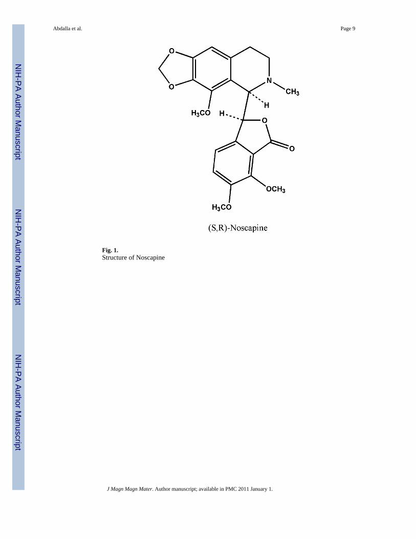
Fig. 1.Structure of Noscapine
Abdalla et al. Page 9
J Magn Magn Mater. Author manuscript; available in PMC 2011 January 1.
NIH
-PA Author Manuscript
NIH
-PA Author Manuscript
NIH
-PA Author Manuscript

Fig. 2.XRD pattern of synthesized magnetite shows a typical pattern for Fe3O4 NPs crystal structurewith six characteristics peaks.
Abdalla et al. Page 10
J Magn Magn Mater. Author manuscript; available in PMC 2011 January 1.
NIH
-PA Author Manuscript
NIH
-PA Author Manuscript
NIH
-PA Author Manuscript

Fig. 3.Elemental analysis of the Fe3O4 NPs which verified that the main elements in the NPs are Feand O. Copper (Cu) element was detected as the sample holder is made of Cu and Carbon wasdetected due to the hexane traces on the surface of the Fe3O4 NPs.
Abdalla et al. Page 11
J Magn Magn Mater. Author manuscript; available in PMC 2011 January 1.
NIH
-PA Author Manuscript
NIH
-PA Author Manuscript
NIH
-PA Author Manuscript

Fig. 4.TEM micrographs on synthesized magnetite, (a) bar = 10 nm, shows that bulk of thenanoparticles synthesized are of uniform size with few aggregates (b) bar = 5 nm scale, amagnified image of shows that an aggregate is a collection of nanoparticles with different latticestructures.
Abdalla et al. Page 12
J Magn Magn Mater. Author manuscript; available in PMC 2011 January 1.
NIH
-PA Author Manuscript
NIH
-PA Author Manuscript
NIH
-PA Author Manuscript

Fig. 5.Field dependent magnetization at 25°C for (a) Fe3O4 NPs and (b) NMNP (prepared with 100mg PLLA, 15 mg Fe3O4 and 7.5 mg noscapine).
Abdalla et al. Page 13
J Magn Magn Mater. Author manuscript; available in PMC 2011 January 1.
NIH
-PA Author Manuscript
NIH
-PA Author Manuscript
NIH
-PA Author Manuscript

Fig. 6.TEM micrographs of NMNP prepared with 100 mg PLLA, 15 mg, Fe3O4 NPs and 7.5 mgnoscapine a) bar = 1.0 um, shows that bulk of the nanoparticles synthesized are of uniform sizewith few aggregates (b) bar = 50 nm scale, a magnified image of shows a spherical shapedNMNP with an approximate size of 250 nm.
Abdalla et al. Page 14
J Magn Magn Mater. Author manuscript; available in PMC 2011 January 1.
NIH
-PA Author Manuscript
NIH
-PA Author Manuscript
NIH
-PA Author Manuscript
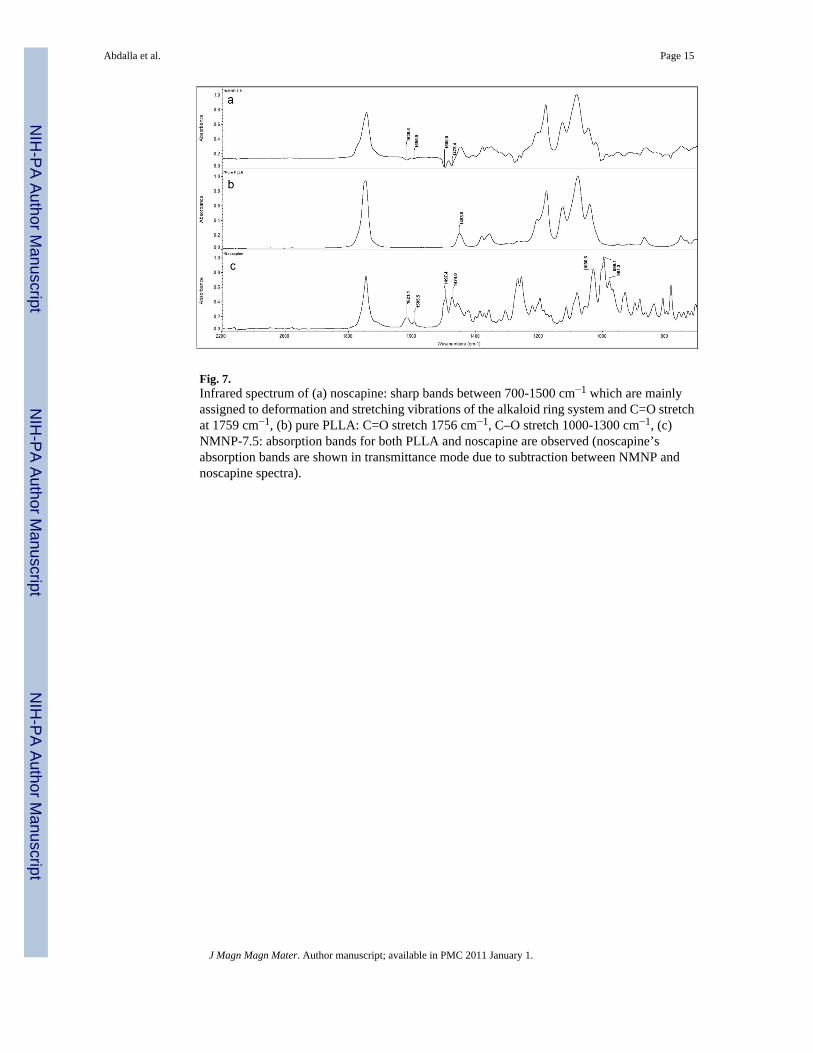
Fig. 7.Infrared spectrum of (a) noscapine: sharp bands between 700-1500 cm−1 which are mainlyassigned to deformation and stretching vibrations of the alkaloid ring system and C=O stretchat 1759 cm−1, (b) pure PLLA: C=O stretch 1756 cm−1, C–O stretch 1000-1300 cm−1, (c)NMNP-7.5: absorption bands for both PLLA and noscapine are observed (noscapine’sabsorption bands are shown in transmittance mode due to subtraction between NMNP andnoscapine spectra).
Abdalla et al. Page 15
J Magn Magn Mater. Author manuscript; available in PMC 2011 January 1.
NIH
-PA Author Manuscript
NIH
-PA Author Manuscript
NIH
-PA Author Manuscript
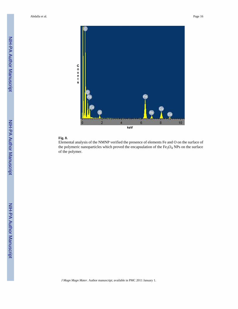
Fig. 8.Elemental analysis of the NMNP verified the presence of elements Fe and O on the surface ofthe polymeric nanoparticles which proved the encapsulation of the Fe3O4 NPs on the surfaceof the polymer.
Abdalla et al. Page 16
J Magn Magn Mater. Author manuscript; available in PMC 2011 January 1.
NIH
-PA Author Manuscript
NIH
-PA Author Manuscript
NIH
-PA Author Manuscript

NIH
-PA Author Manuscript
NIH
-PA Author Manuscript
NIH
-PA Author Manuscript
Abdalla et al. Page 17
Table 1
Size and zeta potential characterization of prepared Fe3O4 NPs and NMNP
Nanoparticles types Fe3O4 NMNP*
Hydrodynamic Size (nm)(TEM) 10 ± 2.5 252 ± 6.3
Hydrodynamic Size (nm)*(DLS)
12.1 ± 5.2 243 ± 23
Zeta Potential (mV) ------ −15.7 ± 2.4
*Prepared with 100 mg of PLLA-HMW, 15 mg Fe3O4 NPs,7.5 mg noscapine. Data represents mean ± SD, n = 3
J Magn Magn Mater. Author manuscript; available in PMC 2011 January 1.

NIH
-PA Author Manuscript
NIH
-PA Author Manuscript
NIH
-PA Author Manuscript
Abdalla et al. Page 18
Tabl
e 2
Nos
capi
ne e
ncap
sula
tion
data
usi
ng P
LLA
-HM
W
Sam
ple
NM
NP-
2.5*
NM
NP-
5N
MN
P-7.
5N
MN
P-10
NM
NP-
12.5
% D
rug
load
ing
0.9
2.1
9.2
10.1
12.9
* All
sam
ples
wer
e pr
epar
ed w
ith 1
00 m
g of
PLL
A-H
MW
, 15
mg
Fe3O
4 N
Ps, s
uffix
pre
sent
s the
am
ount
of n
osca
pine
in m
g. D
ata
repr
esen
ts m
ean
± SD
, n =
3.
J Magn Magn Mater. Author manuscript; available in PMC 2011 January 1.

NIH
-PA Author Manuscript
NIH
-PA Author Manuscript
NIH
-PA Author Manuscript
Abdalla et al. Page 19
Table 3
Noscapine encapsulation data using PLLA-LMW, PLGA-LMW, PLGA-HMW
Sample PLLA-LMW* PLGA-HMW PLGA-LMW
% Drugloading
18.2 22 48
*All samples were prepared using 100 mg of the polymer, 15 mg of iron oxide nanoparticles and 7.5 mg of noscapine. Data represents mean ± SD, n
= 3.
J Magn Magn Mater. Author manuscript; available in PMC 2011 January 1.
Copyright © 2022 FDOKUMEN