pDNA loaded calcium phosphate nanoparticles: highly efficient non-viral vector for gene delivery
12
International Journal of Pharmaceutics 288 (2005) 157–168 Pharmaceutical Nanotechnology pDNA loaded calcium phosphate nanoparticles: highly efficient non-viral vector for gene delivery Savita Bisht, Gajadhar Bhakta, Susmita Mitra, Amarnath Maitra ∗ Department of Chemistry, University of Delhi, Delhi 110007, India Received 13 May 2004; accepted 30 July 2004 Available online 21 November 2004 Abstract Nanoparticles of calcium phosphate encapsulating plasmid DNA (pDNA) of size 100–120 nm in diameter were prepared. XRD studies of these nanoparticles showed them to be crystalline in nature having hydroxyapatite structure. The maximum loading of pDNA and its release from nanoparticles were studied using gel electrophoresis. The time dependent size measurement of these particles demonstrated that these particles show strong aggregational behaviour in aqueous dispersion. Calcium phosphate nanoparticles were found to be dissolved even in low acidic buffer (pH 5.0) releasing the pDNA, which suggested that DNA release from these particles in the endosomal compartment was possible. In vitro transfection efficiency of these calcium phosphate nanoparticles was found to be higher than that of the commercial transfecting reagent Polyfect. © 2004 Elsevier B.V. All rights reserved. Keywords: Calcium phosphate nanoparticles; Non-viral vectors; Gene delivery; Transfection efficiency 1. Introduction The emerging area of inorganic nanoparticles en- trapping biomolecules has exhibited its diversity and potential applications in many frontiers of modern ma- terial science (Avnir et al., 1994; Dong and Chen, 2002). These inorganic particles have a number of ad- vantages over organic ones, in the sense that they are not usually subjected to microbial attack and exhibit ∗ Corresponding author. Tel.: +91 11 725 7995; fax: +91 11 725 6593. E-mail address: [email protected] (A. Maitra). excellent storage stability. They can be prepared at low temperature and are relatively inexpensive. These inorganic compounds have been explored in various biomedical applications such as adjuvant for vacci- nation process, component in dental hygiene agents, carriers for various proteins and other growth factors, drug delivery and gene therapy vectors (Frey et al., 1997; Tischer et al., 2002; Kneuer et al., 2000; Luo and Saltzman, 2000b; Cui and Mumper, 2003). Of these inorganic compounds, the most commonly used salt is calcium phosphate whose microparticles and nanopar- ticles have been developed as delivery system as well as adjuvant for DNA vaccines (He et al., 2000, 2002). Cal- 0378-5173/$ – see front matter © 2004 Elsevier B.V. All rights reserved. doi:10.1016/j.ijpharm.2004.07.035
Transcript of pDNA loaded calcium phosphate nanoparticles: highly efficient non-viral vector for gene delivery
International Journal of Pharmaceutics 288 (2005) 157–168
Pharmaceutical Nanotechnology
pDNA loaded calcium phosphate nanoparticles: highly efficientnon-viral vector for gene delivery
Savita Bisht, Gajadhar Bhakta, Susmita Mitra, Amarnath Maitra∗
Department of Chemistry, University of Delhi, Delhi 110007, India
Received 13 May 2004; accepted 30 July 2004Available online 21 November 2004
Abstract
Nanoparticles of calcium phosphate encapsulating plasmid DNA (pDNA) of size 100–120 nm in diameter were prepared. XRDstudies of these nanoparticles showed them to be crystalline in nature having hydroxyapatite structure. The maximum loadingof pDNA and its release from nanoparticles were studied using gel electrophoresis. The time dependent size measurement ofthese particles demonstrated that these particles show strong aggregational behaviour in aqueous dispersion. Calcium phosphatenanoparticles were found to be dissolved even in low acidic buffer (pH 5.0) releasing the pDNA, which suggested that DNA releasefrom these particles in the endosomal compartment was possible. In vitro transfection efficiency of these calcium phosphatenanoparticles was found to be higher than that of the commercial transfecting reagent Polyfect.© 2004 Elsevier B.V. All rights reserved.
K
1
tpt2vn
f
d atese
iouscci-ents,ors,,
alt ispar-ell as
0
eywords:Calcium phosphate nanoparticles; Non-viral vectors; Gene delivery; Transfection efficiency
. Introduction
The emerging area of inorganic nanoparticles en-rapping biomolecules has exhibited its diversity andotential applications in many frontiers of modern ma-
erial science (Avnir et al., 1994; Dong and Chen,002). These inorganic particles have a number of ad-antages over organic ones, in the sense that they areot usually subjected to microbial attack and exhibit
∗ Corresponding author. Tel.: +91 11 725 7995;ax: +91 11 725 6593.
E-mail address:[email protected] (A. Maitra).
excellent storage stability. They can be preparelow temperature and are relatively inexpensive. Thinorganic compounds have been explored in varbiomedical applications such as adjuvant for vanation process, component in dental hygiene agcarriers for various proteins and other growth factdrug delivery and gene therapy vectors (Frey et al.1997; Tischer et al., 2002; Kneuer et al., 2000; Luoand Saltzman, 2000b; Cui and Mumper, 2003). Of theseinorganic compounds, the most commonly used scalcium phosphate whose microparticles and nanoticles have been developed as delivery system as wadjuvant for DNA vaccines (He et al., 2000, 2002). Cal-
378-5173/$ – see front matter © 2004 Elsevier B.V. All rights reserved.doi:10.1016/j.ijpharm.2004.07.035

158 S. Bisht et al. / International Journal of Pharmaceutics 288 (2005) 157–168
cium ions are known to form ionic complexes with thehelical phosphates of DNA and these complexes haveeasy transportability across the cell membrane via ion-channel mediated endocytosis (Truong-Le et al., 1999).Although the use of precipitated calcium phosphate forin vitro transfection (James and Grosveld, 1987) is aroutine laboratory procedure, the method is hamperedby the difficulty of applying it to in vivo studies, espe-cially delivery of DNA to any particular cell type. Dueto bulk precipitation of calcium phosphate, the methodalso suffers from variation in calcium phosphate-DNAparticle size that causes variation among experiments(Luo and Saltzmann, 2000).
In the use of different types of cationic liposome(Hirko et al., 2003; Kawakami et al., 2002; Audouy etal., 2002), cationic polymers and dendrimers (Chonnand Cullis, 1998; Maurer et al., 1999; Han et al., 2000;Hashida et al., 2001) as non-viral vectors for delivery ofgenes, it has been observed that, in addition to cytotox-icity, these carriers do not lead to satisfactory amountof gene expression in the cells. The reasons are many,particularly, low endosomal escape, no protection ofDNA from nuclease degradation and inefficient nuclearuptake (Orrantia and Chang, 1990). It was envisagedthat the use of inorganic nanoparticles, a largely unex-plored synthetic system that could be used as vectorsfor gene delivery, might eliminate many of these limita-tions. The phosphate salts of Ca2+, Mg2+, Mn2+, Ba2+,etc. have never been explored as non-viral vectors forgene delivery although some works have been reportedo2 ta ri
theu ump andh ellsaMp pro-t n ef-fi bea gedu n oft thatp ghtb ncy.
The probable loss of pDNA, therefore, led us to mod-ify the preparative method in which the ultrasonicationwas completely avoided to enhance the efficiency ofthe transfection process.
In this paper we report an optimized method ofpreparation and a more detailed physicochemicalcharacterization including the in vitro transfectionstudies of calcium phosphate nanoparticles. ThesepDNA-loaded nanoparticles have been characterizedby their size, surface morphology, crystal structure,surface charge, aggregational behaviour and pHdependent pDNA release. We have also reported thatby following the soft preparative method (by avoidingultrasonication), much-improved in vitro transfectionefficiency of these particles in HeLa cell lines usingpSV�gal as a marker plasmid, has been observed.
2. Experimental
2.1. Materials
Surfactant, Aerosol OT (or AOT), i.e. bis (2-ethylhexyl) sulphosuccinate, of analytical grade, wasobtained as a 99% pure product from Sigma, USA.n-Hexane (AR grade), was purchased from SRL (India).Analytical grade agarose, calcium chloride, disodiumhydrogen phosphate, gel-loading dye (bromophenolblue), ethidium bromide were procured from ACROS(USA). Fetal calf serum (FCS) was purchased fromS ),a cinB ur-c em-i allyap cteda ofA un-d r ofD thC byP
2
sism
n the in vitro use of silica nanoparticles (Luo et al.,004; Chen et al., 2003) inorganic nanorods (Salem el., 2003), nanotubes (Sameti et al., 2003) and othe
norganic compounds (Xiang et al., 2003).Based on this idea we have recently reported
se of plasmid DNA (pDNA) encapsulated in calcihosphate nanoparticles as DNA delivery carriersave specifically targeted these particles to liver cfter appropriate surface modification (Roy et al., 2003,aitra et al., 2003; Bhakta et al., 2003). Although theDNA entrapped in these nanoparticles was highly
ected from enzymatic degradation, the transfectiociency of these synthetic systems was found tobout 80% of that exhibited by superfect. As prolonltra-sonication was a prerequisite for re-dispersio
hese particles in aqueous buffer, it was envisagederhaps partial disintegration of DNA molecules mie a plausible reason for lower transfection efficie
igma-Aldrich (USA). Cell culture media (DMEMntibiotics penicillin, streptomycin and amphoteriwere obtained from Genetix, India. X-Gal was p
hased from Genei, Bangalore, (India). All other chcals used were of the high purity grade commercivailable from the local market. pUC19,pSV�gal andEGFP-N1 (expressing GFP protein) were extrand purified in the laboratory of Dr Daman SalujaCBR, Delhi University. Transfection studies wereertaken in the tissue culture unit of Prof D. Sarkaepartment of Biochemistry, University of Delhi Souampus using HeLa cell lines, which were giftedrof Sarkar.
.2. Plasmid isolation by alkaline lysis method
Plasmid isolation was done using alkaline lyethod (Sambrook and Russell, 2001). The detailed

S. Bisht et al. / International Journal of Pharmaceutics 288 (2005) 157–168 159
method is as follows: 1–5 ml culture was centrifugedat 10,000 rpm for 5 min at 4◦C. Alkaline lysis of pel-leted cells was carried out, followed by treatment ofthe cell lysate with potassium acetate solution. Afterincubation in ice for 5 min, the lysed cells were cen-trifuged at 10,000 rpm for 5 min at 4◦C. Five hundredmicrolitres of 1:1 (v/v) of phenol and chloroform mix-ture was added to the supernatant, mixed thoroughlyand centrifuged at 10,000 rpm for 5 min at 4◦C. To500�l of this mixture, 500�l of isopropanol was addedand centrifuged at 10,000 rpm for 5 min at 4◦C. Pelletcontaining DNA was washed twice with 70% ethanoland dried in air. The pellet was dissolved in Tris–EDTAbuffer followed by RNase treatment.
2.3. Preparation of pUC 19, pSV�gal andpEGFP-N1 DNA loaded calcium phosphatenanoparticles
The basic method of preparation of pDNA loadednanoparticles of calcium phosphate is similar to that re-ported in our earlier communication (Roy et al., 2003),but with some modifications. More specifically the de-tails of the method is as follows: 0.1 M AOT solutionin hexane was prepared. In 25 ml of AOT in hexane,70�l aqueous solution of 1.36 M calcium chloride andaqueous solution of 2.94�g of pDNA, were addedby continuous stirring for 12 h to form microemulsionA. In another 25 ml of AOT solution, 50�l of 0.2 MTris–HCl buffer (pH 7.4), 70�l aqueous solution of0p 2 ht i-c totalv rr nsw as,t lowrT an-o ento n int ex-a olu-t asso nol( edf e
(Sigma 3K18). The pelleted nanoparticles were washedwith absolute ethanol three times. Finally, the pelletednanoparticles were dispersed in double distilled waterat 4◦C by vortexing to give clear dispersion. The dis-persed nanoparticles were dialysed overnight in a coldroom using 12 kD dialysis membrane bag, and werelyophilized to dry powder (yield∼1 mg) for furthercharacterization.
2.4. Size and morphology of the nanoparticles
2.4.1. Dynamic light scattering (DLS)In aqueous dispersion, these particles aggregate very
rapidly to form the bigger particles. Therefore, the sizeof the primary particles formed within the aqueous coreof microemulsion droplets were determined by DLSexperiment.
The measurements were done with a BrookhavenBI8000 instrument fitted with a BI200SM goniome-ter. An argon-ion air-cooled laser was operated at488 nm as a light source and the measurements weredone on a scattering angle of 90◦. The time de-pendent autocorrelation function was derived usinga 128-channel digital photon correlator. The particlesize was calculated from the autocorrelation functionusing Stokes–Einstein equation:d=kt/3�ηD, whereD= translational diffusion coefficient,d= particle di-ameter,η = viscosity of the liquid in which parti-cles were suspended,k= Boltzmann’s constant andT= absolute temperature. The particle size of calciump ffer asw werea
2arti-
c cidw tiono hlo-r riedg cope(
2
t4 h aP ed
.35 M Na2HPO4 and aqueous solution of 2.94�g ofDNA, were dissolved by continuous stirring for 1
o form microemulsion B. Before stirring both the mroemulsions, excess water was added to makeolume of water to 450�l, to adjustwo, i.e. the molaatio of water to AOT at 10. Both the microemulsioere optically clear solutions. Microemulsion B w
hen, added to microemulsion A at an extremely sate (20 drops per min) with continuous stirring at 4◦C.he resulting solution was, then, further stirred forther 12 h in the above cold condition. Developmf translucency indicated the nanoparticle formatio
he aqueous core of the microemulsion droplets. Hne was completely removed from the resulting s
ion, and the nanoparticles containing the solid mf AOT was dissolved in 10 ml of absolute etha99.9%) by vortexing. The solutions were centrifugor half an hour at 8000 rpm at 4◦C in a cold centrifug
hosphate nanoparticles dispersed in aqueous buell as the size of the aggregated nanoparticleslso measured by DLS.
.4.2. Transmission electron microscopy (TEM)One drop of the aqueous dispersion of nanop
les followed by one drop of 1% phosphotungstic aere put on a forvmar coated copper grid (1% soluf forvmar was prepared in spectroscopic grade coform) and air dried in a vacuum desiccator. The drid was then examined under an electron microsJEOL JEM 2000 EX 200 model).
.5. X-ray diffractogram (XRD) of nanoparticles
The X-ray source used was Cu K� radiation a0 kV and 20 mA, and diffraction was analyzed witHILIPS PW 3710 diffractometer. 0.2 g of lyophiliz
160 S. Bisht et al. / International Journal of Pharmaceutics 288 (2005) 157–168
nanoparticles was put inside the diffractometer foranalysis.
2.6. Determination of plasmid loading capacity
In order to determine the maximum loading in cal-cium phosphate nanoparticles, different amounts ofpUC19 DNA, (from 3 to 9.0�g) were added in themicroemulsions at the time of preparation of nanopar-ticles, keeping the total amount of metal ion and phos-phate ion concentrations same for all the cases. Thesenanoparticles dispersed in water were then subjected togel electrophoresis. After saturated loading, the excesspDNA remained as unentrapped free pDNA in aqueousbuffer, which was visible as distinct bands in the gel.
2.7. Entrapment efficiency (E%)
The pDNA-loaded nanoparticles in reverse micelleswere separated after ultra centrifugation (40× 103 rpmfor 4 h at 4◦ C) and the pellets after washing withhexane was dissolved in acidic buffer (pH = 3.0). Theamount of pDNA released, [pDNA]r from the nanopar-ticles was estimated spectrophotometrically by mea-suring the OD atλ = 260 nm. The entrapment efficiencywas then calculated from the amount of pDNA origi-nally added in the microemulsion, ([pDNA]0), usingthe equationE% = [pDNA]r/[pDNA]0 × 100.
2.8. Gel electrophoresis experiments
lec-t int ased DNAf lec-t s(c d them -t thes thew
2
asers cal
Microscope). The microscope settings were as follows:excitation at 488 nm, emission at 507 nm/30 BP intochannel 1 to record GFP fluorescence, 63× 1.4 NAoil-immersion lens at an Airy disc setting of 0.9. Whennecessary, serial images were taken, with 1.2�m stepsto get an overall image of fluorescent objects withinthe cell. The experiments were done at the Departmentof Pathology, All India Institute of Medical Sciences,New Delhi.
2.10. In vitro transfection studies in HeLa cell line
For in vitro studies,HeLa cell line were trans-fected with free form as well as encapsulated plasmid,pSV�gal. For these studies,HeLacell lines were grownin DMEM medium containing 10% fetal calf serum,antifungal drug, Amphotericin B and 1.6% (w/v) peni-cillin and streptomycin solutions. Twelve well plateswere seeded with 1× 10⁵ cells per well, after check-ing for cell viability using trypan blue exclusion, andappropriate dilution. Cells were grown under standardconditions for 24 h till 80% confluency. 1ml of aque-ous dispersion of pDNA loaded calcium phosphatenanoparticles (total amount of pDNA = 588 ng) wasadded in each well. After an appropriate time inter-val of 4 h, the added material was replaced with freshmedium containing 10% (v/v) FCS and antibiotics, andincubated for another 36 h. After 36 h, cells were lysedwith 200�l of lysis buffer (pH 7.4) containing 250 mMTris–HCl and 0.5% Triton X-100. Ten microlitres of X-G tionw hen,s -a ureds n-t roma usO
3
d int ane.T ump ousc thep
One percent Agarose gel was used for erophoresis experiments. The entrapment of DNAhe nanoparticles, its protection from external Dnegradation and the pH dependent release of p
rom these nanoparticles were investigated by gel erophoresis. In each set of 100�l of aqueous bufferpH = 5 and 8), 5 mg of lyophilizedpSV�gal loadedalcium phosphate nanoparticles was dispersed anixtures were incubated at 37◦ C for specific time in
ervals (0, 1, 2, 4 and 24 h). Twenty microlitre ofample was removed from each mixture, loaded inell and was subjected to electrophoresis.
.9. Confocal microscopy
HeLa cells preparations were observed with a lcanning confocal microscope (Biorad 206 Confo
al solution was added to each well and the reacas continued for another 36 h. The reaction was, ttopped by the addition of 50�l of 1M sodium carbonte solution. The blue colour developed was measpectrophotometrically atλmax= 616 nm and the quaity of �-galactosidase produced were calculated f
calibration curve (amount of�-galactosidase versD at 616 nm).
. Results and discussion
Nanoparticles encapsulating pDNA were formehe aqueous core of the AOT microemulsion in hexhe strategy involved the precipitation of the calcihosphate in the presence of pDNA in the aqueore of the microemulsion droplets. A flow chart ofreparative method adopted is represented byFig. 1.

S. Bisht et al. / International Journal of Pharmaceutics 288 (2005) 157–168 161
Fig. 1. Flowchart for the preparation of pDNA loaded calcium phosphate nanoparticles.
The centrifuged pellets could be easily redispersedin aqueous buffer. The maximum amount of pDNA,which could be loaded into the nanoparticles, waschecked by preparing samples with different amount ofpDNA and finally subjecting the aqueous dispersionsof these particles to electrophoresis on 1% agarosegel. It was clearly observed that 5.88�g of pDNA permg of calcium phosphate could be easily loaded in thenanoparticles. To measure the entrapment efficiencyof calcium phosphate nanoparticles, 1mg of pUC19loaded nanoparticles was dissolved in pH = 3.0 bufferby overnight incubation and the amount of pDNAreleased was estimated spectrophotometrically at
260 nm and the entrapment efficiency (E%) was foundto be more than 99%.
The physiochemical characteristics and the aggrega-tion behaviour of the calcium phosphate particles werestudied using DLS, TEM and X-Ray diffractometry.The size of the nanoparticles formed was found to bedependent onwo values (i.e. the molar ratio of water toAOT). The mean size distribution of calcium phosphatenanoparticles while dispersed in microemulsuion atwo = 10 was in the range of 30–40 nm.Fig. 2is a repre-sentative size distribution profile of calcium phosphatenanoparticles encapsulatingpSV�gal in AOT mi-croemulsion. The particle size, while dispersed in mi-

162 S. Bisht et al. / International Journal of Pharmaceutics 288 (2005) 157–168
Fig. 2. Representative DLS of pDNA loaded calcium phosphatenanoparticles in microemulsion.
croemulsion, depends, however, on the time of stirring.Instead of 12 h of stirring as reported in the present pro-tocol, if the microemulsion was stirred for 24–36 h, theparticle size was found to be increased from 30–40nmto about 80–90 nm diameter. The size of the particlesin aqueous dispersion was also measured and it wasfound to be in the range of about 100–130 nm diameter.The time dependent size measurement studies at 20◦ C(Fig. 3) clearly indicated that the particles aggregatedrapidly with time (more than 100 nm jump after 24 h)thus leading to the bigger size of the particles.
Fig. 3. Time dependent aggregation studies of pDNA loaded calciumphosphate nanoparticles in aqueous medium at 20◦ C.
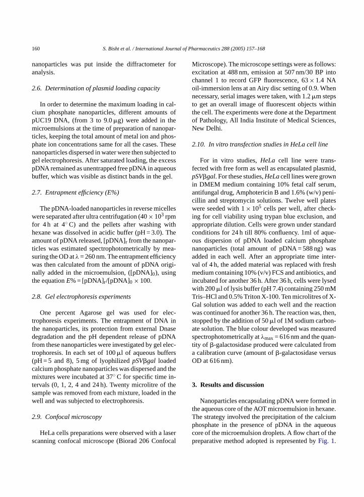
The transmission electron micrographs of pUC 19loaded calcium phosphate nanoparticles (Fig. 4a) andvoid calcium phosphate nanoparticles (Fig. 4b) re-vealed the formation of dense particles with sphericalmorphology.
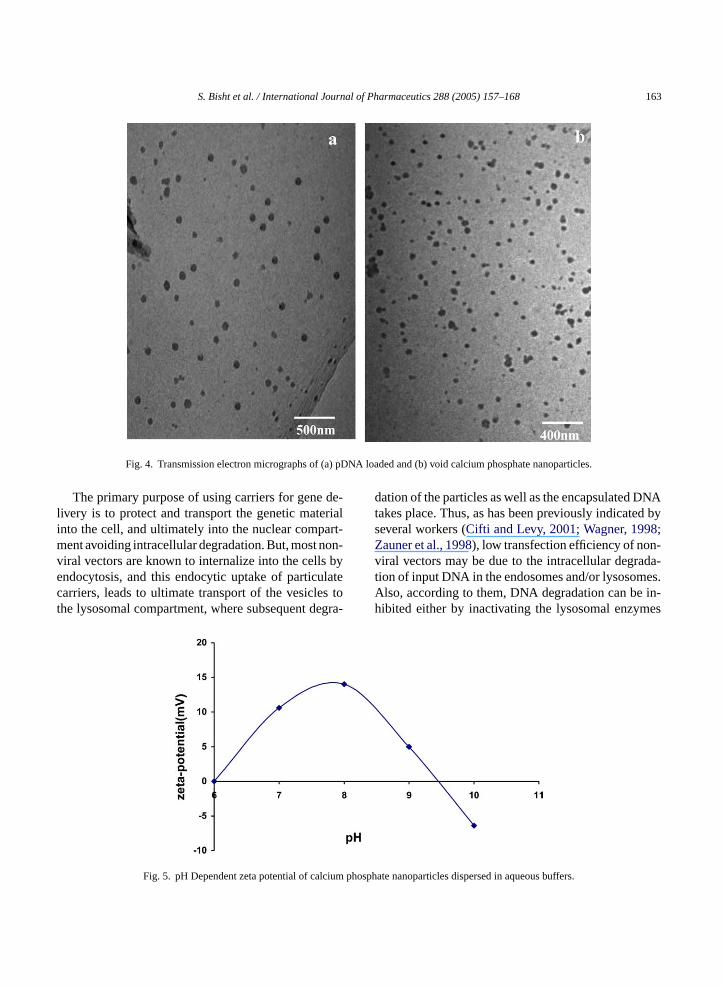
The surface charge of calcium phosphate nanoparti-cles was determined by measuring the zeta potential inthe pH range between 6.0 and 10.0. The pH dependentzeta potential as shown inFig. 5indicated that the par-ticles were positively charged in neutral aqueous bufferand were turned negatively charged in strongly alkalinesolution above pH∼9.5.
The crystallinity of the calcium phosphate nanopar-ticles was studied by means of an X-ray powderdiffractometry. Fig. 6a and b show the diffractionpatterns of pUC19 encapsulated as well as voidcalcium phosphate nanoparticles respectively. Thediffractograms obtained for void and DNA loadedparticles indicated the formation of hydroxyapatite[Ca10(PO4)6(OH)2] crystals as evidenced from thecharacteristic peak at 2θ = 31.8 degree (Shimabayashiet al., 1995).
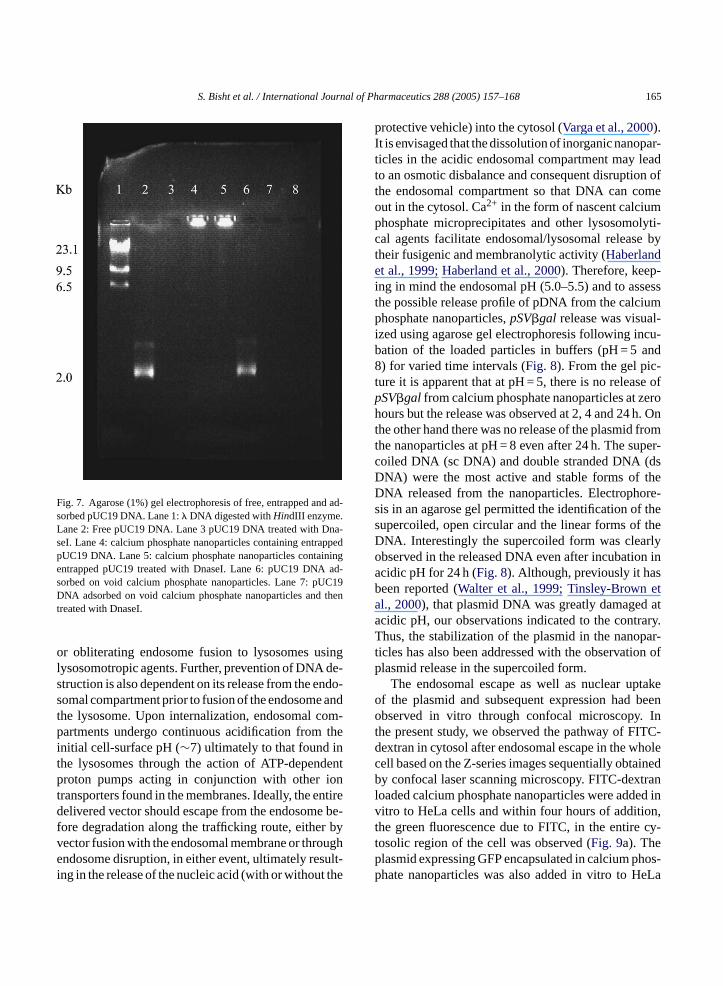
One of the primary reasons for the low transfectionefficiency obtained with non-viral vectors, includingthe precipitated bulk calcium phosphate is the incom-plete protection of the plasmid DNA by the encapsulat-ing material. Such partial protection makes the DNAhighly susceptible to aggressive DNase attack in thebody as well as inside the cell. In order to check the levelof protection being offered to the encapsulated DNA,w es toe ore-sf si-t then rdlym elyd smidw con-t e oft levelo owa da-t thatD trixo bledt rnalD
e subjected the calcium phosphate nanoparticlxtensive DNase treatment followed by electrophis on 1% agarose gel (Fig. 7). We found that whileree plasmid DNA (pUC 19) moved at its usual poion in the gel, pUC19 encapsulated in the matrix ofanoparticle was right at the top of the gel and haoved. Moreover, while free pDNA was completigested by DNase treatment, encapsulated plaas totally protected. As expected, this was quite
rary to the plasmid DNA adsorbed on the surfache nanoparticles. In this case, we found that thef protection offered to the DNA was extremely lnd the DNA was highly prone to nearly total degra
ion by DNase. These results clearly demonstratedNA was completely encapsulated in the rigid maf the calcium phosphate nanoparticles, which ena
he particles to protect the nucleic acid from exteNase environment.
S. Bisht et al. / International Journal of Pharmaceutics 288 (2005) 157–168 163
Fig. 4. Transmission electron micrographs of (a) pDNA loaded and (b) void calcium phosphate nanoparticles.
The primary purpose of using carriers for gene de-livery is to protect and transport the genetic materialinto the cell, and ultimately into the nuclear compart-ment avoiding intracellular degradation. But, most non-viral vectors are known to internalize into the cells byendocytosis, and this endocytic uptake of particulatecarriers, leads to ultimate transport of the vesicles tothe lysosomal compartment, where subsequent degra-
dation of the particles as well as the encapsulated DNAtakes place. Thus, as has been previously indicated byseveral workers (Cifti and Levy, 2001; Wagner, 1998;Zauner et al., 1998), low transfection efficiency of non-viral vectors may be due to the intracellular degrada-tion of input DNA in the endosomes and/or lysosomes.Also, according to them, DNA degradation can be in-hibited either by inactivating the lysosomal enzymes
Fig. 5. pH Dependent zeta potential of calcium phosphate nanoparticles dispersed in aqueous buffers.

164 S. Bisht et al. / International Journal of Pharmaceutics 288 (2005) 157–168
Fig. 6. Powder XRD patterns for (a) pDNA loaded and (b) void calcium phosphate nanoparticles.
S. Bisht et al. / International Journal of Pharmaceutics 288 (2005) 157–168 165
Fig. 7. Agarose (1%) gel electrophoresis of free, entrapped and ad-sorbed pUC19 DNA. Lane 1:� DNA digested withHindIII enzyme.Lane 2: Free pUC19 DNA. Lane 3 pUC19 DNA treated with Dna-seI. Lane 4: calcium phosphate nanoparticles containing entrappedpUC19 DNA. Lane 5: calcium phosphate nanoparticles containingentrapped pUC19 treated with DnaseI. Lane 6: pUC19 DNA ad-sorbed on void calcium phosphate nanoparticles. Lane 7: pUC19DNA adsorbed on void calcium phosphate nanoparticles and thentreated with DnaseI.
or obliterating endosome fusion to lysosomes usinglysosomotropic agents. Further, prevention of DNA de-struction is also dependent on its release from the endo-somal compartment prior to fusion of the endosome andthe lysosome. Upon internalization, endosomal com-partments undergo continuous acidification from theinitial cell-surface pH (∼7) ultimately to that found inthe lysosomes through the action of ATP-dependentproton pumps acting in conjunction with other iontransporters found in the membranes. Ideally, the entiredelivered vector should escape from the endosome be-fore degradation along the trafficking route, either byvector fusion with the endosomal membrane or throughendosome disruption, in either event, ultimately result-ing in the release of the nucleic acid (with or without the
protective vehicle) into the cytosol (Varga et al., 2000).It is envisaged that the dissolution of inorganic nanopar-ticles in the acidic endosomal compartment may leadto an osmotic disbalance and consequent disruption ofthe endosomal compartment so that DNA can comeout in the cytosol. Ca2+ in the form of nascent calciumphosphate microprecipitates and other lysosomolyti-cal agents facilitate endosomal/lysosomal release bytheir fusigenic and membranolytic activity (Haberlandet al., 1999; Haberland et al., 2000). Therefore, keep-ing in mind the endosomal pH (5.0–5.5) and to assessthe possible release profile of pDNA from the calciumphosphate nanoparticles,pSV�gal release was visual-ized using agarose gel electrophoresis following incu-bation of the loaded particles in buffers (pH = 5 and8) for varied time intervals (Fig. 8). From the gel pic-ture it is apparent that at pH = 5, there is no release ofpSV�gal from calcium phosphate nanoparticles at zerohours but the release was observed at 2, 4 and 24 h. Onthe other hand there was no release of the plasmid fromthe nanoparticles at pH = 8 even after 24 h. The super-coiled DNA (sc DNA) and double stranded DNA (dsDNA) were the most active and stable forms of theDNA released from the nanoparticles. Electrophore-sis in an agarose gel permitted the identification of thesupercoiled, open circular and the linear forms of theDNA. Interestingly the supercoiled form was clearlyobserved in the released DNA even after incubation inacidic pH for 24 h (Fig. 8). Although, previously it hasbeen reported (Walter et al., 1999; Tinsley-Brown eta ata ary.T par-t on ofp
takeo beeno Int ITC-d holec inedb tranl ed inv n,t cy-tp hos-p eLa
l., 2000), that plasmid DNA was greatly damagedcidic pH, our observations indicated to the contrhus, the stabilization of the plasmid in the nano
icles has also been addressed with the observatilasmid release in the supercoiled form.
The endosomal escape as well as nuclear upf the plasmid and subsequent expression hadbserved in vitro through confocal microscopy.
he present study, we observed the pathway of Fextran in cytosol after endosomal escape in the well based on the Z-series images sequentially obtay confocal laser scanning microscopy. FITC-dex
oaded calcium phosphate nanoparticles were additro to HeLa cells and within four hours of additiohe green fluorescence due to FITC, in the entireosolic region of the cell was observed (Fig. 9a). Thelasmid expressing GFP encapsulated in calcium phate nanoparticles was also added in vitro to H

166 S. Bisht et al. / International Journal of Pharmaceutics 288 (2005) 157–168
Fig. 8. Agarose (1%) gel electrophoresis to show DNA release at (a) pH 5.0 and (b) pH 8.0. Lane 1 (in a and b):� DNA digested withHindIII enzyme. Lane 2 (in a and b): freepSV�galDNA. Lane 3–7 (in a and b): DNA released on incubation at 37◦C from calcium phosphatenanoparticles after 0, 1, 2, 4 and 24 h.
Fig. 9. (a) Fluorescence picture of FITC-Dx loaded calcium phosphate nanoparticles incubated with HeLa cells. The picture was taken 4 h afterthe addition of nanoparticles. (b–d) Time dependent pEGFP-N1 expression in HeLa cells (b) 12 h, (c) 20 h and (d) 24 h.

S. Bisht et al. / International Journal of Pharmaceutics 288 (2005) 157–168 167
Fig. 10. In vitro transfection studies in HeLa cell-line.
cells and the expression was seen. It was observed(Fig. 9b–d) that, in the initial 12 h, there was practi-cally no GFP expression. But after 12 h of incubationthe green fluorescence appeared which increased withtime indicating more and more of expression of theplasmid.
To estimate the transfection efficiency on mam-malian cells using these nanoparticles, Hela was se-lected andpSV�galwas chosen as a marker DNA. Forcomparing the extent of transfection a standard labora-tory transfecting reagent, Polyfect (a polyamidoaminedendrimer), was used. From the results (Fig. 10), itcould be seen that the calcium phosphate nanoparticlesshowed higher transfection efficiency than exhibited byPolyfect. Considering that Polyfect is a highly compe-tent vector used for in vitro transfection (Tang et al.,1996), our results indicate that under in vitro condi-tions calcium phosphate nanoparticles are more effec-tive as non-viral vectors than Polyfect. In our previouscommunication (Roy et al., 2003) the transfection ef-ficiency of calcium phosphate nanoparticles was notedto be less as compared to that of the Polyfect reagent.The reason could be attributed, besides others, to thepartial degradation of DNA due to ultrasonication.
4. Conclusion
Calcium phosphate nanoparticles encapsulating dif-ferent plasmids were prepared and characterized. The
size dependent studies of these particles clearly showedthe high tendency to aggregation resulting in theirlarger size. The plasmid DNA was well protected in-side these nanoparticles and these particles were easilydissolved in mild acidic environment releasing DNA.In vitro transfection studies indicated that the transfec-tion efficiency of these carriers was as high as Polyfect,a commercially available transfecting reagent. We pre-sume that these calcium phosphate nanoparticles havewell-defined role in DNA delivery so far as endosomalescape, protection against nuclease degradation and nu-clear uptake are concerned and, therefore, can be usedas an effective non-viral vector in gene therapy.
Acknowledgements
The authors thank the Department of Science andTechnology, Government of India for financial assis-tance in the form of a research project. Thanks arealso due to Prof D.Sarkar, Department of Biochem-istry, University of Delhi South Campus for allowingus to use his tissue culture facility. We thank Dr DamanSaluja of Ambedkar Centre of Biomedical Research(ACBR), Delhi University, for providing us facilitiesfor extraction and purification of plasmids and Prof.A.K.Dinda of AIIMS, New Delhi for assisting in con-focal microscopic studies.
R
A ivogene
A 5.B 03.
rapy.
C Z.,s asto
C nolo-Adv.
C ith218,
C s asrrier
eferences
udouy, S.A., de Leij, L.F., Hoekstra, D., Molema, G., 2002. In vcharacterstics of cationic liposomes as delivery vectors fortherapy. Pharm. Res. 19, 1599–1605.
vnir, D., Braun, S. Ottolengchi, O., 1994. Chem. Mater. 6 160hakta, G., Singh, R., Mitra, S., Mozumdar, S., Maitra, A.N., 20
Inorganic nanoparticles as non-viral vectors for gene theProc. Contr. Rel. Soc. 30, 669.
hen, Y., Xue, Z., Zheng, D., Xia, K., Zhao, Y., Liu, T., Long,Xia, J., 2003. Sodium chloride modified silica nanoparticlea non-viral vector with a high efficiency of DNA transfer incells. Curr. Gene Ther. 3, 273–279.
honn, A., Cullis, P.R., 1998. Recent advances in liposome techgies and there applications for systematic gene delivery.Drug. Del. Rev. 30, 73–83.
ifti, K., Levy, R.J., 2001. Enhanced plasmid DNA transfection wlysosomotropic agents in cultured fibroblasts. Int. J. Pharm.81–92.
ui, Z., Mumper, R.J., 2003. Microparticles and nanoparticledelivery systems for DNA vaccines. Crit. Rev. Ther. Drug CaSyst. 20, 103–137.
168 S. Bisht et al. / International Journal of Pharmaceutics 288 (2005) 157–168
Dong, S., Chen, X., 2002. Some new aspects in biosensors. J.Biotechnol. 82, 303–323.
Frey, A., Neutra, M.R., Robey, F.A., 1997. Peptomer aluminum ox-ide nanoparticles conjugates as systemic and mucosal vaccinecandidates: synthesis and characterization of a conjugate derivedfrom the C4 domain of HIV-1MN gp120. Bioconjug. Chem. 8,424–433.
James, R.F.L., Grosveld, F.G., 1987. In: Walker, J.M., Gaastra, W.,(Eds.), Techniques in Molecular Biology. pp. 187–202.
Haberland, A., Knaus, T., Zaitsev, S.V., Stahn, R., Mistry, A.R.,Coutelle, C., Haller, H., Bottger, M., 1999. Calcium ions as ef-ficient co-factor of polycation-mediated gene transfer. Biochem.Biophys. Acta 1445, 21–30.
Haberland, A., Knaus, T., Zaitsev, S.V., Buchberger, B., Lun, A.,Haller, H., Bottger, M., 2000. Histone H1-mediated transfection:serum inhibition can be overcome by Ca2+ ions. Pharm. Res. 17,229–235.
Han, S., Mahato, R.I., Sung, Y.K., Kim, S.W., 2000. Developmentof biomaterials for gene therapy. Mol. Ther. 2, 302–317.
Hashida, M., Nishikawa, M., Yamashita, F., Takakura, Y., 2001. Cell-specific delivery of genes with glycosylated carriers. Adv. DrugDel. Rev. 52, 187–196.
He, Q., Mitchell, A.R., Johnson, S.L., Wagner-Bartak, C., Morcol,T., Bell, S.J.D., 2000. Calocium phosphate nanoparticle adjuvant.Clin. Diag. Lab. Immunol. 7, 899–903.
He, Q., Mitchell, A., Morcol, T., Bell, S.J., 2002. Calcium phosphatenanoparticles induce mucosal immunity and protection againstHerpes Simplex virus type 2. Clin. Diagn. Lab. Immunol. 9,1021–1024.
Hirko, A., Tang, F., Hughes, J.A., 2003. Cationic lipid vectors forplasmid DNA delivery. Curr. Med. Chem. 10, 1185–1193.
Kawakami, S., Yamashita, F., Nishida, K., Nakamura, J., Hashida,M., 2002. Glycosylated cationic liposomes for cell-selective genedelivery. Crit. Rev. Ther. Drug Carrier Syst. 19, 171–190.
Kneuer, C., Sameti, M., Bakowsky, U., Schiestel, T., Schirra, H.,s-
iently
L ms.
L tionat.
L elf-ilica
M rap-anic376,
M i,in-
tracellular delivery of genetic drugs. Mol. Membrane Biol. 16,129–140.
Orrantia, E., Chang, L.C., 1990. Intracellular distribution of DNAinternalized through calcium phosphate precipitation. Exp. Cell.Res. 190, 170–174.
Roy, I., Mitra, S., Maitra, A.N., Mozumdar, S., 2003. Calcium phos-phate nanoparticles as non-viral vectors for targeted gene deliv-ery. Int. J. Pharm. 250, 25–33.
Sambrook, J., Russell, D.W., 2001. Molecular Cloning, a LaboratoryManual, third ed., vol. 2. pp. 132–134.
Salem, A.K., Searson, P.C., Leong, K.W., 2003. Multifunctionalnanorods for gene delivery. Nat. Mater. 2, 668–671.
Sameti, M., Bohr, G., Ravi Kumar, M.N., Kneuer, C., Bakowsky,U., Nacken, M., Schmidt, H., Lehr, C.M., 2003. Stabilizationby freeze-drying of cationically modified silica nanoparticles forgene delivery. Int. J. Pharm. 266, 51–60.
Shimabayashi, S., Hashimoto, N., Kawamura, H., Uno, T., 1995. In:Amjad, Z. (Ed.), Mineral Scale Formation and Inhibition. PlenumPress, New York, pp. 14–15.
Tang, M.X., Redemann, C.T., Szoka Jr., F.C., 1996. In vitro genedelivery by degraded polyamidoamine dendrimers. Bioconjug.Chem. 7, 703.
Tinsley-Brown, A.M., Fretwell, R., Davis, S.L., Farrar, G.H., 2000.Formulation of poly (d,l-lactic-co-glycolic acid) microparti-cles for rapid plasmid DNA delivery. J. Contr. Rel. 66, 229–241.
Tischer, B.K., Schumacher, D., Beer, M., Beyer, J., Teifke, J.P., Os-terrieder, K., Wink, K., Zelnik, V., Fehler, F., Osterrieder, N.,2002. A DNA vaccine containing an infectious Merek’s diseasevirus genome can confer protection against tumorigenic Merek’sdisease in chickens. J. Gen. Virol. 83, 2367–2376.
Truong-Le, V.L., Walsh, S.M., Schwabert, E., Mao, H.Q., Guggino,W.B., August, J.T., Leong, K.W., 1999. Gene transfer by DNA-gelatin nanospheres. Arch. Biochem. Biophys. 361, 47–56.
Varga, C.M., Wickham, T.J., Lauffenburger, D.A., 2000. Receptor-lec-
eng.
W . In:elf-ster,
W en--361–
X ., Li,n-817.
Z sfec-el.
Schmidt, H., Lehr, C.M., 2000. A non-viral DNA delivery sytem based on surface modified silica nanoparticles can effictransfect cells in vitro. Bioconjug. Chem. 11, 926–932.
uo, D., Saltzman, W.M., 2000a. Synthetic DNA delivery systeNat. Biotechnol. 18, 33–37.
uo, D., Saltzman, W.M., 2000b. Enhancement of transfecby physical concentration of DNA at the cell surface. NBiotechnol. 18, 893–895.
uo, D., Han, E., Belcheva, N., Saltzman, W.M., 2004. A sassembled, modular DNA delivery system mediated by snanoparticles. J. Contr. Rel. 95, 333–341.
aitra, A.N., Mozumdar, S., Mitra, S., Roy, I. Process of entping genetic materials in ultralow size nanoparticles of inorgcompounds to form nonviral carriers, US Patent no. 655529April, 2003.
aurer, N., Mori, A., Palmer, L., Monck, M.A., Mok, K.W.C., MuB., Akhong, Q.F., Cullis, P.R., 1999. Lipid-based for the
mediated targeting of gene delivery vectors: insights from moular mechanism for improved vehicle design. Biotech. Bio70, 553–605.
agner, E., 1998. Polylysine-conjugate based DNA deliveryKabanov, A.V., Felgner, P.L., Seymour, L.W. (Eds.), SAssembling Complexes for Gene Delivery. Wiley, Chichepp. 309–322.
alter, E., Moelling, K., Pavlovic, J., Merkle, H.P., 1999. Microcapsulation of DNA using poly(d,l-lactide-co-glycolide): stability issues and release characteristics. J. Contr. Rel. 61,374.
iang, J.J., Tang, J.Q., Zhu, S.G., Nie, X.M., Lu, H.B., Shen, S.RX.L., Tang, K., Zhou, M., Li, G.Y., 2003. IONP-PLL: a novel noviral vector for efficient gene delivery. J. Gene Med. 5, 803–
auner, W., Ogris, M., Wagner, E., 1998. Polylysine based trantion system utilizing receptor-mediated delivery. Adv. Drug DRev. 30, 97–113.