SUMBUL SHAMIM - Pakistan Research Repository
373
STUDY ON PHARMACOLOGY AND TOXICOLOGY OF CARISSA CARANDAS (AUCT.) AND HERBAL PRODUCTS USED IN CARDIOVASCULAR DISEASES THESIS SUBMITTED FOR THE FULFILMENT OF THE DEGREE OF DOCTOR OF PHILOSOPHY IN PHARMACOLOGY SUMBUL SHAMIM M.Phil. FACULTY OF PHARMACY HAMDARD UNIVERSITY KARACHI. 2010 Ph.D SUMBUL SHAMIM 2010
-
Upload
khangminh22 -
Category
Documents
-
view
2 -
download
0
Transcript of SUMBUL SHAMIM - Pakistan Research Repository
STUDY ON PHARMACOLOGY AND TOXICOLOGY OF
CARISSA CARANDAS (AUCT.) AND HERBAL PRODUCTS
USED IN CARDIOVASCULAR DISEASES
THESIS SUBMITTED FOR THE FULFILMENT OF THE
DEGREE OF
DOCTOR OF PHILOSOPHY
IN PHARMACOLOGY
SUMBUL SHAMIM
M.Phil.
FACULTY OF PHARMACY
HAMDARD UNIVERSITY
KARACHI.
2010
Ph.D S
UM
BU
L S
HA
MIM
2010
I
In The Name
Of
ALLAH
II
STUDY ON PHARMACOLOGY AND TOXICOLOGY OF
CARISSA CARANDAS (AUCT.) AND HERBAL PRODUCTS
USED IN CARDIOVASCULAR DISEASES.
THESIS SUBMITTED FOR THE FULFILMENT OF THE
DEGREE OF
DOCTOR OF PHILOSOPHY
IN PHARMACOLOGY
SUMBUL SHAMIM
M.Phil.
FACULTY OF PHARMACY
HAMDARD UNIVERSITY
KARACHI.
2009
III
Dedicated To My Parents, My Husband
and My children

IV
CERTIFICATE
This is to certify that Ms. Sumbul Shamim has worked under my
guidance for Ph.D. (Pharmacology) degree. Her thesis titled “Study
on Pharmacology and Toxicology of Some Medicinal Plants and
Herbal Products Used in Cardiovascular Diseases” contains work
done by her.
Prof. Dr. S.I. Ahmad

V
CONTENTS Page No. Contents ……………………………………………………………………I- XI Abstract …………………………………………………………………….XII-XIV Urdu translation of Abstract ………………………………………………..XV-XVI Acknowledgements ………………………………………………………….XVII-XVIII Abbreviations………………………………………………………................XIX List of figures………………………………………………….........................XX-XXXI List of tables ……………………………………………….............................XXXII-XXXV CHAPTER 1……………………………………………………………………..1 1. GENERAL INTRODUCTION ……………………………………………....2-3 1.1 HEART DISEASES IN PAKISTAN………………………………………….. 3-4
1.2 DRUG PRODUCTS USED IN CARDIOVASCULAR DISEASES ………….
1.2.1 Natural products …………………………………………………….... 4-5
1.2.2 Synthetic drugs ……………………………………………………… 6
1.2.3 Miscellaneous ……………………………………………………… 6-7
CHAPTER 2 2. INRODUCTION AND LITERATURE REVIEW OF EXPERIMENTAL PLANT AND HERBAL PRODUCT ……………...8 2.1 Carissa carandas …………………………………………………………...9 2.1.1 Introduction …………………………………………………………………10
2.1.2 Description …………………………………………………………………11
2.1.3 Pharmacological activity ……………………………………………………12
2.1.4 Literature review…………………………………………………………….13-19
VI
2.2 Mufarreh Yaqooti Motadil (MUYM), the herbal product of …..20 Hamdard laboratories (Waqf) Pakistan. 2.2.1 Quantitative description of ingredients. ……………………………………………..21-22
2.2.2 Method of preparation ………………………………………………………..............23
2.2.3 Dosage and administration ……………………………………………………………23
2.2.4 Uses and indications …………………………………………………………………...23
2.2.5 Qualitative description of ingredients. …………………………………………….23-48
2.2.6 Literature Review…………………………………………………………..49-124
2..3 Objective of Study………………………………………………………....125
2.3.1 Hypotheses of Experiments…………………………………………….......125
CHAPTER 3 3. MATERIAL AND METHODS ……………………………………………126 3.1 Plant material ……………………………………………………........127 3.2 Animals ……………………………………………………..................127 3.3 Drugs and Chemicals…………………………………………………128 3.4 Instruments ………………………………………………………..…129 3.5 Pharmacodynamic studies on cardiovascular system ……………...131 3.5.1 IN VIVO …………………………………………………………...131 3.5.1.1 Study on blood pressure of anesthetized rats……………………….131-134 3.5.1.2 Cardiac Activity on Intact heart of pithed Frogs……………………134-135 3.5.1.3 Diuretic activity on Rats…………………………………………….135-136 3.5.1.4 Antihyperlipidemic activity on Rats ………………………………..137 3.5.1.4.1 Histological studies for antihyperlipidemic activity ………………..138 3.5.1.4.2 Biochemical studies for antihyperlipidemic activity ……………….139
VII
3.5.2 IN VITRO…………………………………………………………….140 3.5.2.1 Cardiac Activity on Isolated heart of Anaesthetized Rabbits ………..140-142 3.6 Safety Evaluation Studies…………………………………………………..143 3.6.1 Acute toxicity on mice………………………………………………………143 3.6.2 Subacute toxicity on mice…………………………………………………...143 3.6.2.1 Histopathological studies of subacute toxicity……………………….144 3.6.3 Chronic toxicity on rats …………………………………………………….144 3.6.3.1 Histopatholological studies of chronic toxicity ……………………..145 3.6.3.2 Biochemical studies of chronic toxicity …………………………….145 3.7 Statistical Analyses ………………………………………………………..145 CHAPTER 4
4. RESULTS ……………………………………………………………..146
4.1 IN VIVO PHARMACODYNAMIC STUDIES ON
CARDIOVASCULAR SYSTEM ……………………………………….146
4.1.1Carissa carandas (Auct.) …………………………………………….146
4.1.1.1. Effects on Mean Arterial Blood Pressure (MABP)………….146-149
4.1.1.1.a Receptor activity for Hypotensive Effect …………………...150-152
4.1.1.1.b Comparative study with Acetylcholine ……………………..153-155
4.1.1.2 Effects on Intact Heart of Frog ……………………..............156-157
4.1.1.2.a Effect on Heart Rate ……………………..............................158
4.1.1.2.b Effect on Cardiac Force …………………….........................159
4.1.1.2.c Effect on Cardiac Cycle …………………….........................160-161
4.1.1.3. Receptor activity For Effect on Heart …………………........162-165
4.1.1.4 Comparative study with DIGOXIN …………………............166-169
4.1.1.5. Diuretic Activity ……………………....................................170-174
VIII
4.1.2 Mufarreh Yaqooti Motadil (MUYM) ………………………….........175 (Hambard laboratories waqf Pakistan)
4.1.2.1. Effects on Mean Arterial Blood Pressure (MABP) .................175-176.
4.1.2.1.a Receptor activity for Hypotensive Effect ...............................177-178
4.1.2.1.b Comparative study with Acetylcholine ....................................178-180
4.1.2.2 Effects on Intact Heart of Frog. ...............................................180-181
4.1.2.2.a Effect on Heart Rate ...............................................................182-183
4.1.2.2.b Effect on Cardiac Force............................................................183-184
4.1.2.2.c Effect on Cardiac Cycle............................................................185-186
4.1.2.3. Receptor activity For Effect on Heart ........................................186-190
4.1.2.4 Comparative study with DIGOXIN............................................190-194
4.1.2.5. Diuretic Activity.........................................................................194-199
4.1.3 ANTIHYPERLIPIDEMIC ACTIVITY ON RATS
4.1.3.1 Behavioural Changes.......................................................................199
4.1.3.2 Body Weights..................................................................................199
4.1.3.3 Autopsy ...........................................................................................200
4.1.3.4 Effects of Tested Drugs on different Biochemical Parameters of
Hyperlipidemic Rats....................................................................................201-214
4.1.3.5 HISTOPATHOLOGICAL STUDIES............................................ 215-224
4.2 IN VITRO CARDIAC ACTIVITY ON ISOLATED HEART OF RABBITS 4 .2.1 Carissa carandas (Auct.) ............................................................................225-226 4.2.1.1Cardiac Force of Isolated Heart .........................................................227
4.2.1.2 Heart Rate of Isolated Heart .............................................................228 4.2.1.3 Cardiac Cycle of Isolated Heart......................................................... 229-231
IX
4.2.2 Mufarreh Yaqooti Motadil (MUYM) (Hambard laboratories waqf Pakistan) ......................................................231-232 4.2.1.1Cardiac Force of Isolated Heart...........................................................232
4.2.1.2 Heart Rate of Isolated Heart ..............................................................232-233 4.2.1.3 Cardiac Cycle of Isolated Heart.........................................................235-236
4.3 SAFETY EVALUATION STUDUES..................................237 4.3.1 ACUTE TOXICITY IN MICE................................................................237 4.3.1.1 Carissa carandas (Auct.) ...................................................................237
4.3.1.1.a. Behaviour.....................................................................................237
4.3.1.1.b. Body Weight (g) .........................................................................237
4.3.1.2 Mufarreh Yaqooti Motadil (MUYM) (Hambard laboratories waqf Pakistan) ..........................................239
4.3.1.2.a. Behaviour ...................................................................................239
4.3.1.2.b. Body Weight (g) ..........................................................................240-241
4.3.2 SUB ACUTE TOXICITY IN MICE .........................................................241
4.3.2.1. Carissa carandas (lin.) ......................................................................242
4.3.2.1.a. Body Weight (g) ..........................................................................242
4.3.2.2 Mufarreh Yaqooti Motadil (MUYM) .............................................242
(Hambard laboratories waqf Pakistan)
4.3.2.2.a. Body Weight (g) ..........................................................................242
4.3.3. HISTOPATHOLOGICAL STUDIES FOR SUBACUTE
TOXICITY IN MICE ....................................................................................243
4.3.3.1 CONTROL GROUP(N)
X
4.3.3.1.A EXAMINATION OF LIVER ...........................................................243
4.3.3.1.B EXAMINATION OF HEART ..........................................................244
4.3.3.1.C EXAMINATION OF KIDNEY.........................................................245
4.3.3.1.D EXAMINATION OF SPLEEN.........................................................246
4.3.3.2 C.C.L.E. TREATED GROUP(S)
4.3.3.2.A EXAMINATION OF LIVER ..........................................................246
4.3.3.2.B EXAMINATION OF HEART .........................................................246
4.3.3.2.C EXAMINATION OF KIDNEY........................................................247
4.3.3.2.D EXAMINATION OF SPLEEN........................................................247
4.3.3.3 MUYM TREATED GROUP(S)
4.3.3.3.A EXAMINATION OF LIVER..........................................................248
4.3.3.3.B EXAMINATION OF HEART.........................................................248
4.3.3.3.C EXAMINATION OF KIDNEY........................................................249
4.3.3.3.D EXAMINATION OF SPLEEN........................................................249
4.3.4 CHRONIC TOXICITY IN MICE
4.3.4.1. Carissa carandas (lin.) .............................................................250
4.3.4.1.a. Body Weight (g) .......................................................................250
4.3.4.2 Mufarreh Yaqooti Motadil (MUYM) ......................................250
(Hambard laboratories Waqf. Pakistan)
4.3.4.2.a. Body Weight (g) ......................................................................250
4.3.5. HISTOPATHOLOGICAL STUDIES OF CHRONIC TOXICITY IN RATS 4.3.5.1 C.C.L.E. TREATED GROUP(S) ............................................251-253
4.3.5.2 MUYM TREATED GROUP(S) ............................................253-255
XI
4.3.6 BIOCHEMICAL STUDIES FOR CHRONIC TOXICITY IN RATS 4.3.6.1. C.C.L.E. Treated Group ......................................................256-260
4.3.6.2. MUYM Treated Group ........................................................261-263
CHAPTER 5 5. DISCUSSIONS AND CONCLUSIONS ...............................................264 5.1 In Vivo Pharmacodynamic studies on cardiovascular system…265
5.1.1 Carissa carandas (Auct.) ........................................................................266-272
5.1.2 Mufarreh Yaqooti Motadil (MUYM) ......................................................273-279
5.1.3 Antihyperlipidemic Activity of Carissa caradas and MUYM on Rats...280-288
5.2 In Vitro pharmacodynamic studies on Cardiovascular System
5.2.1 Carissa carandas leaves extract effect on Isolated Heart of rabbit ..............290
5.2.2 Mufarreh Yaqooti Motadil effect on Isolated Heart of rabbit.........................291
5.3 Safety Evaluation Studies
5.3.1 Acute toxicity in Mice ..................................................................................292
5.3.2 Sub acute toxicity in Mice..............................................................................293
5.3.3 Chronic toxicity in Rats ..........................................................................294-301
6. References ..............................................................................................302-336
7. List of Publications.....................................................................................337

XII
ABSTRACT
BACKGROUND OF RESEARCH: Herbal medicines have been used for thousands of years. The practice continues today
because of its biomedical benefits and place in cultural believes in many parts of the world.
The economic reality of the inaccessibility of modern medications for many societies has
also played a major role in the broad use of herbal medicines.
The World Health Organization has recognized the contribution and value of the herbal
medicines used by a large segment of world’s population. A growing interest in usage has
created the need for greater precision in preparation and evaluation and has stimulated
research into herbal medicines’ various uses and applications.
AIMS OF RESEARCH:
This study was carried out on the pharmacological and toxicological screening of ethanol:
water (1:1) extract of leaves of Carissa carandas (Auct.) and a Poly Herbal Product
Mufarreh Yaqooti Motadil (Hamdard Laboratories (Waqf.) Pakistan), and comparative
study of these with allopathic medicines. This Herbal product also contains extracts from
animal and mineral origin. It has been used traditionally for the treatment of different
cardiovascular disorders particularly it has cardio tonic property. But the use was not having
any scientific evidences or data for the mechanism of action to prove its efficacy and safety.
RESEARCH METHADOLOGY:
In this effort we planned to evaluate the In vivo and In vitro the pharmacodynamic study of
this drug on different cardiovascular parameters. Another Aquous:Ethanol (1:1) extract of
Carissa Carandas leaves was also used for screening and following parameters were studied
in different doses by precise methods:
i. Effect of tested samples on Blood pressure using invasive method of I.V. cannulation
on Sprague Dawley Rats, using four channel Recorder.
ii. Comparative study with the synthetic drugs for hypotensive effect.
iii. Determination of mechanism of action for Hypotensive activity.
XIII
iv. Diuretic activity of the tested materials on healthy male and female Sprague Dawley
rats.
v. Comparison of the diuretic effect with the conventionally used allopathic medicine,
Furosemide.
vi. Effect of the tested samples on different Cardiac parameters using Intact Heart,
model of Frogs (Rana cyanophlictis).
vii. Determination of mechanism of action for cardiac activity.
viii. Comparison of these cardiac parameters with Digoxin using Intact Heart model of
Frog.
ix. Cardiac activity of tested samples by invasive method of Langendorf’s Assembly of
Isolated Heart, using heart models of Rabbits (Oryctolagus cunniculus).
x. Antihyperlipidaemic Activity of the tested samples on rats by the method of
histopathological study of organs like Heart, Liver, Kidney and Spleen for structural
changes
xi. Biochemical screening of serum for different chemicals like Cholesterol, HDL,
LDL, Total Protein, Creatinine Uric acid etc., and enzymes like ALAT, ASAT,
Alkaline Phosphatase, etc., for functional changes of the organs in these models.
xii. Toxicological studies of the tested samples for the Safety Evaluation of these herbal
products after Acute (for determination of LD50), Sub acute and Chronic use of
drugs in NMRI Mice and Sprague Dawley Rats.
xiii. Histopathological and Biochemical Screening for structural and functional changes
respectively, in Rat models for safety evaluation.
FINDINGS AND CONCLUSION:
The Drug and extract has shown very significant results for pharmacological activity on
cardio vascular system.The toxicological studies of the extract and drug on treated animals
showed that, they are very safe in acute treatment even with a very high dose i.e.,
5000mg/kg which did not show any mortality. The high dose was also used for Sub acute
and chronic toxicity testing and was found to have no toxic effects except some adverse
effects on liver, spleen and kidneys, only by Chronic administration of Carisssa carandas
leaves extract in very high dose, as demonstrated by Biochemical and Histopathological
XIV
evaluations. Other Poly Herbal Drug Product did not show any toxic effects.
It is suggested that further studies for the Pharmaceutical preparation of a product form this
extract should be performed in doses less than 5000mg/kg. That will help to introduce a
potent, safe and cost effective drug for our people suffering from cardiovascular disorders.

XV

XVI
XVII
ACKNOWLEDGEMENT
Thanks to All Mighty Allah who is most merciful and beneficent and gave me courage to
complete this Ph.D. project.
I deem it a real privilege and source of pleasure to express my profound and cordial
gratitude to my respected supervisor Prof. Dr. S.I. Ahmad Ph. D. (Wales) FRSH (London)
FNAMS, FPAPS, Director HMI Institute of Pharmacology and Herbal Sciences, for his able
guidance, keen continuous interest, generous encouragement and healthy criticism.
I feel great pleasure in acknowledging my indebtedness to Prof. Dr. Waqar Hussain, Dean,
Faculty of Pharmacy, Hamdard University for his encouragement during this study.
I am profoundly grateful to Prof. Dr. Tarique Sharafatullah, Principal, Sindh Medical
College, for his precious guidelines and valuable suggestions.
I am also very thankful to Prof. Dr. Dilnawaz Shaikh for her encouragement and guidance.
I would like to express my gratitude, which I owe to Prof. Dr. Rubeena Saleem, Dr.
Mohammad Ahmed and Ms Farzana Sadaf who were very helpful in solving the problems
and providing precious guidance and attention during the experimental work.
My special thanks are to Dr. Navaid-ul Zafar, Managing Director. Hamdard Laboratories
(Waqf) Pakistan, for providing the herbal medicines for research project
.
I am also very grateful to Dr. Afzal Rizvi, Assistant Director, Hamdard Institute of Unani
Medicine, Faculty of Eastern Medicine, Hamdard University, for his help during the Urdu
translation of Abstract.
XVIII
I feel pleasure to thank the technical and non technical staff of our Research Lab. and our
Library who helped me a lot.
I am especially indebted to my late mother, my husband and my dear children for their love,
patience and support.
Sumbul Shamim
XIX
Abbreviations C.C.L.E. = Carissa carandas leaves extract
MUYM = Mufarreh Yaqooti Motadil
Ach = Acetylcholine
Atr. = Atropine Sulphate
NaCl = Sodium Chloride
i.v. = Intravenous
p.o. = Per oral
MABP = Mean Arterial Blood Pressure
M/Kg = Molar per Kilogram
EDRF = Endothelial Derived Relaxing Factor
WHO = World Health Organization
LD50 = Lethal dose median
mm Hg = millimeter mercury
mg = milligram (weight)
S.E.M. = Standard Error Mean
% = Percentage
ALAT = Alanine Amino Transferase
ASAT = Aspartate Amino Transferase
GGT = Gamma Glutamyl Transferase
LDH = Lactate Dehydrogenase
LDL = Low Density Lipoprotein
HDL = High Density Lipoprotein
CK = Creatinine Kinase
CS1 = Group of rats Feeding on High Cholesterol+ C.C.L.E.
CS2 = Group of rats Feeding on High Cholesterol+ MUYM
CS3 = Group of rats Feeding on High Cholesterol+ Atorvastatin

XX
List of figures Page No.
Fig. 2.1. Fruits and leaves of Carissa carandas…………………………………....11
Fig 2.2 Whole plant of Carissa carandas………………………………….............12
Fig.2.3. Cymbopogon jwarancusa………………………………….........................23
Fig.2.4. Parmelia perlata…………………………………........................................23
Fig.2.5. Elettaria cardamomum…………………………..........................................25
Fig.2.6 Amomum subulatum………………………….............................................26
Fig.2.7. Nepeta hindostana………………………….................................................28
Fig.2.8. Nardostachys jatamansi………………………….........................................29
Fig.2.9. Santalum album………………………….....................................................30
Fig.2.10 Onosma bracteatum…………………………..............................................31
Fig.2.11. Bambusa arundinacea…………………………..........................................31
Fig.2.12. Centaurea behen…………………………..................................................32
Fig.2.13. Citrus medica…………………………......................................................32
Fig.2.14. Cinnamomum cassia…………………………...........................................33
Fig.2.15 Apium graveolens…………………………................................................34
Fig.2.16 Ocimum basilicum…………………………............................................... 35
Fig.2.17 Lactuca sativa…………………………...................................................... 35
Fig.2.18 Cinnamomum officinalis…………………………..................................... 36
Fig.2.19 Doronicum hookeri………………………….............................................. 36
Fig.2.20 Berberis aristata…………………………................................................... 37
Fig. 2.21Zingiber zerumbet…………………………................................................37
Fig 2.22 Aqualaria agallocha…………………………............................................. 38
Fig. 2.23 Coriandrum sativum…………………………............................................39
Fig. 2.24 Cucumis sativus…………………………..................................................41
Fig. 2.25 Lagenaria siceraria…………………………..............................................41
Fig. 2.26 Mesua ferrea…………………………......................................................42
Fig. 2.27 Camphora officinarium………………………….......................................42
Fig.2.28 Corallium rubrum………………………….................................................43
Fig 2.29 Vateria indica………………………….......................................................43
XXI
Fig. 2.30 Lapis lazuli………………………….......................................................44
Fig. 2.31 Pearls…………………………................................................................44
Fig. 2.32 Bombyx mori…………………………....................................................45
Fig. 2.33 Sodium benzoate…………………………..............................................46
Fig. 2.34 Ambra grasea…………………………....................................................46
Fig. 2.35 Pistacia lentiscus…………………………...............................................47
Fig. 2.36 Crocus sativus…………………………...................................................48
Fig. 3.1 A cannulated rat for Hypotensive activity..............................................131
Fig. 3.2 The blood pressure transducer connected with the left............................131
Carotid arterial cannula of rat and recorder.
Fig. 3.3 A four channel recorder showing the tracing for......................................132
Blood pressure monitoring.
Fig.3.4 Exposed and Intact Heart of Frog..............................................................134
Fig.3.5 Diuretic Cage…………………………......................................................136
Fig.3.6 Flame photometer. ………………………….............................................136
Fig.3.7 and 3.8: Showing Langendorff’s Apparatus for Isolated............................141
Heart of Rabbit.
Fig. 4.1 Effects of C.C.L.E. on MABP of rats in different doses...........................148
Fig. 4.2 Tracing showing the effect of C.C.L.E. (5mg/kg) on MABP of rats.........149
Fig. 4.3 Tracing showing the effect of C.C.L.E. (10mg/kg) on MABP of rats.......149
Fig. 4.4 Tracing showing the effect of C.C.L.E. (15mg/kg) on MABP of rats.......149
Fig. 4.5 Tracing showing the effect of C.C.L.E. (20mg/kg) on MABP of rats ......149
Fig. 4.6 Tracing showing the effect of C.C.L.E. (25mg/kg) on MABP of rats ......149
Fig. 4.7 Tracing showing the effect of C.C.L.E. (30mg/kg) on MABP of rats ......149
Fig. 4.8 Tracing showing the effect of C.C.L.E. (35mg/kg) on MABP of rats.......150
Fig. 4.9 Tracing showing the effect of C.C.L.E. (40mg/kg) on MABP of rats ......150
Fig. 4.10 Tracing showing the effect of C.C.L.E. (45mg/kg) on MABP of rats.....150
Fig 4.11 Receptor Activity for C.C.L.E.( 45mg/kg) ...............................................151
Fig 4.12 Effect of 45mg/kg Dose of C.C.L.E., on MABP. ...................................152

XXII
Fig 4.13 The effect of C.C.L.E. (45mg/kg) could not produced after Atropine....152
administration.
Fig 4.14 Receptor Activity for C.C.L.E. (45mg/kg) with Atropine.......................153
Fig 4.15 Effect of Ach (10-6M) on MABP of rats ..........................................…...154
Fig 4.16 Effect of C.C.L.E. leaves (45mg/kg) on MABP of rats....................…...154
Fig. 4.17 Comparative study between the effect of ..........................................….155
C.C.L.E. (45mg/kg) and Ach 10-6M
Fig 4.18 Tracing for Intact Heart of Control and ..........................................…….155
C.C.L.E. treated (5mg/kg) frogs
Fig 4.19 Tracing for Intact Heart of Control and ..........................................…….156
C.C.L.E. treated (10mg/kg) frogs
Fig 4.20 Tracing for Intact Heart of Control and ..........................................…….156
C.C.L.E. treated (15mg/kg) frogs
Fig 4.21 Tracing for Intact Heart of Control and ..........................................…….156
C.C.L.E. treated (20mg/kg) frogs
Fig 4.22 Tracing for Intact Heart of Control and ..........................................…….156
C.C.L.E. treated (25mg/kg) frogs
Fig 4.23 Tracing for Intact Heart of Control and ..........................................…….156
C.C.L.E. treated (30mg/kg) frogs
Fig 4.24 Tracing for Intact Heart of Control and ..........................................…….157
C.C.L.E. treated (35mg/kg) frogs
Fig 4.25 Tracing for Intact Heart of Control and ..........................................…….157
C.C.L.E. treated (40mg/kg) frogs
Fig.4.26 Effects of C.C.L.E.on Heart rate of frogs in....................................……..158
different doses.
Fig 4.27 Effects of C.C.L.E.on Cardiac force of frogs ....................................…..160
in different doses.
Fig. 4.28 Effects of C.C.L.E. on Cardiac Cycle of frogs....................................…..161
in different doses
Fig. 4.29 Tracing showing the intact heart activity of Control frog.........................162
Fig. 4.30 Effect of C.C.L.E (40mg/kg) on intact heart of frog..................................162

XXIII
Fig. 4.31 Effect of Atropine (10-3M) + C.C.L.E. (40mg/kg) .....................................162
on intact heart of frog
Fig. 4.32 Effect of C.C.L.E. (40mg/kg) and Ach. (10-3M) ......................................163
on Intact heart of frog compared with control.
Fig. 4.33 Atropine (10-3M) + C.C.L.E. (40mg/kg) and .............................................164
Atropine (10-3M)+ Ach (10-3M) for receptor activity of C.C.L.E.
Fig. 4.34 Effect of C.C.L.E. (40mg/kg) and Ach.(10-3M)on.....................................165
Intact heart of frog compared with control.
Fig. 4.35 Atropine (10-3M) + C.C.L.E. (40mg/kg) and .............................................165
Atropine (10-3M) +Ach (10-3M) for receptor activity.
Fig. 4.36 Tracing showing the Intact Heart activity of ............................................166
control frog’s heart
Fig. 4.37 Digoxin (0.0035mg/kg) treated frog’s heart............................................166
Fig 4.38 C.C.L.E. (40mg/kg) treated frog’s heart..................................................166
Fig 4.39 Comparative study of C.C.L.E. with Digoxin .........................................167
showing Effect on Cardiac force of Frog’s heart
Fig 4.40 Comparative study of C.C.L.E. with Digoxin .........................................168
showing effect on Heart rate of Frog’s heart
Fig 4.41 Comparative study of C.C.L.E. with Digoxin .........................................169
showing effect on Cardiac cycle of Frog’s heart
Fig. 4.42 Effect of C.C.L.E., Urea and Furosemide on ..........................................170
urine volume compared with their control
Fig. 4.43 Effect of C.C.L.E., Urea and Furosemide on Na+ excretion....................171
in urine compared with their control
Fig. 4.44 Effect of C.C.L.E., Urea and Furosemide on K+ excretion......................172
in urine compared with their control
Fig. 4.45 Effect of C.C.L.E., Urea and Furosemide on Cl- excretion......................174
in urine compared with their control
Fig 4.46 Effect of Mufarreh Yaqooti Motadil (MUYM) on MABP of rats………176
in different doses.

XXIV
Fig 4.47 Effect of MUYM10mg/kg on MABP of rats.............................................176
Fig 4.48 Effect of MUYM 20mg/kg on MABP of rats............................................176
Fig 4.49 Effect of MUYM30mg/kg on MABP on MABP of rats............................176
Fig 4.50 Effect of MUYM 40mg/kg on MABP of rats............................................176
Fig 4.51 Tracing for Receptor activity for MUYM (40mg/kg) with Atropine........177
Fig 4.52 Tracing for effect of MUYM (40mg/kg) on MABP of Rats....................178
Fig. 4.53 Effect of MUYM 40mg/kg was not observed ..........................................178
after administration of Atropine 10-4M
Fig. 4.54 Tracings showing the comparative study of MUYM with.......................179
Acetylcholine on MABP of rats
Fig. 4.55 Effect of MUYM 40mg/kg on MABP of rats. .......................................179
Fig 4.56 Comparative study of Effect of MUYM on MABP ............................180
with that of Acetylcholine ***= P< 0.005, ****= P< 0.0005
Fig 4.57-4.64 Tracings for the effects of mufarreh yaqooti motadil (MUYM)….180-182
on intact heart of frog in doses 5-40 mg/kg.
Fig 4.65 Effect of different doses of MUYM on heart rate of frog’s heart……...183
Compared with their control.
Fig 4.66 Effect of MUYM in different doses on cardiac force.............................185
of frog’s heart compared with their control.
Fig 4.67 Effect of Mufarreh Yaqooti Motadil (MUYM) on Cardiac Cycle…….186
of frog in different doses, compared with control.
Fig 4.68 Tracing showing the intact heart activity of control frog........................187
Fig 4.69 Effect of MUYM (40mg/kg) on intact heart of frog................................187
Fig 4.70 Tracing showing the effect of MUYM (40mg/kg) on intact...................187
frog’s heart was not produced after Atropine (10-3M) administration.
Fig 4.72 Graph presenting that after Atropine administration effect of ...............188
MUYM (40mg/kg) could not be produced on Cardiac force of frog’s heart.
Fig 4.73Effect of MUYM (40mg/kg)on Heart rate of frog’s heart.........................189

XXV
Fig 4.74 Effect of MUYM (40mg/kg) on Heart rate of.......................................190
frog’s heart was not produced after Atr. Administration
Fig 4.75 Control and Digoxin (0.0035mg/kg) treated intact.................................190
Heart activity of frog’s heart.
Fig 4.76 Control and MUYM (40mg/kg) treated intact heart activity of ............190
frog’s heart.
Fig 4.77 Effect of MUYM (40mg/kg) on cardiac force of frog’s heat..................191
Fig 4.78 Effect of Digoxin (0.0035mg/kg) on cardiac force of frog’s heat............192
Fig 4.79 Effect of MUYM (40mg/kg) on Heart rate of frog’s heat........................192
Fig 4.80 Effect of Digoxin (0.0035mg/kg) on Heart rate of frog’s heat………….193
Fig 4.81 Effect of MUYM (40mg/kg) on cardiac cycle of frog’s heat…………...194
Fig 4.82 Effect of Digoxin (0.0035mg/kg) on cardiac cycle of frog’s heat……....194
Fig 4.83 Effect of MUYM, Urea and Furosemide on urine volume……………..195
Compared with their control
Fig 4.84 Effect of MUYM, Urea and Furosemide on Na+excretion………….....197
in urine compared with their control
Fig 4.85 Effect of MUYM, Urea and Furosemide on K+excretion……………...198
in urine compared with their control
Fig 4.86 Effect of MUYM, Urea and Furosemide on Cl-excretion………….......199
in urine compared with their control.
Fig.4.87 Deposition of fats on Aorta in rats after two months feeding ………….201
on high cholesterol diet. (Group C)
Fig.4.88 Deposition of fats on Aorta and liver tissues in rats after two..………..201
months feeding on high cholesterol diet. (Group C)
Fig.4.90 Deposition of fats on cardiac walls in rats after two months……………202
feeding on high cholesterol diet. (Group C)

XXVI
Fig 4.91 Serum levels of cholesterol, Triglycerides., and LDL in control (N) …...207
and high cholesterol diet (C)Groups of rats
Fig. 4.92 Serum levels of ALAT, ASAT, GGT and Alkaline Phosphatase in…….207
control (N) and high cholesterol diet (C)Groups of rats
Fig. 4.93 Serum levels of Creatinine Kinase in control (N) and …………………..208
high cholesterol diet (C)Groups of rats.
Fig. 4.94 Serum levels of Glucose in control (N) and high cholesterol…………….208
diet (C)Groups of rats.
Fig 4.95 Serum levels of Bilirubin direct and Bilirubin total in……………………208
control (N) and high cholesterol diet (C)Groups of rats.
Fig. 4.96 Serum levels of Albumin in control (N) and high cholesterol……….......209
diet (C)Groups of rats.
Fig. 4.97 Serum levels of Creatinine and Uric acid in control (N) and ……………209
high cholesterol diet (C)Groups of rats.
Fig. 4.98 Serum levels of Urea in control (N) and high cholesterol………………..209
diet (C)Groups of rats.
Fig. 4.99 Serum levels of cholesterol, Triglycerides, HDL ………………………...210
and LDL in High Cholesterol Control (C)and high cholesterol
diet + Drug Treated (CS1, CS2, CS3)Groups of rats
Fig. 4.100 Serum levels of ALAT, ASAT, GG and Alkaline Phosphatase ………...211
in High Cholesterol Control (C) and High cholesterol
diet + Drug Treated(CS1, CS2, CS3)Groups of rats.
Fig. 4.101 Serum levels of Cretinine kinase in High ………………………………..211
Cholesterol Control (C)and high cholesterol diet + Drug
Treated (CS1, CS2, CS3)Groups of rats
Fig. 4.102 Serum levels of Glucose in High Cholesterol Control (C) …………..212
and high cholesterol diet + Drug Treated (CS1, CS2, CS3)Groups of rats
Fig. 4.103 Serum levels of Bilirubin direct and Bilirubin total in ……………….212
High Cholesterol Control (C) and High cholesterol
diet + Drug Treated (CS1, CS2, CS3)Groups of rats

XXVII
Fig 4.104 Serum levels of Albumin in High Cholesterol …………………………213
Control (C) and High cholesterol diet + Drug Treated
(CS1, CS2, CS3)Groups of rats
Fig. 4.105 Serum levels of Cretinine and Uric acid in High ………………………213
Cholesterol Control (C) and high cholesterol diet +
Drug Treated (CS1, CS2, CS3)Groups of rats
Fig. 4.106 Serum levels of Urea in High Cholesterol …………………………….214
Control (C) and high cholesterol diet + Drug Treated
(CS1, CS2, CS3)Groups of rats
Fig. 4.107-A, B ( 20X ) Heart of control Group N of rats. ……………………….215
Fig. 4.108 ( 40X ) Section of heart of Group C hyperlipidemic rats.……………...215
Fig 4.109-A (20X) Section of Heart of Treated group (CS1) ……………………...216
Fig 4.109-B (20X) Section of Heart of Treated group (CS2) ……………………...216
Fig 4.109-C (20X) Section of Heart of Treated group (CS3) ……………………...217
Fig 4.110 ( 20X ) Section of Kidney of Control rat’s Group N ………………….217
Fig.4.111-A, B (40X) Section of Kidney of High Cholesterol diet Group C rats….218
Fig.4.112-A (40X) Section of Kidney of Treated rats’group CS1………………...218
Fig.4.112-B (40X) Section of Kidney of Treated rats’group CS2………………....219
Fig.4.112-C (40X) Section of Kidney of Treated rats’group CS3 …………………219
Fig. 4.113(40X) Section of Liver of Control rats’Group N ……………………….220
Fig 4.114(40X) Section of Liver of High Cholesterol diet Group C rats………......220
Fig. 4.115-A (40X) Section of Liver of Treated group CS1………………………..221
Fig. 4.115-B (40X) Section of Liver of Treated group CS2………………………..221
Fig. 4.115-C (40X) Section of Liver of Treated group CS3………………………..222
Fig. 4.116(20X) Section of Spleen of Control Group N……………………….....222
XXVIII
Fig 4.117 (40X) Section of Spleen of High Cholesterol diet Group C rats……….223
Fig. 4.118-A (20X) Section of Spleen of Treated group CS1……………………..224
Fig. 4.118-B (20X) Section of Spleen of Treated group CS2……………………..224
Fig. 4.118-C (20X) Section of Spleen of Treated group CS3……………………..224
Fig 4.119 Control and C.C.L.E. treated (2mg/kg) isolated heart of rabbit……….225
Fig 4.120 Control and C.C.L.E. treated (5mg/kg) isolated heart of rabbit.………225
Fig 4.121 Control and C.C.L.E. treated (10mg/kg) isolated heart of rabbit……...225
Fig 4.122 Control and C.C.L.E. treated (20mg/kg) isolated heart of rabbit……...226
Fig 4.123 Control and C.C.L.E. treated (30mg/kg) isolated heart of rabbit……...226
Fig 4.124 Control and C.C.L.E. treated (40mg/kg) isolated heart of rabbit……...226
Fig.4.125 Effect of C.C.L.E. on Cardiac force of Isolated heart of rabbit in
different doses. …………………………………………………………………….228
Fig 4.126 Effect of C.C.L.E. on Heart rate of Isolated heart of rabbit in
different doses……………………………………………………………………...229
Fig 4.127 Effect of C.C.L.E. on Cardiac cycle of Isolated heart of rabbit in
different doses……………………………………………………………………...230
Fig 4.128 Tracing of Control and MUYM Treated (5mg/kg)
isolated heart of rabbit……………………………………………………………..231
Fig 4.129 Tracing of Control and MUYM Treated (10mg/kg)
isolated heart of rabbit……………………………………………………………...231
Fig 4.130Tracing of Control and MUYM Treated (20mg/kg)
isolated heart of rabbit……………………………………………………………...231
Fig 4.131 Tracing of Control and MUYM Treated (30mg/kg)
isolated heart of rabbit……………………………………………………………...232
Fig 4.132 Tracing of Control and MUYM Treated (40mg/kg)
isolated heart of rabbit……………………………………………………………...232
XXIX
Fig 4.133 Effect of MUYM on Cardiac force of Isolated heart
of rabbit in different doses…………………………………………………………233
Fig 4.133 Effect of MUYM on Heart rate of Isolated heart
of rabbit in different doses…………………………………………………………235
Fig 4.133 Effect of MUYM on Cardiac cycle of Isolated heart
of rabbit in different doses…………………………………………………………236
Fig 4.136 Change in weight of control and C.C.L.E.
treated mice (1750mg/kg) (LD50). ……….………………………………………..238
Fig 4.137 Change in weight of control and C.C.L.E. treated
Mice (5000mg/kg) (LD50) ……….………………………………………………...238
Fig 4.138 Change in weight of control and MUYM treated
Mice (1750mg/kg) (LD50) ……….………………………………………………...240
Fig 4.139 Change in weight of control and MUYM treated
mice (5000mg/kg) (LD50) ……….………………………………………………...241
Fig 4.140 Change in weight of control and C.C.L.E. treated mice
(5000mg/kg) after 14 days treatment. ……….……………………………………242
Fig 4.141 Change in weight of control and MUYM treated mice
(5000mg/kg) after 14 days treatment. ……….…………………………………….243
Fig 4.142 (20 X) Control mice Liver for Subacute Toxicity. ……………………..244
Fig 4.143 (20 X) Section of Control mice Heart for Subacute Toxicity…………...244

XXX
Fig 4.144 (20 X) Control mice Kidney for Subacute Toxicity. …………………...245
Fig 4.145 (20 X) Control mice Spleen for Subacute Toxicity……………………..246
Fig 4.146 (20 X) C.C.L.E.Leaves Extract effcects on
Mice Liver showing Subacute Toxicity…………………………………………...246
Fig 4.147 (20 X) C.C.L.E.Leaves Extract Effects on
Mice Heart after Subacute Treatment………………………………………………247
Fig 4.148 (20 X) C.C.L.E.Leaves Extract Effects on
Mice Kidney Showing Subacute Toxicity………………………………………….247
Fig 4.149 (20 X) C.C.L.E.Leaves Extract Effects on
Mice Spleen Showing Subacute Toxicity…………………………………………..248
Fig 4.150 (20X) MUYM effects on liver of mice after
subacute treatment. ……………………....................................................................248
Fig 4.151 (20 X) MUYM Effects on mice Heart
after Subacute Treatment…………………………………………………………....249
Fig 4.152 (20 X) MUYM Effects on mice Kidney
after Subacute Treatment…………………………………………………………....249
Fig 4.153 (20 X) MUYM Effects on mice Spleen
after Subacute Treatment……………………………………………………………249
Fig. 4.154 Change in weight of rats after administration of
extract for two diet (C)Groups of rats. …………………………………………….. 250

XXXI
Fig. 4.155 Change in weight of rats after administration of drug
for two months, compared with their control. ……………………………………. 250
Fig. 4.156 (20 X) C.C.L.E. extract Effects on rats’ Heart
after Chronic Treatment…………………………………………………………....251
Fig. 4.157 (20 X) C.C.L.E. extract Effects on rats’ Kidney
after Chronic Treatment…………………………………………………………....252
Fig. 4.158 (20 X) C.C.L.E. extract Effects on rats’ Liver
after Chronic Treatment…………………………………………………………....252
Fig. 4.159 (20 X) C.C.L.E. extract Effects on rats’ Spleen
after Chronic Treatment…………………………………………………………....253
Fig. 4.160 (20 X) Drug MUYM effects on rats’ Heart
after Chronic Treatment…………………………………………………………....254
Fig. 4.161 (20 X) Drug MUYM effects on rats’ Kidney
after Chronic Treatment…………………………………………………………....254
Fig. 4.162 (20 X) Drug MUYM effects on rats’ Liver
after Chronic Treatment…………………………………………………………....255
Fig. 4.163 (20 X) Drug MUYM effects on rats’ Spleen
after Chronic Treatment……………………………………………………………255
Fig 4.164 A-H Graphs showing biochemical changes in
Rats after two months treatment with C.C.L.E. (5000mg/kg) ….………………..257-260
Fig 4.165 A-E. Graphs showing biochemical changes in Rats after
two months treatment with MUYM (5000mg/kg) ….……………………………261-263

XXXII
List of tables
Page No.
Table 3.1: Feed Formula for Cholesterol Control Group (Group C) rats……………..137
Table 4.1: Effects of C.C.L.E. on % change in MABP of rats in different doses…....148
Table 4.2: Receptor Activity for C.C.L.E. 45mg/kg with Adrenaline……………......151
Table 4.3: Receptor activity for C.C.L.E. (45mg/kg) with Atropine…………….......153
Table 4.4: Comparison between the effect of C.C.L.E. (45mg/kg) and Ach 10-6M....155
Table 4.5: Effect of different doses of C.C.L.E. on Heart rate of frog…………….....158
Table 4.6: Effects of C.C.L.E. on Cardiac force of frogs in different doses.…….......159
Table 4.7: Effects of C.C.L.E. on Cardiac Cycle of frogs in different doses.…….....161
Table 4.8: Receptor Activity of C.C.L.E. for the Effect on Cardiac Force.…….........163
Table 4.9: Receptor Activity of C.C.L.E. for the Effect on Heart rate.……...............164
Table 4.10: Comparative study of C.C.L.E. with Digoxin showing effect on
Cardiac force of Frog’s heart .…….............................................................................166
Table 4.11: Comparative study of C.C.L.E. with Digoxin showing effect on
Heart rate of Frog’s heart .……...................................................................................167
Table 4.12: Comparative study of C.C.L.E. with Digoxin showing effect on
Cardiac cycle of Frog’s heart.……..............................................................................168
Table 4.13: Effect of C.C.L.E., Urea and Furosemide on urine volume compared
with their control .……...............................................................................................170
Table 4.14: Effect of C.C.L.E., Urea and Furosemide on Na+ excretion
in urine compared with their control.……..................................................................171

XXXIII
Table 4.15: Effect of C.C.L.E., Urea and Furosemide on K+ excretion
in urine compared with their control.…….................................................................172
Table 4.16: Effect of C.C.L.E., Urea and Furosemide on Cl- excretion
in urine compared with their control .……................................................................173
Table 4.17: Effect of Mufarreh Yaqooti Motadil (MUYM) on MABP of rats
in different doses.……................................................................................................175
Table 4.18: Receptor activity for MUYM (40mg/kg) with Atropine.……................177
Table 4.19: Comparative study of Effect of MUYM with that of Acetylcholine…...180
Table 4.20: Effect of different doses of MUYM on heart rate of frog’s heart………180
Table 4.21: Effect of MUYM in different doses on cardiac force of frog’s heart…..184
Table 4.22: Effect of MUYM in different doses on cardiac cycle of frog’s heart. ….186
Table 4.23: Receptor Activity of MUYM for the Effect on Cardiac Force………….187
Table 4.24: Receptor Activity of MUYM for the Effect on Heart rate……………...189
Table 4.25: Comparative study of the activity of MUYM with that of Digoxin
on frog’s intact heart. .…….........................................................................................191
Table 4.26: Comparative study of the activity of MUYM with that of Digoxin
on frog’s intact heart. .……........................................................................................192
Table 4.27: Comparative study of the activity of MUYM with that of Digoxin
on frog’s intact heart. .……........................................................................................193
Table 4.28: Effect of MUYM, Urea and Furosemide on urine volume
Compared with their control.……..............................................................................195
Table 4.29: Effect of MUYM, Urea and Furosemide on Na+excretion in urine
Compared with their control.…….............................................................................196

XXXIV
Table 4.30: Effect of MUYM, Urea and Furosemide on K+excretion in urine
Compared with their control.…….............................................................................197
Table 4.31: Effect of MUYM, Urea and Furosemide on K+excretion in urine
Compared with their control.…….............................................................................198
Table 4.32: Change in the weights of rats after eight weeks treatment
(mean of six values ±S.E.M.) .……...........................................................................................200
Table 4.33: Biochemical parameters of rats of N, C, CS1, CS2, CS3 groups
after eight weeks treatment. Group C compared with Group N and Groups
CS1, CS2, CS3 compared with Group C. n=6, Mean ± SEM
* P<0.05, * * P<0.025, ***P<0.01, *** * P<0.005 ................................................205-206
Table 4.34: Effect of C.C.L.E. on Cardiac force of Isolated heart of rabbit
in different doses......................................................................................................227
Table 4.35: Effect of C.C.L.E. on Heart rate of Isolated heart of rabbit in
different doses..........................................................................................................229
Table 4.36: Effect of C.C.L.E. on Cardiac cycle of Isolated heart of rabbit
in different doses......................................................................................................230
Table 4.37: Effect of MUYM on Cardiac force of Isolated heart of rabbit
in different doses. n=6, Mean ± SEM .........................................................233
Table 4.38: Effect of MUYM on Heart rate of Isolated heart of rabbit
in different doses ....................................................................................................234
Table 4.39: Effect of MUYM on Cardiac cycle of Isolated heart of rabbit
in different doses.....................................................................................................236
Table 4.40: Change in weight of control and C.C.L.E. treated mice
XXXV
(1750mg/kg) single dose (LD50) after 14 days observation in different doses. .......237
Table 4.41: Change in weight of control and C.C.L.E. treated mice
(5000mg/kg) single dose (LD50) after 14 days observation. ....................................238
Table 4.42:Change in weight of control and MUYM treated mice
(1750mg/kg) (LD50) after 14 days observation. ..............................................................239
Table 4.43: Change in weight of control and MUYM treated mice
(5000mg/kg) (LD50) after 14 days observation. .......................................................240
Table 4.44:Change in weight of control and C.C.L.E. treated mice
(5000mg/kg) after 14 days treatment. n=6, Mean ± SEM..............................242
Table 4.45: Change in weight of control and MUYM treated mice
(5000mg/kg) after 14 days treatment........................................................................243
Table 4.46: Change in weight of rats after administration of extract
for two months..........................................................................................................250
Table 4.47: Change in weight of rats after administration of drug
for two months..........................................................................................................251
Table 4.48: Biochemical studies for chronic toxicity of C.C.L.E.
(5000mg/kg) on Rats................................................................................................256-257
Table 4.49:Biochemical studies for chronic toxicity of
MUYM(5000mg/kg)on Rats……………………………………………………...284

1
CHAPTER 1
GENERAL INTRODUCTION

2
1. GENERAL INTRODUCTION
Since time immemorial, man has made use of plants in the treatment of diseases. The
pharmacopoeias of many countries of the world include even today a large number of
drugs of plant origin. While it is true that purely synthetic compounds are being employed
in increasing measure, in clinical practice, interest in the examinations of plants as
potential source of new drug has never waned. (Behl and Arora 1993)
The task of revival of the old system of medicine however, is not an easy one at the present
time. Advances in knowledge are so fast in every time that an investigator is overwhelmed
by the application of new matter with which is confronted. There comes a time, however,
when we feel like going back to the ancient page and leave where secrets remain to be
revealed and unfold. We are fortunately endowed with a very rich flora, because of the size
of our country and varieties of climatic and soil conditions, obtained in the different parts
and as such there is wonderful opportunity for working on plant products.Another
fortunate factor is that herbal medicines do not produce many side effects commonly seen
after long-term administration of synthetic drugs, resulting in a revival of interest in their
use all over the world in both developing and developed countries (Jain and Nagra 1990)
With the fast growing demand for herbal drugs in the last two decades in every branch of
medical care, it was considered expedient if not imperative to explore the therapeutic
claims of reported herbal drugs in cardiological reference monograph to serve the
clinicians and scientists alike of both modern and herbal system of medicine. (Behl and
Arora 1993)
Cardiovascular disease is a major problem worldwide. The World Health Organization
estimates that this disease is responsible for the deaths of approximately 30,000 people
each day (Middlemiss and Watson 1994). The search for compounds that will prevent or
retard progression of the disease and beneficially effect the impairment of patients with
cardiac failure continues to attract much interest. There are a number of ways in which the

3
heart can malfunction and, in many cases drugs, which alleviate these conditions are
available (Reuben and Wittcoff 1989)
Cardiovascular diseases encompass an immense category of disorders, because many
things can go wrong with the heart and blood vessels. Nevertheless, it is a rather common
classification term for the primary cause of death in the U.S.A. and in most countries
(Faruqui 2001).
Causes of death worldwide in 2020: estimated top 10 causes rank-wise (Faruqui A M,
2001).
1. Ischemic heart disease.
2. Cerebrovascular disease.
3. Chronic obstuctive pulmonary disease.
4. Lower respiratory infections/ HIV?
5. Trachea, bronchus, and lung cancers.
6. Road-traffic accidents
7. Tuberculosis.
8. Stomach cancer
9. HIV
10. Self-inflicted injuries
Based on the above context present study was made on some biologically active plants and
medicinal herbs product which are used in the treatment of different cardiovascular
diseases. However, the main idea was to correlate the findings with special reference to
traditional use of these plants and herbal products with the scientific based evidences.
However, still there is a great need of research with scientific proofs on these medicines.
1.1 HEART DISEASES IN PAKISTAN:
Pakistanis are facing an epidemic of heart diseases, which are the most lethal ailments
confronting them. Oddly enough, most Pakistanis are not aware of this.
Of all heart diseases they face, the most serious one is coronary or ischemic heart diseases
which results in angina and heart attacks. This particular ailment used to occur mainly in
retired people. However, over a period of 30 years the average age of Pakistanis affected
4
by it, has moved into the 40s and 50s. This disease is responsible for over 50% of all
deaths in Pakistani males of working age and about 90% of all sudden deaths. Because it
kills or maims a Pakistani male in the prime of his career, the economic and social burden
of the disease is immense.There are more than 4 million patients of angina or survivors of
heart attacks. The pool of undetected coronary disease is estimated to be at least three
times this number (Staff reporter, Daily DAWN, 2003).
For the treatment of cardiac diseases the drugs used are highly expensive and beyond the
reach of purchase of average Pakistani. There are many side effects of these commonly
used medicines. There is a strong need of developing new drugs of plant origin having
lesser side effects and are cost effective too.
1.2 DRUG PRODUCTS USED IN CARDIOVASCULAR DISEASES:
1.2.1 Natural products
A number of natural products (either formulation or extract) have been studied during the
period of this review. Shaila et al 1998 reported the antiatherogenic activity of Terminalia
arjuna in experimentally induced atherosclerosis in rabbits, Bhatia et also reported the
effect of the aqueous extract of bark of Terminalia arjuna on coronary flow in isolated
perfuse rabbit heart preparation. They found an increase in the coronary flow which
supported its clinically reported antianginal activity and its use in Ayurveda as
cardioprotective agent. (Fahim et al 1995) found Ajmaloon to be a potent antihypertensive
drug which produced dose-dependent fall in blood pressure of rabbit and monkey and did
not interfere with the normal baroreceptor mediated reflex regulatory mechanism and
should thus be free from the problem of postural hypotension.
(Bopanna et al 1997) studied the cell culture extract of Hemidesmus indicus in normal
and the hyperlipidemic rats. They reported that administration of atherogenic diet
concurrently with Hemidesmus indicuslowered the levels of cholesterol and lipids in liver,
heart and serum while faecal excretion of cholesterol and phopholipids were significantly
increased. S-allyl cysteine sulphoxide is a major sulphur containing amino acid in plants of
Lilliaceae and Cruciferae families. Garlic and onion are rich sources. (Bopanna et al.,
1997) reported a significant antagonism of deleterious metabolic effects of monosodium
5
glutamate in rats fed with atherogenic diet. The same group also demonstrated that S-allyl
cysteine sulphoxides produced a significant antioxidant effect in rats getting atherogenic
diet and monosodium glutamate. (Chopra and Singh 1994) found that rutin could limit
the infarct size in rats and concluded that this capacity of rutin could be due to its ability to
impair the generation of reactive oxygen species.
(Bopanna et al 1997) reported significant antidiabetic and antihyperlipaemic activity in
neem seed kernel (NS) powder (500 mg/kg/day, p.o.) alone or in combination with
glibenclamide (0.50 mg/kg/day, p.o.) in alloxan diabetic rabbits. The activity of the
combination was much greater than NS alone. However, liver hexokinase activity was
increased by all the treatments. (Kaley and Lal 1994) studied the effect of Azadiracta
indica (neem)leaf extract on the ECG and blood pressure of rat and suggested that there
was no involvement of muscarinic and histaminergic receptors in neem leaf extract
induced fall in blood pressure.
(Gupta et al 1994) reported that psyllium husk has an effective lipid lowering activity in
non insulin dependent diabetes mellitus with hyperlipidaemia patients. It is a well tolerated
adjunct to diet.
(Bhatt et al 1998) studied the effect of Abana, a proprietary preparation, on normal and
ethinyl estradiolinduced hypertensive rats and found that it protected rats against the
effects of the ethinyl estradiol probably by its sympathetic blocking property. In normal
rats also it lowered blood pressure after 3 week treatment.
1.2.2 Synthetic drugs:
There are very few reports concerning the synthesis of new potential drugs for
cardiovascular activity. Thus (Kannan et al 1996) described the synthesis and preliminary
cardiovascular activity of some phenoxypropanolamines as β-adrenergic antagonists.
These compounds with chlorine substituted at para and ortho positions produced blockade
of isoproterenolinduced tachycardia in rats which was similar to the effect produced by
propranolol. (Reddy 1998) reported some new thrombolytic drugs for the treatment of
acute myocardial infraction such as prourokinase, alteplase, and staphylokinase which have
shown promise in animal models of arterial and venous thrombosis and also in pilot scale
clinical studies in patients with myocardial infarction. However, more clinical trials are

6
needed to determine whether these novel recombinant thrombolytic agents show improved
efficacy and fibrin specificity with minimal bleeding tendencies
1.2.3 Miscellaneous:
(Gaur et al., 1994) studied the platelet functions and lipid profile within 24 hours of an
attack of Transient Ischemic Attacks (TIA), thrombotic and haemorrhagic stroke patients
and concluded that platelet hypofunction has a role in the pathogenesis of hypertensive
haemorrhage while in patients of transient ischaemic attack and thrombotic stroke, lipids
may be major contributing factor in cerebral atherogenesis. (Sreelatha Kumari et al.,
1995) reported elevated level of serum glycosyminoglycans (GAG) associated with
hypomagnesia in patients of proven coronary artery disease and thrombotic stroke. Serum
lipid level was normal in majority of the patients indicating that elevated serum GAG may
be an even more reliable indicator of atherosclerosis than serum total cholesterol or total
LDL cholesterol. Autopsy samples of carotid artery and aorta which had atheroma showed
significantly higher GAG compared to samples which showed no atheroma. Serum Mg
levels were significantly lower in coronary artery disease and thrombotic stroke patients as
compared to controls. Mg deficiency may be one of the factors involved in the increased
level of GAG.
(Kumari et al., 1998) while investigating role of platelets and neutrophils in thrombosis,
found a factor in the neutrophil supernatant which inhibited platelet aggregation that may
have a role in preventing intravascular thrombosis.
(Hussain and Fahim 1994) suggested that both parasympathetic (vagal) and sympathetic
efferents are involved in the hypertensive and hypotensive effects of phenylephrine and
sodium nitroprusside during, acute coronary artery occlusion in dog. The vagal component
seems to play relatively larger role.
(Shah and Goyal 1994) found that yohimbine has a beneficial effect in congestive cardiac
failure associated with the blockade of post sYr.1aptic_-adrenoreceptors in vascular
smooth muscles and some positive inotropic actions of yohimbine causing an increase in
the cardiac output.
(Julka et al., 1994) investigated the mechanism of adriamycin-induced cardiotoxicity and
provided evidence that it could evoke release of free radicals which eventually caused the

7
peroxidation of membrane lipids and a concomitant decrease in various antioxidants that
lead to membrane dysfunction.

8
CHAPTER 2
INTRODUCTION AND LITERATURE
REVIEW OF EXPERIMENTAL PLANT
AND
POLY HERBAL PRODUCT

9
10
2. INRODUCTION AND LITERATURE REVIEW OF EXPERIMENTAL PLANTAND HERBAL PRODUCT
2.1 CARISSA CARANDAS (Auct.)
2.1.1 Introduction
Carissa carandas belongs to family apocynaceae which consists of 300 genera and 1000
species. It is a large shrub with simple thorn and commonly cultivated throughout Pakistan
for hedges and is called "Kakronda". The different parts of this plant have been used for
various systems of medicine. Cardiotonic activity was found in root of this plant. This
plant has been mentioned in the old chemical literature as purgative, antihelmintics and
antidote for snake-bite. The physical characteristics of oil from the fruits of Carissa
carandas were determined by using standard methods (Morton 1987).
Karanda has attracted more interest as a source of fruit and as a medicinal plant than as an
ornamental. Its botanical name was in recent years changed to Carissa congesta Wight
(syn. C. carandas Auct., formerly widely shown as C. carandas L.). It is called kerenda in
Malaya, karaunda in Malaya and India; Bengal currant or Christ's thorn in South India;
nam phrom, or namdaeng in Thailand; caramba, caranda, caraunda and perunkila in the
Philippines.
2.1.2 Description
This species is a rank-growing, straggly, woody, climbing shrub, usually growing to 10 or
15 ft (3-5 m) high, sometimes ascending to the tops of tall trees; and rich in white, gummy
latex. The branches, numerous and spreading, forming dense masses, are set with sharp
thorns, simple or forked, up to 2 in (5 cm) long, in pairs in the axils of the leaves. The
leaves are evergreen, opposite, oval or elliptic, 1 to 3 in (2.5-7.5 cm) long; dark-green,
leathery, glossy on the upper surface, lighter green and dull on the underside. The fragrant
flowers are tubular with 5 hairy lobes which are twisted to the left in the bud instead of to
the right as in other species. They are white, often tinged with pink, and borne in terminal
clusters of 2 to 12. The fruit, in clusters of 3 to 10, is oblong, broad-ovoid or round, 1/2 to

11
1 in (1.25-2.5 cm) long; has fairly thin but tough, purplish-red skin turning dark-purple or
nearly black when ripe; smooth, glossy; enclosing very acid to fairly sweet, often bitter,
juicy, red or pink, juicy pulp, exuding flecks of latex. There may be 2 to 8 small, flat,
brown seeds.
a. Varieties
Formerly there were believed to be 2 distinct varieties: C. carandas var. amara–with oval,
dark-purple, red-fleshed fruits, of acid flavor; and var. dulcis–round, maroon, with pink
flesh and sweet-subacid flavor. However, David Sturrock, a Florida horticulturist who took
a special interest in the karanda, observed these and other variations throughout seedling
populations.
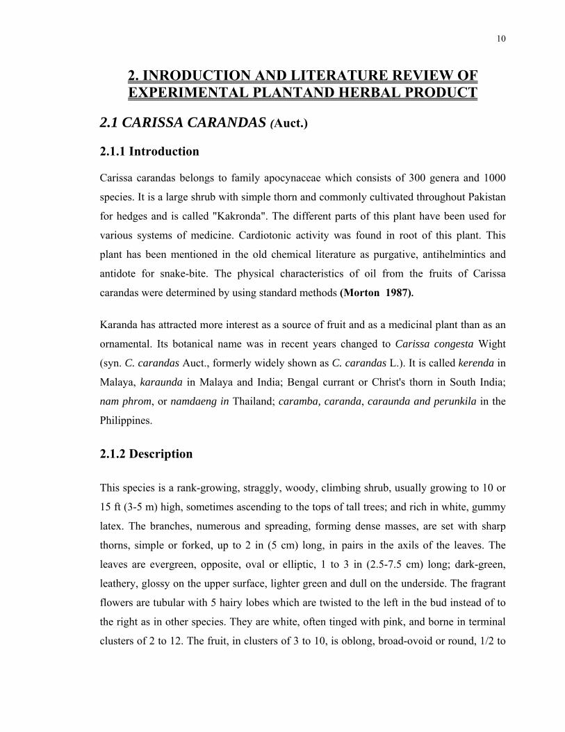
Fig. 2.1. Fruits and leaves of Carissa carandas.
b. Origin and Distribution
The karanda is native and common throughout much of India, Burma and Malacca and dry
areas of Ceylon; is rather commonly cultivated in these areas as a hedge and for its fruit
and the fruit is marketed in villages. It is rare in Malaya except as a potted plant in the
north; often grown in Thailand, Cambodia, South Vietnam and in East Africa. It was
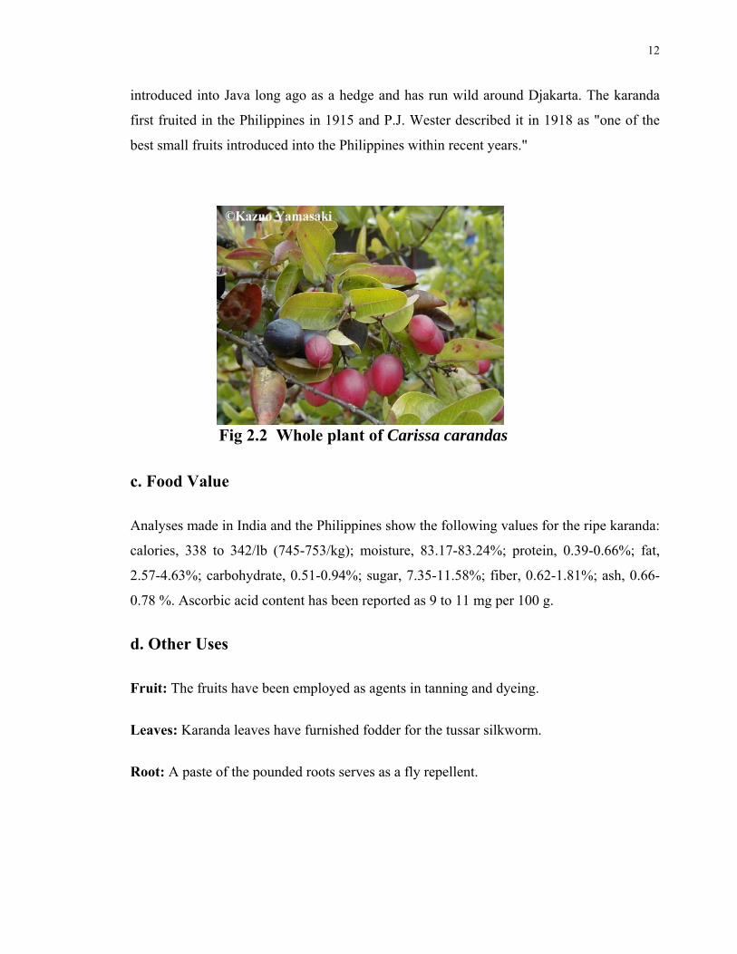
12
introduced into Java long ago as a hedge and has run wild around Djakarta. The karanda
first fruited in the Philippines in 1915 and P.J. Wester described it in 1918 as "one of the
best small fruits introduced into the Philippines within recent years."
Fig 2.2 Whole plant of Carissa carandas
c. Food Value
Analyses made in India and the Philippines show the following values for the ripe karanda:
calories, 338 to 342/lb (745-753/kg); moisture, 83.17-83.24%; protein, 0.39-0.66%; fat,
2.57-4.63%; carbohydrate, 0.51-0.94%; sugar, 7.35-11.58%; fiber, 0.62-1.81%; ash, 0.66-
0.78 %. Ascorbic acid content has been reported as 9 to 11 mg per 100 g.
d. Other Uses
Fruit: The fruits have been employed as agents in tanning and dyeing.
Leaves: Karanda leaves have furnished fodder for the tussar silkworm.
Root: A paste of the pounded roots serves as a fly repellent.
13
Wood: The white or yellow wood is hard, smooth and useful for fashioning spoons,
combs, household utensils and miscellaneous products of turnery. It is sometimes burned
as fuel.
2.1.3 Pharmacological Activity:
The unripe fruit is used medicinally as an astringent. The ripe fruit is taken as an
antiscorbutic and remedy for biliousness. The leaf decoction is valued in cases of
intermittent fever, diarrhea, oral inflammation and earache. The root is employed as a bitter
stomachic and vermifuge and it is an ingredient in a remedy for itches. The roots contain
salicylic acid and cardiac glycosides causing a slight decrease in blood pressure. Also
reported are carissone; the D-glycoside of B-sitosterol; glucosides of odoroside H;
carindone, a terpenoid; lupeol; ursolic acid and its methyl ester; also carinol, a phenolic
lignan. Bark, leaves and fruit contain an unnamed alkaloid (Morton 1987).
2.1.3 LITERATURE REVIEW OF CARISSA CARANDAS (AUCT.)
(Vohra and Den 1963) reported the comparative cardiotonic activity of Carissa carandas
l. and Carissa spinarum a. dc.
(Rastogi et al., 1966) isolated Carissa carandas. II. Polar glycosides. Enzymic and mild
acid hydrolysis of the polar glycoside (loc. cit.) from C. carandas afforded adoroside H (I),
digitoxigenin (II), 14,15-anhydrodigitoxigenin (III), glucose, and D-digitalose. I, II, and
III were isolated by column chromatog. over silica gel using 60:20 CHCl3-EtOAc as the
eluant and by preparative thin layer chromatog. and identified by comparison with
authentic samples. Sugars were identified by paper chromatography. The cardiotonic
activity of the glycosidic fraction was due to the presence of glucoside of I. As reported
earlier (loc. cit.), the monoside, I, or its genin, II, could not be detected as such in this
plant.
(Pakrashi et al., 1968) gave the chemical composition of Apocynaceae (C carandas and C.
spinarum), which grows throughout the dry regions of India. The roots are used as a
purgative and as an antidote for snakebite, and the leaves for remittent fever. Analysis of

14
C. carandas showed the presence of lupeol, β-sitosterol, a triterpene alcohol, ursolic acid,
and Me ursolate. Certain new compounds, as yet unidentified, are described. toxicity
(Joglekar and Gaitonde 1970) worked on Histamine releasing activity of Carissa
carandas roots (apocyaneceae). Roots of C. carandas are used in helminthiasis and to
assess the intensity of snake poisoning. An EtOH root extract caused a free histamine
increase in guinea pig lung, and in conscious cats vomiting, rhinorrhea, and diarrhea. Post
mortem findings suggest cardiovascular collapse due to histamine release. The prepn.
48/80 showed more histamine release than the EtOH extract. The smallest effective
extractdose was 250µg/kg in anesthetized cats; 10 mg/kg i.v. caused death. Blood pressure
(BP) decreased for 4.5 hr after 1 mg/kg of ext.
(Singh and Rastogi 1972) isolated Carindone, a C31-terpenoid from C. carandas, had
structure I according to its NMR, IR, UV, and mass spectra and those of its degradation
products.
(Chandra and Ganesh 1972) examined the benzene extract of the flowers and of the
essential oil of Carissa carandas. The C6H6 extractcontained lupeol, 9-hydroxystearic acid,
and triacontane. Steam distn. of the concrete gave 4.2% essential oil, which contained 2-
phenylethanol 60.0, 1-octanol 3.0, linalool 8.0, and benzyl acetate 22.0 wt. %.
(Pal et al., 1975) Carinol (I) a new lignan was isolated and identified by IR, UV, and
NMR spectra from C. carandas.
(Shrivastava and Bokadia 1979) performed studies in vegetable oils. Composition of
seeds oil of Carissa-carandas. The following fatty acids were isolated from seeds of C.
carandas by the urea adduct method: palmitic (66.42%), stearic (9.36%), arachidic
(21.19%), oleic (2.038%), and linoleic (0.99%) acids. Data on various physico-chemical
properties of the seed oil are presented.
(Zaki, El-Tohamy and El-Fattah 1983) worked on study of lipid content and volatile oil
of the different organs of Carissa carandas Lin. and Carissa grandiflora Dc. growing in
Egypt. TLC and column chromatographic techniques were used to study the
nonsaponifiable fraction and GLC for the methylated fatty acids of the Carissa species.
Both C. carandas and C. grandiflora contained lupeol, β-sitosterol, and ursolic acid in the
nonsaponifiable fraction, and lauric, myristic, palmitic, stearic, oleic, and linoleic acids in
the saponifiable fraction. The volatile oil of fresh flowers of both species was prepared by

15
cold extraction with n-hexane and studied by TLC and GLC. The study revealed the
presence of the hydrocarbons myrcene, limonene, camphene, β-3-carene, and dipentene in
both oils, p-cymene and α-terpinene in C. grandiflora oil. The oxygenated compounds
farnesol, nerolidol, dihydrojasmone, α-terpineol, Me heptanone, and piperitone were
detected in both oils, citronellal, α-ionone, menthol, neryl acetate, linalool, and geranyl
acetate in C. carandas oil, and linalyl acetate and geraniol in C. grandiflora.
(Zaki et al., 1983) Preliminary phytochemical and pharmacological screening of Carissa
carandas L. and Carissa grandiflora Dc. growing in Egypt. Phytochemical and
pharmacolgical studies of the Egyptian ornamental plants C. carandas and C. grandiflora in
Egypt were carried out to determine their main chemical constituents and pharmacological
activity. Successive extractions of different organs of both species were analyzed for
caffeic acid, carindone, carissone, cardenolides, and alkaloids by thin-layer
chromatography. Pharmacological screening was carried out by testing the action of an
aqueous extract of root of C. carandas on blood pressure, toad heart, isolated rabbit
intestine, motility of Ascaris worms in dogs, and on isolated rat uterus.
(Naim et al., 1985) Isolation of a new triterpenic alcohol from Carissa carandas.
Chemical constituents of fresh fruits of C. carandas were investigated. A new triterpenic
alcohol was isolated and named carissol (I) and its structure was elucidated on the basis of
spectral studies. I is an epimer of α-amyrin.
Me
Me
Me
Me
HHO
Me
Me H
H
MeMe I
(Chandra etal., 1985) Investigations on essential oils and isolates of potential value. Data
on the compound of flower (Pandanus odoratissimus, Rosa damasecna, Jasmine sambac, J.
grandiflorum, J. auriculatum, Michelia champaca, Nyctanthes arbortistis, Tagetes erecta,

16
Anthocephalus cadamba, Ocimum basilicum, Alstonia scholaris, Carissa carandas, leaf
(peppermint, Mentha arvensis, M. spicata, O. basilicum, O. kilimandscharicum,
Eupatorium odoratum, mango, Eugenia jambolana, Citrus karna, C. limettiodes), seed and
berry (dill, cumin, Trachyspermum ammi, Zanthoxylum alatum, carrot (Laurus nobilis),
root and tuber (Cyperus scariosus, vetiver, Acorus calamus) and Prunus puddum wood oils
are given. Research on alcohol analysis, new plant sources, yield improvement, processing
and distillation of essential oils, terpene synthesis, and isolation of specific terpenes from
oils are discussed.
(Dhawan and Patnaik 1985) Investigation on some new cardioactive glycosides. Data
from in vivo and in vitro experiments indicated that asclepin (I) [36573-63-4], isolated
from Asclepias curassavica, is a potent cardenolide with a wide safety margin in
comparison with other commonly used cardiac glycosides. A brief review of previous
work is included.
O
O
O
O
OOAc
Me
OH
H
H
H H
OHH
H
Me HCHO
I
(Naim et al., 1988) Isolation of a new isomer of ursolic acid from fruits and leaves of
Carissa carandas. A triterpenoid acid, carissic acid (I), was isolated from fruits and leaves
of C. carandas. The structure of I was elucidated by chemical and spectral means.
Me
Me
Me
MeH
H O
eM
eM
CO 2 HMe
I
17
(Rahman et al., 1991) Free sugars and dietary fiber in some fruits of Bangladesh. Free
sugar and dietary fiber (DF) contents of 10 fruits of Bangladesh were determined. All the
fruits except katamacha (0.6%) contained substantial amts. of glucose (1.1-6.3%) and
fructose (0.6-5.4%), whereas sucrose was either absent or present in a small proportion
(0.1-1.2%). The total DF content of the fruits varied between 0.2% (watermelon, tarmuj)
and 4.9% (jalpai, Elaeocarpus robustus) of fresh fruit. The smaller amounts (0.02-0.97%)
of water-solulble DF of all the fruits were mainly composed of uronic acid 54-85,
arabinose 3-20 and galactose 4-24%. The larger amtounts of (0.17-2.55%) of water-
insoluble nonstarch polysaccharides were mainly composed of residues of glucose 30-68,
uronic acid 9-40, galactose 3-25, arabinose 1-17, and xylose 3-11%. Starch content of the
fruits was very low (trace to 0.015%). Karamcha (Carissa carandas), and jalpai were high
(2.5-4.9%) in DF and, interestingly, also low in free sugars. The results may be used to
evaluate the nutritional status of the fruits.
(Iyer and Dubash 1993) Anthocyanin of Karwand (Carissa carandas) and studies on its
stability in model systems. Anthocyanin from Karwand (Carissa carandas) is characterized,
and its stability is determined in model systems. Based on the chromatographic data and
spectral studies, the pigment is identified as cyanidin-3-rhamnoglucoside. The data on the
stability of the pigment in two model systems showed progressive loss of anthocyanin.
The loss was higher at 30°C than at 5°C. A potential exists for the use of Karwand
anthocyanin as a natural coloring agent for products requiring milder processing treatment
and low temperature storage.
(Nizami et al., 1993) Biosynthesis of carissol and carissic acid. A possible mechanism for
the formation of carissol and carissic acid in Carissa carandas (Apocyanaceae) with acetate
as the precursor is presented. The migration of a Me group in the intermediate is
discussed.
(Dahot and Umer 1995) Lipase activity of Carissa carandas fruit The lipase activity in C.
carandas fruit was analyzed due to its medicinal importance. The data showed the pulp of
premature fruit possess greater activity than seeds and the pulp of mature fruit. The
maximum activity of C. carandas pulp lipase was observed at pH 5.0 and at 30°C. The
lipase activity was found heat labile and was completely lost at 80°C within 15 min. C.

18
carandas lipase activity was slightly increased in the presence of calcium but enzyme
activity was decreased by the addnition of EDTA, PMSCI and o-phenanthroline.
(Sekar and Francis 1998) Some plant species screened for energy, hydrocarbons and
phytochemicals. Fifteen laticiferous plant species of Apocyanaceae, Asclepiadaceae,
Convolvulaceae, Euphorbiaceae were tested for their suitability as alternative sources of
renewable energy, and phytochems. Their polyphenol, oil and hydrocarbon contents were
analyzed. Among them the best species were Carissa carandas L., Ceropegia juncea
Roxb., Hemidesmus indicus R. Br. and Sarcostemma brunourianum W.A.
(Siddiqui et al., 2003) Triterpenoidal constituents of the leaves of Carissa carandas.
Studies undertaken on the fresh leaves of Carissa carandas have led to the isolation of four
pentacyclic triterpenoids (1-4) including one new constituent carissin (1) and two hitherto
unreported compounds The structure of the new triterpenoid has been elucidated as 3α-
hydroxy-27-E-feruloyloxyurs-12-en-28-oic acid. Complete assignment of the protons of 3
α-hydroxy-27-E-feruloyloxyurs-12-en-28-oic acid has also been made based on 2D NMR
studies.
(Augustus et al., 2003) Alternative energy sources from plants of Western Ghats (Tamil
Nadu, India). Twenty two taxa of Western Ghats plants were screened as potential
alternative crops for renewable energy, oil, hydrocarbon and phytochems. The highest
hydrocarbon yields were observed for Carissa carandas (1.7%), and Jatropha gossypifolia
(1.7%). The highest polyphenol fraction was observed for Dodonaea viscosa (17.1%),
Carissa carandas (7.7%), Swietenia mahagoni (6.6%), and Jatropha glandulifera (6.2%).
The highest oil content was observed in Aganosma cymosa (10.3%), Carissa carandas
(5.8%), and Argemone mexicana (5.0%). Swietenia mahagoni yielded the highest protein
content at 8.1%. The gross heat values of 4175.0 cal/g (17.5 MJ/kg) for Lochnera rosea
(pink flowered var.), and 4112.0 cal/g for Dalbergia sissoo were the highest among the
species analyzed. NMR spectra of the hydrocarbon fractions of Alstonia scholaris, Carissa
carandas, Ichnocarpus frutescens, Plumeria rubra, The vetia neriifolia (white flowered
var.), Vallaris solanacea, Lochnera rosea (pink flowered var.), Euphorbia hirta, E.
splendens, Artocarpus integrifolia and Ficus religiosa revealed cis-polyisoprene (natural
rubber), whereas Argemone mexicana showed trans-polyisoprene (gutta). Several new
crop species were identified with potentially useful compounds. The potential exists for
19
growing these alternate crops in areas of underused lands, subsequently stimulating
industrial and economic growth.
(Pino et al., 2004) Volatile flavor constituents of Karanda (Carissa carandas L.) fruit. The
volatile flavor constituents of karanda fruits growing in Cuba were analyzed by GC and
GC/MS. One hundred and fifty compounds were identified in the aroma concentration, of
which isoamyl alcohol, isobutanol and α-caryophyllene were the major constituents.
(Ahmed et al., 2006) Activity of polyphenolic plant extracts as scavengers of free radicals
and inhibitors of xanthine oxidase. The free radical scavenging activity of ethanolic and aq.
extracts from six plants, has been evaluated, in vitro, using 1,1-diphenyl-2-picrylhydrazyl
(DPPH) radical scavenging method and compared with ascorbic acid, quercetin,
kaempferol and apigenin. Extracts of Pistacia integerrima leaves and galls exhibited
highest DPPH radical scavenging activity among all the plants extracts examined Carissa
carandas from Apocynaceae and Vitex negundo of Verbenaceae family also demonstrated
DPPH radical scavenging activity. Inhibitory effects towards the in-vitro reaction of
hypoxanthine and xanthine oxidase (XO) was also carried out in the presence of plant ext.,
aglycons quercetin, kaempferol and apigenin along with allopurinol.
(Rao et al., 2007) Analgesic and anti-inflammatory activity of Carissa carandas linn
fruits and Microstylis wallichii Lindl tubers. The ethanolic (50% vol./vol.) extracts of
Carissa carandas (fruits) (Apocynaceae) and Microstylis wallichii (tubers) (Orchidaceae)
were examined for anti-inflammatory and analgesic activities in experimental animals.
Carissa carandas and Microstylis wallichii (50-200 mg/kg) caused a dose dependent
inhibition of swelling caused by carrageenin significantly in cotton pellet induced
granuloma in rats (P < 0.05 to P < 0.001). The extracts of Carissa carandas and
Microstylis wallichii resulted in an inhibition of stretching episodes and percentage
protection was 16.05-17.58% respectively in acetic acid induced writhin.
20

21
2.2 INTRODUCTION TO HERBAL PRODUCT:
MUFARREH YAQOOTI MOTADIL (MUYM), THE POLY-HERBAL
PRODUCT OF HAMDARD LABORATORIES (Waqf.) PAKISTAN.
Mufarreh Yaqooti Motadil is a herbal product being prescribed for strengthening the heart
so acts as a Cardiac tonic and as such is traditionally used as specified in Hamdard
pharmacopoeia and in Unani Qrabadin. The product is being manufactured by Hamdard
Laboratories (waqf) Pakistan for the last thirty years and has shown significant therapeutic
effects. No side effects have been observed. (Said 1979)
2.2.1 Quantitative description of ingredients: (Said 1979)
Name of Ingredient Part used Quantity
2.2.1.1 Cymbopogon jwarancusa (Jones) Schult., Mant. Root 5grams
2.2.1.2. Parmelia perlata (Huds.) whole plant 20 grams
2.2.1.3. Helix aspira Animal 20 grams
2.2.1.4. Elettaria cardamomum (L.) Maton Fruit 10 grams
2.2.1.5. Amomum subulatum (Roxb.) Fruit 10 grams
2.2.1.6. Nepeta hindostana (Roth.) Root 10 grams
2.2.1.7. Nardostachys jatamansi (D.C.) Aerial parts 10 grams
2.2.1.8. Santalum album (L.) Wood 15 grams
2.2.1.9. Onosma bracteatum (Wall.) Leaves 20 grams
2.2.1.10 Bambusa arundinacea (Schreb.) Exudate 20 grams
2.2.1.11 Centaurea behen (L.) Root 20 grams
2.2.1.12 .Citrus medica (L.) Peel 10 grams
2.2.1.13 .Cinnamomum cassia (L.) Bark 10 grams
2.2.1.14 .Apium graveolens (L.) Seeds 20 grams
2.2.1.15 .Ocimum basilicum (L.) Seeds 20 grams
2.2.1.16 Lactuca sativa (L.) Seeds 20 grams

22
2.2.1.17 .Cinnamomum officinalis (L.) Bark 20 grams
2.2.1.18 .Doronicum hookeri (Clarke.) Gum 30 grams
2.2.1.19. Berberis aristata (DC.) Fruit 20 grams
2.2.1.20. Zingiber zerumbet (L.) Rhizome 20 grams
2.2.1.21 Aquilaria agallocha (Roxb.) Wood 30 grams
2.2.1.22. Coriandrum sativum (L.) Seed 10 grams
2.2.1.23. Armenian bole Mineral 10 grams
2.2.1.24. Rosa damascene(Mill.) Petals 10 grams
2.2.1.25. East Indian Clay Mineral 10 grams
2.2.1.26. Cucumis sativus (L.) Seeds kernel 20 grams
2.2.1.27. Lagenaria siceraria(Molina)Standl. Seed kernel 20 grams
2.2.1.28. Mesua ferrea (L.) Rhizome 10 grams
2.2.1.29. Camphora officinarum Sublimed crystals 5 grams
2.2.1.30. Corallium rubrum (L.) Root 20 grams
2.2.1.31. Vateria indica (L.) Resin exudates 20 grams
2.2.1.32. Lapis lazuli Mineral 20 grams
2.2.1.33. Rubies Mineral 10 grams
2.2.1.34. Pearls Mineral 10 grams
2.2.1.35. Red Rubies Mineral 10 grams
2.2.1.36. Bombyx mori (L.) Animal 20 grams
2.2.1.37. Honey Animal 2kg+300g
2.2.1.38. Sodium benzoate Mineral 5 grams
2.2.1.39. Ambra grasea (L.) Animal 10 grams
2.2.1.40. Pistacia lentiscus (L.) Gum 20 grams
2.2.1.41. Ghee Animal 5 grams
2.2.1.42. Musk Animal 2 grams
2.2.1.43. Crocus sativus (L.) Fruit 15 grams
2.2.1.44. Aqua Onosma bracteatum Extract 60 milliliters
2.2.1.45. Silver foil. Mineral 12grams

23
2.2.2 Method of preparation:
The first 28 ingredients after being ground were triturated with camphor. Then minerals
were also added and triturated. The decoction of Bombyx mori, made in 200 ml of water,
filtered and cooled. Cooled decoction is added to the honey and is heated. When the
solution becomes thick, Sodium Benzoate and ground Ambergeris were added and the
firstly ground material triturated with camphor was gradually added in the honey paste
with stirring. Pistacia lentiscus gum were molten in the ghee and the musk, saffron and
camphor were triturated in the aqua Onosma bracteatumwere also added in the honey paste
with constant stirring. Finally ground silver foils were incorporated. (Said 1979)
2.2.3 Dosage and administration. Dose is 5g in the morning alone, followed by 250
ml of milk or water. (Said 1979)
2.2.4 Uses and indications: (i) Cardiac Stimulant (ii) Cephalic tonic
(iii) Anodyne (iv) Antipalpitant (v) Curative for melancholia. (Said 1979)
2.2.5 Qualitative description of ingredients
2.2.5.1. Cymbopogon jwarancusa Syn: Cymbopogon citrates
Syn: Andropogon jwarancusa (Lemon grass), Family: Gramineae
Plant’s height is 2-6 Feet and width is 3 feet. The leaves and essential oils are used to treat
dyspeptic disorders, colds, nervous conditions and exhaustion (Wyk and Wink 2004).
carminative, stimulant and diuretic. Flowers styptic, whole plant has appetizing, stomachic
properties; resolvent, antiseptic (Usmanghani et al.,1997). The plant is aromatic, bitter,
anthelmintic, laxative, appetizer, alexipharmic and aphrodisiac, and useful in flatulence,
gastric irritation, anorexia, poisonous bites, bronchitis, epilepsy, leprosy, skin diseases,
cholera, neuralgia, sprain and fever (Pullaiah 2002).
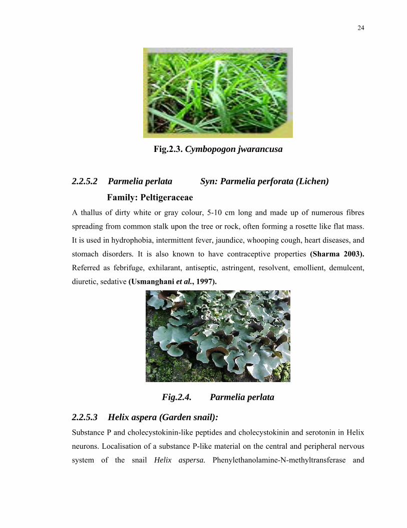
24
Fig.2.3. Cymbopogon jwarancusa
2.2.5.2 Parmelia perlata Syn: Parmelia perforata (Lichen)
Family: Peltigeraceae
A thallus of dirty white or gray colour, 5-10 cm long and made up of numerous fibres
spreading from common stalk upon the tree or rock, often forming a rosette like flat mass.
It is used in hydrophobia, intermittent fever, jaundice, whooping cough, heart diseases, and
stomach disorders. It is also known to have contraceptive properties (Sharma 2003).
Referred as febrifuge, exhilarant, antiseptic, astringent, resolvent, emollient, demulcent,
diuretic, sedative (Usmanghani et al., 1997).
Fig.2.4. Parmelia perlata
2.2.5.3 Helix aspera (Garden snail):
Substance P and cholecystokinin-like peptides and cholecystokinin and serotonin in Helix
neurons. Localisation of a substance P-like material on the central and peripheral nervous
system of the snail Helix aspersa. Phenylethanolamine-N-methyltransferase and

25
dopamine-B-hydroxylase immunoreactivity & the occurence of noradrenaline & adrenaline
in the nervous system of the snail Helix aspera (Osborne 1984).
2.2.5.4 Elettaria cardamomum (Cardamom), Family: labiatae
An aromatic, perennial herb upto 3m tall. Leaves are large, narrow. Flowers are white or
pale green in colour and appear from the base of the stem and lies on the ground (Sharma
2003) Aromatic, flavoring, stimulant, carminative, stomach tonic, exhilarant, astringent,
diuretic, antiemetic, antiflatulant, cardiac tonic (Usmanghani 1997). The drug has
stimulant, carminative, stomachic, diuretic, and digestive properties. The drug is used in
common cold, cough/bronchitis, fever, inflammation of the mouth and pharynx (Pullaiah
2002).
The seeds are aphrodisiac and remedy in case of digestive problems, asthma, bronchitis
and urinary complaints. It is used against bad breath, cough and nausea and may be
combined with laxatives to treat indigestion, stomach pain, griping and flatulence. Its
actions are appetizer, carminative, diaphoretic, digestive, stimulant, expectorant and
stimulant. It is also used for the involuntary urination, nausea, respiratory disorders,
stomach complaints to stimulate the mind. Cardamom seeds are useful for flatulence, but
they are usually used as adjuvants with other remedies. They are also used as a spice in
cooking and as a flavoring in other medicines.
The seeds and pods contain a volatile oil which is used in perfumes and as a stimulant. The
aphrodisiac properties of cardamom are extolled in Arabian Nights- the people in Middle
East still believe that cardamom possesses such properties. Cardamom is a stimulant, it
cools the body in extreme heat and it aids digestion. Remedy for Celiac Disease
(Usmanghani et al., 1997).
Fig.2.5. Elettaria cardamomum
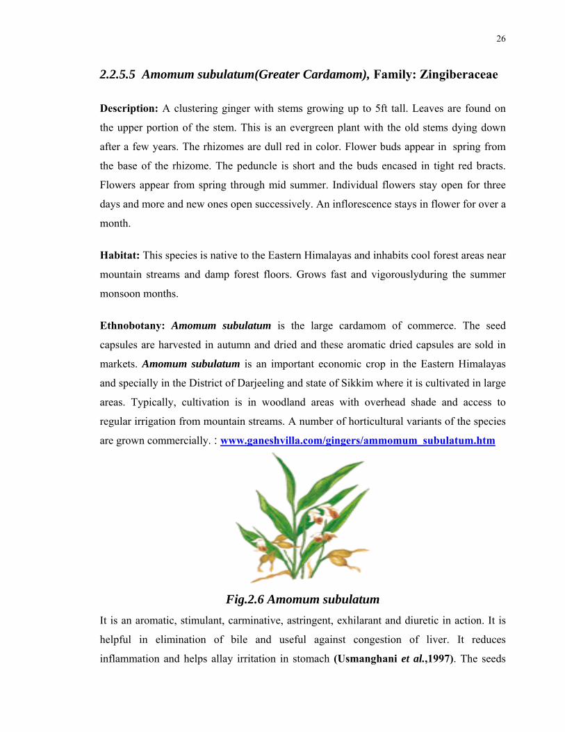
26
2.2.5.5 Amomum subulatum(Greater Cardamom), Family: Zingiberaceae
Description: A clustering ginger with stems growing up to 5ft tall. Leaves are found on
the upper portion of the stem. This is an evergreen plant with the old stems dying down
after a few years. The rhizomes are dull red in color. Flower buds appear in spring from
the base of the rhizome. The peduncle is short and the buds encased in tight red bracts.
Flowers appear from spring through mid summer. Individual flowers stay open for three
days and more and new ones open successively. An inflorescence stays in flower for over a
month.
Habitat: This species is native to the Eastern Himalayas and inhabits cool forest areas near
mountain streams and damp forest floors. Grows fast and vigorouslyduring the summer
monsoon months.
Ethnobotany: Amomum subulatum is the large cardamom of commerce. The seed
capsules are harvested in autumn and dried and these aromatic dried capsules are sold in
markets. Amomum subulatum is an important economic crop in the Eastern Himalayas
and specially in the District of Darjeeling and state of Sikkim where it is cultivated in large
areas. Typically, cultivation is in woodland areas with overhead shade and access to
regular irrigation from mountain streams. A number of horticultural variants of the species
are grown commercially. : www.ganeshvilla.com/gingers/ammomum_subulatum.htm
Fig.2.6 Amomum subulatum
It is an aromatic, stimulant, carminative, astringent, exhilarant and diuretic in action. It is
helpful in elimination of bile and useful against congestion of liver. It reduces
inflammation and helps allay irritation in stomach (Usmanghani et al.,1997). The seeds

27
are acrid, bitter, aromatic, deodorant, appetizer, carminative, digestive, stomachic,
constipating depurative, vulnerary, cardiac and liver tonic. They are useful in anorexia,
dyspepsia, colic, flatulence, hyperacidity, vomiting, diarrhea, skin diseases, pruritis,
wounds, ulcers, cephalagia, neuralgia, cardiac debility, liver congestion, cough , bronchitis
and gonorrhoea. (Pullaiah 2002).
2.2.5.6 Nepeta hindostana (Nepeta herb), Family: Labiateae
Nepeta is a genus of about 250 species of flowering plants in the family Lamiaceae. The
members of this group are known as catnips or catmints due to its famed liking by cats—
nepeta pleasantly stimulates cats' pheromonic receptors.
The genus is native to Europe, Asia and Africa, with the highest species diversity in the
Mediterranean region east to mainland China. It is now common in North America as a
weed. Most of the species are herbaceous perennial plants, but some are annuals. They
have sturdy stems with opposite heart-shaped, green to grayish-green leaves. The flowers
are white, blue, pink or lilac and occur in several clusters toward the tip of the stems. The
flowers are tubular shaped and are spotted with tiny purple dots.
Other uses: Nepeta cataria is weakly psychoactive, variably eliciting sedation, euphoria,
and, in higher doses, nausea. Although its effect is very mild, catnip may be used as a
substitute for marijuana, often in combination with damiana.
A study conducted at Iowa State University suggests that pure nepetalactone, a constituent
of the essential oil of N. cataria, may be 10 times as effective at repelling mosquitos as the
common repellent, DEET. [1] Catnip had been long recognized as a good companion
plant, in that it appeared to repel pest insects, and this study offers insight into the
phenomenon. (www.answer.com/topic/nepeta-1)
Exhilarant, carminative, , resolvent, sedative, hepatic, cardiac, cephalic tonic, antipyretic,
diaphoretic, blood purifier and anticholestrolemic (Usmanghani et al.,1997).
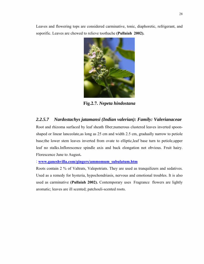
28
Leaves and flowering tops are considered carminative, tonic, diaphoretic, refrigerant, and
soporific. Leaves are chewed to relieve toothache (Pullaiah 2002).
Fig.2.7. Nepeta hindostana
2.2.5.7 Nardostachys jatamansi (Indian valerian): Family: Valerianaceae
Root and rhizoma surfaced by leaf sheath fiber;numerous clustered leaves inverted spoon-
shaped or linear lanceolate,as long as 25 cm and width 2.5 cm, gradually narrow to petiole
base;the lower stem leaves inverted from ovate to elliptic,leaf base turn to petiole,upper
leaf no stalks.Inflorescence spindle axis and back elongation not obvious. Fruit hairy.
Florescence June to August.
: www.ganeshvilla.com/gingers/ammomum_subulatum.htm
Roots contain 2 % of Valtrats, Valepotriats. They are used as tranquilizers and sedatives.
Used as a remedy for hysteria, hypochondriasis, nervous and emotional troubles. It is also
used as carminative (Pullaiah 2002). Contemporary uses Fragrance flowers are lightly
aromatic; leaves are ill scented; patchouli-scented roots.
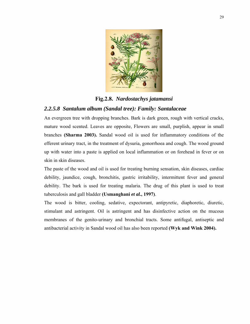
29
Fig.2.8. Nardostachys jatamansi
2.2.5.8 Santalum album (Sandal tree): Family: Santalaceae
An evergreen tree with dropping branches. Bark is dark green, rough with vertical cracks,
mature wood scented. Leaves are opposite, Flowers are small, purplish, appear in small
branches (Sharma 2003). Sandal wood oil is used for inflammatory conditions of the
efferent urinary tract, in the treatment of dysuria, gonorrhoea and cough. The wood ground
up with water into a paste is applied on local inflammation or on forehead in fever or on
skin in skin diseases.
The paste of the wood and oil is used for treating burning sensation, skin diseases, cardiac
debility, jaundice, cough, bronchitis, gastric irritability, intermittent fever and general
debility. The bark is used for treating malaria. The drug of this plant is used to treat
tuberculosis and gall bladder (Usmanghani et al., 1997).
The wood is bitter, cooling, sedative, expectorant, antipyretic, diaphoretic, diuretic,
stimulant and astringent. Oil is astringent and has disinfective action on the mucous
membranes of the genito-urinary and bronchial tracts. Some antifugal, antiseptic and
antibacterial activity in Sandal wood oil has also been reported (Wyk and Wink 2004).
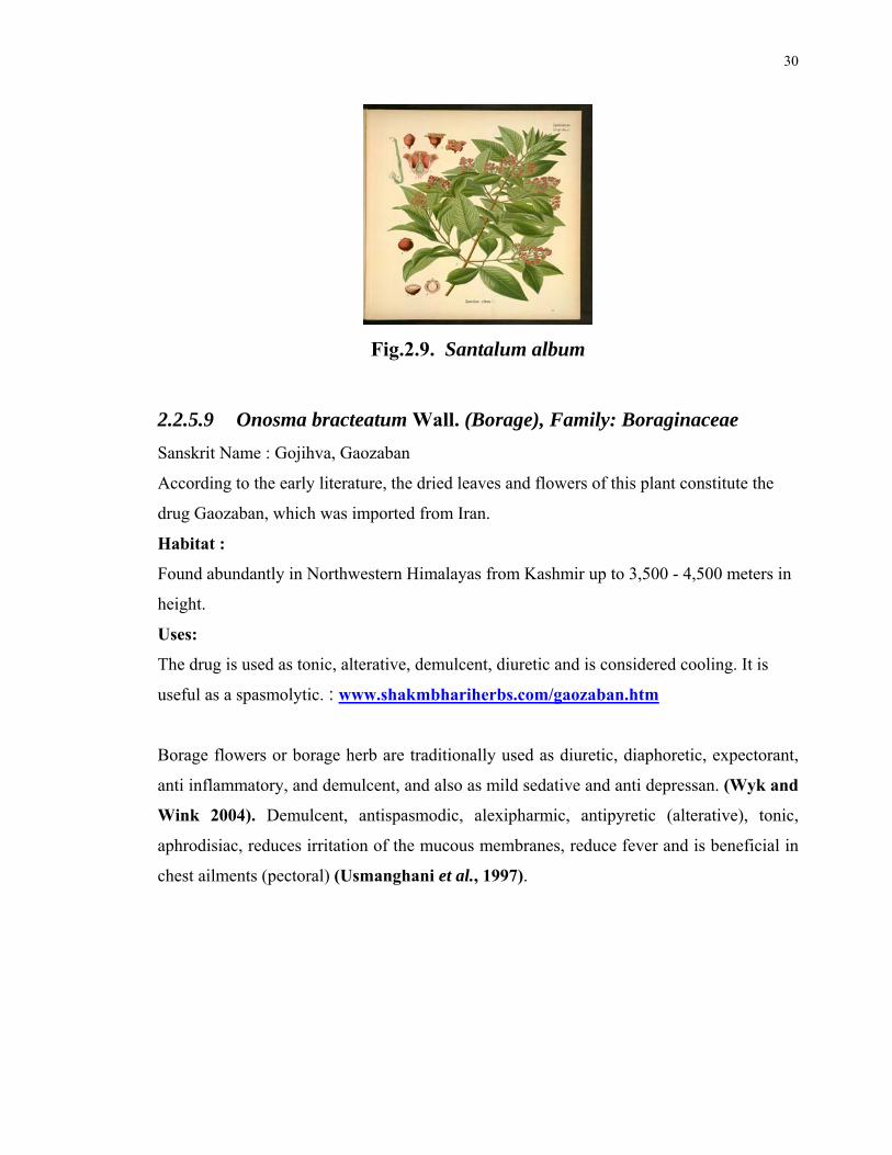
30
Fig.2.9. Santalum album
2.2.5.9 Onosma bracteatum Wall. (Borage), Family: Boraginaceae
Sanskrit Name : Gojihva, Gaozaban
According to the early literature, the dried leaves and flowers of this plant constitute the
drug Gaozaban, which was imported from Iran.
Habitat :
Found abundantly in Northwestern Himalayas from Kashmir up to 3,500 - 4,500 meters in
height.
Uses:
The drug is used as tonic, alterative, demulcent, diuretic and is considered cooling. It is
useful as a spasmolytic. : www.shakmbhariherbs.com/gaozaban.htm
Borage flowers or borage herb are traditionally used as diuretic, diaphoretic, expectorant,
anti inflammatory, and demulcent, and also as mild sedative and anti depressan. (Wyk and
Wink 2004). Demulcent, antispasmodic, alexipharmic, antipyretic (alterative), tonic,
aphrodisiac, reduces irritation of the mucous membranes, reduce fever and is beneficial in
chest ailments (pectoral) (Usmanghani et al., 1997).

31
Fig.2.10 Onosma bracteatum
2.2.5.10 Bambusa arundinacea (Thorny Bamboo), Family: Gramineae
The leaves are emmenagogue, anthelmintic, astringent and fibrifuge. The stems and leaves
are used in the Ayurvedic system of medicine as blood purifier, in leucoderma and
inflammatory conditions (Usmanghani et al.,1997). The bamboo plant has emmenagogue,
anthelmintic, stimulant, astringent, refrigerant, tonic, antispasmodic and aphrodisiac
attributes. Combined with some other suitable ingredients it can also exert styptic,
antidiarrhoeal and useful desiccative activities (Wyk and Wink 2004).
Fig.2.11. Bambusa arundinacea
2.2.5.11 Centaurea behen (Behen, Centaurea): Family: Compositeae
Exhilarant and cardiac tonic, aphrodisiac, antiflatulent, semenagogue, fattening (with
comparatively less warm faculty than the red variety) (Usmanghani et al.,1997).
Used mainly as a bitter tonic and stomachic to treat dyspepsia and loss of appetite. It is a
component of various herbal teas, including cholagogues and remedies for gastrointestinal

32
ailments. The herb has been taken to treat internal cancers, diabetes, gout and rheumatism
and applied topically for wounds and ulcers (Wyk and Wink 2004).
Fig.2.12. Centaurea behen
2.2.5.12 Citrus medica (Citron), Family: Rutaceae
A small tree of 6m tall with short and stout spines. Leaflets are oblong to ovate, upto 10
cm long. Flowers are 5 cm across, reddish in bud stage. Fruits are ovate or oblong, upto
12.5 cm long, yellow in colour and very acidic in taste (Sharma 2003). Juice of fruits is
refrigerant and astringent. Preserved rind is used in dysentery. The fruits are stomachic and
have potent antiscorbutic activity. Fruit decoction is analgesic, it is effective in vomiting.
The fruit and seeds are a cardiac tonic and useful in palpitation (Usmanghani et al., 1997).
Fig.2.13. Citrus medica
2.2.5.13 Cinnamomum cassia (Cassia bark), Family: Lauraceae
Aromatic, carminative, antispasmodic, stimulant, haemostatic, astringent, antiseptic,
demulcent, refrigerant (Usmanghani et al., 1997).
Cinnamon is a traditional remedy for dyspeptic conditions (flatulence, gastrointestinal
spasms, loss of appetite and diarrhoea). It is also used in folk medicine to treat

33
inflammation, rheumatism, colds, nausea and vomiting and menstrual disorders (Pullaiah
2002). Bark or oil is used to treat appetite loss, dyspepsia, and spasm associated with
bloating, flatulence, nausea and diarrhoea. It is also used to treat common cold and
exhaustion. The flowers are used traditionally as blood purifier (Wyk and Wink 2004).
Fig.2.14. Cinnamomum cassia
2.2.5.14 Apium graveolens (Celery),Family: Umbelliferae
An annual or biennial, erect, glabrous herb with fistular, angular stem. Leaves pinnate,
leaflets1-6 cm long, oval-obovate, usually 3-lobed incised, glabrous. Umbellules 20-25
flowered, white in colour and blooming in the month of March to June (Sharma 2003).
Celery seeds are credited with stimulant and carminative properties and are prescribed as
nervine sedative and tonic. The decoction is a popular house holds remedy for rheumatism.
The essential oil (known as Celery seed oil) possesses tranquilizing and anti-convulsant
activities.Seeds are used in the treatment if chronic skin disorders including psoriasis
(Usmanghani etal., 1997).Carminative, deobstruent, diaphoretic, appetitive,
antiphlegmatic, lithontriptic, diuretic, emmenagogue, ecbolic, anthelmintic. Root is
alternative and diuretic, it is given in anasarca and colic. The leaves are taken as salad and
preparation of soup. The rip seeds are pungent, hot, with sharp taste, aphrodisiac,
abortifacient, antirheumatic, antispasmodic, cooling, carmantive, digestive, diuretic,
laxative, stimulent, stomachic, emetic, emmenagogue, nervine sedative, febrifuge,
intestinal antiseptic and tonic. The seeds are used for the treatment of asthma, abdominal
pain, bronchitis, amenorrhoea, calculi of the kidneys and bladder, diseases of heart and

34
spleen, fever with cough, hi-cough, chest pains, ophtalmia, scabies, scorpion and other
stings, inflammations, rheumatism, toothache, tumours, rectal troubles, burning sensations,
urinary discharges, vomiting, catarrh of the nose, lumbago, dysuria, dyspepsia and
menorrhagia. Tincture, prepared from ripe seeds, is used to treat urine retention and other
renal disorders. The seeds are used as spice. Fruit is irritant and poisonous in over dose.
The wild plant is poisonous. (Wyk and Wink 2004)
Fig. 2.15 Apium graveolens
2.2.5.15 Ocimum basilicum (Basil), Family: Lamiaceae
An annual herb. Leaves numerous dot like glands which secrete strongly scented volatile
oils. Leaves are simple, petiolate, ovate or subovate. Flowers appear in clusters (Sharma
2003). Exhilarant, expectorant, ant periodic, diuretic, emmenagogue. Seeds mucilaginous
and demulcent. Leaves fragrant, aromatic and expectorant. (Wyk and Wink 2004).
The plant has been used in Indian system of medicine as an anthelmintic, nervine, diuretic,
diaphoretic, demulcent and to treat piles, bronchitis, rheumatism and inflammation. The
plant is also useful in fever, cough, worms, stomach complaints and gout; juice of leaves is
used as a nasal douche and is applied in skin diseases. Seeds are given to cure dysentery
and chronic diarrhea. Leaves are used to cure earache, rheumatoid arthritis, anorexia,
itching, amenorhoea, dysmenorrhoea and malaria (Usmanghani et al., 1997). Basil species
are widely used as tonics, against indigestion,flatulence, loss of appetite and internal
parasites. Basil is included in wound-healing ointments (Wyk and Wink 2004).

35
Fig.2.16 Ocimum basilicum
2.2.5.16 Lactuca sativa Syn: Lactuca serriola (Lettuce),
Family: Asteraceae
Antibilious, antiflatulant, tonic, diuretic, stomach tonic, appetite stimulant, lactagogue,
preventive of environmental effects, anodyne, sedative, hypnotic, expectorant, tonic
against general debility and for lungs. (Wyk and Wink 2004).
Fig. 2.17 Lactuca sativa
2.2.5.17. Cinnamomum officinalis (Cinnamon), Family: Lauraceae
Cinnamon is used to improve the taste and aroma of some medicinal products. Non-
medicinal uses are well known- it is a popular spice in cooking and the essential oil is used
in perfumery (Wyk and Wink 2004).
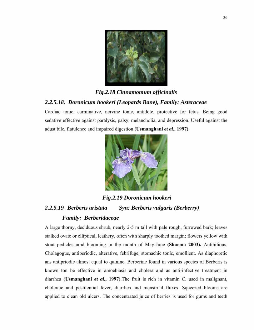
36
Fig.2.18 Cinnamomum officinalis
2.2.5.18. Doronicum hookeri (Leopards Bane), Family: Asteraceae
Cardiac tonic, carminative, nervine tonic, antidote, protective for fetus. Being good
sedative effective against paralysis, palsy, melancholia, and depression. Useful against the
adust bile, flatulence and impaired digestion (Usmanghani et al., 1997).
Fig.2.19 Doronicum hookeri
2.2.5.19 Berberis aristata Syn: Berberis vulgaris (Berberry)
Family: Berberidaceae
A large thorny, deciduous shrub, nearly 2-5 m tall with pale rough, furrowed bark; leaves
stalked ovate or elliptical, leathery, often with sharply toothed margin; flowers yellow with
stout pedicles amd blooming in the month of May-June (Sharma 2003). Antibilious,
Cholagogue, antiperiodic, alterative, febrifuge, stomachic tonic, emollient. As diaphoretic
ans antipriodic almost equal to quinine. Berberine found in various species of Berberis is
known ton be effective in amoebiasis and cholera and as anti-infective treatment in
diarrhea (Usmanghani et al., 1997).The fruit is rich in vitamin C. used in malignant,
choleraic and pestilential fever, diarrhea and menstrual fluxes. Squeezed blooms are
applied to clean old ulcers. The concentrated juice of berries is used for gums and teeth
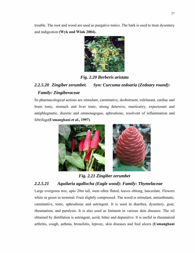
37
trouble. The root and wood are used as purgative tonics. The bark is used to treat dysentery
and indigestion (Wyk and Wink 2004).
Fig. 2.20 Berberis aristata
2.2.5.20 Zingiber zerumbet. Syn: Curcuma zedoaria (Zedoary round):
Family: Zingiberaceae
Its pharmacological actions are stimulant, carminative, deobstruent, exhilarant, cardiac and
brain tonic, stomach and liver tonic, strong detersive, masticatiry, expectorant and
antiphlegmatic, diuretic and emmenagogue, aphrodisiac, resolvent of inflammation and
febrifuge(Usmanghani et al., 1997).
Fig. 2.21 Zingiber zerumbet
2.2.5.21 Aquilaria agallocha (Eagle wood): Family: Thymelaceae
Large evergreen tree, upto 20m tall, stem often fluted, leaves oblong, lanceolate. Flowers
white or green in terminal. Fruit slightly compressed. The wood is stimulant, antiasthmatic,
carminative, tonic, aphrodisiac and astringent. It is used in diarrhea, dysentery, gout,
rheumatism, and paralysis. It is also used as liniment in various skin diseases. The oil
obtained by distillation is astringent, acrid, bitter and depurative. It is useful in rheumatoid
arthritis, cough, asthma, bronchitis, leprosy, skin diseases and foul ulcers (Usmanghani
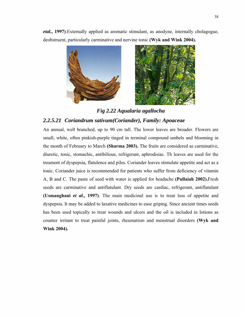
38
etal., 1997).Externally applied as aromatic stimulant, as anodyne, internally cholagogue,
deobstruent, particularly carminative and nervine tonic (Wyk and Wink 2004).
Fig 2.22 Aqualaria agallocha
2.2.5.21 Coriandrum sativum(Coriander), Family: Apoaceae
An annual, well branched, up to 90 cm tall. The lower leaves are broader. Flowers are
small, white, often pinkish-purple tinged in terminal compound umbels and blooming in
the month of February to March (Sharma 2003). The fruits are considered as carminative,
diuretic, tonic, stomachic, antibilious, refrigerant, aphrodisiac. Th leaves are used for the
treament of dyspepsia, flatulence and piles. Coriander leaves stimulate appetite and act as a
tonic. Coriander juice is recommended for patients who suffer from deficiency of vitamin
A, B and C. The paste of seed with water is applied for headache (Pullaiah 2002).Fresh
seeds are carminative and antiflatulant. Dry seeds are cardiac, refrigerant, antiflatulant
(Usmanghani et al., 1997). The main medicinal use is to treat loss of appetite and
dyspepsia. It may be added to laxative medicines to ease griping. Since ancient times seeds
has been used topically to treat wounds and ulcers and the oil is included in lotions as
counter irritant to treat painful joints, rheumatism and menstrual disorders (Wyk and
Wink 2004).
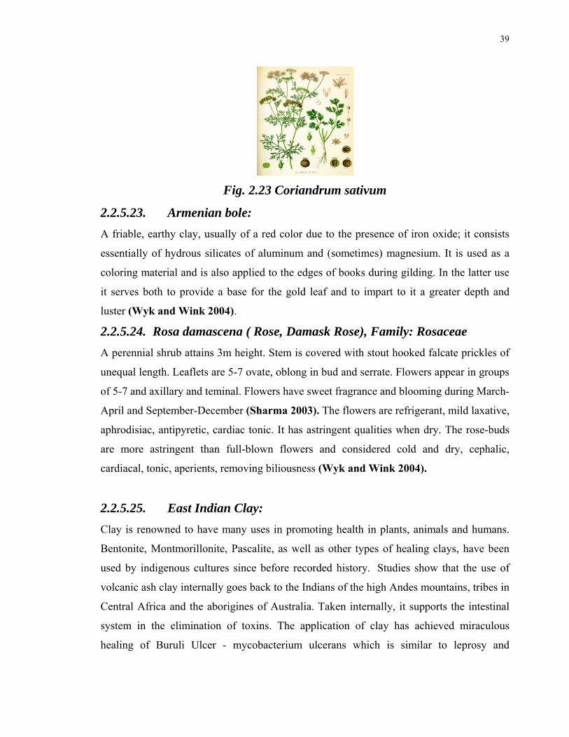
39
Fig. 2.23 Coriandrum sativum
2.2.5.23. Armenian bole:
A friable, earthy clay, usually of a red color due to the presence of iron oxide; it consists
essentially of hydrous silicates of aluminum and (sometimes) magnesium. It is used as a
coloring material and is also applied to the edges of books during gilding. In the latter use
it serves both to provide a base for the gold leaf and to impart to it a greater depth and
luster (Wyk and Wink 2004).
2.2.5.24. Rosa damascena ( Rose, Damask Rose), Family: Rosaceae
A perennial shrub attains 3m height. Stem is covered with stout hooked falcate prickles of
unequal length. Leaflets are 5-7 ovate, oblong in bud and serrate. Flowers appear in groups
of 5-7 and axillary and teminal. Flowers have sweet fragrance and blooming during March-
April and September-December (Sharma 2003). The flowers are refrigerant, mild laxative,
aphrodisiac, antipyretic, cardiac tonic. It has astringent qualities when dry. The rose-buds
are more astringent than full-blown flowers and considered cold and dry, cephalic,
cardiacal, tonic, aperients, removing biliousness (Wyk and Wink 2004).
2.2.5.25. East Indian Clay:
Clay is renowned to have many uses in promoting health in plants, animals and humans.
Bentonite, Montmorillonite, Pascalite, as well as other types of healing clays, have been
used by indigenous cultures since before recorded history. Studies show that the use of
volcanic ash clay internally goes back to the Indians of the high Andes mountains, tribes in
Central Africa and the aborigines of Australia. Taken internally, it supports the intestinal
system in the elimination of toxins. The application of clay has achieved miraculous
healing of Buruli Ulcer - mycobacterium ulcerans which is similar to leprosy and

40
tuberculosis mycobacterium or flesh eating disease. In addition to the role it plays as a
potent detoxifier, “Calcium Montmorillonite Clay” has also been used extensively in the
treatment of pain, open wounds, colitis, diarrhea, hemorrhoids, stomach ulcers, intestinal
problems, acne, anemia, and a variety of other health issues. Clay not only cures minor
problems, such as diarrhea and constipation through local application; it acts on all the
organs—on the whole organism.
The value of specific clays for health care is just being recognized by medicine today,
although native cultures have sought and used these clays since antiquity. Both human and
animal ingestion of calcium montmorillonite minerals have been an accepted practice
throughout the world for many years. Also known as “living clay” for it principally
consists of minerals that enhance the production of enzymes in all living organisms,
Calcium montmorillonite mineral deposits have been used by Native American healers for
centuries as an internal and external healing agent. The Native Americans would use
mineral rich clay on open wounds and for stomach or intestinal distress. The key to these
healing benefits is the natural form in which these minerals are found (Pullaiah 2002).
2.2.5.26. Cucumis sativus (Cucumber): Family: Cucurbitaceae
Annual monoecious herb, trailing or climbing. Leaves broadly cordate, ovate, palmately 3-
5 lobed. Flowers yellow, solitary, axillary. Fruits of various shapes, pale to dark green,
glabrous many seeded. The seeds are reported to be cooling, tonic and diuretic. Fruits and
seeds are used in hyperdipsia, burning sensation, thermoplegia, fever, insomnia, bronchitis,
general debility, constipation, renal calculus and urodynia(Usmanghani et al., 1997).
Nutrient, demulcent, cooling and diuretic. Useful against insomnia, bilious and phlegmatic
fever (Wyk and Wink 2004).

41
Fig. 2.24 Cucumis sativus
2.2.5.27. Lagenaria siceraria (White gourd), Family: Cucurbitaceae:
Large, Softly pubescent climbing or trailing herbs. Leaves long- petioled, 5-lobed, cordate.
Flowers unisexusal, axillary, solitary. Fruit large, bottle or dumb-bell shaped, woody when
ripe, seed many, white.
The fruit pulp around the seed is considered as emetic, purgative, cooling, diuretic and
antibilious and applied externally in delirium. The juice of fruit mixed with lime juice is
applied for pimples; boiled in oil it is used for rheumatism. The leaf juice is used for
baldness. The seeds are used in dropsy and as anthelmintic. The roots are emetic, purgative
and antiiinflammatory (Usmanghani et al., 1997). Nutritive, digestive, diuretic, emollient
and febrifuge, antibilious, sedative (Wyk and Wink 2004).
Fig. 2.25 Lagenaria siceraria
2.2.5.28. Mesua ferrea (Cobras Saffron), Family: Anacardiaceae.
Dried flowers are astringent and stomachic, feebly aromatic, desiccative, exhilarant and
cardiac, liver, stomach and intestinal tonic, aphrodisiac, antispasmodic (Wyk and Wink
2004). The flowers are astringent, acrid, sudorific, digestive, carminative, febrifuge and
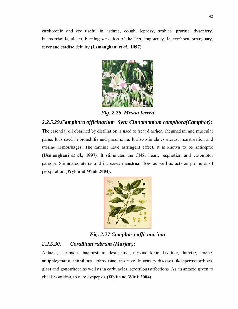
42
cardiotonic and are useful in asthma, cough, leprosy, scabies, pruritis, dysentery,
haemorrhoids, ulcers, burning sensation of the feet, impotency, leucorrhoea, stranguary,
fever and cardiac debility (Usmanghani et al., 1997).
Fig. 2.26 Mesua ferrea
2.2.5.29.Camphora officinarium Syn: Cinnamomum camphora(Camphor):
The essential oil obtained by distillation is used to treat diarrhea, rheumatism and muscular
pains. It is used in bronchitis and pneumonia. It also stimulates uterus, menstruation and
uterine hemorrhages. The tannins have astringent effect. It is known to be antiseptic
(Usmanghani et al., 1997). It stimulates the CNS, heart, respiration and vasomotor
ganglia. Stimulates uterus and increases menstrual flow as well as acts as promoter of
perspiration (Wyk and Wink 2004).
Fig. 2.27 Camphora officinarium
2.2.5.30. Corallium rubrum (Marjan):
Antacid, astringent, haemostatic, desiccative, nervine tonic, laxative, diuretic, emetic,
antiphlegmatic, antibilious, aphrodisiac, resortive. In urinary diseases like spermatorrhoea,
gleet and gonorrhoea as well as in carbuncles, scrofulous affections. As an antacid given to
check vomiting, to cure dyspepsia (Wyk and Wink 2004).

43
Fig.2.28 Corallium rubrum
2.2.5.31 Vateria indica (White Dammar,Piney varnish tree):
Family: Dipterocarpaceae
The resin exudaed by tree is medicinally important. It is credited with tonic, carminative,
and expectorant and is useful in the treatment of throat troubles. Mixed with seasamum oil,
it is given in gonorrhea and with ghee and long pepper for the treatment of syphilis and
ulcers. Fatty oil from fruit is used in local application in chronic rheumatism(Usmanghani
et al., 1997).
Fig 2.29 Vateria indica
2.2.5.32. Lapis lazuli:
Lapis lazuli is one of the stones with the longest tradition of being considered a s a
crystalline rock that can be cut and polished for jewelry. Lapis takes an excellent polish
and has been made into jewellery, carvings, boxes, mosaics, ornaments and vases. In
architecture it has been used for cladding the walls and columns of palaces and churches.
It was also ground and processed to make the dry coloring matter especially an insoluble
powder to be mixed with a liquid to produce paint etc (Usmanghani et al., 1997).
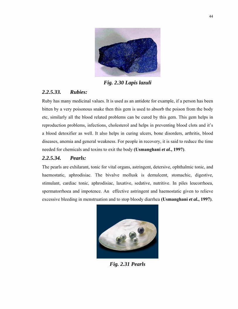
44
Fig. 2.30 Lapis lazuli
2.2.5.33. Rubies:
Ruby has many medicinal values. It is used as an antidote for example, if a person has been
bitten by a very poisonous snake then this gem is used to absorb the poison from the body
etc, similarly all the blood related problems can be cured by this gem. This gem helps in
reproduction problems, infections, cholesterol and helps in preventing blood clots and it’s
a blood detoxifier as well. It also helps in curing ulcers, bone disorders, arthritis, blood
diseases, anemia and general weakness. For people in recovery, it is said to reduce the time
needed for chemicals and toxins to exit the body (Usmanghani et al., 1997).
2.2.5.34. Pearls:
The pearls are exhilarant, tonic for vital organs, astringent, detersive, ophthalmic tonic, and
haemostatic, aphrodisiac. The bivalve mollusk is demulcent, stomachic, digestive,
stimulant, cardiac tonic, aphrodisiac, laxative, sedative, nutritive. In piles leucorrhoea,
spermatorrhoea and impotence. An effective astringent and haemostatic given to relieve
excessive bleeding in menstruation and to stop bloody diarrhea (Usmanghani et al., 1997).
Fig. 2.31 Pearls

45
2.2.5.35. Red Rubies:
Ruby has many medicinal values. Red ruby is a type of most commonly used rubies. It is
used as an antidote for example, if a person has been bitten by a very poisonous snake then
this gem is used to absorb the poison from the body etc, similarly all the blood related
problems can be cured by this gem. This gem helps in reproduction problems, infections,
cholesterol and helps in preventing blood clots and it’s a blood detoxifier as well. It also
helps in curing ulcers, bone disorders, arthritis, blood diseases, anemia and general
weakness. For people in recovery, it is said to reduce the time needed for chemicals and
toxins to exit the body (Usmanghani et al., 1997).
2.2.5.36 Bombyx mori (Silk Cocoon):
Exhilarant, refrigerant, expectorant, detersive, cardiac tonic, styptic, aphrodisiac. Generally
administered with other astringent and tonics. Being expectorant administered with other
suitable drugs in cough, asthma, catarrh and flu due to seasonal affects particularly due to
cold. As styptic, tonic and astringent to check profuse menstruation. In leucorrhoea and
chronic diarrhoea it is also used as restorative tonic (Wyk and Wink 2004).
Fig. 2.332 Bombyx mori
2.2.5.37 Honey:
Honey is nutritive, demulcent, and mild laxative. It is astringent, demulcent, detersive,
pectoral and laxative. Antiseptic applied on wound to heal it. Also used in sore throat in
constipation, malnutrition, scurvy, rickets, and to regulate secretion of glands as well as for
calcium metabolism its various preparations are administered (Wyk and Wink 2004).
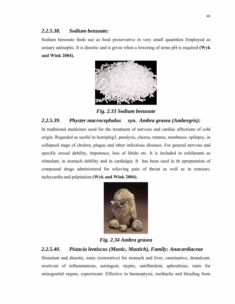
46
2.2.5.38. Sodium benzoate:
Sodium benzoate finds use as food preservative in very small quantities Employed as
urinary antiseptic. It is diuretic and is given when a lowering of urine pH is required (Wyk
and Wink 2004).
Fig. 2.33 Sodium benzoate
2.2.5.39. Physter macrocephalus syn. Ambra grasea (Ambergris):
In traditional medicines used for the treatment of nervous and cardiac affections of cold
origin. Regarded as useful in hemipleg3, paralysis, chorea, tetanus, numbness, epilepsy, in
collapsed stage of cholera, plague and other infectious diseases. For general nervous and
specific sexual debility, impotence, loss of libido etc. It is included in exhilarants as
stimulant, in stomach debility and in cardialgia. It has been used in th epreparation of
compound drugs administered for relieving pain of throat as well as in remours,
tachycardia and palpitation (Wyk and Wink 2004).
Fig. 2.34 Ambra grasea
2.2.5.40. Pistacia lentiscus (Mastic, Mastich), Family: Anacardiaceae
Stimulant and diuretic, tonic (restorative) for stomach and liver, carminative, demulcent,
resolvent of inflammations, astringent, styptic, antiflatulent, aphrodisiac, tonic for
urinogenital organs, expectorant. Effective in haemoptysis, toothache and bleeding from

47
gums. Being demulcent and expectorant, useful against cough and to clear the wind-pipe of
obstruction (Wyk and Wink 2004).
Fig. 2.35 Pistacia lentiscus
2.2.5.41. Ghee(Animal Fat):
Used as a vehicle for roasting various ingredient (Said 1979).
2.2.5.42. Musk:
Musk is an inspissated and dried secretion (testicular extract) from the perpetual follicles
of the male musk deer. It is exhilarant, tonic for vital organs, nervine and aphrodisiac tonic,
demulcent, deobstruent, antispasmodic. Musk is used in cardiac debility, palpitation and as
nervine sedative in spasmodic affections including melancholia, hypochondria, epilepsy,
hysteria, and infantile convulsions, paralysis, anemia, shock, chorea, whooping cough, etc.
It stimulates the respiratory centers as well as the urino-genital canters. As aphrodisiac
given in combination with other drugs for seminal weakness and impotence, in mental and
body fatigue leading to sleeplessness, Musk is also useful in curing dyspepsia and colitis.
For general depression a pill made up of one part musk three part musk and three parts of
camphor is beneficial (Wyk and Wink 2004).
2.2.5.43. Crocus sativus (Saffron): Family: Iridaceae
A bulbous, perennial, herb with long, linear, channeled leaves. Flowers deep violet, funnel
shaped, stalkless and appearing in Autumn with the leaves and blooming in the month of
October to November (Sharma 2003). Saffron is traditionally used to treat spasm and
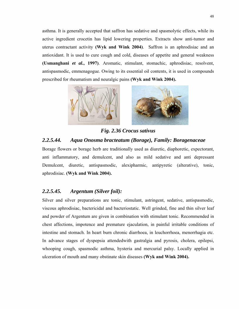
48
asthma. It is generally accepted that saffron has sedative and spasmolytic effects, while its
active ingredient crocetin has lipid lowering properties. Extracts show anti-tumor and
uterus contractant activity (Wyk and Wink 2004). Saffron is an aphrodisiac and an
antioxidant. It is used to cure cough and cold, diseases of appetite and general weakness
(Usmanghani et al., 1997). Aromatic, stimulant, stomachic, aphrodisiac, resolvent,
antispasmodic, emmenagogue. Owing to its essential oil contents, it is used in compounds
prescribed for rheumatism and neuralgic pains (Wyk and Wink 2004).
Fig. 2.36 Crocus sativus
2.2.5.44. Aqua Onosma bracteatum (Borage), Family: Boragenaceae
Borage flowers or borage herb are traditionally used as diuretic, diaphoretic, expectorant,
anti inflammatory, and demulcent, and also as mild sedative and anti depressant
Demulcent, diuretic, antispasmodic, alexipharmic, antipyretic (alterative), tonic,
aphrodisiac. (Wyk and Wink 2004).
2.2.5.45. Argentum (Silver foil):
Silver and silver preparations are tonic, stimulant, astringent, sedative, antispasmodic,
viscous aphrodisiac, bactericidal and bacteriostatic. Well grinded, fine and thin silver leaf
and powder of Argentum are given in combination with stimulant tonic. Recommended in
chest affections, impotence and premature ejaculation, in painful irritable conditions of
intestine and stomach. In heart burn chronic diarrhoea, in leuchorrhoea, menorrhagia etc.
In advance stages of dyspepsia attendedwith gastralgia and pyrosis, cholera, epilepsi,
whooping cough, spasmodic asthma, hysteria and mercurial palsy. Locally applied in
ulceration of mouth and many obstinate skin diseases (Wyk and Wink 2004).

49
2.2.6 LITERATURE REVIEW OF THE INGREDIENTS OF
HERBAL PRODUCT MUFARREH YAQOOTI MOTADIL(MUYM)
This herbal product of Hamdard Laboratories (Waqf.) Pakistan contains several cardio
active plants like Cymbopogon jwarancusa, Parmelia perlata, Elettaria cardamomum,
Nepeta hindostana, Valeriana officinalis, Santalum album and Onosma bracteatum which
have effect on CVS. This product being prescribed for strengthening the heart so acts as a
cardiac tonic and as such is traditionally used. Dose is 5g in the morning alone, followed
by 250ml of milk or water (Said 1979). As described in the ingredients section, the product
has 45 ingredients which belong to not only plant origin but also to mineral and animal
origin.
Here the literature survey of plants is presented which shows the biological and chemical
investigations performed on theses plants.
2.2.6.1. Cymbopogon jwarancusa Syn: Cymbopogon citrates
Syn: Andropogon jwarancusa (Lemon grass) :( Family: poaceae)
(Dhar and Sapru 1997) Ontogenetic variation in the essential oil concn. and major
components in the 5 genotypes of C. jwarancusa (Khavi grass) was studied. Between 0.50
to 1.64% from stage 1 (green plants spikes initiated) to stage 9 (30-50% leaves brown), the
oil content ranged from 0.50 to 1.64%. The main components of Khavi grass oil were
piperitone, (44.9-66.8%), elemol (7.0-29.2%) and β2-carene (8.3-23.5%). The percentage
composition of these constituents changes with the ontogenetic development. The β 2-
carene concentration declined sharply from pre-anthesis (19.8%) to anthesis (5.9%). The
piperitone concn. (68.0%) at the time of seed formation, suggesting regulation of oil and
oil constituents with the plant growth.
(Dhar and Dhar 1997) Ontogenetic variation in the level of carbohydrates in the five
genotypes of Cymbopogon jwarancusa. Samples of leaf and stem bases were harvested at
15 day intervals from 5 selected genotypes of Cymbopogon jwarancusa (Jones) Schultz for
the whole growing season and were analyzed for different carbohydrate fractions. The
concentration of reducing sugars increased at stage 5 (anthesis) through stage 7 (seed
filling) and then decreased towards fall, when the plant is dry. Non-reducing sugars

50
increased at post pollination period. Carbohydrate concentration fluctuated during the
growing period and 3 stages of carbohydrate accumulation were obsseved: vegetative
growth, anthesis and seed filling stage.
(Aggarwal et al., 2000) Process studies have been carried out for the isolation of geraniol
and geranyl acetate from jamrosa essential oil which find extensive use in the perfumery
and flavor industry. The jamrosa oil contained 41-58% geraniol and 22-30% geranyl
acetate and was considered a common source for the production of the aroma compdounds.
The geranyl acetate present in the jamrosa oil was converted into geraniol by hydrolysis
with different concentrations of aqueous Alkali.
(Riaz et al., 2001) Variability in yield and volatile constituents of Cymbopogon
jawarancusa (Jones) Schult from Pakistan. The volatile components of the essential oils of
six cultivars of Cymbopogon jawarancusa were examined for their oil yield and compound
variability. The data indicates that there is no appreciable difference in yield or compound
from the six cultivars.
(Kumar et al., 2007) Development of simple sequence repeat markers in Cymbopogon
species. The genus Cymbopogon comprises about 140 species, which produce
characteristic aromatic essential oils. However, the phenotypic identification of species of
Cymbopogon has been difficult as a result of widespread occurrence of natural variants,
which differ in ploidy levels and chemotaxonomic complexities. Therefore, they have
developed a set of simple sequence repeat markers from a genomic library of Cymbopogon
jwarancusa to help in the precise identification of the species (including accessions) of
Cymbopogon.
2.2.6.2 Parmelia perlata Syn: Parmelia perforata(Lichen) Family: Peltigeraceae
(Mishra et al., 1999) Biological screening of some lichens and isolation of chemical
constituents from P. reticulata. Usnic acid, gyrophoric acid, Me orsellinate, isousnic acid, β
-orsellinate, chloroatranorin and stenosporic acid are reported from Parmelia reticulata for
the first time.
(Bourgeois et al., 1999) Atraric acid, a marker for epiphytic lichens in the wood used in
cooperage: identification and quantification by G/MS/(MS). A phenolic derivative, atraric
acid, was identified by GC/MS and quantified by GC/MS/MS in the outer parts of wood

51
from oaks specifically colonized by lichens. This compd. was correlated to the presence of
a bitter depside, atranorin, which is a natural metabolite of the lichen species belonging to
the genus Parmelia.
(Abdullah etal. 2007) Two new terpenes from the lichen Parmelia perlata. The lichen
Parmelia perlata has yielded a new lanost-2-en type triterpene, named parmelanostene and
a new labdane type diterpenoid, named permelabdone, identified on the basis of
spectroscopic studies as 29, 30-nordimethyl-lanost-2-en 1 and 2'-methoxyphenyl-19-[4α, 8
β, 10 β -trimethyl-9-(13-Me butyl)-decahydronaphthalen-4-yl]-20-methoxybenzoate 2,
resectively which were also found to have antibacterial potential against S. aureus and E.
coli bacterial strains.
2.2.6.3 Elettaria cardamomum (Cardamom): Family: labiatae
(El Malti et al., 2007) Antimicrobial activity of Elettaria cardamomum: Toxicity,
biochemical and histological studies. Elettaria cardamomum is one of the most broadly
used spices in Moroccan gastronomy. Its antimicrobial activity against both Gram-positive
and Gram-negative bacterial species was demonstrated. Likewise, its toxicity was
investigated on Swiss albinos mice. Daily, mice were treated orally with 0.003 and 0.3 mg
during 7 days. Plasmatic markers and antioxidant defense systems were assessed and
histologic alterations were evaluated. A significant increase in creatine phosphokinase
level was observed The microscopic evaluation shows that E. cardamomum induce
morphologic perturbation in mice's heart. The results show also an inhibitory effect of
glyceraldehyde 3-phosphate dehydrogenase and an important increase in the level of
thiobarbituric acid reactive substances, succinate dehydrogenase and catalase activities.
Results show that E. cardamomum induces toxicity at 0.3 mg/g mouse and affect energy
metab. and oxidative stress.
(Karerat et al., 2006) Anticigarette herbal formulation as an antidote to tobacco. The
invention provides a novel nicotine free anticigarette herbal formulation as an anti-dote to
the poisoning effects of tobacco products such as Cigarettes, Gutka, Pan masala and other
similar tobacco related products. Formulation(s) comprises of sterilized dried plant
powder/extracts together with the conventional additives to form the oral dosage forms,
which include tablets, capsules, syrup and powders ready for suspension and mouth spray.

52
The anti tobacco addiction herbal formulation comprises of sesbania grandiflora,
Catharanthus roseus, Ocimum snctum, Myristica fragrans, Elettaria cardamomum, Carum
copticum, Syzygium aromaticum, Cinnamomum zeylanicum, Cuminum cyminum, Nigella
sativum, Cinnamomum camphara, Piper longum Ocimum gratssimum and Hemidesmus
indicus.
(Hpangadan et al., 2007) Herbal health protective and promotive neutraceutical
formulation for diabetics and process for preparing the same. The invention relates to a
herbal health protective, promotive and disease preventive nutraceutical herbal
formulation(s) for diabetics, and also relates to a process for the preparation of a herbal
health protective, promotive and disease preventive nutraceutical herbal formulation as
food supplement to ameliorate the general health of diabetics, said formulation comprises
the base product of microwave roasted seed powders mixture from selected genera of
Glycine, Cicer, Phaseolus, Cyamompsis, Mucuna, Hordeum, Amaranthus and Fagopyrum,
fortified with herbs/medicinal plants used are selected from the genera of Gymnema,
Momordica, Syzgium, Pterocarpus, Trigonella, Cinnamomum, Withania, Coccinia,
Pueraria, Asparagus, Boerhaavia and Aegle and also some other ingredients like Piper
longum, Chlorophytum tuberosum, Curcuma longa and Elettaria cardamomum were also
added to get the final nutraceutical product(s); the nutraceuticals are with optimum
nutrition, non toxic, natural herbal plant products, easy to digest, have health protective
and promotive properties to ameliorate the general health and vigor of diabetics.
(Pradeep et al., 2006) A process for preparing a herbal mixture for mouth soothing and
freshness comprising a mixture of Beetle nut - 35 to 55% by wt., Cardamom, Cinnamon
and Liquorice, each 5 to 15% by wt., Cathecu, lime and Menthol, each 2 to 5% and clove-
10 to 20 % by wt.
(Parry et al., 2007) Phytochemical compositions and free radical scavenging capacities of
selected cold-pressed edible seed oils. Selected specialty cold-pressed edible seed oils
including blueberry, raspberry, marionberry, boysenberry, roasted pumpkin, onion, milk
thistle, and parsley, were examinedfor their phytochemical compositions including
tocopherols, carotenoids, and total phenolic content (TPC). They were also investigated
for their radical scavenging properties against DPPH radicals and for their oxygen radical
absorbing capacity (ORAC). All of the tested cold-pressed edible seed oils differed in
53
their tocopherol profiles, carotenoid compns., TPC, and radical scavenging properties. The
highest concn. of α-tocopherol was seen in the cold-pressed onion seed oil (663.1 mg/kg),
but the highest delta- and gamma- oils. The cold-pressed roasted pumpkin seed oil
exhibited the highest β-carotene, lutein and cryptoxanthin contents at levels of 5981.2,
272.2, and 4916.7 μg/kg, resectively ORAC values of the herb and spice seed oils were
generally higher than those of the fruit seed oils. Parsley seed oil had an ORAC value of
537.0 TE μmol/g, and milk thistle oil had an ORAC value of 125.2 TE μmol/g. The
ORAC values of the fruit seed oils ranged from 1.1 to 77.9 TE μmol/g.
(Freunscht 2007) The present invention relates to an oral care compound comprising rod-
shaped apatite crystals which have a length-to-width ratio of > 5 and in which the ratio of
hydroxide ions to fluoride ions, based on the total amount of the crystals, can be easily
varied. This oral care compound, according to the invention also comprises a surfactant,
abrasive, humectant, anitmicrobial agents, anti-inflammatory agents, etc.
(Freunscht 2006) Oral care composition comprising a 1,2,3,6-tetrahydropyrimidin-2-one
cooling agent. An oral care compound, comprising a therapeutically effective amount of a
compound I (R1, R2 = H, halogen, OH, CN, NO2, SH, carbonyl, sulfone, carboxy, or
optionally substituted alkyl, alkenyl, alkoxy, alkylthio, aryl, aryloxy, arylthio, amino,
siloxy, ester, heterocycle) or a salt thereof, to produce a cooling sensation, characterized by
an oil solubilizing agent for solubilizing or emulsifying the cooling compound. Thus, a
mouthwash was made by emulsifying the cooling agent in the flavor component before
adding to the remainder of the compound. The mouthwash composition contained ethanol
6.28%, Pluronic F127 0.26%, sodium lauryl sulfate 1.07%, flavor 0.21%, 1-(2'-
hydroxyphenyl)-4-(3''-chlorophenyl)-1,2,3,6-tetrahydropyrimidin-2-one (AG35Cl) 0.05%,
and water 92.12%.
N OH
NR1
2RI

54
(Parry et al., 2006) Characterization of cold-pressed onion, parsley, cardamom, mullein,
roasted pumpkin, and milk thistle seed oils. Cold-pressed onion, parsley, cardamom,
mullein, roasted pumpkin, and milk thistle seed oils were characterized for their fatty acid
(FA) composition, tocopherol content, carotenoid profile, total phenolic content (TPC),
oxidative stability index (OSI), color, physical properties, and radical-scavenging
capacities against peroxyl (oxygen radical-scavenging capacity) and stable DPPH
(diphenylpicrylhydrazyl) radicals. Parsley seed oil had the highest oleic acid content, 81
g/100 g total FA, and the lowest satandard fat among the tested oils. Roasted pumpkin
seed oil contained the highest level of total carotenoids, zeaxanthin, β-carotene,
cryptoxanthin, and lutein at 71 μmol/kg and 28.5, 6.0, 4.9, and 0.3 mg/kg oil, resectively
(Pawar et al., 2006) In vitro efficacy of 75 essential oils against Aspergillus niger.
Aspergillus niger is an opportunistic human pathogen and a strong air pollutant. A study
was conducted with 75 different essential oils for the inhibition of hyphal growth and spore
formation in Aspergillus niger. Cinnamomum zeylanicum (bark), Cinnamomum
zeylanicum (leaf), Cinnamomum cassia, Syzygium aromaticum and Cymbopogon citratus
were the top five essential oils which demonstrated marked inhibitory effect against hyphal
growth and spore formation of A. niger. The chemical composition of these five most
active essential oils was investigated by gas chromatography-mass spectra (GC-MS).
Most of the other essential oils were found challenging to combat A. niger, suggesting
their use as strong aroma therapeutic agents.
(Pushpangadan and Prakash 2004) A process for the preparation of herbal health
protective, nutraceutical for females/expectant mothers. A herbal nutraceutical (with
application to women, esp. expectant mothers) is obtained by: (a) selecting one or more
seeds/seed products from legumes (Glycine, Cicer, Phaseolus, Psophocarpus, Mucuna,
Phaseolus vulgaris, and Phaseolus radiatus), cereals (Triticum and Hordeum), and
pseudocereals (Amaranthus, Chenopodium, and Fagopyrum) ranging between 80-90%; (b)
roasting the seed/seed products for 2-15 min; (c) powdering the roasted seeds to 50-400
mesh and mixing to obtain a base product (d) obtaining extracts from herbs/medicinal
plants (Centella, Withania, Pueraria, Asparagus, Chlorophytum, Boerhaavia, Sida, and
Saraca) and drying the extractto obtain a residue; (e) mixing the plant extractresidue
ranging up to 50% with base product obtained from step (c) to obtain the final

55
nutraceutical products and optionally adding one or more additives selected from sugar,
jaggery, milk powder Piper longum, Myristica fragrans, Elettaria cardamomum, and cocoa
powder.
(Thomas et al., 2006) Traded (exported) cardamoms from India, Sri Lanka and Guatemala
are characterized based on physical and biochemical parameters and molecular techniques.
For most of the physical quality parameters and for the biochemical traits, such as starch
and crude fiber, Indian cardamom is found superior to the other produces. Further GC
profile of the oil of Indian cardamom indicated high quantity of α-terpinyl acetate and 1,8-
cineole which imparts aroma and flavor to the cardamom, thus reinforcing the legendary
belief of high intrinsic quality of the Indian cardamom. Molecular profiling using
RAPD/ISSR primers did not reveal much polymorphism among the 3 cardamoms. This
aspect is discussed in relation to the origin and transnational spread of cardamom.
(Murali et al., 2006) Plant-based formulation for bronchial asthma: a controlled clinical
trial to compare its efficacy with oral salbutamol and theophylline. Plant-based medicine is
the 3rd most popular choice of both adults (11%) and children (6%) suffering from
Asthma.
(de Pradier 2006) A trial of a mixture of three essential oils in the treatment of
postoperative nausea and vomiting. The management of postoperative nausea and vomiting
(PONV) that occurs in 30% of all surgical cases is indispensable for a rapid recovery,
patient comfort and the overall image of general anesthesia. Currently, allopathic drugs
proposed in surgery include major drugs such as morphine as well as analgesics of class
IIB such as Nefopam and Tramadol. They wanted to demonstrate that it is possible to treat
these negative effects of anesthesia with a mixture of three essential oils via percutaneous
application on the anterolateral aspect of the neck. This study comprised of 73 cases and
used a mixt. of Zingiber officinale (ginger), Elletaria cardamomum (cardamom) and
Artemisia dracunculus (tarragon) essential oils in equal parts. The results were collected
approximately 30 min following application as soon as the symptoms appeared in the
theater recovery room. Seventy five percent of cases had a favorable outcome. The best
results were obtained with patients who had received a single drug that provoked the
symptoms. In all other cases, results were reduced to a 50% success rate.

56
(Al-Othman et al., 2006) Effect of dietary supplementation of Ellataria cardamomum and
Nigella sativa on the toxicity of rancid corn oil in rats.In the present study Nigella sativa
and Elettaria cardamomum seeds have been studied as inhibitors of oxidative stress caused
by oxidized corn oil (having PV 389.8 meq kg-1) in rats. The 70 days feeding male albino
rats with experimental diets did not produce significant changes in the body wts., organ
wts. and food intake of different groups. Increased lipooxidative damage was noticed in
oxidized oil fed rats. Oxidized oil diet supplemented with cardamom or N. sativa had
marked redn. in RBC hemolysis and plasma AST/ALT activity. The formation of
thiobarbituric acid reactive substances were lowest in rats fed N. sativa supplemented diet.
Reduced glutathione of liver and kidney were significantly raised after the addn. of
cardamom/N. sativa to the diets compared to oxidized oil fed rats (Group F). But heart
reduced glutathione showed a significant increase as compared to oxidized oil fed group
only after the supplementation of N. sativa to the diet. These data indicates that N. sativa
or cardamom supplementation improves the overall antioxidant protection capacity of the
body.
(Rajamani et al., 2005) Mixture Improves Glucose Metabolism and Lipid Profile in
Fructose-Fed Hyperinsulinemic Rats. Fructose feeding has been shown to induce insulin
resistance in rats, associated with hyperinsulinemia, hyperglycemia, and
hypertriglyceridemia. They investigated the effect of administering food seasoning spices
mixture (SM) on glucose, insulin, and lipids in circulation and carbohydrate enzymes in
the erythrocytes of high fructose-fed rats. They also measured the protein glycation status
by assaying the levels of glycated Hb, fructosamine, and plasma protein glycation. Male
Wistar rats received a daily diet containing either 60% fructose or 60% starch (control).
The rats were administered SM at three different doses (10, 30, or 50 mg/day per rat) orally
15 days later. At the end of the 45-day experimental period, fructose-fed rats showed
significantly higher levels of plasma glucose and insulin, dyslipidemia, and alterations in
enzyme activities. Treatment with SM significantly reduced plasma glucose and insulin
levels and brought about a favorable lipid profile. In these rats, the activities of enzymes
of glucose metabolism were normal. These effects were observed at all three doses of SM.
High homeostasis model assessment (HOMA) values indicated insulin resistance in
fructose fed rats, while the HOMA values in SM-treated fructose-fed rats were comparable
57
to those of control rats. They concluded that administration of SM improves glucose
metabolism and plasma lipid profile in fructose-fed rats, possibly through improved
insulin-sensitizing actions of the active constituents.
(Hinneburg et al., 2006) Antioxidant activities of extracts from selected culinary herbs
and spices. Recently, interest in plant-derived food additives has grown, mainly because
synthetic antioxidants suffer from several drawbacks. Furthermore, plant extracts were
shown to possess health-promoting properties. In the present study, hydrodistillation
extracts from basil, laurel, parsley, juniper, aniseed, fennel, cumin, cardamom, and ginger
were assessed for their total phenol content, and antioxidant (Fe(III) reduced, inhibition of
linoleic acid peroxidn., Fe(II) chelation, 1,1-diphenyl-2-picrylhydrazyl radical-scavenging
and inhibition of hydroxyl radical-mediated 2-deoxy-D-ribose degrdn., site and nonsite-
specific) activities. The extracts from basil and laurel possessed the highest antioxidant
activities except for Fe chelation. Although parsley showed the best performance in the Fe
chelation assay, it was less effective at retarding the oxidation of linoleic acid. In the
linoleic acid peroxidation assay, 1 g of the basil and laurel extracts were as effective as 177
and 212 mg of trolox, respectively Thus, both extracts are promising alternatives to
synthetic substances as food ingredients with antioxidant activity.
(Kumar et al., 2005) Essential oil composition of seed and fruit coat of Elettaria
cardamomum from South India. Twenty five constituents of the hydrodistilled essential oil
obtained separated from the seed and fruit coat of the Elettaria cardamomum accounting
for 95.28% and 96.58% of the oil respectively, growing in South India have been analyzed
and identified using GC and GC-MS. Major constituents found common in both seed oil
and fruit coat oil are limonene (4.05% and 3.82%), 1,8-cineole (15.13% and 23.74%), α-
terpineol (4.67% and 5.25%) and α-terpinyl acetate (56.87% and 51.25%) respectively.
(Josephrajkumar et al., 2005) The occurrence of a trypsin-like protease in fresh
cardamom seeds, as shown by the benzoyl-Arg-p-nitroanilide (BApNA)-hydrolyzing
ability of the seed enzyme preparation under alkaline conditions is reported here for the 1st
time. The enzyme had pH and temp. optima of 8 and 45°C, resectively It was inhibited by
aprotinin and phenylmethyl sulfonyl fluoride (PMSF) in a dose-dependent manner,
suggesting the presence of Ser residues at the active site. The enzyme had a Vmax of
98.01 nmol p-nitroaniline released per min per mg protein and a Km of 0.0684 mM with

58
BApNA as substrate. The addition of aprotinin (75.75 μM) increased the Km by 3-fold,
whereas the Vmax was reduced by 23%.
(McCue et al., 2005) Anti-amylase, anti-glucosidase and anti-angiotensin I-converting
enzyme potential of selected foods Program in Molecular and Cellular Biology. α-Amylase
and β-glucosidase were targeted as potential avenues for modulation of postprandial
hyperglycemia through mild inhibition of the enzymic breakdown of complex
carbohydrates to decrease meal-derived glucose absorption. Water-soluble extracts with
optimized phenolic content of selected American and Asian foods were investigated for
inhibitory activity against α-amylase and β-glucosidase, as well as angiotensin I-
converting enzyme (ACE), which was linked to hyperglycemia-assocd. hypertension.
Porcine pancreatic α-amylase (PPA) was allowed to react with each phenolic-optimized
food ext., and the derivatized enzyme-phytochemical mixtures obtained were characterized
for residual amylase activity. The β-glucosidase and ACE activities were determined in the
presence of each phenolic-optimized food extraction The amylase activity was inhibited
more than the glucosidase activity in the presence of these phytochemical extracts, and
more so by Asian foods than by American foods. The Asian spice ginger was found to
possess strong ACE inhibitory activity in addition to significant anti-amylase activity. The
α-amylase enzyme inhibition was positively associated with extract antioxidant activity
and negatively with extract protein content. The significance of food-grade, plant-based
amylase inhibitors for modulation of carbohydrate breakdown and control of glycemic
index of foods in the context of preventing hyperglycemia and diabetes mellitus
complications in the long term and ACE inhibitors for modulation of associated
hypertension is hypothesized and discussed.
(Nostro et al., 2005) Antibacterial effect of plant extracts against Helicobacter pylori. The
aim of this work was to evaluate the antibacterial effect of plant extracts as alternative
and/or as active agents supporting antibiotics for treating Helicobacter pylori infection.
The effect of either, ethanolic or aqueous extracts from 17 plant materials were studied
against one H. pylori standard strain and 11 clinnical isolates using a disk diffusion test
and by evaluating the minimum inhibitory concentration (MIC) on solid media. An
inhibitory activity against H. pylori strains was recorded in a large percentage of tested
plants. MIC values of ethanolic extracts were from two to four concentration steps lower

59
than the aqueous ones. In particular, ethanolic extracts of Cuminum cyminum L. and
Propolis expressed MIC90 values of 0.075 mg/mL. The results show a significant in vitro
effect of plant extracts against H. pylori that could be considered a valuable support in the
treatment of the infection and may contribute to the development of new and safe agents
for inclusion in anti-H. pylori regimens.
(Dumri and Lertsiri 2005) Pro-oxidative activity in some Thai spices. Twenty
methanolic extracts from Thai spice powder (anise, cardamom, chilli, Chinese key,
cinnamon, clove, coriander, cumin, deeplee, dill, fennel, galangal, kencur, lemongrass,
ginger, nutmeg, oregano, pepper, safflower, and turmeric) were screened for pro-oxidative
activity using a lecithin-liposome model system. The oxidative stress was induced by four
systems: (I) non-metal induction; (II) FeCl3/H2O2/EDTA; (III) FeSO4/ascorbate; and (IV)
CuSO4/H2O2. Of these spices, safflower, lemongrass, and nutmeg extracts showed the
pro-oxidative activity observed by thiobarbituric acid reactive substance elevation. Since
pro-oxidn. were metal dependent, hydroxyl radical formation was investigated by
monitoring fluorescence products from benzoate hydroxylation. The formation of hydroxyl
radical was confirmed with the fluorescence detected and the suppression due to addition
of mannitol, a hydroxyl radical scavenger.
(Krishnan et al., 2005) The present work reports on the microencapsulation of cardamom
oleoresin by spray drying using binary and ternary blends of gum arabic, maltodextrin, and
modified starch as wall materials. The microcapsules were evaluated for the content and
stability of volatiles, entrapped 1,8-cineole and entrapped α-terpinyl acetate for 6 wk. A
4/6,1/6,1/6 blend of gum arabic:maltodextrin:modified starch offered a protection, better
than gum arabic as seen from the t1/2, time required for a constituent to reduce to 50% of
its initial value.
(Ninfali et al., 2005) Antioxidant capacity of vegetables, spices and dressings relevant to
nutrition, Vegetables are the most important sources of phenolics in the Mediterranean
diet. Phenolics, especially flavonoids, are suggested as being essential bioactive
compounds providing health benefits. The results can be used in public health campaigns
to stimulate the consumption of vegetables able to provide significant health protection to
prevent chronic diseases.
60
(Choi and Hwang 2005) Screening of Indonesian medicinal plants for inhibitor activity
on nitric oxide production of RAW264.7 cells and antioxidant activity. Traditional
Indonesian medicinal plants were screened for their inhibitory effects on the nitric oxide
(NO) production in lipopolysaccharide (LPS)-stimulated RAW264.7 macrophages and for
the antioxidant activity through the evaluation of free radical scavenging effect and
reducing power. The results of screening indicated that 50 methanolic extracts inhibited
(>50%) lipopolysaccharides (LPS)-induced NO release from RAW264.7 cells at 50
µg/mL, with 18 having greater than 100% inhibition. At 200 μg/mL, 61 methanol extracts
exhibited inhibitory activity (>50%), with 45 showing greater than 100% inhibition. In
addition, the free radical scavenging effects of 6 methanolic extracts were found to be
more than 50% for extractconcn. of 0.5 µg/mL. The results indicate that the extracts
contain active compounds that inhibit NO release and scavenge free radical.
2.2.6.4 Amomum subulatum(Greater Cardamom): Family: Zingiberaceae
(Gupta et al., 1984) Gas chromatographic evaluation of the essential oils of different
strains of Amomum subulatum growing wild in Sikkim. The dried fruits of A. subulatum
(Ellataria major) of 7 strains were steam distillation and analyzed by gas chromatography
The major components were: cineole [470-82-6] 77-89, Ÿ-terpinene [99-85-4] 4.73-8.738
α -bisabolene [17627-44-0] 1.09-7.05, α -terpineol [98-55-5] and α -terpinyl acetate [80-
26-2] 3.52-6.38, and sabinene [3387-41-5] 3.11-4.61%. β -Terpineol [138-87-4] was
found in only 2 of the species. The high cineole content makes these fruits inferior to true
cardamom (E. cardamomum). Oil yields were 2.0-3.3%.
2.2.6.5 Nepeta hindostana (Nepeta herb): Family: Labiateae
(Mueed 2006) A process for preparing a pharmaceutical (Unani) composition for the
treatment of hyperlipidemia and hypercholesterolemia. This invention relates to a process
for preparing a pharmaceutical compounds for the treatment of hyperlipidemia and
hypercholesterolemia comprising the following steps: - preparing extracts of the herbal
ingredients Safoof Seer (Allium sativum), Safoof Chob Zard (Curcuma longa),
Badranjboya (Nepeta hindostana) and Abhal (Juniperus communis), Lak Maghsool
(Caterica Lacca) and Zeera Siyah (Cuminum cyminum); concentrating the extracts and

61
reducing them to a powder by heating, mixing the powders, 120-135 mg Allium sativum,
40-60 mg Curcuma longa, 115-135 mg Nepeta hindostana, 0.5-1.5 mg Juniperus
communis, 30-60 mg Cateria Lacca and 50-100 mg Cumin cyminum, with 40-60 mg of
nicotinic acid, and if desired, adding 75-1OO mg ascorbic acid, mixing the powders with
predetermined quantity of binders and excipients at room temperature, homogenizing the
mixture to ensure uniformity and therapeutic value of the compound
(Mueed 2005) A poly-pharmaceutical unani composition for the treatment of
hyperlipidemias and hypercholesterolemia. The invention relates to a poly -
pharmaceutical Unani composition for the treatment of Hyperlipidemias and
hypercholesterolemia comprising the following ingredients in the proportions mentioned
there. Ingredients Parts Used Quantity by parts Satoff Seer (Alliumsativum, Linn) Bulb
120-135 mg Safoof Chob Zard (Curcuma longa, Linn) Rhizome 40-60 mg Badranjboya
(Nepeta hindostana, Roth, Heines) Flowers, leaves Aqueous Extract. Dry 120-135 mg
Abhal (Juniperus communis, Linn) Oil 0.5-1.5 mg Lak Maghsool (Caeria Lacca) Powder
30-60mg Zeera Siyah (Cuminum cyminum,Linn) Seeds 50- 100mg Nicotinic Acid Powder
40 -60 mg and if desired Ascorbic acid Rhizome 140-160 mg.
(Singh et al., 2001) A note on antibacterial activity of volatile oils of some aromatic
plants.The antibacterial activity of volatile oils of Alpinia carinata, Zingiber officinale,
Lippia alba, Lantana camara, L. indica, Hyptis suaveolens and Nepeta hindostana was
investigated. The volatile oils were obtained by hydrodistilation using Clevenger's
apparatus and dried over anhydrous sodium sulfate. The essential oils exhibited strong
antibacterial potential. While the oils of L. alba and H. suaveolens showed the best
performance, those of L. indica and L. camara were less active. The oils of A. carinata, Z.
officinale and N. hindostana were also found effective. However, the oil of N. hindostana
was not sensitive to Pseudomonas aeruginosa. These volatile oils can be exploited against
Corynebacterium diphtheriae, Escherichia coli, Klebsiella species, Staphylococcus
haemolyticus, S. aureus, and P. aeruginosa. They are more effective than std. antibiotics
and can be developed as an alternative source of natural and safe bactericides.
(Ashraf et al., 1999) Endothelium modulated vasorelaxant response of a
polypharmaceutical herbal drug (lipotab) and it's individual constituents. The present study
was undertaken to examine the endothelium modulated effects of polypharmaceutical drug

62
lipotab and its individual ingredients in isolated aortic rings of rat. Endothelium intact and
denuded aortic rings were precontracted with phenylephrine 10-6 M and drugs were added
in cumulative manner in concentration ranging from 1 to 50 µg/mL. The results
demonstrated an endothelium-dependent vasorelaxant effect of lipotab and its individual
ingredients, with the exception of nicotinic acid. The dose dependent relaxant response of
nicotinic acid was not altered significantly in the endothelium-denuded rings, suggesting a
direct effect of the drug on the vascular smooth muscle. Vasorelaxant effect of lipotab and
its individual constituents suggests the therapeutic potential of these compounds in certain
cardiovascular diseases.
(Varma and Dubey 1997) Insecticidal and insect repellent activity of some essential oils
against Tribolium castaneum. During screening of 14 essential oils against the storage pest,
Tribolium castaneum, the oils of 3 plants viz. Caesulia axillaris, Ocimum gratissimum and
Nepeta hindostana showed strong insect repellent activity. All oils except that of Vetiveria
zinzanioides were found to possess insecticidal property.
2.2.6.6 Nardostachys jatamansi (Indian valerian): Family; Valerianaceae
(Chatterjee et al., 2007) An overview of the genus Nardostachys. Nardostachys
jatamansi, a medicinally important herb of Nepalese origin, has been used for centuries in
the Ayurvedic and Unani systems of medicine. In combination with Marsilea minuta it is
being used as an antistress and anticonvulsant drug and also finds use in the treatment of
epilepsy. Recently, it has been reported that N. jatamansi, which plays an important role in
protecting from cerebral ischemia and liver damage, is also used for the treatment of
osteoporosis and hypercalcemia. The other member of the genus Nardostachys, N.
chinensis, possesses antifungal and antimalarial properties. It is also used in the treatment
of skin dysfunction. A short summary of the chemical constituents of the two species
along with their physical and biological properties is reported.
(Subashini et al., 2007) Biochemical study on the protective potential of Nardostachys
jatamansi extract on lipid profile and lipid metabolizing enzymes in doxorubicin
intoxicated rats. Nardostachys jatamansi is a medicinally important herb of Indian origin
used for centuries in Ayurvedic and Unani systems of medicine for the treatment of
various ailments. The aim of the present work is to evaluate the effect of ethanolic extract

63
of Nardostachys jatamansi rhizomes on doxorubicin induced myocardial injury with
respect to lipid metab. in serum and heart of Wistar albino rats. Altered lipid metab. alters
the cardiac function which is mainly due to changes in the property of the cardiac cell
membrane. Doxorubicin exhibits cardiotoxicity by inhibition of fatty acid oxidation in the
heart. The rats treated with a single dose of doxorubicin (15 mg/kg) i.p. showed an
increase in serum and cardiac lipids (cholesterol, triglycerides, free fatty acids and
phospholipids), along with a significant rise in serum low density lipoproteins (LDL), very
low density lipoproteins (VLDL) and drop in high density lipoproteins (HDL) levels,
resulting in alteration of serum and cardiac lipid metabolizing enzymes. Pretreatment with
a extract of Nardostachys jatamansi (500 mg/kg) orally for seven days to doxorubicin
induced rats showed a significant prevention in the lipid status with the activities of the
lipid metabolizing enzymes. Histopathological observations were also in correlation with
the biochemical parameters. These findings suggest that the protective and hypolipidemic
effect of Nardostachys jatamansi against doxorubicin induced myocardial injury in rats
could possibly be mediated through its anti lipid peroxidative properties.
(Costa et al., 2007) Reliable identification of terpenoids and related compounds by using
linear retention indices interactively with mass spectrometry search. An innovative tool in
GC-MS peak assignment procedures is described.
(Mundewadi 2006) A herbal composition to treat Schizopherenia comprising
therapeutically effective amount of Bacopa monnieri (Jal brahmi) and Nardostachys
jatamansi (Jatamansi).
(Mukherjee et al., 2006) Process of preparing a herbal composition for the management
of insomnia/stress and product thereof. This invention relates to an anti-stress composition
containing various medicinal herbs and this composition prepared by a process comprising
the steps i) mixing about 20 to 35 weight parts of Nardostachys jatamansi, 40 to 50 weight
parts of Nelumbo nucifera, 20 to 25 weight parts of Zingiber officinalis and 8 to 15 weight
parts of Valeriana wallichii ii) extracting the mixture with water or alcohol or mixture of
water and alcohol in suitable ratio. This herbal composition may be useful for the
management of insomnia stress.
(Surveswaran et al., 2007) Systematic evaluation of natural phenolic antioxidants from
133 Indian medicinal plants. Total antioxidant capacities of 133 Indian medicinal plant
64
species sampled from 64 families were assessed by ABTS, DPPH and FRAP assays, and
their total phenolic contents measured by Folin-Ciocalteu assay. Preliminary identification
of the major phenolic compounds from 83 selected medicinal plants by reversed-phase
HPLC revealed phenolic acids, tannins, flavonoids, curcuminoids, coumarins, lignans, and
quinines. The fruit of Terminalia chebula, pericarp of Punica granatum and gall of Rhus
succedanea showed very high levels of hydrolysable tannins, and the gum of Acacia
catechu presented very high levels of catechin and epicatechin in addition to tannins.
Major phenolics in many of the medicinal plants were identified for the first time (e.g.,
Euphorbia lathyrus, Ipomoea turpethum, and Picrorrhiza kurroa). This systematic
investigation of a large no. of Indian medicinal plants proved important for understanding
their chem. constituents and functionality in Ayurvedic medicine, and contributes to the
search for natural sources of potent antioxidants.
(Subashini et al., 2006) Protective effect of Nardostachys jatamansi on oxidative injury
and cellular abnormalities during doxorubicin-induced cardiac damage in rats.
Nardostachys jatamansi is a medicinally important herb of Indian origin. It has been used
for centuries in the Ayurvedic and Unani systems of medicine for the treatment of various
ailments. We have evaluated the effect of N. jatamansi (rhizomes) on the biochem.
changes, tissue peroxidative damage and abnormal antioxidant levels in doxorubicin
(adriamycin)-induced cardiac damage. Preliminary studies on the effect of the graded dose
of extractshowed that 500 mg kg-1 orally for seven days was optimum and hence all
further study was carried out with this particular dose. Rats administered doxorubicin (15
mg kg-1, i.p.) showed myocardial damage that was manifested by the elevation of serum
marker enzymes (lactate dehydrogenase, creatine phosphokinase, aspartate
aminotransaminase and alanine aminotransaminase). The animals showed significant
changes in the antioxidant enzymes (superoxide dismutase, catalase, glutathione
peroxidase and glutathione-S-transferase) and lipid peroxidn. levels. Pretreatment with N.
jatamansi extractsignificantly prevented these alterations and restored the enzyme activity
and lipid peroxides to near normal levels. Restoration of cellular normality accredits the
N. jatamansi with a cytoprotective role in doxorubicin-induced cardiac damage.
(Ahmad et al., 2006) Attenuation by Nardostachys jatamansi of 6-hydroxydopamine-
induced parkinsonism in rats: behavioral, neurochemical, and immunohistochemical

65
studies. Parkinson's disease (PD) is one of the commonest neurodegenerative diseases, and
oxidative stress has been evidenced to play a vital role in its causation. In the present
study, we evaluated whether ethanolic extractof Nardostachys jatamansi roots (ENj), an
antioxidant and enhancer of biogenic amines, can slow the neuronal injury in a 6-OHDA-
rat model of Parkinson's. Rats were treated with 200, 400, and 600 mg/kg body wt. of ENj
for 3 wk. On day 21, 2 µl of 6-OHDA (12 µg in 0.01% in ascorbic acid-saline) was
infused into the right striatum, while the sham-operated group received 2 µl of vehicle.
Three weeks after the 6-OHDA injection, the rats were tested for neurobehavioural activity
and were sacrificed after 6 wk for the estn. of lipid peroxidation, reduced glutathione
content, the activities of glutathione-S-transferase, glutathione reductase, glutathione
peroxidase, superoxide dismutase and catalase, quantification of catecholamines,
dopaminergic D2 receptor binding and tyrosine hydroxylase expression. The increase in
drug-induced rotations and deficits in locomotor activity and muscular coordination due to
6-OHDA injections were significantly and dose-dependently restored by ENj. Lesioning
was followed by an increased lipid peroxidation and significant depletion of reduced
glutathione content in the substantia nigra, which was prevented with ENj pretreatment.
The activities of glutathione-dependent enzymes, catalase and superoxide dismutase in
striatum, which were reduced significantly by lesioning, were dose-dependently restored
by ENj. A significant decrease in the level of dopamine and its metabolites and an
increase in the no. of dopaminergic D2 receptors in striatum were observed after 6-OHDA
injection, and both were significantly recovered following ENj treatment. All of these
results were exhibited by an increased d. of tyrosine hydroxylase immunoreactive (TH-IR)
fibers in the ipsilateral striatum of the lesioned rats following treatment with ENj; 6-
OHDA injection had induced almost a complete loss of TH-IR fibers. This study indicates
that the extractof Jatamansi might be helpful in attenuating Parkinsonism.
(Donaldson et al., 2005) Assessment of Antimicrobial Activity of Fourteen Essential Oils
When Using Dilution and Diffusion Methods.The lack of solublity and the high degree of
volatility of essential oils present significant problems to determining the biological effects
of these oils. The activity of 10 essential oils and 4 essential oil blends against a Gram-
positive bacterium, Staphylococcus aureus, and a yeast, Candida albicans, was compared
using a dilution assay and two diffusion methods. The tube dilution assay, using a 0.2%

66
agar soln. to provide a stable homogeneous dispersion of oils, was used to measure min.
inhibitory concentrations(MICs). The relative merits of using p-iodonitro tetrazolium dye
(INT) or optical d. (OD) in measuring MIC of essential oils also were evaluated. All 14
oils were active against both microorganisms. Diffusion assays are useful as a qual.
assessment of biol. activity of essential oils but are not appropriate in assessing
quantitativeeffects.
(Chatterjee et al. 2005) Studies on the chemical constituents of Nardostachys jatamansi
DC (Valerianaceae). A new sesquiterpene acid, nardin (I), and a new pyranocoumarin (II)
have been isolated from the rhizomes of Nardostachys jatamansi and characterized as E-2-
Me, 3-(5,9-dimethylbicyclo[4.3.0]-nonen-9-yl)-2-propenoic acid and 2',2'-dimethyl-3'-
methoxy-3',4'-dihydropyranocoumarin, resectively using spectral studies and chemical
correlation. The stereochemistry of nardin has been determined using X-ray
crystallographic studies.
Me
HMe
Me
HCO 2 H
O O O
Me
Me
OMe
I
I I
(Kukreja and Singh 2004) Nardostachys jatamansi DC. - the Indian nardostachy. A
review on the biololgical, chemical, cultivation, biotechnological, and pharmacological
properties of the genus Nardostachys, with a focus on N. jatamansi.
(Mody et al., 1997) An improved process for extraction of the Ayurvedic plant Jatamansi
(Nardostachys jatamansi) is described. Jatamansi is used for the treatment of various
disorders like convulsions and palpitation of heart, as sedative in high blood pressure and
cardiac arrhythmias and to prepare hair tonics. Rhizomes of Jatamansi were graded,
shredded and powd. and the powd. plant was exttracted with an extracting solvent at 40-
45°. The extract obtained was filtered, mixed and concentrated to a thick paste under

67
reduced pressure at 40-55° and spray dried to obtain a dry powder extracts. The volatile
oil in the extract was collected.
(Singh et al., 2003) New phytoconstituents from Nardostachys jatamansi rhizomes.
Phytochemical investigation of the rhizomes of Nardostachys jatamansi led to the isolation
of unknown compounds n-eicosan-1-oic acid-15-ol-20-yl-n-decan-3'-enoate, n-eicosan-1-
oic acid-15µ-ol-20-yl heptanoate, n-hexacosanyl propanoate, n-nonadeca-2-oic acid-15-ol-
1-cyclohexane carboxylate, 26-(µ-tetradecahydroanthracenyl)-n-hexacosan-3-on-16µ-ol,
n-docosan-1-yl pentanoate-22-oic acid ethoxylate, 1-decanyl-4-(Bu pentanoate) decaline
and n-heptacosan-1-ol-27-pentanoate, in addition to n-nonacosan-7-ol, n-heneitriacontan-
5-ol and ß-sitosterol. The structures of all these phytoconstituents have been established
on the basis of spectral data analyses and chemical reactions.
(Mahalwal and Ali 2002) Volatile constituents of the rhizomes of Nardostachys
jatamansi DC. A steam distilled volatile oil obtained from the rhizomes of Indian Nard
(Nardostachys jatamansi DC) belonging to Valerianaceae family, grown in Indian
Himalayas, was analyzed by capillary GC and GC/MS. The rhizome volatiles contained
72 components of which 41 constituents comprising 70.0% of the oil were positively
identified. The oil is composed of nine monoterpenes (1.7%), 25 sesquiterpenes (43.9%)
and 7 non-terpenic components (24.4%). The predominant sesquiterpenes were nardol
(10.1%), α-selinene (9.2%), ß-caryophyllene (3.3%), cubebol (2.9%), α -gurjunene (2.5%),
ĝ-gurjunene (2.3%) and α -humulene (2.3%).
(Salim et al., 2003) The protective effect of Nardostachys jatamansi (NJ) on
neurobehavioral activities, thiobarbituric acid reactive substance (TBARS), reduced
glutathione (GSH), thiol group, catalase and sodium-potassium ATPase activities was
studied in middle cerebral artery (MCA) occlusion model of acute cerebral ischemia in
rats. The right MCA of male Wistar rats was occluded for 2 h using intraluminal 4-0
monofilament and reperfusion was allowed for 22 h. MCA occlusion caused significant
depletion in the contents of glutathione and thiol group and a significant elevation in the
level of TBARS. The activities of Na+K+ ATPase and catalase were decreased
significantly by MCA occlusion. The neurobehavioral activities (spontaneous motor
activity and motor coordination) were also decreased significantly in MCA occlusion
group. All the alternations induced by ischemia were significantly attenuated by 15 days
68
pretreatment of NJ (250 mg/kg po) and correlated well with histopathol. by decreasing the
neuronal cell death following MCA occlusion and reperfusion. The study provides first
evidence of effectiveness of NJ in focal ischemia most probably by virtue of its antioxidant
property.
(Bindra et al., 2001) Anti-fungal herbal formulation for treatment of human nails fungus
and process thereof. The invention relates to an antifungal compound for the treatment of
human nails containingextracts of walnut hull, pulverized roots of Nardostachys jatamansi
or Vetiveria zizanioides or Catharanthus roseus, polyols, fixed oil, nonionic emulsifiers,
thickening agents, plasticizers and a base. The invention also relates to a process for the
prepn. of the above synergistic compound in which the pulverized root material is added
and slowly stirred so that a homogeneous emulsion is formed and extractfrom walnut hull
is added and stirred with a controlled stirring motion at a temperature of 35-40°.
(Chatterjee et al., 2000) Structure and Stereochemistry of Nardostachysin, a New
Terpenoid Ester Constituent of the Rhizomes of Nardostachys jatamansi, were established
as the 7',8'-dihydroxy-4'-methylenehexahydrocyclopenta[c]pyran-1'-one-8'-Me ester of 7,9-
guaiadien-14-oic acid, by spectral and chem. studies.
(Rani and Naidu 1998) Subjective and polysomnographic evaluation of a herbal
preparation in insomnia. A herbal preparation consisting of Rauwolfia serpentina root
alcohol extract, Nardostachys jatamansi rhizome powder, and Tinospora cordifolia root,
stem, and leaf aq. extractwas tested as soporific in patients suffering from insomnia. Two
tablets each containing100 mg of each drug were given daily at bed time for 21 days. The
quality of sleep and the total sleep time were enhanced by the preparation. Sleep latency
and the no. and total duration of nocturnal awakenings decreased. A non-significant
decrease of REM sleep was also observed. There was no noticeable alteration in
hematological or biochemical parameters. Most of the patients tolerated the test
preparation very well without feeling of hangover or day time drowsiness. 11/39 Patients
complained only mild epigastric distress. The compound was estimated by HPLC.
(Mukherjee et al., 2007) Screening of Indian medicinal plants for acetylcholinesterase
inhibitory activity. The cholinergic hypothesis of Alzheimer's disease (AD) has provided
the rationale for the current pharmaco-therapy of this disease, in an attempt to reduce the
cognitive decline caused by cholinergic deficits.

69
(Vinutha et al., 2007) Screening of selected Indian medicinal plants for
acetylcholinesterase inhibitory activity. Seventy-six plant extracts including methanolic
and successive water extracts from 37 Indian medicinal plants were investigated for
acetylcholinesterase (AChE) inhibitory activity (in vitro). Results indicated that
methanolic extracts to be more active than water extracts. The potent AChE inhibiting
methanolic plant extracts included Withania somnifera (root), Semecarpus anacardium
(stem bark), Embelia ribes (Root), Tinospora cordifolia (stem), Ficus religiosa (stem bark)
and Nardostachys jatamansi (rhizome). The IC(50) values obtained for these extracts were
33.38, 16.74, 23.04, 38.36, 73.69 and 47.21mug/ml, respectively. These results partly
substantiate the traditional use of these herbs for improvement of cognition.
(Kumar et al., 2006) Search for antibacterial and antifungal agents from selected Indian
medicinal plants. A series of 61 Indian medicinal plants belonging to 33 different families
used in various infectious disorders, were screened for their antimicrobial properties.
Screening was carried out at 1000 and 500 microg/ml concentrations by agar dilution
method against Bacillus cereus var mycoides, Bacillus pumilus, Bacillus subtilis,
Bordetella bronchiseptica, Micrococcus luteus, Staphylococcus aureus, Staphylococcus
epidermidis, Escherichia coli, Klebsiella pneumoniae, Pseudomonas aeruginosa,
Streptococcus faecalis, Candida albicans, Aspergillus niger and Saccharomyces cerevisiae.
Twenty-eight plant extracts showed activity against at least one of the test organisms used
in the screening. On the basis of the results obtained, we conclude that the crude extracts
of Dorema ammoniacum, Sphaeranthus indicus, Dracaena cinnabari, Mallotus
philippinensis, Jatropha gossypifolia, Aristolochia indica, Lantana camara, Nardostachys
jatamansi, Randia dumetorum and Cassia fistula exhibited significant antimicrobial
activity and properties that support folkloric use in the treatment of some diseases as
broad-spectrum antimicrobial agents. This probably explains the use of these plants by the
indigenous people against a number of infections.
(Joshi and Parle 2006) Nardostachys jatamansi improves learning and memory in mice.
Cure of cognitive disorders such as amnesia, attention deficit, and Alzheimer's disease is
still far from being realized in the field of medicine. Nootropic agents such as piracetam,
aniracetam, and choline esterase inhibitors like donepezil are being used for improving
memory, mood, and behavior, but the resulting side effects associated with these agents

70
have made their applicability limited. In Ayurveda, the roots of Nardostachys jatamansi
have been clinically employed for their anti-ischemic, antioxidant, anticonvulsant, and
neuroprotective activities. The present study was undertaken to assess the potential of N.
jatmansi as a memory enhancer. The elevated plus maze and the passive avoidance
paradigm were employed to evaluate learning and memory parameters. Three doses (50,
100, and 200 mg/kg, p.o.) of an ethanolic extract of N. jatamansi were administered for 8
successive days to both young and aged mice. The 200 mg/kg dose of N. jatmansi
ethanolic extract significantly improved learning and memory in young mice and also
reversed the amnesia induced by diazepam (1 mg/kg, i.p.) and scopolamine (0.4 mg/kg,
i.p.). Furthermore, it also reversed aging-induced amnesia due to natural aging of mice.
As scopolamine-induced amnesia was reversed, it is possible that the memory
improvement may be because of facilitation of cholinergic transmission in the brain.
Hence, N. jatmansi might prove to be a useful memory restorative agent in the treatment of
dementia seen in elderly persons. The underlying mechanism of action can be attributed to
its antioxidant property.
(Rao et al., 2005) Anticonvulsant and neurotoxicity profile of Nardostachys jatamansi in
rats. Ethanol extract of the roots of Nardostachys jatamansi DC. (Valerianaceae) was
studied for its anticonvulsant activity and neurotoxicity, alone and in combination with
phenytoin in rats. The results demonstrated a significant increase in the seizure threshold
by Nardostachys jatamansi root extract against maximal electroshock seizure (MES) model
as indicated by a decrease in the extension/flexion (E/F) ratio. However, the extract was
ineffective against pentylenetetrazole (PTZ)-induced seizures. Nardostachys jatamansi
root extract also showed minimal neurotoxicity against rotarod test at doses that increased
the seizure threshold. Further, pretreatment of rats with phenytoin at a dose of 12.5, 25, 50
and 75 mg/kg in combination with 50mg/kg of Nardostachys jatamansi root extract
resulted in a significant increase in the protective index (PI) of phenytoin from 3.63 to
13.18. The dose response studies of phenytoin alone and in combination with
Nardostachys jatamansi extract on the serum levels of phenytoin clearly demonstrated the
synergistic action of both the drugs.
(Ali et al., 2000) Nardostachys jatamansi is a medically important herb of Indian origin
used for centuries in Ayurvedic and Unani systems of medicine for the treatment of

71
various ailments. In the present paper, a 50% ethanolic extract of the rhizomes of N.
jatamansi is shown to possess hepatoprotective activity. Pretreatment of rats with the
extract (800 mg/kg body wt, orally) for three consecutive days significantly ameliorated
the liver damage in rats exposed to the hepatotoxic compound thioacetamide. Elevated
levels of serum transaminases (aminotransferases) and alkaline phosphatase, observed in
thioacetamide alone treated group of animals, were significantly lowered in N. jatamansi
pretreated rats. Pretreatment of the animals with the extract also resulted in an increase in
survival in rats intoxicated with LD90 dose of the hepatotoxic drug.
2.2.6.7 Santalum album (Sandal tree): Family: Santalaceae
(Choudhary 2006) A herbal composition for boosting energy and relieving anxiety and
process for preparing the same. Treatment of patients in the convalescing stage,
particularly after major surgical intervention, needs medication and judicious selection of
diets. Separate medicines are noted for boosting energy and relieving mental anxiety,
which belong to separate domains altogether. These medicines particularly relieving
anxiety are not only costly but also need to be administered for fairly long spell. The
present invention aims at overcoming the foregoing difficulties and provides a herbal
composition for boosting energy and relieving anxiety comprising - Ingredients Amount in
wt % (i) Cannabis Indica (Bhang) - 25 (ii) Santalum album (Chandan) - 23 (iii)
Nardostachya jata mansi (Jatamansi) - 0.3 (iv) Piper longum (Pipal) - 0.3 (v) Borax
(Sohaga) - 0.3 (vi) Myristic officinalis (Javitri) - 0.3 (vii) Hedychium Spicatium
(Gandhali) - 0.3 (viii) Piper nigrum (Marich) - 0.3 (ix) Cepdris deodara (Devdaru) - 0.3 (x)
Asparagus racemosus (Satavar) - 0.3 (xi) Calotropis procera (Akban) - 0.3 (xii)
Cinnamonum (Dalchini) - 0.3 (xiii) Electroa cardamom (Ellaichi (big) } - 0.3. (xiv)
Tribulus terrestris (Gokhru) - 3 (xv) Pedalium Murex (Nagkeshar) - 0.7 (xvi) Convolvulus
agents wat (Hizzal) - 2 (xvii) Deshi ghee (Buffalo) - 8 (xviii) Flour (Maida) - 6 (xix)
Powdered gram (Besan) - 6 (xx) Saccharum officinale (Sugar) - 23 This invention also
provides a process for preparing the above composition in a powdery form.
(Mainkar et al., 2005) Herbal skincare composition. The present invention discloses
herbal skin care compositions for topical application containing extracts from Swertia
decusatta (Kadu), Eclipta Alba (Bhringaraj), Santalum album (Sandalwood), Parmelia

72
species (Dagadphool), Ginkgo biloba (Ginkgo, maidenhair tree), Bacopa monnieri
(Brahmi), Centella asiatica (Mandukparni) and formulations for skin lightening and anti
aging creams thereof. Further the present invention discloses preparation of extracts and
evaluated for their dermatological effects. In vitro screening of the said herbal extracts for
cosmetic applications are carried out for their anti elastase activity, anti hyaluronidase
activity, superoxide scavenging activity and anti tyrosinase activity.
(Heuberger et al., 2006) East Indian Sandalwood and ß -santalol odor increase
physiological and self-rated arousal in humans. In Ayurvedic medicine, East Indian
Sandalwood is an important remedy for the treatment of both somatic and mental
disorders. In this investigation, the effects of inhalation of East Indian Sandalwood
essential oil and its main compd., ß -santalol, on human physiol. parameters (blood oxygen
satn., respiration rate, eye-blink rate, pulse rate, skin conductance, skin temp., surface
electromyogram, and blood pressure) and self-ratings of arousal (alertness, attentiveness,
calmness, mood, relaxation and vigor) were studied in healthy volunteers. Compared to
either an odorless placebo or ß -santalol, Sandalwood oil elevated pulse rate, skin
conductance level, and systolic blood pressure. ß -Santalol, however, elicited higher
ratings of attentiveness and mood than did Sandalwood oil or the placebo. Correlation
analyses revealed that these effects are mainly due to perceived odor quality. The results
suggest a relation between differences in perceived odor quality and differences in arousal
level.
(Inouye et al., 2006) Vapor activity of 72 essential oils against a Trichophyton
mentagrophytes. For practical application for vapor therapy and fumigation, the vapor
activity of 72 essential oils was screened against a Trichophyton mentagrophytes, using a
closed box. The fungicidal activity, expressed as the minimum fungicidal dose (MFD),
was determined from the colony size, which was correlated with the inoculum size. Oils
containing phenol as the major component showed the most potent vapor activity, with an
MFD of 1.56 µg/mL air. This was followed by oils with aldehyde as the major component,
then by those with alcohol as the major component. The vapor activities of oils
containingketone, ester, and ether/oxide components were decreased, in that order. The
oils that were rich in hydrocarbon components had the weakest activity. The same
tendency was observed with the components themselves. Phenols and aldehydes exhibited

73
the highest vapor activity, followed by alcs., ketone, ester, ether/oxide, and hydrocarbon.
There was a rough correlation between the vapor activity determinedby the box vapor
assay and the contact activity determinedby agar diffusion assay. But oils
containingsesquiterpenes showed weaker vapor activity than that expected from the contact
activity. Based on the activity-chemical structure relationship, the 72 oils were classified
into seven functional groups.
(Kim et al., 2006) New antitumor sesquiterpenoids from Santalum album of Indian origin.
Three new campherenane-type (1, 4, 7) and three new santalane-type (9, 11, 12)
sesquiterpenoids, and two arom. glycosides (21, 22) together with 12 known metabolites
including α,ß-santalols (14, 18), (E)- α,ß -santalals (15, 19), α,ß -santaldiols (16, 20), α-
santalenoic acid (17), and vanillic acid 4-O-neohesperidoside were isolated from Santalum
album chips of Indian origin. The structures of the new compounds, including absorption
configurations, were elucidated by 1D- and 2D-NMR spectroscopic and chemical methods.
The antitumor promoting activity of these isolates along with several neolignans
previously isolated from the same source was evaluated for both in vitro Epstein-Barr virus
early antigen (EBV-EA) activation and in vivo two-stage carcinogenesis assays. Among
them, compd. 1 exhibited a potent inhibitory effect on EBV-EA activation, and also
strongly suppressed two-stage carcinogenesis on mouse skin.
(Komori et al., 2006) Application of fragrance in discontinuing the long-term use of
hypnotic benzodiazepines. An evaluation was made of the usefulness of fragrance
application in discontinuing the long-term use of hypnotic benzodiazepines in primary
insomniacs with low-dose dependence. Based on the results of pentobarbital sleep time in
rats, we made a new fragrance consisting primarily of sandalwood (35%), juniper berry
(12%), rose (8%) and orris (6%). This mixed fragrance was found to prolong the
pentobarbital sleep time in rats. A total of 42 outpatients with low-dose dependence on
hypnotic benzodiazepines, all of whom met DSM-IV criteria for primary insomnia,
participated in the study. In advance, all subjects attempted to reduce the doses of drugs
gradually (25% redn. a week if possible) and 29 subjects who had failed to do so at all
participated in the study on the application of fragrance. A mixed fragrance described
above was used. A gradual tapering of hypnotic benzodiazepines (25% redn. a week if
possible) was attempted while sniffing the fragrance in bed. The application of fragrance

74
reduced the doses of hypnotic benzodiazepines in 26 of 29 subjects and 12 subjects did not
require any drug for sound sleep. The present study indicated that a kind of fragrance may
prove effective as an alternative to hypnotic benzodiazepines.
(Kim et al., 2005) Bisabolane- and Santalane-Type Sesquiterpenoids from Santalum
album of Indian Origin. Six new bisabolane-type (1-3) and santalane-type (4-6)
sesquiterpenoids, together with (+)-α-nuciferol, (+)-citronellol, and geraniol, were isolated
from the heartwood of Santalum album of Indian origin. Their structures, including two
bisabolol diastereomers (1, 2), were established on the basis of spectroscopic data
interpretation.
(Kim et al., 2006) Aromatic constituents from the heartwood of Santalum album L. A
phytochem. investigation of the polar constituents in the heartwood of Indian Santalum
album resulted in the isolation of three new neolignans (1-3) and a new arom. ester (4),
along with 14 known components. The structures of the new compounds (1-4) were
established using spectroscopic methods.
(Ochi et al., 2005) Anti-Helicobacter pylori Compounds from Santalum album. Six new
sesquiterpenes, (Z)-2α-hydroxy-14-hydro-β-santalol, (Z)-2 α -hydroxy-albumol, 2R-(Z)-
campherene-2,13-diol, (Z)-campherene-2β,13-diol, (Z)-7-hydroxynuciferol, and (Z)-1 α -
hydroxy-2-hydrolanceol, together with five known compounds, (Z)-� α -santalol, (Z)-� β
-santalol, (Z)-lanceol, β -santaldiol, and β -santaldiol, were isolated from Santalum album,
by using bioassay-guided fractionation for Helicobacter pylori. The structures were
determined by extensive NMR studies. The absorption configuration of 2R-(Z)-
campherene-2,13-diol was determinedby a modified Mosher method. The crude extracts
as well as the isolated compounds showed antibacterial activity against H. pylori.
Especial, compounds (Z)-�-santalol and (Z)-�-santalol have strong anti-H.pylori
activities against a clarithromycin-resistant strain (TS281) as well as other strains.
(Bieri et al., 2004) Olfactory Receptor Neuron Profiling using Sandalwood Odorants. The
mammalian olfactory system can discriminate between volatile molecules with subtle
differences in their molecular structures. One important class of molecules in the fragrance
industry are sandalwood odorants. Sandalwood oil and four synthetic sandalwood
molecules were selected to study the activation profile of endogenous olfactory receptors
when exposed to compounds from the same odorant family. Dissociated rat olfactory

75
receptor neurons were exposed to the sandalwood molecules and the receptor activation
studied by monitoring fluxes in the internal calcium concentration. Olfactory receptor
neurons were identified that were specifically stimulated by sandalwood compounds.
These neurons expressed olfactory receptors that can discriminate between sandalwood
odorants with slight differences in their molecular structures. This is the first study in
which an important class of perfume compounds was analyzed for its ability to activate
endogenous olfactory receptors in olfactory receptor neurons.
(Howes et al., 2004) Evaluation of the quality of sandalwood essential oils by gas
chromatography-mass spectrometry. Trade and historic oils from sandalwoods', labeled as
Amyris balsamifera, Eremophila mitchelli, Fusanus acuminatus (= Santalum acuminatum),
Santalum album, S. austrocaledonicum, S. latifolium, S. spicatum and S. yasi, were
assessed using gas chromatog.-mass spectrometry (GC-MS). Using GC-MS, none of the
oils assessed complied with the internationally recognized std. of a 90% santalol content,
and only about half of the trade sandalwood oils met with recent International Organization
for Standardization stds. The majority of trade oils, reportedly from S. album, contained
approx. 50-70% santalols (Z-α and Z-ß). Thus, the internationally recognized specification
(90% santalols) for S. album requires re-evaluation by more efficient analysismethods. In
view of the issues assocd. with the quality of sandalwood oils being traded, specifications
of 43% Z- α -santalol and 18% Z- ß-santalol for S. album oil esttimated by GC-MS are
suggested. GC-MS are recommended as it assists with authentication and quality control
issues assocd. with sandalwood oils.
(Braun et al,. 2003) Isolation and chiral GC analysis of -bisabolols-trace constituents
from the essential oil of Santalum album L. (Santalaceae). The essential oil produced from
the heartwood of Santalum album L. was analyzed using GC/MS. Forty-four constituents
were identified as one monoterpene and 43 sesquiterpenes. 2,10-Bisaboladien-6-ol (-
bisabolol/epi--bisabolol) was one of the trace compounds found in east Indian
sandalwood oil. Chiral GC analysis showed that besides the main stereoisomer, which
possessed an (R,R)-configuration, all three other isomers were also present.
(Gharia et al., 2002) Anthelmintic activities of essential oils of Bothriochloa pertusa,
Abutilon indicum, Xanthium strumarium, Mentha spicata, Cinnamomum zeylanicum,
Santalum album, Parthenium hysterophorus were studied on earth worm, tape worm, hook

76
worm and nodular worm. It was observed that essential oils from Xanthium strumarium
possessed superior anthelmintic activities against earth worm, tape worm, hook worm and
nodular worm, as compared to the standard substance. The essential oil from Abutilon
indicum also possessed considerable anthelmintic activities against all four worms as
compared to the standard substance.
(Okugawa et al., 2000) Effects of sesquiterpenoids from "Oriental incenses" on acetic
acid-induced writhing and D2 and 5-HT2A receptors in rat brain. Six sesquiterpenoids:
jinkoh-eremol, agarospirol, - and -santalol, dehydrocostus lactone and costunolide,
previously isolated from Oriental incenses (agarwood, Saussurea root, and sandalwood),
inhibited HOAc-induced writhing in mice. The effect of -, - and -antagonists on the
action of these compounds was tested. The analgesic effect of �santalol was blocked by
a -�antagonist, but not by - and -antagonists. This effect of --santalol was different
from that of morphine. -Santalol was the most potent of the 6 compounds as an
antagonist of dopamine D2 and serotonin 5-HT2A receptor binding in the rat brain. The
antipsychotic effect of -santalol was similar to that of chlorpromazine, although --
santalol was less potent than chlorpromazine.
(Hammer et al., 1999) The antimicrobial activity of plant oils and extracts has been
recognized for many years. However, few investigations have compared large nos. of oils
and extracts using methods that are directly comparable. In the present study, 52 plant oils
and extracts were investigated for activity against Acinetobacter baumanii, Aeromonas
veronii biogroup sobria, Candida albicans, Enterococcus faecalis, Escherichia coli,
Klebsiella pneumoniae, Pseudomonas aeruginosa, Salmonella enterica subsp. enterica
serotype typhimurium, Serratia marcescens and Staphylococcus aureus, using an agar
dilution method. Lemongrass, oregano and bay inhibited all organisms at concentrations
of 2.0% (vol./vol.). Six oils did not inhibit any organisms at the highest concentration,
which was 2.0% (vol./vol.) oil for apricot kernel, evening primrose, macadamia, pumpkin,
sage and sweet almond. Variable activity was recorded for the remaining oils. Twenty of
the plant oils and extracts were investigated, using a broth microdilution method, for
activity against C. albicans, S. aureus and E. coli. The lowest min. inhibitory
concentrationswere 0.03% (vol./vol.) thyme oil against C. albicans and E. coli and 0.008%

77
(vol./vol.) vetiver oil against Staph. aureus. These results support the notion that plant
essential oils and extracts may have a role as pharmaceuticals and preservatives.
(Benencia and Courreges 1999) Antiviral activity of sandalwood oil against Herpes
simplex viruses-1 and -2. Sandalwood oil was investigated for its in vitro antiviral activity
against Herpes simplex viruses-1 and -2. Virus replication was inhibited dose-dependently
(7.5-60 µg/mL) and more pronounced against Herpes simplex virus-1. A slight diminution
was observed at higher multiplicity of infections. The oil was not virucidal.
(Adlof 1999) The Lindlar-catalyzed reduction of methyl santalbate: a facile preparation
of methyl 9-cis,11-trans-octadecadienoate-9,10-d2. Conjugated linoleic acid (CLA) has
been associated with the reduction of chemically induced cancers in mice and rats and the
suppression of atherosclerosis in rats. We have found seed oils to be a valuable source of
precursors for the rapid preparation of gram quantities of deuterium-labeled fats. Me
santalbate (Me 11-trans-octadecen-9-ynoate), obtained from Santalum album (Linn.) seed,
was reduced with Lindlar catalyst, quinoline, and deuterium gas to produce, in yields of
65-75%, the gram quantities of Me 9-cis,11-trans-octadecadienoate-9,10-d2 (CLA-d2) we
required for metabolism and oxidation studies. Unlike monoacetylenic and methylene-
interrupted polyacetylenic fatty acid Me esters, the conjugated system was reduced with no
noticeable break in the rate of deuterium uptake. The quantity of poison (quinoline)
present did influence the amount of CLA-d2 produced, but the production of overreduced
fatty acid Me esters (perhaps because of the conjugated system) could not be prevented.
Fractionation of the reaction mixture by silver resin chromatography resulted in the
isolation of >99% CP CLA-d2 in yields of 60-70%.
2.2.6.8 Bambusa arundinacea (Thorny Bamboo): Family: Gramineae
(Chandra et al., 2004) Goitrogenic content of Indian cyanogenic plant foods and their in
vitro anti-thyroidal activity. Consumption of cyanogenic foods was considered as one of
the etiol. factors in certain instances for the persistence of endemic goiter. The present
study was undertaken to study the cyanogenic glucosides, glucosinolates and thiocyanate
content in edible portion of certain selected plant foods of Indian origin. Further in vitro
anti-thyroidal activity using raw, boiled and cooked extracts of these plants with and
without excess iodide was also studied. Cyanogenic plant foods generally vegetables were

78
collected from different areas of West Bengal and Tripura. Cassava was obtained from
Meghalaya and Kerala and their cyanogenic glucosides, glucosinolates and thiocyanate
were estd. Thyroid peroxidase activity (TPO) of human thyroid was assayed from
microsomal fraction following I3 from iodide. The anti-TPO activities of the plants were
assayed after adding raw, boiled and cooked extracts in the assay medium with and without
extra iodide. Relative antithyroidal potency of the plant extracts was also evaluated in
terms of the concn. (IC50) necessary to produce 50 per cent inhibition of TPO activity.
PTU equivalence of the plant foods was also determined Cabbage and cauliflower were
rich in glucosinolates, bamboo shoot and cassava were rich in cyanogenic glucosides,
mustard, turnip and radish were relatively rich in thiocyanate however all the constituents
were present in each plant. Boiled extracts showed max. inhibition of TPO activity
followed by cooked and raw extracts Excess iodide was found relatively effective for raw
extractbut less effective for boiled and cooked extracts in reversing anti-TPO activity.
Inhibition constituents (IC50) was found highest with bamboo shoot and least with
cabbage. Raw, boiled and cooked extracts of the plants showed anti-thyroidal activity in
vitro. Excess iodide reversed the anti-TPO activity to same extent but could not neutralize
it.
(Muniappan et al., 2003) Antiinflammatory and antiulcer activities of Bambusa
arundinacea. The extracts of Bambusa arundinacea have been used in Indian folk medicine
to treat various inflammatory conditions. The plant has got antiulcer activity also. It is
thought that these two properties in the same extract are very much useful in the treatment
of inflammatory conditions. It is well known fact that the most of the available
antiinflammatory drugs are ulcerogenic. The antiinflammatory effect of the methanol
extract of the leaves of Bambusa arundinacea against carrageenin-induced as well as
immunologically induced paw oedema and also its antiulcer activity in albino rats have
been studied and found to be significant when compared to the standard drugs. The
combination of methanol extract and phenylbutazone (Non-Steroidal Antiinflammatory
Agent, NSAIA) has been studied and found to be the most potent antiinflammatory activity
experimentally with least toxic (no ulcerogenic) activity. Thus, the combination of herbal
product (methanol extract of Bambusa arundinacea) with modern medicine (NSAIAs) will
produce the best antiinflammatory drug and will be useful for long-term treatment of

79
chronic inflammatory conditions like rheumatoid arthritis with peptic ulcer, which are
common.
2.2.6.9 Centaurea behen (Behen, Centaurea): Family: Compositeae
(Yadava et al., 2006) Novel bioactive triterpenoid saponin from Centaurea behen linn.
C. behen linn. belongs to family compositae which is commonly known as "Safed
Bahman" in Hindi. Its powd. roots are used in treatment of fistula and gravel. Its
powdered seeds are administered along with wine as a remedy for stone. The roots are
also used in calculus affections and jaundice. Earlier workers have reported various
constituents from this plant. In the present paper, the isolation and structural elucidation of
novel triterpenoid saponin 3α-O-[β-L-arabinopyranosyl(1 α 4)- β --L-rhamnopyranosyl(1 α
4)-O- β -D-xylopyranosyl]-16 α -hydroxy-13,28-epoxyolean-29-oic acid from the roots of
this plant was reported.
(Gurkan et al., 1998) Cytotoxicity assay of some plants from Asteraceae. Sesquiterpene
lactones were tested for cytotoxic activity. The results showed that solstitialin
monoacetate and matricarine are the most cytotoxic agents among tested substances.
(Oksuz et al., 1993) A guaianolide diol monoacetate from Centaurea behen and C
solstitialis ssp. Schouwii. The structure of a guaianolide diol monoacetate previously
reported from C. behen and C. solstitialis ssp. schouwill was revised to I as determinedby
x-ray analysis
O
H
OHeM
O
O
OAc
CH2
H
I
(Bruno et al., 1991) Guaianolides and lignans from Centaurea solstitialis subsp. schouwii.
Aerial parts of Centaurea solstitalis subsp. schouwii afforded the guaianolides cynaropicrin
and aguerin B and the lignans arctigenin and matairesinol. The structure of a third
guaianolide previously found also in Centaurea behen was revised.
80
2.2.6.10 Citrus medica (Citron): Family: Rutaceae
(Conforti et al., 2007) In vitro activities of Citrus medica L. cv. Diamante (Diamante
citron) relevant to treatment of diabetes and Alzheimer's disease. The present study
showed for the first time the in vitro properties (antioxidant, hypoglycemic and
anticholinesterase) of Citrus medica L. cv. Diamante which belongs to the Rutaceae
family. The n-hexane extract of Diamante citron peel is characterized by the presence of
monoterpenes and sesquiterpenes. The most abundant constituents were two
monoterpenes: limonene and α -terpinene. The extract showed significant antioxidant
activity that was carried out using different assays (DPPH test, β-carotene bleaching test
and bovine brain peroxidation assay). Oxidative damage, caused by the action of free
radicals, may initiate and promote the progression of a no. of chronic diseases, including
diabetes and Alzheimer's disease. Diamante citron peel extractshowed hypoglycemic
activity and an anticholinesterase effect.
(Domb et al., 2007) Double-layered bioadhesive tablets for the topical treatment of oral
mucosal disorders. Bioadhesive sticker tablets which are applied directly to vaginal, rectal
and/or oral mucosa are described herein. In one embodiment, the sticker tablets are
applied directly to ulcers or lesions in the oral cavity. The compounds adhere immediately
upon administration, swell over time, and remain adherent to the ulcer or lesion for at least
60 min. The compounds can be in the form of single layer, double layer, or multilayer
sticker tablets. The compoundsprovide immediate pain relief to the patient and promote
rapid healing of the ulcer or lesion. The sticker tablet compound contain one or more
bioadhesive polymers. In one embodiment, the polymers are crosslinked polycarboxylic
acids and polyols. The compounds contain at least one herbal agent and/or irritating
compound, and optionally, a non-herbal active agent. The compoundscan deliver an non-
irritating ED of the agent for at least 60 min. The compounds described herein are stable
upon storage for six months or longer. For example, a sticky tablet contained limonene,
pinenes, myrcene, terpinene, citron oil, orange oil, mint oil, clove oil, lemone oil and
pumela oil, Klucel HF and silica.
(Gans et al., 2007) The present invention relates to a cosmetic compound comprising in a
preblend or in the resting state prior to application to a keratinous surface, at least one
peptide, at least one collagen containing compound, at least one penetration enhancer, at
81
least one mucopolysaccharide, and at least one proteoglycan, wherein these ingredients are
operable to associated in situ when the compound is applied to a keratinous surface and a
method for plumping lips or skin by applying the compound Thus, a lipstick compound
comprised polybutene 5.00, Me paraben 0.20, Pr paraben 0.20, pentaerythrityl
tetraisostearate 5.00, C10-30 cholesterol/lanosterol esters 5.00, ethylhexyl palmitate 10.00,
ceresin 10.00, ozokerite 5.0, microcryst. wax 2.5, caprylic/capric triglyceride 5.0,
octyldodecyl neopentanoate 5.0, trioctyldodecyl citrate 34.94, Red 7 Lake 0.05, Yellow 6
Lake 0.01, iron oxides 0.10, mica 5.0, and complex containingphospholipids, Citrus
medica limonum ext., atelocollagen, collagen pre-peptide, chondroitin sulfate, hyaluronic
acid and tocopherol 1.00 wt.%, resectively
(Green et al., 2007) Determination of polymethoxylated flavones in peels of selected
Jamaican and Mexican citrus (Citrus spp.) cultivars by high-performance liquid
chromatography. The concentrations of the polymethoxylated flavones (PMFs) in peels of
selected citrus cultivars grown in Jamaica and Mexico were determined. The PMFs were
extractedfrom sun-dried citrus peels with reagent-grade methanol. Analyses were carried
out by reverse-phase HPLC and UV detection. The column used was a C18 5 m (150x4.6
mm) Discovery column. Elution was in the gradient mode, using a ternary mobile phase.
The results showed that all the citrus cultivars used contained at least three of the six major
PMFs quantified. Ortanique peel contained the highest quantity of PMFs (34,393 272
ppm), followed by tangerine (28,389 343 ppm) and Mexican sweet orange (sample 1;
21,627494ppm). The major PMFs, i.e. sinensetin, nobiletin, tangeretin,
heptamethoxyflavone, tetramethylscutellarein and hexamethyl-o-quercetagetin, present in
the peels of 20 citrus cultivars, was quantified. The results were compared with those of
Florida citrus peels. A large amtount of citrus peels and byproducts are produced in the
Caribbean which could provide a cheap and convenient source of PMFs.
(Herent et al., 2007) Determination of new retention indices for quick identification of
essential oils compounds. The classical methods of chromatography identification of
compounds were based on calculation of retention indexes by using different stationary
phases.
(Nogata et al., 2007) Isolation of eriocitrin (eriodictyol 7-O-rutinoside) as an arachidonate
lipoxygenase inhibitor from Lumie fruit (Citrus lumia) and its distribution in Citrus
82
species. An inhibitory compound acting against rat platelet 12-lipoxygenase was isolated
from the peel of Lumie fruit (Citrus lumia) by activity-guided separation. It was identified
as eriocitrin (eriodictyol 7-O-rutinoside) by spectroscopic analyses. Erioctrin inhibited 5-
lipoxygenase (IC50 29.1 mol L-1) from rat peritoneal polymorphonuclear leukocytes in
addn. to 12-lipoxygenase (IC50 22.3mol L-1). Its aglycon, eriodictyol (5,7,3',4'-
tetrahydroxyflavanone), was a much more potent inhibitor of both 12-lipoxygenase (IC50
0.07 mol L-1) and 5-lipoxygenase (IC50 0.20 mol L-1). It also inhibited the production
of leukotriene B4 in intact peritoneal polymorphonuclear leukocytes stimulated with
calcium ionophore A23187 (IC50 12.7 mol L-1). The distribution of eriocitrin in 39
citrus fruits was investigated by high-performance liquid chromatography analysis Lumie,
eureka lemon (Citrus limon), Sambokan (Citrus sulcata), Sudachi (Citrus sudachi) and Koji
(Citrus leiocarpa) fruits were found to contain high levels of eriocitrin in both peel and
juice vesicles.
(Jayaprakasha et al., 2006) In vitro evaluation of the antioxidant activities in fruit extracts
from citron and blood orange. Consumers are increasingly aware of diet related health
problems and therefore demanding natural ingredients which are expected to be safe and
health-promoting. Recently, number of studies on health benefits associated with citrus
phytochemicals were demonstrated. In the present study, an attempt was made to isolate
antioxidant fractions from 2 different citrus species such as Citron (Citrus medica) and
blood orange (C. sinensis). Antioxidant fractions were extractedfrom mature, ripe fruits
using 5 different solvents using a Soxhlet extractor. The total phenolic content of the
extracts was determinedby Folin-Ciocalteu method. MeOH:water (80:20) extracts of
citron and acetone extractof blood orange was found to contain maximum phenolics. The
dried fractions were screened for their antioxidant activity potential using in vitro model
systems such as 1, 1-diphenyl-2-picryl hydrazyl (DPPH), phosphomolybdenum method
and as well as by the nitroblue tetrazolium (NBT) redn. test at different concentrations The
MeOH:water (80:20) fraction of citron showed highest radical scavenging activity 42.5%,
77.8% and 92.1% at 250, 500 and 1000 ppm, resectively, while MeOH:water (80:20)
fraction of blood orange showed lowest DPPH radical scavenging activity at all the tested
concentrations Furthermore, all the fractions showed remarkable antioxidant capacity by
the formation of phosphomolybdenum complex.
83
(Lu et al., 2006) Citrus flavonoids in fruit and traditional Chinese medicinal food
ingredients in China. Flavonoids-enriched tissues of citrus such as peel, immature fruit
and flower are consumed as culinary seasonings, tea ingredients in China for centuries.
This HPLC quantitativestudy on the five citrus flavonoids, naringin, hesperidin,
neohesperidin, sinensetin and nobiletin on a wide range of Chinese citrus fruits and several
Traditional Chinese Medicinal food ingredients in East China, revealed a great diversity in
flavonoid compound Huyou peel (C. paradisi cv. Changshanhuyou) was found to be the
best naringin (3.25%) and neohesperidin (2.76%) source; C. aurantium, a major ingredient
of several citrus-related TCM, is also a suitable source of naringin and neohesperidin, and
a good juice source for flavanone glycosides; the peel of Wenzhoumiju (C. unshiu) is one
of the richest local species in hesperidin (up to 6.25%); Zaoju (C. subcompressa) has the
highest content of nobiletin (0.59%), a polymethoxylated flavone. LC-ES-MS analysis of
Zanthoxylum genus for flavonoids revealed for the first time the presence of significant
amts. (0.74%) of hesperidin in the root of Liangmianzhen (Z. nitidum (Roxb.) DC), a
relative of Sichuan pepper, which is a spice widely used in China.
(Monajemi et al., 2005) Cytotoxic effects of essential oils of some Iranian Citrus peels.
There have been efforts to overcome the problem in treatment of cancer using medicinal
plants. It has been shown that Citrus essential oil of contains different terpens with
antitumor activities. The cytotoxicity of essential oils of Iranian Citrus limon (L.), C.
medica (L.), C. sinensis (L.) peels on cancer cell lines was determined. Essential oils were
preparedby hydrodistilation and characterized by GC-MS.
(Nogata et al., 2006) Flavonoid composition of fruit tissues of citrus species. An HPLC
analysis was performed on the concentrations of flavonoids in 42 species and cultivars of
the Citrus genus and those of 2 Fortunella and one Poncirus species according to the
classification system established by Tanaka. The compound of 8 flavanones and 9
flavone/ols for these species was determinedin the albedo, flavedo, segment epidermis and
juice vesicle tissues, and those in the fruit and peel tissues were calcd. from the compound
data of the tissues. A principal component analysisshowed that such neohesperidosyl
flavonoids as neoeriocitrin, naringin, neohesperidin, and rhoifolin had large factor loading
values in the first principal component for each tissue.

84
(Avula et al., 2005) An analytical method was developed for the simultaneous quantitative
analtsis of 6 amines and 20 flavonoids in fruits and extracts of 30 Citrus species, including
C. aurantium, near-Citrus relatives, and dietary supplements by liquid chromatography
with photodiode array detection. The separation was achieved with a Phenomenex Synergi
Hydro reversed-phase column using gradient mobile phase of sodium acetate buffer (pH
5.5) and acetonitrile. Elution was run at a flow rate of 1.0 mL/min and UV at 254, 280,
and 330 nm. Among the amines analyzed, synephrine, the main component, was present
in the levels from 0.11 to 2.0 mg/g dry wt. in 21 Citrus species and 0.07 to 18.62% in
dietary supplements claiming to contain C. aurantium. The flavanones and flavones were
analyzed in the same Citrus samples and were species-specific. The levels of flavones
were very low compared with those of flavanones. The method facilitated the
simultaneous quantification of 6 amines and 20 flavonoids in various Citrus species, the
distinction between the different Citrus species, and the analysis of dietary supplements
containing C. aurantium.
(Caristi et al., 2006) Flavone-di-C-glycosides in citrus juices from Southern Italy. 6,8-Di-
C-glucopyranosylapigenin and 6,8-di-C-glucopyranosyldiosmetin were detected by HPLC-
DAD-ESI-MS-MS in a variety of Southern Italian Citrus juices (orange, lemon, bergamot,
citron, mandarin, clementine). In some juices, the amount of C-glycosides detected were
significant. 6,8-Di-C-glucopyranosylapigenin is characteristic of orange juice, while 6,8-
di-C-glucopyranosyldiosmetin is the most important C-glycoside in lemon and citron juice.
In bergamot juice the concentrationsof 6,8-di-C-glucopyranosylapigenin and 6,8-di-C-
glucopyranosyldiosmetin are similar. Clementine juice is distinctive as the amts. of both
C-glycosides present are negligible.
(Inouye et al., 2001) The purpose of this study was to examine the antibacterial effects of
a wide variety of essential oils on major respiratory tract pathogens. The antibacterial
activity of 14 essential oils and their major components was evaluated by agar-plate
dilution assay under sealed conditions, with agar used as a stabilizer for homogeneous
dispersion. Of the selected strains of four major bacteria causing respiratory tract
infection, Haemophilus influenzae was most susceptible to the essential oils, followed by
Streptococcus pneumoniae and Streptococcus pyogenes. Staphylococcus aureus was less
susceptible. No cross-resistance was observed between penicillin-sensitive and penicillin-

85
resistant S. pneumoniae. Escherichia coli, used as a control bacterium, showed the lowest
susceptibility. Essential oils containing aldehyde or phenol as a major component showed
the highest antibacterial activity, followed by the essential oils containing terpene alcohols.
Other essential oils, containing terpene ketone, or ether, had much weaker activity, and an
oil containingterpene hydrocarbon was inactive. Based on these findings, thyme (wild,
red, and geraniol types), cinnamon bark, lemongrass, perilla, and peppermint oils were
selected for further evaluation of their effects on respiratory tract infection.
(Lota et al., 2002) Volatile Components of Peel and Leaf Oils of Lemon and Lime
Species. Peel and leaf oils of 43 taxa of lemons and limes were obtained from fruits and
leaves collected from trees submitted to the same pedoclimatic and cultural conditions.
Their chemical composition was investigated by capillary GC, GC-MS, and 13C NMR,
and the results were submitted to principal component analysisto check for chemical
variability. Three major chemotypes were distinguished for lemon peel oils: limonene;
limonene/-pinene/-terpinene; and limonene/linalyl acetate/linalool. Two chemotypes
were identified for lemon leaf oils: limonene/-pinene/geranial/neral and linalool/linalyl
acetate/-terpineol. In lime peel oils, four chemotypes were distinguished: limonene;
limonene/-terpinene; limonene/-pinene/-terpinene; and limonene/-terpinene/-
pinene/oxygenated products. Four others were identified for lime leaf oils: -
pinene/limonene; limonene/geranial/neral; limonene/linalool/citronellal;and
limonene/sabinene/citronellal/linalool. These results were interpreted using principal
component analysis.
(Miyazawa et al., 2001) Roles of cytochrome P450 3A enzymes in the 2-hydroxylation of
1,4-cineole, a monoterpene cyclic ether, by rat and human liver microsomes.
(Sawamura 2000) Volatile components of essential oils of the Citrus genus. The volatile
components of essential oils of the Citrus genus was investigated. The volatile
compoundsof 98 kinds of citrus fruits were presented here. Most citrus samples were
obtained in Japan and several samples were from Korea, Italy and Malaysia. All the
samples were obtained at the optimum harvest time in the ripening stage. All the
determinations were carried out under the same method and analysis conditions to obtain
comparable data to each other. The essential oils were prepared by cold pressing as native
as possible. Quantitative determination and identification were carried out with a
86
Shimadzu gas chromatograph GC-14A and a Shimadzu QP-5000 GC-MS equipped with a
Thermon 600T capillary column. One hundred and thirty-seven compounds were
identified and quantitativedetd.
(Huang et al., 2000) The chemical composition of the leaf essential oils from 110 citrus
species, cultivars, hybrids and varieties of Chinese origin. Systematic analysis of the
compounds of leaf oils from Chinese citrus plants was performed to look for new resources
for essential oils and to provide data for the taxonomy of citrus plants. Leaf oils were
obtained by steam distillation in an all-glass apparatus and analyzed by gas
chromatography on OV-101 and OV-17 columns and by GC/MS. The leaf oil of Citrus
tankan contained 63.46% linalool.
(Arias et al., 2005) Pharmacological properties of citrus and their ancient and medieval
uses in the Mediterranean region. This paper reviews the pharmacological properties of
Mediterranean-grown citrus species (Citrus L., Rutaceae), including citron (Citrus medica
L.), lime (Citrus xauantiifolia [Christm.] Swingle), lemon (Citrus xlimon [L.] Osbeck),
bitter orange (Citrus xaurantium L.) and pomelo (Citrus maxima [Burm.] Merr.), as
referred to in ancient, medieval and 16th century sources. The virtues of the species
reported in these texts were compared to those known to modern science. A much broader
spectrum of pharmacological properties was recorded by these early writers than one might
expect. The use of the citron and lemon as antidotes for 'poison and venom' is recorded in
the very earliest material. According to modern scientific literature the citron and the bitter
orange may possess anti-cancer activity, lime may have an immunomodulatory effect in
humans, and the pomelo may be useful for treating circulatory problems. Lemons might
even ease hangover symptoms. Research is required to confirm these properties.
2.2.6.11 Cinnamomum cassia (Cassia bark):Family: Lauraceae
(Hong et al., 2007) Apoptosis induction of 2'-hydroxycinnamaldehyde as a proteasome
inhibitor is associated with ER stress and mitochondrial perturbation in cancer cells. 2'-
Hydroxycinnamaldehyde (HCA), isolated from the stem bark of Cinnamomum cassia, and
2'-benzoyloxycinnamaldehyde (BCA), one of HCA derivs., have antiproliferative activities
on several human cancer cell lines.
87
(Lee 2007) Anticoagulant properties of the active compound derived from Cinnamomum
cassia bark. The anticoagulant properties of Cinnamomum cassia bark-derived materials
were evaluated against platelet aggregation induced by arachidonic acid (AA), collagen,
platelet activating factor (PAF), or thrombin, and these effects were then compared to
those of three commonly available compounds (cinnamic acid, cinnamyl alcohol, and
aspirin). The active constituent obtained from C. cassia barks was isolated by silica gel
column chromatog. and high pressure liquid chromatography (HPLC), and was
characterized as trans-cinnamaldehyde by MS, 1H-NMR, 13C-NMR, and IR spectroscopy.
With regard to 50% inhibitory concentrations (IC50) values, cinnamaldehyde was found to
effectively inhibit platelet aggregation induced by AA (IC50, 43.2 µM) and collagen
(IC50, 3.1 µM). By way of comparison, cinnamaldehyde proved to be a significantly more
potent platelet inhibitor against platelet aggregation induced by collagen than aspirin. The
effect exerted by cinnamaldehyde against platelet aggregation induced by AA was 1.2
times less than that of aspirin. These results indicate that cinnamaldehyde may prove
useful as a lead compound for the inhibition of platelet aggregation induced by AA and
collagen.
(Lee et al., 2007) 2-hydroxycinnamaldehyde inhibits SW620 colon cancer cell growth
through AP-1 inactivation.
(Ko et al., 2007) The present invention relates to a composition for improving intestinal
flora and enhancing immune response containing Cinnamomum cassia bark extract as an
effective ingredient, more precisely, a composition containing Cinnamomum cassia bark
extract is an effective ingredient that has the effects of increasing the growth of such
intestinal beneficial bacteria as Bifidobacterium longum, Lactobacillus sp. and
Lactobacillus acidophilus and enhancing the proliferation of immune cells such as
lymphocytes of general immune system as well as increasing the activity of intestinal
immune cells. The composition of the present invention can be effectively used as a
therapeutic agent for constipation or other intestine- related diseases and an immune
enhancer owing to its activity of increasing immunity, particularly intestinal immunity, by
increasing immune cell proliferation.
(Schachter et al., 2007) Use of natural products for treatment of neurological disorders.
The invention is directed to methods and compounds for using extracts, compounds.
88
isolated from natural products and functional analogs thereof for the prevention and/or
treatment of neurological seizures such as those associated with epilepsy.
(Jang etal. 2007) Principal phenolic phytochemicals and antioxidant activities of three
Chinese medicinal plants. The principal antioxidant components and content of cinnamon
(Cinnamomum cassia), turmeric (Curcuma longa) and golden thread (Coptidis rhizoma)
extracts were determinedusing high performance liquid chromatography (HPLC) with UV
detection. In general, C. cassia, C. longa and C. rhizoma extracts from domestic Taiwan
were rich in cinnamaldehyde, curcumin, and berberine, respectively. The contents of
cinnamaldehyde, curcumin, and berberine in the acetone extracts were 1911, 2029, and 840
mg l-1, respectively. The Folin-Ciocalteu method was used to measure the total phenolic
concentrations of extracts, which had the content of 9.6 (C. cassia), 2.6 (C. longa), and 4.3
(C. rhizoma) mM l-1. In addition, DPPH radical-scavenging, ferric-reducing antioxidant
power (FRAP), and ferric thiocyanate (FTC) assays were employed to measure antioxidant
activities. The C. cassia fresh extracts had higher antioxidant activities which were 84 -
90% (DPPH), 17-33 µmol l-1 (FRAP), and 53-82% (FTC). The activities of C. longa fresh
extracts were 22-44% (DPPH), 7-11 µmol l-1 (FRAP), and 53-81% (FTC) while C.
rhizoma were 53-64% (DPPH), 18-26 µmol l-1 (FRAP), and 59-82% (FTC).
(Tsai et al., 2007) Evaluation of NO-suppressing activity of several Mediterranean
culinary spices. Excess nitric oxide (NO) produced by inducible nitric oxide synthase
(iNOS) is implicated in the development of a number of diseases. Due to the absence of
any natural specific enzymic defense system in vivo, the consumption of certain foods
which exhibit selective suppressive ability as regards NO overproduction might boost the
host's protective effects against NO-mediated toxicity. Spices, rich in phenolics, are
speculated conceivably to act as potential NO-scavengers or iNOS suppressors. The
relative NO-suppressing activity of methanol extracts deriving from nine Mediterranean
culinary spices was determinedby measuring their inhibitory effect upon NO production
for lipopolysaccharide (LPS)-activated RAW 264.7 macrophages. Only cinnamon
displayed excellent NO-scavenging ability, whereas all of the other spices demonstrated
moderate to poor activities in this regard. Moreover, the inhibitory effect of tested spices
upon the iNOS protein level was almost equivalent to their suppressive effect upon NO

89
production. It would appear that inhibition of iNOS expression was the primary
mechanism of action of spices as regards their exerting NO-suppressing activity.
(Baker et al., 2007) Compositions and methods for treating and preventing inflammatory
and/or degenerative diseases in humans and other animals. Disclosed are compounds
useful for treating Alzheimer's disease, atherosclerosis, arteriosclerosis, osteoarthritis and
other degenerative joint diseases, Huntington's chorea, Parkinson's disease, optic atrophy,
retinitis pigmentosa, macular degeneration, muscular dystrophy, aging-associated
degenerative processes, asthma, dermatitis, laminitis, pemphigoid, pemphigus, reactive
airway disease (e.g., COPD, IAD), inflammatory bowel disease (e.g., Crohn's disease,
ulcerative colitis) , multiple sclerosis, rheumatoid arthritis, periodontal disease, systemic
lupus erythematosus, sarcoidosis, psoriasis, type I diabetes, ischemia-reperfusion injury,
chronic inflammatory diseases, geriatric wasting, cancer cachexia, cachexia assocd. with
chronic inflammation, sick feeling syndrome, and other inflammatory and/or degenerative
diseases, disorders, conditions, and processes in humans and other animals.
(Patel 2006) Synergistic cinnamon combinations and methods for enhancing insulin
activity. Novel compounds and methods are provided for modifying adipocyte physiology
in an animal subject such as a human. The methods include administering to the animal
subject a novel pharmaceutical compounds derived from Cinnamomum cassia extracts and
hypoglycemic therapeutics. Also provided are methods of increasing insulin sensitivity in
an animal, which comprise administering to the animal an amount of a combination of
cinnamon and metformin or glipizide sufficient to increase insulin sensitivity. Also
provided are methods of treating disorders related to insulin resistance. For example,
metformin and cinnamon extract combinations containing 10 to 28 % metformin increased
adipogenesis synergistically in the 3T3-L1 adipocyte model.
(Zhu et al., 2006) Adult repellency and larvicidal activity of five plant essential oils
against mosquitoes. The larvicidal activity and repellency of 5 plant essential oils-thyme
oil, catnip oil, amyris oil, eucalyptus oil, and cinnamon oil-were tested against 3 mosquito
species: Aedes albopictus, A. aegypti, and Culex pipiens pallens. Larvicidal activity of
these essentials oils was evaluated in the lab. against 4th instars of each of the 3 mosquito
species, and amyris oil demonstrated the greatest inhibitory effect with LC50 values in 24
h of 58 µg/mL (LC90 = 72 µg/mL) for A. aegypti, 78 µg/mL (LC90 = 130 µg/mL) for A.

90
albopictus, and 77 µg/mL (LC90 = 123 µg/mL) for C. pipiens pallens. The topical
repellency of these selected essential oils and deet against lab.-reared female blood-starved
A. albopictus was examined Catnip oil seemed to be the most effective and provided 6-h
protection at both concentrationstested (23 and 468 µg/cm2). Thyme oil had the highest
effectiveness in repelling this species, but the repellency duration was only 2 h. The
applications using these natural product essential oils in mosquito control are discussed.
(Oussalah et al., 2007) Inhibitory effects of selected plant essential oils on the growth of
four pathogenic bacteria: E. coli O157:H7, Salmonella typhimurium, Staphylococcus
aureus and Listeria monocytogenes. Twenty eight essential oils were evaluated for their
antibacterial properties, against four pathogenic bacteria (Escherichia coli O157:H7,
Listeria monocytogenes 2812 1/2a, Salmonella Typhimurium SL 1344 and Staphylococcus
aureus). Essential oils were introduced into Brain Heart Infusion agar (BHI) (15 mL) at a
concn. of 0.003%, 0.006%, 0.013%, 0.025%, 0.05%, 0.1%, 0.2%, 0.4% and 0.8% (vol/vol)
to det. the min. inhibitory concn. (MIC) and the maximal tolerated concentrations (MTC)
for each pathogen evaluated. Results showed that the most active essential oils against
bacteria tested were Corydothymus capitatus, Cinnamomum cassia, Origanum
heracleoticum, Satureja montana, and Cinnamomum verum (bark). These showed a MIC
0.05% (vol/vol) for all bacteria tested.
(Ooi et al., 2006) Antimicrobial Activities of Cinnamon Oil and Cinnamaldehyde from
the Chinese Medicinal Herb Cinnamomum cassia Blume. Both Cinnamomum verum J.S.
Presl. and Cinnamomum cassia Blume are collectively called Cortex Cinnamonmi for their
medicinal cinnamon bark. Both oil and pure cinnamaldehyde of C. cassia were equally
effective in inhibiting the growth of various isolates of bacteria including Gram-pos. (1
isolate, Staphylococcus aureus), and Gram-negative (7 isolates, E. coli, Enterobacter
aerogenes, Proteus vulgaris, Pseudomonas aeruginosa, Vibrio cholerae, Vibrio
parahaemolyticus and Samonella typhimurium), and fungi including yeasts (four species of
Candida, C. albicans, C. tropicalis, C. glabrata, and C. krusei), filamentous molds (4
isolates, three Aspergillus spp. and one Fusarium sp.) and dermatophytes (three isolates,
Microsporum gypseum, Trichophyton rubrum and T. mentagraphytes). Their min.
inhibition concentrations(MIC) as determinedby agar dilution method varied only slightly.
The MICs of both oil and cinnamaldehyde for bacteria ranged from 75 µg/mL to 600
91
µg/mL, for yeasts from 100 µg/mL to 450 µg/mL, for filamentous fungi from 75 µg/mL to
150 µg/mL, and for dermatophytes from 18.8 µg/mL to 37.5 µg/mL.
(Giordani et al., 2006) Potentiation of antifungal activity of amphotericin b by essential
oil from Cinnamomum cassia. The antifungal activity of the essential oil from
Cinnamomum cassia, alone or combined with amphotericin B, a drug widely used for most
indications despite side-effects was investigated. The compound of the oil was analyzed
by GC/MS and characterized by its very high content of cinnamaldehyde (92.2%). The
minimal inhibitory concn. (MIC 80%), used to evaluate the antifungal activity against
Candida albicans, was determinedby a macrobroth dilution method followed by a
modeling of fungal growth. The essential oil of Cinnamomum cassia exhibited strong
antifungal effect (MIC 80% = 0.169 µL/mL and Kaff = 18,544 µL/mL). The potentiation
of amphotericin B obtained in vitro may show promise for the development of less toxic
and more effective therapies esp. for the treatment of HIV infection.
(Kwon et al., 2006) Composition for preventing cancer comprising 2'-benzoyl-
oxycinnamaldehyde. Disclosed is a compound for preventing cancer comprising 2'-
benzoyl-oxycinnamaldehyde (BCA). BCA has the effects of delaying tumor incidence and
increasing immune cells in a transgenic mouse overexpressing H-ras oncogene playing a
critical role in tumor cell growth. Thus, BCA is useful as a cancer preventive drug.
(Zhao et al., 2006) Effects of cassia oil on serum and hepatic uric acid levels in oxonate-
induced mice and xanthine dehydrogenase and xanthine oxidase activities in mouse liver.
They investigated the hypouricemic effects of cassia oil extractedfrom Cinnamomum
cassia using hyperuricemic mice induced by potassium oxonate, and its inhibitory actions
against liver xanthine dehydrogenase (XDH) and xanthine oxidase (XOD) activities.
(Moon etal. 2006) Delayed occurrence of H-ras12V-induced hepatocellular carcinoma
with long-term treatment with cinnamaldehydes. Cinnamaldehyde from the bark of
Cinnamomum cassia has been reported to have antitumor activity mediated by the
inhibition of farnesyl transferase. They assessed in vivo the chemo-preventive effect of
cinnamaldehydes on H-ras12V-induced hepatocellular carcinoma formation.
(Arnal-Schnebelen et al., 2004) This study presents the inhibitory activity of 31 essential
oils tested by agar diffusion assay/aromatogram on 658 vaginal microorganisms: 287
Candida albicans, 121 Escherichia coli, 114 Streptococcus group B (S. agalactiae), 80

92
Streptococcus group D (Enterococcus faecalis), 30 Staphylococcus aureus, and 26
Staphylococcus epidermis. Statistical analysis(reciprocal averaging) demonstrates high
antimicrobial activity of certain essential oils: Coridothymus capitatus Reichb. (Spanish
oregano), Cinnamomum zeylanicum Ness. (Ceylon cinnamon), Cinnamomum cassia Ness.
(Chinese cinnamon), Thymus vulgaris L. (thyme), Satureja hortensis L. (savory), Thymus
serpyllum L. (creeping thyme), and Pinus pinaster Ait. (turpentine resin).
(He et al., 2005) Authentication and quantitative analysis on the chemical profile of
cassia bark (cortex cinnamomi) by high-pressure liquid chromatography. Cassia bark or
cortex cinnamomi, the dried stem bark of Cinnamomum cassia Presl. (Lauraceae), is a
popular natural spice and a commonly used herb in traditional Chinese medicine.
However, adulterants are frequently found in the market. In this study, 44 samples of
Cassia bark including bark from seven related Cinnamomum species were collected from
fields and market. Four characteristic components, cinnamaldehyde, cinnamic acid,
cinnamyl alcohol, and coumarin were determinedby RP-HPLC, and a fingerprint
comprised of five markers was established.
(Shaw et al., 2004) Screening of spice and herb extracts for cytotoxic activity against
Ehrlich ascites tumor cells and DPPH radical scavenging activity. Thirty-one spices and
herbs were extractedusing solvents with varying polarities (dichloromethane, Et acetate
and water) and the extracts were screened for radical scavenging activity against 1,1-
Biphenyl-2-picrylhydrazyl (DPPH) and cytotoxic activity against Ehrlich, ascites tumor
cells (EATC) to examine the relationship between the antioxidant activity and inhibitory
activity on carcinogenesis.
(Chung et al., 2004) Antioxidative and antimicrobial activities of cassia (Cinnamomum
cassia) and dill (Anethum graveolens L.) essential oils. Antioxidative and antimicrobial
activities of essential oils of cassia (Cinnamomum cassia) and dill (Anethum graveolens
L.) were investigated. Essential oils used in this study were added to soybean oil and
stored at 65°C for 9 days to examine their antioxidant activities using peroxide value
(POV). The results showed that dill essential oil possessed a higher antioxidant activity
than cassia essential oil. Strong antimicrobial activity was observed in cassia essential oil,
whereas low activity was observed in dill essential oil against the test microorganisms.

93
(Lee et al., 2004) Cytotoxic and mutagenic effects of Cinnamomum cassia bark-derived
materials. The cytotoxic activities of Cinnamomum cassia (Blume) bark-derived materials
toward six human HeLa epithelioid cervix, A549 lung, SK-OV-3 ovarian, SK-MEL-2
melanoma, XF-498 central nerve system, and HCT-15 colon tumor cell lines were
evaluated by using sulforhodamine B assay and compared to those of the anticancer agents,
cisplatin and mitomycin C. The biol. active constituent of the Cinnamomum bark was
characterized as trans-cinnamaldehyde by spectroscopic analysis The cytotoxic activity of
cinnamaldehyde against HeLa, SK-MEL-2, and HCT-15 cell lines was comparable to that
of cisplatin and mitomycin C. The compound showed lower activity against A549, SK-
OV-3, and XF-498 cell lines than the anticancer agents.
(Shimada et al., 2004) Protective effect of Keishi-bukuryo-gan and its constituent
medicinal plants against nitric oxide donor-induced neuronal death in cultured cerebellar
granule cells. Keishi-bukuryo-gan (Gui-Zhi-Fu-Ling-Wan) (KBG) is a traditional
Chinese/Japanese medical (Kampo) formulation that has been administered to patients
with "Oketsu" (blood stagnation) syndrome. In the process of neuronal cell death induced
by brain ischemia, excessive generation of nitric oxide (NO) free radicals is implicated in
the neurotoxicity. In the present study, they examined the protective effects of KBG and
its constituent medicinal plants against NO donors, sodium nitroprusside (SNP) and 2,2'-
(hydroxynitrosohydrazino)bis-ethanamine (NOC18)-induced neuronal death in cultured rat
cerebellar granule cells (CGCs).
(Tanaka et al., 2002) Antibacterial activity of essential oils and oleoresins of spices and
herbs against pathogens bacteria in upper airway respiratory tract. The antibacterial
activity of 50 essential oils and 24 oleoresins of spices and herbs against upper airway
respiratory tract bacteria such as Streptococcus pyogenes, Haemophilus influenzae, and
Moraxella catarrhalis was examined
(Tagami et al., 2004) Preventive effect of Juzen-taiho-to on endometrial carcinogenesis in
mice is based on shimotsu-to constituent. Juzen-taiho-to, a Kampo formula, originally
consists of a mixt. of Shimotsu-to and Shikunshi-to formulas together with two other crude
ingredients. Juzen-taiho-to is reported to have a preventive effect on endometrial
carcinogenesis in mice. Shimotsu-to exerts an inhibitory effect on estrogen-induced

94
expression of c-fos, interleukin (IL)-1and tumor necrosis factor (TNF)-� in uteri of
ovarectomized mice.
(Choi et al., 2001) Constituents of the essential oil of the Cinnamomum cassia stem bark
and the biological properties. GC-MS analysison the essential oil (CC-oil) of
Cinnamomum cassia stem bark led to the identification of cinnamaldehyde (CNA, 1), 2-
hydroxycinnamaldehyde (2-CNA), coumarin (2), and cinnamyl acetate. The major volatile
flavor in CC-oil was found to be 2-CNA. Coumarin was first isolated from this plant by
phytochem. isolation and spectroscopic analysis CNA and CC-oil showed potent
cytotoxicity, which was effectively prevented by N-acetyl-L-cysteine (NAC) treatment.
(Sharma et al., 2001) Inhibition of benzo[a]pyrene- and cyclophosphamide-induced
mutagenicity by Cinnamomum cassia. The antimutagenic effect of C. cassia against two
mutagens, viz. benzo[a]pyrene (B[a]P) and cyclophosphamide (CP) was investigated. The
antimutagenic properties of C. cassia were examinedby the Ames test. In vivo
chromosomal aberration (CA) and micronuclei tests were also employed to assess the
antimutagenic effect of C. cassia in mice after pretreatment with the extracts orally for 7
consecutive days. To elucidate the mechanism by which C. cassia exerts its antimutagenic
effect, certain key enzymes involved in bioactivation and detoxification processes were
also investigated. Changes in liver cytochrome P 450 (Cyt P 450), glutathione content
(GSH), glutathione S-transferase (GST), glutathione reductase (GR), and glutathione
peroxidase (GPX) were evaluated in pretreated animals.
(Mau et al., 2001) Antimicrobial Effect of Extracts from Chinese Chive, Cinnamon, and
Corni Fructus. Extracts were preparedfrom Chinese chive (Allium tuberosum), cinnamon
(Cinnamomum cassia), and corni fructus (Cornus officinalis) and used to evaluate their
antimicrobial activity on common foodborne microorganisms, alone and in combination.
The mixed extracts, consisting of three extracts in equal volumes, showed an entire
antimicrobial spectrum and had excellent stability to heat, pH, and storage. The mixed
extractexhibited better inhibition on growth of Escherichia coli than potassium sorbate at
2-5 mg/mL.
2.2.6.13 Apium graveolens (Celery):Family: Umbelliferae
95
(Ehiabhi et al., 2006) Constituents of essential oils of Apium graveolens L., Allium cepa
L., and Voacanga africana Staph. from Nigeria. In this work, the volatile oils from the
plants of Apium graveolens L., Voacanga africana Staph. and Allium cepa L. were
analyzed for their constituents by GC-MS. Limonene(40.5%), -selinene (16.3%), cis-
ocimene (12.5%) and -caryophyllene (10.5%) were the main compounds of A.
graveolens, while V. africana was predominantly comprised of precocene I (7-Methoxy-
2,2-dimethylchromene) (95.2)%. On the other hand, the volatile oil of A. cepa was
characterized by sulfur compounds, of which diallyl trisulfide(58.8%), Me allyl
trisulfide(18.5%) and diallyl disulfide (17.7%) were in abundance.
(Mencherini et al., 2007) An extract of Apium graveolens var. dulce leaves: structure of
the major constituent, apiin, and its anti-inflammatory properties. Flavonoids, natural
compounds widely distributed in the plant kingdom, are reported to affect the
inflammatory process and to possess anti-inflammatory as well as immunomodulatory
activity in-vitro and in-vivo. Since NO produced by inducible NO synthase (iNOS) is one
of the inflammatory mediators, the effects of the EtOH/water (1:1) extract of the leaves of
Apium graveolens var. dulce (celery) on iNOS expression and NO prodn. in the J774.A1
macrophage cell line stimulated for 24 h with Escherichia coli lipopolysaccharide (LPS)
were evaluated.
(Hamza et al., 2007) Apium graveolens modulates sodium valproate-induced reproductive
toxicity in rats. Na valproate (VPA), a common treatment of epilepsy and other diseases,
is known to have severe toxic effects on testis both in experimental animals and in humans.
The present study was designed to investigate the protective effect of Apium graveolens
(AG) against the VPA-induced testis injury. Testicular toxicity was induced by the
administration of VPA (500 mg/kg/day) once daily for 7 consecutive days. Protective
group received daily doses (200 mg/kg/day) of AG crude extracts for 23 days prior to VPA
administration. VPA-induced reproductive toxicity was assessed based on the wt. of
testes, sperm anal., and serum concentrations of sexual hormones. The relative weights of
testes and epididymes and the sperm numbers viability were all decreased following the
valproate administration. Testosterone levels dropped while FSH level increased
following the drug administration. Severe histopathologic changes in testis were observed
such as degeneration of seminiferous tubules and depletion of germ cells. These

96
biochemical and histologic changes were also assocd. with alterations of oxidative stress
markers. Levels of malondialdehyde have increased, while superoxide dismutase activity
has decreased. Pretreatment with A. graveolens extracts has effectively alleviated most of
the VPA-induced effects suggesting a protective role of A. graveolens extracts against
experimental VPA-induced toxicity. Apigenin content was estimated and was shown as a
major fraction of the A. graveolens extracts.
(Li et al., 2007) Effects of mobile phase ratios on the separation of plant carotenoids by
HPLC with a C30 column. Plant products are dietary sources of lutein and zeaxanthin.
Lutein and zeaxanthin were implicated in the protection of age related macular
degeneration (AMD) and in cardiovascular diseases.
(Lin et al., 2007) Detection and Quantification of Glycosylated Flavonoid Malonates in
Celery, Chinese Celery, and Celery Seed by LC-DAD-ESI/MS. A screening method using
LC-DAD-ESI/MS was applied to the analysis of flavonoids in celery, Chinese celery, and
celery seeds (Apium graveolens L. and varieties). Fifteen flavonoid glycosides were
detected in the three celery materials. They were identified as luteolin 7-O-
apiosylglucoside, luteolin 7-O-glucoside, apigenin 7-O-apiosylglucoside, chrysoeriol 7-O-
apiosylglucoside, chrysoeriol 7-O-glucoside, and more than 10 malonyl derivs. of these
glycosides. The identification of the malonyl derivs. was confirmed by their conversion
into glycosides upon heating and by comparison of some of the malonates with malonates
that had previously been identified in red bell pepper and parsley.
Aydemir et al., 2006) Partial purification and characterization of polyphenol oxidase from
celery root (Apium graveolens L.) and the investigation of the effects on the enzyme
activity of some inhibitors.
(Vina et al., 2006) Respiratory activity and phenolic compounds in pre-cut celery. The
effect of temp. on respiratory activity and total phenol and flavonoid contents in pre-cut
celery was assessed within the 24 h following this minimal processing. The flavones,
identified by HPLC in pre-cut celery were apigenin and luteolin, whose concentrationsalso
increased between 2 to 6 h after processing. However, exposure for 24 h at 0 °C produced
a considerable decrease in total flavonoids.

97
(Schwartz et al., 2006) Determination of secoisolariciresinol, lariciresinol, and
isolariciresinol in plant foods by HPLC coupled with coulometric electrode array
detection.
2.2.6.14 Ocimum basilicum (Basil): Family: Lamiaceae
(Abd El-Zaher et al., 2006) Antimicrobial activity of some Egyptian medicinal and
aromatic plant waste extracts. A COMPARATIVE study on the antimicrobial activity of
some medicinal and arom. plant wastes was done on a no. of bacterial and fungal strains
adopting the disk-diffusion assay. Thirty extracts from Allium sativum L., Cympopogon
sp., Nigella sativa L., Ocimum basilicum L. and Thymus basilium were evaluated.
Thymus basilium possessed the highest antimicrobial effect followed by Nigella sativa L.,
with resectively av. inhibition zone diams. of 4.7 and 3.8 mm.
(Gulcin et al., 2007) Determination of antioxidant and radical scavenging activity of basil
(Ocimum basilicum L. family Lamiaceae) assayed by different methodologies. In the
present study, the possible radical scavenging and antioxidant activity of the water (WEB)
and ethanol extracts (EEB) of basil was investigated using different antioxidant
methodologies: 1,1-diphenyl-2-picryl-hydrazyl (DPPH) free radical scavenging,
scavenging of superoxide anion radical-generated non-enzymic system, ferric thiocyanate
method, reducing power, hydrogen peroxide scavenging and metal chelating activities.
Expts. revealed that WEB and EEB have an antioxidant effects which are concn.-
dependent.
(Siddiqui et al., 2007) Studies on the chem. constituents of the aerial parts of Ocimum
basilicum have led to the isolation of three new compounds, basilol (1), ocimol (2), and
basilimoside (3), along with two known constituents betulinic acid and oleanolic acid. The
structures of the new constituents have been elucidated through spectral studies including
2D-NMR expts. (HMQC, HMBC, COSY, NOESY, and J-resolved) and chem.
transformation, as p-formylphenyl 3-hydroxyolean-12-en-28-oate (1), 2-methoxy-4-
carbomethoxyphenyl 3-hydroxy-lup-20(29)-en-28-oate (2), and (22E)-24-ethyl-25-
methylcholesta-5,22-diene-3-ol-3-O-D-glucopyranoside (3).
(Culea et al., 2006) Analysis of free amino acids from plant extracts by chromatographic
methods. Three modern techniques: thin-layer chromatog. (TLC), high performance

98
liquidchromatog. (HPLC), and gas chromatog.-mass spectrometry (GC-MS) have been
used for sepn., identification and quantitativedetn. of free amino acids from Equisetum
arvense and Ocimum basilicum. The results of this research explain the utilization of
Equisetum arvense and Ocimum basilicum leaf extracts in phytopharmaceutical and
cosmetic products.
(Nguyen et al., 2007) Effects of nitrogen application on the antioxidant properties of basil
(Ocimum basilicum L). Many herbs and spices have been shown to contain high levels of
phenolic compounds with potent antioxidant properties. Because eating a phenolic-rich
diet is assocd. with numerous health benefits, they examined how varying nitrogen
application during the plant growth cycle can affect the expression of phenolic compounds
in one of the most common culinary herbs, basil (Ocimum basilicum L.).
(Kivilompolo et al., 2007) Comprehensive two-dimensional liquid chromatography in
analysis of Lamiaceae herbs: Characterization and quantification of antioxidant phenolic
acids. A fast and effective dynamic sonication assisted ethanol extraction method was
developed for extracting phenolic acids from basil, oregano, rosemary, sage, spearmint,
and thyme of the Lamiaceae family. The results were compared with results obtained by
conventional solvent extraction techniques.
(Sajjadi et al., 2006) Analysis of the essential oils of two cultivated basil (Ocimum
basilicum L.) from Iran. The chem. compoundsof the essential oils of Ocimum basilicum
L. cv. purple and Ocimum basilicum L. cv. green cultivated in Iran were investigated by
GC-MS. Twenty constituents (98.5% of the total oil) were identified in the volatile oil of
O. basilicum L. cv. Purple. The main constituents found in the oil were Me chavicol
(52.4%), linalool (20.1%), epi--cadinol (5.9%) and trans--bergamotene (5.2%). In the
volatile oil of O. basilicum L. cv. green, twelve components were characterized
representing 99.4% of the total oil. Me chavicol (40.5%), geranial (27.6%), neral (18.5%)
and caryophyllene oxide (5.4%) were the major components. Me chavicol is the dominant
constituent in each of the two oils. Although the oil of green basil was characterized by a
high content (46.1%) of citral (neral and geranial), citral was not detected in the oil of
purple basil oil.
(Alves et al., 2007) Chemical composition of essential oil from seven Ocimum basilicum
L. Accessions, Brine shrimp lethality bioassay and inhibitory activities against GAPDH

99
and APRT. The leaf essential oils of seven Ocimum basilicum L. accessions were analyzed
by a combination of GC and GC/MS. They were tested for cytotoxicity using brine shrimp
lethality test and for their ability to inhibit the enzymic activity of the glycossomal
glyceraldehyde-3-phosphate dehydrogenase (GAPDH) from Trypanosoma cruzi and
adenine phosphoribosyltransferase (APRT) from Leishmania tarentolae. The oils showed
significant activity (LC50 < 90 µg/mL) in the Brine shrimp lethality test. However, all
samples did not show relevant inhibitory activity against GAPDH and APRT.
(Antadze et al., 2006) Peroxidase isoenzyme spectra alterations in different forms of basil
(Ocimum basilicum L.) affected by acidic spraying. By means of electrophoresis in
polyacrylamide gel the isoenzyme spectra of peroxidase was studied in the leaves of green
and red forms of basil Ocimum basilicum L. The plants were sprayed in field expteriment
with water solution of H2SO4 Ph = 2.5 before and during flowering. In both phenophases
the difference in forms between the peroxidase spectra did not occur in control plants while
it became evident in sprayed ones.
(Tsai et al., 2007) Evaluation of NO-suppressing activity of several Mediterranean
culinary spices. Excess nitric oxide (NO) produced by inducible nitric oxide synthase
(iNOS) is implicated in the development of a number of diseases.
(Nakhaei et al., 2006) In vitro anti-Helicobacter pylori effects of sweet basil (Ocimum
basilicum L.) and purple basil (Ocimum basilicum var. purpurascens). Anti-Helicobacter
pylori effects of leaves of Ocimum basilicum L. (sweet basil, Lamiaceae) and Ocimum
basilicum var. purpurascens (purple basil, Lamiaceae) were evaluated. Macerated aq. and
methanol extracts were tested against 45 clinical isolates of Helicobacter pylori using paper
Disc Diffusion Method (DDM) on modified egg yolk emulsion agar (EYE agar).
Although there were small differences in sensitivity among the isolates tested, but all
isolates were susceptible to methanol and aqueous extracts. The Minimum Inhibitory
Concentrations (MIC) of the extracts from leaves of sweet basil and purple basil were 677
and 729 µg mL-1, resectively using agar dilution method. Antibacterial constituents of
sweet basil extractwere stable after one month of storage at 4°C but lost 31% its activity
after 4 wk at room temp. The extractpreserved its activity when heated to 80ºC for 30 min
and autoclave at 121ºC for 20 min. It was stable at pH 5-8.
100
(Ismail 2006) In the current study, the aerial part of this plant was used, and its essential
oil was obtained by hydrodistn. The essential oil of Ocimum basilicum (OB) was screened
for its compound and some CNS activities (viz., sedative, hypnotic, anticonvulsant, local
anesthetic). When tested in mice, OB essential oil had no effect on motor activity up to a
dose of 1.2 mL kg-1 at 90 min postadministration. However, higher doses produced motor
impairment at all time intervals. Pentobarbitone sleeping time tested in mice was
significantly increased by all doses of the essential oil higher than 0.2 mL kg-1. I.p.
administration of OB essential oil significantly increased in a dose-dependent manner the
latency of convulsion and percent of animals exhibiting clonic seizures.
(Axente et al., 2006) Cytogenetic effects induced by 4-chlorohydrate bromo-6-methyl-3-
di ethylamino-3-chromanone in Ocimum Basilicum L. species. In this study, a series of
modifications issued at the material genetic level of meristematic cells of root tips of
Ocimum basilicum L. are presented, as consequence of the treatment with 4-chlorohydrate-
bromo-6-methyl-3-dimethylamino-3-chromanone. The 1/10000 dilution induces the
increase of frequency of mitotic dividing cells. The cells with chromosome aberrations are
in greater no. in treated variants, comparatively with control. The aberration spectrum is
enough large and comprises: ana-telophases with bridges, lagging chromosomes, expelled
chromosomes, multipolar ana-telophases, as well as binucleate cells and interphases with
micronucleuses.
(Tohti et al., 2006) Aqueous extracts of Ocimum basilicum L. (sweet basil) decrease
platelet aggregation induced by ADP and thrombin in vitro and rats arterio-venous shunt
thrombosis in vivo.
(Dob et al., 2006) Essential oil content and composition of Ocimum basilicum L.
cultivated in Algeria. The essential oil of aerial parts of basil Ocimum basilicum L.
Lamiaceae, cultivated in Algeria was extractedby hydrodistn. with a yield of 0.4%
(wt./wt.). The components of this oil were analyzed by capillary gas chromatograohy,
using both flame ionization and mass spectrometric detection. Fifty-nine compounds
representing 89.8% of this oil were identified. The main ones were linalool (43.8%),
followed by 1,8-cineole (9.7%), and trans--bergamotene (8.5%). Moreover, components
such as T-cadinol (5.8%) and eugenol (4.9%), occur in a significant percentage, while a
small amount of bicyclogermacrene (2.7%), -acoradiene (2.5%) and -terpineol (1.9%)

101
were detected. This oil was characterized by a high percentage of oxygen-containing
monoterpenes (62%).
2.2.6.15 Lactuca sativa Syn: Lactuca serriola (Lettuce): Family: Asteraceae
(Lakshminarayana et al., 2007) This study determinedthe lutein level in various green
leafy vegetables (GLVs) and the influence of olive and sunflower oils on the postprandial
plasma and eye response of dietary lutein in adult rats, previously induced with lutein
depletion (LD). Fresh GLVs (n = 35) were assessed for lutein (L) and its isomer
zeaxanthin (Z) levels by high-performance liquid chromatography and liquid
chromatoraphy.-mass spectrometry. Among GLVs analyzed, Commelina benghalensis L.
contained a higher level of L + Z (183 mg/100 g dry wt) and was used as a lutein source
for feeding studies. Rats with LD were fed a diet containing powdered C. benghalensis
(2.69 mg lutein/kg diet) with either olive oil (OO group), sunflower oil (SFO group), or
groundnut oil (GNO group) for 16 days.
(Burini et al., 2007) Development of a quantitative method for the analysis of total L-
ascorbic acid in foods by HPLC. A new method has been developed for the detn. of total
vitamin C in foods. The method requires less time than the traditional methodologies and
uses a radical oxidn. of L-ascorbic acid (AA) to obtain dehydro-L-ascorbic acid (DHAA)
by means of a peroxyl radical generated in situ by thermal decomposition of an azo-
compounds, 2,2'-azobis(2-amidinopropane) dihydrochloride (AAPH).
(Custodio das Dores et al., 2007) Relationship between diet and anticoagulant response
to warfarin: a factor analysis. Background Diet comosition is one of the factors that may
contribute to intraindividual variability in the anticoagulant response to warfarin. Aim of
the study was to determine the associations between food pattern and anticoagulant
response to warfarin in a group of Brazilian patients with vascular disease. Methods
Recent and usual food intakes were assessed in 115 patients receiving warfarin; and
corresponding plasma phylloquinone (vitamin K1), serum triglyceride concns.,
prothrombin time (PT), and International Normalized Ratio (INR) were determined
2.2.6.16 Berberis aristata Syn: Berberis vulgaris (Berberry) Family: Berberidaceae

102
(Samuel 2006) Sideeffectless curing medicine for diabetes mellitus by immunepathy.
This invention is related to Medical field, where there is no permanent Curing Medicine
like Diabetse mellitus. The medicine is entirely a combination of herbal and Homeopathy,
which helps in secretion of insulin and regeneration of pancreatic secretory cells and
increasing the immunity along with removing the genetic defects. The combination of the
nutritive herbals are 1. Withania Somifera 1 to 5 Parts 2. Berberis Aristata 1 to 4 Parts 3.
Azadirachta Indica 2 to 6 Parts 4. Atropa Acuminata (Root) 2 to 4 Parts 5. Aristolochia
Indica 2 to 3 Parts 6. Andrographis paniculata (Root) 1 to 5 Parts 7. Amukra 1 to 2 Parts
8. Tylophora Indica 2 to 4 Parts 9. Allium cepa mother tincture 3 to 6 Parts As per the
above combination the medicine could be made.
(Saied etal. 2007) Phytochemical studies of Berberis aristata. Four alkaloids, pakistanine
(1), 1-O-methylpakistanine (2), pseudopalmatine chloride (3) and pseudoberberine chloride
(4) were isolated for the first time from Berberis aristata. The structures of these
compounds were identified through comparison of their spectral data with those of the
corresponding compounds reported in literature.
(Gacche et al., 2006) Antioxidant and possible anti-inflammatory potential of selected
medicinal plants prescribed in the Indian traditional system of medicine. The 50% alcohol
extracts of Swertia chirayita (Roxb. ex Flem) (Gentianaceae), Cedrus deodar (Roxb. ex
Lamb) (Pinaceae), Boerhavia diffusa (Linn.) (Nyctaginaceae), Berberis aristata (Linn.)
(Berberidaceae), Withania somnifera Dunal (Solanaceae), Pongamia glabra (Vent)
(Papilionoideae), Pterocarpus santalinus (Linn.) (Papilionoideae), Tinospora cordifolia
(Willd) Miers (Menispermaceae), and Acrous calamus (Linn.) (Araceae), were screened
for antioxidant and possible anti-inflammatory potential.
(Wang et al., 2004) Inhibitory effects of berberine on ion channels of rat hepatocytes. To
examine the effects of berberine, an isoquinoline alkaloid with a long history used as a
tonic remedy for liver and heart, on ion channels of isolated rat hepatocytes. Tight-seal
whole-cell patch-clamp techniques were performed to investigate the effects of berberine
on the delayed outward potassium currents (IK), inward rectifier potassium currents (IK1)
and Ca2+ release-activated Ca2+ currents (ICRAC) in enzymically isolated rat
hepatocytes.

103
(Tsai et al., 2004) Hepatobiliary excretion of berberine. Berberine is a bioactive herbal
ingredient isolated from the roots and bark of Berberis aristata or Coptis chinensis. To
investigate the detailed pharmacokinetics of berberine and its mechanisms of hepatobiliary
excretion, an in vivo microdialysis coupled with high-performance liquidchromatog. was
performed. In the control group, rats received berberine alone; in the drug-treated group,
10 min before berberine administration, the rats were injected with cyclosporin A (CsA), a
P-glycoprotein (P-gp) inhibitor; quinidine, both org. cation transport (OCT) and P-gp
inhibitors; SKF-525A (proadifen), a cytochrome P 450 inhibitor; and probenecid to inhibit
the glucuronidation. The results indicate that berberine displays a linear pharmacokinetic
phenomenon in the dosage range from 10 to 20 mg kg-1, since a proportional increase in
the area under the concn.-time curve (AUC) of berberine was observed in this dosage
range.
(Sreevidya et al., 2003) Spectrophotometric method for estimation of alkaloids
precipitable with Dragendorff's reagent in plant materials. A rapid, easy, and simple
spectrophotometric method was developed for the estn. of total alkaloids pptd. by
Dragendorff's reagent (DR) in plant materials. It is based on the formation of yellow
bismuth complex in nitric acid medium with thiourea. The yellow-colored complex
formed obeys Lambert-Beer's law in the concn. range of 0.06-50 µg/mL with λ max at 435
nm. Using this method, the alkaloidal percentage of certain alkaloids (ajmalicine,
papaverine, cinchonine, piperine, berberine) and some plant materials containingalkaloids
(Berberis aristata, Solanum nigrum, and Piper longum) were determined
(Upadhyay et al., 2001) An experimental study of some indigenous drugs with special
reference to hydraulic permeability. The effect of commonly used indigenous drugs for
hepatic disorders i.e. Tinospora cordifolia, (Guduchi/Amrita), Andrographis paniculata
(Kalmegha), Picrorhiza kurroa (Kutki), Phyllantnus niruri (Bhoomyamalaki) and Berberis
aristata (Daruharidra) was tested on the hydraulic permeability of water in the presence of
bile salt through a transport cell model. The results suggest that these drugs might have
the cell membrane stabilizing property which may lead to prevention of the toxic effect of
bile salts in various hepatic disorders.
(Anis et al., 2001) Inhibition of chemical carcinogenesis by berberine in rats and mice.
Berberine, an alkaloid isolated from the plant Berberis aristata, has been found to inhibit
104
significantly the carcinogenesis induced by 20-methylcholanthrene (200 µg/0.1 mL/mouse)
or N-nitrosodiethylamine (NDEA; 0.02% NDEA in distd. water, 2.5 mL/animal by gavage,
five days a week for 20 wk) in a dose-dependent manner in small animals. Administration
of berberine (0.5, 2.5 or 5.0 mg kg-1) could reduce significantly the incidence of tumor in
animals after an injection of 20-methylcholanthrene and increased their life span compared
with the control. Morphol. of liver tissue and levels of marker enzymes indicated that
berberine offered protection against chem. carcinogenesis.
(Chauhan et al., 2000) A HPTLC and spectrophotometric determination of berberine from
B. aristata D.C. HPTLC and spectrophotometric methods for the determination of
berberine in Berberis aristata were developed and described. The sensitivity of the HPTLC
method was 0.01 ug/uL and the linearity was observed in the range 0.01 to 0.10 µg, while
the spectrophotometric method had the sensitivity at 0.02 mg/mL and the linearity was
observed between 0.02 and 0.10 mg. An average berberine content of 2.76% by HPTLC
and 2.58% by spectrophotometric methods was observed in B. aristata. Both the proposed
methods being simple, precise and sensitive, can be used for detection, monitoring and
quantification of berberine in B. aristata.
(Biswas et al., 2003) Plant medicines of Indian origin for wound healing activity: a review.
Some Ayurvedic medicinal plants, namely, Ficus bengalensis, Cynodon dactylon,
Symplocos racemosa, Rubia cordifolia, Pterocarpus santalinus, Ficus racemosa,
Glycyrrhiza glabra, Berberis aristata, Curcuma longa, Centella asiatica, Euphorbia
nerifolia, and Aloe vera, were found to be effective in experimental models. This paper
presents a limited review of plants used in Ayurvedic medicine.
(Soffar 2001) evaluation of the effect of a plant alkaloid (berberine derived from Berberis
aristata) on Trichomonas vaginalis in vitro. Berberine is a quaternary alkaloid derived from
the plant Berberis aristata having antibacterial, antiamoebic, antifungal, antihelminthic,
leishmanicidal and tuberculostatic properties. The effect of berberine sulphate salt on the
growth of Trichomonas vaginalis in vitro was compared to the efficacy of metronidazole as
a reference drug. Results showed that berberine sulphate was comparable to metronidazole
as regards potency with the advantage of being more safe and possible replacement in
metronidazole resistant cases.
105
2.2.6.17 Zingiber zerumbet. Syn: Curcuma zedoaria (Zedoary round):
Family: Zingiberaceae
(Ruslay et al., 2007) Characterization of the components present in the active fractions of
health gingers (Curcuma xanthorrhiza and Zingiber zerumbet) by HPLC-DAD-ESIMS.
Curcuma xanthorrhiza and Zingiber zerumbet are two of the most commonly used
ingredients in Indo-Malaysian traditional medicines, health supplements and tonics.
Online high performance liquid chromatography, coupled with diode array detection and
electrospray ion trap tandem mass spectroscopy (HPLC-DAD-ESI-MSn), was used to
analyze the components in the antioxidant-active fractions from the rhizomes of these
species.Three components were identified from C. xanthorrhiza, including
bisdemethoxycurcumin (1), demethoxycurcumin (2) and curcumin (3). The active fraction
from Z. zerumbet consisted of 5 components, including kaempferol 3-O-rhamnoside (4),
compd. 5 [kaempferol 3-O-(2''-O-acetyl)rhamnoside (5a) or kaempferol 3-O-(3''-O-
acetyl)rhamnoside (5b)], kaempferol 3-O-(4''-O-acetyl)rhamnoside (6), kaempferol 3-O-
(3'',4''-O-diacetyl)rhamnoside (7) and kaempferol 3-O-(2'',4''-O-diacetyl)rhamnoside (8).
To confirm their identities, the components from Z. zerumbet were isolated conventionally
and were analyzed by spectroscopic techniques as well as by comparison with literature
data.
(Eguchi et al., 2007) Zerumbone suppresses phorbol ester-induced expression of multiple
scavenger receptor genes in THP-1 human monocytic cells. Unregulated uptake of
oxidized low-d. lipoproteins (ox-LDL) via macrophage scavenger receptors (SRs), such as
lectin-like ox-LDL receptor-1 (LOX-1), is a key event in atherosclerosis.
(Malek et al., 2005) Essential oils of Zingiber ottensii Valet. and Zingiber zerumbet (L.)
Sm. from Sabah, Malaysia. The essential oils of Zingiber ottensii Valet, and Zingiber
zerumbet (L.) Sm. from Sabah, Malaysia obtained by hydrodistn. of the fresh rhizomes
were analyzed by a combination of capillary GC and GC-MS. Twenty-eight and eighteen
components were identified from the rhizomes of Z. ottensii and Z. zerumbet resectively
(Huang et al., 2005) Antitumor effects of zerumbone from Zingiber zerumbet in P-
388D1 cells in vitro and in vivo. The fresh rhizome of Zingiber zerumber (L.) Roscoe ex
Smith (Zingiberaceae) is widely used as a folk medicine in Taiwan. In this study, the fresh
rhizome was extractedwith 95% EtOH and partitioned with di-Et ether. The antitumor
106
effects of the di-Et ether extractwere measured in cultured P-388D1 cells and in an animal
model of P-388D1-bearing CDF1 mice. The results indicated that the extractcould induce
DNA fragmentation in P-388D1 cells in vitro, and significantly prolong the life of P-
388D1-bearing CDF1 mice (ILS% = 127.8) at a dosage of 5 mg/kg body wt.
(Murakami et al., 2004) Zerumbone, an anti-inflammatory phytochemical, induces
expression of proinflammatory cytokine genes in human colon adenocarcinoma cell lines.
Zerumbone, a sesquiterpene occurring in zingiberaceous plants in Southeast Asian
countries, was shown to have anti-inflammatory effects in several independent exptl.
studies. The authors examinedits effect on the expression of proinflammatory genes in
human colon adenocarcinoma cell lines, Caco-2, Colo320DM, and HT-29, using reverse
transcription-polymerase chain reaction (RT-PCR) assays.
(Jang et al., 2004) Flavonoids and aromatic compounds from the rhizomes of Zingiber
zerumbet. Repeated column chromatog. of the CHCl3-sol. fraction of Z. zerumbet led to
the isolation and identification of 2 aromatic compounds, p-hydroxybenzaldehyde (I) and
vanillin (II), and 6 kaempferol derivs., kaempferol 3,4',7-O-trimethyl ether (III),
kaempferol 3-O-Me ether, kaempferol 3,4'-O-dimethyl ether, 4''-O-acetylafzelin, 2'',4''-O-
diacetylafzelin, and 3'',4''-O-diacetylafzelin. The structures of all compounds were
identified by analysisof spectroscopic data as well as by comparison with published values.
This is the 1st report on the isolation of I-III from this plant.
(Chane-Ming et al., 2003) Chemical composition of the essential oil from rhizomes,
leaves and flowers of Zingiber zerumbet Smith from Reunion. Sixty-nine constituents were
identified in essential oil from the rhizomes, leaves and flowers of Zingiber zerumbet
Smith from Reunion Island. The oils were obtained by steam distillation
(Tanaka et al., 2001) Chemoprevention of azoxymethane-induced rat aberrant crypt foci
by dietary zerumbone isolated from Zingiber zerumbet. The modifying effects of dietary
zerumbone on the development of azoxymethane (AOM)-induced colonic aberrant crypt
foci (ACF) were investigated in male F344 rats. The expression of cyclooxygenase
(COX)-2 in colonic mucosa exposed to AOM and/or zerumbone, mucosal PGE2 and
PGD2 levels, and liver glutathione S-transferase and quinone reductase were also assayed.
(Sawada et al., 2000) Zerumbones, and their anti-proliferative activity to cultured mouse
t-lymphoma cells, EL4. Zerumbone, a sesquiterpene present as the main component in
107
wild ginger (Zingiber zerumbet Smith) was found to be an easily obtained, useful, and
reproducible natural product. Anti-proliferative effect of zerumbone, its chem. derivs., and
related fractions were assayed in vitro against cultured tumor cells of mouse, EL4. Among
them, zerumbone and zerumboneoxideol showed strong suppression of cell proliferation at
a concn. of 100 mg/l.
(Sawangjaroen et al., 2006) The anti-amoebic activity of some medicinal plants used by
AIDS patients in southern Thailand. The anti-amoebic activities of chloroform, methanol
and water extracts from 12 Thai medicinal plants (39 extracts) commonly used by AIDS
patients in southern Thailand were screened, at a concentration of 1,000 microg/ml, against
Entamoeba histolytica strain HTH-56:MUTM and strain HM1:IMSS growing in vitro.
(Sawangjaroen et al., 2005) The in vitro anti-giardial activity of extracts from plants that
are used for self-medication by AIDS patients in southern Thailand. This study evaluated
the anti-giardial activity of chloroform, methanol and water extracts of 12 medicinal plants
(39 extracts), commonly used as self medication by AIDS patients in southern Thailand.
The plant extracts and a standard drug, metronidazole, were incubated with 2x10(5)
trophozoites of Giardia intestinalis per millilitre of growth medium in 96-well tissue
culture plates under anaerobic conditions for 24 h. The cultures were examined with an
inverted microscope and the minimum inhibitory concentration and the IC50 value for
each extract was determined.
(Husen et al., 2004) Screening for antihyperglycaemic activity in several local herbs of
Malaysia. Screening of aqueous extract of Phyllantus niruri (PL), Zingiber zerumbet (ZG),
Eurycoma longifolia (TA-a and TA-b) and Andrographis paniculata (AP) to determine
their blood glucose lowering effect were conducted in normoglycaemic and
Streptozotocin-induced hyperglycaemic rats. Significant reduction in blood glucose level
at 52.90% was shown when hyperglycaemic rats were treated with 50 mg/kg body weight
(BW) aqueous extract of AP.
2.2.6.18 Aquilaria agallocha (Eagle wood): Family: Thymelaceae
(Meier et al., 2003) Isolation of anisyl acetone from agarwood oil. The essential oil of
Aquilaria agallocha Roxb. was analyzed using GC/MS. The main constituents were found
108
to be agarospirol (12.1%) and jinkoh-eremol (10.0%). Anisyl acetone was isolated as a
minor constituent and fully characterized from a comound sample of agarwood oil.
(Huong et al., 2002) Monoamine oxidase inhibitors from Aquilaria agallocha. From the
bioassay-directed fractionation and isolation of dichloromethane fraction of Aquilaria
agallocha, four compounds having MAO inhibitory effect were isolated by repeated silica
gel column chromatog. Their chem. structures were established as psoralen (1), bergapten
(2), α-amyrin acetate (3) and 5-hydroxymethylfurfural (4) on the basis of their
physicochem. and spectral data. Among these compounds, psoralen and bergapten showed
high inhibitory activities in vitro against mouse brain MAO with IC50 values 21.3 µM and
13.8 µM, resectively
(Naef et al., 1993) Agarwood oil (Aquilaria agallocha Roxb.). Its composition and eight
new valencane-, eremophilane- and vetispirane-derivatives. The profile of the total
extractof agarwood oil (Aquilaria agallocha Roxb.) is discussed and eight new
sesquiterpenoids are presented: two with the eremophilane skeleton, one with the
valencane skeleton and four with the vetispirane skeleton. The eighth compd. were
tentatively identified.
(Naef et al., 1993) New compounds identified in agarwood (Aquilaria agallocha Roxb.).
Four new compounds were isolated from agarwood oil (Aquilaria agallocha) and their
structures confirmed by synthesis: epoxy-α-agarofuran (1R,2R,6S,9R-I), II (E)-8,10-
undecadien-2-one and III (abs. configuration unknown). Several of their diastereoisomers,
such as (1R,2S,6S,9R)-I, were also preparedand fully characterized. The 13C-NMR data
of agarospirol, published earlier, are cor. The assignments are supported by
INADEQUATE expts.
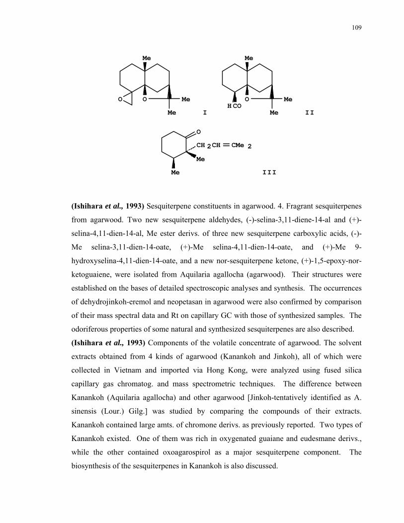
109
O O
Me
Me
Me I
O
Me
Me
MeCOH
II
Me
Me
O
CH 2CH CMe 2
III
(Ishihara et al., 1993) Sesquiterpene constituents in agarwood. 4. Fragrant sesquiterpenes
from agarwood. Two new sesquiterpene aldehydes, (-)-selina-3,11-diene-14-al and (+)-
selina-4,11-dien-14-al, Me ester derivs. of three new sesquiterpene carboxylic acids, (-)-
Me selina-3,11-dien-14-oate, (+)-Me selina-4,11-dien-14-oate, and (+)-Me 9-
hydroxyselina-4,11-dien-14-oate, and a new nor-sesquiterpene ketone, (+)-1,5-epoxy-nor-
ketoguaiene, were isolated from Aquilaria agallocha (agarwood). Their structures were
established on the bases of detailed spectroscopic analyses and synthesis. The occurrences
of dehydrojinkoh-eremol and neopetasan in agarwood were also confirmed by comparison
of their mass spectral data and Rt on capillary GC with those of synthesized samples. The
odoriferous properties of some natural and synthesized sesquiterpenes are also described.
(Ishihara et al., 1993) Components of the volatile concentrate of agarwood. The solvent
extracts obtained from 4 kinds of agarwood (Kanankoh and Jinkoh), all of which were
collected in Vietnam and imported via Hong Kong, were analyzed using fused silica
capillary gas chromatog. and mass spectrometric techniques. The difference between
Kanankoh (Aquilaria agallocha) and other agarwood [Jinkoh-tentatively identified as A.
sinensis (Lour.) Gilg.] was studied by comparing the compounds of their extracts.
Kanankoh contained large amts. of chromone derivs. as previously reported. Two types of
Kanankoh existed. One of them was rich in oxygenated guaiane and eudesmane derivs.,
while the other contained oxoagarospirol as a major sesquiterpene component. The
biosynthesis of the sesquiterpenes in Kanankoh is also discussed.
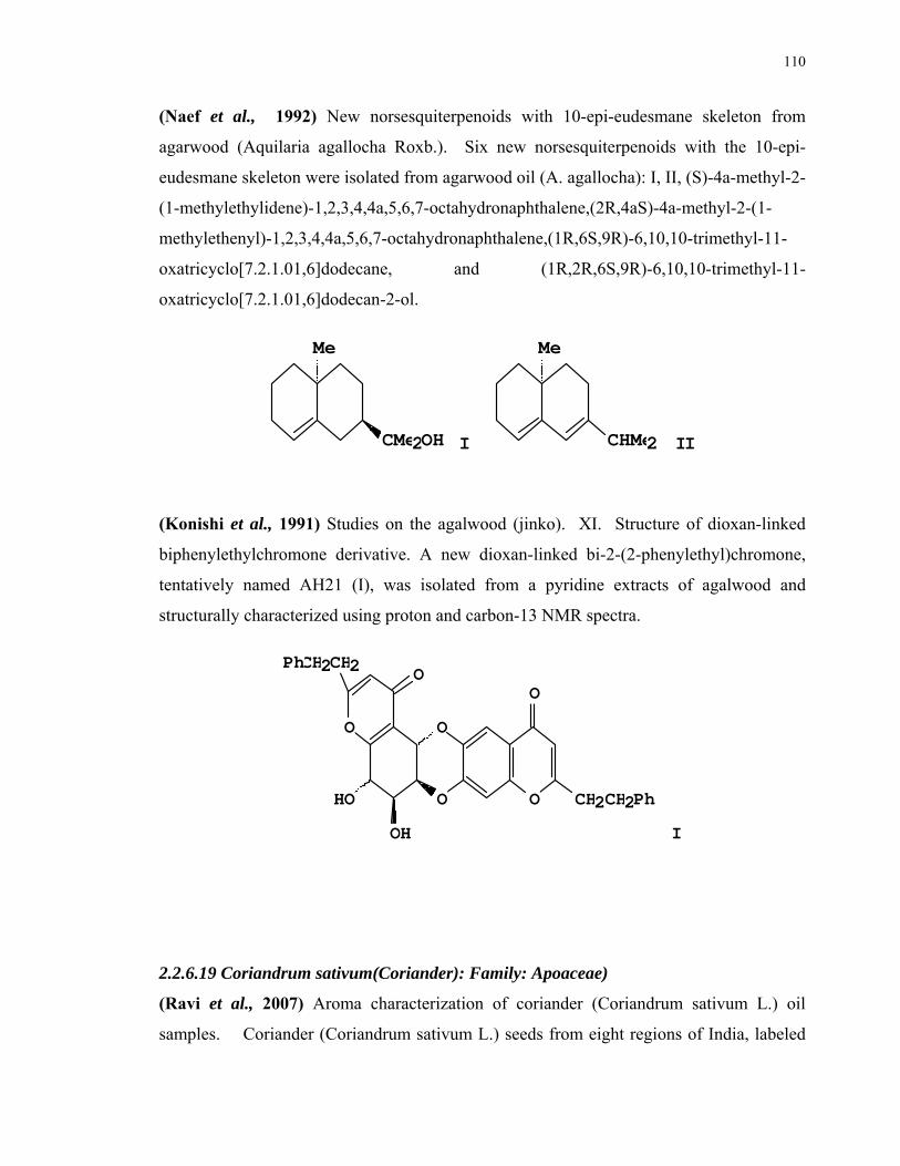
110
(Naef et al., 1992) New norsesquiterpenoids with 10-epi-eudesmane skeleton from
agarwood (Aquilaria agallocha Roxb.). Six new norsesquiterpenoids with the 10-epi-
eudesmane skeleton were isolated from agarwood oil (A. agallocha): I, II, (S)-4a-methyl-2-
(1-methylethylidene)-1,2,3,4,4a,5,6,7-octahydronaphthalene,(2R,4aS)-4a-methyl-2-(1-
methylethenyl)-1,2,3,4,4a,5,6,7-octahydronaphthalene,(1R,6S,9R)-6,10,10-trimethyl-11-
oxatricyclo[7.2.1.01,6]dodecane, and (1R,2R,6S,9R)-6,10,10-trimethyl-11-
oxatricyclo[7.2.1.01,6]dodecan-2-ol.
Me
CMe2OH I
Me
CHMe2 II
(Konishi et al., 1991) Studies on the agalwood (jinko). XI. Structure of dioxan-linked
biphenylethylchromone derivative. A new dioxan-linked bi-2-(2-phenylethyl)chromone,
tentatively named AH21 (I), was isolated from a pyridine extracts of agalwood and
structurally characterized using proton and carbon-13 NMR spectra.
O
O O
O
O
HO
OH
CH2CH2Ph
CH2Ph CH2 O
I
2.2.6.19 Coriandrum sativum(Coriander): Family: Apoaceae)
(Ravi et al., 2007) Aroma characterization of coriander (Coriandrum sativum L.) oil
samples. Coriander (Coriandrum sativum L.) seeds from eight regions of India, labeled

111
as S1 to S8 were examinedfor their volatile constitutents by gas chromatog.-mass
spectroscopy (GC-MS). GC-olfactometry (GC-O) was carried out for major compounds
and odor profiling was done by trained panelists. Essential oil content of coriander
samples ranged from 0.18 to 0.39%. The GC-MS analysisrevealed presence of 30
compounds in coriander oil and around 98% of the compounds were identified in all the
samples. Linalool which has floral and pleasant odor notes was the major compounds
(56.71-75.14%) in the essential oil, but the variation in the linalool content did not
significantly affect the pleasantness of samples as perceived by the panelists. Higher α-
pinene content of S7 and S8 could be related to the higher turpentine note. Sweet and
rose-like odor notes of S1 could be due to occurrence of higher levels of geranyl acetate
and lemonol. The odor profiling depicted the overall odor perceived, while the GC-O
represented the odor notes of specific volatile compounds of coriander. Principal
component analysisshowed that samples S7 and S8 loaded with α-pinene, myrcene and
undecanalysis
(Eyres et al., 2007) The combination of gas chromatography-olfactometry and
multidimensional gas chromatography for the characterization of essential oils.
(Hensel et al., 2007) Occurrence of N-phenylpropenoyl-L-amino acid amides in different
herbal drugs and their influence on human keratinocytes, on human liver cells and on
adhesion of Helicobacter pylori to the human stomach.Thirty commonly used medicinal
plants were screened by a selective and specific LC-MS/MS method for the occurrence of
N-phenylpropenoyl-L-amino acid amides, a new homologous class of secondary products.
In 15 plants, one or more of the respective derivatives (1 to 12) were found and
quantitated.
(Sook 2007) Synergism in antifungal activity against Candida and Trichophyton species
in combination with the essential oil of Coriandrum sativum L. and antibiotics. To
determine whether the essential oil from Coriandrum sativum and its main component,
linalool, exhibit antifungal activity, the authors employed a broth dilution assay and disk
diffusion test using common pathogenic Candida and Trichophyton species. Both
coriander oil and linalool significantly inhibited growth of the tested fungi, with minimal
inhibitory concentrations(MICs) in the range of 0.03.apprx.2 mg/mL. Furthermore, in a
checkerboard titer test, both the oil fraction and linalool exhibited synergism when
112
combined with ketoconazole, with resultant FICIs ranging from 0.06 to 0.53. Notably,
hyphal formation in C. albicans cells was obviously inhibited by C. sativum essential oil in
this experiment.
(Kapanadze et al., 2006) Study of the hypoglycemic effect of CS-Gal lectin with the
presence of monensine and cytochalasin B. The hypoglycemic effect of Galactose-specific
lectin (CS-Gal) from coriander (Coriandrum sativum, Umbelliferae, Apiaceae, herbal
vegetable plant) in human peripheral blood erythrocytes and bovine liver cells has been
studied in the conditions of intracellular alkalization (ionophore monensine) and with the
presence of the inhibitor of glucose transporter GLUT1-cytochalasin B. Inhibition of the
lectin hypoglycemic effect has been revealed with the presence of monensine and
cytochalasin B in erythrocytes but not in the liver cells. It has been supposed that
hypoglycemic effect of CS-Gal lectin is not implemented via the phosphatidylinositol
signaling system in erythrocytes.
(Oussalah et al., 2007) Inhibitory effects of selected plant essential oils on the growth of
four pathogenic bacteria: E. coli O157:H7, Salmonella typhimurium, Staphylococcus
aureus and Listeria monocytogenes. Twenty eight essential oils were evaluated for their
antibacterial properties, against four pathogenic bacteria (Escherichia coli O157:H7,
Listeria monocytogenes 2812 1/2a, Salmonella Typhimurium SL 1344 and Staphylococcus
aureus). Essential oils were introduced into Brain Heart Infusion agar (BHI) (15 mL) at a
concn. of 0.003%, 0.006%, 0.013%, 0.025%, 0.05%, 0.1%, 0.2%, 0.4% and 0.8% (vol/vol)
to det. the min. inhibitory concn. (MIC) and the maximal tolerated concentrations (MTC)
for each pathogen evaluated. Results showed that the most active essential oils against
bacteria tested were Corydothymus capitatus, Cinnamomum cassia, Origanum
heracleoticum, Satureja montana, and Cinnamomum verum (bark). These showed a MIC
0.05% (vol/vol) for all bacteria tested. For the MTC, with the exception of S.
Typhimurium and L. monocytogenes where a MTC of 0.025% (vol/vol) was observed in
presence of Cinnamomum verum and Cinnamomum cassia, respectively, a MTC ~ 0.013%
(vol/vol) was observed for all other bacteria and the three other most active essential oils.
Three oils (Satureja hortensis, Thymus vulgaris carvacroliferum, Origanum compactum)
showed a MIC ~ 0.1% (vol/vol) for all bacteria tested. Seven oils (Thymus vulgaris
thymoliferum, Thymus serpyllum, Thymus satureioides, Cymbopogon martinii, Pimenta

113
dioica, Cinnamomum verum (leaf), Eugenia caryophyllus) showed a lower antimicrobial
activity showing a MIC 0.4% (vol/vol) against the four bacteria tested. Finally, 13
essential oils were less active showing a MIC value 0.8% (vol/vol) against at least one
bacterium.
(Raju et al., 2006) Carotenoid composition and vitamin A activity of medicinally
important green leafy vegetables. Carotenoid compound of green leafy vegetables (GLVs,
n = 30) with medicinal value was analyzed by HPLC; vitamin A activity (as retinol equiv.,
RE) of provitamin A carotenoids was calcd. Results show that among GLVs studied, the
level of -carotene (50-130 mg/100 g dry wt) was higher in 9 GLVs than other carotenoids
while lutein (50-187 mg/100 g dry wt) and zeaxanthin (1-5 mg/100 g dry wt) were higher
in 12 GLVs than other xanthophylls. -Carotene was detected only in 9 GLVs, ranging
from 1 to 37 mg/100 g dry wt. Interestingly, Chenopodium album, Commelina
benghalensis and Solanum nigrum were found to contain higher levels of both lutein and -
carotene in the range of 84-187 and 50-115 mg/100 g dry wt, respectively. The values of
retinol equiv. (RE) ranged from 641 to 19101 and were higher (>10,000) in six GLVs of
the 30. The results demonstrate that GLVs studied contained higher levels of RE and
lutein.
(Vejdani et al., 2006) The Efficacy of an Herbal Medicine, Carmint, on the Relief of
Abdominal Pain and Bloating in Patients with Irritable Bowel Syndrome: a Pilot Study.
Carmint contains total extracts of Melissa officinalis, Mentha spicata, and Coriandrum
sativum, which have antispasmodic, carminative, and sedative effects. This pilot study
suggests that Carmint plus loperamide or Carmint plus psyllium (depending on the irritable
bowel syndrome subtype) might be effective in these patients.
(Yan et al., 2006) Antimicrobial effects of spices and herbs on Vibrio parahaemolyticus.
The antimicrobial effects of spices and herbs from 18 plant species were examinedon a
foodborne pathogen, Vibrio parahaemolyticus, with the use of combinations of
temperatures and nutrient levels. Basil, clove, garlic, horseradish, marjoram, oregano,
rosemary, and thyme exhibited antibacterial activities at incubation of 30°, while with the
exception of horseradish, the same spices and addnl. 7 species exhibited the activities at 5°.
(Singh et al., 2006) Studies on essential oil, part 41. Chemical composition, antifungal,
antioxidant and sprout suppressant activities of coriander (Coriandrum sativum) essential
114
oil and its oleoresin. Gas chromatog.-mass spectrometry (GC-MS) analysisof the seed oil
of coriander (Coriandrum sativum) showed the presence of 52 components, accounting for
98.45% of the total oil. The major components were linalool (75.30%), geranyl acetate
(8.12%) and α-pinene (4.09%). Its oleoresin showed the presence of 28 components.
Oleic acid (36.52%), linoleic acid (33.2%) and palmitic acid (11.05%) were the major
components. The antifungal activities of coriander oil and its oleoresin against eight fungi
were evaluated using the inverted petriplate and food poison techniques. Using the
inverted petriplate method, the essential oil was found to be highly active against
Curvularia palliscens, Fusarium oxysporum, Fusarium moniliforme and Aspergillus
terreus. In the case of the oleoresin, more than 50% mycelial zone inhibition was obtained
for the fungi Fusarium oxysporum, Aspergillus niger and Aspergillus terreus. Using the
food poison technique, the essential oil showed 100% inhibition on the growth of A.
terreus, A. niger, F. graminearum and F. oxysporum, whereas its oleoresin showed weaker
fungitoxic activity, exhibiting 100% inhibition on the growth of F. oxysporum only. The
oleoresin had a better antioxidant activity for sunflower oil than essential oil and synthetic
antioxidants, BHA and BHT. The oil was also found to exert strong sprout suppressant
activity (SSA) for potatoes.
(Al-Mofleh et al., 2006) Protection of gastric mucosal damage by Coriandrum sativum L.
pretreatment in Wistar albino rats. The effect of Coriander pretreatment on gastric mucosal
injuries caused by NaCl, NaOH, ethanol, indomethacin and pylorus ligation accumulated
gastric acid secretions was investigated in rats. Pretreatment at oral doses of 250 and 500
mg/kg, body wt. was found to provide a dose-dependent protection against the (i)
ulcerogenic effects of different necrotizing agents; (ii) ethanol-induced histopathol. lesions;
(iii) pylorus ligated accumulation of gastric acid secretions and ethanol related decrease of
Nonprotein Sulfhydryl groups (NP-SH). Results obtained on the study of gastric mucus
and indomethacin-induced ulcers demonstrated that the gastro protective activity of
Coriander might not be mediated by gastric mucus and/or endogenous stimulation of
prostaglandins. The protective effect against ethanol-induced damage of the gastric tissue
might be related to the free-radical scavenging property of different antioxidant
constituents (linanool, flavonoids, coumarins, catechins, terpenes and polyphenolic
compounds) present in Coriander. The inhibition of ulcers might be due to the formation

115
of a protective layer of either one or more than one of these compounds by hydrophobic
interactions.
(Singh et al., 2006) The in vitro antibacterial efficiency of essential oil and root extract of
Coriandrum sativum Linn. Antibacterial potency of the volatile oil obtained from seeds
and ethanolic and water extracts of Coriandrum sativum Linn. roots was tested against
some Gram-pos. and Gram-neg. bacteria by agar diffusion method. Significant
antibacterial activity was shown by volatile oil and ethanolic extracts of root.
(Eguale et al., 2007) In vitro and in vivo anthelmintic activity of crude extracts of
Coriandrum sativum against Haemonchus contortus. In vitro anthelmintic activities of
crude aqueous and hydro-alcoholic extracts of the seeds of Coriandrum sativum (Apiaceae)
were investigated on the egg and adult nematode parasite Haemonchus contortus. The
aqueous extract of Coriandrum sativum was also investigated for in vivo anthelmintic
activity in sheep infected with Haemonchus contortus. Both extract types of Coriandrum
sativum inhibited hatching of eggs completely at a concentration less than 0.5 mg/ml.
ED(50) of aqueous extract of Coriandrum sativum was 0.12 mg/ml while that of hydro-
alcoholic extract was 0.18 mg/ml. There was no statistically significant difference between
aqueous and hydro-alcoholic extracts (p>0.05). The hydro-alcoholic extract showed better
in vitro activity against adult parasites than the aqueous one. For the in vivo study, 24
sheep artificially infected with Haemonchus contortus were randomly divided into four
groups of six animals each. The first two groups were treated with crude aqueous extract
of Coriandrum sativum at 0.45 and 0.9 g/kg dose levels, the third group with albendazole
at 3.8 mg/kg and the last group was left untreated. Efficacy was tested by faecal egg count
reduction (FECR) and total worm count reduction (TWCR). On day 2 post treatment,
significant FECR was detected in groups treated with higher dose of Coriandrum sativum
(p<0.05) and albendazole (p<0.001). On days 7 and 14 post treatment, significant FECR
was not detected for both doses of Coriandrum sativum (p>0.05). Significant (p<0.05)
TWCR was detected only for higher dose of Coriandrum sativum compared to the
untreated group. Reduction in male worms was higher than female worms. Treatment
with both doses of Coriandrum sativum did not help the animals improve or maintain their
PCV while those treated with albendazole showed significant increase in PCV (p<0.05).

116
2.2.6.20 Rosa damascena ( Rose, Damask Rose): Family: Rosaceae
(Almasirad et al., 2007) Composition of a historical rose oil sample (Rosa damascena
Mill., Rosaceae). Compound of a historical rose oil sample was investigated by GC and
GC/MS. Forty-five components representing 95.5% compound of the essential oil were
identified. The main components of this oil were citronellol (25.1%), geraniol (11.8%) and
nonadecane (13.4%). The composition of the oil was compared with those of Turkish and
Bulgarian rose oils and rose water.
(Kumar et al., 2006) Flavonoids from Rosa damascena Mill. A new flavanone glycoside,
butin 4'-O-(2''-O-α- D-apiofuranosyl)- α -D-glucopyranoside (1), together with liquiritin
(2), liquiritin apioside (3), isoliquiritin apioside (4), davidioside (5), quercetin (6),
kaempferol (7), kaempferol 3-O- α -D-glucopyranoside (8) and kaempferol 3-O- α -L-
arabinofuranoside (9) were isolated from the marc of R. damascena flowers after industrial
distn. of essential oil. This is the 1st report of the occurrence of compounds 2, 3, 4 and 5
in R. damascena. The structures of the isolated constituents were established on the basis
of spectroscopic {UV, IR, 1D, 2D NMR (DEPT, HMQC, HMBC and COSY)},
spectrometric (ESI-QTOF-MS), and chemical evidence.
(Khan 2005) action and analysis of essential oil of Rosa species. Rosa species were
evaluated for oil yield, color and other phys. and chem. properties. Recovery of concrete
oil from petals of Rosa damascena was higher (0.24%) than Rosa centifolia (0.22%) on
fresh wt. basis. Oil color of Rosa centifolia was yellowish brown while the color of abs.
oil of Rosa damascena was yellowish. Refractive index of Rosa centifolia oil was higher
(1.45) at 25ºC than Rosa damascena (1.42). Rosa centifolia was found higher for all the
chem. constituents studied except Ph Et alcohol which was greater in case of Rosa
damascena than Rosa centifolia. The study indicates that there is qual. and
quantitativedifference in chem. compound, aroma constituents of essential oil of Rosa
centifolia and Rosa damascena produced locally or elsewhere in the world.
(Vinokur et al., 2006) Rose petal tea as an antioxidant-rich beverage: cultivar effects.
Twelve rose cultivars were selected by field tasting as potential sources of edible flowers.
Hot water infusions (teas) of air-dried petals of these cultivars were assayed for antioxidant
activity, total phenols, and total anthocyanins contents. Their compound was analyzed by
high-performance liquidchromatog. (HPLC). Green tea was tested in parallel as a ref.
117
antioxidant-rich beverage. Rose petal teas from different cultivars exhibited scavenging
capacity toward 2,2'-azino-bis-(3-ethylbenzothiazoline)-6-sulfonate cation radical
(ABTS+) ranging between 712.7 and 1770.7 µM Trolox equiv. (TE) per g of dry petals, as
compared with 1227.6 µM TE/g dry wt. in the green tea. The range of total phenols
content in rose teas was 50.7 to 119.5 mg gallic acid equiv. (GAE) per g of dry matter, as
compared with 62.1 mg GAE/g dry wt. in the green tea. The rose teas were rich in free
gallic acid. The highest values of antioxidant activity, total phenols, and gallic acid
contents were found in the cultivars San Francisco, Katharina Zeimet, and Mercedes and in
the essential-oil-bearing rose Rosa damascena. The correlation coeffs. between antioxidant
activity, on the 1 hand, and the contents of total phenols and of gallic acid in various rose
cultivars, on the other hand, were 0.79 and 0.81, respectively. No clear relationship
between anthocyanin level and radical-scavenging activity was revealed. Teas from
different rose cultivars significantly differed in their sensory properties. It was concluded
that dried rose petals may be used for prepg. antioxidant-rich caffeine-free beverages,
either sep. or in combination with other herbal materials.
(Ayci et al., 2005) Gas chromatographic investigation of rose concrete, absolute and solid
residue. The chem. compound and phys. properties of the rose abs. produced from rose
concretes and the ext., supplied by three different companies, were determinedby GC and
GC-MS. It was found that rose abs. consists mainly of α-phenylethyl alcohol, citronellol,
geraniol, nerol, eugenol, Me eugenol, geranyl acetate, benzyl alcohol, nonadecane,
nonadecene and farnesol. The solid residue, which is the byproduct of abs. prodn., was
further divided on a silica gel column into three fractions by solvents having different
polarities (n-hexane, 5% di-Et ether in n-hexane, and di-Et ether alone). A fraction eluated
with n-hexane was analyzed directly by GC and GC-MS. Two other fractions, after
sapon., were converted to their Me ester derivs. and then analyzed by GC and GC-MS. It
was observed that the solid residue contains mainly straight-chain satd. hydrocarbons with
high mol. wt. (C15-C31) and the esters of carboxylic acids and a homologous series
between them exists. It was also concluded that the extractobtained by solvent extn. from
the residue of rose flower (Rosa damascena Mill.) which was subjected to steam distn. is
also a valuable raw material.

118
(Schieber et al., 2005) Flavonol glycosides from distilled petals of Rosa damascena Mill.
Flavonol glycosides were extractedfrom petals of Rosa damascena Mill. after industrial
distn. for essential oil recovery and characterized by high-performance liquidchromatog.-
electrospray ionization mass spectrometry. Among the 22 major compounds analyzed,
only kaempferol and quercetin glycosides were detected. To the best of the authors'
knowledge, the presence of quercetin 3-O-galactoside and quercetin 3-O-xyloside has so
far not been reported within the genus Rosa. In addn., based on their fragmentation
patterns, several acylated quercetin and kaempferol glycosides, some of them being
disaccharides, were identified for the first time. The kaempferol glycosides, along with the
kaempferol aglycon, accounted for 80% of the total compounds that were quantified, with
kaempferol 3-O-glucoside being the predominant component. The high flavonol content
of approx. 16 g/kg on a dry wt. basis revealed that distd. rose petals represent a promising
source of phenolic compounds which might be used as functional food ingredients, as
natural antioxidants or as color enhancers.
(Agarwal et al., 2005) Chemical composition of rose water volatiles. A volatile conc.
obtained from rose water (ex. Rosa damascena flowers) by liquid-liquid extraction with
dichloromethane was studied by GC and GC/MS. The volatiles consisted mainly of 2-
phenylethanol (69.7-81.6%), linalool (1.5-3.3%), citronellol (1.8-7.2%), nerol (0.2-4.2%),
geraniol (0.9-7.0%) along with rose oxides and all other characteristic minor rose
compounds
(Suchalatha 2004) Role of Arogh, a polyherbal formulation to mitigate oxidative stress in
experimental myocardial infarction. Antioxidant role of Arogh in isoproterenol induced
myocardial infarction in rats has been studied. The activity of heart tissue antioxidants like
glutathione, superoxide dismutase, catalase, glutathione peroxidase and glutathione-s-
transferase were significantly decreased in isoproterenol administered group. The activity
of ceruloplasmin and levels of glutathione, vitamins E and C were also substantially
decreased in serum with a concomitant rise in lipid peroxide levels after isoproterenol
exposure to rats. The synergistic effect of Arogh pretreatment, significantly suppressed the
alterations induced by isoproterenol alone in rats.
(Basim et al., 2003s) Antibacterial activity of Rosa damascena essential oil. The essential
oil of Rosa damascena petals was evaluated for its antibacterial effects against three strains
119
of Xanthomonas axonopodis spp. vesicatoria. The essential oil may be a potential control
agent in the management of the disease caused by X .a. vesicatoria in tomato and pepper
plants.
(Horne et al., 2001) Antimicrobial effects of essential oils on Streptococcus pneumoniae.
Of 73 essential oils tested for antibacterial activity against Streptococcus pneumoniae
R36A (an unencapsulated strain) with a paper disk diffusion assay, three oils were highly
inhibitory, fifteen moderately inhibitory and the remainder weakly or non-inhibitory. The
oils with high antibacterial activity; oregano, thyme and rosewood, induced rapid lysis of
R36A as indicated by a decrease in OD, and appearance of dechaining and considerable
cell debris within 30 min of addn. The lytic response of R36A to the three oils with
moderate activity was variable but all induced some lysis. Oils that were weak inhibitors
generally caused slowing of growth but little or no lysis. Several oils were also tested
against an encapsulated isolate, S. pneumoniae IC2. Both disk assay and broth results
were similar to those obtained with R36A, except that the oils were slightly less effective.
Disk assay results showed some correlation with the broth assay, but were not always
predictive of an oil's ability to induce bacterial lysis. Essential oils that induce lysis in S.
pneumoniae may have potential as an alternative treatment for infections caused by drug
resistant pneumococci.
(Watanabe et al., 2001) Isolation and identification of 2-phenylethyl disaccharide
glycosides and mono glycosides from rose flowers, and their potential role in scent
formation. 2-Phenylethyl 6-O--L-arabinofuranosyl--D-glucopyranoside (I), and its 6-O-
-D-xylopyranosyl--D-glucopyranoside (II) were identified in the flowers of Rosa
damascena Mill. harvested at the full bloom stage. 2-Phenylethyl -D-glucopyranoside
and its -D-galactopyranoside together with I and II were also found in the flower buds
harvested 44 h before the opening stage. Their potential role in scent formation is
discussed.
(Oka et al., 1999) Glycosidic aromas and glycohydrolases being involved in potential
aroma formation in rose flowers of two different species. The aqueous residues after the
steam distn. of Rosa damascena was found to contain considerable amount of aroma active
compounds The amount of the aroma after the hydrolysis was almost the same as in the
rose oil. We also compared amts. of glycoconjugates of alcohol aroma and hydrolytic

120
enzyme activities among different parts of fragrant and non-fragrant rose plants. The
amounts of glycoconjugates in the buds and the flowers of the fragrant variety were much
higher than in the other parts of fragrant and non-fragrant plant varieties. In addn., -
glucosidase and -galactosidase activities were stronger than those of other
glycohydrolases. -Galactosidase activity was found to be high in all parts of both
varieties, whereas -glucosidase activity was dominant only in the buds and flowers of
fragrant variety.
(Watanabe 1998) Occurrence of a glucosidic progenitor of rose oxide in rose flowers,
Rosa damascena Mill. (3S)-3,7-Dimethyl-7-hydroxyoct-5-enyl-O--D-glucopyranoside (I)
was isolated as an aroma precursor of isomeric rose oxides from flowers of Rosa
damascena Mill. The isolation was guided by the detection of rose oxide formation by GC
and GC-MS analyses after acid-catalyzed hydrolyzes of precursor fractions. The structure
of I was confirmed by spectroscopic analysisand synthesis starting from (3S)-citronellol.
(Najmi et al., 1999) Effect of Qurs-e-Ziabetus-16 in neonatally streptozotocin induced
diabetic rats. Qurs-e-Ziabetus 16 (QZ-16) is a polypharmaceutical herbomineral
formulation used by Unani physicians in non-insulin dependent diabetes mellitus
(NIDDM). It contains five medicinal plants, namely Azadirachta indica, Gossypium
herbaceum, Gymnema sylvestre, Rosa damascena, and Syzygium cumini along with two
Kushtas, Kushta Hartal and Kushta Baiza Murgh. The authors report the glucose tolerance
improving effect of QZ-16 in neonatally streptozotocin-induced diabetic rats.
Streptozotocin treatment significantly increased the blood glucose in the control group
relative to the group treated with QZ-16.
(Boyanova 1999) Inhibitory effect of rose oil products on Helicobacter pylori growth in
vitro: preliminary report.
(Surveswaran et al., 2007) Systematic evaluation of natural phenolic antioxidants from
133 Indian medicinal plants. Total antioxidant capacities of 133 Indian medicinal plant
species sampled from 64 families were assessed by ABTS, DPPH and FRAP assays, and
their total phenolic contents measured by Folin-Ciocalteu assay. These species exhibited a
broad range of antioxidant activities, varying from 0.16 to 500.70 mmol TEAC/100 g DW
in the ABTS assay. The antioxidant activity values similarly varied with the DPPH and
FRAP assays. Significant and pos. linear correlations were found between total
121
antioxidant capacities and phenolic contents (R = 0.89-0.97), indicating that phenolics
were the dominant antioxidant constituents in the tested medicinal plants. Preliminary
identification of the major phenolic compounds from 83 selected medicinal plants by
reversed-phase HPLC revealed phenolic acids, tannins, flavonoids, curcuminoids,
coumarins, lignans, and quinines. The fruit of Terminalia chebula, pericarp of Punica
granatum and gall of Rhus succedanea showed very high levels of hydrolysable tannins,
and the gum of Acacia catechu presented very high levels of catechin and epicatechin in
addn. to tannins. Major phenolics in many of the medicinal plants were identified for the
first time (e.g., Euphorbia lathyrus, Ipomoea turpethum, and Picrorrhiza kurroa). This
systematic investigation of a large number of Indian medicinal plants proved important for
understanding their chem. constituents and functionality in Ayurvedic medicine, and
contributes to the search for natural sources of potent antioxidants.
2.2.6.21 Mesua ferrea (Cobras Saffron): Family: Anacardiaceae.
(Ghosh et al., 1998) Lipase hydrolysis of some minor oils. Some of the minor oils like
wheat germ (Triticum), rice bran (Oryza sativa), mowrah (Madhuca latifolia), mango
kernel fat (Mangifera indica), karanja (Pongamia glabra), nahor (Mesua ferra Linn.),
bantulsi (Ocimum gratissimum Linn.) and marda (Terminalia arjuna) are hydrolyzed by
stirring at 35°C ± 2°C with 0.2% (wt./wt.) Candida cylindracea lipase powder and 100%
(wt./wt.) water. The oils are almost completely hydrolyzed within 48 h.
2.2.6.22 Crocus sativus (Saffron): Family: Iridaceae
(Shinji et al., 2001) Crocin suppresses tumor necrosis factor-α-induced cell death of
neuronally differentiated PC-12 cells. Crocus sativus L. is used in Chinese traditional
medicine to treat some disorders of the central nervous system. Crocin is an ethanol-
extractable component of Crocus sativus L.; it is reported to prevent ethanol-induced
impairment of learning and memory in mice. In this study, we demonstrate that crocin
suppresses the effect of tumor necrosis factor (TNF)-α on neuronally differentiated PC-12
cells. PC-12 cells dead from exposure to TNF-α show apoptotic morphological changes
and DNA fragmentation. These hallmark features of cell death did not appear in cells
treated in the co-presence of 10 μM crocin. Moreover, crocin suppressed the TNF-α-

122
induced expression of Bcl-XS and LICE mRNAs and simultaneously restored the
cytokine-induced reduction of Bcl-XL mRNA expression. The modulating effects of
crocin on the expression of Bcl-2 family proteins led to a marked reduction of a TNF-α-
induced release of cytochrome c from the mitochondria. Crocin also blocked the
cytochrome c-induced activation of caspase-3. To learn how crocin exhibits these anti-
apoptotic actions in PC-12 cells, we tested the effect of crocin on PC-12 cell death induced
by daunorubicin. We found that crocin inhibited the effect of daunorubicin as well. Our
findings suggest that crocin inhibits neuronal cell death induced by both internal and
external apoptotic stimuli.
(Fikrat 2002) Cancer Chemopreventive and Tumoricidal Properties of Saffron (Crocus
sativus L.). Since cancer is the most common cause of death in the world population, the
possibility that readily available natural substances from plants, vegetables, herbs, and
spices may be beneficial in the prevention of cancer warrants closer examination. Saffron
in filaments is the dried, dark red stigmata of Crocus sativus L. flowers and it is used as a
spice, food colorant, and a drug in medicine. A growing body of research has demonstrated
that saffron extract itself and its main constituents, the carotenoids, possess
chemopreventive properties against cancer. This review discusses recent literature data and
our results on the cancer chemopreventive activities of saffron and its main ingredients.
(Termentzi and Kokkalou 2008) LC-DAD-MS (ESI+) analysis and antioxidant capacity
of crocus sativus petal extracts.In this study, various fractions isolated from the petals of
Crocus sativus were assessed at first for their phenolic content both qualitatively and
quantitatively and secondly for their antioxidant activity. The phytochemical analysis was
carried out by LC-DAD-MS (ESI (+)) whereas the antioxidant potential was evaluated by
applying two methodologies, the DPPH. radical scavenging activity test and the Co(II)-
induced luminol chemiluminescence procedure. According to data obtained from these
antioxidant tests, the diethyl ether, ethyl acetate and aqueous fractions demonstrated the
strongest antioxidant capacity. Interestingly, the major constituents identified in these
fractions correspond to kaempferol, quercetin, naringenin and some flavanone and flavanol
derivatives glycosylated and esterified with phenylpropanoic acids. In addition, the
presence of some nitrogen-containing substances, as well as other phenolics and
phenylpropanoic derivatives was also traced. The identification and structural elucidation
123
of all substances isolated in this study was achieved by both comparing available literature
data and by proposed fragmentation mechanisms based on evaluating the LC-DAD-MS
(ESI (+)) experimental data. The quantitative analysis data obtained thus far have shown
that Crocus sativus petals are a rich source of flavonoids. Such a fact suggests that the
good antioxidant capacity detected in the various fractions of Crocus sativus petals could
be attributed to the presence of flavonoids, since it is already known that these molecules
exert antioxidant capability. The latter, along with the use of Crocus sativus in food and
pharmaceutical industry is discussed.
(Liakopoulou and Skubas 1990) Characterization of the platelet aggregation inducer and
inhibitor isolated from Crocus sativus. Bulbs of Crocus sativus variety Cartwrightianus
were found to contain both a platelet aggregation inducer and inhibitor. The aggregating
factor has a Mr of 42 kDa estimated by a Sephadex G75 column and SDS-polyacrylamide
slab gel electrophoresis. It was found to lack enzymatic activity such as proteinase,
esterase and acid or alkaline phosphatase. The inhibitory factor was also purified to
homogeneity by different chromatographic techniques and shows a Mr of 27 kDa as it was
estimated by Biogel P30 column and SDS-polyacrylamide slab gel electrophoresis. It was
found to possess strong proteinase activity.
(Akhondzadeh et al., 2005) Crocus sativus L. in the treatment of mild to moderate
depression: a double-blind,randomized and placebo controlled trial.
(Hosseinzadeh et al., 2005) Protective effect of aqueous saffron extract (Crocus sativus
L.) and crocin, its active constituent, on renal ischemia-reperfusion-induced oxidative
damage in rats. The generation of reactive oxygen species and lipid peroxidation are
associated with tissue injury following ischemic insult; therefore, the use of antioxidants
appears rational in the improvement of kidney diseases therapy. The aim of the present
study was to assess the effect of aqueous saffron extract (Crocus sativus L.) and its active
constituent, crocin, on oxidative stress following renal ischemia-reperfusion injury (IRI) in
rats.. The cellular redox status (thiobarbituric acid reactive species (TBARS) and total thiol
levels) and antioxidant power (using ferric reducing/antioxidant power test) were assessed
in control and ischemic groups. The left kidney was exposed to warm ischemia for 60 min
followed by reperfusion for 90 min. The macerated aqueous extract of saffron (with doses
of 5, 20 and 80 mg/kg, i.p.) and crocin (with doses of 50, 200 and 400 mg/kg, i.p.) were
124
administrated prior to induction of ischemia. Normal saline (10 ml/kg, i.p.) was injected to
control group and a sham group that did not have ischemia-reperfusion. Ischemia-
reperfusion (IR) caused a significant increase in TBARS levels (p<0.001) and decrement in
both antioxidant power (FRAP value) (p<0.05) and total thiol concentration (p<0.001) in
kidney homogenate samples. In crocin pretreated groups, a reduction in TBARS levels
(from 85.8 ± 5.4 to 20.9 ± 1.5 nmol/g tissue, p<0.001; 400 mg/kg) and elevation in
antioxidant power (FRAP value) (from 3.05 ± 0.16 to 4.15 ± 0.16 µmol/g tissue, p<0.001;
400 mg/kg) and total thiol concentrations (from 0.38 ± 0.03 to 0.62 ± 0.03 mM, p<0.001;
200 mg/kg), as compared with control group, were observed. The aqueous extract also
reduced lipid peroxidation products (from 85.8 ± 5.4 to 15.9 ± 2.6 nmol/g tissue, p<0.001;
80 mg/kg) and increased antioxidant power (from 2.98 ± 0.11 to 5.97 ± 0.56 µmol/g tissue,
p<0.001; 80 mg/kg) in ischemia-reperfusion injured rat kidneys.
(Jouad et al., 2001) Ethnobotanical survey of medicinal plants used for the treatment of
diabetes, cardiac and renal diseases in the North centre region of Morocco (Fez–
Boulemane)
(Xiang-Chun and Zhi-Yu 2006) Effects of crocetin on antioxidant enzymatic activities in
cardiac hypertrophy induced by norepinephrine in rats. Crocetin, a carotenoid isolated
from the Chinese herbal medicine Crocus sativus L (Saffron), has been shown to have
cardiovascular protective effects. The present study investigated the protective action of
the antioxidant crocetin against cardiac hypertrophy induced by norepinephrine (NE). This
was evaluated by assaying for pathological histological changes with an optical
microscope and cell image analysis system. Lipid peroxidation was quantified using
thiobarbituric acid-reactive substances (TBARS). Myocardial superoxide dismutase
(SOD), glutathione peroxidase (GSH-Px) and myocardial catalase (CAT) activities were
assayed to evaluate the antioxidant capacity. After long term treatment with NE,
antioxidant enzymatic activities were significantly decreased, while products of lipid
peroxidation increased. Crocetin markedly reduced the content of lipid peroxidation
(LPO), increased the GSHPx and SOD activity in cardiac hypertrophy, and significantly
improved the myocardial pathological histological changes induced by NE. These results
suggest that the cardioprotective effects of crocetin are related to modulation of
endogenous antioxidant enzymatic activities. Comparing crocetin with captopril, our
125
results indicated that antioxidant activity is an important factor in the therapy of cardiac
hypertrophy, but as an antioxidant only, its effects may be limited.

126
2.3 OBJECTIVE OF STUDY:
Objective of this study was to test the cardiovascular activity of the Caissa carandas
(Auct.) and a Poly herbal product Mufarreh yaqooti motadil. The findings were correlated
with the special reference to the traditional use of these products. Modern Scientific
methods were used to analyze the effects on cardiovascular system. Different parameters
of cardiovascular system were tested including Hypotensive activity, Diuretic activity, anti
hyperlipidemic activity and effects on heart. Results obtained wrere compared with the
conventional allopathic medicines used in different cardiovascular diseases.
Another Important aspect of study was the Safety evaluation of tested drugs by different
methods of Acute, Subacute and Chronic Toxicity testing. Other toxicity tests, like tests for
mutagenesis and teratogenecity, require a sparate dimension for in depth study therefore,
these tests were not included in this study
The data obtained were analyzed by using student’s t- distribution test and Analysis of
Variance (ANOVA).
2.3.1 HYPOTHESES OF EXPERIMENTS:
The experiments were performed with following hypotheses:
1. The extract and Herbal Product are as potent as Acetylcholine to reduce the Mean
Arterial Blood Pressure.
2. Diuretic activity of the tasted drug is same as that of furosemide.
3. Antihyperlipidemic activity of tested drug is comparable with that of Atorvastatin.
4. Cardiac activity of tested drugs is comparable with digoxin.
5. All tested drug are safe for use in high doses and free from any toxic effects.

127
CHAPTER 3
MATERIAL AND METHODS

128
3. MATERIAL AND METHODS
3.1 Plant material:
Carissa carandas (Auct.) was collected from the Herb Garden of Hamdard University and
Identified by Professor Dr. M. Qaiser, Department of Botany, University of Karachi, where
a voucher specimen has been deposited.
3.1.1 Extraction.
Air dried leaves of Carissa carandas were crushed into coarse size in a mortor and pastle
and soaked in a mixture of Ethanol and water (1:1) for six days then filtered. Solvents were
evaporated using a Rotary Evaporator (R-114- Buchi) and freeze dried by the help of
Lyophilizer (Tokyo, Rikakikai CO. LTD.)(Fatima et al., 1990). This Carissa carandas
leaves extract (C.C.L.E.) was used for the experiments related to cardiovascular studies.
3.2 Experimental Animals
Sprague- Dawley rats, Rattus norvegicus (Berkenhout.), of either sex, weighing 220-250 g
and NMRI mice, Mus musculus (L.), of either sex, weighing 20-30g were purchased from
animal house of HEJ Research Institute of Chemistry, International Centre for Chemical
and Biological Sciences (ICCBS), University of Karachi and were given standard diet and
water ad-libitum (Gilani 1991). They were housed in a controlled room with a 12 h light-
dark cycles, at room temperature of 22±02oC at Dr. H. M. I. Institute of Pharmacology and
Herbal Sciences, Hamdard University, Karachi, Pakistan. The Institutional Animals Ethics
Committee approved all the protocols of animals maintenance and handling which were in
accordance to internationally accepted standard guidelines for use of laboratory animals.
(Saleem et al., 2005)
Healthy adult Rabbits, Oryctolagus cuniculus (L.) of either sex, weighing from 1.0-1.5 kg
and between 10-12 months of age were used for the study. Rabbits were housed in a well-
lighted air conditioned room (26±01oC) in metallic cages under standard environmental
conditions (12 h light-dark cycles). They were fed on standard green leafy and seasonal
vegetables and green leaves of Alfa Alfa and water ad libitum. (Hemalatha et al., 2006)

129
Both male and female adult frogs, Rana cyanophlyctis (Schn.) weighing 20-40g, were
purchased from local market. ( Bheemchari 2005, Pennacchio 1995).
3.2. Dugs and Chemicals
3.2.1 Experimental poly herbal drug; Mufarreh Yaqooti Motadi
(MUYM):
The drug Mufarreh Yaqooti Motadil (MUYM) is a poly herbal product prepared by
Hamdard Laboratories (Waqf.) Pakistan. It contains 45 ingredients (Said 1979). The drug
was provided by the manufacturers against the order.
3.3.2 Reference drugs:
3.3.2.1 Furosemide (Lasix) 40 mg (Aventis Pharma®. Deutschland GmbH, Frankfurt,
Germany).
3.3.2.2 Digoxin (Lanoxin) 0.25 mg (Glaxo Smith Kline®)
3.3.2.3 Atorvastatin (Lipiget) 10 mg (Getz® Pharmaceuticals)
3.3.2.4 Acetylcholine Chloride (Merck®, Germany)
3.3.2.5 Atropine Sulfate (Boehringer ingelheim®)
3.3.2.6 Adrenaline (Gerhard Buchmann Gmbh®, Tuttlingen/ Germany)
3.3.2.7 Urea (Merck®, Germany)
3.3.3 Other drugs
3.3.3.1 Heparin 5,000 iu/ui/ml, 5ml ( Leo Pharmaceuticals®, Ballerup, Denmark)

130
3.3.3.2 Sodium Thiopental (Abbot Pharmaceuticals®)
3.3.4 Chemicals:
3.3.4.1 Sodium chloride (Merck®, Germany)
3.3.4.2 Tyrode physiological solution: (NaCl=136.89 mM/L, KCl=2.682 mM/L,
CaCl2.2H2O=1.79mM/L, MgCl2=1.05mM/L, NaH2PO4=0.32mM/L, Glucose=5.5mM/L,
NaHCO3=11.90mM/L) (Daher C. F. 2006).
3.3.4.3 Normal Ringer solution solution (NaCl=115mM/L, KCl=2.5mM/L, CaCl2=
1.8mM/L, Na2HPO4=2.15 mM/L, Na H2PO4=0.8mM/L) (Daher C. F. 2006).
3.3.4.4 Chemicals’ kits for Biochemical Estimation (Merck®, Germany)
3.3.4.5 Ethanol (Merck®, Germany)
3.3.4.6 Haematoxylin ( BDH Chemicals Ltd®. Poole England)
3.3.4.7 Eosin Gelbich (Merck®, Germany)
3.3.4.8 Acetic acid (Merck®, Germany)
3.3.4.9 Xylene (Merck®, Germany)
3.4. Instruments:
3.4.1 Spectrophotometer-U.V. Visible (Hitachi U-2000)
3.4.2 Rotary Evaporator (R-114- Buchi)

131
3.4.3 Blood pressure Transducer (Harvard, 60-3003)
3.4.4 Four channel recorder Oscillograph (Harvard)
3.4.5 Langendorff Isolated Heart Assembly (Harvard)
3.4.6 Isotonic Transducer ( Harvard Appl. Ltd. 50-6360)
3.4.7 Kymograph Harvard Universal Model (Cat. No.50-7392)
3.4.8 Microtome (Leica, RM 2145)
3.4.9 Microscope (OPTIPHOT Model X2T-21E) equipped with
Nikon Microphotography system; Model UFX-DX-35 and phase contrast N plan.
3.4.10 Oven (Memmert Gmbh-co.KG D-91126)
3.4.11 Incubator (WTB binder DIN 12880 Klasse)
3.4.12 Centriguge Machine (BHG Hermle Z230)
3.4.13 Lyophilizer (Tokyo, Rikakikai CO. LTD.)
132
3.5 PHARMACODYNAMIC STUDIES ON
CARDIOVASCULAR SYSTEM
3.5.1 IN VIVO STUDIES
3.5.1.1 STUDY ON BLOOD PRESSURE OF ANAESTHETIZED RATS
3.5.1.1.a Experimental protocol:
Normotensive Sprague- Dawley rats of either sex (180g-200g) were anaesthetized with
intraperitoneal injection of Sodium pentothal (50mg/kg) (WHO Research guidelines
1993), (Lahlou, S et al., 2002). Spontaneous respiration was maintained by inserting
tracheal cannula after tracheotomy. The left jugular vein was cannulated with polyethylene
tube for intravenous administration of drug and flushed with 0.9% NaCl (normal saline)
(Lahlou et al., 2002).
An anaesthetized rat was fixed on a supine position on a dissecting table. A longitudinal
midtracheal incision approximately 2 cm long was made in order to expose the trachea, the
right jugular vein and left carotid artery. The trachea was cannulated with polyethylene
tube 92.75 mm diameter to maintain a free airway. For the administration of drugs and
isotonic saline solution, the right jugular vein was cannulated with a saline filled
polyethylene tube (1 mm diameter). The exposed surface was covered with the cotton
moist in warm saline.
The cannulation of the carotid artery was performed in the same manner as the cannulation
of jugular vein and the polyethylene tube (1 mm diameter) was filled with heparin sodium
in saline solution was used. The temperature of the animal was maintained at 37°C with
the use of an overhead lamp (Fig. 3.2). This setting up of the experiment is more or less
same as adopted by Aftab K, 1995 (Fig 3.1).
The arterial blood pressure was measured from the left carotid arterial cannula connected
to a Research grade blood pressure transducer (Harvard, 60-3003) which was connected
with Oscillograph (Havard) for recording. The same apparatus was used for receptor
activity. (Fig. 3.3) (Ferreira et al., 2007).The animal was allowed to equilibrate for at
least 30 min. before commencing the experiment. Blood pressure was monitered until

133
steady baseline levels were obtained. Drugs were then administered by I.V. injections and
flushed in with 0.2 ml saline.
Control response to intravenous injections of acetylcholine, atropine and adrenaline were
taken before the administration of any test material. 6 Rats were used for each dose testing.
Fig. 3.1 A cannulated rat for Hypotensive activity
Fig. 3.2 The blood pressure transducer connected
with the left carotid arterial cannula and recorder.

134
Fig. 3.3 A four channel recorder Oscillograph assembly,
showing the tracing for Blood pressure monitoring.
3.5.1.1.b Calibration:
The Oscillograph was adjusted by keeping its channel amplifier’s knob on DC, and +ve
polarity. The pen position on chart paper was adjusted using zero knobs slightly above the
lowest pen position. The pressure transducer was calibrated by using
“Sphygmomanometer” to standardize 1 mm pen deflection on chart paper for its
equivalencey with pressure (mmHg) (Ahmed et al., 2000)
3.5.1.1.c Calculations:
The mean arterial Blood pressure was calculated using the following formula (Ibarrola et
al., 1999):
MABP = DP + 1/3 ( SP-DP)
Where,
SP= systolic pressure
DP= diastolic pressure
The response of mean arterial blood pressure (MABP) was expressed as percent change
from the control measurement taken immediately before injection. (Adeboye et al., 1999).

135
3.5.1.1.d Preparation of Dilutions of Drugs
Pharmacodynamic screening was carried out by using Acetylcholine (10-6 M i.v.), Atropine
( 10-4 M i.v.), Adrenaline ( 10-5 M i.v.), All dilutions were freshly prepared in 0.9% saline.
(Gilani et al., 1994).
Carissa carandas (C.C.L.E.) extract in doses 5mg/kg – 45mg/kg (i.v.) and Herbal Product
Mufarreh Yaqooti Motadil (MUYM) in doses 5mg/kg –45mg/kg (i.v.) were freshly
prepared by dissolving in warm distilled water before each experiment.
3.5.1.2 CARDIAC ACTIVITY ON INTACT HEART OF PITHED FROGS
3.5.1.2.a Experimental Prtocol:
Pharmacodynamic study was carried out on exposed and intact frog’s heart by using the
method described by (Xiang-Chun et al., 2006, Kimura et al., 1986). The experimental
protocols were approved by the Institutional animals Ethical Committee. For each dose of
the tested sample, 12 frogs were taken. Frogs were made unconscious by pithing procedure
and the unconscious frog was fixed on frog board. An incision was made on ventral side of
body, the skin was carefully removed by using scalpel and thoracic region was cut down
with scissors to expose out the heart. The pericardium was removed by using forceps
(Fig.3.4). Exposed heart was then connected to the lever of Harvard Apparatus (Cat. No.
50-0660) using nylon thread and hook, inserted to the heart apex. During recording of
heart movement, normal ringer solution was continuously poured on the heart to maintain
vitality and prevent drying. The heart activity was recorded by using Harvard Kymograph
Universal Model (Cat. No.50-7392). Its drum revolved at the speed of 10mm/sec. (Talalay
et al., 2001).

136
Fig.3.4 Exposed and Intact Heart of Frog showing attachment to pen and lever
3.5.1.2.b Preparation of Dilutions of Drugs:
The Carissa carandas (C.C.L.E.) extract in doses of 5mg/ml – 45mg/ml, Herbal Product
Mufarreh Yaqooti Motadil ( MUYM ) in doses of 5mg/ml – 45mg/ml and Digoxin
(0.008mg/ml) were freshly prepared by dissolving in warm distilled water before each
experiment. In addition cholinergic agent i.e., Acetylcholine Chloride (10-3 M) and
Anticholinergic agent i.e., Atropin Sulfate (10-3 M) were used for comparison with the
tested drugs. Digoxin (0.008mg/ml) (Wójcicki et al., 1997, Tripathi and Das 1983,
Mikhailov et al., 2004) was used to compare the results on heart. All dilutions were
freshly made in 0.65% saline solution (Isotonic for Amphibions) before each experiment.
(Azmat et al., 2005, Erum et al., 2004)
3.5.1.2.c Calculations:
The records obtained were used for the measurement of Heart Rate (per min.), Cardiac
Force (g), Cardiac Cycle (Sec) at the Kymograph speed (10mm/sec) and heart Lever
Calibration at 2g weight. (Azmat et al., 2005) The force of contraction and the rate of
contraction was counted and tabulated (Ahmed et al., 2004).

137
Heart Rate:
The Calibrated Value is, 10mm = 1Sec.
If the tracing shows, 47 beats = 255 mm
Known value is, 10mm =1 Sec.
Therefore, 255mm= 25.5 Sec.
And 25.5 Sec. = 47 beats
So 60 Sec. = 110.5 beats or 110.5beats/ min.
Cardiac Force:
The Calibrated Value is, 29mm = 2 g
If he tracing shows, 1 beat = 3 mm
Known value is, 29mm =2 g
Therefore, 3mm= 0.206 g
So 1 beat. = 0.206 g
Cardiac Cycle:
The Calibrated Value is, 10mm = 1Sec.
If the tracing shows, 1 Cycle = 6.5 mm
Known value is, 10mm =1 Sec.
Therefore, 6.5mm= 0.65 Sec.
So 1 Cycle = 0.65 Sec.
3.5.1.3 DIURETIC ACTIVITY ON RATS
3.5.1.3.a Experimental Protocol:
The methods of (Fatima et al., 1993), (Lahlou et al., 2007) were employed for the
assessment of Diuretic activity. In this method male albino rats weighing between 150-200
g, kept deprived of food for 18 hours prior to the experiment, were divided in ten groups of
eight rats in each. Before treatment all animals received physiological saline (0.9%) at an
oral dose of 5ml/100g body weight, to impose a uniform water and salt load. Immediately

138
after oral administration of drugs, the animals were placed in metabolic cages, specially
designed to separate urine and faeces, kept at 20°C±0.5°C. Fig.3.5
The volume of urine was measured at the end of 5 h. During this period, no food and water
was made available to animals (Rahman et al., 2001).. The parameters taken were, total
urine volume, Na+, K+ and Cl- concentration in the urine.
Na+ and K+ concentration were measured by Flame Photometer (Fig 3.6) and Cl-
concentration was estimated by Precipitation Titration method (Bose et al., 2006),
(Adebayo et al., 2004) with Silver nitrate solution (N/50) using 3 drops of 5% potassium
chromate as indicator.
Fig.3.5 Diuretic Cage Fig.3.6 Flame photometer
3.5.1.3.b Preparation of Dilutions of Drugs:
The first group of animals serving as Control, received normal saline 0.9% (25ml/kg,p.o.);
the second group received Urea (1500 mg/kg,p.o) in saline (Vetrichelvan et al., 2000) the
third group received furosemide (10mg/kg, p.o.) in saline (Furchgott et al., 1980), the
fourth to sixth groups received extract of C.C.L.E. leaves in different doses (200 mg/kg,
500 mg/kg, 1000mg/kg respectively) in normal saline; the seventh to tenth groups received

139
herbal product MUYM in different dilutions of doses (150mg/kg, 300 mg/kg, 600mg/kg
and 1200mg/kg respectively) in normal saline (Lahlou S., et al., 2007).
3.5.1.4 ANTIHYPERLIPIDEMIC ACTIVITY ON RATS
3.5.1.4.a Experimental Protocol:
The hyperlipidemic condition was induced by adding egg yolk (24%) to the whole feed.
(Table 3.1). For the experiment, animals were divided into five groups of six rats
(Hemalatha et al., 2006). The rats were treated orally for eight weeks and their body
weights were recorded every day to calculate the average change in their body weights.
(Basu et al., 2006)
Table 3.1: Feed Formula for Cholesterol Control Group (Group C )rats.
S. No. Ingredients 1 kg S. No. Ingredients 1 kg
1. Wheat flour 300g 7. Rovi mix 100g
2. Gram flour 90g 8. Yeast 20g
3. Maize flour 50g 9. Common Salt 5g
4. Egg Yolk 240g 10. CaCo3 3g
5. Milk Powder 50g 11. Mn So4 2g
6. Fish meat 60g 12. Fat 80g
Groups of Experimental Animals:
Following groups of animals were made for the antihyperlipidemic activity (Lemhadri
et al., 2006):
Group (N) served as Control, received equivalent volume of distilled water as that of
treated rats receiving drug through oral route.

140
Group (C) served as Control for rats taking tested drug, as they received high cholesterol
diet (24% egg yolk in total diet). (Gupta et al., 2002)
Group (CS1) served as treated rats, receiving cholesterol diet and tested drug i.e., ethanol:
water (1:1) extract of Carissa carandas Leaves (C.C.L.E.). The doses were administered
orally to treated rats once daily for eight weeks (Tsi et al., 1995).
Group (CS2) served as treated rats, receiving cholesterol diet and tested drug i.e., Poly
Herbal Product Mufarreh Yaqooti Motadil (MUYM). The doses were administered orally
to treated rats once daily for eight weeks (Kumar et al., 2006).
Group (CS3) served as treated rats, receiving cholesterol diet and standard drug
Atorvastatin. The Standard drug was administered orally once daily for eight weeks
(Kumar et al., 2006).
All tested drugs were compared with the control rats (Group C).
By the end of 8th week, rats were anaesthetized with Pentothal sodium 50mg/kg i.p
( Mohamed et al., 2002) 24 hours after the last dose of treatment. All animals were
autopsied and their kidneys, heart and liver were dissected out and changes were observed.
3.5.1.4.b Preparation of Dilutions of Drugs:
The dose of MUYM (142.85mg/kg), calculated from human dose i.e.,10g/70kg, was used
for the antihyperlipidemic activity.
The pilot study helped to determine the dose for C.C.L.E antihyperlipidemic activity.So,
the extract of leaves of plant C.C.L.E. was tested at the dose of 1000mg/kg, p.o. by
dissolving the extract in warm distilled water. (Purohit et al., 2006)
The standard Hypolipdemic drug Atorvastatin (10mg) was administered at the dose of 0.2
mg/ kg p.o. dissolved in distilled water. (Hemalatha et al., 2006)
141
4.5.1.4.c Biochemical Studies:
At the end of eighth week, the blood samples from rats, approximately 4 to 6ml from each
rat, were drawn by cardiac puncture with sterile disposable syringe, before dissecting the
animals. Serum was separated by centrifugation at 2000 rpm for 15min by using BHG
Hermle Z230 Centrifuge machine (Tsi et al., 1995). The Chemical Kits were used
(Diagnostica Merck, Gernmany) for biochemical analysis. The serum level of Cholesterol,
Triglyceride, High Density Lipoprotein, Low Density Lipoprotein, Alanine Amino
Traferase, Aspartate Amino Transferase, Bilirubin (Total and direct), Gamma Glutatamyl
Transferase, Alkaline Phosphatase, Creatinine kinase, Glucose, Albumin, Creatinine, Urea,
and Uric acid were determined spectrophotometrically by Hitachi U-2000
spectrophotometer on the same day (Chenni et al., 2007).
4.5.1.4.d Histological Studies:
The Liver, heart, kidney and spleen of control and treated rats (MUYM, 142.85 mg/kg,
C.C.L.E. Extract 1000mg/kg and Atrovastatin 0.2 mg/kg) were taken immediately after the
collection of blood, these organs were weighed and examined for the evidence of gross
lesions. Similar samples were fixed in 10% buffered formaline for 24 hours before being
trimmed and then further processed by tissue processing method (Albrink 1991) i.e.,
dehydrated in graded (80-100%) alcohol, cleared in xylene, and placed and embedded in
paraffin wax. These organs were preserved in the form of small cubes of wax.
To perform histology of tissues, 6-8 µm sections were prepared with the help of
Microtome (Leica, RM 2145). These sections were then deparaffinated in xylene, passed
through 80% to 100% alcohol, and stained with hematoxylin and eosin (H & E) for the
assessment of liver, heart, kidney and spleen tissues histology of control and treated rats
(Daher et al., 2006). The slides prepared by this process were observed under microscope
for the study of their cells. These slides were photographed through Nikon Advanced
Research Microscope OPTIPHOT Model X2T-21E equipped with Nikon
Microphotography system; Model UFX-DX-35 and phase contrast N plan. (Appelbaum-
Bowden et al., 1984).

142
3.5.2 IN VITRO STUDIES
3.5.2.1 CARDIAC ACTIVITY ON ISOLATED HEART OF RABBITS
3.5.2.1.a Experimental Protocol:
Healthy adult Rabbits (Oryctolagus cuniculus) of either sex ranging from 1.0-1.5 kg were
used for the experiment. These experimental animals were killed by decapitation for
immediate isolation of heart. The isolated heart was mounted and perfused through Aorta
by using Langendorff Assembly (Harvard) Fig.3.7. The perfusate was oxygenated Tyrode
Solution having pH regulated at 370C. The perfusion was carried out through Aorta at a
constant pressure for coronary perfusion and its ventricular activity was recorded through
Isotonic transducer (Harvard Appl. Ltd. 50-6360) on Oscillograph (Harvard) Fig.3.8. The
records obtained before and after the infusion of 1 ml of the dilutions of tested drugs were
used to measure the cardiac parameters, i.e., heart rate, force of contraction and cardiac
cycle. These parameters were measured with respect to paper speed of 20mm/sec and
calibration of isotonic transducer at 2g. (Bheemchari J., et al., 2005). Six rabbits were
used for each group. For measuring heart rate, cardiac force and cardiac cycle, calibration
was done at 10mm/sec. and 2g weight (Erum et al., 2004).

143
Fig.3.7 (up) and 3.8 (below): Showing Langendorff’s Apparatus for
Isolated Heart of Rabbit.
144
3.5.2.1.b Calculations:
Heart Rate:
The Calibrated Value is, 10mm = 1Sec.
If the tracing shows, 25 beats = 80 mm
Known value is, 10mm =1 Sec.
Therefore, 80mm= 8 Sec.
And 8 Sec. = 25 beats
So 60 Sec. = 187.5 beats or 187.5beats/ min.
Cardiac Force:
The Calibrated Value is, 10mm = 2 g
If the tracing shows, 1 beat = 7.5 mm
Known value is, 10mm =2 g
Therefore, 7.5mm= 1.5 g
So 1 beat. = 1.5 g
Cardiac Cycle:
The Calibrated Value is, 10mm = 1Sec.
If the tracing shows, 1 Cycle = 5 mm
Known value is, 10mm =1 Sec.
Therefore, 5mm= 0. 5 Sec.
So 1 Cycle = 0.5 Sec
3.5.2.1.c Preparation of Dilutions of Drugs:
Pharmacodynamic screening was carried out by using Acetylcholine (10-6 M) and Atropine
(10-4 M), All dilutions were freshly prepared in 0.9% saline.
Carissa carandas (C.C.L.E.) extract in doses 5mg/ml – 45mg/ml and Herbal Product
Mufarreh Yaqooti Motadil (MUYM) in doses 5mg/ml – 45mg/ml were freshly prepared by
dissolving in warm distilled water before each experiment.

145
3.6 SAFETY EVALUATION STUDIES
3.6.1 ACUTE ORAL TOXICITY STUDIES ON MICE:
3.6.1.a. Experimental Protocal:
Acute oral toxicity was carried out in mice. All solutions were prepared using 2 ml of 0.9%
Saline solution and administered per os using gastric tube. (Yuan et al., 2007).
The Acute oral toxicity study was conducted using the limit dose test of up and down
procedure according to EPA Health Effects Test Guidelines on Acute Oral Toxicty OPPTS
870.1100 developed by United States Environmental Protection Agency (EPA12-C-02-
190 December 2002), at a limit dose of 5000mg/kg, starting with 1750mg/kg keeping
n=10. Preliminary estimates of the LD50 helped in detecting a dose progression factor and
a starting dose for testing. (Solomon et al., 1993).
Each animal was observed for body weight, signs of toxicity and mortality were observed
after the administration at the first, second, fourth and sixth hour and then once daily for 14
days. On the day 15, all rats were fasted for 16-18 h, and then sacrificed for microscopic
examination. (Asha et al., 2007)
3.6.2 SUBACUTE ORAL TOXICITY ON MICE:
3.6.2. a. Experimental Protocal:
According to the procedure adopted by Welt et al., 2007, mice were divided into 2 groups
of 6 mice in each group weighing between 25-30 gm. One group served as control group
(N) and another group as treated group (S). Animals were fed for 14 days with the 5000
mg/kg/day of C.C.L.E. leaves extract. The same pattern of procedure was adapted for the
treatment with the Herbal Product (MUYM) 5000 mg/kg/day. (Asha et al., 2007)

146
3.6.2. b Histopathological Studies:
The Liver, heart, kidney and spleen of control and treated rats were taken out. These
organs were weighed and examined for the evidence of gross lesions. Similar samples were
fixed in 10% buffered formaline for 24 hours before being trimmed and then further
processed by tissue processing method (Thanabhorn et al., 2006) i.e., dehydrated in
graded (80-100%) alcohol, cleared in xylene, and placed and embedded in paraffin wax
and in the form of small cubes of wax these organs were preserved.
To perform histology of tissues, 6-8 µm sections were prepared with the help of
Microtome (Leica, RM 2145). These sections then deparaffinated in xylene, passed
through 80% to 100% alcohol, and stained with hematoxylin and eosin (H & E) for the
assessment of liver, heart, kidney and spleen tissues histology of control and treated rats
(Feres et al., 2006 ). The slides prepared by this process were observed under microscope
for the study of their cells (Curran 1990). These slides were photographed through Nikon
Advanced Research Microscope OPTIPHOT Model X2T-21E equipped with Nikon
Microphotography system; Model UFX-DX-35 and phase contrast N plan. (Konan et al.,
2007).
3.6.3 CHRONIC TOXICITY ON RATS:
3.6.3.a. Experimental Protocal:
Rats were divided in two groups of eight rats in each group. One group was control (N)
and the other were test (S) with MUYM or C.C leaves extract (C.C.L.E.) for eight weeks
(Adeneye et al., 2006), (Nasreen and Zafar 1993).
3.6.3. b Biochemical Studies:
At the end of eighth week, the blood samples from rats, approximately 6 to 7ml from each
rat, were drawn by cardiac puncture with sterile disposable syringe, before dissecting the
animals. Serum was separated by centrifugation at 2000 rpm for 15min by using BHG
Hermle Z230 Centrifuge machine. The Chemical Kits were used (Diagnostica Merck,
Gernmany) for biochemical analysis (Welt et al., 2007). The serum level of Cholesterol,

147
Triglyceride, HDL, LDL, SGPT, SGOT, Bilirubin (Total and direct), Gamma-G.T,
Alkaline Phosphatase, Creatinine kinase, Glucose, Albumin, Creatinine,Urea, and Uric
acid were determined spectrophotmetrically by Hitachi U-2000 spectrophotometer on the
same day (EPA Health Effects Test Guidelines 2002)
3.6.3.c Histopathological Studies:
The Liver, heart, kidney and spleen of control and treated rats (MUYM, 32 mg/ kg,
C.C.L.E. Extract 1000mg/kg and Atrovastatin 0.2 mg/kg for 8 weeks) were taken
immediately after the collection of blood, these organs were weighed and examined for the
evidence of gross lesions. Similar samples were fixed in 10% buffered formaline for 24
hours before being trimmed and then further processed by tissue processing method
(Galen and Gambino 1975) i.e., dehydrated in graded (80-100%) alcohol, cleared in
xylene, and placed and embedded in paraffin wax and in the form of small cubes of wax
these organs were preserved.
To perform histology of tissues 6-8 µm sections were prepared with the help of Microtome
(Leica, RM 2145). These sections then deparaffinated in xylene, passed through 80% to
100% alcohol, and stained with hematoxylin and eosin (H & E) for the assessment of liver,
heart, kidney and spleen tissues histology of control and treated rats (Lemhadri et al.,
2006). The slides prepared by this process were observed under microscope for the study
of their cells. These slides were photographed through Nikon Advanced Research
Microscope OPTIPHOT Model X2T-21E equipped with Nikon Microphotography system;
Model UFX-DX-35 and phase contrast N plan (Cornelius 1989).
3.7 STATISTICAL ANALYSES:
Data obtained during various tests were statistically analyzed using Student’s t-test, one-
way analysis of variance (ANOVA) and two-way ANOVA. All differences are considered
significant at 5% level, therefore P-values less than 0.05 (P<0.05) were considered
statistically significant. Our results are expressed as mean ± S.E.M. (Walpole 1982),
(Hanif etal.,2004)

148
CHAPTER 4
RESULTS

149
4. RESULTS
4.1 IN VIVO PHARMACODYNAMIC STUDIES ON
CARDIOVASCULAR SYSTEM:
4.1.1. CARISSA CARANDAS (Auct.) (C.C. L. E.) 4.1.1.1. EFFECT ON MEAN ARTERIAL BLOOD PRESSURE (MABP) OF RATS:
1. The Carissa carandas leaves extract (C.C.L.E) caused a decrease in Mean Arterial
Blood Pressure (MABP) of rats (WHO Research guidelines 1993), in a dose
dependent manner in anaesthetized rats, when compared with their controls. The
results are summarized as follows: Table 4.1, Fig 4.1
2. At the doses of 5mg/kg, 10mg/kg and 15mg/kg, this extract caused a statistically
non-significant decrease in mean arterial Blood Pressure i.e., 5.77% ± 1.52, 13.89%
± 7.17, 15.15% ± 2.2 respectively with P value >0.05 when compared with their
controls (Fig 4.2, 4.3, 4.4).
3. 20mg/kg dose showed reduction in blood pressure i.e., 41.05% ± 8.94 that was
statistically significant (P<0.05) when compared with its controls (Fig 4.5).
4. 25mg/kg and 30mg/kg doses also showed reduction in blood pressure i.e., 42.36%
± 9.35, 43.76% ± 2.35 respectively that were statistically significant (P<0.025)
when compared with their controls (Fig 4.6, 4.7).
5. While at the dose of 35mg/kg and 40mg/kg reduction in MABP was 47.78% ± 2.6,
48.05% ± 3.27 respectively. These reductions were found to be statistically Highly
Significant (p<0.005) when compared with their controls (Fig 4.8, 4.9).
6. 45mg/kg dose caused a 50.75% ± 2.71 decrease in MABP which was highly
significant with P value< 0.0005 when compared with its controls (Fig 4.10).

150
Table 4.1
Effect of C.C.L.E. on % change in MABP of rats in different doses n= 6, mean± S.E.M. F (11.26) = 33.004; P<0.01, F (3.44) = 1.925; P>0.05.
0
50
100
150
200
5mg/kg 10mg/kg 15mg/kg 20mg/kg 25mg/kg 30mg/kg 35mg/kg 40mg/kg 45mg/kg
MA
BP
(mm
Hg
)
MABP(control)MABP(treated)
* **
** ***
***
****
Fig. 4.1 Effects of C.C.L.E. on MABP of rats in different doses
* = P<0.05 ** = P<0.025 *** = P<0.005 **** = P<0.0005
Dose
MABP(mmHg)(control)
MABP (mmHg)(treated)
%Change in MABP P-value
5mg/kg 137.16±4.61 129.16± 2.67 5.77±1.52 P>0.05
10mg/kg 111.66±8.55 95.66± 7.25 13.89±7.17 P>0.05
15mg/kg 142.16±6.97 120.5± 4.63 15.15± 2.2 P>0.05
20mg/kg 131.16±7.17 78.16±15.43 41.05± 8.94 P<0.05*
25mg/kg 145±6.16 82.83±10.45 42.36± 9.35 P<0.025**
30mg/kg 116.16±10.38 65±3.02 43.76± 2.35 P<0.025**
35mg/kg 128.83±2.27 67.33± 4.42 47.78± 2.6 P<0.005***
40mg/kg 153.87±5.46 80.25± 7.6 48.05± 3.27 P<0.005*** 45mg/kg 117.16±2.74 57.66± 3.35 50.75± 2.71 P<0.0005****

151
Fig. 4.2 Tracing showing the effect of Fig. 4.3 Tracing showing the effect of C.C.L.E. (5mg/kg) on MABP of rats C.C.L.E. (10mg/kg) on MABP of rats
Fig. 4.4 Tracing showing the effect of Fig. 4.5 Tracing showing the effect of C.C.L.E. (15mg/kg) on MABP of rats C.C.L.E. (20mg/kg) on MABP of rats
Fig. 4.6 Tracing showing the effect of Fig. 4.7 Tracing showing the effect of C.C.L.E. (25mg/kg) on MABP of rats C.C.L.E. (30mg/kg) on MABP of rats
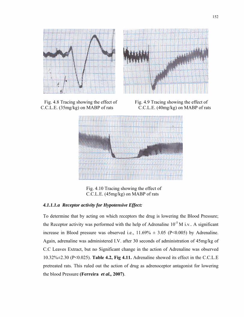
152
Fig. 4.8 Tracing showing the effect of Fig. 4.9 Tracing showing the effect of C.C.L.E. (35mg/kg) on MABP of rats C.C.L.E. (40mg/kg) on MABP of rats
Fig. 4.10 Tracing showing the effect of C.C.L.E. (45mg/kg) on MABP of rats
4.1.1.1.a Receptor activity for Hypotensive Effect:
To determine that by acting on which receptors the drug is lowering the Blood Pressure;
the Receptor activity was performed with the help of Adrenaline 10-5 M i.v.. A significant
increase in Blood pressure was observed i.e., 11.69% ± 3.05 (P<0.005) by Adrenaline.
Again, adrenaline was administered I.V. after 30 seconds of administration of 45mg/kg of
C.C Leaves Extract, but no Significant change in the action of Adrenaline was observed
10.32%±2.30 (P<0.025). Table 4.2, Fig 4.11. Adrenaline showed its effect in the C.C.L.E
pretreated rats. This ruled out the action of drug as adrenoceptor antagonist for lowering
the blood Pressure (Ferreira et al., 2007).
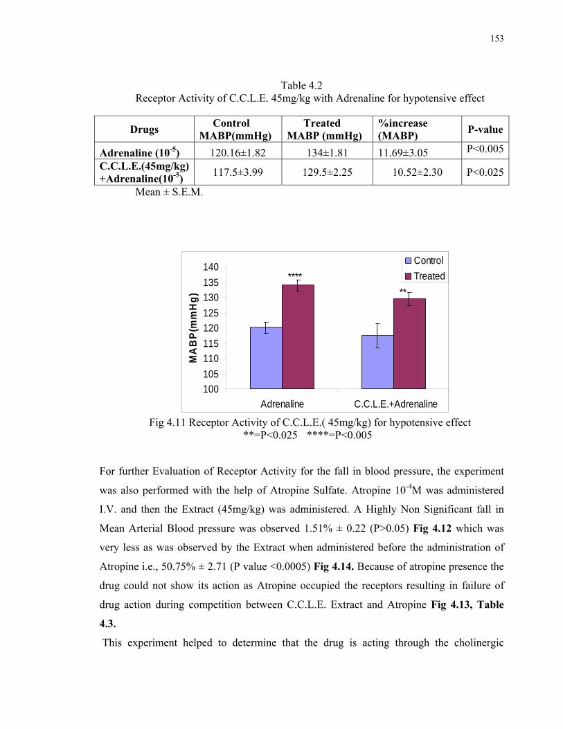
153
Table 4.2
Receptor Activity of C.C.L.E. 45mg/kg with Adrenaline for hypotensive effect
Mean ± S.E.M.
100
105
110
115
120
125
130
135
140
Adrenaline C.C.L.E.+Adrenaline
MA
BP
(mm
Hg
)
Control
Treated****
**
Fig 4.11 Receptor Activity of C.C.L.E.( 45mg/kg) for hypotensive effect
**=P<0.025 ****=P<0.005
For further Evaluation of Receptor Activity for the fall in blood pressure, the experiment
was also performed with the help of Atropine Sulfate. Atropine 10-4M was administered
I.V. and then the Extract (45mg/kg) was administered. A Highly Non Significant fall in
Mean Arterial Blood pressure was observed 1.51% ± 0.22 (P>0.05) Fig 4.12 which was
very less as was observed by the Extract when administered before the administration of
Atropine i.e., 50.75% ± 2.71 (P value <0.0005) Fig 4.14. Because of atropine presence the
drug could not show its action as Atropine occupied the receptors resulting in failure of
drug action during competition between C.C.L.E. Extract and Atropine Fig 4.13, Table
4.3.
This experiment helped to determine that the drug is acting through the cholinergic
Drugs Control MABP(mmHg)
Treated MABP (mmHg)
%increase (MABP)
P-value
Adrenaline (10-5) 120.16±1.82 134±1.81 11.69±3.05 P<0.005
C.C.L.E.(45mg/kg) +Adrenaline(10-5)
117.5±3.99 129.5±2.25 10.52±2.30 P<0.025
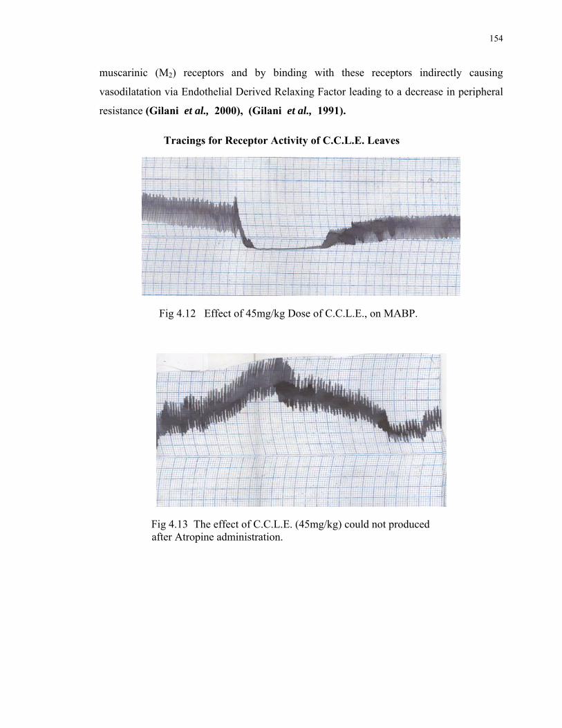
154
muscarinic (M2) receptors and by binding with these receptors indirectly causing
vasodilatation via Endothelial Derived Relaxing Factor leading to a decrease in peripheral
resistance (Gilani et al., 2000), (Gilani et al., 1991).
Tracings for Receptor Activity of C.C.L.E. Leaves
Fig 4.12 Effect of 45mg/kg Dose of C.C.L.E., on MABP.
Fig 4.13 The effect of C.C.L.E. (45mg/kg) could not produced after Atropine administration.
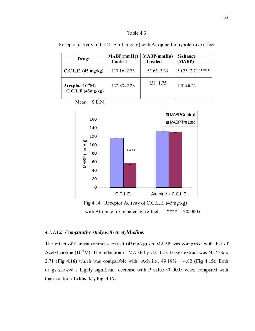
155
Table 4.3
Receptor activity of C.C.L.E. (45mg/kg) with Atropine for hypotensive effect
Drugs MABP(mmHg) Control
MABP(mmHg) Treated
%change (MABP)
C.C.L.E. (45 mg/kg) 117.16±2.75 57.66±3.35 50.75±2.71*****
Atropine(10-4M) +C.C.L.E.(45mg/kg)
132.83±2.28
131±1.75
1.51±0.22
Mean ± S.E.M.
0
20
40
60
80
100
120
140
160
C.C.L.E. Atropine + C.C.L.E.
MA
BP
(m
mH
g)
MABPControl
MABPTreated
*****
Fig 4.14 Receptor Activity of C.C.L.E. (45mg/kg)
with Atropine for hypotensive effect. **** =P<0.0005
4.1.1.1.b Comparative study with Acetylcholine: The effect of Carissa carandas extract (45mg/kg) on MABP was compared with that of
Acetylcholine (10-6M). The reduction in MABP by C.C.L.E. leaves extract was 50.75% ±
2.71 (Fig 4.16) which was comparable with Ach i.e., 49.18% ± 4.02 (Fig 4.15). Both
drugs showed a highly significant decrease with P value <0.0005 when compared with
their controls Table. 4.4, Fig. 4.17.

156
Comparative Study with Acetylcholine
Fig 4.15 Effect of Ach (10-6M) on MABP of rats
Fig 4.16 Effect of C.C.L.E. leaves (45mg/kg) on MABP of rats

157
Table 4.4
Comparison between the effect of C.C.L.E. (45mg/kg) and Ach 10-6M
n= 12 ± =S.E.M.
Fig. 4.17 Comparative study between the effect of
C.C.L.E. (45mg/kg) and Ach 10-6M ****=P<0.0005
4.1.1.2. EFFECTS ON INTACT HEART OF FROG:
The effects of C.C.L.E. in different doses, exhibited on intact heart of frog, are shown by the following tracings. Fig 4.18- 4.25
Fig 4.18 Tracing for Intact Heart of Control (Left) and C.C.L.E. treated (5mg/ml) (right) frogs
Drugs MABP(mmHg)
Control MABP(mmHg)
Treated
% Fall
(MABP)
Ach 10-6 M 146.5±2.9 74.16±5.12
49.18±4.02 45mg/kg (C.C) 117.16±2.74 57.66±3.35
50.75±2.71
0
20
40
60
80
100
120
140
160
Ach 45mg/kg C.C.L.E
MA
BP
(m
mH
g)
control
treated
***** *****

158
Fig 4.19 Tracing for intact Heart of Control (Left) and C.C.L.E. treated (10mg/ml) (right) frogs.
Fig 4.20 Tracing for intact Heart of Control (Left) and C.C.L.E. treated (15mg/ml) (right) frogs.
Fig 4.21 Tracing for intact Heart of Control (Left) and C.C.L.E. treated (20mg/ml) (right) frogs.
Fig 4.22 Tracing for intact Heart of Control (Left) and C.C.L.E. treated (25mg/ml) (right) frogs
Fig 4.23 Tracing for intact Heart of Control (Left) and C.C.L.E. treated (30mg/ml) (right) Frogs.

159
Fig 4.24 Tracing for intact Heart of Control (Left) and C.C.L.E. treated (35 mg/ml) (right) Frogs.
Fig 4.25 Tracing for intact Heart of Control (Left) and C.C.L.E. treated (40mg/ml) (right) Frogs. With the help of above tracings following calculations were obtained: (Pennacchio et al.,
1995), (Azmat et al., 2005)
4.1.1.2.a. Effect on Heart Rate:
The Extract (C.C.L.E.) found to cause a decrease in Heart Rate. The pattern of Heart Rate
reduction was calculated as follows:
1. Extract caused a non significant decrease in heart rate at the doses of 5mg/ml-
30mg/ml (P>0.05) when compared with their controlss.
2. While the dose of 35mg/ml exhibited a statistically Significant decrease (P<0.05).
3. 40mg/ml dose showed a 25.99% ± 0.25 decrease which was statistically highly
Significant (P<0.0005) when compared with its controls Table 4.5, Fig 4.18.

160
Table 4.5
Effect of different doses of C.C.L.E. on Heart rate of frog.
n= 12, Mean ± S.E.M.
F (12.25) =40.39794; P<0.01, F (6.99) =37.55295; P<0.01
Fig.4.26 Effect of C.C.L.E.on Heart rate of frogs in different doses.
* = P<0.05 ***** = P<0.0005
C.C.L.E.
Doses
Heart rate (beats/min.)
(control)
Heart rate (beats/min.)
(treated) % decrease P-value
5mg/ml 41.62±3.74 39.76±2.43 4.23±2.43 P>0.05
10mg/ml 55.11±2.71 50.29±2.58 8.69±1.62 P>0.05
15mg/ml 51.29±2.94 45.26±3.15 11.66±3.63 P>0.05
20mg/ml 44.12±2.8 38.26±3.05 13.45±3.16 P>0.05
25mg/ml 36.88±2.64 31.63±2.21 12.08±3.75 P>0.05
30mg/ml 30.99±3.71 26.61±3.18 14.98±3.5 P>0.05
35mg/ml 37.801±2.67 30.495±0.31 15.55±3.3 P<0.05
40mg/ml 41.67±0.61 30.83±0.39 25.99±0.25 P<0.0005
0
10
20
30
40
50
60
70
5mg/ml 10mg/ml 15mg/ml 20mg/ml 25mg/ml 30mg/ml 35mg/ml 40mg/ml
Hea
rt R
ate
(bea
ts/ m
in.) Control
Treated
* *****

161
4.1.1.2.b. Effect on Cardiac Force: The effect on Cardiac Force produced by C.C.L.E. was calculated and found to have the
following pattern: (Pennacchio et al., 1995), (Azmat et al., 2005)
1. At the doses 5mg/ml- 20mg/ml, caused a non significant increase in cardiac force
(P>0.05).
2. While comparatively higher doses i.e., 25mg/ml, 30mg/ml and 35mg/ml caused a
significant increase in cardiac force when compared with their controls (P<0.05).
3. 40mg/ml caused a 53.79% ± 8.83 increase in cardiac force that was highly
Significant when compared with its controls (P<0.025). Table 4.6, Fig. 4.27
Table 4.6
Effect of C.C.L.E. on Cardiac force of frogs in different doses
n= 12, Mean ± S.E.M.
F (12.25) =67.93; P<0.01, F (6.99) =13.37; P<0.01
Dose Cardiac force
(g) control
Cardiac force
(g) treated % increase P-value
5mg/ml 0.505±0.078 0.551±0.064 10.12±4.95 P>0.05
10mg/ml 0.577±0.076 0.672±0.082 17.17±7.48 P>0.05
15mg/ml 0.509±0.07 0.62±0.066 21.36±7.23 P>0.05
20mg/ml 0.439±0.019 0.56±0.042 28.57±5.67 P<0.05
25mg/ml 0.401±0.039 0.511±0.032 29.66±7.07 P<0.05
30mg/ml 0.383±0.034 0.482±0.025 30.78±13.62 P<0.05
35mg/ml 0.378±0.048 0.509±0.037 40.26±17.6 P<0.05
40mg/ml 0.343±0.027 0.525±0.029 53.79±8.83 P<0.025

162
0
0.2
0.4
0.6
0.8
5mg/kg 10mg/kg 15mg/kg 20mg/kg 25mg/kg 30mg/kg 35mg/kg 40mg/kg
Ca
rdia
c f
orc
e (
g)
Control
Treated***
**
Fig 4.27 Effect of C.C.L.E.on Cardiac force of frogs in different doses. * = P<0.05 ** = P<0.025 4.1.1.2.c. Effct on Cardiac Cycle: The Effect of C.C.L.E. showed an increase in cardiac cycle in the following pattern:
(Pennacchio et al., 1995), (Azmat et al., 2005)
1. Doses from 5mg/ml to 30 mg/ml produced statistically non significant increase in
cardiac cycle (P>0.05) when compared with their controls.
2. While at the dose of 35 mg/ml the increase in cardiac cycle was statistically
significant (P<0.05)
3. Dose of 40mg/ml caused a 50.83% ±11.74 increase in cardiac cycle that was found
to be Highly significant (P<0.01) when compared with its controls Table 4.7, Fig
4.28.

163
Table 4.7
Effect of C.C.L.E. on Cardiac Cycle of frogs in different doses
n=12, Mean ± S.E.M.
F (12.25) =48.13; P<0.01, F (6.99) =107.02; P<0.01
Fig. 4.28 Effect of C.C.L.E. on Cardiac Cycle of frogs in different doses
* = P<0.05 *** = P<0.01
Dose
Cardiac
cycle(sec.)
(control)
Cardiac
cycle(sec.)
(treated)
% increase P-value
5mg/ml 1.51±0.06 1.68±0.05 11.16±1.69 P>0.05
10mg/ml 2.07±0.05 2.37±0.098 14.36±1.94 P>0.05
15mg/ml 2.17±0.05 2.5±0.18 14.68±6.05 P>0.05
20mg/ml 1.02±0.17 1.2±0.21 17.38±4.71 P>0.05
25mg/ml 2.1±0.047 2.5±0.14 18.84±4.07 P>0.05
30mg/ml 0.65±0.07 0.787±0.07 22.89±3.72 P>0.05
35mg/ml 1.7±0.13 2.12±0.11 25.66±4.77 P<0.05*
40mg/ml 0.88±0.01 1.32±0.89 50.83±11.74 P<0.01***
0
0.5
1
1.5
2
2.5
3
5mg/ml 10mg/ml 15mg/ml 20mg/ml 25mg/ml 30mg/ml 35mg/ml 40mg/ml
Car
dia
c C
ycle
(S
eco
nd
s) control
Treated*
***

164
4.1.1.3 RECEPTOR ACTIVITY FOR EFFECT OF C.C.L.E.ON HEART: 4.1.1.3.a Effect on cardiac force: The dose of 40mg/ml C.C.L.E. has demonstrated an increase in the cardiac force that was
53.79% ± 8.83 higher than its control Table 4.6, Fig 4.29 - Fig 4.32. On the other hand
similar dose of C.C.L.E. in the presence of pretreatment with atropine (10-3 M) could not
significantly increase the cardiac force and the increase was only 10.6% ± 2.72 than its
control that was statistically non significant Fig 4.33, Table 4.8. However the Use of Ach
(10-3M) was found to decrease the Cardiac force by 50.94% less than its controls Fig 4.33,
while on pretreatment with Atr.(10-3M) it was only 13.26 % than its controls Table 4.8.
Fig. 4.29 Tracing showing the intact heart activity of Control frog.
Fig. 4.30 Effect of C.C.L.E. (40mg/ml) on
Intact heart of frog
Fig. 4.31 Effect of Atropine (10-3M) +
C.C.L.E. (40mg/ml)on intact heart of frog

165
Table 4.8
Receptor Activity of C.C.L.E.for the Effect on Cardiac Force
n=12, Mean ± S.E.M.
Fig. 4.32 Effect of C.C.L.E. (40mg/ml) and Ach. (10-3M) on
Intact heart of frog compared with control ***=P<0.025
Dose Cardiac force
(g)control
Cardiac force
(g) treated % change P-value
Ach.(10-3M) 0.485±0.03 0.235±0.013 50.947±4.96 P<0.025**
Atropine(10-3M)+
Ach.(10-3M)
0.485±0.03
0.420±0.05
13.26±4.6
P>0.05
C.C.L.E.
(40mg/ml) 0.343±0.027 0.525±0.029 53.79±8.83 P<0.025**
Atropine (10-3M)
+ C.C.L.E.
(40mg/ml)
0.343±0.027 0.387±0.02 10.76±2.72 P>0.05
0
0.1
0.2
0.3
0.4
0.5
0.6
1
Car
dia
c fo
rce
(g)
Control C.C.L.E Control Ach (10-3M)
***
***
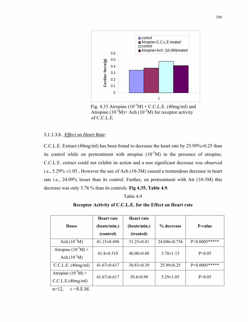
166
0
0.1
0.2
0.3
0.4
0.5
0.6
1
Ca
rdia
c f
orc
e(g
)
controlAtropine+C.C.L.E.treatedcontrolAtropine+Ach. (10-3M)treated
Fig. 4.33 Atropine (10-3M) + C.C.L.E. (40mg/ml) and
Atropine (10-3M)+ Ach (10-3M) for receptor activity of C.C.L.E. 5.1.1.3.b. Effect on Heart Rate: C.C.L.E. Extract (40mg/ml) has been found to decrease the heart rate by 25.99%±0.25 than
its control while on pretreatment with atropine (10-3M) in the presence of atropine,
C.C.L.E. extract could not exhibit its action and a non significant decrease was observed
i.e., 5.29% ±1.05 . However the use of Ach (10-3M) caused a tremendous decrease in heart
rate i.e., 24.09% lesser than its control. Further, on pretreatment with Atr (10-3M) this
decrease was only 3.78 % than its controls. Fig 4.35, Table 4.9.
Table 4.9
Receptor Activity of C.C.L.E. for the Effect on Heart rate
n=12, ± =S.E.M.
Doses
Heart rate
(beats/min.)
(control)
Heart rate
(beats/min.)
(treated)
% decrease P-value
Ach.(10-3M) 41.15±0.496 31.25±0.41 24.046±0.736 P<0.0005*****
Atropine (10-3M) +
Ach.(10-3M) 41.8±0.518 40.00±0.88 3.78±1.13 P>0.05
C.C.L.E. (40mg/ml) 41.67±0.617 30.83±0.39 25.99±0.25 P<0.0005*****
Atropine (10-3M) +
C.C.L.E.(40mg/ml) 41.67±0.617 39.4±0.99 5.29±1.05 P>0.05

167
0
5
10
15
20
25
30
35
40
45
1
He
art
ra
te(b
ea
ts/m
in.)
Contorl C.C.L.E. Control Ach.(10-3M)
***** *****
Fig. 4.34 Effect of C.C.L.E. (40mg/ml) and Ach.(10-3M)on
Intact heart of frog compared with control *****=P<0.0005
36
37
38
39
40
41
42
43
1
Hea
rt r
ate(
bea
ts/m
in)
Control
C.C. After Atropine
Control
Ach. After Atropine
Fig. 4.35 Atropine (10-3M) + C.C.L.E. (40mg/ml) and Atropine (10-3M) +Ach(10-3M) for receptor activity. 5.1.1.4. COMPARATIVE STUDY WITH DIGOXIN:
As the drug C.C.L.E. showed positive Inotropic and negative Chronotropic effects
therefore, the effects of C.C.L.E. (40mg/ml) exhibited on intact heart of frog were
compared with that of Digoxin (0.008mg/ml).The response was recorded in the form of
following tracings. Fig 4.36- 4.38

168
Fig. 4.36 Tracing showing the Intact Heart activity of control frog’s heart
Fig. 4.37 Digoxin (0.008mg/ml) treated frog’s heart
Fig 4.38 C.C.L.E. (40mg/ml) treated frog’s heart
1. The 40mg/ml dose of C.C.L.E. caused 53.79% ± 8.83 (P<0.025) increase in cardiac
force which was less than that produced by Digoxin (0.008mg/ml) i.e., 81.6% ±
20.59 (P<0.005) when compared with their controls. Table 4.10, fig.4.39
Table 4.10 Comparative study of C.C.L.E. with Digoxin showing effect on Cardiac force of
Frog’s heart. n =12, Mean ± S.E.M.
FF (8.02) =28.36; P<0.01
Dose
Cardiac force
(g)control
Cardiac force
(g) treated % increase P-value
C.C.L.E.
(40mg/kg) 0.343±0.027 0.525±0.029 53.79±8.83 P<0.025**
Digoxin
(0.008mg/ml) 0.343±0.027 0.612±0.055 81.6±20.59 P<0.005****

169
Fig 4.39 Comparative study of C.C.L.E. with Digoxin
showing effect on Cardiac force of Frogs’ Heart
2. The Heart rate reduction caused by C.C.L.E. leaves was calculated as 25.99% ±
0.25 (P<0.0005) which was found to be more than that produced by Digoxin i.e.,
12.26% ± 0.91 (P<0.005) Table 4.11, Fig 4.40
Table 4.11 Comparative study of C.C.L.E. with Digoxin showing effect on Heart rate of
Frog’s heart
n= 12, ± =S.E.M.
F (8.02) =126.89; P<0.01
Doses
Heart rate
(beats/min.)
(control)
Heart rate
(beats/min.)
(treated)
% decrease P-value
C.C.L.E. (40mg/ml) 41.67±0.617 30.83±0.39 25.99±0.25 P<0.0005*****
Digoxin
(0.008mg/ml) 41.67±0.617
36.55±1.086
12.26±0.91
P<0.005****
0
0.1
0.2
0.3
0.4
0.5
0.6
0.7
0.8
1
Car
dia
c F
orc
e (g
)
Control C.C.L.E. (40mg/ml) Digoxin(0.008mg/ml)
**
****
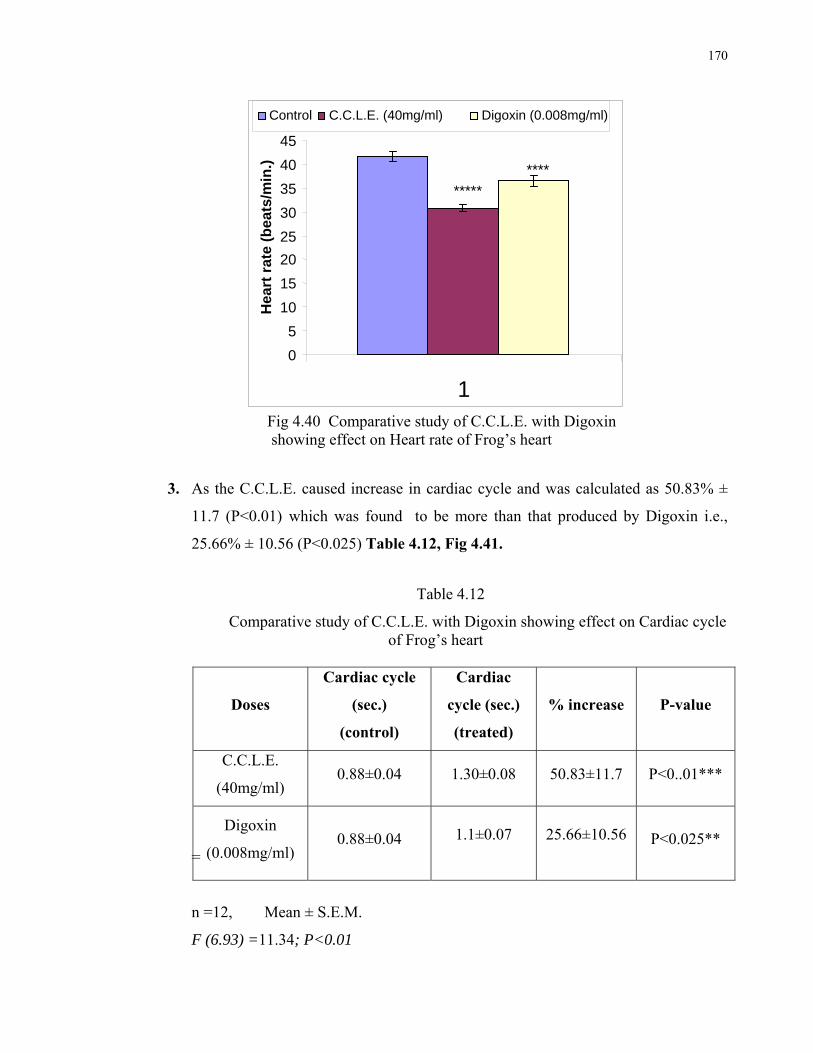
170
Fig 4.40 Comparative study of C.C.L.E. with Digoxin
showing effect on Heart rate of Frog’s heart
3. As the C.C.L.E. caused increase in cardiac cycle and was calculated as 50.83% ±
11.7 (P<0.01) which was found to be more than that produced by Digoxin i.e.,
25.66% ± 10.56 (P<0.025) Table 4.12, Fig 4.41.
Table 4.12
Comparative study of C.C.L.E. with Digoxin showing effect on Cardiac cycle of Frog’s heart
=
n =12, Mean ± S.E.M.
F (6.93) =11.34; P<0.01
Doses
Cardiac cycle
(sec.)
(control)
Cardiac
cycle (sec.)
(treated)
% increase P-value
C.C.L.E.
(40mg/ml) 0.88±0.04 1.30±0.08 50.83±11.7 P<0..01***
Digoxin
(0.008mg/ml) 0.88±0.04
1.1±0.07
25.66±10.56
P<0.025**
0
5
10
15
20
25
30
35
40
45
1
Hea
rt r
ate
(bea
ts/m
in.)
Control C.C.L.E. (40mg/ml) Digoxin (0.008mg/ml)
*****
****
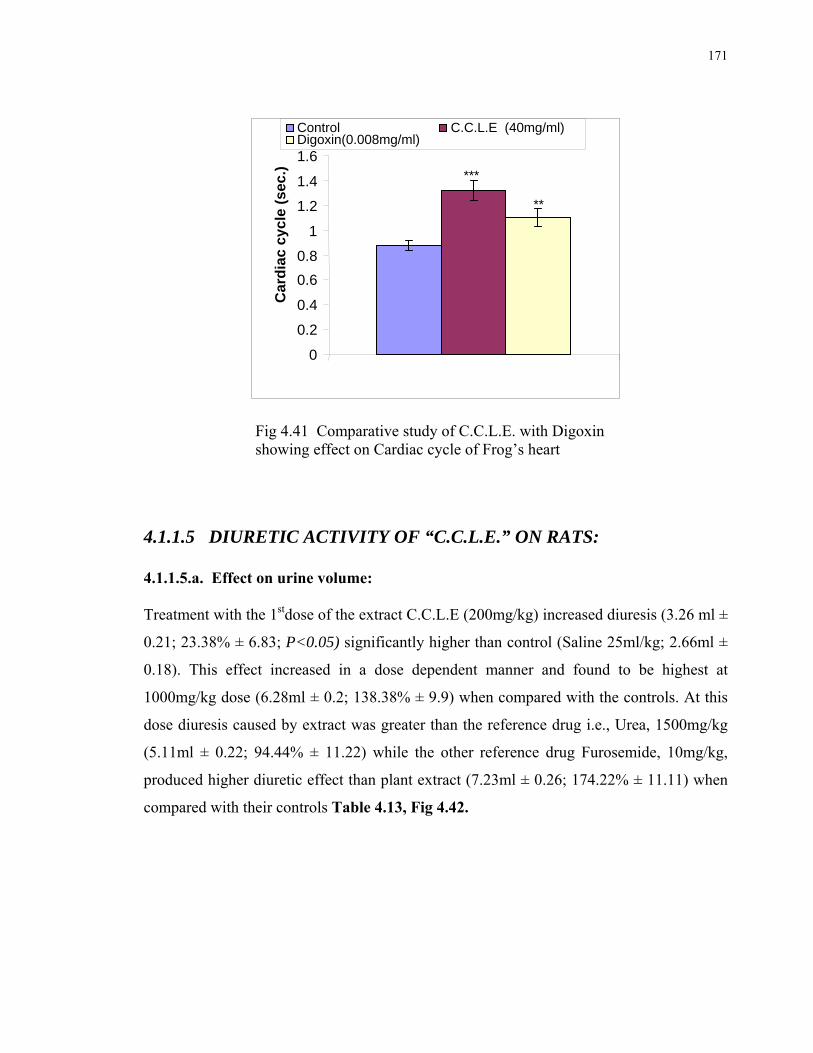
171
Fig 4.41 Comparative study of C.C.L.E. with Digoxin showing effect on Cardiac cycle of Frog’s heart
4.1.1.5 DIURETIC ACTIVITY OF “C.C.L.E.” ON RATS: 4.1.1.5.a. Effect on urine volume: Treatment with the 1stdose of the extract C.C.L.E (200mg/kg) increased diuresis (3.26 ml ±
0.21; 23.38% ± 6.83; P<0.05) significantly higher than control (Saline 25ml/kg; 2.66ml ±
0.18). This effect increased in a dose dependent manner and found to be highest at
1000mg/kg dose (6.28ml ± 0.2; 138.38% ± 9.9) when compared with the controls. At this
dose diuresis caused by extract was greater than the reference drug i.e., Urea, 1500mg/kg
(5.11ml ± 0.22; 94.44% ± 11.22) while the other reference drug Furosemide, 10mg/kg,
produced higher diuretic effect than plant extract (7.23ml ± 0.26; 174.22% ± 11.11) when
compared with their controls Table 4.13, Fig 4.42.
0
0.2
0.4
0.6
0.8
1
1.2
1.4
1.6
Car
dia
c cy
cle
(sec
.)
Control C.C.L.E (40mg/ml)Digoxin(0.008mg/ml)
***
**

172
Table 4.13
Effect of C.C.L.E., Urea and Furosemide in rats on urine volume compared
with their control.
Drugs urine vol. ml S.E.M % increase S.E.M P-value
Saline25ml/kg(Control) 2.66 ±0.182
Urea1500mg/kg 5.11 ±0.22 94.44 ±11.22 p<0.0005
Furosemide10mg/kg 7.23 ±0.26 174.22 ±11.11 p<0.0005
C.C.L.E.200mg/kg 3.26 ±.211 23.38 ±6.83 p<0.05
C.C.L.E.500mg/kg 4.41 ±0.153 67.77 ±8.36 p<0.0005
C.C.L.E.1000mg/kg 6.28 ±0.208 138.38 ±9.9 p<0.0005
n=8, Mean ± S.E.M.
F (3.7) =81.47; P<0.01
0
1
2
3
4
5
6
7
8
Uri
ne
vo
lum
e(m
l)
Saline25ml/kg Urea1500mg/kg Furosemide10mg/kg
C.C.L.E.200mg/kg C.C.L.E.500mg/kg C.C.L.E.1000mg/kg
*
***
***
****
****
Fig. 4.42 Effect of C.C.L.E., Urea and Furosemide in rats on urine
volume compared with their control **** = P< 0.0005
4.1.1.5.b. Effect on Urinary Electrolyte Excretion: Extract of Carissa carandas leaves enhanced the Na+ ion excretion in a dose dependent
manner and found to be highest at the dose of 1000mg/kg (193.11meq/lit. ± 1.03; 134.67%
± 2.47; P<0.0005) when compared with their controls (82.32meq/lit. ± 1.13).This
excretion was found to be higher than the reference drug Urea (132.35 meq/lit ±1.04;

173
60.04% ± 1.04; P<0.0005) and lesser than the reference drug Furosemide (202.37meq/lit ±
3.26; 145.88% ± 4.24; P<0.0005). Table 4.14, Fig 4.43
Table 4.14
Effect of C.C.L.E., Urea and Furosemide on Na+ excretion in urine compared
with their control rats.
Drugs Na+meq./lit. S.E.M. %increase S.E.M. P-value
Saline25ml/kg(control) 82.32 ±1.138
Urea1500mg/kg 132.35 ±1.04 60.04 ±1.04 p<0.0005
Furosemide10mg/kg 202.37 ±3.26 145.88 ±4.24 p<0.0005
C.C.L.E.200mg/kg 131.68 ±0.55 60.04 ±2.76 p<0.0005
C.C.L.E.500mg/kg 187.39 ±0.62 127.73 ±3.57 p<0.0005
C.C.L.E.1000mg/kg 193.11 ±1.03 134.67 ±3.47 p<0.0005
n= 8, Mean ± S.E.M.
F (4.25) = 1195.912; P<0.01
0
50
100
150
200
250
Na
+ (
me
q/l
it.)
Saline25ml/kg Urea1500mg/kg Furosemide10mg/kg
C.C.L.E.200mg/kg C.C.L.E.500mg/kg C.C.L.E.1000mg/kg
****
****
****
**** ****
Fig. 4.43 Effect of C.C.L.E., Urea and Furosemide on Na+ excretion in urine compared with their control **** = P< 0.0005
K+ ion excretion was also increased in a dose dependent manner and found to be highest at
the dose of 1000mg/kg (86.46 meq/lit ±1.59; 40.1% ± 2.11; P<0.0005) when compared

174
with their controls (61.71 meq/lit. ± 0.60).This excretion was found to be higher than the
reference drug Urea (82.12meq/lit ± 0.98; 33.08% ±1.25; P<0.0005) and equal to the
reference drug Furosemide (86.42meq/lit ± 0.75; 40.07% ± 1.92; P<0.0005). Table 4.15,
Fig 4.44
Table 4.15
Effect of C.C.L.E., Urea and Furosemide on K+ excretion in urine compared
with their control rats.
Drugs K+meq./lit. S.E.M. %increase S.E.M. P-value
Saline25ml/kg(control) 61.71 ±0.605
Urea1500mg/kg 82.12 ±0.982 33.08 ±1.25 p<0.0005
Furosemide10mg/kg 86.42 ±0.755 40.07 ±1.92 p<0.0005
C.C.L.E.200mg/kg 81.99 ±0.382 32.89 ±0.805 p<0.0005
C.C.L.E.500mg/kg 82.2 ±0.89 33.25 ±2.41 p<0.0005
C.C.L.E.1000mg/kg 86.46 ±1.59 40.1 ±2.11 p<0.0005
n= 8, ± =S.E.M.
F (4.25) =129.83; P<0.01
0
20
40
60
80
100
K+
(me
q./l
it.)
Saline25ml/kg Urea1500mg/kg Furosemide10mg/kgC.C.L.E.200mg/kg C.C.L.E.500mg/kg C.C.L.E.1000mg/kg
********
**** ********
Fig. 4.44 Effect of C.C.L.E., Urea and Furosemide on K+ excretion in urine compared with their control rats. **** = P< 0.0005

175
Cl- ion excretion was also increased in a dose dependent manner and found to be highest at
the dose of 1000mg/kg (97.82 meq/lit ± 0.17; 13.16% ± 2.11; P<0.0005) when compared
with their controls (86.52 meq/lit ± 1.69).This excretion was found to be higher than the
reference drug Urea (82.12meq/lit ± 0.98; 33.08% ± 1.25; P<0.0005) and equal to the
reference drug Furosemide (86.42meq/lit ± 0.75; 40.07% ± 1.92; P<0.0005). Table 4.16,
Fig. 4.45
Table 4.16
Effect of C.C.L.E., Urea and Furosemide on Cl- excretion in urine compared
with their control rats
n=8, Mean ± S.E.M.
F (4.25) =29.20; P<0.01
Drugs Cl-meq./lit. S.E.M %increase S.E.M P-value
Saline25ml/kg(control) 86.525 ±1.69
Urea1500mg/kg 94.35 ±0.616 9.19 ±2.87 p<0.005
Furosemide10mg/kg 96.212 ±0.998 11.26 ±1.72 p<0.005
C.C.L.E.200mg/kg 94.63 ±0.535 9.478 ±2.45 p<0.005
C.C.L.E.500mg/kg 96.99 ±0.195 12.19 ±2.24 p<0.0005
C.C.L.E.1000mg/kg 97.82 ±0.177 13.16 ±2.35 p<0.0005
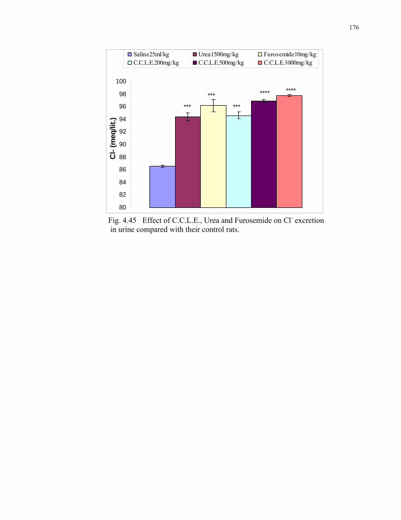
176
80
82
84
86
88
90
92
94
96
98
100
Cl-
(m
eq/li
t.)
Saline25ml/kg Urea1500mg/kg Furosemide10mg/kgC.C.L.E.200mg/kg C.C.L.E.500mg/kg C.C.L.E.1000mg/kg
***
***
***
**** ****
Fig. 4.45 Effect of C.C.L.E., Urea and Furosemide on Cl- excretion in urine compared with their control rats.

177
4.1.2 MUFARREH YAQQOTI MOTADIL (MUYM): 4.1.2.1. EFFECTS ON MEAN ARTERIAL BLOOD PRESSURE (MABP) OF RATS: The effect of MUYM on Mean Arterial Blood Pressure of rats was recorded as shown by
the tracings given below Fig 4.47 – Fig.4.50.
The drug caused a decrease in Mean Arterial Blood Pressure in rats, in a dose dependent
manner, the results are summarized as follows: (Table 4.17)
1. At the doses of 10mg/kg, this poly herbal drug caused a statistically non-significant
decrease in mean arterial Blood Pressure (P>0.05) when compared with its controls.
2. 20mg/kg showed a reduction in blood pressure that was 8.01% ± 0.49
statistically significant (P<0.05) compared with its controls.
.
3. 30mg/kg doses also a reduction in blood pressure that was 8.7% ± 2.07statistically
significant (P<0.01) compared with its controls.
4. While at the dose of 40mg/kg MABP was found to reduce by 15.79% ± 0.57 from
control. This fall in MABP was statistically highly Significant (p<0.005) when
compared with its controls Fig 4.46.
Table 4.17
Effect of Mufarreh Yaqooti Motadil (MUYM) on MABP of rats in different doses.
n= 6, Mean ± S.E.M.
F (10.13) = 14.57; P<0.05, F (9.28) = 4.35; P>0.05.
MUYM MABP(mmHg) (control)
MABP(mmHg) (treated)
%Fall P-value
10mg/kg 147.166±0.8 142.33±2.38 3.3±1.12 P>0.05
20mg/kg 141.33±3.34 129.23±1.99 8.01±0.49 P<0.05
30mg/kg 135.62±3.63 122.5±2.97 8.7±2.07 P<0.01
40mg/kg 154.33±1.95 129.92±1.13 15.79±0.57 P<0.005

178
Fig 4.46 Effect of Mufarreh Yaqooti Motadil (MUYM) on
MABP of rats in different doses. * =P<0.05, ** = P<0.01, *** = P<0.005,
Fig 4.47 Effect of MUYM10mg/kg on Fig 4.48 Effect of MUYM 20mg/kg on
MABP of rats MABP of rats
Fig 4.49 Effect of MUYM 30mg/kg Fig 4.50 Effect of MUYM 40mg/kg on on MABP of rats MABP of Rats.
0
50
100
150
200
10mg/kg 20mg/kg 30mg/kg 40mg/kg M
AB
P (
mm
Hg
)
Control
Treated
* *****
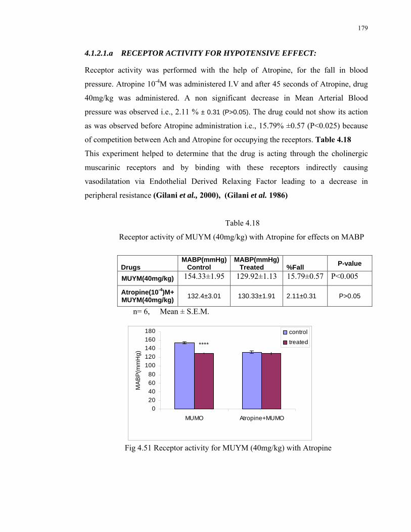
179
4.1.2.1.a RECEPTOR ACTIVITY FOR HYPOTENSIVE EFFECT: Receptor activity was performed with the help of Atropine, for the fall in blood
pressure. Atropine 10-4M was administered I.V and after 45 seconds of Atropine, drug
40mg/kg was administered. A non significant decrease in Mean Arterial Blood
pressure was observed i.e., 2.11 % ± 0.31 (P>0.05). The drug could not show its action
as was observed before Atropine administration i.e., 15.79% ±0.57 (P<0.025) because
of competition between Ach and Atropine for occupying the receptors. Table 4.18
This experiment helped to determine that the drug is acting through the cholinergic
muscarinic receptors and by binding with these receptors indirectly causing
vasodilatation via Endothelial Derived Relaxing Factor leading to a decrease in
peripheral resistance (Gilani et al., 2000), (Gilani et al. 1986)
Table 4.18
Receptor activity of MUYM (40mg/kg) with Atropine for effects on MABP
Drugs MABP(mmHg) Control
MABP(mmHg) Treated %Fall
P-value
MUYM(40mg/kg) 154.33±1.95 129.92±1.13 15.79±0.57 P<0.005
Atropine(10-4)M+MUYM(40mg/kg)
132.4±3.01 130.33±1.91 2.11±0.31 P>0.05
n= 6, Mean ± S.E.M.
0
20
40
60
80
100
120
140
160
180
MUMO Atropine+MUMO
MA
BP
(mm
Hg
)
control
treated****
Fig 4.51 Receptor activity for MUYM (40mg/kg) with Atropine
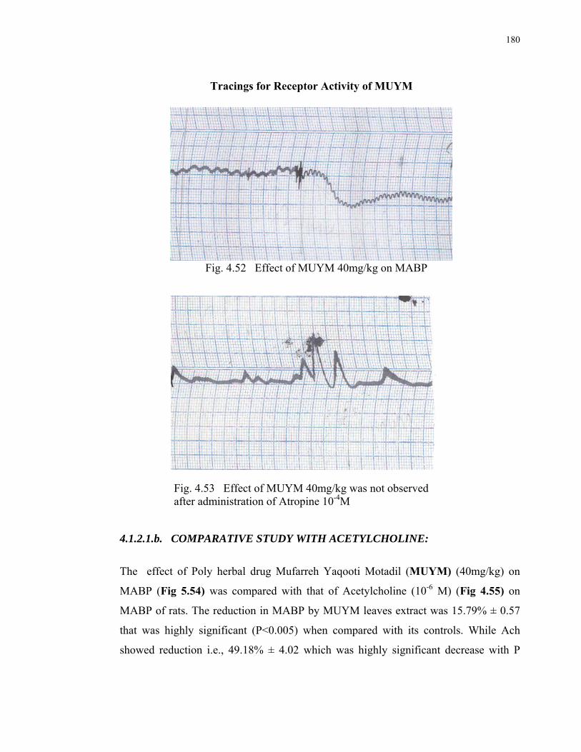
180
Tracings for Receptor Activity of MUYM
Fig. 4.52 Effect of MUYM 40mg/kg on MABP
Fig. 4.53 Effect of MUYM 40mg/kg was not observed after administration of Atropine 10-4M
4.1.2.1.b. COMPARATIVE STUDY WITH ACETYLCHOLINE:
The effect of Poly herbal drug Mufarreh Yaqooti Motadil (MUYM) (40mg/kg) on
MABP (Fig 5.54) was compared with that of Acetylcholine (10-6 M) (Fig 4.55) on
MABP of rats. The reduction in MABP by MUYM leaves extract was 15.79% ± 0.57
that was highly significant (P<0.005) when compared with its controls. While Ach
showed reduction i.e., 49.18% ± 4.02 which was highly significant decrease with P

181
value <0.0005 when compared with their controls. However the Action of Ach was
found to be more potent than the tested drug i.e., MUYM Table 4.19, Fig 4.56.
Tracings showing the comparative study of MUYM with
Acetylcholine on MABP
Fig 4.54 Effect of Ach 10-6 M on MABP of rats.
Fig. 4.55 Effect of MUYM 40mg/kg on MABP of rats.

182
Table 4.19 Comparative study of Effect of MUYM with that of Acetylcholine
n= 6, Mean ± S.E.M.
Fig 4.56 Comparative study of Effect of MUYM on MABP
with that of Acetylcholine ***= P< 0.005, ****= P< 0.0005
4.1.2.2. EFFECTS OF MUFARREH YAQOOTI MOTADIL (MUYM) ON
INTACT HEART OF FROG:
The effects of MUYM in different doses, exhibited on intact heart of frog, are shown by the following tracings. Fig 4.57- 4.64
Fig 4.57 Tracing for Control (left) and 5mg/ml Treated (right) Frog’s heart.
Drug MABP(mmHg) Control
MABP(mmHg) Treated
% Fall
Ach 10-6 M 146.5±2.90 74.16±5.12
49.18± 4.02 MUYM (40mg/kg)
154.33±1.95 129.92±1.13 15.79±0.57
0
20
40
60
80
100
120
140
160
180
Ach (10-6M) MUMO(40mg/kg)
MA
BP
(mm
Hg
)
control
treated
****
***
183
Fig.4.58 Tracing for Control (left) and 10mg/ml Treated (right) Frog’s heart.
Fig.4.59 Tracing for Control (left) and 15mg/ml Treated (right) Frog’s heart.
Fig 4.60 Tracing for Control (left) and 20mg/ml Treated (right) Frog’s heart.
Fig 4.61 Tracing for Control (left) and 25mg/ml Treated (right) Frog’s heart.
Fig 4.62 Tracing for Control (left) and 30mg/ml Treated (right) Frog’s heart.
184
Fig 4.63 Tracing for Control (left) and 35mg/ml Treated (right) Frog’s heart.
Fig 4.64 Tracing for Control (left) and 40mg/ml Treated (right) Frog’s heart.
4.1.2.2.a Effects on Heart Rate:
This herbal product found to cause a decrease in Heart Rate. The pattern of Heart Rate
reduction was calculated as follows:
1. The drug Mufarreh Yaqooti Motadil (MUYM) was found to cause a non significant
decrease in heart rate at the doses 5mg/ml and 10mg/ml (P>0.05) when compared
with their controls.
2. While the dose of 15mg/ml showed a statistically significant decrease i.e., 8.89% ±
0.36 (P<0.05) when compared with its controls.
3. 20mg/ml and 25mg/ml caused a significant decrease in Heart Rate (P<0.025) with
percentage of decrease 8.44% ± 0.85 and 8.63% ± 1.55 respectively when
compared with their controls.

185
4. 30mg/ml, 35mg/ml has been found to present a highly significant reduction in heart
rates 8.18% ± 1.29 and 8.99% ± 1.85 respectively with the p-value as P<0.01, when
compared with its controls.
5. 40mg/ml dose produced a highly significant decrease in heart rate with P<0.005
(14.45% ± 2.39) as compared with its controls Table 4.20, Fig 4.65.
Table 4.20
Effect of different doses of MUYM on heart rate of frog’s heart
MUYM
Heart Rate (Beats/min.) Control
Heart Rate (Beats/min.) treated
%Fall in Heart Rate
P-value
5mg/ml 55.75±3.14 53.39±2.63 4.07±2.46 P>0.05
10mg/ml 57.3±2.23 52.67±4.04 8.28±4.68 P>0.05
15mg/ml 67.79±2.6 61.75±2.29 8.89±0.36 P<0.05
20mg/ml 68.2±0.93 62.47±1.41 8.44±0.85 P<0.025
25mg/ml 72.25±1.49 65.9±0.45 8.63±1.55 P<0.025
30mg/ml 53.89±0.83 49.46±0.55 8.18±1.29 P<0.01
35mg/ml 67.92±0.98 61.78±1.11 8.99±1.85 P<0.01
40mg/ml 72.01±1.74 61.36±1.06 14.45±2.39 P<0.005 n= 12, Mean ± S.E.M.
F(12.25)= 47.78; P<0.01, F(6.99)= 31.57; P<0.01
0
10
20
30
40
50
60
70
80
5mg/ml 10mg/ml 15mg/ml 20mg/ml 25mg/ml 30mg/ml 35mg/ml 40mg/ml
Hea
rt R
ate
(Bea
ts/ m
in.)
controltreated
* ** **
***
*** ****
Fig 4.65 Effect of different doses of MUYM on heart rate of frog’s heart * =P < 0.05, ** = P<0.025, *** =P< 0.01, ****=P<0.005

186
4.1.2.2.b Effects on Cardiac Force:
The effect on Cardiac Force produced by MUYM was found to have an increasing
pattern when compared with control, and calculated from the tracings as follows:
1. At the doses 5mg/ml, 10mg/ml, 15mg/ml and 20mg/ml, the drug caused a non
significant increase in cardiac force, with percentage increase as 9.461% ± 1.259
11.116% ± 2.476, 15.00% ± 3.542 and 16.796% ± 6.605 respectively (P>0.05) as
compared with their controls.
2. While the doses of 25mg/ml and 30mg/ml caused a significant increase in cardiac
force with percentage increase as 30.971% ± 40.436 and 31.635% ± 3.077
respectively (P< 0.05) when compared with their controls.
3. At the doses 35mg/ml and 40mg/ml the increase in cardiac force produced by
MUYM was statistically highly significant. (P<0.025) with the percentage increase
as 32.533% ± 4.577 and 37.61% ± 9.852 respectively, compared with their controls.
(Table 4.21, Fig 4.66)
Table 4.21
Effect of MUYM in different doses on cardiac force of frog’s heart
MUYM Cardiac force (g)
control Cardiac force(g)
treated %increase P-value
5mg/ml 0.425±0.038 0.464±0.04 9.461±1.259 P>0.05
10mg/ml 0.374±0.025 0.413±0.022 11.116±2.476 P>0.05
15mg/ml 0.589±0.059 0.679±0.073 15.00±3.542 P>0.05
20mg/ml 0.544±0.04 0.632±0.047 16.796±6.605 P>0.05
25mg/ml 0.404±0.036 0.528±0.05 30.971±40.436 P<0.05
30mg/ml 0.472±0.048 0.619±0.06 31.635±3.077 P<0.05
35mg/ml 0.453±0.031 0.599±0.041 32.533±4.577 P<0.025
40mg/ml 0.439±0.035 0.600±0.051 37.61±9.852 P<0.025
n= 12, Mean ± S.E.M.
F(12.25)= 37.65; P<0.01, F(6.99)= 10.56; P<0.01

187
Fig 4.66 Effect of MUYM in different doses on cardiac force of frog’s heart compared with their control . * =P < 0.05, ** = P<0.025.
4.1.2.2.c Effects on Cardiac Cycle:
MUYM effects on Cardiac Cycle showed a dose dependent increase when compared
with control:
1. In the doses 5mg/ml, 10mg/ml, 15mg/ml and 20mg/ml, the drug caused a non
significant increase in cardiac cycle, with percentage of decrease as 4.54% ± 3.03,
8.88% ± 7.02, 11.16% ± 3.95, 11.95% ± 6.49 respectively (P>0.05) when
compared with their controls.
2. While the doses of 25mg/ml and 30mg/ml, caused a significant increase in cardiac
cycle when compared with their controls i.e., 14.96% ±1.83, 14.37%±6.27 (P<0.05)
respectively.
3. 35mg/ml, and 40mg/ml produced a highly significant increase in cardiac force of
frog’s heart when compared with their controls i.e., 14.2%±2.37, 16.34%±3.54 and
with P-value<0.025 Table 4.22, Fig 4.67.
00.10.20.30.40.50.60.70.8
5mg/ml 10mg/ml 15mg/ml 20mg/ml 25mg/ml 30mg/ml 35mg/ml 40mg/ml
Car
dia
c fo
rce(
g)
controltreated
** * **
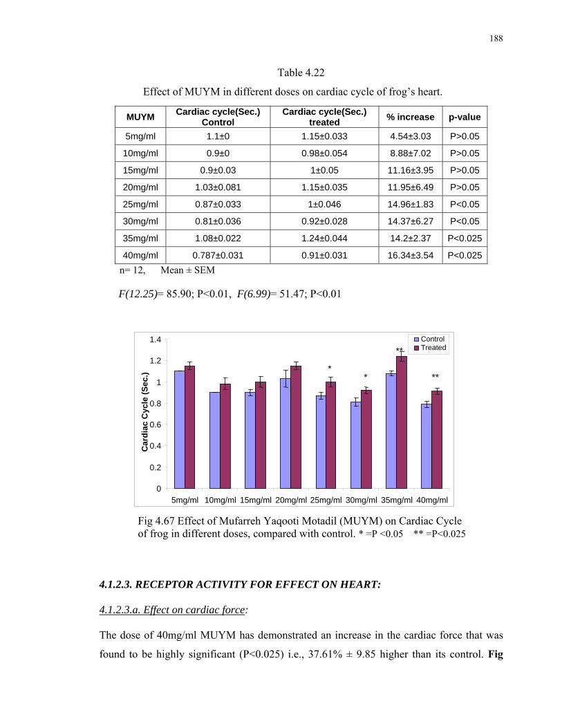
188
Table 4.22
Effect of MUYM in different doses on cardiac cycle of frog’s heart.
n= 12, Mean ± SEM F(12.25)= 85.90; P<0.01, F(6.99)= 51.47; P<0.01
Fig 4.67 Effect of Mufarreh Yaqooti Motadil (MUYM) on Cardiac Cycle of frog in different doses, compared with control. * =P <0.05 ** =P<0.025 4.1.2.3. RECEPTOR ACTIVITY FOR EFFECT ON HEART: 4.1.2.3.a. Effect on cardiac force: The dose of 40mg/ml MUYM has demonstrated an increase in the cardiac force that was
found to be highly significant (P<0.025) i.e., 37.61% ± 9.85 higher than its control. Fig
MUYM Cardiac cycle(Sec.)
Control Cardiac cycle(Sec.)
treated % increase p-value
5mg/ml 1.1±0 1.15±0.033 4.54±3.03 P>0.05
10mg/ml 0.9±0 0.98±0.054 8.88±7.02 P>0.05
15mg/ml 0.9±0.03 1±0.05 11.16±3.95 P>0.05
20mg/ml 1.03±0.081 1.15±0.035 11.95±6.49 P>0.05
25mg/ml 0.87±0.033 1±0.046 14.96±1.83 P<0.05
30mg/ml 0.81±0.036 0.92±0.028 14.37±6.27 P<0.05
35mg/ml 1.08±0.022 1.24±0.044 14.2±2.37 P<0.025
40mg/ml 0.787±0.031 0.91±0.031 16.34±3.54 P<0.025
0
0.2
0.4
0.6
0.8
1
1.2
1.4
5mg/ml 10mg/ml 15mg/ml 20mg/ml 25mg/ml 30mg/ml 35mg/ml 40mg/ml
Car
dia
c C
ycle
(S
ec.)
ControlTreated
*
**
***

189
4.69 On the other hand similar dose of MUYM had increased the cardiac force by only
12.18%±4.21 in the presence of atropine (10-3M), that was statistically non significant
(P>0.05) when compared with their controls. Fig 4.70, Table 4.23
Fig 4.68 Tracing showing the intact heart activity of control frog’s heart.
Fig 4.69 Effect of MUYM (40mg/ml) on intact heart of frog.
Fig 4.70 Effect of MUYM (40mg/ml) on intact frog’s heart was not produced after Atropine (10-3M) administration.
Table 4.23 Receptor Activity of MUYM for the Effect on Cardiac Force
n n= 12, Mean ± S.E.M.
Dose Cardiac force
(g)control
Cardiac force
(g) treated % increase P-value
40mg/ml MUYM 0.439±0.035 0.600±0.051 37.61±9.852 P<0.025**
Atropine (10-3M) +
MUYM (40mg/ml) 0.439±0.035 0.49±0.032 12.18±4.21 P>0.05

190
Fig 4.71 Effect of MUYM (40mg/ml) on Cardiac force
of frog’s heart. **=P<0.025
Fig 4.72 After Atropine administration effect of
MUYM (40mg/ml) could not be produced on Cardiac force of frog’s heart. 4.1.2.3.b. Effect on Heart Rate: MUYM (40mg/ml) has been found to decrease the heart rate by 14.45%±2.39 (P<0.005)
than its control Fig. 4.73 While on pretreatment with atropine (10-3M) in the presence of
atropine, MUYM could not show its effect and tried to decrease the heart rate but a
nonsignificant decrease i.e., 3.69%±0.93 decrease was observed when compared with its
controls. Fig. 4.74, Table 4.24
0
0.1
0.2
0.3
0.4
0.5
0.6
0.7
MUYM(40mg/ml)C
ard
iac
forc
e (g
)
control
treated**
0
0.1
0.2
0.3
0.4
0.5
0.6
Atropine(10-3M) + MUYM (40mg/ml)
Car
dia
c fo
rce
(g)
control
treated
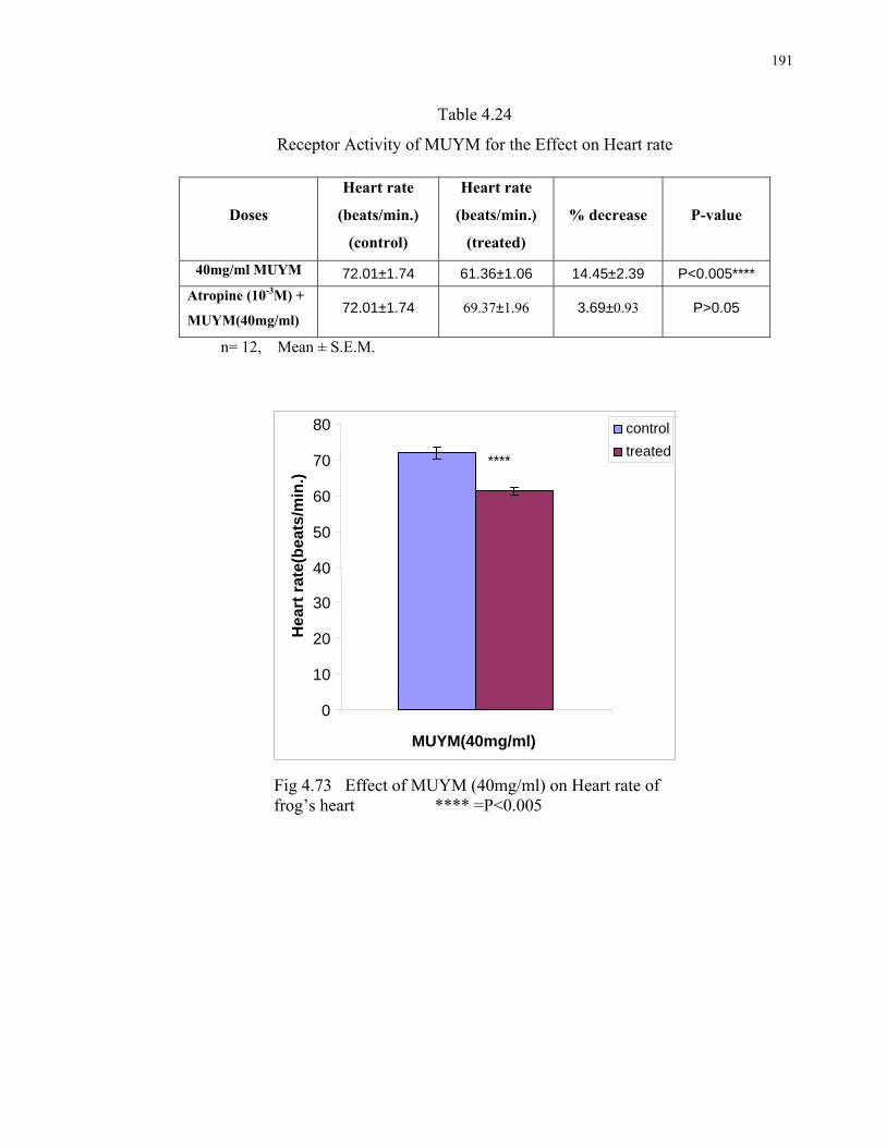
191
Table 4.24
Receptor Activity of MUYM for the Effect on Heart rate
n= 12, Mean ± S.E.M.
Fig 4.73 Effect of MUYM (40mg/ml) on Heart rate of frog’s heart **** =P<0.005
Doses
Heart rate
(beats/min.)
(control)
Heart rate
(beats/min.)
(treated)
% decrease P-value
40mg/ml MUYM 72.01±1.74 61.36±1.06 14.45±2.39 P<0.005****
Atropine (10-3M) +
MUYM(40mg/ml) 72.01±1.74 69.37±1.96 3.69±0.93 P>0.05
0
10
20
30
40
50
60
70
80
MUYM(40mg/ml)
Hea
rt r
ate(
bea
ts/m
in.)
control
treated****

192
Fig 4.74 Effect of MUYM (40mg/ml) on Heart rate of frog’s heart was not produced after Atr. Administration. 4.1.2.4. COMPARATIVE STUDY WITH DIGOXIN: As the drug MUYM showed Positive Inotropic and Negative Chronotropic effects
therefore the effects of MUYM (40mg/ml) exhibited on intact heart of frog was compared
with that of Digoxin. (0.008mg/ml). The response was recorded in the form of following
tracings. Fig 4.75 and 4.76
Fig 4.75 Control and Digoxin (0.008mg/ml) treated intact heart activity of
frog’s heart.
Fig 4.76 Control and MUYM (40mg/ml) treated intact heart activity of frog’s heart.
646566676869707172737475
Hea
rt r
ate
(bea
ts/m
in.)
control
treated
Atropine(10-3M)+MUYM(40mg/ml)

193
Above tracings helped to calculate the following values:
1. The 40mg/ml dose of the product MUYM caused 37.61%±9.85 (Fig 4.77) increase
in cardiac force (P<0.025) which was less than that produced by Digoxin i.e.,
39.1%±13.77 (P<0.005) (Fig 4.78) when compared with their controls(Table 4.25).
Table 4.25
Comparative study of the activity of MUYM with that of Digoxin on frog’s intact heart.
Doses Cardiac force(g) Control
Cardiac force(g) Treated
% increase P-value
MUYM (45mg/ml) 0.439±0.035 0.600±0.051 37.61±9.852 P<0.025
Digoxin(0.008mg/ml) 0.446±0.036 0.612±0.055 39.105±13.77 P<0.005
n= 12, Mean ± SEM
F(3.68)= 6.12; P<0.05
Fig 4.77 Effect of MUYM (40mg/ml) on cardiac force of Frog’s Heart.
0
0.1
0.2
0.3
0.4
0.5
0.6
0.7
Car
dia
c fo
rce
(g)
control
treated**
MUYM (40mg/ml)

194
Fig 4.78 Effect of Digoxin (0.008mg/ml) on cardiac force of Frog’s Heart.
.
2. The Heart rate reduction caused by the product MUYM was calculated as
14.45%±2.39 (Fig 4.79) which was found to be more than that produced by
Digoxin i.e., 12.26%±0.91 (Fig 4.80). Both of the effects were highly significant
(P<0.005) when compared with their controls. (Table 4.26)
Table 4.26
Comparative study of the activity of MUYM with that of Digoxin on frog’s intact heart.
Doses Heart rate (beats/min.)Control
Heart rate (beats/min.) Treated
% decrease
P-value
MUYM (45mg/ml) 72.01±1.74 61.36±1.06 14.45±2.39 P<0.005Digoxin(0.008mg/ml) 41.67±0.617 36.55±0.627 12.26±0.91 P<0.005
n= 12, Mean ± SEM
F (6.36) = 24.63; P<0.01
Fig.4.79 Effect of MUYM on Heart rate of Intact Heart of Frog
****= P<0.025
01020304050607080
Hea
rt r
ate
(bea
ts/m
in.) control
treated****
MUYM (40mg/ml)
00.10.20.30.40.50.60.70.8
C
ard
iac
forc
e (g
)
control
treated****
Digoxin (0.008mg/ml)

195
Fig 4.80 Effect of Digoxin on Heart rate of Intact Heart of Frog ****= P<0.025
3. The Cardiac cycle was increased by the product MUYM was calculated as
16.34%±3.54 (Fig 4.81) which was found to be less than that produced by Digoxin
i.e., 25.66%±10.56 (Fig 4.82) when compared with their controls (Table 4.27).
Table 4.27
Comparative study of the activity of MUYM with that of Digoxin on frog’s intact heart.
n= 12, Mean ± SEM
F (5.78) = 10.88; P<0.01
Doses Cardiac cycle(Sec.)
Control
Cardiac cycle(Sec.) Treated
% increase p-value
MUYM (40mg/ml)
0.787±0.031 0.91±0.031 16.34±3.54 P<0.025
Digoxin (0.008mg/ml)
0.88±0.04 1.1±0.07 25.66±10.56 P<0.05
32
34
36
38
40
42
44
Hea
rt r
ate
(bea
ts/m
in.)
control
treated
****
Digoxin (0.008mg/ml)

196
Fig 4.81 Effect of MUYM on Cardiac cycle of Intact Heart of Frog **= P<0.025
Fig 4.82 Effect of Digoxin on Cardiac cycle of Intact Heart of Frog. *=P<0.05
4.1.2.5. DIURETIC ACTIVITY OF “MUYM” ON RATS:
4.1.2.5.a. Effect on urine volume:
Treatment with the 1stdose of the MUYM (150mg/kg) caused non significant increase in
Urine volume (3.18±0.13ml; 20.55% ± 4.84; P>0.05) when compared with controls
(Saline 25ml/kg; 2.66ml±0.18). This Effect was increased in a dose dependent manner and
found to be highest at 1200mg/kg dose (5.25ml±0.3; 99.16%±11.68; P<0.0005) when
compared with the controls. At this dose diuresis caused by the product MUYM was found
to be greater than the reference drug i.e., Urea, 1500mg/kg (5.11ml±0.22; 94.44%±11.22)
00.1
0.20.3
0.40.50.6
0.70.8
0.91
Car
dia
c cy
cle
(sec
.)
control
treated**
MUYM (40mg/ml)
0
0.2
0.4
0.6
0.8
1
1.2
1.4
Car
dia
c cy
cle
(sec
.) control
treated *
Digoxin (0.008mg/ml)

197
while the other reference drug Furosemide, 10mg/kg, produced higher diuretic effect than
MUYM (7.23ml±0.26; 174.22%±11.11) ( Table 4.28, Fig.4.83)
Table 4.28
Effect of MUYM, Urea and Furosemide on urine volume
compared with their controls
n= 8, Mean ± SEM
Fig 4.83 Effect of MUYM, Urea and Furosemide on urine volume compared
with their control.
Drugs
urine
vol. ml S.E.M. %increase S.E.M. P-value
Saline25ml/kg
(control) 2.66 ±0.182
Urea1500mg/kg 5.11 ±0.22 94.44 ±11.22 p<0.0005
Furosemide10mg/kg 7.23 ±0.26 174.22 ±11.11 p<0.0005
MUYM150mg/kg 3.18 ±0.13 20.55 ±4.84 P<0.025
MUYM300mg/kg 4.08 ±0.32 54.44 ±11.87 p<0.005
MUYM600mg/kg 5 ±0.44 89.16 ±16.02 p<0.0005
MUYM1200mg/kg 5.25 ±0.30 99.16 ±11.68 p<0.0005
01
23
45
67
8
Uri
ne
Vo
lum
e(m
l)
Saline25ml/kg Urea1500mg/kg Furosemide10mg/kgMUYM 150mg/kg MUYM 300mg/kg MUYM 600mg/kg MUYM 1200mg/kg
****
****
**** *******
**
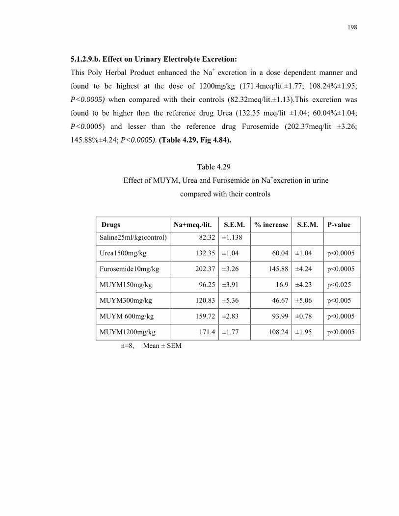
198
5.1.2.9.b. Effect on Urinary Electrolyte Excretion:
This Poly Herbal Product enhanced the Na+ excretion in a dose dependent manner and
found to be highest at the dose of 1200mg/kg (171.4meq/lit.±1.77; 108.24%±1.95;
P<0.0005) when compared with their controls (82.32meq/lit.±1.13).This excretion was
found to be higher than the reference drug Urea (132.35 meq/lit ±1.04; 60.04%±1.04;
P<0.0005) and lesser than the reference drug Furosemide (202.37meq/lit ±3.26;
145.88%±4.24; P<0.0005). (Table 4.29, Fig 4.84).
Table 4.29
Effect of MUYM, Urea and Furosemide on Na+excretion in urine
compared with their controls
n=8, Mean ± SEM
Drugs Na+meq./lit. S.E.M. % increase S.E.M. P-value
Saline25ml/kg(control) 82.32 ±1.138
Urea1500mg/kg 132.35 ±1.04 60.04 ±1.04 p<0.0005
Furosemide10mg/kg 202.37 ±3.26 145.88 ±4.24 p<0.0005
MUYM150mg/kg 96.25 ±3.91 16.9 ±4.23 p<0.025
MUYM300mg/kg 120.83 ±5.36 46.67 ±5.06 p<0.005
MUYM 600mg/kg 159.72 ±2.83 93.99 ±0.78 p<0.0005
MUYM1200mg/kg 171.4 ±1.77 108.24 ±1.95 p<0.0005

199
0
50
100
150
200
250N
a+
(me
q./l
it.)
Saline25ml/kg Urea1500mg/kg Furosemide10mg/kgMUMO 150mg/kg MUMO 300mg/kg MUMO 600mg/kgMUMO 1200mg/kg
*******
**** ****
****
****
Fig 4.84 Effect of MUYM, Urea and Furosemide on Na+excretion in urine compared with their controls K+excretion was also increased in a dose dependent manner and found to be highest at the
dose of 1200mg/kg (92.71 meq/lit ±1.06; 50.28%±2.59; P<0.0005) when compared with
their controls (61.71 meq/lit.±0.60).This excretion was found to be higher than the
reference drugs Urea (82.12meq/lit ±0.98; 33.08%±1.25; P<0.0005) and Furosemide
(86.42meq/lit ±0.75; 40.07%±1.92; P<0.0005). (Table 4.30, Fig 4.85).
Table 4.30
Effect of MUYM, Urea and Furosemide on K+excretion in urine
compared with their controls
n=8, Mean ± SEM
Drugs K+meq./lit. S.E.M. % increase S.E.M. P-value
Saline25ml/kg
(control) 61.71 ±0.605
Urea1500mg/kg 82.12 ±0.982 33.08 ±1.25 p<0.0005
Furosemide10mg/kg 86.42 ±0.755 40.07 ±1.92 p<0.0005
MUYM150mg/kg 71.77 ±0.96 16.31 ±1.32 p<0.0005
MUYM 300mg/kg 82.2 ±0.89 33.25 ±2.41 p<0.0005
MUYM 600mg/kg 87.8 ±1.23 42.3 ±1.85 p<0.0005
MUYM 1200mg/kg 92.71 ±1.06 50.28 ±2.59 p<0.0005

200
0
20
40
60
80
100
K+
(me
q./l
it.)
Saline25ml/kg Urea1500mg/kg Furosemide10mg/kgMUMO 150mg/kg MUMO 300mg/kg MUMO 600mg/kgMUMO 1200mg/kg
********
********
********
Fig 4.85 Effect of MUYM, Urea and Furosemide on K+excretion in urine compared
with their controls. Cl-excretion was also increased in a dose dependent manner and found to be highest at the
dose of 1200mg/kg (101.27 meq/lit ±0.70; 17.14%±2.56; P<0.0005) when compared with
their controls (86.52 meq/lit ±1.69).This excretion was found to be higher than the
reference drug Urea (82.12meq/lit ±0.98; 33.08%±1.25; P<0.005) and Furosemide
(86.42meq/lit ±0.75; 40.07%±1.92; P<0.005) (Table 4.31, Fig 4.86).
Table 4.31
Effect of MUYM, Urea and Furosemide on K+excretion in urine
compared with their controls
n=8, Mean ± SEM
Drugs Cl-meq./lit. S.E.M. % increase S.E.M. P-value
Saline25ml/kg
(control) 86.525 ±1.69
Urea1500mg/kg 94.35 ±0.616 9.19 ±2.87 p<0.005
Furosemide10mg/kg 96.212 ±0.998 11.26 ±1.72 p<0.005
MUYM150mg/kg 95.19 ±0.81 10.1 ±2.14 p<0.005
MUYM300mg/kg 97.81 ±0.79 13.11 ±1.71 p<0.0005
MUYM600mg/kg 99.55 ±0.67 15.12 ±1.60 p<0.0005
MUYM1200mg/kg 101.27 ±0.70 17.14 ±2.56 p<0.0005

201
Fig 4.86 Effect of MUYM, Urea and Furosemide on Cl-excretion in urine compared with their controls.
4.1.3 ANTIHYPERLIPIDEMIC ACTIVITY ON RATS
5.1.3.1 Behavioural Changes:
Male and female rats treated with Extract of C.C .leaves (1000mg / kg-body weight) and
with the Product MUYM (142.85mg/kg) for the period of eight weeks, did not show any
mortality. Rats from C group (fed on high cholesterol diet) were compared with those of
the N group (control) for the lipid profile. Rats from CS groups (fed on high cholesterol
diet + treated drugs) were compared with those of C group for the effects of drugs. Rats
from the C group spent most of the time in sleeping and resting. High cholesterol diet
perhaps induced sluggishness (Brisson 1981) but some physical behavioral changes were
observed in first hour after dosing like uncoordinated motor activity and corner sitting.
4.1.3.2 Body Weights:
Rats of N, C, CS1, CS2 and CS3 groups showed an increase in the body weight. In groups
N, CS1, CS2 and CS3 that increase was non significant (P>0.05) while highly significant
increase was observed in the weight of group C rats when compared with control group N
(P<0.01). (Table 4.32)
0
20
40
60
80
100
120
1
Cl-
(meq
./lit
.)
Saline25ml/kg Urea1500mg/kgFurosemide10mg/kg MUYM 150mg/kg MUYM 300mg/kg MUYM 600mg/kg MUYM 1200mg/kg
*** *** ******* **** ****

202
Table 4.32: Change in the weights of rats after eight weeks treatment (mean of six values ±S.E.M.)
GROUPS Weeks Initial
body
weight
(gm)
Final
body
weight
(gm)
% gain in
body weight
P-value Morta
lity
Toxic
effect
Control(saline) N 8 244.05
±12.31
270.44
±20.13
10.64 ±5.534 P>0.05 (non significant increase)
Nil Nil
Control(High
cholesterol) C
8
223.85
±4.60
253.47
±9.56
13.00±2.292 **P<0.01 (highly significant increase)
Nil Nil
Treated with
C.C.L.E. Extract
1000mg/kg (CS1)
8 236.75
±4.43
254.66
±8.27
9.08±0.866 P>0.05 (non significant increase)
Nil Nil
Treated with
MUYM
142.85mg/kg
(CS2)
8 227.5
±8.20
250.38
±10.45
10.02±0.76 P>0.05 (non significant increase)
Nil Nil
Treated with
Atorvastatin
0.2mg/kg (CS3)
8 240.25
±4.28
256.52
±9.05
8.25±1.228 P>0.05 (non significant increase)
Nil Nil
4.1.3.3 Autopsy:
Autopsy revealed that no gross changes were observed in organs like liver, spleen,
heart and kidney among the animals of group N, and CS groups, while the animals of
group C that were fed on 24% cholesterol diet for eight weeks showed marked

203
deposition of fats on heart muscles, aorta and on the liver. (Fig.4.87-4.90). Brown
spots were observed on spleen causing a change in colour of spleen.
Fig.4.87 Deposition of fats on Aorta in rats after two months feeding on high cholesterol diet. (Group C)
Fig.4.88 Deposition of fats on Aorta and liver tissues in rats after two months feeding on high cholesterol diet. (Group C)

204
Fig.4.89 Deposition of fats in liver tissues of rats after two months
feeding on high cholesterol diet. (Group C)
Fig. 4.90 Deposition of fats on cardiac walls in rats after two months feeding on high cholesterol diet. (Group C)

205
4.1.3.4 Effects of Tested Drugs on different Biochemical Parameters of
Hyperlipidemic Rats :
The effects of C.C.L.E. leaves extract and the product MUYM on Rats’ Blood chemistry
were measured and presented in Table 5.32. The alterations in the concentration of,
Cholesterol, Triglycerides, High Density Lipoprotein (HDL) , Alanine amino transferase
(ALAT), Aspartate Amino Transferase (ASAT), Creatinine, Creatinine Kinase (CK),
Alkaline Phosphatase, Bilirubin Direct, Bilirubin Total, Albumin, and Glucose were
recorded in all groups.
4.1.3.4. a Group C:
The rats fed with 24% egg yolk in their diet for eight weeks i.e., Group C rats’ serum were
found to have highly significant increase in cholesterol i.e., 120.609 % ±12.05 (P<0.005),
significant increase in Triglycerides i.e., 70.62 % ± 29.21 (P<0.05), highly significant
increase in LDL i.e., 327.61% ± 97.73 (P<0.005), non significant increase in HDL i.e.,
40.70% ± 23.77(P>0.05). A non significant decrease in Creatinine i.e., 20.02%± 38.63
(P>0.05) was observed when compared with their controls (N). A non significant increase
in ALAT, ASAT, GGT Alkaline Phosphatase, and Albumin, Bilirubin Direct (P>0.05), and
highly significant increase in Bilirubin total i.e., 79.84% ± 36.52 (P<0.01) was observed
when compared with their control. The level of Creatinine Kinase, Urea, and Glucose were
non significantly increased (P>0.05) and no change in serum was observed when compared
with their control group (N). (Table 4.33), (Panel a)
4.1.3.4 b Group CS1:
Rats fed on 24% egg yolk in their diet as well as treated with extract C.C.L.E.(1000mg/kg)
for eight weeks had significant decrease in cholesterol (P<0.025), Triglycerides, and HDL
(P<0.05). A highly significant decrease in LDL (P<0.025) and Albumin (P<0.01). ALAT,
ASAT, Alkaline Phosphatase, Bilirubin Direct, Bilirubin Total , CK, Creatinine, and Urea
were decreased non significantly (P>0.05). GGT was significantly decreased (P<0.05). A
significant increase in Serum Glucose (P<0.05) and highly significant decrease in Uric acid
(P<0.01) was observed as compared to their respective control group (C). (Table 4.33),
(Panel b)
206
5.1.3.4.c Group CS2:
Rats fed on 24% egg yolk in their diet as well as treated with MUYM (142.85mg/kg) for
eight weeks had a highly significant decrease in cholesterol and LDL (P<0.025) while
Triglycerides, HDL and GGT were significantly decreased (P<0.05). ASAT, ALAT,
Alkaline Phosphatase, CK, Creatinine, Bilirubin Direct, Bilirubin Total,and Urea
decreased non significantly (P>0.05) and Serum Glucose increased (P<0.05) and a highly
significant decrease in Albumin (P<0.025) and Uric acid (P<0.01) was observed as
compared to their respective control group (C). (Table 4.33), (Panel b)
5.1.3.4. d Group CS3:
Rats fed on 24% egg yolk in their diet as well as treated with Atorvastatin (0.2mg/kg) for
eight weeks had a highly significant decrease in cholesterol and LDL (P<0.025) while
Triglycerides, HDL, GGT, ALAT, CK and Creatinine were significantly decreased non
significantly (P>0.05). ASAT, Alkaline Phosphatase, Bilirubin Direct, Bilirubin Total,
Serum Glucose and Urea decreased non significantly (P>0.05) and Uric acid increased
significantly (P<0.05) and a highly significant decrease in Albumin (P<0.01) and Uric acid
(P<0.01) was observed as compared to their respective control group (C). (Table 4.33),
(Panel b)

207
Table 4.33: Biochemical parameters of rats of N, C, CS1, CS2, CS3 groups after eight weeks treatment. Group C compared with Group N and Groups CS1, CS2, CS3 compared with Group C. n=6, Mean ± SEM, P<0.05*, P<0.025* *, P<0.01***, P<0.005*** *
Parameters Group N
(control group for group C)
Group C (high cholesterol diet)
(control group for groups CS1, CS2
and CS3)
Group CS1.
( high cholesterol diet+
C.C.L.E. 1000mg/kg)
Group CS2
(high cholesterol diet+ MUYM 142.85mg/kg
Group CS3 (high cholesterol
diet+ Atorvastatin (0.2mg/kg)
Cholesterol
(mg/dl) 122.69±19.1 261.66±29.72*** 166.39±8.96** 135.67±2.0** 132.22±6.6**
Triglycerides (mg/dl)
87.0±19.28
129.70±12.71* 90.63±15.53* 100.50±4.42* 90.23±7.74**
HDL (mg/dl)
108.85±22.22
143.61±22.36
115.23±15.70*
119.62±16.82* 135.25±9.05
HDL / Total Cholesterol
Ratio 0.887 0.544 0.692 0.881 1.02
LDL(mg/dl) 40.09±4.02 179.84±52.22*** 94.15±32.61 **96.025±30.12 99.012±32.61**
ALAT(u/l) 10.595±3.11 16.525±4.28** 15.091±5.47 11.2071±5.40 9.628±2.62*
ASAT(u/l) 2.6898±1.24
4.638±2.59
2.1825±0.504
2.24±2.52 1.268±0.45
GGT (u/l) 10.84±5.47
21.53±13.45
4.16±0.77*
2.02±0.334* 2.16±0.54*
Alkaline
Phosphatase (u/l)
21.187±8.69
36.388±4.52** 29.327±10.01 30.07±0.504 24.366±4.56
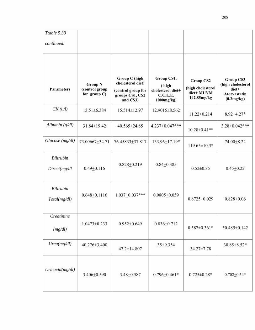
208
Ttable 5.33
continued.
Parameters
Group N (control group for group C)
Group C (high cholesterol diet)
(control group for groups CS1, CS2
and CS3)
Group CS1.
( high cholesterol diet+
C.C.L.E. 1000mg/kg)
Group CS2
(high cholesterol diet+ MUYM 142.85mg/kg
Group CS3 (high cholesterol
diet+ Atorvastatin (0.2mg/kg)
CK (u/l) 13.51±6.384
15.514±12.97
12.9015±8.562
11.22±0.214 8.92±4.27*
Albumin (g/dl) 31.84±19.42
40.565+24.85
4.237+0.047***
10.28±0.41**
3.28+0.042***
Glucose (mg/dl) 73.00667+34.71
76.45833+37.817
133.96+17.19*
119.65±10.3*
74.00+8.22
Bilirubin
Direct(mg/dl 0.49+0.116 0.828+0.219
0.84+0.385
0.52±0.35 0.45+0.22
Bilirubin
Total(mg/dl) 0.648+0.1116
1.037+0.037***
0.9805+0.059
0.8725±0.029 0.828+0.06
Creatinine
(mg/dl) 1.0473+0.233
0.952+0.649
0.836+0.712
0.587±0.361* *0.485+0.142
Urea(mg/dl) 40.276+3.400
47.2+14.807
35+9.354
34.27±7.78
30.85+8.52*
Uricacid(mg/dl) 3.406+0.590 3.48+0.587 0.796+0.461* 0.725±0.28*
0.702+0.54*

209
Panel a: Fig 4.91- 4.98 Treated Group C (hyperlipidaemic) compared
with Group N (control)
0
50
100
150
200
250
300
350
Cholesterol Triglycerides HDL LDL
mg
/dl
control(N)
treated(C)***
*
***
Fig. 4.91 Serum levels of cholesterol, Triglycerides, HDL
and LDL in control (N)and high cholesterol diet (C)Groups of rats
0
5
10
15
20
25
30
35
40
45
ALAT ASAT GGT
Allkalinephosphatase
u/l
Control (N)Treated (C)
**
**
Fig. 4.92 Serum levels of ALAT, ASAT, GGT and Alkaline
Phosphatase in control (N)and high cholesterol diet (C)Groups of rats

210
0
5
10
15
20
25
30
Creatinine kinase
U/l
Control (N)
Treated (C)
Fig.4.93 Serum levels of Creatinine Kinase in control (N)
and high cholesterol diet (C)Groups of rats.
0
20
40
60
80
100
120
Glucose
mg
/dl
Control (N)
Treated (C)
Fig. 4.94 Serum levels of Glucose in control (N) and
high cholesterol diet (C)Groups of rats.
0
0.2
0.4
0.6
0.8
1
1.2
Bilirubin direct Bilirubin Total
mg
/dl
Control (N)
Treated (C)***
Fig 4.95 Serum levels of Bilirubin direct and Bilirubin total
in control (N)and high cholesterol diet (C)Groups of rats.
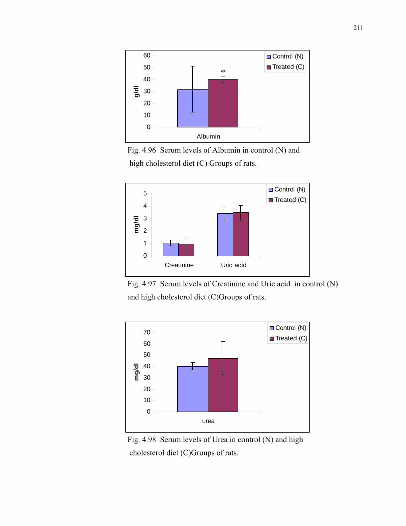
211
0
10
20
30
40
50
60
Albuming
/dl
Control (N)
Treated (C)**
Fig. 4.96 Serum levels of Albumin in control (N) and
high cholesterol diet (C) Groups of rats.
0
1
2
3
4
5
Creatinine Uric acid
mg
/dl
Control (N)
Treated (C)
Fig. 4.97 Serum levels of Creatinine and Uric acid in control (N)
and high cholesterol diet (C)Groups of rats.
0
10
20
30
40
50
60
70
urea
mg
/dl
Control (N)
Treated (C)
Fig. 4.98 Serum levels of Urea in control (N) and high
cholesterol diet (C)Groups of rats.

212
Panel b: Fig.4.99-4.106 Treated Groups CS1, CS2, CS3 compared
with hyperlipidaemic Group C (control)
Fig. 4.99 Serum levels of cholesterol, Triglycerides, HDL and LDL
in High Cholesterol Control (C) and high cholesterol diet
+ Drug Treated (CS1, CS2, CS3)Groups of rats
0
50
100
150
200
250
300
350
Cholesterol Triglycerides HDL LDL
mg/
dl
Control (C) Treated (CS1)Treated (CS2) Treated (CS3)
**
** **
* * *
* * *

213
Fig. 4.100 Serum levels of ALAT, ASAT, GG and Alkaline Phosphatase
in High Cholesterol Control (C) and High cholesterol diet + Drug Treated(CS1, CS2, CS3)Groups of rats.
Fig. 4.101 Serum levels of Cretinine kinase in High Cholesterol
Control (C) and high cholesterol diet + Drug Treated
(CS1, CS2, CS3)Groups of rats
-5
0
5
10
15
20
25
30
35
40
45
ALAT ASAT GGT
Allkalinephosphatase
U/l
Control (C) Treated (CS1)Treated (CS2) Treated (CS3)
*
* * *
0
5
10
15
20
25
30
Creatinine kinase
U/l
Control (C) Treated (CS1) Treated (CS2) Treated (CS3)
*
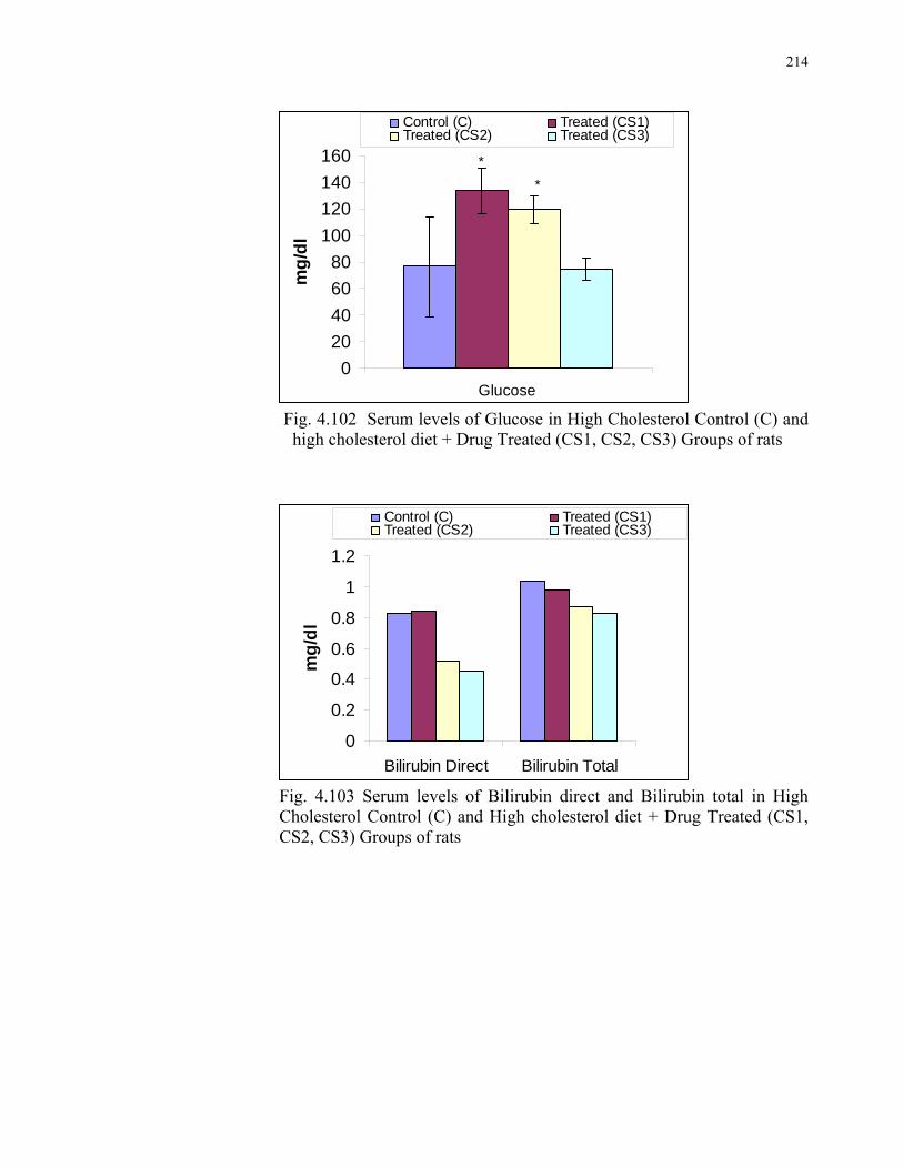
214
0
20
40
60
80
100
120
140
160
Glucose
mg
/dl
Control (C) Treated (CS1)Treated (CS2) Treated (CS3)
*
*
Fig. 4.102 Serum levels of Glucose in High Cholesterol Control (C) and
high cholesterol diet + Drug Treated (CS1, CS2, CS3) Groups of rats
0
0.2
0.4
0.6
0.8
1
1.2
Bilirubin Direct Bilirubin Total
mg
/dl
Control (C) Treated (CS1)Treated (CS2) Treated (CS3)
Fig. 4.103 Serum levels of Bilirubin direct and Bilirubin total in High
Cholesterol Control (C) and High cholesterol diet + Drug Treated (CS1, CS2, CS3) Groups of rats

215
05
10152025
3035404550
Albumin
g/d
l
Control (C) Treated (CS1)Treated (CS2) Treated (CS3)
Fig 4.104 Serum levels of Albumin in High Cholesterol
Control (C) and High cholesterol diet + Drug Treated
(CS1, CS2, CS3)Groups of rats
Fig. 4.105 Serum levels of Cretinine and Uric acid in High
Cholesterol Control (C) and high cholesterol diet +
Drug Treated (CS1, CS2, CS3)Groups of rats
00.5
11.5
22.5
33.5
44.5
Creatinine Uric acid
mg
/dl
Control (C) Treated (CS1) Treated (CS2) Treated (CS3)
* **
**
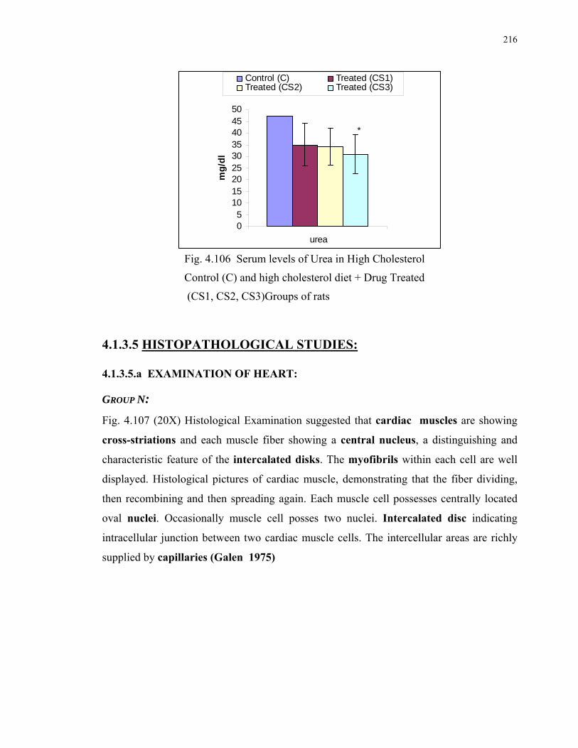
216
05
101520253035404550
urea
mg
/dl
Control (C) Treated (CS1)Treated (CS2) Treated (CS3)
*
Fig. 4.106 Serum levels of Urea in High Cholesterol
Control (C) and high cholesterol diet + Drug Treated
(CS1, CS2, CS3)Groups of rats
4.1.3.5 HISTOPATHOLOGICAL STUDIES: 4.1.3.5.a EXAMINATION OF HEART:
GROUP N:
Fig. 4.107 (20X) Histological Examination suggested that cardiac muscles are showing
cross-striations and each muscle fiber showing a central nucleus, a distinguishing and
characteristic feature of the intercalated disks. The myofibrils within each cell are well
displayed. Histological pictures of cardiac muscle, demonstrating that the fiber dividing,
then recombining and then spreading again. Each muscle cell possesses centrally located
oval nuclei. Occasionally muscle cell posses two nuclei. Intercalated disc indicating
intracellular junction between two cardiac muscle cells. The intercellular areas are richly
supplied by capillaries (Galen 1975)

217
Fig. 4.107-A, B ( 20X ) Sections of Heart of control Group N of rats. GROUP C: Fig. 4.108 (40X) Showing the necrotic muscle fibers of the infarct at higher magnification.
The fibers are themselves more eosinophilic than normal fibers. Degenerative and fatty
changes are visible. Muscle fibres are scattered. The striatations are however still
detectable. There is no nuclear staining, the nuclei having disappeared by karyolysis. In the
interstitial tissues there are, in addition to some nuclear fragments, macrophages that have
migrated into the dead muscle. (Cornelius 1989)
Fig. 4.108 ( 40X ) Section of heart of Group C hyperlipidemic rats. GROUPS CS1, CS2 AND CS3:
Fig 4.109-A, B, C (20X). These pictures are showing the healing or protective property of
the drugs as compared to their control group(C). The endocardium, the innermost layer of
the heart, consists of an endothelial lining and its supporting tissue. The endothelium is a
single layer of flattened epithelial cells. The necrotic muscles are replaced by fibrous

218
tissues. Each muscle cell possesses centrally located oval nuclei. Occasionally muscle cell
possesses two nuclei. The intercellular areas are well supplied by capillaries.
Fig 4.109-A (20X) Section of Heart of Treated group (CS1) of rats.
Fig 4.109-B (20X) Section of Heart of rats’ Treated group (CS2)
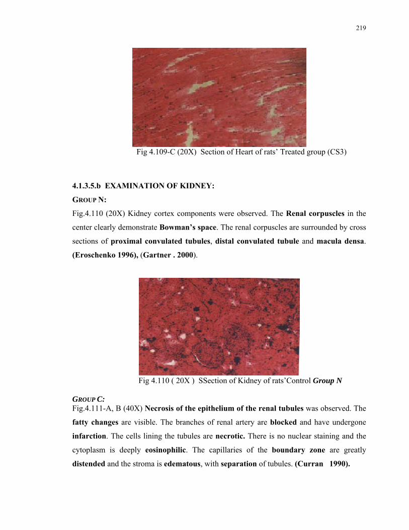
219
Fig 4.109-C (20X) Section of Heart of rats’ Treated group (CS3)
4.1.3.5.b EXAMINATION OF KIDNEY:
GROUP N:
Fig.4.110 (20X) Kidney cortex components were observed. The Renal corpuscles in the
center clearly demonstrate Bowman’s space. The renal corpuscles are surrounded by cross
sections of proximal convulated tubules, distal convulated tubule and macula densa.
(Eroschenko 1996), (Gartner . 2000).
Fig 4.110 ( 20X ) SSection of Kidney of rats’Control Group N GROUP C: Fig.4.111-A, B (40X) Necrosis of the epithelium of the renal tubules was observed. The
fatty changes are visible. The branches of renal artery are blocked and have undergone
infarction. The cells lining the tubules are necrotic. There is no nuclear staining and the
cytoplasm is deeply eosinophilic. The capillaries of the boundary zone are greatly
distended and the stroma is edematous, with separation of tubules. (Curran 1990).

220
Enlargement of many glomeruli, capillary tufts and of subcapsular urinary space is visible.
Intertubular connective tissue appears regionally increased. (Welt et al., 2007)
Fig.4.111-A (40X) Fig.4.111- B (40X)
Sections of Kidney of High Cholesterol diet Group C rats
GROUPS CS1, CS2, AND CS3: Fig.4.112-A, B, C (40X) Glomeruli are smaller and the subcapsular space are very much
less dilated, than the Group C rats. Many Armanni-Ebstein cells are present. (K. Welt.,
2007). The renal corpuscles (RC) clearly demonstrates Bowmen’s capsule. The renal
Corpuscles are surrounded by cross- sections of proximal convulated tubules, distal
convulated tubules and macula densa. . The medullary rays contain the pars recta of the
proximal tubule, the ascending thick limb of Henle’s loop and collecting tubules. (Gartner
2000)
Fig.4.112-A (40X) Section of Kidney of rats’ Treated group CS1

221
Fig.4.112-B (40X) Section of Kidney of rats’ Treated group CS2
Fig.4.112-C (40X) Section of Kidney of rats’Treated group CS3
5.1.3.5.c EXAMINATION OF LIVER:
Group N
Fig.4.113(40X) The portal area of the liver houses terminal branches of Hepatic artery
Portal vein. The vein is much larger than the artery and its wall is very thin in comparison
to the size of its lumen. Liver cells hepatocytes are flat and arranged. Branches of Lymph
Vessels and bile ducts are also present in portal area. Plates of liver cells and Sinusoids
extend from the portal area. A discontinuous layer of cells lines the sinusoids. Central
vein is lined by epithelial cells and filled with biconcave Red Blood cells. Hepatic
sinusoids appear to radiate from central vein. (Gartner 2000).

222
Fig. 4.113(40X) Section of Liver of rats’Control Group N Group C: Fig 4.114(40X) Portal area of the liver shows hepatic artery and portal vein in addition
lymph vessels and bile ducts are also present but are congested. Sinusoids are much
congested. In the centrilobular zone and in the mid-zonal region the sinusoids are
congested. The liver cells adjoining the portal tract in which a branch of the portal vein and
a small bile duct are visible and relatively normal, but the centrilobular zone shows very
fatty changes and many hepatocytes are atrophic(Fig 4.114) (Curran 1990).
Fig 4.114(40X) Section of Liver of High Cholesterol diet Group C rats

223
GROUPS CS1, CS2, AND CS3: Fig. 4.115-A, B, C (40X) Portal area of the liver shows hepatic artery and portal vein in
addition lymph vessels and bile ducts are also present but no fatty changes are visible.
(Gartner 2000). Necrotic lesions were not seen in treated animals and hepatocytes were
comparable with the control as shown in Fig. 4.114
Fig. 4.115-A (40X) Section of Liver of rats’Treated group CS1
Fig. 4.115-B (40X) Section of Liver of rats’ Treated group CS2

224
Fig. 4.115-C (40X) Section of Liver of rats’ Treated group CS3
5.1.3.5.d EXAMINATION OF SPLEEN:
Group N:
Fig. 4.116 (20X) Spleen is subdivided into red pulp and white pulp. White pulp is
arranged as a cylindrical sheath of lymphocytes i.e., periarterial lymphatic sheath
(PALS) it surrounds a blood vessel known as central artery as shown in both control and
treated spleen (Fig. 22 and 23). Red pulp consists of sinusoids meandering through a
cellulartissue known as pulp cords.The zone of lymphocytes at the junction of the PALS
and the red pulp is known as the marginal zone. (Gartner L. P. 2000).
Fig. 4.116 (20X) Section of Spleen of rats’ Control Group N

225
Group C:
Fig 4.117 (40X) Increased venous pressure in portal vein tends to cause repeated
haemorrhages from the congested vessels within the spleen. When the extravasated blood
is organized it gives rise to fibrous nodules rich in hemosiderine, the so called siderotic
nodules. This nodule consists of dense collagenous fibrous tissues heavily impregnated
with iron and calcium salts and therefore deeply basophilic. (bluish black). (Curran. A. C.
1990).
Fig 4.117 (40X) Section of Spleen of High Cholesterol diet Group C rats
GROUPS CS1, CS2, AND CS3: Fig. 4.118-A, B, C (20X) Some what decreased lesions are observed, with white pulp as
lymphocytes around central artery. Red pulp is present consisting of sinusoids. (Fig. 25)
(Gartner L. P. 2000).

226
Fig. 4.118-A (20X) Section of Spleen of rats’Treated group CS1
Fig. 4.118-B (40X) Section of Spleen of rats’ Treated group CS2
Fig. 4.118-C (20X) Section of Spleen of rats’Treated group CS3

227
4.2 IN VITRO PHARMACODYNAMIC STUDIES ON
CARDIOVASCULAR SYSTEM:
4.2.1 CARISSA CRANDAS LEAVES EXTRACT (C.C.L.E.) EFFECT ON ISOLATED
HEART OF RABBITS: The effects of C.C.L.E. in different doses, exhibited on Isolated heart of rabbits, are shown by the following tracings. Fig 5.119- 5.124
Fig 4.119 Tracing for Control (Left) and C.C.L.E. 2mg/ml treated (Right) isolated heart of rabbit
Fig 4.120 Tracing for Control (Left) and C.C.L.E. 5mg/ml treated (Right) isolated heart of rabbit
Fig 4.121 Tracing for Control (Left) and C.C.L.E. 10mg/ml treated (Right) isolated heart of rabbit

228
Fig 4.122 Tracing for Control (Left) and C.C.L.E. 20 mg/ml treated (Right) isolated heart of rabbit
Fig 4.123 Tracing for Control (Left) and C.C.L.E. 30mg/ml treated (Right) isolated heart of rabbit
Fig 4.124 Tracing for Control (Left) and C.C.L.E. 40mg/ml treated (Right) isolated heart of rabbit With the help of above tracings following calculations were obtained: (362, 363)
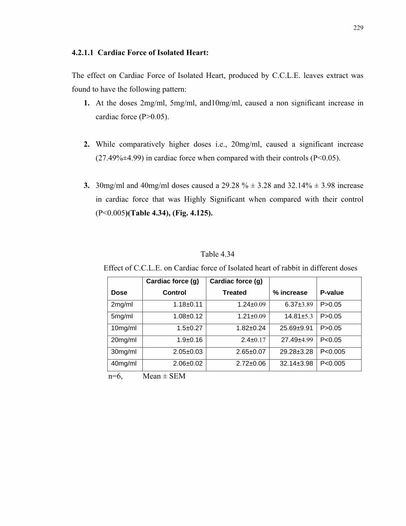
229
4.2.1.1 Cardiac Force of Isolated Heart:
The effect on Cardiac Force of Isolated Heart, produced by C.C.L.E. leaves extract was
found to have the following pattern:
1. At the doses 2mg/ml, 5mg/ml, and10mg/ml, caused a non significant increase in
cardiac force (P>0.05).
2. While comparatively higher doses i.e., 20mg/ml, caused a significant increase
(27.49%±4.99) in cardiac force when compared with their controls (P<0.05).
3. 30mg/ml and 40mg/ml doses caused a 29.28 % ± 3.28 and 32.14% ± 3.98 increase
in cardiac force that was Highly Significant when compared with their control
(P<0.005)(Table 4.34), (Fig. 4.125).
Table 4.34
Effect of C.C.L.E. on Cardiac force of Isolated heart of rabbit in different doses
Dose
Cardiac force (g)
Control
Cardiac force (g)
Treated % increase P-value
2mg/ml 1.18±0.11 1.24±0.09 6.37±3.89 P>0.05
5mg/ml 1.08±0.12 1.21±0.09 14.81±5.3 P>0.05
10mg/ml 1.5±0.27 1.82±0.24 25.69±9.91 P>0.05
20mg/ml 1.9±0.16 2.4±0.17 27.49±4.99 P<0.05
30mg/ml 2.05±0.03 2.65±0.07 29.28±3.28 P<0.005
40mg/ml 2.06±0.02 2.72±0.06 32.14±3.98 P<0.005
n=6, Mean ± SEM
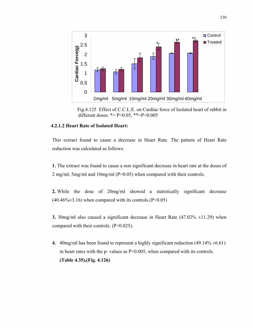
230
Fig.4.125 Effect of C.C.L.E. on Cardiac force of Isolated heart of rabbit in
different doses. *= P<0.05, **=P<0.005 4.2.1.2 Heart Rate of Isolated Heart:
This extract found to cause a decrease in Heart Rate. The pattern of Heart Rate
reduction was calculated as follows:
1. The extract was found to cause a non significant decrease in heart rate at the doses of
2 mg/ml, 5mg/ml and 10mg/ml (P>0.05) when compared with their controls.
2. While the dose of 20mg/ml showed a statistically significant decrease
(40.46%±3.16) when compared with its controls.(P<0.05)
3. 30mg/ml also caused a significant decrease in Heart Rate (47.02% ±11.29) when
compared with their controls. (P<0.025).
4. 40mg/ml has been found to represent a highly significant reduction (49.14% ±6.61)
in heart rates with the p- values as P<0.005, when compared with its controls.
(Table 4.35),(Fig. 4.126)
0
0.5
1
1.5
2
2.5
3
2mg/ml 5mg/ml 10mg/ml 20mg/ml 30mg/ml 40mg/ml
Car
dia
c F
orc
e(g
)
Control
Treated*** **
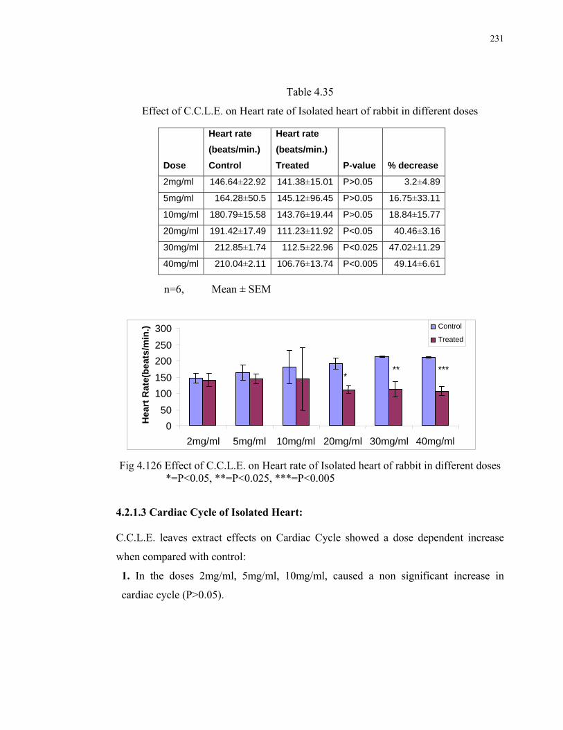
231
Table 4.35
Effect of C.C.L.E. on Heart rate of Isolated heart of rabbit in different doses
n=6, Mean ± SEM
Fig 4.126 Effect of C.C.L.E. on Heart rate of Isolated heart of rabbit in different doses
*=P<0.05, **=P<0.025, ***=P<0.005 4.2.1.3 Cardiac Cycle of Isolated Heart:
C.C.L.E. leaves extract effects on Cardiac Cycle showed a dose dependent increase
when compared with control:
1. In the doses 2mg/ml, 5mg/ml, 10mg/ml, caused a non significant increase in
cardiac cycle (P>0.05).
Dose
Heart rate
(beats/min.)
Control
Heart rate
(beats/min.)
Treated P-value % decrease
2mg/ml 146.64±22.92 141.38±15.01 P>0.05 3.2±4.89
5mg/ml 164.28±50.5 145.12±96.45 P>0.05 16.75±33.11
10mg/ml 180.79±15.58 143.76±19.44 P>0.05 18.84±15.77
20mg/ml 191.42±17.49 111.23±11.92 P<0.05 40.46±3.16
30mg/ml 212.85±1.74 112.5±22.96 P<0.025 47.02±11.29
40mg/ml 210.04±2.11 106.76±13.74 P<0.005 49.14±6.61
0
50
100
150
200
250
300
2mg/ml 5mg/ml 10mg/ml 20mg/ml 30mg/ml 40mg/ml
Hea
rt R
ate(
bea
ts/m
in.) Control
Treated
*** ***

232
2. While the doses of 20mg/ml, and 30mg/ml caused a significant increase i.e.,
17.41% ± 4.32 and 32.61% ± 3.12 in cardiac cycle when compared with their
controls showing P<0.05.
3. The dose of 40mg/ml, caused a highly significant increase (38.33% ± 12.27)in
cardiac cycle when compared with its controls i.e., P<0.025 (Table 4.36, Fig
4.127)
Table 4.36
Effect of C.C.L.E. on Cardiac cycle of Isolated heart of rabbit in different doses
n=6, Mean ± SEM
Fig 4.127 Effect of C.C.L.E. on Cardiac cycle of Isolated heart of rabbit in different doses
*= P<0.05, **=P<0.025
Dose
Cardiac cycle (Sec.)
Control
Cardiac cycle (Sec.)
Treated P-value % Increase
2mg/ml 0.5251±0.05 0.625±0.05 P>0.05 19.5±2.15
5mg/ml 0.566±0.108 0.666±0.10 P>0.05 18.65±3.97
10mg/ml 0.43±0.04 0.5±0 P>0.05 16.66±10.2
20mg/ml 0.72±0.02 0.85±0.03 P<0.05 17.41±4.32
30mg/ml 0.62±0.05 0.82±0.05 P<0.05 32.61±3.12
40mg/ml 0.68±0.05 0.92±0.02 P<0.025 38.33±12.27
0
0.2
0.4
0.6
0.8
1
2mg/ml 5mg/ml 10mg/ml 20mg/ml 30mg/ml 40mg/ml
Car
dia
c C
ycle
(S
ec.)
Control
Treated* *
**

233
4.2.2 EFFECT OF MUFARREH YAQOOTI MOTADIL (MUYM) ON ISOLATED HEART OF RABBITS: The effects of MUYM in different doses, exhibited on Isolated heart of rabbits, are
shown by the following tracings. Fig 4.128- 4.132
Fig 4.128 Tracing of Control (left) and MUYM Treated (5mg/ml) (right) isolated heart of rabbit
Fig 4.129 Tracing of Control (left) and MUYM Treated (10mg/ml) (right) isolated heart of rabbit
Fig 4.130 Tracing of Control (left) and MUYM Treated (20mg/ml) (right) isolated heart of rabbit

234
Fig 4.131 Tracing of Control (left) and MUYM Treated (30mg/ml) (right) isolated heart of rabbit
Fig 4.132 Tracing of Control (left) and MUYM Treated (40mg/ml) (right) isolated heart of rabbit 4.2.2.1 Cardiac Force of Isolated Heart:
The effect on Cardiac Force of Isolated Heart, produced by the product MUYM extract
was found to have the following pattern:
1. At the doses 2mg/ml, 5mg/ml, and10mg/ml, caused a non significant increase in
cardiac force (P>0.05).
2. While comparatively higher dose i.e., 20mg/ml, caused a significant increase
(30.4%±5.44) in cardiac force when compared with their controls (P<0.05).
3. 30mg/ml and 40mg/ml doses caused a 40.31% ± 3.98 and 50.9% ± 5.86 increase in
cardiac force respectively, that were Highly Significant when compared with their
controls (P<0.005)(Table 4.37, Fig 4.133).

235
Table 4.37
Effect of MUYM on Cardiac force of Isolated heart of rabbit in different doses.
n=6, Mean ±SEM
Fig 4.133 Effect of MUYM on Cardiac force of Isolated heart of rabbit in different doses. *= P<0.05, **=P<0.005
4.2.2.2 Heart Rate of Isolated Heart:
This product found to cause a decrease in Heart Rate. The pattern of Heart Rate
reduction was calculated as follows:
1. The drug was found to cause a non significant decrease in heart rate at the doses of 2
mg/ml, 5mg/ml and 10mg/ml (P>0.05) when compared with its controls.
Dose
Cardiac force (g)
Control
Cardiac force (g)
Treated P-value % increase
2mg/ml 1.12±0.21 1.22±0.18 P>0.05 11.16±6.09
5mg/ml 1.08±0.12 1.26±0.11 P>0.05 18.38±3.15
10mg/ml 1.67±0.08 1.95±0.13 P>0.05 16.25±4.33
20mg/ml 1.82±0.16 2.37±0.2 P<0.05 30.4±5.44
30mg/ml 1.86±0.12 2.6±0.12 P<0.005 40.31±3.98
40mg/ml 1.82±0.14 2.72±0.12 P<0.005 50.9±5.86
0
0.5
1
1.5
2
2.5
3
2mg/ml 5mg/ml 10mg/ml 20mg/ml 30mg/ml 40mg/ml
Car
dia
c fo
rce
(g)
Control
Treated
*** **
236
2. While the dose of 20mg/ml showed a statistically significant decrease
(39.64%±11.29) when compared with its controls (P<0.05).
3. 30mg/ml also caused a significant decrease in Heart Rate (46.05% ±10.75) when
compared with their controls (P<0.025).
4. 40mg/ml has been found to represent a highly significant reduction (51.08% ±6.44)
in heart rates with the p- values as P<0.005, when compared with its controls (Table
4.38, Fig 4.134).
Table 4.38
Effect of MUYM on Heart rate of Isolated heart of rabbit in
different doses n=6, Mean± SEM
Dose
Heart rate
(beats/ min.)
Control
Heart rate
(beats/ min.)
Treated P-value % decrease
2mg/ml 180.79±15.58 162.95±17.07 P>0.05 10.05±1.91
5mg/ml 146.87±7.89 131.77±8.02 P>0.05 10.71±4.28
10mg/ml 179.6±31.48 156.08±39.99 P>0.05 13.94±12.71
20mg/ml 191.42±17.49 112.92±9.84 P<0.05 39.64±11.29
30mg/ml 212.85±1.74 114.58±21.8 P<0.025 46.05±10.75
40mg/ml 210.04±2.11 102.81±13.68 P<0.005 51.08±6.44

237
Fig 4.134 Effect of MUYM on Heart rate of Isolated heart of rabbit in
different doses *=P<0.05, **=P<0.025, ***=P<0.005 4.2.2.3 Cardiac Cycle of Isolated Heart:
The Product MUYM effects on Cardiac Cycle showed a dose dependent increase when
compared with control:
1. At the doses 2mg/ml, 5mg/ml, 10mg/ml, caused a non significant increase in
cardiac cycle (P>0.05).
2. While the doses of 20mg/ml, and 30mg/ml caused a significant increase i.e.,
44.94% ± 11.83 and 47.14% ± 7.64 in cardiac cycle respectively when compared
with its controls showing P<0.05.
4. The dose of 40mg/ml, caused a highly significant increase (55.83% ± 4.06) in
cardiac cycle when compared with its control i.e., P<0.025 (Table 4.39, Fig 4.135)
0
50
100
150
200
250
2mg/ml 5mg/ml 10mg/ml 20mg/ml 30mg/ml 40mg/ml
Hea
rt R
ate
(bet
a/m
in) Control
Treated
* *****

238
Table 4.39
Effect of MUYM on Cardiac cycle of Isolated heart of rabbit in different doses
n=6, Mean ± S.E.M
Fig 4.135 Effect of MUYM on Cardiac cycle of Isolated heart of rabbit in
different doses. *= P<0.05, **=P<0.025
Dose
Cardiac cycle
(Sec.) Control
Cardiac cycle
(Sec.) Treated P-value % Increase
2mg/ml 0.525±0.05 0.637±0.05 P>0.05 21.78±2.97
5mg/ml 0.6±0.07 0.76±0.04 P>0.05 30.31±18.19
10mg/ml 0.5±0.05 0.65±0.09 P>0.05 29.58±6.26
20mg/ml 0.85±0.07 1.22±0.11 P<0.05 44.94±11.83
30mg/ml 0.65±0.07 0.95±0.1 P<0.05 47.14±7.64
40mg/ml 0.68±0.05 1.06±0.09 P<0.025 55.83±4.06
00.2 0.4 0.6 0.8
11.2 1.4
2mg/ml 5mg/ml 10mg/ml 20mg/ml 30mg/ml 40mg/ml
Car
dia
c cy
cle(
Sec
.) Control
Treated***
*

239
4.3. SAFETY EVALUATION STUDIES
4.3.1 ACUTE TOXICITY IN MICE:
4.3.1.1 Carissa carandas leaves Extract (C.C.L.E.): Acute toxicity studies were conducted to check the LD50 values in experimental animals. 4.3.1.1.a. Behaviour
Male and female mice treated with C.C leaves after single dose administration i.e.,1750
mg/kg (p.o) and 5000 mg/kg (p.o) respectively, did not show any mortality during the 14
days period of observation. None of these animals showed any sign of toxicity.
4.3.1.1.b. Body Weight (g): Table 4.40 shows a non significant increase in body weight of control animals, and non
significant decrease in treated animals’ wt. was observed at the end of 14th day of
observation of single high dose oral treatment i.e., 1750 mg/kg. (Fig 4.136)
Table 4.40 Change in weight of control and C.C.L.E. (1750mg/kg) single dose (LD50) treated mice after 14 days observation.
n=10, Mean ± SEM
C.C.L.E. (1750mg/kg)
1st Day body wt. (g)
14th Day body wt. (g)
% change in wt.
Mortality
Control (N) 27.4±1.026 27.8±1.772 -1.11±2.66 No expiry
Treated (S) 24.56±0.72 24.4±0.515 0.53±1.70 No expiry
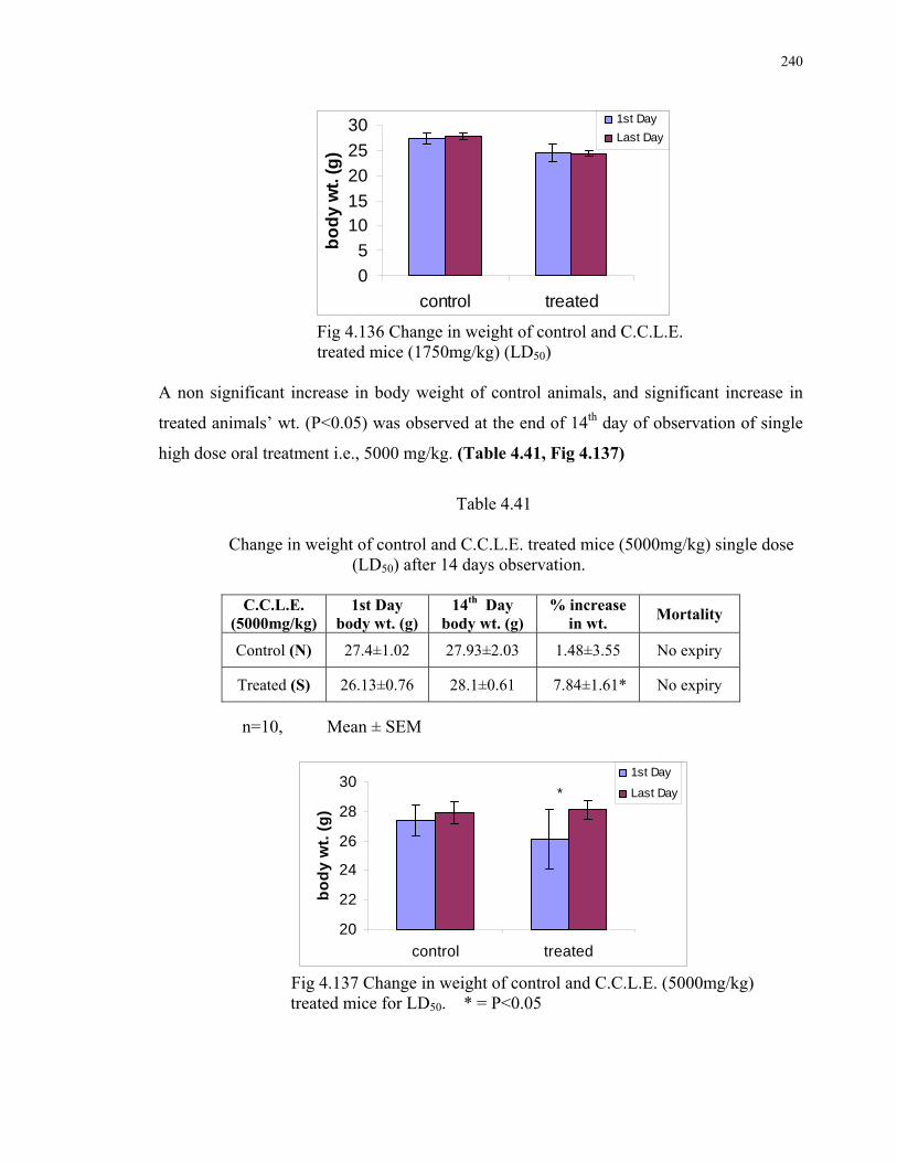
240
0
5
1015
20
25
30
control treated
bo
dy
wt.
(g
)
1st Day
Last Day
Fig 4.136 Change in weight of control and C.C.L.E. treated mice (1750mg/kg) (LD50)
A non significant increase in body weight of control animals, and significant increase in
treated animals’ wt. (P<0.05) was observed at the end of 14th day of observation of single
high dose oral treatment i.e., 5000 mg/kg. (Table 4.41, Fig 4.137)
Table 4.41
Change in weight of control and C.C.L.E. treated mice (5000mg/kg) single dose (LD50) after 14 days observation.
n=10, Mean ± SEM
20
22
24
26
28
30
control treated
bo
dy
wt.
(g
)
1st Day
Last Day*
Fig 4.137 Change in weight of control and C.C.L.E. (5000mg/kg)
treated mice for LD50. * = P<0.05
C.C.L.E. (5000mg/kg)
1st Day body wt. (g)
14th Day body wt. (g)
% increase in wt.
Mortality
Control (N) 27.4±1.02 27.93±2.03 1.48±3.55 No expiry
Treated (S) 26.13±0.76 28.1±0.61 7.84±1.61* No expiry

241
4.3.1.2 Mufarreh Yaqooti Motadil (MUYM): 4.3.1.2.a. Behaviour
Male and female mice treated with C.C leaves 1750 mg/kg dose, did not show any
mortality. None of these animals showed any sign of toxicity. However mice treated with
5000mg/kg dose, showed some physical behavioral changes that were observed in first 2
hour after dosing like uncoordinated motor activity and corner sitting. Afterwards animals
behaved normally.
4.3.1.2. b. Body Weight (g):
A non significant decrease in body weight of control animals, and non significant increase
in treated animals’ wt. was observed at the end of 14th day of observation of single high
dose oral treatment i.e., 1750 mg/kg. (Table 4.42, Fig 4.138)
Table 4.42
Change in weight of control and MUYM treated mice (1750mg/kg) (LD50) after 14 days observation.
n=10, Mean ± SEM
MUYM (1750 mg/kg)
1st Day body wt. (g)
14thDay body wt. (g)
% change in wt.
Mortality
Control (N) 28.9±1.33 28.85±1.22 0.008±1.15 No expiry
Treated (S) 27.27±1.51 27.52±1.61 -0.77±0.44 No expiry

242
23
24
25
26
27
28
29
30
31
control treatedB
od
y w
t. (
g)
1st Day
Last Day
Fig 4.138 Change in weight of control and MUY treated
mice (1750mg/kg) (LD50).
Table 4.43 shows that a non significant increase in body weight of control animals, while a
significant increase in treated animals’ wt. (P<0.05) was observed at the end of 14th day of
observation of single high dose oral treatment i.e., 5000 mg/kg. (Fig 4.139)
Table 4.43 Change in weight of control and MUYM treated mice (5000mg/kg) (LD50) after 14 days observation.
n=10, Mean ± SEM
MUYM (5000mg/kg)
1st Day body wt. (g)
14th Day body wt. (g)
% change in wt.
Mortality
Control (N) 28.9±1.33 28.85±1.22 0.008±1.15 No expiry
Treated (S) 30.37±0.93 30.76±0.89 -1.32±0.50 No expiry

243
25
26
27
28
29
30
31
32
control treated
Bo
dy w
t. (g
)
1st Day
Last Day
Fig 4.139 Change in weight of control and MUYM treated
mice (5000mg/kg) (LD50).
Autopsy:
Autopsy revealed that no gross changes were observed in organs like liver, spleen, heart
and kidney among the animals of group N (control) and S (treated). The appearance of
organs of Group S animals was comparable with that of Group N animals.
4.3.2 SUBACUTE TOXICITY IN MICE:
4.3.2.1 C.C.L.E. Leaves: 4.3.2.1.a. Body Weight (g): For the 14 consecutive days the 5000 mg/kg/day dose was administered to mice. No signs
of toxicity and mortality were observed in treated group as compared to the control group.
Table 4.44 shows the effects of C.C.L.E. on the body weight. A non significant increase in
body weight was detected on 14th day (Fig 4.140).
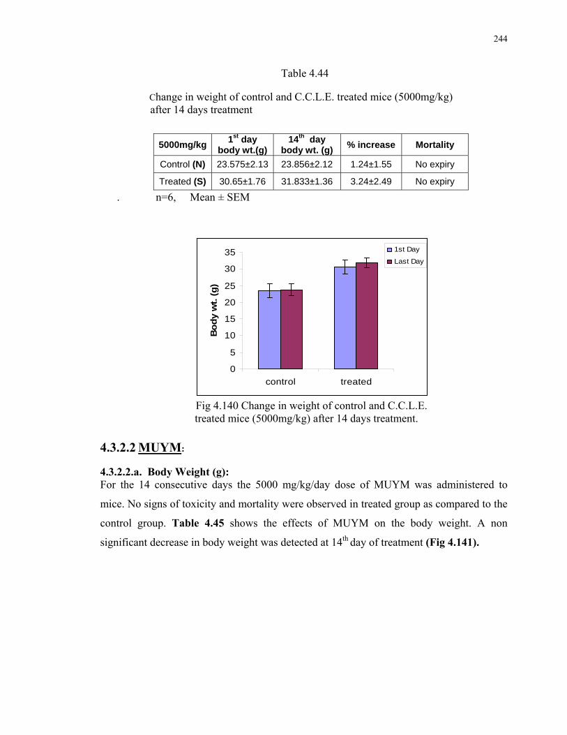
244
Table 4.44
Change in weight of control and C.C.L.E. treated mice (5000mg/kg) after 14 days treatment
. n=6, Mean ± SEM
0
5
10
15
20
25
30
35
control treated
Bo
dy w
t. (
g)
1st Day
Last Day
Fig 4.140 Change in weight of control and C.C.L.E. treated mice (5000mg/kg) after 14 days treatment.
4.3.2.2 MUYM: 4.3.2.2.a. Body Weight (g): For the 14 consecutive days the 5000 mg/kg/day dose of MUYM was administered to
mice. No signs of toxicity and mortality were observed in treated group as compared to the
control group. Table 4.45 shows the effects of MUYM on the body weight. A non
significant decrease in body weight was detected at 14th day of treatment (Fig 4.141).
5000mg/kg 1st day
body wt.(g) 14th day
body wt. (g) % increase Mortality
Control (N) 23.575±2.13 23.856±2.12 1.24±1.55 No expiry
Treated (S) 30.65±1.76 31.833±1.36 3.24±2.49 No expiry

245
Table 4.45 Change in weight of control and MUYM treated mice (5000mg/kg) after
14 days treatment.
5000mg/kg (MUYM)
1st day body wt.(g)
14th day body wt. (g)
% decrease Mortality
Control (N) 33.52±2.39 32.79±1.87 1.65±3.00 No expiry
Treated (S) 37.33±1.38 36.31±1.88 5.02±2.97 No expiry n=6, Mean ± SEM
Fig 4.141 Change in weight of control and MUYM treated mice(5000mg/kg)after 14 days treatment.
4.3.3 HISTOPATHOLOGICAL STUDIES OF SUBACUTE TOXICITY
IN MICE: 4.3.3.1 CONTROL GROUP:( N)
4.3.3.1.A EXAMINATION OF LIVER:
Fig 4.142 (20 X) The liver cells are arranged into lobules in both control and treated slides.
Liver cells hepatocytes are flat and arranged. A discontinuous layer of cells lines the
sinusoids. Central vein is lined by epithelial cells and filled with biconcave Red Blood
cells. Hepatic sinusoids appear to radiate from central vein.
05
1015202530354045
control treated
Bo
dy
wt.
(g
)
1st day
Last Day
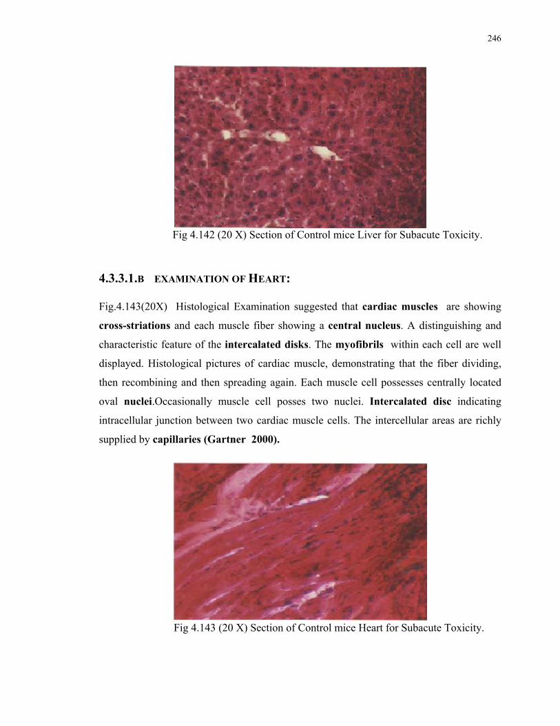
246
Fig 4.142 (20 X) Section of Control mice Liver for Subacute Toxicity.
4.3.3.1.B EXAMINATION OF HEART:
Fig.4.143(20X) Histological Examination suggested that cardiac muscles are showing
cross-striations and each muscle fiber showing a central nucleus. A distinguishing and
characteristic feature of the intercalated disks. The myofibrils within each cell are well
displayed. Histological pictures of cardiac muscle, demonstrating that the fiber dividing,
then recombining and then spreading again. Each muscle cell possesses centrally located
oval nuclei.Occasionally muscle cell posses two nuclei. Intercalated disc indicating
intracellular junction between two cardiac muscle cells. The intercellular areas are richly
supplied by capillaries (Gartner 2000).
Fig 4.143 (20 X) Section of Control mice Heart for Subacute Toxicity.
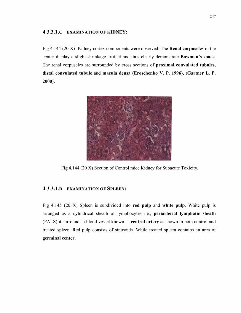
247
4.3.3.1.C EXAMINATION OF KIDNEY:
Fig 4.144 (20 X) Kidney cortex components were observed. The Renal corpuscles in the
center display a slight shrinkage artifact and thus clearly demonstrate Bowman’s space.
The renal corpuscles are surrounded by cross sections of proximal convulated tubules,
distal convulated tubule and macula densa (Eroschenko V. P. 1996), (Gartner L. P.
2000).
Fig 4.144 (20 X) Section of Control mice Kidney for Subacute Toxicity.
4.3.3.1.D EXAMINATION OF SPLEEN:
Fig 4.145 (20 X) Spleen is subdivided into red pulp and white pulp. White pulp is
arranged as a cylindrical sheath of lymphocytes i.e., periarterial lymphatic sheath
(PALS) it surrounds a blood vessel known as central artery as shown in both control and
treated spleen. Red pulp consists of sinusoids. While treated spleen contains an area of
germinal center.

248
Fig 4.145 (20 X) Section of Control mice Spleen for Subacute Toxicity.
4.3.3.2 C.C.L.E. LEAVES TREATED GROUP: (S) LIVER: Fig 4.146 (20 X) showed spotty necrosis and mild inflammation. (Yuan 2007)
Fig 4.146 (20 X) Section for C.C.L.E.Leaves Extract effcects on Liver of mice showing Subacute Toxicity

249
HEART: Fig 4.147 (20 X) Heart tissues are comparable to that of control animals. (Fig 5.143)
Fig 4.147 (20 X) Section for C.C.L.E.Leaves Extract effects
on Heart of mice after Subacute Treatment KIDNEY: Fig 4.148 (20 X) The Cells lining the tubule are necrotic. The capillaries of the
boundary zones are greatly distended and the stroma is edematous.
Fig 4.148 (20 X) Section for C.C.L.E.Leaves Extract effects on Kidney of mice showing Subacute Toxicity
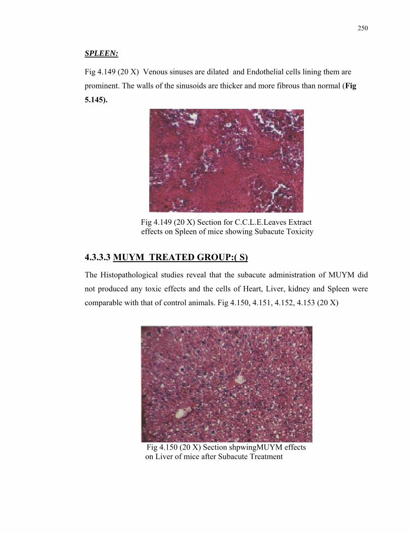
250
SPLEEN: Fig 4.149 (20 X) Venous sinuses are dilated and Endothelial cells lining them are
prominent. The walls of the sinusoids are thicker and more fibrous than normal (Fig
5.145).
Fig 4.149 (20 X) Section for C.C.L.E.Leaves Extract effects on Spleen of mice showing Subacute Toxicity
4.3.3.3 MUYM TREATED GROUP:( S) The Histopathological studies reveal that the subacute administration of MUYM did
not produced any toxic effects and the cells of Heart, Liver, kidney and Spleen were
comparable with that of control animals. Fig 4.150, 4.151, 4.152, 4.153 (20 X)
Fig 4.150 (20 X) Section shpwingMUYM effects
on Liver of mice after Subacute Treatment

251
Fig 4.151 (20 X) Section showing MUYM effects
on Heart of mice after Subacute Treatment
Fig 4.152 (20 X) Section showing MUYM effects
on Kidney of mice after Subacute Treatment
Fig 4.153 (20 X) Section showing MUYM effects on Spleen of mice after Subacute Treatment.

252
4.3.4 CHRONIC TOXICITY IN RATS: 4.3.4.1 C.C.L.E. leaves group: 4.3.4.1.a Boby Weight (g): In a two months chronic oral toxicity evaluation, a non significant increase in body
weight of treated rats (S), receiving 5000 mg/kg of C.C.L.E. leaves extract, was
observed when compared with their control group (N).
Table 5.46 Change in weight of rats after administration of extract for two months.
n=8, Mean ± SEM
0
50
100
150
200
250
300
control treated
bo
dy
wt.
(g
)
1st Day
Last Day
Fig. 5.154 Change in weight of rats after administration of extract (C.C.L.E.) for two months, compared with their control.
4.3.4.2 MUYM group: 4.3.4.2.a Boby Weight: A non significant decrease in body weight of treated rats (S), receiving 5000 mg/kg of
the Product MUYM. While a non significant increase in body weight of rats of group
(N) was observed after two months treatment. (Table 4.47)
C.C.L.E. (5000mg/kg)
1st day body wt.(g)
Last day body wt.(g)
% increase
Control (N)
244.05±12.31 270.44±20.13 10.67±5.53
Treated (S)
225.75±4.83 234.03±14.67 7.00±2.15

253
Table 4.47
Change in weight of rats after administration of drug for two months.
n=8, Mean ± SEM
0
50
100
150
200
250
300
control treated
Bo
dy
wt.
(g)
1st Day
Last Day
Fig. 4.155 Change in weight of rats after administration of drug (MUYM)for two months for chronic toxicity studies, compared with their control.
AUTOPSY: Autopsy revealed no pathological signs on gross examination of organs like Heart, Liver,
Kidney and Spleen.
4.3.5 HISTOPATHOLOLOGICAL STUDIES OF CHRONIC TOXICITY ON RATS: 4.3.5.1. C.C.L.E. Leaves Group: Fig. 4.156, 4.158, 4.159 (20X) Photomicrography of Heart, Liver and Spleen shows that all
the organs of rats treated with 5000mg/kg of Carissa carandas leaves extract were
comparable with their control (Fig. 4.107, 4.113, 4.116) on Histopathological
investigation. While changes in the structure of kidney tissues were noted. Spaces in some
5000mg/kg 1st day
body wt.(g) Last day
body wt.(g) % change
Control (N) 253±25.22 284.38±43.44 -12.05±12.0
Treated (S) 221.58±4.87 214.18±10.58 3.45±2.084
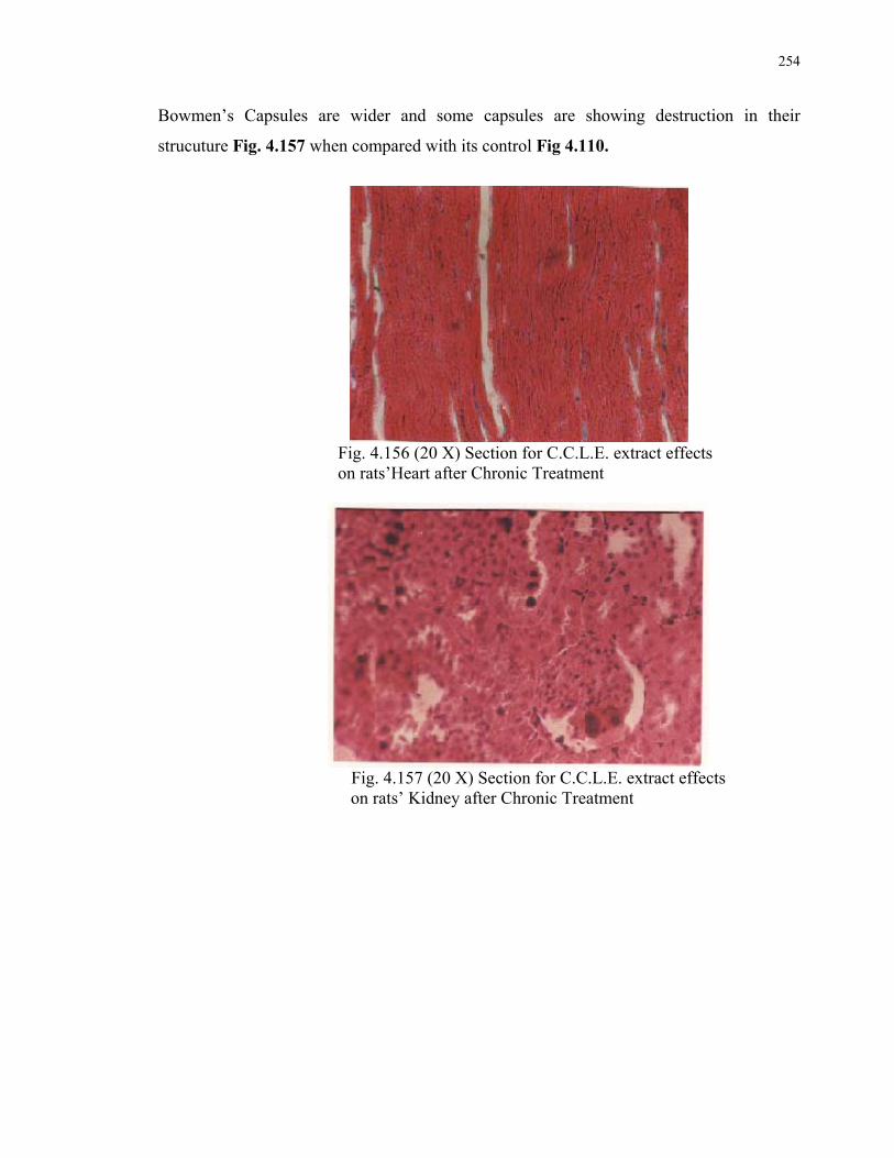
254
Bowmen’s Capsules are wider and some capsules are showing destruction in their
strucuture Fig. 4.157 when compared with its control Fig 4.110.
Fig. 4.156 (20 X) Section for C.C.L.E. extract effects
on rats’Heart after Chronic Treatment
Fig. 4.157 (20 X) Section for C.C.L.E. extract effects
on rats’ Kidney after Chronic Treatment

255
Fig. 4.158 (20 X) Section for.C.C.L.E. extract effects
on rats’Liver after Chronic Treatment.
Fig. 4.159 (20 X) Section for C.C.L.E. extract effects
on rats’ Spleen after Chronic Treatment
The heart cross section illustrates muscle fibres, connective tissue and nucleus of
myocytes. The liver cross section shows the hepatic artery, bile duct, central vein,
sinusoids and hepatocytes, all clearly conserved. Spleen cross section shows red pulp and
white pulp containing a central artery. All the structures were found to be conserved.
4.3.5.2. MUYM Group:
4.3.5.2.A EXAMINATION OF HEART:
Fig. 4.159 (20 X) The myofibrils within each cell are well displayed. Each muscle cell
possesses centrally located oval nuclei. Occasionally muscle cell posses two nuclei.
Intercalated disc indicating intracellular junction between two cardiac muscle cells. The
intercellular areas are richly supplied by capillaries .
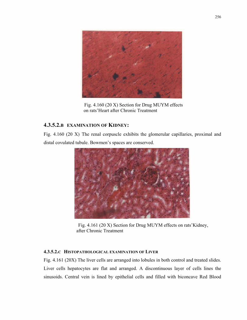
256
Fig. 4.160 (20 X) Section for Drug MUYM effects on rats’Heart after Chronic Treatment
4.3.5.2.B EXAMINATION OF KIDNEY:
Fig. 4.160 (20 X) The renal corpuscle exhibits the glomerular capillaries, proximal and
distal covulated tubule. Bowmen’s spaces are conserved.
Fig. 4.161 (20 X) Section for Drug MUYM effects on rats’Kidney, after Chronic Treatment
4.3.5.2.C HISTOPATHOLOGICAL EXAMINATION OF LIVER
Fig. 4.161 (20X) The liver cells are arranged into lobules in both control and treated slides.
Liver cells hepatocytes are flat and arranged. A discontinuous layer of cells lines the
sinusoids. Central vein is lined by epithelial cells and filled with biconcave Red Blood
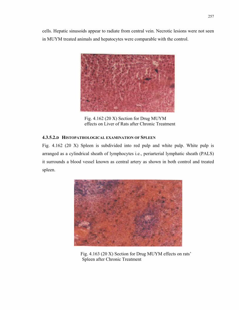
257
cells. Hepatic sinusoids appear to radiate from central vein. Necrotic lesions were not seen
in MUYM treated animals and hepatocytes were comparable with the control.
Fig. 4.162 (20 X) Section for Drug MUYM effects on Liver of Rats after Chronic Treatment
4.3.5.2.D HISTOPATHOLOGICAL EXAMINATION OF SPLEEN
Fig. 4.162 (20 X) Spleen is subdivided into red pulp and white pulp. White pulp is
arranged as a cylindrical sheath of lymphocytes i.e., periarterial lymphatic sheath (PALS)
it surrounds a blood vessel known as central artery as shown in both control and treated
spleen.
Fig. 4.163 (20 X) Section for Drug MUYM effects on rats’ Spleen after Chronic Treatment

258
4.3.6 BIOCHEMICAL STUDIES FOR CHRONIC TOXICITY: 4.3.6.1 C.C.L.E. Leaves Group: Biochemical Alterations in the serum of rats, treated chronically with 5000 mg/kg of
C.C.L.E. leaves extract (Group S), were compared with their control groups’ animals
(Group N).
Cholesterol was decreased in treated animals. That decrease was Statistically Highly
Significant i.e., P<0.025, HDL increase was highly significant P<0.01, LDL decreased
Highly Significantly P<0.025, Bilirubin total decrease was highly significant P<0.025,
when compared with their control groups.
Following biochemicals were altered in the Plasma of Treated Groups animals in a
statistically non significant manner i.e., P>0.05, when compared with their control
groups.
Bilirubin direct decreased, Triglycerides decreased, ALAT increased, ASAT decreased,
GGT decreased, Alkaline phosphatase increased, CK decreased, LDH increased, Total
Protein increased, Glucose increased, Albumin decreased, Urea and Uric Acid decreased,
Creatinine increased. All mentioned enzymes and chemicals changes were statistically non
significant. The whole result is summarized in Table 4.48 and can be understood through.
Fig. 4.164 A-H.
Table 4.48
Biochemical studies for chronic toxicity of C.C.L.E. (5000mg/kg) on Rats:
Biochemicals Control Treated P- value
cholesterol (mg/dl) 125.68±31.66 59.12**±9.32 P<0.025
triglycerides (mg/dl) 89.83±19.07 52.38±9.10 P>0.05
HDL (mg/dl) 98.23±2.8 128.24***±2.8 P<0.01
LDL (mg/dl) -57.73±16.22 -190.228**±47.98 P<0.025
ALAT (u/l) 3.88±0.49 5.31±0.72 P>0.05
ASAT (u/l) 3.78±1.98 1.92±0.21 P>0.05
GGT (u/l) 3.24±0.54 2.46±0.66 P>0.05
Alkaline phosphatase (u/l) 28.63±14.08 36.71±2.24 P>0.05
CK (u/l) 20.08±9.06 15.79±4.67 P>0.05

259
Table 4.48
Continued……….
Biochemicals Control Treated P- value
LDH (u/l) 807.1±46.61 906.6±29.74 P>0.05
Total Proetein (g/dl) 4.2±1.07 7.85±1.65 P>0.05
Albumin (g/dl) 3.8±0.34 3.7±0.41 P>0.05
Glucose (mg/dl) 143.74±5.18 155.31±19.50 P>0.05
Bilirubin Direct (mg/dl) 0.359±0.015 0.345±0.23 P>0.05
Bilirubin Total (mg/dl) 0.53±0.16 0.067**±0.026 P<0.025
Urea (mg/dl) 62.15±10.21 43.05±25.05 P>0.05
Uric Acid (mg/dl) 3.4±0.59 2.89±0.47 P>0.05
Creatinine (mg/dl) 1.04±0.23 1.39±0.69 P>0.05
n=8, Mean ± SEM
Fig 4.164 A-H Graphs showing biochemical changes in Rats after two months treatment with C.C.L.E. (5000mg/kg)
Fig 4.164 A
-300
-250
-200
-150
-100
-50
0
50
100
150
200
cholesterol(mg/dl)
triglycerides(mg/dl)
HDL (mg/dl) LDL (mg/dl)
Co
nc.
(m
g/d
l)
control
treated***
**
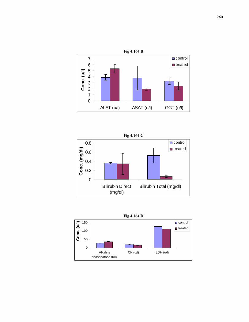
260
Fig 4.164 B
Fig 4.164 C
Fig 4.164 D
0
50
100
150
Alkaline
phosphatase (u/l)
CK (u/l) LDH (u/l)
Co
nc.
(u
/l) control
treated
01234567
ALAT (u/l) ASAT (u/l) GGT (u/l)
Co
nc.
(u
/l)
control
treated
0
0.2
0.4
0.6
0.8
Bilirubin Direct(mg/dl)
Bilirubin Total (mg/dl)
Co
nc.
(m
g/d
l)
control
treated

261
Fig4.164 E
0
2
4
6
8
10
Total Proetein (g/dl) Albumin (g/dl)
Co
nc.
(g
/dl)
control
treated
Fig 4.164 F
0
50
100
150
200
Glucose (mg/dl)
Co
nc.
(m
g/d
l)
control
treated
Fig 4.164 G
0
20
40
60
80
Urea (mg/dl)
Co
nc
. (m
g/d
l)
control
treated

262
Fig 4.164 H
0
1
2
3
4
5
Uric Acid (mg/dl) Creatinine (mg/dl)
Co
nc
.(m
g/d
l)
control
treated
4.3.6.2 MUYM Group: The effect of MUYM (5000 mg/kg) on Rats’ Blood chemistry was measured and presented
in Table 5.49. The alterations in the activities of ,Cholesterol, Triglycerides, HDL Bilirubin
Direct, Bilirubin Total, ALAT, ASAT, GGT, CK, Alkaline Phosphatase, Albumin and
Glucose (Fig 4.165 A-E) were recorded.
The dose of MUYM (5000 mg/kg) had non significantly decreased the serum levels of
Cholesterol and HDL (P>0.05). ALAT, ASAT, were decreased significantly (P<0.005),
Bilirubin Direct decreased significantly (P<0.05) and Bilirubin Total was also found to be
decreased significantly (P<0.025). No changes were found in serum Triglycerides and
Albumin levels. The GGT and CK were decreased non significantly (P>0.05). Non-
significant increase was observed in Alkaline Phosphatase and Glucose (P>0.05). All these
values were compared with their respective control groups (N). Table 4.49

263
Table 4.49
Biochemical studies for chronic toxicity of MUYM (5000mg/kg) on Rats:
n=6, Mean ± SEM
Fig 4.165 A-E. Graphs showing biochemical changes in Rats after two months treatment with MUYM (5000mg/kg)
Fig 4.165 A
0
50
100
150
200
cholesterol(mg/dl)
triglycerides(mg/dl)
HDL (mg/dl)
Co
nc
. (m
g/d
l)
control
treated
Biochemicals Control treated P-Value
cholesterol (mg/dl) 116.7±0.14 116.3±0.28 P>0.05
triglycerides (mg/dl) 157.74±3.64 157.79±5.03 No Change
HDL (mg/dl) 75.98±9.02 77.94±22.56 P>0.05
ALAT (u/l) 1.75±0.01 1.08****±0.05 P<0.005
ASAT (u/l) 15.25±0.63 2.75****±0.07 P<0.005
GGT (u/l) 1.05±0.14 0.6±0 P>0.05
Alkaline phosphatase (u/l) 26.06±7.26 52.82±7.07 P>0.05
CK (u/l) 10.01±0.26 5.18±0.09 P>0.05
Albumin (g/dl) 3.67±0.098 3.63±0.098 P>0.05
Glucose (mg/dl) 87.92±0.85 88.15±0.16 P>0.05
Bilirubin Direct (mg/dl) 2.37±0.41 1.16*±0 P<0.05
Bilirubin Total (mg/dl) 0.81±0.014 0.74**±0 P<0.025
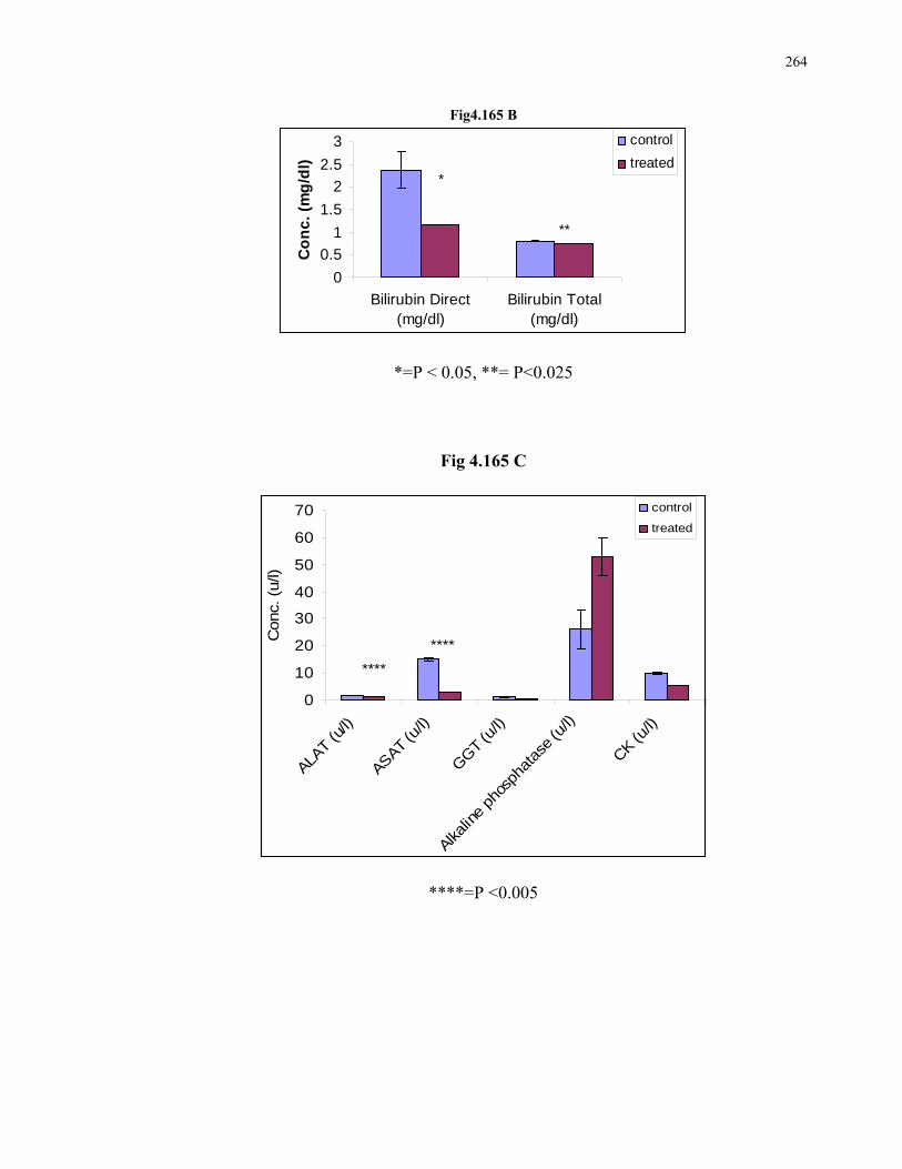
264
Fig4.165 B
0
0.5
1
1.5
2
2.5
3
Bilirubin Direct(mg/dl)
Bilirubin Total(mg/dl)
Co
nc
. (m
g/d
l)
control
treated*
**
*=P < 0.05, **= P<0.025
Fig 4.165 C
0
10
20
30
40
50
60
70
ALAT (u
/l)
ASAT (u/l)
GGT
(u/l)
Alkalin
e ph
osph
atas
e (u
/l)
CK (u/l)
Conc.
(u/l)
control
treated
****
****
****=P <0.005

265
Fig 4.165 D
3.43.453.5
3.553.6
3.653.7
3.753.8
Albumin (g/dl)C
on
c. (
g/d
l)
cotrol
treated
Fig 4.165 E
86
86.5
87
87.5
88
88.5
89
Glucose (mg/dl)
Co
nc
. (m
g/d
l)
control
treated

266
CHAPTER 5
DISCUSSIONS AND
CONCLUSION

267

268
269
5. DISCUSSIONS AND CONCLUSION
5.1 IN VIVO PHARMACODYNAMIC STUDIES ON
CARDIOVASCULAR SYSTEM:
5.1.1. CARISSA CARANDAS (Auct.) (C.C. L. E.) 5.1.1.1. EFFECTS ON MEAN ARTERIAL BLOOD PRESSURE (MABP) OF RATS:
In the present study different doses of the Aqueous: Ethanol extract (50:50) of Carissa
carandas leaves were tested and were found to have a dose dependent hypotensive effect
on Mean Arterial Blood Pressure of Normotensive rats. Ach was used as positive controls
or reference drug (Ahmed et al., 2000), (Lahlou et al., 1999). Different doses of the
extract i.e., 5mg/kg, 10, 15, 20, 25, 30, 35, 40, and 45mg/kg were given I.V to obtain a
dose response curve and the 45mg/kg dose caused a 50.75% ± 2.71 decrease in MABP
which was highly significant with P value< 0.0005 when compared with its controls.
The literature survey of the plant has confirmed the hypotensive effect of Carissa carandas
as the activity performed by Zaki et al., 1983.
Physiologically, the reduction in blood pressure has been reported to to be either due to
their direct or indirect effect on the pumping ability of heart (Aftab 1995), (Kimura et al.,
1986) or associated with decrease in peripheral resistance ( vascular muscle tone) leading
to vasodilation (Ebeigbe and Ezimokhai 1988). On these bases it is difficult to pin point
the exact mechanism of extract’s action. For this purpose the Receptor Activity is
performed.
5.1.1.1.a Receptor activity for Hypotensive Effect: It is reported that Ach causes a generalized vasodilatation that is an indirect effect
mediated by the release of nitric oxide from vascular endothelial cells (Katzung 1998).
That is why it is hypothesized that this extract might be acting like Ach and to confirm this
opinion the receptor activity was performed.
But firstly, to rule out the action of drug as adrenoceptor antagonist for lowering the blood
pressure. After 30 seconds of I.V. administration of 45mg/kg of C.C Leaves Extract,
adrenaline was administered I.V., but no Significant change in the action of Adrenaline
270
was observed 10.32% ± 2.30 (P<0.025). This ruled out the action of drug as adrenoceptor
antagonist for lowering the blood pressure. (Table 4.2, Fig. 4.11)
After the exclusion of action as adrenoceptor blocker, the receptor activity was performed
by using cholinergic agonist Ach and cholinergic competitive antagonist Atropine. The
results demonstrate that Ach and different doses of C.C.L.E. when administered alone have
significantly reduced the MABP. On the other hand use of Ach and different doses of
C.C.L.E., on Atropine pre- treated rats did not show such decline in blood pressure as was
shown earlier without Atropine Pre-treatment. These results clearly indicate that the effect
of Carissa carandas leaves extract is mediated through same receptors and mechanism as
established for the action of Ach. So, it is suggested that the Extract of Carissa carandas
stimulates the muscarinic receptors located on the endothelial cells of the vasculature. This
stimulation results in the release of endothelial-derived relaxing factors (EDRFs) or nitric
oxide that diffuses to vasculature smooth muscles and causes their relaxation (Katzung
1998).
5.1.1.1.b Comparative study with Acetylcholine: The reduction in MABP by C.C.L.E. leaves extract was comparable with Ach i.e., both
showed a highly significant decrease with P value <0.0005 when compared with their
controls. (Table. 4.4, Fig. 4.17). This experiment suggests that the drug is acting through
the same mechanism and efficacy as that of acetylcholine, responsible for the hypotensive
property of the extract when administered intravenously.
5.1.1.2. EFFECTS ON INTACT HEART OF FROG:
In the present study the C.C.L E. Extract has been tested for its effects on cardiac
parameters. The results presented (Table 4.6, Fig. 4.27) shows that the extract tested in
different doses has the ability to increase the cardiac force in dose dependent manner. This
was found to be highly significant at the dose of 45mg/ml (53.79% ± 8.83, P<0.025). This
effect of Carissa carandas was also explained by Rastogi 1967. He isolated by the help of
column chromatography, three polar glycosides named (I) Digitoxigenin (II) 14,15-
anhydrodigitoxigenin (III) Glucose and D- digitalose. He reported that the cardiotonic

271
activity was due to the presence of glucoside of compound (I). So presence of this
glycosidic compound confirms the cardiotonic activity of C.C.L.E. Extract.
In the present study it is also observed that the Extract has the ability to decrease the Heart
rate (Table 4.5, Fig 4.26). It is important to note that heart rate reduction is associated with
an increase in cardiac force. It suggests that this extract is increasing the efficacy of heart
by reducing the heart rate, increasing the cardiac force and the duration of cardiac cycle.
(Table 5.6, Fig 5.28).
5.1.1.3. RECEPTOR ACTIVITY OF C.C.L.E. FOR EFFECT ON HEART: The receptor activity of C.C.L.E. Extract for the action on intact heart of frog has been
tested by using cholinergic agonist Ach and Cholinergic competitive antagonist Atropine.
The results demonstrate that Ach and Extract both when administered alone have reduced
the heart rate by 24.046% ± 0.736 and 25.99 % ± 0.25, P<0.0005 respectively (Fig 4.34).
On the other hand the use of Ach and Carissa carandas on Atropine pretreated heart did
not show such decline in heart rate as shown earlier without atropine pretreatment. It
means that Atropine has occupied the cholinergic receptors and Ach and C.C.L.E. have
decreased the heart rate by only 3.78%±1.13 and 5.29%±1.05 respectively (Fig 4.35).
These results clearly indicate that Extract’s effect is mediated through same receptors and
mechanisms as established for Ach. and decreases heart rate by activation of Muscarinic
receptors Table 4.9 (Gilani et al., 1994), (Ibarrola et al., 1996), (Ferreira et al., 2007).
5.1.1.4. COMPARATIVE STUDY WITH DIGOXIN: As the extract C.C.L.E. showed positive Inotropic and negative Chronotropic effects
therefore the effects of C.C.L.E. (40mg/ml) exhibited on intact heart of frog were
compared with that of Digoxin,.
The 40mg/ml dose of C.C.L.E. caused 53.79% ± 8.83 (P<0.025) increase in cardiac force
of intact frog heart perfused with normal ringer solution. This increase in cardiac force was
less than that produced by Digoxin (0.008mg/ml) i.e., 81.6 ± 20.59 (P<0.005) when
compared with their controls. Table 4.10, fig.4.39
272
The Heart rate reduction caused by C.C.L.E. leaves was calculated as 25.99% ± 0.25
(P<0.0005) which was found to be more than that produced by Digoxin i.e., 12.26% ± 0.91
(P<0.005) Table 4.11, Fig 4.40
The increase in duration of cardiac cycle caused by extract and Digoxin were 50.83% ±
11.7 and 25.66% ± 10.56 respectively.
So it can be said on the basis of scientific evidences that the extract is significantly
increasing the cardiac force and is more potent than Digoxin to decrease the heart rate and
to increase the duration of cardiac cycle.
The documented literature for the activity and chemical constituents of Carissa carandas,
also supports the above mentioned activity of Carissa carandas.
Rastogi 1967 isolated three glycosides from the plant by column chromatography over
silica gel using 60:20 CHCl3- EtOAc as the eluant and by preparative thin layer
chromatography and identified by comparison with authentic samples. Sugars were
identified by paper chromatography. He reported that the cardiotonic activity of glycosidic
fraction was due to the presence of glucoside of digitoxigenin.
Further Zaki 1983 tested the action of aqueous extract of root of Carissa carndas on blood
pressure, toad heart, isolated rabbit intestine, motility of Ascaris worms in dogs, and on
isolated rat uterus.
Dhawan 1985 investigsted some new cardioactive glycosides as cardinolide from the plant
and compared it with other commonly used cardiac glycosides for the safety margin.
5.1.1.5 DIURETIC ACTIVITY OF “C.C.L.E.” ON RATS: In the present study, diuretic effect of orally administered Ethanol: aqueous (1:1) extracts
of Carissa carandas leaves were evaluated in normal rats in three different doses i.e.,
200mg/kg, 500mg/kg and 1000mg/kg. The pharmacological response was compared with
that produced by furosemide, a widely used diuretic in clinical practice. Diuresis has two
components: increase in urine (water excretion) and a net loss of solutes (i.e., electrolytes
in urine). These processes result from suppression of renal tubular reabsorption of water
and electrolytes into the blood stream. The reference drug, Furosemide, increases Na+/ K+/
2Cl- symporter (co-transport system) in the thick ascending limb of loop of Henley
(Ebeigbe and Ezimokhai 1988).

273
5.1.1.5.a. Effect on urine volume: The extract caused a significant increase in urine output in 4 h after the dose
administration. In comparison a single dose of Furosemide induced a brisk and significant
diuresis within 60 min. of administration. The difference in the time of onset of the diuretic
action of these substances may be related to the Gastrointestinal absorption characteristics
of the active ingredients (Fatima and Navaidul 1993). Urea (1500mg/kg) as positive
controls, after 5 hours produced 94.44% ±11.22 increase in urine out put, while 1000mg/kg
dose of extract produced a more significant response on urine volume i.e., 138.38% ± 9.9.
So the extract was found to be more potent than Urea.While the highest response was
observed by Furosemide, 10mg/kg, as reference drug. i.e., 174.22% ±11.1Table 4.13,
Fig 4.42.
5.1.1.5.b. Effect on Urinary Electrolyte Excretion:
The extract caused a significant increase in the excretion of Na+, K+ and Cl- in urine in a
dose dependent manner. The Na+ excretion 134.67%±3.47, K+ excretion 40.1% ± 2.11 and
Cl- excretion 13.16% ± 2.35 were observed as highly significant at the dose of
1000mg/kg.when compared with their respective controls. Urea (1500mg/kg) was found to
excrete electrolytes in urine as: Na+,60.04% ± 1.04; K+, 33.08% ± 1.25 and the Cl-
excretion was 9.19% ±2.87 which were statistically significant but less than that excreted
by the extract. While the reference drug furosemide (10mg/kg) exhibited the highest
response and the excretions were as: Na+ 145.88%±4.24, K+ 40.07%±1.92 and Cl-
11.26%±1.72 when compared with their controls. The values obtained from extract were
comparable with that of Furosomide so the efficacy of extract is found to be similar to that
of Furosemide. (Table 4.43, 4.44, 4.45)
CONCLUSION:
It may therefore be concluded that M2 receptors on cardiac muscles as well as vascular
dilation by endothelium derived relaxing factor may contribute to the cholinergic
hypotensive effect of theses extracts. This hypotensive efficacy of C.C.L.E. is comparable
to that of Ach.
274
The Diuretic activity of the extract performed on rats was similar to that of Furosemide and
the extract was found to be more effective than Urea.
From the studies performed on Intact heart of Frog and Isolated heart of rabbit, it is
concluded that the extract is Pharmacologically active on heart and it regulates the cardiac
activity by lowering the heart rate and increasing the cardiac cycle and force of contraction
of heart. These effects have verified the claim of Scientist named Rastogi 1967, that the
plant contains some glycosides having effects on heart.
Hence it may be concluded that the Aqueous: Ethanol (1:1) extract of Carissa carandas
leaves is endowed with significant hypotensive and positive inotropic and negative
chronotropic effects with antioxidant (Ahmed 2006), (Augustus 2003) cardioprotective
and vascular strengthening beneficial properties. The significant scientific data obtained
during our studies, further encourages the use of this extract as active ingredient for the
preparation of a cardiovascular protective formulations.
Safety evaluation studies are further performed on rats and mice to determine the safe use
of Extract in a particular dose range and for the particular period of time.

275

276
5.1.2 MUFARREH YAQQOTI MOTADIL (MUYM): 5.1.2.1. EFFECTS ON MEAN ARTERIAL BLOOD PRESSURE (MABP) OF RATS:
In the present study different doses of the Poly herbal product Mufarreh Yaqooti Motadil
(MUYM) were tested and were found to have a dose dependent hypotensive effect on
Mean Arterial Blood Pressure of Normotensive rats. Ach was used as positive controls or
reference drug (Lahlou 2002). Different doses of the drug i.e., 10mg/kg, 20, 30, and
40mg/kg were given I.V to obtain a dose response curve and the 40mg/kg dose caused a
9.45% ± 1.55 decrease in MABP which was highly significant with P value< 0.025 when
compared with its controls. Fig 4.46, Table 4.17
The literature survey of the plants has confirmed the hypotensive effect of this herbal
product, as the activity performed by McCue, 2005 for one of its ingredients named
Bambusa arundinacea shows the Anti-amylase, anti-glucosidase and anti-angiotensin I-
converting enzyme potential of selected foods. ACE inhibitors for modulation of assocd.
hypertension is hypothesized and discussed.
Further, for another ingredient Nepeta hindostana Ashraf 1999 studied the Endothelium
modulated vasorelaxant response of a polypharmaceutical herbal drug (lipotab) and it's
individual constituents. The present study was undertaken to examine the endothelium
modulated effects of polypharmaceutical drug lipotab and its individual ingredients in
isolated aortic rings of rat.
5.1.2.1.a Receptor activity for Hypotensive Effect: It is reported that Ach causes a generalized vasodilatation that is an indirect effect
mediated by the release of nitric oxide from vascular endothelial cells (Furchgott 1980).
That is why it is hypothesized that this extract might be acting like Ach and to confirm this
opinion the receptor activity was performed.
The receptor activity was performed by using cholinergic agonist Ach and cholinergic
competitive antagonist Atropine. The results demonstrate that Ach and different doses of
MUYM when administered alone have significantly reduced the MABP (Table 4.19). On
the other hand use of Ach and different doses of MUYM, on Atropine pre- treated rats did

277
not show such decline in blood pressure as was shown earlier without Atropine Pre-
treatment.(Table 4.18, Fig 4.51). These results clearly indicate that the effect of MUYM is
mediated through same receptors and mechanism as established for the action of Ach. So,
it is suggested that the drug MUYM stimulates the muscarinic receptors located on the
endothelial cells of the vasculature. This stimulation results in the release of endothelial-
derived relaxing factors (EDRFs) or nitric oxide that diffuses to vasculature smooth
muscles and causes their relaxation (Furchgott 1980)
5.1.2.1.b Comparative study with Acetylcholine: The reduction in MABP by the drug MUYM (9.45% ± 1.55) was found to be less than that
produced by Ach (49.18% ± 4.02) when compared with their controls. Table 4.19, Fig
4.56. This experiment suggests that the drug is acting through the same mechanism as that
of acetylcholine (10-6M), responsible for the hypotensive property of the drug when
administered intravenously. However the Action of Ach was found to be more potent than
the tested drug i.e., MUYM (40mg/kg).
5.1.2.2. EFFECTS ON INTACT HEART OF FROG:
In the present study the drug MUYM in different doses i.e., 5mg/ml, 10, 15, 20 , 25, 30,
35, 40 mg/ml has been tested for its effects on cardiac parameters. The results presented
shows that the drug tested in different doses has the ability to increase the cardiac force in
dose dependent manner. This is found to be highly significant at the dose of 40mg/kg
(37.61%±9.852, P<0.025) (Table 4.21, Fig 4.66).
In the present study it is also observed that the drug has the ability to decrease the Heart
rate (Table 4.20, Fig 4.65). It is important to note that heart rate reduction is associated
with an increase in cardiac force. It suggests that this drug MUYM is increasing the
efficacy of heart by reducing the heart rate, increasing the cardiac force and the duration of
cardiac cycle. (Table 4.22, Fig 4.67).

278
5.1.2.3. RECEPTOR ACTIVITY OF MUYM FOR EFFECT ON HEART: The receptor activity of MUYM for the action on intact heart of frog has been tested by
using cholinergic agonist Ach (10-3M) and cholinergic competitive antagonist Atropine
(10-3M). The results demonstrate that Ach and MUYM both when administered alone have
Significantly reduced the heart rate. On the other hand the use of Ach and MUYM on
Atropine pretreated heart did not show such decline in heart rate as shown earlier without
atropine pretreatment. These results clearly indicate that Drug MUYM’s effect is mediated
through same receptors and mechanisms as established for Ach. and decreases heart rate by
activation of Muscarinic receptors Fig 4.70, Table 4.23 (Erum etal., 2004).
5.1.2.4. COMPARATIVE STUDY WITH DIGOXIN: As the drug MUYM showed positive Inotropic and negative Chronotropic effects therefore
the effects of MUYM (40mg/ml) exhibited on intact heart of frog was compared with that
of Digoxin (Wang et al., 2001).
The 40mg/kg dose of MUYM caused increase in cardiac force of intact frog heart perfused
with normal ringer solution. This increase in cardiac force was less than that produced by
Digoxin (0.008mg/ml) when compared with their controlss. (Table 4.25)
The Heart rate reduction caused by MUYM was found to be more than that produced by
Digoxin (Table 4.26). The cardiac cycle was also increased by MUYM and by Digoxin.
The Cardiac cycle increased by the product MUYM was found to be less than that
produced by Digoxin when compared with their controls. (Table 4.27)
So it can be said on the basis of scientific evidences that the poly herbal product MUYM is
significantly increasing the cardiac force, decreasing the heart rate and increasing the
duration of cardiac cycle but it is found to be less effective than Digoxin. (Tripathi and
Das 1983)

279
5.1.2.5 DIURETIC ACTIVITY OF “MUYM” ON RATS: Diuretic activity of orally administered poly herbal drug MUYM was evaluated in normal
rats in four different doses i.e., 150mg/kg, 300mg/kg, 600mg/kg and 1200mg/kg. The
pharmacological response was compared with that produced by furosemide, a widely used
diuretic in clinical practice. Diuresis has two components: increase in urine (water
excretion) and a net loss of solutes (i.e., electrolytes in urine). These processes result from
suppression of renal tubular reabsorption of water and electrolytes into the blood stream.
The reference drug, Furosemide, increases Na+/ K+/ 2Cl- symporter (co-transport system)
in the thick ascending limb of loop of Henley (Murugesan et al., 2000).
5.1.2.5.a. Effect on urine volume: The extract caused a significant increase in urine output in 4 h after the dose
administration. In comparison a single dose of Furosemide induced a brisk and significant
diuresis within 60 min. of administration. The difference in the time of onset of the diuretic
action of these substances may be related to the Gastrointestinal absorption characteristics
of the active ingredients (Lahlou et al., 2007), (Bose et al., 2006). Urea (1500mg/kg) as
positive controls, after 5 hours produced 94.44%±11.22 increase in urine out put, while for
the drug MUYM the increase in urine volume was observed in a dose dependent manner
and was found to be highly significant in the dose of 1200mg/kg i.e., 99.16%± 11.68. So
the extract was found to be as potent as Urea. While the highest response was observed by
Furosemide, 10mg/kg, as reference drug. i.e., 174.22% ±11.1 (Table 4.28, Fig 4.83).
5.1.2.5.b. Effect on Urinary Electrolyte Excretion:
The Drug caused a significant increase in the excretion of Na+, K+ and Cl- in urine in a
dose dependent manner. The Na+ excretion 108.24% ± 1.95, K+ excretion 50.28% ± 2.59
and Cl- excretion 17.14% ± 2.56 were observed as highly significant at the dose of
1200mg/kg.when compared with their respective controls. Urea (1500mg/kg) was found to
excrete electrolytes in urine as: Na+, 60.04% ± 1.04; K+, 33.08% ± 1.25 and the Cl-
excretion was 9.19% ± 2.87 which were statistically significant but less than that excreted

280
by MUYM. While the reference drug furosemide (10mg/kg) exhibited the highest response
and the excretions were as: Na+ 145.88% ± 4.24, K+ 40.07% ± 1.92 and Cl- 11.26% ± 1.72
when compared with their controls. The values obtained from extract were comparable
with that of Furosomide. So, the efficacy of MUYM is found to be more or less similar to
that of Furosemide (Table 4.29, 4.30, 4.31).
CONCLUSION:
It may therefore be concluded that M2 receptors on cardiac muscles as well as vascular
dilation by endothelium derived relaxing factor may contribute to the cholinergic
hypotensive effect of the drug MUYM and Ach. This hypotensive efficacy of MUYM is
comparable to that of Ach.
The Diuretic activity of the Drug, performed on rats was similar to that of Furosemide and
the extract was found to be more effective than Urea and effective to controls blood
pressure by Diuresis.
From the studies performed on Intact heart of Frog and Isolated heart of rabbit, it is
concluded that the extract is Pharmacologically active on heart and it regulates the cardiac
activity by lowering the heart rate and increasing the cardiac cycle and force of contraction
of heart. These effects have verified the claim of Scientist named Rastogi 1967, that the
plant contains some glycosides having effects on heart.
Hence it may be concluded that the ply herbal drug named MUYM is endowed with
significant hypotensive and positive inotropic and negative chronotropic effects with
antioxidant (Ahmed 2006), (Augustus 2003) cardioprotective and vascular strengthening
beneficial properties. The significant scientific data obtained during our studies, further
encourages the use of this extract as active ingredient for the preparation of a
cardiovascular protective formulations.
Safety evaluation studies are further performed on rats and mice to determine the safe use
of Extract in a particular dose range and for the particular period of time.

281
5.1.3 ANTIHYPERLIPIDEMIC ACTIVITY OF “C.C.L.E” AND “MUYM”IN RATS 5.1.3.1 Behavioural Changes:
These changes like uncoordinated motor activity and corner sitting were observed only in
one hour after dosing of tested drug and after one hour the rats behaved normally as
compared with their controls group.
5.1.3.2 Body Weights:
Body weight was reported to correlate well with serum cholesterol levels (Hayes etal.,
1991). Long term intake of cholesterol in rats (C group) resulted in high blood cholesterol
and caused accumulation of fats in tissues. It is possible that the gain in body weight of
these rats was due to deposition of excess lipid that escaped the body’s threshold
metabolism (Ohlogre et al., 1981). Group CS3 rats were lightest among four groups,
while CS1 and CS2 were lighter than their controls group N, indicating that the weight
gain was least in the group receiving Extract as tested drug.
5.1.3.3 Autopsy:
Rats in group C were showing the changes in heart, liver, kidney and spleen in the form of
fatty deposition on these tissues. While, such depositions were not found in rats on tested
drugs. (Fig. 4.87- Fig. 4.90)
5.1.3.4 Effect of Tested Drugs on different Biochemical Parameters of
Hyperlipidemic Rats :
Serum Cholesterol:
The high level of blood cholesterol observed in Group C (Fig. 4.91) rats surpassed those
found in the other groups. This is due to the direct effect of high dietary cholesterol in
increasing the total serum cholesterol levels and low density lipoprotiens (LDL)-

282
cholesterol (Snetselar and Lauer 1992). The high cholesterol absorbed is said to be
transported to the liver via chylomicron remnants, which supresses the synthesis of LDL
receptors hence raises LDL concentration by decreasing the uptake of VLDL remnants,
resulting in conversion of VLDL to LDL which delayed the clearance of circulating LDL
(Appelbaum-Bowden et al., 1984).
Low density lipoprotein (LDL): was measured and it was reported that cholesterol
feeding elevated LDL, and reduced HDL (Fungwe et al., 1992), due to the suppression of
LDL receptor activity (Graundy and duke 1990) or newly secreted lipoproteins enriched
with cholesteryl ester at the expense of triglycerides (TG) (Noel et al., 1979), (Swift et al.,
1979), (Kris-Etherton and cooper 1980), (Johnson et al., 1983).
Liver regulates total body and plasma cholesterol level, by decreasing biliary cholesterol
synthesis and absorption efficiency whereby excess cholesterol is converted to bile acid
and eventually excreted through feces.
Although the serum cholesterol level of rats of N and CS1, CS2, CS3 groups were
comparable i.e., 122.69 ± 19.1, 166.39 ± 8.96**, 135.67 ± 2.0** and 132.22 ± 6.6**
respectively (Table 4.33), which were significantly lower (p<0.025) than rats on
hypercholesterol diet i.e., 261.66 ± 29.72*** (Fig. 4.99)
The result showed that the extract (C.C.L.E) and the herbal product (MUYM) were able to
reduce total blood cholesterol in rats on normal diet or high cholesterol diets. But the effect
of Herbal Product MUYM were comparable to that produced by Reference Drug i.e.,
Atorvastatin.
Triglycerides Level (Tg)
At the end of study, the drugs were found to cause a decrease in high level of TG. Among
group C (129.70 ± 12.71*) and CS3 groups (90.23 ± 7.74**) highly significant decrease
(P< 0.025) was seen (Fig. 4.91), while CS1 (190.63±15.53*) and CS2 (100.50 ± 4.42*)
caused significant decrease i.e., (P<0.05) when compared with group C rats (Fig. 4.99)
Triglyceride metabolism is inversely related to the HDL concentration (Albrink 1991).
When the metabolism of TG is sluggish, the increased TG and decreased HDL levels are
not associated with elevated LDL. The liver converts excess calories to TG, which is
283
packed into VLDL and secreted into the circulation. In the fasting state, the VLDLs are the
largest lipoproteins that carry a big load of buoyant TG through the blood stream. As the
TG is removed by adipose tissue for storage of for muscle fuel, the VLDLs become
successively smaller. They are converted first into intermediate lipoprotein (IDLs), and
then LDLs, each lower in TG content and thus smaller and denser than its precursor. As
VLDL is metabolized, HDL is formed in the circulation from the excess part of the VLDL,
mainly cholesterol, phospholipid and certain lipoprotein (Albrink 1991).
High Density Lipoprotein (HDL)
This lipoprotein is the main cholesterol carrier from the body cells to the liver, including
those from the arterial walls. In the liver, cholesterol is transformed into bile acids and then
excreted through the intestine (Brisson 1981). This would be the reason why the level of
HDL is high in blood of rats fed with hypercholesterolaemic diets (C) (Fig 4.91) than
controls group (N). The drugs of Groups CS1 and CS2 were found to cause a significant
decrease in HDL of C groups, while drug in CS3 group caused highly significant decrease
(Fig 4.99).
Blood HDL levels is dependent on alcohol intake, exercise, drugs such as clofibrate,
nicotinic acids, dietary fat and cholesterol. Cholesterol feeding in several animals species
resulted in the appearance of a HDL subfraction designated HDLc. In contrast to HDL, the
HDLc is strongly bound by receptors at endothelial cells that have a high affinity for LDL.
This suggests that HDL may protect against atherosclerosis by interfering with LDL
binding to the endothelial cells, which would reduce the cholesterol uptake by arterial
cells.
Creatine Kinase (Ck)
Creatine kinase was applied as a tool for diagnosing myocardial infraction in humans
(Galen and Gambino 1975). This is probably why the mean value of CK was non
significantly higher in group C (Fig. 4.97) rats as compared to rats of group N (P<0.05).
The drugs were found to cause a non significant decrease in CK value in group CS1, CS2
and CS3 rats (P<0.025) in the results reported here. (Fig. 4.105)
284
Earlier investigators recorded higher values than normal range in the healthy population of
men and women. Newborns generally have increased CK activities from skeletal muscle
trauma during birth and the transient hypoxia that may cause the enzymes released
(Amadeo and Lawrence 1987).
Serum AST
Aspartate aminotransferase is one of several transaminases responsible for amino group
transfer in gluconeogenesis. AST is responsible for transferring an amino group from
aspartate to , - Glutaric acid, forming glutamate and oxaloacetate. The highest
concentration of AST is located in cardiac and hepatic tissues. AST is the second enzyme
to increase after an acute MI.
Rats of group C had non significant increase in AST serum level (Fig. 4.92). While rats of
treated group CS showed some healing effect by non significantly decreasing the serum
AST except that there was a significant decrease in serum AST in Group CS1 as
compared to there controls group C (p<0.025) (Fig. 4.100).
Serum ALT And GGT Levels
The levels of ALT and GGT were used to monitor possible adverse effect of
hypercholesterolaemic diets on the liver and biliary tract functions (Billeck 1985). The
blood activity level of these enzymes is balance between the rates. It enters the circulation
from changes in permeability of the hepatic cells and the rate at which it is inactivated or
removed is changed. The ALT was a reliable indicator of liver necrosis in small animals
(Cornelius 1989). On the other hand, GGT is an enzyme of the biliary tract, indicative of
disorders/injury of the hepatobiliary system resulting in cholestasis (Coles 1986)
cholecystitis and hepatic necrosis (Balistreri and Shaw 1986), (Moss et al., 1986). This
enzyme was also released during treatments with certain drugs, medicaments and toxic
agents (Moss et al., 1986), (Hohnadel 1989). Changes in the rate of enzymes entering the
circulation rates may result from (i) changes to the plasma membrane, (ii) altered rates of
enzyme production, and (iii) proliferation of the enzyme producing cells.
There was no significant difference in the blood ALT levels between all experimental
groups. Cholesterol fed animals had significant increase in ALT and non significant
increase in GGT in their blood that might be due to hepatocellular damage and obstructive
285
changes as observed by histopathology (Fig. 4.92) and the gross changes in liver. The
drugs caused a non significant decrease in blood ALT of cholesterol fed rats, but drug
produced a significant decrease in GGT in rats fed on high cholesterol diet. The cholesterol
group receiving reference drug showed a highly significant decrease in serum GGT level.
(Fig. 4.100)
Bilirubin Direct and Total
Bilirubin is a metabolic end product of lysis of erythrocytes. The reticuloendothelial
system catabolizes hemoglobin into free iron, globin, and biliverdin, which is rapidly
converted to bilirubin. Unconjugated bilirubin is poorly soluble in serum; therefore it is
transported to liver bound to albumin. Unconjugated formis known as indirect or
prehepatic bilirubin.In liver , Glucuronyl transferase conjugates bilirubin with two
molecules of glucuronic acid, forming bilirubin diglucuronide. This form of bilirubin is
highly soluble in serum and is known as direct or hepatic bilirubin (Herfindal 1988).
In our study the rats on high cholesterol diet showed a non significant increase in direct
bilirubin and a highly significant increase in total bilirubin showing the hepatocellular
injury (Fig. 4.95) as is also revealed by the histopathololgical study when compared with
its controls. The drug (MUYM) and extract (C.C.L.E) was found to decrease the bilirubin
direct and total showing its healing property. (Fig. 4.103)
Serum Albumin
Significant changes were seen in all groups of experimental animals. Non Significant
change in serum Albumin was seen between rats of group N and C, (Fig. 5.96) while the
drug (MUYM) and extract caused a highly significant decrease in serum albumin as seen
in groups CS1, CS2 and CS3, suggesting that the drug is bound with serum albumin (Fig.
4.104) Increased serum cholesterol raised LDL and VLDL levels which could alter the
permeability of the glomerular basement membrane by neutralizing its negative charges
(Klahr et al., 1988), resulting in more albumin molecules being excreted. Same was
observed in the study.
286
Serum Glucose
Glucose is one of the clinically important carbohydrates. Disorders of carbohydrate
metabolism such as diabetes are evaluated in part by measurement of plasma glucose or
after suppression or stimulation. Glucose is a major source of energy for brain,muscle and
fat. The brain is only tissue not requiring insulin for glucose utilization. If glucose is not
available exogenously (fasting state), the body through hormonal mechanisms (counter-
regulatory hormones; glucagon, epinephrie, cortisol, and somatostatin), will form its own
glucose by tissue and hepatic gluconeogenesis and hepatic glycogenolysis. (Herfindal
1988).
In our study serum glucose level was non significantly increased in rats fed on high
cholesterol diet (group C) (P>0.05). (Fig. 4.94) The reason might be the liver cell damage.
But the use of drug (MUYM) and Extract (C.C.L.E) significantly increased the serum
glucose level, suggesting that the presence of sugar as ingredient during the manufacturing
of MUYM and high contents of Glucose and Glycosides in Extract may be responsible for
increasing the blood sugar level. While the reference drug Atorvastatin did not produced
any effect on blood sugar level when compared with their controls group (Fig. 4.102)
Serum Urea, Uric acid and Creatinine:
Non significant changes in the level of Creatinine, Urea and Uric acid were observed
among groups C, (Fig. 4.97), (Fig. 4.98) . While, the animals in Group of reference drug
i.e., CS3 showed a signifant decrease in Creatinine, Urea and Uric acid when compared
with their controls group C. (Fig. 4.107), (Fig. 4.108). Rats in CS2 group showed a
significant decrease in serum creatinine and Uric acid, while CS1 group showed significant
decrease only in Uric acid. This result shows the healing property of tested drugs at the
renal level, for biochemical alterations produced by hypercholesterolemia.
287
5.1.3.5 HISTOPATHOLOGICAL STUDIES: The rats fed on high cholesterol diet for the period of Eight weeks (Group C) were
showing the destruction and damages at the organ level as exhibited by the Histological
Examination of these animals’ organs including Heart, Liver Kidney and spleen. These
changes at the tissue levels were compared with the histological examination of controls
animals that were on normal diet (Group N).
The healing ability of Drug MUYM and Extract C.C.L.E. were determined for the rats
induced with hypercholesterolemia (Group C). Rats receiving high cholesterol diet and
Extract C.C.L.E. (Group CS1) and group receiving high cholesterol diet and Herbal
Product MUYM (Group CS2) were sacrificed at the end of eighth week of study and the
organs were isolated for preparation of histological slides and compared with the slides of
Group N and Group C rats.. Microscopic examination showed that C.C.L.E. and MUYM
have a very good potential to treat the hazards and tissue damages produced by
hypercholesterolemia (Fig. 4.107- 4.118). All these effects were compared with the
reference drug Atorvastatin (Group CS3) to confirm the healing property of the tested
drugs.
CONCLUSION
MUYM was administered to rats (group CS2) and it is concluded that MUYM is non-toxic
herbal drug and well tolerated in human dose even if treated for a longer period and did not
produce any toxic effects and mortality when compared to their controls (group N). The
Biochemical studies revealed that MUYM did not produce any significant alterations
except a drug dependent increase in blood sugar level. Histopathological examination also
suggests that it did not produce any significant change. The antihyperlipidemic studies
showed the results, which lead to the conclusion that the drug has some ability to reduce
the cholesterol and hypercholesterolemia induced cardiovascular, hepatic and renal
damages. The drug (MUYM) did significantly reduced the serum elevated cholesterol,
triglyceride and LDL levels in hypercholesterolemic rats. Cholesterol (p<0.025) increased
serum CK levels, indicative of cardiomyocytic damages or early degeneration and
cholesterol deposition in the wall of arteries in Group C rats. The drug also showed some

288
ability to reduce cholesterol injuries to heart and arteries. The weight gain and fat
deposition in organs were reduced. The drug (MUYM) in human doses, significantly
restored the alterations in hepatic and renal functions, caused by hypercholesterolemia.
A process for preparing a pharmaceutical (Unani) composition for the treatment of
hyperlipidemia and hypercholesterolemia. This invention relates to a process for prepg. a
pharmaceutical compound for the treatment of hyperlipidemia and hypercholesterolemia
(Mueed 2006). A poly-pharmaceutical unani composition for the treatment of
hyperlipidemias and hypercholesterolemia (Mueed 2005). Endothelium modulated
vasorelaxant response of a polypharmaceutical herbal drug (lipotab) and it's individual
constituents. This study was undertaken to examine the endothelium modulated effects of
polypharmaceutical drug lipotab and its individual ingredients in isolated aortic rings of rat
(Ashraf 1999).
Biochemical study on the protective potential of Nardostachys jatamansi extract on lipid
profile and lipid metabolizing enzymes in doxorubicin intoxicated rats. Surveswaran
Systematic evaluation of natural phenolic antioxidants from 133 Indian medicinal plants
Subashini 2007), (Siddharthan 2007). Protective effect of Nardostachys jatamansi on
oxidative injury and cellular abnormalities during doxorubicin-induced cardiac damage in
rats (Subashini 2006).
Mixture Improves Glucose Metabolism and Lipid Profile in Fructose-Fed
Hyperinsulinemic Rats (Rajamani 2005).
The extract (C.C.L.E) group CS1 also proved to be beneficial for treating the hazards
produced by hypercholesterolemia. This plant has cardiotonic, (Dhawan 1985), (Vohra
1963), (Rastogi 1967) hypotensive (Zaki 1983), antioxidant, Radical scavenging (Ahmad
2006) and anti-inflammatory properties (Sharma 2007). In addition our studies have
proven it to be an Antihyperlipidemic extract. Histopathological screening also confirmed
that the cellular damages caused by hyperlipidemia were healed by the tested extract. So
the cardiovascular effects as seen during the above mentioned In-vivo Pharmacodynamic
Studies are accompanied by the Antihyperlipidemic effects of the tested drugs, which
prove this Extract to be effective to treat cardiovascular disorders.

290

291
5.2 IN VITRO PHARMACODYNAMIC STUDIES
ON CARDIOVASCULAR SYSTEM:
5.2.1 CARISSA CRANDAS LEAVES EXTRACT (C.C.L.E.) EFFECT ON ISOLATED HEART OF RABBITS:
A dose dependent increase in cardiac force of isolated heart of Rabbits was caused by the
Ethanol: water (1:1) extract of Carissa carandas leaves. Increse in Cardiac force was
found to be highly significant at 40mg/ml dose (32.14% ± 3.98, P<0.005). A decrease
highly significant decrease in heart rate (49.14% ± 6.61, P<0.005) and increase in cardiac
cycle (38.33% ± 12.27, P<0.025) was observed in the same dose when compared with their
control group of rats (Table 4.34, 4.35, 4.36)
Our results show that that this crude extract caused an initial, but transient, positive
inotropic and negative chronotropic effects. All induced effects lasted for almost 45 min at
40mg/ml but, comparatively less duration was observed at lower doses after which the
isolated rabbit hearts recovered fully and returned to the original levels of contraction.
At further higher doses i.e., 50 mg/ml cardiac block was observed showing a narrow
therapeutic index of the extract. The involvement of cholinergic receptors can not be
excluded because the effects of rats’ blood pressure were affected by the presence of
atropine and Inhibition of the NA+-K+- ATPase enzyme seems to be the most likely cause
of Positive Inotropic effects of drug as the literature survey reveals the presence of some
Glycosides in this plant having structural relationship with cardiac glycosides. (Rastogi
1967), (Zaki 1983), (Dhawan 1985).
When myocardium is excited action potential is generated followed by contraction of
myocardium. In this process one electrical and other mechanical event occurs coupled
together. And the coupling agent is calcium. Na+- K+- ATPase inhibition by cardiac
glycosides leads to increased intracellular Ca2+ concentration through Na+/ Ca2+ exchange
and an associated increase in slow inward Ca2+ current (Wang et al., 2001) as well as in
transient Ca2+ current (Mc Garry and Williams 1993). Ca2+ induced Ca2+ release is a
general mechanism that most cells use to amplify Ca2+ signals. In heart cells this
mechanism is operated between voltage gated L-type calcium channels in the plasma

292
membrane and calcium release channels generally known as ryanodine receptors in the
sarcoplasmic reticulum (Fabiato 1985).
Moreover, studies of this extract on Intact heart of Frogs were comparable with that of
Digoxin. Table 4.12
5.2.2 EFFECT OF MUFARREH YAQOOTI MOTADIL (MUYM) ON ISOLATED HEART OF RABBITS:
During this study a dose dependent increase in cardiac force of isolated heart of Rabbits
was observed. 30mg/ml and 40mg/ml doses caused a 40.31% ± 3.98 and 50.9% ± 5.86
increase in cardiac force respectively, that was Highly Significant when compared with
their control (P<0.005). A highly significant reduction (51.08% ± 6.44) in heart rates with
the p- values as P<0.005, and a highly significant increase (55.83% ± 4.06) in cardiac cycle
when compared with its control i.e., P<0.025 at 40 mg/kg dose was noticed when
compared with their control groups (Table 4.37, 4.38, 4.39).
Our results show that that this Poly herbal drug caused a positive inotropic and negative
chronotropic effects. All induced effects lasted for almost 15 min at 40mg/kg but,
comparatively less duration was observed at lower doses. After which the isolated rabbit
hearts recovered fully and returned to the original levels of contraction.
The involvement of cholinergic receptors can not be excluded because the effects of rats’
blood pressure were affected by the presence of atropine and Inhibition of the NA+-K+-
ATPase enzyme seems to be the most likely cause of Positive Inotropic effects of drug as
the literature survey reveals the presence of some Glycosides in this plant having structural
relationship with cardiac glycosides. Moreover studies of this extract on Intact heart of
Frogs were comparable with that of Digoxin.

293

294
5.3. SAFETY EVALUATION STUDIES Phytotherapeutic products are, many times, mistakenly regarded as safe because they are
natural (Gesler 1992). Nevertheless, those products contain bioactive principles with
potential to cause adverse effects (Bent and Ko 2004). In addition, the poor
pharmacovigilance services in this area makes it difficult to determine the frequency of
adverse effects caused by the use of phytotherapeutic products (Eisenenberg et al., 1998).
Thus all the natural products used in therapeutics must be submitted to efficacy and safety
tests by the same methods used for new synthetic drugs (Talalay and Talalay 2001).
5.3.1 ACUTE TOXICITY IN MICE:
Acute toxicity study was performed, on mice, for the determination of LD50. The extract
C.C.L.E. and Poly herbal product MUYM were administered orally to different groups of
animals in doses of 1750 mg/kg, 5000 mg/kg for each drug, for the period of 8 weeks. All
the treated animals with the single oral dose of each tested drug were observed for
mortality with in 24 hour and this observation was continued further for 14 days. No death
was observed while a non significant decrease in body weight of C.C.L.E (1750mg/kg)
treated animals and increase in body weight of all the remaining group animals was
observed, showing the tested drug safe in acutely administered single high dose.
On Autopsy no gross changes were observed in organs like liver, spleen, heart and kidney
among the animals of group N (control) and S (treated). The appearance of organs of
Group S animals was comparable with that of Group N animals.
5.3.2 SUBACUTE TOXICITY IN MICE:
Based on the Acute toxicity studies, a high dose i.e., 5000 mg/kg was administered orally
to mice for 14 consecutive days. No signs of toxicity and mortality were observed in
treated group as compared to the control group, except that a non significant increase in wt.
295
of C.C.L.E. treated mice (3.24%±2.49) and a non significant decrease in MUYM treated
mice (5.02% ± 2.97) was recorded.
5.3.2.1 Histopathological Studies For Sub Acute Toxicity On Mice
5.3.2.1.a C.C.L.E.:
Some deleterious effects were observed on Liver, Kidney and Spleen, while Heart was not
affected until the end of 14th day of dosing. Heart tissues are comparable to that of control
animals. Liver showed spotty necrosis and mild inflammation as compared to the control
group. The histopathology of kidney showed that the cells lining the tubule are necrotic.
The capillaries of the boundary zone are greatly distended and the stroma is edematous. In
Spleen venous sinuses are dilated and Endothelial cells lining them are prominent. The
walls of the sinusoids are thicker and more fibrous than normal.
This gives conclusion that although the extract is not lethal in this high dose after Sub
acute exposure but, is proved to be harmful at some organic and tissue level. This effect
has to be confirmed by performing the chronic administration of the same dose to different
animal groups.
5.3.2.1.b MUYM:
The high dose administration (5000 mg/kg) for 14 consecutive days exhibited no injurious
or harmful effects on Heart, Liver, Kidney and Spleen of treated mice as revealed by
Histopathological Studies of these organs of mice.
A conclusion we can say that even a very high dose of this Herbal Product is safe for the
Sub acute administration. This effect has to be confirmed by Chronic administration of the
drug.
5.3.4 CHRONIC TOXICITY IN RATS: 5.3.4.1 C.C.L.E. leaves group:
In a two months chronic oral toxicity evaluation, a non significant increase in body weight
of treated rats (S), receiving 5000 mg/kg of C.C.L.E. leaves extract, was observed when
compared with their control group (N).

296
Histopathological Evaluation suggest that all the tissues structure of heart, liver, and spleen
were found to be conserved and found comparable with their control group. While some
changes at the bowman’s capsule structure were seen as is shown in Fig. 5.157
Biochemical Evaluation results are summarized in Table 4.48 and can be understood
through. Fig. 4.164 A-H. The biochemicals are under the normal level except the increase
in serum creatinine and albumin, showing that the extract in a very high dose has created
some injurious effects on kidney and other organs were not affected in two months
treatment with a high dose of 5000mg/kg.
5.3.4.2 MUYM group:
In a two months chronic oral toxicity evaluation, a non significant decrease in body weight
of treated rats (S), receiving 5000 mg/kg of the Product MUYM. While a non significant
increase in body weight of rats of group (N) was observed.
Histopathological Evaluation suggest that all the tissues structure of heart, liver, kidney
and spleen were found to be conserved and found comparable with their control group.
Fig. 4.160-4.163.
Biochemical Evaluation results are summarized in Table 4.49 and can be understood
through. Fig. 4.165 A-E. All the biochemicals are towards the normal level, the changes
are non significant and comparable to their Control N Group.
CONCLUSION
It is concluded by the toxicological studies of the extract on treated animals that the Extract
C.C.L.E. is very safe in acute treatment even with a very high dose i.e., 5000mg/kg which
did not show any mortality. But, the same dose has produced some deleterious effects on
different tissues in mice, when treated sub acutely. These effects were further cross
checked by treatment of the same high dose for a longer period on different group of
animals now having rats as treated animals i.e., Chronic toxicity testing. Results have
shown that some histopathological changes are occurring only at the renal levels which are
confirmed by the Biochemical testing showing a high level of creatinine, albumin and
glucose in serum. Other organs like Heart, Liver and Spleen are not affected as the
biochemical data also confirms the safe use of this Extract.

297
Mufarreh Yaqooti Motadil (MUYM) is a polyherbal product that also contains extracts
from animal and mineral origin. This product has been used traditionally for the treatment
of different cardiovascular disorders particularly it has cardio tonic property. But the use
was not having scientific evidences for the mechanism of action to prove its efficacy and
safety. In this effort we planned to evaluate the pharmacodynamic study of this drug on
different cardiovascular parameters like Blood pressure, Intact Heart, Isolated Heart,
Diuretic activity, Antihyperlipidaemic Activity in animal models. The Drug has shown
very significant results.
But after the determination of efficacy our next target was to perform the toxicological
studies. As tradionally this drug is prescribed by the physicians for a long period of time
and physicians claim it to be safe as no toxic effects were reported by them. So, for safety
evaluation studies different groups of experimental animals were made to evaluate Acute,
Sub acute and Chronic toxicity of this product.
It is concluded by the toxicological studies of the extract on treated animals that the drug is
very safe in acute treatment even with a very high dose i.e., 5000mg/kg which did not
show any mortality. This high dose was also used for Sub acute and chronic toxicity
testing and was found to have no toxic effects as demonstrated by Biochemical and
Histopathological evaluations.
As this polyherbal preparation contains 45 ingredients in it and the effects obtained are due
to the medicinal and protective properties of these ingredients on cardiovascular system.
This conclusion of our results was also further supported by the Chemical and Biological
survey of each herb present in this product (See Chapter 3).
From the safety evaluation parameters, our results were statistically very significant.
Further, the results have very much justified the previous data, regarding the protective
property of this drug, obtained from the literature review. A summary is mentioned here.
Effects of Aqueous Celery (Apium graveolens) Extract on lipid parameters of rats fed a
high fat diet (Tsi and Das 1995).
Protective effect of aqueous saffron extract (Crocus sativus L.) and crocin, its active
constituent, on renal ischemia-reperfusion-induced oxidative damage in rats

298
(Hosseinzadeh 2005). Ethnobotanical survey of medicinal plants used for the treatment of
diabetes, cardiac and renal diseases in the North centre region of Morocco
(Jouad et al., 2001). Effects of crocetin on antioxidant enzymatic activities in cardiac
hypertrophy induced by norepinephrine in rats. Crocetin, a carotenoid isolated from the
Chinese herbal medicine Crocus sativus L (Saffron), has been shown to have
cardiovascular protective effects (Xiang-Chun Shen 2006). Characterization of the
platelet aggregation inducer and inhibitor isolated from Crocus sativus. Bulbs of Crocus
sativus variety Cartwrightianus were found to contain both a platelet aggregation inducer
and inhibitor (Liakopoulou 1990). LC-DAD-MS (ESI+) analysis and antioxidant capacity
of crocus sativus petal extracts (Termentzi 2008). Cancer Chemopreventive and
Tumoricidal Properties of Saffron (Crocus sativus L.) (Fikrat 2002). Crocin suppresses
tumor necrosis factor-α-induced cell death of neuronally differentiated PC-12 cells (Shinji
2001).
Flavonoids from Rosa damascena Mill. A new flavanone glycoside, butin 4'-O-(2''-O-α- D-
apiofuranosyl)- α -D-glucopyranoside (1), together with liquiritin (2), liquiritin apioside
(3), isoliquiritin apioside (4), davidioside (5), quercetin (6), kaempferol (7), kaempferol 3-
O- α -D-glucopyranoside (8) and kaempferol 3-O- α -L-arabinofuranoside (9) were
isolated from the marc of R. damascena flowers after industrial distn. of essential oil. This
is the 1st report of the occurrence of compounds 2, 3, 4 and 5 in R. damascena (Kumar
2006).
Rose petal tea as an antioxidant-rich beverage: cultivar effects. Twelve rose cultivars were
selected by field tasting as potential sources of edible flowers. Hot water infusions (teas)
of air-dried petals of these cultivars were assayed for antioxidant activity, total phenols,
and total anthocyanins contents. Their compound was analyzed by high-performance
liquidchromatog. (HPLC). Green tea was tested in parallel as a ref. antioxidant-rich
beverage (Vinokur 2006).
Role of Arogh, a polyherbal formulation to mitigate oxidative stress in experimental
myocardial infarction. Antioxidant role of Arogh in isoproterenol induced myocardial
infarction in rats has been studied. The activity of heart tissue antioxidants like
glutathione, superoxide dismutase, catalase, glutathione peroxidase and glutathione-s-
transferase were significantly decreased in isoproterenol administered group. The activity

299
of ceruloplasmin and levels of glutathione, vitamins E and C were also substantially
decreased in serum with a concomitant rise in lipid peroxide levels after isoproterenol
exposure to rats. The synergistic effect of Arogh pretreatment, significantly suppressed the
alterations induced by isoproterenol alone in rats (Suchalatha 2004). Systematic
evaluation of natural phenolic antioxidants from 133 Indian medicinal plants. Total
antioxidant capacities of 133 Indian medicinal plant species sampled from 64 families were
assessed by ABTS, DPPH and FRAP assays, and their total phenolic contents measured by
Folin-Ciocalteu assay (Surveswaran et al., 2007). Study of the hypoglycemic effect of
CS-Gal lectin with the presence of monensine and cytochalasin B. The hypoglycemic
effect of Galactose-specific lectin (CS-Gal) from coriander (Coriandrum sativum,
Umbelliferae, Apiaceae, herbal vegetable plant) in human peripheral blood erythrocytes
and bovine liver cells has been studied in the conditions of intracellular alkalization
(ionophore monensine) and with the presence of the inhibitor of glucose transporter
GLUT1-cytochalasin B. Inhibition of the lectin hypoglycemic effect has been revealed
with the presence of monensine and cytochalasin B in erythrocytes but not in the liver
cells. It has been supposed that hypoglycemic effect of CS-Gal lectin is not implemented
via the phosphatidylinositol signaling system in erythrocytes (Kapanadze 2006).
Monoamine oxidase inhibitors from Aquilaria agallocha. From the bioassay-directed
fractionation and isolation of dichloromethane fraction of Aquilaria agallocha, four
compounds having MAO inhibitory effect were isolated by repeated silica gel column
chromatog. Their chem. structures were established as psoralen (1), bergapten (2), α-
amyrin acetate (3) and 5-hydroxymethylfurfural (4) on the basis of their physicochem. and
spectral data. Among these compounds, psoralen and bergapten showed high inhibitory
activities in vitro against mouse brain MAO with IC50 values 21.3 µM and 13.8 µM,
resectively (Huong 2002).
Screening for antihyperglycaemic activity in several local herbs of Malaysia Zerumbone
suppresses phorbol ester-induced expression of multiple scavenger receptor genes in THP-
1 human monocytic cells. Unregulated uptake of oxidized low-d. lipoproteins (ox-LDL)
via macrophage scavenger receptors (SRs), such as lectin-like ox-LDL receptor-1 (LOX-
1), is a key event in atherosclerosis (Husen 2004).

300
Antioxidant and possible anti-inflammatory potential of selected medicinal plants
prescribed in the Indian traditional system of medicine. The 50% alcohol extracts of
Swertia chirayita (Roxb. ex Flem) (Gentianaceae), Cedrus deodar (Roxb. ex Lamb)
(Pinaceae), Boerhavia diffusa (Linn.) (Nyctaginaceae), Berberis aristata (Linn.)
(Berberidaceae), Withania somnifera Dunal (Solanaceae), Pongamia glabra (Vent)
(Papilionoideae), Pterocarpus santalinus (Linn.) (Papilionoideae), Tinospora cordifolia
(Willd) Miers (Menispermaceae), and Acrous calamus (Linn.) (Araceae), were screened
for antioxidant and possible anti-inflammatory potential (Gacche 2006).
Sideeffectless curing medicine for diabetes mellitus by immunepathy. This invention is
related to Medical field, where there is no permanent Curing Medicine like Diabetse
mellitus. The medicine is entirely a combination of herbal and Homeopathy, which helps
in secretion of insulin and regeneration of pancreatic secretory cells and increasing the
immunity along with removing the genetic defects. The combination of the nutritive
herbals are 1. Withania Somifera 1 to 5 Parts 2. Berberis Aristata 1 to 4 Parts 3.
Azadirachta Indica 2 to 6 Parts 4. Atropa Acuminata (Root) 2 to 4 Parts 5. Aristolochia
Indica 2 to 3 Parts 6. Andrographis paniculata (Root) 1 to 5 Parts 7. Amukra 1 to 2 Parts
8. Tylophora Indica 2 to 4 Parts 9. Allium cepa mother tincture 3 to 6 Parts As per the
above combination the medicine could be made (Samuel 2006).
Evaluation of NO-suppressing activity of several Mediterranean culinary spices (Tsai et
al., 2007).
Effects of nitrogen application on the antioxidant properties of basil (Ocimum basilicum
L). Many herbs and spices have been shown to contain high levels of phenolic compounds
with potent antioxidant properties. Because eating a phenolic-rich diet is assocd. with
numerous health benefits, they examinedhow varying nitrogen application during the plant
growth cycle can affect the expression of phenolic compounds in one of the most common
culinary herbs, basil (Ocimum basilicum L.) (Nguyen et al., 2007).
Determination of antioxidant and radical scavenging activity of basil (Ocimum basilicum
L. family Lamiaceae) assayed by different methodologies (Gulcin 2007) An extract of
Apium graveolens var. dulce leaves: structure of the major constituent, apiin, and its anti-
inflammatory properties. Flavonoids, natural compounds widely distributed in the plant
kingdom, are reported to affect the inflammatory process and to possess anti-inflammatory

301
as well as immunomodulatory activity in-vitro and in-vivo. Since NO produced by
inducible NO synthase (iNOS) is one of the inflammatory mediators, the effects of the
EtOH/water (1:1) extractof the leaves of Apium graveolens var. dulce (celery) on iNOS
expression and NO prodn. in the J774.A1 macrophage cell line stimulated for 24 h with
Escherichia coli lipopolysaccharide (LPS) were evaluated. (Mencherini 2007).
Screening of spice and herb extracts for cytotoxic activity against Ehrlich ascites tumor
cells and DPPH radical scavenging activity (Shaw 2004).
Antiinflammatory and antiulcer activities of Bambusa arundinacea. The extracts of
Bambusa arundinacea have been used in Indian folk medicine to treat various
inflammatory conditions (Muniappan 2003).
Anti-amylase, anti-glucosidase and anti-angiotensin I-converting enzyme potential of
selected foods. ACE inhibitors for modulation of assocd. hypertension is hypothesized and
discussed (McCue 2005). Phytochemical compositions and free radical scavenging
capacities of selected cold-pressed edible seed oils (Parry 2007). Effect of dietary
supplementation of Ellataria cardamomum and Nigella sativa on the toxicity of rancid corn
oil in rats. In the present study Nigella sativa and Elettaria cardamomum seeds have been
studied as inhibitors of oxidative stress caused by oxidized corn oil (having PV 389.8 meq
kg-1) in rats (Al- Othman 2006). Mixture Improves Glucose Metabolism and Lipid
Profile in Fructose-Fed Hyperinsulinemic Rats (Rajamani 2005). Antioxidant activities of
extracts from selected culinary herbs and spices. Recently, interest in plant-derived food
additives has grown, mainly because synthetic antioxidants suffer from several drawbacks.
Furthermore, plant extracts were shown to possess health-promoting properties. In the
present study, hydrodistd. extracts from basil, laurel, parsley, juniper, aniseed, fennel,
cumin, cardamom, and ginger were assessed for their total phenol content, and antioxidant
(Fe(III) redn., inhibition of linoleic acid peroxidn., Fe(II) chelation, 1,1-diphenyl-2-
picrylhydrazyl radical-scavenging and inhibition of hydroxyl radical-mediated 2-deoxy-D-
ribose degrdn., site and nonsite-specific) activities (Hinneburg 2006).
Antioxidant capacity of vegetables, spices and dressings relevant to nutrition, Vegetables
are the most important sources of phenolics in the Mediterranean diet. Phenolics, esp.
flavonoids, are suggested as being essential bioactive compounds providing health benefits

302
(Ninfali 2005). Screening of Indonesian medicinal plants for inhibitor activity on nitric
oxide production of RAW264.7 cells and antioxidant activity (Choi and Hwang 2005).
A process for preparing a pharmaceutical (Unani) composition for the treatment of
hyperlipidemia and hypercholesterolemia. This invention relates to a process for prepg. a
pharmaceutical compound for the treatment of hyperlipidemia and hypercholesterolemia
(Mueed 2006). A poly-pharmaceutical unani composition for the treatment of
hyperlipidemias and hypercholesterolemia (Mueed 2005). Endothelium modulated
vasorelaxant response of a polypharmaceutical herbal drug (lipotab) and it's individual
constituents. The present study was undertaken to examine the endothelium modulated
effects of polypharmaceutical drug lipotab and its individual ingredients in isolated aortic
rings of rat (Ashraf 1999). Biochemical study on the protective potential of Nardostachys
jatamansi extract on lipid profile and lipid metabolizing enzymes in doxorubicin
intoxicated rats (Subashini 2007). Systematic evaluation of natural phenolic antioxidants
from 133 Indian medicinal plants (Surveswaran 2007). Protective effect of Nardostachys
jatamansi on oxidative injury and cellular abnormalities during doxorubicin-induced
cardiac damage in rats (Subashini 2006).

303
CHAPTER 6 REFERENCES
304
6. REFERENCES
Abd El-Zaher F H, Fayez M, Abd El-Maksoud H K, Hosny I. 2006. Antimicrobial activity
of some Egyptian medicinal and aromatic plant waste extracts. Bulletin of the National
Research Centre 31(1): 1-20.
Abdullah S T, Hamid H, Ali M, Ansari S H, Alam M S. 2007. Two new terpenes from the
lichen Parmelia perlata. Indian Journal of Chemistry Section B: Organic Chemistry
Including Medicinal Chemistry 46 B(1): 173-176.
Adebayo MA, Adebayo JO, Ajaiyeoba EO, 2004. Preliminary phytochemical investigation
and diuretic studies of Alstonia boonei stem bark in male wistar rats. J. Nat. Rem 4 (2):
179-182.
Adeboye J O, Fajonyomi M O Makinde J M, Taiwo OB. 1999. A preliminary study on the
Hypotensive activity of Persea Americana leaf extracts in anaesthetized normotensive
rats. Fitoterapia 70: 15-20.
Adeneye A A, Ajabonna O P etal., 2006. Preliminar toxicity and phytochemical studies of
the stem bark aqueous extrac of Muanga cecropioides in rats. Journal of
Ethnopharmacolog. 105: 374-379.
Adlof R O.1999. The Lindlar-catalyzed reduction of methyl santalbate: a facile preparation
of methyl 9-cis, 11-trans-octadecadienoate-9,10-d2. Journal of the American Oil
Chemists' Society 76(3): 301-304.
Aftab K. 1995. Pharmacological screening of natural products for their antihypertensive
action. Thesis. Department of Pharmacology, Faculty of Pharmacy. University of
Karachi. Pakistan.
Agarwal SG, Gupt A, Kapahi B K, Thappa R K, Suri O P. 2005. Chemical composition of
rose water volatiles. Journal of Essential Oil Research 17(3): 265-267.
Aggarwal K K, Tandon S, Raina V K, Ahmad J, Kumar S. 2000. Process for the isolation
of geraniol and geranyl acetate from jamrosa oil. Fafai Journal 2(1): 57-61.
Ahmad M,Yousuf S, Khan M B, Hoda M. N, Ahmad A S, Ansari M A, Ishrat T, Agrawal
A K, Islam F. 2006. Attenuation by Nardostachys jatamansi of 6-hydroxydopamine-
induced parkinsonism in rats: behavioral, neurochemical, and immunohistochemical
studies. Pharmacology, Biochemistry and Behavior 83(1): 150-160.
305
Ahmad N S, Farman M, Najmi M H, Mian K B, Hasan A, Mian K B, Hasan A. 2006.
Activity of polyphenolic plant extracts as scavengers of free radicals and inhibitors of
xanthine oxidase. Journal of Basic & Applied Sciences 2(1): 1-6.
Ahmed M, Azeem M A, Navaidul-Zafar, Ahmad S I. 2000. Hypotensive activity of
Dilsukh (Herbal Drug). Pakistan Journal of Pharmacology 17 (1): 7-14.
Ahmed M, Azmat A, Azeem M A, etal., 2004. Dose response curve of Somina (Herbal
Preparation): A study on Frog Heart. Pakistan Journal of Pharmacology 21(2): 19-22.
Akhondzadeh S, Noorbala A A, Tahmacebi-Pour N, Kashani L. 2005. Crocus sativus L. in
the treatment of mild to moderate depression: a double-blind, randomized and placebo
controlled trial. British Journal of Clinical Pharmacology 59(5):630.
Albrink M J 1991. Age Altered Dietary Guidance and Cardiovascular Risk Assessment.
Nutrition Today 14-17.
Ali S, Ansari K A, Jafry M A, Kabeer H, Diwakar G. 2000. Nardostachys jatamansi
protects against liver damage induced by thioacetamide in rats. Journal of
ethnopharmacology 71(3): 359-63.
Almasirad A, Amanzadeh Y, Taheri A, Iranshahi M. 2007. Composition of a historical
rose oil sample (Rosa damascena Mill., Rosaceae). Journal of Essential Oil Research
.19(2): 110-112.
Al-Mofleh I A, Alhaider A A, Mossa J S, Al-Sohaibani M O, Rafatullah S, Qureshi S.
2006. Protection of gastric mucosal damage by Coriandrum sativum L. pretreatment in
Wistar albino rats. Environmental Toxicology and Pharmacology 22(1): 64-69.
Al-Othman A M, Ahmad F, Al-Orf S, Al-Murshed K S, Arif Z. 2006. Effect of dietary
supplementation of Ellataria cardamomum and Nigella sativa on the toxicity of rancid
corn oil in rats. International Journal of Pharmacology 2(1): 60-65.
Alves P B, Freire F P S, Moraes V R S, Blank A F, de Carvalho F, Jose L S, Arrigoni-
Blank M de F, Oliva G, Thiemann O H. 2007. Chemical composition of essential oil
from seven Ocimum basilicum L. Accessions, Brine shrimp lethality bioassay and
inhibitory activities against GAPDH and APRT. Journal of Essential Oil Research
19(1): 89-92.
Amadeo J P and Lawreence A K. 1987. Method in Clinical Chemistry. The C.V. Mosby
Company, Toronto.

306
Amos S, Binda L, Adamu M, etal., 2001. Cardiovascular effects of the aqueous extract of
Chrysanthellum indicum. Journal of Natural Remedies 1 (2): 116-120.
Anis K V, Rajeshkumar N V, Kuttan R. 2001. Inhibition of chemical carcinogenesis by
berberine in rats and mice. Journal of Pharmacy and Pharmacology 53(5): 763-768.
Antadze L N. 2006. Peroxidase isoenzyme spectra alterations in different forms of basil
(Ocimum basilicum L.) affected by acidic spraying. Bulletin of the Georgian National
Academy of Sciences 174(3): 491-492.
Antimicrobial activity; Environmental Protection Agency (EPA): Health Effects Test
Guidelines. OPPTS 870.1100 Acute Oral Toxicity. EPA 712-C-02-190 .2002. Pp 1-17.
Appelbaum-Bowden D, Hoffner S M, et al., 1984. Down regulation of the Low Density
Lipoprotein Receptors by Dietary Cholesterol. Am. J. Clin. Nutr. 39: 360-367.
Arias B A, Ramon-Laca L. 2005. Pharmacological properties of citrus and their ancient
and medieval uses in the Mediterranean region. Journal of ethnopharmacology 97(1):
89-95.
Arnal-Schnebelen B, Hadji-Minaglou F, Peroteau J F, Ribeyre F, de Billerbeck V G.
2004. Essential oils in infectious gynaecological disease: A statistical study of 658
cases. International Journal of Aromatherapy 14(4): 192-197.
Asha V V, Sheeba M S, Suresh V, Wills P J. 2007. Hepatoprtection of Phyllanthus
maderaspatensis against experimentally induced liver injury. Fitoterapia 78: 134-141.
Ashraf M, Zahid K M S Y, Hameed H A, Hussain M E, Fahim M. 1999. Endothelium
modulated vasorelaxant response of a polypharmaceutical herbal drug (lipotab) and it's
individual constituents. Journal of Ethnopharmacology 66(1): 97-102.
Augustus G D P S, Jayabalan M, Seiler G J. 2003. Alternative energy sources from plants
of Western Ghats (Tamil Nadu, India). Biomass and Bioenergy 24(6): 437-444.
Avula B, Upparapalli S K, Navarrete A, Khan I A. 2005. Simultaneous quantification of
adrenergic amines and flavonoids in C. aurantium, various Citrus species, and dietary
supplements by liquid chromatography. Journal of AOAC International 88(6): 1593-
1606.
Axente M F, Capraru G, Cimpeanu M M, Bara I I. 2006. Cytogenetic effects induced by 4-
chlorohydrate bromo-6-methyl-3-dimethylamino-3-chromanone in Ocimum Basilicum

307
L. species. Analele Stiintifice ale Universitatii "Al. I. Cuza" din Iasi, Sectiunea II a:
Genetica si Biologie Moleculara 7: 221-226.
Ayci F, Aydinli M, Bozdemir O A, Tutas M. 2005. Gas chromatographic investigation of
rose concrete, absolute and solid residue. Flavour and Fragrance Journal 20(5): 481-
486.
Aydemir T, Akkanli G. 2006. Partial purification and characterization of polyphenol
oxidase from celery root (Apium graveolens L.) and the investigation of the effects on
the enzyme activity of some inhibitors. International Journal of Food Science and
Technology 41(9): 1090-1098.
Azmat A, Azeem M A, Zafar N U, Ahmed S I. 2005. Physiological and Pharmacological
effect of Somina (Herbal preparation) on Cardiac parameters. Pakistan Journal of
Pharmacology 22(2): 35-40.
Baker D J. 2007. Compositions and methods for treating and preventing inflammatory
and/or degenerative diseases in humans and other animals. PCT Int. Appl. 75pp.
CODEN: PIXXD2 WO 2007011674 A2 20070125.
Balistreri W F, and Shaw L M.1986. Liver Function: Text Book of Clinical Chemistry
Ttetz, N.W. (eds). Saunders, Philadelphia. Pp 1373-1433.
Baptist Heart, The Health Library: Cardiovascular Diseases.
(http.//healthlibrary.stanford.edu/resources/bodysystems/cardio_intro.html)
Baptist Heart, The Health Library: Cardiovascular Diseases. Copyright ©2001 Baptist
Health System. (www.baptist health system.com/services_cardio.aspx)
Basim E, Basim H. 2003. Antibacterial activity of Rosa damascena essential oil.
Fitoterapia 74(4): 394-396.
Basu S K, Arivukkarasu R. 2006. Acute toxicity and diuretic studies of Rungia repens
aerial parts in rats. Fitoterapia 77: 83-85.
Bateman J, Chapman R D, Simpson D. 1998. Possible toxicity of herbal remedies. Scottish
Medical Journal 43: 7-15.
Behl P N, Arora R B, etal., 1993.. Herbs useful in dermatological therapy. CBS Publishers
and Distributors, Dehli. Pp 6-9, 20-24.
Benencia F, Courreges M C. 1999. Antiviral activity of sandalwood oil against Herpes
simplex viruses-1 and -2. Phytomedicine 6(2): 119-123.

308
Bent S, Ko, R. 2004. Commonly used herbal medicines in the United States: a review. The
American Journal of Medicine 116: 478-485.
Bhatt JD, Panchakshari UD, Hemavathi KG, Gulati A.D. 1998. Effect of Abana, an
ayurvedic preparation on ethinyl estradiol-induced hypertension in rats. Indian J
Pharmacal 30:399-403.
Bheemchari J, Ashok K, Gupta V R M, Narsimahachar J. 2005. Effect of Arq Gauzaban a
Unanipathy product on the isolated Frog Heart. Acta Pharmaceutica Turcica 47: 159-
164.
Bieri S, Monastyrskaia K, Schilling B, Givaudan S A G, Duebendorf S. 2004. Olfactory
Receptor Neuron Profiling using Sandalwood Odorants. Chemical Senses 29(6): 483-
487.
Billeck G. 1985. Heated Fats ‘M The Diet: The Role of Fats in Human Nutrition. Ellis
Horwood, Chichester, U.K. Pp 163-172.
Bindra R L, Singh A K, Shawl A S, Kumar S. 2001. Anti-fungal herbal formulation for
treatment of human nails fungus and process thereof. 4 pp. Patent No. US 6296838
B1 20011002.
Biswas T K, Mukherjee B. 2003. Plant medicines of Indian origin for wound healing
activity: a review. The international journal of lower extremity wounds 2(1): 25-39.
Blumenthal M. 1998. The Complete German Commission E Monographs : Therapeutic
Guide to Herbal Medicines, American Botanical Council, Massachusetts, 1st Edition.
Bopanna KN, Kannan J, Gadgil S, Balaraman R, Rathod SP. 1997. Antidiabetic and
antihyperlipaemic effects of NEEM seed kernel powder on alloxan diabetic rabbits.
Indian J Pharmaco/ 29:162-7.
Bopanna KN, Bhagyalakshmi N, Rathod SP, Balaraman R. Kannan J. 1997. Cell culture
derived Hemidesmus indicus in the prevention of hypocholesterolemia in normal and
hyperlipidemic rats. Indian J Pharmaco/ 29:105-9.
Bose A, Mondal S, Gupta J, K, etal., 2006. Studies on diuretic and laxative activity of
ethanolic extract and its fractions of Cleome rutidosperma aerial parts. Pharmacognosy
Magazine 2(7): 178-182.
Bourgeois G, Suire C, Vivas N, Benoist F, Vitry C. 1999. Atraric acid, a marker for
epiphytic lichens in the wood used in cooperage: identification and quantification by
309
G/MS/(MS). Analusis 27(3): 281-283.
Boyanova L, Neshev G. 1999. Inhibitory effect of rose oil products on Helicobacter pylori
growth in vitro: preliminary report. Journal of Medical Microbiology 48(7): 705-706.
Braun N A, Meier M, Pickenhagen W. 2003. Isolation and chiral GC analysis of ß -
bisabolols-trace constituents from the essential oil of Santalum album L. (Santalaceae).
Journal of Essential Oil Research 15(2): 63-65.
Brisson G J. 1981. Lipid in Human Nutrition. Jack K.B. Inc., Englewood. New Jersy. Pp1-
175.
Bruno M, Diaz J G, Herz W. 1991. Guaianolides and lignans from Centaurea solstitialis
subsp. Schouwii Phytochemistry 30(12): 4165-6.
Burini G. 2007. Development of a quantitative method for the analysis of total L-ascorbic
acid in foods by HPLC. Journal of Chromatography A1154(1-2): 97-102.
Burton-Freeman B, and Schneeman BO 1996. Lipid Infused into the Duodenum of Rats at
Varied Rates Influences Food intake and Body Weight Gain. Journal of Nutrition.126:
2934-2939.
Caristi C, Bellocco E, Gargiulli C, Toscano G, Leuzzi U. 2006. Flavone-di-C-glycosides
in citrus juices from Southern Italy. Food Chemistry 95(3): 431-437.
Chandra A K. 2004. Goitrogenic content of Indian cyanogenic plant foods and their in
vitro anti-thyroidal activity. Indian Journal of Medical Research 119(5): 180-185.
Chandra G. 1985. Investigations on essential oils and isolates of potential value at H. B.
Technological Institute, Kanpur. Indian Perfumer 29(1-2): 23-30.
Chandra G. 1972. Essential oil of Carissa carandas. Examination of the benzene extract of
the flowers and of the essential oil. Soap, Perfumery & Cosmetic. 45(9): 551-2, 556.
Chane-Ming J, Vera R, Chalchat J. 2003. Chemical composition of the essential oil from
rhizomes, leaves and flowers of Zingiber zerumbet Smith from Reunion. Journal of
Essential Oil Research 15(3): 02-205.
Chatterjee A, Basak B, Datta U, Banerji J, Neuman A, Prange T. 2005. Studies on the
chemical constituents of Nardostachys jatamansi DC (Valerianaceae). Indian Journal
of Chemistr 44B(2): 430-433.
Chatterjee A B, Bidyut M, Dutta U, Mukhopadhyay C, Banerji J K Y, Harigaya Y. 2000.
Structure and Stereochemistry of Nardostachysin, a New Terpenoid Ester Constituent
310
of the Rhizomes of Nardostachys jatamansi. Journal of Natural Products 63(11):
1531-1533.
Chatterjee A, Dutta U, Bandyopadhyay D, Nayak N, B, Banerji A, Banerji J. 2007. An
overview of the genus Nardostachys. Natural Product Communications 2(11): 1163-
1173.
Chauhan S K, Singh B P, Bhattacharya R, Agrawal S. 2000. A HPTLC and
spectrophotometric determination of berberine from B. aristata D.C. Indian Drugs
37(6): 305-308.
Chenni A, Yahia D A, Boukortt F O, etal. 2007. Effect of aqueous extract of Ajuga iva
supplementation on plasma lipid profile tissue antioxidant status in rats fed a high-
cholesterol diet. Journal of Ethnopharmacology 109: 207-213.
Choi E, Hwang J. 2005. Screening of Indonesian medicinal plants for inhibitor activity on
nitric oxide production of RAW264.7 cells and antioxidant activity. Fitoterapia
76(2):194-203.
Choi J, Lee K, Ka H J, W, Jung H, Park H. 2001. Constituents of the essential oil of the
Cinnamomum cassia stem bark and the biological properties. Archives of Pharmacal
Research 24(5): 418-423.
Chopr K, Singh M. 1994. Involvemeni of oxygen free radicals in cardioprotective effect of
Rutin: a naturally occurring flavonoid. Indian J Pharmaco/ 26:13-18.
Choudhary G P. (India). Indian Pat. Appl. 2006. A herbal composition for boosting
energy and relieving anxiety and process for preparing the same. Patent No. IN
2005KO00770.
Chung H. 2004. Antioxidative and antimicrobial activities of cassia (Cinnamomum cassia)
and dill (Anethum graveolens L.) essential oils. Journal of Food Science and Nutrition
9(4): 300-305.
Coles E H. 1986. Veterinary Clinical Pathology. W.B. saunders company Philadelphia.Pp
129-151.
Conforti F, Statti G A, Tundis R, Loizzo M R, Menichini F. 2007. In vitro activities of
Citrus medica L. cv. Diamante (Diamante citron) relevant to treatment of diabetes and
Alzheimer's disease. Phytotherapy Research 21(5): 427-433.
Cornelius C E. 1989. Liver Function: Clinical Biochemistry of Domestic Animals,
311
Academic Press Limited, London. Pp 364-397.
Costa R, De Fina M R, Valentino M R, Dugo P, Mondello L. 2007. Reliable identification
of terpenoids and related compounds by using linear retention indices interactively
with mass spectrometry search. Natural Product Communications 2(4): 413-418.
Culea M, Hodisan S, Hosu A, Cimpoiu C. 2006. Analysis of free amino acids from plant
extracts by chromatographic methods. Chemia 51(1): 91-101.
Curran R C, 1990. Colour Atlas of Histopathology. Harvey Miller Publishers. Oxford
University Press. Pp 30-107.
Daher C F, Baroody K G, Baroody G M. 2006. Effect of Urtica diodica extract intake
upon blood lipid profile in the rats. Fitoterapia 77: 183-186.
Dahot V, Umar M. 1995. Lipase activity of Carissa carandas fruit. Science International
7(2): 161-4.
de Pradier E, Aix en P Fr. 2006. A trial of a mixture of three essential oils in the treatment
of postoperative nausea and vomiting. International Journal of Aromatherapy 16(1):
15-20.
Dhar R, Sapru D A K. 1997. Ontogenetic variation in the essential oil concentration and its
constituents in the five genotypes of Cymbopogon jwarancusa (Jones) Schultz.
Journal of Essential Oil Research 9(4): 433- 439.
Dhar R S, Dhar A K. 1997. Ontogenetic variation in the level of carbohydrates in the five
genotypes of Cymbopogon jwarancusa. Journal of Medicinal and Aromatic Plant
Sciences. 19(3): 688-694.
Dhawan B N, Patnaik G K. 1985. Investigation on some new cardioactive glycosides.
Indian Drugs 2(6): 285-90.
Dob T, Benabdelkader T, Chelghoum C. 2006. Essential oil content and composition of
Ocimum basilicum L. cultivated in Algeria. Rivista Indiana 41: 13-21.
Domb A J, Wolnerman J S. 2007. Double-layered bioadhesive tablets for the topical
treatment of oral mucosal disorders. Patent No. US 611448 US 83413.
Donaldson J R, Warner S L, Cates R G, Gary Y D. 2005. Assessment of Antimicrobial
Activity of Fourteen Essential Oils When Using Dilution and Diffusion Methods.
Pharmaceutical Biology 43(8): 687-695.

312
Dumri K, Lertsiri S. 2005. Pro-oxidative activity in some Thai spices. Acta Horticulturae
680 (Proceedings of WOCMAP III: The IIIrd World Congress on Medicinal and
Aromatic Plants, 2003), 25-29.
Ebeigbe A B and Ezimokhai M. 1988. Vascular smooth muscle responces in pregnancy
induced hypertension. Trends pharmacol. Sci 9: 455-457.
Eguale T, Tilahun G, Debella A, Feleke A, Makonnen E. 2007. In vitro and in vivo
anthelmintic activity of crude extracts of Coriandrum sativum against Haemonchus
contortus. Journal of ethnopharmacology 110(3): 428-33.
Eguchi A, Kaneko Y, Murakami A, Ohigashi H. 2007. Zerumbone suppresses phorbol
ester-induced expression of multiple scavenger receptor genes in THP-1 human
monocytic cells. Bioscience, Biotechnology, and Biochemistry 71(4): 935-945.
Ehiabhi O S, Edet U U, Walker T M, Schmidt J M, Setzer W N, Ogunwande I A, Essien E,
Ekundayo O. 2006. Constituents of essential oils of Apium graveolens L., Allium cepa
L., and Voacanga africana Staph. from Nigeria. Journal of Essential Oil-Bearing
Plants 9(2): 126-132.
Eisenberg DM, Davis R B, Ettener S L. 1998. Trends in Alternative medicine use in the
United States, 1990-1997: Results of a follow up national urvey. The Journal of the
American Medical Association 280: 1569-1575.
El Malti J, Mountassif D, Amarouch H. 2007. Food Chemistry of Elettaria cardamomum:
Toxicity, biochemical and histological studies. Food Chemistry 104(4): 1560-1568.
Eroschenko V P. 1996. DiFiore’s Atlas of Histology with functional correlations. 8th
edition. Lippincot Williams and Wilkins Philadelphia
Erum A, Azeem M A, Arifa S, Ahmed S I. 2004. A comparison of the effect of Simple and
Dilutions of Acetylcholine on Isolated heart. Pakistan Journal of Pharmacology.
21(1): 1-8.
Eyres G, Marriott P J, Dufour J. 2007. The combination of gas chromatography-
olfactometry and multidimensional gas chromatography for the characterization of
essential oils. Journal of Chromatography A 1150(1-2): 70-77.
Fabiato A.1985. Time and calcium dependence of activation and inactivation of calcium
induced release of calcium from sarcoplasmic reticulum of skinned canine cardiac
Purkinjee cell. J.Gen physiol 85:247-89.

313
Fahim M, Khan MSY, Hamid HA. 1995. Effect of ajmaloon on the baroreceptor-heart rate
reflexes in anaesthetised rabbits and monkeys. Indian J Physiol Pharmaco/; 39: 101-5.
Faruqui A M. 2001. Leading cause of death. Health Updates: Daily DAWN Karachi
News Paper. Pp 5.
Fatima N, Navaidul Z. 1993. Pharmacological and toxicological studies of Icterene, A
herbal formulation of Tamarix pakistanica, prepared by Hamdard Laboratories (Waqf.)
Pakistan, in rodents. Proc. Pakistan Congr. Zool. 13: 185-194.
Fatima N, Shah Z, Atta U R. 1990. Antihypertensive and tocolytic activities of Ervatamia
coronaria. Fitoterapia 31(2): 156-159.
Feres C A O, Madalosso R C, Rocha O A. 2006. Acute and chronic toxicological studies
of Dimorphandra mollis in experimental animals. Journal of Ethnopharmacology 108:
450-456.
Ferreira H C, Serra C P, Lemos S V. 2007. Nitric oxide-dependent vasodilatation by
ethanolic extract of Hancornia speciosa via Phosphatidyl-inositol 3- kinase. Journal of
Ethnopharmacology 109: 161-164.
Fikrat I A. 2002. Cancer Chemopreventive and Tumoricidal Properties of Saffron (Crocus
sativus L.) Experimental Biology and Medicine 227: 20-25
Fransworth N R, Bingel A S, Wagner H W P. 1977. New Natural Products and Plant
Drugs with Pharmacological, Biological and therapeutical Activity. Berlin: Springer-
Verlag, 1-10.
Freunscht P. 2007. Oral care composition comprising rod-shaped apatite crystals. Eur. Pat.
App l9pp. Patent No. WO 2006131203.
Freunscht P. 2006. Oral care composition comprising a 1,2,3,6-tetrahydropyrimidin-2-one
cooling agent. PCT Int. Appl. 28pp. Patent No.EP 1762215.
Fungwe T V et al., 1992. Regulation of HepaticSecretion of Very Low Density
Lipoprotiens by Dietary Cholesterol. Journal of Lipid Research 33: 179-191.
Furchgott R F, Zawadzki J V. 1980. The obligatory role of endothelial cells in the
relaxation of arterial smooth muscle by Acetylcholine. Nature 288: 373-376.
Gacche R N, Dhole N A. 2006. Antioxidant and possible anti-inflammatory potential of
selected medicinal plants prescribed in the Indian traditional system of medicine.
Pharmaceutical Biology 44(5): 389-395.
314
Galen R S and Gambino S R. 1975. Creatine Kinase Isoenzymes MB and Heart Diseases.
Clinical Chemistry 21:1848
Gans R J, Ureneck A M, Frankfurt C C. 2007.Cosmetic compositions comprising a
peptide, a collagen, a mucopolysaccharide and a proteoglycan. U.S. Pat. Appl. Publ.
21pp. US 257197.
Gartner P L, Hialt J L. 2000. Colour Atlas of Histology, 3rd ed. Lippincott Williams and
Wilkins, Philadelphia. Pp.160-328.
Gartner L P, Hiatt J L. 2000. Colour Atlas of Histology 3rd edition. Lippincot Williams and
Wilkins Philadelphia.
Gaur SPS, Garg RK, Agarwal S, Kar AM, Srimal RC. 1994. Platelet functions and lipid
profile within 24 hours following an attack of TIA, thrombotic and haemorrhagic
stroke. Indian J Med Res 99:259-63.
Gesler WM, 1992. Therapeutic landscapes: Mdical issues in light of the new cultural
geography. Social Science and medicine 34: 735-746.
Gharia A K, Thakkar A M, Topiwala K A, Muktibodh S V. 2002. Anthelmintic activity of
some essential oils. Journal of Chemistry 18(1): 165-166.
Ghisalberti E L, etal., 1998. Survey of Secondary plant metabolites with cardiovascular
activity. Pharmaceutical Biology 36: 237-279.
Ghosh S, De B K, Bhattacharyya D K, Bandhu C. 1998. Lipase hydrolysis of some minor
oils. Journal of the Oil Technologists' Association of India 30(1): 19-21
Gilani A H. 1991. Antihypertensive activity of himbacine in anaesthetized cats. Drug Dev.
Res 24: 127-133.
Gilani A H, Cobbin L B. 1986. Cardiselectivity of himbacine: a muscarinic receptor
antagonist. Naunyn-Schmeideberg’s Arch Pharmcol 332: 16-20.
Gilani A H, Shaheen F, Saeed S A. 1994. Cardiovascular actions of Daucus carota Arch.
Pharm. Res 17: 150-153.
Gilani A H, Shaheen F, Saeed S A, etal., 2000. Hypotensive action of Coumarine
glycosides from Dacus Carota. Phytomedicine 7 (5): 423-426.
Giordani R, Regli P, Kaloustian J, Portugal H. 2006. Potentiation of antifungal activity of
amphotericin b by essential oil from Cinnamomum cassia. Phytotherapy Research
20(1): 58-61.
315
Graundy S M and Denke M A. 1990. Dietary influences on Serum Lipids and
Lipoprotiens. Journal of Lipid Research 31:1149-1172.
Green C O, Wheatley A O, Osagie A U, Morrison E Y S A, Asemota H N. 2007.
Determination of polymethoxylated flavones in peels of selected Jamaican and
Mexican citrus (Citrus spp.) cultivars by high-performance liquid chromatography.
Biomedical Chromatography 21(1): 48-54.
Gulcin I, Elmastas M, Aboul E H Y. 2007. Determination of antioxidant and radical
scavenging activity of basil (Ocimum basilicum L. family Lamiaceae) assayed by
different methodologies. Phytotherapy Research 21(4): 354-361.
Gupta R S, Dixit V P, Dohal M P. 2002. Hypocholesteroaemic effect of the Oleoresin of
Capsicum annum L. in Gerbils (Meriones hurricane Jerdon) Phytotherapy Research
16: 273-275.
Gupta RR, Agrawal CG, Singh GP, Ghatak A. 1994. Lipid lowering efficacy of psyllium
hydrophilic mucilloid in non insulin dependent diabetes mellitus with hyperlipidaemia.
Indian J Med Res 100:237-41.
Gupta P N, Naqvi A, Misra L N, Sen T, Nigam M C. 1984. Gas chromatographic
evaluation of the essential oils of different strains of Amomum subulatum growing
wild in Sikkim. Parfuemerie und Kosmetik 65(9): 528-9.
Gurkan E, Sarioglu I, Oksoz S. 1998. Cytotoxicity assay of some plants from Asteraceae.
Fitoterapia 69(1): 81-82.
Hammer K A, Carson C F, Riley T V. 1999. Antimicrobial activity of essential oils and
other plant extracts Journal of Applied Microbiology 86(6): 985-990.
Hamza A A, Amin A. 2007. Apium graveolens modulates sodium valproate-induced
reproductive toxicity in rats. Journal of Experimental Zoology, Part A: Ecological
Genetics and Physiology 307A(4): 199-206.
Hanif M, Ahmad M, Ahmad A M. 2004. Biostatistics for Health Students. Islamic Society
of Statistical Sciences, Lahore, Pakistan. Pp.145-177.
Hardman J G, Limbird L E, Molinof P B, Ruddon R W.1996. Goodman and Gilman’s The
Pharmacological basis of Therapeutics. 9th ed. Mc Graw Hill, New York. P109.

316
Harnafi H, Bouanani N H, Aziz M. etal. 2007. The hypolipidaemic activity of aqueous
Erica multiflora flowers extract in Triton WR-1339 induced hyperlipidaemic rats: A
comparison with fenofibrate. Journal of Ethnopharmacology 109: 156-160.
Hayes K C, Khosia P and Pronezuk A. 1991. Diet- induced Type IV-like Hyperlipidemia
and Increased Body Weight. Lipids 26(9): 729-735.
Hayward M A. 2007. Lozenge containing acrylic polymers and sugar alcohols and acids
and bases for treatment of dry mouth. Pat. No. IN2004DE02504. 6pp.
He Z D, Qiao C F, Han Q B, Cheng C L, Xu H X, Jiang R W, But P P, Shaw P C. 2005.
Authentication and quantitative analysis on the chemical profile of cassia bark (cortex
cinnamomi) by high-pressure liquid chromatography. ournal of Agricultural and Food
Chemistry 53(7): 2424-2428.
Hemalatha S, Wahi P N, etal., 2006. Hyoplipidemic activity of aqueous extract of
Withania coagulans Dunal in albino rats. Phytother. Res 20:614-617.
Hensel A, Deters A M, Mueller G, Stark T, Wittschier N, Hofmann T. 2007. Occurrence
of N-phenylpropenoyl-L-amino acid amides in different herbal drugs and their
influence on human keratinocytes, on human liver cells and on adhesion of
Helicobacter pylori to the human stomach. Planta Medica 73(2): 142-150.
Herent M F, De Bie V, Tilquin B. 2007. Determination of new retention indices for quick
identification of essential oils compounds. Journal of Pharmaceutical and Biomedical
Analysis 43(3): 886-892.
Herfindal E T, Gourley D R, Hart L L. 1988. Clinical Pharmacy and Therapeutics.5th
edition. Williams and Willkins, baltimore. Pp. 61-71.
Heuberger E, Hongratanaworakit T, Buchbauer G. 2006. East Indian Sandalwood and α -
santalol odor increase physiological and self-rated arousal in humans. Planta Medica
72(9): 792-800.
Hinneburg I, Dorman H J D, Hiltunen R. 2006. Antioxidant activities of extracts from
selected culinary herbs and spices. Food Chemistry 97(1): 122-129.
Hohnadle D C. 1989. Enzymes: Clinical chemistry, Theory Analysis and Correlations.
Mosby, Princeton, New Jersy.
Hong S H, Kim J, Kim J M, Lee S Y, Shin D S, Son K H, Han D C, Sung, Y K, Kwon B
M. 2007. Apoptosis induction of 2'-hydroxycinnamaldehyde as a proteasome inhibitor
317
is associated with ER stress and mitochondrial perturbation in cancer cells.
Biochemical Pharmacology 74(4): 557-565.
Horne D, Holm M, Oberg C, Chao S, Young D G. 2001. Antimicrobial effects of essential
oils on Streptococcus pneumoniae. Journal of Essential Oil Research 13(5): 387-392.
Hosseinzadeh H, Hamid R S, Toktam Z and Aghdas D. 2005. Protective effect of aqueous
saffron extract (Crocus sativus L.) and crocin, its active constituent, on renal ischemia-
reperfusion-induced oxidative damage in rats. J Pharm Pharmaceut Sci 8(3):387-393.
Howes M J R, Simmonds M S J, Kite G C. 2004. Evaluation of the quality of sandalwood
essential oils by gas chromatography-mass spectrometry. Journal of Chromatography
A 1028(2): 307-312.
Hpangadan P P, Prakash D. 2007. Herbal health protective and promotive nutraceutical
formulation for diabetics and process for preparing the same. Indian Pat. Appl. Patent
No.IN 2003DE00135.
Huang G C, Chien T Y, Chen L G, Wang C C. 2005. Antitumor effects of zerumbone from
Zingiber zerumbet in P-388D1 cells in vitro and in vivo. Planta Medica 71(3): 219-
224.
Huang Y, Pu Z, Chen Q. 2000. The chemical composition of the leaf essential oils from
110 citrus species, cultivars, hybrids and varieties of Chinese origin. Perfumer &
Flavorist 25(1): 53-66.
Hugo de C, Padovani C R, Maffei F H A, Campana A O, Rupp de P S A. 2007.
Relationship between diet and anticoagulant response to warfarin: a factor analysis.
European Journal of Nutrition 46 (3): 147-154.
Huong D T L, Dat N T, Minh C V, Kang J S, Kim Y H. 2002. Monoamine oxidase
inhibitors from Aquilaria agallocha. Natural Product Sciences 8(1): 30-33.
Husen R, Pihie A H L, Nallappan M. 2004. Screening for antihyperglycaemic activity in
several local herbs of Malaysia. Journal of Ethnopharmacology 95(2-3): 205-208.
Hussain ME, Fahim M. 1994. Cardiovascular responses to phenylephrine and sodium
nitroprusside during acute coronary occlusion in dogs. Indian J Physiol Pharmaco/
38:252-8.
318
Ibarrola D A, Ibarrola M H. etal. 1996. Hypotensive Effect of crude root extract of
Solanum sisymbriifolium (Solanaceae) in normo- and hypertensive rats. Journal of
Ethnopharmacology 54: 7-12.
Inouye S, Uchida K, Abe S. 2006. Vapor activity of 72 essential oils against a
Trichophyton mentagrophytes. Journal of Infection and Chemotherapy 12(4): 210-216.
Inouye S, Yamaguchi H, Takizawa T. 2001. Screening of the antibacterial effects of a
variety of essential oils on respiratory tract pathogens, using a modified dilution assay
method. Journal of Infection and Chemotherapy 7(4): 251-254.
Ishihara M, Tsuneya T, Uneyama K.1993. Components of the volatile concentrate of
agarwood. Journal of Essential Oil Research 5(3): 283-9.
Ishihara S M, Tsuneya T, Uneyama K. 1993. Sesquiterpene constituents in agarwood. 4.
Fragrant sesquiterpenes from agarwood. Phytochemistry 33(5): 1147-55.
Ismail M. 2006, Central properties and chemical composition of Ocimum basilicum
essential oil. Pharmaceutical Biology 4(8): 619-626.
Iyer C M, Dubash P J. 1993. Anthocyanin of Karwand (Carissa carandas) and studies on
its stability in model systems. Journal of Food Science and Technology. 30(4): 246-8.
Jain H C, Nagra J S. 1990. Retrival and dissemination of scientific informations on
medicinal plants in Asia and Pacific region. J. Res. Edu. Ind. Med. 9: 5.
Jang D S, Han A R, Park G, Jhon G, Seo E K. 2004. Flavonoids and aromatic compounds
from the rhizomes of Zingiber zerumbet. Archives of Pharmacal Research 27(4): 386-
389.
Jang H D, Chang K S, Huang Y S, Hsu, Chuan-Liang; Lee, Sheng-Hsien; Su, Min-Sheng.
(2007), Principal phenolic phytochemicals and antioxidant activities of three Chinese
medicinal plants. Food Chemistry 103(3), 749-756.
Jayaprakasha G K, Patil B S. 2006. In vitro evaluation of the antioxidant activities in fruit
extracts from citron and blood orange. Food Chemistry 101(1): 410-418.
Joglekar S N, Gaitonde B B. 1970. Histamine releasing activity of carissa carandas roots
(apocyaneceae). Japanese Journal of Pharmacology 20(3): 367-72.
Johnson F L, St. Clair F W and Rudel L L. 1983. Studies on Low Density Lipoproteins by
Perfused Livers from Nonhuman Primates effects Dietary Cholesterol. J.Cli. Invest 72:
221-236.
319
Josephrajkumar A, Chakrabarty R, Thomas G. 2005. Occurrence of trypsin-like protease in
cardamom (Elettaria cardamomum Maton). Indian Journal of Biochemistry &
Biophysics 42(4): 243-245.
Joshi H, Parle M. 2006. Nardostachys jatamansi improves learning and memory in mice.
Journal of medicinal food 9(1): 113-8.
Jouad H, Haloui M, Rhiouani H, Hilaly J E and Eddouks M. 2001. Ethnobotanical survey
of medicinal plants used for the treatment of diabetes, cardiac and renal diseases in the
North centre region of Morocco (Fez–Boulemane). Journal of Ethnopharmacology 77
(2-3): 175-182.
Julka D, Saini S, Gill KD. 1994. A possible mechanism for adriamycin ioduced
cardiotoxicity. Indian J Exp Bssssio/ 32:146.
Kannan J, Giridhar R, Balaraman R. 1996 Synthesis and preliminary cardiovascular
activity of some phenoxypropanolamines as beta blockers. Indian J Pharmaco/ 28:120-
22.
Kapanadze E, Davitashvili E, Solomonia R, Nadiradze N, Aleksidze N. 2006. Study of
the hypoglycemic effect of CS-Gal lectin with the presence of monensine and
cytochalasin B. Bulletin of the Georgian National Academy of Sciences 174(2): 337-
339.
Katzung B G. 1998. Antihypertensive drugs, In “Basic and Clinical Pharmacology”. Edn
7th. Appleton and Lange Stamford, Connecticut. Pp 154-155.
Khan M A, ShoaibU R. 2005. Extraction and analysis of essential oil of Rosa species.
International Journal of Agriculture and Biology 7(6): 973-974.
Kim T H, Ito H, Hatano T, Hasegawa T, Akiba, A, Machiguchi T, Yoshida T. 2005.
Bisabolane- and Santalane-Type Sesquiterpenoids from Santalum album of Indian
Origin. Journal of Natural Products 68(12): 1805-1808.
Kim T H, Ito H, Hatano T, Takayasu J, Tokuda H, Nishino H, Machiguchi T, Yoshida T.
2006. New antitumor sesquiterpenoids from Santalum album of Indian origin.
Pharmaceutical Sciences 62(29), 6981-6989.
Kim T H, Ito H, Hatano T, Takayasu J, Tokuda H, Nishino H, Machiguchi T, Yoshida T.
2005. Aromatic constituents from the heartwood of Santalum album L. Chemical &
Pharmaceutical Bulletin 53(6): 641-644.
320
Kimura Y, Fukui H, Tanaka M, et al., 1986. Antihypertensive effect of a new
dohydropyridine calcium antagonist. Arzneimittelforsch. Drug res. 36: 1329-1335.
Kivilompolo M, Hyoetylaeinen T. 2007.Comprehensive two-dimensional liquid
chromatography in analysis of Lamiaceae herbs: Characterization and quantification of
antioxidant phenolic acids. Journal of Chromatography A 1145(1-2): 155-164.
Klahr S, Schreiner G, and Ichikawa I. 1988. The Progression of Renal Diseases. The New
England Journal of Medicine 318(25):1657-1666.
Ko B S, Jeon W K, Im S H, Kwon H K, Hwang J S, Kwon D Y. 2007. The composition
comprising extract of cinnamomum cassia bark for improving normal flora and
enhancing immune response. PCT Int. Appl. Patent No. WO 2007049917 A1
20070503.
Koley K, La! J. 1994. Pharmacological effects of Azadiracta indica (Neem) leaf extract on
the ECG and blood pressure of rat. Indian J Physiol Pharmaco/ 38:223-5.
Komori T, Matsumoto T, Yamamoto M, Motomura E, Shiroyama T, Okazaki Y. 2006.
Application of fragrance in discontinuing the long-term use of hypnotic
benzodiazepines. International Journal of Aromatherapy 16(1): 3-7.
Konan N A, Bacchi E M, Loncopan N, etal., 2007. Acute, subacute toxicity and genotoxic
effect of a hydroethanolic extract of the cashew (Anacardium occidentale L.). Journal
of Ethnopharmacology 110:30-38.
Konishi T, Iwagoe K, Kiyosawa S, Fujiwara Y. 1991. Studies on the agalwood (jinko).
XI. Structure of dioxan-linked biphenylethylchromone derivative. Chemical &
Pharmaceutical Bulletin 39(7): 1869-70.
Kris E P M, and Cooper P J. 1980. Studies on the Etiology of the Hyperlipidemia in Rats
Fed on Atherogenic Diet. J. Lipid Res. 21:435-442.
Krishnan S, Bhosale R, Singhal R S. 2005. Microencapsulation of cardamom oleoresin:
Evaluation of blends of gum arabic, maltodextrin and a modified starch as wall
materials. Carbohydrate Polymers 61(1): 95-102.
Kukreja A K, Singh A K. 2004. Nardostachys jatamansi DC. - the Indian nard. Editor(s):
Ramawat, K. G. Biotechnology of Medicinal Plants. Science Publishers, Inc.,
Enfield, N. H. 203-224.
321
Kumar V P, Chauhan N S, Padh H, Rajani M B V. 2006. Search for antibacterial and
antifungal agents from selected Indian medicinal plants. Journal of ethnopharmacology
107(2): 82-8.
Kumar A, Tandon S, Ahmad J, Yadav A, Kahol A P. 2005. Essential oil composition of
seed and fruit coat of Elettaria cardamomum from South India. Journal of Essential
Oil-Bearing Plants 8(2): 204-207.
Kumar J, Verma V, Shahi A K, Qazi G N, Balyan H S. 2007. Development of simple
sequence repeat markers in Cymbopogon species. Planta Medica 73(3): 262-266.
Kumar N, Singh B, Kaul V K. 2006. Flavonoids from Rosa damascena Mill. Natural
Product Communications 1(8): 623-6
Kumar C S, Govindasamy S, Sukumar E. 2006. Lipid lowerin activity of Eclipta prostrata
in experimental hyperlipidaemia. Journal of Ethnopharmacology 105: 332-335.
Kumari R, Singh MP, Seth P, Dikshit M. 1998. Inhibition of platelet aggregation by a
factor present in the rat peripheral neutrophil supernatant. Thrombosis Res 91: 75-82.
Kwon B M, Yu D Y, Moon E, Son K, Han D C. 2006. Composition for preventing cancer
comprising 2'-benzoyl-oxycinnamaldehyde. PCT Int. Appl. 20 pp. CODEN: PIXXD2
WO 2006016741 A1 20060216.
Lahlou S, Carneiro-Leao R F L, Leal-Cardoso J H. 2002. Cardiovascular effects of the
essential oil of Mentha x villosa in DOCA-salt-hypertensive rats. 9: 715-720.
Lahlou S, Leal- Cardoso J H, Magalhaes P J C. 1999. Essential oil of Croton nepetaefolius
decreases blood Pressure through an action upon vascular smooth muscle: Studies in
DOCA-Salt Hypertensive Rats. Planta Medica 66: 138-143.
Lahlou S, Tahraoui A, Israili Z, Loussi B. 2007. Diuretic activity of the aqueous extracts of
Carum carvi and Tanacetum vulgare in normal rats. Journal of Ethnopharmacology
110: 458-463.
Lahlou S, Galindo C A B, Leal-Cadoso J H, Fonteles M C, and Durate G P. 2002.
Cardiovascular effects of the essential oil of Alpinia zerumbet leaves and its main
constituents, Terpinen-4-ol, in rats: Role of the autonomic nervous system. Planta
medica 68: 1097-1102.
Lakshminarayana R, Raju M, Krishnakantha T P, Baskaran V. 2007. Lutein and
Zeaxanthin in Leafy Greens and Their Bioavailability: Olive Oil Influences the

322
Absorption of Dietary Lutein and Its Accumulation in Adult Rats. Journal of
Agricultural and Food Chemistry 55(15): 6395-6400.
Lawrence M, Tierney Jr., Stephen J, Mc Phee Mayine A P. 2002. Current Medical
Diagnosis and treatment. Lange medical books/ Mc Graw Hill Medical Publication
Division New York, Chicago. Pp 363-364.
Lee C W, Lee S H, Lee J W, Ban J, Lee S Y, Yoo H S, Jung J K, Moon D C, Oh K W,
Hong J T. 2007. 2-hydroxycinnamaldehyde inhibits SW620 colon cancer cell growth
through AP-1 inactivation. Journal of Pharmacological Sciences 104(1):19-28.
Lee H. 2007. Anticoagulant properties of the active compound derived from Cinnamomum
cassia bark. Food Science and Biotechnology 16(2): 218-222.
Lee H, Kim S, Lee C, Ahn Y. 2004. Cytotoxic and mutagenic effects of Cinnamomum
cassia bark-derived materials.Journal of Microbiology and Biotechnology 14(6): 1176-
1181.
Lemhadri A, Hajji L, Michel J B, Eddouks M. 2006. Cholesterol and triglycerides
lowering activities of caraway fruits in normal and streptozotac in diabetic rats.
Journal of Ethnopharmacology. 106: 321-326.
Li L, Qin J, Yin S, Tang G. 2007. Effects of mobile phase ratios on the separation of plant
carotenoids by HPLC with a C30 column. Chromatographia 65(1/2): 91-94.
Liakopoulou K M, Skubas A.1990 haracterization of the platelet aggregation inducer and
inhibitor isolated from Crocus sativus. Biochem Int. 22(1):103-10
Lifton R P et al. 2004. A Cluster of Metabolic Defects Caused by Mutation in a
Mitochondrial tRNA. Science Express. Published online October 21.
(www.sciencemag.org/cgi/content/abstract/1102521v1). Genome Network News, an
editorially independent online publication of the J. Craig Venter Institute.
Lin L, Lu S, Harnly J M. 2007. Detection and Quantification of Glycosylated Flavonoid
Malonates in Celery, Chinese Celery, and Celery Seed by LC-DAD-ESI/MS.
Journal of Agricultural and Food Chemistry 55(4): 1321-1326.
Lota M, de Serra D, Tomi F, Jacquemond C, Casanova J. 2002. Volatile Components of
Peel and Leaf Oils of Lemon and Lime Species. Journal of Agricultural and Food
Chemistry 50(4): 796-805.
323
Lu Y, Zhang C, Bucheli P, Wei D. 2006. Citrus flavonoids in fruit and traditional Chinese
medicinal food ingredients in China. Plant Foods for Human 61(2): 57-65.
Mahalwal V S, Ali M. 2002. Volatile constituents of the rhizomes of Nardostachys
jatamansi DC. Journal of Essential Oil-Bearing Plants 5(2): 83-89.
Mainkar A R, Kelkar G D, Heble M R, Sundaram S. 2005. Herbal skincare composition.
Patent No. IN 2003MU00233.
Malek S N A, Ibrahim H, Lai H S, Serm L G. etal., 2005. Essential oils of Zingiber
ottensii Valet. and Zingiber zerumbet (L.) Sm. from Sabah, Malaysia. Malaysian
Journal of Science 24(1): 49-58.
Mau J, Chen C, Hsieh P. 2001. Antimicrobial Effect of Extracts from Chinese Chive,
Cinnamon, and Corni Fructus. Journal of Agricultural and Food Chemistry 49(1):183-
188.
McCue P, Kwon Y S K. 2005. Anti-amylase, anti glucosidase and anti-angiotensin I-
converting enzyme potential of selected foods. Journal of Food Biochemistry 29(3),
278-294.
McGarry S J, Willium A J. 1993. Digoxin activates sarcoplasmic reticulum Ca2+ release
channels: a possible role in cardiac inotropy. Br. J. Pharmacol. 108:1043-50.
Meier M, Kohlenberg B, Braun N A. 2003. Isolation of anisyl acetone from agarwood oil.
Journal of Essential Oil Research 15(1): 54-56.
Mencherini T, Cau A, Bianco G, Della L R, Aquino R P, Autore G. 2007. An extract of
Apium graveolens var. dulce leaves: structure of the major constituent, apiin, and its
anti-inflammatory properties. Journal of Pharmacy and Pharmacology 59(6): 891-
897.
Middlemoss D, Watson S P. 1994. A meicinal chemistry case study: an account of an
angiotensin II antagonist drug discovery programme. Tetrahedron 50: 13049-13080.
Mikhailov I B. 2004. Effect of Strophenthin an digoxin on activity of an experimental
epiletogenic focus in the frog hippocampus. Bulletin of Experimental Biology and
Medicine. 104 (5): 1572-1574.
Mishra U, Manandhar M D, Elix J A. 1999. Biological screening of some lichens and
isolation of chemical constituents from P. reticulata. Journal of Nepal Chemical
Society 17(18): 43-46.
324
Miyazawa M, Shindo M, Shimada T. 2001. Roles of cytochrome P450 3A enzymes in the
2-hydroxylation of 1,4-cineole, a monoterpene cyclic ether, by rat and human liver
microsomes. Xenobiotica 31(10): 713-723.
Mody S B, Mody P D, Vasavada S A. 1997. An improved process for extraction of the
Ayurvedic plant Jatamansi (Nardostachys jatamansi). (J. B. Chemicals &
Pharmaceuticals Ltd., India). Indian 13 pp. Patent No. IN 178534 A1 19970510.
Mohamed S, Noordin M M, Baharuddin I N, Abdullah R, Kharian H. 2002. The effect of
Cymbopogon citrates (Serai) on the Heart, Liver and Kidney of Normal and
Hypercholesterolaemic rats. J. Trop. Med. Plants 3(1): 1-11.
Monajemi R, Oryan S, Haeri-Roohani A, Ghannadi A, Jafarian A. 2005. Cytotoxic effects
of essential oils of some Iranian Citrus peels. Iranian Journal of Pharmaceutical
Research 4(3): 183-187.
Moon E, Lee M, Wang A, Lee J, Kim Hyoung-Chin; Kim, Hwan-Mook; Kim, Jin-Man;
Kwon, Byoung-Mog; Yu, D. 2006.Delayed occurrence of H-ras12V-induced
hepatocellular carcinoma with long-term treatment with cinnamaldehydes.
European Journal of Pharmacology 530(3): 270-275.
Morton J. 1987. Karanda. In: Fruits of warm climates. Julia F. Morton, Miami, FL. p. 422–
4
Moss D W, Henderson A R, and Kachmar J F. 1986. Enzymes: Textbook of Clinical
Chemistry. Tietz, N.W. (eds). Saunders, Philadelphia.
Mueed A. 2005. A poly-pharmaceutical unani composition for the treatment of
hyperlipidemias and hypercholesterolemia. Indian Pat. Appl. CODEN: INXXBQ IN
1999CA00951 A 20051014 Patent written in English. Application: IN 99-CA951
19991203.
Mueed A. 2006. A process for preparing a pharmaceutical (Unani) composition for the
treatment of hyperlipidemia and hypercholesterolemia. Indian Pat. Appl. 20pp. Patent
No. IN 1999CA00926 .
Mukherjee P K, Kumar V, Houghton P J. 2007. Screening of Indian medicinal plants for
acetylcholinesterase inhibitory activity. Phytotherapy research 21(12): 1142-5.

325
Mukherjee P K, Rai S, Mukherjee K, Ahamed K N, Saha B P, Chowdhury S R. Indian Pat.
Appl. (2006), Process of preparing a herbal composition for the management of
insomnia/stress and product thereof. Patent No. IN 2005KO00929.
Mundewadi A A. Indian Pat. Appl. (2006), A herbal composition to treat
schizophrenia.Patent No. BQ IN 2006MU00505.
Muniappan M, Sundararaj T. 2003. Antiinflammatory and antiulcer activities of Bambusa
arundinacea. Journal of ethnopharmacology 88(2-3): 161-7.
Murakami A, Miyamoto M, Ohigashi H. 2004. Zerumbone, an anti-inflammatory
phytochemical, induces expression of proinflammatory cytokine genes in human colon
adenocarcinoma cell lines. Bio Factors 21(1-4): 95-101.
Murali P M, Rajasekaran S, Krishnarajasekar O R, Perumal T, Nalini K,
Lakshmisubramanian S, Deivanayagam C N. 2006. Plant-based formulation for
bronchial asthma: a controlled clinical trial to compare its efficacy with oral salbutamol
and theophylline. Respiration 73(4): 457-463.
Murphy J M. 1999. Preoperative considerations with herbal medicines. American
Organization of Registered Nurses Journal 69: 173-183.
Murugesan T, Manikandan L, Suresh K B. etal., 2000. Evaluation of diuretic potential of
Jussiaea suffruticosa Linn. extract in rats. Indian J. Pharm. Sci. 62 (2): 150-151.
Naef R, Velluz A, Thommen W, Brauchli R, Sigwart C, Gaudin J M. 1993. New
compounds identified in agarwood (Aquilaria agallocha Roxb.). Flavour and
Fragrance Journal 8(6): 307-13.
Naef R, Velluz A, Thommen W. 1995. Agarwood oil (Aquilaria agallocha Roxb.). Its
composition and eight new valencane-, eremophilane- and vetispirane-derivatives.
Flavour and Fragrance Journal 10(3): 147-52.
Naef R, Velluz A, Busset N, Gaudin J M. 1992. New norsesquiterpenoids with 10-epi-
eudesmane skeleton from agarwood (Aquilaria agallocha Roxb.). Flavour and
Fragrance Journal 7(6): 295-8.
Naim Z, Khan M A, Nizami S S. 1985. Isolation of a new triterpenic alcohol from Carissa
carandas. Pakistan Journal of Scientific and Industrial Research 28 (6): 378-81.
Naim Z, Khan M A, Nizami S S. 1986. Amino acids and sugars constituents of Carissa
carandas Pakistan Journal of Scientific and Industrial Research 29 (5): 350-1.
326
Naim Z, Khan M A, Nizami S S. 1988. Isolation of a new isomer of ursolic acid from
fruits and leaves of Carissa carandas. Pakistan Journal of Scientific and Industrial
Research. 31(11): 753-5.
Najmi A K, Sachdeva M, Shah S, Bhandari U, Pillai K K 1999. Effect of Qurs-e-Ziabetus-
16 in neonatally streptozotocin induced diabetic rats. Indian Journal of Pharmacology
31(3): 237.
Nakhaei M. M, Malekzadeh F, Khaje-Karamoddin M, Ramezani M. 2006. In vitro anti-
Helicobacter pylori effects of sweet basil (Ocimum basilicum L.) and purple basil
(Ocimum basilicum var. purpurascens Pakistan Journal of Biological Sciences 9(15):
2887-2891.
Nasreen F, Zafar N. 1993. Pharmacological and toxicological studies of Tamarix
pakistanica, prepared by Hamdard Laboratories (Waqf.) Pakistan in rodents. Proc.
Pakistan Congr. Zool. 13: 185-194.
Nguyen P M, Taub D R, Niemeyer E D. 2007. Effects of nitrogen application on the
antioxidant properties of basil (Ocimum basilicum L.). Abstracts of Papers, 233rd
ACS National Meeting, Chicago, IL, United States, March 25-29, 2007
Nick H, Mashour M D, George I L M D, William H F. 1998. Herbal Medicine for the
Treatment of Cardiovascular Disease Clinical Considerations Arch Intern Med.
158:2225-2234
Ninfali P, Mea G, Giorgini S, Rocchi M, Bacchiocca M. 2005. Antioxidant capacity of
vegetables, spices and dressings relevant to nutrition. British Journal of Nutrition.
93(2): 257-266.
Nizami S S, Khan M A, Naim Z, Khan M N I, Azeem S W. 1993. Biosynthesis of
carissol and carissic acid. Pakistan Journal of Pharmaceutical Sciences 6(1): 97-9.
Noel S P, et al., 1979. Secretion of Cholesterol-rich Lipoproteins by Perfused Liver of
Hypercholesterolemic Rats. J. Clin. Invest. 64: 674-683.
Nogata Y, Ohta H, Ishii T, Sekiya K. 2007 Isolation of eriocitrin (eriodictyol 7-O-
rutinoside) as an arachidonate lipoxygenase inhibitor from Lumie fruit (Citrus lumia)
and its distribution in Citrus species. Journal of the Science of Food and Agriculture
87(1): 82-89.
327
Nogata Y, Sakamoto K, Shiratsuchi H, Ishii T, Yano M, Ohta H. 2006. Flavonoid
composition of fruit tissues of citrus species. Bioscience, Biotechnology, and
Biochemistry, 70(1): 178-192.
Nostro A, Cellini L, di Bartolomeo S, di Campli E, Grande R, Cannatelli M A, Marzio L,
Alonzo V. 2005. Antibacterial effect of plant extracts against Helicobacter pylori.
Phytotherapy Research 19(3): 198-202
Ochi T, Shibata H, Higuti T, Kodama K, Kusumi T, Takaishi Y. 2005. Anti-Helicobacter
pylori Compounds from Santalum album. Journal of Natural Products 68(6): 819-
824.
Ohlorge J B, Emken E A, and Gullery R M. 1981. Human Tissue Lipid Occurance of Fatty
Acid Isomer from Dietary Hydrogenated Oil. Lipid Research 22:955-966.
Oka N, Shimosato I, Ohishi H, Hatano T, Gunata Z, Baumes R, Sakata K, Watanabe N.
1999. Glycosidic aromas and glycohydrolases being involved in potential aroma
formation in rose flowers of two different species. Journal of Essential Oil-Bearing
Plants 2(3): 91-100.
Oksuz S, Clark R J, Herz W. 1993. A guaianolide diol monoacetate from Centaurea behen
and C solstitialis ssp. Schouwii. Phytochemistry 33(5): 1267-8.
Okugawa H, Ueda R, Matsumoto K, Kawanishi K, Kato K. 2000. Effects of
sesquiterpenoids from "Oriental incenses" on acetic acid-induced writhing and D2 and
5-HT2A receptors in rat brain. Phytomedicine 7(5): 417-422.
Ooi L S M, Li Y, Kam S, Wang H, Wong E Y L, Ooi V E C. 2006. Antimicrobial
Activities of Cinnamon Oil and Cinnamaldehyde from the Chinese Medicinal Herb
Cinnamomum cassia Blume. American Journal of Chinese Medicine 34(3): 511-522.
Osborne N N. 1984. Phenylethanolamine-N-methyltransferase & dopamine-B-hydroxylase
immunoreactivity & the occurence of noradrenaline & adrenaline in the nervous
system of the snail Helix aspera. Cell & Tissue Research 237: 605-608.
Oussalah M C, Stephane S L, Lacroix M. 2007. Inhibitory effects of selected plant
essential oils on the growth of four pathogenic bacteria: E. coli O157:H7, Salmonella
typhimurium, Staphylococcus aureus and Listeria monocytogenes. Food Control
18(5): 414-420.
Pakrashi S C, Datta S, Ghosh-Dastidar P P. 1968. Indian medicinal plants. XVII.

328
Phytochemical examination of Carissa species. Phytochemistry 7(3): 495-6.
Pal R, Kulshreshtha D K, Rastogi R P. 1975. New lignan from Carissa carandas.
Phytochemistry 14(10): 2302-3.
Parry J, Hao Z, Luther M, Su L, Zhou K, Yu, L. 2006. Characterization of cold-pressed
onion, parsley, cardamom, mullein, roasted pumpkin, and milk thistle seed oils.
Journal of the American Oil Chemists' Society 83(10): 847-854.
Parry John, Zhou Kequan, Luther Maria, Yu L. 2007. Phytochemical compositions and
free radical scavenging capacities of selected cold-pressed edible seed oils. ACS
Symposium Series 956(Antioxidant Measurement and Applications), 255-267.
Patel D C. 2006. Synergistic cinnamon combinations and methods for enhancing insulin
activity. U.S. Pat. Appl. Publ. 17pp. CODEN: USXXCO US 2006286182 A1
20061221.
Pawar V C, Thaker V S. 2006. In vitro efficacy of 75 essential oils against Aspergillus
niger. Mycoses. 49 (4): 316-323.
Pennacchio M, Aleander E, Ghisalberti E L, Richmond G S. 1995. Cardioactive effects of
Eremophila alternifolia extracts. Journal of Ethnopharmacology. 47: 91-95.
Pino J A, Marbot R, Vazquez C. 2004. Volatile flavor constituents of Karanda (Carissa
carandas L.) fruit. Journal of Essential Oil Research 16(5): 432-434.
Pradeep P. 2006. A process for preparing a herbal mix. Indian Pat. Appl. 4pp. Patent No.
IN 2000MU00015.
Pullaiah T. 2002. Medicinal Plants in India. Regency Publications, New Delhi, vol. I and II
Pp10-500.
Purohit A, Vyas K B. 2006. Antiatherosclerotic effect of Caparis decidua fruit extract in
cholesterol- fed rabbits. Pharmaceutical Biology 44 (3): 172-177.
Pushpangadan P, Prakash D. 2004. A process for the preparation of herbal health
protective, nutraceutical females/expectant mothers. 29 pp.
Rahman A, Chaudhry M I. 2001. Diuretic Activity Assay: Bioassay Techniques for Drug
Development. Harword Academy Publishers. Australia Pp.87
Rahman S M M, Mosihuzzaman M, Westerlund E. 1991. Free sugars and dietary fiber
in some fruits of Bangladesh. Food Chemistry. 42 (1): 19-28.

329
Rajamani S, Suganthi R, Ravichandran M K, Anuradha C V 2005. Food Seasoning Spices
Mixture Improves Glucose Metabolism and Lipid Profile in Fructose-Fed
Hyperinsulinemic Rats. Journal of Medicinal Food .8(4): 502-507.
Raju M, Varakumar S, Lakshminarayana R, Krishnakantha T P, Baskaran V. 2006.
Carotenoid composition and vitamin A activity of medicinally important green leafy
vegetables. Food Chemistry 101(4): 1598-1605.
Rani P U, Naidu M U R. 1998. Subjective and polysomnographic evaluation of a herbal
preparation in insomnia. Phytomedicine 5(4): 253-257.
Rao V S, Rao A, Karanth K S. 2005. Anticonvulsant and neurotoxicity profile of
Nardostachys jatamansi in rats. Journal of ethnopharmacology 102(3): 351-6.
Rastogi R C, Vohra M M, Rastogi R P, Dhar M L. 1966. Carissa carandas. I. Isolation of
the cardiac-active principles. Indian Journal of Chemistry 4(3): 132-8.
Ravi R, Prakash M, Bhat K K. 2007. Aroma characterization of coriander (Coriandrum
sativum L.) oil samples. European Food Research and Technology 225(3-4): 367-374.
Reddy D S. 1998. Newer thrombolytic drugs for acute myocardial infraction. Indian J Exp
8io/ 36:1-15.
Research guidelines for evaluating the Safety and Efficacy of Herbal Medicines.1993.
World Health Organization, Regional Office for the Pacific Manila. Pp 30-40.
Reuben B G, Wittcoff H A. 1989. Cardiovascular Drugs In:Pharmaceutical chemicals in
Perspective, Ch..7. New York, John Wiley an. d Sons.Pp.174-223.
Riaz M, Ahmad R, Chaudhary F M. 2001. Variability in yield and volatile constituents
of Cymbopogon jawarancusa (Jones) Schult from Pakistan. Pakistan Journal of
Scientific and Industrial Research 44(1): 56-57.
Ruslay S, Abas F, Shaari K, Zainal Z, Maulidiani S, Hasnah I D A, Lajis N H. 2007.
Characterization of the components present in the active fractions of health gingers
(Curcuma xanthorrhiza and Zingiber zerumbet) by HPLC-DAD-ESIMS. Food
Chemistry 104(3): 1183-1191.
Said H K. 1979. Hamdard Pharmacopoeia of Eastern Medicine. Hamdard Lab. (Waqf)
Pakistan.

330
Said M, Saeed A, D’Silva L A, Zubairy N, and Bano Z. (1996). Medicinal Herbal : A
Text Book for Medical Students and Doctors, Hamdard Foundation Pakistan, Karachi,
Vol. I, 1st Edition,
Saied S, Batool S, Naz S. 2007. Phytochemical studies of Berberis aristata. Journal of
Basic & Applied Sciences 3(1): 1-3.
Sajjadi S E. 2006. Analysis of the essential oils of two cultivated basil (Ocimum basilicum
L.) from iran. Daru, Journal of Faculty of Pharmacy, Tehran University of Medical
Sciences 14(3): 128-130.
Saleem R, Ahmad S I, Ahmed M, etal., 2003. Hypotensive activity and toxicology of
constituents from Bombax ceiba stem bark. Biol. Pharm. Bull 26(1): 41-46.
Saleem R, Ahmed M, Azmat A, Ahmad S I, et al 2005. Hypotensive activity, toxicology
and histopathology pf Opuntioside-I and methanolic extract of Opuntia dillenii. Biol
Pharm Bull 28(10): 1844-1851.
Salim S, Ahmad M, Zafar K S, Ahmad A S, Islam F. 2003. Protective effect of
Nardostachys jatamansi in rat cerebral ischemia. Pharmacology, Biochemistry and
Behavior 74(2): 481-486.
Samuel M. 2006. Sideeffectless curing medicine for diabetis mellitus by immunepathy.
Indian Pat. Appl. CODEN: INXXBQ IN 2004CH00679 A 20060602 Patent
Satyavati G V, Raina M K and Sharma M. 1976. Medicinal Plants of India, Indian
Council of Medical Research, New Delhi, Vol. 1.
Satyavati G V, Gupta A K, and Tandon N. 1987. Medicinal Plants of India, Indian
Council of Medical Research, New Delhi, Vol. 2.
Sawada S, Hosokawa T. 2000. Zerumbones, and their anti-proliferative activity to cultured
mouse t-lymphoma cells, EL4. Bulletin of Kyoto University of Education, Series B:
Mathematics and Natural Science 95/96: 11-17.
Sawamura M. 2000. Volatile components of essential oils of the Citrus genus. Recent
Research Developments in Agricultural & Food Chemistry 4(1): 131-164.
Sawangjaroen N, Subhadhirasakul S, Phongpaichit S, Siripanth C, Jamjaroen K,
Sawangjaroen K. 2005. The in vitro anti-giardial activity of extracts from plants that
are used for self-medication by AIDS patients in southern Thailand. Parasitology
research 95(1): 17-21.
331
Sawangjaroen N, Phongpaichit S, Subhadhirasakul S, Visutthi M, Srisuwan N,
Thammapalerd N. 2006. The anti-amoebic activity of some medicinal plants used by
AIDS patients in southern Thailand. Parasitology research 98(6): 588-92.
Schachter S C. 2007. Use of natural products for treatment of neurological disorders.
(President & Fellows of Harvard College, USA). PCT Int. Appl. 61pp. Patent
No.WO 2007038610 A2 20070405.
Schieber A, Mihalev K, Berardini N, Mollov P, Carle R. 2005. Flavonol glycosides from
distilled petals of Rosa damascena Mill. Journal of Biosciences 60(5/6): 379-384.
Schwartz H, Sontag G. 2006. Determination of secoisolariciresinol, lariciresinol, and
isolariciresinol in plant foods by HPLC coupled with coulometric electrode array
detection. Journal of Chromatography, B: Analytical Technologies in the Biomedical
and Life Sciences 838(2): 78-85.
Sekar T, Francis K. 1998. Some plant species screened for energy, hydrocarbons and
phytochemicals. Bioresource Technology 65(3): 257-259.
Shah G B, Goyal R K. 1994. Effect of Yohimbine in congestive cardiac failure. Indian J
Physiol Pharmaco/ 58:145-49.
Shaila H P, Udupa S L, Udupa A L. 1998. Hypolipidemic activity of three indigenous
drugs in experimentally induced atherosclerosis. Int J Cardiology 67:119-24.
Sharma R. 2003. Medicinal plants of India: An Encyclopedia. Daya Publishing House,
Delhi. Pp 23 – 222.
Sharma A, Reddy G D, Kaushik A, Shanker K, Tiwari R K, Mukherjee A, Rao C V.
2007. Natural Product Sciences 13(1): 6-10.
Sharma N, Trikha P, Athar M, Raisuddin S. 2001. Inhibition of benzo[a]pyrene- and
cyclophosphamide-induced mutagenicity by Cinnamomum cassia. Mutation Research,
Fundamental and Molecular Mechanisms of Mutagenesis 480-481, 179-188.
Shaw P, Zhang Y, Wong K, Jiang R, But P, He Z, Qiao C, Han Q, Cheng C, Xu H. 2005.
Authentication of herbal medicinal material by molecular and chemical approaches.
Abstracts of Papers, 229th ACS National Meeting, San Diego, CA, United States,
March 13-17, 2005, AGFD-052.
Shimada Y, Yokoyama K, Goto H, Sekiya N, Mantani N, Tahara E, Hikiami H, Terasawa
K. 2004. Protective effect of Keishi-bukuryo-gan and its constituent medicinal plants

332
against nitric oxide donor-induced neuronal death in cultured cerebellar granule cells.
Phytomedicine 11(5): 404-410.
Shinji S, Takashi O, et al., 2001. Crocin suppresses tumor necrosis factor-α-induced cell
death of neuronally differentiated PC-12 cells. Life Sciences 69(24): 2887-2898.
Shrivastava R M, Bokadia M M. 1979. Studies in vegetable oils. Composition of seeds oil
of Carissa carandas. Journal of Scientific Research 1(2): 57-60.
Siddiqui B S, Ghani U, Ali S T, Usmani S B, Begum S. 2003. Triterpenoidal constituents
of the leaves of Carissa carandas. Natural Product Research 17(3): 153-158.
Siddiqui B S, Aslam H, Ali S T, Begum S, Khatoon N. 2007. Two new triterpenoids and
a steroidal glycoside from the aerial parts of Ocimum basilicum. Chemical &
Pharmaceutical Bulletin 55(4): 516-519.
Singh D R. 2003. Principles and Techniques in Histology, Microscopy and
Photomicrography.1st ed. CBS Publishers and Distributors. New Delhi, Bangalore
(India). Pp 71-80.
Singh M P, Srivatava J L, Pandey S N. 2003. Indigenous Medicinal Plants, Social Foreatry
and Tribals. Daya Publishing House- Delhi. Pp. 102-114, 216-232.
Singh B, Rastogi R P. 1972. Carissa carandas III Structure of carindone. Phytochemistry
11(5): 1797-801.
Singh, G, Kapoor I P S, Pandey S K, Singh O P, Singh U K, Singh R K. 2001. A note
on antibacterial activity of volatile oils of some aromatic plants. Indian Perfumer
45(4): 275-278.
Singh G, Maurya S, de Lampasona M P, Catalan C A N. 2006. Studies on essential oil,
part 41. Chemical composition, antifungal, antioxidant and sprout suppressant
activities of coriander (Coriandrum sativum) essential oil and its oleoresin. Flavour
and Fragrance Journal 21(3): 472-479.
Singh S, Sharma S K. 2006. The in vitro antibacterial efficiency of essential oil and root
extract of Coriandrum sativum Linn. Agricultural & Biological Research 22(2): 144-
149.
Singh V, Ali M. 2003. New phytoconstituents from Nardostachys jatamansi rhizomes.
Journal of Saudi Chemical Society 7(1): 119-128.
Snetselar L, and Lauer R. 1992. Childhood Diet and the Atherosclerosis Process. Nutrition

333
Today 27: 23.
Soffar S A, Metwali D M, Abdel-Aziz S S, el-Wakil H S, Saad G A. 2001. Evaluation of
the effect of a plant alkaloid (berberine derived from Berberis aristata) on Trichomonas
vaginalis in vitro. Journal of the Egyptian Society of Parasitology 31(3): 893-904 + 1p
plate.
Solomon F E, Sharada A C, Devi P U. 1993. Toxic effects of crude root extract of
Plumbago rosea (Rakta chitraka) on mice and rats. Journal of Ethnopharmacology 38:
79-84.
Sook L, Shin S. 2007. Synergism in antifungal activity against Candida and Trichophyton
species in combination with the essential oil of Coriandrum sativum L. and antibiotics.
Natural Product Sciences 13(1): 85-89.
Sreelatha K K T, Augustine J, Leelamma S, Kurup P A, Ravikumar A et al. 1995. Elevated
serum glycosyminoglycans with hypomagnesemia in patients with coronary artery dis-
ease and thrombotic stroke. Indian J Med Res 101: 115-9.
Sreevidya N, Mehrotra S. 2003. Spectrophotometric method for estimation of alkaloids
precipitable with Dragendorff's reagent in plant materials. Journal of AOAC
International 86(6): 1124-1127.
Sri mal R C, Shukla R. 2000. Pharmacology of Cardiovascular System. Indian Journal of
Pharmacology 32: 525-532.
Staff reporter, Daily DAWN. 2003. Heart Dieases Cause most deaths in Pakistan. Daily
DAWN Karachi News Paper.
Subashini R, Ragavendran B, Gnanapragasam A, Yogeeta S K, Devaki T. 2007.
Biochemical study on the protective potential of Nardostachys jatamansi extract on
lipid profile and lipid metabolizing enzymes in doxorubicin intoxicated rats. Pharmazie
62(5): 382-387.
Subashini R, Yogeeta S, Gnanapragasam A, Devaki T. 2006. Protective effect of
Nardostachys jatamansi on oxidative injury and cellular abnormalities during
doxorubicin-induced cardiac damage in rats. Journal of Pharmacy and Pharmacology.
58(2): 257-262.

334
Suchalatha S, Thirugnanasambandam P, Maheswaran E, Devi C S S. 2004. Role of
Arogh, a polyherbal formulation to mitigate oxidative stress in experimental
myocardial infarction. Indian Journal of Experimental Biology 42(2): 224-226.
Surveswaran S, Cai Y, Corke H, Sun M. 2007. Systematic evaluation of natural phenolic
antioxidants from 133 Indian medicinal plants. Food Chemistry 102(3): 938-953.
Swift L L. et al., 1979. Cholesterol and Saturated Fat Diet Induces Hepatic Synthesis of
Cholesterol-rich Lipoproteins. Clin. Res 27: 378A.
Tagami K, Niwa K, Lian Z, Gao J, Mori H, Tamaya T. 2004. Preventive effect of Juzen-
taiho-to on endometrial carcinogenesis in mice is based on shimotsu-to constituent.
Biological & Pharmaceutical Bulletin 27(2): 156-161.
Talalay P, Talalay P. 2001. The Importance of using scientific principles in the
development of medicinal agents from plants. Academic medicine 76: 238-247.
Tanaka T, Shimizu M, Kohno H, Yoshitani S, Tsukio Y, Murakami A, Safitri R, etal.,
2001. Chemoprevention of azoxymethane-induced rat aberrant crypt foci by dietary
zerumbone isolated from Zingiber zerumbet. Life Sciences 69(16): 1935-1945.
Tanaka Y, Kikuzaki H, Nakatani N. 2002. Antibacterial activity of essential oils and
oleoresins of spices and herbs against pathogens bacteria in upper airway respiratory
tract. Nippon Shokuhin Kagaku Gakkaishi 9(2): 67-76.
Termentzi A, Kokkalou E. 2008. LC-DAD-MS (ESI+) analysis and antioxidant capacity of
crocus sativus petal extracts. Planta Med. 74(5):573-81.
Thanabhorn S, Jaijoy K, Thamaree S, etal. 2006. Acute and Subacute toxicity study of the
ethanol extract from Lonicera japonica Thunb. Journal of Ethnopharmacology 107:
370-373.
Thomas E, Kizhakkayil J, Zachariah T J, Syamkumar S, Sasikumar B. 2006. Comparative
quality characterization and molecular profiling on Indian, Sri Lankan and Guatemalan
cardamoms. Journal of Food, Agriculture & Environment 4(2): 129-133.
Tohti I, Tursun M, Umar A, Turdi S, Imin H, Moore N. 2006. Aqueous extracts of
Ocimum basilicum L. (sweet basil) decrease platelet aggregation induced by ADP and
thrombin in vitro and rats arterio-venous shunt thrombosis in vivo. Thrombosis
Research 118(6): 733-739.
335
Tripathi H C, Das P K. 1983. Interactions of Digoxin with Autonomic drugs in Isolated
Frog’s Heart. Indian Journal of Pharmacology 15 (4): 279-291.
Trivedi C P. 2006. Medicinal Plants Ethnobotanical Approach. Updesh Purohit for
Agrobios (India) Pp. 1- 129.
Tsai P, Tsai T. 2004. Hepatobiliary excretion of berberine. Drug Metabolism and
Disposition 32(4): 405-412.
Tsai P, Tsai T, Yu C, Ho S. 2007. Evaluation of NO-suppressing activity of several
Mediterranean culinary spices. Food and Chemical Toxicology 45(3): 440-447.
Tsi D, Das N P, Tan B K H. 1995. Effects of Aqueous Celery (Apium graveolens) Extract
on lipid parameters of rats fed a high fat diet. Planta Medica 61: 18-21.
Upadhyay L, Mehrotra A, Srivastava A K, Rai N P, Tripathi K. 2001. An experimental
study of some indigenous drugs with special reference to hydraulic permeability.
Indian Journal of Experimental Biology 39(12): 1308-1310.
Usmanghani K, Saeed A, Alam M T.1997. Indusyunic Medicine: traditional medicine of
Herbal, Animal and Mineral origin in Pakistan. Department of
Pharmacognosy.University of Karachi. Pp.223-224.
Varma J, Dubey N K. 1997. Insecticidal and insect repellent activity of some essential oils
against Tribolium castaneum. National Academy Science Letters 20(11&12): 143-147.
Vejdani R, Shalmani H, Reza M, Mir-Fattahi M, Sajed-Nia F, Abdollahi M, Zali M R,
etal., 2006. The Efficacy of an Herbal Medicine, Carmint, on the Relief of Abdominal
Pain and Bloating in Patients with Irritable Bowel Syndrome: a Pilot Study. Digestive
Diseases and Sciences 51(8): 1501-1507.
Vetrichelvan T, Jegadeesan M, Palaniappan M S, etal., 2000. Diuretic and
antiinflammatory activities of Aerva lanata in rats. Indian J. Pharm. Sci 62(4): 300-
302.
Vina S Z, Chaves A R. 2006. Respiratory activity and phenolic compounds in pre-cut
celery. Food Chemistry 100(4): 1654-1660.
Vinokur Y, Rodov V, Reznick N, Goldman G, Horev B, Umiel N, Friedman H. 2006.
Rose petal tea as an antioxidant-rich beverage: cultivar effects. Journal of Food
Science 71(1): S42-S47.

336
Vinutha B, Prashanth D, Salma K, Sreeja S L, Pratiti D, Padmaja R, Radhika S, Amit A,
Venkateshwarlu K, Deepak M. 2007. Screening of selected Indian medicinal plants for
acetylcholinesterase inhibitory activity. Journal of ethnopharmacology 109(2): 359-63.
Vohra M M, Den N. 1963. Comparative cardiotonic activity of carissa carandas l. and
carissa spinarum a. dc. The Indian Journal of Medical Research 51: 937-40.
Walpole R E. 1982. Introduction to Statistics, 3rd ed. Macmillan Publishing Co, Inc.
Newyork. Pp. 209-236, 386-424.
Wang F, Zhou H, Zhao G, Fu L, Cheng L, Chen J, Yao W. 2004. Inhibitory effects of
berberine on ion channels of rat hepatocytes. World Journal of Gastroenterology
10(19): 2842-2845.
Wang S Q, Song L S, Lakatta E G, Cheng H. 2001. Ca2+ signaling between single L-type
Ca2+ channels and rynodive receptors in heart cells. Nature 410: 592-6
Watanabe N, Washio H, Straubinger M, Knapp H, Winterhalter P. 1998. Occurrence of a
glucosidic progenitor of rose oxide in rose flowers, Rosa damascena Mill. Natural
Product Letters 12(1): 5-10.
Watanabe S, Hashimoto I, Hayashi K, Yagi K, Asai T, Knapp H, Straubinger M,
Winterhalter P, Watanabe N. 2001. Isolation and identification of 2-phenylethyl
disaccharide glycosides and mono glycosides from rose flowers, and their potential
role in scent formation. Biotechnology, and Biochemistry 65(2): 442-445.
Welt K, Weiss J, Martin R, etal., 2007. Ginkgo biloba Extract protects rat kidney from
diabetic and hypoxic damage. Phytomedicine 14: 196-203.
Wójcicki M, Drozdzik M, Sulikowski T, Gawronska-Szklarz B, Wójcicki J, Rózewicka L,
Skowron J, Zielinski S, Musial H D, Zakrzewski J. 1997. Pharmacokinetics of
intragastrically administered digoxin in rabbits with experimental bile duct obstruction.
J Pham Pharmacol 49 (11): 1082-5.
Wyk B, Wink M, 2004. Medicinal Plants of the World. Briza Publishers, South
Africa.Pp 28-100.
Xiang-Chun S, Zhi-Yu Q. 2006. Effects of crocetin on antioxidant enzymatic activities in
cardiac hypertrophy induced by norepinephrine in rats. Pharmazie 61(4): 348-35.
Yadava R N, Chakravarti N. 2006. Novel bioactive triterpenoid saponin from Centaurea
behen linn. Journal of the Institution of Chemists 78(5): 135-141.

337
Yano Y, Satomi M, Oikawa H. 2006. Antimicrobial effects of spices and herbs on Vibrio
parahaemolyticus. International Journal of Food Microbiology 111(1): 6-11.
Yuan G, Gong Z, Li J, Li X, 2007. Ginkgo biloba Extract protects against alcohol-induced
liver injure in rats. Phytotherapy Research 21, 234-238.
Zaki R C, Vohra M M, Rastogi R P, Dhar M L. 1966. Carissa carandas. I. Isolation of the
cardiac-active principles. Indian Journal of Chemistry 4(3): 132-8.
Zaki A Y, El-Tohamy S F, El-Fattah S A. 1983. Study of lipid content and volatile oil of
the different organs of Carissa carandas Lin. and Carissa grandiflora Dc. growing in
Egypt. Egyptian Journal of Pharmaceutical Sciences 22(1-4): 127-41
Zaki A Y, El-Tohamy S F, Hashem M, Abd El-Fattah S. 1983. Preliminary phytochemical
and pharmacological screening of Carissa carandas L. and Carissa grandiflora Dc.
growing in Egypt, Egyptian Journal of Pharmaceutical Sciences 22(1-4): 113-26
Zhao X, Zhu J X, Mo S F, Pan Y, Kong L D. 2006. Effects of cassia oil on serum and
hepatic uric acid levels in oxonate-induced mice and xanthine dehydrogenase and
xanthine oxidase activities in mouse liver. Journal of Ethnopharmacology 103(3):
357-365.
Zhu J, Zeng X Y, Liu T, Qian K, Han Y, Xue S, Tucker B, Schultz G, Coats J,
Rowley W, Zhang A. 2006. Adult repellency and larvicidal activity of five plant
essential oils against mosquitoes. Journal of the American Mosquito Control
Association 22(3): 515-522.

338
7. LIST OF PUBLICATIONS
1 Sumbul S., Tariq S., Ahmed S.I., “Evaluation of Hypotensive Activity of Mufarreh Yaqooti Motadil (a poly-herbal product).” Proceedings of 3rd Biennial Conference on Pharmacology and Therapeutics. January 14 and17. Karachi, Pakistan (2005). Pp. 41-47.
2 Sumbul S., Ahmed S. I., “Toxicological Studies of a Poly Herbal Product Mufarreh Yaqooti Motadil” Pakistan Journal of Pharmacology. (2006), 23(2). 41-47.
3 Sumbul S., Ahmed S. I., “Antihyperlipidemic activity of a Poly Herbal
Product Mufarreh Yaqooti Motadil” Pakistan Journal of Pharmacology. ( Accepted for publication)
4 Sumbul S., Ahmed S. I., Evaluation of cardiovascular effects of Mufarreh Yaqooti Motadil (a herbal product), Journal of Ethnopharmacology. Neterland. (Sent for publication). (Netherlands).