Experimental Characterization and Modeling of RF Jamming ...
Upload
khangminh22Category
view
3download
0
Structural Modeling and Characterization of Protein Interactions of Biomedical Interest:
The Challenge of Molecular Flexibility
Chiara Pallara
ADVERTIMENT. La consulta d’aquesta tesi queda condicionada a l’acceptació de les següents condicions d'ús: La difusió d’aquesta tesi per mitjà del servei TDX (www.tdx.cat) i a través del Dipòsit Digital de la UB (diposit.ub.edu) ha estat autoritzada pels titulars dels drets de propietat intel·lectual únicament per a usos privats emmarcats en activitats d’investigació i docència. No s’autoritza la seva reproducció amb finalitats de lucre ni la seva difusió i posada a disposició des d’un lloc aliè al servei TDX ni al Dipòsit Digital de la UB. No s’autoritza la presentació del seu contingut en una finestra o marc aliè a TDX o al Dipòsit Digital de la UB (framing). Aquesta reserva de drets afecta tant al resum de presentació de la tesi com als seus continguts. En la utilització o cita de parts de la tesi és obligat indicar el nom de la persona autora. ADVERTENCIA. La consulta de esta tesis queda condicionada a la aceptación de las siguientes condiciones de uso: La difusión de esta tesis por medio del servicio TDR (www.tdx.cat) y a través del Repositorio Digital de la UB (diposit.ub.edu) ha sido autorizada por los titulares de los derechos de propiedad intelectual únicamente para usos privados enmarcados en actividades de investigación y docencia. No se autoriza su reproducción con finalidades de lucro ni su difusión y puesta a disposición desde un sitio ajeno al servicio TDR o al Repositorio Digital de la UB. No se autoriza la presentación de su contenido en una ventana o marco ajeno a TDR o al Repositorio Digital de la UB (framing). Esta reserva de derechos afecta tanto al resumen de presentación de la tesis como a sus contenidos. En la utilización o cita de partes de la tesis es obligado indicar el nombre de la persona autora. WARNING. On having consulted this thesis you’re accepting the following use conditions: Spreading this thesis by the TDX (www.tdx.cat) service and by the UB Digital Repository (diposit.ub.edu) has been authorized by the titular of the intellectual property rights only for private uses placed in investigation and teaching activities. Reproduction with lucrative aims is not authorized nor its spreading and availability from a site foreign to the TDX service or to the UB Digital Repository. Introducing its content in a window or frame foreign to the TDX service or to the UB Digital Repository is not authorized (framing). Those rights affect to the presentation summary of the thesis as well as to its contents. In the using or citation of parts of the thesis it’s obliged to indicate the name of the author.

UNIVERSITAT DE BARCELONA Facultat de Fàrmacia
Programa de Doctorat en Biomedicina
RD 778/1998
Structural Modeling and Characterization of Protein Interactions of Biomedical Interest:
The Challenge of Molecular Flexibility
Director de tesis Doctorand Dr. Juan Fernández-Recio Chiara Pallara Barcelona Supercomputing Center
Chiara Pallara Barcelona, 2015


To my parents for their endless support and understanding.


Acknowledgements
i
Acknowledgements
“The only true voyage of discovery, the only fountain of Eternal
Youth, would be not to visit strange lands but to possess other eyes,
to behold the universe through the eyes of another, of a hundred
others, to behold the hundred universes that each of them beholds,
that each of them is.”
Marcel Proust
El barco ya ha llegado a puerto y mi viaje termina aquí. Ha sido una
aventura estimulante, quizá dura a veces, pero sin duda gratificante y
sobretodo constructiva. Los retos perseguido me han apasionado, las
metas logradas entusiasmado y los obstáculos encontrados
fortalecido. No sé cual será mi próximo destino pero este viaje no
puede acabar sin dar las gracias a todas las personas sin las cuales
todo esto no hubiera sido posible.
Primero, quiero agradecer al capitán Juan, por haberme
acogido en su barco y haberme dado el privilegio de estar a su lado.
Por confiar en mí desde el principio, por toda su paciencia, por su
disponibilidad y dedicación, gracias.

Acknowledgements
ii
Quiero también agradecer el trabajo y el esfuerzo realizado por
todas las personas involucradas en los diferentes proyectos de
colaboración llevados a cabo durante la tesis, asi como a los miembros
de la commisión de seguimiento y del tribunal por el tiempo dedicado a
este trabajo.
Quiero dar las gracias también a mis compañeros de viaje, los
que ya se fueron, lo que desde le principio compartieron conmigo esta
aventura y los que la revitalizaron con oxigeno nuevo. Mil gracias a
Laura y a Solène para acogerme con infinito cariño desde el primer día
y gracias también a Carles y Albert por sus ilimitada disponibilidad.
Gracias a Brian por estar siempre, por compartir conmigo penas y
alegrías y ofrecer su más sincero apoyo cada día. Gracias a Miguel por
mitigar mi entropía con su inviolable coherencia y racionalidad. Gracias
a Didier porque vencer los sempiternos miedos de uno es más fácil con
una plácida sonrisa a tu lado. Gracias a Mireia por su alegría y su
simpatía inagotable y a Lucía por su infinita dulzura. Gracias a Sergio
por la tranquilad que su mirada trasmite y a Luis por su vivacidad y
impetuoso entusiasmo.
Un gracias especial a Laia por su apoyo incondicional y por
ayudarme a creer que era posible. Gracias a Dmitry por su inalterable
tranquilidad, aún en los momentos más estresantes, y a Montse
porque las mañanas son más amenas si estan acompañadas de su
sonrisa.
Un infinito gracias también a Francesco por darme la
oportunidad de colaborar con él, acogerme en su grupo, ofrecer su
disponibilidad y compartir conmigo sus ideas. Muchas gracias también
a su encantador grupo, Silvia, Giorgio, Ludovico y Kristen, por
haberme hecho sentir en familia en tan poco tiempo juntos.

Acknowledgements
iii
Un gracias a todos los compañeros del departamento de Life
Science por todo el apoyo que he recibido en estos años vividos
juntos, y también a todos los que indirectamente han hecho posible
llevar a cabo esta aventura, el equipo de suporte de Mare Nostrum y
todo el personal del BSC.
Finalmente un inmenso gracias a todas mi familia y mis amigos
por creer en mi, por animarme, alentarme y apoyarme cada día.
Muchas gracias a todos


Contents
v
Contents
Acknowledgements ........................................................ i
Contents ......................................................................... v
1. Introduction ............................................................. 1 1.1. Biological and biomedical importance of proteins ......... 3
1.1.1. Physiological function of proteins ................................. 3
1.1.2. Protein dysfunction and disease ................................... 9
1.1.3. Fundaments of protein structure ................................. 10
1.2. Protein-protein interactions: a broad overview ............ 19
1.2.1. Large-scale identification of protein-protein interactions
(PPIs) ........................................................................... 20
1.2.2. Experimental characterization of protein-protein
complexes .................................................................... 26
1.2.3. Theoretical models for protein binding ........................ 32
1.2.4. Computational methods for protein-protein complex
structure prediction ...................................................... 36
1.2.5. Evaluation of protein complex structural prediction .... 45
1.2.6. Interface and hot-spot residues prediction .................. 48
1.3. Protein conformational plasticity ................................... 53
1.3.1. Computational exploration of protein plasticity ........... 54
1.3.2. Beyond standard Molecular Dynamics ....................... 57
1.3.3. Integration of molecular flexibility into protein-protein
docking ......................................................................... 62
2. Objectives ............................................................. 67
3. Articles .................................................................. 71

Acknowledgements
vi
3.1. Advances and new challenges in modeling of protein interactions ...................................................................... 73
3.1.1. Modeling binding affinity of pathological mutations for
computational protein design. ...................................... 75
3.1.2. Expanding the frontiers of protein-protein modeling:
from docking and scoring to binding affinity predictions
and other challenges. ................................................. 103
3.2. Protein plasticity improves protein-protein docking .. 115
3.2.1. Conformational heterogeneity in unbound state
enhances recognition in protein-protein encounters .. 117
3.2.2. Protein-protein ensemble docking at low-cost: improving
predictive performance for medium-flexible cases ..... 155
3.3. Modeling protein interactions: application to cases of
interest ............................................................................ 183
3.3.1. Structural basis for the recruitment and activation of the
Legionella phospholipase VipD by the host GTPase
Rab5. ......................................................................... 185
3.3.2. Interaction of photosystem I from Phaeodactylum
tricornutum with plastocyanins as compared with its
native cytochrome c6: reunion with a lost donor. ....... 207
3.4. Description of protein plasticity: an example of
biomedical interest ......................................................... 225
3.4.1. Structural and dynamic effects of MEK1 pathological
mutations (I): unphosphorylated apo and
phosphorylated ATP-bound ....................................... 227
3.4.2. Structural and dynamic effects of MEK1 pathological
mutations (II): enhanced sampling Metadynamics
simulations ................................................................. 247
4. Results summary ................................................ 273 4.1. Advances and new challenges in modeling of protein
interactions .................................................................... 275
4.1.1. Energetic characterization of protein-protein interfaces
................................................................................... 275

Contents
vii
4.1.2. New challenges in protein docking: improving energetics
and description of flexibility ........................................ 277
4.2. Protein plasticity improves protein-protein docking .. 280
4.2.1. Protein plasticity enhances protein-protein recognition
................................................................................... 281
4.2.2. Development of a new protocol for ensemble docking
................................................................................... 283
4.3. Modeling protein interactions: application to cases of
interest ............................................................................ 285
4.3.1. Energetics of host-pathogen protein interactions ..... 286
4.3.2. Modeling redox complexes by docking ..................... 287
4.4. Description of protein plasticity: an example of biomedical interest ........................................................ 289
Structural and dynamic effects of pathological
mutations by extended Molecular Dynamics ............ 290
5. Discussion .......................................................... 293 5.1. Energetic characterization of protein-protein interfaces:
advances and new challenges ..................................... 296
5.2. Structural prediction of protein interactions: from energetics to molecular flexibility and other challenges
......................................................................................... 298
5.3. The role of conformational heterogeneity in protein-protein association process ......................................... 300
5.4. Integration of unbound conformational ensembles in
protein-protein docking: development and
benchmarking of a novel protocol ............................... 306
5.5. Characterization of protein interactions of biological
interest: insights from computational models ........... 310
5.5.1. Energetics of host-pathogen protein interactions ..... 310
5.5.2. Modeling redox complexes by docking ..................... 311
5.6. Dynamic basis of protein dysfunction: understanding
the effect of pathological mutations ............................ 317

Acknowledgements
viii
6. Conclusions ........................................................ 321
7. Bibliography ........................................................ 325
List of publications and thesis advisor report ........ 361
Congress contributions ............................................ 367

1. Introduction

“Men love to wonder, and that is the seed of science.”
Ralph Waldo Emerson

Introduction
3
1.1. Biological and biomedical importance of proteins
Accounting for about half of the total dry mass of cells, proteins play a
major role in nature (Alberts, 1998). Often described as the factories
of the cell, proteins are large biomolecules that play essential
functional and structural roles within cells.
The building blocks of proteins are the amino acids, which are
small molecules composed of an amine and a carboxylic group and
differ in the side chain they carry on the alpha carbon (Cα) atom.
Amino acids polymerize by forming a peptide bond between the
carboxyl group of one amino acid and the amide group of another
one, yielding large polypeptide chains. Through transcription and
translation, the information carried by DNA is transformed into a
polypeptide chain that might eventually be chemically modified by
post-translational modifications. A total of 20 amino acids are
encoded in the genome.
Once formed, proteins only exist for a certain period of time
(typically ranging from minutes to years) and are then degraded and
recycled by the cell's machinery through a process referred to as
protein turnover.
1.1.1. Physiological function of proteins
According to their functions, different protein types have been
identified. Enzymes are known to catalyze more than 5,000
biochemical reactions (e.g., pepsin is a digestive enzyme in the

Biological and biomedical importance of proteins
4
stomach that degrades food proteins into peptides). Antibodies,
produced by the immune system, identify and neutralize pathogens
such as bacteria and viruses. DNA-associated protein, like histones
or cohesin proteins, arrange chromosome structure during cell
division and play a role in regulating gene expression. Contractile
proteins, such as actin and myosin, are involved in muscle contraction
and movement. Hormone proteins coordinate bodily functions (e.g.,
insulin controls our blood sugar concentration by regulating the
uptake of glucose into cells). Finally transport proteins move
molecules within our bodies (e.g., hemoglobin transports oxygen
through the blood).
Given their central role as executive machinery of a cell,
proteins cover nearly every task in all the biological processes that
occur within and between cells. Examples of such important
processes are signal transduction (Furge, 2008), cellular energy
metabolism (Atkinson, 1977), transcriptional regulation (Lee and
Young, 2013) or membrane trafficking (Cheung and de Vries, 2008),
some of which are relevant for the work of this thesis.
Signal transduction
Signal transduction generally refers to any basic cellular process
involving the conversion of a signal from outside to a functional
change within the cell. It generally starts when an extracellular
signaling molecule (usually hormones, neurotransmitters, cytokines,
growth factors or cell recognition molecules) activates a specific
receptor located on the cell surface or inside the cell. In turn, this
receptor triggers a signaling relay inside the cell, eventually ending to
the modulation of DNA-related processes in the nucleus, which finally
provokes a response (e.g., altering cell's metabolism, shape or ability

Introduction
5
to divide). The signal is amplified at any step so that one signaling
molecule can cause many responses (Furge, 2008).
Figure 1. Example of signaling transduction process: MAPK/ERK signaling pathway. (From Kim and Bar-Sagi, 2004)
MAPK/ERK signaling pathway (also known as the RAS-RAF-
MEK-ERK cascade) (Wortzel and Seger, 2011) is one of the principal
and better-known signal transduction processes in cells. Indeed, it is
involved in the tight regulation of many biological events, such as
meiosis, gastrulation, embryogenesis, cell fate determination,
angiogenesis and immune response. As shown in Figure 1, the
starting point of the cascade consists in the binding of a ligand (e.g., a
growth factor, cytokine, or hormone) to the extracellular portion of two
subunits of a transmembrane protein (i.e., a receptor tyrosine kinase

Biological and biomedical importance of proteins
6
(RTK)). This interaction, in turn, leads to RTK dimerization,
phosphorylation of its cytoplasmic domains and the consequent
binding of a cytoplasmic adaptor protein (CAP), such as GRB2. The
newly formed complex attracts SOS protein, a guanine-nucleotide
exchange factor (GEF), to the plasma membrane, which activates a
small G proteins belonging to RAS superfamily. During the time it is
active, RAS stimulates BRAF, a mitogen-activated protein kinase
kinase kinase, which in turn binds and activates MEK dual-specificity
protein kinase (Roskoski, 2012). MEK in turn prompts the stimulation
of ERK, the third and final kinase in the cascade, which is responsible
for the activation of a huge rooster of substrates, at least 160
proteins, including several transcriptional factors (e.g., ELK1, ETS,
and c-FOS).
Cellular energy metabolism
Cellular metabolism is the set of life-sustaining chemical
transformations within the cells. It includes all the reactions involved
in the breakdown of molecules to obtain energy (catabolism) as well
as in synthesizing macromolecules or small precursors (e.g., amino
acids) needed by the cell (anabolism). Metabolic pathways can be
simple linear sequences of a few reactions, but they can also be
extensively branched with reactions converging on or diverging from a
central main pathway. Alternatively, they can be cyclic, with a
precursor of an early reaction regenerated at the end of a pathway
(Atkinson, 1977).
One of the more complex and widely studied anabolic
pathways is photosynthesis (Whitmarsh and Govindjee, 1999). This
physicochemical process is carried out in all autotrophic organisms,
such like green plants, algae and photosynthetic bacteria. It mainly

Introduction
7
includes two steps: (i) sunlight absorption and its conversion into
chemical energy through Photosystem I and II (Caffarri et al., 2014);
(ii) usage of the previously stored energy to assemble carbohydrates
from carbon dioxide molecules by means of the so-called Calvin
cycle.
Transcriptional regulation
Transcriptional regulation is the mechanism by which a cell regulates
the conversion of DNA to RNA, thereby orchestrating gene activity
modulation. This basic process, shared within all the living organisms,
is tightly coordinated by transcription factors and other proteins, which
work in concert to finely tune the amount of RNA being produced.
(Lee and Young, 2013).
Figure 2. Example of transcriptional regulation: Androgen receptor (AR) signaling pathway. (From Azad et al., 2015)
An example of transcription factor is the androgen receptor
(AR) (Gao et al., 2005), a member of the steroid hormone receptor.
As shown in Figure 2, AR signaling pathway involves (i) the direct

Biological and biomedical importance of proteins
8
binding of the receptor to either androgenic hormone (i.e.,
testosterone or dihydrotestosterone) in the cytosol; (ii) the consequent
conformational change in the AR that triggers dissociation of heat
shock protein 90 (HSP90), dimerization and binding to HSP27; (iii) AR
translocation into the cell nucleus and its final binding to specific DNA
sequences (i.e, androgen response elements (ARE)) that eventually
regulates cellular transcriptional activity.
Membrane vesicle trafficking
Membrane trafficking refers to a fundamental activity in eukaryotic
cells which supports different basic processes (e.g., intercellular
communication, extracellular matrix building through secretion,
biomolecules import or export by endocytosis or exocytosis, periodic
turnover of cellular organelles and pathogen phagocytosis). These
tasks are typically mediated by membrane-bounded carriers serving
as shuttles that link specialized cellular compartments with the cell
surface in a highly organized and dynamic network (Cheung and de
Vries, 2008). Although each specific pathway is governed by its own
set of controlling factors, they all contain Rab GTPase proteins
(Hutagalung and Novick, 2011) that serve as master regulators,
modulating many steps of membrane trafficking, including vesicle
formation, vesicle movement along actin and tubulin networks and
membrane fusion. Therefore, it is not surprising that several human
intracellular bacterial pathogens (e.g., Chlamydiae, Coxiella burnetii,
Helicobacter pylori, Legionella pneumophila, Listeria monocytogenes,
Pseudomonas aeruginosa and Salmonella enterica and several
Mycobacteria) target Rab through post-translational modifications to
precisely manipulate host cell functions and colonize its vacuolar
compartments (Muller et al., 2010).

Introduction
9
1.1.2. Protein dysfunction and disease
DNA carries and transmits the genetic information by specifying the
amino acid sequence template for protein synthesis. Therefore,
pathological mutations in genes can affect the folding and stability
(Ode et al., 2007; Alfalah et al., 2009), the function (Yamada et al.,
2006), the interactions (Jones et al., 2007) as well as the expression
and subcellular localization of the proteins they encode (Krumbholz et
al., 2006). Moreover, since proteins have a variety of functions and
many of them are active as multimeric complexes (e.g., interacting
with small molecules, other proteins or cellular membranes), the
molecular mechanisms underlying even the simplest of genetic
disorders are typically composite and heterogeneous.
An example of such complexity is the MAPK/ERK signaling
pathway. Given its involvement in a large variety of cellular activities
(see section 1.1.1), deviation from the strict control of this pathway
has been implicated in the development of many human diseases
including Alzheimer's disease (AD), Parkinson's disease (PD),
amyotrophic lateral sclerosis (ALS), various types of cancers (e.g.,
pancreas, colon, lung, ovary (Shields et al., 2000; Davies et al., 2002;
Rajagopalan et al., 2002; Hingorani et al., 2003; Mercer and
Pritchard, 2003; Singer et al., 2003; Vos et al., 2003; Sieben et al.,
2004; Sharma et al., 2005; Hoeflich et al., 2006; Sumimoto et al.,
2006; Dhillon et al., 2007) as well as the Ras/MAPK syndromes (the
“RASopathies”). The latter are a group of rare germline
developmental disorders, e.g., Noonan, cardio-facio-cutaneous
(CFC), Costello and LEOPARD syndromes (Tartaglia and Gelb, 2005;
Bentires-Alj et al., 2006; Schubbert et al., 2007; Aoki et al., 2008;
Tartaglia et al., 2010), sharing phenotypic features that include
postnatal reduced growth, facial dysmorphism, cardiac defects,

Biological and biomedical importance of proteins
10
mental retardation, skin defects, musculo-skeletal defects, short
stature and cryptorchidism (Rauen, 2013).
Interestingly, both cancer and RASopathies-related mutations
share the same 15 genes of this pathway (i.e., PTPN11, SOS1,
RASA1, NF1, KRAS, HRAS, NRA S, BRAF, RAF1, MAP2K1,
MAP2K2, SPRED1, RIT1, SHOC2 and CBL (Aoki et al., 2013; Rauen,
2013). The phenotypic fate of a given mutation seems to be related to
the structural and energetic effect at molecular level, although all the
details of the mechanisms underlying each disorder are not yet fully
understood (Kiel and Serrano, 2014).
Therefore, the knowledge of the structural details of a protein
is fundamental not only to characterize its biological function in
physiological conditions, but also to understand its role in pathological
situations. Mapping disease-related mutations on the 3D structure of
a protein can provide invaluable insights on the disease-causing
mechanism and can help to explain the phenotypic outcome
associated to a specific mutation.
1.1.3. Fundaments of protein structure
The sequence of a polypeptide chain determines how it folds
into one or several specific spatial conformations, which in turn define
its function. Therefore, the study of the 3D structure of a given protein
can provide invaluable details about its functional role at molecular
level (Shakhnovich et al., 2003).

Introduction
11
Protein folds
Nowadays, protein structure is generally referred to in terms of four
aspects: (i) the primary structure consisting of the amino acids
sequence; (ii) the secondary structure, which contains regularly
repeating arrangements (i.e., alpha-(α)-helices and beta-(β)-sheets)
mainly stabilized by hydrogen bonds; (iii) the tertiary structure, which
defines the final folding pattern incorporating various secondary
structures; and finally (iv) a quaternary structure involving the
clustering of several individual protein chains into a final specific
configuration. An example of a protein with quaternary structure is the
photosystem I (PSI), an integral membrane protein complex that uses
light energy to mediate electron transfer from plastocyanin (or
cytochrome c6) to ferredoxin metalloproteins during the
photosynthesis. It is composed of a reaction center of up to 14
subunits and a membrane-associated antenna complex (LHCI) that
captures light and guides its energy to the reaction center (Golbeck,
1987).
According to their overall structural features, proteins
represent a highly heterogeneous class of biological macromolecules,
differing both in topology, shape and size. As defined by the two main
structure classification databases, namely SCOP (Murzin et al., 1995)
and CATH (Orengo et al., 1997) four large fold classes have been
established in order to describe all existing protein topologies: the all-
α and all-β proteins, as well as the α+β proteins and α/β proteins
classes in which the secondary structure is composed of α-helices
and β-strands that occur separately or alternatively along the
backbone, respectively. A more generic classification consists in the
overall shape of proteins, labeling them as globular, fibrous,
disordered and membrane proteins. Such protein classes mainly

Biological and biomedical importance of proteins
12
differ in the secondary and tertiary structure as well as in several
physico-chemical properties (e.g., thermal stability, solubility and
inter-residue interactions types). Apart from in topology and shape,
proteins significantly differ in size. Indeed, they range from 100
residues, as found in some short ribosomal proteins, to several
thousand residues (Brocchieri and Karlin, 2005), as observed in titin,
a giant multifunctional protein involved in the contraction of human
striated muscle tissues, which is composed by ~33000 residues and
reaches 1 µm in length.
Experimental determination of protein structure
The tertiary and quaternary structures of a large number of proteins
have become available in the World Wide Protein Data Bank
(WWPDB; (Berman et al., 2007)), a single worldwide repository of
information about the 3D structures of large biological molecules
(including proteins and nucleic acids), which originated from a
collaborative effort of RCSB Protein Data Bank (RCSB-PDB, (Berman
et al., 2000)), Protein Data Bank Europe (PDBe, (Velankar and
Kleywegt, 2011)), Protein Data Bank Japan (PDBj; (Standley et al.,
2008), and Biological Magnetic Resonance Databank (BMRB;
BioMagResBank, (Ulrich et al., 2008)). Established in 1971, WWPDB
recently archived its 100,000th molecule structure, doubling its size in
just six years and reaching a releasing rate of 200 structures per
week.
Accounting in July 2015 for 89.8% of the biomolecules
deposited in the WWPDB, the most known and widely used
experimental method for the structural resolution of proteins is X-ray
crystallography (Smyth and Martin, 2000). This technique allows the
3D structural description at atomic resolution of crystallized

Introduction
13
macromolecules, based on the scattering produced by an X-ray beam
after contacting the electrons of a protein in a crystal. The diffraction
produced contains information about the electron density of the
macromolecule, from which atom positions and chemical bonds can
be calculated. In spite of its unquestionable success, X-ray
crystallography certainly shows intrinsic limitations that affect its
applicability in some protein systems: (i) not all the proteins can easily
crystallize (e.g., membrane proteins); (ii) flexible parts of the proteins
sometimes cannot be solved (e.g., loops); (iii) intrinsically disordered
proteins and some proteins that may adopt many different
conformations in solution represent a serious challenge for this
technique; and finally, (iv) not all the contacts reported in the crystal
are biologically relevant and the experimental conditions may not
represent accurately those of the in vivo environment.
Some of these problems are solved by Nuclear Magnetic
Resonance (NMR) spectroscopy (Wuthrich, 1990), which represents
the second most widely used technique after X-ray crystallography
and accounts for roughly 9.4% of the proteins structures deposited in
the WWPDB. By this method, the protein is placed under a strong
magnetic field and short radio frequency pulses are aimed at the
sample. This allows the detection of distinct chemical shift produced
by each of the nuclei of the macromolecule, which depend on their
chemical environment. Using different radio frequency pulses and
analyzing the chemical shift of the different nuclei, it is possible to
determine the distance between the different atoms in the protein and
therefore obtain its 3D structure. The main advantage over X-ray
crystallography is the description of the dynamics of the protein under
study: thus, mobile loops, different conformations of a protein and
intrinsically disordered structures can be efficiently described by

Biological and biomedical importance of proteins
14
NMR. Moreover, the sample is studied in solution, which represents
more realistic conditions as compared to X-ray crystallography.
However, NMR appears scarcely suitable for large proteins of more
than 40kDa (Krishnan and Rupp, 2001) unlike X-ray crystallography
whose applicability is not significantly affected by protein size.
The remaining protein structures deposited in the WWPDB
are mainly solved by electron microscopy (EM) techniques (Bernadó,
2011). For many years, EM has been limited to large complexes or
low-resolution models (typically around 15 Å) and thus typically used
in combination with complementary tools (e.g., computational
modeling) (Petoukhov and Svergun, 2005)). Indeed, thanks to recent
advances in electron detection and image processing, the technique
has experienced a dramatic improvement in resolution, reaching
roughly 5 Å by cryo-EM (Alushin et al., 2014), and thus beginning to
rival NMR and X-ray crystallography.
Despite impressive progress in automating experimental
structure determination techniques, they still remain highly time-
consuming and with no guaranteed success. On the contrary, the
advances in DNA sequencing techniques are giving rise to an
unprecedented avalanche of new sequences (UniProt, 2013),
dramatically widening the gap between protein solved structures and
annotated sequences. This is reflected by the fact that the number of
structurally characterized proteins deposited in Protein Data Bank is
about two orders of magnitude smaller than the number of known
protein sequences in the SwissProt and TrEMBL (recently exceeding
50 million) (Boeckmann et al., 2003) (Figure 3).

Introduction
15
Figure 3. (A) Comparison between the number of entries in the SwissProt (in red), TrEMBL (in blue) and PDB (in green) from 1986 to 2014. (From Schwede, 2013) (B) Number of structures available in the PDB per year, as of May 14, 2014. Highlighted examples include: 1) myoglobin, one of the first structures solved by X-ray crystallography; 2) small enzymes; 3) examples of tRNA; 4) viruses; 5) antibodies; 6) protein-DNA complexes; 7) ribosomes and 8) chaperones. (Image courtesy of wwPDB)
Computational modeling of protein structure
The above cited findings make obvious that it will be impossible to
determine experimentally the structure of every protein of interest with
current techniques, despite the huge efforts of the ongoing PSI
(Protein Structure Initiative) worldwide project and similar structural
genomics efforts. However, based on the observation that
homologous proteins sharing detectable sequence similarity have
similar 3D structures and that their structural diversity is increasing

Biological and biomedical importance of proteins
16
with evolutionary distance (Chothia and Lesk, 1986), during the last
two decades several comparative modeling techniques, also known
as template-based modeling (or TBM), have been developed. More
recently, TBM techniques have been extended to model tertiary
structure of remote homologs through threading methods (Bowie et
al., 1991), which aim to recognize the template structures without
evolutionary relation to the target by incorporating structure
information into sequence alignments.
Thanks to recent computational advances in large-scale data
management, several different TBM methods have been developed
(Ginalski, 2006; Zhang, 2008b), based on fully automated stable
pipelines which typically include: (i) finding one or more appropriate
templates; (ii) aligning the target sequence with the templates using
sequence alignment, profile-based alignment, or threading; (iii)
building an initial model for the target by copying the structural
fragments from the aligned regions of the template(s); (iv) replacing
the side chains to match the sequence of the target; (v) constructing
missing loops and termini; and finally (vi) realigning the model to
obtain a full-length atomic structure.
Moreover, the notable advance in the homology modeling
tools led to the development of different web servers which allow (i)
the interactive extrapolation of the available experimental structure
information of homologous proteins and (ii) the supply of reliable
three-dimensional (3D) models, starting from the uncharacterized
protein sequences. Additionally extended databases of annotated 3D
comparative protein structure models have been recently compiled.
Some well-known comparative modeling servers and databases are
listed in Table 1.

Introduction
17
Table 1. (A) CASP-cited comparative modeling servers and (B) protein most-cited databases of comparative protein structure models.
A. Comparative modeling servers
I-TASSER http://zhanglab.ccmb.med.umich.edu/I-TASSER/ (Yang et al., 2015)
ROBETTA http://robetta.bakerlab.org/ (Raman et al., 2009)
HHpred http://toolkit.tuebingen.mpg.de/hhpred (Soding et al., 2005)
METATASSER http://cssb.biology.gatech.edu/skolnick/webservice/MetaTASSER/index.html (Zhou et al., 2009)
MULTICOM http://sysbio.rnet.missouri.edu/multicom_cluster/ (Cao et al., 2014)
Pcons http://pcons.net/ (Larsson et al., 2011)
SAM-T08 http://compbio.soe.ucsc.edu/SAM_T08/T08-query.html (Karplus et al., 1998)
3D-Jury http://BioInfo.PL/Meta/ (Ginalski et al., 2003)
RaptorX http://raptorx.uchicago.edu/ (Kallberg et al., 2012)
THREADER http://bioinf.cs.ucl.ac.uk/?id=747 (Jones et al., 1992; Jones et al., 1995)
SwissModel http://swissmodel.expasy.org/ (Biasini et al., 2014)
ModWeb http://modbase.compbio.ucsf.edu/modweb/ (Pieper et al., 2011)
B. Comparative protein structure models databases
Swiss-Model http://swissmodel.expasy.org/repository/ (Kiefer et al., 2009)
ModBase http://modbase.compbio.ucsf.edu/modbase-cgi/index.cgi (Pieper et al., 2011)
Protein Model Portal (PMP)
http://www.proteinmodelportal.org/ (Arnold et al., 2009)
PMDB Protein Model DataBase
https://bioinformatics.cineca.it/PMDB/ (Castrignano et al., 2006)
Swiss-Model http://swissmodel.expasy.org/repository/ (Kiefer et al., 2009)
The current state of homology modeling field is periodically
assessed in a biennial large-scale experiment known as the Critical
Assessment of Techniques for Protein Structure Prediction, or CASP

Biological and biomedical importance of proteins
18
(Moult et al., 1995). As arisen from the last CASP editions, the major
inaccuracies in homology modeling (which typically worsen with lower
sequence identity) derive from errors in the initial sequence alignment
and from improper template selection (Joo et al., 2014).
The three dimensional structure of a protein defines not only
its size and shape, but also its function. Nevertheless, the structural
charachterization of an isolated protein is often not enough to
understand its function. Indeed, proteins act by forming complexes
with other molecules. Moreover, proteins in solution are not static
objects but rather ensembles of varying heterogeneous conformations
constantly interconverting from one to another. Thus, consideration of
molecular recognition phenomena as well as the dynamic nature of
proteins cannot be neglected for a complete understanding of protein
function at molecular level. Given the importance of these protein
features, they will be revised more in details in the following two
sections (section 1.2 and 1.3, respectively).

Introduction
19
1.2. Protein-protein interactions: a broad overview
Figure 4. Time-line of protein research. In the top part conceptual advances and discoveries are indicated, in the lower part technological advances and inventions are indicated. From Braun and Gingras, 2012.
Until the late 1990's, protein function analyses had been mainly
focused on single proteins. However more recent conceptual and
technological advances in biochemistry and molecular biology as well
as the several ongoing projects on protein-protein interaction mapping
for many model species and humans (Rolland et al., 2014) confirmed
that the majority of proteins mediate their functions by physically
interacting with different biomolecules (i.e., other proteins, lipids,
nucleic acids or small molecules) and thus forming intricate, highly
organized and dynamic interaction networks (Rual et al., 2005; Stelzl
et al., 2005) (Figure 4). These findings definitely suggested the
necessity of exhaustively studying each protein in its proper biological

Protein-protein interactions: a broad overview
20
context in order to fully comprehend their functions within the cell and
thus paved the way for today's system-wide approaches to protein-
protein interaction (PPI) analysis (Braun and Gingras, 2012).
1.2.1. Large-scale identification of protein-protein interactions (PPIs)
The observation of the involvement of protein interactions in almost
every cellular process as well as the implication of aberrant PPIs in an
increasingly number of diseases have clearly shown the necessity to
identify and characterize such interactions. For this reason, protein-
protein interactions are currently the object of intense research in
many biological fields.
High-throughput experimental methods for PPIs detection
Different experimental techniques have been developed to measure
physical interactions between proteins; these methods vary
considerably in terms of time, costs, resources and their applicability
to proteome-scale mapping. Two widely used methods adapted for
high-throughput approaches are yeast two-hybrid (Y2H) system
(Fields and Song, 1989) and tandem affinity purification followed by
mass spectroscopy (TAP-MS) (Rigaut et al., 1999) (Figure 5).

Introduction
21
Figure 5. Schematic diagrams describing the key steps of (A) yeast two-hybrid (Y2H) and (B) tandem-affinity-purification (TAP) techniques. After http://technologyinscience.blogspot.com.es/ and Huber, 2003
The Y2H screening assays whether two proteins physically
interact with each other: a bait and a prey protein are thus expressed
using genetically modified yeast triggering the expression of a
reporter gene as consequence of their interaction, if it happens. Y2H

Protein-protein interactions: a broad overview
22
techniques have been used for many large-scale screening studies
providing an extraordinary amount of protein-protein interaction data
for a variety of model organisms including yeast (Uetz et al., 2000; Ito
et al., 2001), fly (Giot et al., 2003), worm (Li et al., 2004) and human
(Rual et al., 2005; Stelzl et al., 2005). Nevertheless, this technique is
known to report false positives, including interactions of proteins that
will never physically meet in vivo because of being expressed in
incompatible cellular states or being present in different cellular
compartments.
In contrast to Y2H approach, TAP-MS experiments allow
high-throughput identification of protein interactions under near-
physiological conditions. The protein of interest is firstly fused to a
large protein (i.e., the tag), which easily allow its isolation; the
resulting tagged protein is then expressed in the host cell, allowing
the binding with its native partners, and consequentially purified from
the cell extract using the tag (e.g., by specific antibodies). Tagged
protein binders are finally co-purified and subsequently identified by
MS. Large-scale TAP-MS experiments have been performed for yeast
(Gavin et al., 2002; Ho et al., 2002; Krogan et al., 2006), bacteria
(Butland et al., 2005) or human (Ewing et al., 2007) proteins.
Interestingly, data extracted from both Y2H and TAP-MS
techniques weakly overlap and result highly complementary (Aloy and
Russell, 2002b; von Mering et al., 2002; Titz et al., 2004). Indeed,
Y2H experiments usually reveals more transient and binary
interactions, whether tandem affinity purification screenings typically
detect more stable complexes, involving two or more proteins (Aloy
and Russell, 2002b). However, in spite of the great and valuable
amount of data provided, high-throughput experimental methods

Introduction
23
could show lack of acceptable reproducibility in the results (Collins et
al., 2007).
Protein interaction networks databases
Thanks to the considerable advances in high-throughput methods
gained over the past few years, massive PPIs data of various
organisms have became available and currently stored in several
databases. Indeed, more than 100 related repositories have been
published and are now available online (Orchard et al., 2012) showing
high diversity in their overall features. Firstly, their size can range
from less than 100 (like in HUGE (Nakayama et al., 2002)) to millions
of interactions (such as in Prolinks (Bowers et al., 2004) or STRING
(Szklarczyk et al., 2015) databases). Secondly, they can gather data
of thousands of organisms (like in BIND (Bader et al., 2003) which
contains interactions of more than 1500 different species) or focus on
a specific class (such as MPPI with only mammalian data (Pagel et
al., 2005)), single organisms (like HPRD database (Keshava Prasad
et al., 2009) containing only human data) or even converge on a
specific type of interaction, such as human cancer associated protein
interactions (i.e., HCPIN (Huang et al., 2008)) or interactions between
HIV-1 and human proteins (i.e., HIV PI Database (Fu et al., 2009)).
Moreover, although almost all the databases archive interactions
detected by different experimental methodologies, they can also
collect only data obtained using one specific technique (e.g., Yeast
Interaction Protein Database with only yeast two-hybrid analysis
data). Finally, despite the majority of the databases exclusively collect
protein-protein interactions, a small fraction of them also includes
many other types of interactions involving RNA, DNA or small
molecules (Bader et al., 2003; Kerrien et al., 2012). The main overall
features of the most popular repositories are summarized in Table 2.

Protein-protein interactions: a broad overview
24
Table 2. A general overview on the most popular PPIs repositories*.
Database #Inter #Organisms Last Update
URL
BioGrida ,c ~750000 30 2015 http://thebiogrid.org/ (Breitkreutz et al., 2008)
BIND/ BONDa,c
~200000 ~1500 2014 http://download.baderlab.org/BINDTranslation/ (Isserlin et al., 2011)
DIPb,c 79646 749 2014 http://dip.doe-mbi.ucla.edu/dip (Salwinski et al., 2004)
HPRDb 41327 Human 2010 http://www.hprd.org/ (Keshava Prasad et al., 2009)
I2Db,c 900529 6 2013 http://ophid.utoronto.ca/ophidv2.204/ (Brown and Jurisica, 2007)
IntActa,c 351397 > 8 2015 http://www.ebi.ac.uk/intact/ (Orchard et al., 2014)
MINTa,c 241458 > 30 2012 http://mint.bio.uniroma2.it/mint/ (Licata et al., 2012)
STRINGa > 200 MM (predicted)
2031 2015 http://string-db.org/ (Szklarczyk et al., 2015)
*databases with more than 500 citations by June 2015 according to Google Scholar; aFree to all users; bFree only to academic users; ciMEX partner; MM=million;
Since many resources are independently funded, use
different identifiers and often contain redundant data from overlapping
sets of publications, accessing all publicly available data (even on a
specific biological or biomedical topic) is often a challenging and time-
consuming task that requires the user to query multiple resources,
each with a different interface (De Las Rivas and Fontanillo, 2010).
Therefore, efforts to address this problem and thus integrate data
from PPIs disparate repositories have recently given rise to (i) the
Introduction
25
definition of the MIMIX (Minimum Information about a Molecular
Interaction eXperiment) guidelines (Orchard et al., 2007), (ii) the
development of the PSI-MI XML format (i.e., a unified file format for
representing PPIs data) and finally (iii) the establishment of the IMEx
(International Molecular Exchange) consortium (Orchard et al., 2012).
IMEx consists in an international collaboration between the major
public protein interaction data providers (e.g., DIP, IntAct, MINT, I2D)
cooperating in the creation of a single non-redundant set of
homogeneously curated protein-interaction data available in a single
search interface on a common website
(http://www.imexconsortium.org/)
However, in spite of the huge amount of data available
nowadays, a large fraction of them lack reliability and suffer from the
integration of a large number of spurious interactions. Indeed, the
estimated size of the human interactome ranges from about 130,000
(Venkatesan et al., 2009) to around 650,000 PPIs (Stumpf et al.,
2008), but only around 50,000 of them have been annotated with high
confidence (Mosca et al., 2013).
In silico prediction of PPIs
The considerable amount of information provided by the genomic
sequencing projects and high throughput screening techniques during
the last years has fostered the development of several new methods
for the prediction of PPIs.
Genomic context-based methods, such as gene-neighboring
(or co-localization), phylogenic profile, gene fusion, phylogenetic tree,
correlated mutation or in silico two-hybrid are highly successful
methods in which genetic information are used to derive network of

Protein-protein interactions: a broad overview
26
protein interactions (Valencia and Pazos, 2002). Moreover, PPIs can
be also predicted by integrating evidence of known interactions with
information regarding sequential homology, such as in ortholog- or
domain-pairs-based approaches (Lee et al., 2008; Lo et al., 2015). A
different approach consists in structural similarity-based methods in
which the likeliness of the interaction is determined through homology
modeling of the complex structure and consequent scoring of the
modeled interface using empirical (Aloy and Russell, 2002a) or
statistical (Lu et al., 2002) potentials.
A novel and highly promising alternative to the above cited
techniques are text-mining algorithms, based on the data screening of
both scientific articles and extensive databases. One of the first
examples of such tools was PubGene (Jenssen et al., 2001) followed
by iHOP (Hoffmann and Valencia, 2004), iProLINK (Hu et al., 2004),
GoPubMed (Doms and Schroeder, 2005), CbioC (Baral et al., 2007),
Chilibot (Chen and Sharp, 2004) and lastly Whatizit (Rebholz-
Schuhmann et al., 2008).
Finally, an additional option in the detection of PPIs consists
in the integration of experimental or computational data in machine
learning algorithms, such as support vector machines (SVM), Naïve
Bayes, K-Nearest neighbors, Decision tree or Random Forest (Zahiri
et al., 2013).
1.2.2. Experimental characterization of protein-protein complexes
In the last few decades, our knowledge about PPIs has grown
exponentially (Ceol et al., 2008). Nevertheless, the need of integrating

Introduction
27
the binary information provided by the interactions with detailed
structural data of the interacting proteins has become increasingly
evident. Indeed, this synergic approach appears compulsory to
extract the mechanistic basis of protein association and design new
therapies to modulate these interactions.
Protein-protein complex structure determination
The significant efforts in traditional structural biology and the
structural genomics projects (Montelione, 2012) as well as the
important technical advances in the last few decades have produced
a consistent increase in the amount of high-resolution experimental
structures available in the PDB (see section 1.1.3). Nevertheless, X-
ray crystallography and NMR spectroscopy still remain labor-intensive
and time-consuming techniques especially in the determination of
multi-monomer assemblies. Indeed, despite a complete or even
partial experimental structure is already available for roughly 30% of
human proteins, only 8% of the high-confidence identified PPIs in the
human interactome have an associated complex structure (either
experimentally solved or built by homology modeling) (Mosca et al.,
2013; Szilagyi and Zhang, 2014).
Available structural data of protein-protein interactions is
compiled in several existing databases that collect large sets of
protein-protein complex structures. Some of the most renowned are
DOCKGROUND (Douguet et al., 2006), PDBePISA (Krissinel and
Henrick, 2007), Interactome3D (Mosca et al., 2013) and
3DCOMPLEX (Levy et al., 2006). In addition, 3did (Levy et al., 2006),
PIBASE (Davis and Sali, 2005), InterPare (Gong et al., 2005),
SCOWLP (Teyra et al., 2006), SCOPPI (Winter et al., 2006) and
PRINT (Tuncbag et al., 2008) provide collections of high-resolution

Protein-protein interactions: a broad overview
28
3D structures of protein-protein interfaces classified at different levels
of definition.
Characterization of protein-protein interfaces
Given the rather limited applicability and the high costs of atomic-
resolution structural techniques, many approaches (e.g., cross-
linking, site-directed mutagenesis or NMR chemical shift perturbation)
result attractive options for a faster characterization of protein
interfaces often suitable for high-throughput application.
Especially in the case of low affinity complexes, cross-linking
represents a highly successful technique, which allows to freeze two
proteins together while they are interacting via covalent attachment of
a small cross-linker (such as carboxyl, amine, sulfhydryl or hydroxyl).
This allows the creation of a stable protein pair that can be studied by
gel electrophoresis, Western Blotting or mass spectrometry (MS)
(Sato et al., 2011; Holding, 2015).
Site-directed mutagenesis (SDM), consisting on the exchange
of a single amino acid in the protein sequence for another with
different chemical properties, enables to assess the function of a
single residue side chain at a specific site in the protein. Although
being commonly used in functional studies on enzymes (Ahn et al.,
2014), SDM has been also proved to be remarkably effective in
identifying key residues in protein-protein interactions (Liu et al.,
2000). Moreover, combinatorial libraries of alanine-substituted
proteins can be used to rapidly identify residues important for protein
function, stability and shape (Morrison and Weiss, 2001). Indeed,
thanks to the non-bulky and chemically inert alanine side chain methyl

Introduction
29
group, each substitution can easily examine the contribution of an
individual amino acid side chain to the functionality of the protein.
Finally, one of the most widely used approaches for probing
protein-protein interfaces by NMR spectroscopy consists in the
chemical shift perturbation (CSP) analysis (Hall et al., 2001). Its utility
and popularity are due to the straightforward nature and its high
sensitivity in mapping putative sites of interaction on a protein surface
by detecting perturbations caused by the addition of the protein
partner (O'Connell et al., 2009).
Many of the above mentioned analyses aim to identify hot-
spot residues, which those that contribute the most to the protein-
protein binding affinity, and are important for mechanistic reasons as
well as for being putative targets for drug discovery. Information on
experimentally determined hot-spot residues has been collected in
the last few decades and is now freely available for many complexes
of interest. Some well-known databases are ASEdb (Thorn and
Bogan, 2001), which was the first alanine mutation database, and BID
(Fischer et al., 2003), which gathers the majority of the experimentally
verified hot-spots located in protein interfaces and collected from
literature.
Structural features of protein-protein interfaces
Protein-protein interfaces are generally planar, although sometimes
they can be protruding or concave (as in the case of enzyme/inhibitor
complexes (Jones and Thornton, 1997)). They may cover a wide
range of the all monomer surface area (from 5 to 30% (Stites, 1997)),
spanning from less than one thousand to several thousand Å2 (Lo
Conte et al., 1999). Deeper analysis of interface surfaces reveals a

Protein-protein interactions: a broad overview
30
rather high degree of complementarity between the complex partners
(Jones and Thornton, 1996), whose extent varies depending on the
type of protein interaction: permanent complexes exhibit highest
complementarity, while non-obligate complexes and protein-inhibitor
complexes are characterized by lower complementarity that results
even worst in antigen-antibody complexes.
Although with some discrepancy, several studies provided
strong evidence for a significant enrichment of aromatic (i.e., His,
Phe, Tyr and Trp) and aliphatic (e.g., Leu, Val, Ile and Met) residues
(Jones and Thornton, 1996) within the interfaces as well as a scarcity
of charged residues (except for Arg) (Bahadur and Zacharias, 2008).
Thus, it seems clear that hydrophobicity plays an important role as a
stabilizing factor in protein-protein interactions.
However, no secondary structure types resulted to be
essential for protein interactions, although a higher propensity to be
involved in protein-protein interfaces has been observed for random
coils and α-helices with respect to β-sheets (Jones and Thornton,
1996). On the other side, several structural domains involved in
protein-protein interactions have been defined (e.g., Src homology,
phosphotyrosine-binding (PTB), LIM domain and Sterile Alpha Motif
(SAM) domain) and stored in several freely available databases such
as 3did (Stein et al., 2005), CBM (Shoemaker et al., 2006), iPfam
(Finn et al., 2005), PIBASE (Davis and Sali, 2005), PSIbase (Gong et
al., 2005b) and SNAPPI (Jefferson et al., 2007).
Finally, the important issue of the degree of conformational
change upon protein binding has received relatively low attention in
literature. Despite the scarcity of proteins whose structure has been
structurally determined before and after the binding and assuming the

Introduction
31
unbound structure as representative of the solvated state, various
levels of conformational changes has been distinguished: (i) side
chain movements alone, (ii) secondary structure segments (e.g.,
hinged loop), (iii) entire domains movements (e.g., enzyme/substrate
complexes) or, (iv) in some extreme cases, disorder-to-order
transitions (Janin et al., 2008).
Energetic details of protein-protein complexes
As expected from the significant enrichment in apolar residues found
in the interfaces, the hydrophobic effect provides a significant
contribution to the protein-protein interaction (Tsai et al., 1996, 1997),
due to desolvation energy, associated to the removal of the solvent
from the interface upon binding, as well as because of tight,
unspecific and short-distance van der Waals contacts created
between non-polar residues. These are generally clustered in several
patches whose size ranges from 200 to 400 Å, reaching even 3000 Å
in some cases (Lijnzaad and Argos, 1997).
Besides hydrophobicity, electrostatics is the other significant
force involved in protein-protein interactions (Xu et al., 1997;
Sheinerman et al., 2000). Indeed, apart from their influence on
protein-protein affinity and specificity, long-range electrostatic forces
have been proposed to have an influence on the binding process
(Sheinerman et al., 2000) (i.e., pre-orienting protein partners,
promoting encounter complexes formation and therefore accelerating
the rate of association) as well as on its lifetime.
Together with their involvement in electrostatic interactions,
polar groups at interfaces have been found to be regularly involved in
hydrogen bonds (one per about 200 Å2 of buried surface area (Jones

Protein-protein interactions: a broad overview
32
and Thornton, 1996)), either interacting with protein groups of the
complex partner or with water molecules located at the interface.
Indeed, water molecules are often found specifically located at the
protein-protein interfaces, and play a major role in polar interactions
that stabilize the complexes (Rodier et al., 2005).
1.2.3. Theoretical models for protein binding
During the last two centuries considerable research effort has been
focused on understanding the mechanism of association between
proteins. This led to an increasingly comprehensive knowledge of the
physicochemical properties of the binding (e.g., thermodynamics and
kinetics) and the consequent postulation of various theoretical models
aimed to accurately describe such process.
Lock-and-key paradigm
The first attempt to explain the protein complex binding consisted in
the lock-and-key paradigm that was formulated in 1894 even before
any structural knowledge of proteins. Developed by H.E. Fisher, it
offered a schematic and static representation of protein interactions
emphasizing the importance of steric complementarity between the
partners to achieve affinity and specificity upon the binding (Fischer,
1894).
The lock-and-key paradigm was later found to provide an
excessively simplistic description of binding, which is not adequate to
describe the expected conformational flexibility of the interacting
proteins in most of the cases.
Introduction
33
Induced-fit mechanism
During the last century, the lock-and-key paradigm was replaced with
a more dynamic representation of protein binding, expressed in the
so-called induced-fit mechanism (Koshland, 1958). According to this
model, proteins initially interact in an unbound conformation, while the
bound state is induced by the physical-chemical environment
provided by the protein partner. Indeed, induced-fit paradigm is
consistent with the conformational flexibility frequently observed
during binding (Echols et al., 2003) as well as the promiscuity of some
proteins in their interactions (Tidow and Nissen, 2013).
Nevertheless, the induced-fit mechanism does not explain the
intrinsic plasticity of several systems, existing as an ensemble of
conformations dynamically fluctuating between them, as supported by
some X-ray and cryo-electron microscope images, kinetics studies
and, above all, NMR data showing a repertoire of conformational
states of unbound protein, including conformations similar to the
bound state (Boehr et al., 2009; Esteban-Martín et al., 2012).
Conformational-selection and population-shift model
Firstly suggested in 1964 by Straub (Straub and Szabolcsi, 1964) and
experimentally supported by Zavodszky et al. in 1966 (Závodszky et
al., 1966), the conformational-selection model initially postulated that
the unbound proteins naturally sample a variety of conformational
states, a subset of which are suitable to bind the other protein.
This original formulation was partially reassessed by
Frauenfelder, Sligar and Wolynes over 25 years later (Frauenfelder et
al., 1991) and finally formalized in 1999 (Ma et al., 1999; Tsai et al.,

Protein-protein interactions: a broad overview
34
1999; Kumar et al., 2000): in solution proteins exist in a range of
conformations which regularly interchange between high populated
lowest-energy conformations and low populated higher-energy ones
that are more suitable to bind the bound state. However, given their
optimal geometry and physical-chemical complementarity, the high-
energy bound-like conformations get preferentially selected and
stabilized by the interacting partner, thus shifting the population of the
protein microstates in favor of the bound state.
Extended Conformational-selection model
The recent growth in volume and precision of data related to protein
dynamics suggested that the distinction between the original
conformational-selection and induced-fit models is not absolute
(Grunberg et al., 2004; Wlodarski and Zagrovic, 2009).
These findings provided support for an extended version of
the original conformational-selection model where both selection and
adjustment-type steps would follow each other. Thus, proteins in
solution would contain an ensemble of conformational states, not
necessarily structurally similar to the bound state, available for the
mutual selection and adjustment. As binding proceeds, the partners'
conformations change, as well as their position on the energy
landscape, whose shape results in turn altered by increasing
adjustments of the binding environment related to emerging
electrostatic and water-mediated hydrogen-bonding signals between
the protein partners (Kovacs et al., 2005; Antal et al., 2009). Upon this
mutual adaptation, although converging to a common end-state,
protein partners can follow alternative 'binding trajectories' (Tsai et al.,
2008; Antal et al., 2009) (i.e., sequences of conformational selection
and adjustment steps), where every step of the encounter by each

Introduction
35
subunit depends on the proceeding conformational change by the
protein partner, generating a kind of 'interdependent protein dance'
(Antal et al., 2009).
Figure 6. Schematic representation of the extended conformational-selection model. (a) the classical lock-and-key model (b) the classical induced-fit (c) the classical conformational-selection model (d) the conformational-selection-plus-induce-fit model. From Csermely et al., 2010
All in all, protein binding process would be triggered by the
formation of transient encounter complexes, mainly stabilized by
electrostatic forces (Tang et al., 2006; Bashir et al., 2010), whereas
its completion would involve induced-fit-based events including (i)
anchor residues rearrangement (Rajamani et al., 2004), (ii) hinge and
hinge-like motions (Ma et al., 2002), (iii) rearrangements of crucial

Protein-protein interactions: a broad overview
36
nodes located between communities of amino acids networks
responsible for the structural reorganization of each subunit upon the
binding (Bode et al., 2007; Del Sol et al., 2007; Csermely, 2008; Sethi
et al., 2009).
Within the above described binding scenario, the
mechanisms previously proposed (i.e., lock-and-key and induced-fit)
are not rejected but reinterpreted as special cases of a unique binding
paradigm (Figure 6). Thus, the lock-and-key model would represent
the case in which both the partners are either rigid or have exactly
matching binding surfaces. Moreover, the induced-fit mechanism
would be interpreted as an evolution of the extended conformational-
selection type binding scenarios triggered by some specific binding
conditions: (i) the occurrence of the strong and long-range or directed
interaction, such as ionic forces or hydrogen bonding (Csermely et al.,
2010); (ii) high partner's concentration (Junker et al., 2009; Weikl and
von Deuster, 2009) or (iii) large difference in size or cooperativity
between the complex partners (Pereira-Leal et al., 2006).
1.2.4. Computational methods for protein-protein complex structure prediction
The number of experimentally determined protein structures accounts
only for a tiny fraction of the massive amount of protein known and
sequenced proteins (Anishchenko et al., 2014) (see section 1.1.3),
and this discrepancy between sequence and structural data results
even more evident when considering protein-protein complexes (see
section 1.2.2).

Introduction
37
Nevertheless, computational approaches provide useful
resources to bridge both these gaps. Indeed, they not only succeed in
modeling isolated protein structures, using experimentally determined
structures as templates (see section 1.1.3), but also, clearly more
challengingly, provide the structural characterization of unknown
multimeric protein structures either using template-based modeling
(TBM) or ab initio docking (Figure 7).
Figure 7. Two principal protocols for protein complex structure prediction. Red and blue represent sequences and structures of two individual chains. (a) Ab initio docking and (b) Template-based modeling (TBM) methods. From Szilagyi and Zhang, 2014.
Template-based modeling of protein complexes
Homology modeling of protein-protein complexes appears as an
extension of TBM of isolated proteins and consists in building the
structure of a protein-protein complex by using as template other
related protein-protein complexes whose structure has been

Protein-protein interactions: a broad overview
38
experimentally solved. This approach have become increasingly
popular over the last few years (Vakser, 2013), mainly supported by
the awareness that accurate PPI models can be yielded using proper
templates (Aloy and Russell, 2002a) as well as the potential
availability of templates suitable to model nearly all PPIs (Kundrotas
et al., 2012).
In the majority of TBM protocols, the complex structure
templates are generally detected by homology-based sequence
alignments, applying the technique of threading (Bowie et al., 1991),
as observed in MULTIPROSPECTOR (Lu et al., 2002) or COTH
(Mukherjee and Zhang, 2011) pipeline. However, since the
components isolated structures are typically known, a growing
number of approaches (e.g., ISEARCH (Gunther et al., 2007), iAlign
(Gao and Skolnick, 2010), KBDOCK (Ghoorah et al., 2011), PrISE
(Jordan et al., 2012), SCPC (Koike and Ota, 2012), ProBiS (Konc et
al., 2012), PRISM (Tuncbag et al., 2012), TrixP (von Behren et al.,
2013)), typically referred as template-based structure-comparison
approaches (Zhang et al., 2012), exploit structural alignment
techniques for the alignment of backbone, secondary structure,
and/or coarse-grained elements of the overall structure or the
interface alone.
A standard procedure of conventional template-based
complex modeling, starting from the sequence of the complex
components, consists of four steps which are essentially identical to
those used in TBM of isolated protein: (i) finding known structures
related to the sequence to be modeled; (ii) aligning the target
sequences to the template structure; (iii) constructing structural
frameworks by coping the aligned regions of the template structures;

Introduction
39
(iv) constructing the unaligned loop regions and adding side chain
atoms.
As the quality of the TBM models essentially depends on the
accuracy of the template identification whereas the full-length
complex structure construction and refinement are in general more
complicated and time-consuming, most current TBM algorithms (i.e.,
COTH (Mukherjee and Zhang, 2011), SPRING (Guerler et al., 2013),
MULTIPROSPECTOR (Lu et al., 2002), HOMBACOP (Kundrotas et
al., 2008), Struct2Net (Singh et al., 2010) and iWRAP (Hosur et al.,
2011)) focus on the identification of templates, while only a few
methods, such as M-TASSER (Chen and Skolnick, 2008) perform a
full pipeline. All these algorithms mainly differ in the strategies applied
during the complex template identification and structure combination
step, which typically are (i) dimeric threading, (ii) monomer threading
and oligomer mapping or (iii) template based docking, as summarized
in Figure 8.

Protein-protein interactions: a broad overview
40
Figure 8. Flowcharts for the three representative template-based complex structure prediction strategies. (a) Dimeric threading method. (b) Monomer threading and oligomer mapping. (c) Templatebased docking. From Szilagyi and Zhang, 2014.

Introduction
41
Ab initio modeling of protein complexes
Protein-protein docking methods aim to build complex models by
assembling known structures of the interacting components,
previously predicted or solved in the unbound state. They basically
consist in an initial exhaustive search and consequent selection of
various binding orientations and thus ideally provide a realistic
description of the association process and the complex energy
landscape.
Since the first attempt, performed by Wodak and Janin in the
late 1970s (Wodak and Janin, 1978), the number of protein-protein
docking programs is continuously increasing. All the currently used
docking frameworks address the modeling task usually through two
(or three) independent and consecutive processes: (i) sampling of the
rotational and translational space of the two interacting proteins; (ii)
scoring of the generated docking orientations; and finally (iii) an
optional refinement and minimization of the complexes. Nevertheless,
they typically differ in (i) the sampling method implemented, (ii) the
scoring function used to rank the docking models, and (iii) the
strategy applied for the treatment of protein flexibility. According to
these key features, all the docking methodologies developed so far
can be divided onto two main categories, namely the geometry and
energy minimization-based docking methods.
In the heart of the geometry-based docking methods is the
steric complementarity at the protein-protein interface. Thus, several
simplified protein models and approximate functions have been
devised in order to find the best fitting between interacting surfaces. A
wide-used approach consists in the discretization of the interacting
protein into grids and the consequent exploration of the rotational and

Protein-protein interactions: a broad overview
42
translational space at a certain resolution by searching for the best
correlation between both grids. However, although this approach
clearly reduces the computational costs of the sampling with respect
to a full-atom representation of the interacting proteins, an exhaustive
conformational exploration remains still prohibitive even for standard
size complexes.
Nevertheless, the sampling of both the translational or
rotational space can be dramatically speed up (around four orders of
magnitude) by computing the correlation function between the two
discrete grids, thanks to the application of Fast Fourier Transform
(FFT) algorithms (Katchalski-Katzir et al., 1992). Since its first
implementation in MolFit, this technique has been applied in different
docking methods, where geometry has been combined in different
ways with electrostatics and physico-chemical terms during the
sampling. For instance FTDock (Gabb et al., 1997) added an
electrostatic grid; PIPER (Kozakov et al., 2006), GRAMM-X
(Tovchigrechko and Vakser, 2006) and BIGGER (Palma et al., 2000)
introduced pairwise interaction potentials; ZDOCK (Chen et al.,
2003a) implemented (in successive versions) geometry-based
complementarity, electrostatics, desolvatation and statistical
potentials terms (Pierce et al., 2011). Other successful shape-based
methods use Fourier Transform (FT) on the rotational instead of the
translational space as previously described. Among such approaches,
Hex (Ritchie and Kemp, 2000) uses 2D spherical harmonics to
represent the surface of the interacting proteins whereas FRODOCK
(Garzon et al., 2009) is based on fast rotational matching (FRM),
where for each translational point, the rotational search is accelerated
by Fourier Transform (FT) using radial spherical harmonics.

Introduction
43
Another well-known geometric-based method is PatchDock
(Schneidman-Duhovny et al., 2005) based on the extraction of
geometric features from the interacting proteins, and the use of
geometric hashing algorithms to compute the complementarity
between surfaces (which are described by shape representations like
knobs, hole and flat areas).
Finally, the relatively low computational cost associated to
geometric-based docking algorithms makes them suitable for the
application to multi-molecular docking for the prediction of small multi-
protein assemblies. Noteworthy examples are CombDock (Inbar et
al., 2003), where the combinatorial problem during the sampling is
solved by the application of graph-based algorithm, or SymmDock
(Schneidman-Duhovny et al., 2005), which allows the prediction of
multimeric complexes with a given rotational symmetric starting from
its asymmetric unit.
Despite the remarkable speed and exhaustive sampling, a
major drawback of geometry-based docking methods arises from the
approximations made in protein shape and energy description as well
as the null or limited consideration of protein flexibility during the
binding. Alternative solutions to this limitation, represented by energy
minimization-based docking methods, will be described in section
1.3.3.
In parallel to the above mentioned docking protocols, in which
scoring is implicitly considered within the sampling procedure, many
other programs, exclusively specialized on independent scoring of
docking poses generated in a previous rigid-body step, has been
developed. One of the most successful scoring schemes is pyDock
(Cheng et al., 2007), which uses an energy function composed of van

Protein-protein interactions: a broad overview
44
der Waals, electrostatics and atomic solvation parameter (ASP)-
based desolvation energy. Moreover, several sets of residue-based
potentials have been recently reported, such as SIPPER (Pons et al.,
2011) or PIE (Ravikant and Elber, 2010), whose major benefit is the
speed of the calculation, especially those defined at residue level.
Ab initio docking versus TBM methods
Compared to ab initio docking, the main advantage of TBM lies in the
fact that only the sequence and not the structure of the monomer
components are pre-required. Moreover, as the models are built
based on the complex templates that are in a bound form (in contrast
to the unbound structures used in ab initio docking), TBM methods
are not sensitive to the type of the complex (large or small interface
area, permanent or transient interaction) and to the extent of
conformational changes upon binding.
Nevertheless, the most crucial limitation of TBM consists in
the complete neglect of mutations or post-translational modifications
effects, which might seriously perturb monomer components folding,
modulate the interaction or create new aberrant interactions. In
addition, as happens with isolated proteins (see section 1.1.3), the
quality of a model is strikingly subjected to the target-template
sequence identity (Aloy et al., 2003; Launay and Simonson, 2008;
Kundrotas and Vakser, 2013) and thus modeling of interactions in the
absence of close homologous templates is still a challenging task.
Indeed, although it was reported to be ideally possible to find
templates for nearly all known interactions (Kundrotas et al., 2012), it
was recently revealed that the quality of the resulting models appears
to be quite poor and significantly worse than those obtained by ab
initio docking in cases where the available template shares a low

Introduction
45
sequence identity with the target (i.e., below 30%) (Negroni et al.,
2014).
1.2.5. Evaluation of protein complex structural prediction
In order to be properly evaluated, any docking approach should
generally be tested on a statistically significant, non-redundant and
representative subset of all the complexes with known structure (i.e.,
docking benchmark sets) and objectively compared to other existing
approaches on such benchmark sets or even better in blind
community-wide assessments, such as CAPRI (Critical Assessment
of Predicted Interactions) (Janin et al., 2003).
Protein-protein docking benchmarks
The widely used benchmark sets of protein-protein complexes were
developed in Weng's (Chen et al., 2003b) and Vakser's (Gao et al.,
2007) groups. After several updates, the current versions of the two
benchmarks (version 5.0 (Vreven et al., 2015) with 230 entries and
version 2.0 (Anishchenko et al., 2015) with 165 entries, respectively)
contain more than one hundred complexes of co-crystallized proteins
and either their isolated components (unbound structures) or arrays of
low sequence identity homology-based models.
Moreover, several groups compiled decoy sets of docking
models containing false positive matches of proteins that result useful
in the optimization of potentials and scoring functions for the
discrimination of false positive predictions. The ones generated by
ZDOCK, FTDock and Rosetta are publicly available at the web pages

Protein-protein interactions: a broad overview
46
of the respective groups, while another one was recently provided by
Vakser lab (Liu et al., 2008).
In the last few years, great efforts have been devoted to
assembly datasets combining structural and energetic information.
Indeed, a non-redundant set of 144 protein-protein complexes for
which not only the unbound and bound structures but also their
dissociation constants are available was recently published (Kastritis
et al., 2011) and consequently updated (Vreven et al., 2015) with 35
additional cases. Moreover, in 2012 Fernández-Recio’s group
published SKEMPI, a database containing data on the changes in
thermodynamic parameters and/or kinetic rate constants upon more
than 3000 mutations for protein-protein interactions of which at least
one co-crystallized complex structure has been solved and is
available in the PDB (Moal and Fernandez-Recio, 2012).
All together these datasets offer remarkable tools for the
development, assessment, optimization and comparison of new
docking algorithms.
CAPRI (Critical Assessment of Predicted Interactions)
CAPRI, established in 2001 (Vajda et al., 2002), currently consists in
a community-wide scientific experiment, conducted on a discretionary
basis, which allows the comparison of different docking methods on a
set of targets (i.e., experimentally determined complex structures
unknown to the participants) based on two different prediction
assessments, namely predictors and scorers.
Thus, in a given CAPRI Round, predictors are asked to
generate, score and finally submit a total of ten own complex models

Introduction
47
starting from the separately crystallized structures of the complex
components, or their homologous supplied by the CAPRI organizers.
In a second step, the scorers are invited to evaluate a common pool
of docking models made up from the contributions of different
participating groups (uploaders) and finally submit their own ten best
ranking ones. At the end of each Round, the 10-model sets submitted
by each group of the predictors and scorers community are evaluated
by the organizers by comparison with the corresponding complex
structure which is still unpublished and made known only to the
organizers. The regular criteria for the evaluation of protein-protein
interaction models are described in Figure 1 of Lensink et al., 2007
and Table II of Lensink and Wodak, 2010. More recently, they have
been slightly adapted to the assessment of protein-peptide interaction
(http://www.ebi.ac.uk/msd-srv/capri/round28/round28.html).
Since its inception, five CAPRI editions were completed
corresponding to 34 prediction Rounds and a total of more than 100
targets. Moreover, in the last CAPRI edition, taken place in the years
2010-2012, in addition to the standard protein assemblies predictions,
several different assessments were proposed (including binding
affinity, sugar binding and interface water molecule prediction)
(Lensink and Wodak, 2013; Moretti et al., 2013; Lensink et al., 2014).
The analysis of the docking results obtained in all the previous CAPRI
editions offer a useful resource to track the evolution of the protein
docking field (Mendez et al., 2003; Mendez et al., 2005; Lensink et
al., 2007; Lensink and Wodak, 2010, 2013), as well as to identify its
main challenges and its major ventures for the years to come.

Protein-protein interactions: a broad overview
48
1.2.6. Interface and hot-spot residues prediction
The docking methods described above aim to model the binding
mode of two interacting proteins at atomic resolution. However, given
the accuracy limitations of these methods, especially in some difficult
cases, sometimes it may be easier and more reliable trying first to
identify the residues that are involved in the interaction, which in turn
could be also helpful in the target characterization step during a drug
discovery program.
Identification of potential protein binding sites
Taking into account specific properties, which distinguish protein-
protein interfaces from the rest of the protein surface (Jones and
Thornton, 1996, 1997), diverse binding site prediction methods have
been developed in the last few decades.
Some of the better-known are InterproSurf (Negi et al., 2007),
based on solvent accessibility and statistical potential; PINUP (Liang
et al., 2006), using an empirical scoring function; ProMate (Neuvirth et
al., 2004), combining residues types, secondary structure and
sequence conservation; WHISCY (de Vries et al., 2006), related to
conservation and surface properties; ISIS (Ofran and Rost, 2007a),
identifying interacting residues from protein sequence only; and finally
ODA (Fernandez-Recio et al., 2005), based on pyDock (Cheng et al.,
2007) desolvation energy.
A more specific interface analysis is the one supplied by
PRISM server (Ogmen et al., 2005; Keskin et al., 2008), which detect
the specific interaction between two given proteins. Finally, although

Introduction
49
phylogenetic conservation alone is often insufficient to reliably predict
protein binding sites, it can be successfully combined with other
interface properties. ConSurf (Glaser et al., 2003) and SCORECONS
(Valdar, 2002) are web servers that can provide these data.
Detection of protein hot-spot residues
Despite protein-protein interfaces are often large, flat and do not have
clear binding cavities (Jones and Thornton, 1997; Chakrabarti and
Janin, 2002), it has been reported that just a small number of
residues, typically referred to as hot-spots, are responsible for the
stabilization of the complex (i.e., contributing in more than 1-2 kcal to
the overall complex binding energy) (Clackson and Wells, 1995), and
thus are interesting targets for drug discovery or for a better
understanding of the mechanism of association between proteins.
These findings have inspired the development of a large number of
computational tools focused on the prediction of such hot-spot
residues as well as the compilation of different databases.
The vast majority of the predictive methods reported until now
strongly relied on the availability of the complex structure. Some
renowned examples are energy-based tools, such as ROBETTA
(Kortemme and Baker, 2002), FoldX (Schymkowitz et al., 2005),
HSPred (Lise et al., 2011) or Molecular Dynamics (MD) with
generalized Born model in a continuum medium (Moreira et al.,
2007), supported in several MD platforms (e.g., AMBER (Salomon-
Ferrer et al., 2013) and GROMACS (Pronk et al., 2013)), which are
based on computational alanine scanning of protein-protein interfaces
and subsequent evaluation of the change in binding affinity.

Protein-protein interactions: a broad overview
50
Other valuable approaches are machine learning-based tools.
Some of the most recently reported methods are KFC2 (Zhu and
Mitchell, 2011), based on interface solvation, atomic density and
plasticity features; PCRPi (Assi et al., 2010), combining sequence
conservation, energy score and contact number information; PPI-Pred
(Bradford and Westhead, 2005), considering surface shape and
electrostatics; MINERVA, which weights atomic packing density and
hydrophobicity (Cho et al., 2009) or a recent neural network-based
protocol (an adaptation of ISIS), which combines several interface
features such as sequence profiles, solvent accessibility and
evolutionary conservation (Ofran and Rost, 2007b). Another well-
known machine learning-based tool is PocketQuery web-server (Koes
and Camacho, 2012), which provides an assortment of metrics
(including changes in solvent accessible surface area, energy-based
scores, and sequence conservation) extremely useful for hot-spots,
anchor residues and hot regions prediction.
Empirical formula-based methods are also used instead of
machine learning algorithms. Some example are MAPPIS (Shulman-
Peleg et al., 2007), whose prediction relies on the evolutionary
conservation of hot-spots in the interface along the members of a
given family; HotSpot Wizard (Pavelka et al., 2009), based on the
integration of structural, functional and evolutionary information
provided by several databases; DrugScorePPI (Kruger and Gohlke,
2010), performing fast and accurate alanine scanning calculation
derived from experimental alanine scanning results; iPRED (Geppert
et al., 2011), using pairwise potentials atom types and residue
properties; APIS (Xia et al., 2010), where the hot-spots identification
is performed by combining residue physical/biochemical features,
such as protrusion index and solvent accessibility; and finally

Introduction
51
HotPoint (Tuncbag et al., 2010) that incorporates a few simple rules
consisting of occlusion from solvent and total knowledge-based pair
potentials of residues. Very recently, ECMIS (Shingate et al., 2014)
has been reported, using a new algorithm combining energetic,
evolutionary and structural features.
In spite of their high accuracy in the identification of hot-spot
residues, a major limitation of all the above cited tools lies in the
mandatory requirement of the protein-protein complex structure (or
that of a homologous one). By the contrary, in cases with no available
complex structure, very few hot-spot prediction methods have been
reported until now. One of them, pyDock (Cheng et al., 2007) module
pyDockNIP (Grosdidier and Fernandez-Recio, 2008) is based on
protein-protein docking simulations and computes the propensity of a
given residue to be located at the interface in the 100 lowest-energy
rigid body docking solutions. A novel computational tool, laying in the
same category, is SIM (Agrawal et al., 2014), which consists in
predicting hot-spot residues involved in evolutionarily conserved
protein-protein interactions starting from the unbound protein
structure.
Besides the huge number of predictive methods and web-
servers available, in the last few decades the undisputed biological
relevance of hot-spot residues has also inspired the creation of the
several hot-spots databases based on computational prediction,
including HotRegion (Cukuroglu et al., 2012), HotSprint (Guney et al.,
2008) and PCRPi-DB (Segura and Fernandez-Fuentes, 2011).


Introduction
53
1.3. Protein conformational plasticity
Proteins are not static objects. Their structure in solution can be
described as ensembles of variously heterogeneous conformations,
whose transitions between one to another are mainly related to
environmental changes (e.g., temperature (Caldwell, 1989), pH (Di
Russo et al., 2012), voltage (Navarro-Polanco et al., 2011), ion
concentration (Negi, 2014)) or post-translational modifications
(Karunatilaka and Rueda, 2014) (e.g., phosphorylation) and occur on
a variety of length scales (typically from tenths of Å to nm) and time
scales (ranging from ns to s). This new dynamic perspective has been
conceptually synthesized in an energy landscape paradigm, in which
highly populated protein states and the transitions between them can
be described by the depths of energy wells and the heights of energy
barriers, respectively (Frauenfelder et al., 1991).
However, although the dynamic nature of proteins is
absolutely unquestionable, its description and incorporation into an
intuitive perception of protein function remain challenging. Indeed, this
status results further exacerbate by the fact that although
conformational sub-states (located in energy well) and their rates of
interconversion can be detected experimentally (i.e., from the
relaxation of the nuclei after excitation through NMR data), a
description of the transition pathway on an atomic-scale is out of the
reach for any currently available experimental technique because of
the extremely low probability and short lifetime of the high-energy
conformers. On the contrary, computational modeling has the
unbeatable advantage to offer an exhaustive description of protein
plasticity.

Protein conformational plasticity
54
1.3.1. Computational exploration of protein plasticity
Despite being theoretically accessible with computational techniques,
in-depth characterization of proteins in action is not trivial. Indeed, their
various dynamics processes cover an extensive spectrum of
amplitudes and energies as well as a huge time-scale range spanning
13 orders of magnitude, from femtoseconds to hours. Thus, from the
fastest to the slowest motions one can find covalent bond vibrations
occurring in femtoseconds; side chain rotations and loop flips usually
on the pico- to nanosecond timescale; large domain motions,
macromolecular associations and protein folding that might take
several minutes or even hours (Figure 9).
Figure 9. The timescale of the conformational events that underlie protein flexibility: from the fast vibrations of covalent bonds to slow protein (un)folding events. After Teilum et al., 2009.

Introduction
55
Exploring protein plasticity by Molecular Dynamics (MD)
Since the publication of the first Molecular Dynamics simulation of a
protein in 1977 (McCammon et al., 1977), specific aspects of
biomolecular structure, kinetics and thermodynamics has been
investigated via MD (such as macromolecular stability (Tiana et al.,
2004), conformational and allosteric properties (Kim et al., 1994),
enzyme activity (Warshel, 2003), molecular recognition (Wang et al.,
2001), ion and small molecule transport (Roux, 2002), protein
association (Abriata and Dal Peraro, 2015) and folding (Day and
Daggett, 2003)). These finding provided significant advances in
several research fields ranging from drug (Kerrigan, 2013) and protein
design (Kiss et al., 2013) to material sciences and biophysics.
MD simulations can provide a detailed description of the
thermodynamic properties and time-dependent phenomena of
proteins through discrete integration of Newton's equation of motions
(Lindahl, 2008). Each simulation requires only three items: (i) the
initial coordinates of the system, (ii) a force field and (iii) a solvent
model.
The initial coordinates are generally obtained from
experimental structures (e.g., NMR or X-ray) or from homology-based
models. The force field model consists in sets of ab initio and
empirical parameters combined with detailed mathematical functions,
which basically provide the parametrization of the energy surface of
the protein. Although each force field uses own parameters sets and
slightly different energy terms to calculate a system potential energy,
globally all consider that the potential energy of the system is additive
and composed of a potential from bonded (or covalent) and non-

Protein conformational plasticity
56
bonded (non covalent) interactions. Several force field models have
been developed so far, including the popular latest CHARMM
(MacKerell et al., 1998), AMBER (Ponder and Case, 2003) and
GROMACS (Oostenbrink et al., 2004) force field versions. Although
giving quite consisting results among each others (Price and Brooks,
2002), some of the existent force fields can lack agreement with
experimental measurements (Beauchamp et al., 2012).
The next crucial step in MD simulations is the decision upon
the solvent model (Xia et al., 2002). The simplest and commonly used
is explicit solvation, in which water molecules and ions are explicitly
represented in the force field (Bizzarri and Cannistraro, 2002), such
as in TIP3P, TIP4P, TIP5P, SPC and SPC/E models. However, given
the high computational costs of such models, sometimes an implicit
consideration of the solvent is preferred (Orozco and Luque, 2000;
Tsui and Case, 2000; Simonson, 2001; Hassan and Mehler, 2002;
Lee et al., 2002). Here the solvent is treated as a continuous medium
having the average properties of the real solvent. Much longer
trajectories are thus accessible, although with lower accuracy,
especially in protein complexes and conformational analysis (Roe et
al., 2007; Yeh and Wallqvist, 2009).
All this variety of force fields and solvent models is
implemented in a considerable amount of available software
packages, such as CHARMM (Brooks et al., 2009), AMBER (Case et
al., 2005), GROMACS (Pronk et al., 2013) and NAMD (Phillips et al.,
2005). They typically share common basic features but also bear
peculiar strengths and weaknesses, regarding force field, flexibility,
licensing models, functionality and scalability (Salsbury, 2010).

Introduction
57
1.3.2. Beyond standard Molecular Dynamics
The introduction of special-purpose machines such as Anton (Shaw
et al., 2010), the adaptation of MD codes to specialized graphics-
processing-units (GPUs) (Friedrichs et al., 2009) and the evolution of
parallel codes (Pronk et al., 2013) have enormously increased the
time scales accessible by fully atomistic MD. Current MD simulations
can perform trajectories lasting up to a few ms, which enables the
description of protein folding and unfolding processes (Shaw et al.,
2010) as well as simulations of entire molecular machines composed
by large multiple subunits (e.g., the nicotinic acetylcholine receptor
(Kraszewski et al., 2015), ATP synthase (Bockmann and Grubmuller,
2002), virus capsids (Zhao et al., 2013) or the entire ribosome
(Sothiselvam et al., 2014)).
However, although these and future techniques are likely to
make great progress in the applicability of MD, its routine applicability
is still limited by the intense computational demands that are required
for atomic-detailed simulations longer than microsecond scale in
medium-sized systems. This makes it virtually prohibitive the
exploration of slow molecular motions that occur at the scale of the
whole protein using conventional MD simulations and thus has
fostered the development of alternative sampling methods, which lead
to a more exhaustive exploration of the conformational space at lower
time and computational costs.
Noteworthy solutions to leverage the present-day power of
atomistic MD simulations consisted on the application of novel
enhanced sampling algorithms, such as Umbrella Sambling (Patey
and Valleau, 1975), Replica-Exchange Molecular Dynamics (REMD)
(Sugita and Okamoto, 1999), Metadynamics (Laio and Parrinello,

Protein conformational plasticity
58
2002), steered MD (Isralewitz et al., 2001), milestoning (Faradjian and
Elber, 2004), accelerated MD (Hamelberg et al., 2004), transition path
sampling (Bolhuis et al., 2002) and their many combinations and
derivatives in which large energy barriers are artificially reduced,
allowing proteins to shift between conformations that would not be
accessible within the time scales of conventional MD.
Alternative widespread strategies are based on Normal Mode
Analysis (NMA) approach, which allow to extract large-amplitude
macromolecular motions (expected to be involved in functionally
important transition pathways) by approximating the complex
dynamical behavior of a macromolecule to a simple set of harmonic
oscillators vibrating around a given equilibrium conformation (Brooks
and Karplus, 1985).
Metadynamics (MetaD)
During the past decade, Metadynamics (MetaD) (Laio and Parrinello,
2002), especially in the well-tempered formulation (Barducci et al.,
2008), has become increasingly popular as a powerful approach to
accelerate rare events (i.e., those which occur infrequently in a
simulation trajectory, regardless of the trajectory timescale) in
macromolecular systems, by biasing specific degrees of freedom
(generally referred as collective variables, CVs) and computing
multidimensional free energy surfaces (FESs) as a function of such
CVs. Thus, the diffusion in the CVs space is enhanced by disfavoring
already visited regions through the cumulative addition of a repulsive
Gaussian potential to the physical force field potential, which flatters
the FES and thus prevents the system from being trapped in local
free energy minima (Figure 10). This framework successfully
produces the exploration of new reaction pathway without a priori

Introduction
59
knowledge of the landscape and the following estimation of the FES
without any CVs bias. Nevertheless, given the pivotal relevance of the
CVs during all the procedure, the accuracy of the results is
dramatically dependent on their appropriate choice. Generally
speaking, they should (i) enable to distinguish between the initial and
final state of the transition studied, (ii) include all the slow modes of
the system and (iii) be limited in number. However, the identification
of the correct CVs is usually far from being trivial, and hidden degrees
of freedom, which may not be accurately described by the chosen
CVs, often frustrate the sampling and thus limit the extent of
convergence and the accuracy of results (Sutto et al., 2012).
Figure 10. Pictorial representation of the way the MetaD algorithm fills the free energy landscapes. From Cavalli et al., 2015
A more efficient approach recently developed, generally
referred as PTMetaD, consists in manipulating all degrees of freedom
in a more general way (e.g., by increasing system temperature) by
combining MetaD with parallel tempering (PT) protocols. Here, a
series of replicas of a system are simulated at different temperatures,
and periodical exchanges between adjacent replicas are performed
using the Metropolis criterion of acceptance (Sugita and Okamoto,
1999; Hansmann, Dic 1997).

Protein conformational plasticity
60
Although PTMetaD protocol succeeds in overcoming hidden
energy barriers and comprehensively explores the CV space of a
system, its applicability is dramatically limited by high computational
costs given by the dramatic increase in replicas required to guarantee
an efficient exchange between the energy distributions of neighboring
temperatures. However, the combination between PTMetaD with the
well-tempered ensemble (WTE), a novel alternative sampling
framework lately proposed (Bonomi and Parrinello, 2010), enable to
amplify the potential energy fluctuations of each replica, dramatically
reducing the number of trajectories required and thus the consequent
computational costs of the overall simulation (Deighan et al., 2012).
Thus PTMetaD-WTE represents a bridge toward different enhanced
sampling protocols, definitely extending the applicability and the
performance of MetaD method.
Finally, the development of PLUMED (Bonomi et al., 2009),
an open source plug-in implementation working with many widely
used MD suites (e.g., Amber, NAMD, GROMACS, ACEDM) has
further enlarged the notoriety of MetaD frameworks.
Normal Mode Analysis (NMA)
Since its first application in structural biology in the early 80s (Brooks
and Karplus, 1983; Go et al., 1983), normal mode analysis (NMA) has
proved to be a useful and reliable approach to study collective and
large amplitude motions of either single small proteins or large
molecular machines (e.g., lysozyme (Brooks and Karplus, 1983),
HIV1-protease (Zoete et al., 2002), myosin (Adamovic et al., 2008),
integrins (Gaillard et al., 2007)) apart from being much less
demanding than MD in term of computer resources required.

Introduction
61
During the last decades several algorithms, based on either
coarse-grained or all-atoms models, have been developed (Skjaerven
et al., 2009). A novel example is represented by eNMA (enhanced
NMA), a Anisotropic Network Model (Atilgan et al., 2001) based
framework, recently developed by Rueda et al. (upcoming
publication). Indeed, it enables to create enriched structurally diverse
ensembles by performing an iterative exploratory search among the
NMA models created at each sampling step.
The versatility and simplicity of NMA-based methods in
calculating and storing data have also supported the development of
several web servers performing NMA calculations or gathering large
databases of pre-calculated protein motions. ProMode (Atilgan et al.,
2001), MoViES (Cao et al., 2004), MolMovDB (Flores et al., 2006)
and iGNM (Yang et al., 2005) are some of the most currently used
databases, while ElNémo web server (Suhre and Sanejouand, 2004)
quickly performs all-atom calculations starting from a given protein
structure, and provides a comprehensive set of post-processing tools
to analyze and display results.
Apart from capturing functional movements of proteins, NMA
can be used in a wide variety of applications to (i) automatically
predict hinge residues in protein structures (as performed in
HingeProt server (Emekli et al., 2008)), (ii) refine low-resolution
structures (experimental or predicted) or (iii) calculate the transition
path between two conformations (as in MinActionPath (Franklin et al.,
2007)).

Protein conformational plasticity
62
1.3.3. Integration of molecular flexibility into protein-protein docking
As mentioned in section 1.2.4, one of the main challenges in ab initio
prediction of protein-protein complex structure is properly dealing with
molecular flexibility. During the last decades, several approaches
have been proposed to address this issue. The easier and simpler
approaches consist in an implicit treatment of flexibility by using soft
potentials. Basically inspired by induce-fit and conformational-
selection protein binding mechanisms, more complex and innovative
strategies have been recently developed. They basically consist in
implementing a final refinement step (Bonvin, 2006; Zacharias, 2010)
or performing multiple docking runs from various precomputed
conformations, respectively.
Soft-docking methods
Although relatively fast, one of the main limitations of the FFT-based
docking methods is their incompatibility with an explicit treatment of
protein flexibility. Alternative strategies to overcome this limitation
consist in implementing a soft surface layer that allow overlapping of
the proteins in the models (i.e., soft-core approach (Palma et al.,
2000), or trimming long side chains (Heifetz and Eisenstein, 2003)).
Soft potentials are successfully applied in pyDock scoring
function (Cheng et al., 2007), where the van der Waals and the
electrostatic energies are truncated (to a maximum of 1 kcal/mol and
between -1 kcal/mol and +1 kcal/mol, respectively) in order to avoid
excessive penalization for the clashes generated during the rigid-body
docking phase.

Introduction
63
Flexible refinement
The majority of strategies to include flexibility in docking mimic the
induced-fit association model, by involving a first exploration of the
docking space using simplified/course grain and rigid-body protein
representation, followed by a local refinement to a higher resolution,
where a limited degree of flexibility is introduced by using specific
energy optimization protocols involving only side chains or including
also backbone atoms.
In ICM-DISCO (Fernandez-Recio et al., 2002) a Monte-Carlo
(MC) optimization of the ligand side chains is performed after a soft-
grid docking, while HADDOCK (Dominguez et al., 2003) explicitly
provides backbone and side chain flexibility of both the docking
partners during an MD simulated annealing refinement step. Finally,
in RosettaDock (Lyskov and Gray, 2008) an initial low-resolution
search is followed by a repacking and further MC optimization of the
side chains, combined with small backbone deviations and rigid-body
displacements. Finally, other methods involved a more exhaustive
consideration of the protein plasticity by integrating small
deformations of the global structures along soft harmonic modes
during the initial sampling step, as implemented in ATTRACT
(Zacharias, 2003, 2004) or SwarmDock (Moal and Bates, 2010)
programs.
Docking of conformational ensembles
Based on the conformational-selection association model, a strategy
to include flexibility in docking would consist in integrating
precomputed conformational ensembles of the interacting proteins
into a rigid-body framework, by repeating the docking process through

Protein conformational plasticity
64
various combinations of the docking partners. Such structural
ensemble can be obtained experimentally (e.g., from NMR
experiments) or be generated computationally by any sampling
method (e.g., MD, NMA or homology modeling) thus spanning various
degrees of flexibility, from small local rearrangement to large-scale
global motions.
Although potentially promising, to date this strategy has not
been really used for practical docking predictions, and very few
systematic studies has been published so far on exploring the use of
either conformational ensembles derived from theoretical simulation
(Grunberg et al., 2004; Smith et al., 2005; Chaudhury and Gray,
2008) or experimental data (i.e., NMR spectroscopy) (Chaudhury and
Gray, 2008). Indeed, both the studies from Grümberg et al. and Smith
et al. agreed that the ensemble docking failed to improve structure
prediction of protein complexes, although leading to an increase in
the number of native solutions generated. Intriguingly, no clear
correlation was found between success rate and RMSD from the
bound structure (Grunberg et al., 2004). A more successful approach
was reported by Chaudhury et al., where flexibility was restrained to
the smaller protein in the complex. Indeed, a real improvement of the
docking results was observed using MD structures, while the
performance dramatically dropped with NMR structures (Chaudhury
and Gray, 2008). However, the ensembles used in all these studies
do not really represent the population of unbound state, as only few
conformers were used in the docking procedure.
A more recent example of ensemble docking was reported,
consisting in the successful integration of large RDC-based
ensembles of free ubiquitin into a rigid-body docking protocol (Pons et

Introduction
65
al., 2013). Experimental limitations precluded the application of RDC-
based ensembles at a large-scale, and therefore more research work
was needed in finding practical ways for generating successful
ensembles of unbound proteins in solution and their optimal use in
docking protocols, which is one of the goals of this thesis.


2. Objectives

“The impossible of today will become the possible of tomorrow.”
Konstantin Tsiolkovsky

Objectives
69
Proteins function through their interaction with other proteins and
biomolecules, forming specific complexes that are determined by the
3D structure and energetics of the interacting subunits. Computational
methods can successfully contribute to predict and characterize these
mechanistic aspects of protein function at atomic level, in which
conformational flexibility plays a major role. However, an accurate
consideration of protein plasticity within computational modeling of
protein function at molecular level is still far from trivial, mostly
because of both technical and methodological limitations. In this
context the main purpose of this PhD thesis has been the
assessment, development and application of computational tools for
the structural, energetic and dynamic characterization of protein
molecules and their interactions. This general purpose englobes
several specific objectives:
1. Analysis of advances and new challenges of methods for the energetic characterization of protein-protein interfaces;
2. Assessment of current in silico techniques for the structural prediction of protein interactions;
3. Systematic study on the role of conformational heterogeneity in protein-protein association process;
4. Development and benchmarking of a novel protocol to integrate unbound conformational ensembles in protein-protein docking;
5. Application of computational methods for the prediction and characterization of protein interactions in cases of biological interest;
6. Application of computational methods to elucidate the dynamic basis of protein dysfunction for biomedical applications.


3. Articles

“If you are out to describe the truth, leave elegance to the tailor.”
Albert Einstein

Articles
73
3.1. Advances and new challenges in modeling of protein interactions
Given the growing interest in protein-protein interactions and the
technical advances in computational field, an increasing number of in
silico tools have been developed with the aim of (i) identifying
residues that significantly contribute to binding, and (ii) modeling
protein complexes starting from the isolated component structures
(docking problem). Testing and comparing these computational
methodologies is fundamental in order to assess their performance,
identify their limitations, and finally guide new developments in the
field. In this context, CAPRI experiment provides a common ground
for testing the predictive capability of currently available docking
methods.
Firstly, this section will be focused on the analysis of several
existing computational protocols for the characterization of protein-
protein interfaces. Secondly, the performance of our pyDock protocol
(Cheng et al., 2007) on the last CAPRI round (Lensink and Wodak,
2013) will be evaluated and discussed.

Advances and new challenges in modeling of protein interactions
74
Manuscripts presented in this section:
1. Romero-Durana M, Pallara C, Glaser F, Fernández-Recio J. Modeling Binding Affinity of Pathological Mutations for Computational Protein Design. Methods Mol Biol. (accepted)
2. Pallara C, Jiménez-García B, Pérez-Cano L, Romero-Durana M, Solernou A, Grosdidier S, Pons C, Moal IH, Fernandez-Recio J. (2013) Expanding the frontiers of protein-protein modeling: From docking and scoring to binding affinity predictions and other challenges. Proteins 81(12), 2192-200

Articles
75
3.1.1. Modeling binding affinity of pathological mutations for computational protein design.
Miguel Romero-Durana,1# Chiara Pallara,1# Fabian Glaser,2 Juan
Fernández-Recio1*
1Joint BSC-CRG-IRB Research Program in Computational Biology,
Barcelona Supercomputing Center, Barcelona, Spain.
2Bioinformatics Knowledge Unit, The Lorry I. Lokey Interdisciplinary
Center for Life Sciences and Engineering Technion, Israel
# Joined first authors
* Corresponding author


Articles
77
Modeling Binding Affinity of Pathological Mutations for Computational Protein Design
Miguel Romero-Durana,1# Chiara Pallara,1# Fabian Glaser,2 Juan Fernández-Recio1*
1Joint BSC-CRG-IRB Research Program in Computational Biology, Barcelona Supercomputing Center, Barcelona, Spain.
2Bioinformatics Knowledge Unit, The Lorry I. Lokey Interdisciplinary Center for Life Sciences and Engineering Technion, Israel
# Joined first authors
* Corresponding author
Abstract
An important aspect of protein functionality is the formation of specific complexes with other proteins, which are involved in the majority of biological processes. The functional characterization of such interactions at molecular level is necessary, not only to understand biological and pathological phenomena, but also to design improved, or even new interfaces, or to develop new therapeutic approaches. X-ray crystallography and NMR spectroscopy have increased the number of 3D protein complex structures deposited in the Protein Data Bank (PDB). However, one of the more challenging objectives in biological research is to functionally characterize protein interactions and thus, identify residues that significantly contribute to the binding. Considering that the experimental characterization of protein interfaces remains expensive, time-consuming and labor-intensive, computational approaches represent a significant breakthrough in proteomics, assisting or even replacing experimental efforts. Thanks to the technological advances in computing and data processing, these techniques now cover a vast range of protocols, from the estimation of the evolutionary conservation of amino acid positions in a protein, to the energetic contribution of each residue to the binding affinity. In this chapter, we will review several existing computational protocols to model the phylogenetic, structural and energetic properties of residues within protein-protein interfaces.

Advances and new challenges in modeling of protein interactions
78
Key Words Protein-protein interactions, hot-spots identification, interface prediction, evolutionary conservation, protein protein docking, biomolecular dynamics simulation, in silico alanine scanning, pyDock, AMBER package, ConSurf.
Introduction One of the current goals of proteomics is to predict and characterize protein-protein complex interfaces. Access to such information is highly valuable as it helps to elucidate large protein interaction networks, increases the current knowledge on biochemical pathways, improves comprehensive description of disease pathogenesis and finally suggests putative new therapeutic targets [1-3]. Moreover, the use of computational approaches offers faster and more cost-efficient procedures in comparison to experimental methods such as co-immunoprecipitation, affinity chromatography, yeast two-hybrid or mass spectroscopy.
Fig. 1 MEK1-BRAF interface characterization. MEK1 and BRAF interface characterization using different computational techniques (first and second line respectively): ConSurf evolutionary conservation, pyDockNIP calculation, pyDock binding energy decomposition, binding free energy change (ΔΔG) estimated by in silico alanine scanning.
In this chapter, we will review several computational methods that exploit phylogenetic, structural and energetic properties of interface residues for the computational design of protein complexes, or the characterization of pathological mutations involved in protein-protein interfaces. First, we will describe two methods that do not need the structure of the protein-protein complex, namely ConSurf [4-7] and Normalized Interface Propensity (NIP) [8]. ConSurf identifies

Articles
79
functionally and structurally important residues (e.g., involved in enzymatic activity, in ligand binding or protein-protein interactions [9]) on a protein by estimating the degree of conservation of each amino acid site among their close sequence homologues. NIP computes the tendency of a given residue to be located at the interface, based on rigid-body docking poses evaluated by pyDock scoring function [10] (based on accessible surface area-based desolvation, coulombic electrostatics and van der Waals energy). Then, we will describe two other protocols which require previous knowledge of the complex structure: residue contribution to binding energy computed with pyDock, and in silico Alanine (Ala) scanning, based on Molecular Dynamics simulations with AMBER14 package [11] and binding energy calculations using the MM-GBSA method [12]. The use of these methods will be illustrated on one example, the MEK1-BRAF complex (PDB ID 4MNE) [13], in which several pathological mutations are annotated [14].
Materials
ConSurf Server 1. ConSurf Server is a bioinformatics tool that estimates the
evolutionary conservation of amino acid positions in protein molecules based on the phylogenetic relations among close homologous sequences. It can be found at http://consurf.tau.ac.il.
PyDock 1. PyDock is docking package freely available to academic
users. Go to pyDock download web page http://life.bsc.es/pid/pydock/get_pydock.html [15] and fill in the form with the requested information. pyDock team will quickly send you a copy of the application and instructions to install it.
FTDock 1. From the FTDock [16] web page
http://www.sbg.bio.ic.ac.uk/docking/download.html, download file gnu_licensed_3D_Dock.tar.gz to the folder of your choice.
2. From the FFTW web page http://www.fftw.org/download.html, download file fftw-2.1.5.tar.gz.
3. Move to the folder where you have downloaded the file fftw-2.1.5.tar.gz and unpack the package with the following

Advances and new challenges in modeling of protein interactions
80
commands: > cd folder-where-fftw-2.1.5.tar.gz-has-been-downloaded > gunzip fftw-2.1.5.tar.gz > tar xvf fftw-2.1.5.tar
4. Move into directory fftw-2.1.5 and compile the library: > cd fftw-2.1.5; > ./configure; > make
5. Move to the folder where you have downloaded gnu_licensed_3D_Dock.tar.gz and unpack FTDock package.
6. Move to the unpacked folder 3D_Dock/progs. Edit file Makefile and set the correct complete path to the fftw-2.1.5 directory. This is done by setting the variable FFTW_DIR on line 15. You should also check the value of the CC_FLAGS variable, and make it fit to your system (e.g: for a x86_64 Linux system, CC_FLAGS variable has been modified and set to '-O -m64'.
7. Type the following command: > make
8. You should now have the executable files ftdock, build and randomspin available. Optional: Edit your .bashrc file to include 3D_Dock/progs folder in your system path (PATH variable).
UCSF CHIMERA molecular viewer UCSF Chimera [17] is a highly extensible program for interactive visualization, molecular structure analysis and high-quality images generation. Here are the instructions to install UCSF Chimera Molecular viewer:
1. Go to UCSF Chimera Molecular viewer web page at http://www.cgl.ucsf.edu/chimera.
2. Go to the download session, clicking on Download in the menu on the top-left of the web page and selecting the UCSF Chimera Molecular viewer installer appropriate for you platform.
3. Install UCSF Chimera Molecular viewer on your computer following the platform specific installation instructions available on the same page.

Articles
81
AMBER package AMBER is a package of programs for Molecular Dynamics simulations of proteins and nucleic acids. It is distributed in two parts: AmberTools14 and Amber14. Here are the instructions to install AMBER package:
1. Go to the AMBER web page at http://ambermd.org/#Amber14.
2. After filling the registration form located on its own section at http://ambermd.org/AmberTools14-get.html, download AmberTools14 clicking on the Download button.
3. Download the Amber 14 License Agreement, print this form, fill it in, sign and return it to the address given at the bottom of the license agreement. Once the order is processed, download the AMBER program package following the download information you will receive via email.
4. Install AMBER on your machine and compile the source code format using Fortran 95, C or C++ compilers following the instructions in the Amber Reference Manual at http://ambermd.org/doc12/Amber14.pdf.
Methods
Analysis of residue conservation by ConSurf 1. Go to ConSurf web server page at http://consurf.tau.ac.il.
Then, ConSurf web server will ask you several questions regarding the computation you want to run.
2. To the question Analyze Nucleotides or Amino Acids? select Amino-Acids option.
3. To the question Is there a known protein structure? select Yes option.
4. Provide the PDB ID (e.g., 4MNE) of the structure you want to analyze or upload your own PDB file, browsing to its location. Press Next button.
5. Select the chain identifier of the molecule to be analyzed.
6. Indicate whether there is a multiple sequence alignment (MSA) to upload. If there is not, ConSurf server will generate it. You may set the parameters ConSurf server will use to generate the MSA. For this work, ConSurf server has been run with default parameters.

Advances and new challenges in modeling of protein interactions
82
7. At the bottom of the page, fill the Job title field to identify the job.
8. Fill the User E-Mail field, check the Send a link to the results by e-mail check-box and click the submit button. Thus, ConSurf server will send you an e-mail with a link to the results when it has finished.
9. Open the e-mail sent by ConSurf and go to the results page link.
10. Click on the Download all Consurf outputs in a click! link, save the ConSurf results file and unzip it.
11. Open consurf.grades file. From all the columns of the file, focus on three: 3LATOM, SCORE and COLOR. The 3LATOM column contains an id code of the analyzed residues. The SCORE column contains the computed normalized conservation score. Lower scores (more negative) correspond to more conserved residues, while higher scores (more positive) correspond to less conserved residues. A similar information is shown in column COLOR where, in order to ease visualization of the results, the continuous conservation scores have been partitioned into nine different bins, with bin 9 representing the most conserved positions and bin 1 the most variable positions. It is important to remark that neither the SCORE values nor the COLOR values indicate absolute magnitudes of conservation, but rather the relative degree of conservation of a given residue in the specific protein under study (i.e., neither SCORE nor COLOR values of residues of different proteins are generally comparable).
12. ConSurf provides two PDB files where the SCORE and COLOR values are assigned to the bfactor field. This is quite useful in order to get a picture of which residues are more conserved. With your favorite molecular visualization application open *.pdb_With_Conservation_Scores.pdb and *.pdb_ATOMS_section_With_Consurf files for displaying SCORE and COLOR values respectively (Fig. 1).
Prediction of binding hot-spots by NIP
NIP computation can be divided in four different steps: 1) initial setup, where the receptor and ligand PDB files of the complex are preprocessed in order to generate the input files that FTDock and pyDock require, 2) sampling phase, where FTDock generates a set of docking poses, 3) scoring phase, where pyDock dockser module

Articles
83
scores and ranks the poses generated by FTDock and 4) NIP computation, where the first 100 ranked docking poses (those with lower binding energy) are selected from the whole set of generated docking poses, and pyDock patch module is used to compute the NIP values.
Next, we describe each one of these phases in more detail.
1. Initial setup
a) Create a project folder and move to it.
b) From the PDB web site, download the receptor and ligand structures: e.g. download the PDB files of receptor (3EQI) and ligand (4MNE) into the project_folder (see Note 1).
c) Create pyDock ini file: open your favorite text editor and create the file 4mne.ini as shown in Fig. 2.
d) Run pyDock setup module:
> pydock3 4mne setup
e) pyDock setup module should have generated several new files (see Table 1).
Fig. 2 Example of pyDock input file. The input file is typically divided into two sections, [receptor] and [ligand], designed to specify the variables related to the receptor and ligand, respectively. The pdb line defines the PDB file name. The mol line specifies the original chain name in each PDB file, whereas the newmol indicates the new one in the pyDock output files. Please, be aware that the newmol chain names must be different for the receptor and the ligand.
Table 1. pyDock modules input and output files.
Module name
Input files Output files
setup docking_name.ini docking_name_rec.pdb docking_name_lig.pdb docking_name_rec.pdb.H docking_name_lig.pdb.H docking_name_rec.pdb.amber

Advances and new challenges in modeling of protein interactions
84
docking_name_lig.pdb.amber rotftdock docking_name_rec.pdb
docking_name_lig.pdb docking_name.rot
rotzdock docking_name_rec.pdb docking_name_lig.pdb
docking_name.rot
dockser docking_name_rec.pdb docking_name_lig.pdb docking_name_rec.pdb.H docking_name_lig.pdb.H docking_name_rec.pdb.amber docking_name_lig.pdb.amber docking_name.rot
docking_name.ene
patch docking_name_rec.pdb docking_name_lig.pdb docking_name.rot docking_name.ene
docking_name.recNIP docking_name.rec.pdb.nip docking_name.ligNIP docking_name.lig.pdb.nip
BindEy docking_name.ini docking_name_rec.pdb docking_name_lig.pdb docking_name_rec.pdb.H docking_name_lig.pdb.H docking_name_rec.pdb.amber docking_name_lig.pdb.amber docking_name.rot docking_name.ene
resEnergy docking_name_rec.pdb docking_name_lig.pdb docking_name_rec.pdb.H docking_name_lig.pdb.H docking_name_rec.pdb.amber docking_name_lig.pdb.amber docking_name.rot
docking_name.receptor.residueEne docking_name.ligand.residueEne docking_name.receptor.atomEne docking_name.ligand.atomEne
2. FTDock sampling
a) Run FTDock:
> ftdock -static 4mne_rec.pdb -mobile 4mne_lig.pdb -calculate_grid 0.7 -angle_step 12 -internal -15 -surface 1.3 -keep 3 -out 4mne.ftdock
b) When FTDock is finished, you should have a new file named 4mne.ftdock in the folder.
3. Scoring
In this phase, the docking poses generated in the sampling phase are scored and ranked with pyDock dockser module.
a) Run pyDock rotftdock module:
> pydock3 4mne rotftdock
b) There should now be a new file 4mne.rot. Each line in this file represents a rotation and translation matrix. FTDock 4mne.rot file should have 10000 different lines.

Articles
85
c) Score and rank FTDock poses by running pyDock dockser module:
> pydock3 4mne dockser
d) Once dockser module has finished, it should have created file 4mne.ene with 10002 different lines (see Note 2 for a detailed description of this file).
4. NIP computation
a) Run pyDock patch module:
> pydock3 4mne patch
b) 4mne.recNIP and 4mne.ligNIP files should have been created. These files show the computed NIP value for each residue of receptor and ligand respectively. Those residues with NIP values greater than 0.2 are predicted to be hot-spots.
c) For visualization proposes, patch module output includes two PDB files, with extension *.pdb.nip, where the NIP values have been assigned to the bfactor field. With your favorite molecular visualization application open *_rec.pdb.nip or *_lig.pdb.nip files for displaying the NIP values of receptor and ligand respectively (Fig. 1).
Computation of binding energy per residue with pyDock 1. Create a folder for computing residue binding energy.
2. From the PDB web site, download the structure of a protein-protein complex, e.g. BRAF/MEK1 (PDB ID 4MNE).
3. Create pyDock ini file. Open your favorite text editor and create the 4mne.ini file specifying receptor and ligand subunits.
4. Compute pyDock binding energy by running the following command:
> pydock3 4mne bindEy
5. pyDock should have generated several new files. Please, see Table 1 to confirm.
6. Run pyDock residue energy module:
> pydock3 4mne resEnergy

Advances and new challenges in modeling of protein interactions
86
7. The module should have created for ligand and receptor *.atomEne and *.residueEne files with the contribution to the binding energy of each individual atom and residue respectively.
8. You may get a graphical representation of the residue binding energy (Fig. 1), by assigning the binding energy values given in *.residueEne files to the bfactor field of the corresponding PDB file of the target molecules.
In-silico alanine scanning with AMBER The Alanine scanning workflow can be divided into three different steps: 1) the preparation of the PDB files for both the wild type and the mutated structures, 2) the Molecular Dynamics simulation of the wild type complex and 3) the binding free energy calculation on both the wild type and the mutated structures.
1. Wild type and mutated structures PDB files preparation
a) Start a new session of UCSF Chimera Molecular viewer and open 4MNE PDB file clicking on File → Fetch by ID entering 4mne as PDB ID in the new window and then clicking on the Fetch button. Delete all chains but A and B, and all existing water molecules from the system.
b) Build missing segments starting the Chimera interface to MODELLER. Click on Tools → Structure Editing → Model/Refine Loops. In the new window, select all missing structure as model/remodel option and one as both number of residues adjacent to missing region allowed to move and number of models to generate. Write the MODELLER license key and start the rebuilding by clicking on OK. The MODELLER license key is freely available only for academic use and can be requested at the MODELLER web page https://salilab.org/modeller/registration.html, filling up the license agreement and clicking on agreed and accepted buttom.
c) Save the PDB files of the complex and each subunit in the wild type form. Go to File → SavePDB. In the new window enter MEK1-BRAF.pdb as file name of the refined complex structure and finally click on Save. Select each subunit of the complex by its chain name from Select → Chain. Go to File → SavePDB, specify the subunit new file name (i.e., MEK1.pdb for chain A and

Articles
87
BRAF.pdb for chain B), pick the save selected atom only option and finally click on Save.
d) Save the complex and the subunit PDB files for each mutant. Start a new session of UCSF Chimera Molecular viewer, open MEK1-BRAF.pdb file, select only one residue to be mutated then go to Tools → Structure Editing → Rotamers, choose ALA as rotamer type and click on OK. Save the resulting mutated complex structure going to File → Save PDB and specifying the mutation in the new file name (e.g., MEK1-BRAF_F468A.pdb). Finally, select the mutated subunit structure only and save it in a separate file (e.g., BRAF_F468A.pdb). Repeat the same protocol for each BRAF and MEK1 residue to be mutated.
e) Edit all MEK1-BRAF.pdb and MEK1.pdb files (both wild type and mutated). Rename MG residue to MG2 and convert ACP molecule to ATP.

Advances and new challenges in modeling of protein interactions
88
Fig. 3 Example of AMBER LEaP input file to build topology and coordinates files of wild type solvated system. The source command tells LEaP AMBER tool to execute the start-up script for ff99SB and GAFF force fields. First, ATP parameters are loaded and checked, then MEK1-BRAF.pdb file is loaded into a new unit called 4mne, the structure is checked (i.e., close contacts and bond distances, bond and angle parameters) and the total charge is computed. Then, the system is solvated by adding a truncated octahedral 12 Å-box of TIP3P water molecules around the protein, and neutralized by adding 4 Na+ ions. Finally, the topology and coordinate files are saved in the prmtop and inpcrd AMBER format respectively.
2. Molecular Dynamics simulation
a) Download the ATP molecule parameters from the AMBER parameter database (see Note 3). Go to the AMBER parameter database web page at http://www.pharmacy.manchester.ac.uk/bryce/amber/. Search the row ATP (revised phosphate parameters) in the Cofactors table and save the PREP and FRCMOD files as ATP.prep and ATP.frcmod, respectively.
b) Modify the ATP atom names in your PDB file to match the atom names in the ATP.prep file so that LEaP AMBER tool will be able to match them up.
c) Create the input files for the MD simulation (topology and coordinate files) using LEaP AMBER tool. Run the input

Articles
89
script tleap-solv.in (Fig. 3, see Note 4) using the following command:
> $AMBERHOME/bin/tleap -f tleap-solv.in > tleap-solv.out
Flag -f tells tleap to execute the start-up script after-specified.
Fig. 4 Example of AMBER pmemd input file for solvent minimization. In the input file, imin=1 specifies that minimization instead of Molecular Dynamics will be performed, the parameter maxcyc specifies the total number of minimization cycles to be run while ncyc specify the number of steepest descent minimization followed by maxcyc-ncyc steps of conjugate gradient minimization, drms sets the convergence criterion for the energy gradient (in Å). The parameter ntb=1 means that a period boundary will be set around the system to maintain a constant volume while cut sets the cutoff value (in Å) applied for non-bonded interactions. The flag ntr=1 indicates that the positional restraint method is applied for the energy minimization, restraintmask specifies the atoms to be restrained (in this cases all but water and ions molecule) and finally restraint_wt defines the restraints strength in terms of force constant in kcal mol-1 Å-2 applied on each restrained atom.
d) Run a short solvent minimization step using AMBER pmemd input script min_solv.in (Fig. 4) and the following input command:
> $AMBERHOME/bin/pmemd -i min_solv.in -o min_solv.out -c MEK1-BRAF_solv.inpcrd -p MEK1-BRAF_solv.prmtop -r MEK1-BRAF_min.rst -ref MEK1-BRAF_solv.inpcrd
Flag -i specifies the input file, -o the output file, -c the coordinate file, -p the parameter and topology file, -r the output restart file with coordinates and velocities, and -ref the reference coordinates file for positional restraints, if this option is specified in the input file.

Advances and new challenges in modeling of protein interactions
90
Fig. 5 Example of AMBER pmemd input file for first step equilibration. In the input file, imin=0 specifies that Molecular Dynamics instead of minimization will be performed, the parameters irest=0 and ntx =1 indicate that only coordinates but no velocity information will be taken from the previous restart file, the flag ntc=2 indicates that all bonds involving hydrogen bonds are constrained by the SHAKE algorithm to eliminate high frequency oscillations in the system while ntf=2 means that all types of forces in the force filed are being calculated except bond interaction involving H-atoms. The parameters temp0 and tempi define the initial and the temperature at which the system is to be kept respectively, ntt=3 indicates that the temperature Langevin thermostat will be used while gamma_ln=1.0 sets the collision frequency to 1fs. The flag nstlim defines the number of simulation steps, dt defines the length of each frame (set at 2 fs, here) while ntwx, ntwr, ntpr define the frequency of data deposition (coordinates, energy and restart respectively). Finally ig=-1 sets the random seed based on the current date and time and hence will be different for every run. The meaning of the rest of the parameters listed in the input file was previously explained.
e) Run a 5-step equilibration by which the system temperature is raised from 0 to 300K, and a gradual relaxation is performed by progressively releasing positional restraints, initially set. The following protocol should be used:
• As a first equilibration step, run a 40-ps simulation in isovolume condition applying harmonic restraints to all the protein atoms and heating the system to 300K. Run equil1.in input script (Fig. 5) using the following command:

Articles
91
> $AMBERHOME/bin/pmemd -i equil1.in -o equil1.out -c MEK1-BRAF_min.rst -p MEK1-BRAF_solv.prmtop -r MEK1-BRAF_eq1.rst -ref MEK1-BRAF_min.rst -x MEK1-BRAF_eq1.mdcrd
• Perform an additional 20-ps step in isothermal-isovolume condition reducing the harmonic restraints to all the protein atoms from 25 to 10 kcal/(mol·Å2). Run equil2.in input script (Fig. 6) using the following command:
> $AMBERHOME/bin/pmemd -i equil2.in -o equil2.out -c MEK1-BRAF_eq1.rst -p MEK1-BRAF_solv.prmtop -r MEK1-BRAF_eq2.rst -ref MEK1-BRAF_eq1.rst -x MEK1-BRAF_eq2.mdcrd
Fig. 6 Example of AMBER pmemd input file for second step equilibration. In the input file, the flags ntx=5 and irest=1 mean that velocity and coordinate information will be taken from the previous restart file. The meaning of the rest of the parameters listed in the input file was previously explained.
• Run another 20-ps step applying the harmonic restraints only to the backbone atoms. Run equil3.in input script (Fig. 7) using the following command:
> $AMBERHOME/bin/pmemd -i equil3.in -o equil3.out -c MEK1-BRAF_eq2.rst -p MEK1-BRAF_solv.prmtop -r MEK1-BRAF_eq3.rst -ref MEK1-BRAF_eq2.rst -x MEK1-BRAF_eq3.mdcrd

Advances and new challenges in modeling of protein interactions
92
Fig. 7 Example of AMBER pmemd input file for third step equilibration. In the input file the flags ntb=2 and ntp=1 indicate that constant pressure instead of constant volume is applied. The meaning of the rest of the parameters listed in the input file was previously explained.
• Run further 20-ps step decreasing protein backbone restraints to 5 kcal/(mol·Å2). Run equil4.in input script (Fig. 8) using the following command:
> $AMBERHOME/bin/pmemd -i equil4.in -o equil4.out -c MEK1-BRAF_eq3.rst -p MEK1-BRAF_solv.prmtop -r MEK1-BRAF_eq4.rst -ref MEK1-BRAF_eq3.rst -x MEK1-BRAF_eq4.mdcrd
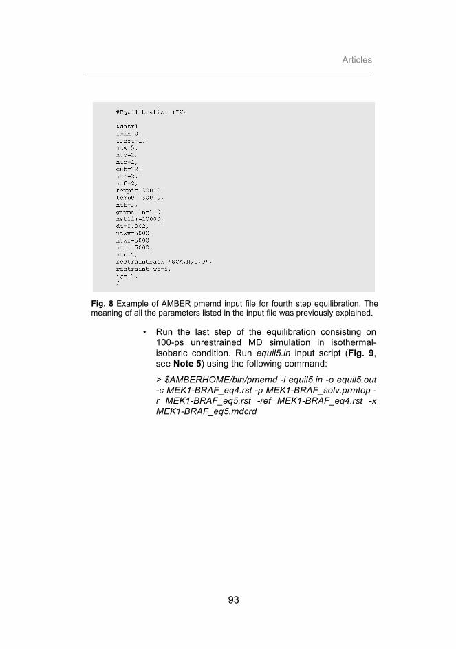
Articles
93
Fig. 8 Example of AMBER pmemd input file for fourth step equilibration. The meaning of all the parameters listed in the input file was previously explained.
• Run the last step of the equilibration consisting on 100-ps unrestrained MD simulation in isothermal-isobaric condition. Run equil5.in input script (Fig. 9, see Note 5) using the following command:
> $AMBERHOME/bin/pmemd -i equil5.in -o equil5.out -c MEK1-BRAF_eq4.rst -p MEK1-BRAF_solv.prmtop -r MEK1-BRAF_eq5.rst -ref MEK1-BRAF_eq4.rst -x MEK1-BRAF_eq5.mdcrd

Advances and new challenges in modeling of protein interactions
94
Fig. 9 Example of AMBER pmemd input file for fifth step equilibration. In the input file, the flag ntr=0 indicates that the positional restraint method is turned off. The meaning of the rest of the parameters listed in the input file was previously explained.
f) Finally, perform 5-ns MD unrestrained simulation keeping the same system condition as the last equilibration step. Run prod.in input script (Fig. 10, see Note 6) using the following command:
> $AMBERHOME/bin/pmemd -i prod.in -o prod.out -c MEK1-BRAF_eq5.rst -p MEK1-BRAF_solv.prmtop -r MEK1-BRAF_prod.rst -ref MEK1-BRAF_eq5.rst -x MEK1-BRAF_prod.mdcrd

Articles
95
Fig. 10 Example of AMBER pmemd input file for unrestrained MD. The meaning of all the parameters listed in the input file was previously explained.
3. Binding free energy calculation
a) Build the topology and coordinate files of the unsolvated wild type (WT) structure for both the complex and its single subunits using tleap-WT.in input file (Fig. 11). Run LEaP AMBER tool using the following command:
> $AMBERHOME/bin/tleap -f tleap-WT.in > tleap-WT.out

Advances and new challenges in modeling of protein interactions
96
Fig. 11 Example of AMBER LEaP input file to build topology and coordinates files of wild type dry systems.
b) For each mutation studied, build the topology and coordinate files of the mutated structure for both the complex and mutated subunit using tleap-mut.in input file (Fig. 12). Run LEaP AMBER tool using the following command:
> $AMBERHOME/bin/tleap -f tleap-mut.in > tleap-mut.out
Fig. 12 Example of AMBER LEaP input file to build topology and coordinates files of mutated dry systems. Here, F468 BRAF residue is taken as example.
c) Perform alanine scanning calculation on 200 snapshots extracted from the last 2 ns of each MD trajectory. Run mmpbsa.in input file for MMPBSA.py script in AMBER14 (Fig. 13) using the following command:
> $AMBERHOME/bin/MMPBSA.py -i mmpbsa.in -sp MEK1-BRAF_solv.prmtop -cp MEK1-BRAF.prmtop -rp

Articles
97
MEK1-BRAF.prmtop -lp MEK1-BRAF.prmtop -y MEK1-BRAF_prod.mdcrd -mc MEK1-BRAF_F468A.prmtop -ml BRAF_F468A.prmtop
Flag -i specifies the input file, -sp the solvated WT complex topology file, -cp the unsolvated WT complex topology file, -rp the unsolvated WT receptor topology file, -lp the unsolvated WT ligand topology file, -y the complex trajectory file to analyze, -mc the unsolvent mutated complex topology file and -ml the unsolvated mutated subunit topology file. Please, be aware that as MEK1 is the first molecule in the complex, for alanine scanning calculations the unsolvated mutated subunit topology file will be specified with the flag -mr.
Fig. 13 Example of MMPBSA.py input file to perform alanine scanning calculation. The input file is typically divided into four sections (&general, &gb, &pb, &alanine_scanning). The &general section is designed to specify generic variables related to the overall calculation. For instance, the flag startframe and endframe specifies the frame from which to begin and to stop extracting snapshots respectively, the parameter interval indicates the offset from which to choose frames from the trajectory file, verbose=1 means that complex, ligand and receptor energy terms will be printed in the output file. The &gb and &pb section markers tells the script to perform MM-GBSA and MM-PBSA calculations with the given values defined within those sections (i.e., the variables saltcon and istrng that specify the salt concentration and the ionic strength, respectively). Finally the &alanine_scanning section marker initializes alanine scanning in the script. Please be aware that given the higher computational costs of MM-PBSA calculation, only MM-GBSA calculation is performed in this work.
d) Extract the ΔΔG of binding related to the specific mutations estimated as the difference between the binding ΔG of the WT and that of the mutated complex. All these data are easily available in the final output file,

Advances and new challenges in modeling of protein interactions
98
FINAL_RESULTS_MMPBSA.dat, including all the wild type and mutated system average binding energies (reported as van der Waals, electrostatic and non polar energy contributions), as shown in Fig. 14.
Fig. 14 Extract from the MMPBSA.py FINAL_RESULTS_MMPBSA.dat output file. The file includes all the average energies, standard deviations, and standard error of the mean for GB followed by PB calculations (if calculated). After each section, the ΔG of binding is given along with the error values. After each method, the ΔΔG of binding is reported, corresponding to the relative effect the mutation has on the ΔG of binding for the complex. The specific mutation is also printed at the end of the file. Here, F468 residue alanine scanning is taken as example.
e) You may get a graphical representation of the ΔΔG of binding (Fig. 1), by assigning the values given in FINAL_RESULTS_MMPBSA.dat file to the bfactor field of the corresponding PDB file of the complex structure.

Articles
99
Notes 1) As there is no unbound structure for the ligand yet, the ligand
structure contained on the complex PDB file (4MNE) is used here instead for illustration purposes. However, in a standard NIP computation, unbound structures should be used.
2) The principal columns of the 4mne.ene file are:
• Conf: Conformation number of the docking pose as in the last column of the rot file.
• Ele: Electrostatic energy of the pose.
• Desolv: Desolvation energy of the pose.
• VDW: Van der Waals energy of the pose.
• Total: Total docking energy of the pose, computed as ele + Desolv + 0.1 * VDW (note a 0.1 weight for VDW).
• RANK: Pose rank according to its computed total binding energy.
3) Files from the PDB may contain bound ligands, cofactors or non-standard residues whose parameters are not available in the AMBER parameters database. In this case you should make use of the Antechamber tools, which ship with AmberTools, to create PREP and FRCMOD files. For more information, see the ANTECHAMBER tutorial (http://ambermd.org/tutorials/basic/tutorial4b/) and the AMBER manual.
4) LEaP AMBER tool renumbers PDB residues starting from 1. Thus, the original numeration of your PDB file may not be always kept.
5) Since your system may not start from an equilibrium state, additional time steps may be required during the minimization and equilibration steps of the MD simulation. One can check for equilibrium by verifying whether properties, such as potential energy, temperature or pressure, no longer change in any systematic fashion and are just fluctuating around a mean value.
6) To guarantee reliable results in the in silico Alanine scanning calculation, RMSD simulation should be highly equilibrated. Ideally one should probably run a much longer production run than 5ns (e.g., 100 ns).

Advances and new challenges in modeling of protein interactions
100
References 1. Arkin M.R.,Wells J.A. (2004) Small-molecule inhibitors of
protein-protein interactions: progressing towards the dream. Nat Rev Drug Discov 3, 301-317.
2. DeLano W.L. (2002) Unraveling hot spots in binding interfaces: progress and challenges. Curr Opin Struct Biol 12, 14-20.
3. Toogood P.L. (2002) Inhibition of protein-protein association by small molecules: approaches and progress. J Med Chem 45, 1543-1558.
4. Glaser F., Pupko T., Paz I. et al.(2003) ConSurf: identification of functional regions in proteins by surface-mapping of phylogenetic information. Bioinformatics 19, 163-164.
5. Ashkenazy H., Erez E., Martz E. et al. (2010) ConSurf 2010: calculating evolutionary conservation in sequence and structure of proteins and nucleic acids. Nucleic Acids Res 38, W529-533.
6. Landau M., Mayrose I., Rosenberg Y. et al. (2005) ConSurf 2005: the projection of evolutionary conservation scores of residues on protein structures. Nucleic Acids Res 33, W299-302.
7. Celniker G., Nimrod G., Ashkenazy H. et al. (2013) ConSurf: Using Evolutionary Data to Raise Testable Hypotheses about Protein Function. . Isr. J. Chem. 53, 199–206.
8. Grosdidier S., Fernandez-Recio J. (2008) Identification of hot-spot residues in protein-protein interactions by computational docking. BMC Bioinformatics 9, 447.
9. Branden C.I., Tooze J. (1999) Introduction to protein structure. 2 ed. Garland Pub., New York.
10. Cheng T.M., Blundell T.L., Fernandez-Recio J. (2007) pyDock: electrostatics and desolvation for effective scoring of rigid-body protein-protein docking. Proteins 68, 503-515.
11. Case D.A., Cheatham T.E., Darden T. et al. (2005) The Amber biomolecular simulation programs. J Comput Chem 26, 1668-1688.
12. Miller B.R.I., McGee D.T.J., Swails J.M., et al (2012) MMPBSA.py: An Efficient Program for End-State Free Energy Calculations. J. Chem. Theory Comput. 8, 3314–3321.
13. Haling J.R., Sudhamsu J., Yen I., et al. (2014) Structure of the BRAF-MEK complex reveals a kinase activity independent role for BRAF in MAPK signaling. Cancer Cell 26, 402-413.
14. Kiel C., Serrano L. (2014) Structure-energy-based predictions and network modelling of RASopathy and cancer missense mutations. Mol Syst Biol 10, 727.

Articles
101
15. Jimenez-Garcia B., Pons C., Fernandez-Recio J. (2013) pyDockWEB: a web server for rigid-body protein-protein docking using electrostatics and desolvation scoring. Bioinformatics 29, 1698-1699.
16. Gabb H.A., Jackson R.M., Sternberg M.J. (1997) Modelling protein docking using shape complementarity, electrostatics and biochemical information. J Mol Biol 272, 106-120.
17. Pettersen E.F., Goddard T.D., Huang C.C. et al. (2004) UCSF Chimera--a visualization system for exploratory research and analysis. J Comput Chem 25, 1605-1612.


Articles
103
3.1.2. Expanding the frontiers of protein-protein modeling: from docking and scoring to binding affinity predictions and other challenges.
Chiara Pallara,1# Brian Jiménez-García,1# Laura Perez-Cano,1 Miguel
Romero,1 Albert Solernou,1 Solène Grosdidier,1 Carles Pons,1,2 Iain
H. Moal,1 Juan Fernandez-Recio1*
1Joint BSC-IRB Research Programme in Computational Biology,
Barcelona Supercomputing Center, Barcelona, Spain
2Computational Bioinformatics, National Institute of Bioinformatics
(INB), Barcelona, Spain
# Equal contribution
* Corresponding author


proteinsSTRUCTURE O FUNCTION O BIOINFORMATICS
Expanding the frontiers of protein–proteinmodeling: From docking and scoring tobinding affinity predictions and otherchallengesChiara Pallara,1 Brian Jim!enez-Garc!ıa,1 Laura P!erez-Cano,1 Miguel Romero-Durana,1
Albert Solernou,1 Solene Grosdidier,1 Carles Pons,1,2 Iain H. Moal,1 andJuan Fernandez-Recio1*1 Joint BSC-IRB Research Programme in Computational Biology, Barcelona Supercomputing Center, Barcelona, Spain
2 Computational Bioinformatics, National Institute of Bioinformatics (INB), Barcelona, Spain
ABSTRACT
In addition to protein–protein docking, this CAPRI edition included new challenges, like protein–water and protein–sugarinteractions, or the prediction of binding affinities and DDG changes upon mutation. Regarding the standard protein–proteindocking cases, our approach, mostly based on the pyDock scheme, submitted correct models as predictors and as scorers for67% and 57% of the evaluated targets, respectively. In this edition, available information on known interface residues hardlymade any difference for our predictions. In one of the targets, the inclusion of available experimental small-angle X-rayscattering (SAXS) data using our pyDockSAXS approach slightly improved the predictions. In addition to the standardprotein–protein docking assessment, new challenges were proposed. One of the new problems was predicting the position ofthe interface water molecules, for which we submitted models with 20% and 43% of the water-mediated native contactspredicted as predictors and scorers, respectively. Another new problem was the prediction of protein–carbohydrate binding,where our submitted model was very close to being acceptable. A set of targets were related to the prediction of bindingaffinities, in which our pyDock scheme was able to discriminate between natural and designed complexes with area under thecurve 5 83%. It was also proposed to estimate the effect of point mutations on binding affinity. Our approach, based onmachine learning methods, showed high rates of correctly classified mutations for all cases. The overall results were highlyrewarding, and show that the field is ready to move forward and face new interesting challenges in interactomics.
Proteins 2013; 81:2192–2200.VC 2013 Wiley Periodicals, Inc.
Key words: complex structure; CAPRI; protein–protein docking; pyDock; protein–carbohydrate interactions.
INTRODUCTION
One of the major challenges in structural biology is toprovide structural data for all complexes formed betweenproteins and other macromolecules. Current structuralcoverage of protein–protein interactions (i.e., availableexperimental structures plus potential models based onhomologous complex structures) is below 4% of the esti-mated number of possible complexes formed betweenhuman proteins.1,2 The pace of experimental determina-tion of complex structures is still behind the determina-tion of individual protein structures. In addition, manyof these interactions will never be determined by X-raycrystallography because of their transient nature. For thesereasons, computational docking methods aim to become a
complementary approach to solve the structural interactome.The field of protein docking has experienced an explosionin recent years, partially propelled by the CAPRI experi-ment. Past editions showed an increasing amount of par-ticipant groups and computational approaches, and a
Grant sponsor: Spanish Ministry of Science; Grant number: BIO2010-22324; Grantsponsor: FPU (to LP-C) and FPI (to BJ-G) fellowships from the Spanish Ministryof Science; Grant sponsor: People Programme (Marie Curie Actions) of the Euro-pean Union’s Seventh Framework Programme (FP7/2007-2013) under REA (toIHM); Grant number: PIEF-GA-2012-327899.Pallara and Jim!enez-Garc!ıa contributed equally to this work.*Correspondence to: Juan Fernandez-Recio, Barcelona Supercomputing Center(BSC), Jordi Girona 29, 08034 Barcelona, Spain. E-mail: [email protected] 14 June 2013; Revised 31 July 2013; Accepted 1 August 2013Published online 12 August 2013 in Wiley Online Library (wileyonlinelibrary.com).DOI: 10.1002/prot.24387
2192 PROTEINS VVC 2013 WILEY PERIODICALS, INC.

large variety of targets. We have participated in all targetsof this fifth CAPRI edition. In addition to the standardprediction of protein–protein targets, this edition hasentered into related areas including binding affinity pre-dictions and free energy changes upon mutation, as wellas prediction of sugar binding and interface water mole-cules. Our overall experience has been highly rewardingand we describe here the details of our participation andthe key factors of our success.
MATERIALS AND METHODS
Generation of rigid-body docking poses forthe predicting experiment
In all targets, we used FTDock3 with electrostatics and0.7 A grid resolution and ZDOCK 2.14 to generate10,000 and 2000 rigid-body docking poses, respectively,as previously described.5 For the final four targets of thisedition (T53, T54, T57, and T58) we generated an addi-tional pool of flexible docking poses using SwarmDock.For these runs, the standard protocol was employed,6–8
with the Dcomplex score used as the objective function,9
but without the final clustering and rescoring phase. InT46 we generated an additional pool of 10,000 solutionsusing FTDock without electrostatics and at lower resolu-tion (1.2 A), as part of an old protocol used with previ-ous targets, but these conditions were not applied for therest of the targets since we saw in the past that this stepwas not increasing the chances of correct predictions. InT46 and T47, we used RotBUS10 to generate 59,112 and41,021 additional docking poses, respectively, but thismethod was not used for the rest of the targets since wepreviously checked that this procedure did not improvethe results. In Target T50, given the large size of 1918H1N1 influenza virus hemagglutinin protein, we gener-ated a total of 92,432 FTDock docking poses, increasingthe number of translations selected from each rotationfrom 3 (default) to 10. Cofactors, water molecules andsolvent ions were not included in our dockingcalculations.
Scoring of rigid-body docking poses for boththe predicting and the scoring experiments
We scored the docking models generated by the abovedescribed methods with our pyDock protocol,11 basedon energy terms previously optimized for rigid-bodydocking. The binding energy is basically composed ofaccessible surface area-based desolvation, Coulombicelectrostatics and van der Waals energy (with a weightingfactor of 0.1 to reduce the noise of the scoring function).Electrostatics and van der Waals were limited to 61.0and 1.0 kcal/mol for each interatomic energy value,respectively, to avoid excessive penalization from possibleclashes in the structures generated by the rigid-body
approach. The same protocol was used in the scoringexperiment to score all the docking models that wereproposed. We did not include van der Waals in the T46scoring experiment, although this did not affect theresults. Cofactors, water molecules and solvent ions werenot considered for scoring.
Removal of redundant docking poses
After scoring, we eliminated redundant predictions toincrease the variability of the predictions and maximizethe success chances using a simple clustering algorithmwith a distance cutoff of 4.0 A, as previously described.12
In target T47, since the resulting solutions looked correct[according to the available structure of a highly homolo-gous complex with protein data bank (PDB) code2WPT], we reduced this cutoff to 0.5 A.
Minimization of final models
The final 10 selected docking poses were minimized toimprove the quality of the docking models and reducethe number of interatomic clashes. In the majority of thetargets we used TINKER13 as previously described.12,14
In targets T53 and T54 we used CHARMM (50 stepsconjugate gradient, 500 steps adopted-basis Newton–Raphson and 50 steps steepest decent, with theCHARMM19 force field).15 In target T58 we usedAMBER10 with AMBER parm99 force field.16,17 Theminimization protocol consisted of a 500-cycle steepestdescent minimization with harmonic restraints applied ata force constant of 25 kcal/(mol!A2) to all the backboneatoms to optimize the side chains, followed by another500-cycle conjugate gradient minimization withoutrestraints. This minimization step was performed afterranking, solely to remove clashes.
Modeling of subunits with no availablestructure
For several targets, the structures of the subunitswere not available and needed to be modeled. In mostof the targets, we used Modeler 9v6 with default param-eters18 based on the template/s suggested by the organ-izers or on other homologue proteins found byBLAST19 search. The final selected model was that withthe lowest DOPE score.20 For targets T53 and T54 weused POPULUS (http://bmm.cancerresearchuk.org/~pop-ulus/) with default template selection and model buildingsettings.21
RESULTS AND DISCUSSION
In this CAPRI edition we submitted predictions for allthe proposed targets. Our results for the standardprotein–protein docking assessment are summarized inTable I. In addition, there were new challenges like the
Expanding Protein–Protein Docking Frontiers
PROTEINS 2193

prediction of protein–water and protein–sugar interac-tions, as well as the estimation of binding affinities andenergy changes upon mutation. Hereinafter, we describein detail our submissions for each of the targets.
Standard protein–protein dockingassessment: successful predictions
Target T47 (model/pseudounbound)
Target T47 was the structural prediction of the com-plex between the DNase domain of colicin E2 and theimmunity protein Im2. The real challenge in this targetwas the prediction of interface water molecules, however,the protein–protein docking predictions were alreadyassessed, and therefore we have included them in thissection. The colicin E2 was modeled based on the struc-ture of colicin E9 (85% sequence identity) in complexwith Im9 immunity protein (PDB 1EMV).22 The coordi-nates of the immunity protein Im2 were extracted fromits structure in complex with colicin E9 (PDB 2WPT).Given the existence of this homologous colicin E9/Im2complex structure (PDB 2WPT),23 the binding mode fortarget T47 was easy to determine by template-baseddocking. However, we performed the template-free dock-ing calculations to assess the automatic docking protocol.We only applied distance restraints after pyDock protocolby selecting those docking poses in which two key con-tacting residues, Im2 Y54 and colicin E2 F85 (equivalentto colicin E9 F86 in 2WPT),23 were within an arbitrarydistance of 6 A (same distance used in standard restraintswith pyDockRST module.24 We submitted five correctmodels (one high accuracy, one medium accuracy, andthree acceptable). Our first submitted model (Rank 1according to pyDock energy, and generated by ZDOCK),was a high-quality model (Table I), with 75% native
contacts, 2.48 A ligand root mean square deviation(RMSD), and 0.75 A interface RMSD with respect to thecrystal structure (Fig. 1; PDB 3U43).25 This docking modelhad the lowest ligand RMSD with respect to the homolo-gous colicin E9/Im2 complex (PDB 2WPT) amongst allsolutions (although we did not use this homologous struc-ture for docking), and even more interestingly, we wouldhave obtained exactly the same result without applying theabove-mentioned distance restraint filter.
For the scoring experiment, we evaluated the provided1051 models with our pyDock scoring function, andapplied the same distance filter that we used as predic-tors (see above). All our submitted predictions resultedto be successful, consisting of six medium and four high-quality models. We had a high-accuracy model rankedsecond after pyDock scoring and distance filter(uploaded by Weng), with 77% native contacts, 0.9 Aligand RMSD, and 0.4 A interface RMSD with respect tothe crystal structure (PDB 3U4325; Table I; Fig. 1). Inter-estingly, our Rank 5 model was the best model submittedamong all 14 participants, with 79% native contacts,0.7 A ligand RMSD, and 0.5 A interface RMSD. Two bet-ter models uploaded by Weng were not found by any ofthe participants. Remarkably, as in predictors, our resultswould not have changed had we not applied the distancerestraints filter.
Target T48 (unbound/unbound)
Target T48 was the structural prediction of thecomplex between the diiron-hydroxylase toluene4-monooxygenase and the Rieske-type ferredoxin T4moCprotein (PDB 1VM9).26 As suggested by the organizers,the heterohexameric biological unit of the diiron-hydroxylase was built by applying crystal symmetry oper-ations to its trimeric structure in complex with the
Table IResults of Our pyDock Protocol for All Protein–Protein Targets of the Last CAPRI Edition
Target Type
Predictors Scorers
Submission ranka Qualityb Successful groupsc Submission ranka Qualityb Successful groupsc
T46 HH — — 2 (40) — — 8 (16)T47 HU 1 *** 25 (29) 2d *** 13 (14)T48 UU 3 * 14 (32) No scorers No scorers No scorersT49 UU 4 * 14 (33) 6 * 7 (13)T50 UH 1 ** 18 (40) 4 ** 12 (17)T51 DHD — — 3 (46) — — 5 (13)T53 UH 3e ** 20 (42) 1 ** 11 (13)T54 UH — — 4 (41) — — 0 (13)T58 UU 5 ** 11 (23) No scorers No scorers No scorers
U, unbound; H, homology-based model; D, domain.aRank of the best model within our submission to CAPRI.bQuality of our best model according to CAPRI criteria.cNumber of successful groups for each target; in brackets, total number of participants.dModel Rank 1 had medium accuracy (**).eModel Rank 1 had acceptable accuracy (*).
C. Pallara et al.
2194 PROTEINS

T4moD effector protein (PDB 3DHH).27 We used thehexameric construct for the generation of docking poses,which were scored by pyDock. Then, we selected thosedocking poses that had any of the diiron-hydroxylaseFe21 and ferredoxin S2Fe2 atoms within 23 A distance toallow for the electron transfer between these groups27
(the distance cutoff we used was arbitrary, based on theexpected distance of 16 A in 3DHH plus a margin to
allow the inclusion of some low-energy solutions). Forthe submission, we removed chains D, E, and F from thehexamer as we misinterpreted some of the organizers’instructions, but this did not affect the quality of thesubmitted models. The analysis of the results showedthat we submitted three models of acceptable quality.Our prediction ranked third after pyDock scoring andelectron transfer distance filtering (generated by FTDock)
Figure 1Representation of our best models for targets T47, T48, T49, T50, T53, T57, and T58. For each target, receptors are superimposed and shown inwhite. Ligand in our best model as predictors is shown in red, and as scorers in blue. For comparison, the structure of the experimental complex(if available) is represented in green.
Expanding Protein–Protein Docking Frontiers
PROTEINS 2195

had 14% native contacts, 8.4 A ligand RMSD, and 3.6 Ainterface RMSD with respect to the complex crystalstructure (not yet available). We found another accepta-ble model (ranked 10th in our submission set) that had49% native contacts, 6.3 A ligand RMSD, and 2.2 Ainterface RMSD with respect to the complex crystalstructure.
Target 49 (unbound/unbound)
Target T49 was the same complex as T48 but with adifferent hexameric conformation for diiron-hydroxylasetoluene 4-monooxygenase (unbound coordinates notreleased). We applied the same protocol as for target T48(pyDock scoring and electron transfer distance filtering).We submitted four acceptable quality models. The modelranked fourth of our submission set had acceptable qual-ity, with 26% native contacts, 12.4 A ligand RMSD, and3.5 A interface RMSD with respect to the complex crystalstructure (not yet available). We also submitted anothermodel with 11% native contacts, 6.9 A ligand RMSD,and 2.7 A interface RMSD.
For the scoring experiment, the 1085 solutions werescored by the same protocol, based on pyDock scoringand electron transfer distance filtering. In some models,the monooxygenase was uploaded as a trimer, thereforewe reconstructed the biological hexamer (based on sym-metry) to calculate the electron transfer distance filter.Since it was not clear whether in these cases the hexamerwas going to be rebuilt for the assessment, our submis-sion set was formed by the top five solutions obtainedafter rebuilding the hexamer, and by the top five solu-tions obtained by just considering the structure submit-ted by uploaders (i.e., without rebuilding the hexamer incases of uploaded trimer). Our ranked sixth submissionwas an acceptable model (uploaded by Nakamura), with11% native contacts, 7.9 A ligand RMSD, and 2.9A inter-face RMSD with respect to the complex crystal structure(not yet available).
Target 50 (unbound/model)
Target T50 was the structural prediction of the com-plex between the 1918 H1N1 influenza virus hemaggluti-nin and the HB36.3 de novo designed protein. Thecoordinates of the hemagglutinin were taken from itsstructure in complex with an antibody (PDB 3GBN)28
and the biological hexamer was rebuilt by applying sym-metry operations. We modeled the HB36.3 based on thecrystal structure of the homologous (83% sequence iden-tity) protein APC36109 from Bacillus stearothermophilus(PDB 1U84), using the target-template protein alignmentoffered by the organizers. Given the size of the system,we increased the number of rigid-body docking solutionsgenerated by FTDock (see Materials and Methods sec-tion). Our submission as predictors contained nine suc-cessful models (five acceptable and four medium-quality
solutions). Our Rank 1 submission (generated byFTDock) was a medium-quality model with 47% nativecontacts, 6.1 A ligand RMSD, and 1.8 A interface RMSDwith respect to the complex crystal structure (Fig. 1;PDB 3R2X).29 Interestingly, our Rank 4 submission,with 41% native contacts, 3.4 A ligand RMSD, and 1.6 Ainterface RMSD, was the best model submitted amongall participants as predictors.
For the scoring experiment, we evaluated the 1451models in the same conditions as in predictors. Wefound five acceptable and one medium-quality solutions.Our Rank 4 submission was a medium-quality model,with 44.9% native contacts, 4.71 A ligand RMSD, and1.93 A interface RMSD with respect to the complex crys-tal structure (PDB 3R2X29; Fig. 1).
Target T53 (unbound/model)
Target T53 was a complex between two artificial alphahelicoidal repeat proteins (alpha-Rep), alpha-rep4 (PDB3LTJ)30 and alpha-rep2, both designed on the basis ofthermostable HEAT-like repeats. The ligand alpha-rep2was built using as template alpha-rep4 (PDB 3LTJ), with77% sequence identity. All the docking poses, generatedusing Zdock, Ftdock, and SwarmDock, were scored bypyDock. We submitted four successful predictions (threeacceptable and one medium-quality models). Our Rank3 submission, a medium accuracy model generated bySwarmDock, had 44% native contacts, ligand RMSD 4.4A, and interface RMSD 1.8 A with respect to the crystalstructure (not yet available).
For the scoring experiment, we evaluated 1400 alpha-rep4/alpha-rep2 complex models applying the same pro-tocol as in predictors in a completely automated fashion.We found three acceptable and a medium-quality mod-els. Our Rank 1 submission, a medium-quality model(uploaded by Yan Shen), had 62% native contacts, 3.6 Aligand RMSD, and 1.3 A interface RMSD with respect tothe complex crystal structure (not yet available).
Target T58 (unbound/unbound)
This target was a complex between the unboundG-Type Lysozyme (PDB 3MGW)31 and the unboundEscherichia coli Plig lysozyme inhibitor (PDB 4DY3).32
There was available small-angle X-ray scattering (SAXS)data for this complex, which we used for scoring withour module pyDockSAXS, previously developed to com-bine pyDock scoring and fitting to SAXS data.33 Inaddition, there was some available information indicatinga central role of the G-type lysozyme E73, D86, and D97residues and the E. coli Plig lysozyme inhibitor R119 andY47 residues.34 Based on these residues, we imposedambiguous distance restraints with our modulepyDockRST.35 We submitted one medium-accuracy andtwo acceptable models. Our Rank 5 model, generated bySwarmDock, was a medium-quality model, and resulted
C. Pallara et al.
2196 PROTEINS

to be the fourth best model submitted among all the 23participants, with 43% native contacts, 4.9 A ligandRMSD, and 1.8 A interface RMSD with respect to thecomplex crystal structure (PDB 4G9S).36 Interestingly,although the distance restraints proved to be essential forthis target, we would have obtained only slightly worseresults without using the SAXS data (Rank 10 mediumaccuracy model). This is probably due to the shape ofthe complex, classified as spherical according to the ani-sotropy value (1.4) computed from the ratio between thesize of the largest axis and the smallest ones. Indeed, wepreviously showed that SAXS data does not providemuch beneficial information in this type of cases.33
Protein–protein docking: unsuccessful cases
In three of the protein–protein cases (T46, T51, andT54) we were not able to submit any correct model,either as predictors or as scorers. These cases seemed tobe highly difficult for the majority of participants, sincein all of them there were no more than three successfulgroups as predictors or as scorers or both (Table I). Intarget T46 (model/model), the interacting subunits Mtq2and Trm112 were modeled based on the homologue tem-plates with low sequence identity (Mtq2 was based ontemplate with PDB code 1T43, 28% sequence identity;Trm112 was based on template with PDB code 2J6A,36% sequence identity). The inaccuracies in the model-ing added too much error and the docking was not suc-cessful. Target T51 (bound/model/unbound) was adifficult case of a multidomain protein, with interactionsbetween GH5-CBM6/CBM13/Fn3 domains. This couldbe divided in two different docking cases both involvingCBM13 domain, which needed to be modeled based ontemplate with PDB code 1KNL (38% sequence identity).Again, a model based on a template with that level ofhomology can deteriorate docking results. Target 54(unbound/model) was in principle easy, involving themodeling of Rep16 based on the template with PDBcode 3LTJ (88% sequence identity), but the submittedsolutions were incorrect for us as well as for the majorityof participants. Indeed, despite the scoring set containedseveral acceptable models, no group was able to identifythem (Table I).
Prediction of protein–water interactions
Target T47 was the prediction of a protein–proteincomplex structure, as described in above sections, butthe real challenge was to predict the location of watermolecules. After generating the protein–protein dockingposes as above described, we predicted the water posi-tions in each docking model using DOWSER37 withdefault parameters (with a probe radius of 0.2A and thedefault atoms dictionary). Our Rank 1 submitted model(generated by ZDOCK) had 20% of water native
contacts, and was classified as fair (1). If we consideronly the prediction of the buried water molecules, oursuccess rates do not significantly change.
For the scoring experiment, we just applied our stand-ard pyDock scoring function, plus distance restraints asdescribed in above sections. The water molecules pro-posed in the different docking poses were not includedin the scoring. Our Rank 8 submitted model (uploadedby Bates) had 43% of water native contacts and was clas-sified as good (11). More details can be found in anupcoming publication.
Prediction of protein–carbohydrate complexstructure
Target 57 (unbound/model) was a challenging targetconsisting in the prediction of the interaction betweenBT4661 protein and heparin. The structure of heparin inthe complex was not known, so we modeled it usingmolecular dynamics starting with the provided confor-mation. We ran 10 ns using the force field AMBERparm99 of the Amber10 package16,17 and extracted1000 representative snapshots. Since our pyDock proto-col was not intended for protein–sugar interactions, wehad to devise a new ad hoc docking procedure. For that,we used FTDock to dock each of the 1000 heparin con-formations to BT4661 protein. We selected the top10,000 docking poses as scored by FTDock (no electro-statics). Then we applied different scoring functions tothis set of docking poses: (i) PScore without minimiza-tion; (ii) PScore with minimization; and (iii) AMBERafter minimization. We selected the 1000 best-scoringsolutions from each method and finally we removedredundant solutions within 6.5 A ligand RMSD. No cor-rect submission was submitted. However, our Rank 4submission was almost acceptable, with 65% native con-tacts, 11.2 A ligand RMSD, and 4.3 A interface RMSDwith respect to the complex crystal structure (PDB4AK2; Fig. 1). We checked a posteriori that there wereseveral correct models within our docking sets, but ourscoring approach failed to place them in the lowest scor-ing positions.
Other challenges: binding affinity and DDGpredictions
This CAPRI edition also involved the challengingproblem of predicting binding affinities and energychanges upon mutations. Round 21 was the discrimina-tion between 87 designed protein–protein interactionsinvolving three proteins of interest (Spanish influenzaHA; Mt ACP-2; Fc region of human IgG1) and 120 natu-rally occurring complexes. The pyDock function,although initially developed for the scoring of dockingposes, was previously shown to have some correlationwith the binding affinity data collected by Kastritis andBonvin.38 This was later confirmed on a subset of
Expanding Protein–Protein Docking Frontiers
PROTEINS 2197

complexes with high-confidence affinity data, wherepyDock ranked among the best performing scoring func-tions with a correlation of 0.63.39 For round 21 predic-tions, we evaluated the correlation of each of thedifferent pyDock individual terms with the binding affin-ities on the provided set of 120 naturally occurring com-plexes. We found that desolvation correlation withbinding affinity data was not clear, showing even negativecorrelation with data obtained by ITC experiments. Itseems that, although desolvation is essential for rigid-body docking (perhaps to compensate inaccurate calcula-tion of electrostatics and van der Waals), it is not themost important factor for binding affinity predictionsfrom the complex structure (in which electrostatics canbe more accurately calculated). Based on these results,we devised a binding affinity descriptor (pyDockAFF 5electrostatics 21.0 3 desolvation), with confidencethresholds for the discrimination of complexes accordingto their binding affinities. Our predictions had areaunder the curve 83%, with good discrimination betweendesigned and native interfaces. More details can be foundin a recent publication.40 It remains to be seen whetherthe pyDockAFF binding affinity predictor is suitable onlyfor the cases in this CAPRI round, or it has more generalapplicability (further details in an upcoming publication).
Targets T55-56 aimed to predict the binding affinitychanges upon mutations on two designed influenzahemagglutinin protein binders. We applied a multipara-metric predictive model with 85 descriptors using anensemble of models which were combined to produceconsensus predictions. The models were trained upon adata set of 930 changes in affinity upon mutation whichwere taken from the literature. Due to the fairly lowcases to descriptors ratio (10.9), we preferentiallyemployed models with inherent overfitting avoidancebias, such as prepruning or feature selection using theAkaike information criterion, methods which constructmultiple models using subsets of the descriptors and thetraining data, and by rejecting learners that performedpoorly using leave-complex-out cross-validation.41 Tofurther avoid overfitting, we did not combine theselected learners together using stacking, instead optingfor the unweighted mean for our consensus predictions.This approach provided an excellent ability to predictthe effect of mutation, more details of which can befound in a recent publication.42 We have since expandedthis data set to form the SKEMPI database, which nowincludes 3047 DDG values, as well as kinetic and thermo-dynamic data,43 and have used the data to derive contactpotentials that can circumvent some of the approxima-tions associated with statistical potentials.44
CONCLUSIONS
We have continued our participation in CAPRI withpyDock, submitting models for all the predicting, scoring,
and binding affinity prediction experiments. For the gener-ation of docking poses, the better grid resolution used forFTDock and the use of flexible SwarmDock for the lasttargets were key for the success. This produced dockingposes of sufficient quality to be identified by thepyDockSER scoring scheme. In selected targets, distancerestraints were introduced by pyDockRST, but in mostcases this did not make a difference. In one target, SAXSdata was used for complementary scoring with pyDock-SAXS, which slightly improved the scoring. We obtainedconsistently good models for all nondifficult cases,although they were far from being trivial, since their subu-nits were unbound or needed to be modeled based onhomology templates. In all cases but one our successfulmodels were ranked within our first five submitted solu-tions, being ranked first in several cases. In this CAPRIedition we learned that our automated protocol is usefulto provide correct models in easy-to-medium difficultyprotein–protein docking cases, but we need further meth-odological development for difficult cases, especially whensubunits need to be modeled based on homologues withlow sequence identity. On the other side, interface waterplacement and sugar-binding proved to be highly challeng-ing for our protein–protein methodology, but the resultshave encouraged us to develop new methods for theseproblems. Finally, prediction of binding affinity based onthe pyDockSER scoring, and energy changes upon muta-tion based on multiparametric regression models showedexcellent results. The overall experience has been highlyrewarding and has shown once again the importance ofcommunity-wide assessment of prediction methods.
REFERENCES
1. Venkatesan K, Rual JF, Vazquez A, Stelzl U, Lemmens I, Hirozane-Kishikawa T, Hao T, Zenkner M, Xin X, Goh KI, Yildirim MA,Simonis N, Heinzmann K, Gebreab F, Sahalie JM, Cevik S, SimonC, de Smet AS, Dann E, Smolyar A, Vinayagam A, Yu H, Szeto D,Borick H, Dricot A, Klitgord N, Murray RR, Lin C, Lalowski M,Timm J, Rau K, Boone C, Braun P, Cusick ME, Roth FP, Hill DE,Tavernier J, Wanker EE, Barabasi AL, Vidal M. An empirical frame-work for binary interactome mapping. Nat Methods 2009;6:83-90.
2. Stumpf MP, Thorne T, de Silva E, Stewart R, An HJ, Lappe M,Wiuf C. Estimating the size of the human interactome. Proc NatlAcad Sci USA 2008;105:6959-3964.
3. Gabb HA, Jackson RM, Sternberg MJ. Modelling protein dockingusing shape complementarity, electrostatics and biochemical infor-mation. J Mol Biol 1997;272:106-120.
4. Chen R, Weng Z. A novel shape complementarity scoring functionfor protein–protein docking. Proteins 2003;51:397-408.
5. Grosdidier S, Pons C, Solernou A, Fernandez-Recio J. Prediction andscoring of docking poses with pyDock. Proteins 2007;69:852-858.
6. Moal IH, Bates PA. SwarmDock and the use of normal modes inprotein–protein docking. Int J Mol Sci 2010;11:3623-3648.
7. Torchala M, Moal IH, Chaleil RA, Fernandez-Recio J, Bates PA.SwarmDock: a server for flexible protein–protein docking. Bioinfor-matics 2013;29:807-809.
8. Li X, Moal IH, Bates PA. Detection and refinement of encountercomplexes for protein–protein docking: taking account of macro-molecular crowding. Proteins 2010;78:3189-3196.
C. Pallara et al.
2198 PROTEINS

9. Liu S, Zhang C, Zhou H, Zhou Y. A physical reference state unifiesthe structure-derived potential of mean force for protein foldingand binding. Proteins 2004;56:93-101.
10. Solernou A, Fernandez-Recio J. Protein docking by Rotation-BasedUniform Sampling (RotBUS) with fast computing of intermolecularcontact distance and residue desolvation. BMC Bioinformatics 2010;11:352.
11. Cheng TM, Blundell TL, Fernandez-Recio J. pyDock: electrostaticsand desolvation for effective scoring of rigid-body protein–proteindocking. Proteins 2007;68:503-515.
12. Pons C, Solernou A, Perez-Cano L, Grosdidier S, Fernandez-Recio J.Optimization of pyDock for the new CAPRI challenges: docking ofhomology-based models, domain–domain assembly and protein–RNA binding. Proteins 2010;78:3182-3188.
13. Ponder JW, Richards FM. An efficient Newton-like method formolecular mechanics energy minimization of large molecules.J Comput Chem 1987;8:1016-1024.
14. Pons C, Grosdidier S, Solernou A, Perez-Cano L, Fernandez-RecioJ. Present and future challenges and limitations in protein–proteindocking. Proteins 2010;78:95-108.
15. Brooks BR, Brooks CL, 3rd, Mackerell AD, Jr., Nilsson L, PetrellaRJ, Roux B, Won Y, Archontis G, Bartels C, Boresch S, Caflisch A,Caves L, Cui Q, Dinner AR, Feig M, Fischer S, Gao J, Hodoscek M,Im W, Kuczera K, Lazaridis T, Ma J, Ovchinnikov V, Paci E, PastorRW, Post CB, Pu JZ, Schaefer M, Tidor B, Venable RM, WoodcockHL, Wu X, Yang W, York DM, Karplus M. CHARMM: the biomo-lecular simulation program. J Comput Chem 2009;30:1545-1614.
16. Case DA, Cheatham TE, III, Darden T, Gohlke H, Luo R, MerzKM, Jr., Onufriev A, Simmerling C, Wang B, Woods RJ. The Amberbiomolecular simulation programs. J Comput Chem 2005;26:1668-1688.
17. Wang J, Cieplak P, Kollman PA. How well does a restrained electro-static potential (RESP) model perform in calculating conformationalenergies of organic and biological molecules? J Comput Chem 2000;21:1049-1074.
18. Sali A, Blundell TL. Comparative protein modelling by satisfactionof spatial restraints. J Mol Biol 1993;234:779-815.
19. Altschul SF, Gish W, Miller W, Myers EW, Lipman DJ. Basic localalignment search tool. J Mol Biol 1990;215:403-410.
20. Shen MY, Sali A. Statistical potential for assessment and predictionof protein structures. Protein Sci 2006;15:2507-2524.
21. Offman MN, Fitzjohn PW, Bates PA. Developing a move-set forprotein model refinement. Bioinformatics 2006;22:1838-1845.
22. Kuhlmann UC, Pommer AJ, Moore GR, James R, Kleanthous C.Specificity in protein–protein interactions: the structural basis fordual recognition in endonuclease colicin-immunity proteincomplexes. J Mol Biol 2000;301:1163–1178.
23. Meenan NA, Sharma A, Fleishman SJ, Macdonald CJ, Morel B,Boetzel R, Moore GR, Baker D, Kleanthous C. The structural andenergetic basis for high selectivity in a high-affinity protein–proteininteraction. Proc Natl Acad Sci USA 2010;107:10080-10085.
24. Cheng TM, Blundell TL, Fernandez-Recio J. Structural assembly oftwo-domain proteins by rigid-body docking. BMC Bioinformatics2008;9:441.
25. Wojdyla JA, Fleishman SJ, Baker D, Kleanthous C. Structure of theultra-high-affinity colicin E2 DNase–Im2 complex. J Mol Biol 2012;417:79-94.
26. Moe LA, Bingman CA, Wesenberg GE, Phillips GN, Jr., Fox BG.Structure of T4moC, the Rieske-type ferredoxin component of tolu-ene 4-monooxygenase. Acta Crystallogr D Biol Crystallogr 2006;62:476-482.
27. Bailey LJ, McCoy JG, Phillips GN, Jr, Fox BG. Structural consequen-ces of effector protein complex formation in a diiron hydroxylase.Proc Natl Acad Sci USA 2008;105:19194-19198.
28. Ekiert DC, Bhabha G, Elsliger MA, Friesen RH, Jongeneelen M,Throsby M, Goudsmit J, Wilson IA. Antibody recognition of ahighly conserved influenza virus epitope. Science 2009;324:246-251.
29. Fleishman SJ, Whitehead TA, Ekiert DC, Dreyfus C, Corn JE,Strauch EM, Wilson IA, Baker D. Computational design of proteinstargeting the conserved stem region of influenza hemagglutinin. Sci-ence 2011;332:816-821.
30. Urvoas A, Guellouz A, Valerio-Lepiniec M, Graille M, DurandD, Desravines DC, van Tilbeurgh H, Desmadril M, Minard P.Design, production and molecular structure of a new familyof artificial alpha-helicoidal repeat proteins (alphaRep) basedon thermostable HEAT-like repeats. J Mol Biol 2010;404:307-327.
31. Kyomuhendo P, Myrnes B, Brandsdal BO, Smalas AO, Nilsen IW,Helland R. Thermodynamics and structure of a salmon cold activegoose-type lysozyme. Comp Biochem Physiol B Biochem Mol Biol2010;156:254-263.
32. Leysen S, Vanderkelen L, Van Asten K, Vanheuverzwijn S,Theuwis V, Michiels CW, Strelkov SV. Structural characterizationof the PliG lysozyme inhibitor family. J Struct Biol 2012;180:235-242.
33. Pons C, D’Abramo M, Svergun DI, Orozco M, Bernado P,Fernandez-Recio J. Structural characterization of protein–proteincomplexes by integrating computational docking with small-anglescattering data. J Mol Biol 2010;403:217-230.
34. Helland R, Larsen RL, Finstad S, Kyomuhendo P, Larsen AN. Crys-tal structures of g-type lysozyme from Atlantic cod shed new lighton substrate binding and the catalytic mechanism. Cell Mol Life Sci2009;66:2585-2598.
35. Chelliah V, Blundell TL, Fernandez-Recio J. Efficient restraints forprotein–protein docking by comparison of observed amino acidsubstitution patterns with those predicted from local environment.J Mol Biol 2006;357:1669-1682.
36. Leysen S, Vanderkelen L, Weeks SD, Michiels CW, Strelkov SV.Structural basis of bacterial defense against g-type lysozyme-basedinnate immunity. Cell Mol Life Sci 2013;70:1113-1122.
37. Zhang L, Hermans J. Hydrophilicity of cavities in proteins. Proteins1996;24:433-438.
38. Kastritis PL, Bonvin AM. Are scoring functions in protein–proteindocking ready to predict interactomes? Clues from a novel bindingaffinity benchmark. J Proteome Res 2010;9:2216-2225.
39. Moal IH, Agius R, Bates PA. Protein–protein binding affinity pre-diction on a diverse set of structures. Bioinformatics 2011;27:3002-3009.
40. Fleishman SJ, Whitehead TA, Strauch EM, Corn JE, Qin S, ZhouHX, Mitchell JC, Demerdash ON, Takeda-Shitaka M, Terashi G,Moal IH, Li X, Bates PA, Zacharias M, Park H, Ko JS, Lee H, SeokC, Bourquard T, Bernauer J, Poupon A, Aze J, Soner S, Ovali SK,Ozbek P, Tal NB, Haliloglu T, Hwang H, Vreven T, Pierce BG,Weng Z, Perez-Cano L, Pons C, Fernandez-Recio J, Jiang F, Yang F,Gong X, Cao L, Xu X, Liu B, Wang P, Li C, Wang C, Robert CH,Guharoy M, Liu S, Huang Y, Li L, Guo D, Chen Y, Xiao Y,London N, Itzhaki Z, Schueler-Furman O, Inbar Y, Potapov V,Cohen M, Schreiber G, Tsuchiya Y, Kanamori E, Standley DM,Nakamura H, Kinoshita K, Driggers CM, Hall RG, Morgan JL,Hsu VL, Zhan J, Yang Y, Zhou Y, Kastritis PL, Bonvin AM, ZhangW, Camacho CJ, Kilambi KP, Sircar A, Gray JJ, Ohue M, UchikogaN, Matsuzaki Y, Ishida T, Akiyama Y, Khashan R, Bush S, FouchesD, Tropsha A, Esquivel-Rodriguez J, Kihara D, Stranges PB, JacakR, Kuhlman B, Huang SY, Zou X, Wodak SJ, Janin J, Baker D.Community-wide assessment of protein–interface modeling sug-gests improvements to design methodology. J Mol Biol 2011;414:289-302.
41. Witten IH, Frank E, Hall MA. Data mining: practical machinelearning tools and techniques, 3rd ed. San Francisco, CA: MorganKaufmann; 2011.
42. Moretti R, Fleishman SJ, Agius R, Torchala M, Bates PA, KastritisPL, Rodrigues JPGLM, Trellet M, Bonvin AMJJ, Cui M, RoomanM, Gillis D, Dehouck Y, Moal IH, Romero M, Perez-Cano L,Pallara C, Jimenez B, Fernandez-Recio J, Samuel Flores S, Pacella
Expanding Protein–Protein Docking Frontiers
PROTEINS 2199

M, Kilambi KP, Gray JJ, Grudinin S, Umeyama H, Iwadate M,Esquivel-Rodr!ıguez J, Kihara D, Zhao N, Korkin D, Zhu X,Demerdash ON, Mitchell JC, Nakamura H, Lee H, Park H, SeokC, Standley D, Shimoyama H, Terashi G, Takeda-Shitaka M,Beglov D, Hall DR, Kozakov D, Vajda S, Pierce BG, Hwang H,Vreven T, Weng Z, Huang Y, Li H, Yang X, Ji X, Liu S, Xiao Y,Zacharias M, Qin S, Zhou H-X, Huang S-Y, Zou X, Velankar S,Janin J, Wodak SJ, Baker D. Community-wide evaluation of meth-
ods for predicting the effect of mutations on protein–proteininteractions. Proteins, in press.
43. Moal IH, Fernandez-Recio J. SKEMPI: a structural kinetic andenergetic database of mutant protein interactions and its use inempirical models. Bioinformatics 2012;28:2600-2607.
44. Moal IH, Fernandez-Recio J. Intermolecular contact potentials forprotein–protein interactions extracted from binding free energychanges upon mutation. J Chem Theory Comput 2013;9:3715-3727.
C. Pallara et al.
2200 PROTEINS


Articles
115
3.2. Protein plasticity improves protein-protein docking
Despite recent methodological advances in currently used docking
protocols, as shown by CAPRI (Critical Assessment of PRediction of
Interactions) experiment, dealing with protein plasticity is still a crucial
bottle-neck (see section 3.1.2). The development of efficient flexible
docking algorithms is mostly hampered by our limited theoretical
knowledge about the protein-protein association mechanism.
Firstly, this section will report a systematic study on the role of
conformational heterogeneity in protein-protein recognition. Then, a
novel protocol to integrate unbound conformational ensembles in
protein-protein docking will be presented.

Protein plasticity improves protein-protein docking
116
Manuscripts presented in this section:
1. Pallara C, Rueda M, Abagyan R, Fernández-Recio J. Conformational heterogeneity in unbound state enhances recognition in protein-protein encounters. PLoS Comput Biol. (submitted)
2. Pallara C, Fernández-Recio J. Protein-protein ensemble docking at low-cost: improving predictive performance for medium-flexible cases. (in preparation)

Articles
117
3.2.1. Conformational heterogeneity in unbound state enhances recognition in protein-protein encounters
Chiara Pallara,1 Manuel Rueda,2 Ruben Abagyan,2 Juan Fernández-
Recio1*
1Joint BSC-CRG-IRB Research Program in Computational Biology,
Life Sciences Department, Barcelona Supercomputing Center,
Barcelona, Spain
2Skaggs School of Pharmacy and Pharmaceutical Sciences,
University of California, San Diego, 9500 Gilman Drive, La Jolla, CA
92093, USA
*Corresponding author


Articles
119
Conformational Heterogeneity in Unbound State Enhances Recognition in Protein-Protein Encounters
Chiara Pallara1, Manuel Rueda2, Ruben Abagyan2, Juan Fernández-Recio1*
1Joint BSC-CRG-IRB Research Program in Computational Biology, Life Sciences Department, Barcelona Supercomputing Center, Barcelona, Spain
2Skaggs School of Pharmacy and Pharmaceutical Sciences, University of California, San Diego, 9500 Gilman Drive, La Jolla, CA 92093, USA
*Corresponding author
E-mail: [email protected] (JF-R)
Abstract
To understand cellular processes at the molecular level we need to improve our knowledge of protein-protein interactions, from a structural, mechanistic and energetic point of view. Current theoretical studies and computational docking simulations show that protein dynamics plays a key role in protein association, and support the need for including protein flexibility in modeling protein interactions. A strategy to include flexibility in docking predictions would be using conformational ensembles originated from unbound protein structures. This strategy assumes the conformational selection binding mechanism, in which the unbound state can sample bound conformers. Here we present an exhaustive computational study about the use of precomputed unbound ensembles in the context of protein docking, performed on a set of 124 cases of the Protein-Protein Docking Benchmark 3.0. Conformational ensembles were initially generated by modeling minimization with MODELLER. We identified those conformers providing optimal binding and investigated the role of protein conformational heterogeneity in protein-protein recognition. Our results show that a simple molecular mechanics minimization approach can generate conformers with better binding properties as well as improve docking encounters in medium-flexible cases. For comparison, we analyzed ensembles generated by short Molecular Dynamics trajectories with AMBER, which did not provide significantly better conformers for docking. For more flexible cases, a more extended conformational sampling based on Normal

Protein plasticity improves protein-protein docking
120
Mode Analysis was proven helpful. We found that successful conformers provide better energetic complementarity to the docking partners but not necessarily higher similarity with respect to the bound state, which is compatible with recent views of binding association. In addition to the mechanistic considerations, these findings could be exploited for practical docking predictions of improved efficiency.
Author Summary
Proteins act as building blocks of the cells, forming complex interaction networks that are essential for almost any biological process. The comprehension of such interactions at the molecular level is necessary to improve our understanding of basic cell processes as well as for advances in biomedical and biotechnological applications. In that regard, computational methods complement experimental efforts by helping to structurally model and characterize protein interactions. However, still a major crusade is how to deal with the intrinsically flexible nature of proteins. A largely unexplored strategy to overcome this limitation is the use of precomputed conformational ensembles. Here we present a systematic study about the role of protein plasticity in protein association, based on docking simulations of unbound structural models derived from ensembles generated by different conformational sampling approaches. The results show that the description of the conformational heterogeneity of the unbound states improves their binding capabilities towards their partners, especially in cases of moderate unbound-to-bound mobility. This improvement is not necessarily related to better structural similarity to the bound state, which is consistent with an extended conformational selection mechanism.
Introduction
Proteins are key components in the cell and function through intricate networks of interactions [1] that are involved in virtually all relevant biological processes, such as gene expression and regulation, enzyme catalysis, immune response, or signal transduction [2-3]. Understanding such interactions at the molecular level is essential to target them for therapeutic or biotechnological purposes. X-ray crystallography and NMR techniques have produced a wealth of structural data on protein-protein complexes, which has largely extended our knowledge on molecular recognition and protein association mechanism and has fostered drug discovery. However, such structural data covers only a tiny fraction of the estimated number of protein-protein complexes formed in cell [4-6], and therefore, computational approaches that can

Articles
121
complement such experimental efforts are strongly needed. One approach is template-based modeling [7], which could be potentially used to provide models at interactomics scale [4, 8-10]. However, its applicability is currently limited by the relative low number of available structures of protein complexes that can be used as accurate templates, and the difficulties in the identification of the correct templates in cases of remote homology [11-13]. On the other side, ab initio modeling of protein-protein complexes by computational docking shows higher applicability. The idea is to explore thousands or millions of possible orientations between two interacting proteins in order to identify the native orientation(s), based on different criteria ranging from simple geometrical considerations to a complete energy description of the interaction. In recent years, a variety of protein-protein docking methods have been reported. Geometry-based methods try to find the best surface complementarity between interacting proteins, using simplified structural models and approximate scoring functions. A popular strategy is to discretize the proteins into grids and use Fast Fourier Transform (FFT) algorithms [14] to accelerate search on the translational space, such as in FTDock [15], PIPER [16], GRAMM-X [17], ZDOCK [18], or on the rotational space, as in Hex [19] or FRODOCK [20]. Another strategy to explore surface complementarity is geometric hashing, as used in PatchDock [21]. Docking methods based on energy optimization use a variety of sampling strategies based on molecular mechanics, such as Molecular Dynamics in HADDOCK [22], or Monte-Carlo minimization in RosettaDock [23] or ICM-DISCO [24]. The function used to identify the best orientations is an important aspect of docking, and dedicated scoring schemes have been developed, based on energy terms, such as in pyDock [25], or on statistical potentials as in SIPPER [26] or PIE [27]. The Critical Assessment of PRediction of Interactions (CAPRI; http://www.ebi.ac.uk/msd-srv/capri/) experiment has indeed shown that accurate models can be produced by docking in most of the cases [28]. However, there are other cases in which all docking methods systematically fail, typically the most flexible ones [28].
Thus, one of the major challenges in docking is how to deal with molecular flexibility and conformational changes that happen upon association [29-30]. A major hurdle is the computational cost of integrating docking and conformational search, aggravated by our limited knowledge of the protein-protein association mechanism. Different mechanisms for flexible protein-protein binding have been proposed. Perhaps the most widespread view is the induced-fit mechanism, in which the interacting partners are involved in initial encounters that evolve towards the final specific complex by adjusting their interfaces. Most of the reported methods for flexible docking try to

Protein plasticity improves protein-protein docking
122
mimic this mechanism, typically using an initial rigid-body search followed by a final refinement of the interfaces as in ICM-DISCO [31], HADDOCK [22], RosettaDock [23] or FiberDock [32], or by integrating small deformations of the global structures during the sampling based on normal modes as in ATTRACT [33-34] or SwarmDock [35].
An alternative mechanism is conformational selection, which was initially proposed for systems in which the ligand selectively bound one of the conformers of the dynamically fluctuating receptor protein [36-37]. This was generalized to the "conformational selection and population shift" concept, which postulated that flexible proteins in solution naturally sample a variety of conformational states, and the ligand protein preferentially binds to a pre-existing subpopulation of such conformers, thus adjusting the equilibrium in favor of them [38-40]. Recently, the conformational selection model has been extended to include different mutual conformational selection and adjustment steps [41], so that the unbound conformational states that are available for mutual selection and adjustment might not be initially in the bound conformation. The conformational selection model has been largely supported by several structural studies including MD, NMA, X-ray crystallography and NMR experiments [41-45] and later strongly confirmed by theoretical analysis based on the correlation between complex association and dissociation rates and several molecular descriptors detailing specific features of both protein intrinsic flexibility and complex formation [42, 46]. This mechanism can be implemented in a computational docking strategy by using precomputed ensembles of unbound proteins, which hopefully contain conformers that are suitable for binding the partner. However, to date this strategy has not been really used for practical docking predictions. Most of prior studies were limited to the use of a few selected conformers and applied to specific cases of interest [47-49]. Unexpectedly, the few systematic analyses published so far [50-52] failed to improve structure prediction of protein complexes with respect to the unbound structure. This could be related to unrealistic representation of the motions occurring in the time scale of molecular association [50-52]. Indeed, for small proteins like ubiquitin it is possible to obtain more representative ensembles, based on RDC data, which are definitely useful in docking predictions [53]. However, this approach is difficult to generalize for large scale predictions due to experimental limitations. Therefore, it would be important to find practical ways of generating ensembles that include conformers that improve binding. This could help not only to improve docking predictions but also to advance towards a better understanding of flexible protein-protein association mechanism. With this purpose in mind, here we used three different computational approaches to represent the conformational heterogeneity of unbound proteins, and

Articles
123
tested them on a standard protein-protein docking benchmark. Our analysis clearly shows that a simple molecular mechanics minimization approach provides sufficient conformational heterogeneity to improve docking predictions in medium-flexible cases, which are the most likely to follow the conformational selection mechanism.
Results
Unbound conformational ensembles by energy optimization and Molecular Dynamics contain conformers with better binding capabilities than the unbound structure
Fig 1. Representative conformational ensembles generated by MODELLER minimization. 100 conformers independently generated by MODELLER for receptor and ligand are shown for two benchmark cases: (A) 1PXV and (B) 1ACB. Conformers were superimposed onto the corresponding molecules in the reference complexes for visualization. Only interface side chains are shown for the sake of clarity.
Here we explored in a systematic way whether a minimal description of the conformational heterogeneity of the interacting proteins could significantly improve their binding capabilities. For that purpose, we created conformational ensembles from the unbound structures (for both the receptor and the ligand) of complexes from the protein-protein benchmark 3.0 [54]. Ensembles of 100 conformers were initially generated by using two distinct conformational sampling procedures,

Protein plasticity improves protein-protein docking
124
one being a fast energy optimization as implemented in MODELLER, and the other being the much more computationally demanding Molecular Dynamics method, as implemented in AMBER package (see Methods). Fig 1 shows examples of the typical conformational heterogeneity (at backbone and side chain level) generated by MODELLER minimization (MM). The deviation of the interface atoms from the initial unbound structure was 1.2 Å RMSD on average (ranging from 0.6 Å for 1R0R receptor to 7.7 Å RMSD for 2QFW receptor).
Fig 2. Distribution of geometrical and energetic values for ensemble conformers. Correlation between the full atom interface RMSD (Int-RMSD) with respect to the bound state and the binding energy towards the bound partner in the native orientation (bound BE) for all conformers in MODELLER ensembles are shown for two benchmark cases: (A) 2F0R (1S1Q receptor) and (B) 1MKF (1ML0 receptor). Distribution of Int-RMSD and bound BE values are shown as histograms. Data for the unbound x-ray structure are shown in red.
We compared the unbound models with respect to their native poses in the complex to structurally characterize these conformers and to estimate their capabilities for binding. In order to do that, we first superimposed each model into the native conformation and then computed the following parameters i) the RMSD for all Cα atoms (Cα-RMSD) with respect to the native structure; ii) the RMSD for all interface atoms (Int-RMSD) after superimposing only those interface atoms; iii) the binding energy with the bound partner.; iv) the binding energy with the unbound partner; and v) the number of clashes with the bound partner. The values for these parameters in the different conformers generated by MODELLER are randomly distributed following a Gaussian function (S1 Fig). Except for a few cases, like the viral chemokine binding protein M3 (1ML0 receptor), there is no significant correlation between the binding energy of the different conformers in

Articles
125
the native orientation and their similarity with respect to the bound structure (Fig 2). Perhaps the main reason for this is that, in general, these conformers are not exploring the vicinity of the bound state. Indeed, only 20% of the benchmark proteins contain conformers within 1.0 Å Int-RMSD from the bound state (actually, in virtually all of these cases the unbound state already had Int-RMSD < 1.0 Å from bound).
Ensembles generated by MD showed larger conformational variability, but in general they were not closer to the bound state (S1 and S2 Fig). Increasing the number of conformers to 1,000 (S3 Fig) did not significantly modify the range of conformational variability for either sampling method.
We aimed to identify which conformers of the ensemble seemed more promising for binding. Thus, we selected the best conformers of the ensemble according to the criteria analyzed in the previous section. Fig 3 shows the best conformer according to each parameter as compared to that of the unbound structure for all benchmark cases. Regarding the RMSD with respect to the complex structure, only in a few cases (21% and 6%, according to Cα-RMSD and Int-RMSD, respectively) the best pair of conformers were significantly better (i.e., more than 10% change) than the unbound X-ray structure (and were not particularly enriched in conformers with Int-RMSD < 1.0 Å).

Protein plasticity improves protein-protein docking
126
Fig 3. Best ensemble conformers according to quality criteria based on the complex native orientation. For each benchmark case, it is shown the best pairs of receptor and ligand conformers in the conformational ensemble according to the following criteria: (A) Cα-RMSD, (B) Int-RMSD, (C) binding energy with the bound partner, (D) binding energy with the unbound partner, (E) number of clashes with respect to the bound partner. The above described descriptors were calculated independently for the best receptor and ligand conformers and then averaged. These are compared to those of the unbound X-ray structures. Dashed lines represent the (arbitrary) range of variation that we used to consider a change as significant, and it was defined as 10% in the RMSD- and clash-based criteria, or 10 a.u. in the energy-based criteria.
Interestingly, we found a much higher number of cases in which the best conformers showed significantly better binding energy (in 46% and 51% of cases, when considering the bound or unbound structure as partner, respectively), or fewer clashes (in 69% of cases) than the unbound X-ray structure. It is remarkable that the improvement in binding energy was independent of the structural similarity to the bound. Again, the reason can be that in the majority of cases there is no real sampling around the bound state, and therefore, in such unbound minima any small improvement towards the bound state is not relevant in binding energy terms.
Although MD ensembles showed larger conformational variability (S1 and S2 Figs), the percentage of cases with conformers that became significantly better than the unbound state according to each of

Articles
127
the above mentioned criteria (12%, 7%, 37%, 62%, and 69%, respectively) was very similar to those observed for the MODELLER ensembles. Surprisingly, MD showed worse conformational sampling around the bound state, since less than 4% of the cases had conformers with Int-RMSD < 1.0 Å with respect to the bound state (as compared to 20% in MODELLER).
Selected conformers can yield significantly better docking results than unbound subunits
The fact that in the majority of cases the conformational ensembles contained conformers that showed better binding energy capabilities than the unbound X-ray structure encouraged us to evaluate their use for docking. Since the systematic cross-docking of all conformers for receptor and ligand would be impractical, we tried instead to estimate the expected performance of the unbound ensembles for docking in the best-case scenario. Therefore, based on the native orientation we selected conformers that seemed the best candidates to improve docking predictions, that is, those with: i) the lowest Cα-RMSD with respect to the bound state, ii) the lowest Int-RMSD, iii) the best binding energy with the bound partner, iv) the best binding energy with the unbound partner, and v) the smallest number of clashes with the bound partner. These conformers were used in protein-protein docking as described in the Methods section.
Fig 4A shows the docking success rates for the top 10 predictions when using these selected conformers, with all the details in Table 1. Interestingly, the results do not significantly change when using a larger number of conformers (1,000) generated by MODELLER (and applying the same procedure for selecting the best expected conformers), or when conformers were generated by Molecular Dynamics, either using 100 or 1,000 conformers (S4 Fig). Strikingly, when we used the best conformers based on Cα- or Int- RMSD with respect to the complex structure, the docking results were slightly worse than those of unbound docking, as can be seen in Fig 4A (the results did not significantly change when selecting only those cases in which the best conformer had significantly better Cα- or Int-RMSD than that of the unbound structure). This can be due to the fact that either MODELLER minimization or a short MD trajectory cannot generally sample too far from the unbound structure, and therefore cannot reach the vicinity of the bound state in most of the cases. However, when using the conformers that would give the best binding energy or the smallest number of clashes when in the native orientation, the docking results significantly improved with respect to those of the unbound

Protein plasticity improves protein-protein docking
128
structures, as can be seen in Fig 4A. Again, this did not correspond to an improvement in geometrical terms (e.g., in 99% of the cases in which the best-energy conformer improved the docking predictions, such conformer did not have significantly better Int-RMSD than the unbound structure). For comparison, we show the success rates that we would obtain when using the bound structures, which establish the upper limit for docking with this approach. The success rates of the binding energy-based selected conformers are more than half of the maximum expected success rates when using the bound structures.
Fig 4. Docking performance for selected conformers. (A) Docking success rates for the top 10 predicted models on the protein-protein docking benchmark when using selected conformers according to specific criteria: Cα-RMSD (green), Int-RMSD (yellow), binding energy towards the bound partner (orange), binding energy towards the unbound partner (blue), number of clashes with respect to the bound partner (magenta). For comparison, the docking success rates for bound (white) and unbound (dark gray) X-ray structures are also shown. To show the significance, docking rates for five random conformers pairs (green gradations) and five random initial rotations of the unbound docking partners (gray gradations) are also shown. (B) Docking success rates according to conformational variability between unbound and bound structures for selected conformers (same color code as above). For comparison, docking success rates for bound and unbound X-ray structures, as well as for one random conformers pair (light green) and one random initial rotation of the unbound docking partners (light gray) are also shown. (C) Docking success

Articles
129
rates according to unbound-bound conformational variability on the 28 cases of the benchmark with reported high affinity (ΔG < -12.0 kcal/mol) when using selected conformers, as well as bound and unbound X-ray structures, one random conformers pair and one random initial rotation of the unbound docking partners (same color code as above).
Table 1. Docking performance of conformers selected from MODELLER ensembles. PDB Bound Unb. Cα-
RMSD Int-RMSD
Bound BE
Unb. BE
Clashes
Rigid (I-RMSDCα < 0.5 Å) (18 cases) 1AVX 2 102 - 33 40 1 231 1FSK 3 3 39 34 1 1 514 1GHQ 7455 - - 6528 - 1878 - 1IQD 1 8 64 3 6 6 3 1KLU 18 1246 6002 4468 2587 6498 1647 1KTZ 48 3725 6333 - - - 309 1NCA 14 7 1269 - 1332 7 1 1NSN 405 500 254 5587 33 33 1085 1PPE 28 6 12 2 5 1 4 1R0R 1 3 258 230 9 17 37 1SBB 161 298 73 - - - - 1WEJ 1 274 5 456 64 2 9 2JEL 1 42 16 25 12 2 1 2MTA 2 78 61 187 48 3 554 2PCC 12 6 91 12 6 4 11 2SIC 1 8 3378 1 2 249 1 2SNI 1 3 1 16 1 1 1 2UUY 69 4472 4801 64 159 11 1997 Low-flexible (I-RMSDCα 0.5-1.0 Å) (45 cases) 1AHW 1043 4049 6796 838 431 836 2974 1AY7 1 24 130 118 4 2 7 1AZS 1 30 - - 6 6 - 1BJ1 9 - - - 18 9 25 1BUH 71 66 209 426 36 24 119 1BVN 1 2 2 1 1 1 687 1DQJ 216 604 261 3363 75 25 223 1E96 113 1 59 168 73 5 130 1EAW 8 622 297 86 42 25 1 1EFN 6 166 197 1684 203 97 172 1EWY 4 8 200 5 10 10 1 1F34 1 139 174 226 52 280 2 1F51 2 7 13 375 1505 130 8 1FQJ 14 309 396 482 218 438 101 1GCQ 274 1091 574 1540 5 5 364 1GLA 61 50 - 12 6 21 131 1GPW 1 1 1 1 1 1 1 1HE1 1 3958 102 4506 2425 523 2629

Protein plasticity improves protein-protein docking
130
1HE8 138 2917 2612 1503 277 242 3437 1IJK 16 1309 69 61 493 487 388 1J2J 46 19 303 18 2 3 5 1JPS 709 481 - 2135 1 2 - 1K4C - - 3036 3369 2275 - 2379 1K74 150 14 172 82 1 1 24 1KAC 4737 1286 3545 990 107 19 917 1KXQ 1 250 8 4 4 1 1 1MAH 1 19 2 4 4 1 1 1MLC 2 37 50 10 1 97 144 1N8O 3 53 - - 5 - 90 1QA9 3253 7378 5902 6152 1546 37 7973 1QFW 81 239 234 - 26 21 72 1RLB 1319 4094 - 7917 - - - 1S1Q 147 1211 2994 541 164 175 87 1T6B 3 56 802 1464 2 11 3 1TMQ 1 1 27 4 54 - 4 1UDI 1 1 2 47 1 1 420 1YVB 1 19 1 2 3 21 7 1Z0K 2 8 523 57 42 11 44 1ZHI 5 3 7450 - 196 5 5 2AJF 5 1788 - 311 562 2268 2122 2B42 1 1 2 37 1 2 21 2BTF 1 33 120 26 60 9 250 2OOB 588 112 131 217 106 547 432 2VIS 64 - - - - - - 7CEI 1 19 11 1 1 1 20 Medium-flexible (I-RMSDCα 1.0-2.0 Å) (35 cases) 1A2K 36 114 5641 284 - - 782 1AK4 2420 2040 3983 3619 - 2721 1055 1AKJ 89 656 345 261 204 162 1168 1B6C 1 3 6 11 1 1 21 1BGX 1 - - - - - - 1BVK 7 18 4 146 87 85 2 1D6R 1050 2128 227 888 669 785 102 1DFJ 6 557 2 1 1 1 4 1E6E 1 3 2 1 1 8 1 1E6J - 33 34 3 1 2 5 1EZU 1 2048 3633 - 1449 1547 102 1FC2 127 - 233 326 1256 683 171 1GP2 1 - - 842 85 - 87 1GRN 2 858 184 1184 450 23 2909 1HIA 99 40 415 42 23 166 7 1I4D 1 - - 642 - 44 132 1I9R 15 846 568 212 - 99 - 1K5D 1 360 85 - 2 610 - 1KXP 1 16 14 1 1 1 1 1ML0 1 173 80 140 1 1 9 1NW9 1 9 181 36 43 39 181 1OPH 59 14 - 469 - - 2584

Articles
131
1VFB 37 59 86 59 128 31 95 1WQ1 4 2448 5 1077 16 6 6 1XD3 1 1 3 13 2 1 1 1XQS 1 14 55 628 1 8564 7 1Z5Y 1 16 320 - 4 39 17 2CFH 1 1904 202 1394 4066 43 5 2FD6 68 31 - - - 81 1 2H7V 1 - 734 - 1091 - - 2HLE 1 13 1 1 2 1 3 2HQS 1 30 2 30 146 146 129 2I25 1 40 443 1520 15 948 3599 2O8V 1 60 5 186 220 1 - 2QFW 1 - 19 - - 7 73 Flexible (I-RMSDCα 2.0-3.0 Å) (18 cases) 1ACB 1 361 144 668 6 4 15 1BKD 2 522 157 1050 99 114 646 1CGI 1 19 98 13 1 12 5 1DE4 1 - - 366 - - - 1E4K 104 1215 722 148 200 4249 74 1EER 3 1821 91 21 81 37 675 1I2M 1 - 683 632 50 149 247 1IB1 34 - 2116 7028 255 2775 1626 1IBR 1 - - - - - - 1KKL 88 49 271 176 1 2 289 1M10 1 81 5742 574 - 21 2873 1N2C 1 - - - - 16 - 1PXV 1 2073 100 429 673 1498 2375 2C0L 83 3958 1024 1589 - 5105 3834 2HMI 2 - - - - - - 2HRK 49 16 23 47 83 83 241 2NZ8 1 10 5509 247 2 168 5848 2OT3 1 5 212 14 91 - 131 Highly-flexible (I-RMSDCα > 3.0 Å) (8 cases) 1ATN 7 2568 - - - 665 - 1FAK 41 5327 - - 43 41 - 1FQ1 6 3865 4315 7901 927 - 4833 1H1V 537 - - - - - - 1IRA 1 - - - - - - 1JMO 1 5325 - 5398 2969 5510 5547 1R8S 1 - - - - 4043 - 1Y64 1420 - - - - 1329 - In bold: high affinity cases
As mentioned above, the results of bound docking are not optimal mostly due to the FFT-based discrete searching algorithm. This would be particularly critical in low-affinity cases, in which the small number of interactions would make them less tolerant to small errors in the atomic positions. To minimize the impact of this limitation in our

Protein plasticity improves protein-protein docking
132
evaluation, we have performed the same analysis as above but focusing only on the 28 cases of the benchmark that have been experimentally defined as high-affinity (DG < -12.0 kcal/mol), for which the results of bound docking are close to optimal. Under these conditions, we can observe even more clearly that the selected conformers improved the docking success rates in the low-flexible cases (Fig 4C).
In order to provide a statistical significance for these results, we randomly selected five conformers from the conformational ensemble. The results for each random conformer were similar (within experimental error) to those of the unbound structure (Fig 4A), which shows that the conformers selected according to the optimal binding energy improved significantly the docking results with respect to the randomly chosen conformers. An alternative possible explanation for the docking improvement when using ensemble conformers might be related to the limited sampling of FTDock discrete searching algorithm derived from the fix number of ligand rotations (which makes coarser surface sampling for large proteins) and the grid resolution of 0.7 Å (which introduces inaccuracies in the atomic coordinates). This creates a stochastic dependence of the FTDock docking algorithm on the initial rotation of the interacting subunits, and is indeed the cause of the suboptimal results shown even for bound docking, given that the exact complex orientation is very unlikely to be sampled. This is a limitation of any FFT-based algorithm, and it was shown before that performing parallel docking runs using several initial rotations provided more consistent docking results than using just a single one [20]. To evaluate the possibility that the extensive sampling in the atomic positions provided by the use of conformational ensembles prior to docking could compensate the suboptimal grid-based sampling of FTDock, we performed five different docking runs with random initial rotations for the unbound receptor and ligand molecules. The results from the individual random rotations were similar, within experimental error, to the unbound docking results (Fig 4A).
These results suggest that the selected conformers according to specific criteria (i.e., optimal energy, number of clashes) were more beneficial for docking than just a random selection of conformers or initial rotations. Overall, this clearly shows that conformational heterogeneity in the interacting subunits improves the binding capabilities of the unbound X-ray structures.

Articles
133
Conformational heterogeneity is particularly beneficial for low- and medium-flexible cases
We have analyzed whether the docking improvement when using ensembles depends on the conformational rearrangement of the interacting proteins upon binding (see Methods). The largest docking improvement when using the selected conformers is observed in the low- and medium-flexible cases, i.e. those with I-RMSDCα between 0.5 and 2.0 Å (Fig 4B). The ensemble success rates are particularly good in the low-flexible cases, for which they reach predictive docking values similar to the optimal ones when using the bound structures. This could be related to the limited sampling used here, which did not explore too far from the unbound (1.2 Å of Int-RMSD as average) and therefore they can only sample in the vicinity of the bound state in low-flexible or rigid cases. Indeed, in the rigid cases (I-RMSDCα < 0.5 Å), the selected conformers yield similar results to the unbound structures. In these cases, unbound structures already produced optimal results, similar to the optimal success rates obtained when using the bound structures. In flexible or highly-flexible cases (I-RMSDCα > 2.0 Å), the docking results for the ensembles are as poor as those for the unbound structures, very far from the optimal success rates when using the bound structures. Using MD or more conformers does not significantly change the results (S5 Fig).
Discussion
Conformers providing better binding energy in the native orientation are more likely to improve docking
We have shown that set of discrete conformers representing the conformational heterogeneity of the unbound structure yielded better docking results than the unbound structures themselves. It would be important to analyze the reasons for the success of such conformers. Surprisingly, the conformers that were structurally more similar to the reference structure did not yield better docking results than the unbound structures. On the other side, selected conformers with the best binding energy in the native orientation yielded better docking results than the unbound structures. Thus, the capacity to provide favorable binding energy in the native orientation seems to be a major determinant for the success of docking, as opposed to the criterion of structural similarity to the native conformation. This might be due to the fact that in the majority of cases, ensembles are not exploring the conformational

Protein plasticity improves protein-protein docking
134
space close to the bound state, because sampling is limited to a region in the vicinity of the unbound.
Fig 5. Docking performance dependence on energetic complementarity of the docking partners. Best rank of any near-native docking solution vs. average native-oriented binding energy towards the bound partner calculated for (A) best pair of conformers according to binding energy towards the bound state, and (B) unbound X-ray structures. Highlighted in black are the cases that largely improve docking performance (from near-native rank > 10 to rank ≤ 10) using the energy-based selected conformers.
Fig 5A shows, for each case, the best ranked near-native solution obtained when docking the conformers that had the best native-oriented energy with the bound partner (i.e., best near-native rank in ordinates; average native-oriented energy of best pair of conformers in abscissas). As we can see in Fig 5A, 90% of the successful cases (i.e., near-native solution ranked within top 10) have average conformer binding energy < -20.0 a.u. in the native orientation. Actually, 71% of the docking cases with conformers with binding energy in the native orientation < -40.0 a.u. were successful. This confirms that the existence of conformers with good optimal energy in the native orientation is determining the success of docking. Fig 5A highlights the cases that significantly improved, i.e. which had a near-native ranked ≤ 10 when using the energy-based selected conformers but not when

Articles
135
using the unbound structures. In many of these cases, the unbound structures in the native orientation had binding energy < -20.0 a.u. (Fig 5B) but were not successful in unbound docking. In these cases, a little bit of conformational sampling seems to be sufficient to generate conformers that significantly improve docking results.
Ensembles in docking: does size really matter?
For a practical use in docking, the conformational ensembles should provide a reasonable coverage of the conformational space, using a minimal number of conformers. We have shown here that the selected conformers (based on the reference complex structure) from the 1000-member ensembles generated by MODELLER or MD yielded similar results to those selected from the 100-member ensembles (S4 Fig). Especially in the more rigid cases, a larger conformational ensemble does not seem to help to find better conformers to improve docking results. However, we can observe a small improvement in the flexible cases when using the larger ensembles (S5 Fig). Perhaps, in addition to larger ensembles, higher conformational variability would be needed in order to see further improvement in the flexible cases. In this sense, we have performed extended MD simulations (100 ns), at different temperatures (300K and 340K), on a random selection of 11 cases with no missing long loops (comprising all ranges of flexibility values). The 1000-member ensembles from these extended MD simulations showed larger conformational variability as compared to the shorter simulations. However, these larger ensembles did not increase the number of cases with conformers significantly closer to the bound structure, neither provided better docking success rates (S1 Table). Given the known convergence issues in MD [55], it seems that much longer MD trajectories would be needed in order to achieve exhaustive sampling of the unbound conformational space.

Protein plasticity improves protein-protein docking
136
Binding mechanism: What can we learn from docking?
Fig 6. Binding energy towards the bound of unbound, best conformer and bound. Average binding energy towards the bound in the native orientation for the unbound proteins, best conformer according to native-oriented bound energy, and bound state, computed for examples of cases with different degrees of unbound-to-bound conformational changes. Similar unbound, conformer and bound binding energies suggest lock-and-key binding mechanism (as in 2SNI). Conformer binding energy better than unbound and similar to bound suggest conformational selection model (as in 1KXP, 1ACB). Conformer binding energy similar to unbound and worse than bound could be compatible with conformational selection (1I2M; see main text) or induced fit mechanism (1IRA).
The different possible mechanisms that have been proposed for protein-protein association could be described by existing computational approaches. In this context, we can consider several possible scenarios. For protein complexes following rigid association (similar to "lock-and-key" mechanism), the use of rigid-body docking with the unbound subunits could be a suitable approach to describe the binding process and obtain good predictive models. Indeed, this seems to be the case for complexes with small conformational changes

Articles
137
between unbound and bound states (I-RMSDCα < 0.5 Å), in which unbound docking already gives as similar success rates as bound docking (Figs 4B and 4C). In these cases, the use of energy-based selected conformers from unbound ensembles gives also similarly good docking rates as unbound and bound docking. Indeed, Fig 6 shows one example of rigid-body docking (2SNI) in which the unbound proteins in the native orientation showed good average binding energy towards the bound partner (-32.8 a.u.), not far from that of the bound structures (-40.8 a.u.). Consistently, the average binding energy of the best conformers were similar to that of the unbound or bound pairs (-37.8 a.u.). However, when conformers were selected by criteria of structural similarity to bound state, docking success rates were much worse than unbound or bound docking, because in these cases conformational heterogeneity is more likely to produce conformers that are further from the bound state than the unbound one (given that the unbound was already close to the bound state). Indeed, in none of these cases there were a single conformer that was significantly closer (in terms of Int-RMSD) to the bound state than the unbound structure.
On the other side, we know that in complexes involving flexible association rigid-body docking with the unbound structures is not going to produce correct models. For such cases, different binding mechanisms have been proposed, such as conformational selection or induced fit. For cases following the conformational selection mechanism, the hypothesis is that the unbound proteins naturally sample a variety of conformational states, a subset of which are suitable to bind the other protein. Therefore, for these cases the use of precomputed unbound ensembles describing conformational variability of free proteins in solution should generate conformers that would improve rigid-body docking predictions with respect to those with the unbound structures. Indeed, this is the case for the complexes undergoing unbound to bound transitions between 0.5 and 1.0 Å I-RMSDCα. In these cases, selected conformers from the unbound ensembles yielded much better docking predictions than the unbound structures, virtually achieving the success of bound docking (Fig 4B). For cases undergoing unbound-to-bound transition between 1.0 and 2.0 Å I-RMSDCα, the use of unbound ensembles also improved the predictions with respect to the unbound docking results, although to a lesser extent (Fig 4B). Fig 6 shows one of these cases, 1KXP, in which binding energy of the selected pair of conformers in the native orientation (-51.4 a.u.) is better than the unbound structures (-31.3 a.u.) and similar to the bound structures (-63.6 a.u.). Some residues in the best pair of conformers show better energy contribution than in the unbound state, which explains why this specific pair of conformers improves docking results. In these cases, the existence of a sub-

Protein plasticity improves protein-protein docking
138
population of "active" conformers, i.e. with good binding capabilities towards the bound partner, would be consistent with a conformational selection mechanism. The fact that these conformers with improved binding capabilities are not geometrically closer to the bound state seems counterintuitive. However, recent views of binding mechanism show that active conformers that are selected by partner (initial encounters) do not necessarily need to be in the bound state, as they can adjust their conformations during the association process [41]. Our docking poses are likely to represent these initial encounters between the most populated conformational states of the interacting proteins and would be compatible with this extended conformational selection view [41]. However, in other cases the limited conformational sampling used here might not be sufficient to explore all conformational states available in solution and therefore the specific binding mechanism cannot be easily identified.
As for the other extreme, in cases following an "induced-fit" mechanism the bound complexes would only be obtained after rearrangement of the interfaces when interacting proteins are approaching to each other, in which case the use of precomputed conformational ensembles in docking (even if generated by exhaustive sampling) would not produce favorable encounters around the native complex structure. This seems the case for complexes undergoing unbound to bound transitions above 3.0 Å I-RMSDCα. In all these cases, rigid-body docking, either with unbound structures or with selected conformers, fails to reproduce the experimental complex structure. Fig 6 shows one of these highly-flexible cases, 1IRA, in which binding energy of the selected pair of conformers is similar to the unbound structures and much worse than the bound conformation. For these complexes, the use of precomputed unbound ensembles does not seem to see advantageous, and they would probably need to include flexibility during docking search, mimicking the induced fit mechanism. However, in the flexible category (i.e., unbound to bound transitions between 2.0 and 3.0 Å I-RMSDCα,), there are cases like 1ACB, which seem to follow the (extended) conformational selection mechanism, since the use of conformers helps to improve the docking results, and the conformers show better energy than the unbound structures (Fig 6). Again, there might be other complexes under this category that could still follow the conformational selection mechanism, but our conformational search was not sufficient to sample conformations that may exist in solution and could be productive for docking. This seems to be the case for 1I2M, in which ensembles based on MODELLER did not produce pairs of conformers with sufficiently good binding energy in the native orientation (Fig 6), but the docking rates improved when using extended sampling based on NMA (see later).
Articles
139
Of course the use of docking calculations to learn about the binding mechanism has some limitations, in addition to the ones already mentioned. The timescale of transitions between inactive and active conformers can play an important role in controlling the binding mechanism [56]. In the present work, we can only assume that our ensembles are formed by conformers that are the most accessible in solution, so the existence of active conformers that can be preferentially selected by the bound partner would be compatible (but not exclusively) with a mainly conformational selection mechanism. However, in a situation in which the active conformers are not easily accessible, as those that can only be generated with extended sampling, we could not identify the type of mechanism unless transition rates between conformers were considered.
Future perspectives: improving sampling with normal mode analysis for flexible cases
We have shown here that cases with large deformation after binding (I-RMSDCα > 2 Å) do not generally benefit from the use of conformers from unbound ensembles generated by MODELLER or MD. This suggests that these complexes could follow the induced fit binding mechanism, and therefore, the use of precomputed unbound ensembles would not be appropriate to describe their association. However, we should not disregard that some of these complexes could still follow a conformational selection mechanism, but for some reason a dramatically larger conformational sampling would be needed to find suitable conformers.
One way to extend conformational sampling is by using Normal Mode Analysis (NMA). When generating 100 conformers for this group of cases (strong and flexible I-RMSDCα > 2.0 Å) with an ad-hoc Monte-Carlo sampling method based on Cα NMA and full-atom rebuilding with MODELLER (S6 Fig; see Methods), the results were not better than those obtained with the conformers directly generated by MODELLER (Tables 2 and S2). However, when generating 1000 conformers based on NMA (either 1000 NMA-based conformers rebuilt by MODELLER, or 100 NMA-based conformers with 10 models rebuilt by MODELLER for each of them), the success rates largely improved with respect to those obtained when generating conformers with only MODELLER (either 100 or 1000 conformers). It is interesting to comment on the flexible case 1I2M, which showed failing docking rates with the unbound structure and also with the best conformers from MODELLER or MD ensembles, but yielded successful docking results with 1000-member NMA-based ensembles. This shows that new sampling approaches based on NMA

Protein plasticity improves protein-protein docking
140
could produce the type of enhanced sampling needed for the most flexible cases following a conformational selection mechanism.
Table 2. Docking performance of conformers selected from NMA-based ensembles.
Bound Unb. MM (100
confs)
MM (1000 confs)
NMA (100 conf) * MM (1conf)
NMA (100 conf) * MM
(10conf)
NMA (1000 conf) * MM (1conf)
1ACB 1 361 4d 34d 46c 3d 1d 1ATN 7 2568 665d - 292c 3245a 1788a 1EER 3 1821 21b 13c 17d 3c 3b 1I2M 1 - 50c 13c 23c 1c,d 1d 1IBR 1 - - 1108e 87a 146e 88a 1PXV 1 2073 100a 822c 168d 168d 232d aCα global RMSD . bFull-atom interface RMSD. cNative-oriented binding energy with bound partner. dNative-oriented binding energy with unbound partner. eNumber of clashes with bound partner in the native orientation.
We present here the most complete systematic study so far about the use of precomputed unbound ensembles in docking. The results show that considering conformational heterogeneity in the unbound state of the interacting proteins can improve their binding capabilities in cases of moderate unbound-to-bound mobility. In these cases, the existence of conformers with better binding energy in the native orientation is associated to a significantly improvement in the docking predictions. It seems that protein plasticity increases chances of finding conformations with better binding energy, not necessarily related to bound geometries. This is compatible with the extended conformational selection mechanism, since successful conformers are not necessarily more similar to the bound conformation in structural terms. Other moderately flexible cases have conformers that look promising from a binding energy perspective but did not provide good docking predictions. These cases could also follow a conformational selection mechanism, but they would need extensive sampling to find suitable conformers for binding. The most flexible cases would show larger induced fit effects and therefore would not be well described by ensemble binding. This work helps to set guidelines for future strategies in practical docking predictions based on unbound ensembles generated by molecular mechanics minimization.

Articles
141
Methods
Generation of protein conformational ensembles
We applied three different computational techniques to generate conformational ensembles starting from the unbound protein structures: modelling minimization (MM), Molecular Dynamics simulations (MD), and Normal Modes Analysis (NMA).
Conformational search based on modelling minimization (MM) was performed with the comparative modelling program MODELLER version 9v10 [57], using as template the unbound X-ray structure of the same protein. Cofactors and ligands, if present in the template structure, were taken into account during the modelling procedure.
Conformational search based on Molecular Dynamics (MD) was performed by a 10-ns-long explicit solvent unrestrained MD simulation on the unbound structure using the force field AMBER parm99 and the AMBER8 package [58]. As a first preparation step, all the missing loops in the protein structures, if any, were modeled using MODELLER program, in order to avoid an over-estimation of the protein flexibility during the simulations. The parameterization of each system was performed using AMBER’s module LEAP, whereas the cofactor and ligand libraries, when needed, were written with the AMBER modules ANTECHAMBER and LEAP. Each system was then minimized, solvated and equilibrated at the same conditions as previously described for the MoDEL database [59]. Then, a 10-ns MD simulation was performed in isothermal-isobaric ensemble, setting pressure to 1 atm and temperature to 300K. Finally, two conformational ensembles were created by extracting trajectory snapshots every 10ps or 100ps. Additionally, a random subset of 11 benchmark cases (1ACB, 1AY7, 1D6R, 1E6J, 1GCQ, 1IRA, 1JMO, 1PXV, 2HRK, 2CFH, 2C0L) was selected for longer simulations. Each protein underwent two 100-ns-long explicit solvent unrestrained NPT-MD simulations, at the temperatures of 300K and 340K, respectively, using the same force field as above.
Conformational search based on Normal Mode Analysis (NMA) was performed by an in-house protocol on a small subset of 6 high-affinity and flexible benchmark cases. NMA is a powerful modeling technique that allows for a fast and accurate description of the intrinsic movements of biomolecules. Modern interpretations of the procedure use the elastic network model (ENM), first described by Tirion as an all-

Protein plasticity improves protein-protein docking
142
atom version [60], and later re-formulated as coarse-grained [61-63]. In the ENM, the biomolecule is represented as a network of connected atoms, where each node is connected to all the atoms within a cutoff, and the springs represent the interactions between the nodes. Here we used the Anisotropic Network Model [62] that describes the protein as a Ca model, and we assigned the spring constants by a continuum distance function that assumes an inverse exponential relationship with the distance [64]. We tried to enhance the conformational space by introducing an iterative exploratory search. The proposed method is called eNMA (enhanced NMA) and creates enriched structurally diverse ensembles. The algorithm works as follows:
Step 1 – Starting from the unbound Ca atoms, we created 100 discrete cartesian conformers from random combinations of displacements along the first 10 Normal Modes (as described elsewhere [65]). The average Ca displacement with respect to the original structure was set to ~1 Å.
Step 2 – The resulting conformers were then clustered hierarchically via average linkage method (as implemented in ptraj10 [58]) to obtain 100 diverse conformations.
Step 3 – Each conformer from the cluster was sent to Step 1, and the whole cycle was started.
Step 4 – The process was ended up after 8 iterations.
In total, a maximum of 70100 intermediate structures were created per protein, but we only kept the ones resulting from the clustering (i.e., 100 x 10 = 1000 discrete conformers). The final structures underwent a last modeling step with MODELLER 9.10. All-atom models were rebuilt by adding missing atoms and side chains and were atomically refined with MODELLER (using the original Ca model as template) to fix incorrect bond distances [57, 66]. In addition, 100 discrete conformers were randomly selected and for each of them 10 MODELLER models were built. The whole procedure took around 4 hours per protein on 1 CPU of a standard Linux workstation. Note that our conformational search was unguided, but it could be also guided in future applications (i.e., selecting the combination of models that provides the best score on a given fitness function).

Articles
143
Docking Simulations
For all the dockings experiments, FTDock docking program [15] was used to generate 10,000 rigid-docking poses based on surface complementary and electrostatics at 0.7 Å grid resolution, and then, each docking solution was evaluated by the energy-based pyDock scoring scheme [25], based on desolvatation, electrostatics and Van der Waals energy contributions. Cofactors and ions were excluded during the sampling and the scoring calculations.
Benchmark
In order to validate the approach proposed here, we used protein-protein docking benchmark 3.0 [54], comprising a total of 124 test cases in which the structure of both the free components and the complex are known. We have classified these cases according to the conformational variation of the proteins from the unbound to the bound state (based on the RMSD of Cα atoms of the interface residues as defined in the mentioned protein-protein benchmark 3.0), which resulted in the following categories: "rigid" (I-RMSDCα < 0.5 Å), "low-flexible" (0.5 Å < I-RMSDCα < 1.0 Å), "medium-flexible" (1.0 Å < I-RMSDCα < 2.0 Å), "flexible" (2.0 Å < I-RMSDCα < 3.0 Å), and "highly-flexible" (I-RMSDCα > 3.0 Å). The quality of the docking predictions was evaluated according to the ligand Cα-RMSD with respect to the complex crystal structure (after superimposing the receptor molecules). A docking experiment was considered successful if a near native solution (a docking pose with ligand Cα-RMSD < 10 Å) was ranked among the top 10 predictions according to the pyDock scoring function. Structural analyses of proteins, including RMSD and clashes calculations, were performed using ICM program [67] (www.molsoft.com).
Acknowledgments
We thank Dr. Iain Moal for the extended Molecular Dynamics trajectories.
References
1. Alberts B. The cell as a collection of protein machines: preparing the next generation of molecular biologists. Cell. 1998 Feb 6;92(3):291-4. 2. Rual JF, Venkatesan K, Hao T, Hirozane-Kishikawa T, Dricot A, Li N, et al. Towards a proteome-scale map of the human protein-protein interaction network. Nature. 2005 Oct 20;437(7062):1173-8.

Protein plasticity improves protein-protein docking
144
3. Stelzl U, Worm U, Lalowski M, Haenig C, Brembeck FH, Goehler H, et al. A human protein-protein interaction network: a resource for annotating the proteome. Cell. 2005 Sep 23;122(6):957-68. 4. Mosca R, Ceol A, Aloy P. Interactome3D: adding structural details to protein networks. Nat Methods. 2013 Jan;10(1):47-53. 5. Venkatesan K, Rual JF, Vazquez A, Stelzl U, Lemmens I, Hirozane-Kishikawa T, et al. An empirical framework for binary interactome mapping. Nat Methods. 2009 Jan;6(1):83-90. 6. Stumpf MP, Thorne T, de Silva E, Stewart R, An HJ, Lappe M, et al. Estimating the size of the human interactome. Proc Natl Acad Sci U S A. 2008 May 13;105(19):6959-64. 7. Szilagyi A, Zhang Y. Template-based structure modeling of protein-protein interactions. Curr Opin Struct Biol. 2014 Feb;24:10-23. 8. Kundrotas PJ, Zhu Z, Janin J, Vakser IA. Templates are available to model nearly all complexes of structurally characterized proteins. Proc Natl Acad Sci U S A. 2012 Jun 12;109(24):9438-41. 9. Aloy P, Ciccarelli FD, Leutwein C, Gavin AC, Superti-Furga G, Bork P, et al. A complex prediction: three-dimensional model of the yeast exosome. EMBO Rep. 2002 Jul;3(7):628-35. 10. Sinha R, Kundrotas PJ, Vakser IA. Docking by structural similarity at protein-protein interfaces. Proteins. 2010 Nov 15;78(15):3235-41. 11. Aloy P, Ceulemans H, Stark A, Russell RB. The relationship between sequence and interaction divergence in proteins. J Mol Biol. 2003 Oct 3;332(5):989-98. 12. Launay G, Simonson T. Homology modelling of protein-protein complexes: a simple method and its possibilities and limitations. BMC Bioinformatics. 2008;9:427. 13. Kundrotas PJ, Vakser IA. Global and local structural similarity in protein-protein complexes: implications for template-based docking. Proteins. 2013 Dec;81(12):2137-42. 14. Katchalski-Katzir E, Shariv I, Eisenstein M, Friesem AA, Aflalo C, Vakser IA. Molecular surface recognition: determination of geometric fit between proteins and their ligands by correlation techniques. Proc Natl Acad Sci U S A. 1992 Mar 15;89(6):2195-9. 15. Gabb HA, Jackson RM, Sternberg MJ. Modelling protein docking using shape complementarity, electrostatics and biochemical information. J Mol Biol. 1997 Sep 12;272(1):106-20. 16. Kozakov D, Brenke R, Comeau SR, Vajda S. PIPER: an FFT-based protein docking program with pairwise potentials. Proteins. 2006 Nov 1;65(2):392-406. 17. Tovchigrechko A, Vakser IA. GRAMM-X public web server for protein-protein docking. Nucleic Acids Res. 2006 Jul 1;34(Web Server issue):W310-4. 18. Chen R, Li L, Weng Z. ZDOCK: an initial-stage protein-docking algorithm. Proteins. 2003 Jul 1;52(1):80-7. 19. Ritchie DW, Kemp GJ. Protein docking using spherical polar Fourier correlations. Proteins. 2000 May 1;39(2):178-94. 20. Garzon JI, Lopez-Blanco JR, Pons C, Kovacs J, Abagyan R, Fernandez-Recio J, et al. FRODOCK: a new approach for fast rotational protein-protein docking. Bioinformatics. 2009 Oct 1;25(19):2544-51.

Articles
145
21. Schneidman-Duhovny D, Inbar Y, Nussinov R, Wolfson HJ. PatchDock and SymmDock: servers for rigid and symmetric docking. Nucleic Acids Res. 2005 Jul 1;33(Web Server issue):W363-7. 22. Dominguez C, Boelens R, Bonvin AM. HADDOCK: a protein-protein docking approach based on biochemical or biophysical information. J Am Chem Soc. 2003 Feb 19;125(7):1731-7. 23. Lyskov S, Gray JJ. The RosettaDock server for local protein-protein docking. Nucleic Acids Res. 2008 Jul 1;36(Web Server issue):W233-8. 24. Fernandez-Recio J, Totrov M, Abagyan R. Soft protein-protein docking in internal coordinates. Protein Sci. 2002 Feb;11(2):280-91. 25. Cheng TM, Blundell TL, Fernandez-Recio J. pyDock: electrostatics and desolvation for effective scoring of rigid-body protein-protein docking. Proteins. 2007 Aug 1;68(2):503-15. 26. Pons C, Talavera D, de la Cruz X, Orozco M, Fernandez-Recio J. Scoring by intermolecular pairwise propensities of exposed residues (SIPPER): a new efficient potential for protein-protein docking. J Chem Inf Model. 2011 Feb 28;51(2):370-7. 27. Ravikant DV, Elber R. PIE-efficient filters and coarse grained potentials for unbound protein-protein docking. Proteins. 2010 Feb 1;78(2):400-19. 28. Lensink MF, Wodak SJ. Docking, scoring, and affinity prediction in CAPRI. Proteins. 2013 Dec;81(12):2082-95. 29. Pons C, Grosdidier S, Solernou A, Perez-Cano L, Fernandez-Recio J. Present and future challenges and limitations in protein-protein docking. Proteins. 2010 Jan;78(1):95-108. 30. Pallara C, Jimenez-Garcia B, Perez-Cano L, Romero-Durana M, Solernou A, Grosdidier S, et al. Expanding the frontiers of protein-protein modeling: from docking and scoring to binding affinity predictions and other challenges. Proteins. 2013 Dec;81(12):2192-200. 31. Fernandez-Recio J, Totrov M, Abagyan R. ICM-DISCO docking by global energy optimization with fully flexible side chains. Proteins. 2003 Jul 1;52(1):113-7. 32. Mashiach E, Nussinov R, Wolfson HJ. FiberDock: Flexible induced-fit backbone refinement in molecular docking. Proteins. 2010 May 1;78(6):1503-19. 33. Zacharias M. Protein-protein docking with a reduced protein model accounting for side chain flexibility. Protein Sci. 2003 Jun;12(6):1271-82. 34. Zacharias M. Rapid protein-ligand docking using soft modes from Molecular Dynamics simulations to account for protein deformability: binding of FK506 to FKBP. Proteins. 2004 Mar 1;54(4):759-67. 35. Moal IH, Bates PA. SwarmDock and the use of normal modes in protein-protein docking. Int J Mol Sci. 2010;11(10):3623-48. 36. Straub FB, Szabolcsi G. O dinamicseszkij aszpektah sztukturü fermentov (On the dynamic aspects of protein structure). In: Braunstein AE, editor. Molecular Biology, Problems and Perspectives. Moscow: Izdat. Nauka; 1964. p. 182-7. 37. Závodszky P, Abaturov LV, Varshavsky YM. Structure of glyceraldehyde-3-phosphate dehydrogenase and its alteration by coenzyme binding. . Acta Biochim Biophys Acad Sci Hung. 1966;1:389–403.

Protein plasticity improves protein-protein docking
146
38. Foote J, Milstein C. Conformational isomerism and the diversity of antibodies. Proc Natl Acad Sci U S A. 1994 Oct 25;91(22):10370-4. 39. Tsai CJ, Ma B, Nussinov R. Folding and binding cascades: shifts in energy landscapes. Proc Natl Acad Sci U S A. 1999 Aug 31;96(18):9970-2. 40. Ma B, Kumar S, Tsai CJ, Nussinov R. Folding funnels and binding mechanisms. Protein Eng. 1999 Sep;12(9):713-20. 41. Csermely P, Palotai R, Nussinov R. Induced fit, conformational selection and independent dynamic segments: an extended view of binding events. Trends Biochem Sci. 2010 Oct;35(10):539-46. 42. Stein A, Rueda M, Panjkovich A, Orozco M, Aloy P. A systematic study of the energetics involved in structural changes upon association and connectivity in protein interaction networks. Structure. 2011 Jun 8;19(6):881-9. 43. Boehr DD, Nussinov R, Wright PE. The role of dynamic conformational ensembles in biomolecular recognition. Nat Chem Biol. 2009 Nov;5(11):789-96. 44. Fraser JS, Clarkson MW, Degnan SC, Erion R, Kern D, Alber T. Hidden alternative structures of proline isomerase essential for catalysis. Nature. 2009 Dec 3;462(7273):669-73. 45. Volkman BF, Lipson D, Wemmer DE, Kern D. Two-state allosteric behavior in a single-domain signaling protein. Science. 2001 Mar 23;291(5512):2429-33. 46. Moal IH, Bates PA. Kinetic rate constant prediction supports the conformational selection mechanism of protein binding. PLoS Comput Biol. 2012 Jan;8(1):e1002351. 47. Bastard K, Thureau A, Lavery R, Prevost C. Docking macromolecules with flexible segments. J Comput Chem. 2003 Nov 30;24(15):1910-20. 48. Bastard K, Prevost C, Zacharias M. Accounting for loop flexibility during protein-protein docking. Proteins. 2006 Mar 1;62(4):956-69. 49. van Dijk M, van Dijk AD, Hsu V, Boelens R, Bonvin AM. Information-driven protein-DNA docking using HADDOCK: it is a matter of flexibility. Nucleic Acids Res. 2006;34(11):3317-25. 50. Grunberg R, Leckner J, Nilges M. Complementarity of structure ensembles in protein-protein binding. Structure. 2004 Dec;12(12):2125-36. 51. Smith GR, Sternberg MJ, Bates PA. The relationship between the flexibility of proteins and their conformational states on forming protein-protein complexes with an application to protein-protein docking. J Mol Biol. 2005 Apr 15;347(5):1077-101. 52. Chaudhury S, Gray JJ. Conformer selection and induced fit in flexible backbone protein-protein docking using computational and NMR ensembles. J Mol Biol. 2008 Sep 12;381(4):1068-87. 53. Pons C, Fenwick RB, Esteban-Martín S, Salvatella X, Fernandez-Recio J. Validated Conformational Ensembles Are Key for the Successful Prediction of Protein Complexes. Journal of Chemical Theory and Computation. 2013;9(3):1830-7. 54. Hwang H, Pierce B, Mintseris J, Janin J, Weng Z. Protein-protein docking benchmark version 3.0. Proteins. 2008 Nov 15;73(3):705-9. 55. Lyman E, Zuckerman DM. On the structural convergence of biomolecular simulations by determination of the effective sample size. J Phys Chem B. 2007 Nov 8;111(44):12876-82.

Articles
147
56. Zhou HX. From induced fit to conformational selection: a continuum of binding mechanism controlled by the timescale of conformational transitions. Biophys J. 2010 Mar 17;98(6):L15-7. 57. Eswar N, Webb B, Marti-Renom MA, Madhusudhan MS, Eramian D, Shen MY, et al. Comparative protein structure modeling using Modeller. Curr Protoc Bioinformatics. 2006 Oct;Chapter 5:Unit 5 6. 58. Case DA, Cheatham TE, 3rd, Darden T, Gohlke H, Luo R, Merz KM, Jr., et al. The Amber biomolecular simulation programs. J Comput Chem. 2005 Dec;26(16):1668-88. 59. Meyer T, D'Abramo M, Hospital A, Rueda M, Ferrer-Costa C, Perez A, et al. MoDEL (Molecular Dynamics Extended Library): a database of atomistic Molecular Dynamics trajectories. Structure. 2010 Nov 10;18(11):1399-409. 60. Tirion MM. Large Amplitude Elastic Motions in Proteins from a Single-Parameter, Atomic Analysis. Phys Rev Lett. 1996 Aug 26;77(9):1905-8. 61. Hinsen K. Analysis of domain motions by approximate normal mode calculations. Proteins. 1998 Nov 15;33(3):417-29. 62. Atilgan AR, Durell SR, Jernigan RL, Demirel MC, Keskin O, Bahar I. Anisotropy of fluctuation dynamics of proteins with an elastic network model. Biophys J. 2001 Jan;80(1):505-15. 63. Doruker P, Atilgan AR, Bahar I. Dynamics of proteins predicted by Molecular Dynamics simulations and analytical approaches: application to alpha-amylase inhibitor. Proteins. 2000 Aug 15;40(3):512-24. 64. Kovacs JA, Chacon P, Abagyan R. Predictions of protein flexibility: first-order measures. Proteins. 2004 Sep 1;56(4):661-8. 65. Rueda M, Chacon P, Orozco M. Thorough validation of protein normal mode analysis: a comparative study with essential dynamics. Structure. 2007 May;15(5):565-75. 66. Sali A, Blundell TL. Comparative protein modelling by satisfaction of spatial restraints. J Mol Biol. 1993 Dec 5;234(3):779-815. 67. Abagyan R, Lee WH, Raush E, Budagyan L, Totrov M, Sundstrom M, et al. Disseminating structural genomics data to the public: from a data dump to an animated story. Trends Biochem Sci. 2006 Feb;31(2):76-8.

Protein plasticity improves protein-protein docking
148
Supporting information
S1 Fig. Distribution of conformers according to different quality criteria in the 100-member ensembles. Distribution of conformers for different benchmark cases according to specific criteria based on the complex native orientation: Cα-RMSD, Int-RMSD, binding energy with the bound partner, binding energy with the unbound partner, number of clashes with respect to the bound partner (from top to bottom). Ensembles were generated by MODELLER (blue) and MD (magenta). Values for unbound X-ray structures are shown as red lines.

Articles
149
S2 Fig. Representative conformational ensembles generated by MD. 100 conformers independently generated by MD for receptor and ligand are shown for two benchmark cases: (A) 1PXV and (B) 1ACB. Conformers were superimposed onto the corresponding molecules in the reference complexes for visualization. Only interface side chains are shown for the sake of clarity.

Protein plasticity improves protein-protein docking
150
S3 Fig. Distribution of conformers according to different quality criteria in the 1000-member ensembles. Distribution of conformers for different benchmark cases according to specific criteria based on the complex native orientation: Cα-RMSD, Int-RMSD, binding energy with the bound partner, binding energy with the unbound partner, number of clashes with respect to the bound partner (from top to bottom). Ensembles were generated by MODELLER (blue) and MD (magenta). Values for unbound X-ray structures are shown as red lines.

Articles
151
S4 Fig. Docking performance for the best conformers of different ensembles. Docking success rates for the top 1 to 1000 predicted models on the protein-protein docking benchmark when using conformers selected by specific criteria based on the complex native orientation: Cα-RMSD (green), Int-RMSD (yellow), binding energy with the bound partner (orange), binding energy with the unbound partner (blue), number of clashes with respect to the bound partner (violet). Ensembles were composed of (A) 100 conformers generated by MODELLER, (B) 1000 conformers generated by MODELLER, (C) 100 conformers generated by MD, and (D) 1000 conformers generated by MD. For comparison, docking success rates for unbound X-ray structures are also shown (dark gray).

Protein plasticity improves protein-protein docking
152
S5 Fig. Docking performance for the different ensembles according to unbound-to-bound variability. Docking success rates for the top 10 predicted models on the protein-protein docking benchmark with cases classified according to unbound-to-bound conformational variability, when using conformers selected by specific criteria based on the complex native orientation: Cα-RMSD (green), Int-RMSD (yellow), binding energy with the bound partner (orange), binding energy with the unbound partner (blue), number of clashes with respect to the bound partner (violet). Ensembles were composed of (A) 100 conformers generated by MODELLER, (B) 1000 conformers generated by MODELLER, (C) 100 conformers generated by MD, and (D) 1000 conformers generated by MD. For comparison, docking success rates for unbound X-ray structures are also shown (dark gray).

Articles
153
S6 Fig. Representative conformational ensembles generated by NMA-based sampling. 100 conformers independently generated by NMA-based sampling for receptor and ligand are shown for two benchmark cases: (A) 1PXV and (B) 1ACB. Conformers were superimposed onto the corresponding molecules in the reference complexes for visualization. Only interface side chains are shown for the sake of clarity.
S1 Table. Docking performance with conformers selected from extended MD ensembles (100ns trajectories, at 300K or 340K temperature).
100ns-MD 300K (1000 confs)
100ns-MD 340K (1000 confs)
Cα-RMSD Int-RMSD Cα-RMSD Int-RMSD 1AY7 19 1 4 155 1D6R 2181 2181 2018 2018 2HRK 162 161 314 1285 1GCQ 33 76 348 1025 1E6J - 5 9 3 1ACB 48 2 99 434 1PXV 4118 182 3209 2027 2CFH 134 134 440 3647 1JMO - 387 153 - 2C0L 1493 - 4121 974 1IRA - - - -

Protein plasticity improves protein-protein docking
154
S2 Table. Docking performance of conformers selected from NMA-based ensembles. For each sampling method, docking results of all selected conformers are shown. For comparison, the docking results with the 1000-member ensembles generated by MODELLER are also shown, as well as those with the unbound and bound X-ray structures.
(A) MM1000
PDB Bound Unbound
MM1000 Cα-
RMSD Int-
RMSD Bound
BE Bound
BE #Clashes
1ACB 1 361 429 651 36 34 145 1ATN 7 2568 - - - - - 1EER 3 1821 371 132 13 48 24 1I2M 1 - 184 70 13 57 18 1IBR 1 - 681 - - - 1108 1PXV 1 2073 2400 1393 970 822 2332 (B) NMA (100 Conf) *MM(1 conf)
PDB Bound Unbound
NMA (100 conf) * MM (1 conf) Cα-
RMSD Int-
RMSD Bound
BE Bound
BE #Clashes
1ACB 1 361 47 366 46 19 2614 1ATN 7 2568 2615 478 292 544 5818 1EER 3 1821 233 25 - 17 89 1I2M 1 - 5117 4566 23 310 506 1IBR 1 - 87 - - - 91 1PXV 1 2073 1712 - 315 168 1098 (C) NMA (100 Conf) *MM(10 conf)
PDB Bound Unbound
NMA (100 conf) * MM (1 conf) Cα-
RMSD Int-
RMSD Bound
BE Bound
BE #Clashes
1ACB 1 361 122 - 9 3 - 1ATN 7 2568 3245 - - - - 1EER 3 1821 - - 3 16 - 1I2M 1 - 383 45 1 1 - 1IBR 1 - 326 - - - 146 1PXV 1 2073 687 - 168 2210 5868
(D) NMA (1000 Conf) *MM(1 conf)
PDB Bound Unbound
NMA (1000 conf) * MM (1conf) Cα-
RMSD Int-
RMSD Bound
BE Bound
BE #Clashes
1ACB 1 361 759 589 11 1 229 1ATN 7 2568 1788 - - 4332 - 1EER 3 1821 35 3 - 4 - 1I2M 1 - 2320 154 173 1 1344 1IBR 1 - 88 - 135 - 984 1PXV 1 2073 1717 1342 1183 232 866

Articles
155
3.2.2. Protein-protein ensemble docking at low-cost: improving predictive performance for medium-flexible cases
Chiara Pallara and Juan Fernández-Recio*
Joint BSC-CRG-IRB Research Program in Computational Biology,
Life Sciences Department, Barcelona Supercomputing Center,
Barcelona, Spain
*Corresponding author


Articles
157
Protein-protein ensemble docking at low-cost: improving predictive performance for medium-flexible cases
Chiara Pallara and Juan Fernández-Recio*
Joint BSC-CRG-IRB Research Program in Computational Biology, Life Sciences Department, Barcelona Supercomputing Center, Barcelona, Spain
*Corresponding author
Abstract
Protein-protein interactions are essential to understand cellular processes at molecular level. However, determining the atomic structure of the immense majority of protein-protein complexes is still highly challenging and constitutes one of the major goals of computational biology. Despite methodological advances in docking protocols, dealing with molecular flexibility is still a crucial bottleneck. Indeed, state-of-the-art rigid-body docking approaches, like pyDock, have difficulties in cases with large conformational changes upon binding. Although several protocols have been proposed to include flexibility as a refinement step, the approach of using precomputed conformational ensembles generated from unbound protein structures has been less explored. In the past we used ensembles derived from residue dipolar coupling (RDC) data to significantly improve docking predictions for ubiquitin complexes. More recently, we found that a simple molecular mechanics minimization method can generate conformers that improve energetic complementarity of the docking partners. Based on these studies, we have devised here a protocol to integrate unbound conformational ensembles within a rigid-body docking framework and systematically tested it on a data set of 124 protein-protein docking cases. For that, we docked every conformer from the receptor with a random one from the ligand, ranked all the resulting docking poses, and removed redundant solutions. Conformational ensembles generated here at low computational cost significantly improved docking predictions for cases in which interacting proteins showed moderate conformational changes upon binding. Future works on increasing the size and quality of these ensembles will expectedly extend the applicability of this docking strategy to more flexible cases.

Protein plasticity improves protein-protein docking
158
Introduction
Proteins act as building blocks and functional components of a cell [1], and their interactions are crucially important for the virtual totality of biological processes. In this context, understanding the structural and functional details of the hundreds of thousands of protein-protein interactions that are formed in a living organism is essential to advance in biological knowledge and biomedical applications. However, current structural coverage of protein–protein interactions in human is below 4% of the estimated number of possible complexes [2-3].
Computational protein-protein docking aims to complement experimental efforts and provide structural models for protein complexes starting from the isolated component structures [4-5]. However, despite general methodological advances in docking, properly dealing with molecular flexibility is still a major bottleneck. Indeed, protein dynamics plays a key role in protein association and the need of integrating protein flexibility in docking simulations is now evident [4]. Several methodologies have been proposed to address this issue. The easier and simpler approaches consist in implicit treatment of flexibility by implementing soft potentials within FFT-based docking protocols [6], thus allowing a certain degree of inter-penetration between the interacting protein atoms. A more realistic and accurate description of the association process, although at higher computational cost, is the inclusion of conformational flexibility after a first rigid-body docking step, somehow mimicking the induced-fit binding model [7]. The majority of current docking methods that include flexibility follow this approach: ICM-DISCO [8], HADDOCK [9] or RosettaDock [10] protocols. A more recent strategy to include plasticity during the sampling step is by small deformation of the global structures along soft harmonic modes, such as in ATTRACT [11-12] or SwarmDock [13] programs.
An alternative approach, which has been largely unexplored, is to mimic the conformational-selection binding model [14-16] by docking separately a number of conformers selected from precomputed conformational ensembles of the docking partners. These conformational ensembles can be obtained experimentally (e.g., from NMR experiments) or computationally by conformational sampling methods (e.g., MD, NMA or homology modeling), ideally spanning various degrees of flexibility, from small local rearrangements to large-scale global motions. Although highly promising, to date this strategy has not been really used for practical

Articles
159
docking predictions. Actually, very few systematic studies have been reported on the use of either conformational ensembles derived from theoretical simulation [17-19] or experimental data (i.e., NMR spectroscopy) [19]. Besides, the ensembles used in the majority of these studies do not really represent the population of the unbound state, as only few conformers were used in the docking procedure.
In this context, we recently showed that the use of ensembles generated with residual dipolar coupling (RDC) data in docking significantly improved the predictive rates in ubiquitin complexes [20]. However, the experimental limitations in the ensemble generation make it difficult to extend this protocol to large-scale application. More recently, we found that a simple molecular mechanics minimization approach using MODELLER (MM) can rapidly generate conformers with better binding properties, thus can improve docking predictions thanks to better energetic complementarity of the docking partners (Pallara et al., submitted).
Based on these findings, we have devised here a novel prococol for ensemble docking, which has been systematically tested on a large dataset of 124 protein-protein docking cases. For that, we docked every conformer from the receptor with another one randomly selected from the ligand. All the resulting docking poses were merged and clustered to remove redundant ones, and finally ranked according to an energy-based scoring function. The global docking predictions significantly improved the results with respect to the unbound docking, especially for medium-flexibility complexes.
Methods
Generation of protein conformational ensembles
MODELLER comparative protein structure modeling program [21] was used to obtain an ensemble of 100 conformations for the unbound interacting subunits of each docking case, using as template the unbound X-ray structure of the same protein. Ions and cofactors molecules, if any, were included during the modeling in order to preserve a reasonable accuracy in the structures obtained.
Ensemble docking
For each docking case, every conformer from the receptor ensemble was docked with another one randomly selected from the ligand

Protein plasticity improves protein-protein docking
160
ensemble, as described below. First, the Fast Fourier Trasform (FFT) program FTDock [22] was used to generate a set of 10,000 rigid-docking poses using surface complementary and electrostatics, at 0.7 Å grid resolution. All the models resulting from docking each pair of conformers from receptor and ligand ensembles were merged together, and sorted according to pyDock scoring scheme [23], based on desolvatation, electrostatics and Van der Waals energy contributions. Both ions and cofactors molecules were excluded during the sampling and the scoring calculations. Finally, the redundant predictions were eliminated by using a clustering algorithm starting from the top-ranked docking solution and removing all the following models with ligand RMSD lower than 10 Å.
Benchmark
In order to validate the protocol proposed here, we used the Weng's protein-protein docking benchmark version 3.0 [24], composed of 124 cases in which the structures of both the free components (unbound) and the complex (bound) are known.
For all the docking experiments, the predictive performance was evaluated by comparing the coordinates of each docking pose with the corresponding X-ray structure of the complex. A near-native solution (NNs) was defined as a docking pose with ligand RMSD lower than 10 Å (RMSD was calculates for the ligand c-alpha atoms with respect to the equivalent one in the X-ray structure of the complex after optimal superimposition between bound and unbound receptor molecules). The success rate is defined as the percentage of cases in which a near-native solution is found within the top N docking poses, as sorted by pyDock. For the evaluation and comparison of the docking results special attention was taken for the top 10 success rate. For the completeness of the analysis, additional docking simulations were performed using 100 random initial rotations of the unbound X-ray structures of the docking partners, followed by the same merging and clustering protocols as with the ensemble conformers.
As previously described in Pallara et al., submitted, all the 124 benchmark cases were classified according to the conformational variation of the proteins from the unbound to the bound state (based on the RMSD of Cα atoms of the interface residues as defined in the mentioned protein-protein benchmark 3.0), which resulted in the following categories: "rigid" (I-RMSDCα < 0.5 Å), "low-flexible" (0.5 Å < I-RMSDCα < 1.0 Å), "medium-flexible" (1.0 Å < I-RMSDCα < 2.0 Å),

Articles
161
"flexible" (2.0 Å < I-RMSDCα < 3.0 Å), and "highly-flexible" (I-RMSDCα > 3.0 Å).
Anchor residues were defined by the ANCHOR web server, (http://structure.pitt.edu/anchor/ [25-26]), as those with a predicted contribution to binding energy of more than 2.0 kcal/mol. The RMSD calculations were executed with ICM program (www.molsoft.com).
Results
Ensemble docking significantly improves complex structure predictions
We previously observed that conformational ensembles generated by a simple molecular mechanics approach contain specific conformers that can be highly useful for docking predictions. (Pallara et al., submitted) However, in a realistic situation it would be virtually impossible to identify which conformers are the best for docking, and thus it would be necessary to use all ensemble members within a docking protocol framework. The problem with this approach is that docking all 100 conformers from the receptor ensemble against all 100 conformers from the ligand ensemble would involve performing 10,000 individual docking runs for each case. This would be clearly impractical for the majority of cases and would need using high-performance computing facilities.
Here we have used an alternative strategy to dramatically reduce the computational costs, by docking each conformer of the receptor with a randomly selected conformer of the ligand, and thus running 100 docking jobs per case (see Methods). In this way, all conformers from receptor and ligand are used in docking, although obviously not all combinations of conformers are considered.
Fig 1A shows the docking success rates for the top 10 predictions on the overall benchmark when using this protocol, with all the details in Table 1 and Table S1. Interestingly, ensemble docking (using 100 random receptor-ligand pairs of conformers) clearly improved the success rates (32.3%) with respect to the unbound subunits (19.4%). When only five conformers were used for the ensemble docking protocol, the prediction success rates (24.2%) significantly dropped with respect to those of the larger ensembles. For comparison, we also show the success rates that would be obtained when using the bound structures, which establish the upper

Protein plasticity improves protein-protein docking
162
limit for the expected docking results with this approach (61.3%). In order to provide a statistical significance for these results, we performed 100 different docking runs with random initial rotations of the unbound receptor and ligand molecule. The docking performance obtained (26.6% success rate) stood halfway between the ensemble docking and the unbound results.
Fig 1. Ensemble docking predictive performance. (A) Predictive success rate obtained on the overall benchmark for the top 10 ranked docking poses when using the ensemble docking protocol described here (red). For comparison, the docking results for the bound (white) and unbound (dark gray) X-ray structures are shown. To assess the significance of the predictions, the figure also shows the docking results for random ensembles consisting on: five random initial rotations of the unbound docking partners (light gray), five random MM-derived conformers pairs (light green), and 100 random initial rotations of the unbound docking partners (blue). These random ensembles were used with the same protocol as the conformational ensembles. (B) Predictive success rates obtained on the 28 with experimental high binding affinity (ΔG < -12.0 kcal/mol) (same color code as above). (C) Predictive success rate obtained on the benchmark cases classified according to unbound-to-bound conformational motion (same color code as above). (D) Predictive success rate obtained on the high-affinity cases (ΔG < -12.0 kcal/mol) classified according to unbound-to-bound conformational motion (same color code as above).

Articles
163
These results suggest that conformational heterogeneity in the interacting subunits improves the binding capabilities of the unbound X-ray structures. Nevertheless, part of such improvement might be associated with the larger sampling in the atomic positions provided by the set of random initial rotations, which could somehow compensate the suboptimal grid-based sampling of FTDock as previously discussed (Pallara et al., submitted). Interestingly, lowering the number of random initial rotations (i.e., five rotations) did not substantially change the docking performance (27.4%), showing the importance of performing a small change in position of the starting molecules instead of using only the unbound X-ray structure.
The results of bound docking are far from optimal, probably due to the FFT-based discrete searching algorithm that makes it difficult to sample the exact native orientation. This would be particularly critical in low-affinity cases, in which the small number of interactions would make them less tolerant to small errors in the atomic positions. To minimize the impact of this limitation in our evaluation, we performed the same analysis as above but focusing only on the 28 cases of the benchmark that have been experimentally defined as high-affinity cases (with ΔG lower than -12 Kcal/mol). For these cases, the results of bound docking are much closer to optimal (89.3%). For these cases, we can observe more clearly that ensemble docking improved the success rates (53.6%) with respect to the unbound docking (28.6%). This improvement was clearly above that observed for the set of 100 random rotations (35.7%). (Fig 1B).
Table 1. Predictive performance of ensemble docking. Best rank of a near-native docking pose
Complexa Bound Unbound Ensemble dockingb
Rigid (I-RMSDCα < 0.5 Å) (18 cases) 1AVX 2 102 (29) 1FSK 3 3 1 1GHQ 7455 - (107172) 1IQD 1 8 3 1KLU 18 1246 (59167) 1KTZ 48 3725 (7981) 1NCA 14 7 1 1NSN 405 500 (303) 1PPE 28 6 4 1R0R 1 3 (222) 1SBB 161 298 (2491) 1WEJ 1 274 3 2JEL 1 42 2 2MTA 2 78 (105)

Protein plasticity improves protein-protein docking
164
2PCC 12 6 3 2SIC 1 8 2 2SNI 1 3 3 2UUY 69 4472 (6203) Low-flexible (I-RMSDCα 0.5-1.0 Å) (45 cases) 1AHW 1043 4049 (430) 1AY7 1 24 4 1AZS 1 30 (363) 1BJ1 9 - (388) 1BUH 71 66 (1004) 1BVN 1 2 1 1DQJ 216 604 (19) 1E96 113 1 (8) 1EAW 8 622 8 1EFN 6 166 (1014) 1EWY 4 8 (39) 1F34 1 139 (1713) 1F51 2 7 (12) 1FQJ 14 309 (1390) 1GCQ 274 1091 (3479) 1GLA 61 50 (150) 1GPW 1 1 1 1HE1 1 3958 (672) 1HE8 138 2917 (9893) 1IJK 16 1309 (1122) 1J2J 46 19 - 1JPS 709 481 (248) 1K4C - - (6742) 1K74 150 14 3 1KAC 4737 1286 (3610) 1KXQ 1 250 1 1MAH 1 19 3 1MLC 2 37 2 1N8O 3 53 - 1QA9 3253 7378 (18552) 1QFW 81 239 (3338) 1RLB 1319 4094 (2803) 1S1Q 147 1211 (1465) 1T6B 3 56 (713) 1TMQ 1 1 7 1UDI 1 1 1 1YVB 1 19 5 1Z0K 2 8 (105) 1ZHI 5 3 2 2AJF 5 1788 (44037) 2B42 1 1 2 2BTF 1 33 7 2OOB 588 112 (3994) 2VIS 64 - - 7CEI 1 19 5

Articles
165
Medium-flexible (I-RMSDCα 1.0-2.0 Å) (35 cases) 1A2K 36 114 (886) 1AK4 2420 2040 (5697) 1AKJ 89 656 (635) 1B6C 1 3 3 1BGX 1 - (108647) 1BVK 7 18 (17) 1D6R 1050 2128 (2088) 1DFJ 6 557 1 1E6E 1 3 1 1E6J - 33 6 1EZU 1 2048 (2107) 1FC2 127 - (2869) 1GP2 1 - (2278) 1GRN 2 858 (364) 1HIA 99 40 4 1I4D 1 - (184) 1I9R 15 846 (871) 1K5D 1 360 (194) 1KXP 1 16 1 1ML0 1 173 10 1NW9 1 9 (140) 1OPH 59 14 (4056) 1VFB 37 59 (168) 1WQ1 4 2448 (76) 1XD3 1 1 3 1XQS 1 14 (49) 1Z5Y 1 16 (47) 2CFH 1 1904 (34) 2FD6 68 31 (367) 2H7V 1 - (1828) 2HLE 1 13 1 2HQS 1 30 (13) 2I25 1 40 (774) 2O8V 1 60 2 2QFW 1 - 1 Flexible (I-RMSDCα 2.0-3.0 Å) (18 cases) 1ACB 1 361 1 1BKD 2 522 (915) 1CGI 1 19 1 1DE4 1 - (189) 1E4K 104 1215 (7940) 1EER 3 1821 (177) 1I2M 1 - 1 1IB1 34 - 8447 1IBR 1 - (5453) 1KKL 88 49 (15) 1M10 1 81 (14) 1N2C 1 - (49) 1PXV 1 2073 (5089)

Protein plasticity improves protein-protein docking
166
2C0L 83 3958 (34623) 2HMI 2 - - 2HRK 49 16 (76) 2NZ8 1 10 (50) 2OT3 1 5 2 Highly-flexible (I-RMSDCα > 3.0 Å) (8 cases) 1ATN 7 2568 (1071) 1FAK 41 5327 (1252) 1FQ1 6 3865 (1349) 1H1V 537 - (70803) 1IRA 1 - - 1JMO 1 5325 - 1R8S 1 - (1572) 1Y64 1420 - -
a PDB of the complex; b In brackets, rank before clustering
(in bold: high affinity cases)
Low- and medium-flexible cases are the ones most benefited by precomputed ensembles
We expected that the docking improvement when using conformational sampling would depend on the flexibility of the interacting proteins upon binding. Therefore, we explored for which level of molecular flexibility our ensembles could be more beneficial. We classified the docking cases according to the conformational movement of the proteins from the unbound to the bound state (based on interface RMSD, I-RMSDCα). As show in Fig 1C, we found that the largest improvement when using the ensemble docking occurred in the medium-flexible cases, i.e. those with I-RMSDCα between 1.0 and 2.0 Å. In the rigid cases (I-RMSDCα lower than 0.5 Å), the ensemble docking results were as good as when using the unbound structures (close to optimal), whereas for the most flexible cases (I-RMSDCα higher than 3.0 Å) the ensemble docking results were as poor as those for the unbound structures (very far from optimal). The improvement for the low-, medium- and flexible cases is even more evident when the analysis is focused on the high-affinity cases (Fig 1D).
Ensemble size in docking: the higher the better
We explored the question of which is the minimal number of conformers that would be needed in order to observe a significant improvement in the docking results. To avoid the complexity of the interpretation of the results in the weak affinity cases (see above), we focused our analysis on the high-affinity ones. Fig 2 shows the

Articles
167
ensemble docking results after randomly selecting a different number of receptor-ligand pairs of conformers (each sub-set is repeated five times and the results are averaged). We found that the docking performance increases linearly with the size of the ensemble (in grey). Interestingly, when only the high-affinity, low-flexible cases are considered (in red), the docking success improves dramatically with just a few conformer pairs, so that 30 conformers provide similar same success rates as 100 conformers. All these data suggest that the results might be further improved by using a higher number of conformer pairs, i.e. increasing the number of receptor-ligand conformer combinations, but the computational cost would be
impractical and beyond this work.
Fig 2. Ensemble docking success rates as a function of the ensemble size. Ensemble docking performance for the top 10 predictions when different number of randomly chosen conformer pairs are considered. Results are shown for the 28 high-affinity benchmark cases (in grey) and the 8 high-affinity and low-flexible cases (in red).
Discussion
Ensemble docking provides more near-native poses and as a consequence better predictive rates
As emerged from the previous analysis, a minimal structural heterogeneity provided by the ensembles generated by MODELLER minimization (MM) improves docking results with respect to the unbound X-ray structures. We have further studied the reasons of such improvement. We first explored for each receptor-ligand pair of

Protein plasticity improves protein-protein docking
168
conformers whether the docking energy of the best near-native solution (determinant for the docking success) depended on the number of near-native solutions obtained for such conformer pair.
As can be seen in Fig S1, for the majority of cases it can be observed that the higher the number of near-native solutions generated by a given conformer pair, the higher the probability of obtaining good docking energies by such near-native solutions. In this line, the conformers that generated more near-native solutions than the unbound structure provided in general better near-native docking energies than those generated by the unbound structure. We also observed that the bound X-ray structure typically yields more near-native solutions and with better docking energy than the unbound. Interestingly, for many cases (e.g., 1NSN, 1DFJ, 1I2M), there are a few conformers that generated even more near-native solutions than the bound structure. This is consistent with the previously observed correlation between the number of near-native solutions generated by docking and the predictive success rates [27]. Therefore, increasing the ratio between near-native solutions and false positives is the main reason for the beneficial effect of some of the conformers found in the precomputed unbound ensembles.
For each case, the percentage of conformers that produced more near-native solutions than the unbound structure is also a determinant of the ensemble docking success. Cases with more than 70% of the conformers producing more near-native solutions than the unbound structure show much higher success rate (72.7%) than the unbound docking (36.4%), almost reaching the optimal bound docking results. On the contrary, in those benchmark cases in which there were less than 70% of conformers that produced more near-native solutions, ensemble docking had similar success rate (41.2%) to when using unbound X-ray structures (35.3%) and far from the bound docking results.
Successful conformers are not necessarily more similar to bound state
As we have just seen, consideration of conformational heterogeneity in docking can increase the number of near-native solutions generated by FTDock, as well as their docking energy, which is a key determinant for the docking success of each conformer pair. However, neither the number of near-native solutions found for each conformer pair or their best binding energy (and as a consequence the success rate) depended on the similarity of such conformer pair to

Articles
169
the bound state (Fig S2 and S3). This is in agreement with previous findings (Pallara et al., submitted) and is consistent with an extended conformational selection mechanism [28].
Fig 3. Ensemble docking performance as dependent on the structural similarity of anchor residues to the bound structure. Binding energy of the best ranked near-native solution for each conformer pair versus the full-atom RMSD of predicted anchor residues in such conformers with respect to those in the bound state, for (A) 1AY7 and (B) 1MAH benchmark cases. For comparison, unbound X-ray structure is shown in red. (C, D) Selected native key interface contacting pairs that are lost in the unbound docking near-native solutions and found in the ensemble docking near-native models: R65A with D39B from 1AY7; Y72A with V34B from 1MAH. Anchor residues are shown in orange.
In a few cases (e.g., 1AY7, 1MAH) we could observe that the most successful pair of conformers were the most similar to the bound state in terms of the RMSD of the predicted anchor residues [25-26] (Fig 3A and 3B). In these cases, unbound ensembles can explore bound-like orientations of specific interface key residues that can improve the interacting capability of such conformers upon the

Protein plasticity improves protein-protein docking
170
docking and thus yield better docking results with respect to the unbound. Fig 3C and 3D show two examples of such cases in which ensembles generated successful conformers that would reproduce key interface contacting pairs in the native orientation and that could not be obtained using the unbound docking partners. Additional key interface contacting pair involving anchor residues are shown in Fig S4.
Conformer pairs providing better binding energy in the native orientation are more likely to improve docking results
We reported that the structural similarity of the docking partners to the native conformation is not determinant for the docking success in general. However, what we found is that the better the binding energy of a conformer pair in the native orientation (i.e, after optimal superimposition on complex structure), the better the docking energy of the produced near-native solutions (and therefore the success rates) (Fig S5). This is in agreement with our previous findings (Pallara et al., submitted). Thus, capacity to provide favorable binding energy in the native orientation seems to be a major determinant for the success of a given conformer pair.
In this regard, Fig 4A shows that for a given case the predictive success (i.e., best ranked near-native solution) of unbound docking strongly depends on the expected optimal binding energy of the unbound subunits as calculated in the native orientation. All successful docking cases (i.e., best near-native rank ≤ 10) had optimal binding energy of the unbound subunits in the native orientation < 0.0 a.u. A number of unsuccessful cases had also optimal unbound binding energy < 0.0 a.u., but the majority of them (70%) significantly improved in the ensemble docking (highlighted as black circles). Only two cases out of 7 having a pair binding energy < -20.0 a.u. were unsuccessful in docking (1DFJ and 1MAH). Interestingly, the cases with pair binding energy between -20.0 and 0 a.u. seem the ones more benefited by the ensemble docking, since 62% of the successful cases had an optimal binding energy within such range. On the contrary, for cases with worse unbound optimal binding energy (> 0.0 a.u.), the docking results for the ensemble were as poor as those when using the unbound X-ray structures.

Articles
171
Fig 4. Dependence of docking performance on the energetic complementary of the docking partners. Distribution of the (A) best rank of any near-native solution for unbound docking versus the binding energy of the unbound partners in the native orientation; (B) best rank of any near-native solution from all conformer pairs vs. the optimal binding energy of the best pair of conformers in the native orientation. Cases that significantly improve docking performance after ensemble docking are highlighted as black circles.
Fig 4B shows the docking success for each case as compared with the best binding energy of all docked pairs of conformers in the native orientation. After ensemble docking, 87% of the successful cases had optimal conformer pair binding energy < -20.0 a.u.. The majority of cases with optimal conformer pair binding energy > -20 a.u. were unsuccessful after ensemble docking. All this confirms that the existence of conformers capable of providing favorable binding energy in the native orientation is a major determinant for the success of the ensemble docking.
Conclusions
We reported here the first systematic study about the unbiased use of precomputed unbound ensemble in docking. A novel docking strategy was devised consisting on the use of conformational ensembles of

Protein plasticity improves protein-protein docking
172
the interacting subunits derived from molecular mechanics minimization. Randomly selected pairs of receptor and ligand conformers were docked with an FFT-based method, and all the non-redundant resulting docking poses were scored by an energy-based function. The results showed improved predictive rates as compared with those of the unbound structures, especially in those cases with medium-flexible conformational changes between unbound and bound states. Docking success is not linked to an improvement in the structural similarity of the conformers with respect to the bound state, but rather to the better binding energy capabilities of the conformers in the native orientation. We have shown here that a minimal conformational heterogeneity can be used in a practical docking protocol to improve the results of unbound docking. This has the potential of further improving the predictive results by extending conformational sampling and/or considering larger ensembles, although this would involve an enormous computational cost. In this line, much more efficient algorithms to use larger ensembles in practical docking protocols will be needed.
References 1. Alberts B. The cell as a collection of protein machines: preparing the next generation of molecular biologists. Cell. 1998 Feb 6;92(3):291-4. 2. Venkatesan K, Rual JF, Vazquez A, Stelzl U, Lemmens I, Hirozane-Kishikawa T, et al. An empirical framework for binary interactome mapping. Nat Methods. 2009 Jan;6(1):83-90. 3. Stumpf MP, Thorne T, de Silva E, Stewart R, An HJ, Lappe M, et al. Estimating the size of the human interactome. Proc Natl Acad Sci U S A. 2008 May 13;105(19):6959-64. 4. Lensink MF, Wodak SJ. Docking, scoring, and affinity prediction in CAPRI. Proteins. 2013 Dec;81(12):2082-95. 5. Ritchie DW. Recent progress and future directions in protein-protein docking. Curr Protein Pept Sci. 2008 Feb;9(1):1-15. 6. Palma PN, Krippahl L, Wampler JE, Moura JJ. BiGGER: a new (soft) docking algorithm for predicting protein interactions. Proteins. 2000 Jun 1;39(4):372-84. 7. Koshland DE. Application of a Theory of Enzyme Specificity to Protein Synthesis. Proceedings of the National Academy of Sciences of the United States of America. 1958;44(2):98-104. 8. Fernandez-Recio J, Totrov M, Abagyan R. Soft protein-protein docking in internal coordinates. Protein Sci. 2002 Feb;11(2):280-91. 9. Dominguez C, Boelens R, Bonvin AM. HADDOCK: a protein-protein docking approach based on biochemical or biophysical information. J Am Chem Soc. 2003 Feb 19;125(7):1731-7. 10. Lyskov S, Gray JJ. The RosettaDock server for local protein-protein docking. Nucleic Acids Res. 2008 Jul 1;36(Web Server issue):W233-8. 11. Zacharias M. Protein-protein docking with a reduced protein model accounting for side chain flexibility. Protein Sci. 2003 Jun;12(6):1271-82.

Articles
173
12. Zacharias M. Rapid protein-ligand docking using soft modes from Molecular Dynamics simulations to account for protein deformability: binding of FK506 to FKBP. Proteins. 2004 Mar 1;54(4):759-67. 13. Moal IH, Bates PA. SwarmDock and the use of normal modes in protein-protein docking. Int J Mol Sci. 2010;11(10):3623-48. 14. Kumar S, Ma B, Tsai CJ, Sinha N, Nussinov R. Folding and binding cascades: dynamic landscapes and population shifts. Protein Sci. 2000 Jan;9(1):10-9. 15. Foote J, Milstein C. Conformational isomerism and the diversity of antibodies. Proc Natl Acad Sci U S A. 1994 Oct 25;91(22):10370-4. 16. Tsai CJ, Ma B, Nussinov R. Folding and binding cascades: shifts in energy landscapes. Proc Natl Acad Sci U S A. 1999 Aug 31;96(18):9970-2. 17. Grunberg R, Leckner J, Nilges M. Complementarity of structure ensembles in protein-protein binding. Structure. 2004 Dec;12(12):2125-36. 18. Smith GR, Sternberg MJ, Bates PA. The relationship between the flexibility of proteins and their conformational states on forming protein-protein complexes with an application to protein-protein docking. J Mol Biol. 2005 Apr 15;347(5):1077-101. 19. Chaudhury S, Gray JJ. Conformer selection and induced fit in flexible backbone protein-protein docking using computational and NMR ensembles. J Mol Biol. 2008 Sep 12;381(4):1068-87. 20. Pons C, Fenwick RB, Esteban-Martín S, Salvatella X, Fernandez-Recio J. Validated Conformational Ensembles Are Key for the Successful Prediction of Protein Complexes. Journal of Chemical Theory and Computation. [doi: 10.1021/ct300990h]. 2013 2013/03/12;9(3):1830-7. 21. Eswar N, Webb B, Marti-Renom MA, Madhusudhan MS, Eramian D, Shen MY, et al. Comparative protein structure modeling using Modeller. Curr Protoc Bioinformatics. 2006 Oct;Chapter 5:Unit 5 6. 22. Gabb HA, Jackson RM, Sternberg MJ. Modelling protein docking using shape complementarity, electrostatics and biochemical information. J Mol Biol. 1997 Sep 12;272(1):106-20. 23. Cheng TM, Blundell TL, Fernandez-Recio J. pyDock: electrostatics and desolvation for effective scoring of rigid-body protein-protein docking. Proteins. 2007 Aug 1;68(2):503-15. 24. Hwang H, Pierce B, Mintseris J, Janin J, Weng Z. Protein-protein docking benchmark version 3.0. Proteins. 2008 Nov 15;73(3):705-9. 25. Meireles LM, Domling AS, Camacho CJ. ANCHOR: a web server and database for analysis of protein-protein interaction binding pockets for drug discovery. Nucleic Acids Res. 2010 Jul;38(Web Server issue):W407-11. 26. Rajamani D, Thiel S, Vajda S, Camacho CJ. Anchor residues in protein-protein interactions. Proc Natl Acad Sci U S A. 2004 Aug 3;101(31):11287-92. 27. Pons C, Grosdidier S, Solernou A, Perez-Cano L, Fernandez-Recio J. Present and future challenges and limitations in protein-protein docking. Proteins. 2010 Jan;78(1):95-108. 28. Csermely P, Palotai R, Nussinov R. Induced fit, conformational selection and independent dynamic segments: an extended view of binding events. Trends Biochem Sci. 2010 Oct;35(10):539-46.

Protein plasticity improves protein-protein docking
174
Supporting information
Fig S1. Relation between docking performance and sampling efficiency. Distribution of the best near-native solution binding energy for each receptor-ligand conformer pair versus the number of near-native solutions generated by FTDock for such conformer pair. Only high-affinity cases are shown, and they are classified according to conformational motion between unbound and bound states. For comparison, the results for the docking of unbound and bound X-ray structures are shown in red and green, respectively. Cases with correlation coefficient < -0.4 are shown in blue background.

Articles
175
Fig S2. Relation between sampling efficiency and structural similarity to the bound. Distribution of the number of near-native solutions generated by FTDock for each conformer pair versus the average full-atom interface RMSD for receptor and ligand with respect to the bound structures. Only high-affinity cases are shown, and they are classified according to conformational motion between unbound and bound states. For comparison, the docking results for the unbound X-ray structures are shown in red.

Protein plasticity improves protein-protein docking
176
Fig S3. Relation between docking performance and structural similarity to the bound. Distribution of the best near-native solution binding energy for each conformer pair versus the average full-atom interface RMSD for receptor and ligand with respect to the bound structures. Only high-affinity cases are shown, and they are classified according to conformational motion between unbound and bound states. For comparison, docking results for the unbound X-ray structures are shown in red.
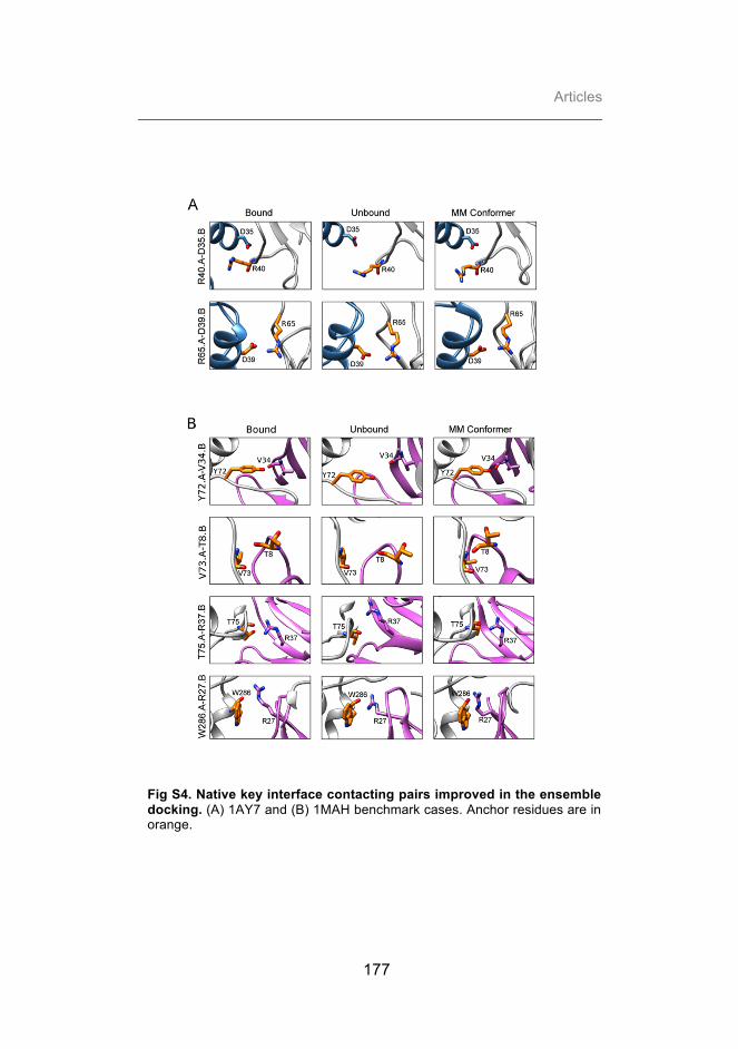
Articles
177
Fig S4. Native key interface contacting pairs improved in the ensemble docking. (A) 1AY7 and (B) 1MAH benchmark cases. Anchor residues are in orange.

Protein plasticity improves protein-protein docking
178
Fig S5. Relation between docking performance and energetic complementary of the docking partners. Distribution of the best near-native solution binding energy for each conformer pair versus the binding energy of the conformers in the complex native orientation. Only high-affinity cases are shown, and they are classified according to conformational motion between unbound and bound states. For comparison, the results for the docking of unbound and bound X-ray structures are shown in red and green, respectively. Cases with correlation coefficient < -0.2 are shown in blue background.

Articles
179
Table S1. Docking with random ensembles: Best rank of a near-native docking pose
Complexa Random Conf. (5)
Random Unb. Rot. (5)
Random Unb. Rot. (100)
Rigid (I-RMSDCα < 0.5 Å) (18 cases) 1AVX (64) (211) (976) 1FSK 3 3 4 1GHQ (8001) (3290) (23384) 1IQD (7) 4 2 1KLU (3807) (19418) (98251) 1KTZ (1562) (795) (9270) 1NCA 10 (50) (420) 1NSN (65) (580) (6396) 1PPE (8) 2 (21) 1R0R (91) (360) (92) 1SBB (512) (1205) (5424) 1WEJ 2 (29) 9 2JEL 1 3 (41) 2MTA 5 2 (23) 2PCC 1 5 9 2SIC 6 6 10 2SNI 5 4 4 2UUY (3166) (892) (13165) Low-flexible (I-RMSDCα 0.5-1.0 Å) (45 cases) 1AHW (13454) (9777) (360248) 1AY7 (46) (111) (784) 1AZS (-) 8 (254) 1BJ1 (364) (293) (3180) 1BUH (266) (189) (2301) 1BVN 1 1 1 1DQJ (1118) (328) (1037) 1E96 7 5 (16) 1EAW 1 (197) (250) 1EFN (1001) (490) (6233) 1EWY 1 5 6 1F34 (3200) (670) (4025) 1F51 (43) 3 9 1FQJ (668) (1576) (6501) 1GCQ (48) (62) (2788) 1GLA (38) 1 9 1GPW 1 1 1 1HE1 (616) (4542) (118475) 1HE8 (1046) (4301) (44116) 1IJK (1121) (966) (777290) 1J2J (16) 2 (5) 1JPS (24) (1420) (12959) 1K4C (1890) (-) (28657) 1K74 (61) (54) 10 1KAC (4359) (8321) (18603)

Protein plasticity improves protein-protein docking
180
1KXQ (29) 1 6 1MAH 3 1 1 1MLC 1 2 1 1N8O (121) (119) (-) 1QA9 (15092) (11140) (103447) 1QFW (675) (614) (5876) 1RLB (42005) (1801) (4773) 1S1Q (99) (756) (9589) 1T6B (205) (196) (580) 1TMQ 1 8 4 1UDI 2 1 4 1YVB 4 2 (22) 1Z0K (319) (103) 10 1ZHI 7 1 4 2AJF (3914) (2430) (69360) 2B42 (39) 1 1 2BTF (77) (167) (34) 2OOB (1234) (1571) (2788) 2VIS (-) (-) (-) 7CEI (27) (35) 6 Medium-flexible (I-RMSDCα 1.0-2.0 Å) (35 cases) 1A2K (3415) (1290) (3966) 1AK4 (2229) (21200) (14322) 1AKJ (438) (372) (-) 1B6C 3 1 1 1BGX (17600) (-) (841301) 1BVK (46) (289) (462) 1D6R (8) (331) (2660) 1DFJ 5 5 9 1E6E 1 1 1 1E6J 3 2 3 1EZU (1337) (4350) (6654) 1FC2 (205) (4336) 0 1GP2 (129) (352) (172024) 1GRN (71) (2171) (11864) 1HIA (62) (50) (37506) 1I4D (305) (18465) (2033) 1I9R (-) (101) (243) 1K5D (1475) (1776) (912) 1KXP 1 2 (7) 1ML0 (78) (41) (424) 1NW9 (53) 9 9 1OPH (376) (70) (660) 1VFB (11) (88) (202) 1WQ1 (96) (1618) (2081) 1XD3 2 1 1 1XQS 7 (17) (266) 1Z5Y 10 (22) (23) 2CFH (892) (1908) (27689) 2FD6 6 (114) (762)

Articles
181
2H7V (2345) (-) (1413) 2HLE (17) 4 2 2HQS (7) (30) (15) 2I25 (231) (553) (284) 2O8V (80) (11820) (74) 2QFW (132) 1 2 Flexible (I-RMSDCα 2.0-3.0 Å) (18 cases) 1ACB (135) (369) (555) 1BKD (23) (966) (1937) 1CGI (45) 5 (87) 1DE4 (-) (-) (1725) 1E4K (1197) (14392) (79673) 1EER (43) (7292) (325) 1I2M (199) (247) 5 1IB1 (28994) (18819) (70454) 1IBR (-) (-) (-) 1KKL (280) (458) (222) 1M10 9 (497) (1411) 1N2C 1 (22) 1 1PXV (1837) (318) (2852) 2C0L (3449) (7473) (62804) 2HMI (-) (-) (-) 2HRK (73) (79) (580) 2NZ8 (40) (28) (100) 2OT3 (213) (-) (555) Highly-flexible (I-RMSDCα > 3.0 Å) (8 cases) 1ATN (2402) (2503) (3597) 1FAK (25) (1670) (34273) 1FQ1 (239) (16535) (35535) 1H1V (-) (-) (259770) 1IRA (-) (-) (-) 1JMO (2982) (13867) (121326) 1R8S (1174) (8370) (5277) 1Y64 (-) (-) (-) a PDB of the complex (in bold: high affinity cases)


Articles
183
3.3. Modeling protein interactions: application to cases of interest
The expertise acquired during the first part of this PhD thesis on the
modeling and characterization of structure and dynamics of protein
interactions has facilitated the successful application of computational
methods to the modeling of protein interactions within different real-
life contexts.
This section will be mainly focused on (i) the energetic
characterization of host-pathogen protein interactions, and (ii) the ab
initio modeling of encounter complex ensembles of redox proteins.

Modeling protein interactions: application to cases of interest
184
Manuscripts presented in this section:
1. Lucas M, Gaspar A, Pallara C, Rojas A, Fernández-Recio J, Machner M, Hierro A. (2014) Structural basis for the recruitment and activation of the Legionella phospholipase VipD by the host GTPase Rab5. Proc Natl Acad Sci U S A 111(34):E3514-23.
2. Bernal-Bayard P, Pallara C, Castell MC, Molina-Heredia FP, Fernández-Recio J, Hervás, Navarro JA. (2015) Interaction of photosystem I from Phaeodactylum tricornutum with plastocyanins as compared with its native cytochrome c6: reunion with a lost donor. BBA Bioenergetics 1847(12):1549-1559.)

Articles
185
3.3.1. Structural basis for the recruitment and activation of the Legionella phospholipase VipD by the host GTPase Rab5.
Maria Lucas1, Andrew H. Gaspar2, Chiara Pallara3, Adriana L. Rojas1,
Juan Fernández-Recio3, Matthias P. Machner2,*, and Aitor Hierro1,4,*
1Structural Biology Unit, CIC bioGUNE, Bizkaia Technology Park,
48160 Derio, Spain;
2Cell Biology and Metabolism Program, Eunice Kennedy Shriver
National Institute of Child Health and Human Development, National
Institutes of Health, Bethesda, Maryland 20892, USA;
3Joint BSC-IRB research program in Computational Biology,
Barcelona Supercomputing Center, Barcelona, 08034, Spain;
4IKERBASQUE, Basque Foundation for Science, 48011 Bilbao,
Spain.
*Corresponding authors


Structural basis for the recruitment and activation ofthe Legionella phospholipase VipD by the hostGTPase Rab5María Lucasa, Andrew H. Gasparb, Chiara Pallarac, Adriana Lucely Rojasa, Juan Fernández-Recioc, Matthias P. Machnerb,1,and Aitor Hierroa,d,1
aStructural Biology Unit, Center for Cooperative Research in Biosciences, 48160 Derio, Spain; bCell Biology and Metabolism Program, Eunice Kennedy ShriverNational Institute of Child Health and Human Development, National Institutes of Health, Bethesda, MD 20892; cJoint Barcelona Supercomputing Center-Institute for Research in Biomedicine Research Program in Computational Biology, Barcelona Supercomputing Center, 08034 Barcelona, Spain; anddIKERBASQUE, Basque Foundation for Science, 48011 Bilbao, Spain
Edited by Ralph R. Isberg, Howard Hughes Medical Institute, Tufts University School of Medicine, Boston, MA, and approved July 15, 2014 (received for reviewMarch 22, 2014)
A challenge for microbial pathogens is to assure that their trans-located effector proteins target only the correct host cell compart-ment during infection. The Legionella pneumophila effectorvacuolar protein sorting inhibitor protein D (VipD) localizes toearly endosomal membranes and alters their lipid and proteincomposition, thereby protecting the pathogen from endosomalfusion. This process requires the phospholipase A1 (PLA1) activityof VipD that is triggered specifically on VipD binding to the hostcell GTPase Rab5, a key regulator of endosomes. Here, we presentthe crystal structure of VipD in complex with constitutively activeRab5 and reveal the molecular mechanism underlying PLA1 activa-tion. An active site-obstructing loop that originates from the C-terminal domain of VipD is repositioned on Rab5 binding, therebyexposing the catalytic pocket within the N-terminal PLA1 domain.Substitution of amino acid residues located within the VipD–Rab5interface prevented Rab5 binding and PLA1 activation and causeda failure of VipD mutant proteins to target to Rab5-enriched endo-somal structures within cells. Experimental and computational anal-yses confirmed an extended VipD-binding interface on Rab5,explaining why this L. pneumophila effector can compete with cel-lular ligands for Rab5 binding. Together, our data explain how thecatalytic activity of a microbial effector can be precisely linked to itssubcellular localization.
pathogenic bacteria | allosteric modulation | membrane composition |X-ray crystallography
Microbial pathogens have evolved numerous ways to subvertand exploit normal host cell processes and to cause dis-
ease. Intravacuolar pathogens use specialized translocationdevices such as type IV secretion systems (T4SS) to deliver vir-ulence proteins, so-called effectors, across the bacterial andhost cell membrane into the cytosol of the infected cell (1–3).Many of the translocated effectors studied to date alter cellularevents such as vesicle trafficking, apoptosis, autophagy, proteinubiquitylation, or protein synthesis, among others, thereby cre-ating conditions that support intracellular survival and replica-tion of the microbe (4, 5). Bacteria with a nonfunctional T4SS areoften avirulent and degraded along the endolysosomal pathway,thus underscoring the importance of translocated effectors formicrobial pathogenesis.Although T4SS-mediated effector translocation may be a
convenient way for pathogens to manipulate host cells fromwithin the safety of their membrane-enclosed compartment, italso creates a challenging dilemma: how can the bacteria ensurethat their translocated effectors reach the correct host cell targetfor manipulation, and how can they prevent them from indis-criminately affecting bystander organelles or proteins that mayotherwise be beneficial for intracellular survival and replication ofthe microbe? It is reasonable to expect that regulatory mecha-
nisms have evolved that restrain the catalytic activity of effectors.Although detailed insight into these processes is scarce, anemerging theme among effectors is that their enzymatic activity isfunctionally coupled to their interaction with a particular hostfactor. For example, SseJ from Salmonella enterica serovarTyphimurium displays glycerophospholipid-cholesterol acyl-transferase activity only on binding to the active GTPases RhoA,RhoB, or RhoC (6–8). Likewise, Pseudomonas aeruginosa ExoUrequires mono- or poly-ubiquitinated proteins for the activationof its phospholipase A2 (PLA2) domain (9), whereas YersiniaYpkA exhibits kinase activity only in the presence of host cellactin (10). Exactly how binding to host ligands results in the ac-tivation of these translocated effectors remains unclear becauseno structural information for these protein complexes is available.VipD is a T4SS-translocated substrate of Legionella pneumo-
phila, the causative agent of a potentially fatal pneumonia knownas Legionnaires’ disease, and another example of an effectorwhose catalytic activity depends on the presence of a host factor(11–14). Following uptake by human alveolar macrophages,L. pneumophila translocates VipD together with more than 250other effector proteins through its Dot/Icm T4SS into the host cellcytoplasm (15). These effectors act on numerous host processes to
Significance
A long-standing question in the field of microbial pathogenesisis how virulence factors are regulated within host cells andhow their activity is specifically directed toward a particularhost cell compartment. Legionella pneumophila resolves thisdilemma by tightly coupling the phospholipase A1 activity ofone of its effectors, vacuolar protein sorting inhibitor protein D(VipD), to this protein’s interaction with endosomal host GTPa-ses. We now present the crystal structure of VipD in complexwith host cell Rab5c, providing a detailed look into the ingeniousmolecular mechanisms underlying the allosteric activation ofa virulence factor by a host protein and its spatiotemporal reg-ulation. These results open the path for the development ofnovel therapeutics aimed at blocking the VipD activation processrather than the enzyme’s active site.
Author contributions: M.P.M. and A.H. designed research; M.L., A.H.G., C.P., A.L.R., andJ.F.-R. performed research; M.L., A.H.G., C.P., A.L.R., J.F.-R., M.P.M., and A.H. analyzeddata; and M.P.M. and A.H. wrote the paper.
The authors declare no conflict of interest.
This article is a PNAS Direct Submission.
Data deposition: The structural coordinates of the VipD19-564-Rab5c18-182(Q80L):GMP–PNPcomplex have been deposited in the Protein Data Bank, www.pdb.org (PDB ID code 4KYI).1To whom correspondence may be addressed. Email: [email protected] [email protected].
This article contains supporting information online at www.pnas.org/lookup/suppl/doi:10.1073/pnas.1405391111/-/DCSupplemental.
E3514–E3523 | PNAS | Published online August 11, 2014 www.pnas.org/cgi/doi/10.1073/pnas.1405391111

mediate evasion of the endolysosomal compartment and to es-tablish a Legionella-containing vacuole (LCV) that supports bac-terial growth (16). Although the precise biological role of most L.pneumophila effectors remains unclear, we recently showed thatVipD is important for endosomal avoidance by LCVs. The pro-tein localizes to endosomes presumably by binding to the smallGTPases Rab5 or Rab22, key regulators of endosomal function(13, 14). Rab GTPase binding to the C-terminal domain of VipDtriggers robust phospholipase A1 (PLA1) activity within the N-terminal domain, resulting in the removal of phosphatidylinositol3-phosphate [PI(3)P] and potentially other lipids from endosomalmembranes (14). Without PI(3)P, endosomal markers such asearly endosomal antigen 1 (EEA1) are lost from these mem-branes, most likely rendering the endosomal compartment fusionincompetent (17). L. pneumophila mutants lacking vipD are at-tenuated in avoiding endosomal fusion, and their LCVs acquirethe endosomal marker Rab5 more frequently than LCVs con-taining the parental strain producing VipD (14). Thus, by cou-pling PLA1 activity to Rab5 binding, the catalytic activity of VipDis directed specifically against the endosomal compartment with-out visibly affecting neighboring cell organelles.VipD was originally identified in a screen for L. pneumophila
effectors that interfere with the vacuolar sorting pathway in yeast(11). The N-terminal half of VipD possesses high homology topatatin, a lipid acyl hydrolase present in the potato tuber (12,13). Analogous to other patatin-like proteins, VipD harborsa conserved serine lipase motif Gly-x-Ser-x-Gly (x = any aminoacid) as part of a Ser-Asp catalytic dyad that, together with twoconsecutive glycine residues (Asp-Gly-Gly motif), is expected tostabilize the oxyanion intermediate during the acyl chain cleav-age (13). Mutation of these conserved catalytic residues in VipDresults in loss of PLA1 activity (14), confirming their role insubstrate hydrolysis.The recently reported crystal structure of VipD confirmed the
predicted bimodular organization (13) and, in addition, revealeda surface loop, called “lid” in other phospholipases, that shieldsthe entry to the catalytic site. The inhibitory lid may explain whypurified recombinant VipD alone exhibits little or no PLA1 activityin vitro. However, given that binding of Rab5 or Rab22 to VipDactivates the PLA1 activity within the N-terminal region (14), wewondered if and how this binding event causes the inhibitory lid tobe removed to render the active site substrate accessible.Using an integrative approach involving X-ray crystallography,
molecular dynamics, biochemistry, and cellular imaging, we nowdeciphered at a molecular level the mechanism that stimulatesthe intrinsic PLA1 activity of VipD and determined the underlyingspecificity for the VipD–Rab5 interaction and endosomal targeting.
ResultsOverall Structure of the VipD–Rab5 Complex. To determine themolecular basis underlying VipD binding and activation by Rab5,we initiated a crystallographic analysis of this complex. For that,we used a truncated form (residues 18–182) of constitutivelyactive Rab5c(Q80L) lacking the N- and C-terminal hypervariableregions, and a VipD fragment [amino acid (aa) 19–564; VipD19–564]that was designed based on a previously solved structure offull-length VipDFL–Rab5c18–182 at lower resolution in whichthe terminal residues (1–18 and 565–621) of VipD were not struc-tured. We obtained well-diffracting crystals of VipD19–564in complex with Rab5c18–182(Q80L) bound to nonhydrolyzableguanosine 5!-[!,"-imido]triphosphate (GppNHp) and solved thestructure by molecular replacement (Fig. 1). Only the last sevenC-terminal residues of VipD19–564 and a connecting loop formedby residues 345–354 could not be modeled because of poorelectron density in these regions. The final model for theVipD19–564-Rab5c18–182(Q80L)-GppNHp structure was refinedat 3.1 Å, with values for Rfactor and Rfree of 0.23 and 0.28, re-spectively (Table 1 and Fig. S1A).
The crystallographic asymmetric unit contained fourVipD19–564–Rab5c18–182(Q80L) heterodimers with almost iden-tical interaction modes (Fig. S1B). Superposition of the atomiccoordinates showed a root mean square deviation (RMSD) of
Fig. 1. The sites for substrate catalysis and Rab5 binding are situated atopposite ends of VipD. Two orthogonal tube drawing representations of thecrystal structure of VipD19–564 (slate) in complex with GppNHp-Rab5c18–182(pink). (A) Side view. (B) Top view. VipD comprises two distinguishable butinterconnected domains highlighted by gray elliptical shadows. The N-ter-minal half of VipD comprises a patatin-like phospholipase domain, whereasthe C-terminal domain interacts with Rab5c. Note that the catalytic site andthe Rab5 binding interface are located at opposite ends of VipD.
Table 1. Data collection and refinement statistics for theVipD19–564–Rab5c18–182(Q80L):GppNHp complex
VipD19–564–Rab5c18–182(Q80L)
Data collectionSpace group P1Cell dimensions
a, b, c (Å) 94.3, 98.0, 109.9#, !, " (°) 76.6, 80.8, 78.9Resolution (Å) 30–3.07 (3.26–3.07)*
Rmeas 0.07 (0.74)I/$ 17.0 (2.1)Completeness (%) 97.4 (92.6)Redundancy 3.5 (3.6)
RefinementResolution (Å) 3.07No. reflections 67,479Rwork/Rfree 0.23/0.28No. atoms
Protein 21,659Ligand/ion 132
B-factorsProtein 54Ligand/ion 74
RMSDsBond lengths (Å) 0.002Bond angles (°) 0.631
*Highest resolution shell is shown in parentheses.
Lucas et al. PNAS | Published online August 11, 2014 | E3515
BIOPH
YSICSAND
COMPU
TATIONALBIOLO
GY
PNASPL
US

0.65–0.69 Å among the four VipD19–564–Rab5c18–182(Q80L)complexes. Rab5c18–182(Q80L) was in its active conformation andbound to one molecule of GppNHp and one Mg2+ ion (Fig. 1). Itadopted the classical GTPase fold consisting of a central six-stranded !-sheet surrounded by five "-helices (18). The structureof VipD19–564 exhibited two discernible but interconnected domains.Rab5c18–182(Q80L) interacted extensively with a helical hairpinsituated at the C-terminal domain of VipD19–564, and, thus, atthe distal end relative to the N-terminal catalytic site (Fig. 1). Itis worth noting that, although the structure of active Rab5c18–182(Q80L) remained essentially unaltered, VipD19–564 exhibitedseveral dramatic conformational rearrangements compared withthe uncomplexed crystallographic model (13), as discussed next.
Rab5 Binding to VipD Induces Conformational Changes That Exposethe Active Site.On Rab5, binding the largest RMSD in VipD19–564occurred in its C-terminal domain and in the structural elementsthat connect it to the N-terminal phospholipase domain(Fig. 2A). Residue Phe442 located in helix "17 of the C-terminaldomain of VipD19–564 undergoes a 90° rotation and enters a hy-drophobic pocket in Rab5c18–182(Q80L) formed by Arg82,Tyr83, and Leu86 (Fig. 2B). This rotation pulls the adjacent"16-"17 loop of VipD19–564 toward Rab5c18–182(Q80L), therebyfacilitating the hydrophobic interaction of Ile433 of VipD19–564with Ile54 in the switch I region of Rab5c18–182(Q80L) (Fig. 2B).The displacement of loop "16–"17 in VipD19–564 induces a par-tial reorientation of the adjacent !-sheet formed by !1, !2, and!11, together with small shifts in helices of the C-terminal do-main of VipD19–564. These cumulative movements cause helices"13 and "14 of VipD19–564 to swing out 14.5° and 6.6°, re-spectively, which is coupled with a coil-helix transition of the!9-"13 loop to adjoin helix "13 (Fig. 2A). This hinge motion ofhelices "13 and "14 (“chop-stick” mechanism) facilitates an
outward displacement of the adjacent !10–"14 loop (sub-sequently named lid), resulting in the eventual opening of theactive site (Fig. 2 C–E). Notably, there were no mayor crystal-lographic contacts in the areas corresponding to "13, "14, and thelid, making the displacement of the lid due to the proximity ofneighboring protein molecules within the crystal lattice unlikely(Fig. S1C). The exposed cleft, with its catalytic residues and theoxyanion hole situated at one end, measures 16–18 Å in length andthus has the potential to accommodate a C16–C18 acyl chain froma lipid substrate within the adjacent hydrophobic ridge (Fig. 2E andFig. S1D). Together, these findings provide evidence for an un-precedented heterotropic allosteric activation mechanism in whichlocally induced structural changes through Rab5c18–182(Q80L)binding are transmitted from the C-terminal domain of VipD19–564to the N-terminal phospholipase domain, causing the displacementof the lid and exposure of the active site.
VipD–Rab5 Interface. Our complex structure revealed a single in-teraction path between VipD19–564 and Rab5c18–182(Q80L) thatoccluded !722 Å2 of solvent-accessible surfaces. AlthoughRab5c18–182(Q80L) interacted with residues in the "16–"17 loopof VipD19–564 and residues in an helical hairpin formed by he-lices "17 and "18 (Fig. 3A), the VipD binding surface inRab5c18–182(Q80L) included parts of the segment between "1and !2 (the switch I region), the strands !2 and !3 (the inter-switch region), and the !3–"2 segment (the switch II region)(Fig. 3A). The interface was composed of a core of hydrophobiccontacts complemented by several polar interactions in the sur-rounding rim area (Fig. 3B). Specifically, the VipD bindingepitope in Rab5c18–182(Q80L) included nonpolar residues in theswitch I (Ile54, Gly55, Ala56, and Phe58), the interswitch(Trp75), and the switch II element (Tyr83, Leu86, Met89, andTyr90), as well as polar/charged residues in the interswitch
Fig. 2. Allosteric activation of VipD through Rab5 binding. (A) Structural changes in VipD on Rab5 binding. Rab5c18–182 (colored in pink) is complexed toVipD19–564, which is colored from slate to red based on the root mean square deviation (RMSD) of C-" atom pairs when superimposed with the unbound formof VipD19–564 (PDB ID code 4AKF) shown in transparent gray. The black line represents the membrane plane. (B) Close-up of VipD–Rab5 interaction. The "17-"18loop of VipD undergoes a Rab5-induced conformational rearrangement resulting in residue Phe442 of VipD being inserted into a hydrophobic pocket formed byArg82, Tyr83, and Leu86 of Rab5. The displacement of the "16–17 loop favors the hydrophobic interaction between Leu432 and Ile433 of VipD with Ile54 of Rab5.Color code as in A. The remaining VipD structure has been omitted for clarity. (C) Close-up view of the catalytic site highlighting displacement of the lid (!10-"14loop, light blue). (D and E) Surface representation of the unbound (D) and Rab5-bound (E) VipD molecule, respectively. Same view as in C.
E3516 | www.pnas.org/cgi/doi/10.1073/pnas.1405391111 Lucas et al.

(Thr60, Lys71, and Glu73) and switch II element (Arg82 andArg92) (Fig. 3 A and B). A comparison of the primary sequenceof human Rab5 and Rab22 with Rab5 from several naturalamoebean hosts found conserved residues at equivalent contactsites in the switch I (Ile54, Gly55, and Ala56), interswitch(Lys71), and switch II region (Arg82, Leu86, and Met89) thatwere variable in other Rab proteins (Fig. 3C), suggesting theseresidues are involved in the specific recognition by VipD. Thecorresponding epitope in VipD included several hydrophobicresidues in helix !17 (Phe442, Ala446, Ala450, and Leu454) andin helix !18 (Tyr473, Ile480, and Val483) that wrapped aroundan elongated hydrophobic path in Rab5 formed by the conservedtriad (Phe58, Trp75, and Tyr90) and Leu86. Surrounding thishydrophobic core were additional hydrogen bonds that enhancedthe interaction.Like all GTPases, Rab5 exhibits structural changes within its
switch regions dependent on its nucleotide-binding state (GDPvs. GTP), with the largest conformational variation in switch I(19). The structure of the VipD19–564–Rab5c18–182(Q80L) com-plex revealed that Leu432 and Ile433 of VipD19–564 interacted
with Ile54 in switch I of Rab5c18–182(Q80L), therefore sensing itsGTP-bound state (Fig. 2B). In fact, the conformation adopted byRab5 in its GDP-bound state (19) resulted in a prominent stericclash between the switch I region and helix !17 of VipD19–564(Fig. S1E), thus explaining why this activation state of Rab5 isonly a poor ligand for VipD (13, 14).
Validation of the VipD–Rab5 Interface Through Mutational Analysis.To experimentally validate the VipD–Rab5 binding interfaceseen in the crystal structure, we mutated several residues pre-dicted to contribute to this protein–protein interaction and ex-amined their role for complex formation in coprecipitationstudies (Fig. 4A). Substitution of individual contact residueswithin VipD abrogated Rab5 binding either severely (F442A andH453D) or moderately (Q449A, E461R, Y473A, and D479H),whereas only a few of the tested substitutions in VipD weretolerated (Q476A and D484H). Similar results were observed forRab5 interface mutants (Fig. 4B), with binding defects rangingfrom severe (F58A, Y83A, and R92E) to mild (E73R andY90A). We also studied the mode of interaction between VipD
Fig. 3. Molecular interactions at the VipD–Rab5c interface. (A) (Left) Semitransparent surface of GppNHp-Rab5c18–182 in complex with the minimal Rabbinding domain of VipD (slate ribbon model) highlighting the interfacial residues below 4.0-Å distance. (Right) Schematic diagram of interfacial residues inthe VipD–Rab5c complex. (B) Detailed description of per-residue contribution from van der Waals (vdW) energy (blue), nonpolar solvation energy (purple),and the sum of electrostatic and polar solvation energy (orange) calculated by computational alanine scanning for interfacial residues in the VipD–Rab5ccomplex. Existing glycines and alanines are excluded in the calculation. (C) Sequence conservation between Rab5 GTPases from amoebean species and humanhomologs. Dd, Dictyostelium discoideum; Df, Dictyostelium fasciculatum; Dp, Dictyostelium purpureum; Pp, Polysphondylium pallidum; Ac, Acanthamoebacastellanii; Hs, Homo sapiens. Rab5c residues contacting VipD at a distance less than 4 Å are colored in light brown. Amino acid substitutions within theequivalently aligned interfacial residues of other Rabs are highlighted in a red box. Interfacial residues strictly conserved between Rab5 and Rab22, butvariable in any of the other Rabs, are depicted in the bottom line of the alignment. Protein accession numbers are in brackets.
Lucas et al. PNAS | Published online August 11, 2014 | E3517
BIOPH
YSICSAND
COMPU
TATIONALBIOLO
GY
PNASPL
US

and Rab22. As expected, substitution of individual contact resi-dues of VipD required for Rab5c18–182(Q80L) binding (Fig. 4A)also resulted in a failure to stably associate with Rab22(Q64L)(Fig. 4C), suggesting that Rab22 occupies an epitope in VipDvery similar to that of Rab5. None of the amino acid sub-stitutions significantly altered the overall structure of the mutantproteins as evaluated by circular dichroism (CD) (Fig. S2), in-dicating that a reduction in binding was most likely not a conse-quence of protein misfolding.Given that the PLA1 activity of VipD is triggered only in re-
sponse to Rab5 binding, we analyzed how amino acid substitutionsthat attenuate VipD–Rab5 complex formation affect the PLA1activity of VipD. Using a generic fluorogenic substrate (bis-BOD-IPY FL C11-PC), we found a tight correlation between loss of PLA1activity and the inability of VipD mutant proteins (F442A, H453D,and D479H) to enter a stable complex with Rab5c18–182(Q80L) orRab2216–181(Q64L) (Fig. 4D). Similar results were observed forRab5c18–182(Q80L) mutant proteins (F58A, Y83A, and R92E) thathad failed to stably associate with VipD and were hence unable totrigger its PLA1 activity (Fig. 4E). The observed crystallographicinteraction between VipD and Rab5 thus corresponded to theirmolecular association in solution, and failure to form a stableVipD–Rab5 or –Rab22 complex caused the PLA1 domain to re-main in its catalytically inactive state.
Disruption of the Interaction with Rab5 Precludes EndosomalTargeting of VipD. Within transiently transfected COS1 cells,fluorescently tagged VipD was enriched on Rab5-containingearly endosomes, and this colocalization required the C-terminalRab5 binding domain but not the N-terminal PLA1 domain (13,14). A recent study reported that depletion of Rab5 (isoformsa-c) and Rab22a from HeLa cells by RNA interference (RNAi)did not affect VipD targeting to endosomes, claiming thatendosomal localization of VipD would not simply depend on theinteraction with Rab proteins (20). Given that RNAi rarelydepletes the entire pool of a given cellular target and that VipDrecruitment to endosomes could have been mediated not onlyby Rab5 and/or Rab22 but by additional yet unidentified Rab
GTPases, we set out to reevaluate VipD’s endosomal targetingmechanism. For that, we analyzed the intracellular distributionpattern of four VipD mutant proteins that were either severely(F442A) or moderately (E461R and Q476A) attenuated forRab5c binding in vitro (Fig. 4). Although WT VipD displayedrobust colocalization with GFP-Rab5c18–182(Q80L)-positiveendosomes, VipD(F442A) was entirely cytosolic (Fig. 5), consis-tent with this mutant’s inability to bind Rab5c. In contrast, VipD(E461R) and VipD(Q476A) showed no apparent difference inlocalization compared with WT VipD (Fig. 5). These findingsstrongly suggest that endosomal targeting of VipD is in fact de-pendent on the interaction with host cell Rab GTPases and thatinterference with the formation of these protein complexes resultsin the failure of VipD to properly localize to endosomes.
The N-Terminal Tail of VipD Is Crucial for PLA1 Activity. In theuncomplexed structure of VipD, the N-terminal tail (residues1–18; N18) contained a small amphipathic helix (H1) that wasinvolved in an intermolecular crystal contact (13). The structureof full-length VipD1–621 bound to Rab5c18–182(Q80L), on theother hand, contained no clear electron density for N18, sug-gesting that this region of VipD possessed high flexibility. Smallangle X-ray scattering (SAXS) and gel filtration chromatographyanalysis suggest a heterodimeric VipD1–621–Rab5c18–182(Q80L)complex in solution, indicating that N18 was not involved in anyoligomer formation (Fig. 6 A and B and Fig. S3). Given that thecomplexes of Rab5c18–182(Q80L) with either full-length VipD1–621or truncated VipD19–564 exhibited nearly indistinguishable struc-tures, we concluded that N18 was dispensable for the conforma-tional changes induced by Rab5 binding. Consequently, weevaluated whether this short region was also dispensable for PLA1activity of VipD. Unexpectedly, we found that, unlike VipD1–621,the truncated fragment VipD19–564 lacking N18 was strongly at-tenuated for PLA1 activity (Fig. 5C). To exclude the possibilitythat loss of PLA1 activity in VipD19–564 was caused by the lack ofthe C-terminal region (aa 565–621), we tested two additionalconstructs, VipD1–564 and VipD19–621, and detected robust PLA1activity only in VipD1–564, indicating that N18 but not the
Fig. 4. Mutational analysis of interfacial recognition determinants. (A–C) Pulldown assays using the indicated VipD (A and C) or Rab5 (B) mutant proteins. Thegraphs are a densitometry-based quantification of the amount of query protein precipitated by the respective bait-coated beads. Input, total amount of query. (Dand E) Fluorescence-based PLA activity assays using VipD (D) or Rab5c (E) variants. (D) The indicated VipD protein was incubatedwith Rab5(Q80L)18–182:GppNHp orRab22(Q64L)16–181:GppNHp (molar ratio 1:2) or with buffer alone, and PLA1-dependent cleavage of the substrate Bis-BODIPYFL C11-PC was detected as an increasein fluorescence emission [relative fluorescence units (RFUs)]. (E) Same assay as in D using the indicated Rab protein variants.
E3518 | www.pnas.org/cgi/doi/10.1073/pnas.1405391111 Lucas et al.

C-terminal region critically contributed to the catalytic activity ofthis L. pneumophila effector (Fig. 5C). According to these ob-servations, we propose that the flexible N18 with its amphipathichelix H1 and its close distance to the membrane plane may pro-mote peripheral association of VipD with the lipid bilayer, pos-sibly by orienting the catalytic site toward the membrane and/orassisting in substrate transfer.
Competitive Rab5 Binding Through Interface Expansion. To localizeto and stably associate with endosomal membranes, VipD needs tooutcompete cellular ligands for Rab5 binding. EEA1, Rabaptin-5,and Rabenosyn-5 are each bound by Rab5 through a surface that
includes the switch and interswitch region and that significantlyoverlaps with the epitope for VipD binding (19, 21, 22). Todetermine if and how the distribution of interaction energies differswithin each of these complexes, we extended the computationalalanine scan to the EEA1, Rabaptin-5, and Rabenosyn-5 epitopesand calculated the free binding energy for each of their residues(Fig. 7A and Figs. S4 and S5). All four analyzed protein interfacesshare a number of nonpolar interacting residues in Rab5, namelythe conserved triad (Phe58, Trp75, and Tyr90), with relativelysimilar energetic contributions to binding (Fig. 7A and Fig. S5B).The polar interactions surrounding this hydrophobic triad,however, determine their differential affinity, with the contact of
Fig. 5. VipD localization to endosomes requires a functional Rab5 binding interface. Transiently transfected COS-1 cells producing Rab5a(Q79L) and theindicated mCherry-tagged VipD variants were analyzed by fluorescence microscopy to determine protein localization. The merged images (bottom row) showRab5a(Q79L) in green and VipD variants in red. (Insets) Magnified view of endosomes marked by an arrowhead. Control, mCherry. (Scale bar, 2 μm.)
Fig. 6. The N-terminal 18 residues of VipD are essential for its PLA activity. (A) Fit of the optimized crystallographic VipD1–564-Rab5c18–182(Q80L) model (redline) to the experimental SAXS data of the complex (blue dots). (B) Fitting of the VipD19–564-Rab5c18–182(Q80L) crystallographic model (VipD in slate and Rab5in pink) into the averaged ab initio envelope in two orthogonal views and superimposed with the unbound form of VipD (PDB 4AKF) in gray. Note theproximity of the N18 (residues 1–18 of VipD, PDB 4AKF) in red to the catalytic site. (C) Fluorescence-based PLA activity assays showing that the N18 segment ofVipD is essential for its PLA1 activity.
Lucas et al. PNAS | Published online August 11, 2014 | E3519
BIOPH
YSICSAND
COMPU
TATIONALBIOLO
GY
PNASPL
US

Arg92Rab5 with Asp479VipD and Asp484VipD providing a particu-larly large energetic contribution to the interaction of VipD withRab5 compared with the other cellular ligands (Fig. 7A and Fig.S5B). To verify the importance of this predicted hot-spot for VipDbinding, we analyzed the affinities of either Rab5c18–182(Q80L) orRab5c18–182(Q80L, R92A) toward VipD by surface plasmon res-onance (SPR) spectroscopy. R92A mutation in Rab5 severelydecreases the binding for VipD 124-fold while affecting the in-teraction with EEA1, Rabenosyn-5, and Rabaptin-5 to a muchlesser extent (3.2-, 1.7- and 1.0-fold, respectively) (Fig. 7 B and Cand Fig. S6). These findings pinpoint a binding hotspot for thesuperior affinity of VipD over the endogenous Rab5 ligands andconfirm a good qualitative correlation between the computationalanalysis and the experimentally observed results.
DiscussionVipD from L. pneumophila has long been predicted to functionas a phospholipase during infection (11, 12), yet its catalyticactivity had only recently been confirmed when it was shown thatbinding of host cell Rab GTPases (Rab5 and Rab22) is necessaryfor VipD to exhibit robust PLA1 activity (14). The crystallo-graphic analysis described here provides an in-depth view of theRab5-mediated activation mechanism. Above all, it uncovereda complex cascade of structural rearrangements within theC-terminal domain of VipD that result in the relocation of anactive site-occluding lid and the exposure of the substrate bind-ing pocket within the N-terminal PLA1 domain of VipD.
The structure of VipD in complex with Rab5c(Q80L) pre-sented here is, to our knowledge, the first of a bacterial phos-pholipase bound to a host cell protein and the first of anytranslocated effector in complex with its allosteric activatormolecule. Phospholipases constitute a common cellular tool toalter the lipid composition of membranes, and their activity mustbe carefully dosed and precisely directed toward the respectivetarget membrane. There are more than 10,000 proteins (8,101 inBacteria and 2,374 in Eukaryotes) containing potential patatin-like domains, most of them within a modular domain arrange-ment (23). Many members of the family of cytosolic phospholi-pases A2 (cPLA2), all of which share a patatin-like fold, containa C2 domain crucial for membrane localization (24, 25). Thepatatin-like fold is also highly homologous to the group of cal-cium-independent phospholipases A2 (iPLA2), in which manymembers contain ankyrin repeats, a repetitive helix-turn-helix-loop structure considered to be a common platform for protein–protein interactions (24). Considering that Rab GTPases are keyplayers in defining membrane identity and that many effectorsfrom L. pneumophila have been acquired via horizontal genetransfer (26, 27), it is plausible that the scheme presented here forthe concomitant localization and activation of VipD can be gen-eralized across other microbial and eukaryotic phospholipases.Human Rab5 interacted with VipD through a helix-turn-helix
element that was similar to that used for Rabenosyn-5 binding(21), although the interface was slightly shifted toward the switch IIregion. Despite the observed overlapping contacts, the energy for
Fig. 7. Energy distribution between different effector binding epitopes in Rab5. (A) Space-filling model of Rab5c highlighting the epitopes for the interactionwith VipD (Far Left), EEA1 C2H2 Zinc Finger (PDB ID code 3MJH) (Center Left), Rabenosyn-5 (rebuilt from PDB ID code 1Z0J) (Center Right), and Rabaptin-5 (PDBID code 1TU3) (Far Right). Epitopes are colored as an intensity gradient according to the binding free energy change (ΔΔG) estimated as the difference betweenthe binding ΔG of the WT and that of the alanine mutated complex. Existing glycines and alanines are excluded in the calculation. (B) Concentration de-pendence of the equilibrium surface plasmon resonance response for the binding of VipD WT, EEA1 C2H2 Zinc Finger (aa 36–91), Rabenosyn-5 (aa 1–70), andRabaptin-5 (aa 739–862) to Rab5c18–182(Q80L) or Rab5c18–182(Q80L, R92A). Req represents the equilibrium SPR response normalized to the fitted maximum valuefor each dataset. (C) Table of the mean Kd values and SDs for at least two independent experiments showing the Kd ratio variation.
E3520 | www.pnas.org/cgi/doi/10.1073/pnas.1405391111 Lucas et al.

VipD binding was not distributed uniformly across the interface butinstead concentrated into a combination of hotspots that providesuperior binding affinity and specificity (Fig. 7 and Fig. S6). Aconserved hydrophobic triad in Rab5 (Phe58, Trp75, and Tyr90)supplied the core binding energy that was complemented by morespecific polar and nonpolar contacts. Notably, most of these resi-dues were highly conserved among Rab5 homologs from amoe-bean species, the natural host of L. pneumophila, or from thesurrogate host Dictyostelium sp. (Fig. 3C). The ability to discrimi-nate between GDP- and GTP-bound Rab5 and to compete withendogenous ligands evidences a remarkable adaptation for direct-ing and retaining VipD on endosomal membranes. Interferencewith VipD–Rab5 complex formation, for example, by substitutingPhe442 or His453 of VipD, strongly reduced the capability of thesemutant proteins to interact with Rab5 (Fig. 4A), to exhibit PLA1activity (Fig. 4D), and to efficiently localize to the endosomalcompartment (Fig. 5), thus demonstrating that the function ofVipD is intimately coupled to the presence of this host GTPase.A hallmark feature of many phospholipases is to be minimally
active on monomeric lipid substrates but undergo a substantialactivation on binding to the surface of phospholipid membranesor micelles, a phenomenon known as interfacial activation (28–31). This behavior has been attributed to a flexible lid that at thelipid–water interface facilitates substrate diffusion to the cata-lytic site rather than being allosterically modulated throughdistant ligand binding (25). VipD does not display any interfacialactivation despite having a short lid occluding the access to thecatalytic site. Rather, when VipD is bound to Rab5, the lid isdisplaced through a chopstick-like activation mechanism inwhich the swing movement of two α-helices (α13 and α14) al-losterically controls accessibility of the catalytic site. We cannotexclude the possibility that additional mechanisms contribute tothe activity and/or specificity of the substrate catalysis by VipD.For example, the coil–helix transition of the β9–α13 loop toadjoin helix α13 relocates several charged residues closer to thecatalytic groove, which might result in interactions with lipidhead groups or other membrane components. Consistent withthis notion, we discovered that the flexible N-terminal segmentN18 of VipD is critical for the catalysis of a generic membrane-embedded substrate (Fig. 6). Deletion of N18 reduced the PLA1activity of VipD19–564 but did not interfere with allosteric acti-vation of the catalytic site. We hypothesize that N18 bearinga short amphipathic α-helix may facilitate the correct positioningof the PLA1 domain toward the lipid layer, promote substratediffusion from the lipid–water interface into the catalytic site, ora combination of both effects as has been previously describedfor secretory PLA2 enzymes (32, 33). Interestingly, the N-ter-minal tail of VpdA (residues 11–54) and the N-terminal segmentof P. aeruginosa ExoU (residues 57–96) showed structural simi-larity to the equivalent region of VipD despite lacking sequencehomology (Fig. S7). Moreover, region 57–96 of ExoU, althoughnot part of the phospholipase domain (34), was critical for cyto-toxicity within transfected mammalian cells (35), suggesting thatthis segment contributes to the phospholipase activity of ExoU.We hypothesize that the equivalent region in VpdA may beequally important for the catalytic activity of this L. pneumophilaeffector and that appendixes, such as the N-terminal domains, mayplay important yet unresolved roles in membrane association and/or substrate transfer in other bacterial phospholipases.In summary, our findings disclose an unexpected mode of
long-range allosteric regulation of the PLA1 activity of VipD andexplain how endosomal targeting is accomplished through com-petitive Rab5 binding. Our study also provides the basis for thedevelopment of novel therapeutic approaches that, rather thandirectly targeting the enzyme’s active site, specifically disturb thehost factor-mediated activation process of VipD and relatedmicrobial phospholipases.
Materials and MethodsPlasmids and Cloning. The DNA sequences encoding VipD, VipD19–564, VipD1–564,VipD19–621, Rab5c18–182(Q80L), Rab22a16–181(Q64L), EEA136–91, and Rabeno-syn51–70 were cloned into the vector pGST-Parallel2 (36) using BamHI and XhoIrestriction sites. Rabaptin5739–862 and Rab7a(Q67L) were cloned between theNcoI and XhoI restriction sites of pGST-Parallel2. PCR was performed usingPhusion polymerase (Thermo). The PCR product was purified with QIAquickGel Extraction Kit (NewEngland) and ligated into the digested pGST-Parallel2vector using Quickligase (BioLab). The ligation mixture was used to transformEscherichia coli XL1 Blue competent cells, and transformants were then se-lected on Luria-Bertani (LB) plates containing 100 μg/mL ampicillin. Thepresence of the insert in the plasmid was tested by colony PCR. Quickchangemutagenesis was used to make directed mutations. The correct transformantswere grown to isolate the plasmids that were sequenced on both strands.Plasmids and oligonucleotides used in this study are listed in Tables S1 andS2, respectively.
Protein Expression and Purification. VipD was purified from E. coli BL21 (DE3)grown in LB medium and induced at an OD600 = 0.8 by the addition of0.5 mM isopropyl β-D-1-thiogalactopyranoside. Cells were harvested after16 h of growth at 18 °C. The cell pellet was resuspended in buffer A (50 mMTris·HCl, pH 8.0, 300 mM NaCl, and 1 mM DTT) supplemented with 0.1 mMphenylmethylsulfonyl fluoride, 1 mM benzamidine, and 1 mg/mL lysozymeand disrupted by sonication, and the lysate was cleared by centrifugation at50,000 ! g for 45 min. The supernatant was incubated for 2 h in batch withglutathione Sepharose beads (GE Healthcare) followed by extensive washingof the beads with buffer A in a gravity column. The N-terminal glutathioneS-transferase (GST)-tag and linker were proteolytically removed by over-night incubation at 4 °C in the presence of tobacco etch virus (TEV) proteasein 50 mM Tris·HCl, pH 8.0, 150 mM NaCl, and 1 mM DTT. The cleaved proteinwas eluted and further purified by ion exchange chromatography (HitrapQ;GE Healthcare) using a gradient of 50–1,000 mM NaCl, followed by sizeexclusion chromatography (Superdex200 16/60; GE Healthcare) in buffer B[25 mM Tris·HCl, pH 7.5, 150 mM NaCl, 5% (vol/vol) glycerol, and 1 mM DTT].VipD mutants and truncated constructs were purified following the sameprocedure. The concentration of these proteins was calculated using thetheoretical extinction coefficient.
Rab5c18–182(Q80L) was purified as described for VipD, with the differencethat the HitrapQ column gradient was 25–1,000 mM, and the size exclusionchromatography was performed in a Superdex75 16/60 (GE Healthcare). Nu-cleotide exchange was achieved by incubation of the purified protein witha 20-fold excess of GppNHp in 50 mM Tris, pH 7.5, 150 mM NaCl, and 5 mMEDTA for 12 h at 4 °C. The exchange reaction was stopped by addition ofMgCl2 (10 mM final concentration). Excess nucleotide was removed by gelfiltration using a Superdex75 16/60 column in buffer C [25 mM Tris·HCl,pH 7.5, 150 mM NaCl, 5% (vol/vol) glycerol, 1 mM MgCl2, and 1 mM DTT].Rab5c18–182, Rab5c18–182(Q80L) mutants, Rab22a16–181(Q64L), and Rab7a(Q67L)were purified as previously described. The concentration of the Rab proteinswas determined by using Bradford’s procedure with BSA as standard.
For complex formation, VipD was incubated with GppNHp-boundRab5c18–182(Q80L) in a 1:3 molar ratio for 2 h at 4 °C. The complex was furtherpurified using a Superdex200 16/60 column equilibrated in buffer C andconcentrated to 50mg/mL using Amicon centrifugal concentrators (Millipore).
GST, GST-VipD, GST-Rabenosyn51–70, GST-EEA136–91, and GST-Rabap-tin5739–862 were purified with the same protocol as described for VipD withthe only difference that no TEV protease cleavage was performed duringthe purification.
Crystallization and Structure Determination. Crystals were obtained byhanging-drop vapor diffusion at 18 °C by mixing 1 μL purified VipD19–564–
Rab5c18–182(Q80L):GppNHp complex (50 mg/mL) and 1 μL precipitant solu-tion (16% PEG6000, 0.1 M Tris·HCl, pH 8.0, and 0.2 M LiCl). Rod-shapedcrystals grew within 2–3 d. Individual crystals were cryo-protected by a briefsoak in well buffer supplemented with 25% (vol/vol) ethylene glycol andflash frozen in liquid nitrogen.
Diffraction datawere collected at 100 K using radiationwith a wavelength of0.976 Å on beamline I04 at the Diamond Light Source (Didcot, UK). The datawere integrated and scaled using XDS (37). The structure was solved by mo-lecular replacement using the coordinates of VipD [Protein Data Bank (PDB) IDcode 4AKF) and Rab5c (PDB ID code 1Z07) as a search model in Phaser (38).Subsequent rounds of refinement and interactive manual building were per-formed using Phenix (39) and Coot (40). For cross-validation, 5% of the originalreflections was omitted from refinement and used to calculate the free R factor.The final model contained four complexes of VipD–Rab5c(Q80L). Only 10 resi-dues (345–354) located in a connecting loop could not be modeled because of
Lucas et al. PNAS | Published online August 11, 2014 | E3521
BIOPH
YSICSAND
COMPU
TATIONALBIOLO
GY
PNASPL
US

poor electron density in this region. Crystallographic data collection and modelstatistics are summarized in Table 1. The Ramachandran plot of the modelcalculated with the Rampage evaluation tool (41) shows 96.0% of the residuesin the favored regions, whereas 3.9% fall in the allowed regions and 0.1% indisallowed regions. Graphics presented in this manuscript were generated usingthe program PyMOL (The PyMOL Molecular Graphics System; Schrödinger).
Pulldown Assays. In vitro pulldown assays involving VipD–Rab5 interfacemutants included GST-VipD, GST-Rab5c18–182(Q80L), VipD point mutants,and Rab5c18–182(Q80L) point mutants. The pulldown between VipD andRab22 proteins included Rab22a16–181(Q64L) and VipD point mutants. NoTEV protease cleavage was performed during the purification of GST-VipD,GST-Rab5c18–182(Q80L), or GST-Rab22a16–181(Q64L). For pulldowns involvingVipD point mutants and GST-Rab5c18–182(Q80L), 13 μM VipD (WT or mutants)was mixed with 10 μM of GST-Rab5c18–182(Q80L) in binding buffer (50 mMTris·HCl, pH 7.5, 150 mM NaCl, 1 mM MgCl2, and 1 mM DTT). Then 10 μL ofequilibrated glutathione Sepharose beads were added to 70 μL of the pro-tein mixture and incubated for 2 h at 4 °C with gentle agitation. Beads werewashed several times with 0.5 mL of binding buffer and resuspended insample buffer. The samples were subjected to 4–12% SDS/PAGE analysis, andgels were stained with Coomassie brilliant blue. The pulldown experimentbetween GST-Rab22a16–181(Q64L) and VipD point mutants was performed ina like manner. The binding between GST-VipD and Rab5c18–182(Q80L)point mutants was analyzed similarly using 20 μM GST-VipD and 30 μMRab5c18–182(Q80L) (WT or mutants) in each individual reaction. Each pull-down was performed in triplicate with similar outcomes.
Phospholipase Assays. The phospholipase activity of VipD and its mutants wasassayed using bis-BODIPY FL C11-PC (Invitrogen), a glycerophosphocholinewith BODIPY FL dye-labeled sn-1 and sn-2 acyl chains, respectively. Thisfluoregenic substrate is selfquenched and release of the fluorophores by acylchain cleavage by either PLA1 or PLA2 results in increased fluorescence.To prepare fluorescence-labeled liposomes, we mixed 30 μL 10 mM di-oleoylphosphatidylcholine (DOPC), 30 μL 10 mM dieloylphosphatidylglycerol(DOPG), and 30 μL of 1 mM bis-BODIPY FL C11-PC. All these compounds weredissolved in ethanol. The substrate was incorporated into liposomes by aslow injection of this ethanolic lipid mix into 5 mL assay buffer (50 mM Tris,pH 7.5, 150 mM NaCl, and 1 mM MgCl2) under continuous stirring. Themixture was pipetted into the side of the vortex using a narrow orifice gel-loading tip. Fifty microliters of each substrate solution was incubated with50 μL bis-BODIPY FL C11-PC–labeled liposomes in 96-well plates for 2 h at25 °C. The reaction mixtures contained VipD 2.5 μM and Rab 5 μM or thecorresponding mutants in assay buffer. The fluorescence intensity wasmeasured at 485-nm excitation and 530-nm emission in a multiwell reader(Biotek Synergy HT-1). All of the measurements were performed in triplicate.The assay buffer in the absence of enzyme was used as a blank.
ImmunofluorescenceMicroscopy. VipD localization was analyzed in COS-1 cellsgrown on coverslips in 24-well plates in 5% CO2 at 37 °C in DMEM mediasupplemented with 10% FBS. Semiconfluent monolayers were transientlytransfected using Lipofectamine 2000 (Invitrogen) to produce fluorescentlytagged (mCherry or GFP) proteins. Cells were fixed 10 h after transfection,and images were analyzed on a Zeiss Axio Observer.Z1 inverted light mi-croscope using a Zeiss Plan-Apochromat 63!/ oil M27 objective and pro-cessed with Zeiss AxioVision 4.7.2 software.
SAXS. Synchrotron SAXS data were collected on beamline BM29 at ESRF(Grenoble, France) with a 2D detector (Pilatus 1M) over an angular rangeqmin = 0.01 Å!1 to qmax = 0.5 Å!1. X-ray scattering patterns were recordedwith the VipD1–564–Rab5c18–182(Q80L) complex at 2.2 and 6.4 mg/mL in 150 mMNaCl, 0.5 mM tris-(2-carboxyethyl)phosphine, and 25 mM Hepes, pH 7.5.
Data collection, processing, and initial analysis were performed usingbeamline software BsxCuBE. Further analyses were performed with the ATSASsuite. PRIMUS (42) was used for Rg determination with the Guinier method,and maximum distance (Dmax) was evaluated using GNOM (43), which was alsoused to calculate the distance distribution functions. Fitting of the model ofthe VipD–Rab5c structure to the SAXS data was calculated with CRYSOL (44)with a χ2 against raw data of 2.12 and 3.2 for samples at 2.2 and 6.4 mg/mL,respectively. To generate an ab initio model of the VipD–Rab5c complex, 20runs of GASBOR (45) were performed using the merge of the two datasets(2 and 6.4 mg/mL) as raw data. Then, the most probable model was filteredwith DAMSEL (46), and a 720 bead model was produced. Superposition of thebead model on the crystallographic VipD–Rab5c structure was carried outusing the program SUPCOMB13 (47). The resulting bead model was convertedto a mesh envelope and visualized using PYMOL (Schrödinger).
Molecular Dynamics Simulations. A total of four molecular dynamics (MD)simulations were performed starting from the Rab5–VipD crystallographicstructure and from three different Rab5 complexes previously described(Rab5–EEA1 C2H2 Zinc Finger, Rab5-Rabaptin5, and Rab5-Rabenosyn5). Theinitial coordinates of Rab5–EEA1 C2H2 Zinc Finger (PDB ID code 3MJH) andRab5–Rabaptin5 (PDB ID code 1TU3) were taken from the Protein DataBank, whereas the Rab5–Rabenosyn5 complex was rebuilt using theRab22–Rabenosyn5 crystal structure (PDB ID code 1Z0J) as a template. InRab5–VipD, Rab5–Rabaptin5, and Rab5–Rabenosyn5 structures, GppNHp mole-cules were replaced by GTP. In case of incomplete chains, acetyl and amidecapping groups were added to the N-term and C-term residues flanking themission regions to avoid improper charges on them. The protonation state of theionizable residues was estimated at pH 6.5 using the server H++ (http://biophysics.cs.vt.edu/H++) (48–50). The parameter files for the GTP molecule andZn2+ ion were prepared with the AMBER (51) module ANTECHAMBER, and thetopology files for the protein complexes were generated using LEAP. Beforerunning the molecular dynamics simulations, a short minimization and a five-step equilibration protocol were performed on the solvated structure, as pre-viously described (52). On Rab5–VipD, Rab5–Rabaptin5, and Rab5–Rabenosyn5complexes, unrestrained 10-ns MD simulations were performed in an isothermal-isobaric ensemble, setting pressure to 1 atm and temperature to 300 K. In theRab5–EEA1 complex, 2.5-Å distance restraints between Zn2+ ion and each EEA1zinc finger motif residue (Cys43, Cys46, His59, and His64) were applied during allof the equilibration andMD simulation step, to keep the same coordination as inthe initial structure. The RMSD for the Cα atoms of each complex along the MDtrajectory were calculated with the ptraj AMBER12 tool (51).
Computational Alanine Scanning.We used the MMPBSA.py script in AMBER12(51) to perform Computational Alanine Scanning calculations on 200 snap-shots extracted from the last 2 ns of each complex MD trajectory (see above).All of the interface residues (defined as those located within 4-Å distancefrom the protein partner in the most representative structure along the last2 ns of the trajectory) were mutated to alanine, and then the binding freeenergy change (ΔΔG) was estimated as the difference between the bindingΔG (MM-GBSA method) of the WT and that of the mutated complex. Thecontribution of conformational entropy was not included here, given thedifficulty of computing it for a large protein– protein complex but that shouldnot significantly affect the comparison of mutant and WT free energies.
Surface Plasmon Resonance Measurements. The binding affinity of VipD,Rabenosyn51–70, EEA136–91, and Rabaptin5739–862 for Rab5c(Q80L) or Rab5c(Q80L,R92A) was calculated using SPR. SPR data were collected using a Biacore3000 instrument (GE Healthcare) and a GST sensor chip. A research gradeCM5 chip was first conditioned with three 5-μL injections of 100 mM glycine-NaOH, pH 12. Anti-GST antibody was covalently immobilized on the CM5sensor chip injecting 45 μL at 30 μg/mL in 10 mM sodium acetate, pH 5.0,using the amine coupling kit [1-ethyl-3-(3-dimethylaminopropyl)carbodii-mide hydrochloride and N-hydrosuccinimide] supplied by GE Healthcare.Nearly 30,000 resonance units (RUs) of the antibody were immobilized underthese conditions in each flow cell, where 1 RU corresponds to immobilizedprotein concentration of "1 pg/mm2. The unreacted moieties on the surfacewere blocked with ethanolamine. The immobilization procedure was doneat 5 μL/min with the running buffer containing 10 mM Hepes, pH 7.5,150 mM NaCl, and 0.005% Tween20. Binding experiments were performedwith the same buffer supplemented with 2 mM MgCl2. All of the proteinswere dialyzed into this buffer. GST, GST-VipD, GST-Rabenosyn51–70, GST-EEA136–91, and GST-Rabaptin5739–862 were captured on the sensor chip witha 5-μL injection of 100 nM ligand at 5 μL/min on a reference and sample flowcell. Rab5c(Q80L) and Rab5c(Q80L, R92A) incubated with GppNHp wereinjected at different concentrations for a contact time of 2 min. Bindingexperiments were carried out at a flow rate of 20 μL/min at 25 °C. The anti-GST sensor chip was regenerated after each analyte injection with a 2-mininjection of 10 mM glycine-HCl, pH 2.1. This regeneration procedure did notalter to any measurable extent the ability of the immobilized antibody tobind protein in subsequent cycles. Analysis of the data was performed usingthe BIAevaluation software supplied with the instrument. The steady-statebinding response was determined by averaging the response over 5 s at theend of the injection and was corrected for background binding. The satu-ration binding values were fitted according to a one-site binding model.Each experiment was repeated in triplicate. Values of KD are reported as themeans of independent experiments with corresponding SDs.
ACKNOWLEDGMENTS. We thank A. Vidaurrazaga for technical assistance.We thank all the staff from the facilities where work was done for technicaland human support. This work was supported by Carlos III Health Institute
E3522 | www.pnas.org/cgi/doi/10.1073/pnas.1405391111 Lucas et al.

Grant PI11/00121 and Basque Government Grant PI2011-26 (to A.H.).This study made use of the Diamond Light Source (Oxfordshire, UnitedKingdom), partly funded by the BioStruct-X program (Proposal 2370), the
European Synchrotron Radiation Facility (Grenoble, France) under the BlockAllocation Group MX1283/MX1420, and ALBA synchrotron beamline BL13-XALOC under Proposal 2012100351.
1. Christie PJ, Whitaker N, González-Rivera C (2014) Mechanism and structure of thebacterial type IV secretion systems. Biochim Biophys Acta 1843(8):1578–1591.
2. Fronzes R, et al. (2009) Structure of a type IV secretion system core complex. Science323(5911):266–268.
3. Voth DE, Broederdorf LJ, Graham JG (2012) Bacterial Type IV secretion systems:Versatile virulence machines. Future Microbiol 7(2):241–257.
4. Ribet D, Cossart P (2010) Pathogen-mediated posttranslational modifications: A re-emerging field. Cell 143(5):694–702.
5. Galán JE (2009) Common themes in the design and function of bacterial effectors. CellHost Microbe 5(6):571–579.
6. Christen M, et al. (2009) Activation of a bacterial virulence protein by the GTPaseRhoA. Sci Signal 2(95):ra71.
7. LaRock DL, Brzovic PS, Levin I, Blanc MP, Miller SI (2012) A Salmonella typhimurium-translocated glycerophospholipid:cholesterol acyltransferase promotes virulence bybinding to the RhoA protein switch regions. J Biol Chem 287(35):29654–29663.
8. Ohlson MB, et al. (2008) Structure and function of Salmonella SifA indicate that itsinteractions with SKIP, SseJ, and RhoA family GTPases induce endosomal tubulation.Cell Host Microbe 4(5):434–446.
9. Anderson DM, et al. (2011) Ubiquitin and ubiquitin-modified proteins activate thePseudomonas aeruginosa T3SS cytotoxin, ExoU. Mol Microbiol 82(6):1454–1467.
10. Juris SJ, Rudolph AE, Huddler D, Orth K, Dixon JE (2000) A distinctive role for theYersinia protein kinase: Actin binding, kinase activation, and cytoskeleton disruption.Proc Natl Acad Sci USA 97(17):9431–9436.
11. Shohdy N, Efe JA, Emr SD, Shuman HA (2005) Pathogen effector protein screening inyeast identifies Legionella factors that interfere with membrane trafficking. Proc NatlAcad Sci USA 102(13):4866–4871.
12. VanRheenen SM, Luo ZQ, O’Connor T, Isberg RR (2006) Members of a Legionellapneumophila family of proteins with ExoU (phospholipase A) active sites are trans-located to target cells. Infect Immun 74(6):3597–3606.
13. Ku B, et al. (2012) VipD of Legionella pneumophila targets activated Rab5 and Rab22to interfere with endosomal trafficking in macrophages. PLoS Pathog 8(12):e1003082.
14. Gaspar AH, Machner MP (2014) VipD is a Rab5-activated phospholipase A1 thatprotects Legionella pneumophila from endosomal fusion. Proc Natl Acad Sci USA111(12):4560–4565.
15. Ensminger AW, Isberg RR (2009) Legionella pneumophila Dot/Icm translocated sub-strates: A sum of parts. Curr Opin Microbiol 12(1):67–73.
16. Horwitz MA (1983) The Legionnaires’ disease bacterium (Legionella pneumophila)inhibits phagosome-lysosome fusion in human monocytes. J Exp Med 158(6):2108–2126.
17. Vieira OV, Botelho RJ, Grinstein S (2002) Phagosome maturation: Aging gracefully.Biochem J 366(Pt 3):689–704.
18. Merithew E, et al. (2001) Structural plasticity of an invariant hydrophobic triad in theswitch regions of Rab GTPases is a determinant of effector recognition. J Biol Chem276(17):13982–13988.
19. Zhu G, et al. (2004) Structural basis of Rab5-Rabaptin5 interaction in endocytosis. NatStruct Mol Biol 11(10):975–983.
20. Zhu W, Hammad LA, Hsu F, Mao Y, Luo ZQ (2013) Induction of caspase 3 activation bymultiple Legionella pneumophila Dot/Icm substrates. Cell Microbiol 15(11):1783–1795.
21. Eathiraj S, Pan X, Ritacco C, Lambright DG (2005) Structural basis of family-wide RabGTPase recognition by rabenosyn-5. Nature 436(7049):415–419.
22. Mishra A, Eathiraj S, Corvera S, Lambright DG (2010) Structural basis for Rab GTPaserecognition and endosome tethering by the C2H2 zinc finger of Early EndosomalAutoantigen 1 (EEA1). Proc Natl Acad Sci USA 107(24):10866–10871.
23. Finn RD, et al. (2014) Pfam: The protein families database. Nucleic Acids Res42(Database issue):D222–D230.
24. Dennis EA, Cao J, Hsu YH, Magrioti V, Kokotos G (2011) Phospholipase A2 enzymes:Physical structure, biological function, disease implication, chemical inhibition, andtherapeutic intervention. Chem Rev 111(10):6130–6185.
25. Dessen A, et al. (1999) Crystal structure of human cytosolic phospholipase A2 revealsa novel topology and catalytic mechanism. Cell 97(3):349–360.
26. de Felipe KS, et al. (2005) Evidence for acquisition of Legionella type IV secretionsubstrates via interdomain horizontal gene transfer. J Bacteriol 187(22):7716–7726.
27. Al-Quadan T, Price CT, Abu Kwaik Y (2012) Exploitation of evolutionarily conservedamoeba and mammalian processes by Legionella. Trends Microbiol 20(6):299–306.
28. Brzozowski AM, et al. (1991) A model for interfacial activation in lipases from thestructure of a fungal lipase-inhibitor complex. Nature 351(6326):491–494.
29. van Tilbeurgh H, et al. (1993) Interfacial activation of the lipase-procolipase complexby mixed micelles revealed by X-ray crystallography. Nature 362(6423):814–820.
30. Roussel A, et al. (2002) Crystal structure of the open form of dog gastric lipase incomplex with a phosphonate inhibitor. J Biol Chem 277(3):2266–2274.
31. Aloulou A, et al. (2006) Exploring the specific features of interfacial enzymologybased on lipase studies. Biochim Biophys Acta 1761(9):995–1013.
32. Qin S, Pande AH, Nemec KN, Tatulian SA (2004) The N-terminal alpha-helix of pan-creatic phospholipase A2 determines productive-mode orientation of the enzyme atthe membrane surface. J Mol Biol 344(1):71–89.
33. Qin S, Pande AH, Nemec KN, He X, Tatulian SA (2005) Evidence for the regulatory roleof the N-terminal helix of secretory phospholipase A(2) from studies on native andchimeric proteins. J Biol Chem 280(44):36773–36783.
34. Gendrin C, et al. (2012) Structural basis of cytotoxicity mediated by the type III se-cretion toxin ExoU from Pseudomonas aeruginosa. PLoS Pathog 8(4):e1002637.
35. Finck-Barbançon V, Frank DW (2001) Multiple domains are required for the toxicactivity of Pseudomonas aeruginosa ExoU. J Bacteriol 183(14):4330–4344.
36. Sheffield P, Garrard S, Derewenda Z (1999) Overcoming expression and purificationproblems of RhoGDI using a family of “parallel” expression vectors. Protein Expr Purif15(1):34–39.
37. Kabsch W (2010) Integration, scaling, space-group assignment and post-refinement.Acta Crystallogr D Biol Crystallogr 66(Pt 2):133–144.
38. McCoy AJ, et al. (2007) Phaser crystallographic software. J Appl Cryst 40(Pt 4):658–674.39. Adams PD, et al. (2010) PHENIX: A comprehensive Python-based system for macro-
molecular structure solution. Acta Crystallogr D Biol Crystallogr 66(Pt 2):213–221.40. Emsley P, Lohkamp B, Scott WG, Cowtan K (2010) Features and development of Coot.
Acta Crystallogr D Biol Crystallogr 66(Pt 4):486–501.41. Lovell SC, et al. (2003) Structure validation by Calpha geometry: Phi,psi and Cbeta
deviation. Proteins 50(3):437–450.42. Konarev PV, Volkov VV, Sokolova AV, Koch MHJ, Svergun DI (2003) PRIMUS: A
Windows PC-based system for small-angle scattering data analysis. J Appl Cryst 36(5):1277–1282.
43. Svergun D (1992) Determination of the regularization parameter in indirect-trans-form methods using perceptual criteria. J Appl Cryst 25(4):495–503.
44. Svergun D, Barberato C, Koch MHJ (1995) CRYSOL: A program to evaluate X-ray so-lution scattering of biological macromolecules from atomic coordinates. J Appl Cryst28(6):768–773.
45. Svergun DI, Petoukhov MV, Koch MH (2001) Determination of domain structure ofproteins from X-ray solution scattering. Biophys J 80(6):2946–2953.
46. Volkov VV, Svergun DI (2003) Uniqueness of ab initio shape determination in small-angle scattering. J Appl Cryst 36(3 Part 1):860–864.
47. Kozin MB, Svergun DI (2001) Automated matching of high- and low-resolutionstructural models. J Appl Cryst 34(1):33–41.
48. Anandakrishnan R, Aguilar B, Onufriev AV (2012) H++ 3.0: Automating pK predictionand the preparation of biomolecular structures for atomistic molecular modeling andsimulations. Nucleic Acids Res 40(Web Server issue):W537–541.
49. Myers J, Grothaus G, Narayanan S, Onufriev A (2006) A simple clustering algorithmcan be accurate enough for use in calculations of pKs in macromolecules. Proteins63(4):928–938.
50. Gordon JC, et al. (2005) H++: A server for estimating pKas and adding missing hy-drogens to macromolecules. Nucleic Acids Res 33(Web Server issue):W368–W371.
51. Case DA, et al. (2012) AMBER 12 (University of California, San Francisco).52. Meyer T, et al. (2010) MoDEL (Molecular Dynamics Extended Library): A database of
atomistic molecular dynamics trajectories. Structure 18(11):1399–1409.
Lucas et al. PNAS | Published online August 11, 2014 | E3523
BIOPH
YSICSAND
COMPU
TATIONALBIOLO
GY
PNASPL
US

Articles
197
Supporting InformationLucas et al. 10.1073/pnas.1405391111
Fig. S1. (A) Electron density map (2Fo-Fc) calculated with phases derived from the final refined model and contoured at 1.5 sigma in the vicinity of the VipD-Rab5c interface. (B) Packing of four VipD–Rab5 complexes that form an asymmetric unit. VipD is colored in blue/slate; Rab5 is in light and dark pink. (C)Distribution of VipD surface areas with crystal lattice contacts below 4.5 Å (same orientation as in Fig. 2 C–E). Note that there are no mayor crystallographiccontacts in areas corresponding to !13, !14, and the lid except for two residues (Thr244 and His246) from a symmetrically related VipD molecule that arepartially inserted at the edge of the catalytic groove, most probably favored by the opening of the lid. The symmetrically related VipD molecule is omitted fromthe figure for reasons of clarity. (D) Close up view of the catalytic domain of VipD in ribbon diagram showing the surface clipped at the catalytic cleft. Thesurface is colored from white to green according to the Eisenberg hydrophobicity scale. (E) Superposition of the Rab5-GDP crystal structure [Protein Data Bank(PDB) ID code 1TU4] on the VipD:Rab5–GTP complex, illustrating the steric collision that prevents Rab-GDP from binding to VipD. The remainder of the VipDstructure is omitted for clarity.
Lucas et al. www.pnas.org/cgi/content/short/1405391111 1 of 9

Fig. S2. Circular dichroism spectra of the the indicated wildtype and mutant proteins of (A) VipD and (B) Rab5c used in this study.
Lucas et al. www.pnas.org/cgi/content/short/1405391111 2 of 9

Fig. S3. Characterization of VipD–Rab5c complex in solution. (A) Analysis of the VipD1–621–Rab5c18–182 oligomeric state by gel filtration (Superdex 200 HR10/30 column), leading to a Stokes radius estimation of !3.6 nm by comparison with the elution of model proteins (thyroglobulin, 8.5 nm; ferritin, 6.2 nm;γ-globulin, 5.3 nm; catalase, 5.2 nm; aldolase, 4.7 nm; BSA monomer, 3.5 nm; myoglobin, 2.1 nm; vitamin B12, 2.7 nm; Inset). Gel filtration of VipD (70 μM),Rab5c (140 μM), and VipD (70 μM) + Rab5c (140 μM). Note that the complex can be isolated using an excess of the Rab protein. (B) The guinier plot of theVipD1–564–Rab5c18–182 complex data at 2.2 and 6.4 mg/mL indicates a gyration radius of !3.6 nm. (C) This value is confirmed by the distance distributionfunction P(r), which suggests a bilobular structure with a radius of gyration of !36 Å and a maximum diameter of !120 Å fully compatible with the crys-tallographic structure of the VipD–Rab5c complex (Inset).
Lucas et al. www.pnas.org/cgi/content/short/1405391111 3 of 9

Fig. S4. (A) Plot of root mean square deviation (RMSD) relative to the coordinates of the initial (energy-minimized) structures during molecular dynamicsimulations. (B–D) Per-residue contribution from van der Waals (vdW) energy (blue), nonpolar solvation energy (purple), and the sum of electrostatic and polarsolvation energy (orange) calculated by computational alanine scanning for interfacial residues in the Rabenosyn5–Rab5c complex (B), EEA1–Rab5c complex(C), and Rabaptin5–Rab5c (D). Existing glycines and alanines are excluded in the calculation.
Lucas et al. www.pnas.org/cgi/content/short/1405391111 4 of 9

Fig. S5. (A) Superposition of effector binding epitopes in tube representation over Rab5c in transparent gray surface. EEA1 C2H2 Zinc Finger (PDB ID code3MJH) in purple, Rabenosyn-5 (rebuilt from PDB ID code 1Z0J) in green, Rabaptin-5 (PDB ID code 1TU3) in orange, and VipD in slate. (B) Comparison of the ΔΔGvalues in the binding interface of Rab5c. Note that some residues at the preserved binding core show similar ΔΔG values, whereas other residues such as Arg92constitute an effector-specific contact.
Lucas et al. www.pnas.org/cgi/content/short/1405391111 5 of 9

Fig. S6. Surface plasmon resonance (SPR) sensograms for binding of (A) VipD WT, (B) EEA1 C2H2 Zinc Finger [amino acids (aa) 36–91], (C) Rabenosyn-5(aa 1–70), and (D) Rabaptin-5 (aa 739–862) to Rab5c18–182(Q80L) (Left) or Rab5c18–182(Q80L, R92A) (Right).
Fig. S7. The N-terminal tail of VipD shows structural similarity to equivalent regions in VpdA and ExoU. (A) Representative diagrams of VipD, VpdA, and ExoUhighlighting the equivalent N-terminal tail. (B) PROMALS3D (PROfile Multiple Alignment with Predicted Local Structures and 3D Constraints) web serveralignment (http://prodata.swmed.edu/promals3d/promals3d.php) of VipD, VpdA, and ExoU N-terminal regions.
Lucas et al. www.pnas.org/cgi/content/short/1405391111 6 of 9

Table S1. Plasmids used in this study
Plasmid Properties Reference
pGST-Parallel2-VipD(19-564)
Expression construct for N-terminal glutathione S-transferase(GST)-tagged L. pneumophila VipD domain covering residues19–564 used for crystallization and PLA assays
This study
pGST-Parallel2-Rab5c18–182(Q80L)
Expression construct for N-terminal GST-tagged human Rab5c(Q80L)domain covering residues 18–182 used for crystallization, PLAassays and SPR assays
This study
pGST-Parallel2-VipD
Expression construct for N-terminal GST-tagged L. pneumophilaVipD used for pull-down assays, PLA assays and SPR assays
This study
pGST-Parallel2-VipD(1-564)
Expression construct for N-terminal GST-tagged L. pneumophilaVipD domain covering residues 1–564 used for PLA assays
This study
pGST-Parallel2-VipD(19-621)
Expression construct for N-terminal GST-tagged L. pneumophilaVipD domain covering residues 19–621 used for PLA assays
This study
pGST-Parallel2-VipD(S73A)
Expression construct for N-terminal GST-tagged L. pneumophilaVipD containing mutation S73A used for pull-down assays andPLA assays
This study
pGST-Parallel2-VipD(F442A)
Expression construct for N-terminal GST-tagged L. pneumophilaVipD containing mutation F442A used for pull-down assays andPLA assays
This study
pGST-Parallel2-VipD(Q449A)
Expression construct for N-terminal GST-tagged L. pneumophilaVipD containing mutation Q449A used for pull-down assays
This study
pGST-Parallel2-VipD(H453D)
Expression construct for N-terminal GST-tagged L. pneumophilaVipD containing mutation H453D used for pull-down assays andPLA assays
This study
pGST-Parallel2-VipD(E461R)
Expression construct for N-terminal GST-tagged L. pneumophilaVipD containing mutation E461R used for pull-down assays
This study
pGST-Parallel2-VipD(Y473A)
Expression construct for N-terminal GST-tagged L. pneumophilaVipD containing mutation Y473A used for pull-down assays
This study
pGST-Parallel2-VipD(Q476A)
Expression construct for N-terminal GST-tagged L. pneumophilaVipD containing mutation Q476A used for pull-down assays
This study
pGST-Parallel2-VipD(D479H)
Expression construct for N-terminal GST-tagged L. pneumophilaVipD containing mutation D479H used for pull-down assays andPLA assays
This study
pGST-Parallel2-VipD(D484H)
Expression construct for N-terminal GST-tagged L. pneumophilaVipD containing mutation D484H used for pull-down assays
This study
pGST-Parallel2-Rab5c18–182
Expression construct for N-terminal GST-tagged human Rab5c(18-182)used for PLA assays
This study
pGST-Parallel2-Rab5c18–182(Q80L,F58A)
Expression construct for N-terminal GST-tagged human Rab5c(18-182)containing mutations Q80L and F58A used for pull-down assays andPLA assays
This study
pGST-Parallel2-Rab5c18–182(Q80L,E73R)
Expression construct for N-terminal GST-tagged human Rab5c(18-182)containing mutations Q80L and E73R used for pull-down assays
This study
pGST-Parallel2-Rab5c18–182(Q80L,W75A)
Expression construct for N-terminal GST-tagged human Rab5c(18-182)containing mutations Q80L and W75A used for pull-down assays
This study
pGST-Parallel2-Rab5c18–182(Q80L, R82A)
Expression construct for N-terminal GST-tagged human Rab5c(18-182)containing mutations Q80L and R82A used for pull-down assays
This study
pGST-Parallel2-Rab5c18–182(Q80L,Y83A)
Expression construct for N-terminal GST-tagged human Rab5c(18-182)containing mutations Q80L and Y83A used for pull-down assays
This study
pGST-Parallel2-Rab5c18–182(Q80L,M89A)
Expression construct for N-terminal GST-tagged human Rab5c(18-182)containing mutations Q80L and M89A used for pull-down assays
This study
pGST-Parallel2-Rab5c18–182(Q80L,Y90A)
Expression construct for N-terminal GST-tagged human Rab5c(18-182)containing mutations Q80L and Y90A used for pull-down assays
This study
pGST-Parallel2-Rab5c18–182(Q80L,R92E)
Expression construct for N-terminal GST-tagged human Rab5c(18-182)containing mutations Q80L and R92E used for pull-down assays
This study
pGST-Parallel2-Rab5c18–182(Q80L,R92A)
Expression construct for N-terminal GST-tagged human Rab5c(18-182)containing mutations Q80L and R92A used in SPR assays
This study
Lucas et al. www.pnas.org/cgi/content/short/1405391111 7 of 9

Table S1. Cont.
Plasmid Properties Reference
pGST-Parallel2-Rab22a16–181(Q64L)
Expression construct for N-terminal GST-tagged human Rab22a(16-181)containing mutation Q64L used in PLA and pull-down assays
This study
pGST-Parallel2-Rab7a(Q67L)
Expression construct for N-terminal GST-tagged human Rab7acontaining mutation Q67L used in PLA assays
This study
pGST-Parallel2-EEA136–91
Expression construct for N-terminal GST-tagged human Rabenosyn5domain covering residues 36–91 used in SPR assays
This study
pGST-Parallel2-Rabenosyn51–70
Expression construct for N-terminal GST-tagged human Rabenosyn5domain covering residues 1–70 used in SPR assays
This study
pGST-Parallel2-Rabaptin5739–862
Expression construct for N-terminal GST-tagged human Rabaptin5domain covering residues 739–862 used in SPR assays
This study
pGST-Parallel2 Expression construct for GST used in SPR assays (1)pmCherry-C1 Mammalian expression vector generating an mCherry fusion to
the N terminus of the protein of interestClontech (cat. 632524)
pmCherry-VipD Expression construct generating an mCherry fusion to theN terminus of L. pneumophila full-length VipD
(2)
pmCherry-VipD(F442A) pmCherry-VipD containing mutation F442A in theVipD-Rab5 interface
This study
pmCherry-VipD(H453D) pmCherry-VipD containing mutation H453D in theVipD-Rab5 interface
This study
pmCherry-VipD(E461R) pmCherry-VipD containing mutation E461R in theVipD-Rab5 interface
This study
pmCherry-VipD(Q476A) pmCherry-VipD containing mutation Q476A in theVipD-Rab5 interface
This study
pEGFP-Rab5a(Q79L) Expression construct generating a GFP fusion to the N terminus ofhuman full-length Rab5a containing mutation Q79L generatingconstitutively active Rab5a
(3)
1. Sheffield P, Garrard S, Derewenda Z (1999) Overcoming expression and purification problems of RhoGDI using a family of “parallel” expression vectors. Protein Expr Purif 15(1):34–39.2. Gaspar AH, Machner MP (2014) VipD is a Rab5-activated phospholipase A1 that protects Legionella pneumophila from endosomal fusion. Proc Natl Acad Sci USA 111(12):4560–4565.3. Mattera R, Bonifacino JS (2008) Ubiquitin binding and conjugation regulate the recruitment of Rabex-5 to early endosomes. EMBO J 27(19):2484–2494.
Lucas et al. www.pnas.org/cgi/content/short/1405391111 8 of 9

Table S2. Oligonucleotides used in this study
Sequence Plasmid Cloning
TTTTTGGATCCGAAATATCAAAGACTGAGGCAGGACAATATTCTG pGST-Parallel2-VipD(19-564) BamHI/XhoITTTTTCTCGAGTCACGGTTCAGGTTGAACTTCAACTTTAAAGTCTTGTTTTTGGATCCAAGATCTGTCAATTTAAGCTGGTTCTGCTGGGGG pGST-Parallel2-Rab5c18–182(Q80L) BamHI/XhoITTTTTCTCGAGTCAAAGCTTCTTAGCTATTGCCATGAAGATTTCGTTCACGTTTTTGGATCCATGAAACTTGCTGAAATTATGACAAAAAGCCGTAAATTAAAAAG pGST-Parallel2-VipD BamHI/XhoITTTTTCTCGAGTCAATGGCCGCCAAATGTGGTTGAAAGACAATCTGACCCATGTTAGCGGAGCAGCAGCCGGAGCAATGACGGCGAGTAT pGST-Parallel2-VipD(S73A) SDATACTCGCCGTCATTGCTCCGGCTGCTGCTCCGCTAACATGGGTCAGATTGAGAAAGAGATTGCTGAGGCATCAGCGCATG pGST-Parallel2-VipD(F442A) SDCATGCGCTGATGCCTCAGCAATCTCTTTCTCGAGATTTTTGAGGCATCAGCGCATGCAGCAGCTATTTTGCATCTTCAAGAACAAATCG pGST-Parallel2-VipD(Q449A) SDCGATTTGTTCTTGAAGATGCAAAATAGCTGCTGCATGCGCTGATGCCTCAAAAATCTCGGCATCAGCGCATGCACAAGCTATTTTGGATCTTCAAGAACAAATCGTCAAAGAAATG pGST-Parallel2-VipD(H453D) SDCATTTCTTTGACGATTTGTTCTTGAAGATCCAAAATAGCTTGTGCATGCGCTGATGCCGCTATTTTGCATCTTCAAGAACAAATCGTCAAACGAATGAATGATGGTGATTACAGTAG pGST-Parallel2-VipD(E461R) SDGCTACTGTAATCACCATCATTCATTCGTTTGACGATTTGTTCTTGAAGATGCAAAATAGCGATGGTGATTACAGTAGCGTGCAAAATGCCTTAGATCAAATTGAAGACATTCTGACAG pGST-Parallel2-VipD(Y473A) SDCTGTCAGAATGTCTTCAATTTGATCTAAGGCATTTTGCACGCTACTGTAATCACCATCGCGTGCAAAATTATTTAGATGCAATTGAAGACATTCTGAC pGST-Parallel2-VipD(Q476A) SDGTCAGAATGTCTTCAATTGCATCTAAATAATTTTGCACGCCGTGCAAAATTATTTAGATCAAATTGAACACATTCTGACAGTCGATGCCAAAATGGATG pGST-Parallel2-VipD(D479H) SDCATCCATTTTGGCATCGACTGTCAGAATGTGTTCAATTTGATCTAAATAATTTTGCACGAGATCAAATTGAAGACATTCTGACAGTCCATGCCAAAATGGATGACATCCAGAAAGAG pGST-Parallel2-VipD(D484H) SDCTCTTTCTGGATGTCATCCATTTTGGCATGGACTGTCAGAATGTCTTCAATTTGATCTCAATTGGAGCGGCCGCACTCACACAGACTGTC pGST-Parallel2-Rab5c18–182(Q80L,F58A) SDGACAGTCTGTGTGAGTGCGGCCGCTCCAATTGCAACAGTCAAGTTTCGGATCTGGGACACAGC pGST-Parallel2-Rab5c18–182 (Q80L,E73R) SDGCTGTGTCCCAGATCCGAAACTTGACTGTTGCAAGTTTGAGATCGCGGACACAGCTGGACAG pGST-Parallel2-Rab5c18–182(Q80L,W75A) SDCTGTCCAGCTGTGTCCGCGATCTCAAACTTGGACACAGCTGGACTGGAGGCATATCACAGCCTGGC pGST-Parallel2-Rab5c18–182(Q80L, R82A) SDGCCAGGCTGTGATATGCCTCCAGTCCAGCTGTGTCGACACAGCTGGACTAGAGCGGGCTCACAGCCTGGCCCCCATG pGST-Parallel2-Rab5c18–182(Q80L,Y83A) SDCATGGGGGCCAGGCTGTGAGCCCGCTCTAGTCCAGCTGTGTCGAGCGGTATCACAGCCTGGCCCCCGCATACTATCGGGGGGCCCAGGC pGST-Parallel2-Rab5c18–182(Q80L,M89A) SDGCCTGGGCCCCCCGATAGTATGCGGGGGCCAGGCTGTGATACCGCTCGTATCACAGCCTGGCCCCCATGGCATATCGGGGGGCCCAGGCTGCC pGST-Parallel2-Rab5c18–182(Q80L,Y90A) SDGGCAGCCTGGGCCCCCCGATATGCCATGGGGGCCAGGCTGTGATACTATCACAGCCTGGCCCCCATGTACTATGAAGGGGCCCAGGCTGCCATCGTGGTCTAT pGST-Parallel2-Rab5c18–182 (Q80L,R92E) SDATAGACCACGATGGCAGCCTGGGCCCCTTCATAGTACATGGGGGCCAGGCTGTGATATATCACAGCCTGGCCCCCATGTACTATGCGGGGGCCCAGGCTGCCATCGTGGTCTAT pGST-Parallel2-Rab5c18–182 (Q80L,R92A) SDATAGACCACGATGGCAGCCTGGGCCCCCGCATAGTACATGGGGGCCAGGCTGTGATATTTTTGGATCCGCGCTGAGGGAACTTAAAGTGTGCCTG pGST-Parallel2-Rab22a16–181 (Q64L) BamHI/XhoITTTTTCTCGAGTCAGGATGGAATTCTTCGACTAATTTCTATAAAGAGTTCAAAAAACCATGGGAATGACCTCTAGGAAGAAAGTGTTGCTGAAGG pGST-Parallel2-Rab7a(Q67L) NcoI/XhoIAAAAAACTCGAGTTAGCAACTGCAGCTTTCTGCCGAGGCCTTTTTGGATCCAGCTCTTCAGAGGGTTTCATATGTC pGST-Parallel2-EEA136–91 BamHI/XhoITTTTTCTCGAGTTACTCTTGTCTGAGCAGTGTTACATCTTTTTGGATCCATGGCTTCTCTGGACGACCCAG pGST-Parallel2-Rabenosyn51–70 BamHI/XhoITTTTTCTCGAGTTATGCTCGATCATCCCCTTCTCGTTTTTTCCATGGCTTCTATTTCTAGCCTAAAAGCTGAATTAG pGST-Parallel2-Rabaptin5739–862 NcoI/XhoITTTTTCTCGAGTCATGTCTCAGGAAGCTGGTTAATGAAAAGTCGACATGAAACTTGCTGAAATTATGACAAAAAGC pmCherry-VipD SalI/BamHIAAGGATCCTTAATGGCCGCCAAATGTGGTTGAAAGACGAAATCAGAGAAAGAGATTGCTGAGGCATCAGCGCATGCAC pmCherry-VipD(F442A) SDGTGCATGCGCTGATGCCTCAGCAATCTCTTTCTCTGATTTCCATGCACAAGCTATTTTGGATCTTCAAGAACAAATCG pmCherry-VipD(H453D) SDCGATTTGTTCTTGAAGATCCAAAATAGCTTGTGCATGCAAATCGTCAAAGAAATGAATCGTGGTGATTACAGTAGCGTG pmCherry-VipD(E461R) SDCACGCTACTGTAATCACCACGATTCATTTCTTTGACGATTTGGTGCAAAATTATTTAGATGCAATTGAAGACATTCTGAC pmCherry-VipD(Q476A) SDGTCAGAATGTCTTCAATTGCATCTAAATAATTTTGCAC
SD, site-directed mutagenesis.
Lucas et al. www.pnas.org/cgi/content/short/1405391111 9 of 9


Articles
207
3.3.2. Interaction of photosystem I from Phaeodactylum tricornutum with plastocyanins as compared with its native cytochrome c6: reunion with a lost donor.
Pilar Bernal-Bayard1, Chiara Pallara2, M. Carmen Castell1, Fernando
P. Molina-Heredia1, Juan Fernández-Recio2, 1Manuel Hervás1 and
José A. Navarro1*
1Instituto de Bioquímica Vegetal y Fotosíntesis, Universidad de
Sevilla and CSIC, Américo Vespucio 49, 41092-Sevilla, Spain
2Joint BSC-CRG-IRB Research Program in Computational Biology,
Life Department, Barcelona Supercomputing Center, Barcelona
08034, Spain
*Corresponding author


Interaction of photosystem I from Phaeodactylum tricornutum withplastocyanins as comparedwith its native cytochrome c6: Reunionwith alost donor
Pilar Bernal-Bayard a, Chiara Pallara b, M. Carmen Castell a, Fernando P. Molina-Heredia a,Juan Fernández-Recio b, Manuel Hervás a, José A. Navarro a,⁎a Instituto de Bioquímica Vegetal y Fotosíntesis, Universidad de Sevilla and CSIC, Américo Vespucio 49, 41092 Sevilla, Spainb Joint BSC–CRG–IRB Research Program in Computational Biology, Life Sciences Department, Barcelona Supercomputing Center, Barcelona 08034, Spain
a b s t r a c ta r t i c l e i n f o
Article history:Received 16 June 2015Received in revised form 2 September 2015Accepted 20 September 2015Available online 25 September 2015
Keywords:Cytochrome c6Electron transferComputational dockingLaser flash photolysisPhaeodactylumPhotosystem IPlastocyanin
In the Phaeodactylum tricornutum alga, as inmost diatoms, cytochrome c6 is the only electron donor to photosys-tem I, and thus they lack plastocyanin as an alternative electron carrier.Wehave investigated, by using laser-flashabsorption spectroscopy, the electron transfer to Phaeodactylum photosystem I from plastocyanins fromcyanobacteria, green algae and plants, as compared with its own cytochrome c6. Diatom photosystem I is ableto effectively react with eukaryotic acidic plastocyanins, although with less efficiency than with Phaeodactylumcytochrome c6. This efficiency, however, increases in some green alga plastocyanin mutants mimicking the elec-trostatics of the interaction site on the diatom cytochrome. In addition, the structure of the transient electrontransfer complex between cytochrome c6 and photosystem I from Phaeodactylum has been analyzed by compu-tational docking and compared to that of green lineage and mixed systems. Taking together, the results explainwhy the Phaeodactylum system shows a lower efficiency than the green systems, both in the formation of theproperly arranged [cytochrome c6-photosystem I] complex and in the electron transfer itself.
© 2015 Elsevier B.V. All rights reserved.
1. Introduction
Diatoms are unicellular photosynthetic eukaryotes that are estimat-ed to contribute to 30–40% of the global carbon fixation in the oceans,and thus can be considered major primary producers [1,2]. Diatomshave a complex evolutionary history, belonging to the red lineage ofalga that diverged along evolution from the green lineage that led toplants [3,4]. Consequently, the photosynthetic machinery in diatomspossesses some singularities, arising from their evolutionary historyand endosymbiotic origin [5–7]. In particular, while most cyanobacteriaand unicellular green algae contain both the copper-protein plastocya-nin (Pc) and the iron-containing cytochrome c6 (Cyt) as alternativesoluble electron carriers between the b6f and photosystem I (PSI)mem-brane complexes, most diatoms lack Pc, thus containing Cyt as the only
soluble carrier between these complexes [8], with the remarkable ex-ception of the oceanic centric diatom Thalassiosira oceanica, for whichthe presence of an unusual Pc has been reported [9].
In green algae and plants (the green lineage of photosyntheticeukaryotes), the very acidic donor proteins to PSI interact, by meansof strong attractive electrostatic interactions, with a well-conservedpositively-charged docking site located in an extra loop extension ofthe PsaF subunit in PSI [10–12]. However, PSI from cyanobacteria,where both Pc and Cyt can be acidic, neutral or basic, lacks this extraloop, and thus the role of electrostatic forces in the interaction withPSI varies consequently [13–16]. By its turn, although the evolution ofthe electron transfer (ET) to PSI in diatoms has also led to complemen-tary electrostatic interactions between acidic and basic patches in Cytand the PsaF subunit, respectively, the electrostatic character of bothpartners is similarly reduced, the intensity of the interaction being ac-cordingly weakened as compared with the strongest electrostatic prop-erties of the Cyt(Pc)/PSI complex in the green lineage [17].
Although the ET from Pc and Cyt to PSI usually follows similarmech-anisms in the same organism, electron donation to PSI has increased incomplexity and efficiency in eukaryotic cells as comparedwith prokary-otic cyanobacteria [13,18–20]. Recently, the ET reaction mechanismfrom Cyt to PSI from the diatom Phaeodactylum tricornutum has beenfirst analyzed by laser-flash absorption spectroscopy [17], indicatingthat ET occurs within a Cyt/PSI transient complex that undergoes a
Biochimica et Biophysica Acta 1847 (2015) 1549–1559
Abbreviations: !-DM, !-dodecyl-maltoside; Cyt, cytochrome c6; ET, electron transfer;k2, second-order rate constant; k2HI, second-order rate constant at high ionic strength; KA,equilibrium constant for the complex formation reaction; kET, first-order electron transferrate constant; kON and kOFF, association and dissociation rate constants, respectively, forcomplex formation; kOBS, observed pseudo first-order rate constant; kSAT, first-order limit-ing rate constant at infinite protein concentration; Pc, plastocyanin; PSI, photosystem I⁎ Corresponding author at: Instituto de Bioquímica Vegetal y Fotosíntesis, Centro de
Investigaciones Científicas Isla de la Cartuja, Universidad de Sevilla & CSIC, AméricoVespucio 49, 41092 Sevilla, Spain.
E-mail address: [email protected] (J.A. Navarro).
http://dx.doi.org/10.1016/j.bbabio.2015.09.0060005-2728/© 2015 Elsevier B.V. All rights reserved.
Contents lists available at ScienceDirect
Biochimica et Biophysica Acta
j ourna l homepage: www.e lsev ie r .com/ locate /bbab io

reorganization process from the initial encounter complex to the opti-mized final configuration, as already described in “green” PSI systems.However, the results also demonstrated that the “red” Phaeodactylumsystem possesses a lower efficiency than “green” systems, both in theformation of the properly arranged [Cyt–PSI] complex and in the elec-tron transfer itself [17]. In addition, the relatively weak electrostaticallyattractive nature of the Cyt/PSI interaction seems to represent a com-promise between the efficiency in the ET process and the need for afast exchange of the protein donor [17].
In this work, the structure of the transient electron transfer complexbetween Cyt and PSI from the diatom P. tricornutum has been modeledby computational docking, and comparedwith that from green systems.Moreover, we have studied the cross-reactions between PhaeodactylumPSI and different prokaryotic and eukaryotic Pcs— including somemu-tant variants — in order to obtain relevant data on the differences andsimilarities of the diatom couple with respect to other well character-ized systems, and on the evolution of the reactionmechanism in the dif-ferent branches of photosynthetic organisms.
2. Experimental procedures
2.1. Protein purification
PSI particles from the diatom P. tricornutumwere obtained by !-do-decyl-maltoside (!-DM) solubilization as previously described [17]. Pcsfrom the cyanobacterium Nostoc sp. PCC 7119, the green algaMonoraphidium braunii and the plants Arabidopsis thaliana and spinachwere purified as described elsewhere [21,22]. Fern Dryopteriscrassirhizoma Pc was generously provided by Prof. Marcellus Ubbink(Leiden University, The Netherlands). Chlamydomonas reinhardtii Pcand Phaeodactylum Cyt were obtained by cloning and expression inEscherichia coli cells using synthetic genes from the available databaseprotein sequences, but with the codon usage optimized for E. coli, andfused in the amino-terminal to the transit peptide of Cyt from Nostocsp. PCC 7119 [23]. Protein purifications from the periplasmic fractionswere carried out as previously described [17,21]. Chlamydomonas Pcmutantswere generated by site-directedmutagenesis using oligonucle-otide pairs containing the sequence change desired (Fig. S1, in Supple-mentary section). Single mutants were generated by mutagenic PCRusing the synthetic Pc WT gene as template. However, in order to gen-erate the double E85K/Q88R mutant, the E85K simple mutant wasused as template. Mutant proteins purification was carried out withthe same procedure than for the WT Pc with minor changes. In allcases protein fractions were concentrated and finally frozen at!80 °Cuntil use. The correct expression of all the proteins was checked byMALDI-TOF mass spectrometry, to estimate molecular weights and tocompare with the theoretical expected ones. The concentration of theCyt and Pcs was calculated using the published extinction coefficientsat 553 nm (reduced form, Cyt) or 597 nm (oxidized form, Pcs) [21].The P700 content in PSI samples was calculated from the photoinducedabsorbance changes at 820 nm using the absorption coefficient of6.5 mM!1 cm!1 determined by Mathis and Sétif [24]. Chlorophyll con-centration was determined according to Arnon [25].
2.2. Laser flash absorption spectroscopy
Kinetics of flash-induced absorbance changes associated to PSI re-duction were followed at 830 nm as previously described [17,26]. Un-less otherwise stated, the standard reaction mixture contained, in afinal volume of 0.2 mL, 20 mM Tricine–KOH, pH 7.5, 10 mM MgCl2,0.03% !-DM, an amount of PSI particles equivalent to 0.5 mg Chl mL!1,0.1 mM methyl viologen, 2 mM sodium ascorbate and Cyt or Pc at theindicated concentration. To study the ionic strength effect, theNaCl con-centration was progressively increased by adding small amounts of aconcentrated salt solution to the reaction cell. All the experimentswere performed at 22 °C in a 1 mm path-length cuvette. Kinetic data
collection and analyses were as previously described [18,26]. Eachkinetic trace was the average of 8–12 independent measurements. Theestimated error in the observed rate constant (kOBS) determinationwas "15%, based on reproducibility and signal-to-noise ratios. For thePhaeodactylum Cyt/PSI native system biphasic kinetic profiles werefitted according to a minimal three-step reaction mechanism involvingintracomplex partners rearrangement [18,27]. Values for kON and kOFF,the association and dissociation rate constants, respectively, for Cyt/PSI complex formation (and the equilibrium constant, KA = kON /kOFF), the ET first-order rate constant (kET), the first-order limiting rateconstant at infinite protein concentration (kSAT), and the amplitude ofthe fast phase for PSI reduction extrapolated to infinite Cyt concentra-tion (RMAX), were estimated as previously described [27] (Fig. S2, inSupplementary section). For the Pc/PSI cross-reactions, monophasic ki-netic profiles were fitted according to a more simple two-step reactionmechanism [18,28]. Minimal values for kON, and kOFF (andKA), aswell asthe kSAT values (equal to kET) were estimated applying the formalismpreviously described [28] (Fig. S2, in Supplementary section) by a non-linear least-squares computer-fitting iterative procedure using theKaleidaGraph program fitting routine.
2.3. Redox titrations
The redox potential value for WT and each mutant ChlamydomonasPc was determined as reported previously [15] by following the differ-ential absorbance changes at 597 minus 500 nm. Errors in the experi-mental determinations were less than ±5 mV.
2.4. Structural modeling of proteins
The Phaeodactylum PSI complex was built as follows. PsaA and PsaBsubunits were modeled based on the X-ray crystal structures of PsaAand PsaB from Pisum sativum (PDB 2WSC), with which they shared79% and 76% sequence identity, respectively [29]. The PhaeodactylumPsaF subunit was modeled based on the theoretical model of PsaF [30]from Phaseolus aureus (PDB 1YO9), with which it shared 51% sequenceidentity (slightly better than PsaF from spinach in PDB 2WSC,whichhad48% sequence identity with Phaeodactylum PsaF). In addition, in thistheoretical model the well conserved positively-charged PsaF residues(K16, R17, K23, K24), expected to be involved in the binding to thedonor metalloproteins, are more exposed (especially K23) and seemto be in a better orientation than in the spinach PsaF X-ray crystal struc-ture of PDB code 2WSC. All the sequence alignments were performedusing BLAST [31]. Then, a total of ten homology models were builtusing MODELLER version 9v10 with default settings [32], and themodel with the best DOPE score [33] was finally selected.
Mutants of Pc from Chlamydomonas (E85K, Q88R, E85K/Q88R, E85Vand V93K) were modeled with UCSF Chimera program [34], using theWT Pc crystal structure (PDB entry 2PLT) [35] as scaffold.
2.5. Protein–protein docking simulations
Docking simulations were performed by FTDock [36], with electro-statics and 0.7 Å grid resolution, and ZDOCK 2.1 [37], which generated10,000 and 2000 rigid-body docking poses, respectively. All dockingposes were evaluated by the energy-based pyDock 1.0 scoring scheme[38], based on desolvation and electrostatics, with limited van derWaals energy contribution. Cofactors and ions were both includedduring the sampling and the scoring calculations, using a recentlyrevamped version (upcoming publication) of pyDock 3.0 [39]. Afterscoring, each docking pose was inserted in a bilayer lipid membraneusing the Membrane Builder tool in the CHARMM-GUI website(http://www.charmm-gui.org) [40] with default parameters. Finally,all the docking solutions in which the soluble electron carrier wasclashing with the membrane (i.e., any Pc or Cyt atom within a distanceof less than 3 Å from any lipid molecule of the membrane) were
1550 P. Bernal-Bayard et al. / Biochimica et Biophysica Acta 1847 (2015) 1549–1559

removed. Atomic distances and clashes were computed with ICM-Browser program [41] (www.molsoft.com).
3. Results
3.1. Kinetic analysis
In Phaeodactylum, Cyt acts as the only electron donor to PSI since thisorganism, as most diatoms, lacks Pc [3,8]. Here, by using laser-flash ab-sorption spectroscopy, we have analyzed cross-reactions between Pcdonor proteins from cyanobacteria, green alga and plant species withPhaeodactylum PSI. The rationale of this study is to check if diatom PSIstill recognizes the lost Pc donor, and thus to shed new light on theevolution of the ET mechanism to PSI in the different branches of theevolutionary tree of photosynthetic oxygenic organisms.
Control experiments analyzing the interaction of clonedWT diatomCyt with PSI showed biphasic kinetics for the re-reduction of photo-oxidized P700+ by Cyt (not shown), the Cyt concentration dependenceof the kOBS for both phases confirming the occurrence of themechanismpreviously described for the Phaeodactylum Cyt/PSI couple and othereukaryotic donor/PSI systems [13,17,18,27,42–44]:
PSIOX ! CytRED !kON
kOFFPSIOX…CytRED" #!KR PSIOX…CytRED" #$ "kET PSIRED ! CytOX
where KA corresponds to kON/kOFF, KR is the equilibrium constant forthe rearrangement of the initial transient complex to achieve anoptimized ([PSIOX…CytRED]⁎) ET configuration, and kET is the ETrate, which approximates to the kOBS of the initial fast phase (ca.20,000 s!1; not shown), that in Phaeodactylum does not representmore than "30% of the kinetics amplitude (not shown) [17]. TheKA value, estimated by using the formalisms previously reported[27] is shown in Table 1 together with kSAT, the first-order limitingrate constant of the predominant slower phase at infinite proteinconcentration (Fig. 1, left) that does not discriminate the rate for re-arrangement from the pure intracomplex ET reaction. All the ob-served kinetics features are similar to those previously describedfor the Cyt purified from diatom cells [17].
When studying the interaction of Pcs with Phaeodactylum PSI, onlymonophasic kinetics were observed (Fig. 2A), even though the proteinconcentration dependence of kOBS exhibits in some cases saturation pro-files (someexamples are shown in Fig. 1, left). It is important tomentionthat a possible fast kinetic component (if any) with a low amplitude(#10% of total absorbance change) would not be detectable in our sys-tem, due to the signal to noise ratio of the kinetic traces (Fig. 2). Conse-quently the results have been here analyzed considering a more simplemechanism, consisting in transient complex formation without any
detectable rearrangement within the complex previous to the ET step[13,19,28]:
PSIOX ! PcRED !KA PSIOX…PcRED" #"kET PSIRED ! PcOX
where the estimated kSAT limiting rate constant of the observed singlekinetic phase at infinite Pc concentration now corresponds to theprotein-independent kET rate [18,28]. Fig. 1 also includes, for compara-tive purposes, the concentration dependence of the slower (and pre-dominant) phase of the Phaeodactylum Cyt/PSI system. The estimatedKA values (equal to kON/kOFF) for acidic Pcs are comparable to that calcu-lated for the Phaeodactylum native system ("1 ! 104 M!1; Table 1).However, all these Pcs exhibited lower kSAT (i.e., kET) values (ca. 300–400 s!1) as compared both with the kSAT of the slower phase (ca.900 s!1) (Table 1) and the kET (20,000 s!1) of the diatom system, indi-cating the formation of less-optimized Pc/PSI ET complexes. On theother hand, in the case of Pcs from the cyanobacterium Nostoc and thefern Dryopteris, much slower kinetics (not shown) with kOBS values de-pending linearly on Pc concentration were obtained (Fig. 1, left), indi-cating the occurrence of a simple oriented collisional mechanism, withno formation of transient complex [13,19]. Table 1 shows values forthe second-order rate constants (k2) inferred from such linear proteinconcentration dependence. Thus, whereas diatom PSI is able to effec-tively bind eukaryotic acidic Pcs, although with a minor ET efficiencyas compared with Phaeodactylum Cyt (Fig. 1 and Table 1), both thepositively-charged Nostoc Pc and the fern Pc, in which the relocationof the acidic region results in very distinct electrostatic properties [45,46], showed a drastic decrease in the affinity and the ET efficiency toPSI. Finally, the absence of a detectable fast phase when any Pc acts aselectron donor to diatom PSI points to the subtle and precise interac-tions involved in the rearrangement process leading to the optimizedconfiguration for ET in the native Cyt/PSI diatom couple [17].
Considering the different electrostatic features of Phaeodactylum Cytand the different Pcs [8,17,20], an analysis of the effect of ionic strengthon the process was also performed (Fig. 1, right; and Fig. 2B). As previ-ously shown, the dependence of kOBS for the predominant slower phaseof PSI reduction in the native Cyt/PSI system on NaCl concentrationshowed a bell-shaped profile when increasing ionic strength, indicatingthe existence of some reorientation of redox partners inside the tran-sient complex prior to the ET step [[17]; and see below]. Regardingthe Pc/PSI interaction, cyanobacterial and eukaryotic Pcs behave in anopposite way when increasing NaCl concentration (Fig. 1, right). Thus,eukaryotic Pcs interact with diatom PSI by means of attractive forces,as inferred from the continuous decrease of the kOBS at increasing saltconcentrations. However, Nostoc Pc shows repulsive electrostatic inter-actions with the diatom PSI, as deduced from the increase of the kOBS
Table 1Kinetic parameters for Phaeodactylum PSI reduction by cytochrome c6 and prokaryotic and eukaryotic plastocyanins.
Donor protein k2 (M!1 s!1) KAa (M!1) kSATa,b (s!1) k2HIc (M!1 s!1) EO (mV)
Phaeodactylum Cyt ! 0.8 ± 0.16 ! 104 930 ± 19 2.9 ± 0.08 ! 106
Nostoc Pc 2.4 ± 0.02 ! 105 ! ! 5.0 ± 0.25 ! 105
Dryopteris Pc 2.5 ± 0.30 ! 104 ! ! 6.0 ± 0.50 ! 104
Arabidopsis Pc ! 1.5 ± 0.05 ! 104 390 ± 13 4.5 ± 0.25 ! 105
Spinach Pc ! 0.8 ± 0.20 ! 104 290 ± 70 5.6 ± 0.09 ! 105
Monorapidium Pc ! 1.0 ± 0.30 ! 104 290 ± 77 6.0 ± 0.50 ! 105
Chlamydomonas WT Pc ! 1.0 ± 0.08 ! 104 360 ± 29 4.0 ± 0.10 ! 105 +370Chlamydomonas E85K Pc ! 0.5 ± 0.07 ! 104 690 ± 97 1.1 ± 0.20 ! 106 +373Chlamydomonas E85V Pc ! 1.8 ± 0.23 ! 104 120 ± 15 3.0 ± 0.10 ! 105 +364Chlamydomonas Q88R Pc ! 0.4 ± 0.09 ! 104 610 ± 120 1.0 ± 0.04 ! 106 +368Chlamydomonas E85K/Q88R Pc ! 0.9 ± 0.15 ! 104 260 ± 43 6.2 ± 0.60 ! 105 +389Chlamydomonas V93K Pc ! 1.0 ± 0.08 ! 104 170 ± 14 4.0 ± 0.25 ! 105 +378a Estimated according to the formalisms previously described [27,28].b Value corresponding to the limiting rate constant at infinite protein concentration of the slower andmajor phase in the Phaeodactylum Cyt/PSI native system [17] and to the ET first-
order rate constant, kET, in the Pc/PSI cross-reactions.c Estimated at 200 mM NaCl. Error values are given by standard deviations. See the Experimental procedures section for more details.
1551P. Bernal-Bayard et al. / Biochimica et Biophysica Acta 1847 (2015) 1549–1559

when increasing NaCl. Table 1 shows the second-order rate constantsvalues (k2HI) estimated at high ionic strength (200 mM NaCl concentra-tion) for the different donor/PSI systems. It is interesting to note that,with the exception of the divergent fern Pc, the k2HI values at high ionicstrength are similar, but also sensibly lower than that obtained withthe diatom Cyt, indicating that the intrinsic reactivity of the differentPcs with PSI in the absence of electrostatic interactions is similar, butlower than the native Cyt. This behavior could be explained by the dif-ferent intrinsic efficiency of cofactors — exposed heme vs. a hinderedCu cofactor — or different surface steric properties.
From the solved crystal structure of Phaeodactylum Cyt, turns outthat this protein has evolved towards a decrease in the acidic areathought to be involved in the interaction with PSI (revised in [47],and see [8,17]) (Fig. 3). Taking into account the different intensity inthe electrostatic character of green Pcs and the diatom Cyt, a set ofChlamydomonasPcmutantswere constructed by replacingnegative res-idues by neutral or positive groups (Fig. 3) in order to mimic the Cytproperties, and so trying to increase in this way the Pc ET efficiencywith PSI. Although Arabidopsis Pc showed to be slightly more reactivethan Chlamydomonas Pc, the green alga protein was selected becauseof its closer evolutive relation with the diatom. When analyzing thereduction of Phaeodactylum PSI by the mutated Pcs, no fast phase wasobserved in most cases, although the non-linear donor protein concen-tration dependence of kOBS shown in Fig. 4 (left) again indicates the
formation of transient bimolecular complexes. The estimated minimalvalues for KA and kET for the different Pc mutants are shown inTable 1. The only exception to this behavior is the Q88R mutant, forwhich biphasic kinetics were observed at Pc concentration !150 μM(not shown). However, the very low amplitude of the initial fast phase(b15%) does not allow to obtain reliable kOBS values ("1000 s#1, notshown), which are in any case of the same order of magnitude as thesaturation value of the slower phase at high protein concentration (ca.600 s#1, Table 1). Thus the occurrence of the kinetic mechanism involv-ing complex formation without intracomplex protein rearrangementwas here assumed for the interaction of this mutant with PSI. In addi-tion, this mutation brings back one arginine residue into the northernsurface of Chlamydomonas Pc that has been previously described as im-portant for the binding to PSI in prokaryotic Pcs [15].
The kinetic data shown in Table 1 indicate a moderate effect of thedifferent Pc mutations at low ionic strength. Thus, the E85K and Q88RPc showed a slightly increased efficiency in the ET to PSI, whereas mu-tants E85V, V93K and E85K/Q88R, displayed a diminished reactivity to-wards PSI. In the case of the E85K and Q88R mutants, the less saturatedprofiles at high donor concentration, as compared both with the WT Pcand the other mutants (Fig. 4, left), generates a higher inaccuracy in thedetermination of the kinetic constants (Table 1). However, this behavioralready reflects a diminished KA towards PSI, as also indicated by thelower estimated values. It is interesting to note that these two mutants
Fig. 1. Phaeodactylum PSI reduction by Phaeodactylum Cyt (slow predominant phase) or Pc fromNostoc, Chlamydomonas, Dryopteris and Arabidopsis, as indicated. (Left) Dependence of theobserved rate constant (kOBS) upon donor protein concentration. The standard reactionmixture contained, in a final volume of 0.2mL, 20mMTricine–KOH, pH7.5, 10mMMgCl2, 0.03%β-DM, an amount of PSI-enriched particles equivalent to 0.5 mg of chlorophyll mL#1, 0.1 mMmethyl viologen, 2 mM sodium ascorbate and the indicated concentrations of either Cyt or Pc.(Right) Plots of kOBS versus NaCl concentration. The salt content of the samples was increased by adding small amounts of concentrated NaCl stock solutions. Cyt or Pc concentration was200 μM. Other experimental conditions were as described in Experimental procedures section.
Fig. 2. Kinetic traces showing Phaeodactylum PSI reduction by ChlamydomonasWTPc in (A) absence of added salt or (B) in the presence of 200mMNaCl. (C) Phaeodactylum PSI reductionby the E85K Chlamydomonas Pc mutant in the presence of 200 mM NaCl. The vertical arrow shows direction of absorbance increase. Pc concentration was 200 μM. Other experimentalconditions were as described in Fig. 1.
1552 P. Bernal-Bayard et al. / Biochimica et Biophysica Acta 1847 (2015) 1549–1559

showing the lower KA values (E85K and Q88R) also have the higher kETrates. The opposite effect is observed in the E85V Pc, in which a higheraffinity in the binding to PSI is accompanied by the lower efficiency inthe ET reaction (Table 1). The different effect of the mutations has tobe explained mostly in terms of changes in the electrostatics of proteinsurfaces or steric modifications induced by the amino acid replace-ments, as the redox potential is not significantly altered in the Pc mu-tants (Table 1), with the only exception of the E85K/Q88R doublemutant, for which a ca. 20 mV potential increase would only partiallyexplain the diminished kET. In addition, the effect of the mutations canbe highlighted when analyzing the effect of increasing ionic strengthon PSI reduction by the different Pc mutants (Figs. 2C and 4, right). Asindicated before, WT Pc showed a marked decrease of the kOBS with in-creasing salt concentration, thus reflecting relatively strong attractiveinteractions with PSI. However, all the mutants present a dependenceon ionic strength much smoother than the WT protein, which is againparticularly relevant in the case of the E85K and Q88R Pcs, bothmutants
being significantly more reactive towards PSI than theWT Pc as the saltconcentration increases (Figs. 2B and C and 4, right), and consequentlyshowing in both cases higher k2HI values (Table 1).
3.2. Structural model of Cyt/PSI interaction by computational docking
To understand the structural and energetic determinants of the dif-ferences in efficiency observed in diatom PSI reduction with respect tothe green systems [17], computational docking simulations were per-formed between the modeled structure of PSI from Phaeodactylum(see Experimental procedures section) and both its native Cyt (PDBentry 3DMI) [8] and the corresponding Cyt from the green algaMonoraphidium (PDB entry 1CTJ) [48], for which kinetic data for theET to diatom PSI have been previously reported [17]. We note thatwhile we were preparing this manuscript, a new structure for PSIfrom the plant P. sativum (PDB 4Y28) has been reported [49], with aslightly better sequence identity with Phaeodactylum PsaF (53%).
Fig. 3. (Top) Backbone and surface electrostatic potential distribution ofWT Cyt from Phaeodactylum andWT Pc from Chlamydomonas. The replaced groups in Pc are depicted in red (E85),blue (Q88) and green (V93) on the structure. (Bottom) Surface electrostatic potential distribution of Chlamydomonas Pcmutants. The views display in front the protein surface proposed tobe responsible for electrostatic interactionswith PSI, as shown by the backbone draws. Calculations of surface electrostatic potential distribution were performedwith UCSF Chimera pro-gram using default parameters and based on Coulomb's electrostatics with distance dependent dielectric constant (ε=4d). Electrostatic potential values are shown in a scale from red toblue, corresponding to !10.0 and +10.0 kcal/(mol"e), respectively, at 298 K.
Fig. 4. Phaeodactylum PSI reduction by Chlamydomonas WT Pc and mutants, as indicated. (Left) Dependence of kOBS upon donor protein concentration. (Right) Plots of kOBS versus NaClconcentration at 200 μM Pc. Other experimental conditions were as described in Fig. 1.
1553P. Bernal-Bayard et al. / Biochimica et Biophysica Acta 1847 (2015) 1549–1559

Actually, the theoretical model that we used as template for PsaF has1.6 Å C!-RMSD from this new structure (4Y28), therefore much closerto it than the spinach PsaF structure (2WSC) that we used as templatefor PsaA and PsaB (2.8 Å C!-RMSD). Even though the choice of one oranother structure among the similar templates that are available forPsaF is not going to significantly affect the modeling, all the above con-siderations suggest that the choice of the theoretical model as templatefor PsaF was an appropriate decision.
The docking simulations between PSI and Cyt from Phaeodactylumshowed a funnel-like binding energy landscape (Fig. S3, in Supplemen-tary section), with the lowest-energy docking orientations in which theresiduesW652 of PsaA andW624 of PsaB (which form the expected hy-drophobic recognition site and the ET pathway to P700 for Cyt and Pc[50,51]) and the propionate groups of the heme molecule of Cyt werelocated at a distance of around 10–20 Å (minimum distance amongthe top 20 docking poses is 8.1 Å) (Fig. S3, in Supplementary sectionand Table 2). There are also docking orientations with short-distancebetween W652 of PsaA, W624 of PsaB and the Cyt heme, but at higherdocking energy. This is consistent with the reorganization processfrom the initial encounter complex observed for this interaction [[17],and see above].
Remarkably, the docking between Phaeodactylum PSI andMonoraphidium Cyt yielded a much larger population of low-energydocking orientations in which PsaA W652, PsaB W624 and the Cytheme were at very short distance (minimum distance among the top20 docking poses is 2.8 Å) as compared with the native Phaeodactylumcomplex (Fig. S3, in Supplementary section). This is consistent withthe more efficient ET kinetics (and higher kET) to diatom PSI foundwhen analyzing Monoraphidium Cyt as compared with the native pro-tein, and with the smaller reorganization effect seen for this interaction[17]. In addition, the docking poses forMonoraphidium Cyt showed bet-ter scoring values (average scoring for the top 20 docking poses is!39.9 a.u.) than for Phaeodactylum Cyt (average scoring for the top20 docking poses is !32.7 a.u.) (Table 2). Since the docking scoringvalues can be related to the binding affinity, these findings are also con-sistent with the better association constant (KA) found in the cross-reaction with Monoraphidium Cyt [17].
We have analyzed in detail the most efficient docking models for ET(e.g., PsaAW652, PsaBW624 andCyt hemegroups at less than 3.0 Å dis-tance) in the interaction of the diatom PSI with Phaeodactylum andMonoraphidium Cyt (Fig. 5). The best energymodel from PhaeodactylumCyt docking (PsaA W652, PsaB W624 and Cyt heme at 3.0 Å distance)does not show electrostatics interactions with the conserved positivepatch in PsaF (Fig. 5B), neither favorable interactions at the binding in-terface (Fig. 5C). This can explain why the efficient docking orientationsin the Phaeodactylum Cyt docking are energetically penalized. On thecontrary, the bestmodel fromMonoraphidiumCyt docking shows strongelectrostatic interactions between the conserved positive patch in PsaF
and the Cyt residues D69, E70, D71, and E72 (Fig. 5D and E) in the Cytacidic patch, previously proposed to be involved in the interactionwith PSI in green systems [12,43,51]. Although this acidic patch is con-served in Phaeodactylum Cyt (residues S113, D114, E115 and E116)[8], as stated above these residues in Phaeodactylum Cyt are notinteractingwith PsaF in the ET efficient docking orientations. The reasonfor these differences in binding is that in Monoraphidium Cyt thisdocking orientation is further stabilized by electrostatic interactions be-tween PsaA residues R747 and R648, and Monoraphidium Cyt residuesD42 and H30 (Fig. 5E). However, in Phaeodactylum Cyt these residuesare A86 and K74, respectively, which thus can explain why an equiva-lent docking orientation would be energetically penalized in the inter-action with Phaeodactylum PSI, and, as a consequence, other less-efficient orientations for ET becomemore populated (Fig. S3, in Supple-mentary section).
3.3. Structural model of Pc/PSI interaction by computational docking
We also applied computational docking to investigate the interac-tion between Phaeodactylum PSI and Pc from Chlamydomonas, both forthe WT protein and mutant variants previously designed to try tomimic Phaeodactylum Cyt electrostatic properties (Pc mutants E85K,Q88R, E85K/Q88R, E85V and V93K) (Fig. 3). In the WT Pc docking sim-ulation, the 20 lowest-energy docking orientations had the PsaAW652, PsaB W624 residues and the Pc His87 residue [involved in theactive-site catalytic triad [35,43,50]] at a distance above 4.2 Å (Fig. S4,in Supplementary section). The average docking scoring for these top20 docking poses is !37.4 a.u. (Table 2). There are other dockingposes with shorter distances between the redox groups (and therefore,expectedly more efficient for ET) but with clearly worse docking ener-gies. In any case, the fact that the lowest-energy docking orientationshave redox groups not very far from each other is consistent with theabsence of reorganization effects for these interactions. According tothe docking model, the minor ET efficiency of Chamydomonas Pc toPhaeodactylum PSI— as compared with the diatom Cyt— is not justifiedby a longer distance between redox groups in docking, but it should beagain explained by an intrinsic different efficiency of cofactors and/orsurface composition, given that they are essentially two different sys-tems. A deep theoretical analysis of all these considerations would in-volve large quantum and molecular mechanics calculations, which arebeyond the focus of the present work. On the other hand, whenperforming docking with Pc E85K and Q88R mutants, the 20 lowest-energy docking poses included models that had PsaA W652, PsaBW624 and Pc His87 located at very short distance (1.4 Å), with an aver-age docking scoring of around!30 a.u. (Fig. S4, in Supplementary sec-tion; and Table 2). This is consistent with the above describedobservation that these mutants slightly increased ET but decreasedbinding affinity.
Table 2Computational docking results for the interaction of Phaeodactylum PSI with cytochromes c6 and plastocyanins.
Donor protein Low-energy docking modelsa Best ET docking modelsb
Best ET modelc
Energy Rank Distance Average energy Lowest energy Rank Distance
(a.u.) (Å) (a.u.) (a.u.) (Å)
Phaeodactylum Cyt !29.8 20 8.1 !32.7 !17.6 209 3.0Monoraphidium Cyt !39.2 14 2.8 -39.9 !39.2 14 2.8Chlamydomonas WT Pc !40.7 2 4.2 !37.4 !29.0 79 1.9Chlamydomonas E85K Pc !29.8 14 1.4 !31.1 !29.8 14 1.4Chlamydomonas E85V Pc !33.5 15 4.2 !35.2 !28.5 60 1.4Chlamydomonas Q88R Pc !28.6 9 1.4 !29.9 !28.6 9 1.4Chlamydomonas E85K/Q88R Pc !33.0 8 3.2 !33.0 !28.4 22 1.9Chlamydomonas V93K Pc !40.3 3 4.1 !37.3 !28.7 74 1.9a The 20 lowest-energy docking models.b The most efficient docking models for ET, in which PsaAW652/PsaB W624 are located at less than 3.0 Å from Cyt heme, or less than 2.0 Å from Pc His87.c Themost efficient docking orientation for ET (i.e., shortest distance between PsaAW652/PsaBW624 and Cyt heme or Pc His87 groups), among the 20 lowest-energy dockingmodels.
1554 P. Bernal-Bayard et al. / Biochimica et Biophysica Acta 1847 (2015) 1549–1559

It is interesting to analyze in atomic detail the results from the Pc/PSIdocking. Fig. 6 shows two representative models from the docking be-tween Phaeodactylum PSI and Chamydomonas WT Pc. One (Fig. 6B, C)is the best-energymodel among themost efficient docking orientationsfor ET (e.g., PsaAW652, PsaB W624 and Pc His87 at less than 2.0 Å dis-tance). More specifically, this docking model has PsaA W652, PsaBW624 and Pc His87 at a distance of 1.9 Å, and shows a strong electro-static interaction between the conserved positive patch in PSI subunitPsaF and the Pc residues D42, E43 and D44 in the “east” negativepatch [35], thus delineating an orientation equivalent to that above de-scribed forMonoraphidium Cyt (compare Figs. 5D and 6B). Interestingly,PsaA R747 is interacting with Pc residue N64 (located in same positionas Monoraphidium Cyt D42 in the ET efficient docking model), andPsaA W652 is interacting with Pc H87 (Fig. 6C). This shows that greenPc is able to form similar contacts as green Cyt with Phaeodactylum PSI
for efficient ET. This docking orientation would not be affected in theE85K and Q88R mutants, and thus the reason of the effect of thesemutations must be found in other docking orientations that are not soefficient for ET (see below).
In this sense, Fig. 6 also shows the expectedly most efficient orienta-tion for ET among the 20 lowest-energy docking models in which PsaAW652, PsaBW624 and Pc His87 are located at 4.2 Å distance (Fig. 6D, E),which in principle would be less efficient for ET than the previously de-scribed dockingmodel. In this newmodel, while there is still some elec-trostatic interactions with the conserved positive patch of PsaF subunitin PSI (Fig. 6D), there are other interactions that could additionally con-tribute to its binding affinity, such as the one between Pc Q88 and PsaAK638, or more especially, between Pc E85 and PsaA R463 and R648 (Fig.6E). Replacement of these two Pc residues by positively charged ones, asin E85K and Q88R mutations, will cause destabilization of this docking
Fig. 5. Representative docking models between Phaeodactylum PSI and Phaeodactylum orMonoraphidium Cyts. (A) Best-energy docking models for efficient ET (PsaA W652/PsaB W624and Cyt heme groups at less than 3.0 Å distance) are shown for Phaeodactylum (light blue; rank 209, docking energy !17.6 a.u., distance between Trp residues and cofactors 3.0 Å)andMonoraphidium (dark blue; rank 14, docking energy!39.2 a.u., distance between Trp residues and cofactors 2.8 Å) Cyts. (B–D)Details of atomic interactions for (B, C) PhaeodactylumCyt and (C, D)Monoraphidium Cyt dockingmodels. In (D) Phaeodactylum Cyt is shown as superimposed onto theMonoraphidium Cyt, for the sake of comparison. The PsaA, PsaB, and PsaFsubunits of PSI are depicted in light gray, dark gray, and red, respectively.
1555P. Bernal-Bayard et al. / Biochimica et Biophysica Acta 1847 (2015) 1549–1559

orientation,which has redox centers atmediumdistance (and thereforenot optimal for ET), and now the docking orientations with short dis-tance between redox groups (expectedly more efficient for ET) wouldbecome more populated.
On the other side, in the docking simulations with the Pc mutantsE85K/Q88R, E85V and V93K, no significant differences were found inthe distribution of the low-energy docking pose orientations with re-spect to the WT (not shown). Indeed, the average docking energies forthe top 20 docking poses were of !33.0, !35.2 and !37.3 a.u., forwhich theminimumdistance between PsaAW652, PsaBW624 residuesand Pc H87 were 3.2, 4.2 and 4.1 Å respectively (Table 2).
4. Discussion
PSI reduction has been extensively analyzed in vitro and in vivo in awide variety of organisms, revealing that the kineticmechanisms for the
reaction of either Pc or Cyt with PSI from the same organism are similar,although they have increased in complexity and efficiency while evolv-ing from prokaryotic cyanobacteria to green alga and plant eukaryoticorganisms [13]. Thus, PSI reduction by the donor proteins, isolatedfrom different sources, can follow either an oriented collisional mecha-nism (type I), a mechanism involving transient complex formation(type II), or complex formation with rearrangement of the interface(type III), the latter mechanism classically observed in green alga andplant eukaryotic systems [13].
It has been previously described that the ET reaction from Cyt to PSIin the diatom Phaeodactylum follows the type III three-stepsmechanismfound in eukaryotic green systems [17], in which an initial Cyt/PSI en-counter complex reorganizes to a more productive final configuration.This is consistent with the docking models shown here, in which themost stable docking orientations are not expected to be efficient forET due to the longer distance between redox centers. In addition, the
Fig. 6. Representative docking models between Phaeodactylum PSI and Chlamydomonas Pc. (A) Rank 79 orientation (dark blue; docking energy!29.0 a.u.), with PsaAW652/PsaBW624and Pc His87 located at 1.9 Å; rank 2 orientation (light blue; docking energy!40.7 a.u.), with PsaAW652, PsaB W624 and Pc His87 located at 4.2 Å distance. (B–D) Details of atomic in-teractions for ET efficient (B, C) and low-energy (D, C) docking models. The PsaA, PsaB, and PsaF subunits of PSI are depicted in light gray, dark gray, and red, respectively.
1556 P. Bernal-Bayard et al. / Biochimica et Biophysica Acta 1847 (2015) 1549–1559

diatom system shows lower efficiencies than the green systems both inthe formation of the properly arranged [Cyt–PSI] complex and in the ETreaction itself [13,17,22,27,43,50]. This apparent decreased reactivity isthe consequence of diminished basic patches on PsaF and acidic regionson Cyt, both resulting in a weaker electrostatic interaction betweenpartners. This feature of diatoms has been proposed to denote a com-promise between ET efficiency and optimal protein donor turnover[17], as in the green systems it has been suggested that the strongdonor/PSI electrostatic interaction limits the donor exchange and sothe overall ET turnover [12,44].
It is interesting to compare the Phaeodactylum native Cyt/PSIdocking complex (Fig. 5B, C) with those described previously in greensystems [10,12,43,51]. It is widely accepted that the lumenal loops i/jof PsaA/B in PSI, including the PsaA W651 and PsaB W627 residues(Chlamydomonas numbering), form the hydrophobic recognition sitefor binding of Pc and Cyt, by means of complementary hydrophobicareas around the donors ET site [43,50]. Electrostatic interactions arealso established between negatively charged residues of Pc and Cytwith the positively charged N-terminal domain of PsaF [12,51]. Particu-larly, Chlamydomonas Cyt seems to establish specific interactions in-volving residues K23/K27 of PsaF and the E69/E70 groups located atthe “eastern” negatively charged area of Cyt. Additionally, the positivecharge on the “northern” site of Cyt (R66) and the adjacent D65 canform a strong salt bridge with the R623/D624 pair of PsaB [51]. Accord-ing to thismodel, the distance between the donor/acceptor redox cofac-tors is !14 Å [12,51]. The Phaeodactylum Cyt/PSI docking complexdescribed here (Fig. 5B, C) has a different orientation than theChlamydomonas Cyt/PSI complex. The reason is that the D65 group inChlamydomonas Cyt is not conserved in Phaeodactylum Cyt (equivalentresidue is Gly109), and thus it cannot stabilize this orientation. As a con-sequence, it also loses the electrostatic interactions with PsaB and theoverall binding energy is less favorable.
Cyt is the only electron carrier between the b6f and PSI complexes inPhaeodactylum, and thus a pertinent question is if diatom PSI is still ableto recognize the lost Pc donor. It is first interesting to note that Nostoccyanobacterial Pc reacts with low efficiency with diatom PSI by meansof a simple oriented collisional mechanism. This is in agreement withthe low reactivity previously described in cross-reactions of Cyt/PSI sys-tems from cyanobacteria and Phaeodactylum [17]. On the other hand,most cross-reactions involving acidic green Pcs and diatom PSI show ki-netic parameters comparable in themain limiting steps (intermolecularaffinity and efficiency at saturating donor concentration) to the nativediatom system (Table 1), even though they follow different kineticmechanisms. Thus, the interaction of green alga and plant Pcs with dia-tom PSI proceeds via transient complex formation, but apparently withthe absence of the final rearrangement step observed in the nativePhaeodactylum system, and with sensibly lower kET rates, indicatingthe formation of less-optimal functional complexes. This emphasizesthe fine adjustment involved in the formation of the final productivestructure in the Cyt/PSI diatom couple, that is lost in the interactionswith the non-native Pc donors [17].
The results obtained in the cross-reactions should be explained ac-cording to the different structural and electrostatic features of Pcs fromdifferent sources, as well as of the donor docking site in diatom PSI.Thus, whereas the low efficiency in the interaction of the positively-charged Nostoc Pc agrees with the occurrence of repulsive electrostaticinteractions with the also positively-charged binding site on the diatomPSI [17], eukaryotic acidic Pcs seem to interact with diatom PSI bymeans of attractive forces, as previously described for the interaction ofMonoraphidium Cyt with Phaeodactylum PSI [17]. However, it is interest-ing to note that, in spite of the different electrostatic character of Pcsfrom the different sources, the reactivity towards PSI at high ionicstrength is very similar in most cases, indicating a comparable intrinsicreactivity of the different Pcs in the absence of electrostatic forces. This in-trinsic reactivity is, nevertheless, about 4–5 times lower when comparedwith the diatom native system (Fig. 1, and Table 1), suggesting that other
factors beyond the pure electrostatics, i.e., hydrophobic and/or solvent ef-fects or structural steric factors, contribute to this difference in reactivity.
Fern Pc represents an interesting exception to the main features ofthe reactivity of green Pcs with diatom PSI, as Dryopteris Pc shows thelowest efficiency of the systems here analyzed (up to ten times less ef-ficient than the cyanobacterialNostoc Pc). It has been previously report-ed that fern Pc conserves both the same global structure and negativeelectrostatic character of eukaryotic Pcs, but its acidic region hasmoved from the canonical east position and is surrounding the hydro-phobic ET north site, this change resulting in very distinct electrostaticand steric properties [45,46]. Thus, the unusual structure of DryopterisPc impedes an efficient interaction with diatom PSI, as previously de-scribed in its interaction with spinach PSI [46], confirming that fern Pchas followed a relatively independent evolutionary pathway sinceferns diverged from other vascular plants [45,46].
Previous results obtained with cross-reactions of different Cyt/PSIeukaryotic systems suggested that the different electrostatic propertiesof Cyt, more than the PSI, mainly make the difference in behavior of di-atoms with respect to other photosynthetic eukaryotes from the greenlineage [17]. This has been confirmed here by computational modeling.TheMonoraphidium Cyt/Phaeodactylum PSI docking complex shows vir-tually the same orientation as the native Chlamydomonas Cyt/PSI greencomplex, and is able to form similar interactionswith the positive patchin PsaF (Fig. 5D) [51]. In addition, the salt-bridges formed by D65 andR66 of Chlamydomonas Cyt with PsaB R623 and D624 residues areconserved in theMonoraphidium Cyt/diatom PSI interaction (equivalentresidues: D65 and R67; and R620 and D621, respectively). The keyinterface D42/R747 salt-bridge found in our Monoraphidium Cyt/Phaeodactylum PsaA model was not previously reported for theChlamydomonas Cyt/PSI complex [51], but since these residues are con-served (equivalent ones areD41 and R746),we can expect that this salt-bridge is also formed in Chlamydomonas Cyt/PSI complex. Interestingly,the redox centers inMonoraphidium Cyt/Phaeodactylum PSI are found ata shorter distance (11.6 Å) than in Chlamydomonas Cyt/PSI (!14 Å)[51]. In addition, the reduction of diatom PSI by the strongly acidic Cytfrom green alga showed an increased affinity and kET but a lower effi-ciency in the formation of the properly arranged Cyt/PSI complex ascomparedwith thenative Cyt, because the too strong electrostatic inter-actions [17]. Thus, Chlamydomonas Pc mutants are here designed by re-placing negative groups of the acidic patch — widely accepted to beresponsible for electrostatic interactions with PSI [12,35,47] — by neu-tral or positive residues (Fig. 3). The rationale for these designs hasbeen to mimic the Cyt electrostatic properties, trying to increase the ef-ficiency of a green Pc in reducing diatom PSI by decreasing the negativecharacter of its acidic patch (Fig. 3).
The effect of the different Pcmutations, althoughmoderate, gives in-teresting information about the bindingmechanism to PSI. Thus, higherkET rates, as compared with the WT Pc, are observed with the twomutants (E85K and Q88R) showing about half of the KA value of theWT protein (Table 1). Just the opposite effect is however obtained inthe E85V Pc, in which the lower kET rate goes together with the higheraffinity towards PSI (Table 1). Actually, an inverse exponential relation-ship between the estimated KA and kET values is observed for all theChlamydomonas Pc variants (Table 1). Thus, WT Chlamydomonas Pcseems to be fixed, by means of strong electrostatic interactions, in aless productive complex configuration, that can be improved inmutantsshowing an increased flexibility in the binding to PSI. Our dockingmodel shows that E85K and Q88R mutants are destabilizing this lessproductive complex configuration, which effectively increases the pop-ulation of the productive orientations and therefore are more efficientfor ET. In this sense it is interesting again to compare the dockingmodel of Chlamydomonas Pc/diatom PSI with the previously proposedPc/PSI interactions in green systems, in which electrostatic interactionsinvolve D42/D44 and E43/E45 of Pc with residues K17/K23/K30 in PsaF[11,12]. The Chlamydomonas Pc/Phaeodactylum PSI most productivedocking model (Fig. 6B) conserves such interactions and thus would
1557P. Bernal-Bayard et al. / Biochimica et Biophysica Acta 1847 (2015) 1549–1559

be able to yield efficient orientations for ET. However, Pc E85 and Q88residues are stabilizing alternative, but less productive, orientations inthe Chlamydomonas Pc/diatom PSI complex (Fig. 6E). This is consistentwith the smaller ET efficiency found for Chlamydomonas Pc, and the ETincrease in E85K and Q88R mutants. Interestingly, Chlamydomonas Pcdoes not possess a positively charged amino acid at a position equiva-lent to the R66 found in Chlamydomonas and PhaeodactylumCyts (corre-sponding to the R87 position of prokaryotic Pcs). In cyanobacteria, thispositively charged amino acid is important for efficient ET to PSI [15].Thus, by bringing back this arginine residue in the Q88R mutant of thegreen alga Pc, an improved reactivity has been observed.
On the other side, we should note that the effect of the two individ-ual E85K and Q88R mutations is counteracted in the double mutantE85K/Q88R, which shows a similar KA and a slightly diminished kETcompared with the WT Pc. This would be at least partially explainedin terms of the small increase of the double mutant redox potential.However, theremust be some additional effect that cannot be describedin our rigid-body docking simulations, like a conformational change ofthe two new positive residues that avoids the destabilization effect ofthe lesser productive configuration by the two individual mutations.Lastly, the results obtainedwith the V93K protein indicates that this hy-drophobic residue is relevant in the ET process, as this mutant shows asignificantly decreased kET, in spite of maintaining the same affinity forPSI than the WT Pc (Table 1).
To conclude, the kinetic and mutagenic analysis herein reported forPhaeodactylum PSI reduction by green Pcs contrastswith the results pre-viously obtained with a eukaryotic Cyt [17]. Whereas the green alga Cytoverall reacts more efficiently with diatom PSI than the native Cyt —both in affinity and ET rate— because its stronger electrostatic character[17], green Pcs are together less efficient in the ET process, while main-taining a similar affinity than the Phaeodactylum Cyt towards diatomPSI. In addition, our analysis withmutated green alga Pcs shows that in-troducing positive groups, and thus weakening the interaction with PSI,can in some cases enhance the ET step. This is the result of an improvedintracomplex flexibility to optimize ET, indicating that in theWT Pc toostrong electrostatic interactions determine a non-optimal complex con-figuration of the redox partners. These differences in the Cyt/Pc interac-tion with the diatom PSI cannot be explained only in terms ofdissimilarities in the electrostatics of the studied systems, but also inthe existence of differential structural and steric factors in the two fam-ilies of soluble electron carriers.
Supplementary data to this article can be found online at http://dx.doi.org/10.1016/j.bbabio.2015.09.006.
Conflict of interest
The authors declare no conflict of interest.
Acknowledgements
Researchwork was supported by FEDER and the SpanishMinistry ofEconomy and Competitiveness (MINECO, Grants BIO2012-35271 toJAN, and BIO2013-48213-R to JFR) and the Andalusian Government(PAIDI BIO-022, to JAN). The authors thank Prof. Marcellus Ubbink(Leiden Institute of Chemistry, The Netherlands) for the supply of fernplastocyanin, and to Rocío Rodríguez (Proteomic Service, IBVF) fortechnical assistance.
References
[1] C. Bowler, A. Vardi, A.E. Allen, Oceanographic and biogeochemical insights from di-atom genomes, Ann. Rev. Mar. Sci. 2 (2010) 333–365.
[2] D.M. Nelson, P. Tréguer, M.A. Brzezinski, A. Leynaert, B. Quéguiner, Production anddissolution of biogenic silica in the ocean: revised global estimates, comparisonwith regional data and relationship to biogenic sedimentation, Glob. Biogeochem.Cycles 9 (1995) 359–372.
[3] C. Bowler, A.E. Allen, J.H. Badger, et al., The Phaeodactylum genome reveals the evo-lutionary history of diatom genomes, Nature 456 (2008) 239–244.
[4] I. Grouneva, P.J. Gollan, S. Kangasjärvi, M. Suorsa, M. Tikkanen, E.-M. Aro, Phyloge-netic viewpoints on regulation of light harvesting and electron transport in eukary-otic photosynthetic organisms, Planta 237 (2013) 399–412.
[5] R. Nagao, A. Moriguchi, T. Tomo, A. Niikura, S. Nakajima, T. Suzuki, A. Okumura, M.Iwai, R.-S. Shen, M. Ikeuchi, I. Enami, Binding and functional properties of five ex-trinsic proteins in oxygen-evolving photosystem II from a marine centric diatom,Chaetoceros gracilis, J. Biol. Chem. 285 (2010) 29191–29199.
[6] R. Nagao, A. Ishii, O. Tada, T. Suzuki, N. Dohmae, A. Okumura, M. Iwai, T. Takahashi,Y. Kashino, I. Enami, Isolation and characterization of oxygen-evolving thylakoidmembranes and photosystem II particles from a marine diatom Chaetoceros gracilis,Biochim. Biophys. Acta 1767 (2007) 1353–1362.
[7] T. Veith, C. Büchel, The monomeric photosystem I-complex of the diatomPhaeodactylum tricornutum binds specific fucoxanthin chlorophyll proteins (FCPs)as light-harvesting complexes, Biochim. Biophys. Acta 1767 (2007) 1428–1435.
[8] H. Akazaki, F. Kawai, M. Hosokawa, T. Hama, H. Chida, T. Hirano, B.-K. Lim, N.Sakurai, W. Hakamata, S.-Y. Park, T. Nishio, T. Oku, Crystallization and structuralanalysis of cytochrome c6 from the diatom Phaeodactylum tricornutum at 1.5 Å res-olution, Biosci. Biotechnol. Biochem. 73 (2009) 189–191.
[9] G. Peers, N.M. Price, Copper-containing plastocyanin used for electron transport byan oceanic diatom, Nature 441 (2006) 341–344.
[10] A. Ben-Shem, F. Frolow, N. Nelson, Crystal structure of plant photosystem I, Nature426 (2003) 630–635.
[11] P. Fromme, A. Melkozernov, P. Jordan, N. Krauss, Structure and function of photosys-tem I: interaction with its soluble electron carriers and external antenna systems,FEBS Lett. 555 (2003) 40–44.
[12] A. Busch, M. Hippler, The structure and function of eukaryotic photosystem I,Biochim. Biophys. Acta 1807 (2011) 864–877.
[13] M. Hervás, J.A. Navarro, M.A. De la Rosa, Electron transfer between soluble proteinsand membrane complexes in photosynthesis, Acc. Chem. Res. 36 (2003) 798–805.
[14] M. Hervás, J.A. Navarro, A. Díaz, M.A. De la Rosa, A comparative thermodynamicanalysis by laser-flash absorption spectroscopy of photosystem I reduction by plas-tocyanin and cytochrome c6 in Anabaena PCC 7119, Synechocystis PCC 6803, andspinach, Biochemistry 35 (1996) 2693–2698.
[15] F.P. Molina-Heredia, M. Hervás, J.A. Navarro, M.A. De la Rosa, A single arginyl residuein plastocyanin and cytochrome c6 from the cyanobacterium Anabaena sp. PCC 7119is required for efficient reduction of photosystem I, J. Biol. Chem. 276 (2001)601–605.
[16] M. Hervás, A. Díaz-Quintana, C. Kerfeld, D. Krogmann, M.A. De la Rosa, J.A. Navarro,Cyanobacterial photosystem I lacks specificity in its interaction with cytochrome c6electron donors, Photosynth. Res. 83 (2005) 329–333.
[17] P. Bernal-Bayard, F.P. Molina-Heredia, M. Hervás, J.A. Navarro, Photosystem I reduc-tion in diatoms: as complex as the green lineage systems but less efficient, Biochem-istry 52 (2013) 8687–8695.
[18] M. Hervás, J.A. Navarro, A. Díaz, H. Bottin, M.A. De la Rosa, Laser-flash kinetic analy-sis of the fast electron transfer from plastocyanin and cytochrome c6 to photosystemI. Experimental evidence on the evolution of the reaction mechanism, Biochemistry34 (1995) 11321–11326.
[19] A.B. Hope, Electron transfer amongst cytochrome f, plastocyanin and photosystem I:kinetics and mechanisms, Biochim. Biophys. Acta 1456 (2000) 5–26.
[20] M.A. De la Rosa, J.A. Navarro, A. Díaz-Quintana, B. De la Cerda, F.P. Molina-Heredia, A.Balme, P.S. Murdoch, I. Díaz-Moreno, R.V. Durán, M. Hervás, An evolutionary analy-sis of the reaction mechanisms of photosystem I reduction by cytochrome c6 andplastocyanin, Bioelectrochemistry 55 (2002) 41–45.
[21] J.A. Navarro, M. Hervás, M.A. De la Rosa, Purification of plastocyanin and cytochromec6 from plants, green algae, and cyanobacteria, in: R. Carpentier (Ed.), Photosynthe-sis Research Protocols, 684, Humana Press Inc., Totowa, NJ 2011, pp. 79–94.
[22] F.P. Molina-Heredia, J. Wastl, J.A. Navarro, D. Bendall, M. Hervás, C. Howe, M.A. De laRosa, A new function for an old cytochrome? Nature 424 (2003) 33–34.
[23] F.P. Molina-Heredia, A. Balme, M. Hervás, J.A. Navarro, M.A. De la Rosa, A compara-tive structural and functional analysis of cytochrome cM, cytochrome c6 and plasto-cyanin from the cyanobacterium Synechocystis sp. PCC 6803, FEBS Lett. 517 (2002)50–54.
[24] P. Mathis, P. Sétif, Near infra-red absorption spectra of the chlorophyll a cations andtriplet state in vitro and in vivo, Isr. J. Chem. 21 (1981) 316–320.
[25] D.I. Arnon, Copper enzymes in isolated chloroplasts, Plant Physiol. 24 (1949) 1–15.[26] M. Hervás, J.A. Navarro, Effect of crowding on the electron transfer process from
plastocyanin and cytochrome c6 to photosystem-I: a comparative study fromcyanobacteria to green algae, Photosynth. Res. 107 (2011) 279–286.
[27] K. Sigfridsson, S. He, S. Modi, D.S. Bendall, J. Gray, Ö. Hansson, A comparative flash-photolysis study of electron transfer from pea and spinach plastocyanins to spinachphotosystem I. A reaction involving a rate-limiting conformational change,Photosynth. Res. 50 (1996) 11–21.
[28] G. Tollin, T.E. Meyer, M.A. Cusanovich, Elucidation of the factors which determinereaction-rate constants and biological specificity for electron-transfer proteins,Biochim. Biophys. Acta 853 (1986) 29–41.
[29] A. Amunts, H. Toporik, A. Borovikova, N. Nelson, Structure determination and im-proved model of plant photosystem I, J. Biol. Chem. 285 (2010) 3478–3486.
[30] C. Jolley, A. Ben-Shem, N. Nelson, P. Fromme, Structure of plant photosystem I re-vealed by theoretical modeling, J. Biol. Chem. 280 (2005) 33627–33636.
[31] S.F. Altschul, W. Gish, W. Miller, E.W. Myers, D.J. Lipman, Basic local alignmentsearch tool, J. Mol. Biol. 215 (1990) 403–410.
[32] N. Eswar, B.Webb, M.A. Marti-Renom,M.S. Madhusudhan, D. Eramian, M.Y. Shen, U.Pieper, A. Sali, Comparative protein structure modeling using MODELLER, Curr.Protoc. Protein Sci. 2 (9) (2007) 1–31 (Unit).
1558 P. Bernal-Bayard et al. / Biochimica et Biophysica Acta 1847 (2015) 1549–1559

[33] M.Y. Shen, A. Sali, Statistical potential for assessment and prediction of proteinstructures, Protein Sci. 15 (2006) 2507–2524.
[34] E.F. Pettersen, T.D. Goddard, C.C. Huang, G.S. Couch, D.M. Greenblatt, E.C. Meng, T.E.Ferrin, UCSF chimera-a visualization system for exploratory research and analysis, J.Comput. Chem. 25 (2004) 1605–1612.
[35] M.R. Redinbo, D. Cascio, M.K. Choukair, D. Rice, S. Merchant, T.O. Yeates, The 1.5-Åcrystal structure of plastocyanin from the green alga Chlamydomonas reinhardtii,Biochemistry 32 (1993) 10560–10567.
[36] H.A. Gabb, R.M. Jackson, M.J. Sternberg, Modelling protein docking using shape com-plementarity, electrostatics and biochemical information, J. Mol. Biol. 272 (1997)106–120.
[37] R. Chen, Z. Weng, A novel shape complementarity scoring function for protein–pro-tein docking, Proteins 51 (2003) 397–408.
[38] T.M. Cheng, T.L. Blundell, J. Fernandez-Recio, pyDock: electrostatics and desolvationfor effective scoring of rigid-body protein-protein docking, Proteins 68 (2007)503–515.
[39] B. Jiménez-García, C. Pons, J. Fernández-Recio, pyDockWEB: a web server for rigid-body protein-protein docking using electrostatics and desolvation scoring, Bioinfor-matics 29 (2013) 1698–1699.
[40] S. Jo, T. Kim, V.G. Iyer, W. Im, CHARMM-GUI: a web-based graphical user interfacefor CHARMM, J. Comput. Chem. 29 (2008) 1859–1865.
[41] R. Abagyan,W.H. Lee, E. Raush, L. Budagyan, M. Totrov, M. Sundstrom, B.D. Marsden,Disseminating structural genomics data to the public: from a data dump to an ani-mated story, Trends Biochem. Sci. 31 (2006) 76–78.
[42] K. Sigfridsson, S. Young, Ö. Hansson, Electron transfer between spinach plastocyaninmutants and photosystem 1, Eur. J. Biochem. 245 (1997) 801–812.
[43] F. Sommer, F. Drepper, M. Hippler, The luminal helix l of PsaB is essential for recog-nition of plastocyanin or cytochrome c6 and fast electron transfer to photosystem Iin Chlamydomonas reinhardtii, J. Biol. Chem. 277 (2002) 6573–6581.
[44] F. Drepper, M. Hippler, W. Nitschke, W. Haehnel, Binding dynamics and electrontransfer between plastocyanin and photosystem I, Biochemistry 35 (1996)1282–1295.
[45] T. Kohzuma, T. Inoue, F. Yoshizaki, Y. Sasakawa, K. Onodera, S. Nagatomo, T.Kitagawa, S. Uzawa, Y. Isobe, Y. Sugimura, M. Gotowda, Y. Kai, The structure and un-usual pH dependence of plastocyanin from the fern Dryopteris crassirhizoma. Theprotonation of an active site histidine is hindered by !–! interactions, J. Biol.Chem. 274 (1999) 11817–11823.
[46] J.A. Navarro, C.E. Lowe, R. Amons, T. Kohzuma, G.W. Canters, M.A. De la Rosa, M.Ubbink, M. Hervás, Functional characterization of the evolutionarily divergent fernplastocyanin, Eur. J. Biochem. 271 (2004) 3449–3456.
[47] A. Díaz-Quintana, M. Hervás, J.A. Navarro, M.A. De la Rosa, Plastocyanin and cyto-chrome c6: the soluble electron carriers between the cytochrome b6f complex andphotosystem I, in: P. Fromme (Ed.), Photosynthetic Protein Complexes: A StructuralApproach, Wiley-VCH Verlag Gmbh & Co. KGaA, Weinheim 2008, pp. 181–200.
[48] C. Frazão, C.M. Soares, M.A. Carrondo, E. Pohl, Z. Dauter, K.S. Wilson, M. Hervás, J.A.Navarro, M.A. De la Rosa, G.M. Sheldrick, Ab initio determination of the crystal struc-ture of cytochrome c6 and comparison with plastocyanin, Structure 3 (1995)1159–1169.
[49] Y. Mazor, A. Borovikova, N. Nelson, The structure of plant photosystem I super-complex at 2.8 Å resolution, eLife 4 (2015), e07433http://dx.doi.org/10.7554/eLife.07.
[50] F. Sommer, F. Drepper, W. Haehnel, M. Hippler, The hydrophobic recognition siteformed by residues PsaA-Trp651 and PsaB-Trp627 of photosystem I in Chlamydomonasreinhardtii confers distinct selectivity for binding of plastocyanin and cytochrome c6, J.Biol Chem. 279 (2004) 20009–20017.
[51] F. Sommer, F. Drepper, W. Haehnel, M. Hippler, Identification of precise electrostaticrecognition sites between cytochrome c6 and the photosystem I subunit PsaF usingmass spectrometry, J. Biol. Chem. 281 (2006) 35097–35103.
1559P. Bernal-Bayard et al. / Biochimica et Biophysica Acta 1847 (2015) 1549–1559

Modeling protein interactions: application to cases of interest
220
Supporting information
Fig. S1. (Top) Sequence of Chlamydomonas reinhardtii Pc synthetic gen. (Bottom) DNA primers used for site-directed mutagenesis of Chlamydomonas Pc.

Articles
221
Fig. S2. Kinetic models and equations used in the fitting of the experimental data for the determination of kinetic rate constants for Phaeodactylum PSI reduction by (A) its native Cyt (27) or (B) by plastocyanins (28). kET, first-order electron transfer rate constant; kFAST, observed first-order rate constant for the first fast phase of PSI reduction by Cyt; kOBS, observed pseudo first-order rate constant; kON and kOFF, association and dissociation rate constants, respectively, for complex formation; kSAT, observed pseudo first-order rate constant extrapolated to infinite donor protein concentration (equal to kET for PSI reduction by plastocyanins); k1 and k-1, forward and reverse rate constants, respectively, for complex rearrangement; RMAX, amplitude of the fast phase for PSI reduction by Cyt extrapolated to infinite donor concentration.

Modeling protein interactions: application to cases of interest
222
Fig. S3. Computational docking results for the interaction between Phaeodactylum PSI and Cyt from (A) Phaeodactylum or (B) Monoraphidium. The 20 lowest-energy docking orientations are highlighted.

Articles
223
Fig. S4. Computational docking results for the interaction between Phaeodactylum PSI and Chlamydomonas WT Pc (A), or the E85K (B) and Q88R (C) mutants. The 20 lowest-energy docking orientations are highlighted.


Articles
225
3.4. Description of protein plasticity: an example of biomedical interest
Understanding the effects of pathologic mutations on protein function
at molecular level requires the consideration of several factors, such
as protein stability, molecular recognition or conformational flexibility.
Protein kinases constitute a paradigmatic example of the close link
between dynamics and function. These enzymes regularly switch
between distinctive inactive and active states undergoing large
conformational changes, whose complete computational description
is still highly challenging.
This section will report the application of (i) large-scale
conventional Molecular Dynamics and (ii) state-of-the-art enhanced
sampling with metadynamics to elucidate the structural and dynamic
basis of several protein kinase dysfunctional mutations involved in
severe pathologies.
Description of protein plasticity: an example of biomedical interest
226
Manuscripts presented in this section:
1. Pallara C, Glaser F, Fernández-Recio J. Structural and dynamic effects of MEK1 pathological mutations (I): unphosphorylated apo and phosphorylated ATP-bound. (in preparation)
2. Pallara C, Sutto L, Gervasio FL, Fernández-Recio J. Structural and dynamic effects of MEK1 pathological mutations (II): enhanced sampling Metadynamics simulations. (in preparation)

Articles
227
3.4.1. Structural and dynamic effects of MEK1 pathological mutations (I): unphosphorylated apo and phosphorylated ATP-bound
Chiara Pallara, 1 Fabian Glaser,2 Juan Fernández-Recio1*
1Joint BSC-CRG-IRB Research Program in Computational Biology,
Barcelona Supercomputing Center, Barcelona, Spain.
2Bioinformatics Knowledge Unit, The Lorry I. Lokey Interdisciplinary
Center for Life Sciences and Engineering Technion, Israel
* Corresponding author


Articles
229
Structural and dynamic effects of MEK1 pathological mutations (I): unphosphorylated apo and phosphorylated ATP-bound
Chiara Pallara, 1 Fabian Glaser,2 Juan Fernández-Recio1*
1Joint BSC-CRG-IRB Research Program in Computational Biology, Life Sciences Department, Barcelona Supercomputing Center, Barcelona, Spain
2Bioinformatics Knowledge Unit, The Lorry I. Lokey Interdisciplinary Center for Life Sciences and Engineering Technion, Israel
*Corresponding author
Abstract
MAPK/ERK signaling pathway constitutes one of the principal and better known signal transduction processes in cells, which is involved in the tight regulation of many biological events such as cell proliferation, differentiation and apoptosis. Dysregulation of MEK1/2, a key component of this cascade, is known to be related to different pathologies including several cancer types (melanoma, lung and ovarian cancer) or many congenital RASopathies, such as the Cardio-Facio-Cutaneous (CFC) syndrome. Apart from the conventional hallmarks shared within essentially all known protein kinases, MEK1 N-lobe features a peculiar N-terminal α-helix (αA-helix), which has been reported to play a negative regulatory role on MEK1 catalytic activity. In agreement with these findings, some oncogenic mutations, as well as all the mutations associated to CFC-syndrome described so far, lie on or face to this regulatory helix and result in MEK1 overactivation. Despite the known biomedical consequences of such specific mutations, the mechanistic explanation underlying their functional impact remains elusive. Hence, we used Molecular Dynamics (MD) simulations to investigate the structural and dynamic effects in different biologically relevant states of MEK1 protein kinase of selected pathological mutations related to cancer and/or CFC syndrome. In light of the present results, all the mutations described here seem to favor the transition from the inactive to active state by either increasing αA-helix structural flexibility or promoting the close-

Description of protein plasticity: an example of biomedical interest
230
to-open transition of the activation loop. This study provides a better understanding of the effects of these mutations at molecular level.
Introduction
The MAPK/ERK signaling cascade is a central cellular pathway that controls several biological processes such as proliferation, differentiation, development, and, under some conditions, also apoptosis. During the last decades, different studies confirmed that up regulation of this pathway plays a key role in the pathogenesis and progression of various diseases, including many cancer types (e.g., pancreas, colon, lung, ovary) [1-12]. Moreover, it was recently observed that germline mutations on this pathway are associated with a class of developmental disorders, the so-called RASopathies [13-16], which include cardio-facio-cutaneous (CFC) syndrome [17-18]. This is a rare genetic condition that typically affects the heart (cardio), facial features (facio) and skin (cutaneous) and which is generally associated with a varying degree of learning difficulty and developmental delay [19-25].
Fig 1. Structure of MEK1 protein kinase. The main structural hallmarks are shown in different colors: αA-helix in blue, αC-helix in orange, P-loop in green and A-loop in magenta. The phosphorylation sites and the mutated residues described herein (i.e., Q56, Y130 and E203) are highlighted.
Among the numerous and heterogeneous proteins involved in MAPK/ERK pathway, MEK1 or MAP2K1 (mitogen-activated protein

Articles
231
kinase kinase 1), as well as its highly homologous MEK2, plays a crucial regulatory role since it generally functions as modulating funnel in the transmission of various up- to downstream signals [26]. Like other serine/threonine protein kinases, MEK1 protein structure contains functionally critical regions shared within essentially all known protein kinases (Fig 1): the Glycine-rich P-loop, consisting in a highly conserved sequence motif (GxGxGG), which locks the catalytic cleft and is involved in the ATP phosphate positioning; αC-helix, which contributes to forming or breaking the ATP binding site; and finally the activation segment (A-loop), involved in the modulation of MEK1 activity and hosting two putative phosphorylation sites (i.e., S218 and S22). Apart from these conventional hallmarks, MEK1 N-lobe features a peculiar N-terminal α-helix (αA-helix), which has been reported to play a negative regulatory role on MEK1 catalytic activity [27] related to the stabilization of αC-helix outward displacement [28].
As the majority of protein kinases, MEK1 exists in at least two conformational states generally referred to as inactive and active state, whose interconversion is driven by phosphorylation/dephosphorylation cycles. Indeed, although conserving a weak basal efficiency, MEK1 catalytic activity dramatically increases upon A-loop phosphorylation [29]. The recently solved X-ray crystal structure of MEK1 inactive conformation, as well as its similarity to the other STE group kinases have provided some hints about the structural basis for MEK1 regulatory mechanism. Large conformational changes, mainly involving A-loop and αC-helix, could be reasonably expected: (i) the highly packed and helical conformation of the activation loop in the inactive state should assume a full unpacked and extended conformation upon its phosphorylation; (ii) αC-helix, initially locked in a αC-out conformation should tilt toward the N-lobe, completing the catalytic cleft and thus assuming the so-called αC-in conformation. Such alternative orientations of αC-helix are defined by specific contacts of E114 residue, a highly conserved glutamate located on the αC-helix N-terminal region, which appears involved in a hydrogen bond with the A-loop Q214 residue or engaged in a salt bridge with K97 (the catalytic lysine located on the β3-strand) in the αC-out or αC-in conformation, respectively.
In agreement with the above mentioned regulatory role of MEK1 αA-helix, some oncogenic mutations as well as all the mutations associated to CFC-syndrome described so far lie on or face to the regulatory αA-helix [28] and lead to an increase of the MEK1 kinase activity with respect to the wild type (WT) [17]. Nevertheless

Description of protein plasticity: an example of biomedical interest
232
clear differences have arisen between cancer and CFC syndrome related mutations. Indeed, the former are usually related to a constitutive activation of MEK1 protein kinase while the latter are associated with a milder increase in MEK1 catalytic activity [17, 30-32].
Despite all the above considerations, the mechanism underlying the MEK1 inactive-to-active transition is not yet understood at atomic level and only a few studies have been reported so far concerning the functional and mechanistic impact of some MEK1 mutations [33-34]. Here, we have studied the structural and dynamics effects in different biologically relevant states of MEK1 of selected pathological mutations related to cancer and/or CFC syndrome. For this, long Molecular Dynamics (MD) simulations were performed on the WT MEK1 and three pathological mutants associated to an increase of the MEK1 kinase activity: one lying on the regulatory helix (Q56P) and other two falling within the MEK1 kinase domain boundaries and facing αA-helix (Y130C and E203K). These mutations show different biochemical and clinical effects: Y130C is known to cause a slight increase of the MEK1 basal activity [17], occurs in one of the most documented mutation site found in CFC syndrome [35] but has never been described in any type of cancer; E203K is related to a constitutive activation of MEK1 [31, 33], occurs in 8% of melanoma [31, 36] but is not associated with CFC syndrome; finally Q56P causes a significant increase of the kinase activity in vitro [37], and has been reported to occur in patients with CFC syndrome [38] as well as in lung adenocarcinoma cell lines [37, 39-40].
Materials and Methods
Structural models of MEK1 variants
The crystal structure of MEK1 in an inactive conformation (PDB ID: 3EQD; resolution 2.1 Å) was used as a scaffold to model all the MEK1 variants studied here (i.e., WT, Y130C, Q56P and E203K), either in the unphosphorylated apo or in the phosphorylated ATP-bound state. All crystallographic water molecules and non-catalytic ions were removed. The unphosphorylated apo state of MEK1 was modeled by removing the AGS molecule and the Mg+ ion contained in the crystallographic structure. The phosphorylated ATP-bound state of MEK1 was modeled by substituting the co-crystallized cofactor by ATP and adding phosphate groups on S218 and S222 residues from the A-loop. Mutants, phosphorylated residues and other structural manipulations were done with UCSF Chimera program [41].

Articles
233
Molecular Dynamics simulation protocol
Classical MD simulations were performed on each MEK1 structure using GROMACS 4.6.7 program [42]. The CHARMM22* force field [43] was used to parameterize the proteins (together with cofactor, ions and water molecules). Phosphorylated serine residues were described by Gromos43a1p force field.
Each system was solvated in TIP3P water molecules and enclosed in a dodecahedron box with periodic boundary conditions and a minimum distance of 12 Å between the protein and the boundaries. The molecular charges were neutralized by adding the proper number of positive ions (i.e., Na+). Van der Waals interaction cutoffs were set to 10 Å and the long-range electrostatic interactions were calculated by the particle-mesh Ewald algorithm [44], with mesh spaced 1.2 Å. Each solvated system underwent a short energy minimization and a three-step equilibration (as previously described [45]) and then was used as input for three 1-µs-long unbiased simulations in isothermal-isobaric ensemble (setting the pressure to 1 atm and temperature to 300K). Finally, a total of 24 1-µs-long unbiased trajectories were collected.
GROMACS g_rms and g_mindist tools were executed to compute RMSD and residues contacts respectively. The most representative structure along the multiple simulations was selected as follows: for each system the three trajectories were merged and then clusterized according to the c-alpha atoms positions using the single-linkage clustering algorithm of g_cluster program from the GROMACS package and setting an RMSD cutoff value of 2 Å with each cluster, finally the most populated cluster was selected and the structure with smallest distance to all the other members was chosen.
Results
Dynamic effect of mutations on αA-helix
To compare and understand the structural, dynamics and energetics impact of selected pathological mutants that are reported to induce over-activating effects on MEK1 catalytic activity, the unphosphorylated apo and phosphorylated ATP-bound MEK1 protein kinase structural models were constructed both for the WT as well as for the three pathological mutants described here (i.e., Y130C, Q56P

Description of protein plasticity: an example of biomedical interest
234
and E203K). Each of the eight models was then subjected to three 1-µs-long MD simulations, and the trajectories analyzed as follows.
In addition to the conventional hallmarks shared among virtually all the kinases, MEK1 N-lobe features αA-helix, which has been reported to play a negative regulatory role on MEK1 basal activity [27]. Moreover, since all the mutants studied here lie on or are located close to this helix, our first goal was to investigate the impact of the mutations on this region. To this aim, for each MEK1 variant (i.e., WT, Y130C, Q56P and E203K mutants) we calculated the RMSD of the αA-helix in the unphosphorylated apo model derived from MD simulations with respect to that in the X-ray crystal structure of MEK1 inactive state (PDB 3EQD). As shown in Fig 2 and Table 1, all the mutated MEK1 structures had larger deviation compared with the WT.
Fig 2. Dynamic effects of mutations on MEK1 αA-helix. (A-D) αA-helix most representative structures for unphosphorylated apo WT, Y130C, Q56P and E203K MEK1 simulations (in gray, blue, green and red respectively). For Y130C mutant two representative structures (corresponding to the WT-like and odd simulations) are depicted. (E-F) RMSD of the αA-helix with respect to that in 3EQD MEK1 X-ray crystal structure: WT, Y130C, Q56P and E203K (same color code as above). Dotted lines indicate the RMSD average value among the three simulations.

Articles
235
Table 1. Average RMSD and contact frequency values along the MD simulations.
RMSDavg (Å) Pair contact frequency (%)
αA-helixa
P-loop
a
αC-helixb
A-loopb
A52/ Q56a
Q56/ Y130Ca
R49/ E203a
E114/ Q214
b WT 2.5 2.6 3.8 4.7 84 76 80 83 Y130C 3.9 3.3 4.3 5.5 63 5.7 59 29 Q56P 3.2 3.6 4.3 5.7 0 18 62 54 E203K 10.3 3.0 4.4 5.9 37 1 0 45 WT 2.5 2.6 3.8 4.7 84 76 80 83 A pair contact is defined as residue-residue minimal interatomic distance < 4 Å; aunphosphorylated apo MEK1 structure; bphosphorylated ATP-bound MEK1 structure. RMSD values are referred to backbone atoms
Interestingly, not only these findings appeared in agreement with the increase of MEK1 activity caused by the mutations (as previously reported) but also confirmed the differences in the extent of the mutants impact. Indeed, the conformational flexibility led by E203K mutation (the one with the highest MEK1 constitutively activation) appeared much higher with respect to Y130C and Q56P. However, it is important to notice that while all the Q56P and E203K trajectories showed quite comparable behaviors, two out of three Y130C simulations results similar to the WT system while the third exhibited much higher fluctuations. These data suggested that the effects induced by this mutation might be milder than suggested by its average RMSD value.
In order to obtain more in-depth understanding on the effects caused by the mutations, we analyzed more in detail the structural changes involving αA-helix as well as some crucial contacts between such helix and the kinase core (Table 1 and Fig S1). As shown in Fig 2, αA-helix remained folded and tightly packed against the kinase core region along the WT trajectories. On the contrary, all the mutations induced weakening of αA-helix/core contacts typically associated with its partial unfolding. Regarding Y130C case, the tyrosine residue in the WT structure makes extensive hydrophobic contacts with F53 and K57, and a long-range hydrogen bond with Q56 side chain, all located on αA-helix. All these interactions are deeply affected by the mutation of the tyrosine to a cysteine, a much smaller and polar residue, leading to the weakening of the contacts between αA-helix and the core region. Moreover, the loose of the contact with Q56 residue is combined with the break of the salt bridge between K203 and R49 and perturbation of Q56/A52 backbone

Description of protein plasticity: an example of biomedical interest
236
hydrogen bond. Unlike Y130, Q56 is located on αA-helix and is involved in fewer interactions with the kinase domain. However the introduction of a proline residue on this position causes a structural disruption of the helix by introducing a strong kink, which completely displaces the N-terminal half of the αA-helix and strongly impact αA-helix/core interaction. Finally, the dramatic consequences associated to the mutation of E203 residue to a lysine might be explained not only by the disruption of the salt bridge between E203K and R49 but also by the electrostatic repulsion between the positively charged K203 and R49 residues.
Increase of P-loop flexibility upon mutations
Phosphate-binding loop (P-loop) plays a crucial role in the cofactor binding in virtually all protein kinases, acting as a lid for the ATP binding pocket. Moreover its opening may be useful to facilitate ATP intake in the apo state. For this reason, we investigated the effects of the mutations on P-loop flexibility along the MD trajectories. Indeed, the MD simulations showed that all mutants had much broader deviations for the residues of the P-loop as compared to the WT (Fig 3E-G) (Table 1). Interestingly, since the slowest step in the phosphorylation cycle corresponds in most kinases to product release [46], such increase of the P-loop intrinsic flexibility might be related to a higher cofactor turnover a thus explain the over-activation caused by the mutations. Moreover, since steric blockage of the binding site is a very common mechanism used by kinases to maintain their inactive state, in the apo state P-loop is likely to collapses onto the C-lobe, adopting a conformation that disfavor cofactor binding and restricting the ATP binding site. Actually we found that in WT, as well as in Y130C and E203K simulations, P-loop tended to flop into the active site, adopting a conformation that would have a significant number of steric clashes with ATP. On the contrary, in Q56P mutation P-loop conformation would have fewer clashes with ATP (Fig 3 A, D).

Articles
237
Fig 3. Effects of mutations on P-loop flexibility. (A-D) P-loop most representative structures for unphosphorylated apo WT, Y130C, Q56P and E203K MEK1 simulations (in gray, blue, green and red, respectively). (E-F) Residue RMSD of the P-loop with respect to 3EQD MEK1 X-ray crystal structure: WT, Y130C, Q56P and E203K (same color code as above). Dotted lines indicate the RMSD average value among the three simulations.
Conformational changes in the activation loop (A-loop) upon phosphorylation
It is well known that phosphorylation is required for the activation of many kinases [47] and the activation loop (A-loop) undergoes conformational changes upon switching between inactive and active state of the kinase. The number and the location of phosphorylation sites within A-loop vary among kinases [48]. S218 and S222 are known phosphorylation sites in the activation loop of MEK1 protein kinases [29].
First of all, we were interested in understanding the structural changes in A-loop (residues 207-233) in MEK1 protein kinases upon phosphorylation in physiological conditions. Thus, we compared the structural flexibility of A-loop along the MD trajectories between the wild type of unphosphorylated apo MEK1 and phosphorylated ATP-bound MEK1. Interestingly, we found that MEK1 phosphorylation induced a significant increase of the conformational flexibility not only in the activation segment but also in the αC-helix, as indicated by their average RMSD values along the MD simulation, switching from 3.8 to 4.7 Å and from 2.4 to 3.8 Å, respectively (Fig 4 A, B).

Description of protein plasticity: an example of biomedical interest
238
Fig 4. A-loop and αC-helix conformational flexibility upon phosphorylation. (A) A-loop and (B) αC-helix RMSD in unphosphorylated apo (grey) and phosphorylated ATP-bound (yellow) MEK1 states with respect to the X-ray crystal structure (3EQD) of MEK1 inactive state; (C) Most representative structures along the MD simulation for unphosphorylated apo (gray) and phosphorylated ATP-bound MEK1 (yellow) states as compared to the X-ray crystal structure (white); (D) Minimal distance between Q214 backbone and E114 side chain residues (same color code as above). Dotted lines indicate the RMSD average value among the three simulations.
As shown in Fig 4C, considerable structural variations occurred both in A-loop and in αC-helix upon phosphorylation. Indeed, A-loop underwent a partial unfolding of the highly packed and helical conformation adopted in the X-ray crystal structure, whereas αC-helix suffered a significant distortion and outward shift. Nevertheless, we could not observe a complete inactive-to-active transition of A-loop even upon phosphorylation. Indeed, is still assumed a rather packed conformation strongly interacting with αC-helix, which, in turn, appeared locked in a αC-out orientation by a highly stable hydrogen bond between E114 and Q214 residues (Fig 4D). Finally, it is interesting to notice that also in absence of any phosphorylation, the A-loop maintained a rather considerable instability, undergoing a slight tilt towards C-terminal lobe. These

Articles
239
finding might explain the existence of a significant, although low basal activity of MEK1 protein kinase [29].
Mutations favor MEK1 inactive-to-active transition
Fig 5. Effects of mutations on A-loop flexibility. (A-D) A-loop most representative structures for unphosphorylated apo WT, Y130C, Q56P and E203K MEK1 simulations (in gray, blue, green and red respectively). (E-F) RMSD of the A-loop with respect to that in 3EQD MEK1 X-ray crystal structure for WT, Y130C, Q56P and E203K (same color code as above). Dotted lines indicate the RMSD average value among the three simulations.
The above results clearly suggest that A-loop phosphorylation triggers structural variation in both the activation segment and αC-helix, which are supposed to facilitate transition to the active state. Thus, in order to understand the functional impact of the mutations on such process, we compared the structural variations in the activation segment and the αC-helix upon phosphorylation for the mutated system with the ones observed in the WT. The RMSD plots showed that any mutated system induced larger deviations compared with the WT in both the A-loop (Fig 5E-G) as well as the αC-helix (Fig S2A). Moreover, some differences in the extent of the effects led by the mutations could be disclosed, as indicated by the average RMSD values of A-loop along the simulations: more significant effects were led by E203K mutant, followed by Q56P and finally Y130C (Table 1).

Description of protein plasticity: an example of biomedical interest
240
To examine in more detail the structural consequences induced by the mutations, we compared the most populated A-loop conformations explored along the simulation by the WT and mutated systems. Actually, none of the mutated structures underwent a complete inactive-to-active transition during any of the three simulations and the extent of the induced A-loop unfolding resulted comparable with that exhibited by the WT. (Fig 5A-D). Nevertheless, they did induce a dramatic destabilization of the hydrogen bond between αC-helix E114 and A-loop Q214 residues, whose interacting frequency dropped from 85% in the WT to a range between 29% and 54% for the mutants (Table 1 and Fig S2). These data suggested that not only the mutations cause an increase of the A-loop flexibility but even more interestingly they could result in an acceleration of the inactive-to-active transition process, which would prompt the formation of the salt bridge between E114 and the catalytic lysine K97.
Discussion
In order to investigate the intrinsic propensity for inactive-to-active transition upon either oncogenic or CFC syndrome-related mutations, 1-µsec MD simulations were performed on two biologically relevant forms (i.e., the unphosphorylated apo and the phosphorylated ATP-bound state) of MEK1 protein kinase for the WT and three pathological mutants: Y130C, exclusively associated with CFC-syndrome, E203K exclusively related to cancer and Q56P observed in both the diseases.
In light of the present results, all the mutations described here seemed to favor the transition from inactive to active state in MEK1 protein kinase. All the mutations resulted in increasing αA-helix structural flexibility and promoting the close-to-open transition of the activation loop. However the effects produced by E203K resulted more dramatic as compared to Q56P and even more with respect to Y130C. Indeed these findings are in agreement not only with the constitutive activation induced by E203K, but also with the pathological degrees of the different mutations. Finally a distinctive over-activating effect was found in Q56P with respect to Y103C and E203K mutants involving the increase of P-loop flexibility that could be related to the promotion of the cofactor turnover.

Articles
241
Conclusions
The findings described herein can help to rationalize and quantify the activating effects induced by Y130C, Q56P and E203K mutations offering a mechanistic explanation to the different extent of MEK1 over-activation observed for each specific mutation.
References 1. Dhillon AS, Hagan S, Rath O, Kolch W. MAP kinase signalling pathways in cancer. Oncogene. 2007 May 14;26(22):3279-90. 2. Shields JM, Pruitt K, McFall A, Shaub A, Der CJ. Understanding Ras: 'it ain't over 'til it's over'. Trends Cell Biol. 2000 Apr;10(4):147-54. 3. Davies H, Bignell GR, Cox C, Stephens P, Edkins S, Clegg S, et al. Mutations of the BRAF gene in human cancer. Nature. 2002 Jun 27;417(6892):949-54. 4. Rajagopalan H, Bardelli A, Lengauer C, Kinzler KW, Vogelstein B, Velculescu VE. Tumorigenesis: RAF/RAS oncogenes and mismatch-repair status. Nature. 2002 Aug 29;418(6901):934. 5. Mercer KE, Pritchard CA. Raf proteins and cancer: B-Raf is identified as a mutational target. Biochim Biophys Acta. 2003 Jun 5;1653(1):25-40. 6. Singer G, Oldt R, 3rd, Cohen Y, Wang BG, Sidransky D, Kurman RJ, et al. Mutations in BRAF and KRAS characterize the development of low-grade ovarian serous carcinoma. J Natl Cancer Inst. 2003 Mar 19;95(6):484-6. 7. Vos MD, Martinez A, Ellis CA, Vallecorsa T, Clark GJ. The pro-apoptotic Ras effector Nore1 may serve as a Ras-regulated tumor suppressor in the lung. J Biol Chem. 2003 Jun 13;278(24):21938-43. 8. Sieben NL, Macropoulos P, Roemen GM, Kolkman-Uljee SM, Jan Fleuren G, Houmadi R, et al. In ovarian neoplasms, BRAF, but not KRAS, mutations are restricted to low-grade serous tumours. J Pathol. 2004 Mar;202(3):336-40. 9. Hingorani SR, Jacobetz MA, Robertson GP, Herlyn M, Tuveson DA. Suppression of BRAF(V599E) in human melanoma abrogates transformation. Cancer Res. 2003 Sep 1;63(17):5198-202. 10. Sharma A, Trivedi NR, Zimmerman MA, Tuveson DA, Smith CD, Robertson GP. Mutant V599EB-Raf regulates growth and vascular development of malignant melanoma tumors. Cancer Res. 2005 Mar 15;65(6):2412-21. 11. Hoeflich KP, Gray DC, Eby MT, Tien JY, Wong L, Bower J, et al. Oncogenic BRAF is required for tumor growth and maintenance in melanoma models. Cancer Res. 2006 Jan 15;66(2):999-1006. 12. Sumimoto H, Hirata K, Yamagata S, Miyoshi H, Miyagishi M, Taira K, et al. Effective inhibition of cell growth and invasion of melanoma by combined suppression of BRAF (V599E) and Skp2 with lentiviral RNAi. Int J Cancer. 2006 Jan 15;118(2):472-6.

Description of protein plasticity: an example of biomedical interest
242
13. Tidyman WE, Rauen KA. The RASopathies: developmental syndromes of Ras/MAPK pathway dysregulation. Curr Opin Genet Dev. 2009 Jun;19(3):230-6. 14. Sarkozy A, Carta C, Moretti S, Zampino G, Digilio MC, Pantaleoni F, et al. Germline BRAF mutations in Noonan, LEOPARD, and cardiofaciocutaneous syndromes: molecular diversity and associated phenotypic spectrum. Hum Mutat. 2009 Apr;30(4):695-702. 15. Aoki Y, Niihori T, Kawame H, Kurosawa K, Ohashi H, Tanaka Y, et al. Germline mutations in HRAS proto-oncogene cause Costello syndrome. Nat Genet. 2005 Oct;37(10):1038-40. 16. Schubbert S, Zenker M, Rowe SL, Boll S, Klein C, Bollag G, et al. Germline KRAS mutations cause Noonan syndrome. Nat Genet. 2006 Mar;38(3):331-6. 17. Rodriguez-Viciana P, Tetsu O, Tidyman WE, Estep AL, Conger BA, Cruz MS, et al. Germline mutations in genes within the MAPK pathway cause cardio-facio-cutaneous syndrome. Science. 2006 Mar 3;311(5765):1287-90. 18. Niihori T, Aoki Y, Narumi Y, Neri G, Cave H, Verloes A, et al. Germline KRAS and BRAF mutations in cardio-facio-cutaneous syndrome. Nat Genet. 2006 Mar;38(3):294-6. 19. Roberts A, Allanson J, Jadico SK, Kavamura MI, Noonan J, Opitz JM, et al. The cardiofaciocutaneous syndrome. J Med Genet. 2006 Nov;43(11):833-42. 20. Narumi Y, Aoki Y, Niihori T, Neri G, Cave H, Verloes A, et al. Molecular and clinical characterization of cardio-facio-cutaneous (CFC) syndrome: overlapping clinical manifestations with Costello syndrome. Am J Med Genet A. 2007 Apr 15;143A(8):799-807. 21. Aoki Y, Niihori T, Narumi Y, Kure S, Matsubara Y. The RAS/MAPK syndromes: novel roles of the RAS pathway in human genetic disorders. Hum Mutat. 2008 Aug;29(8):992-1006. 22. Rodriguez-Viciana P, Rauen KA. Biochemical characterization of novel germline BRAF and MEK mutations in cardio-facio-cutaneous syndrome. Methods Enzymol. 2008;438:277-89. 23. Kratz CP, Rapisuwon S, Reed H, Hasle H, Rosenberg PS. Cancer in Noonan, Costello, cardiofaciocutaneous and LEOPARD syndromes. Am J Med Genet C Semin Med Genet. 2011 May 15;157C(2):83-9. 24. Rauen KA, Banerjee A, Bishop WR, Lauchle JO, McCormick F, McMahon M, et al. Costello and cardio-facio-cutaneous syndromes: Moving toward clinical trials in RASopathies. Am J Med Genet C Semin Med Genet. 2011 May 15;157C(2):136-46. 25. Anastasaki C, Rauen KA, Patton EE. Continual low-level MEK inhibition ameliorates cardio-facio-cutaneous phenotypes in zebrafish. Dis Model Mech. 2012 Jul;5(4):546-52. 26. Herskowitz I. MAP kinase pathways in yeast: for mating and more. Cell. 1995 Jan 27;80(2):187-97. 27. Mansour SJ, Candia JM, Matsuura JE, Manning MC, Ahn NG. Interdependent domains controlling the enzymatic activity of mitogen-activated protein kinase kinase 1. Biochemistry. 1996 Dec 3;35(48):15529-36. 28. Fischmann TO, Smith CK, Mayhood TW, Myers JE, Reichert P, Mannarino A, et al. Crystal structures of MEK1 binary and ternary complexes with nucleotides and inhibitors. Biochemistry. 2009 Mar 31;48(12):2661-74.

Articles
243
29. Yan M, Templeton DJ. Identification of 2 serine residues of MEK-1 that are differentially phosphorylated during activation by raf and MEK kinase. J Biol Chem. 1994 Jul 22;269(29):19067-73. 30. Brunet A, Pages G, Pouyssegur J. Constitutively active mutants of MAP kinase kinase (MEK1) induce growth factor-relaxation and oncogenicity when expressed in fibroblasts. Oncogene. 1994 Nov;9(11):3379-87. 31. Delaney AM, Printen JA, Chen H, Fauman EB, Dudley DT. Identification of a novel mitogen-activated protein kinase kinase activation domain recognized by the inhibitor PD 184352. Mol Cell Biol. 2002 Nov;22(21):7593-602. 32. Bentivegna S, Zheng J, Namsaraev E, Carlton VE, Pavlicek A, Moorhead M, et al. Rapid identification of somatic mutations in colorectal and breast cancer tissues using mismatch repair detection (MRD). Hum Mutat. 2008 Mar;29(3):441-50. 33. Nikolaev SI, Rimoldi D, Iseli C, Valsesia A, Robyr D, Gehrig C, et al. Exome sequencing identifies recurrent somatic MAP2K1 and MAP2K2 mutations in melanoma. Nat Genet. 2012 Feb;44(2):133-9. 34. Kiel C, Serrano L. Structure-energy-based predictions and network modelling of RASopathy and cancer missense mutations. Mol Syst Biol. 2014;10:727. 35. Dentici ML, Sarkozy A, Pantaleoni F, Carta C, Lepri F, Ferese R, et al. Spectrum of MEK1 and MEK2 gene mutations in cardio-facio-cutaneous syndrome and genotype-phenotype correlations. Eur J Hum Genet. 2009 Jun;17(6):733-40. 36. Bromberg-White JL, Andersen NJ, Duesbery NS. MEK genomics in development and disease. Brief Funct Genomics. 2012 Jul;11(4):300-10. 37. Emery CM, Vijayendran KG, Zipser MC, Sawyer AM, Niu L, Kim JJ, et al. MEK1 mutations confer resistance to MEK and B-RAF inhibition. Proc Natl Acad Sci U S A. 2009 Dec 1;106(48):20411-6. 38. Procaccia S, Seger R. Mek. In: Choi S, editor. Encyclopedia of Signaling Molecules: Springer New York; 2012. p. 1051-8. 39. Marks JL, Gong Y, Chitale D, Golas B, McLellan MD, Kasai Y, et al. Novel MEK1 mutation identified by mutational analysis of epidermal growth factor receptor signaling pathway genes in lung adenocarcinoma. Cancer Res. 2008 Jul 15;68(14):5524-8. 40. Arcila ME, Drilon A, Sylvester BE, Lovly CM, Borsu L, Reva B, et al. MAP2K1 (MEK1) Mutations Define a Distinct Subset of Lung Adenocarcinoma Associated with Smoking. Clin Cancer Res. 2015 Apr 15;21(8):1935-43. 41. Pettersen EF, Goddard TD, Huang CC, Couch GS, Greenblatt DM, Meng EC, et al. UCSF Chimera--a visualization system for exploratory research and analysis. J Comput Chem. 2004 Oct;25(13):1605-12. 42. Hess B, Kutzner C, van der Spoel D, Lindahl E. GROMACS 4: Algorithms for Highly Efficient, Load-Balanced, and Scalable Molecular Simulation. Journal of Chemical Theory and Computation. [doi: 10.1021/ct700301q]. 2008 2008/03/01;4(3):435-47. 43. Piana S, Lindorff-Larsen K, Shaw DE. How robust are protein folding simulations with respect to force field parameterization? Biophys J. 2011 May 4;100(9):L47-9.

Description of protein plasticity: an example of biomedical interest
244
44. Essmann U, Perera L, Berkowitz ML, Darden T, Lee H, Pedersen LG. A smooth particle mesh Ewald method. The Journal of Chemical Physics. 1995;103(19):8577-93. 45. Sutto L, Gervasio FL. Effects of oncogenic mutations on the conformational free-energy landscape of EGFR kinase. Proc Natl Acad Sci U S A. 2013 Jun 25;110(26):10616-21. 46. Adams JA. Kinetic and catalytic mechanisms of protein kinases. Chem Rev. 2001 Aug;101(8):2271-90. 47. Steichen JM, Iyer GH, Li S, Saldanha SA, Deal MS, Woods VL, Jr., et al. Global consequences of activation loop phosphorylation on protein kinase A. J Biol Chem. 2010 Feb 5;285(6):3825-32. 48. Johnson LN, Noble ME, Owen DJ. Active and inactive protein kinases: structural basis for regulation. Cell. 1996 Apr 19;85(2):149-58.

Articles
245
Supporting information
Fig S1. Molecular interaction between αA-helix and kinase core. Residue-residue minimal distance of (A) A52/Q56, (B) Q56/Y130C and (C) R49/E203 contacts. WT, Y130C, Q56P and E203K unphosphorylated apo MEK1 data are represented in gray, blue, green and red respectively.

Description of protein plasticity: an example of biomedical interest
246
Fig S2. Effects of mutations on αC-helix flexibility. (A) αC-helix RMSD with respect to the X-ray crystal structure (3EQD) of MEK1 inactive state; (B) Minimal distance between Q214 backbone and E114 side chain residues. WT, Y130C, Q56P and E203K phosphorylated ATP-bound MEK1 data are in gray, blue, green and red respectively.
Articles
247
3.4.2. Structural and dynamic effects of MEK1 pathological mutations (II): enhanced sampling Metadynamics simulations
Chiara Pallara,1 Ludovico Sutto,2 Francesco Luigi Gervasio,2* Juan
Fernández-Recio,1*
1Joint BSC-CRG-IRB Research Program in Computational Biology,
Barcelona Supercomputing Center, Barcelona, Spain.
2Department of Chemistry, University College London, London, U.K.
* Corresponding authors


Articles
249
Structural and dynamic effects of MEK1 pathological mutations (II): enhanced sampling Metadynamics simulations
Chiara Pallara,1 Ludovico Sutto,2 Francesco Luigi Gervasio,2* Juan Fernández-Recio,1*
1Joint BSC-CRG-IRB Research Program in Computational Biology, Life Sciences Department, Barcelona Supercomputing Center, Barcelona, Spain
2Department of Chemistry, University College London, London, U.K.
*Corresponding authors
Abstract
Protein kinases are key regulators of eukaryotic living cells, since they are involved in crucial biochemical functions and signaling networks. These enzymes share a common fold and, in response to specific cellular signals, switch between distinctive inactive and active states undergoing large conformational changes. Describing these large conformational changes exhaustively is highly challenging. One relevant example within the protein kinase family is MEK1 (Mitogen-activated protein kinase kinase 1), involved in the MAPK/ERK pathway. Given its regulatory role in many important cell processes (such as gene expression, cell differentiation and apoptosis), over-activating mutations on MEK1 are known to cause different serious pathologies, such as several cancer types (melanoma, lung and ovarian cancer) or different congenital anomaly disorders, like the Cardio-Facio-Cutaneous (CFC) syndrome. Thus, a comprehensive elucidation of the large-scale conformational transitions that rule MEK1 functional mechanism is of great value to identify druggable spots, which play a key role during such motions and thus guide the rational design of selective inhibitors. In order to investigating the intrinsic propensity for inactive-to-active transition of MEK1 of pathological mutations, we recently employed conventional Molecular Dynamics (MD) on MEK1 in different biologically relevant states. Indeed, the analysis of such simulations helped to disclose interesting dynamic events regarding MEK1 activation process and the specific destabilization effects caused by each mutation. Nevertheless, given the long time scales involved in the protein kinases activation process

Description of protein plasticity: an example of biomedical interest
250
and the intrinsic limitations of the MD simulations in the exhaustive sampling of biomolecule energy landscape, some important aspects of this topic remained hidden. Therefore the main purpose of the present study consisted on enhancing the conformational exploration by using a state-of-the-art enhanced sampling method, such as PT-MetaD.
The present findings, combined with those previously reported, could not only help to rationalize and quantify the activating effects induced by pathological mutations but also offer a mechanistic explanation to the different extent of MEK1 over-activation observed for oncogenic or CFC-related mutations and eventually open the path for the development of disease specific therapeutic approaches.
Introduction
One of the largest and most functionally diverse protein families, kinases represent key regulators of eukaryotic living cells, since they are involved in crucial biochemical functions and signaling networks. Among the over 500 already characterized members, the large majority of human protein kinases, has been found to share a common fold and switch between distinctive inactive and active states in response to specific cellular signals [1].
One relevant example within this multifunctional protein family is MEK1 (Mitogen-activated protein kinase kinase 1), involved in the MAPK/ERK pathway. As a result of its regulatory role in many important cell processes, like gene expression, cell differentiation and apoptosis, deregulation of MEK1/2 is known to cause several important pathologies. At least 15 activating mutations of MEK1/2 are associated with the CFC syndrome [2-3], while at least other 10 have been identified in several cancer types (melanoma, lung and ovarian cancer) [4-15]. As the majority of the protein kinases, MEK1 is known to exist in at least two states, associated to either high or low catalytic activity and fine-tuned by protein phosphorylation. These two states can adopt different forms (usually referred as active and inactive) structurally featured by specific hallmarks described as follow. By analogy with other STE group kinases, MEK1 activation is thought to be mainly controlled by extensive conformational arrangements in three conserved structural motifs close to the active site: the activation loop (A-loop), the Asp-Phe-Gly (DFG) motif and αC-helix (Fig 1).

Articles
251
Fig 1. Comparison of (A) the active homology model and (B) inactive crystal structure (PDB 3EQD) of MEK1 protein kinase. Key structural elements are colored in blue (αA-helix), orange (αC-helix), green (P-Loop) and magenta (A-loop). Residues either high-conserved or used to define the collective variables (K97, E114, L180, D190, D208, F209, T226) are shown in stick. The mutated residues described herein (i.e., Q56, Y130 and E203) are in red. It is noteworthy that, although the inactive state is generally marked by the DFG-out conformation, 3EQD crystal structure shows D208 pointing inward the ATP binding site.
More in detail, as virtually all the protein kinases, MEK1 active state conformation is characterized by a highly unpacked and extended conformation of A-loop, exhibiting a hairpin in its N-terminal region. Such peculiar orientation is fixed by a conserved hydrogen bond between the carboxyl of the D190 and the hydroxyl group of T226 (i.e., the aspartic acid of the HRD motif and the threonine of the conserved A-loop GT motif, respectively) which in turn is responsible for the correct orientation of the P-site hydroxyl acceptor group of the substrate during the MEK1/ERK phosphoryl transfer reaction [16]. Moreover αC-helix tilts toward the N-lobe, completing the active site and assuming the so-called αC-in conformation, which is sealed by a salt bridge between E114, the conserved glutamate of αC-helix and K97, the catalytic lysine located on the β3-strand. Finally, the aspartate of the DFG motif assumes a conformation generally referred as DFG-in, facing its side chain into the ATP-binding pocket in order to coordinate the Mg2+ ion and properly orientate the ATP substrate.
On the contrary, in the inactive conformation, this latter interaction is often disrupted by the turning of the DFG phenilanine toward the ATP site (defined as DFG-out conformation) and is usually

Description of protein plasticity: an example of biomedical interest
252
coupled with marked changes in A-loop, which adopts a highly packed and helical conformation, causing an extensive displacement of T226 hydroxyl group, that results placed about 9 Å away from D190 carboxyl. Finally, the αC-helix adopts the so-called αC-out conformation, rotating out of the catalytic cleft and tilts away from the N-terminal lobe. As a result, E144 side chain is unable to form the critical ion pair with the catalytic lysine, resulting involved in a hydrogen bond with the A-loop Q214 residue.
With conventional MD and the currently available computer hardware, events on the microsecond time-scale can be sampled without the need of any a priori knowledge of the relevant reaction coordinates, which can be useful to observe unexpected events. We recently used MD simulation to investigate the intrinsic propensity for inactive-to-active transition upon either oncogenic or CFC syndrome-related mutations in different biologically relevant states of MEK1 protein kinase. The analysis of such simulations helped to disclose interesting dynamic events regarding MEK1 activation process and the specific destabilization effects caused by each mutation. Nevertheless, given the large conformation changes involved in protein kinases activation, the complete process cannot be fully described by conventional MD and thus some important aspects may remain hidden.
Reported solutions to overcome this limitation consist in the application of novel algorithms for enhanced Molecular Dynamics (eMD), such as metadynamics [17-18], where large energy barriers are artificially reduced, allowing proteins to shift between conformations that would not be accessible within the time scales of MD. Indeed, the efficiency of such technique has been recently boosted by its combination with multiple-replica approaches, as in the case of the parallel-tempering metadynamics simulations approach (PT-MetaD), which has been successfully applied to the exploration of very complex conformational free-energy surfaces of kinases [19-21]. Thus, the application of PT-MetaD appeared a promising strategy to fully address the exhaustive exploration of MEK1 protein kinase energy landscape under both physiological and pathological conditions.
Extensive PT-MetaD simulations were performed on the wild type (WT-) MEK1 and the previously described pathological mutations. As already mentioned, they mainly differ in the pathogenic effects as well as in the extent of their impact on MEK1 catalytic activity. More in detail, Q56P mutant has been reported to be involved

Articles
253
in CFC syndrome [3] as well as in lung adenocarcinoma cell lines [22-23], while Y130C and E203K are associated with CFC-syndrome [2] and cancer [24-25], respectively. Moreover, E203K mutation generally results in MEK1 constitutive activation [26], whereas Q56P and Y130C have been found to be related to lower overactivating effects [27-28].
Materials and Methods
Protein structural models
Inactive MEK1 structure was taken from the Protein Data Bank (www.rcsb.org) [29], with PDB code 3EQD [30] and it was used as scaffold to model the mutants studied here. Mutants and missing residues were modeled using UCSF Chimera program [31]. Although a crystal structure of a presumably activated MEK1 (3W8Q) is available in the PDB, we decided not to use it. The main reasons of this choice are explained below. Firstly the unavailability of the corresponding manuscript prevents us from knowing the protocol used for the crystallization; secondly, the DFG-motif adopts a DFG-out orientation that should not be found in a canonical active state. Thus, MEK1 active conformation was built by homology modeling as follows: the kinase domain (65-382 residues) was built based on the human MST3 kinase X-ray structure (PDB 3A7I) [32], while αA-helix (39-54 residues), missing in the template, was derived from the MEK1 inactive structure (PDB 3EQD). The sequence alignment between MEK1 and MST3 protein kinase was performed using BLAST [33], and then a total of 20 homology models were built using MODELLER [34]. Finally, the model with the best DOPE score was selected and subjected to 1000 steps of steepest descent energy minimization. The human MST3 kinase was used as template because it has the highest sequence similarity to MEK1 (34% according to BLAST) among the STE kinases for which a canonical active structure is available in the PDB.
Simulation setup
The MD simulations were performed using GROMACS 4.6.7 [35] with the PLUMED plug-in [36] for Metadynamics calculations. Each system was described by the CHARMM22* force field with the correction of Piana et al. [37] and enclosed in a dodecahedron box with periodic boundary conditions containing a 12 Å buffer of TIP3P water molecules. Long-range electrostatics interactions were

Description of protein plasticity: an example of biomedical interest
254
calculated by the particle-mesh Ewald algorithm [38], with mesh spaced 1.2 Å. Both electrostatics and van der Waals interactions cutoffs were set to 10 Å. All the simulations were performed in a canonical ensemble, coupled with a velocity-rescale thermostat [39] and a time step of 2 fs.
Each solvated system was prepared performing a short energy minimization and a three-step equilibration protocol as described before [21]. Then, a preliminary parallel tempering metadynamics (PT-MetaD) was performed with 10 replicas at increasing temperatures (from 300 to 450K), biasing only the potential energy. Each Gaussian was added every 500 timesteps, with an initial hill height of 10 kJ/mol and a width of 1000 kJ/mol. The bias factor was set to 200. This protocol allowed to enlarge the energy fluctuations at different temperatures, improve the overlap of the potential energy distributions and thus sample the Well-Tempered Ensemble (WTE).
At this point, PT-WTE was combined with a metadynamics simulation (PTMetaD-WTE [40]). Two collective variables (CV1 and CV2) were used to study the inactive-to-active transition of the A-loop, namely the distance in contact map space to the closed and open A-loop conformation (as defined before [21]). Thus, when their values are close to 0, A-loop assumes a conformation similar to that found in the reference structure while higher values correspond to higher similarity to the alternative conformation. Moreover, as the DFG motif was found in an in-conformation in both the inactive and active reference structures used herein and given its relevance as hallmark to define kinases activity states, an additional collective variable (CV3) was specified describing its in-to-out transition as the combination of the distances between the center of mass of K97 and D208 as well as F209 and L180 side chain residues, setting the weighing factors to -0.73 and -0.68, respectively. Thus, values ranging around -1.0 stand for the DFG motif pointing in toward the ATP active site (DFG-in conformation) while values centered around -1.5 correspond to the DFG motif pointing outward (DFG-out conformation), finally values higher than -2.0 indicate intermediate DFG conformations (e.g., Asp- up or down conformation). During the PTMetaD-WTE simulations, each Gaussian was added every 1000 time steps, with an initial hill height of 10 kJ/mol and a width of 0.5 units for CV1 and CV2 and 0.1 for CV3. The bias factor was set to 10.

Articles
255
PTMetaD-WTE simulations analysis The free energy surfaces (FES) of the WT and the three mutants were obtained for the replica at 300K by integrating the deposited bias during the PTMetaD-WTE simulations, as required by the metadynamics algorithm. For convenience, they are shown as function of two CVs at a time (CV1, CV2 and CV1, CV3).
To obtain the representative structure of each basin observed in all the 300K FESs, a set of structures within a small patch surrounding each basin was selected setting the range cutoff values to ±1 for CV1 and CV2 and ±0.1 for CV3 around the center. The structures were then clusterized according to the Cα atoms positions using the single-linkage clustering algorithm of g_cluster program from the GROMACS package [35] and setting an RMSD cutoff value of 2 Å within each cluster. The residues located on unstructured modeled loop (residues 278-306) were excluded during the RMSD calculations. Finally the most populated cluster for each basin was selected and the structure with smallest distance to all the other members was picked as the most representative structure of the basin.
Results
PTMetaD-WTE simulations on MEK1 WT and pathological mutations
To elucidate the structural and energetic consequences of Y130C, Q56P and E203K mutations on the catalytic domain of MEK1 protein kinase, four extended PTMetaD-WTE simulations were performed. The total sampling time for each system was at least 10 µs, leading to well-converged free-energy surfaces. A total of three collective variables (CVs) were selected in order to characterize both A-loop transition from closed to open state (CV1 and CV2) and the flip of DFG-motif (CV3) (see Materials and Methods). The simulations were run until the free energy projected along each collective variable at a time did not change noticeably in the last 50 ns (Fig S1). This convergence criterion led to 1.3 µs long simulations for WT MEK1 and to 1.0, 1.3 and 1.0 µs for each Y130C, Q56P and E203K replica, respectively. To further check the convergence, the free energy profiles in the mono-dimensional projection obtained by PLUMED module sumhills were compared with the ones obtained by the time-independent re-weighting technique of Tiwary and Parrinello [41]. The

Description of protein plasticity: an example of biomedical interest
256
profiles extracted by the two methods did not change significantly and thus confirmed the convergence of the simulations (Fig S2).
For each system, either the WT MEK1 protein kinase and the mutants, the FES was projected along two collective variables at a time, CV1, CV2 and CV1, CV3 respectively, where CV1 and CV2 account for a broad characterization of A-loop conformation (i.e., the distance in contact map space to A-loop closed and open conformation, respectively) while CV3 describes the DFG-motif orientation. On the whole, the projections of the FES as function of CV1 and CV2 revealed that all mutations led to a considerable destabilization of A-loop closed conformation, a clear stabilization of the open state and a remarkable lowering of the free energy barrier for the closed-to-open transition. In addition, significant differences were found in the FESs projected on the CV1 and CV3 dimensions: all the mutants lead to a flattening of the free energy barrier for DFG in-to-out transition although to different extents (Fig S3).
Effect of the mutations on the A-loop closed-to-open transition
Wild type MEK1
In the FES projected along the first two collective variables, the deepest free-energy minimum of WT-MEK1 protein kinase corresponds to the auto-inhibited state, in which the αA-helix is folded and tightly packed against the kinase core region (Fig 2). This state appears strongly stabilized by a crucial salt bridges network, which acts as a clamp between the αA-helix and the N-lobe: R49, located on the αA-helix interacts with E144 (lying on the β5-strand), which in turn is linked to E203 and K205 (of the β7/β8 loop). These interactions aid in keeping the αC-helix shifted away from its active state orientation through a cascade of intramolecular contacts, which spread from the αA-helix across the entire N-terminal region and involve the hydrogen bonds between the side chain of Q56 and K57 (on the αA-helix) with Y130 and H119 (on the β4-strand and the αC-helix), respectively. Moreover, similarly to the inactive MEK1 crystal structure, the αC-out orientation is stabilized by a network of hydrophobic interactions between L215 and V211 A-loop residues with I99, L101, I111 and L115 located on αC-helix and β1-strand. As a result, the highly conserved glutamate 114 of the αC-helix is pointing out of the active site and is thus unable to form the critical ion pair with the catalytic K97 (kept at a distance of roughly 17 Å). On the contrary, the E114 outward orientation is stabilized by the formation of a hydrogen bond between the side chains of E114 and Q214.
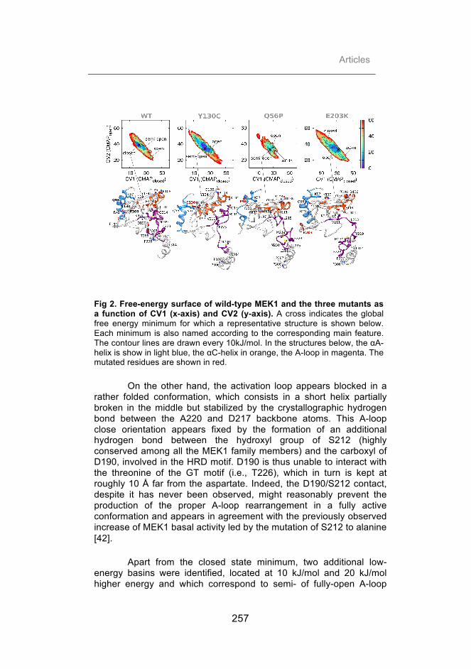
Articles
257
Fig 2. Free-energy surface of wild-type MEK1 and the three mutants as a function of CV1 (x-axis) and CV2 (y-axis). A cross indicates the global free energy minimum for which a representative structure is shown below. Each minimum is also named according to the corresponding main feature. The contour lines are drawn every 10kJ/mol. In the structures below, the αA-helix is show in light blue, the αC-helix in orange, the A-loop in magenta. The mutated residues are shown in red.
On the other hand, the activation loop appears blocked in a rather folded conformation, which consists in a short helix partially broken in the middle but stabilized by the crystallographic hydrogen bond between the A220 and D217 backbone atoms. This A-loop close orientation appears fixed by the formation of an additional hydrogen bond between the hydroxyl group of S212 (highly conserved among all the MEK1 family members) and the carboxyl of D190, involved in the HRD motif. D190 is thus unable to interact with the threonine of the GT motif (i.e., T226), which in turn is kept at roughly 10 Å far from the aspartate. Indeed, the D190/S212 contact, despite it has never been observed, might reasonably prevent the production of the proper A-loop rearrangement in a fully active conformation and appears in agreement with the previously observed increase of MEK1 basal activity led by the mutation of S212 to alanine [42].
Apart from the closed state minimum, two additional low-energy basins were identified, located at 10 kJ/mol and 20 kJ/mol higher energy and which correspond to semi- of fully-open A-loop

Description of protein plasticity: an example of biomedical interest
258
structures, respectively. In the semi-open state, the A-loop assumes an intermediate conformation between canonical open and closed ones. Thus, it adopts a reasonably closed but rather unfolded rearrangement in which only one turn of the original two-turn helix is kept folded. On the contrary, the fully open state recalls the A-loop observed in several protein kinases active structures although the αC-helix appears in the out-conformation, rotated out of the catalytic site (Fig S4).
It is worth to note that none of the states found hereby corresponds to the fully active structure, which confirms that the active conformation of WT, in absent of phosphorylation, is energetically unfavorable and infrequently sampled [43]. This is further supported by the comparison between the free energy barriers extracted by the closed to semi-open, semi-open to closed and semi-open to open transition, corresponding to around 30kJ/mol, 20kJ/mol and 40kJ/mol, respectively.
Y130C-MEK1 mutant
The Y130C mutant dramatically changes the conformational free-energy landscape projected along CV1 and CV2, shifting the equilibrium toward the active conformation. Unlike the WT, Y130C FES shows only one significantly broad minimum, corresponding to a rather heterogeneous bunch of structures that mainly differ in the conformation of A-loop. The most populated cluster is marked by a partially unfolded A-loop that forms a short one-turn helix at its N-terminal (still stabilized by L215/S212 backbone hydrogen bond) and an extended disordered region at its C-terminal. As shown in Fig 2, the single point mutation on the 130 residue causes the break of the hydrogen bond with the αA-helix Q56 and leads to a slight outward slipping of β4 and β5-strand, locked by the formation of a new salt bridge between D136 and R108 and coupled with Y134 side chain outward flipping. This leads to the distortion and inward shift of central region of the αC-helix and thus the weakening of some hydrophobic contacts with the A-loop (e.g., V211/I111 and V211/L115) which in turn lead to a partial unfolding of the A-loop, stabilized by a new K104/D217 salt bridge. Although the extent of the αC-helix turn results to be insufficient to prompt the formation of the salt bridge between E114 and the catalytic lysine K97, it does reduce the distance between the two catalytic residues which are placed at a distance of roughly 12 Å. On the other hand, the second cluster, much less populated than the previous one, shows a full distension of the A-loop and a partial deformation of the αC-helix in which the first

Articles
259
three turns are maintained while the other two appear totally disordered (Fig S4).
Interestingly, not only both the semi- and fully- open states are energetically very similar, suggesting that in the Y130C mutant, in contrast to the WT, are equally likely to occur but also they share the same basin representing alternative structures that can rapidly convert from one to the other.
Q56P-MEK1 mutant
Similarly to Y130C mutant, Q56P clearly shifts the equilibrium toward the active conformation, indicating that the mutant, unlike the WT, spends most of its time in an A-loop semi-open or open state. However, the changes observed in the conformational free-energy landscape appear even more dramatic than Y130C resulting in the complete loss of the basin corresponding to the closed state. Indeed, Q56P mutant FES shows three narrow minima located at roughly CV1 25-35, which are energetically equivalent and divided by rather similar energy barriers.
The most populated basin contains structures marked by (i) an almost-open A-loop that results in a full unfolded but not broadly extended conformation and (ii) the αC-helix rotated out of the catalytic site (αC-out conformation). Interestingly enough, the opening extent of A-loop caused by Q56P appears larger than the one observed in Y130C lower-energy basin. As shown in Fig 2, the mutation of glutamine 56 to a proline obviously prevents the formation of the native Q56/Y130 hydrogen bond and lead to an outward shift of the αA-helix, which in turn end with the switch of the H119 side chain with its backbone in the αA-helix/αC-helix hydrogen bond with K57 side chain. As a result, a slight drop on the C-terminal region of the αC-helix (stabilized by D136/R108 salt bridge, as found in Y130C) promotes the extensive unfolding of A-loop helical region. This new A-loop conformation is featured by the loss of several hydrophobic interactions with the αC-helix (such as V211/L115, V211/V117, M219/I99, L215/I111 and L215/L101) and sealed by the formation of new contacts, specific of the canonical open conformation (namely F223/L314 F223/L235 and M230/L314).
The second basin is characterized by a fully open conformation of A-loop and a partial unfolding of the αC-helix coupled with its rearrangement in an atypical orientation, which corresponds to an intermediate state rather equidistant between the canonical αC-in and αC-out conformation.

Description of protein plasticity: an example of biomedical interest
260
On the other hand, the third minimum corresponds to an ensemble of conformations in which the total extension of A-loop is combined by the stabilization of the αC-helix rotation toward the catalytic site similarly to many active kinases structures (Fig S4). Although the salt bridge between E114 and K97 is never formed, as in the fully active conformation, the side chains of the two catalytic residues are found at a minimum distance of 8.5 Å, which result remarkably lower than the one observed in the WT closed structure, equivalent to 17 Å. As shown in Fig 3B, this αC-helix orientation is triggered by the strong hydrophobic interaction between the αA-helix mutated residue P56 and the αC-helix L118 side chain and thus stabilized by the formation of a hydrogen bond between the H119 imidazole ring (released by the interaction with K57) and G213 backbone.
Fig 3. Comparison of (A) WT A-loop closed conformation with (B) Q56P and (C) E203K mutants structures of the free energy minima corresponding to the fully open state showing a partial αC-helix rotation toward the catalytic site (αC-in): the αA-helix is show in light blue, the P-loop in green, the αC-helix in orange, A-loop in magenta. The mutated residues are in red.
On the whole, the structural and energetic impact of Q56P mutant not only appears stronger than that exerted by Y130C mutant, but also induces additional effects on the αC-helix displacement, that were not observed in Y130C simulations. All these findings, in agreement with the effects previously observed with the MD, could be

Articles
261
related to its deeper response on the kinase activity observed in vitro biochemical studies [27] and the oncogenic nature of this mutation.
E203K-MEK1 mutant
Similarly to Y130C and Q56P mutants, E203K also induces a shift toward the active conformation. However, the changes observed in the CV1/CV2 FES appear even more striking than the mutations described above. Similarly to what observed for Q56P mutant, the fully closed state appears rather destabilized. On the contrary, the lower-energy basin contains an ensemble of structures, which are marked by an almost-open A-loop (resulting fully unfolded and partially extended) combined with the αC-helix rotated out of the catalytic site (Fig 2). Indeed the electrostatic repulsion between the positively charged K203 and R49 residues leads to the weakening of the contacts between the αA-helix and the core region which produces very similar effects as the ones observed in Q56P mutant. Thus, the formation of a new hydrogen bond between K57 side chain amine group and H119 backbone leads to an extensive distortion of the αC-helix (stabilized by D136/R108 salt bridge, as found in the other mutants), which in turn weakens the packing contacts with the A-loop (such as V211/L115, V211/V117, L215/I111 and L215/L101) and thus boosts its unfolded and opened conformation. Moreover, similarly to Q56P, the new A-loop state appears stabilized by new hydrophobic interaction involving M230 and F223 residues (e.g., M230/L314 and F223/P306 and F223/P307 contacts)
On the other hand, the FES reveals an additional minimum at CV1=38 and CV2=22 corresponding to the rather active conformation involving the complete unfolding of A-loop that appears in the fully extended conformation (as previously observed in Y130C and Q56P simulation), as well as a significant rearrangement of the αC-helix (as found only in Q56P simulation but missing in Y130C). Interestingly, although a complete αC-helix rotation was never observed and the salt bridge between E114 and K97 is never formed, the ensemble of states found in this basin corresponds to a quite stable intermediate state, which shows a partial unfolding of the αC-helix N-terminal region that might precedes the canonical αC-helix in-conformation. As shown in Fig 3C, the new αC-helix orientation is triggered by the formation of the hydrogen bond between the guanidine of arginine 49 and the amide group of the glutamine 56 residues causing an extensive rearrangement of the αA-helix and thus the break of the K57/H119 native contact. As a result, the above mentioned residues appear involved in two new hydrogen bonds, engaging E114 and Q116 respectively. This causes a partial unfolding and broad inward

Description of protein plasticity: an example of biomedical interest
262
switch of the αC-helix, which in turn is further stabilized by an additional hydrogen bond between Q110 and S212 residue. It also worth noting that, although E114 always remains at an average distance of roughly 15 Å from K97, its salt bridge with K57 observed herein might boost the inward rotation of E114 and thus prelude the E114/K97 contact formation.
In summary, the effect of E203K mutant appears deeper than that exerted by both Y130C and Q56P mutant. Moreover, as well as Q56P (but not Y130C) a significant αC-helix displacement was observed. All these findings, in agreement with the effects previously observed in the MD, could be related to the constitutive activation led by this mutation and its oncogenic nature.
Effect of the mutations on the DFG-motif in-to-out transition Wild type MEK1
Fig 4. Free-energy surface of wild-type MEK1 and the three mutants as a function of CV1 (x-axis) and CV3 (y-axis). Each minimum is named according to the corresponding main feature. The contour lines are drawn every 10 kJ/mol. In the structures below, the αA-helix is show in light blue, the P-loop in green, the αC-helix in orange, the A-loop in magenta. DFG-motif D208 and F209 residues are shown in yellow, and the mutated residues in red.
The wild type MEK1 free energy surface computed as function of CV1 and CV3 shows two deepest free-energy minima (Fig 4). These two minima correspond to ensembles of inactive structures where the αA-helix is folded and tightly packed against the core region, the αC-helix is rotated out of the active site and the A-loop is

Articles
263
rather folded. However, the global minimum corresponds to structures in which the DFG motif results in an active conformation with D208 forming a salt bridge with K95 and F209 tightly interacting with L180 (DFG-in conformation) and perfectly fitted in the hydrophobic pocket delimited by L118, I126, V127, L180, I186, L206 and V211.
In agreement with previous proposals that claimed the existence of certain degree of basal activity of MEK1 protein kinase, the DFG-in conformation was found to be energetically favorable with respect to the DFG-out conformation [44].
Y130C MEK1 mutant
In addition to the shift between the A-loop closed to open conformation, the free energy surface projected as a function of CV1 and CV3 also reveals a number of crucial differences between the WT and Y130C mutant (Fig 4). First of all, the basin corresponding to the DFG-in conformation is much broader than the corresponding basin for the WT, suggesting that this state can adopt many more conformations in the mutant than in the WT. However, the most remarkable change consists in a significant flattening of the free energy barrier for the DFG-in to DFG-out transition (reduced from roughly 40kJ/mol to 30kJ/mol). Indeed, this could promote the ADP release [45], which is known to be the rate-limiting step in kinase catalysis [46-47] and thus explain the catalytic activity increase observed in the mutant with respect to the WT. A further difference involves the appearing of a third local minimum, virtually absent in WT, that lies 10 kJ/mol above the other minima and corresponds to an intermediate conformation of the DFG flip transition in which the aspartate is pointing downward (Asp-down state). Interestingly, as shown in Fig 4, this state is triggered by the formation of a hydrogen bond between the K57 positively charged ε-amino group (released from the interaction with H119 side chain) and the backbone of F129 residue located on the underlying β4-strand. This contact stabilizes a new orientation of the bulky phenylalanine side chain, which in turn fills the position usually occupied by F209 residue when DFG-motif adopts an in-conformation.
Q56P MEK1 mutant
As in Y130C mutant, the DFG-motif flip also results deeply affected by the Q56P mutation. The free energy landscape computed as a function of CV1 and CV3 shows a total of three minima: the canonical DFG-out and DFG-in conformations, plus an additional local minimum, located at CV1=30 and CV3=-2.1. Interestingly, unlike

Description of protein plasticity: an example of biomedical interest
264
Y130C mutant, virtually all the structures corresponding to the third basin are characterized by an intermediate conformation of the DFG flip transition in which the aspartate is pointing upward (Asp-up state). As shown in Fig 4, the release of the αA-helix from the packing interaction with the kinase core region leads to the formation of a new hydrogen bond involving the backbone of F129 residue located on the underlying β4-strand. However, unlike the previous mutant, the contact with the H119 imidazole ring (released from the interaction with K57 side chain) stabilizes a different, less bulky orientation of the F129 side chain, that does not reject the DFG-motif aromatic residue but instead complete the hydrophobic pocket formed by I141, I139, L101, L115, V127, M143 and Y134 thus stabilizing this intermediate state.
Finally, it is worth noting that, although the free energy of the transition state between DFG-in and DFG-out conformation (equivalent to around 40 kJ/mol) does not significantly change with respect to the WT, the destabilization of the in-conformation, which lies 20kJ/mol above the out-conformation, considerably reduce the free energy barrier of the in-to-out transition (shifted from roughly 40kJ/mol to 20kJ/mol) and thus increase the ATP turnover rate. These findings could properly explain the increase in kinase activity observed with respect to both the WT and Y130C mutant. Nevertheless, DFG-out appears as a lower-energy state with respect to DFG-in conformation, which in principle would be in disagreement with what one would expect. Thus, deeper analysis will be needed in the future in order to clarify this issue.
E203K MEK1 mutant
As previously observed in A-loop close-to-open transition, the changes promoted by E203K on the CV1/CV3 free energy landscape appear more profound than the mutations described above. Similar to Y130C, E230K FES shows two energetically equivalent low-energy basins that correspond to the DFG-in and DFG-out conformations. However, as found in Q56P but not in Y130C, a third local minimum corresponding to an intermediate Asp-up state was observed. Interestingly, the molecular mechanism underlying its stabilization is virtually the same as the one previously described for Q56P, although it is triggered by different interactions. Indeed, the ion pair between E144 and K203 side chain residues favors the hydrogen bond formation between the H119 imidazole ring (released from the interaction with H119 side chain) and the backbone of F129. This in turn stabilizes a beneficial orientation of its side chain that can thus extensively interact with the DFG-motif phenylalanine and stabilize

Articles
265
this intermediate state. Moreover, as seen for Q56P, the new orientation of D208 results further fastened by a new hydrogen bond with N195 side chain (Fig 4).
Nevertheless, the most significant effect promoted by E203K mutation, consists in an additional flattening of the energy barrier for the DFG in-to-out transition (reduced to roughly to 10 kJ/mol) with respect to both the mutations described herein and which could be related to the MEK1 constitutive activation experimentally observed.
Discussion
Both oncogenic and CFC related mutations are bound to lead an increase of the MEK1 kinase activity although to different extents. We recently employed large-scale Molecular Dynamics (MD) in order to investigate the intrinsic propensity for inactive-to-active transition upon such mutations in different biologically relevant states. Indeed, MD simulations helped to disclose interesting dynamic events regarding MEK1 activation process and the specific destabilization effects caused by each mutation. Nevertheless, given the large conformation changes involved in protein kinases activation process and the limits of the conventional MD simulation in the exhaustive sampling of biomolecule energy landscape, some important aspects of this topic remained hidden. Thus, in order to enhance the exploration of the energy landscape of our system, a state-of-the-art enhanced sampling method, such as PTMetaD-WTE, was applied on the same case study.
As observed through MD simulations and in agreement with the experimental data reported, all the mutations described herein result in easing the shift of the equilibrium from the inactive to active state to a different extent. As a rule, the effects produced by E203K results generally more dramatic if compered with Q56P and even more with respect to Y130C. Nevertheless, in light of the present study, additional hints about specific molecular mechanisms accounting for oncogenic and non-oncogenic mutations were disclosed.
First of all, E203K and Q56P show remarkable effects on the closed-to-open transition of A-loop, stabilizing a virtually equivalent intermediate state in which the A-loop result completely unfolded while the consequences of Y130C mutation (although clearly appreciable) results less striking, promoting the stabilization of an intermediate state in which the A-loop is still partially folded. Another

Description of protein plasticity: an example of biomedical interest
266
significant difference arises from the αC-helix flexibility, consisting on a higher propensity for out-to-in transition observed only in oncogenic mutations (Q56P and E203K). Finally, it is worth noting the crucial effects on the DFG-motif plasticity caused by the mutations. Indeed, although by different mechanisms, all the mutants significantly flatten the energy barrier for the DFG in-to-out transition thus promoting the ADP release and increasing the ATP turnover rate. Nevertheless, the effects produced by E203K results generally more dramatic if compered with Q56P and even more with respect to Y130C.
Conclusions
In light of the present results, all the mutations described here reveal a double effect regarding the energetics of the A-loop as well as the DFG-motif: they might facilitate the protein kinase phosphorylation by stabilizing A-loop open conformations and increase the ATP turnover rate by lowering the free energy barrier of the DFG in-to-out transition. Overall, the present findings, combined to those previously reported, could help to rationalize and quantify the activating effects induced by Y130C, Q56P and E203K mutations, offer a mechanistic explanation to the different extent of MEK1 over-activation observed for oncogenic or CFC-related mutations and eventually open the path for the development of disease specific therapeutic approaches.
References 1. Manning G, Whyte DB, Martinez R, Hunter T, Sudarsanam S. The protein kinase complement of the human genome. Science. 2002 Dec 6;298(5600):1912-34. 2. Dentici ML, Sarkozy A, Pantaleoni F, Carta C, Lepri F, Ferese R, et al. Spectrum of MEK1 and MEK2 gene mutations in cardio-facio-cutaneous syndrome and genotype-phenotype correlations. Eur J Hum Genet. 2009 Jun;17(6):733-40. 3. Procaccia S, Seger R. Mek. In: Choi S, editor. Encyclopedia of Signaling Molecules: Springer New York; 2012. p. 1051-8. 4. Dhillon AS, Hagan S, Rath O, Kolch W. MAP kinase signalling pathways in cancer. Oncogene. 2007 May 14;26(22):3279-90. 5. Shields JM, Pruitt K, McFall A, Shaub A, Der CJ. Understanding Ras: 'it ain't over 'til it's over'. Trends Cell Biol. 2000 Apr;10(4):147-54. 6. Davies H, Bignell GR, Cox C, Stephens P, Edkins S, Clegg S, et al. Mutations of the BRAF gene in human cancer. Nature. 2002 Jun 27;417(6892):949-54. 7. Rajagopalan H, Bardelli A, Lengauer C, Kinzler KW, Vogelstein B, Velculescu VE. Tumorigenesis: RAF/RAS oncogenes and mismatch-repair status. Nature. 2002 Aug 29;418(6901):934.

Articles
267
8. Mercer KE, Pritchard CA. Raf proteins and cancer: B-Raf is identified as a mutational target. Biochim Biophys Acta. 2003 Jun 5;1653(1):25-40. 9. Singer G, Oldt R, 3rd, Cohen Y, Wang BG, Sidransky D, Kurman RJ, et al. Mutations in BRAF and KRAS characterize the development of low-grade ovarian serous carcinoma. J Natl Cancer Inst. 2003 Mar 19;95(6):484-6. 10. Vos MD, Martinez A, Ellis CA, Vallecorsa T, Clark GJ. The pro-apoptotic Ras effector Nore1 may serve as a Ras-regulated tumor suppressor in the lung. J Biol Chem. 2003 Jun 13;278(24):21938-43. 11. Sieben NL, Macropoulos P, Roemen GM, Kolkman-Uljee SM, Jan Fleuren G, Houmadi R, et al. In ovarian neoplasms, BRAF, but not KRAS, mutations are restricted to low-grade serous tumours. J Pathol. 2004 Mar;202(3):336-40. 12. Hingorani SR, Jacobetz MA, Robertson GP, Herlyn M, Tuveson DA. Suppression of BRAF(V599E) in human melanoma abrogates transformation. Cancer Res. 2003 Sep 1;63(17):5198-202. 13. Sharma A, Trivedi NR, Zimmerman MA, Tuveson DA, Smith CD, Robertson GP. Mutant V599EB-Raf regulates growth and vascular development of malignant melanoma tumors. Cancer Res. 2005 Mar 15;65(6):2412-21. 14. Hoeflich KP, Gray DC, Eby MT, Tien JY, Wong L, Bower J, et al. Oncogenic BRAF is required for tumor growth and maintenance in melanoma models. Cancer Res. 2006 Jan 15;66(2):999-1006. 15. Sumimoto H, Hirata K, Yamagata S, Miyoshi H, Miyagishi M, Taira K, et al. Effective inhibition of cell growth and invasion of melanoma by combined suppression of BRAF (V599E) and Skp2 with lentiviral RNAi. Int J Cancer. 2006 Jan 15;118(2):472-6. 16. Adams JA. Activation loop phosphorylation and catalysis in protein kinases: is there functional evidence for the autoinhibitor model? Biochemistry. 2003 Jan 28;42(3):601-7. 17. Sutto L, Marsili S, Gervasio FL. New advances in metadynamics. Wiley Interdisciplinary Reviews: Computational Molecular Science. 2012;2(5):771-9. 18. Laio A, Parrinello M. Escaping free-energy minima. Proc Natl Acad Sci U S A. 2002 Oct 1;99(20):12562-6. 19. Berteotti A, Cavalli A, Branduardi D, Gervasio FL, Recanatini M, Parrinello M. Protein conformational transitions: the closure mechanism of a kinase explored by atomistic simulations. J Am Chem Soc. 2009 Jan 14;131(1):244-50. 20. Lovera S, Sutto L, Boubeva R, Scapozza L, Dolker N, Gervasio FL. The different flexibility of c-Src and c-Abl kinases regulates the accessibility of a druggable inactive conformation. J Am Chem Soc. 2012 Feb 8;134(5):2496-9. 21. Sutto L, Gervasio FL. Effects of oncogenic mutations on the conformational free-energy landscape of EGFR kinase. Proc Natl Acad Sci U S A. 2013 Jun 25;110(26):10616-21. 22. Marks JL, Gong Y, Chitale D, Golas B, McLellan MD, Kasai Y, et al. Novel MEK1 mutation identified by mutational analysis of epidermal growth

Description of protein plasticity: an example of biomedical interest
268
factor receptor signaling pathway genes in lung adenocarcinoma. Cancer Res. 2008 Jul 15;68(14):5524-8. 23. Arcila ME, Drilon A, Sylvester BE, Lovly CM, Borsu L, Reva B, et al. MAP2K1 (MEK1) Mutations Define a Distinct Subset of Lung Adenocarcinoma Associated with Smoking. Clin Cancer Res. 2015 Apr 15;21(8):1935-43. 24. Bromberg-White JL, Andersen NJ, Duesbery NS. MEK genomics in development and disease. Brief Funct Genomics. 2012 Jul;11(4):300-10. 25. Delaney AM, Printen JA, Chen H, Fauman EB, Dudley DT. Identification of a novel mitogen-activated protein kinase kinase activation domain recognized by the inhibitor PD 184352. Mol Cell Biol. 2002 Nov;22(21):7593-602. 26. Nikolaev SI, Rimoldi D, Iseli C, Valsesia A, Robyr D, Gehrig C, et al. Exome sequencing identifies recurrent somatic MAP2K1 and MAP2K2 mutations in melanoma. Nat Genet. 2012 Feb;44(2):133-9. 27. Emery CM, Vijayendran KG, Zipser MC, Sawyer AM, Niu L, Kim JJ, et al. MEK1 mutations confer resistance to MEK and B-RAF inhibition. Proc Natl Acad Sci U S A. 2009 Dec 1;106(48):20411-6. 28. Rodriguez-Viciana P, Tetsu O, Tidyman WE, Estep AL, Conger BA, Cruz MS, et al. Germline mutations in genes within the MAPK pathway cause cardio-facio-cutaneous syndrome. Science. 2006 Mar 3;311(5765):1287-90. 29. Berman HM, Westbrook J, Feng Z, Gilliland G, Bhat TN, Weissig H, et al. The Protein Data Bank. Nucleic Acids Res. 2000 Jan 1;28(1):235-42. 30. Fischmann TO, Smith CK, Mayhood TW, Myers JE, Reichert P, Mannarino A, et al. Crystal structures of MEK1 binary and ternary complexes with nucleotides and inhibitors. Biochemistry. 2009 Mar 31;48(12):2661-74. 31. Pettersen EF, Goddard TD, Huang CC, Couch GS, Greenblatt DM, Meng EC, et al. UCSF Chimera--a visualization system for exploratory research and analysis. J Comput Chem. 2004 Oct;25(13):1605-12. 32. Ko TP, Jeng WY, Liu CI, Lai MD, Wu CL, Chang WJ, et al. Structures of human MST3 kinase in complex with adenine, ADP and Mn2+. Acta Crystallogr D Biol Crystallogr. 2010 Feb;66(Pt 2):145-54. 33. Altschul SF, Gish W, Miller W, Myers EW, Lipman DJ. Basic local alignment search tool. J Mol Biol. 1990 Oct 5;215(3):403-10. 34. Eswar N, Webb B, Marti-Renom MA, Madhusudhan MS, Eramian D, Shen MY, et al. Comparative protein structure modeling using MODELLER. Curr Protoc Protein Sci. 2007 Nov;Chapter 2:Unit 2 9. 35. Hess B, Kutzner C, van der Spoel D, Lindahl E. GROMACS 4: Algorithms for Highly Efficient, Load-Balanced, and Scalable Molecular Simulation. Journal of Chemical Theory and Computation. [doi: 10.1021/ct700301q]. 2008 2008/03/01;4(3):435-47. 36. Tribello GA, Bonomi M, Branduardi D, Camilloni C, Bussi G. PLUMED 2: New feathers for an old bird. Comput Phys Commun 2014;185(2):604-13. 37. Piana S, Lindorff-Larsen K, Shaw DE. How robust are protein folding simulations with respect to force field parameterization? Biophys J. 2011 May 4;100(9):L47-9. 38. Petersen HG. Accuracy and efficiency of the particle mesh Ewald method. The Journal of Chemical Physics. 1995;103(9):3668-79.

Articles
269
39. Bussi G, Donadio D, Parrinello M. Canonical sampling through velocity rescaling. J Chem Phys. 2007 Jan 7;126(1):014101. 40. Deighan M, Bonomi M, Pfaendtner J. Efficient Simulation of Explicitly Solvated Proteins in the Well-Tempered Ensemble. Journal of Chemical Theory and Computation. [doi: 10.1021/ct300297t]. 2012 2012/07/10;8(7):2189-92. 41. Tiwary P, Parrinello M. A time-independent free energy estimator for metadynamics. J Phys Chem B. 2015 Jan 22;119(3):736-42. 42. Gopalbhai K, Jansen G, Beauregard G, Whiteway M, Dumas F, Wu C, et al. Negative regulation of MAPKK by phosphorylation of a conserved serine residue equivalent to Ser212 of MEK1. J Biol Chem. 2003 Mar 7;278(10):8118-25. 43. Zheng CF, Guan KL. Activation of MEK family kinases requires phosphorylation of two conserved Ser/Thr residues. EMBO J. 1994 Mar 1;13(5):1123-31. 44. Yan M, Templeton DJ. Identification of 2 serine residues of MEK-1 that are differentially phosphorylated during activation by raf and MEK kinase. J Biol Chem. 1994 Jul 22;269(29):19067-73. 45. Kannan N, Neuwald AF. Did protein kinase regulatory mechanisms evolve through elaboration of a simple structural component? J Mol Biol. 2005 Sep 2;351(5):956-72. 46. Lew J, Taylor SS, Adams JA. Identification of a partially rate-determining step in the catalytic mechanism of cAMP-dependent protein kinase: a transient kinetic study using stopped-flow fluorescence spectroscopy. Biochemistry. 1997 Jun 3;36(22):6717-24. 47. Shaffer J, Sun G, Adams JA. Nucleotide release and associated conformational changes regulate function in the COOH-terminal Src kinase, Csk. Biochemistry. 2001 Sep 18;40(37):11149-55.

Description of protein plasticity: an example of biomedical interest
270
Supporting Informations
Fig S1. Free-energy mono-dimensional projection as a function of CV1, CV2 (the distance in contact map space to A-loop closed and open state, respectively) and CV3 (DFG-motif orientation): (A) wild-type MEK1 protein kinase and the three mutants: (B) Y130C, (C) Q56P and (D) E203K. In each plot three curves are shown, separated by 50 ns of simulation time and corresponding to the last 150 ns of each simulation. The final converged FES is shown in red.

Articles
271
Fig S2. Comparison between the free-energy profile in the mono-dimensional projection obtained by PLUMED module sumhills (in red) and the ones obtained by the time-independent re-weighting technique of Tiwary and Parrinello (in gray): CV1, CV2 (the distance in contact map space to the A-loop closed and open state, respectively) and CV3 (DFG-motif orientation) for (A) wild-type MEK1 protein kinase and the three mutants: (B) Y130C, (C) Q56P and (D) E203K.

Description of protein plasticity: an example of biomedical interest
272
Fig S3. Comparison between the free-energy profile in the bi-dimensional projection: (A) CV1, CV2 and (B) CV1, CV3 for wild-type MEK1 protein kinase and the three mutants. The contour lines are drawn every 10kJ/mol.
Fig S4. Free-energy surface of wild-type MEK1 and the three mutants as a function of CV1 (x-axis) and CV2 (y-axis). A cross indicates the global free energy minimum. The contour lines are drawn every 10kJ/mol. The representative structures of the secondary free energy minima are shown with the αA-helix in light blue, the P-loop in green, the αC-helix in orange, the A-loop in magenta.

4. Results summary

“if I don't know something, I look into it.”
Luis Pasteur
Results summary
275
4.1. Advances and new challenges in modeling of protein interactions
Given the growing interest in protein-protein interactions and the
technical advances in computational field, an increasing number of in
silico tools have been developed with the aim of (i) identifying
residues that significantly contribute to binding, and (ii) modeling
protein complexes starting from the isolated component structures
(docking problem). Testing and comparing these computational
methodologies is fundamental in order to assess their performance,
identify their limitations, and finally guide new developments in the
field. In this context, CAPRI experiment provides a common ground
for testing the predictive capability of currently available docking
methods.
Firstly, this section will be focused on the analysis of several
existing computational protocols for the characterization of protein-
protein interfaces. Secondly, the performance of our pyDock protocol
(Cheng et al., 2007) on the last CAPRI round (Lensink and Wodak,
2013) will be evaluated and discussed.
4.1.1. Energetic characterization of protein-protein interfaces
The performance of four different computational methods for
the characterization of protein-protein interfaces was assessed using
the recently solved complex between MEK1 and BRAF protein
kinases (PDB 4MNE) (Haling et al., 2014) as a test case. The
analysis was focused on (i) ConSurf (Glaser et al., 2003), based on

Advances and new challenges in modeling of protein interactions
276
the degree of conservation of each amino acid site among their close
sequence homologues (see section 1.2.6 and 3.1.1); (ii) pyDock
module pyDockNIP (Grosdidier and Fernandez-Recio, 2008), which
computes the tendency of a given residue to be located at the
interface based on rigid-body docking poses (see section 1.2.6); (iii)
the residue contribution to complex binding energy computed with
pyDock (upcoming publication); and finally, (iv) in silico Alanine (Ala) -
scanning based on free-energy calculations involving Molecular
Mechanics/Generalized Born Surface Area method (MM-GBSA) using
MMPBSA.py script (Miller et al., 2012) from AMBER14 tools
(Salomon-Ferrer et al., 2013).
Indeed, rather similar predictions were obtained using the
different methods (Fig 1 from section 3.1.1) although some
peculiarities should be highlighted. Firstly, MM-GBSA Ala-scanning
resulted much more computationally expensive and time consuming
than the other methods. In addition, NIP and ConSurf have the
considerable advantage that a priori knowledge of the complex is not
required. On the other side, pyDock binding energy residue
contribution and MM-GBSA Ala-scanning have the convenience of
providing a quantitative analysis that can be easily compared with the
experimental data. Finally, it is noteworthy that pyDock scheme
revealed a rather similar performance to MM-GBSA Ala-scanning in
the prediction of binding energy residue contribution but at much
lower computational cost.

Results summary
277
4.1.2. New challenges in protein docking: improving energetics and description of flexibility
pyDock docking program (Cheng et al., 2007) is regularly tested by
participating in the CAPRI experiment (http://www.ebi.ac.uk/msd-
srv/capri/), a community-wide blind prediction of macromolecular
interactions based on the three-dimensional structures of the
interacting proteins (see section 1.2.6).
The fifth CAPRI edition (2010-2012) (Lensink and Wodak,
2013) consisted of a total of 15 targets that, in addition to the
standard docking predictions, included binding affinity predictions and
free energy changes upon mutation (Moretti et al., 2013), as well as
prediction of sugar binding and interface water molecules (Lensink et
al., 2014). We have participated in all the proposed targets with high
success, since our models were globally placed among the top 5
ranked groups out of more than 60 participants (Lensink and Wodak,
2013) (Fig 1 from section 3.1.2 and Table S11A from Lensink and
Wodak, 2013).
Major determinants for the success were the generation of
docking poses with FTDock (Gabb et al., 1997) at a grid resolution of
0.7 Å (instead of 1.2 Å as in the past), as well as with SwarmDock
program (Moal and Bates, 2010) for part of the targets. In selected
targets (T47, T48 and T58), distance restraints were used, although
this hardly made a difference. In target T58, SAXS data were used for
complementary scoring with pyDockSAXS (Pons et al., 2010), which
slightly improved the predictive performance. Overall, pyDock
submitted consistently good models for all non-difficult cases,
although they were far from being trivial, since their subunits were in

Advances and new challenges in modeling of protein interactions
278
the unbound conformation or needed to be modeled based on
homology templates. In all cases but one, pyDock successful models
were ranked within the first five submitted solutions, being ranked as
first in two out of six successful cases.
As mentioned above, T47, T55, T56 and T57 were non-
standard targets for which new ad hoc protocols had to be developed.
Target T47 was a rather trivial modeling of a protein-protein complex
structure but the real challenge consisted in the prediction of the
location of water molecules within the complex interface. Our
approach was based on DOWSER ab initio optimization procedure
(Zhang and Hermans, 1996). Indeed, this choice was reasonably
successful, although a posteriori analysis showed that the most
successful approaches were based on deriving initial water positions
from interfaces of related complexes followed by energy minimization
(as in Nakamura’s group protocol). More details on the protocols used
by all the participants and the results obtained are reported in Lensink
et al., 2014. T55 and T56 targets aimed to predict the binding affinity
changes upon mutations on two designed influenza hemagglutinin
protein binders. By applying a machine learning protocol based on 85
energy descriptors, our predictions were placed within the top 3 out of
22 groups. Overall, the CAPRI community faced a big challenge with
these targets (Moretti et al., 2013). Finally, for T57, which involved the
prediction of a protein-sugar interaction, we developed a new protocol
based on a combination of different scoring functions, such as PScore
and AMBER (Case et al., 2005). Although no correct models were
submitted, at least one almost acceptable model was found (11.2 and
4.3 Å ligand- and interface- RMSD, respectively).
In the on-going (not yet evaluated) CAPRI edition (2013-to
date), one of the new challenges consisted in the prediction of eight

Results summary
279
protein/peptide complexes (T60-67), in which pyDock obtained highly
successful results as predictor and rather satisfying as server with 7
and 5 successfully predicted targets, respectively. However a much
more demanding test was recently proposed for CAPRI Round 30,
the first joint CASP-CAPRI experiment, consisting in 25 targets of
homo-oligomers from the CASP11 2014 Round. pyDock submitted at
least one acceptable model in 11 out of 12 easy homodimer targets,
either as predictor or scorers, while, as scorers, it successfully
predicted two out of six difficult homodimer targets and one out of two
hetero-complex targets. On the contrary, pyDock failed in the
prediction or evaluation of any tetramer target, where the inaccuracy
of the homology-built subunit models and the smaller pair-wise
interfaces severely limited the ability to derive the correct assembly
mode. Globally, pyDock predictions were placed among the top 10
ranked groups out of about 25 predictors, and among the top 5
ranked groups out of about 12 scorers participating in Round 30
(Table 4 from Lensink M et al., submitted).
Overall, the recent results of the CAPRI experiment
demonstrated a general robustness of current docking algorithms,
since their performance remained relatively consistent when facing
targets of different difficulty. Two of the most challenging targets were
T46, where subunits needed to be modelled based on distantly
related templates, and T51, where the isolated structures of the
docking partners were largely different from those of the native
complex. Regarding the estimation of binding affinity changes upon
mutation, although this is still far from being solved (as shown in T55
and T56), it is interesting to note that the incorporation of backbone
flexibility and more extensive sampling of side-chain conformations

Advances and new challenges in modeling of protein interactions
280
were in general positive for the predictions (Lensink and Wodak,
2013).
All these observations show that the intrinsic molecular
flexibility plays a crucial role in the protein-protein association
mechanism and thus should deserve special consideration for the
structural and energetic modeling of protein-protein complexes.
4.2. Protein plasticity improves protein-protein docking
Given the key role of conformational flexibility in protein-protein
association, accurate description of protein plasticity in docking
protocols appears crucial for the successful prediction of complex
structure (see sections 3.1.2 and 4.1.2). However, in addition to the
higher computational cost, the development of efficient flexible
docking algorithms is hampered by the limited theoretical knowledge
about the protein-protein association mechanism.
Thus, an important aim of this PhD thesis has been to study
the role of protein conformational heterogeneity in protein-protein
recognition and the exploration of ways to integrate unbound
conformational ensembles within protein-protein docking.

Results summary
281
4.2.1. Protein plasticity enhances protein-protein recognition
This study has explored whether a minimal description of the
conformational heterogeneity of the interacting proteins could
significantly improve their binding capabilities.
To this aim, first of all, small conformational ensembles for all
interacting proteins in the protein-protein docking benchmark 3.0
(Hwang et al., 2008) were automatically generated, starting from the
unbound docking partners, by using two distinct conformational
sampling procedures: (i) a fast energy optimization as implemented in
MODELLER (Eswar et al., 2007), and (ii) Molecular Dynamics
simulation, as implemented in AMBER package (Salomon-Ferrer et
al., 2013). As shown in Fig 1 and S2 from section 3.2.1, the deviation
of the interface atoms from the initial unbound structure were rather
modest in both approaches, reaching a maximum of about 1.2 and
2.7 Å RMSD values for MODELLER and MD ensembles, respectively.
Moreover, only in about 20% of the benchmark cases, the
conformational ensembles (either from MODELLER or MD simulation)
contained conformers that were significantly more similar to the native
conformation than the unbound X-ray structure. Interestingly, in the
majority of cases the conformational ensembles contained
conformers that showed better binding energy capabilities than the
unbound X-ray structure.
The following step was to evaluate whether the
conformational ensembles contained conformers that could be
beneficial for docking. To this aim, we selected these conformers that
seemed more promising for binding, either because of higher
similarity to the bound state or higher complementarity to the docking

Protein plasticity improves protein-protein docking
282
partner (in terms of binding energy or number of clashes). Finally, a
total of five conformers were collected for each benchmark case and
consequently used as input for rigid-body docking simulations, as
implemented in pyDock (Cheng et al., 2007). Surprisingly, the
conformers that were structurally more similar to the reference did not
yield better docking results than the unbound structures. On the
contrary, when using the conformers selected according to the
binding energy or the smallest number of clashes when in the native
orientation, the docking performance significantly improved (Fig 4A
from section 3.2.1). Moreover, a clear dependence of the docking
improvement on the conformational rearrangement of the interacting
proteins upon binding was observed: the ensemble success rates
were particularly good in the low-flexible cases, for which they
reached values similar to those when using the bound X-ray
structures; on the contrary, in rigid and highly-flexible cases the
selected conformers yielded similar results to the unbound structures
(Fig 4B and 4C from section 3.2.1).
Globally, the ensemble size and method of sampling did not
have a large effect on the docking performance. The selected
conformers from the 1000-member ensembles generated by
MODELLER or MD yielded similar results to those selected from the
100-member ensembles (S4 Fig from section 3.2.1), although a
small improvement was observed in the flexible cases when using the
larger ensembles (S5 Fig from section 3.2.1). Moreover, no better
docking performance was observed by extending the MD simulation
time to 100 ns (S1 Table from section 3.2.1). However, new
sampling approaches based on NMA did appear useful to produce a
significant improvement of the success rate in the most flexible cases
(Table 2 from section 3.2.1).

Results summary
283
In summary, this study showed that considering
conformational heterogeneity in the unbound state of the interacting
proteins can improve their binding capabilities in cases of moderate
unbound-to-bound mobility. In these cases, the existence of
conformers with better binding energy (not necessarily related to
bound geometries) in the native orientation is associated to a
significantly improvement in the docking predictions. These findings
set useful guidelines for the development of novel protocols in
practical docking prediction.
4.2.2. Development of a new protocol for ensemble docking
The previous study showed that a simple molecular mechanics
approach, as the one implemented in MODELLER program (Eswar et
al., 2007), can generate conformers with better binding properties and
thus improve docking predictions (see section 3.2.1). Based on this
study, we have devised a novel protocol to integrate unbound
conformational ensemble within protein-protein docking (using
pyDock) and tested it on a data set of 124 protein-protein docking
cases (Chen et al., 2003). In order to reduce the computational costs
associated with a cross-docking approach, a total of only 100 docking
runs were preformed randomly pairing every conformer from the
receptor with another one from the ligand. All the docking poses
generated were thus merged together and finally ranked according to
pyDock scoring function (filtering out similar conformations with ligand
RMSD cutoff 10 Å).
As shown in Fig 1A and 1B from section 3.2.2, ensemble
docking clearly showed better success rates (32.3%) than the

Protein plasticity improves protein-protein docking
284
unbound subunits (19.4%). Moreover, in order to provide a statistical
significance for these results, 100 different docking runs were
performed using random initial rotations of the unbound receptor and
ligand molecule. The resulting docking performance obtained (26.6%
success rate) stood halfway between the ensemble docking and the
unbound results. These results suggest that conformational
heterogeneity in the interacting subunits improves the binding
capabilities of the unbound X-ray structures. Interestingly, lowering
the number of random initial rotations (i.e., five rotations) did not
substantially change the docking performance (27.4%), showing the
importance of performing a small sampling of the position of the
starting molecules instead of using only the unbound X-ray structures.
Moreover, it was observed that the docking improvement when using
conformational ensembles depended on the flexibility of the
interacting proteins upon binding. Indeed, as shown in Fig 1C and 1D
from section 3.2.2, the largest improvement occurs in medium-
flexible cases, those with I-RMSDCα between 1.0 and 2.0 Å.
Finally, we also explored the question of which is the minimal
number of conformers that would be needed in order to observe a
significant improvement in the docking results. As shown in Fig. 2
from section 3.2.2, it was found that the docking performance
increases linearly with the size of the ensemble. For instance, when
only five conformers were used for the ensemble docking protocol,
the prediction success rates (24.2%) were similar to random.
Interestingly, when only the high-affinity, low-flexible cases are
considered, the docking success improves dramatically with just a few
conformer pairs, so that 30 conformers provide similar success rates
as 100 conformers. All these data suggest that the results might be
further improved by using a higher number of conformer pairs,

Results summary
285
increasing the number of receptor-ligand conformer combinations,
although for this, new algorithms would need to be developed and
optimized for high-performing computing in order to deal with the
humongous computational cost.
In conclusion, this study shows that a minimal conformational
heterogeneity can be used in a practical docking protocol to improve
the results of unbound docking. Moreover, this proved to have the
potential of further improving the predictive results by extending
conformational sampling and/or considering larger ensembles,
although this would involve an enormous computational cost.
4.3. Modeling protein interactions: application to cases of interest
The expertise acquired during the first part of this PhD thesis on the
theoretical basis of protein interactions has favored my participation
in several multidisciplinary projects, basically through the application
of computational methods to the modeling of protein interactions of
biological interest. Two of the most interesting collaborative projects
will be shown here in detail.
The first study consisted on the energetic characterization of
host-pathogen protein interactions, which are key steps of virtually
every infection process (i.e, pathogen replication and survival within
the host system). Such processes are typically achieved by precise
mechanisms developed by the pathogen to subvert and exploit
normal host cell processes, very often by mimicking specific host cell
Modeling protein interactions: application to cases of interest
286
interactions. The second project included the ab initio modeling of
redox complexes, which usually involve transient interactions
characterized by the existence of encounter complexes formed by
“microcollisions” that properly align the reactive groups.
4.3.1. Energetics of host-pathogen protein interactions
Legionella pneumophila effector Vacuolar Inhibitor protein D (VipD)
localizes to the host early endosomal membrane where succeeds in
binding GTPase Rab5 host protein, competing with its endogenous
ligands. This key interaction triggers the activation of the
phospholipase A1 (PLA1) activity of VipD, which in turn leads to the
alteration of the endosomial membranes composition and thereby
protect the pathogen from endosomial partitioning (Gaspar and
Machner, 2014). Thus, the energetic description of such interaction as
well as the disclosure of the molecular basis of this host-pathogen
competing process is of fundamental importance to open the path for
the development of novel antimicrobial therapeutics.
For this purpose, systematic in silico Alanine scanning
calculations were performed on all the interface residues of GTPase
Rab5 in complex with Legionella pneumophila VipD as well as in
complex with the endogenous human ligand proteins EEA1,
RAbaptin-5 and Rabenosyn-5. Indeed, the analysis of the VipD-Rab5
interface pinpointed a core of hydrophobic contacts complemented by
several polar interactions in the surrounding rim area (Fig 3B from
section 3.3.1). In particular, the VipD binding epitope in Rab5
included non-polar residues in the switch I (I54, G55, A56 and F58),
the interswitch (W75) and the switch II element (Y83, L86, M89 and

Results summary
287
Y90) as well as polar/charged residues in the interswitch (T60, K71
and E73) and switch II element (R82 and R92). On the other hand,
the corresponding epitope in VipD included several hydrophobic
residues in helixα17 (F442, A446, A450 and L454) and in helixα18
(Y473, I480, V483) that wrapped around an elongated hydrophobic
path in Rab5 formed by the conserved triad (F58, W75 and Y90) and
L86.
Moreover, a much more interesting observation arose from
the comparison with the endogenous Rab5 complexes, which
revealed a shared hydrophobic core (F58, W75 and Y90)
complemented by a unique polar interaction in VipD-Rab5 interface
surrounding this hydrophobic triad (R92 from Rab5 with D484 and
D479 from VipD) and which provide a rather stronger energetic
contribution to the interaction in comparison with the other cellular
ligands (Fig 7A from section 3.3.1). The role of R92 in VipD-Rab5
complex (not existing in those between Rab5 and the endogenous
ligands) was confirmed by experimental mutagenesis. These findings
offer a useful help for the development of novel treatments aimed at
selectively blocking the VipD activation process rather than the
enzyme's active site.
4.3.2. Modeling redox complexes by docking
The interaction of photosystem I (PSI) with electron transfer proteins
(such as plastocyanin or cytochrome) catalyzes the first step of the
photosynthesis process. Unlike most cyanobacteria and unicellular
green algae, Phaeodactylum tricornutum alga, as most diatoms,
lacks plastocyanin and thus leaves cytochrome c6 (Cyt) as the only
electron donor to photosystem I (Akazaki et al., 2009). Nevertheless,

Modeling protein interactions: application to cases of interest
288
diatom PSI is still able to recognize and functionally interact with
different eukaryotic acidic plastocyanins, although with lower
efficiency than the native electron carrier (Bernal-Bayard et al., 2013).
Thus, the principal goal of this study was to understand the structural
and energetic determinants of the differences in the efficiency
observed in diatoms PSI reduction with respect to the green systems.
To this aim, docking simulations were initially performed
between the homology‐built PSI model from Phaeodactylum and both
the native and the green alga Monoraphidium braunii Cyt structures.
Additional docking analyses were performed to investigate the
interaction of PSI from Phaeodactylum with the Pc of green alga
Chlamydomonas reinhardtii and particular efforts were addressed to
identify energetic differences between the wild type and some
mutants designed to mimic Phaeodactylum Cyt electrostatic
properties (i.e., E85K, Q88R, E85K/Q88R, E85V and V93K).
The docking simulations between PSI and Cyt from
Monoraphidium yielded a much larger population of low‐energy
productive orientations and showed better binding energy values as
compared with the native Phaeodactylum (Table 2 and Fig S3 from
section 3.3.2). Further analyses revealed that the Monoraphidium
Cyt best-energy productive docking model was stabilized by
electrostatic interactions between PsaA residues R747 and R648 and
Cyt residues D42 and H30, which corresponded to an alanine and a
lysine (i.e., A86 and K74) in Phaeodactylum Cyt. Indeed, these
differences can explain why an equivalent docking orientation would
be energetically penalized in the interaction with Phaeodactylum PSI
(Fig 5 from section 3.3.2).

Results summary
289
On the other hand, in agreement with its lower similarity to
Cyt electrostatic patches, the wild type Pc yielded a much larger
population of low-energy less productive docking orientations in
comparison with some of the mutants, such as E85K and Q88R.
Indeed, these orientations revealed to be stabilized by electrostatic
interactions between PSI positively charged residues (namely R747,
R648 and K638) with Chlamydomonas Pc E85 and Q88 residues (Fig
6 from section 3.3.2). On the contrary, the loss of these contacts by
the substitution of E85 and Q88 with positively charged residues
facilitated a higher population of the more productive binding modes,
as observed in the docking landscapes of the E85K and Q88R Pc
mutants.
All these results not only improve the understanding of the
mechanism and energetics of PSI reduction but also shed new light
on the evolution of the electron transfer mechanism to PSI in the
different branches of the evolutionary tree of photosynthetic oxygenic
organisms.
4.4. Description of protein plasticity: an example of biomedical interest
In order to understand the mechanistic aspects of several biological
processes, such as the functional effects of a single mutation, the
consideration of protein plasticity is of paramount importance. Protein
kinases represent a paradigmatic example of the importance of a
close link between dynamics and function. These enzymes regularly
switch between characteristic inactive and active states undergoing

Description of protein plasticity: an example of biomedical interest
290
large conformational changes, whose complete computational
description is still challenging. Standard all-atom Molecular Dynamics
(MD) simulations are useful computational tools to explore small-to-
medium conformational rearrangement, but large conformational
transitions in proteins can only be described by enhanced sampling
methods.
Thus, this PhD thesis concluded with the application of
conventional Molecular Dynamics (MD) and state-of-the-art
enhanced sampling using metadynamics to elucidate the effects at
molecular level of pathological mutations in MEK1 protein kinase
(Y130C, exclusively associated with Cardiofaciocutaneous- (CFC)
syndrome; E203K, exclusively related to cancer; and Q56P, observed
in both diseases) (see sections 3.4.1 and 3.4.2).
Structural and dynamic effects of pathological mutations by extended Molecular Dynamics
In order to understand the effect of the above mentioned pathological
mutations at molecular level as well as their impact on the intrinsic
propensity for MEK1 inactive-to-active transition, 1-µs-long MD
simulations were initially performed on the MEK1 unphosphorylated
apo as well as on the phosphorylated ATP-bound state. Then, four
additional 1-µs-long simulations were run using PTmetaD-WTE
protocol (Deighan et al., 2012), an enhanced sampling approach
which combines parallel tempering with well-tempered metadynamics.
According to the MD simulations, all the mutations described
herein resulted in easing the inactive-to-active transition in both the
unphosphorylated apo and the phosphorylated ATP-bound MEK1

Results summary
291
state. More in detail, in the basal state they increased αA-helix
structural flexibility, promoting both its partial unfolding and the loose
of αA-helix/core native contacts (e.g., Y130/Q56, R49/E203) (Fig 2,
S1 and Table 1 from section 3.4.1). However, a distinctive effect was
found in Q56P with respect to Y103C and E203K mutants, involving
the increase of P-loop flexibility, which could be causing a higher
cofactor turnover (Fig 3 and Table 1 from section 3.4.1). Over-
activating effects were also found in the phosphorylated A-loop,
where all of the mutants studied herein induced a significant increase
of the A-loop flexibility as compared with that in the WT (Fig 5 and
Table 1 from section 3.4.1) and favoured a dramatic destabilization
of the hydrogen bond between αC-helix E114 and A-loop Q214
residues, whose interacting frequency dropped from 85% in the WT to
a range between 29 and 54% for the mutants (Fig S2 and Table 1
from section 3.4.1). All these data suggest that the mutants could
result in the acceleration of active-to-inactive process with significant
impact on both the biological state analyzed. Nevertheless, the effects
produced by E203K resulted more dramatic if compared with Q56P
and even more with respect to Y130C.
In agreement with MD, the free-energy landscapes obtained
using PTMetaD-WTE confirmed that all the mutations seemed to
favour the inactive to active state transition, although to a different
extent. In addition, new hints about specific molecular mechanisms
accounting for oncogenic and non-oncogenic mutations were
discovered. First of all, E203K and Q56P showed significant effects
on the closed-to-open transition of the A-loop, stabilizing an
intermediate state in which the A-loop resulted completely unfolded.
On the other hand, Y130C mutation showed milder effects (although
clearly appreciable) on the stabilization of an intermediate state in

Description of protein plasticity: an example of biomedical interest
292
which the A-loop is still partially folded (Fig 2 from section 3.4.2).
Regarding αC-helix flexibility, a higher propensity for out-to-in
transition was observed only in oncogenic mutations (Q56P and
E203K) (Fig 3 from section 3.4.2). Finally, it is worth noting the
crucial effects of the mutations on the DFG-motif plasticity. Indeed, all
the mutants, although through different mechanisms, significantly
flattened the energy barrier for the DFG in-to-out transition, which
could promote the ADP release and increase the ATP turnover rate.
In conclusion, the combination of MD and PTMetaD-WTE
simulations could help to rationalize and quantify the activating effects
induced by Y130C, Q56P and E203K pathological mutations in
MEK1, as well as to propose a mechanistic explanation to the
different extent of MEK1 over-activation observed for oncogenic or
CFC-related mutations and eventually open the path for the
development of disease specific therapeutic approaches.

5. Discussion

“The important thing is not to stop questioning.”
Albert Einstein

Discussion
295
After sequencing the complete genomes of several organisms we are
starting to unravel their intricate protein-protein interaction networks,
which is essential to understand biological processes at molecular
level, with the ultimate goal of contributing to improve therapeutic
intervention. In this context, one of the current biological challenges is
to provide structural details at atomic level for such interactomes.
However, given the intrinsic limitations of available experimental
methods to determine 3D structures (e.g., X-ray crystallography or
NMR), large-scale structural determination of complete interactomes
seems beyond current capabilities.
Fortunately, computational methods can complement
experimental efforts by providing structural and energetic large-scale
modeling of protein interactions. In spite of the most recent advances
in computational modeling, many important challenges remain.
Among them, efficient consideration of conformational flexibility is
arguably the most important problem to solve in order to improve the
structural and energetics modeling of proteins and their interactions.
The consideration of protein dynamics appears essential in several in
silico studies, from the prediction of their interactions to the study of
the mechanistic aspects of their function. Indeed, this issue is not well
addressed in most of the currently available docking programs, which
this is actually one of the main limitations in their predictive
performance.
The work of this thesis has focused on analyzing the
importance of structural, energetics and dynamic aspects of protein
interactions, the development of new computational tools to help
solving current problems, and the application of computational
modeling to cases of biological and biomedical interest.

Energetic characterization of protein-protein interfaces
296
5.1. Energetic characterization of protein-protein interfaces: advances and new challenges
Residues in protein-protein interfaces do not equally contribute to the
overall binding energy; in fact a few hot-spot residues typically
contribute the most to the binding energy (Clackson and Wells, 1995;
Bogan and Thorn, 1998). The identification of such residues and thus
the functional characterization of protein interactions at molecular
level is mandatory, not only to understand biological and pathological
phenomena, but also to design improved, or even new interfaces, or
to develop new therapeutic approaches (Wells and McClendon, 2007;
Kar et al., 2012; Thangudu et al., 2012).
Considering that the experimental characterization of protein
interfaces remains expensive, time-consuming and labor-intensive,
computational approaches represent a significant breakthrough in
proteomics, assisting or even replacing experimental efforts. Thanks
to the technological advances in computing and data processing,
these techniques now cover a vast range of protocols, from the
estimation of the evolutionary conservation of amino acid positions in
a protein, to the energetic contribution of each residue to the binding
affinity. In this thesis, several existing computational protocols to
model the phylogenetic, structural and energetic properties of
residues within protein-protein interfaces have been reviewed and
their performance compared, using MEK1-BRAF complex (PDB ID
4MNE) as example (see section 3.1.1). Although the different
protocols differed in time and computational costs, type of the
analysis provided (e.g., qualitative or quantitative) and the degree of
structural information required, the predictions obtained were quite
consistent between themselves (Fig 1 from section 3.1.1).

Discussion
297
Interestingly, a new protocol for pyDock per-residue free energy
decomposition had a similar performance to the widely used MM-
GBSA Ala-scanning although at much lower computational cost.
Based on this observation, future systematic comparisons between
these two methods would be interesting in order to produce new
developments in the field.
Figure 11. (A) SidD interface surface contacting AMPylated Rab1, colored according to the predicted ΔΔG; (B) In vitro de-AMPylation assay on selected mutants located within and outside the interface. From Chen et al., 2013.
Apart from the above cited study, MM-GBSA Ala-scanning
protocol has been successfully applied in an additional
multidisciplinary project during this thesis for the characterization of
Rab1/SidD complex in collaboration with Aitor Hierro research group
(CIC bioGUNE) (Chen et al., 2013). As shown in Figure 11 in silico
predicted hot-spots are in substantial agreement with the
experimental data on in vitro SidD-mediated de-AMPylation of Rab1.
Overall, all these observations confirm that in silico protocols
provide a reliable aid to the energetic characterization of protein-

Energetic characterization of protein-protein interfaces
298
protein interfaces.
5.2. Structural prediction of protein interactions: from energetics to molecular flexibility and other challenges
The last CAPRI edition, in which I have actively participated, sparked
a large number of ideas and new strategies for structural modeling of
protein interactions. In addition, our overall experience has been
highly rewarding, since pyDock docking scheme confirmed its high
performance in protein complexes prediction being placed among the
top5 ranked groups out of more than 60 participants (see section
3.1.2 and Table 3).
Table 3. Overall pyDock performance among the top 10 ranked groups.
Rank Group #Targets / *** + ** + * 1 Bonvin 9 / 1 *** + 3 ** + 5 * 2 Bates 8 / 2 ** + 6 * 3 Vakser 7 / 1 *** + 6 * 4 Vajda 6 / 2 *** + 3 ** + 1 * 5 Fernandez-Recio 6 / 1 *** + 3 ** + 2 * 5 Shen 6 / 1 *** + 3 ** + 2 * 7 Zou 6 / 1 *** + 2 ** + 3 * 8 Zacharias 6 / 1 *** + 5 * 9 ClusPro 6 / 4 ** + 2 * 10 Eisenstein 5 / 1 *** + 2 ** + 2 *
Predictions are classified as acceptable (*), medium (**), and high (***).
Besides this, many targets were proposed that go beyond the
traditional protein-protein complex structure prediction, but that
constitute important current challenges in modeling protein
interactions. For instance, regarding the prediction of binding energy
changes upon mutation, the application of multiparametric regression
models, as implemented in our protocol, showed excellent results.

Discussion
299
The overall evaluation showed that current protocols are more
successful in predicting deleterious mutations than beneficial ones.
Furthermore, the best procedures were those that took into account
the effect of the mutation on the stability of the monomer, and that
considered both sterical and energetic parameters (i.e, packing
metrics, Lennard–Jones type potentials, electrostatic and solvation
terms). Finally, an additional advantage was conferred by extensive
sampling of side chain conformations and the incorporation of some
backbone flexibility (Moretti et al., 2013).
The general analysis of the protocols used by the CAPRI
community for predicting the location of water molecules within the
complex interface showed interesting results. First of all, it seems that
high- to medium-quality models for the protein complex are required
for successful interface water predictions. Moreover, the combination
of established molecular mechanics force fields with some
conformational sampling step and a final energy minimization, as well
as the consideration of initial water positions derived from interfaces
of related complexes, proved to be more successful than much
simpler water placement methods (Lensink et al., 2014).
Regarding last CAPRI round in combination with CASP (i.e.,
Round 30) for the modeling of homo-oligomeric proteins, the majority
of participants showed a general poor success in docking of
homology-built subunits (especially if modeled on distantly related
templates) (Lensink et al, submitted), thus confirming what was
indeed previously reported (Lensink and Wodak, 2010) and showing
again that more work is needed in this direction.
As for the most traditional targets involving the prediction of a
protein-protein complex structure, which is still the main focus of

Structural prediction of protein interactions
300
CAPRI, the analysis of the results showed that dealing with protein
flexibility in docking simulations still remains a major challenge.
Indeed, docking performance dramatically drops in cases that
undergo medium-to-large conformational changes upon binding or in
which the interacting subunits need to be modeled based on low-
homology templates (Lensink and Wodak, 2013; Pallara et al., 2013).
This, in principle, could be explained by poor geometrical (and thus,
energetic) complementarity of the docking partners in the unbound
form.
Therefore, in order to develop new strategies to include
flexibility in docking, a much better understanding of molecular
recognition process at structural and energetic level is required, which
has been a major motivation in this PhD thesis.
5.3. The role of conformational heterogeneity in protein-protein association process
Conformers providing better binding energy in the native orientation are more likely to produce more effective docking encounters.
The systematic study on the role of conformational heterogeneity in
protein-protein association process, as described in section 3.2.1 and summarized in Figure 12A, showed that sets of discrete
conformers representing the conformational heterogeneity of the
unbound structure yielded better docking results than the unbound
structures themselves.

Discussion
301
Figure 12. Schematic representation of (A) selected conformer docking (selection of the best ensemble conformers according to quality criteria based on the complex native orientation: Cα-RMSD, Int-RMSD, binding energy with the bound partner, binding energy with the unbound partner, number of clashes with respect to the bound partner); (B) ensemble docking (combination of every conformer from the receptor ensemble with another one randomly selected from the ligand ensemble)
In this context, it is interesting to analyze the reasons for the
success of such conformers. Surprisingly, the conformers that were
structurally more similar to the reference did not yield better docking
results than the unbound structures. On the other side, selected
conformers with the best binding energy in the native orientation
performed better than the unbound structures. Thus, the capacity to
provide favorable binding energy in the native orientation seems to be
a major determinant for the success of docking, as opposed to the

The role of conformational heterogeneity in PP association
302
criterion of structural similarity to the native conformation. This might
be due to the fact that in the majority of cases, ensembles were not
exploring the conformational space close to the bound state, because
sampling was limited to a region in the vicinity of the unbound.
Indeed, it was found that 90% of the successful cases (i.e.,
near-native solution ranked within top 10) had average conformer
binding energy < -20.0 a.u. in the native orientation. Actually, 71% of
the docking cases with conformers with binding energy in the native
orientation < -40.0 a.u. were successful. This confirms that the
existence of conformers with good optimal energy in the native
orientation is determining the success of docking. In many of the
cases that significantly improved (i.e., those which had a near-native
ranked ≤ 10 when using the energy-based selected conformers but
not when using the unbound structures), the unbound structures in
the native orientation had binding energy < -20.0 a.u. but were not
successful in unbound docking. In these cases, a little bit of
conformational sampling seems to be sufficient to generate
conformers that significantly improve docking results (Fig 5 from
section 3.1.2).
Binding mechanism: What can we learn from docking?
Docking simulations can provide interesting insights into the protein
binding mechanism. Indeed, the different possible mechanisms that
have been proposed for protein-protein association could be
described by existing computational approaches (Fig 6 from section
3.1.2). In this context, several possible scenarios can be considered.
For protein complexes following rigid association (similar to lock-and-
key mechanism), the use of rigid-body docking with the unbound
subunits could be a suitable approach to describe the binding process

Discussion
303
and obtain good predictive models. Indeed, this seems to be the case
for complexes with small conformational changes between unbound
and bound states (I-RMSDCα < 0.5 Å), in which unbound docking
already gives as similar success rates as bound docking. In these
cases, the use of energy-based selected conformers from unbound
ensembles gave also similarly good docking rates as unbound and
bound docking. Indeed, for these cases, the unbound proteins in the
native orientation generally showed good average binding energy
towards the bound partner, not far from that of the bound structures.
Consistently, the average binding energy of the best conformers was
typically similar to that of the unbound or bound pairs. However, when
conformers were selected by criteria based on the structural similarity
to bound state, docking success rates were much worse than
unbound or bound docking, because in these cases conformational
heterogeneity was more likely to produce conformers that are further
from the bound state than the unbound one (given that the unbound
was already close to the bound state). Indeed, in none of these cases
there were a single conformer that was significantly closer to the
bound state than the unbound structure.
On the other side, it is known that in complexes involving
flexible association, rigid-body docking with the unbound structures is
not going to produce correct models. For such cases, different binding
mechanisms have been proposed, such as conformational-selection
or induced-fit. For cases following the conformational-selection
mechanism, the hypothesis is that the unbound proteins naturally
sample a variety of conformational states, a subset of which are
suitable to bind the other protein. Therefore, for these cases the use
of precomputed unbound ensembles describing conformational
variability of free proteins in solution should generate conformers that

The role of conformational heterogeneity in PP association
304
would improve rigid-body docking predictions with respect to those
with the unbound structures. Indeed, this is the case for the
complexes undergoing unbound to bound transitions between 0.5 and
1.0 Å I-RMSDCα. In these cases, selected conformers from the
unbound ensembles yielded much better docking predictions than the
unbound structures, virtually achieving the success of bound docking.
For cases undergoing unbound-to-bound transition between
1.0 and 2.0 Å I-RMSDCα, the use of unbound ensembles also
improved the predictions with respect to the unbound docking results,
although to a lesser extent. In these cases, the binding energy of the
selected pair of conformers in the native orientation was typically
better than the unbound structures and similar to the bound
structures. Some residues in the best pair of conformers show better
energy contribution than in the unbound state, which explains why
this specific pair of conformers improves docking results. In these
cases, the existence of a sub-population of "active" conformers, with
good binding capabilities towards the bound partner, would be
consistent with a conformational-selection mechanism. The fact that
these conformers with improved binding capabilities are not
geometrically closer to the bound state seems counterintuitive.
However, recent views of binding mechanism show that active
conformers that are selected by partner (initial encounters) do not
necessarily need to be in the bound state, as they can adjust their
conformations during the association process (Csermely et al., 2010).
Indeed, these docking poses are likely to represent these initial
encounters between the most populated conformational states of the
interacting proteins and would be compatible with this extended
conformational selection view (Csermely et al., 2010). However, in
other cases the limited conformational sampling used might not be

Discussion
305
sufficient to explore all conformational states available in solution and
therefore the specific binding mechanism cannot be easily identified.
As for the other extreme, in cases following an induced-fit
mechanism the bound complexes would only be obtained after
rearrangement of the interfaces when interacting proteins are
approaching to each other, in which case the use of precomputed
conformational ensembles in docking (even if generated by
exhaustive sampling) would not produce favorable encounters around
the native complex structure. This seems the case for complexes
undergoing unbound to bound transitions above 3.0 Å I-RMSDCα. In
all these cases, rigid-body docking, either with unbound structures or
with selected conformers, failed to reproduce the experimental
complex structure. In this context, an emblematic case is 1IRA, in
which binding energy of the selected pair of conformers is similar to
the unbound structures and much worse than the bound
conformation. For these complexes, the use of precomputed unbound
ensembles does not seem to see advantageous, and they would
probably need to include flexibility during docking search, mimicking
the induced-fit mechanism. However, in the flexible category (i.e.,
unbound to bound transitions between 2.0 and 3.0 Å I-RMSDCα,),
there are also other cases (i.e., 1ACB), which seem to follow the
(extended) conformational-selection mechanism, since the use of
conformers helped to improve the docking results, and the
conformers showed better energy than the unbound structures.
Again, there might be other complexes under this category that could
still follow the conformational-selection mechanism, but our
conformational search was not sufficient to sample conformations that
may exist in solution and could be productive for docking. This seems
to be the case for 1I2M, in which ensembles based on MODELLER

The role of conformational heterogeneity in PP association
306
did not produce pairs of conformers with sufficiently good binding
energy in the native orientation, but the docking rates improved when
using extended sampling based on NMA.
Of course the use of docking calculations to learn about the
binding mechanism has some limitations, in addition to the ones
already mentioned. The timescale of transitions between inactive and
active conformers can play an important role in controlling the binding
mechanism (Zhou, 2010). In the work presented in this thesis, we can
only assume that our ensembles are formed by conformers that are
the most accessible in solution, so the existence of active conformers
that can be preferentially selected by the bound partner would be
compatible (but not exclusively) with a mainly conformational-
selection mechanism. However, in a situation in which the active
conformers are not easily accessible, as those that can only be
generated with extended sampling, we could not identify the type of
mechanism unless transition rates between conformers were
considered.
5.4. Integration of unbound conformational ensembles in protein-protein docking: development and benchmarking of a novel protocol
Ensemble docking provides more near-native poses and as a consequence better predictive rates
In this thesis, we described a protocol to efficiently use in docking the
conformational ensembles generated by MODELLER minimization
(MM) from the unbound subunits (see section 3.2.2 and Figure 12B).
The results showed that a minimal structural heterogeneity provided

Discussion
307
by such ensembles can improve docking results with respect to the
unbound X-ray structures.
Thus, in order to further study the reasons of such
improvement, it was first explored for each receptor-ligand pair of
conformers whether the docking energy of the best near-native
solution (determinant for the docking success) depended on the
number of near-native solutions obtained for such conformer pair (Fig
S1 from section 3.2.2). For the majority of cases it can be observed
that the higher the number of near-native solutions generated by a
given conformer pair, the higher the probability of obtaining good
docking energies by such near-native solutions. In this line, the
conformers that generated more near-native solutions than the
unbound structure provided in general better near-native docking
energies than those generated by the unbound structure. It was also
observed that the bound X-ray structure typically yields more near-
native solutions and with better docking energy than the unbound.
Interestingly, for many cases, there were a few conformers that
generated even more near-native solutions than the bound structure
(e.g., 1NSN, 1DFJ, 1I2M). Therefore, increasing the ratio between
near-native solutions and false positives is the main reason for the
beneficial effect of some of the conformers found in the precomputed
unbound ensembles. Indeed, this is consistent with the previously
observed correlation between the number of near-native solutions
generated by docking and the predictive success rates (Pons et al.,
2010b).
For each case, the percentage of conformers that produced
more near-native solutions than the unbound structure was also a
determinant of the ensemble docking success. Cases with more than

Integration of unbound conformational ensembles in PP docking
308
70% of the conformers producing more near-native solutions than the
unbound structure showed much higher success rate (72.7%) than
the unbound docking (36.4%), almost reaching the optimal bound
docking results. On the contrary, in those benchmark cases in which
there were less than 70% of conformers that produced more near-
native solutions, ensemble docking had similar success rate (41.2%)
to when using unbound X-ray structures (35.3%) and far from the
bound docking results.
Successful conformers are not necessarily more similar to bound state
As above mentioned, the consideration of conformational
heterogeneity in docking can increase the number of near-native
solutions generated by FTDock, as well as their docking energy,
which is a key determinant for the docking success of each conformer
pair. However, neither the number of near-native solutions found for
each conformer pair or their best binding energy (and as a
consequence the success rate) depended on the similarity of such
conformer pair to the bound state (Figs S2 and S3 from section
3.2.1). This is in agreement with previous findings (see sections
3.2.2) and is consistent with an extended conformational-selection
mechanism (Csermely et al., 2010). Nevertheless, in a few cases
(e.g., 1AY7, 1MAH) we did observe that the most successful pair of
conformers were the most similar to the bound state in terms of the
RMSD of the predicted anchor residues (Rajamani et al., 2004;
Meireles et al., 2010) (Fig 3 from section 3.2.2). In these cases,
unbound ensembles can explore bound-like orientations of specific
interface key residues that can improve the interacting capability of
such conformers upon the docking and thus yield better docking
results with respect to the unbound.

Discussion
309
Conformer pairs providing better binding energy in the native orientation are more likely to improve docking results
We reported that the structural similarity of the docking partners to the
native conformation is not determinant for the docking success in
general. However, what we found is that the better the binding energy
of a conformer pair in the native orientation (i.e., after optimal
superimposition on complex structure), the better the docking energy
of the produced near-native solutions (and therefore the success
rates) (Fig S5 from section 3.2.2), which is also in agreement with
our previous findings (see section 3.2.1). Thus, capacity to provide
favorable binding energy in the native orientation seems to be a major
determinant for the success of a given conformer pair.
In this regard, we found that for a given case the predictive
success (i.e., best ranked near-native solution) of unbound docking
strongly depended on the expected optimal binding energy of the
unbound subunits as calculated in the native orientation (Fig 4 from
section 3.2.2). All successful docking cases (i.e., best near-native
rank ≤ 10) had optimal binding energy of the unbound subunits in the
native orientation < 0.0 a.u. A number of unsuccessful cases had also
optimal unbound binding energy < 0.0 a.u., but the majority of them
(70%) significantly improved in the ensemble docking. Only two cases
out of 7 having a pair binding energy < -20.0 a.u. were unsuccessful
in docking (1DFJ and 1MAH). Interestingly, the cases with pair
binding energy between -20.0 and 0 a.u. seem the ones more
benefited by the ensemble docking, since 62% of the successful
cases had an optimal binding energy within such range. On the
contrary, in cases with worse unbound optimal binding energy (> 0.0
a.u.), the docking results for the ensemble were as poor as those
when using the unbound X-ray structures. After ensemble docking,

Integration of unbound conformational ensembles in PP docking
310
87% of the successful cases had optimal conformer pair binding
energy < -20.0 a.u.. The majority of cases with optimal conformer pair
binding energy > -20.0 a.u. were unsuccessful after ensemble
docking. All this confirms that the existence of conformers capable of
providing favorable binding energy in the native orientation is a major
determinant for the success of the ensemble docking.
5.5. Characterization of protein interactions of biological interest: insights from computational models
This PhD thesis included the application of different computational
methods to the study of specific cases of biomedical interest. These
provided significant insights into the molecular and functional basis of
such interactions and could potentially open the path for new
challenges and opportunities in biology and biomedicine.
5.5.1. Energetics of host-pathogen protein interactions
Host-pathogen protein interactions control virtually all the key steps of
every infection process (e.g., pathogen replication and survival within
the host system). Such interactions are devised by the pathogen to
subvert and exploit normal host cell processes and typically involve
mimicking specific host cell interactions. Analysis of the X-ray
crystallographic structure of a protein-protein complex between host
and pathogen proteins is essential to provide details at atomic
resolution, but it does not always help to identify the residues that are
actually responsible for the interaction. In this context, either in silico

Discussion
311
or experimental mutational analysis are useful techniques for the
characterization of protein complex interfaces.
One of the studies performed during this thesis consisted on
elucidating the energetic basis of the interaction between VipD from
L. pneumophila and human Rab5 GTPases using in silico alanine
scanning (see section 3.3.1). Human Rab5 interacted with VipD
through a helix-turn-helix element that was rather similar to that used
by other endogenous ligands (i.e., EEA1, RAbaptin-5 and
Rabenosyn-5). Despite the observed overlapping contacts, the
energy for VipD binding was not distributed uniformly across the
interface but instead concentrated into a combination of hot-spots that
provide superior binding affinity and specificity (Fig 7 from section
3.3.1). Notably, these data were confirmed by both experimental
mutagenesis and evolutionary conservation analysis. Moreover the
identification of a specific polar interaction on VipD-Rab5 interface,
responsible for a rather stronger energetic contribution to the
interaction in comparison with the other cellular ligands, provides the
basis for future development of novel therapeutic approaches that,
rather than directly targeting the enzyme’s active site, could
specifically disturb the host factor-mediated activation process of
VipD and related microbial phospholipases.
5.5.2. Modeling redox complexes by docking
Despite impressive progress in automating procedures, experimental
structure determination remains highly challenging in cases of multi-
monomer assemblies, protein membrane and transient complexes. In
this context, ab initio modeling is a promising alternative technique for
the structural prediction of such complexes starting from the isolated

Characterization of protein interactions of biological interest
312
component structures. In addition, the analysis of the docking energy
landscapes can also provide useful insights into the energetic basis
of the association mechanism. In this context, we have analyzed the
structural and energetic determinants of Phaeodactylum photosystem
I (PSI) reduction in different organisms by computational docking,
revealing interesting aspects for the kinetics of the reaction of either
cytochrome (Cyt) c6 or plastocyanin (Pc) (see sections 3.3.2).
Phaeodactylum Cyt/Phaeodactylum PSI docking model
The analysis of the binding energy landscape of Cyt/PSI for the
diatom Phaeodactylum showed that the most stable docking
orientations are not expected to be efficient for ET due to the longer
distance between redox centers. This is consistent with the type III
three-steps mechanism found in eukaryotic green systems and
previously described for the diatom Phaeodactylum (Bernal-Bayard et
al., 2013), in which an initial Cyt/PSI encounter complex reorganizes
to a more productive final configuration.
In addition, the diatom system shows lower efficiencies than
the green systems both in the formation of the properly arranged
[Cyt–PSI] complex and in the ET reaction itself (Sigfridsson et al.,
1996; Sommer et al., 2002; Hervas et al., 2003; Molina-Heredia et al.,
2003; Sommer et al., 2004; Bernal-Bayard et al., 2013). This apparent
decreased reactivity is the consequence of diminished basic patches
on PsaF and acidic regions on Cyt, both resulting in a weaker
electrostatic interaction between partners. This feature of diatoms has
been proposed to denote a compromise between ET efficiency and
optimal protein donor turnover (Bernal-Bayard et al., 2013), as in the
green systems it has been suggested that the strong donor/PSI
electrostatic interaction limits the donor exchange and so the overall
ET turnover (Drepper et al., 1996; Busch and Hippler, 2011).

Discussion
313
In this context, it is interesting to compare the Phaeodactylum
native Cyt/PSI docking complex (Fig 5 from section 3.3.2) with those
described previously in green systems (Sommer et al., 2002; Ben-
Shem et al., 2003; Sommer et al., 2006; Busch and Hippler, 2011). It
is widely accepted that the lumenal loops i/j of PsaA/B in PSI,
including the PsaA W651 and PsaB W627 residues (Chlamydomonas
numbering), form the hydrophobic recognition site for binding of Pc
and Cyt, by means of complementary hydrophobic areas around the
donors ET site (Sommer et al., 2002; Sommer et al., 2004).
Electrostatic interactions are also established between negatively
charged residues of Pc and Cyt with the positively charged N-terminal
domain of PsaF (Sommer et al., 2006; Busch and Hippler, 2011).
Particularly, Chlamydomonas Cyt seems to establish specific
interactions involving residues K23/K27 of PsaF and the E69/E70
groups located at the “eastern” negatively charged area of Cyt.
Additionally, the positive charge on the “northern” site of Cyt (R66)
and the adjacent D65 can form a strong salt bridge with the
R623/D624 pair of PsaB (Sommer et al., 2006). According to this
model, the distance between the donor/acceptor redox cofactors is
≈14 Å (Sommer et al., 2006; Busch and Hippler, 2011).
On the contrary, the Phaeodactylum Cyt/PSI complex built by
docking has a different orientation than the Chlamydomonas Cyt/PSI
complex. The reason is that the D65 group in Chlamydomonas Cyt is
not conserved in Phaeodactylum Cyt (equivalent residue is Gly109),
and thus it cannot stabilize this orientation. As a consequence, it also
loses the electrostatic interactions with PsaB and the overall binding
energy is less favorable.

Characterization of protein interactions of biological interest
314
Monoraphidium Cyt/Phaeodactylum PSI docking model
Previous results obtained with cross-reactions of different Cyt/PSI
eukaryotic systems suggested that the different electrostatic
properties of Cyt, more than the PSI, mainly make the difference in
behavior of diatoms with respect to other photosynthetic eukaryotes
from the green lineage (Bernal-Bayard et al., 2013). Indeed, this has
been confirmed by the docking models. The Monoraphidium
Cyt/Phaeodactylum PSI docking complex shows virtually the same
orientation as the native Chlamydomonas Cyt/PSI green complex,
and is able to form similar interactions with the positive patch in PsaF
(Figure 13) (Sommer et al., 2006). In addition, the salt-bridges
formed by D65 and R66 of Chlamydomonas Cyt with PsaB R623 and
D624 residues are conserved in the Monoraphidium Cyt/diatom PSI
interaction (equivalent residues: D65 and R67; and R620 and D621,
respectively).
The key interface D42/R747 salt-bridge found in our
Monoraphidium Cyt/Phaeodactylum PsaA model was not previously
reported for the Chlamydomonas Cyt/PSI complex (Sommer et al.,
2006), but since these residues are conserved (equivalent ones are
D41 and R746), it can be expected that this salt-bridge is also formed
in Chlamydomonas Cyt/PSI complex. Interestingly, the redox centers
in Monoraphidium Cyt/Phaeodactylum PSI are found at a shorter
distance (11.6 Å) than in Chlamydomonas Cyt/PSI (≈ 14 Å) (Sommer
et al., 2006).

Discussion
315
Figure 13. Representative docking model between Phaeodactylum PSI and Monoraphidium cytochrome c6. Details of atomic interactions for best-energy docking models for efficient ET (PsaA W652 / PsaB W624 and Cyt heme groups at less than 3.0 Å distance), rank 14, docking energy -39.2 a.u., distance between Trp residues and cofactors 2.8 Å. Monoraphidium cytochrome c6 is depicted in dark blue; the PsaA, PsaB, and PsaF subunits of PSI are depicted in light grey, dark grey, and red, respectively.
Chlamydomonas Pc/Phaeodactylum docking models
The reduction of diatom PSI by the strongly acidic Cyt from green
alga showed an increased affinity and kET but a lower efficiency in the
formation of the properly arranged Cyt/PSI complex as compared with
the native Cyt, because the too strong electrostatic interactions
(Bernal-Bayard et al., 2013). Thus, Chlamydomonas Pc mutants were
designed by replacing negative groups of the acidic patch –widely
accepted to be responsible for electrostatic interactions with PSI
(Redinbo et al., 1993; Díaz-Quintana et al., 2008; Busch and Hippler,
2011) -by neutral or positive residues. The rationale for these designs
has been to mimic the Cyt electrostatic properties, trying to increase

Characterization of protein interactions of biological interest
316
the efficiency of a green Pc in reducing diatom PSI by decreasing the
negative character of its acidic patch (Fig 3 from section 3.3.2).
The effect of the different Pc mutations, although moderate,
gives interesting information about the binding mechanism to PSI.
Thus, from the docking model, WT Chlamydomonas Pc seems to be
fixed, by means of strong electrostatic interactions, in a less
productive complex configuration, that can be improved in mutants
showing an increased flexibility in the binding to PSI. Indeed, our
docking model shows that E85K and Q88R mutants are
destabilizing this less productive complex configuration, which
effectively increases the population of the productive orientations
and therefore are more efficient for ET.
In this sense it is interesting again to compare the docking
model of Chlamydomonas Pc/diatom PSI with the previously
proposed Pc/PSI interactions in green systems, in which
electrostatic interactions involve D42/D44 and E43/E45 of Pc with
residues K17/K23/K30 in PsaF (Redinbo et al., 1993; Busch and
Hippler, 2011). The Chlamydomonas Pc/Phaeodactylum PSI most
productive docking model conserves such interactions and thus
would be able to yield efficient orientations for ET. However, Pc E85
and Q88 residues are stabilizing alternative, but less productive,
orientations in the Chlamydomonas Pc/diatom PSI complex (Fig 6
from section 3.3.2). This is consistent with the smaller ET efficiency
found for Chlamydomonas Pc, and the ET increase in E85K and
Q88R mutants. Interestingly, Chlamydomonas Pc does not possess
a positively charged amino acid at a position equivalent to the R66
found in Chlamydomonas and Phaeodactylum Cyts (corresponding
to the R87 position of prokaryotic Pcs). In cyanobacteria, this
positively charged amino acid is important for efficient ET to PSI

Discussion
317
(Molina-Heredia et al., 2001). Thus, by bringing back this arginine
residue (or positively charged guanidinium group) in the Q88R
mutant of the green alga Pc, an improved reactivity has been
observed.
On the other side, it should be noted that the effect of the
two individual E85K and Q88R mutations is counteracted in the
double mutant E85K/Q88R, which shows a similar KA and a slightly
diminished kET compared with the WT Pc. This would be at least
partially explained in terms of the small increase of the double
mutant redox potential. However, there must be some additional
effect that cannot be described in our rigid-body docking
simulations, like a conformational change of the two new positive
residues that avoids the destabilization effect of the lesser
productive configuration by the two individual mutations. Lastly, the
results obtained with the V93K protein indicates that this
hydrophobic residue is relevant in the ET process, as this mutant
shows a significantly decreased kET, in spite of maintaining the same
affinity for PSI than the WT Pc (Table 2 from section 3.3.2).
5.6. Dynamic basis of protein dysfunction: understanding the effect of pathological mutations
In order to understand the mechanistic aspects of several biological
processes, such as the functional effects of a single mutation, the
consideration of protein plasticity is of paramount importance. Protein
kinases constitute a paradigmatic example of the importance of a
close link between dynamics and function. These enzymes regularly
switch between characteristic inactive and active states undergoing

Dynamic basis of protein dysfunction
318
large conformational changes, whose exhaustive computational
description is still challenging. Indeed, a comprehensive elucidation of
these large-scale transitions is of great value to identify druggable
spots, which play a key role during such motions and thus guide the
rational design of selective therapeutics.
In this context, we elucidated the molecular basis of
oncogenic and CFC-related mutations in MEK1 protein kinase by the
application of both large-scale conventional Molecular Dynamics and
a state-of-the-art enhanced sampling approach (i.e., PTMetaD-WTE
protocol) (see sections 3.4.1 and 3.4.2).
According to the conventional MD simulations, all the
mutations analyzed seemed to favor the transition from inactive to
active state, resulted in destabilizing αA-helix (Fig 2 from section
3.4.1) and promoting the close-to-open transition of the activation
loop (Fig 5 from section 3.4.1). Indeed the first event could be
related to an inhibitory effect on the negative regulation of MEK1
basal activity modulated by this helix (Mansour et al., 1996;
Fischmann et al., 2009), whereas the second could favor the
substrate (i.e., ERK) recognition and turnover (Masterson et al., 2010;
Masterson et al., 2011). It is also interesting to notice that the effects
produced by E203K resulted more dramatic as compared to Q56P
and even more with respect to Y130C. Indeed these findings are in
agreement not only with the constitutive activation induced by E203K
(Nikolaev et al., 2012), but also with the pathological degrees of the
different mutations, as previously reported (Rodriguez-Viciana et al.,
2006; Emery et al., 2009). Moreover, a distinctive over-activating
effect was found in Q56P with respect to Y103C and E203K mutants
involving the increase of P-loop flexibility (Fig 3 from section 3.4.1)
that could be related to a favouring effect on the cofactor turnover. In

Discussion
319
general, conventional MD simulations helped to disclose interesting
dynamic events regarding MEK1 activation process and the specific
destabilization effects caused by each mutation. Nevertheless, given
the large conformation changes involved in protein kinases activation
process and the intrinsic limits of this methodology in the exhaustive
sampling of biomolecule energy landscape (Bowman, 2015), some
important aspects of this topic remained hidden.
Thus, in order to improve the exploration of the energy
landscape of our system, a state-of-the-art enhanced sampling
method, such as PTMetaD-WTE, was applied. As observed through
conventional MD simulations and in agreement with the experimental
data reported, all the mutations described here facilitated, in more or
less degree, the shift of the equilibrium from the inactive to active
state. Interesting differences in the specific molecular mechanisms
accounting for oncogenic and non-oncogenic mutations were
observed. First of all, E203K and Q56P showed significant effects on
the closed-to-open transition of the A-loop, stabilizing a virtually
equivalent intermediate state in which the A-loop result completely
unfolded while the consequences of Y130C mutation (although clearly
appreciable) were milder, promoting the stabilization of an
intermediate state in which the A-loop is still partially folded (Fig 2
from section 3.4.2). Another significant difference arises from the αC-
helix flexibility, with a higher propensity for out-to-in transition
observed only in oncogenic mutations (Q56P and E203K) (Fig 3 from
section 3.4.2). Finally, it is worth noting the crucial effects on the
DFG-motif plasticity caused by the mutations. Indeed, although by
using different mechanisms, all the mutants significantly flattened the
energy barrier for the DFG in-to-out transition thus promoting the ADP
release and increasing the ATP turnover rate. Nevertheless, as

Dynamic basis of protein dysfunction
320
previously observed, the effects produced by E203K are generally
more dramatic if compared with Q56P and even more with respect to
Y130C (Fig 4 from section 3.4.2).
Indeed, the combination of conventional MD and PTMetaD-
WTE succeeded in rationalizing and quantifying the activating effects
induced by the mutations but also in offering a mechanistic
explanation to the different extent of MEK1 over-activation observed
for oncogenic or CFC-related mutations, which eventually opens the
path for the development of disease specific therapeutic approaches.

6. Conclusions

“Science is forever a search not a discovery, a journey never an arrival.”
Karl Popper

Conclusions
323
1. Current computational protocols aimed to model the
phylogenetic, structural and energetic properties of residues within
protein-protein interfaces show reasonably good predicting
performance and consistency;
2. pyDock showed excellent performance in the blind CAPRI
experiment, and was classified 5th out of more than 60 participants.
The most difficult targets for pyDock included highly flexible proteins
or the use of homology-built subunits;
3. The analysis of conformational heterogeneity in precomputed
unbound ensembles revealed that docking encounters are favoured
by improving the energetic complementarity of the docking partners
rather than the geometrical similarity to the bound state;
4. The unbiased use of precomputed conformational ensembles
is a successful strategy to incorporate flexibility into a docking
approach for low-medium flexible cases, which might follow a
conformational selection mechanism. On the contrary, new strategies
to integrate flexibility during docking search are needed to improve
the performance prediction in high-flexible cases;
5. Computational modeling can complement experimental data
to improve our understanding of biological processes involving protein
interactions, such as in host-pathogen interactions or electron transfer
complexes;
6. Computational modeling of protein plasticity is essential to
rationalize and quantify the effects induced by pathological mutations
in MEK1.


7. Bibliography

“Wisdom is not a product of schooling but of the lifelong attempt to acquire it.”
Albert Einstein

Bibliography
327
Abriata LA, Dal Peraro M. (2015) Assessing the potential of atomistic molecular dynamics simulations to probe reversible protein-protein recognition and binding. Sci Rep 5:10549.
Adamovic I, Mijailovich SM, Karplus M. (2008) The elastic properties of the structurally characterized myosin II S2 subdomain: a molecular dynamics and normal mode analysis. Biophys J 94:3779-3789.
Agrawal NJ, Helk B, Trout BL. (2014) A computational tool to predict the evolutionarily conserved protein-protein interaction hot-spot residues from the structure of the unbound protein. FEBS Lett 588:326-333.
Ahn YO, Mahinthichaichan P, Lee HJ, Ouyang H, Kaluka D, Yeh SR, Arjona D, Rousseau DL, Tajkhorshid E, Adelroth P, Gennis RB. (2014) Conformational coupling between the active site and residues within the K(C)-channel of the Vibrio cholerae cbb3-type (C-family) oxygen reductase. Proc Natl Acad Sci U S A 111:E4419-4428.
Akazaki H, Kawai F, Hosokawa M, Hama T, Chida H, Hirano T, Lim BK, Sakurai N, Hakamata W, Park SY, Nishio T, Oku T. (2009) Crystallization and structural analysis of cytochrome c(6) from the diatom Phaeodactylum tricornutum at 1.5 A resolution. Biosci Biotechnol Biochem 73:189-191.
Alberts B. (1998) The cell as a collection of protein machines: preparing the next generation of molecular biologists. Cell 92:291-294.
Alfalah M, Keiser M, Leeb T, Zimmer KP, Naim HY. (2009) Compound heterozygous mutations affect protein folding and function in patients with congenital sucrase-isomaltase deficiency. Gastroenterology 136:883-892.
Aloy P, Russell RB. (2002a) Interrogating protein interaction networks through structural biology. Proc Natl Acad Sci U S A 99:5896-5901.
Aloy P, Russell RB. (2002b) The third dimension for protein interactions and complexes. Trends Biochem Sci 27:633-638.
Alushin GM, Lander GC, Kellogg EH, Zhang R, Baker D, Nogales E. (2014) High-resolution microtubule structures reveal the structural transitions in alphabeta-tubulin upon GTP hydrolysis. Cell 157:1117-1129.

Bibliography
328
Anishchenko I, Kundrotas PJ, Tuzikov AV, Vakser IA. (2014) Protein models: the Grand Challenge of protein docking. Proteins 82:278-287.
Anishchenko I, Kundrotas PJ, Tuzikov AV, Vakser IA. (2015) Protein models docking benchmark 2. Proteins 83:891-897.
Antal MA, Bode C, Csermely P. (2009) Perturbation waves in proteins and protein networks: applications of percolation and game theories in signaling and drug design. Curr Protein Pept Sci 10:161-172.
Aoki Y, Niihori T, Banjo T, Okamoto N, Mizuno S, Kurosawa K, Ogata T, Takada F, Yano M, Ando T, Hoshika T, Barnett C, Ohashi H, Kawame H, Hasegawa T, Okutani T, Nagashima T, Hasegawa S, Funayama R, Nakayama K, Inoue S, Watanabe Y, Ogura T, Matsubara Y. (2013) Gain-of-function mutations in RIT1 cause Noonan syndrome, a RAS/MAPK pathway syndrome. Am J Hum Genet 93:173-180.
Aoki Y, Niihori T, Narumi Y, Kure S, Matsubara Y. (2008) The RAS/MAPK syndromes: novel roles of the RAS pathway in human genetic disorders. Hum Mutat 29:992-1006.
Arnold K, Kiefer F, Kopp J, Battey JN, Podvinec M, Westbrook JD, Berman HM, Bordoli L, Schwede T. (2009) The Protein Model Portal. J Struct Funct Genomics 10:1-8.
Assi SA, Tanaka T, Rabbitts TH, Fernandez-Fuentes N. (2010) PCRPi: Presaging Critical Residues in Protein interfaces, a new computational tool to chart hot spots in protein interfaces. Nucleic Acids Res 38:e86.
Atilgan AR, Durell SR, Jernigan RL, Demirel MC, Keskin O, Bahar I. (2001) Anisotropy of fluctuation dynamics of proteins with an elastic network model. Biophys J 80:505-515.
Atkinson DE. (1977) 5 - Enzymes as Control Elements. In: Atkinson DE, editor. Cellular Energy Metabolism and its Regulation. San Diego: Academic Press. p 109-174.
Azad AA, Zoubeidi A, Gleave ME, Chi KN. (2015) Targeting heat shock proteins in metastatic castration-resistant prostate cancer. Nat Rev Urol 12:26-36.
Bader GD, Betel D, Hogue CW. (2003) BIND: the Biomolecular Interaction Network Database. Nucleic Acids Res 31:248-250.

Bibliography
329
Bahadur RP, Zacharias M. (2008) The interface of protein-protein complexes: analysis of contacts and prediction of interactions. Cell Mol Life Sci 65:1059-1072.
Baral C, Gonzalez G, Gitter A, Teegarden C, Zeigler A, Joshi-Tope G. (2007) CBioC: beyond a prototype for collaborative annotation of molecular interactions from the literature. Comput Syst Bioinformatics Conf 6:381-384.
Barducci A, Bussi G, Parrinello M. (2008) Well-tempered metadynamics: a smoothly converging and tunable free-energy method. Phys Rev Lett 100:020603.
Bashir Q, Volkov AN, Ullmann GM, Ubbink M. (2010) Visualization of the encounter ensemble of the transient electron transfer complex of cytochrome c and cytochrome c peroxidase. J Am Chem Soc 132:241-247.
Beauchamp KA, Lin YS, Das R, Pande VS. (2012) Are Protein Force Fields Getting Better? A Systematic Benchmark on 524 Diverse NMR Measurements. J Chem Theory Comput 8:1409-1414.
Ben-Shem A, Frolow F, Nelson N. (2003) Crystal structure of plant photosystem I. Nature 426:630-635.
Bentires-Alj M, Kontaridis MI, Neel BG. (2006) Stops along the RAS pathway in human genetic disease. Nat Med 12:283-285.
Berman H, Henrick K, Nakamura H, Markley JL. (2007) The worldwide Protein Data Bank (wwPDB): ensuring a single, uniform archive of PDB data. Nucleic Acids Res 35:D301-303.
Berman HM, Westbrook J, Feng Z, Gilliland G, Bhat TN, Weissig H, Shindyalov IN, Bourne PE. (2000) The Protein Data Bank. Nucleic Acids Res 28:235-242.
Bernadó P. (2011) Low-resolution structural approaches to study biomolecular assemblies. Wiley Interdisciplinary Reviews: Computational Molecular Science 1:283-297.
Bernal-Bayard P, Molina-Heredia FP, Hervás M, Navarro JA. (2013) Photosystem I Reduction in Diatoms: As Complex as the Green Lineage Systems but Less Efficient. Biochemistry 52:8687-8695.
Biasini M, Bienert S, Waterhouse A, Arnold K, Studer G, Schmidt T, Kiefer F, Cassarino TG, Bertoni M, Bordoli L, Schwede T. (2014) SWISS-MODEL: modelling protein tertiary and quaternary structure using evolutionary information. Nucleic Acids Res 42:W252-258.

Bibliography
330
Bizzarri AR, Cannistraro S. (2002) Molecular Dynamics of Water at the Protein−Solvent Interface. The Journal of Physical Chemistry B 106:6617-6633.
Bockmann RA, Grubmuller H. (2002) Nanoseconds molecular dynamics simulation of primary mechanical energy transfer steps in F1-ATP synthase. Nat Struct Biol 9:198-202.
Boeckmann B, Bairoch A, Apweiler R, Blatter MC, Estreicher A, Gasteiger E, Martin MJ, Michoud K, O'Donovan C, Phan I, Pilbout S, Schneider M. (2003) The SWISS-PROT protein knowledgebase and its supplement TrEMBL in 2003. Nucleic Acids Res 31:365-370.
Boehr DD, Nussinov R, Wright PE. (2009) The role of dynamic conformational ensembles in biomolecular recognition. Nat Chem Biol 5:789-796.
Bogan AA, Thorn KS. (1998) Anatomy of hot spots in protein interfaces. J Mol Biol 280:1-9.
Bolhuis PG, Chandler D, Dellago C, Geissler PL. (2002) Transition path sampling: throwing ropes over rough mountain passes, in the dark. Annu Rev Phys Chem 53:291-318.
Bonomi M, Branduardi D, Bussi G, Camilloni C, Provasi D, Raiteri P, Donadio D, Marinelli F, Pietrucci F, Broglia RA, M P. (2009) PLUMED: a portable plugin for free-energy calculations with molecular dynamics. Comp. Phys. Comm. 180:1961–1972.
Bonomi M, Parrinello M. (2010) Enhanced sampling in the well-tempered ensemble. Phys Rev Lett 104:190601.
Bonvin AM. (2006) Flexible protein-protein docking. Curr Opin Struct Biol 16:194-200.
Bowers PM, Pellegrini M, Thompson MJ, Fierro J, Yeates TO, Eisenberg D. (2004) Prolinks: a database of protein functional linkages derived from coevolution. Genome Biol 5:R35.
Bowie JU, Luthy R, Eisenberg D. (1991) A method to identify protein sequences that fold into a known three-dimensional structure. Science 253:164-170.
Bowman GR. (2015) Accurately modeling nanosecond protein dynamics requires at least microseconds of simulation. J Comput Chem.
Bradford JR, Westhead DR. (2005) Improved prediction of protein-protein binding sites using a support vector machines approach. Bioinformatics 21:1487-1494.

Bibliography
331
Braun P, Gingras AC. (2012) History of protein-protein interactions: from egg-white to complex networks. Proteomics 12:1478-1498.
Breitkreutz BJ, Stark C, Reguly T, Boucher L, Breitkreutz A, Livstone M, Oughtred R, Lackner DH, Bahler J, Wood V, Dolinski K, Tyers M. (2008) The BioGRID Interaction Database: 2008 update. Nucleic Acids Res 36:D637-640.
Brocchieri L, Karlin S. (2005) Protein length in eukaryotic and prokaryotic proteomes. Nucleic Acids Res 33:3390-3400.
Brooks B, Karplus M. (1985) Normal modes for specific motions of macromolecules: application to the hinge-bending mode of lysozyme. Proc Natl Acad Sci U S A 82:4995-4999.
Brown KR, Jurisica I. (2007) Unequal evolutionary conservation of human protein interactions in interologous networks. Genome Biol 8:R95.
Busch A, Hippler M. (2011) The structure and function of eukaryotic photosystem I. Biochim Biophys Acta 1807:864-877.
Butland G, Peregrin-Alvarez JM, Li J, Yang W, Yang X, Canadien V, Starostine A, Richards D, Beattie B, Krogan N, Davey M, Parkinson J, Greenblatt J, Emili A. (2005) Interaction network containing conserved and essential protein complexes in Escherichia coli. Nature 433:531-537.
Caffarri S, Tibiletti T, Jennings RC, Santabarbara S. (2014) A comparison between plant photosystem I and photosystem II architecture and functioning. Curr Protein Pept Sci 15:296-331.
Caldwell CR. (1989) Temperature-Induced Protein Conformational Changes in Barley Root Plasma Membrane-Enriched Microsomes: III. Effect of Temperature and Cations on Protein Sulfhydryl Reactivity. Plant Physiol 91:1339-1344.
Cao R, Wang Z, Cheng J. (2014) Designing and evaluating the MULTICOM protein local and global model quality prediction methods in the CASP10 experiment. BMC Struct Biol 14:13.
Cao ZW, Xue Y, Han LY, Xie B, Zhou H, Zheng CJ, Lin HH, Chen YZ. (2004) MoViES: molecular vibrations evaluation server for analysis of fluctuational dynamics of proteins and nucleic acids. Nucleic Acids Res 32:W679-685.
Case DA, Cheatham TE, 3rd, Darden T, Gohlke H, Luo R, Merz KM, Jr., Onufriev A, Simmerling C, Wang B, Woods RJ. (2005) The Amber biomolecular simulation programs. J Comput Chem 26:1668-1688.

Bibliography
332
Castrignano T, De Meo PD, Cozzetto D, Talamo IG, Tramontano A. (2006) The PMDB Protein Model Database. Nucleic Acids Res 34:D306-309.
Cavalli A, Spitaleri A, Saladino G, Gervasio FL. (2015) Investigating drug-target association and dissociation mechanisms using metadynamics-based algorithms. Acc Chem Res 48:277-285.
Ceol A, Chatr-Aryamontri A, Licata L, Cesareni G. (2008) Linking entries in protein interaction database to structured text: the FEBS Letters experiment. FEBS Lett 582:1171-1177.
Clackson T, Wells JA. (1995) A hot spot of binding energy in a hormone-receptor interface. Science 267:383-386.
Collins SR, Kemmeren P, Zhao XC, Greenblatt JF, Spencer F, Holstege FC, Weissman JS, Krogan NJ. (2007) Toward a comprehensive atlas of the physical interactome of Saccharomyces cerevisiae. Mol Cell Proteomics 6:439-450.
Csermely P, Palotai R, Nussinov R. (2010) Induced fit, conformational selection and independent dynamic segments: an extended view of binding events. Trends Biochem Sci 35:539-546.
Chaudhury S, Gray JJ. (2008) Conformer selection and induced fit in flexible backbone protein-protein docking using computational and NMR ensembles. J Mol Biol 381:1068-1087.
Chen H, Sharp BM. (2004) Content-rich biological network constructed by mining PubMed abstracts. BMC Bioinformatics 5:147.
Chen H, Skolnick J. (2008) M-TASSER: an algorithm for protein quaternary structure prediction. Biophys J 94:918-928.
Chen R, Li L, Weng Z. (2003a) ZDOCK: an initial-stage protein-docking algorithm. Proteins 52:80-87.
Chen R, Mintseris J, Janin J, Weng Z. (2003b) A protein-protein docking benchmark. Proteins 52:88-91.
Chen Y, Tascon I, Neunuebel MR, Pallara C, Brady J, Kinch LN, Fernandez-Recio J, Rojas AL, Machner MP, Hierro A. (2013) Structural basis for Rab1 de-AMPylation by the Legionella pneumophila effector SidD. PLoS Pathog 9:e1003382.
Cheng TM, Blundell TL, Fernandez-Recio J. (2007) pyDock: electrostatics and desolvation for effective scoring of rigid-body protein-protein docking. Proteins 68:503-515.

Bibliography
333
Cheung AY, de Vries SC. (2008) Membrane trafficking: intracellular highways and country roads. Plant Physiol 147:1451-1453.
Cho KI, Kim D, Lee D. (2009) A feature-based approach to modeling protein-protein interaction hot spots. Nucleic Acids Res 37:2672-2687.
Chothia C, Lesk AM. (1986) The relation between the divergence of sequence and structure in proteins. EMBO J 5:823-826.
Davies H, Bignell GR, Cox C, Stephens P, Edkins S, Clegg S, Teague J, Woffendin H, Garnett MJ, Bottomley W, Davis N, Dicks E, Ewing R, Floyd Y, Gray K, Hall S, Hawes R, Hughes J, Kosmidou V, Menzies A, Mould C, Parker A, Stevens C, Watt S, Hooper S, Wilson R, Jayatilake H, Gusterson BA, Cooper C, Shipley J, Hargrave D, Pritchard-Jones K, Maitland N, Chenevix-Trench G, Riggins GJ, Bigner DD, Palmieri G, Cossu A, Flanagan A, Nicholson A, Ho JW, Leung SY, Yuen ST, Weber BL, Seigler HF, Darrow TL, Paterson H, Marais R, Marshall CJ, Wooster R, Stratton MR, Futreal PA. (2002) Mutations of the BRAF gene in human cancer. Nature 417:949-954.
Davis FP, Sali A. (2005) PIBASE: a comprehensive database of structurally defined protein interfaces. Bioinformatics 21:1901-1907.
Day R, Daggett V. (2003) All-atom simulations of protein folding and unfolding. Adv Protein Chem 66:373-403.
De Las Rivas J, Fontanillo C. (2010) Protein-protein interactions essentials: key concepts to building and analyzing interactome networks. PLoS Comput Biol 6:e1000807.
de Vries SJ, van Dijk AD, Bonvin AM. (2006) WHISCY: what information does surface conservation yield? Application to data-driven docking. Proteins 63:479-489.
Deighan M, Bonomi M, Pfaendtner J. (2012) Efficient Simulation of Explicitly Solvated Proteins in the Well-Tempered Ensemble. Journal of Chemical Theory and Computation 8:2189-2192.
Dhillon AS, Hagan S, Rath O, Kolch W. (2007) MAP kinase signalling pathways in cancer. Oncogene 26:3279-3290.
Di Russo NV, Estrin DA, Marti MA, Roitberg AE. (2012) pH-Dependent conformational changes in proteins and their effect on experimental pK(a)s: the case of Nitrophorin 4. PLoS Comput Biol 8:e1002761.

Bibliography
334
Díaz-Quintana A, Hervás M, Navarro JA, De la Rosa MA. (2008) Plastocyanin and Cytochrome c6: the Soluble Electron Carriers between the Cytochrome b6f Complex and Photosystem I. In: Photosynthetic Protein Complexes: Wiley-VCH Verlag GmbH & Co. KGaA. p 181-200.
Dominguez C, Boelens R, Bonvin AM. (2003) HADDOCK: a protein-protein docking approach based on biochemical or biophysical information. J Am Chem Soc 125:1731-1737.
Doms A, Schroeder M. (2005) GoPubMed: exploring PubMed with the Gene Ontology. Nucleic Acids Res 33:W783-786.
Douguet D, Chen HC, Tovchigrechko A, Vakser IA. (2006) DOCKGROUND resource for studying protein-protein interfaces. Bioinformatics 22:2612-2618.
Drepper F, Hippler M, Nitschke W, Haehnel W. (1996) Binding dynamics and electron transfer between plastocyanin and photosystem I. Biochemistry 35:1282-1295.
Echols N, Milburn D, Gerstein M. (2003) MolMovDB: analysis and visualization of conformational change and structural flexibility. Nucleic Acids Res 31:478-482.
Emekli U, Schneidman-Duhovny D, Wolfson HJ, Nussinov R, Haliloglu T. (2008) HingeProt: automated prediction of hinges in protein structures. Proteins 70:1219-1227.
Emery CM, Vijayendran KG, Zipser MC, Sawyer AM, Niu L, Kim JJ, Hatton C, Chopra R, Oberholzer PA, Karpova MB, MacConaill LE, Zhang J, Gray NS, Sellers WR, Dummer R, Garraway LA. (2009) MEK1 mutations confer resistance to MEK and B-RAF inhibition. Proc Natl Acad Sci U S A 106:20411-20416.
Esteban-Martín S, Bryn Fenwick R, Salvatella X. (2012) Synergistic use of NMR and MD simulations to study the structural heterogeneity of proteins. Wiley Interdisciplinary Reviews: Computational Molecular Science 2:466-478.
Eswar N, Webb B, Marti-Renom MA, Madhusudhan MS, Eramian D, Shen MY, Pieper U, Sali A. (2007) Comparative protein structure modeling using MODELLER. Curr Protoc Protein Sci Chapter 2:Unit 2 9.
Ewing RM, Chu P, Elisma F, Li H, Taylor P, Climie S, McBroom-Cerajewski L, Robinson MD, O'Connor L, Li M, Taylor R, Dharsee M, Ho Y, Heilbut A, Moore L, Zhang S, Ornatsky O, Bukhman YV, Ethier M, Sheng Y, Vasilescu J, Abu-Farha M, Lambert JP, Duewel HS, Stewart, II, Kuehl B, Hogue K, Colwill

Bibliography
335
K, Gladwish K, Muskat B, Kinach R, Adams SL, Moran MF, Morin GB, Topaloglou T, Figeys D. (2007) Large-scale mapping of human protein-protein interactions by mass spectrometry. Mol Syst Biol 3:89.
Faradjian AK, Elber R. (2004) Computing time scales from reaction coordinates by milestoning. J Chem Phys 120:10880-10889.
Fernandez-Recio J, Totrov M, Abagyan R. (2002) Soft protein-protein docking in internal coordinates. Protein Sci 11:280-291.
Fernandez-Recio J, Totrov M, Skorodumov C, Abagyan R. (2005) Optimal docking area: a new method for predicting protein-protein interaction sites. Proteins 58:134-143.
Fields S, Song O. (1989) A novel genetic system to detect protein-protein interactions. Nature 340:245-246.
Finn RD, Marshall M, Bateman A. (2005) iPfam: visualization of protein-protein interactions in PDB at domain and amino acid resolutions. Bioinformatics 21:410-412.
Fischer E. (1894) Einfluss der Configuration auf die Wirkung der Enzyme. Ber. Dtsch. Chem. Ges. 27:2984–2993.
Fischer TB, Arunachalam KV, Bailey D, Mangual V, Bakhru S, Russo R, Huang D, Paczkowski M, Lalchandani V, Ramachandra C, Ellison B, Galer S, Shapley J, Fuentes E, Tsai J. (2003) The binding interface database (BID): a compilation of amino acid hot spots in protein interfaces. Bioinformatics 19:1453-1454.
Fischmann TO, Smith CK, Mayhood TW, Myers JE, Reichert P, Mannarino A, Carr D, Zhu H, Wong J, Yang RS, Le HV, Madison VS. (2009) Crystal structures of MEK1 binary and ternary complexes with nucleotides and inhibitors. Biochemistry 48:2661-2674.
Flores S, Echols N, Milburn D, Hespenheide B, Keating K, Lu J, Wells S, Yu EZ, Thorpe M, Gerstein M. (2006) The Database of Macromolecular Motions: new features added at the decade mark. Nucleic Acids Res 34:D296-301.
Franklin J, Koehl P, Doniach S, Delarue M. (2007) MinActionPath: maximum likelihood trajectory for large-scale structural transitions in a coarse-grained locally harmonic energy landscape. Nucleic Acids Res 35:W477-482.
Frauenfelder H, Sligar SG, Wolynes PG. (1991) The energy landscapes and motions of proteins. Science 254:1598-1603.

Bibliography
336
Friedrichs MS, Eastman P, Vaidyanathan V, Houston M, Legrand S, Beberg AL, Ensign DL, Bruns CM, Pande VS. (2009) Accelerating molecular dynamic simulation on graphics processing units. J Comput Chem 30:864-872.
Fu W, Sanders-Beer BE, Katz KS, Maglott DR, Pruitt KD, Ptak RG. (2009) Human immunodeficiency virus type 1, human protein interaction database at NCBI. Nucleic Acids Res 37:D417-422.
Furge LL. (2008) Biochemistry of signal transduction and regulation, 4th Ed. by G. Krauss. Biochemistry and Molecular Biology Education 36:385-385.
Gabb HA, Jackson RM, Sternberg MJ. (1997) Modelling protein docking using shape complementarity, electrostatics and biochemical information. J Mol Biol 272:106-120.
Gaillard T, Martin E, San Sebastian E, Cossio FP, Lopez X, Dejaegere A, Stote RH. (2007) Comparative normal mode analysis of LFA-1 integrin I-domains. J Mol Biol 374:231-249.
Gao M, Skolnick J. (2010) iAlign: a method for the structural comparison of protein-protein interfaces. Bioinformatics 26:2259-2265.
Gao W, Bohl CE, Dalton JT. (2005) Chemistry and structural biology of androgen receptor. Chem Rev 105:3352-3370.
Gao Y, Douguet D, Tovchigrechko A, Vakser IA. (2007) DOCKGROUND system of databases for protein recognition studies: unbound structures for docking. Proteins 69:845-851.
Garzon JI, Lopez-Blanco JR, Pons C, Kovacs J, Abagyan R, Fernandez-Recio J, Chacon P. (2009) FRODOCK: a new approach for fast rotational protein-protein docking. Bioinformatics 25:2544-2551.
Gaspar AH, Machner MP. (2014) VipD is a Rab5-activated phospholipase A1 that protects Legionella pneumophila from endosomal fusion. Proc Natl Acad Sci U S A 111:4560-4565.
Gavin AC, Bosche M, Krause R, Grandi P, Marzioch M, Bauer A, Schultz J, Rick JM, Michon AM, Cruciat CM, Remor M, Hofert C, Schelder M, Brajenovic M, Ruffner H, Merino A, Klein K, Hudak M, Dickson D, Rudi T, Gnau V, Bauch A, Bastuck S, Huhse B, Leutwein C, Heurtier MA, Copley RR, Edelmann A, Querfurth E, Rybin V, Drewes G, Raida M, Bouwmeester T, Bork P, Seraphin B, Kuster B, Neubauer G, Superti-Furga G. (2002) Functional organization of the yeast proteome by systematic analysis of protein complexes. Nature 415:141-147.

Bibliography
337
Geppert T, Hoy B, Wessler S, Schneider G. (2011) Context-based identification of protein-protein interfaces and "hot-spot" residues. Chem Biol 18:344-353.
Ghoorah AW, Devignes MD, Smail-Tabbone M, Ritchie DW. (2011) Spatial clustering of protein binding sites for template based protein docking. Bioinformatics 27:2820-2827.
Ginalski K. (2006) Comparative modeling for protein structure prediction. Curr Opin Struct Biol 16:172-177.
Ginalski K, Elofsson A, Fischer D, Rychlewski L. (2003) 3D-Jury: a simple approach to improve protein structure predictions. Bioinformatics 19:1015-1018.
Giot L, Bader JS, Brouwer C, Chaudhuri A, Kuang B, Li Y, Hao YL, Ooi CE, Godwin B, Vitols E, Vijayadamodar G, Pochart P, Machineni H, Welsh M, Kong Y, Zerhusen B, Malcolm R, Varrone Z, Collis A, Minto M, Burgess S, McDaniel L, Stimpson E, Spriggs F, Williams J, Neurath K, Ioime N, Agee M, Voss E, Furtak K, Renzulli R, Aanensen N, Carrolla S, Bickelhaupt E, Lazovatsky Y, DaSilva A, Zhong J, Stanyon CA, Finley RL, Jr., White KP, Braverman M, Jarvie T, Gold S, Leach M, Knight J, Shimkets RA, McKenna MP, Chant J, Rothberg JM. (2003) A protein interaction map of Drosophila melanogaster. Science 302:1727-1736.
Glaser F, Pupko T, Paz I, Bell RE, Bechor-Shental D, Martz E, Ben-Tal N. (2003) ConSurf: identification of functional regions in proteins by surface-mapping of phylogenetic information. Bioinformatics 19:163-164.
Golbeck JH. (1987) Structure, function and organization of the Photosystem I reaction center complex. Biochim Biophys Acta 895:167-204.
Gong S, Park C, Choi H, Ko J, Jang I, Lee J, Bolser DM, Oh D, Kim DS, Bhak J. (2005a) A protein domain interaction interface database: InterPare. BMC Bioinformatics 6:207.
Gong S, Yoon G, Jang I, Bolser D, Dafas P, Schroeder M, Choi H, Cho Y, Han K, Lee S, Lappe M, Holm L, Kim S, Oh D, Bhak J. (2005b) PSIbase: a database of Protein Structural Interactome map (PSIMAP). Bioinformatics 21:2541-2543.
Grosdidier S, Fernandez-Recio J. (2008) Identification of hot-spot residues in protein-protein interactions by computational docking. BMC Bioinformatics 9:447.

Bibliography
338
Grunberg R, Leckner J, Nilges M. (2004) Complementarity of structure ensembles in protein-protein binding. Structure 12:2125-2136.
Guerler A, Govindarajoo B, Zhang Y. (2013) Mapping monomeric threading to protein-protein structure prediction. J Chem Inf Model 53:717-725.
Guney E, Tuncbag N, Keskin O, Gursoy A. (2008) HotSprint: database of computational hot spots in protein interfaces. Nucleic Acids Res 36:D662-666.
Gunther S, May P, Hoppe A, Frommel C, Preissner R. (2007) Docking without docking: ISEARCH--prediction of interactions using known interfaces. Proteins 69:839-844.
Haling JR, Sudhamsu J, Yen I, Sideris S, Sandoval W, Phung W, Bravo BJ, Giannetti AM, Peck A, Masselot A, Morales T, Smith D, Brandhuber BJ, Hymowitz SG, Malek S. (2014) Structure of the BRAF-MEK complex reveals a kinase activity independent role for BRAF in MAPK signaling. Cancer Cell 26:402-413.
Hall DA, Vander Kooi CW, Stasik CN, Stevens SY, Zuiderweg ER, Matthews RG. (2001) Mapping the interactions between flavodoxin and its physiological partners flavodoxin reductase and cobalamin-dependent methionine synthase. Proc Natl Acad Sci U S A 98:9521-9526.
Hamelberg D, Mongan J, McCammon JA. (2004) Accelerated molecular dynamics: a promising and efficient simulation method for biomolecules. J Chem Phys 120:11919-11929.
Hansmann UHE. (1997) Parallel tempering algorithm for conformational studies of biological molecules. Chemical Physics Letters 281:140-150.
Heifetz A, Eisenstein M. (2003) Effect of local shape modifications of molecular surfaces on rigid-body protein-protein docking. Protein Eng 16:179-185.
Hervas M, Navarro JA, De La Rosa MA. (2003) Electron transfer between membrane complexes and soluble proteins in photosynthesis. Acc Chem Res 36:798-805.
Hingorani SR, Jacobetz MA, Robertson GP, Herlyn M, Tuveson DA. (2003) Suppression of BRAF(V599E) in human melanoma abrogates transformation. Cancer Res 63:5198-5202.
Ho Y, Gruhler A, Heilbut A, Bader GD, Moore L, Adams SL, Millar A, Taylor P, Bennett K, Boutilier K, Yang L, Wolting C, Donaldson I,

Bibliography
339
Schandorff S, Shewnarane J, Vo M, Taggart J, Goudreault M, Muskat B, Alfarano C, Dewar D, Lin Z, Michalickova K, Willems AR, Sassi H, Nielsen PA, Rasmussen KJ, Andersen JR, Johansen LE, Hansen LH, Jespersen H, Podtelejnikov A, Nielsen E, Crawford J, Poulsen V, Sorensen BD, Matthiesen J, Hendrickson RC, Gleeson F, Pawson T, Moran MF, Durocher D, Mann M, Hogue CW, Figeys D, Tyers M. (2002) Systematic identification of protein complexes in Saccharomyces cerevisiae by mass spectrometry. Nature 415:180-183.
Hoeflich KP, Gray DC, Eby MT, Tien JY, Wong L, Bower J, Gogineni A, Zha J, Cole MJ, Stern HM, Murray LJ, Davis DP, Seshagiri S. (2006) Oncogenic BRAF is required for tumor growth and maintenance in melanoma models. Cancer Res 66:999-1006.
Hoffmann R, Valencia A. (2004) A gene network for navigating the literature. Nat Genet 36:664.
Holding AN. (2015) XL-MS: Protein cross-linking coupled with mass spectrometry. Methods.
Hosur R, Xu J, Bienkowska J, Berger B. (2011) iWRAP: An interface threading approach with application to prediction of cancer-related protein-protein interactions. J Mol Biol 405:1295-1310.
Hu ZZ, Mani I, Hermoso V, Liu H, Wu CH. (2004) iProLINK: an integrated protein resource for literature mining. Comput Biol Chem 28:409-416.
Huang YJ, Hang D, Lu LJ, Tong L, Gerstein MB, Montelione GT. (2008) Targeting the human cancer pathway protein interaction network by structural genomics. Mol Cell Proteomics 7:2048-2060.
Huber LA. (2003) Is proteomics heading in the wrong direction? Nat Rev Mol Cell Biol 4:74-80.
Hutagalung AH, Novick PJ. (2011) Role of Rab GTPases in membrane traffic and cell physiology. Physiol Rev 91:119-149.
Hwang H, Pierce B, Mintseris J, Janin J, Weng Z. (2008) Protein-protein docking benchmark version 3.0. Proteins 73:705-709.
Inbar Y, Benyamini H, Nussinov R, Wolfson HJ. (2003) Protein structure prediction via combinatorial assembly of sub-structural units. Bioinformatics 19 Suppl 1:i158-168.
Isralewitz B, Gao M, Schulten K. (2001) Steered molecular dynamics and mechanical functions of proteins. Curr Opin Struct Biol 11:224-230.

Bibliography
340
Isserlin R, El-Badrawi RA, Bader GD. (2011) The Biomolecular Interaction Network Database in PSI-MI 2.5. Database (Oxford) 2011:baq037.
Ito T, Chiba T, Ozawa R, Yoshida M, Hattori M, Sakaki Y. (2001) A comprehensive two-hybrid analysis to explore the yeast protein interactome. Proc Natl Acad Sci U S A 98:4569-4574.
Janin J, Bahadur RP, Chakrabarti P. (2008) Protein-protein interaction and quaternary structure. Q Rev Biophys 41:133-180.
Janin J, Henrick K, Moult J, Eyck LT, Sternberg MJ, Vajda S, Vakser I, Wodak SJ. (2003) CAPRI: a Critical Assessment of PRedicted Interactions. Proteins 52:2-9.
Jefferson ER, Walsh TP, Roberts TJ, Barton GJ. (2007) SNAPPI-DB: a database and API of Structures, iNterfaces and Alignments for Protein-Protein Interactions. Nucleic Acids Res 35:D580-589.
Jenssen TK, Laegreid A, Komorowski J, Hovig E. (2001) A literature network of human genes for high-throughput analysis of gene expression. Nat Genet 28:21-28.
Jones DT, Miller RT, Thornton JM. (1995) Successful protein fold recognition by optimal sequence threading validated by rigorous blind testing. Proteins 23:387-397.
Jones DT, Taylor WR, Thornton JM. (1992) A new approach to protein fold recognition. Nature 358:86-89.
Jones R, Ruas M, Gregory F, Moulin S, Delia D, Manoukian S, Rowe J, Brookes S, Peters G. (2007) A CDKN2A mutation in familial melanoma that abrogates binding of p16INK4a to CDK4 but not CDK6. Cancer Res 67:9134-9141.
Jones S, Thornton JM. (1996) Principles of protein-protein interactions. Proc Natl Acad Sci U S A 93:13-20.
Jones S, Thornton JM. (1997) Analysis of protein-protein interaction sites using surface patches. J Mol Biol 272:121-132.
Joo K, Lee J, Sim S, Lee SY, Lee K, Heo S, Lee IH, Lee SJ. (2014) Protein structure modeling for CASP10 by multiple layers of global optimization. Proteins 82 Suppl 2:188-195.
Jordan RA, El-Manzalawy Y, Dobbs D, Honavar V. (2012) Predicting protein-protein interface residues using local surface structural similarity. BMC Bioinformatics 13:41.

Bibliography
341
Kallberg M, Wang H, Wang S, Peng J, Wang Z, Lu H, Xu J. (2012) Template-based protein structure modeling using the RaptorX web server. Nat Protoc 7:1511-1522.
Kar G, Kuzu G, Keskin O, Gursoy A. (2012) Protein-protein interfaces integrated into interaction networks: implications on drug design. Curr Pharm Des 18:4697-4705.
Karplus K, Barrett C, Hughey R. (1998) Hidden Markov models for detecting remote protein homologies. Bioinformatics 14:846-856.
Karunatilaka KS, Rueda D. (2014) Post-transcriptional modifications modulate conformational dynamics in human U2-U6 snRNA complex. RNA 20:16-23.
Kastritis PL, Moal IH, Hwang H, Weng Z, Bates PA, Bonvin AM, Janin J. (2011) A structure-based benchmark for protein-protein binding affinity. Protein Sci 20:482-491.
Katchalski-Katzir E, Shariv I, Eisenstein M, Friesem AA, Aflalo C, Vakser IA. (1992) Molecular surface recognition: determination of geometric fit between proteins and their ligands by correlation techniques. Proc Natl Acad Sci U S A 89:2195-2199.
Kerrien S, Aranda B, Breuza L, Bridge A, Broackes-Carter F, Chen C, Duesbury M, Dumousseau M, Feuermann M, Hinz U, Jandrasits C, Jimenez RC, Khadake J, Mahadevan U, Masson P, Pedruzzi I, Pfeiffenberger E, Porras P, Raghunath A, Roechert B, Orchard S, Hermjakob H. (2012) The IntAct molecular interaction database in 2012. Nucleic Acids Res 40:D841-846.
Kerrigan JE. (2013) Molecular dynamics simulations in drug design. Methods Mol Biol 993:95-113.
Keshava Prasad TS, Goel R, Kandasamy K, Keerthikumar S, Kumar S, Mathivanan S, Telikicherla D, Raju R, Shafreen B, Venugopal A, Balakrishnan L, Marimuthu A, Banerjee S, Somanathan DS, Sebastian A, Rani S, Ray S, Harrys Kishore CJ, Kanth S, Ahmed M, Kashyap MK, Mohmood R, Ramachandra YL, Krishna V, Rahiman BA, Mohan S, Ranganathan P, Ramabadran S, Chaerkady R, Pandey A. (2009) Human Protein Reference Database--2009 update. Nucleic Acids Res 37:D767-772.
Kiefer F, Arnold K, Kunzli M, Bordoli L, Schwede T. (2009) The SWISS-MODEL Repository and associated resources. Nucleic Acids Res 37:D387-392.
Kiel C, Serrano L. (2014) Structure-energy-based predictions and network modelling of RASopathy and cancer missense mutations. Mol Syst Biol 10:727.

Bibliography
342
Kim HJ, Bar-Sagi D. (2004) Modulation of signalling by Sprouty: a developing story. Nat Rev Mol Cell Biol 5:441-450.
Kim HW, Shen TJ, Sun DP, Ho NT, Madrid M, Tam MF, Zou M, Cottam PF, Ho C. (1994) Restoring allosterism with compensatory mutations in hemoglobin. Proc Natl Acad Sci U S A 91:11547-11551.
Kiss G, Pande VS, Houk KN. (2013) Molecular dynamics simulations for the ranking, evaluation, and refinement of computationally designed proteins. Methods Enzymol 523:145-170.
Koes DR, Camacho CJ. (2012) PocketQuery: protein-protein interaction inhibitor starting points from protein-protein interaction structure. Nucleic Acids Res 40:W387-392.
Koike R, Ota M. (2012) SCPC: a method to structurally compare protein complexes. Bioinformatics 28:324-330.
Konc J, Depolli M, Trobec R, Rozman K, Janezic D. (2012) Parallel-ProBiS: fast parallel algorithm for local structural comparison of protein structures and binding sites. J Comput Chem 33:2199-2203.
Kortemme T, Baker D. (2002) A simple physical model for binding energy hot spots in protein-protein complexes. Proc Natl Acad Sci U S A 99:14116-14121.
Koshland DE. (1958) Application of a Theory of Enzyme Specificity to Protein Synthesis. Proc Natl Acad Sci U S A 44:98-104.
Kovacs IA, Szalay MS, Csermely P. (2005) Water and molecular chaperones act as weak links of protein folding networks: energy landscape and punctuated equilibrium changes point towards a game theory of proteins. FEBS Lett 579:2254-2260.
Kozakov D, Brenke R, Comeau SR, Vajda S. (2006) PIPER: an FFT-based protein docking program with pairwise potentials. Proteins 65:392-406.
Kraszewski S, Drabik D, Langner M, Ramseyer C, Kembubpha S, Yasothornsrikul S. (2015) A molecular dynamics study of catestatin docked on nicotinic acetylcholine receptors to identify amino acids potentially involved in the binding of chromogranin A fragments. Phys Chem Chem Phys 17:17454-17460.
Krishnan VV, Rupp B. (2001) Macromolecular Structure Determination: Comparison of X-ray Crystallography and NMR Spectroscopy. In: eLS: John Wiley & Sons, Ltd.

Bibliography
343
Krissinel E, Henrick K. (2007) Inference of macromolecular assemblies from crystalline state. J Mol Biol 372:774-797.
Krogan NJ, Cagney G, Yu H, Zhong G, Guo X, Ignatchenko A, Li J, Pu S, Datta N, Tikuisis AP, Punna T, Peregrin-Alvarez JM, Shales M, Zhang X, Davey M, Robinson MD, Paccanaro A, Bray JE, Sheung A, Beattie B, Richards DP, Canadien V, Lalev A, Mena F, Wong P, Starostine A, Canete MM, Vlasblom J, Wu S, Orsi C, Collins SR, Chandran S, Haw R, Rilstone JJ, Gandi K, Thompson NJ, Musso G, St Onge P, Ghanny S, Lam MH, Butland G, Altaf-Ul AM, Kanaya S, Shilatifard A, O'Shea E, Weissman JS, Ingles CJ, Hughes TR, Parkinson J, Gerstein M, Wodak SJ, Emili A, Greenblatt JF. (2006) Global landscape of protein complexes in the yeast Saccharomyces cerevisiae. Nature 440:637-643.
Kruger DM, Gohlke H. (2010) DrugScorePPI webserver: fast and accurate in silico alanine scanning for scoring protein-protein interactions. Nucleic Acids Res 38:W480-486.
Krumbholz M, Koehler K, Huebner A. (2006) Cellular localization of 17 natural mutant variants of ALADIN protein in triple A syndrome - shedding light on an unexpected splice mutation. Biochem Cell Biol 84:243-249.
Kumar S, Ma B, Tsai CJ, Sinha N, Nussinov R. (2000) Folding and binding cascades: dynamic landscapes and population shifts. Protein Sci 9:10-19.
Kundrotas PJ, Lensink MF, Alexov E. (2008) Homology-based modeling of 3D structures of protein-protein complexes using alignments of modified sequence profiles. Int J Biol Macromol 43:198-208.
Kundrotas PJ, Zhu Z, Janin J, Vakser IA. (2012) Templates are available to model nearly all complexes of structurally characterized proteins. Proc Natl Acad Sci U S A 109:9438-9441.
Laio A, Parrinello M. (2002) Escaping free-energy minima. Proc Natl Acad Sci U S A 99:12562-12566.
Larsson P, Skwark MJ, Wallner B, Elofsson A. (2011) Improved predictions by Pcons.net using multiple templates. Bioinformatics 27:426-427.
Lee SA, Chan CH, Tsai CH, Lai JM, Wang FS, Kao CY, Huang CY. (2008) Ortholog-based protein-protein interaction prediction and

Bibliography
344
its application to inter-species interactions. BMC Bioinformatics 9 Suppl 12:S11.
Lee TI, Young RA. (2013) Transcriptional regulation and its misregulation in disease. Cell 152:1237-1251.
Lensink MF, Mendez R, Wodak SJ. (2007) Docking and scoring protein complexes: CAPRI 3rd Edition. Proteins 69:704-718.
Lensink MF, Moal IH, Bates PA, Kastritis PL, Melquiond AS, Karaca E, Schmitz C, van Dijk M, Bonvin AM, Eisenstein M, Jimenez-Garcia B, Grosdidier S, Solernou A, Perez-Cano L, Pallara C, Fernandez-Recio J, Xu J, Muthu P, Praneeth Kilambi K, Gray JJ, Grudinin S, Derevyanko G, Mitchell JC, Wieting J, Kanamori E, Tsuchiya Y, Murakami Y, Sarmiento J, Standley DM, Shirota M, Kinoshita K, Nakamura H, Chavent M, Ritchie DW, Park H, Ko J, Lee H, Seok C, Shen Y, Kozakov D, Vajda S, Kundrotas PJ, Vakser IA, Pierce BG, Hwang H, Vreven T, Weng Z, Buch I, Farkash E, Wolfson HJ, Zacharias M, Qin S, Zhou HX, Huang SY, Zou X, Wojdyla JA, Kleanthous C, Wodak SJ. (2014) Blind prediction of interfacial water positions in CAPRI. Proteins 82:620-632.
Lensink MF, Wodak SJ. (2010) Docking and scoring protein interactions: CAPRI 2009. Proteins 78:3073-3084.
Lensink MF, Wodak SJ. (2013) Docking, scoring, and affinity prediction in CAPRI. Proteins 81:2082-2095.
Levy ED, Pereira-Leal JB, Chothia C, Teichmann SA. (2006) 3D complex: a structural classification of protein complexes. PLoS Comput Biol 2:e155.
Li S, Armstrong CM, Bertin N, Ge H, Milstein S, Boxem M, Vidalain PO, Han JD, Chesneau A, Hao T, Goldberg DS, Li N, Martinez M, Rual JF, Lamesch P, Xu L, Tewari M, Wong SL, Zhang LV, Berriz GF, Jacotot L, Vaglio P, Reboul J, Hirozane-Kishikawa T, Li Q, Gabel HW, Elewa A, Baumgartner B, Rose DJ, Yu H, Bosak S, Sequerra R, Fraser A, Mango SE, Saxton WM, Strome S, Van Den Heuvel S, Piano F, Vandenhaute J, Sardet C, Gerstein M, Doucette-Stamm L, Gunsalus KC, Harper JW, Cusick ME, Roth FP, Hill DE, Vidal M. (2004) A map of the interactome network of the metazoan C. elegans. Science 303:540-543.
Liang S, Zhang C, Liu S, Zhou Y. (2006) Protein binding site prediction using an empirical scoring function. Nucleic Acids Res 34:3698-3707.

Bibliography
345
Licata L, Briganti L, Peluso D, Perfetto L, Iannuccelli M, Galeota E, Sacco F, Palma A, Nardozza AP, Santonico E, Castagnoli L, Cesareni G. (2012) MINT, the molecular interaction database: 2012 update. Nucleic Acids Res 40:D857-861.
Lijnzaad P, Argos P. (1997) Hydrophobic patches on protein subunit interfaces: characteristics and prediction. Proteins 28:333-343.
Lindahl ER. (2008) Molecular dynamics simulations. Methods Mol Biol 443:3-23.
Lise S, Buchan D, Pontil M, Jones DT. (2011) Predictions of hot spot residues at protein-protein interfaces using support vector machines. PLoS One 6:e16774.
Liu S, Gao Y, Vakser IA. (2008) DOCKGROUND protein-protein docking decoy set. Bioinformatics 24:2634-2635.
Liu Z, Sun C, Olejniczak ET, Meadows RP, Betz SF, Oost T, Herrmann J, Wu JC, Fesik SW. (2000) Structural basis for binding of Smac/DIABLO to the XIAP BIR3 domain. Nature 408:1004-1008.
Lo Conte L, Chothia C, Janin J. (1999) The atomic structure of protein-protein recognition sites. J Mol Biol 285:2177-2198.
Lo YS, Huang SH, Luo YC, Lin CY, Yang JM. (2015) Reconstructing genome-wide protein-protein interaction networks using multiple strategies with homologous mapping. PLoS One 10:e0116347.
Lu L, Lu H, Skolnick J. (2002) MULTIPROSPECTOR: an algorithm for the prediction of protein-protein interactions by multimeric threading. Proteins 49:350-364.
Lyskov S, Gray JJ. (2008) The RosettaDock server for local protein-protein docking. Nucleic Acids Res 36:W233-238.
Ma B, Kumar S, Tsai CJ, Nussinov R. (1999) Folding funnels and binding mechanisms. Protein Eng 12:713-720.
Ma B, Shatsky M, Wolfson HJ, Nussinov R. (2002) Multiple diverse ligands binding at a single protein site: a matter of pre-existing populations. Protein Sci 11:184-197.
Mansour SJ, Candia JM, Matsuura JE, Manning MC, Ahn NG. (1996) Interdependent domains controlling the enzymatic activity of mitogen-activated protein kinase kinase 1. Biochemistry 35:15529-15536.

Bibliography
346
Masterson LR, Cheng C, Yu T, Tonelli M, Kornev A, Taylor SS, Veglia G. (2010) Dynamics connect substrate recognition to catalysis in protein kinase A. Nat Chem Biol 6:821-828.
Masterson LR, Shi L, Metcalfe E, Gao J, Taylor SS, Veglia G. (2011) Dynamically committed, uncommitted, and quenched states encoded in protein kinase A revealed by NMR spectroscopy. Proc Natl Acad Sci U S A 108:6969-6974.
McCammon JA, Gelin BR, Karplus M. (1977) Dynamics of folded proteins. Nature 267:585-590.
Meireles LM, Domling AS, Camacho CJ. (2010) ANCHOR: a web server and database for analysis of protein-protein interaction binding pockets for drug discovery. Nucleic Acids Res 38:W407-411.
Mercer KE, Pritchard CA. (2003) Raf proteins and cancer: B-Raf is identified as a mutational target. Biochim Biophys Acta 1653:25-40.
Miller BR, McGee TD, Swails JM, Homeyer N, Gohlke H, Roitberg AE. (2012) MMPBSA.py: An Efficient Program for End-State Free Energy Calculations. Journal of Chemical Theory and Computation 8:3314-3321.
Moal IH, Bates PA. (2010) SwarmDock and the use of normal modes in protein-protein docking. Int J Mol Sci 11:3623-3648.
Moal IH, Fernandez-Recio J. (2012) SKEMPI: a Structural Kinetic and Energetic database of Mutant Protein Interactions and its use in empirical models. Bioinformatics 28:2600-2607.
Molina-Heredia FP, Hervas M, Navarro JA, De la Rosa MA. (2001) A single arginyl residue in plastocyanin and in cytochrome c(6) from the cyanobacterium Anabaena sp. PCC 7119 is required for efficient reduction of photosystem I. J Biol Chem 276:601-605.
Molina-Heredia FP, Wastl J, Navarro JA, Bendall DS, Hervas M, Howe CJ, De La Rosa MA. (2003) Photosynthesis: a new function for an old cytochrome? Nature 424:33-34.
Montelione GT. (2012) The Protein Structure Initiative: achievements and visions for the future. F1000 Biol Rep 4:7.
Moreira IS, Fernandes PA, Ramos MJ. (2007) Computational alanine scanning mutagenesis--an improved methodological approach. J Comput Chem 28:644-654.
Moretti R, Fleishman SJ, Agius R, Torchala M, Bates PA, Kastritis PL, Rodrigues JP, Trellet M, Bonvin AM, Cui M, Rooman M, Gillis D,

Bibliography
347
Dehouck Y, Moal I, Romero-Durana M, Perez-Cano L, Pallara C, Jimenez B, Fernandez-Recio J, Flores S, Pacella M, Praneeth Kilambi K, Gray JJ, Popov P, Grudinin S, Esquivel-Rodriguez J, Kihara D, Zhao N, Korkin D, Zhu X, Demerdash ON, Mitchell JC, Kanamori E, Tsuchiya Y, Nakamura H, Lee H, Park H, Seok C, Sarmiento J, Liang S, Teraguchi S, Standley DM, Shimoyama H, Terashi G, Takeda-Shitaka M, Iwadate M, Umeyama H, Beglov D, Hall DR, Kozakov D, Vajda S, Pierce BG, Hwang H, Vreven T, Weng Z, Huang Y, Li H, Yang X, Ji X, Liu S, Xiao Y, Zacharias M, Qin S, Zhou HX, Huang SY, Zou X, Velankar S, Janin J, Wodak SJ, Baker D. (2013) Community-wide evaluation of methods for predicting the effect of mutations on protein-protein interactions. Proteins 81:1980-1987.
Morrison KL, Weiss GA. (2001) Combinatorial alanine-scanning. Curr Opin Chem Biol 5:302-307.
Mosca R, Ceol A, Aloy P. (2013) Interactome3D: adding structural details to protein networks. Nat Methods 10:47-53.
Moult J, Pedersen JT, Judson R, Fidelis K. (1995) A large-scale experiment to assess protein structure prediction methods. Proteins 23:ii-v.
Muegge I. (2006) PMF scoring revisited. J Med Chem 49:5895-5902.
Mukherjee S, Zhang Y. (2011) Protein-protein complex structure predictions by multimeric threading and template recombination. Structure 19:955-966.
Muller MP, Peters H, Blumer J, Blankenfeldt W, Goody RS, Itzen A. (2010) The Legionella effector protein DrrA AMPylates the membrane traffic regulator Rab1b. Science 329:946-949.
Murzin AG, Brenner SE, Hubbard T, Chothia C. (1995) SCOP: a structural classification of proteins database for the investigation of sequences and structures. J Mol Biol 247:536-540.
Nakayama M, Kikuno R, Ohara O. (2002) Protein-protein interactions between large proteins: two-hybrid screening using a functionally classified library composed of long cDNAs. Genome Res 12:1773-1784.
Negi S. (2014) Effect of Calcium Ion Removal, Ionic Strength, and Temperature on the Conformation Change in Calmodulin Protein at Physiological pH. J Biophys 2014:329703.
Negi SS, Schein CH, Oezguen N, Power TD, Braun W. (2007) InterProSurf: a web server for predicting interacting sites on protein surfaces. Bioinformatics 23:3397-3399.

Bibliography
348
Negroni J, Mosca R, Aloy P. (2014) Assessing the applicability of template-based protein docking in the twilight zone. Structure 22:1356-1362.
Neuvirth H, Raz R, Schreiber G. (2004) ProMate: a structure based prediction program to identify the location of protein-protein binding sites. J Mol Biol 338:181-199.
Nikolaev SI, Rimoldi D, Iseli C, Valsesia A, Robyr D, Gehrig C, Harshman K, Guipponi M, Bukach O, Zoete V, Michielin O, Muehlethaler K, Speiser D, Beckmann JS, Xenarios I, Halazonetis TD, Jongeneel CV, Stevenson BJ, Antonarakis SE. (2012) Exome sequencing identifies recurrent somatic MAP2K1 and MAP2K2 mutations in melanoma. Nat Genet 44:133-139.
O'Connell MR, Gamsjaeger R, Mackay JP. (2009) The structural analysis of protein-protein interactions by NMR spectroscopy. Proteomics 9:5224-5232.
Ode H, Matsuyama S, Hata M, Neya S, Kakizawa J, Sugiura W, Hoshino T. (2007) Computational characterization of structural role of the non-active site mutation M36I of human immunodeficiency virus type 1 protease. J Mol Biol 370:598-607.
Ofran Y, Rost B. (2007a) ISIS: interaction sites identified from sequence. Bioinformatics 23:e13-16.
Ofran Y, Rost B. (2007b) Protein-protein interaction hotspots carved into sequences. PLoS Comput Biol 3:e119.
Oostenbrink C, Villa A, Mark AE, van Gunsteren WF. (2004) A biomolecular force field based on the free enthalpy of hydration and solvation: the GROMOS force-field parameter sets 53A5 and 53A6. J Comput Chem 25:1656-1676.
Orchard S, Ammari M, Aranda B, Breuza L, Briganti L, Broackes-Carter F, Campbell NH, Chavali G, Chen C, del-Toro N, Duesbury M, Dumousseau M, Galeota E, Hinz U, Iannuccelli M, Jagannathan S, Jimenez R, Khadake J, Lagreid A, Licata L, Lovering RC, Meldal B, Melidoni AN, Milagros M, Peluso D, Perfetto L, Porras P, Raghunath A, Ricard-Blum S, Roechert B, Stutz A, Tognolli M, van Roey K, Cesareni G, Hermjakob H. (2014) The MIntAct project--IntAct as a common curation platform for 11 molecular interaction databases. Nucleic Acids Res 42:D358-363.
Orchard S, Kerrien S, Abbani S, Aranda B, Bhate J, Bidwell S, Bridge A, Briganti L, Brinkman FS, Cesareni G, Chatr-aryamontri A, Chautard E, Chen C, Dumousseau M, Goll J, Hancock RE,

Bibliography
349
Hannick LI, Jurisica I, Khadake J, Lynn DJ, Mahadevan U, Perfetto L, Raghunath A, Ricard-Blum S, Roechert B, Salwinski L, Stumpflen V, Tyers M, Uetz P, Xenarios I, Hermjakob H. (2012) Protein interaction data curation: the International Molecular Exchange (IMEx) consortium. Nat Methods 9:345-350.
Orchard S, Salwinski L, Kerrien S, Montecchi-Palazzi L, Oesterheld M, Stumpflen V, Ceol A, Chatr-aryamontri A, Armstrong J, Woollard P, Salama JJ, Moore S, Wojcik J, Bader GD, Vidal M, Cusick ME, Gerstein M, Gavin AC, Superti-Furga G, Greenblatt J, Bader J, Uetz P, Tyers M, Legrain P, Fields S, Mulder N, Gilson M, Niepmann M, Burgoon L, De Las Rivas J, Prieto C, Perreau VM, Hogue C, Mewes HW, Apweiler R, Xenarios I, Eisenberg D, Cesareni G, Hermjakob H. (2007) The minimum information required for reporting a molecular interaction experiment (MIMIx). Nat Biotechnol 25:894-898.
Orengo CA, Michie AD, Jones S, Jones DT, Swindells MB, Thornton JM. (1997) CATH--a hierarchic classification of protein domain structures. Structure 5:1093-1108.
Pagel P, Kovac S, Oesterheld M, Brauner B, Dunger-Kaltenbach I, Frishman G, Montrone C, Mark P, Stumpflen V, Mewes HW, Ruepp A, Frishman D. (2005) The MIPS mammalian protein-protein interaction database. Bioinformatics 21:832-834.
Palma PN, Krippahl L, Wampler JE, Moura JJ. (2000) BiGGER: a new (soft) docking algorithm for predicting protein interactions. Proteins 39:372-384.
Patey GN, Valleau JP. (1975) A Monte Carlo method for obtaining the interionic potential of mean force in ionic solution. The Journal of Chemical Physics 63:2334-2339.
Pavelka A, Chovancova E, Damborsky J. (2009) HotSpot Wizard: a web server for identification of hot spots in protein engineering. Nucleic Acids Res 37:W376-383.
Pereira-Leal JB, Levy ED, Teichmann SA. (2006) The origins and evolution of functional modules: lessons from protein complexes. Philos Trans R Soc Lond B Biol Sci 361:507-517.
Petoukhov MV, Svergun DI. (2005) Global rigid body modeling of macromolecular complexes against small-angle scattering data. Biophys J 89:1237-1250.
Phillips JC, Braun R, Wang W, Gumbart J, Tajkhorshid E, Villa E, Chipot C, Skeel RD, Kale L, Schulten K. (2005) Scalable molecular dynamics with NAMD. J Comput Chem 26:1781-1802.

Bibliography
350
Pieper U, Webb BM, Barkan DT, Schneidman-Duhovny D, Schlessinger A, Braberg H, Yang Z, Meng EC, Pettersen EF, Huang CC, Datta RS, Sampathkumar P, Madhusudhan MS, Sjolander K, Ferrin TE, Burley SK, Sali A. (2011) ModBase, a database of annotated comparative protein structure models, and associated resources. Nucleic Acids Res 39:D465-474.
Pierce BG, Hourai Y, Weng Z. (2011) Accelerating protein docking in ZDOCK using an advanced 3D convolution library. PLoS One 6:e24657.
Ponder JW, Case DA. (2003) Force fields for protein simulations. Adv Protein Chem 66:27-85.
Pons C, D'Abramo M, Svergun DI, Orozco M, Bernado P, Fernandez-Recio J. (2010a) Structural characterization of protein-protein complexes by integrating computational docking with small-angle scattering data. J Mol Biol 403:217-230.
Pons C, Fenwick RB, Esteban-Martín S, Salvatella X, Fernandez-Recio J. (2013) Validated Conformational Ensembles Are Key for the Successful Prediction of Protein Complexes. Journal of Chemical Theory and Computation 9:1830-1837.
Pons C, Grosdidier S, Solernou A, Perez-Cano L, Fernandez-Recio J. (2010b) Present and future challenges and limitations in protein-protein docking. Proteins 78:95-108.
Pons C, Talavera D, de la Cruz X, Orozco M, Fernandez-Recio J. (2011) Scoring by intermolecular pairwise propensities of exposed residues (SIPPER): a new efficient potential for protein-protein docking. J Chem Inf Model 51:370-377.
Pronk S, Pall S, Schulz R, Larsson P, Bjelkmar P, Apostolov R, Shirts MR, Smith JC, Kasson PM, van der Spoel D, Hess B, Lindahl E. (2013) GROMACS 4.5: a high-throughput and highly parallel open source molecular simulation toolkit. Bioinformatics 29:845-854.
Rajagopalan H, Bardelli A, Lengauer C, Kinzler KW, Vogelstein B, Velculescu VE. (2002) Tumorigenesis: RAF/RAS oncogenes and mismatch-repair status. Nature 418:934.
Rajamani D, Thiel S, Vajda S, Camacho CJ. (2004) Anchor residues in protein-protein interactions. Proc Natl Acad Sci U S A 101:11287-11292.
Raman S, Vernon R, Thompson J, Tyka M, Sadreyev R, Pei J, Kim D, Kellogg E, DiMaio F, Lange O, Kinch L, Sheffler W, Kim BH, Das R, Grishin NV, Baker D. (2009) Structure prediction for CASP8

Bibliography
351
with all-atom refinement using Rosetta. Proteins 77 Suppl 9:89-99.
Rauen KA. (2013) The RASopathies. Annu Rev Genomics Hum Genet 14:355-369.
Ravikant DV, Elber R. (2010) PIE-efficient filters and coarse grained potentials for unbound protein-protein docking. Proteins 78:400-419.
Rebholz-Schuhmann D, Arregui M, Gaudan S, Kirsch H, Jimeno A. (2008) Text processing through Web services: calling Whatizit. Bioinformatics 24:296-298.
Redinbo MR, Cascio D, Choukair MK, Rice D, Merchant S, Yeates TO. (1993) The 1.5-.ANG. crystal structure of plastocyanin from the green alga Chlamydomonas reinhardtii. Biochemistry 32:10560-10567.
Rigaut G, Shevchenko A, Rutz B, Wilm M, Mann M, Seraphin B. (1999) A generic protein purification method for protein complex characterization and proteome exploration. Nat Biotechnol 17:1030-1032.
Ritchie DW, Kemp GJ. (2000) Protein docking using spherical polar Fourier correlations. Proteins 39:178-194.
Rodier F, Bahadur RP, Chakrabarti P, Janin J. (2005) Hydration of protein-protein interfaces. Proteins 60:36-45.
Rodriguez-Viciana P, Tetsu O, Tidyman WE, Estep AL, Conger BA, Cruz MS, McCormick F, Rauen KA. (2006) Germline mutations in genes within the MAPK pathway cause cardio-facio-cutaneous syndrome. Science 311:1287-1290.
Rolland T, Tasan M, Charloteaux B, Pevzner SJ, Zhong Q, Sahni N, Yi S, Lemmens I, Fontanillo C, Mosca R, Kamburov A, Ghiassian SD, Yang X, Ghamsari L, Balcha D, Begg BE, Braun P, Brehme M, Broly MP, Carvunis AR, Convery-Zupan D, Corominas R, Coulombe-Huntington J, Dann E, Dreze M, Dricot A, Fan C, Franzosa E, Gebreab F, Gutierrez BJ, Hardy MF, Jin M, Kang S, Kiros R, Lin GN, Luck K, MacWilliams A, Menche J, Murray RR, Palagi A, Poulin MM, Rambout X, Rasla J, Reichert P, Romero V, Ruyssinck E, Sahalie JM, Scholz A, Shah AA, Sharma A, Shen Y, Spirohn K, Tam S, Tejeda AO, Trigg SA, Twizere JC, Vega K, Walsh J, Cusick ME, Xia Y, Barabasi AL, Iakoucheva LM, Aloy P, De Las Rivas J, Tavernier J, Calderwood MA, Hill DE, Hao T, Roth FP, Vidal M. (2014) A proteome-scale map of the human interactome network. Cell 159:1212-1226.

Bibliography
352
Roskoski R, Jr. (2012) MEK1/2 dual-specificity protein kinases: structure and regulation. Biochem Biophys Res Commun 417:5-10.
Roux B. (2002) Computational studies of the gramicidin channel. Acc Chem Res 35:366-375.
Rual JF, Venkatesan K, Hao T, Hirozane-Kishikawa T, Dricot A, Li N, Berriz GF, Gibbons FD, Dreze M, Ayivi-Guedehoussou N, Klitgord N, Simon C, Boxem M, Milstein S, Rosenberg J, Goldberg DS, Zhang LV, Wong SL, Franklin G, Li S, Albala JS, Lim J, Fraughton C, Llamosas E, Cevik S, Bex C, Lamesch P, Sikorski RS, Vandenhaute J, Zoghbi HY, Smolyar A, Bosak S, Sequerra R, Doucette-Stamm L, Cusick ME, Hill DE, Roth FP, Vidal M. (2005) Towards a proteome-scale map of the human protein-protein interaction network. Nature 437:1173-1178.
Salomon-Ferrer R, Case DA, Walker RC. (2013) An overview of the Amber biomolecular simulation package. Wiley Interdisciplinary Reviews: Computational Molecular Science 3:198-210.
Salsbury FR, Jr. (2010) Molecular dynamics simulations of protein dynamics and their relevance to drug discovery. Curr Opin Pharmacol 10:738-744.
Salwinski L, Miller CS, Smith AJ, Pettit FK, Bowie JU, Eisenberg D. (2004) The Database of Interacting Proteins: 2004 update. Nucleic Acids Res 32:D449-451.
Sato Y, Kameya M, Arai H, Ishii M, Igarashi Y. (2011) Detecting weak protein-protein interactions by modified far-western blotting. J Biosci Bioeng 112:304-307.
Schneidman-Duhovny D, Inbar Y, Nussinov R, Wolfson HJ. (2005) PatchDock and SymmDock: servers for rigid and symmetric docking. Nucleic Acids Res 33:W363-367.
Schubbert S, Shannon K, Bollag G. (2007) Hyperactive Ras in developmental disorders and cancer. Nat Rev Cancer 7:295-308.
Schwede T. (2013) Protein modeling: what happened to the "protein structure gap"? Structure 21:1531-1540.
Schymkowitz J, Borg J, Stricher F, Nys R, Rousseau F, Serrano L. (2005) The FoldX web server: an online force field. Nucleic Acids Res 33:W382-388.

Bibliography
353
Segura J, Fernandez-Fuentes N. (2011) PCRPi-DB: a database of computationally annotated hot spots in protein interfaces. Nucleic Acids Res 39:D755-760.
Shakhnovich BE, Dokholyan NV, DeLisi C, Shakhnovich EI. (2003) Functional fingerprints of folds: evidence for correlated structure-function evolution. J Mol Biol 326:1-9.
Sharma A, Trivedi NR, Zimmerman MA, Tuveson DA, Smith CD, Robertson GP. (2005) Mutant V599EB-Raf regulates growth and vascular development of malignant melanoma tumors. Cancer Res 65:2412-2421.
Sheinerman FB, Norel R, Honig B. (2000) Electrostatic aspects of protein-protein interactions. Curr Opin Struct Biol 10:153-159.
Shields JM, Pruitt K, McFall A, Shaub A, Der CJ. (2000) Understanding Ras: 'it ain't over 'til it's over'. Trends Cell Biol 10:147-154.
Shingate P, Manoharan M, Sukhwal A, Sowdhamini R. (2014) ECMIS: computational approach for the identification of hotspots at protein-protein interfaces. BMC Bioinformatics 15:303.
Shoemaker BA, Panchenko AR, Bryant SH. (2006) Finding biologically relevant protein domain interactions: conserved binding mode analysis. Protein Sci 15:352-361.
Shulman-Peleg A, Shatsky M, Nussinov R, Wolfson HJ. (2007) Spatial chemical conservation of hot spot interactions in protein-protein complexes. BMC Biol 5:43.
Sieben NL, Macropoulos P, Roemen GM, Kolkman-Uljee SM, Jan Fleuren G, Houmadi R, Diss T, Warren B, Al Adnani M, De Goeij AP, Krausz T, Flanagan AM. (2004) In ovarian neoplasms, BRAF, but not KRAS, mutations are restricted to low-grade serous tumours. J Pathol 202:336-340.
Sigfridsson K, He S, Modi S, Bendall DS, Gray J, Hansson O. (1996) A comparative flash-photolysis study of electron transfer from pea and spinach plastocyanins to spinach Photosystem 1. A reaction involving a rate-limiting conformational change. Photosynth Res 50:11-21.
Singer G, Oldt R, 3rd, Cohen Y, Wang BG, Sidransky D, Kurman RJ, Shih Ie M. (2003) Mutations in BRAF and KRAS characterize the development of low-grade ovarian serous carcinoma. J Natl Cancer Inst 95:484-486.

Bibliography
354
Singh R, Park D, Xu J, Hosur R, Berger B. (2010) Struct2Net: a web service to predict protein-protein interactions using a structure-based approach. Nucleic Acids Res 38:W508-515.
Skjaerven L, Hollup SM, Reuter N. (2009) Normal mode analysis for proteins. Journal of Molecular Structure: THEOCHEM 898:42-48.
Smith GR, Sternberg MJ, Bates PA. (2005) The relationship between the flexibility of proteins and their conformational states on forming protein-protein complexes with an application to protein-protein docking. J Mol Biol 347:1077-1101.
Smyth MS, Martin JH. (2000) x ray crystallography. Mol Pathol 53:8-14.
Soding J, Biegert A, Lupas AN. (2005) The HHpred interactive server for protein homology detection and structure prediction. Nucleic Acids Res 33:W244-248.
Sommer F, Drepper F, Haehnel W, Hippler M. (2004) The hydrophobic recognition site formed by residues PsaA-Trp651 and PsaB-Trp627 of photosystem I in Chlamydomonas reinhardtii confers distinct selectivity for binding of plastocyanin and cytochrome c6. J Biol Chem 279:20009-20017.
Sommer F, Drepper F, Haehnel W, Hippler M. (2006) Identification of precise electrostatic recognition sites between cytochrome c6 and the photosystem I subunit PsaF using mass spectrometry. J Biol Chem 281:35097-35103.
Sommer F, Drepper F, Hippler M. (2002) The luminal helix l of PsaB is essential for recognition of plastocyanin or cytochrome c6 and fast electron transfer to photosystem I in Chlamydomonas reinhardtii. J Biol Chem 277:6573-6581.
Standley DM, Kinjo AR, Kinoshita K, Nakamura H. (2008) Protein structure databases with new web services for structural biology and biomedical research. Brief Bioinform 9:276-285.
Stein A, Russell RB, Aloy P. (2005) 3did: interacting protein domains of known three-dimensional structure. Nucleic Acids Res 33:D413-417.
Stelzl U, Worm U, Lalowski M, Haenig C, Brembeck FH, Goehler H, Stroedicke M, Zenkner M, Schoenherr A, Koeppen S, Timm J, Mintzlaff S, Abraham C, Bock N, Kietzmann S, Goedde A, Toksoz E, Droege A, Krobitsch S, Korn B, Birchmeier W, Lehrach H, Wanker EE. (2005) A human protein-protein interaction network: a resource for annotating the proteome. Cell 122:957-968.

Bibliography
355
Stites WE. (1997) Protein−Protein Interactions: Interface Structure, Binding Thermodynamics, and Mutational Analysis. Chemical Reviews 97:1233-1250.
Straub FB, Szabolcsi G. (1964) O dinamicseszkij aszpektah sztukturü fermentov (On the dynamic aspects of protein structure). In: Braunstein AE, editor. Molecular Biology, Problems and Perspectives. Moscow: Izdat. Nauka. p 182-187.
Stumpf MP, Thorne T, de Silva E, Stewart R, An HJ, Lappe M, Wiuf C. (2008) Estimating the size of the human interactome. Proc Natl Acad Sci U S A 105:6959-6964.
Sugita Y, Okamoto Y. (1999) Replica-exchange molecular dynamics method for protein folding. Chemical Physics Letters 314:141-151.
Suhre K, Sanejouand YH. (2004) ElNemo: a normal mode web server for protein movement analysis and the generation of templates for molecular replacement. Nucleic Acids Res 32:W610-614.
Sumimoto H, Hirata K, Yamagata S, Miyoshi H, Miyagishi M, Taira K, Kawakami Y. (2006) Effective inhibition of cell growth and invasion of melanoma by combined suppression of BRAF (V599E) and Skp2 with lentiviral RNAi. Int J Cancer 118:472-476.
Sutto L, Marsili S, Gervasio FL. (2012) New advances in metadynamics. Wiley Interdisciplinary Reviews: Computational Molecular Science 2:771-779.
Szilagyi A, Zhang Y. (2014) Template-based structure modeling of protein-protein interactions. Curr Opin Struct Biol 24:10-23.
Szklarczyk D, Franceschini A, Wyder S, Forslund K, Heller D, Huerta-Cepas J, Simonovic M, Roth A, Santos A, Tsafou KP, Kuhn M, Bork P, Jensen LJ, von Mering C. (2015) STRING v10: protein-protein interaction networks, integrated over the tree of life. Nucleic Acids Res 43:D447-452.
Tang C, Iwahara J, Clore GM. (2006) Visualization of transient encounter complexes in protein-protein association. Nature 444:383-386.
Tartaglia M, Gelb BD. (2005) Noonan syndrome and related disorders: genetics and pathogenesis. Annu Rev Genomics Hum Genet 6:45-68.
Tartaglia M, Zampino G, Gelb BD. (2010) Noonan syndrome: clinical aspects and molecular pathogenesis. Mol Syndromol 1:2-26.

Bibliography
356
Teilum K, Olsen JG, Kragelund BB. (2009) Functional aspects of protein flexibility. Cell Mol Life Sci 66:2231-2247.
Teyra J, Doms A, Schroeder M, Pisabarro MT. (2006) SCOWLP: a web-based database for detailed characterization and visualization of protein interfaces. BMC Bioinformatics 7:104.
Thangudu RR, Bryant SH, Panchenko AR, Madej T. (2012) Modulating protein-protein interactions with small molecules: the importance of binding hotspots. J Mol Biol 415:443-453.
Thorn KS, Bogan AA. (2001) ASEdb: a database of alanine mutations and their effects on the free energy of binding in protein interactions. Bioinformatics 17:284-285.
Tidow H, Nissen P. (2013) Structural diversity of calmodulin binding to its target sites. FEBS J 280:5551-5565.
Tovchigrechko A, Vakser IA. (2006) GRAMM-X public web server for protein-protein docking. Nucleic Acids Res 34:W310-314.
Tsai CJ, del Sol A, Nussinov R. (2008) Allostery: absence of a change in shape does not imply that allostery is not at play. J Mol Biol 378:1-11.
Tsai CJ, Kumar S, Ma B, Nussinov R. (1999) Folding funnels, binding funnels, and protein function. Protein Sci 8:1181-1190.
Tsai CJ, Lin SL, Wolfson HJ, Nussinov R. (1996) Protein-protein interfaces: architectures and interactions in protein-protein interfaces and in protein cores. Their similarities and differences. Crit Rev Biochem Mol Biol 31:127-152.
Tsai CJ, Lin SL, Wolfson HJ, Nussinov R. (1997) Studies of protein-protein interfaces: a statistical analysis of the hydrophobic effect. Protein Sci 6:53-64.
Tuncbag N, Gursoy A, Guney E, Nussinov R, Keskin O. (2008) Architectures and functional coverage of protein-protein interfaces. J Mol Biol 381:785-802.
Tuncbag N, Keskin O, Gursoy A. (2010) HotPoint: hot spot prediction server for protein interfaces. Nucleic Acids Res 38:W402-406.
Tuncbag N, Keskin O, Nussinov R, Gursoy A. (2012) Fast and accurate modeling of protein-protein interactions by combining template-interface-based docking with flexible refinement. Proteins 80:1239-1249.
Uetz P, Giot L, Cagney G, Mansfield TA, Judson RS, Knight JR, Lockshon D, Narayan V, Srinivasan M, Pochart P, Qureshi-Emili

Bibliography
357
A, Li Y, Godwin B, Conover D, Kalbfleisch T, Vijayadamodar G, Yang M, Johnston M, Fields S, Rothberg JM. (2000) A comprehensive analysis of protein-protein interactions in Saccharomyces cerevisiae. Nature 403:623-627.
Ulrich EL, Akutsu H, Doreleijers JF, Harano Y, Ioannidis YE, Lin J, Livny M, Mading S, Maziuk D, Miller Z, Nakatani E, Schulte CF, Tolmie DE, Kent Wenger R, Yao H, Markley JL. (2008) BioMagResBank. Nucleic Acids Res 36:D402-408.
UniProt C. (2013) Update on activities at the Universal Protein Resource (UniProt) in 2013. Nucleic Acids Res 41:D43-47.
Vajda S, Vakser IA, Sternberg MJ, Janin J. (2002) Modeling of protein interactions in genomes. Proteins 47:444-446.
Vakser IA. (2013) Low-resolution structural modeling of protein interactome. Curr Opin Struct Biol 23:198-205.
Valdar WS. (2002) Scoring residue conservation. Proteins 48:227-241.
Valencia A, Pazos F. (2002) Computational methods for the prediction of protein interactions. Curr Opin Struct Biol 12:368-373.
Velankar S, Kleywegt GJ. (2011) The Protein Data Bank in Europe (PDBe): bringing structure to biology. Acta Crystallogr D Biol Crystallogr 67:324-330.
Venkatesan K, Rual JF, Vazquez A, Stelzl U, Lemmens I, Hirozane-Kishikawa T, Hao T, Zenkner M, Xin X, Goh KI, Yildirim MA, Simonis N, Heinzmann K, Gebreab F, Sahalie JM, Cevik S, Simon C, de Smet AS, Dann E, Smolyar A, Vinayagam A, Yu H, Szeto D, Borick H, Dricot A, Klitgord N, Murray RR, Lin C, Lalowski M, Timm J, Rau K, Boone C, Braun P, Cusick ME, Roth FP, Hill DE, Tavernier J, Wanker EE, Barabasi AL, Vidal M. (2009) An empirical framework for binary interactome mapping. Nat Methods 6:83-90.
von Behren MM, Volkamer A, Henzler AM, Schomburg KT, Urbaczek S, Rarey M. (2013) Fast protein binding site comparison via an index-based screening technology. J Chem Inf Model 53:411-422.
von Mering C, Krause R, Snel B, Cornell M, Oliver SG, Fields S, Bork P. (2002) Comparative assessment of large-scale data sets of protein-protein interactions. Nature 417:399-403.

Bibliography
358
Vos MD, Martinez A, Ellis CA, Vallecorsa T, Clark GJ. (2003) The pro-apoptotic Ras effector Nore1 may serve as a Ras-regulated tumor suppressor in the lung. J Biol Chem 278:21938-21943.
Vreven T, Moal IH, Vangone A, Pierce BG, Kastritis PL, Torchala M, Chaleil R, Jimenez-Garcia B, Bates PA, Fernandez-Recio J, Bonvin AM, Weng Z. (2015) Updates to the Integrated Protein-Protein Interaction Benchmarks: Docking Benchmark Version 5 and Affinity Benchmark Version 2. J Mol Biol 427:3031-3041.
Wang W, Donini O, Reyes CM, Kollman PA. (2001) Biomolecular simulations: recent developments in force fields, simulations of enzyme catalysis, protein-ligand, protein-protein, and protein-nucleic acid noncovalent interactions. Annu Rev Biophys Biomol Struct 30:211-243.
Warshel A. (2003) Computer simulations of enzyme catalysis: methods, progress, and insights. Annu Rev Biophys Biomol Struct 32:425-443.
Wells JA, McClendon CL. (2007) Reaching for high-hanging fruit in drug discovery at protein-protein interfaces. Nature 450:1001-1009.
Whitmarsh J, Govindjee. (1999) The Photosynthetic Process. In: Singhal GS, Renger G, Sopory SK, Irrgang KD, Govindjee, editors. Concepts in Photobiology: Springer Netherlands. p 11-51.
Winter C, Henschel A, Kim WK, Schroeder M. (2006) SCOPPI: a structural classification of protein-protein interfaces. Nucleic Acids Res 34:D310-314.
Wlodarski T, Zagrovic B. (2009) Conformational selection and induced fit mechanism underlie specificity in noncovalent interactions with ubiquitin. Proc Natl Acad Sci U S A 106:19346-19351.
Wodak SJ, Janin J. (1978) Computer analysis of protein-protein interaction. J Mol Biol 124:323-342.
Wortzel I, Seger R. (2011) The ERK Cascade: Distinct Functions within Various Subcellular Organelles. Genes Cancer 2:195-209.
Wuthrich K. (1990) Protein structure determination in solution by NMR spectroscopy. J Biol Chem 265:22059-22062.
Xia B, Tsui V, Case DA, Dyson HJ, Wright PE. (2002) Comparison of protein solution structures refined by molecular dynamics

Bibliography
359
simulation in vacuum, with a generalized Born model, and with explicit water. J Biomol NMR 22:317-331.
Xia JF, Zhao XM, Song J, Huang DS. (2010) APIS: accurate prediction of hot spots in protein interfaces by combining protrusion index with solvent accessibility. BMC Bioinformatics 11:174.
Xu D, Lin SL, Nussinov R. (1997) Protein binding versus protein folding: the role of hydrophilic bridges in protein associations. J Mol Biol 265:68-84.
Yamada Y, Banno Y, Yoshida H, Kikuchi R, Akao Y, Murate T, Nozawa Y. (2006) Catalytic inactivation of human phospholipase D2 by a naturally occurring Gly901Asp mutation. Arch Med Res 37:696-699.
Yang J, Yan R, Roy A, Xu D, Poisson J, Zhang Y. (2015) The I-TASSER Suite: protein structure and function prediction. Nat Methods 12:7-8.
Yang LW, Liu X, Jursa CJ, Holliman M, Rader AJ, Karimi HA, Bahar I. (2005) iGNM: a database of protein functional motions based on Gaussian Network Model. Bioinformatics 21:2978-2987.
Zacharias M. (2003) Protein-protein docking with a reduced protein model accounting for side-chain flexibility. Protein Sci 12:1271-1282.
Zacharias M. (2004) Rapid protein-ligand docking using soft modes from molecular dynamics simulations to account for protein deformability: binding of FK506 to FKBP. Proteins 54:759-767.
Zacharias M. (2010) Accounting for conformational changes during protein-protein docking. Curr Opin Struct Biol 20:180-186.
Zahiri J, Bozorgmehr JH, Masoudi-Nejad A. (2013) Computational Prediction of Protein–Protein Interaction Networks: Algo-rithms and Resources. Current Genomics 14:397-414.
Závodszky P, Abaturov LV, Varshavsky YM. (1966) Structure of glyceraldehyde-3-phosphate dehydrogenase and its alteration by coenzyme binding. . Acta Biochim. Biophys. Acad. Sci. Hung. 1:389–403.
Zhang L, Hermans J. (1996) Hydrophilicity of cavities in proteins. Proteins 24:433-438.
Zhang Y. (2008) Progress and challenges in protein structure prediction. Curr Opin Struct Biol 18:342-348.

Bibliography
360
Zhou H, Pandit SB, Skolnick J. (2009) Performance of the Pro-sp3-TASSER server in CASP8. Proteins 77 Suppl 9:123-127.
Zhou HX. (2010) From induced fit to conformational selection: a continuum of binding mechanism controlled by the timescale of conformational transitions. Biophys J 98:L15-17.
Zhu X, Mitchell JC. (2011) KFC2: a knowledge-based hot spot prediction method based on interface solvation, atomic density, and plasticity features. Proteins 79:2671-2683.
Zoete V, Michielin O, Karplus M. (2002) Relation between sequence and structure of HIV-1 protease inhibitor complexes: a model system for the analysis of protein flexibility. J Mol Biol 315:21-52.

List of publications and thesis advisor report
361
List of publications and thesis advisor report
The PhD thesis of Chiara Pallara is based on eight scientific articles,
six of them as first author. Four of these articles have been already
published in international peer-reviewed journals with impact factor
between 1.290 and 9.674 (as indexed in ISI). One more article is
currently under consideration by a journal of similar impact factor, and
another three articles are soon to be submitted. Here is a list of all the
articles to which Chiara Pallara has contributed during the PhD thesis.
Only the articles in bold are part of the thesis. Authors marked with #
contributed equally to the work.
1. Pallara C#, Jiménez-García B#, Pérez-Cano L, Romero-Durana M, Solernou A, Grosdidier S, Pons C, Moal IH, Fernandez-Recio J. (2013) Expanding the frontiers of protein-protein modeling: From docking and scoring to binding affinity predictions and other challenges. Proteins 81(12), 2192-200. (Impact factor: 2.921; 6 citations)
CP actively participated in the generation, analysis and submission of the docking models for all the proposed targets within the fifth CAPRI edition (2010-2012), collected and analyzed pyDock results, and partially wrote the draft.
2. Chen Y, Tascón I, Neunuebel MR, Pallara C, Brady J, Kinch LN, Fernández-Recio J, Rojas AL, Machner MP, Hierro A. (2013) Structural basis for Rab1 de-AMPylation by the

List of publications and thesis advisor report
362
Legionella pneumophila effector SidD. PLoS Pathog 9:e1003382. (Impact factor: 8.057; 6 citations)
3. Moretti R, Fleishman SJ, Agius R, Torchala M, Bates PA, Kastritis PL, Rodrigues JP, Trellet M, Bonvin AM, Cui M, Rooman M, Gillis D, Dehouck Y, Moal I, Romero-Durana M, Perez-Cano L, Pallara C, Jimenez B, Fernandez-Recio J, Flores S, Pacella M, Praneeth Kilambi K, Gray JJ, Popov P, Grudinin S, Esquivel-Rodriguez J, Kihara D, Zhao N, Korkin D, Zhu X, Demerdash ON, Mitchell JC, Kanamori E, Tsuchiya Y, Nakamura H, Lee H, Park H, Seok C, Sarmiento J, Liang S, Teraguchi S, Standley DM, Shimoyama H, Terashi G, Takeda-Shitaka M, Iwadate M, Umeyama H, Beglov D, Hall DR, Kozakov D, Vajda S, Pierce BG, Hwang H, Vreven T, Weng Z, Huang Y, Li H, Yang X, Ji X, Liu S, Xiao Y, Zacharias M, Qin S, Zhou HX, Huang SY, Zou X, Velankar S, Janin J, Wodak SJ, Baker D. (2013) Community-wide evaluation of methods for predicting the effect of mutations on protein-protein interactions. Proteins 81(11), 1980-7 (Impact factor: 2.921; 33 citations)
4. Lensink MF, Moal IH, Bates PA, Kastritis PL, Melquiond AS, Karaca E, Schmitz C, van Dijk M, Bonvin AM, Eisenstein M, Jimenez-Garcia B, Grosdidier S, Solernou A, Perez-Cano L, Pallara C, Fernandez-Recio J, Xu J, Muthu P, Praneeth Kilambi K, Gray JJ, Grudinin S, Derevyanko G, Mitchell JC, Wieting J, Kanamori E, Tsuchiya Y, Murakami Y, Sarmiento J, Standley DM, Shirota M, Kinoshita K, Nakamura H, Chavent M, Ritchie DW, Park H, Ko J, Lee H, Seok C, Shen Y, Kozakov D, Vajda S, Kundrotas PJ, Vakser IA, Pierce BG, Hwang H, Vreven T, Weng Z, Buch I, Farkash E, Wolfson HJ, Zacharias M, Qin S, Zhou HX, Huang SY, Zou X, Wojdyla JA, Kleanthous C, Wodak SJ. (2014) Blind prediction of interfacial water positions in CAPRI. Proteins 82:620-632. (Impact factor: 2.627; 2 citations)
5. Lucas M, Gaspar A, Pallara C, Rojas A, Fernández-Recio J, Machner M, Hierro A. (2014) Structural basis for the

List of publications and thesis advisor report
363
recruitment and activation of the Legionella phospholipase VipD by the host GTPase Rab5. Proc Natl Acad Sci USA 111(34): E3514-23. (Impact factor: 9.674; 7 citations)
CP performed the interface characterization of the human Rab5 GTPase in complex with several endogenous and pathogen ligands through in silico Alanine (Ala) scanning calculations, participated in the analysis and interpretation of results, and partially wrote the draft.
6. Lang V, Pallara C, Zabala A, Lobato-Gil S, Lopitz-Otsoa F, Farrás R, Hjerpe R, Torres-Ramos M, Zabaleta L, Blattner C, Hay RT, Barrio R, Carracedo A, Fernandez-Recio J, Rodríguez MS, Aillet F. (2014) Tetramerization-defects of p53 result in aberrant ubiquitylation and transcriptional activity. Mol Oncol. 8(5):1026-42. (Impact factor: 5.331; 2 citations)
7. Romero-Durana M#, Pallara C#, Glaser F, Fernández-Recio J. Modeling Binding Affinity of Pathological Mutations for Computational Protein Design. Methods Mol Biol. (accepted) (Impact factor: 1.290)
CP performed and described in silico Alanine (Ala) scanning protocol, actively participate in the assessment and comparison of the computational tools discussed, and partially wrote the draft.
8. Bernal-Bayard P, Pallara C, Castell MC, Molina-Heredia FP, Fernández-Recio J, Hervás, Navarro JA. Interaction of photosystem I from Phaeodactylum tricornutum with plastocyanins as compared with its native cytochrome c6: reunion with a lost donor. Biochim Biophys Acta. 2015 Dec; 1847(12):1549-59 (Impact factor: 5.353)

List of publications and thesis advisor report
364
CP generated the 3D structure Phaeodactylum tricornutum photosystem I (PSI) by homology-based modeling, performed computational docking simulations between PSI and different native and non-native electron transfer proteins, participated in the analysis and interpretation of results, and partially wrote the draft.
9. Lensink MF, Velankar S, Kryshtafovych A, Huang SY, Schneidman D, Sali A, Segura J, Fernandez-Fuentes N, Viswanath S, Elber R, Grudinin S, Popov P, Neveu E, Lee H, Baek M, Park S, Heo L, Lee GR, Seok C, Qin S, Zhou HX, Ritchie DW, Maigret B, Devignes MD, Ghoorah A, Torchala M, Chaleil RAG, Bates PA, Ben-Zeev E, Eisenstein M, Negi SS, Weng Z, Vreven T, Pierce BG, Borrman TM, Yu J, Ochsenbein F, Guerois R, Vangone A, Rodrigues JPGLM, van Zundert G, Nellen M, Xue L, Karaca E, Melquiond ASJ, Visscher K, Kastritis PL, Bonvin AMJJ, Xu X, Qiu L, Chengfei Yan C, Li J, Ma Z, Cheng J, Zou X, Shen Y, Peterson LX, Kim HR, Roy A, Han X, Esquivel-Rodriguez J, Kihara D, Yu X, Bruce NJ, Fuller JC, Wade RC, Anishchenko I, Kundrotas PJ, Vakser IA, Imai K, Yamada K, Oda T, Nakamura T, Tomii K, Pallara C, Romero-Durana M, Jiménez-García B, Moal IH, Fernández-Recio J, Joung JY, Kim JY, Joo K, Lee J, Kozakov D, Vajda S, Mottarella S, Hall DR, Beglov D, Mamonov A, Xia B, Bohnuud T, Del Carpio CA, Ichiishi E, Gray J, Chermak E, Cavallo L, Oliva R and Wodak SJ. Prediction of homo- and hetero-protein complexes by ab-initio and template-based docking: a CASP-CAPRI experiment. Proteins (submitted) (Impact factor: 2.627)
10. Pallara C, Rueda M, Abagyan R, Fernández-Recio J. Conformational Heterogeneity in Unbound State Enhances Recognition in Protein-Protein Encounters PLoS Comput Biol. (submitted) (Impact factor: 4.620)
CP generated the MM and MD-based conformational ensembles, performed the docking simulations, participated in the analysis and interpretation of the results, and partially wrote the draft.

List of publications and thesis advisor report
365
11. Pallara C, Fernández-Recio J. Protein-protein ensemble docking at low-cost: improving predictive performance for medium-flexible cases (in preparation)
CP generated the MM-based conformational ensembles, performed the docking simulations, implemented the ensemble docking procedure, participated in the analysis and interpretation of the results, and partially wrote the draft.
12. Pallara C, Glaser F, Fernández-Recio J. Structural and dynamic effects of MEK1 pathological mutations: unphosphorylated apo and phosphorylated ATP-bound (in preparation)
CP performed the MD simulations on MEK1 protein kinase structure, participated in the analysis and interpretation of the results, and wrote the draft.
13. Pallara C, Sutto L, Gervasio FL, Fernández-Recio J. Structural and dynamic effects of MEK1 pathological mutations: enhanced sampling Metadynamics simulations (in preparation)
CP performed the PTMetaD-WTE simulations on MEK1 protein kinase structure, participated in the analysis and interpretation of the results, and wrote the draft.
14. Pallara C#, Gallastegui N#, Carbó LR, Zieliñska K, MacKinnon JAG, Chen D, Fernández-Recio J, Estébanez-Perpiñá E. Suppression of Androgen Receptor Activities by the NEDD8 Activating Enzyme UBA3 (in preparation)

List of publications and thesis advisor report
366
15. Pallara C, Gallastegui N, Carbó LR, Osguthorpe DJ, Hagler AT, Estébanez-Perpiñá E, Fernández-Recio J. Profiling the pharmacological outcomes and coregulator binding repertoire of ligand-bound androgen receptor (in preparation)

Congress contributions
“You do not really understand something until you can explain it to your grandmother.”
Albert Einstein
Posters
2012
1. Pallara C, Fernández-Recio J. (2012) Precalculated conformational ensembles to improve protein-protein docking results. Stanford-Sweden multiresolution Molecular simulation workshop. Uppsala (Sweden).
2. Pallara C, Fernández-Recio J. (2012) Unbound conformational ensembles can improve protein-protein docking predictions. XI Jornadas de Bioinformatica (JBI2012). Barcelona (Spain).
3. Pallara C, Fernández-Recio J. (2012) Protein-protein docking with precomputed conformational sampling. BioNMR 2012 Meeting. Barcelona (Spain).
4. Pallara C, Fernández-Recio J. (2012) Exploring conformational selection mechanism in protein-protein association by docking. IUBMB-FEBS 2012. Seville (Spain).
5. Pallara C, Glaser F, Fernández-Recio J. (2012) Understanding MEK1 mutation causing the CFC (Cardio-Facio-Cutaneous) syndrome by Molecular Dynamics. Biochemistry, biology and pathology of MAP kinases conference. Jerusalem (Israel)

Congress Contributions
368
2013
6. Jiménez-García B, Pallara C, Romero M, Triki D, Fernández-Recio J. (2013) pyDock version 3: Improvement for high-performence docking and general applicability to non peptidic molecules. 5th CAPRI Evaluation Meeting. Utrecht (Netherlands)
7. Pallara C, Glaser F, Fernández-Recio J. (2013) Insight into the effect of CFC syndrome related MEK1 mutations from MD simulations. Technical Meeting on High-Throughput Molecular Dynamics 2013. Barcelona (Spain).
8. Pallara C, Glaser F, Fernández-Recio J. (2013) Structural basis for the CFC syndrome-related mutations in MEK1 protein kinase from MD simulations. I Jornada en Bioinformàtica i Biologia Computacional. Barcelona (Spain).
9. Pallara C, Rueda M, Fernández-Recio J. (2013) Conformational selection mechanism in protein-protein association: insights from docking. XIV Congress of the Spanish Biophysical Society. Alcalá de Henares (Spain).
2014
10. Pallara C, Gallastegui N, Carbó LR, Osguthorpe DJ, Hagler AT, Estébanez-Perpiñá E, Fernández-Recio J. (2014) Profiling the pharmacological outcomes and coregulator binding repertoire of ligand-bound androgen receptor. Androgens 2014. London (UK).
11. Pallara C, Gallastegui N, Carbó LR, Zieliñska K, MacKinnon JAG, Don Chen J, Fernández-Recio J, Estébanez-Perpiñá E. (2014) Suppression of AR Activities by the NEDD8 Activating Enzyme UBA3. Androgens 2014. London (UK).
12. Pallara C, Rueda M, Fernández-Recio J. (2014) Conformational selection mechanism in protein-protein association: precomputed conformational ensembles improve docking predictions. Societat Catalana de Biologia and Bioinformatics Barcelona (BIB). Barcelona (Spain)

Congress contributions
369
2015
13. Pallara C, Fernández-Recio J. (2015) Protein plasticity improves protein-protein description. 29th Annual Symposium of The Protein Society. Barcelona (Spain)
14. Pallara C, Fernández-Recio J. (2015) Conformational heterogeneity enhances protein-protein recognition. SEBBM-Sociedad Española de Bioquímica y Biología Molecular. Valencia (Spain)
Oral communications
2013
1. Pallara C, Fernández-Recio J. (2013) Unbound conformational ensembles for docking: a complete benchmark study. Life Sciences Seminars (BSC). Barcelona (Spain)
2014
2. Pallara C, Rueda M, Fernández-Recio J. (2014) Conformational selection mechanism in protein-protein association: insights from docking. XIV Congress of the Spanish Biophysical Society. Alcalá de Henares (Spain)
3. Pallara C, Rueda M, Fernández-Recio J. (2014) Conformational selection mechanism in protein-protein association: precomputed conformational ensembles improve docking predictions. II Jornada de Bioinformàtica i Biologia Computacional. Barcelona (Spain)
4. Pallara C, Fernández-Recio J. (2014) Understanding MEK1 pathological mutations by enhanced Molecular Dynamics. Severo Ochoa Research Seminar Lecture Series (SORS). Barcelona (Spain)
2015

Congress Contributions
370
5. Pallara C, Jiménez-García B, Romero M, Fernández-Recio J. (2015) pyDock performance in 5th CAPRI edition: from docking and scoring to binding affinity predictions and other challenges. BSC 2nd International Doctoral Symposium 2015. Barcelona (Spain)
6. Pallara C, Fernández-Recio J. (2015) Identification of the structural and energetic basis of MEK1 pathological mutations: an MD and PTMetaD study. XXII Jornades de Biologia Molecular. Barcelona (Spain)
7. Pallara C, Fernández-Recio J. (2015) Conformational heterogeneity enhances protein-protein recognition. XXXVIII Congreso SEBBM 2015. Valencia (Spain)
8. Pallara C, Bernal-Bayard P, Castell MC, Molina-Heredia FP, Hervás M, Navarro JA, Fernández-Recio J. (2015) The binding of photosystem I from the diatom Phaeodactylum tricornutum with alternative donors: mechanistic and evolutionary insights from computational docking. IX Reunión Temática de la Red de Estructura y Función de Proteínas. Seville (Spain)

Copyright © 2022 FDOKUMEN