Mononuclear manganese carboxylate complexes: Synthesis and structural studies
Upload
khangminh22Category
view
0download
0
Available free online at www.medjchem.com
Mediterranean Journal of Chemistry 2017, 6(3), 88-97
*Corresponding authors: P. Murali Krishna, K. Hussain Reddy Received February 27, 2017
Email address: [email protected], [email protected] Accepted March 11, 2017 DOI: http://dx.doi.org/10.13171/mjc63/01704031712-krishna Published April 3, 2017
Synthesis, structural characterization and DNA studies of
trivalent cobalt complexes of (2E)-4N-substituted-2-
[4-(propan-2-yl)benzylidene]hydrazinecarbothioamide
P. Murali Krishna 1,*, K. Hussain Reddy 2,*
1 Department of Chemistry, Ramaiah Institute of Technology, Bengaluru-560 054, Karnataka, India
2 Department of Chemistry, Sri Krishnadevaraya University, Anantapur-515 003, AP, India
Abstract: This paper describes trivalent cobalt complexes of hydrazinecarbothioamides derived from
4-(propan-2-yl) benzaldehyde and substituted thiosemicarbazides NH2NHC(S)NHR, where R = H (1), Me (2),
Et (3) or Ph (4) have been synthesized and characterized. The prepared ligands and complexes were characterized
using various physicochemical techniques viz. elemental analysis, molar conductance, magnetic susceptibility
measurements, IR, electronic absorption spectral studies and cyclic voltammetry. The electronic spectra in DMSO
solvent and magnetic susceptibility data of complexes reveal that the complexes are diamagnetic with low spin
octahedral cobalt(III) complexes. The absorption titration studies revealed that each of these complexes is an avid
binder to calf thymus-DNA. The apparent binding constants are in the order of 107–108 M-1. The nucleolytic
cleavage activities of the ligands and their complexes were assayed on pUC18 plasmid DNA using gel
electrophoresis in the presence and absence of H2O2. The ligands showed increased nuclease activity when
administered as cobalt complexes. All the complexes behave as efficient chemical nucleases with hydrogen
peroxide activation. These studies revealed that the complexes exhibit both oxidative and hydrolytic chemistry in
DNA cleavage.
Keywords: 4-(Propan-2-yl)benzaldehyde; thiosemicarbazones; Co(III) complexes; DNA studies.
Introduction
Thiosemicarbazones (TSCs) are a class of
compounds obtained by condensation of
thiosemicarbazide with suitable aldehydes or ketones.
Designing of novel thiosemicarbazone ligands have
been growing interest due to their diverse
chelating properties and pharmacological activities
viz. antibacterial 1, antifungal 2 antihypertensive,
antineoplastic, antiproliferative activity 3-6, anticancer
activity 7,8 etc. The biological activity of the ligands is
due to the ability to form chelates with transition metal
ions bonding with azomethine nitrogen and sulphur.
The presence of additional groups makes them
potential polydentate ligands. In most complexes
thiosemicarbazones behave as bidentate ligands
because they can bond to metals through sulphur and
the hydrazinic nitrogen atoms 9, although in a few
cases they behave as unidentate ligands and bond
through only sulphur atom 10. In some cases,
thiosemicarbazones act as a C, N, S donor, forming
cyclometallated complexes11, 12. The metal complexes
of thiosemicarbazones are not only the bioinorganic
relevance but also the chemistry of transition metal
complexes of the thiosemicarbazones is receiving
significant current attention as potent Analytical
agents 13-15, Photocatalysts 16,17, intermediates for the
synthesis of pharmaceutical, dyes, photographic
films, plastic and in textile industry.
It is well established that the transition metal
complexes of TSCs are more biologically active than
the free ligands, probably due to the increased
lipophilicity (which controls the rate of entry into the
cell) of the complexes. The presence of metal ions
does not only improve upon their biological activities,
selectivity, chemical stability, and their usually low
water solubility, but also mitigates their side effects 18.
Recently, Pd(II), Pt(II) 19,20, Zn(II), Cd(II) 21 and
Cu(II) 9, Cu(I)10, Ni(II) 22 complexes of
(2E)-4N-substituted-2-[4-(propan-2-
yl)benzylidene]hydrazinecarbothio-amide have been
synthesized, characterized, and found to exhibit
strong to moderate biological activities. In the view of
these finding and continuation of our work on
thiosemicarbazones 9,10,16,17,24-28, we report the
Mediterr. J. Chem., 2017, 6(3) P. M. Krishna et al. 89
use of 4N-substituted 2-[4-(propan-2-yl)benzylidene]
hydrazinecarbothioamides.
Experimental
Materials and methods
Thiosemicarbazide, 4-methyl-3-thiosemicarba-
zide, 4-ethyl-3-thiosemicarbazide, 4-phenyl-3-thio-
semi-carbazide and cuminaldehyde (p-isopropyl
benzaldehyde) were of reagent grade purchased from
Sigma-Aldrich. All other chemicals were of AR grade
and used as supplied. The solvents were distilled
before use. Calf thymus DNA was purchased from
Genie Bio labs, Bangalore, India. The plasmid pUC18
DNA was isolated from E. coli DH5a strains in
Lusbria Broth (LB) medium supplemented by
ampicillin cells from 5 ml culture by Qiagen column
following the manufacturer’s protocol.
Physical measurements
Elemental analysis was carried out on a Perkin-
Elmer 2400 CHNS elemental analyzer. Magnetic
susceptibility measurements were carried out on a
magnetic susceptibility balance (Sherwood Scientific,
Cambridge, England), high purity CuSO4.5H2O was
used as a standard. Molar conductance (10-3M) in
DMF at 30±2oC was measured with a CC180 model
(ELICO) direct reading conductivity bridge. The
electronic spectra were recorded in DMSO with a
Shimadzu UV-160A spectrophotometer. FT-IR
spectra were recorded in the range 4,000–270 cm-1 in
KBr discs on a Nicolet protégé 460 IR Spectrometer.
The cyclic voltammetric measurements were
performed on a Bio Analytical System (BAS) CV-27
assembly equipped with an X-Y recorder.
Measurements were made on degassed (N2 bubbling
for 5 min) ligand/complex solutions (10-3 M) in DMF
and ethanol containing tetrabutylammonium
perchlorate (0.1 M) as a supporting electrolyte. The
three-electrode system consisted of a glassy carbon
(working), platinum wire (auxiliary) and Ag/AgCl
(reference). The 1H- and 13C{1H}-NMR spectra were
recorded on a Bruker Spectrospin DPX-300 NMR
spectrometer at 300.13 and 75.47 MHz, respectively.
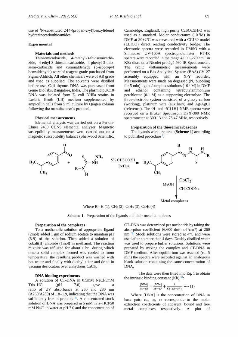
Preparation of the thiosemicarbazones
The ligands were prepared (Scheme 1) according
to published procedure 9.
Where R= H (1), CH3 (2), C2H5 (3), C6H5 (4)
Scheme 1. Preparation of the ligands and their metal complexes
Preparation of the complexes
To a methanolic solution of appropriate ligand
(2mol) added 1 gm of sodium acetate to maintain pH
(8-9) of the solution. Then added a solution of
cobalt(II) chloride (1mol) in methanol. The reaction
mixture was refluxed for about 1 hr., during which
time a solid complex formed was cooled to room
temperature, the resulting product was washed with
hot water and finally with diethyl ether and dried in
vacuum desiccators over anhydrous CaCl2.
DNA binding experiments
A solution of CT-DNA in 0.5mM NaCl/5mM
Tris–HCl (pH 7.0) gave a
ratio of UV absorbance at 260 and 280 nm
(A260/A280) of 1.8–1.9, indicating that the DNA was
sufficiently free of proteins 29. A concentrated stock
solution of DNA was prepared in 5 mM Tris–HCl/50
mM NaCl in water at pH 7.0 and the concentration of
CT-DNA was determined per nucleotide by taking the
absorption coefficient (6,600 dm3mol-1cm-1) at 260
nm 30. Stock solutions were stored at 4oC and were
used after no more than 4 days. Doubly distilled water
was used to prepare buffer solutions. Solutions were
prepared by mixing the complex and CT-DNA in
DMF medium. After equilibrium was reached (ca. 5
min) the spectra were recorded against an analogous
blank solution containing the same concentration of
DNA.
The data were then fitted into Eq. 1 to obtain
the intrinsic binding constant (Kb) 31:
[𝐷𝑁𝐴]
𝜀𝐴−𝜀𝐵=
[𝐷𝑁𝐴]
𝜀𝐵−𝜀𝐹+
1
𝐾𝑏(𝜀𝐵−𝜀𝐹) ---- (1)
Where [DNA] is the concentration of DNA in
base pair, ɛA, ɛB, ɛF corresponds to the molar
extinction coefficients of apparent, bound and free
metal complexes respectively. A plot of
Mediterr. J. Chem., 2017, 6(3) P. M. Krishna et al. 90
[DNA]/(ɛA-ɛF) Vs [DNA], gave a slope 1/(ɛB-ɛF) and
a Y-intercept equal to 1 / Kb (ɛB-ɛF); Kb is the ratio of
slope to the intercept.
Assay of nuclease activity
DMF solutions of the complexes were placed in
clean Eppendorf tubes and 1 µg of pUC18 DNA was
added. The contents were incubated for 30 min at
370C and loaded on 0.8% Agarose gel after mixing 5
µl of loading buffer (0.25% bromophenol blue +
0.25% Xylene cyanol + 30% glycerol sterilized
distilled water). Electrophoresis was performed at
constant voltage (100 V) until the bromophenol blue
reached to the 3/4th of the gel. The gel was stained for
10 min by immersing in an ethidium bromide
solution. The gel was then destained for 10 min by
keeping in sterilized distilled water and the plasmid
bands visualized by photographing the gel under a UV
Transilluminator. The efficiency of DNA cleavage
was measured by determining the ability of the
complex to form open circular (OC) or nicked circular
(NC) DNA from its super coiled (SC) form. The
reactions were carried out under oxidative and/or
hydrolytic conditions. Control experiments were done
in the presence of hydroxyl scavenger, DMSO.
Results and discussion
Characterization of the free
thiosemicarbazones and their metal complexes
The detailed characterization of the ligands was
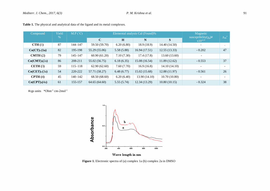
reported from the same group 9, 10, 22. The analytical
data of the thiosemicarbazones are given in Table 1.
A Brown colored cobalt(III) complexes of
thiosemicarbazones (1-4) are stable at room
temperature, non-hygroscopic, sparingly soluble in
methanol, ethanol, and readily soluble in chloroform,
pyridine, dimethylformamide (DMF) and
dimethylsulphoxide (DMSO). The analytical data
(Table 1) suggest 1: 3 (M: L) composition for the
complexes.
Conductivity and Magnetic susceptibility
measurements:
All cobalt complexes are highly soluble in DMF.
Therefore, the metal complexes were dissolved in
DMF to perform conductivity measurements. The
molar conductivities of cobalt complexes in DMF at
room temperature are found in the range of 26-46
Ohm-1cm-2mol-1 suggest the non-electrolytic nature 32
of complexes. The Magnetic susceptibility of cobalt
complexes are presented in Table 1. The data reveal
that the complexes are diamagnetic in favour of
formation of low spin octahedral cobalt(III)
complexes.
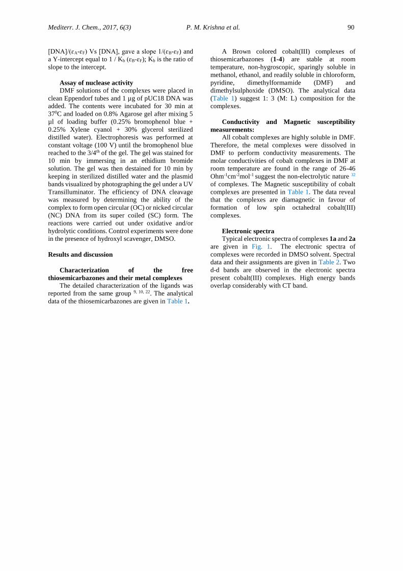
Electronic spectra
Typical electronic spectra of complexes 1a and 2a
are given in Fig. 1. The electronic spectra of
complexes were recorded in DMSO solvent. Spectral
data and their assignments are given in Table 2. Two
d-d bands are observed in the electronic spectra
present cobalt(III) complexes. High energy bands
overlap considerably with CT band.
Mediterr. J. Chem., 2017, 6(3) P. M. Krishna et al. 91
Table 1. The physical and analytical data of the ligand and its metal complexes.
#cgs units *Ohm-1 cm-2mol-1
Compound Yield
%
M.P (˚C) Elemental analysis Cal (Found)% Magnetic
susceptibility(χg)#
x10-6 #
M*
C H N S
CTH (1) 87 144–147 59.50 (59.70) 6.20 (6.80) 18.9 (18.9) 14.40 (14.50) - -
Co(CT)3 (1a) 82 195-198 55.29 (55.06) 5.58 (5.88) 16.94 (17.51) 12.55 (13.33) - 0.202 47
CMTH (2) 79 145–147 60.90 (61.20) 7.10 (7.30) 17.4 (17.8) 13.60 (13.60) -
Co(CMT)3(2a) 86 208-211 55.02 (56.75) 6.18 (6.35) 15.08 (16.54) 11.89 (12.62) - 0.553 37
CETH (3) 59 115–118 62.90 (62.60) 7.60 (7.70) 16.9 (16.8) 14.10 (14.10) - -
Co(CET)3 (3a) 54 220-222 57.71 (58.27) 6.48 (6.77) 15.02 (15.68) 12.88 (11.97) - 0.561 26
CPTH (4) 45 140–142 68.50 (68.60) 6.20 (6.40) 13.90 (14.10) 10.70 (10.80) - -
Co(CPT)3(4a) 61 155-157 64.65 (64.60) 5.55 (5.74) 12.34 (13.29) 10.80 (10.15) - 0.324 38
Figure 1. Electronic spectra of (a) complex 1a (b) complex 2a in DMSO
Ab
sorb
an
ce
Mediterr. J. Chem., 2017, 6(3) P. M. Krishna et al. 92
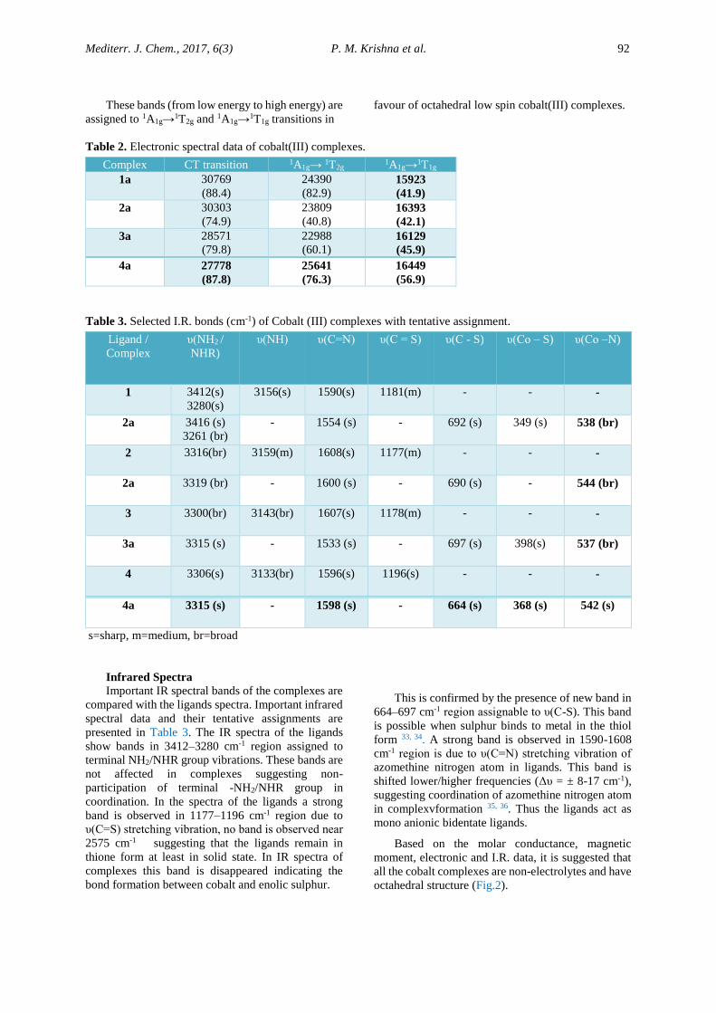
These bands (from low energy to high energy) are
assigned to 1A1g→1T2g and 1A1g→1T1g transitions in
favour of octahedral low spin cobalt(III) complexes.
Table 2. Electronic spectral data of cobalt(III) complexes.
Complex CT transition 1A1g→ 1T2g 1A1g→1T1g
1a 30769
(88.4)
24390
(82.9) 15923
(41.9)
2a 30303
(74.9)
23809
(40.8) 16393
(42.1)
3a 28571
(79.8)
22988
(60.1) 16129
(45.9)
4a 27778
(87.8)
25641
(76.3)
16449
(56.9)
Table 3. Selected I.R. bonds (cm-1) of Cobalt (III) complexes with tentative assignment.
Ligand /
Complex
υ(NH2 /
NHR)
υ(NH) υ(C=N) υ(C = S) υ(C - S) υ(Co – S) υ(Co –N)
1 3412(s)
3280(s)
3156(s) 1590(s) 1181(m) - - -
2a 3416 (s)
3261 (br)
- 1554 (s) - 692 (s) 349 (s) 538 (br)
2 3316(br) 3159(m) 1608(s) 1177(m) - - -
2a 3319 (br) - 1600 (s) - 690 (s) - 544 (br)
3 3300(br) 3143(br) 1607(s) 1178(m) - - -
3a 3315 (s) - 1533 (s) - 697 (s) 398(s) 537 (br)
4 3306(s) 3133(br) 1596(s) 1196(s) - - -
4a 3315 (s) - 1598 (s) - 664 (s) 368 (s) 542 (s)
s=sharp, m=medium, br=broad
Infrared Spectra
Important IR spectral bands of the complexes are
compared with the ligands spectra. Important infrared
spectral data and their tentative assignments are
presented in Table 3. The IR spectra of the ligands
show bands in 3412–3280 cm-1 region assigned to
terminal NH2/NHR group vibrations. These bands are
not affected in complexes suggesting non-
participation of terminal -NH2/NHR group in
coordination. In the spectra of the ligands a strong
band is observed in 1177–1196 cm-1 region due to
υ(C=S) stretching vibration, no band is observed near
2575 cm-1 suggesting that the ligands remain in
thione form at least in solid state. In IR spectra of
complexes this band is disappeared indicating the
bond formation between cobalt and enolic sulphur.
This is confirmed by the presence of new band in
664–697 cm-1 region assignable to υ(C-S). This band
is possible when sulphur binds to metal in the thiol
form 33, 34. A strong band is observed in 1590-1608
cm-1 region is due to υ(C=N) stretching vibration of
azomethine nitrogen atom in ligands. This band is
shifted lower/higher frequencies (Δυ = ± 8-17 cm-1),
suggesting coordination of azomethine nitrogen atom
in complexvformation 35, 36. Thus the ligands act as
mono anionic bidentate ligands.
Based on the molar conductance, magnetic
moment, electronic and I.R. data, it is suggested that
all the cobalt complexes are non-electrolytes and have
octahedral structure (Fig.2).
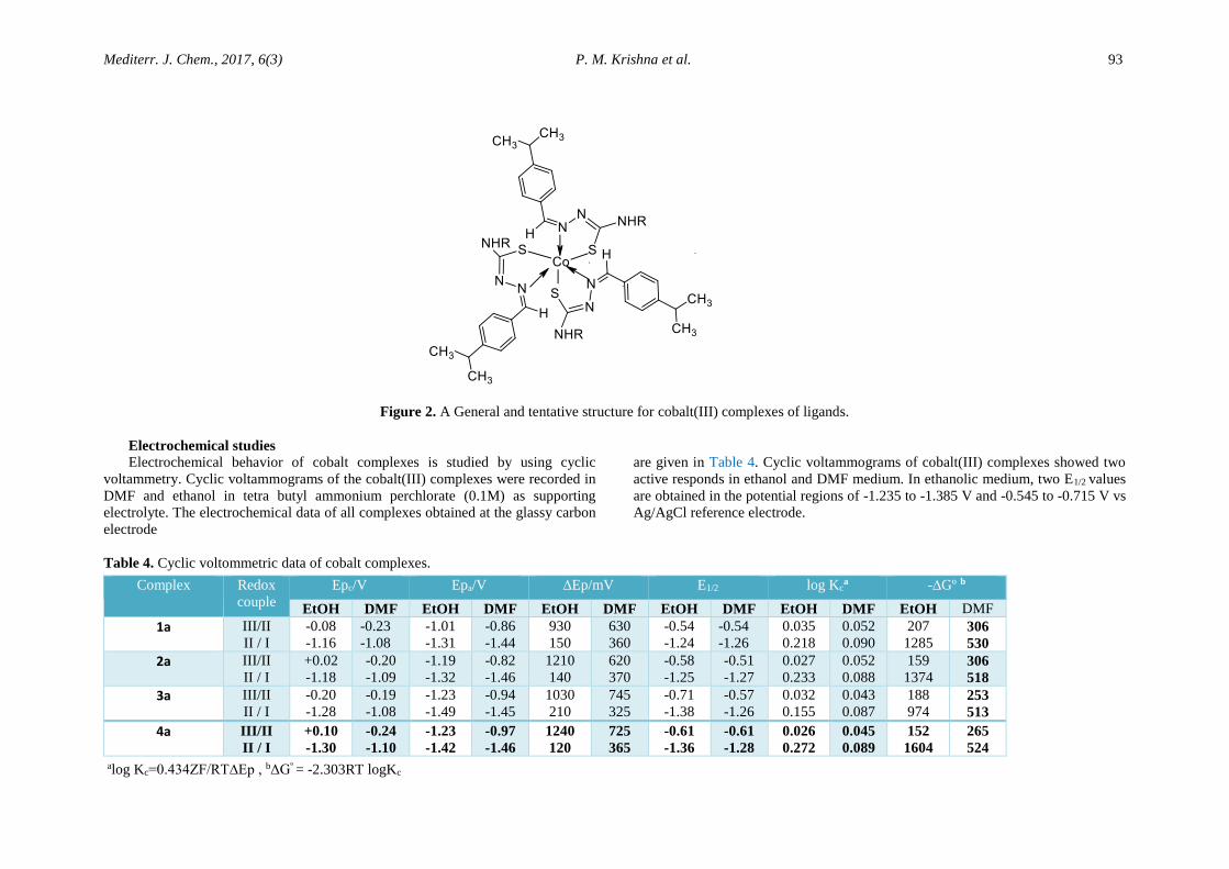
Mediterr. J. Chem., 2017, 6(3) P. M. Krishna et al. 93
Figure 2. A General and tentative structure for cobalt(III) complexes of ligands.
Electrochemical studies
Electrochemical behavior of cobalt complexes is studied by using cyclic
voltammetry. Cyclic voltammograms of the cobalt(III) complexes were recorded in
DMF and ethanol in tetra butyl ammonium perchlorate (0.1M) as supporting
electrolyte. The electrochemical data of all complexes obtained at the glassy carbon
electrode
are given in Table 4. Cyclic voltammograms of cobalt(III) complexes showed two
active responds in ethanol and DMF medium. In ethanolic medium, two E1/2 values
are obtained in the potential regions of -1.235 to -1.385 V and -0.545 to -0.715 V vs
Ag/AgCl reference electrode.
Table 4. Cyclic voltommetric data of cobalt complexes.
Complex Redox
couple
Epc/V Epa/V ∆Ep/mV E1/2 log Kca -∆Gº b
EtOH DMF EtOH DMF EtOH DMF EtOH DMF EtOH DMF EtOH DMF
1a III/II
II / I
-0.08
-1.16
-0.23
-1.08
-1.01
-1.31
-0.86
-1.44
930
150
630
360
-0.54
-1.24
-0.54
-1.26
0.035
0.218
0.052
0.090
207
1285 306
530
2a III/II
II / I
+0.02
-1.18
-0.20
-1.09
-1.19
-1.32
-0.82
-1.46
1210
140
620
370
-0.58
-1.25
-0.51
-1.27
0.027
0.233
0.052
0.088
159
1374 306
518
3a III/II
II / I
-0.20
-1.28
-0.19
-1.08
-1.23
-1.49
-0.94
-1.45
1030
210
745
325
-0.71
-1.38
-0.57
-1.26
0.032
0.155
0.043
0.087
188
974 253
513
4a III/II
II / I
+0.10
-1.30
-0.24
-1.10
-1.23
-1.42
-0.97
-1.46
1240
120
725
365
-0.61
-1.36
-0.61
-1.28
0.026
0.272
0.045
0.089
152
1604
265
524
alog Kc=0.434ZF/RT∆Ep , bΔGº = -2.303RT logKc
Mediterr. J. Chem., 2017, 6(3) P. M. Krishna et al. 94
These are respectively assigned to Co(II)/ Co(I)
and Co(III)/ Co(II) redox couples. Repeated scans as
well as various scan rates showed that dissociation
does not takes place in these complexes. The non-
equivalent current intensity of cathodic and anodic
peak difference (∆Ep =120-1240 mV) indicates quasi
reversible behaviour of these complexes. The ΔEp
values are greater than the Nernstian values (ΔEp ≈
59mV) for one electron redox system. This indicates
a considerable reorganization of the coordination
sphere during electron transfer has been observed for
a number of other cobalt (III) complexes. From
Table 4, E1/2 values of the complexes in DMF medium
are slightly higher than the values obtained in ethanol.
A comparison of the E1/2 values of this redox
couple of the present complexes with other
analogous nitrogen donor macro cycles reveal that
these complexes undergo more facile redox change
which seem to be a requirement to the DNA
cleavage 37.
DNA binding studies
The interaction of cobalt complexes with calf
thymus DNA was studied by absorption titrations
using spectrophotometer. The absorption titrations
were carried out with increasing amount of CT-DNA
in 363-367 nm regions. With addition of DNA all
cobalt complexes showed hypercromic shift.
Table 5. Electronic absorption data upon addition of CT-DNA to nickel complex.
Complex λmax/nm Δλ /nm H (%) Kb (M-1)
Free Bound
1a 365 364 1.0 -25.85 9.58 x 10⁷
2a 365.5 364 1.5 -33.29 2.84 X10⁷
3a 365 366 1.0 -28.35 4.54 x 10⁷
4a 365 366 1.0 -24.92 1.21 x 10⁸
From Table 5, it is revealed that in the presence
of increasing amount of CT-DNA absorption spectra
of complexes show either red-shift or blue-shift
(Δλmax:1.0-1.5 nm) and hypercromism
[hypercromism: -25.85 % for 1a, -33.29 % for 2a, -
28.35 % for 3a, and -24.92 for 4a. The orders of binding
constants of complexes are 2a < 3a <1a <4a.
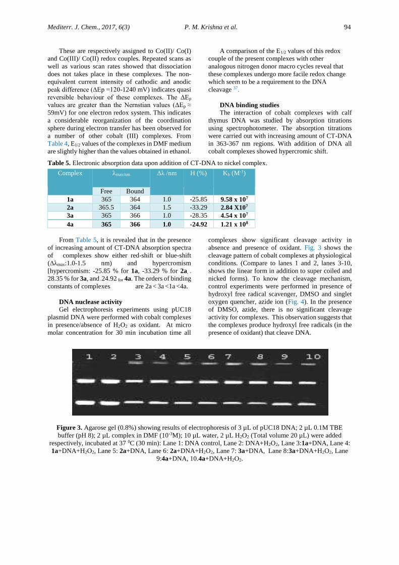
DNA nuclease activity Gel electrophoresis experiments using pUC18
plasmid DNA were performed with cobalt complexes
in presence/absence of H2O2 as oxidant. At micro
molar concentration for 30 min incubation time all
complexes show significant cleavage activity in
absence and presence of oxidant. Fig. 3 shows the
cleavage pattern of cobalt complexes at physiological
conditions. (Compare to lanes 1 and 2, lanes 3-10,
shows the linear form in addition to super coiled and
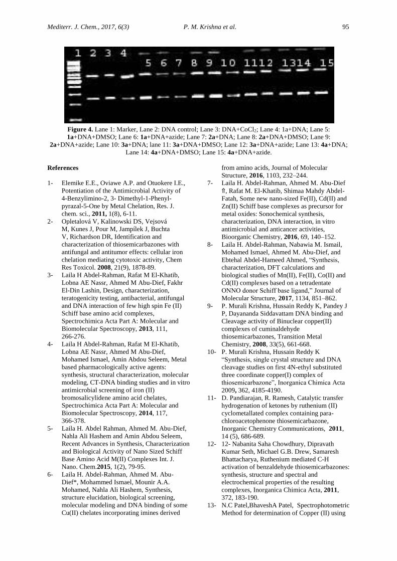
nicked forms). To know the cleavage mechanism,
control experiments were performed in presence of
hydroxyl free radical scavenger, DMSO and singlet
oxygen quencher, azide ion (Fig. 4). In the presence
of DMSO, azide, there is no significant cleavage
activity for complexes. This observation suggests that
the complexes produce hydroxyl free radicals (in the
presence of oxidant) that cleave DNA.
Figure 3. Agarose gel (0.8%) showing results of electrophoresis of 3 µL of pUC18 DNA; 2 µL 0.1M TBE
buffer (pH 8); 2 µL complex in DMF (10-3M); 10 µL water, 2 µL H2O2 (Total volume 20 µL) were added
respectively, incubated at 37 0C (30 min): Lane 1: DNA control, Lane 2: DNA+H2O2, Lane 3:1a+DNA, Lane 4:
1a+DNA+H2O2, Lane 5: 2a+DNA, Lane 6: 2a+DNA+H2O2, Lane 7: 3a+DNA, Lane 8:3a+DNA+H2O2, Lane
9:4a+DNA, 10.4a+DNA+H2O2.
Mediterr. J. Chem., 2017, 6(3) P. M. Krishna et al. 95
Figure 4. Lane 1: Marker, Lane 2: DNA control; Lane 3: DNA+CoCl2; Lane 4: 1a+DNA; Lane 5:
1a+DNA+DMSO; Lane 6: 1a+DNA+azide; Lane 7: 2a+DNA; Lane 8: 2a+DNA+DMSO; Lane 9:
2a+DNA+azide; Lane 10: 3a+DNA; lane 11: 3a+DNA+DMSO; Lane 12: 3a+DNA+azide; Lane 13: 4a+DNA;
Lane 14: 4a+DNA+DMSO; Lane 15: 4a+DNA+azide.
References
1- Elemike E.E., Oviawe A.P. and Otuokere I.E.,
Potentiation of the Antimicrobial Activity of
4-Benzylimino-2, 3- Dimethyl-1-Phenyl-
pyrazal-5-One by Metal Chelation, Res. J.
chem. sci., 2011, 1(8), 6-11.
2- Opletalová V, Kalinowski DS, Vejsová
M, Kunes J, Pour M, Jampílek J, Buchta
V, Richardson DR, Identification and
characterization of thiosemicarbazones with
antifungal and antitumor effects: cellular iron
chelation mediating cytotoxic activity, Chem
Res Toxicol. 2008, 21(9), 1878-89.
3- Laila H Abdel-Rahman, Rafat M El-Khatib,
Lobna AE Nassr, Ahmed M Abu-Dief, Fakhr
El-Din Lashin, Design, characterization,
teratogenicity testing, antibacterial, antifungal
and DNA interaction of few high spin Fe (II)
Schiff base amino acid complexes,
Spectrochimica Acta Part A: Molecular and
Biomolecular Spectroscopy, 2013, 111,
266-276.
4- Laila H Abdel-Rahman, Rafat M El-Khatib,
Lobna AE Nassr, Ahmed M Abu-Dief,
Mohamed Ismael, Amin Abdou Seleem, Metal
based pharmacologically active agents:
synthesis, structural characterization, molecular
modeling, CT-DNA binding studies and in vitro
antimicrobial screening of iron (II)
bromosalicylidene amino acid chelates,
Spectrochimica Acta Part A: Molecular and
Biomolecular Spectroscopy, 2014, 117,
366-378.
5- Laila H. Abdel Rahman, Ahmed M. Abu-Dief,
Nahla Ali Hashem and Amin Abdou Seleem,
Recent Advances in Synthesis, Characterization
and Biological Activity of Nano Sized Schiff
Base Amino Acid M(II) Complexes Int. J.
Nano. Chem.2015, 1(2), 79-95.
6- Laila H. Abdel-Rahman, Ahmed M. Abu-
Dief*, Mohammed Ismael, Mounir A.A.
Mohamed, Nahla Ali Hashem, Synthesis,
structure elucidation, biological screening,
molecular modeling and DNA binding of some
Cu(II) chelates incorporating imines derived
from amino acids, Journal of Molecular
Structure, 2016, 1103, 232–244.
7- Laila H. Abdel-Rahman, Ahmed M. Abu-Dief
⇑, Rafat M. El-Khatib, Shimaa Mahdy Abdel-
Fatah, Some new nano-sized Fe(II), Cd(II) and
Zn(II) Schiff base complexes as precursor for
metal oxides: Sonochemical synthesis,
characterization, DNA interaction, in vitro
antimicrobial and anticancer activities,
Bioorganic Chemistry, 2016, 69, 140–152.
8- Laila H. Abdel-Rahman, Nabawia M. Ismail,
Mohamed Ismael, Ahmed M. Abu-Dief, and
Ebtehal Abdel-Hameed Ahmed, “Synthesis,
characterization, DFT calculations and
biological studies of Mn(II), Fe(II), Co(II) and
Cd(II) complexes based on a tetradentate
ONNO donor Schiff base ligand,” Journal of
Molecular Structure, 2017, 1134, 851–862.
9- P. Murali Krishna, Hussain Reddy K, Pandey J
P, Dayananda Siddavattam DNA binding and
Cleavage activity of Binuclear copper(II)
complexes of cuminaldehyde
thiosemicarbazones, Transition Metal
Chemistry, 2008, 33(5), 661-668.
10- P. Murali Krishna, Hussain Reddy K
“Synthesis, single crystal structure and DNA
cleavage studies on first 4N-ethyl substituted
three coordinate copper(I) complex of
thiosemicarbazone”, Inorganica Chimica Acta
2009, 362, 4185-4190.
11- D. Pandiarajan, R. Ramesh, Catalytic transfer
hydrogenation of ketones by ruthenium (II)
cyclometallated complex containing para-
chloroacetophenone thiosemicarbazone,
Inorganic Chemistry Communications, 2011,
14 (5), 686-689.
12- 12- Nabanita Saha Chowdhury, Dipravath
Kumar Seth, Michael G.B. Drew, Samaresh
Bhattacharya, Ruthenium mediated C-H
activation of benzaldehyde thiosemicarbazones:
synthesis, structure and spectral and
electrochemical properties of the resulting
complexes, Inorganica Chimica Acta, 2011,
372, 183-190.
13- N.C Patel,BhaveshA Patel, Spectrophotometric
Method for determination of Copper (II) using
Mediterr. J. Chem., 2017, 6(3) P. M. Krishna et al. 96
pChlorobenzaldehyde-4-(2’-carboxy-5-
sulphophenyl)-3-thiosemicarbazone [p-
CBCST], Research Journal of Chemical
Sciences, 2014, 4(2), 1-6.
14- Tetsumi T., Sumi M., Taraka M. and Shono T,
Direct reaction of meta powders with several
sodium dithiocarbamates Polyhedron, 1986,
5(3), 707-710.
15- Buttrus N.H and Mohamed S.M, Synthesis and
Characterization of Ni+2, Cu+2 and Zn+2
complexes with Benzoxazole-2-thionate,
Diphenyl Phosphinomethane and Iodine,
Research Journal of Chemical Sciences, 2013,
3(6), 54-59.
16- N.B. Gopal Reddy, P. Murali Krishna,
Nagaraju Kottam, Novel metal–organic
photocatalysts: Synthesis, characterization and
decomposition of organic dyes, Spectrochimica
Acta Part A: Molecular and Biomolecular
Spectroscopy 2015, 137, 371–377
17- P. Murali Krishna, N. B. Gopal Reddy, Dr.
Nagaraja K, and Yallur B. C, Design and
Synthesis of Metal Complexes of (2E)-2-[(2E)-
3-Phenylprop-2-En-1-Ylidene]
Hydrazinecarbothioamide and Their
Photocatalytic Degradation of Methylene Blue,
The Scientific World Journal, Volume 2013,
Article ID 828313, 7 pages
http://dx.doi.org/10.1155/2013/828313.
18- G. Pelosi, Thiosemicarbazone Metal
Complexes: From Structure to Activity, The
Open Crystallography Journal, 2010, 3(2),
16-28.
19- Franco Bisceglie, Silvana Pinelli, Rossella
Alinovi, Matteo Goldoni, Antonio Mutti,
Alessandro Camerini, Lorenzo Piola,
Pieralberto Tarasconi, Giorgio Pelosi,
Cinnamaldehyde and cuminaldehyde
thiosemicarbazones and their copper(II) and
nickel(II) complexes: A study to understand
their biological activity Journal of Inorganic
Biochemistry, 2014, 140, 111-125.
20- Quiroga AG, Perez JM, Montero EI, West DX,
Alonso C, Navarro-Ranninger C, Synthesis and
characterization of Pd(II) and Pt(II) complexes
of p-isopropylbenzaldehyde N-protected
thiosemicarbazones. Cytotoxic activity
against ras-transformed cells, Journal of
Inorganic Biochemistry, 1999, 75(4), 293-301.
21- Perez JM, Matesanz AI, Martin-Ambite A,
Navarro P, Alonso C, Souza P, Synthesis and
characterization of complexes of p-isopropyl
benzaldehyde and methyl 2-pyridyl ketone
thiosemicarbazones with Zn(II) and Cd(II)
metallic centers. Cytotoxic activity and
induction of apoptosis in Pam-ras cells, Journal
of Inorganic Biochemistry, 1999, 75(4),
255-261.
22- 22 P Murali Krishna and K Hussain Reddy,
“Synthesis, Structural Characterization and
DNA Studies of Nickel (II) Complexes of (2E)-
4N-Substituted-2-[4-(propan-2-yl)
Benzylidene]hydrazinecarbothioamide Schiff’s
Bases” Journal of Chemical and
Pharmaceutical Research, 2016, 8(10):61-68.
23- 23. N.B. Gopal Reddy, P. Murali Krishna, S.S.
Shantha Kumar, Yogesh P. Patil, Munirathinam
Nethaji, “Structure and spectroscopic
investigations of a bi-dentate N'-[(4-ethyl-
phenyl)methylidene]-4-hydroxybenzohydrazide
and its Co(II), Ni(II), Cu(II) and Cd(II)
complexes: Insights relevant to biological
properties” Journal of Molecular Structure,
2017, 1137, 543-552.
24- 24. B. S. Shankar, N. Shashidhar, Yogesh
Prakash Patil, P. Murali Krishna and
Munirathinam Nethaji, (2E)-2-(2-hydroxy-3-
methylbenzylidene)-N-methylhydrazine
carbothioamide, Acta Cryst. 2013, E69, o61
25- 25. P. Murali Krishna, B. S. Shankara,
N.Shashidhar, Synthesis, characterization and
biological studies of binuclear copper(II)
complexes of (2E)-2-(2-hydroxy-3-
methoxybenzylidene)-4N-substituted
hydrazinecarbothioamides”, International
Journal of Inorganic Chemistry, vol. 2013,
Article ID 741269, 11 pages, 2013.
(doi:10.1155/2013/741269).
26- 26. P. Murali Krishna, K. Hussain Reddy,
“Synthesis, characterization, molecular docking
and DNA studies of copper(II) complexes of
(2E)-3-phenylprop-2-enal thiosemicarbazones”,
Der Pharma Chemica, 2013, 5 (5), 258-269.
27- 27. P. Murali Krishna, G. N. Anil Kumar,
Hussain Reddy K and M. K. Kokila, (2E)-
N-methyl-2-(3-phenylpropylidene)
hydrazinecarbothioamide, Acta Cryst., 2012,
E68, o2842.
28- 28. P. Murali Krishna, Hussain Reddy K,
Pitchika G Krishna & G H Philip “DNA
interactions of mixed ligand copper(II)
complexes with sulphur containing ligands”,
Indian Journal of Chemistry, 2007, 46A,
904–908.
29- 29. Marmur J, A procedure for the isolation of
deoxyribonuclcic acid from microorganisms,
Journal of Molecular Biology, 1961, 3(2),
208-218.
30- 30. M. E. Reichmann, S. A. Rice, C. A.
Thomas, Paul Doty, A Further Examination of
the Molecular Weight and Size of
Desoxypentose Nucleic Acid, Journal of
American Chemical Soceity, 1954, 76 (11),
3047–3053.
31- 31. Wolfe A, Chimer GH, Meechan T,
Polycyclic aromatic hydrocarbons physically
intercalate into duplex regions of denatured
DNA, Biochemistry, 1987, 26(20), 6392-6396.
32- 32 W. J. Geary, “The use of conductivity
measurements in organic solvents for the
characterisation of coordination compounds,”
Coordination Chemistry Reviews, 1971, 7(1),
81–122.
Mediterr. J. Chem., 2017, 6(3) P. M. Krishna et al. 97
33- 33 J. S. Casas, A. Sanchez, J. Sorda, A.
Vazquez–Lopez, E. E. Castellano, J. kermann-
Schechter, M. C. Rodriguez–Arguelles, U.
Russo, Diorganotin(IV) derivatives of
salicylaldehydethiosemicarbazone. The crystal
structure of dimethyl- and diphenyl-
(salicylaldehydethiosemicarbazonato)tin(IV),
Inorganica Chimica Acta, 1994, 216,169-175.
34- 34. J. S. Casas, A. Castineirras, A. Sanchez,
J. Sorda, A. Vazquez–Lopez, M. C Rodriguez–
Arguelles, U. Russo, Synthesis and
spectroscopic properties of diorganotin(IV)
derivatives of 2,6-diacetylpyridine
bis(thiosemicarbazone). Crystal structure of
diphenyl{2,6-diacetylpyridine
bis(thiosemicarbazonato)}tin(IV)
bis(dimethylformamide) solvate, Inorganica
Chimica Acta, 1994, 221, 61-68.
35- 35. Michel J.M. Campbell, Transition metal
complexes of thiosemicarbazide and
thiosemicarbazone, Coordination Chemistry
Review, 1975, 15(2-3), 279-319.
36- 36. D. X. West, A. E. Liberta, S. B. Padhye, R.
C. Chikte, P. B. Sonawane, A.S. Kumbhar, R.
G. Yerande, Thiosemicarbazone complexes of
copper(II): structural and biological studies,
Coordination Chemistry Review, 1993, 123
(1-2), 49-71.
37- 37. C. V. Sastri, D. Eswaramoorthy, L.
Giribabu, and B. G. Maiya, “DNA interactions
of new mixed-ligand complexes of cobalt(III)
and nickel(II) that incorporate modified
phenanthroline ligands,” Journal of Inorganic
Biochemistry, 2003, 94(1-2), 138–145.
Copyright © 2022 FDOKUMEN