
Structural modeling of high-affinity thyroid receptor–ligand complexes
14
ORIGINAL PAPER Structural modeling of high-affinity thyroid receptor–ligand complexes Alexandre Suman de Araujo • Leandro Martı ´nez • Ricardo de Paula Nicoluci • Munir S. Skaf • Igor Polikarpov Received: 5 February 2010 / Revised: 27 April 2010 / Accepted: 4 May 2010 / Published online: 30 May 2010 Ó European Biophysical Societies’ Association 2010 Abstract Understanding the molecular basis of the bind- ing modes of natural and synthetic ligands to nuclear receptors is fundamental to our comprehension of the acti- vation mechanism of this important class of hormone reg- ulated transcription factors and to the development of new ligands. Thyroid hormone receptors (TR) are particularly important targets for pharmaceuticals development because TRs are associated with the regulation of metabolic rates, body weight, and circulating levels of cholesterol and triglycerides in humans. While several high-affinity ligands are known, structural information is only partially available. In this work we obtain structural models of several TR–ligand complexes with unknown structure by docking high affinity ligands to the receptors’ ligand binding domain with subsequent relaxation by molecular dynamics simu- lations. The binding modes of these ligands are discussed providing novel insights into the development of TR ligands. The experimental binding free energies are rea- sonably well-reproduced from the proposed models using a simple linear interaction energy free-energy calculation scheme. Keywords Molecular dynamics Nuclear receptor Binding free energy Thyroid hormone receptor Thyromimetics-receptor binding modes High affinity thyromimetics Introduction The nuclear receptor (NR) superfamily is an important group of proteins that regulates the expression of genes related to organism development and metabolism control. It includes receptors for steroids, retinoids, fatty and bile acids, vitamin D, cholesterol metabolites, xenobiotics and thyroid hormones (Ribeiro et al. 1998, 1995; Weatherman et al. 1999; Yen 2001; Togashi et al. 2005). NRs tran- scription regulation relies on specific molecular interac- tions between the receptor and sequences of DNA known as hormone responsive elements (HRE) (Alberts 2002). It is triggered by hormone binding to the NRs, and involves formation of large macromolecular complexes that include coactivator and corepressor proteins. The NRs recognize both hormones and HREs in a highly selective fashion (Ribeiro et al. 1995; Weatherman et al. 1999; Togashi et al. 2005; Alberts 2002; Kumar and Thompson 1999). NRs are composed of three main domains: the N-terminal region, which is largely unstructured and the least conserved within the superfamily, harbors an important transactivation function known as AF1. A bulk of experimental evidence indicates that AF1 modulates cell-specific and promoter- specific transcription (Yen 2001; Kumar and Thompson 1999). The second NR domain is the DNA binding domain (DBD), responsible for the recognition of HREs. The DBD is also involved in dimer stabilization (Ribeiro et al. 1998, 1995; Weatherman et al. 1999; Lazar 1993). The C-terminal region contains the ligand binding domain (LBD), which, as Electronic supplementary material The online version of this article (doi:10.1007/s00249-010-0610-2) contains supplementary material, which is available to authorized users. A. S. de Araujo R. de Paula Nicoluci I. Polikarpov Instituto de Fı ´sica de Sa ˜o Carlos, Universidade de Sa ˜o Paulo, Av. Trabalhador SaoCarlense 400, IFSC, Grupo de Cristalografia, P.O. Box 369, Sao Carlos, SP 13560-970, Brazil L. Martı ´nez M. S. Skaf (&) Institute of Chemistry, State University of Campinas-UNICAMP, P.O. Box 6154, Campinas, SP 13084-862, Brazil e-mail: [email protected] 123 Eur Biophys J (2010) 39:1523–1536 DOI 10.1007/s00249-010-0610-2
-
Upload
independent -
Category
Documents
-
view
5 -
download
0
Transcript of Structural modeling of high-affinity thyroid receptor–ligand complexes
ORIGINAL PAPER
Structural modeling of high-affinity thyroid receptor–ligandcomplexes
Alexandre Suman de Araujo • Leandro Martınez •
Ricardo de Paula Nicoluci • Munir S. Skaf •
Igor Polikarpov
Received: 5 February 2010 / Revised: 27 April 2010 / Accepted: 4 May 2010 / Published online: 30 May 2010
� European Biophysical Societies’ Association 2010
Abstract Understanding the molecular basis of the bind-
ing modes of natural and synthetic ligands to nuclear
receptors is fundamental to our comprehension of the acti-
vation mechanism of this important class of hormone reg-
ulated transcription factors and to the development of new
ligands. Thyroid hormone receptors (TR) are particularly
important targets for pharmaceuticals development because
TRs are associated with the regulation of metabolic rates,
body weight, and circulating levels of cholesterol and
triglycerides in humans. While several high-affinity ligands
are known, structural information is only partially available.
In this work we obtain structural models of several
TR–ligand complexes with unknown structure by docking
high affinity ligands to the receptors’ ligand binding domain
with subsequent relaxation by molecular dynamics simu-
lations. The binding modes of these ligands are discussed
providing novel insights into the development of TR
ligands. The experimental binding free energies are rea-
sonably well-reproduced from the proposed models using a
simple linear interaction energy free-energy calculation
scheme.
Keywords Molecular dynamics � Nuclear receptor �Binding free energy � Thyroid hormone receptor �Thyromimetics-receptor binding modes �High affinity thyromimetics
Introduction
The nuclear receptor (NR) superfamily is an important
group of proteins that regulates the expression of genes
related to organism development and metabolism control.
It includes receptors for steroids, retinoids, fatty and bile
acids, vitamin D, cholesterol metabolites, xenobiotics and
thyroid hormones (Ribeiro et al. 1998, 1995; Weatherman
et al. 1999; Yen 2001; Togashi et al. 2005). NRs tran-
scription regulation relies on specific molecular interac-
tions between the receptor and sequences of DNA known
as hormone responsive elements (HRE) (Alberts 2002). It
is triggered by hormone binding to the NRs, and involves
formation of large macromolecular complexes that include
coactivator and corepressor proteins. The NRs recognize
both hormones and HREs in a highly selective fashion
(Ribeiro et al. 1995; Weatherman et al. 1999; Togashi et al.
2005; Alberts 2002; Kumar and Thompson 1999).
NRs are composed of three main domains: the N-terminal
region, which is largely unstructured and the least conserved
within the superfamily, harbors an important transactivation
function known as AF1. A bulk of experimental evidence
indicates that AF1 modulates cell-specific and promoter-
specific transcription (Yen 2001; Kumar and Thompson
1999). The second NR domain is the DNA binding domain
(DBD), responsible for the recognition of HREs. The DBD
is also involved in dimer stabilization (Ribeiro et al. 1998,
1995; Weatherman et al. 1999; Lazar 1993). The C-terminal
region contains the ligand binding domain (LBD), which, as
Electronic supplementary material The online version of thisarticle (doi:10.1007/s00249-010-0610-2) contains supplementarymaterial, which is available to authorized users.
A. S. de Araujo � R. de Paula Nicoluci � I. Polikarpov
Instituto de Fısica de Sao Carlos, Universidade de Sao Paulo,
Av. Trabalhador SaoCarlense 400, IFSC, Grupo de
Cristalografia, P.O. Box 369, Sao Carlos, SP 13560-970, Brazil
L. Martınez � M. S. Skaf (&)
Institute of Chemistry, State University
of Campinas-UNICAMP, P.O. Box 6154,
Campinas, SP 13084-862, Brazil
e-mail: [email protected]
123
Eur Biophys J (2010) 39:1523–1536
DOI 10.1007/s00249-010-0610-2

its name suggests, recognizes and binds specific hormones.
It also plays critical roles in dimerization, cofactor recruit-
ment, and in gene transactivation and silencing (Yen 2001;
Togashi et al. 2005; Kumar and Thompson 1999). Under-
standing ligand affinity and recognition by the LBD is of
major importance for pharmaceutical design.
Thyroid hormone receptors (TR) regulate the tran-
scription of several genes related to embrionary and post-
natal development of eyes, ears and the brain (Lazar 1993;
Forrest et al. 1996; Fraichard et al. 1997; Gauthier et al.
1999; Ng et al. 2001). TRs influence several important
physiological processes related to alert state such as heart
frequency and metabolic rates, affecting body weight, and
circulating cholesterol, lipoprotein and triglycerides levels
(Baxter et al. 2004; Johansson et al. 2005). The known
natural TR ligands are 3,5,30-triiodo-L-thyronine (thyronine
or T3), 3,5,30,50-tetra-iodothyronine (thyroxine or T4), and
Triac. There are two major TR isoforms, a (TRa) and b(TRb), which, in humans, are encoded by the NR1A1 and
NR1A2 genes located in chromosomes 3 and 17, respec-
tively (Wagner et al. 2001). TR isoforms are found in
different concentrations in different tissues. TRa is highly
expressed in the heart, skeletal muscle and brown fat
tissues and its activation is associated with observed
modifications in cardiac behavior and loss of bone and
muscle masses (Lazar 1993; Wagner et al. 2001). TRb is
predominantly found in the liver and its activation is
associated with increasing metabolic rates and energy
consumption, resulting in weight loss and reduction of
blood plasma lipid levels (Baxter et al. 2004; Johansson
et al. 2005; Ye et al. 2003). Both subtypes present
approximately the same affinity (around 10-10 M) and
activation responses to T3. Thus, increasing T3 levels can
result in beneficial results due to TRb activation, but also
evoke undesirable deleterious responses resulting from
activation of TRa. Therefore, developing synthetic thyr-
omimetics that act as b-selective TR modulators is of great
pharmacological interest (Togashi et al. 2005; Baxter et al.
2004; Johansson et al. 2005; Wagner et al. 2001; Yoshihara
et al. 2003).
The 3D structures of TR LBDs are composed of twelve
a-helices (H1–H12) and four small b-strands, a fold that is
mostly conserved between NRs’ LBDs. The binding cavity
is deeply buried within the hydrophobic core of the LBD
and is almost completely filled by the ligand. The binding
pockets of TRa and TRb are composed of several hydro-
phobic amino acids and by two polar regions, as depicted
in Fig. 1a. A single histidine located in H11 forms one of
the polar interactions: a hydrogen bond with the phenolic
hydroxyl of the outer thyronine ring of agonist ligands.
Most polar ligand interactions, however, are formed with
three arginine residues located on H3 and on the b-strands,
and by a serine in TRa or an asparagine in TRb, as shown
in Fig. 1b. The polar interactions of the ligand with this
region of the protein depends on an intricate hydrogen
bond network which involves surrounding water molecules
(Martınez et al. 2006, 2009). However, the high affinity of
TR ligands is mostly dependent on their hydrophobicity, as
their thyronine rings are stabilized by a series of non-polar
interactions (Fig. 1b).
The binding pocket of TRs is mostly conserved between
TRa and TRb, with the exception of Ser277 in TRa being
substituted by an asparagine (Asn331) in TRb. Many high
affinity ligands are known, and the crystal structures are
Fig. 1 a Description of TR
binding site in terms of polar
and non-polar residues. Non-
polar residues, which largely
constitute the binding pocket,
are shown in light gray, whereas
polar residues are shown black.
b General aspect of agonist
ligands and their interactions
with binding pocket residues.
The ligand’s polar head and tail
interacts with the few polar
residues in the binding site. The
hydrophobic body interacts with
a large number of non-polar
residues
1524 Eur Biophys J (2010) 39:1523–1536
123
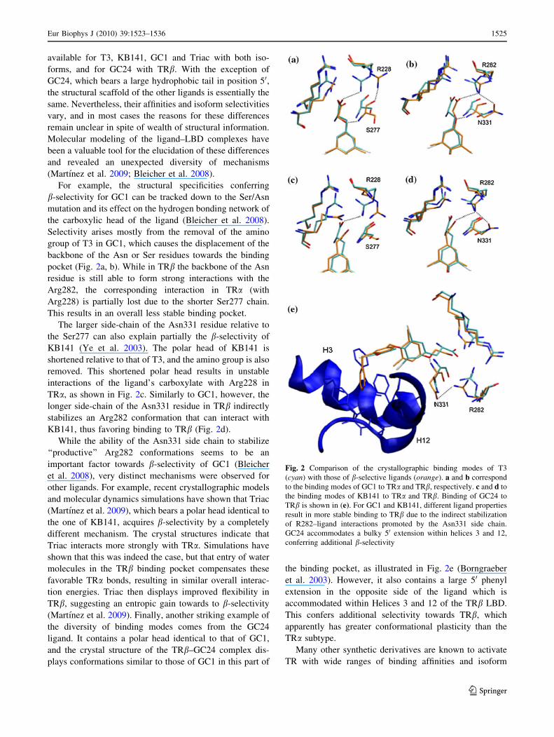
available for T3, KB141, GC1 and Triac with both iso-
forms, and for GC24 with TRb. With the exception of
GC24, which bears a large hydrophobic tail in position 50,the structural scaffold of the other ligands is essentially the
same. Nevertheless, their affinities and isoform selectivities
vary, and in most cases the reasons for these differences
remain unclear in spite of wealth of structural information.
Molecular modeling of the ligand–LBD complexes have
been a valuable tool for the elucidation of these differences
and revealed an unexpected diversity of mechanisms
(Martınez et al. 2009; Bleicher et al. 2008).
For example, the structural specificities conferring
b-selectivity for GC1 can be tracked down to the Ser/Asn
mutation and its effect on the hydrogen bonding network of
the carboxylic head of the ligand (Bleicher et al. 2008).
Selectivity arises mostly from the removal of the amino
group of T3 in GC1, which causes the displacement of the
backbone of the Asn or Ser residues towards the binding
pocket (Fig. 2a, b). While in TRb the backbone of the Asn
residue is still able to form strong interactions with the
Arg282, the corresponding interaction in TRa (with
Arg228) is partially lost due to the shorter Ser277 chain.
This results in an overall less stable binding pocket.
The larger side-chain of the Asn331 residue relative to
the Ser277 can also explain partially the b-selectivity of
KB141 (Ye et al. 2003). The polar head of KB141 is
shortened relative to that of T3, and the amino group is also
removed. This shortened polar head results in unstable
interactions of the ligand’s carboxylate with Arg228 in
TRa, as shown in Fig. 2c. Similarly to GC1, however, the
longer side-chain of the Asn331 residue in TRb indirectly
stabilizes an Arg282 conformation that can interact with
KB141, thus favoring binding to TRb (Fig. 2d).
While the ability of the Asn331 side chain to stabilize
‘‘productive’’ Arg282 conformations seems to be an
important factor towards b-selectivity of GC1 (Bleicher
et al. 2008), very distinct mechanisms were observed for
other ligands. For example, recent crystallographic models
and molecular dynamics simulations have shown that Triac
(Martınez et al. 2009), which bears a polar head identical to
the one of KB141, acquires b-selectivity by a completely
different mechanism. The crystal structures indicate that
Triac interacts more strongly with TRa. Simulations have
shown that this was indeed the case, but that entry of water
molecules in the TRb binding pocket compensates these
favorable TRa bonds, resulting in similar overall interac-
tion energies. Triac then displays improved flexibility in
TRb, suggesting an entropic gain towards to b-selectivity
(Martınez et al. 2009). Finally, another striking example of
the diversity of binding modes comes from the GC24
ligand. It contains a polar head identical to that of GC1,
and the crystal structure of the TRb–GC24 complex dis-
plays conformations similar to those of GC1 in this part of
the binding pocket, as illustrated in Fig. 2e (Borngraeber
et al. 2003). However, it also contains a large 50 phenyl
extension in the opposite side of the ligand which is
accommodated within Helices 3 and 12 of the TRb LBD.
This confers additional selectivity towards TRb, which
apparently has greater conformational plasticity than the
TRa subtype.
Many other synthetic derivatives are known to activate
TR with wide ranges of binding affinities and isoform
Fig. 2 Comparison of the crystallographic binding modes of T3
(cyan) with those of b-selective ligands (orange). a and b correspond
to the binding modes of GC1 to TRa and TRb, respectively. c and d to
the binding modes of KB141 to TRa and TRb. Binding of GC24 to
TRb is shown in (e). For GC1 and KB141, different ligand properties
result in more stable binding to TRb due to the indirect stabilization
of R282–ligand interactions promoted by the Asn331 side chain.
GC24 accommodates a bulky 50 extension within helices 3 and 12,
conferring additional b-selectivity
Eur Biophys J (2010) 39:1523–1536 1525
123

selectivities. Our molecular level understanding of the
affinities of such ligands remains very limited due to the
lack of the corresponding holo-LBD crystal structures, thus
motivating molecular modeling studies. In the context of
nuclear receptors, molecular dynamics (MD) simulations
have been reported focusing on structural variations and
conformational adaptations of LBD mutants (Carlsson
et al. 2005), ligand–LBD interactions (Martınez et al. 2009;
Bleicher et al. 2008), and dynamics of ligand binding
(Martınez et al. 2008) and dissociation (Blondel et al. 1999;
Carlsson et al. 2006; Celik et al. 2007, 2008; Genest et al.
2008; Kosztin et al. 1999; Martınez et al. 2005, 2006;
Perakyla 2009; Sonoda et al. 2008).
In this work we use a well-known docking program
combined with MD simulations in order to propose struc-
tural models for the binding of several high-affinity ligands
to the LBDs of both TRa and TRb isoforms, for which
there are no known crystallographic structures available so
far. The models, thus, generated provide for the first time
insights into the binding modes of structurally quite distinct
TR agonists and help elucidating the molecular reasons for
their binding affinities and isoform selectivity. MD simu-
lations are performed on the proposed structures and the
ligand binding free energies are estimated within the linear
interaction energy (LIE) approximation (Nam et al. 2003;
Oostenbrink et al. 2000; Oostenbrink and van Gunsteren
2005; Stjernschantz et al. 2006; van Lipzig et al. 2004),
showing reasonable agreement with experimental values.
Given the limitations of the LIE approximation, it would be
desirable to further validate the docked structures with
more elaborate binding free energy calculations. However,
this is beyond the scope of the present study.
In ‘‘Theoretical and computational details’’ we sum-
marize the theoretical framework and details of the cal-
culations. In ‘‘Results and discussion’’ we present the
structural models we obtain and discuss their relevance in
terms of the comprehension of TR ligand recognition and
pharmacological implications. Concluding remarks are
presented in last section.
Theoretical and computational details
Two sets of ligands are considered in this study: a ‘‘train-
ing-set’’, composed of ligands for which crystallographic
models are available (Table 1), and a ‘‘model-set’’ of
ligands for which structural models are being proposed.
The training-set is composed by five ligands shown in
Fig. 3, and models are proposed for other eight ligands
with unknown structures, which are represented in Fig. 4.
Experimental binding affinities for all these ligands are
known. Our primary goal is to obtain biophysically sensi-
ble structural models for the model-set ligands which can
serve as a first-time view of their binding modes within the
ligand binding pockets of TR.
Parameterization of ligands
The molecular models for the ligands pertaining to the
training set were taken from our previous study (Martınez
et al. 2005) and the parameterization of the model-set
ligands was performed using the same methodology. All
molecules were treated as fully flexible. Bonded and van
der Waals interaction potential parameters were obtained
by group analogy from the CHARMM force field
(MacKerell et al. 1998), except the ones for iodine atoms,
which were taken from our previous T3 model (Martınez
et al. 2005). The partial atomic charges of the model set
ligands were obtained from ab initio quantum chemical
calculations consistent with the protein force field per-
formed with Gaussian03 (Frisch et al. 2004). Calculations
for molecules without iodine atoms were performed at the
RHF/6-31G** level of theory and Merz–Kollman (MK)
charges. Since iodine atoms are not described by 6-31G**
basis set and do not support MK fitting of the electrostatic
surface potential, iodine containing ligands were parame-
terized at the MP2/LanL2DZ level of theory and Mulliken
partial charges (Martınez et al. 2005). All ligand parame-
ters are supplied as supplementary material.
Docking
Docking calculations were performed using GOLD v3.0.1
(CCDC 2007) in order generate initial ligand–LBD struc-
tures for complexes with no crystallographic structures
available (model-set). GOLD utilizes a genetic algorithm
(GA) to perform the pose of the compound into the protein
active site. The genetic algorithm parameters to determine
this operation were automatically defined by GOLD based
on pre-defined parameter settings built from a diversified
test-set of ligands and proteins. The active site was defined
as the region within 10 A radius from the nearly central
carbon of the co-crystallized ligand. GOLD has two built-
in score functions. The default fitness function GoldScore
Table 1 Crystal structures used in MD simulations of the training set
Isoform Ligand PDB-ID Isoform Ligand PDB-ID
a GC1 3HZF b GC1 3IMY
a IH5 1NAV b IH5 1NAX
a T3 2H77 b T3 2H6 W
a Triac 3JZB b Triac 3JZC
b GC24 1Q4X
IH5 and KB141 are used interchangeably here to specify the same
ligand
1526 Eur Biophys J (2010) 39:1523–1536
123

consists of four components (protein–ligand hydrogen
bond energy, protein–ligand van der Waals interaction
energy, ligand internal van der Waals energy, and ligand
torsional strain energy). The ChemScore scoring function
was derived empirically from a set of 82 protein–ligand
complexes for which measured binding affinities were
available. Unlike GoldScore, the ChemScore function was
trained by regression against the reported affinity data.
Because the two scoring functions emphasize different
aspects of the ligand–protein coupling, we have employed
both functions to evaluate the posing procedure. GOLD
was set to output up to 10 best conformations after each
run. From these computationally evaluated sets, we choose
the best ranked pose within those that were consistent with
general aspects of ligand orientation in the binding pocket.
Different protein targets were chosen according to
availability of known ligands and diversity of binding-site
properties: the four co-crystallized structures of hTRa and
hTRb available on PDB containing T3 and IH5 ligands
were retrieved (Codes: 2H77 and 2H6W for T3 (Nasci-
mento et al. 2006), and 1NAV and 1NAX for IH5 (Ye et al.
2003)). The co-crystallized structures of hTRa and hTRbwith GC1 were recently obtained in our lab (Bleicher et al.
2008) with PDB IDs 3HZF and 3IMY, respectively. These
Fig. 3 Training set ligands.
All of them have solved
crystallographic structures in
complex with TR–LBD
Fig. 4 High affinity test set ligands. Every ligand in this group has been derived from some training set ligand and present KD to TRb under
10 nM
Eur Biophys J (2010) 39:1523–1536 1527
123

structures were processed in order to remove ligands and
water molecules. The compounds GC1, IH5 and T3 were
extracted from the proteins files and used as the other
ligands in docking and calculations steps, so that they
could be used to monitor the accuracy of the results.
Ligands without crystallographic structures available were
built with the Sybyl (Sybyl 2005). Hydrogen atoms were
added to all ligands and their Tripos force field energies
(Clark et al. 1989) were minimized.
Molecular dynamics simulations
MD simulations were performed using the NAMD package
(Phillips et al. 2005) with CHARMM force field parame-
ters for the protein (MacKerell et al. 1998) and TIP3P for
water (Jorgensen et al. 1983). We used the crystal struc-
tures displayed in Table 1 for the simulations of the
training-set. For simulating the ligands in solution, we built
40 A wide cubic boxes containing a single ligand mole-
cule, approximately 2,000 water molecules and sodium and
chloride ions in order to render the systems electrically
neutral. Simulations for ligand–LBD complexes were
performed with 80 A wide cubic boxes, nearly 15,000
water molecules and sufficient sodium and chloride ions
for electroneutrality. The systems’ energies were mini-
mized by 500 conjugate-gradient steps. All MD simula-
tions were performed in NPT ensemble at 298 K and
1 atm, with periodic boundary conditions, using a time step
of 2 fs and constraining all covalent bonds involving
hydrogen atoms to the corresponding equilibrium posi-
tions. The van der Waals interactions were cutoff at 15 A
with a switching function starting at 12 A. The electrostatic
interactions were treated using the particle mesh Ewald
(PME) method (Darden et al. 1993) with the following
parameters: tolerance 10-6, Ewald real space coefficient
0.219398, grid dimensions 81 9 81 9 81 A3, and maxi-
mum grid spacing 1.5 A. PME electrostatics were used for
generating the dynamics but not to compute the average
ligand–surroundings interaction energy needed for esti-
mating the binding affinities (see below). For each system,
the energy minimized structure was equilibrated by a 1 ns
simulation, followed by 500 ps runs from which interaction
energies were evaluated by pair-wise sums of van der
Waals and Coulomb interactions between the ligand its
surroundings (Eq. 1).
Linear interaction energy
The LIE method provides approximate estimates to the
absolute binding and hydration free energies with low
computational effort. Within this approximation, the
ligand–protein binding free energy is given by
DGBind ¼ b Vell�s
� �bound� Vel
l�s
� �free
� �
þa Vvdwl�s
� �bound� Vvdw
l�s
� �free
� �þ c; ð1Þ
where Vell�s
� �Y
and Vvdwl�s
� �Y
are the average values of the
electrostatic and van der Waals interactions between the
ligand (l) and its surroundings (s) in the ligand state Y,
which can be either free in solution or bound to the protein.
The a and b parameters are, respectively, dispersion and
electrostatic adjustable energy scale factors and c is a
constant term dependent of ligand or binding site hydro-
phobicity and accessible surface-area (Hansson et al.
1998). Recent developments of the method have shown
that reasonable LIE free energy estimates for a given
ligand–protein system require calibrating the energy and
hydrophobicity parameters to the particular system of
interest by tuning a, b and c in order to reproduce the
experimental binding free energies of a group of ligands
named training-set (Stjernschantz et al. 2006; Bren et al.
2006; Ganguly and Mukhopadhyay 2006; Tounge et al.
2006; Raineri et al. 2005). The optimized set of parameters
is subsequently utilized to estimate the binding free energy
of other ligands, usually referred to as the test set. Cali-
bration of parameters has been performed for TRa and TRbseparately, but the adjusted parameter values turned out
only slightly dependent on TR isoform.
Here, we assembled the training set with ligands for
which holo-LBD crystallographic structures are available.
These ligands are shown in Fig. 3. All training set ligands
are agonists and have high affinity to TR, showing the
general structural features described in Fig. 1b: a phenol, a
highly polar carboxylate, and a hydrophobic body. The
experimental dissociation constant (KD), the binding free
energies obtained from these KD values, and the TR iso-
form selectivity of each ligand are shown in Table S1 of the
supplemental material (Ye et al. 2003; Yoshihara et al.
2003; Martınez et al. 2009; Borngraeber et al. 2003;
Chiellini et al. 1998; Nguyen et al. 2002). The endogen
ligand T3 has approximately the same affinity to both LBD
isoforms, while the exogen ones (GC1, GC24, IH5 and
Triac) are all b-selective.
Binding free energy estimates were obtained from the
proposed structural models. This group of ligands, shown
in Fig. 4, have experimental dissociation constants in the
nanomolar range, thus being high-affinity synthetic TR
ligands and, consequently, relevant for ligand design.
GC24 was included in the model-set for TRa since no
crystallographic structure for the TRa–GC24 complex is
available. Experimental KD’s from which free energies
were derived are shown in Table S2 of supplemental
material (Ye et al. 2003; Yoshihara et al. 2003; Borng-
raeber et al. 2003). The b-selectivity of the ligands are
correlated with that of their parents, ranging from the
1528 Eur Biophys J (2010) 39:1523–1536
123

most b-selective ligand GC24 to the weakly a-selective
ligand T31.
Ligand–surroundings non-bonded average interaction
energies for the model set ligands are shown in Table 2.
The LIE parameters were obtained by least-square fitting
the LIE equation to the experimental binding free energies
and are listed in Table 3 for TRa and TRb, respectively.
The low value of the electrostatic b parameter may reflect
the low polarity of the ligands and the fact that the
important ligand–protein hydrophilic contacts in the bind-
ing pocket are nearly compensated by ligand–water inter-
actions in solution (Martınez et al. 2006), thus masking the
role of the electrostatic interactions in the LIE calculations.
The replacement of ligand–protein hydrophilic contacts by
ligand–solvent interactions has been suggested to facilitate
ligand dissociation from the binding pocket through the
b-sheet region (Martınez et al. 2006). Small b (*0.06) has
also been found in the context of other nuclear receptors
(Stjernschantz et al. 2006). Table 4 lists the calculated and
experimental binding free energies for each ligand con-
sidered. Scatter plots of the experimental and calculated
binding free energies for the model set ligands to both
isoforms are depicted in Fig. 5. The agreement is fairly
reasonable given the rather approximate nature of the LIE
method. The root mean square deviations from the ideal
Table 2 Ligand-neighborhood non-bonded interaction energies for free and bound states obtained from MD simulations for high affinity ligands
for TRa and TRb isoforms
Ligand b-selectivity hVelFreei hVel
Boundi hVvdWFree i hVvdW
Boundi DVel DVvdw
TRa (TRb)
Training-set T3 1.00 -143.03 -89.73 -26.97 -50.20 53.30 -23.23
(-95.75) (-48.14) (47.28) (-21.17)
Triac 6.00 -183.57 -160.23 -28.90 -47.26 23.34 -18.36
(-160.27) (-46.60) (23.30) (-17.70)
IH5 11.11 -164.59 -136.70 -23.32 -43.79 27.89 -20.47
(-146.67) (-44.42) (17.92) (-21.10)
GC1 8.52 -173.85 -179.24 -22.86 -44.38 -5.39 -21.52
(-165.99) (-47.33) (7.86) (-24.47)
GC24 33.81 -177.56 – -27.66 – – –
(-186.56) (-48.67) (-9.00) (-21.01)
Model-set T31 0.62 -166.83 -81.43 -24.91 -52.43 85.40 -27.52
(-89.96) (-50.90) (76.87) (-25.99)
T32 1.00 -193.85 -169.74 -30.24 -51.39 24.11 -21.15
(-171.37) (-51.26) (22.48) (-21.02)
IH51 3.03 -157.47 -159.74 -21.97 -39.94 -2.27 -17.97
(-153.62) (-39.77) (3.85) (-17.80)
IH52 2.47 -170.53 -158.19 -23.94 -44.09 12.34 -20.15
(-170.97) (-43.94) (-0.44) (-20.00)
DIMIT 1.07 -118.80 -88.91 -23.49 -43.03 29.89 -19.54
(-96.62) (-39.84) (22.18) (-16.35)
EBGC1 5.21 -174.38 -189.99 -22.18 -40.70 -15.61 -18.52
(-175.48) (-44.16) (-1.10) (-21.98)
MBDMT 1.25 -120.35 -72.93 -23.31 -47.17 47.42 -23.86
(-84.04) (-46.16) (36.31) (-22.85)
PAGC1 2.45 -181.70 -180.06 -22.14 -43.95 1.64 -21.81
(-176.69) (-45.53) (5.01) (-23.39)
GC24 33.81 -178.10 -171.65 -28.02 -48.19 6.45 -20.17
– – – –
Selectivity of each ligand is obtained from Ka-TRb/ Ka-TRa, normalized by Ka-TRb/ Ka-TRa for T3. All energies are in kcal/mol
Table 3 LIE parameters for the training-sets of ligands for TRa and
TRb
TRa TRb
a 0.42 0.45
b 0.01 0.01
c/kcal mol-1 -4.00 -4.00
Eur Biophys J (2010) 39:1523–1536 1529
123

behavior (dashed lines) are only 1.31 and 1.24 kcal/mol for
TRa and TRb, respectively. The correlation factor between
DGcalc and DGexp is low (R *0.54). However, there are too
few data points for each TR isoform for a meaningful
quality assessment of the calculated free energies in terms
of this parameter.
Results and discussion
Structural models of ligand–LBD complexes were built
for the eight high affinity ligands (Fig. 4) bound to both aand b isoforms, and for GC-24 bound to TRa. Figures 6
and 7 display the average structures from MD simulations
of the modeled ligands (orange) superimposed with the
crystal structure most relevant for each comparison
(cyan). Models built on TRa and TRb are displayed in
left and right panels, respectively, except for the TRa–
GC24 model, which is superimposed with the TRb-GC24
crystal structure. The structures depicted here were pro-
duced to indicate the most relevant differences in the
binding modes of the structural models. The correspond-
ing PDB files of the docked conformations are available
as supplementary material. The most important structural
features of each ligand–LBD complex and an analysis of
their relevance follows.
DIMIT
The DIMIT ligand (Fig. 4) shares the hydrophobic body
with GC1 and the polar head with T3. Its affinity to both
isoforms is reduced relative to the ones of T3 and GC1
(refer to Table 4 for binding free energies), and it is
slightly b-selective. The structural models obtained were
able to reproduce this reduced affinity with good precision
(Table 4): deviations relative to experimental values being
-0.91 and 0.05 kcal mol-1 for TRa and TRb, respectively.
Table 4 Calculated and experimental binding free energies of high affinity ligands with both isoforms
TRa TRb
DGcalc DGexp DGcalc DGexp
Training-set T3 -13.22 -13.53 -13.05 -13.69
Triac -11.48 -13.86 -11.73 -14.04
KB141 -12.32 -11.25 -13.32 -12.83
GC1 -13.09 -12.28 -14.93 -13.70
GC24 – – -13.54 -13.54
RMS = 0.98 kcal mol-1 RMS = 1.26 kcal mol-1
Model-set T31 -14.70 -14.32 -14.93 -14.18
T32 -12.64 -15.04 -13.23 -15.22
IH51 -11.57 -10.27 -11.97 -11.07
IH52 -12.34 -13.31 -13.00 -14.00
DIMIT -11.91 -11.00 -11.14 -11.19
EBGC1 -11.93 -11.75 -13.90 -12.87
MBDMT -13.55 -11.85 -13.92 -12.13
PAGC1 -13.14 -12.49 -14.48 -13.16
GC24 -12.41 -10.73 – –
RMS = 1.31 kcal mol-1 RMS = 1.24 kcal mol-1
All energies are in kcal/mol
Fig. 5 Calculated versus experimental binding free energies for the
model-set ligands to TRa and TRb isoforms. All values are in kcal/
mol. The error bars for DGcalc were calculated from the average
energy values obtained from the simulations. Experimental errors for
DGexp are roughly estimated at 0.2 and 0.4 kcal/mol for TRa and TRbfrom different T3 binding affinity data (see Supporting information)
1530 Eur Biophys J (2010) 39:1523–1536
123
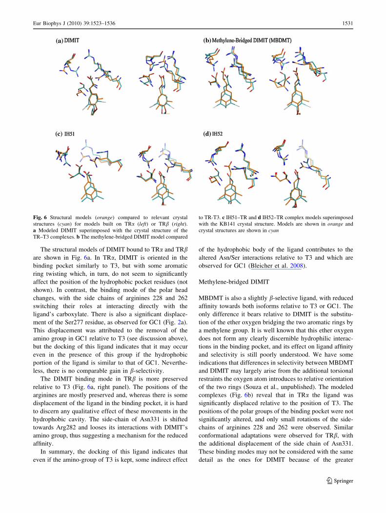
The structural models of DIMIT bound to TRa and TRbare shown in Fig. 6a. In TRa, DIMIT is oriented in the
binding pocket similarly to T3, but with some aromatic
ring twisting which, in turn, do not seem to significantly
affect the position of the hydrophobic pocket residues (not
shown). In contrast, the binding mode of the polar head
changes, with the side chains of arginines 228 and 262
switching their roles at interacting directly with the
ligand’s carboxylate. There is also a significant displace-
ment of the Ser277 residue, as observed for GC1 (Fig. 2a).
This displacement was attributed to the removal of the
amino group in GC1 relative to T3 (see discussion above),
but the docking of this ligand indicates that it may occur
even in the presence of this group if the hydrophobic
portion of the ligand is similar to that of GC1. Neverthe-
less, there is no comparable gain in b-selectivity.
The DIMIT binding mode in TRb is more preserved
relative to T3 (Fig. 6a, right panel). The positions of the
arginines are mostly preserved and, whereas there is some
displacement of the ligand in the binding pocket, it is hard
to discern any qualitative effect of these movements in the
hydrophobic cavity. The side-chain of Asn331 is shifted
towards Arg282 and looses its interactions with DIMIT’s
amino group, thus suggesting a mechanism for the reduced
affinity.
In summary, the docking of this ligand indicates that
even if the amino-group of T3 is kept, some indirect effect
of the hydrophobic body of the ligand contributes to the
altered Asn/Ser interactions relative to T3 and which are
observed for GC1 (Bleicher et al. 2008).
Methylene-bridged DIMIT
MBDMT is also a slightly b-selective ligand, with reduced
affinity towards both isoforms relative to T3 or GC1. The
only difference it bears relative to DIMIT is the substitu-
tion of the ether oxygen bridging the two aromatic rings by
a methylene group. It is well known that this ether oxygen
does not form any clearly discernible hydrophilic interac-
tions in the binding pocket, and its effect on ligand affinity
and selectivity is still poorly understood. We have some
indications that differences in selectivity between MBDMT
and DIMIT may largely arise from the additional torsional
restraints the oxygen atom introduces to relative orientation
of the two rings (Souza et al., unpublished). The modeled
complexes (Fig. 6b) reveal that in TRa the ligand was
significantly displaced relative to the position of T3. The
positions of the polar groups of the binding pocket were not
significantly altered, and only small rotations of the side-
chains of arginines 228 and 262 were observed. Similar
conformational adaptations were observed for TRb, with
the additional displacement of the side chain of Asn331.
These binding modes may not be considered with the same
detail as the ones for DIMIT because of the greater
Fig. 6 Structural models (orange) compared to relevant crystal
structures (cyan) for models built on TRa (left) or TRb (right).a Modeled DIMIT superimposed with the crystal structure of the
TR–T3 complexes. b The methylene-bridged DIMIT model compared
to TR-T3. c IH51–TR and d IH52–TR complex models superimposed
with the KB141 crystal structure. Models are shown in orange and
crystal structures are shown in cyan
Eur Biophys J (2010) 39:1523–1536 1531
123
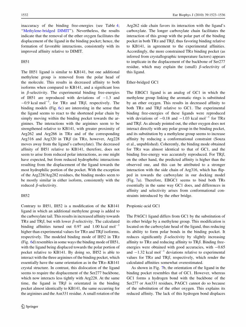
inaccuracy of the binding free-energies (see Table 4;
‘‘Methylene-bridged DIMIT’’). Nevertheless, the results
indicate that the removal of the ether oxygen facilitates the
displacement of the ligand in the binding pocket; hence, the
formation of favorable interactions, consistently with its
improved affinity relative to DIMIT.
IH51
The IH51 ligand is similar to KB141, but one additional
methylene group is removed from the polar head of
the molecule. This results in decreased affinity to both
isoforms when compared to KB141, and a significant loss
in b-selectivity. The experimental binding free-energies
of IH51 are reproduced with deviations of -1.3 and
-0.9 kcal mol-1, for TRa and TRb, respectively. The
binding models (Fig. 6c) are interesting in the sense that
the ligand seems to react to the shortened polar chain by
simply moving within the binding pocket towards the ar-
ginines. The interactions with the arginines seem to be
strengthened relative to KB141, with greater proximity of
Arg262 and Arg266 in TRa and of the corresponding
Arg316 and Arg320 in TRb (in TRa, however, Arg228
moves away from the ligand’s carboxylate). The decreased
affinity of IH51 relative to KB141, therefore, does not
seem to arise from reduced polar interactions, as one might
have expected, but from reduced hydrophobic interactions
resulting from the displacement of the ligand towards the
most hydrophilic portion of the pocket. With the exception
of the Arg228/Arg282 residues, the binding modes seem to
be mostly similar in either isoform, consistently with the
reduced b-selectivity.
IH52
Contrary to IH51, IH52 is a modification of the KB141
ligand in which an additional methylene group is added to
the carboxylate tail. This results in increased affinity towards
TRa and TRb, but with lower b-selectivity. The calculated
binding affinities turned out 0.97 and 1.00 kcal mol-1
higher than experimental values for TRa and TRb isoforms,
respectively. The modeled binding mode of IH52 in TRa(Fig. 6d) resembles in some ways the binding mode of IH51,
with the ligand being displaced towards the polar portion of
pocket relative to KB141. By doing so, IH52 is able to
interact with the three arginines of the binding pocket, which
essentially have the same orientation as in the TRa–KB141
crystal structure. In contrast, this dislocation of the ligand
seems to require the displacement of the Ser277 backbone,
which now interacts less tightly with Arg228. At the same
time, the ligand in TRb is orientated in the binding
pocket almost identically to KB141, the same occurring for
the arginines and the Asn331 residue. A small rotation of the
Arg262 side chain favors its interaction with the ligand’s
carboxylate. The longer carboxylate chain facilitates the
interaction of this group with the polar part of the binding
pocket in both TRa and TRb, thus favoring binding relative
to KB141, in agreement to the experimental affinities.
Accordingly, the more constrained TRa binding pocket (as
inferred from crystallographic temperature factors) appears
to implicate in the displacement of the backbone of Ser277
residue, which may explain the (small) b-selectivity of
this ligand.
Ether-bridged GC1
The EBGC1 ligand is an analog of GC1 in which the
methylene group linking the aromatic rings is substituted
by an ether oxygen. This results in decreased affinity to
both TRa and TRb relative to GC1. The experimental
binding free-energies of these ligands were reproduced
with deviations of -0.18 and -1.03 kcal mol-1 for TRaand TRb. As already pointed out, the ether oxygen does not
interact directly with any polar group in the binding pocket,
and its substitution by a methylene group seems to increase
affinity by reducing a conformational constraint (Souza
et al., unpublished). Coherently, the binding mode obtained
for TRa was almost identical to that of GC1, and the
binding free-energy was accurately reproduced. For TRb,
on the other hand, the predicted affinity is higher than the
observed one, and this can be attributed to a stronger
interaction with the side chain of Arg316, which has flip-
ped in towards the carboxylate in our docking model
(Fig. 7a). Therefore, EBGC1 seems to bind both TRs
essentially in the same way GC1 does, and differences in
affinity and selectivity arises from conformational con-
straints introduced by the ether bridge.
Propionic-acid GC1
The PAGC1 ligand differs from GC1 by the substitution of
its ether bridge by a methylene group. This modification is
located on the carboxylate head of the ligand, thus reducing
its ability to form polar bonds in the binding pocket. It
reduces significantly b-selectivity by slightly increasing
affinity to TRa and reducing affinity to TRb. Binding free-
energies were obtained with good accuracies, with -0.65
and -1.32 kcal mol-1 deviations relative to experimental
values for TRa and TRb, respectively, which render the
calculated affinities somewhat overestimated.
As shown in Fig. 7b, the orientation of the ligand in the
binding pocket resembles that of GC1. However, whereas
GC1 forms a hydrogen bond with the backbone of the
Ser277 or Asn331 residues, PAGC1 cannot do so because
of the substitution of the ether oxygen. This explains its
reduced affinity. The lack of this hydrogen bond displaces
1532 Eur Biophys J (2010) 39:1523–1536
123

PAGC1 slightly away from the Ser277/Asn331 residues
and allows for PAGC1 interactions with Arg228 in TRaand Arg316 in TRb that are actually stronger than those
exhibited by GC1 with these residues. The fact that these
ligand–arginine interactions do not compensate the reduced
ligand–Ser277/Asn331 interactions can be explained by the
presence of water molecules in the binding pocket. While
the interaction with the Ser/Asn residues occur deeply in
the binding cavity and in a position where the ligand is
tightly packed, the polar interactions of the carboxylate
head can be substituted by interactions with water molec-
ular with little or no energetic cost (Martınez et al. 2006).
In this case, the absence of direct GC1–arginine interac-
tions that appear to be present in PAGC1 are intermediated
by water molecules not necessarily visible in the crystal
structures. This ligand, therefore, illustrates that the loss
polar interactions between ligand and binding pocket is
mostly deleterious when occurring in tightly packed sol-
vent inaccessible groups.
GC24 in TRa
GC24 is a surprising ligand containing a large 50-hyrophobic
extension that can be accommodated by the LBD of TRbmaintaining essentially the same affinity as T3. GC24
affinity towards TRa is much reduced, thus making it highly
b-selective. We predicted the binding free-energy of GC24
to TRa with a deviation of -1.68 kcal mol-1 (affinity being
again overestimated). Nevertheless, the superposition of the
crystal structure of the TRb–GC24 complex with the cor-
responding TRa docking model (Fig. 7c) provides for sev-
eral interesting observations. First, the binding modes of the
polar part of the ligand are essentially identical to those of
GC1 in both isoforms, indicating that the selectivity arises
Fig. 7 Structural models superimposed to relevant crystal structures
for TRa (left) and TRb (right). a Ether-bridged GC1 models superim-
posed with corresponding TR–GC1 crystal structures. b Propionic-
acid GC1 models superimposed with TR–GC1 crystal structures.
c Superposition of the TRa–GC24 model with the TRb–GC24 crystal
structure. Structural models of the d T32 and e T31 ligands superim-
posed with the LBD–T3 crystal structures. Models are shown in orangeand crystal structures are shown in cyan
Eur Biophys J (2010) 39:1523–1536 1533
123
partially from the same structural reasons as for GC1
(Bleicher et al. 2008). However, additional loss of affinity to
TRa may come from conformational stress observed in H3
in the presence of GC24 relative to the crystal structure of
the TRb complex. Unlike the TRb LBD structure, whose
flexibility allows for the accommodation of the 50 extension
of GC24, the modeled TRa–GC24 structure shows that the
Phe215 residue in H3 (Phe269 in TRb) experiences a sig-
nificant displacement due to the interaction with the GC24
phenyl substituent.
T31 and T32
T31 and T32 are T3 analogs. In T31 the 30 iodine of T3 is
substituted by an isopropyl radical, while in T32 the
amino-group is removed. T31 is curious for being the sole
ligand which displays slight a-selectivity, whereas T32 is
not selective. Both ligands also display improved affinities
to both isoforms. In our calculations, while the affinity of
T31 was accurately reproduced (deviations of -0.38 and
-0.75 kcal mol-1), the binding free-energy of T32 was
missed by approximately 2 kcal mol-1.
The models suggest that T31 binding to TRa is mostly
preserved relative to T3 (Fig. 6d), with some flipping of the
side chains of Arg228 and Arg262, the former loosing
interactions with the ligand’s carboxylate and the later
augmenting them. Interactions of T31 with TRb are modi-
fied by a significant rotation of the phenolic ring, and there
is an important displacement of the Asn331 residue. This
structural difference is surprising in view of the similarity
between these ligands, although it follows the trend dis-
cussed above for MBDMT ligand, for which displacement
of the Asn331 backbone was also observed for a hydro-
phobic body similar to GC1’s. The structural basis for the
improved affinities resulting from substituting the iodine
atom by a propyl radical remains elusive. The differences in
affinity seem to arise from improved ligand–protein dis-
persive interactions without inducing any clearly detectable
side chain conformational adaptations.
The improved affinity of T32 probably comes from its
increased hydrophobicity resulting from the removal of the
amino group. As observed for GC1, displacement of the
Ser277 residue in TRa occurs and tighter bonds to Arg
residues in TRb are clearly visible. However, we should
refrain from further analysis based on the modeled struc-
tures given the inaccuracy of our binding free-energy
estimates for this ligand.
Concluding remarks
In this work, structural models were obtained for several
TR high-affinity ligands with unknown structures by means
of a well-respected docking procedure and MD simula-
tions. We use the structural models to gain insights into the
putative binding modes of these high-affinity ligand–TR
complexes and estimate their binding free energy within
the approximated LIE method. Docked models and average
interaction energies obtained from MD trajectories indicate
that if, at the one hand, substituting the interactions of the
ligand’s polar head with the binding pocket residues by
ligand–water interactions involves little or no energetic
cost. The observed affinities, on the other hand, seem to
depend on the chemical nature of the ligand’s polar head in
most cases. This is an indirect consequence of the fact that
most ligands were designed from known leads (mostly T3,
GC1 and KB141), and for which structural alterations were
made in their polar head envisioning isoform selectivity by
altered interactions with the Ser277/Asn331 binding pocket
mutation.
Fine tuning of binding affinities at the polar interaction
level seems possible, but the structural basis underlying
each effect is very complex. A previously unsuspected
diversity of mechanisms is progressively being observed
for ligand–TR interactions, even in the cases where ligands
seem to bear overall similar structural scaffolds. The
crystal structures of complexes with T3, GC1, Triac,
KB141 and GC24 already provided a glimpse on the
complexity of these interactions. The binding modes of
other high-affinity ligands, modeled here, complement this
picture. For instance, we have previously attributed the
movements of the Ser277/Asn331 backbones in the pres-
ence of GC1, to the absence of the amino group relative to
T3 (Bleicher et al. 2008). The binding modes of DIMIT
and T31 proposed here, however, suggest that these
movements may be also caused by the hydrophobic body
of GC1, since the presence of the amino group in DIMIT
and T31 does not lead to b-selectivity. Controlling the size
of the carboxylate chain of the ligands has also been used
to improve protein–ligand polar head interactions. Our
results show that shorter chains are capable of interfering
with hydrophobic packing effects, reducing the affinity (see
IH51 and IH52). Large polar heads, on the other hand,
generally enhance affinity, but, alone, cannot promote
isoform selectivity. The carboxylate chains of Triac and
KB141, of intermediate sizes, result in intermediate affin-
ities and binding modes that give rise to b-selectivity.
Finally, in order to elucidate the role of hydrophobic
interactions in binding affinity, it is still necessary to refine
the interaction models, both by generating additional and
more detailed crystal structures as well as improving
modeling procedures. While it is clear that these interac-
tions are essential for the attachment of the ligands to the
binding pocket, the lack of specificity is still a barrier for
their understanding at the molecular level and use in
rational drug design. This is illustrated particularly by the
1534 Eur Biophys J (2010) 39:1523–1536
123
effects of replacing the iodine by an isopropyl group in
T31, which leads to a-selectivity. The molecular nature of
this a-selectivity remains elusive. Apart from subtle vari-
ations in dispersive interactions, some binding effects may
result from properties of the ligand charge distribution or
flexibility, being the methylene-bridged and T31 thyromi-
metics good examples of this phenomenon.
Acknowledgments A.S.A. and L.M. thank the financial support
provided by the Brazilian agency FAPESP (grants 2006/01977-4 and
2006/06831-8, respectively). I.P. and M.S. thank financial supports
from FAPESP (2006/00182-8) and CNPq.
References
Alberts B (2002) Molecular biology of the cell, 4th edn. Garland
Science, New York
Baxter JD, Webb P, Grover G, Scanlan TS (2004) Selective activation
of thyroid hormone signaling pathways by GC-1: a new
approach to controlling cholesterol and body weight. Trends
Endocr Met 15:154–157
Bleicher L, Aparicio R, Nunes FM, Martınez L, Dias SMG, Figueira
ACM, Santos MAM, Venturelli WH, da Silva R, Donate PM,
Neves FAR, Simeoni LA, Baxter JD, Webb P, Skaf MS,
Polikarpov I (2008) Structural basis of GC-1 selectivity for
thyroid hormone receptor isoforms. BMC Struc Biol 8:8
Blondel A, Renaud JP, Fischer S, Moras D, Karplus M (1999)
Retinoic acid receptor: a simulation analysis of retinoic acid
binding and the resulting conformational changes. J Mol Biol
291:101–115
Borngraeber S, Budny MJ, Chiellini G, Cunha-Lima ST, Togashi M,
Webb P, Baxter JD, Scanlan TS, Fletterick RJ (2003) Ligand
selectivity by seeking hydrophobicity in thyroid hormone
receptor. Proc Nat Acad Sci USA 100:15358–15363
Bren U, Martinek V, Florian J (2006) Free energy simulations of
uncatalyzed DNA replication fidelity: structure and stability of T
center dot G and dTTP center dot G terminal DNA mismatches
flanked by a single dangling nucleotide. J Phys Chem B
110:10557–10566
Carlsson P, Koehler KF, Nilsson L (2005) Glucocorticoid receptor
point mutation V571M facilitates coactivator and ligand binding
by structural rearrangement and stabilization. Mol Endocr
19:1960–1977
Carlsson P, Burendahl S, Nilsson L (2006) Unbinding of retinoic acid
from the retinoic acid receptor by random expulsion molecular
dynamics. Biophys J 91:3151–3161
CCDC (2007) Gold 3.0.1. Cambridge Crystallographic Data Centre,
Cambridge, UK
Celik L, Lund JDD, Schiott B (2007) Conformational dynamics of the
estrogen receptor alpha: molecular dynamics simulations of the
influence of binding site structure on protein dynamics. Biochem
46:1743–1758
Celik L, Lund JDD, Schiott B (2008) Exploring interactions of
endocrine-disrupting compounds with different conformations
of the human estrogen receptor alpha ligand binding domain:
a molecular docking study. Chem Res Toxicol 21:2195–2206
Chiellini G, Apriletti JW, Yoshihara HA, Baxter JD, Ribeiro RCJ,
Scanlan TS (1998) A high-affinity subtype-selective agonist
ligand for the thyroid hormone receptor. Chem. Biol.
1998(5):299–306
Clark M, Cramer RD, Vanopdenbosch N (1989) Validation of the
general-purpose tripos 5.2 force-field. J Comp Chem 10:982–
1012
Darden T, York D, Pedersen L (1993) Particle mesh Ewald—an
N.Log(N) method for Ewald Sums in large systems. J Chem
Phys 98:10089–10092
Forrest D, Erway LC, Ng L, Altschuler R, Curran T (1996) Thyroid
hormone receptor beta is essential for development of auditory
function. Nat Genet 13:354–357
Fraichard A, Chassande O, Plateroti M, Roux JP, Trouillas J, Dehay C
et al (1997) The T3R alpha gene encoding a thyroid hormone
receptor is essential for post-natal development and thyroid
hormone production. EMBO J 16:4412–4420
Frisch MJ, Trucks GW, Schlegel HB, Scuseria GE, Robb MA,
Cheeseman JR et al (2004) Gaussian 03, revision C.02.
Gaussian, Inc., Wallingford CT
Ganguly D, Mukhopadhyay C (2006) Binding diversity of the two
binding sites of ricin B lectin. Biopolymers 83:83–94
Gauthier K, Chassande O, Plateroti M, Roux JP, Legrand C, Pain B
et al (1999) Different functions for the thyroid hormone
receptors TR alpha and TR beta in the control of thyroid
hormone production and post-natal development. EMBO J
18:623–631
Genest D, Garnier N, Arrault A, Marot C, Morin-Allory L, Genest M
(2008) Ligand-escape pathways from the ligand-binding domain
of PPAR gamma receptor as probed by molecular dynamics
simulations. Eur Biophys J 37:369–379
Hansson T, Marelius J, Aqvist J (1998) Ligand binding affinity
prediction by linear interaction energy methods. J Comp Aid
Mol Des 12:27–35
Johansson L, Rudling M, Scanlan TS, Lundasen T, Webb P, Baxter
JD et al (2005) Selective thyroid receptor modulation by GC-1
reduces serum lipids and stimulates steps of reverse cholesterol
transport in euthyroid mice. Proc Nat Acad Sci USA 102:10297–
10302
Jorgensen WL, Chandrasekhar J, Madura JD, Impey RW, Klein ML
(1983) Comparison of simple potential functions for simulating
liquid water. J Chem Phys 79:926–935
Kosztin D, Izrailev S, Schulten K (1999) Unbinding of retinoic acid
from its receptor studied by steered molecular dynamics.
Biophys J 76:188–197
Kumar R, Thompson EB (1999) The structure of the nuclear hormone
receptors. Steroids 64:310–319
Lazar MA (1993) Thyroid-hormone receptors—multiple forms,
multiple possibilities. Endocr Rev 14:184–193
MacKerell AD, Bashford D, Bellott M, Dunbrack RL, Evanseck JD,
Field MJ et al (1998) All-atom empirical potential for molecular
modeling and dynamics studies of proteins. J Phys Chem B
102:3586–3616
Martınez L, Sonoda MT, Webb P, Baxter JD, Skaf MS, Polikarpov I
(2005) Molecular dynamics simulations reveal multiple path-
ways of ligand dissociation from thyroid hormone receptors.
Biophys J 89:2011–2023
Martınez L, Webb P, Polikarpov I, Skaf MS (2006) Molecular
dynamics simulations of ligand dissociation from thyroid
hormone receptors: evidence of the likeliest escape pathway
and its implications for the design of novel ligands. J Med Chem
49:23–26
Martınez L, Polikarpov I, Skaf MS (2008) Only subtle protein
conformational adaptations are required for ligand binding to
thyroid hormone receptors: simulations using a novel multipoint
steered molecular dynamics approach. J Phys Chem B
112:10741–10751
Martınez L, Nascimento AS, Nunes FM, Phillips K, Aparicio R, Dias
SMG et al (2009) Gaining ligand selectivity in thyroid hormone
Eur Biophys J (2010) 39:1523–1536 1535
123
receptors via entropy. Proc Natl Acad Sci USA 106:20717–
20722
Nam K, Marshall P, Wolf RM, Cornell W (2003) Simulation of the
different biological activities of diethylstilbestrol (DES) on
estrogen receptor alpha and estrogen-related receptor gamma.
Biopolymers 68:130–138
Nascimento AS, Dias SM, Nunes FM, Aparicio R, Ambrosio ALB,
Bleicher L et al (2006) Structural rearrangements in the thyroid
hormone receptor hinge domain and their putative role in the
receptor function. J Mol Biol 360:586–598
Ng L, Hurley LB, Dierks B, Srinivas M, Salto C, Vennstrom B et al
(2001) A thyroid hormone receptor that is required for the
development of green cone photoreceptors. Nat Genet 27:94–98
Nguyen NH, Apriletti JW, Lima STC, Webb P, Baxter JD, Scanlan
TS (2002) Rational design and synthesis of a novel thyroid
hormone antagonist that blocks coactivator recruitment. J Med
Chem 45:3310–3320
Oostenbrink C, van Gunsteren WF (2005) Free energies of ligand
binding for structurally diverse compounds. Proc Nat Acad Sci
USA 102:6750–6754
Oostenbrink C, Pitera JW, van Lipzig MMH, Meerman JHN, van
Gunsteren WF (2000) Simulations of the estrogen receptor
ligand-binding domain: affinity of natural ligands and xenoes-
trogens. J Med Chem 43:4594–4605
Perakyla M (2009) Ligand unbinding pathways from the vitamin D
receptor studied by molecular dynamics simulations. Eur
Biophys J 38:185–198
Phillips JC, Braun R, Wang W, Gumbart J, Tajkhorshid E, Villa E
et al (2005) Scalable molecular dynamics with NAMD. J Comp
Chem 26:1781–1802
Raineri FO, Stell G, Ben-Amotz D (2005) New mean-energy
formulae for free energy differences. Mol Phys 103:3209–3221
Ribeiro RCJ, Kushner PJ, Baxter JD (1995) The nuclear hormone-
receptor gene superfamily. Annu Rev Med 46:443–453
Ribeiro RCJ, Apriletti JW, Wagner RL, Feng WJ, Kushner PJ,
Nilsson S et al (1998) X-ray crystallographic and functional
studies of thyroid hormone receptor. J Steroid Biochem 65:133–
141
Sonoda MT, Martınez L, Webb P, Skaf MS, Polikarpov I (2008)
Ligand dissociation from estrogen receptor is mediated by
receptor dimerization: evidence from molecular dynamics sim-
ulations. Mol Endocr 22:1565–1578
Souza PCT, Martınez L, Polikarpov I, Skaf MS (2009) In preparation
Stjernschantz E, Marelius J, Medina C, Jacobsson M, Vermeulen
NPE, Oostenbrink C (2006) Are automated molecular dynamics
simulations and binding free energy calculations realistic tools in
lead optimization? An evaluation of the linear interaction energy
(LIE) method. J Chem Info Mod 46:1972–1983
Sybyl (2005) Version 7.1. Tripos Inc.
Togashi M, Borngraeber S, Sandler B, Fletterick RJ, Webb P, Baxter
JD (2005) Conformational adaptation of nuclear receptor ligand
binding domains to agonists: potential for novel approaches to
ligand design. J Steroid Biochem 93:127–137
Tounge BA, Rajarnani RT, Baxter EW, Reitz AB, Reynolds CH
(2006) Linear interaction energy models for beta-secretase
(BACE) inhibitors: role of van der Waals, electrostatic, and
continuum-solvation terms. J Mol Graph Mod 24:475–484
van Lipzig MMH, ter Laak AM, Jongejan A, Vermeulen NPE,
Wamelink M, Geerke D, Meerman JHN (2004) Prediction of
ligand binding affinity and orientation of xenoestrogens to the
estrogen receptor by molecular dynamics simulations and the
linear interaction energy method. J Med Chem 47:1018–1030
Wagner RL, Huber BR, Shiau AK, Kelly A, Lima STC, Scanlan TS
et al (2001) Hormone selectivity in thyroid hormone receptors.
Mol Endocr 15:398–410
Weatherman RV, Fletterick RJ, Scanlan TS (1999) Nuclear-receptor
ligands and ligand-binding domains. Annu Rev Biochem
68:559–581
Ye L, Li YL, Mellstrom K, Mellin C, Bladh LG, Koehler K et al
(2003) Thyroid receptor ligands. 1. Agonist ligands selective for
the thyroid receptor beta(1). J Med Chem 46:1580–1588
Yen PM (2001) Physiological and molecular basis of thyroid hormone
action. Physiol Rev 81:1097–1142
Yoshihara HAI, Apriletti JW, Baxter JD, Scanlan TS (2003)
Structural determinants of selective thyromimetics. J Med Chem
46:3152–3161
1536 Eur Biophys J (2010) 39:1523–1536
123