Molecular detection of circulating thyroid specific transcripts (TSHR/Tg-mRNAs) in thyroid cancer...
9
ORIGINAL ARTICLE Molecular detection of circulating thyroid specific transcripts (TSHR/Tg-mRNAs) in thyroid cancer patients: Their diagnostic significance Salwa H. Teama a , Sara H.A. Agwa a, * , Amal Fawzy b , Moataz M. Sayed c , Wesam A. Ibrahim c , Yara M. Eid d a Medical Research Center – Molecular Biology Unit, Ain-Shams University, Egypt b National Cancer Institute – Clinical Pathology Department, Cairo University, Egypt c Internal Medicine Department, Ain-Shams University Hospital, Egypt d Endocrinology and Metabolism Department, Ain-Shams University Hospital, Egypt Available online 14 October 2011 KEYWORDS Differentiated thyroid Cancer; TSHR/Tg-mRNAs; Fine-needle aspiration cytology; Thyroid nodules; Indeterminate lesions; Molecular marker Abstract Thyroid cancer is the most prevalent endocrine malignancy. The preoperative diagnosis of differentiated thyroid cancer (DTC) that relies solely on fine-needle aspiration (FNAC) biopsy, sometimes possesses conflicting results. New molecular markers for thyroid cancer have been inves- tigated with most of them based on the detection in thyroid nodules or tumor tissue specimens. Recently, it was possible to detect thyroid cancer cells in the circulation by measuring the mRNA of thyroid specific genes. Among these, thyroglobulin and more recently thyroid stimulating hor- mone receptor mRNAs, TSHR/Tg-mRNAs in peripheral blood might serve as cancer-specific markers. These have become promising new circulating markers for thyroid cancer. The purpose of this study is to assess TSHR/Tg-mRNAs as diagnostic molecular markers for thy- roid cancer and if they can be used preoperatively in synergy with FNAC. This study was performed on 60 subjects; 20 healthy volunteers and 40 patients; including 16 patients with benign thyroid diseases, 24 patients with thyroid cancer; 18 patients with newly diag- nosed (DTC) and 6 patients with recurrent thyroid cancer. Diagnosis of cancer was based on FNAC * Corresponding author. Address: Nasr City El Waha, Building 1, Flat 10, Egypt. Tel.: +20 123431070. E-mail address: [email protected] (S.H.A. Agwa). 1110-8630 Ó 2011 Ain Shams University. Production and hosting by Elsevier B.V. All rights reserved. Peer review under responsibility of Ain Shams University. doi:10.1016/j.ejmhg.2011.08.002 Production and hosting by Elsevier The Egyptian Journal of Medical Human Genetics (2011) 12, 201–209 Ain Shams University The Egyptian Journal of Medical Human Genetics www.ejmhg.eg.net www.sciencedirect.com
-
Upload
independent -
Category
Documents
-
view
2 -
download
0
Transcript of Molecular detection of circulating thyroid specific transcripts (TSHR/Tg-mRNAs) in thyroid cancer...

The Egyptian Journal of Medical Human Genetics (2011) 12, 201–209
Ain Shams University
The Egyptian Journal of Medical Human Genetics
www.ejmhg.eg.netwww.sciencedirect.com
ORIGINAL ARTICLE
Molecular detection of circulating thyroid
specific transcripts (TSHR/Tg-mRNAs) in thyroid
cancer patients: Their diagnostic significance
Salwa H. Teamaa, Sara H.A. Agwa
a,*, Amal Fawzyb, Moataz M. Sayed
c,
Wesam A. Ibrahim c, Yara M. Eid d
a Medical Research Center – Molecular Biology Unit, Ain-Shams University, Egyptb National Cancer Institute – Clinical Pathology Department, Cairo University, Egyptc Internal Medicine Department, Ain-Shams University Hospital, Egyptd Endocrinology and Metabolism Department, Ain-Shams University Hospital, Egypt
Available online 14 October 2011
*
Fl
E-
11
El
Pe
do
KEYWORDS
Differentiated thyroid
Cancer;
TSHR/Tg-mRNAs;
Fine-needle aspiration
cytology;
Thyroid nodules;
Indeterminate lesions;
Molecular marker
Corresponding author. Add
at 10, Egypt. Tel.: +20 1234
mail address: sarakariem@h
10-8630 � 2011 Ain Shams
sevier B.V. All rights reserve
er review under responsibilit
i:10.1016/j.ejmhg.2011.08.00
Production and h
ress: Nas
31070.
otmail.co
Universit
d.
y of Ain
2
osting by E
Abstract Thyroid cancer is the most prevalent endocrine malignancy. The preoperative diagnosis
of differentiated thyroid cancer (DTC) that relies solely on fine-needle aspiration (FNAC) biopsy,
sometimes possesses conflicting results. New molecular markers for thyroid cancer have been inves-
tigated with most of them based on the detection in thyroid nodules or tumor tissue specimens.
Recently, it was possible to detect thyroid cancer cells in the circulation by measuring the mRNA
of thyroid specific genes. Among these, thyroglobulin and more recently thyroid stimulating hor-
mone receptor mRNAs, TSHR/Tg-mRNAs in peripheral blood might serve as cancer-specific
markers. These have become promising new circulating markers for thyroid cancer.
The purpose of this study is to assess TSHR/Tg-mRNAs as diagnostic molecular markers for thy-
roid cancer and if they can be used preoperatively in synergy with FNAC.
This study was performed on 60 subjects; 20 healthy volunteers and 40 patients; including 16
patients with benign thyroid diseases, 24 patients with thyroid cancer; 18 patients with newly diag-
nosed (DTC) and 6 patients with recurrent thyroid cancer. Diagnosis of cancer was based on FNAC
r City El Waha, Building 1,
m (S.H.A. Agwa).
y. Production and hosting by
Shams University.
lsevier
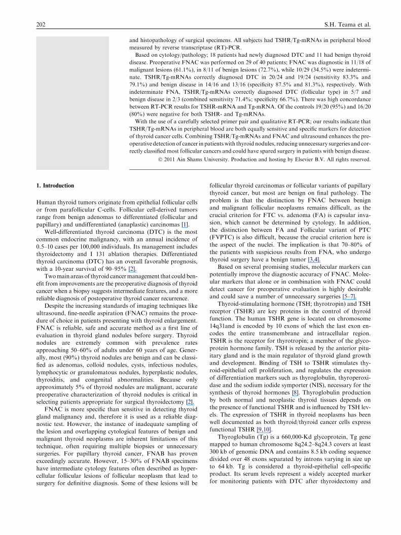
202 S.H. Teama et al.
and histopathology of surgical specimens. All subjects had TSHR/Tg-mRNAs in peripheral blood
measured by reverse transcriptase (RT)-PCR.
Based on cytology/pathology; 18 patients had newly diagnosed DTC and 11 had benign thyroid
disease. Preoperative FNAC was performed on 29 of 40 patients; FNAC was diagnostic in 11/18 of
malignant lesions (61.1%), in 8/11 of benign lesions (72.7%), while 10/29 (34.5%) were indetermi-
nate. TSHR/Tg-mRNAs correctly diagnosed DTC in 20/24 and 19/24 (sensitivity 83.3% and
79.1%) and benign disease in 14/16 and 13/16 (specificity 87.5% and 81.3%), respectively. With
indeterminate FNA, TSHR/Tg-mRNAs correctly diagnosed DTC (follicular type) in 5/7 and
benign disease in 2/3 (combined sensitivity 71.4%; specificity 66.7%). There was high concordance
between RT-PCR results for TSHR-mRNA and Tg-mRNA. Of the controls 19/20 (95%) and 16/20
(80%) were negative for both TSHR- and Tg-mRNAs.
With the use of a carefully selected primer pair and qualitative RT-PCR; our results indicate that
TSHR/Tg-mRNAs in peripheral blood are both equally sensitive and specific markers for detection
of thyroid cancer cells. Combining TSHR/Tg-mRNAs and FNAC and ultrasound enhances the pre-
operative detection of cancer in patientswith thyroid nodules, reducing unnecessary surgeries and cor-
rectly classified most follicular cancers and could have spared surgery in patients with benign disease.
� 2011 Ain Shams University. Production and hosting by Elsevier B.V. All rights reserved.
1. Introduction
Human thyroid tumors originate from epithelial follicular cellsor from parafollicular C-cells. Follicular cell-derived tumorsrange from benign adenomas to differentiated (follicular and
papillary) and undifferentiated (anaplastic) carcinomas [1].Well-differentiated thyroid carcinoma (DTC) is the most
common endocrine malignancy, with an annual incidence of
0.5–10 cases per 100,000 individuals. Its management includesthyroidectomy and I 131 ablation therapies. Differentiatedthyroid carcinoma (DTC) has an overall favorable prognosis,
with a 10-year survival of 90–95% [2].Twomainareas of thyroid cancermanagement that couldben-
efit from improvements are the preoperative diagnosis of thyroidcancer when a biopsy suggests intermediate features, and a more
reliable diagnosis of postoperative thyroid cancer recurrence.Despite the increasing standards of imaging techniques like
ultrasound, fine-needle aspiration (FNAC) remains the proce-
dure of choice in patients presenting with thyroid enlargement.FNAC is reliable, safe and accurate method as a first line ofevaluation in thyroid gland nodules before surgery. Thyroid
nodules are extremely common with prevalence ratesapproaching 50–60% of adults under 60 years of age. Gener-ally, most (90%) thyroid nodules are benign and can be classi-fied as adenomas, colloid nodules, cysts, infectious nodules,
lymphocytic or granulomatous nodules, hyperplastic nodules,thyroiditis, and congenital abnormalities. Because onlyapproximately 5% of thyroid nodules are malignant, accurate
preoperative characterization of thyroid nodules is critical inselecting patients appropriate for surgical thyroidectomy [2].
FNAC is more specific than sensitive in detecting thyroid
gland malignancy and, therefore it is used as a reliable diag-nostic test. However, the instance of inadequate sampling ofthe lesion and overlapping cytological features of benign and
malignant thyroid neoplasms are inherent limitations of thistechnique, often requiring multiple biopsies or unnecessarysurgeries. For papillary thyroid cancer, FNAB has provenexceedingly accurate. However, 15–30% of FNAB specimens
have intermediate cytology features often described as hyper-cellular follicular lesions of follicular neoplasm that lead tosurgery for definitive diagnosis. Some of these lesions will be
follicular thyroid carcinomas or follicular variants of papillary
thyroid cancer, but most are benign on final pathology. Theproblem is that the distinction by FNAC between benignand malignant follicular neoplasms remains difficult, as the
crucial criterion for FTC vs. adenoma (FA) is capsular inva-sion, which cannot be determined by cytology. In addition,the distinction between FA and Follicular variant of PTC(FVPTC) is also difficult, because the crucial criterion here is
the aspect of the nuclei. The implication is that 70–80% ofthe patients with suspicious results from FNA, who undergothyroid surgery have a benign tumor [3,4].
Based on several promising studies, molecular markers canpotentially improve the diagnostic accuracy of FNAC. Molec-ular markers that alone or in combination with FNAC could
detect cancer for preoperative evaluation is highly desirableand could save a number of unnecessary surgeries [5–7].
Thyroid-stimulating hormone (TSH; thyrotropin) and TSH
receptor (TSHR) are key proteins in the control of thyroidfunction. The human TSHR gene is located on chromosome14q31and is encoded by 10 exons of which the last exon en-codes the entire transmembrane and intracellular region.
TSHR is the receptor for thyrotropin; a member of the glyco-protein hormone family. TSH is released by the anterior pitu-itary gland and is the main regulator of thyroid gland growth
and development. Binding of TSH to TSHR stimulates thy-roid-epithelial cell proliferation, and regulates the expressionof differentiation markers such as thyroglobulin, thyroperoxi-
dase and the sodium iodide symporter (NIS), necessary for thesynthesis of thyroid hormones [8]. Thyroglobulin productionby both normal and neoplastic thyroid tissues depends onthe presence of functional TSHR and is influenced by TSH lev-
els. The expression of TSHR in thyroid neoplasms has beenwell documented as both thyroid/thyroid cancer cells expressfunctional TSHR [9,10].
Thyroglobulin (Tg) is a 660,000-Kd glycoprotein, Tg genemapped to human chromosome 8q24.2–8q24.3 covers at least300 kb of genomic DNA and contains 8.5 kb coding sequence
divided over 48 exons separated by introns varying in size upto 64 kb. Tg is considered a thyroid-epithelial cell-specificproduct. Its serum levels represent a widely accepted marker
for monitoring patients with DTC after thyroidectomy and

Molecular detection of circulating thyroid specific transcripts (TSHR/Tg-mRNAs) 203
ablative doses of radioiodine, as they usually show good corre-lation with the volume of differentiated thyroid tissue present.However, the various sTg assays are plagued by a number of
methodological problems, occasionally limiting the clinical va-lue of routine sTg measurements [11,12].
Molecular markers have been explored as alternative meth-
od for the detection of thyroid cancer with variable success andsuffer from the lack of specificity. The variable results in thesestudies may relate to the differences in methods used including
RNA isolation, use of different primers, and the normalizationprocess [6].
Our objective was to assess these two markers TSHR/Tg-mRNAs as diagnostic markers for thyroid cancer to determine
its sensitivity and specificity and if they can be used in synergywith current diagnostic modalities; fine needle aspiration andultrasound for preoperative diagnosis (Fig. 1).
2. Subjects and methods
2.1. Subjects
The subjects were conducted from National Cancer Institute,
Cairo University and Endocrine Department of Ain-ShamsUniversity Hospitals during period from December 2009 toJune 2010. Follow-up: postoperative pathological diagnosis
Figure 1 Algorithm for the evaluation of thyroid disorders. TSH= t
HB [13].
was used to categorize the patients into benign and malignant,a total of 60 subjects including 3 groups.
2.1.1. Control group
Group A: Included 20 apparently healthy people of matchedage and sex as the patient groups without history of diseases.
2.1.2. Patient groups
Group B: Comprised 16 patients with benign thyroid diseases.Among those patients; two patients had thyroiditis, 9 had thy-
roid nodules/multinodular goiter, and five had Grave’s disease(diffuse toxic goiter).
Group C: Comprised 24 patients with differentiated thyroidcancer (DTC); 18 patients newly diagnosed cases differentiated
thyroid carcinoma and 6 patients with recurrent or residualDTC on T4 replacement therapy.
All subjects were subjected to history taking, clinical exam-
ination, laboratory investigations including: CBC, liver func-tion test, kidney function test, prothrombin time, thyroidprofile, thyroid antibodies and thyroid U/S, while U/S guided
FNAC was done only for 29 patients who were presenting withnodular thyroid disease out of forty patients (in patients withMNG; FNAC was done on the predominant cold nodule
according to isotope scan finding). The remaining 11 patientsdid not require FNAC as five patients were proven clinically
hyroid-stimulating hormone; FNA = fine-needle aspiration Burch

Table 1 Setting of PCR primers 7.
Sequence of primers Amplified product
TSHR F 50-GCTTTTCAGGGACTATGCAATGAA-3’
R 30-AGAGTTTGGTCAC-AGTGACGGGAA-5’ 212
Tg F 50-AGGGAAACGGCCTTTCTGAA-3’
R 30-CTTTAGCAGCAGAAGAGGTG-5’ 407
GAPDH F 50-CGTCTTCACCACCATGGAGA 3’
R 50-CGGCCATCACGCCACAGTTT 3’ 300
204 S.H. Teama et al.
and by lab results to have Grave’s disease, while the remaining6 patients had recurrent thyroid cancer. In selected cases thy-roid isotope scans, CT scan were required and were done. Datawere included in chart review. Written informed consent was
obtained before enrollment into the study.Patients had FNA biopsy performed and blood samples
drawn before surgery. Postoperative pathological diagnosis
was used to categorize the patients into benign and malignant.FNA sample adequacy included sufficient number of cells,abundant colloid, and the presence of at least six groups of be-
nign follicular cells composed of at least 10 cells each. The re-sults were compared with final postoperative pathologicaldiagnosis. Indeterminate FNA included the following cytolog-ical categories: hypercellular follicular nodule (oxyphilic or
non oxyphilic), atypical epithelial cells and hemorrhagic cystcontents with few follicular cells.
2.2. Methods
2.2.1. Blood samples
Ten milliliters was sampled from the peripheral veins fromeach patient and from controls for separation of mononuclearcells.
2.2.1.1. Isolation of peripheral blood mononuclear cells. Periph-eral blood mononuclear cells (PBMCs) were isolated fromblood by density gradient centrifugation using Ficoll-hypaque
1077 (Sigma, USA) at 1200g for 30 min at 4 �C. The interfacecells were removed, washed twice with 25 ml of sterile PBS (pH7.3), pelleted, and resuspended in 1 ml of PBS. The cells were
pelleted again at 1200g for 2 min. The cell pellets were kept at�80 �C until RNA extraction.
2.2.1.2. Extraction of total RNA from nuclear cells. Total RNAof nuclear cells separated from whole blood was extractedusing RNA extraction kit (Qiagen). The isolated RNA was
resuspended in RNAase-free water and stored at �80 �C until
Table 2 Patients characteristic in various diagnostic groups.
Normal Benign
No. of patients 20 16
Age 48.8 ± 11.33 54.8 ± 10.1
Sex
Male 6 (30%) 7 (43.8%)
Female 14 (70%) 9 (56.2%)
Surgical confirmation 11
FNAC 8/11
Cytology/pathology nodules/MNG, 2 thyroditis
assay. The RNA concentration was assessed by absorbancereading at 260 nm with UV spectrophotometry (Du series650, Beckman Inc., USA).
2.2.1.3. Reversetranscription (complementary cDNA synthesis).Reverse transcription reaction was carried out in 20 ll reactionmixture by using first strand cDNA synthesis kit (Promega;
USA) according to the manufactures instructions.
2.2.1.4. PCR amplification of TSHR and Tg. PCR was per-
formed using the selected primer pairs. Five microliters offirst-strand cDNA was used as a template for the PCR reac-tion. Each reaction mixture consisted of 0.5 mM of each pri-mer, 10· Taq Buffer, 2.5 mM dNTP mix, 0.5 U of Taq
polymerase and nuclease-free water to a final volume of50 ll. Thermocycling in either an MJ Research PTC 200 (MJResearch, Inc., Boston, MA) or Perkin-Elmer 9600 (Perkin-El-
mer, Cambridge, UK) consisted of 38 cycles of denaturation(94 �C, 1 min; (first cycle for 2 min), annealing (62 �C, 1 min)and extension (72 �C, 1 min) (10 min for the last cycle). PCR
amplification of GAPDH to assess the cDNA integrity isshown in Table 1.
2.2.1.5. Analysis of amplified thyroglobulin/TSHR cDNA. Tenmicroliters of PCR product was mixed with 2 ll 6· and theamplified PCR products were analyzed by agarose gel electro-phoresis, stained with ethidium bromide (Sigma), and visual-
ized with an UV transilluminator (Bio-Rad).
2.3. Statistical analysis
The results were analyzed using the Statistical Package of So-cial Sciences (SPSS) computer software program, version 16.0(Chicago, IL, USA). Quantitative data were presented as
mean ± SD for normally distributed data. Qualitative datawere presented in the form of frequencies and percentages.For normally distributed parameters, differences among
Newly diagnosed DTC Recurrent thyroid cance
18 6 (PTC)
60.2 ± 7.3 63.5 ± 6
6 (33.3%) 1 (16.7%)
12 (66.7%) 5 (83.3%)
18
11/18
and 3FA 12PTC and 6F
r
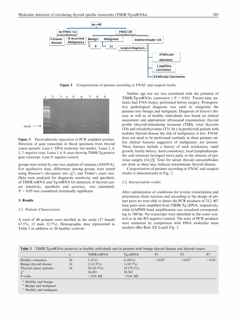
Figure 2 Categorization of patients according to FNAC and surgical results.
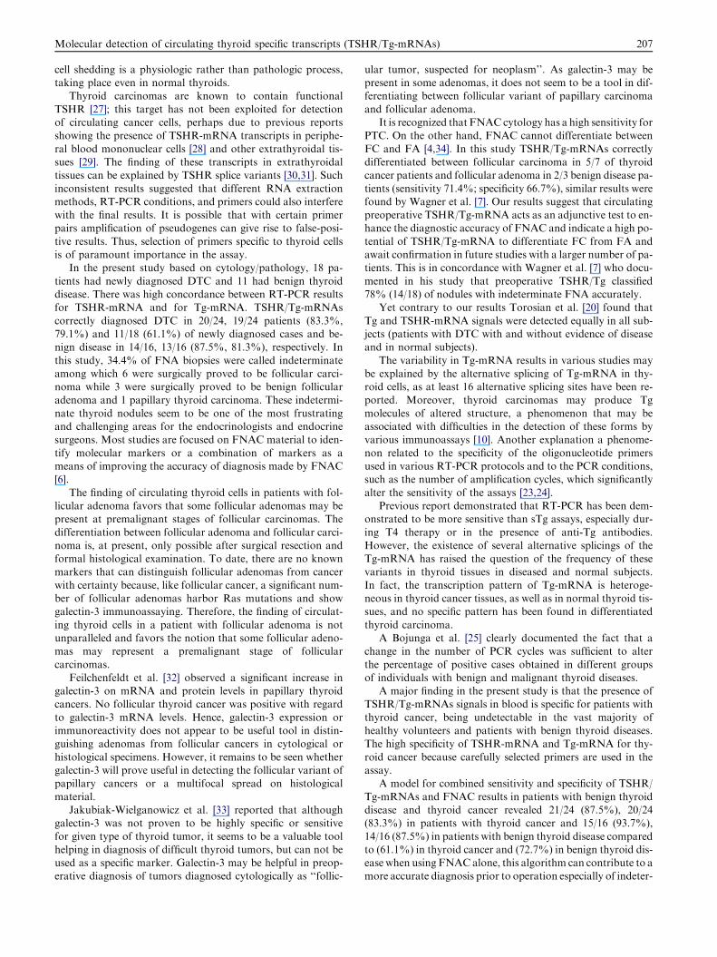
Figure 3 Electrophoretic separation of PCR amplified product.
Detection of gene transcripts in blood specimens from thyroid
cancer patients. Lane 1: DNA molecular size marker. Lanes 2 ,4,
5, 7: negative cases. Lanes 3, 6, 8: cases showing TSHR/Tg positive
gene transcript. Lane 9: negative control.
Molecular detection of circulating thyroid specific transcripts (TSHR/Tg-mRNAs) 205
groups were tested by one-way analysis of variance (ANOVA).For qualitative data, differences among groups were tested
using Pearson’s chi-square test (v2) and Fisher’s exact test.Data were analyzed for diagnostic sensitivity and specificityof TSHR-mRNA and Tg-mRNA for detection of thyroid can-
cer sensitivity, specificity and accuracy was calculated.P < 0.05 was considered statistically significant.
3. Results
3.1. Patients Characteristic
A total of 40 patients were enrolled in the study (27 female67.5%; 13 male 32.5%). Demographic data represented inTable 2 in addition to 20 healthy controls.
Table 3 TSHR/Tg-mRNAs positivity in healthy individuals and in
n TSHR-mRNA
Healthy volunteers 20 1 (5%)
Benign thyroid disease 16 2 (12.5%)
Thyroid cancer patients 24 20 (83.3%)
v2 36.893
P-value <0.01 HS
a Healthy and benign.b Benign and malignant.c Healthy and malignant.
Neither age nor sex was correlated with the presence ofTSHR/Tg-mRNAs expression ( P > 0.05). Twenty-nine pa-tients had FNA biopsy performed before surgery. Postopera-
tive pathological diagnosis was used to categorize thepatients into benign and malignant. Diagnosis of Grave’s dis-ease as well as of healthy individuals was based on clinicalassessment and appropriate ultrasound examination; thyroid
profile (thyroid-stimulating hormone (TSH), total thyroxin(T4) and triiodothyronine (T3). In a hyperthyroid patient withnodular thyroid disease the risk of malignancy is low. FNAC
does not need to be performed routinely in these patients un-less clinical features suggestive of malignancy are present.These features include a history of neck irradiation, rapid
growth, family history, hard consistency, local lymphadenopa-thy and recurrent laryngeal nerve palsy in the absence of pre-vious surgery [14,15]. Tests for serum thyroid autoantibodies
are done as these may indicate autoimmune thyroid disease.Categorization of patients according to FNAC and surgical
results is demonstrated in Fig. 2.
3.2. Interpretation results
After optimization of conditions for reverse transcription andpolymerase chain reaction and according to the design of pri-
mer pairs we were able to detect the PCR products of 212; 407base pairs were amplified from TSHR/Tg cDNA, respectively;while GAPDH band amplification was visualized correspond-
ing to 300 bp. No transcripts were identified in the water con-trol or in the RT-negative control. The sizes of PCR productswere estimated by comparison with DNA molecular mass
markers (Bio-Rad; EZ Load) Fig. 3.
patients with benign thyroid disease and thyroid cancer.
Tg-mRNA P1 P2 P3
4 (20%) >0.05a <0.01b <0.01c
3 (18.7%)
19 (79.1%)
30.343
<0.01 HS
Table 4 Statistical analysis of TSHR/Tg-mRNAs’ results.
Disease Positive Disease Negative
Test (TSHR-mNA) n= 40
Positive 20 TP 2 FP
Negative 4 FN 14 TN
Test (Tg-mNA) n = 40
Positive 19 TP 3 FP
Negative 5 FN 13 TN
Sensitivity = TP/TP+FN · 100, specificity = TN/TN+FP · 100,
PPV= TP /TP + FP · 100, NPV= TN/TN+ FN · 100, accu-
racy = TP+ TN/total no. · 100; TP = true positive, TN= true
negative, FP = false positive, FN= false negative, PPV= positive
predictive value, NPV= negative predictive value.
Table 5 Diagnostic performance of TSHR, Tg and FNA
result in patients with benign thyroid disease and thyroid
cancer.
Variables TSHR-mRNA (%) Tg-mRNA (%)
Sensitivity 83.3 79.1
Specificity 87.5 81.3
PPV 90.9 86.4
NPV 77.8 72.2
Accuracy 85 80
206 S.H. Teama et al.
3.3. FNAC result compared with histological diagnosis
Based on cytology/pathology; 18 had surgically confirmedDTC. 11 patients had benign conditions; FNA result of malig-
nant conditions: 11/18 (61.1%) were FNAC results of newlydiagnosed DTC (papillary carcinoma), FNA result of benignconditions: 8 were FNAC results of benign condition 6 pa-
tients diagnosed nodule/MNG, and 2 patients with Hashimotothyroiditis and 3/11 (27.3%) patients had sufficient specimenbut inconclusive results; surgically confirmed follicular ade-noma. FNAC of indeterminate result with 6 were surgically
proven to be follicular carcinoma while 3 were surgically pro-ven to be benign follicular adenoma and 1 papillary thyroidcarcinoma.
3.4. Evaluation of reverse transcriptase-PCR (RT-PCR) results
in controls and patients
Forty patients (24 DTC; 16 benign) were enrolled at initialdiagnosis and 20 control patients without clinical signs or ahistory of thyroid disease.
Table 6 Sensitivity and specificity of combined TSHR/Tg-mRNA
thyroid cancer.
Thyroid cancer
Positive Sensitivi
FNA 11/18 61.1
TSHR/Tg-mRNA 21/24a 87.5
FNA 20/24b 83.3
a TSHR-mRNA.b Tg-mRNA.
3.4.1. TSHR/Tg-mRNAs in normal control and benign thyroid
disease
Of the controls 19/20 (95%) and 16/20 (80%) were negative forboth TSHR- and Tg-mRNAs. Of the patients with benign thy-roid disease, 14/16 (87.5%) and 13/16 (81.3%) were negative
for both TSHR- and Tg-mRNA. False positive for TSHR/Tg included two patients with follicular adenoma (Tables 3and 4).
3.4.2. TSHR/Tg-mRNAs in thyroid cancer
Twenty four patients were evaluated (six patients had recur-rent or residual malignant disease at the time of enrollment
and 18 were recently diagnosed with differentiated thyroidcancer (12 papillary, 6 follicular cancers). There were 20/24,19/24 patients (83.3%, 79.1%) with DTC who were positive
for TSHR/Tg-mRNAs in the peripheral blood. (All six pa-tients with recurrent disease were positive by RT-PCR.) (Ta-bles 3 and 4).
There was high concordance between RT-PCR results for
TSHR-mRNA and for Tg-mRNA, also combined model forboth markers with FNAC showed improved sensitivity andspecificity (Tables 5 and 6).
4. Discussion
Molecular detection of tissue-specific gene expression in
peripheral blood is a new diagnostic approach and has beendescribed as a tumor marker in different solid tumors. TheRT-PCR is a highly sensitive technique for the detection of tis-
sue-specific mRNA transcripts. This method has been testedwith the amplification of cancer-specific mRNA from periphe-ral blood to detect micrometastases in cases of prostate cancer
and neuroblastoma [6]. Many studies explored blood-bornethyroglobulin/TSHR-mRNAs as a potential tumor marker inthyroid cancer [16,17]. A reliable and satisfying method thatis able to differentiate preoperative malignant potential in pa-
tients presenting with thyroid nodules has not yet beenproposed.
Ditkoff et al. [18] reported, for the first time, a method for
detecting Tg-mRNA in peripheral blood of patients with well-differentiated thyroid tumors. Since the authors were not ableto document Tg expression either in controls or in patients
with benign thyroid disease, Tg-mRNA appeared to be a spe-cific tumor marker. In contrast, different studies revealed thatTg transcripts could be found in blood from healthy control
[19–21] in a variety of human cell lines and in different humantissues and organs [22]. This could be attributed to illegitimatetranscription of Tg-mRNA [23–26]; any gene is expressed inany cell at very low, but detectable levels, or could mean that
s and FNA result in patients with benign thyroid disease and
Benign conditions
ty (%) Negative Specificity (%)
8/11 72.7
15/16a 93.7
14/16b 87.5
Molecular detection of circulating thyroid specific transcripts (TSHR/Tg-mRNAs) 207
cell shedding is a physiologic rather than pathologic process,taking place even in normal thyroids.
Thyroid carcinomas are known to contain functional
TSHR [27]; this target has not been exploited for detectionof circulating cancer cells, perhaps due to previous reportsshowing the presence of TSHR-mRNA transcripts in periphe-
ral blood mononuclear cells [28] and other extrathyroidal tis-sues [29]. The finding of these transcripts in extrathyroidaltissues can be explained by TSHR splice variants [30,31]. Such
inconsistent results suggested that different RNA extractionmethods, RT-PCR conditions, and primers could also interferewith the final results. It is possible that with certain primerpairs amplification of pseudogenes can give rise to false-posi-
tive results. Thus, selection of primers specific to thyroid cellsis of paramount importance in the assay.
In the present study based on cytology/pathology, 18 pa-
tients had newly diagnosed DTC and 11 had benign thyroiddisease. There was high concordance between RT-PCR resultsfor TSHR-mRNA and for Tg-mRNA. TSHR/Tg-mRNAs
correctly diagnosed DTC in 20/24, 19/24 patients (83.3%,79.1%) and 11/18 (61.1%) of newly diagnosed cases and be-nign disease in 14/16, 13/16 (87.5%, 81.3%), respectively. In
this study, 34.4% of FNA biopsies were called indeterminateamong which 6 were surgically proved to be follicular carci-noma while 3 were surgically proved to be benign follicularadenoma and 1 papillary thyroid carcinoma. These indetermi-
nate thyroid nodules seem to be one of the most frustratingand challenging areas for the endocrinologists and endocrinesurgeons. Most studies are focused on FNAC material to iden-
tify molecular markers or a combination of markers as ameans of improving the accuracy of diagnosis made by FNAC[6].
The finding of circulating thyroid cells in patients with fol-licular adenoma favors that some follicular adenomas may bepresent at premalignant stages of follicular carcinomas. The
differentiation between follicular adenoma and follicular carci-noma is, at present, only possible after surgical resection andformal histological examination. To date, there are no knownmarkers that can distinguish follicular adenomas from cancer
with certainty because, like follicular cancer, a significant num-ber of follicular adenomas harbor Ras mutations and showgalectin-3 immunoassaying. Therefore, the finding of circulat-
ing thyroid cells in a patient with follicular adenoma is notunparalleled and favors the notion that some follicular adeno-mas may represent a premalignant stage of follicular
carcinomas.Feilchenfeldt et al. [32] observed a significant increase in
galectin-3 on mRNA and protein levels in papillary thyroidcancers. No follicular thyroid cancer was positive with regard
to galectin-3 mRNA levels. Hence, galectin-3 expression orimmunoreactivity does not appear to be useful tool in distin-guishing adenomas from follicular cancers in cytological or
histological specimens. However, it remains to be seen whethergalectin-3 will prove useful in detecting the follicular variant ofpapillary cancers or a multifocal spread on histological
material.Jakubiak-Wielganowicz et al. [33] reported that although
galectin-3 was not proven to be highly specific or sensitive
for given type of thyroid tumor, it seems to be a valuable toolhelping in diagnosis of difficult thyroid tumors, but can not beused as a specific marker. Galectin-3 may be helpful in preop-erative diagnosis of tumors diagnosed cytologically as ‘‘follic-
ular tumor, suspected for neoplasm’’. As galectin-3 may bepresent in some adenomas, it does not seem to be a tool in dif-ferentiating between follicular variant of papillary carcinoma
and follicular adenoma.It is recognized that FNAC cytology has a high sensitivity for
PTC. On the other hand, FNAC cannot differentiate between
FC and FA [4,34]. In this study TSHR/Tg-mRNAs correctlydifferentiated between follicular carcinoma in 5/7 of thyroidcancer patients and follicular adenoma in 2/3 benign disease pa-
tients (sensitivity 71.4%; specificity 66.7%), similar results werefound by Wagner et al. [7]. Our results suggest that circulatingpreoperative TSHR/Tg-mRNA acts as an adjunctive test to en-hance the diagnostic accuracy of FNAC and indicate a high po-
tential of TSHR/Tg-mRNA to differentiate FC from FA andawait confirmation in future studies with a larger number of pa-tients. This is in concordance with Wagner et al. [7] who docu-
mented in his study that preoperative TSHR/Tg classified78% (14/18) of nodules with indeterminate FNA accurately.
Yet contrary to our results Torosian et al. [20] found that
Tg and TSHR-mRNA signals were detected equally in all sub-jects (patients with DTC with and without evidence of diseaseand in normal subjects).
The variability in Tg-mRNA results in various studies maybe explained by the alternative splicing of Tg-mRNA in thy-roid cells, as at least 16 alternative splicing sites have been re-ported. Moreover, thyroid carcinomas may produce Tg
molecules of altered structure, a phenomenon that may beassociated with difficulties in the detection of these forms byvarious immunoassays [10]. Another explanation a phenome-
non related to the specificity of the oligonucleotide primersused in various RT-PCR protocols and to the PCR conditions,such as the number of amplification cycles, which significantly
alter the sensitivity of the assays [23,24].Previous report demonstrated that RT-PCR has been dem-
onstrated to be more sensitive than sTg assays, especially dur-
ing T4 therapy or in the presence of anti-Tg antibodies.However, the existence of several alternative splicings of theTg-mRNA has raised the question of the frequency of thesevariants in thyroid tissues in diseased and normal subjects.
In fact, the transcription pattern of Tg-mRNA is heteroge-neous in thyroid cancer tissues, as well as in normal thyroid tis-sues, and no specific pattern has been found in differentiated
thyroid carcinoma.A Bojunga et al. [25] clearly documented the fact that a
change in the number of PCR cycles was sufficient to alter
the percentage of positive cases obtained in different groupsof individuals with benign and malignant thyroid diseases.
A major finding in the present study is that the presence ofTSHR/Tg-mRNAs signals in blood is specific for patients with
thyroid cancer, being undetectable in the vast majority ofhealthy volunteers and patients with benign thyroid diseases.The high specificity of TSHR-mRNA and Tg-mRNA for thy-
roid cancer because carefully selected primers are used in theassay.
A model for combined sensitivity and specificity of TSHR/
Tg-mRNAs and FNAC results in patients with benign thyroiddisease and thyroid cancer revealed 21/24 (87.5%), 20/24(83.3%) in patients with thyroid cancer and 15/16 (93.7%),
14/16 (87.5%) in patients with benign thyroid disease comparedto (61.1%) in thyroid cancer and (72.7%) in benign thyroid dis-ease when using FNAC alone, this algorithm can contribute to amore accurate diagnosis prior to operation especially of indeter-

208 S.H. Teama et al.
minate thyroid lesions or follicular neoplasms than other cur-rently available strategy, nevertheless this algorithm requiresprospective validation and comparison with other suggested
models combiningmolecularmarkers withUS features .Finally,it is worth noting that TSHR/Tg-mRNAs correctly diagnosedall recurrent thyroid cancer cases, which implies the utility of
TSHR/Tg-mRNAs in long-term monitoring and detection ofcancer recurrence and residual disease which is concordant withearlier reports of correlation of elevated levels with the presence
of residual/ metastatic disease and that it could postoperativelypredict recurrent cancer [6,10].
A standardization of cDNA preparation, gene-specific pri-mer pairs, optimal RT-PCR conditions and the determination
of possible threshold caused by cells of non-thyroid origin ismandatory to obtain reliable and comparable results. In viewof our data obtained for sensitivity and specificity, we believe
that a clinically useful assay will require further assay refine-ment, e.g. quantitative assay to permit the distinction of weaklyand more strongly positive RT-PCR results. A number of ques-
tions remain to be answered and further studies are necessary.
5. Conclusion
In summary, the presence of either TSHR-mRNA or Tg-mRNA in peripheral blood is specific for the presence of circu-lating thyroid cells; and represent specific and sensitive mark-
ers for thyroid cancer and expression of this marker showspromise for detecting FC, which is often missed by FNAC.Overall, the management of cancer in general and thyroid can-cer in particular is moving away from reliance on a single test
that replaces others. The ultimate usefulness of molecularmarkers as TSHR/Tg-mRNAs may reside in being part of amultimodality panel of clinical tests. An envisioned new algo-
rithm may combine the best of all management criteria clinicalassessment, radiologic and histologic information, and the bio-logic implications of a molecular marker.
5.1. Future perspectives
The use of molecular diagnostic techniques, such as RT-PCR,
which enable the specific amplification of small numbers ofmRNA molecules, has led to the development of several meth-ods for the specific amplification of small foci of malignant tis-sue, either primary or metastatic. PCR-based techniques may
improve the preoperative diagnosis of patients with thyroidmalignancies and therefore help to avoid unnecessary radicalprocedures. Quantitative TSHR-mRNA assay is helpful in
the preoperative diagnosis of thyroid nodules, particularly inthe subgroup of patients with indeterminate FNA. The imme-diate postsurgical levels can serve as a sensitive marker for
detecting residual/metastatic disease. Furthermore, it detectsrecurrent cancer with high sensitivity and can be valuable asan alternative to serum Tg measurement in patients who har-
bor Tg antibodies. Future studies are required to establish itsrole in long-term monitoring.
Conflicts of interest
The authors indicate that they do not have any conflict ofinterest.
Acknowledgments
This study was done at Medical Research Center, Ain-Shams
University.
References
[1] Ringel MD. Diagnostic molecular markers in thyroid cancer.
Cancer Treat Res 2004;122:295–316.
[2] Gianoukakis AG, Giannellia SM, Salameh WA, McPhau LW.
Well differentiated follicular thyroid neoplasia: impact of molec-
ular and technological advances on detection, monitoring and
treatment. Mol Cell Endocrinol 2011;332(1–2):9–20.
[3] Sahin M, Gursoy A, Tutuncu NB, Guvener DN. Prevalence and
prediction of malignancy in cytologically indeterminate thyroid
nodules. Clin Endocrinol 2006;65(4):514–8.
[4] Tseng CE, Wei CK, Kuo CS, Yan ST, Chen PF, Lien WC, et al.
Fine needle aspiration cytology of thyroid nodules: evaluation of
diagnostic accuracy. Tzu Chi Med J 2008;20(4).
[5] Haugen B, Pacini F, Reiners C, Schlumberger M, Ladenson P,
Sherman S, et al. A comparison of recombinant human thyrot-
ropin and thyroid hormone withdrawal for the detection of
thyroid remnant or cancer. J Clin Endocrinol Metab
1999;84(38):77–85.
[6] Yip L, Kebebew E, Milas M, Carty SE, Fahey TJ, Parangi S,
et al. Summary statement: utility of molecular marker testing in
thyroid cancer. Surgery 2010;148(6):1313–5.
[7] Wagner K, Arciaga R, Siperstein A, Milas M, Warshawsky I,
Sethu S, et al. Thyrotropin receptor/thyroglobulin messenger
ribonucleic acid in peripheral blood and fine needle aspiration
cytology: diagnostic synergy for detecting thyroid cancer. J Clin
Endocr Metab 2005;90(4):1921–4.
[8] Chia SY, Milas M, Reddy SK, Siperstein A, Skugor M, Brainard
J, et al. Thyroid-stimulating hormone receptor messenger ribo-
nucleic acid measurement in blood as a marker for circulating
thyroid cancer cells and its role in the preoperative diagnosis of
thyroid cancer. J Clin Endocrinol Metab 2007;92(2):468–75.
[9] Szkudlinski M, Fremont V, Ronin C, Weintraub B. Thyroid-
stimulating hormone and thyroid-stimulating hormone receptor
structure–function relationships. Physiol Rev 2002;82:473–502.
[10] Chinnappa P, Taguba L, Arciaga R, Faiman C, Siperstein A,
Mehta AE, et al. Detection of thyrotropin-receptor messenger
ribonucleic acid (mRNA) and thyroglobulin mRNA transcripts in
peripheral blood of patients with thyroid disease: sensitive and
specific markers for thyroid cancer. J Clin Endocr Metab
2004;89(8):3705–9.
[11] Mendive FM, Rivolta CM, Vassart G, Targovnik HM. Genomic
organization of the 30 regio n of the human thyroglobulin gene.
Thyroid 1999;9:903–12.
[12] Van Sande J, Dequanter D, Lothaire P, Massart C, Dumont JE,
Erneux C. Thyrotropin stimulates the generation of inositol 1, 4,
5-trisphosphate in human thyroid cells. J Clin Endocrinol Metab
2006;91(3):1099–107.
[13] Evaluation and management of the solid thyroid nodule. Endo-
crinol Metab Clin North Am 1995;24:663–710.
[14] American Association of Clinical Endocrinologists. AACE clin-
ical practice guidelines for the diagnosis and management of
thyroid nodules. Endocr Pract 1996;2:80–4.
[15] Grammatopoulos D, Elliott Y, Smith SC, Brown I, Grieve RJ,
Hillhouse EW, et al. Measurement of thyroglobulin mRNA in
peripheral blood as an adjunctive test for monitoring thyroid
cancer. J Clin Pathol Mol Pathol 2003;56:162–6.
[16] Gupta M, Taguba L, Arciaga R, Siperstein A, Faiman C, Mehta
A, et al. Detection of circulating thyroid cells by reverse
transcription-PCR for thyroid stimulating hormone receptor and

Molecular detection of circulating thyroid specific transcripts (TSHR/Tg-mRNAs) 209
thyroglobulin: the importance of primer selection. Clin Chem
2002;48:1862–5.
[17] Ghossein R, Bhattacharya S, Rosai J. Molecular detection of
micrometastases and circulating tumor cells in solid tumors. Clin
Cancer Res 1999;5:1950–60.
[18] Ditkoff B, Marvin M, Yemul S, Shi Y, Chabot J, Feind C, et al.
Detection of circulating thyroid cells in peripheral blood. Surgery
1996;120:959–65.
[19] Tallini G, Ghossein R, Emanuel J, Gill J, Kinder B, Dimich AB,
et al. Detection of thyroglobulin, thyroid peroxidase, and RET/
PTC1 mRNA transcripts in the peripheral blood of patients with
thyroid disease. J Clin Oncol 1998;16:1158–66.
[20] Torosian L, Manrique G, Alvarez B, Lago G, Roca R, Belzarena
C. Blood thyroglobulin and TSH receptor mRNA detection by
RT-PCR in the follow-up of differentiated thyroid cancer
patients. Rev Esp Med Nucl 2010;29(3):109–13.
[21] Ringel MD, Ladenson PW, Levine MA. Molecular diagnosis of
residual and recurrent thyroid cancer by amplification of thyro-
globulin messenger ribonucleic acid in peripheral blood. J Clin
Endocrinol Metab 1998;83:4435–42.
[22] Tallini G, Ghossein RA, Emanuel J, Gill J, Kinder B, Dimich AB,
et al. Detection of thyroglobulin, thyroid peroxidase, and RET/
PTC1 mRNA transcripts in the peripheral blood of patients with
thyroid disease. J Clin Oncol 1998;16(3):1158–66.
[23] Selliti D, Akamizu T, Doi S, Kim G, Kariyil J, Koshiyama H.
Renal expression of two thyroid specific genes: thyrotropin
receptor and thyroglobulin. Exp Nephrol 2000;8:235–43.
[24] Chelly J, Concordet J, Kaplan J, Kahn A. Illegitimate transcrip-
tion: transcription of any gene in any cell type. Proc Natl Acad Sci
USA 1989;86:2617–21.
[25] Bojunga J, Roddiger S, Stanisch M, Kusterer K, Kurek R,
Renneberg H, et al. Molecular detection of thyroglobulin mRNA
transcripts in peripheral blood of patients with thyroid disease by
RT-PCR. Brit J Cancer 2000;82(10):1650–5.
[26] Bugalho MJ, Domingues RS, Pinto AC, Garrao A, Catarino AL,
Ferreira T, et al. Detection of thyroglobulin mRNA transcripts in
peripheral blood of individuals with and without thyroid glands:
evidence for thyroglobulin expression by blood cells. Eur J
Endocrinol 2001;145(4):409–13.
[27] Ohta K, Endo T, Onaya T. The mRNA levels of thyrotropin
receptor, thyroglobulin, and thyroid peroxidase in neoplastic
human thyroid tissues. Biochem Biophys Res Commun
1991;165:1250–5.
[28] Sheils OM, Sweeney EC. TSH receptor status of thyroid
neoplasms TaqMan RT-PCR analysis of archival material. J
Pathol 1999;188:87–92.
[29] Arturi F, Russo D, Giuffrida D, Ippolito A, Perroti N, Vigneri R,
et al. Early diagnosis by genetic analysis of differentiated thyroid
cancer metastases in small lymph nodes. J Clin Endocrinol Metab
1997;82:1638–41.
[30] Francis T, Burch H, Cai W, Lukes Y, Peele M, Carr F, et al.
Lymphocytes express thyrotropin receptor-specific mRNA as
detected by the PCR technique. Thyroid 1991;1:223–8.
[31] Paschke R, Metcalfe A, Alcalde L, Vassart G, Weetman A.
Presence of nonfunctional thyrotropin receptor variant transcripts
in retroocular and other tissues. J Clin Endocrinol Metab
1994;79:1234–8.
[32] Feilchenfeldt Jonas, Totsch Martin, Sien-Yi Sheu, John Robert,
Anastase Spiliopoulos, Frilling A, et al. Expression of galectin-3
in normal and malignant thyroid tissue by quantitative PCR and
immunohistochemistry. Mod Pathol 2003;16(11):1117–23.
[33] Jakubiak-Wielganowicz Magdalena, Kubiak Robert, Sygut Jacek,
Pomorski Lech, Kordek Radzisaw. Usefulness of galectin-3
immunohistochemistry in differential diagnosis between thyroid
follicular carcinoma and follicular adenoma. Pol J Pathol
2003;54(2):111–5 [PL ISSN 1233-9687].
[34] Mira M, Peter M, Su-Ynn C, Mario S, Eren B, Sethu R, et al.
The utility of peripheral thyrotropin mRNA in the diagnosis of
follicular neoplasms and surveillance of thyroid cancers. Surgery
2007;141(2):137–46.