Dislocation Configurations in Nanocrystalline FeMo Sintered Components
ISSN 2394–7799 IJAETCS (2014) Vol.1, No.1, 28-35
Research Article
International Journal of Advancement in Engineering, Technology and
Computer Sciences
INVESTIGATIONS OF STRUCTURAL AND
SPECTROSCOPIC CHARACTERIZATION OF
LANTHANUM SILICATE NANOCRYSTALLINE
A. M. El NahrawyI*
, H. H. AfifyI, A. I. Ali
II,III
I*National Research Center (NRC) Department of Solid State Physics, Doki, Giza, Egypt ([email protected])
IIEnergy Harvest-Storage Research Center and Department of Physics, University of Ulsan, Ulsan 680-749 South Korea.
III Basic Science Department, Faculty of Industrial Education, Helwan University, Saray El-Quba, 11281 Cairo, Egypt.
-----------------------------------------------------------------------------------------------------------------------------------------------------------
ABSTRACT: Oxyapatite nanostructure pure silica and lanthanum silicate (10 La2O3-90 SiO2)
nanoparticles were synthesized using sol–gel technique. La2O3 ions were introduced in sol–gel derived
silica matrix by direct mixing dissolved lanthanum nitrate into a silica sol. The effects of H2O/TEOS
ratio on the structural and spectroscopic properties of (La2O3-SiO2) and the effect of La2O3 ions on
the silica gel were studied. The gel and oxide powders were characterized by X-ray diffraction (XRD),
Fourier transformed infrared (FTIR) spectroscopy, transmission electron microscopy (TEM),
scanning electron microscopy (SEM/EDS) and optical properties. The observed XRD patterns for the
prepared samples calcined at different temperature confirmed the formation of nano-crystalline
lanthanum silicate phases. The refractive indices and UV-Vis absorption for pure and doped silicate
systems have been achieved.
KEY WORDS: lanthanum silicate, nano-particles, sol-gel process, XRD and spectroscopic
properties.
----------------------------------------------------------------------------------------------------------------------------------------------------------- 1. INTRODUCTION: Recently, the development of novel low-cost engineering materials with promising
properties and stability are of vital importance. Oxyapitatite materials based silicate have a great interest due
their uses in different industrial application especially as electrolytes for intermediate temperature solid
oxide fuel cells (SOFC) because of their high ionic conductivity at low temperatures as well as the low cost
of raw materials [(1)-(4)]. For modified silica-based nano-crystalline materials obtained using sol–gel
technique, the chemical composition and the stabilization temperature are the main required synthesis
conditions that will define the final network product. Oxyapitate silicate materials is the most common type
and has the formula (M10 (AO4)6O2-δ) where M are the rare-earth and alkaline earth metal cations and A is a
p-block element such as P,As, Si, Ge,V,Zn [(5)-(7)]. While the apatite structure of lanthanum silicates
consists of isolated SiO4 tetrahedra and M-site cations, occupying cavities between SiO4 units creating a
new silicate systems ; and additional oxygen sites (O4) surrounded by La cations.
Over the past decades, it has been well known that the sol–gel technique has become a simple,
attractive and a potential alternative to the traditional methods. The sol–gel process, in which metal alkoxide
precursors could readily undergo, catalyzed hydrolysis and poly-condensation reactions in the formation of
a sol with controllable size and narrow particle size distribution. Sol gel technology used as alternative
technology to prepare a wide variety of applications such as powders, monoliths, thin films, nanotube and
nanofibers [(8)-(10)]. Therefore, in order to reduce the synthesis temperature, sol gel technique was used as
synthetic procedure for pure and doped lanthanum silicate oxyapatites instead of the conventional solid-state
reaction method
The aim of the preset work is to explore and decrease the synthesis temperature of lanthanum silicate
(10 La2O3-90 SiO2) nanoparticles in comparing with pure silica and study the effect of lanthanum on pure
silica gel. In this paper, the structural evolution of pure silica and lanthanum silicate (La2O3-SiO2) with
El Nahrawy, A. M. et al./ International Journal of Advancement in Engineering, Technology and Computer Sciences, Vol.1, No.1 29
annealing temperature was investigated by X-ray diffraction (XRD), Fourier transform infrared
spectroscopy (FTIR), transmission electron microscopy, scanning electron microscopy (SEM/EDX) The
relationship between preparation conditions, structures and optical properties of the prepared samples were
also discussed.
2. EXPERIMENTAL:
2.1. MATERIALS: Tetraethylorthosilicate (TEOS, Aldrich Chemicals, 98%), ethanol and lanthanum
nitrate (La (NO3)
3·6H
2O (Merck)) as source of La2O3 ions, nitric acid. Water for preparing the samples was
purified by ion exchange followed by distillation. These chemicals were in the analytical grade and used as
provided.
2.2. SAMPLE PREPARATION: Pure silica and lanthanum silicate (10 La2O3-90 SiO2) nanoparticles
were prepared using sol gel process. Tetraethylorthosilicate was dissolved in excess of absolute ethanol and
distilled water in the presence of HNO3 (N.A) under vigorous stirring (950 rpm) for 2h. About lanthanum
silicate system, tetraethyorthosilicate mixed with lanthanum hexahydrate nitrate dissolved in a mixture of
ethanol and nitric acid and distilled water under vigorous stirring for 2h at 750C, and the solution gradually
became clear sol. The experimental procedures flow chart as in Fig. I. The sol transferred to a clear gel after
one night. The wet gel was dried at 100 ºC for 24 h. Then the gel was heated at different temperatures from
200 to 8000C for 4 h to remove the water, organic components and decompose the nitrates. The influences
of different H2O/TEOS ratio on the physicochemical properties are studied as summarized in Table 1.
sample N.A/TEOS volumetric
ratio
EtOH/TEOS
molar ratio
H2O/TEOS
molar ratio= r
La2O3-SiO2 %
Gel 1 2 4 4 0-100
Gel 2 2 4 6 10-90
Gel 3 2 4 8 10-90
Gel 4 2 4 11 10-90
Table 1: Composition and H2O/TEOS ratio for lanthanum silicate.
2.3. X-RAY DIFFRACTION: The phase purity and the crystal structure of the powders were checked
using X-ray diffraction (XRD, D8–ADVANCE).The measurements were done using Cu-Kα radiation (λ
=1,5418Å). The average crystallite sizes for the samples were calculated using the Debye-Sherrer equation:
D= , Where is the X-ray wavelength (1.54 Å), is the Bragg diffraction angle, and B is full width at
half maximum [(11)].
2.4. TRANSMISSION AND SCANNING ELECTRON MICROSCOPY (SEM/EDS): The particle
distribution and the surface morphology with element ration were examined by transmission and scanning
electron microscopy ((JEOL model JSM 840) with energy dispersive analyser (SEM/EDS).
2.5. FOURIER TRANSFORMS INFRARED SPECTROSCOPY (FTIR): FTIR spectra were recorded
using a Perkin Elmer FTIR spectrometer in KBr medium in the wavenumber range 470–4000 cm-1
.
2.6. OPTICAL PROPERTIES: The refractive indices of these materials calculated using nearly normal
transmittance and reflectance spectra were done by Jasco V-570 spectrophotomete and UV–vis spectra were
recorded by means of spectrophotometer (Mini-D2T, Ocean Optics Inc.).

El Nahrawy, A. M. et al./ International Journal of Advancement in Engineering, Technology and Computer Sciences, Vol.1, No.1 30
Stirring for 2hTEOS + ETOH +H2O+nitic acid
Silica (SiO2)(solution)
TEOS+ETOH+H2O+La(NO3) 3.6H2O+ nitric acid
La2O3-SiO2 solution
Oxyapatite lanthanum silicate
Draying 1 night at 750C
Calcinations at different temperatures
Stirring for 2h
transparent gel
Fig.I. Flow chart of sol gel process for the preparation of pure silica and lanthanum silicates nanoparticles.
3. RESULTS AND DISCUSSION:
3.1. SOL–GEL PROCESS: One of the most advantages of sol–gel technique comparing with other
chemical techniques is that they can be synthesized different organic / inorganic materials in normal
ambient conditions; they do not require high temperatures, high pressures, vacuum media, gas or water flow
or critical chemical conditions for production of organic and inorganic materials. The sol–gel mechanisms
involve hydrolysis and poly-condensation reactions of molecular precursors in water and co-solvent media
[(12), (13), (14)]. It is well known that the sol–gel preparation condition has a significant effect on the
response of the sol–gel-derived nanoparticles. The formation of sol gel pure silica (SiO2) and lanthanum
silicate (La2O3-SiO2) occurred by direct decomposition of tetraethoxysilane in the presence of dispersed
La2O3 ions under vigorous stirring at room temperature for 2 h. the ratio of water/alkoxide in the sol–gel
solution for the acid-catalyzed hydrolysis strongly influences the nature of the sol gel end product.
The mechanism involves hydrolysis of a pure and doped alkoxysilane and condensation of alcohol and
water to yield the silica gel and lanthanum silicate gel as in the following [(8), (12), (15), (16)]:
≡Si-OC2H5 + H2O ≡Si-OH C2H5OH hydrolysis
≡Si OH HO Si ≡Si O Si≡ H2O water condensation
≡Si OC2H5+ HO Si ≡ ≡Si O Si≡ + C2H5OH alcohol condensation
≡Si OH+ La (OH)n+ Si OH≡→≡Si O La Si≡+ n H2O
≡Si OC2H5 + La (OH)n+ Si OC2H5 ≡→≡Si O La Si≡ + n C2H5OH
When producing a multi-component silicate as lanthanum silicate, tetraethylorthosilicate as a source of SiO2
(which only takes minutes to hydrolyze is the chosen precursor in order to have better control on the rate of
hydrolysis) and lanthanum nitrate as a source of La2O3 were used in the preparation of apatite silicate.
Condensation reaction was catalyzed by using nitric acid result in sol particles grow and collide poly-
condensation occurs and macro-particles form. The hydrolysis is not completed before gelation occurs and
the sol becomes a gel when it can support a stress elastically [(17)-(20)]. The gelation process changed
slowly during the aging one night at 750C for lanthanum silicate system with different molar H2O ratio,
during the careful removal of the solvent from the liquid phase to yields the dense xerogel but pure silica
was completely changed to gel. The removal of excess liquids during the gel shrinkage can be represented
as in the previous equations for both pure and doped silicate systems. By increasing the calcination
temperature from 100oC up to 800
oC for 4 h, the mass loss increased and the contours changes from the
amorphous phase to the crystalline material resulted due to the dehydroxylation process in pure and doped
silicate systems includes the breaking of the Si-OH bond and La-OH that leads to the densification and the
formation of new bonds.

El Nahrawy, A. M. et al./ International Journal of Advancement in Engineering, Technology and Computer Sciences, Vol.1, No.1 31
3.2. CRYSTALLIZATION BEHAVIOR OF THE GEL- POWDERS BY XRD: To study the
crystallization behavior of the phases appeared at different temperatures for pure silica and lanthanum
silicate (10 La2O3- 90 SiO2), XRD patterns of the gel –nanocrystalline powders are shown in Figs. II(a, b)
and 3, calcined at different temperatures 1000C, 400
oC and 800
oC for 4h.
All the samples remained amorphous materials at 100oC. By increasing the calcinations temperature
up to 400oC the crystallization of lanthanum silicate developed with low intensity peaks observed at 28.29,
30.06, 33, 47and 55.560 due to the formation of short range chains of lanthanum silicate composed in silica
system but pure silica was still in the amorphous form as shown in Fig.II(a, b). Fig. III shows XRD patterns
for lanthanum silicate samples (gel 2, gel 3 and gel 4) were found to exhibit similar behavior with increasing
the calcinations temperature from 400 up to 800oC, except the sample gel 4 that prepared with higher H2O
ratio the diffraction peaks become sharper and intensify indicating a higher degree of crystallinity. This
figure indicates that the nano-crystalline powders calcined at 800oC consist of two phases tetragonal
lanthanum silicate and hexagonal lanthanum silicate according to (JCPDS -72-2456) and (JCPDS-49-0443).
The average crystallite sizes for lanthanum silicate samples between 16 and 27 nm. The inset in Fig.IIIfor
pure silica reveals that up to 800oC, the crystallization did not occur. Which, indicate that the addition of
lanthanum ions into the silicate system rearrangement and reduce the crystallization temperature of silicate.
10 20 30 40 50 60 70 80
(a)
gel 2
gel 3
gel 4
In
tens
ity (a
.u.)
20
gel 1
10 20 30 40 50 60 70 80
(b)
gel 3
gel 4
Inte
nsit
y (
a.u
.)
gel 2
Fig. II XRD patterns of pure silica (gel 1) and lanthanum silicate with different H2O ratio gel 2 (r = 6), gel 3
(r = 8) and gel 4(r = 11) calcined at two different temperatures 100oC and 400
oC for 4 h.
10 20 30 40 50 60 70 80
10 20 30 40 50 60 70 80
SiO2
Inte
nsit
y (
a.u
.)
20
gel 3
gel 4
gel 2
Fig. III XRD patterns of nano-particles lanthanum silicate obtained after decomposition and calcination at
800oC for 4h of gel 2, gel 3 and gel 4 and the inset for pure silica calcined at 800
0C for 4h , o La9.33 Si6O26
phase and * La2 Si2O7 phase.

El Nahrawy, A. M. et al./ International Journal of Advancement in Engineering, Technology and Computer Sciences, Vol.1, No.1 32
3.3. TEM ANALYSIS: To confirm the structure, TEM analysis has been performed for two samples gel 1
and gel 4 calcined at 800oC for 4 h. The micrograph show approximately spherical shape of pure silica and
lanthanum silicate nanoparticles synthesized, as shown in Fig. IV (a, b). In addition, there are
agglomerations as can be seen in Fig. IV (a, b). The diameter of the nanoparticles mainly ranged from 10 to
25 nm, which is in good agreement with that estimated from the XRD pattern.
Gel 4Gel 1
Fig. IV(a, b) TEM pictures for gel 1 and gel 4 calcined at 800oC for 4 h.
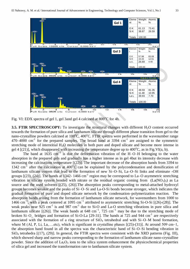
3.4. SEM-EDX ANALYSIS: Fig. (V, VI) show the morphology and the elements ration in pure and dope
were had silicate system. The morphological images of pure silica (gel 1) and lanthanum silicate (gel 2, gel
3 and gel 4) nanoparticles after calcinations at 8000C for 4h were observed by using scanning electron
microscopy (SEM) as shown in Fig. VI. It is observed that pure and doped silicate particles and are nano-
sized with nearly spherical shapes. While, the aggregation (interconnected mass of fine, nearly spherical
grains) of silica nanoparticles is very serious and appeared in all images, which is a typical problem for
nano-sized particles. It is found that the morphology of the obtained lanthanum silicate systems is different
from that of pure silica especially gel 4 has very fine particles. The average grain sizes measured from the
SEM micrographs are in the range 75 nm to 29 nm for gel 4, and all grains exhibit nearly spherical shape. It
is clear that the particle sizes evaluated from the SEM micrographs are larger than those evaluated from the
XRD patterns. Fig. VI show the energy dispersive spectra of the precursors used in synthesis of pure silica
(gel 1) and lanthanum silicate (gel 2, gel 4) calcined at 800oC for 4h, indicating the existence of O and Si in
pure silica also the presence of O, Si and La element in the lanthanum silicate nanoparticles.
gel 1
gel 3
gel 2
gel 4
Fig. V: SEM micrographs of pure silica (gel 1) and lanthanum silicate (gel 2, gel 3& gel4), calcined at
800oC for 4h.
El Nahrawy, A. M. et al./ International Journal of Advancement in Engineering, Technology and Computer Sciences, Vol.1, No.1 33
Eleme
nt
Weight
%
Atomic
%
O K 61.13 73.41
Si K 38.87 26.59
Totals 100.00
Eleme
nt
Weight
%
Atomic
%
O K 48.59 81.35
Si K 11.48 10.95
La L 39.93 7.70
Totals 100.00
Elemen
t
Weight
%
Atomic
%
O K 57.98 83.08
Si K 15.33 12.51
La L 26.69 4.40
Totals 100.00
Gel 1
Gel 3
Gel 4
Fig. VI: EDX spectra of gel 1, gel 3and gel 4 calcined at 800oC for 4h.
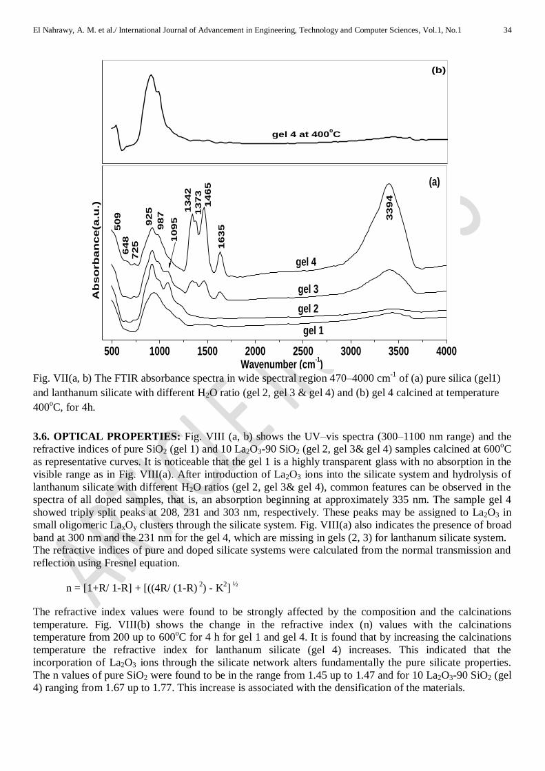
3.5. FTIR SPECTROSCOPY: To investigate the structural changes with different H2O content occurred
towards the formation of pure silica and lanthanum silicate through different phase transition from gel to the
nano-crystalline powders calcined at 100oC, 400
oC. FTIR spectra were performed in the wavenumber range
470–4000 cm-1
for the prepared samples. The broad band at 3394 cm-1
are assigned to the symmetric
stretching mode of interstitial H2O molecules in both pure and doped silicate and become more intense in
gel 4 [(21)], which disappeared with increasing the temperature degree up to 400oC, as in Fig.VI(a, b).
The band at 1635 cm−1
is due the deformation vibration of the H–O–H belonging to the water
absorption in the prepared gels and gradually has a higher intense as in gel 4but its intensity decrease with
increasing the calcinations temperature [(22)]. The important decrease of the absorption bands from 3394 to
1342 cm-1
after the calcination at 4000C can be explained by the polycondensation and densification of
lanthanum silicate system that lead to the formation of new Si–O–Si, La–O–Si links and eliminate -OH
groups [(23), (24)]. The bands at 1342- 1466 cm-1
region may be correspond to La–O asymmetric stretching
vibration in silicate system bonded with nitrate or the residual organic coming from (La(NO3)3) as La
source and the used solvents [(25), (26)].The absorption peaks corresponding to metal-attached hydroxyl
groups becomes weaker and the peaks of Si–O–Si and La-O-Si bonds become stronger, which indicates the
gradual formation of pure and doped silicate network by the condensation–polymerization reaction. The
absorption bands arising from the formation of lanthanum silicate network, for wavenumbers from 1000 to
1466 cm−1
, with a peak centered at 1095 cm−1
attributed to asymmetric stretching Si-O-Si [(26)-(28)]. The
weak peaks near 925 cm-1
is and 987 cm-1
due to Si-O and La-O stretching vibrations in pure silica and
lanthanum silicate [(26)]. The weak bands at 648 cm−1
, 725 cm−1
may be due to the stretching mode of
broken Si–O_ bridges and formation of Si-O-La [29-31]. The bands at 725 and 944 cm-1
are respectively
associated with the formation of a ring structure of SiO4 tetrahedral and with Si–O–M bond formation,
where M (Al, P, Li, La,….etc), which is significant in crystalline phases [(25)-(31)]. At around 509 cm−1,
the absorption band found in all the spectra was the characteristic band of Si–O–Si bending vibration in
SiO4 tetrahedra [(17), (29)]. In general, the FTIR spectra were consistent with the XRD patterns (Fig. III),
which showed sharp and narrow peaks corresponding to the formation of lanthanum silicate nano-crystalline
powder. Since the addition of La2O3 ions to the silica system enhancement the physicochemical properties
of silica gel and increased the transformation rate to lanthanum silicate system.
El Nahrawy, A. M. et al./ International Journal of Advancement in Engineering, Technology and Computer Sciences, Vol.1, No.1 34
500 1000 1500 2000 2500 3000 3500 4000
(a)
64
8
3394
16
35
14
65
13
73
13
42
10
95
98
7
92
5
Ab
so
rb
an
ce(a
.u.)
Wavenumber (cm-1)
gel 2
gel 3
gel 4
gel 1
72
5
50
9
gel 4 at 4000C
(b)
Fig. VII(a, b) The FTIR absorbance spectra in wide spectral region 470–4000 cm
-1 of (a) pure silica (gel1)
and lanthanum silicate with different H2O ratio (gel 2, gel 3 & gel 4) and (b) gel 4 calcined at temperature
400oC, for 4h.
3.6. OPTICAL PROPERTIES: Fig. VIII (a, b) shows the UV–vis spectra (300–1100 nm range) and the
refractive indices of pure SiO2 (gel 1) and 10 La2O3-90 SiO2 (gel 2, gel 3& gel 4) samples calcined at 600oC
as representative curves. It is noticeable that the gel 1 is a highly transparent glass with no absorption in the
visible range as in Fig. VIII(a). After introduction of La2O3 ions into the silicate system and hydrolysis of
lanthanum silicate with different H2O ratios (gel 2, gel 3& gel 4), common features can be observed in the
spectra of all doped samples, that is, an absorption beginning at approximately 335 nm. The sample gel 4
showed triply split peaks at 208, 231 and 303 nm, respectively. These peaks may be assigned to La2O3 in
small oligomeric LaxOy clusters through the silicate system. Fig. VIII(a) also indicates the presence of broad
band at 300 nm and the 231 nm for the gel 4, which are missing in gels (2, 3) for lanthanum silicate system.
The refractive indices of pure and doped silicate systems were calculated from the normal transmission and
reflection using Fresnel equation.
n = [1+R/ 1-R] + [((4R/ (1-R) 2) - K
2]
½
The refractive index values were found to be strongly affected by the composition and the calcinations
temperature. Fig. VIII(b) shows the change in the refractive index (n) values with the calcinations
temperature from 200 up to 600oC for 4 h for gel 1 and gel 4. It is found that by increasing the calcinations
temperature the refractive index for lanthanum silicate (gel 4) increases. This indicated that the
incorporation of La2O3 ions through the silicate network alters fundamentally the pure silicate properties.
The n values of pure SiO2 were found to be in the range from 1.45 up to 1.47 and for 10 La2O3-90 SiO2 (gel
4) ranging from 1.67 up to 1.77. This increase is associated with the densification of the materials.

El Nahrawy, A. M. et al./ International Journal of Advancement in Engineering, Technology and Computer Sciences, Vol.1, No.1 35
200 300 400 500 600
1.45
1.50
1.55
1.60
1.65
1.70
1.75
1.80 (a)
gel 4
gel 1Re
fra
cti
ve
in
de
x (
n)
Temperature ( 0C)
(b)
200 300 400 500 600 700 800 900
(a)
gel 4
gel 2
gel 3
Ab
so
rba
nc
e
Wavelength (nm)
gel 1
Fig. VIII(a, b) UV–vis spectra of gel 1, gel 2gel 3and gel 4 calcined at 6000C for 4h and (b) the refractive
index as a function in temperature for two samples gel1 and gel 4.
4. CONCLUSIONS: SiO2 and 10 La2O3- 90 SiO2 nano-crystalline have been synthesized successfully
using sol–gel technique. The crystallinity of 10 La2O3- 90 SiO2 powders increases with increasing
calcinations temperature and H2O content and the grain size of nanoparticles mainly ranged from 10 to 27
nm. XRD and FTIR studies confirmed that a better homogeneous reaction took place with the constituents
La2O3 and SiO2 in lanthanum silicate powders. The optical properties changed gradually with increasing the
calcinations temperature. SEM indicated the spherical nanoparticles morphology for the prepared samples.
The reported results are encourage to confirm the possibility to incorporation of different molecules through
silicate matrix as nanocomposite could be identified as potential materials towards the development of
silicate system for future applications.
ACKNOWLEDGEMENTS: Dr. A. M. El Nahrawy is expressing their grateful thanks to Dr. A. I. Ali,
from Energy Harvest-Storage Research Center and Department of Physics, University of Ulsan, Ulsan,
Korea and prof. G. Turky, from Microwave physics and Dielectrics Department, National research centre
(NRC),Cairo, Egypt for your great help to me.
REFERENCES:
1. Shi, Q, Lu. L, Jin, H, Zhang, H, Zeng, Y, (2012), Electrical properties and thermal expansion of cobalt doped apatite-type lanthanum silicates based electrolytes for IT-SOFC, Materials Research Bulletin 47, 719–723.
2. Schuster, K, Litzkendorf, D , Grimm, S, Kobelke, J, Schwuchow, A, et al., (March 11, 2013), " Study of lanthanum aluminum silicate glasses for passive and active optical fibers ", Proc. SPIE 8621, Optical Components and Materials.
3. Vieira M. M, Oliveira J. C, Shaula, A, L, Cavaleiro, A, Trindade,B, (2012) , Lanthanum silicate thin films for SOFC electrolytes synthesized by magnetron sputtering and subsequent annealing, Surf. & Coat. Techn. J, 206, 3316–3322.
4. Yang, T, Zhao, H, Han, J, Xu, N, Shen, Y, Du, Z, Wang, J, (2014), Synthesis and densification of lanthanum silicate apatite electrolyte for intermediate temperature solid oxide fuel cell via co-precipitation method, J. Europ. Cera. Soci. , 34, 1563–1569.
5. Sansom, H. E, Tolchard, R. J, Slater, R. P, Islam, S. M, (2004), Synthesis and structural characterisation of the apatite-type phases La10−xSi6O26+z doped with Ga, J. Solid State Ion, 167, 17–22.
6. Pivak, Y. V, Kharton, V. V, Yaremchenko, A. A, Yakovlev, S. O,Kovalevsky, A. V, Frade, J. R, Marques, F. M, B,(2007), Phase relationships and transport in Ti
-, Ce
- and Zr
-substituted lanthanum silicate systems, J. Europ. Cera. Soci. , 27,
2445–2454. 7. Xiang,J, Liu, Z-G, Ouyang J-H, Yan, F-Y, (2014), Ionic conductivity of oxy-apatite La10Si6_xInxO27_δ solid electrolyte
ceramics, J. Power Sources 251, 305-310.
El Nahrawy, A. M. et al./ International Journal of Advancement in Engineering, Technology and Computer Sciences, Vol.1, No.1 36
8. Brinker, C, J, Scherer, G,W, (1990), Sol–Gel Science: The Physics and Chemistry of Sol–Gel Processing, Academic Press, New York.
9. MacCraith, B, D, McDonagh, C, M, O’Keefe, G, McEvoy, A, K, Butler, T, Sheridan, F, R, (1995), Sol–gel coatings for optical chemical sensors and biosensors, Sens, Actuators B 29, 51–57.
10. Kajihara,K, (2013), Recent advances in sol–gel synthesis of monolithic silica and silica-based glasses, J . Asian Cera. Socie., 1,121–133.
11. atterson, A. L, (1939), The Scherrer formula for X-ray particle size determination, Phys. Rev. J. 56(10), 978–982. 12. Dominko, R, (2008), Li2MSiO4 (M = Fe and/or Mn) cathode materials, J. Pow. Sour.,184 , 462–468. 13. Deng,C, Zhang, S, Fu, B. L, Yang, S. Y, Ma, L, (2010), Characterization of Li2MnSiO4 and Li2FeSiO4 cathode materials
synthesized via a citric acid assisted sol–gel method, J. Mater. Chem. Phys., 120, 14–17. 14. Mizuta,Y, Daiko, Y, Mineshige, A, Yazawa, T,(2013), Effect of plastics substrate on phase separation behavior and
adhesion for R Si(OC2H5)3–Si(OC2H5)4 coatings prepared by sol–gel process, J. Cera. Internt. J. 39, 925–930. 15. Béchade, E, Julien, I, Iwata, T, Masson, O, Thomas, P, Champion, E, Fukuda, K, (2008), Synthesis of lanthanum silicate
oxyapatite materials as a solid oxide fuel cell electrolyte, J. Euro. Cera. Soci. J. 28, 2717–2724. 16. Hosseini, S. M, Shvareva, T, Navrotsky, A,(2013), Energetics of lanthanum silicate apatite: Influence of interstitial
oxygen and cation vacancy concentrations in La9.33+x(SiO4)6O2+3x/2 and a10−xSrx(SiO4)6O3−0.5x,J. Solid State Ion., 233, 62–66.
17. Blaker, J, J, Nazhat, N, S, Boccaccini R, A, (2004), Development and characterization of silver-doped bioactive glass-coated sutures for tissue engineering and wound healing applications, J. Biomat. , 25, 1319–29.
18. Dima, A, Della Corte, F. G, Williams, C. J, Watkins, K. G, Dearden, G, O’Hare, N, Casalino, M, Rendina, I, Dima, M, (2008), Silicon nano-particles in SiO2 sol–gel film for nano-crystal memory device applications, J. Microelectr., 39, 768–770.
19. Shin, D-Y, Cao, G, Kim, K-N, (2011), Prepration and photoluminescence properties of Ce doped lutetium silicate nanopowders by solegel method, J. Curr. Appl. Phys., 11, 309-312.
20. Heshmatpuor, F, Adelkhani, H, Jangholi, M, (2011), Studying of optical and morphological properties of SiO2–MOx (M: Co/Cu) glasses prepared by the sol–gel method, J. Non-Cryst. Soli., 357, 1409–1413.
21. Ghotbi, M,Y, Bagheri, N, Sadrnezhaad, S,K, (2011), Zinc-stearate-layered hydroxide nanohybrid material as a precursor to produce carbon nanoparticles, J. Alloys. Compd., 509, 2441–2444.
22. Bangi, U, K,H, Jung, In-K, Park, C-S, Baek, S, H-Ho Park, (2013), Optically transparent silica aerogels based on sodium silicate by a two step sole gel process and ambient pressure drying, J. Solid State Scie., 18, 50-57.
23. Pinheiro, R. C, Soares, C. M. F, dos Santos, O. A. A, Castro, H. F, de Moraes, F. F, Zanin, G. M, (2008), Influence of gelation time on the morphological and physico-chemical properties of the sol–gel entrapped lipase, J. Mole. Catal. B: Enzymatic., 52–53, 27–33.
24. Aguiar, H, Serra, J, Gonz lez, P, Le n, B, (2009), Structural study of sol–gel silicate glasses by IR and Raman spectroscopies, J. Non-Cryst. Solids, 355, 475–480.
25. Nallamuthu, N, Prakash, I, Satyanarayana, N, Venkateswarlu, M, (2011), Electrical conductivity studies of nanocrystalline lanthanum silicate synthesized by sol–gel route, J. Allo. and Comp., 509, 1138–1145.
26. Zhang, J, Shen, Z, Shan, W, Mei, Z, Wang, W, (2011), Adsorption behavior of phosphate on lanthanum(III)-coordinated diamino-functionalized 3D hybrid mesoporous silicates material, J. Hazard. Mater., 186, 76–83.
27. Secu, C. E, S. Polosan, Secu, M, (2011), Magneto-optical investigations of rare earth doped sol–gel derived silicate xerogels, J. Lumine.,131, 1747–1752.
28. Liu, T, Lai, D, Feng, X, Zhu, H, Chen, J, (2013), Carbon sphere influence on textural properties and bioactivity of mesoporous bioactive glass/ hydroxyl apatite nanocomposite, J. Ceram. Internat., 39, 3947–3956.
29. Muralidharan, P, Venkateswarlu, M, Satyanarayana, N, (2004), Sol–gel synthesis, characterization and impedance studies of lithium borosilicate glass, J. Mater. Resea. Bulle., 39, 1753–1762.
30. Zhong, X, Yang, B, Zhang, X, Jia, J, Yia, G, (2012), Effect of calcining temperature and time on the characteristics of Sb-doped SnO2 nanoparticles synthesized by the sol–gel method, J. Particu., 10, 365– 370.
31. Lakshminarayana, G, Torres, J. A, Lin, T. C, Kityk, I. V, Hehlen, M. P, (2014), Sol–gel synthesis and characterization of fluoride-rich lanthanum-alumino-silicate gels doped with Ce
3+ and Ti
4+, J. Allo. and Comp., 601, 67–74.
Copyright © 2022 FDOKUMEN