Structural and chemical characterization of BaTiO3 nanorods
6
Structural and chemical characterization of BaTiO 3 nanorods K. Z ˇ agar a , A. Rec ˇnik a , S. S ˇ turm a , A. Gajovic ´ b , M. C ˇ eh a, * a Department for Nanostructured Materials, Jozˇef Stefan Institute, Jamova cesta 39, SI-1000 Ljubljana, Slovenia b Molecular Physics Laboratory, Division of Materials Physics, RuperBosˇkovic ´ Institute, Bijenicˇka 54, HR-1002 Zagreb, Croatia 1. Introduction Complex metal oxides with the perovskite structure ATiO 3 (A = Ba, Sr, Ca, Pb, etc.) are important materials for a variety of applications in the microelectronics industry, and more recently in nanotechnology, due to their interesting and unique chemical and physical properties [1,2]. Among them, BaTiO 3 possesses a variety of attractive properties, such as ferroelectricity, piezoelectricity and even semiconductivity, when doped with aliovalent dopants, and therefore serves as one of the most versatile functional materials for a wide range of technological uses [3–7]. In the case when an increased surface-to-volume ratio is preferred, this can be achieved by processing BaTiO 3 in the form of one-dimensional nanostructures, such as nanorods, nanowires and/or nanotubes, since these morphologies exhibit a greatly increased surface area in comparison to their bulk counterparts [1,7–9]. Recently, BaTiO 3 in the form of nanorods and nanotubes has been shown to be a good candidate for applications involving positive-temperature- coefficient thermistors and sensors [5,6,11]. Many efforts have been made to synthesize and understand the growth of nanorods and nanotubes of functional ceramics using different processing procedures [1,12]. Among the many proposed methods, the template-assisted growth process has shown itself to be very promising [1,10,12–14]. In this method, the starting material is a sol that fills a porous template, which is then annealed at a higher temperature in order to ensure the formation of the material in polycrystalline form. The membranes that serve as templates are usually anodized alumina (AAO) or polycarbonate membranes (PC) with a more-or-less regular pore structure that defines the final shape and geometry of the processed materials. Using the so-called direct-template-filling technique, various perovskite materials were successfully synthesized in the form of one-dimensional nanostructures [15–18]. Alternatively, a modified approach was developed [19] to synthesize such morphologies, i.e., the so-called template-assisted, sol–gel elec- trophoretic deposition (EPD). In this technique an electric potential is used as a driving force to pack the sol particles into the template pores. Using EPD, various simple and complex ceramic oxides, such as BaTiO 3 , SrTiO 3 , TiO 2 , PZT, V 2 O 5 , SiO 2 , and ZnO, were successfully prepared in the form of nanorods and/or nanotubes [19–22]. Despite numerous reports on the synthesis of different ceramic oxides in the shape of nanorods and/or nanotubes, using either a direct-template-filling method or EPD, there are scarce data on the microstructural and nanostrucutral characterizations of these materials using highly spatially resolved electron-microscopy techniques. Data on the structure and possible chemical composi- tion variations of the materials on the nano- and atomic scales are indispensible if we want to predict the physical properties of the investigated materials. Also, based on such data, it should be possible to tailor the relevant processing parameters. This is why in our work we report on the FSEM- and TEM-based electron- microscopy characterization of polycrystalline BaTiO 3 nanorods that were synthesized by the optimized EPD of a BaTiO 3 sol in AAO templates. The objective of the investigation was to determine the chemical homogeneity of the BaTiO 3 nanorods and to study the morphology and the crystal structure of the nanosized BaTiO 3 Materials Research Bulletin 46 (2011) 366–371 ARTICLE INFO Article history: Received 29 April 2010 Received in revised form 14 October 2010 Accepted 13 December 2010 Available online 21 December 2010 Keywords: A. Ceramics A. Nanostructures B. Sol–gel chemistry C. Electron microscopy D. Microstructure ABSTRACT An electron-microscopy investigation was performed on BaTiO 3 nanorods that were processed by sol– gel electrophoretic deposition (EPD) into anodic aluminium oxide (AAO) membranes. The BaTiO 3 nanorods grown within the template membranes had diameters ranging from 150 to 200 nm, with an average length of 10–50 mm. By using various electron-microscopy techniques we showed that the processed BaTiO 3 nanorods were homogeneous in their chemical composition. The BaTiO 3 nanorods were always polycrystalline and were composed of well-crystallized, defect-free, pseudo-cubic BaTiO 3 grains, ranging from 10 to 30 nm. No intergranular phases were observed between the BaTiO 3 grains. A low-temperature hexagonal polymorph that is coherently intergrown with the BaTiO 3 perovskite matrix was also observed as a minor phase. When annealing the AAO templates containing the BaTiO 3 sol in an oxygen atmosphere the presence of the hexagonal polymorph was diminished. ß 2010 Elsevier Ltd. All rights reserved. * Corresponding author. Tel.: +386 14773341; fax: +386 14773221. E-mail address: [email protected] (M. C ˇ eh). Contents lists available at ScienceDirect Materials Research Bulletin journal homepage: www.elsevier.com/locate/matresbu 0025-5408/$ – see front matter ß 2010 Elsevier Ltd. All rights reserved. doi:10.1016/j.materresbull.2010.12.012
-
Upload
independent -
Category
Documents
-
view
0 -
download
0
Transcript of Structural and chemical characterization of BaTiO3 nanorods

Materials Research Bulletin 46 (2011) 366–371
Structural and chemical characterization of BaTiO3 nanorods
K. Zagar a, A. Recnik a, S. Sturm a, A. Gajovic b, M. Ceh a,*a Department for Nanostructured Materials, Jozef Stefan Institute, Jamova cesta 39, SI-1000 Ljubljana, Sloveniab Molecular Physics Laboratory, Division of Materials Physics, Ruper Boskovic Institute, Bijenicka 54, HR-1002 Zagreb, Croatia
A R T I C L E I N F O
Article history:
Received 29 April 2010
Received in revised form 14 October 2010
Accepted 13 December 2010
Available online 21 December 2010
Keywords:
A. Ceramics
A. Nanostructures
B. Sol–gel chemistry
C. Electron microscopy
D. Microstructure
A B S T R A C T
An electron-microscopy investigation was performed on BaTiO3 nanorods that were processed by sol–
gel electrophoretic deposition (EPD) into anodic aluminium oxide (AAO) membranes. The BaTiO3
nanorods grown within the template membranes had diameters ranging from 150 to 200 nm, with an
average length of 10–50 mm. By using various electron-microscopy techniques we showed that the
processed BaTiO3 nanorods were homogeneous in their chemical composition. The BaTiO3 nanorods
were always polycrystalline and were composed of well-crystallized, defect-free, pseudo-cubic BaTiO3
grains, ranging from 10 to 30 nm. No intergranular phases were observed between the BaTiO3 grains. A
low-temperature hexagonal polymorph that is coherently intergrown with the BaTiO3 perovskite matrix
was also observed as a minor phase. When annealing the AAO templates containing the BaTiO3 sol in an
oxygen atmosphere the presence of the hexagonal polymorph was diminished.
� 2010 Elsevier Ltd. All rights reserved.
Contents lists available at ScienceDirect
Materials Research Bulletin
journal homepage: www.e lsev ier .com/ locate /mat resbu
1. Introduction
Complex metal oxides with the perovskite structure ATiO3
(A = Ba, Sr, Ca, Pb, etc.) are important materials for a variety ofapplications in the microelectronics industry, and more recently innanotechnology, due to their interesting and unique chemical andphysical properties [1,2]. Among them, BaTiO3 possesses a varietyof attractive properties, such as ferroelectricity, piezoelectricityand even semiconductivity, when doped with aliovalent dopants,and therefore serves as one of the most versatile functionalmaterials for a wide range of technological uses [3–7]. In the casewhen an increased surface-to-volume ratio is preferred, this can beachieved by processing BaTiO3 in the form of one-dimensionalnanostructures, such as nanorods, nanowires and/or nanotubes,since these morphologies exhibit a greatly increased surface areain comparison to their bulk counterparts [1,7–9]. Recently, BaTiO3
in the form of nanorods and nanotubes has been shown to be agood candidate for applications involving positive-temperature-coefficient thermistors and sensors [5,6,11].
Many efforts have been made to synthesize and understand thegrowth of nanorods and nanotubes of functional ceramics usingdifferent processing procedures [1,12]. Among the many proposedmethods, the template-assisted growth process has shown itself tobe very promising [1,10,12–14]. In this method, the startingmaterial is a sol that fills a porous template, which is then annealedat a higher temperature in order to ensure the formation of the
* Corresponding author. Tel.: +386 14773341; fax: +386 14773221.
E-mail address: [email protected] (M. Ceh).
0025-5408/$ – see front matter � 2010 Elsevier Ltd. All rights reserved.
doi:10.1016/j.materresbull.2010.12.012
material in polycrystalline form. The membranes that serve astemplates are usually anodized alumina (AAO) or polycarbonatemembranes (PC) with a more-or-less regular pore structure thatdefines the final shape and geometry of the processed materials.Using the so-called direct-template-filling technique, variousperovskite materials were successfully synthesized in the formof one-dimensional nanostructures [15–18]. Alternatively, amodified approach was developed [19] to synthesize suchmorphologies, i.e., the so-called template-assisted, sol–gel elec-trophoretic deposition (EPD). In this technique an electric potentialis used as a driving force to pack the sol particles into the templatepores. Using EPD, various simple and complex ceramic oxides, suchas BaTiO3, SrTiO3, TiO2, PZT, V2O5, SiO2, and ZnO, were successfullyprepared in the form of nanorods and/or nanotubes [19–22].
Despite numerous reports on the synthesis of different ceramicoxides in the shape of nanorods and/or nanotubes, using either adirect-template-filling method or EPD, there are scarce data on themicrostructural and nanostrucutral characterizations of thesematerials using highly spatially resolved electron-microscopytechniques. Data on the structure and possible chemical composi-tion variations of the materials on the nano- and atomic scales areindispensible if we want to predict the physical properties of theinvestigated materials. Also, based on such data, it should bepossible to tailor the relevant processing parameters. This is why inour work we report on the FSEM- and TEM-based electron-microscopy characterization of polycrystalline BaTiO3 nanorodsthat were synthesized by the optimized EPD of a BaTiO3 sol in AAOtemplates. The objective of the investigation was to determine thechemical homogeneity of the BaTiO3 nanorods and to study themorphology and the crystal structure of the nanosized BaTiO3

K. Zagar et al. / Materials Research Bulletin 46 (2011) 366–371 367
grains and present the interfaces in these polycrystalline, one-dimensional nanostructures.
2. Experimental
2.1. Synthesis
A BaTiO3 sol was prepared according to a procedure describedelsewhere [23]. Barium acetate (�99.0%, Fluka) was first dissolvedin glacial acetic acid (p.a., Fluka) by stirring at room temperature.Then titanium isopropoxide (p.a., Fluka), in an amount corre-sponding to the stoichiometric molar ratio Ba/Ti = 1.00, was addedto the barium acetate solution during constant-rate stirring.Finally, ethylene glycol (p.a., Riedel-de Haen) was added to thesolution in order to adjust the viscosity and to stabilize the sol.
The template membranes used for the processing of the BaTiO3
nanostructures were anodic aluminium oxide membranes (What-man) with pore diameters of approximately 200 nm and with athickness of up to 60 mm. For the electrophoretic deposition of theBaTiO3 sol into the template pores, the template membranes wereattached to an aluminium working electrode, and a Pt meshelectrode was used as the counter electrode. Both electrodes wereplaced into the sol, 15 mm apart. Then a potential of 30 V(determined as the optimum value under these conditions) wasapplied between both electrodes, and this potential was main-tained for 30 min to ensure complete filling of the AAO template.After the EPD, the samples were dried at 200 8C for 12 h andsubsequently annealed at 700 8C for 1 h. Afterwards, the AAOtemplates were removed in 6-M NaOH. The isolated rod-likeBaTiO3 nanostructures in the NaOH solution were repeatedlywashed in water and ethanol and were concentrated by means ofcentrifugation.
2.2. Characterization
The crystal structure of the BaTiO3 nanostructures wasdetermined by X-ray powder diffraction (XRD) (Bruker AXS ModelD4 Endeavor) and by Raman spectroscopy (RS) (Jobin YvonT64000Jo). Scanning electron microscopy (SEM) was used tocharacterize the morphology of the nanostructures. The specimensfor the SEM observations (Jeol JSM-7600F) were prepared byplacing the synthesized nanostructures on a conductive tape.Transmission electron microscopy (TEM) with energy-dispersiveX-ray spectroscopy (EDXS) and high-angle annular dark-fieldscanning–transmission electron microscopy (HAADF–STEM) wereused to study the structure and the chemical composition of theBaTiO3 nanostructures on the nano and atomic scales (JEOL JEM-2100, JEOL JEM-2010F). The samples for the TEM observationswere ultrasonically dispersed and placed onto lacey, carbon-coated nickel grids. For the quantitative EDXS analyses of the[()TD$FIG]
Fig. 1. (a) Top view SEM image of 60-mm-thick AAO template with a pore diameter o
nanostructures prepared by sol–gel EPD into AAO template and annealed at 700 8C for
BaTiO3 nanostructures, pure, stoichiometric BaTiO3 nanosizedpowder was used as a standard reference material. The experi-mental k-factors were calculated from 10 EDXS spectra acquiredfrom the BaTiO3 standard. These k-factors were then used toquantify the EDXS spectra from our samples using the Cliff–Lorimer procedure [24].
3. Results and discussion
The top view of an AAO template membrane used in ourexperiments shows that the membranes have a close-packed porestructure with a more-or-less uniform pore-diameter distribution(Fig. 1a). After the EPD of the BaTiO3 sol into the AAO pores at aselected potential and after the removal of the AAO templates inthe NaOH solution, the remaining material was first examined byX-ray powder diffraction and by Raman spectroscopy. In order toobtain a sufficient amount of material for the measurements,material was collected from many AAO templates. The character-istic X-ray diffractogram showed only the presence of diffractionpeaks that can be assigned to cubic BaTiO3 according to the JCPSDcard No. 89-2475. No other phases could be detected by the XRD.On the other hand, the Raman spectra from the BaTiO3 nanorodsshowed bands of tetragonal and hexagonal structures, as reportedelsewhere [25–28]. This is why we refer to the BaTiO3 crystalstructure as ‘‘pseudo-cubic’’ instead of ‘‘cubic’’ throughout theentire paper. It was concluded that after annealing the AAOtemplates with filled sol at 700 8C the transformation of the sol intocrystalline BaTiO3 was complete. A low-magnification FSEM imageof the BaTiO3 material that was thus obtained is shown in Fig. 1b.One can observe typical rod-like-shaped BaTiO3 nanostructuresthat form bundles of various dimensions, which is a consequenceof the sample preparation for scanning electron microscopy.Single, not agglomerated, rod-like BaTiO3 nanostructures, can alsobe observed. The rod-like BaTiO3 nanostructures exhibited aregular shape with a uniform diameter.
Since it is not possible to unambiguously determine the cross-sectional shape of such nanostructures using conventional bright-field TEM imaging due to the various contributions to the imagecontrast, we used HAADF–STEM to determine the shape of the rod-like BaTiO3. Namely, in HAADF–STEM, the intensity in theexperimental image is proportional to the specimen’s thickness[29] in the case when the chemical composition of the specimen ishomogeneous and when the inner collection angle of the ADFdetector is large enough to ensure that thermal diffuse scattering(TDS) is the main contribution to the image signal. An experimen-tal HAADF–STEM image that was taken from part of a single,BaTiO3 rod-like nanostructure shows that the intensity in themiddle of the rod-like nanostructure is higher than the intensity atthe edge of the rod-like nanostructure (Fig. 2a). The average HAADFexperimental intensity profile across the rod-like nanostructure
f approximately 200 nm. (b) FSEM image of isolated and bundled rod-like BaTiO3
1 h.
[()TD$FIG]
Fig. 2. (a) HAADF–STEM image of single BaTiO3 nanorod. (b) The corresponding average intensity profile across the nanorod.
K. Zagar et al. / Materials Research Bulletin 46 (2011) 366–371368
that was taken from the marked region furthermore reveals thatthe intensity profile was more-or-less semicircular-like shaped(Fig. 2b), which indicates the cylindrical-like shape of the observedBaTiO3 nanorods. The observed intensity fluctuations in themeasured intensity profile showed the rough surface of thepolycrystalline nanorods.
The bright-field, low-magnification TEM image in Fig. 3 shows atypical isolated BaTiO3 nanorod with a diameter of approximately250 nm, which is comparable to the diameter of the templatepores. The regular longitudinal shape of the BaTiO3 nanorodindicates that the pores within the AAO template retain theirregular shape even after annealing at 700 8C. A higher magnifica-tion TEM image reveals that these nanostructures are dense andpolycrystalline in nature with grain sizes ranging from 20 to 50 nm(Fig. 3b).
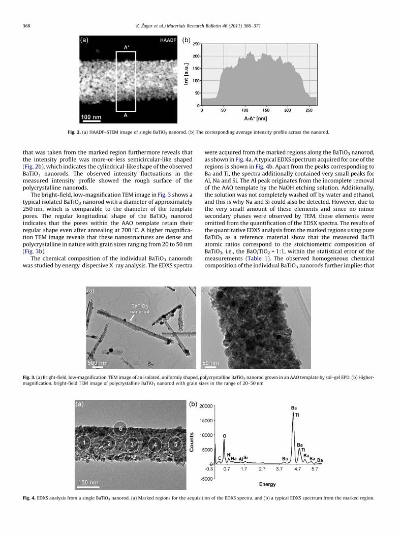
The chemical composition of the individual BaTiO3 nanorodswas studied by energy-dispersive X-ray analysis. The EDXS spectra
[()TD$FIG]
Fig. 4. EDXS analysis from a single BaTiO3 nanorod. (a) Marked regions for the acquisit
[()TD$FIG]
Fig. 3. (a) Bright-field, low-magnification, TEM image of an isolated, uniformly shaped, po
magnification, bright-field TEM image of polycrystalline BaTiO3 nanorod with grain siz
were acquired from the marked regions along the BaTiO3 nanorod,as shown in Fig. 4a. A typical EDXS spectrum acquired for one of theregions is shown in Fig. 4b. Apart from the peaks corresponding toBa and Ti, the spectra additionally contained very small peaks forAl, Na and Si. The Al peak originates from the incomplete removalof the AAO template by the NaOH etching solution. Additionally,the solution was not completely washed off by water and ethanol,and this is why Na and Si could also be detected. However, due tothe very small amount of these elements and since no minorsecondary phases were observed by TEM, these elements wereomitted from the quantification of the EDSX spectra. The results ofthe quantitative EDXS analysis from the marked regions using pureBaTiO3 as a reference material show that the measured Ba:Tiatomic ratios correspond to the stoichiometric composition ofBaTiO3, i.e., the BaO/TiO2 = 1:1, within the statistical error of themeasurements (Table 1). The observed homogeneous chemicalcomposition of the individual BaTiO3 nanorods further implies that
ion of the EDXS spectra, and (b) a typical EDXS spectrum from the marked region.
lycrystalline BaTiO3 nanorod grown in an AAO template by sol–gel EPD. (b) Higher-
es in the range of 20–50 nm.

Table 1Results of quantitative EDX analysis from marked regions on BaTiO3 nanorod (Fig. 4).
Region 1 2 3 4 5 6
Ti K (at.%) 19.7� 0.3 19.4�0.3 19.6� 0.3 19.9� 0.3 19.3�0.3 19.1� 0.3
Ba L (at.%) 20.4� 0.5 20.7�0.5 20.4� 0.5 20.2� 0.5 20.8�0.5 21.0� 0.5
Ba/Ti 1.04� 0.6 1.07�0.6 1.04� 0.6 1.01� 0.6 1.08�0.6 1.1� 0.6
[()TD$FIG]
Fig. 5. (a) Bright-field TEM image of polycrystalline BaTiO3 nanorod with the (b) corresponding SAED pattern. Diffraction rings corresponding to the hexagonal polymorph are
marked.
K. Zagar et al. / Materials Research Bulletin 46 (2011) 366–371 369
during the process of EPD into the AAO template membranes,stoichiometric BaTiO3 particles are already formed in the sol andthat there is no change in the chemical composition of theseparticles during the process of electrophoretic deposition.
The selected-area electron-diffraction (SAED) pattern takenfrom a few hundred grains in a BaTiO3 nanorod showed that theexperimental diffraction pattern corresponds to the calculatedelectron-diffraction pattern for the cubic BaTiO3 crystal structure,which is in accordance with the results of the XRD analysis (Fig. 5).However, additional weak diffraction rings with lattice spacingvalues of D1 = 0.47 nm, D2 = 0.34 nm and D3 = 0.28 nm (Table 2)were also observed in the experimental electron-diffractionpattern. These d-values correspond to the high-temperature,hexagonal BaTiO3 polymorph (JCPDS 82-1175) that was alsoobserved by RS. The weak diffraction rings for the hexagonal phaseindicate that the hexagonal polymorph is a minor phase within theBaTiO3 nanorods.
Both the pseudo-cubic BaTiO3 nanosized grains and theobserved hexagonal BaTiO3 polymorph were examined by high-resolution transmission electron microscopy (HRTEM). It wasfound that the BaTiO3 grains with the pseudo-cubic crystalstructure were well crystallized and that very often, low-indexorthogonal facets were nicely developed (Fig. 6a). No planar faultsor any other defects were observed in these BaTiO3 nanosizedpseudo-cubic grains. It is well known that even slight deviationsfrom the non-stoichiometry in BaTiO3 would result either in theformation of Ruddlesden–Popper-type planar faults in the case of
Table 2Measured d-values (from SAED) for marked regions in Fig. 5 and comparison with
the published crystallographic data (JCPDS 82-1175 for hexagonal and JCPSD 89-
2475 for cubic modification).
SAED dexp dc h k lc Modific.
D1 0.466 0.46714 {1 0 1} Hex
D2 0.342 0.33933 {1 0 3} Hex
D3 0.284 0.28409 {1 1 0} Cub and hex
local BaO excess [30], or in Ti-rich phases in the case of TiO2 excess.Since the HRTEM and EDXS showed no such experimentalevidence, it was additionally substantiated that the chemicalcomposition of these pseudo-cubic BaTiO3 nanorods was stoichio-metric and homogeneous, even at the nano-scale. The grainboundaries between the BaTiO3 pseudo-cubic grains were cleanand showed no amorphous phase present.
The hexagonal polymorph of BaTiO3 that was detected byselected-area electron diffraction was never observed as a stand-alone phase in the form of single, nanosized grains, as one wouldexpect according to the corresponding phase-equilibria diagram.Since hexagonal BaTiO3 can be structurally viewed as an idealordering of the (1 1 1) twins, the observed reflections in the SAEDpatterns must arise from the regions of ordered (1 1 1) twins.However, the ordering of the (1 1 1) twins in the nanosized BaTiO3 isnot ideal and also regions of more-or-less ordered twins can beobserved, as shown in the HRTEM image in Fig. 6b. The existence ofthe hexagonal polymorph was previously reported for BaTiO3
nanocrystalline powders that were synthesized by a sol–gelprocedure [31,32], but was so far never observed and reported forBaTiO3 obtained by EPD. The most plausible mechanism for theformation of the hexagonal polymorph is the reduction of Ti4+ to Ti3+,resulting from the local reducing atmosphere caused by thedecomposition of the organic components of the sol at the annealingtemperature of 700 8C, since it is known that Ti is in the form of Ti3+
along the (1 1 1) twin in BaTiO3 [33,34]. In order to confirm that thepresence of the hexagonal polymorph is a consequence of thereductive atmosphere during annealing, the AAO templates filledwith the BaTiO3 sol after EPD were annealed in a pure-oxygenatmosphere at 10 bar (Fig. 7). In that way the annealed BaTiO3
nanorods contained significantly less BaTiO3 hexagonal polymorph(Fig. 7b). It was concluded that the reductive environment, whenannealing the AAO templates filled with the BaTiO3 sol in the air,indeed caused the formation of (1 1 1) twins within the BaTiO3
perovskite matrix. In the case that these twins tended to order, theslabs of hexagonal polymorph were formed that were coherentlyintergrown with pseudo-cubic perovskite structure.

[()TD$FIG]
Fig. 6. (a) Bright-field, HRTEM image of a pseudo-cubic BaTiO3 grain and (b) slab of hexagonal BaTiO3 polymorph intergrown with pseudo-cubic BaTiO3.
[()TD$FIG]
Fig. 7. (a) Bright-field TEM image of BaTiO3 nanorod annealed in air showing the presence of pseudo-cubic and hexagonal polymorphs. (b) Bright-field TEM image of BaTiO3
nanorod annealed in pure-oxygen atmosphere. No hexagonal polymorph is present.
K. Zagar et al. / Materials Research Bulletin 46 (2011) 366–371370
4. Conclusions
BaTiO3 nanorods that were obtained by the EPD of astoichiometric BaTiO3 sol into AAO templates were investigatedby various electron-microscopy techniques. It was found that theBaTiO3 nanorods were polycrystalline in nature with diameterssimilar to the pore diameters within the AAO templates and withlengths ranging from a few micrometres up to a few tens ofmicrometres. The polycrystalline nanorods were composed ofwell-crystallized, defect-free, nanosized BaTiO3 grains with apseudo-cubic structure and with grain sizes ranging from 20 to50 nm. The grain boundaries between the BaTiO3 nanosized grainscontained no amorphous phase. The EDXS analysis showed thatthe chemical composition of the BaTiO3 nanorods was homoge-neous, which indicates that the starting stoichiometry of the solwas retained throughout the whole EPD and annealing process. Asmall fraction of the BaTiO3 grains contained an intergrown BaTiO3
hexagonal polymorph, which was manifested structurally as asequence of more-or-less ordered (1 1 1) twins. The formation ofthe hexagonal polymorph was a consequence of the local reductiveenvironment, due to the decomposition of the organic precursorsduring the annealing process, which leads to the reduction of Ti4+
to Ti3+ and the formation of twins. This was substantiated byannealing the AAO templates with the BaTiO3 sol in an oxygenatmosphere. In this way the processed BaTiO3 nanorods containedno hexagonal polymorph.
Acknowledgements
This work is based in part on the Ph.D. thesis of Kristina Zagarunder Grant No. 1000-06-310008 by the MHEST of Republic ofSlovenia. The research was supported by the EU-FP6 programmefor an Integrated Infrastructure initiative under a contract No.026019 ESTEEM.
References
[1] Y. Xia, P. Yang, Y. Sun, Y. Wu, B. Mayers, B. Gates, Y. Yin, F. Kim, H. Yan, Adv. Mater.15 (2003) 353–389.
[2] G.R. Patzke, F. Krumeich, R. Nesper, Angew. Chem. Int. Ed. 41 (2002) 2447–2460.[3] J.E. Spanier, A.M. Kolpak, J.J. Urban, I. Grinberg, L. Ouyang, W.S. Yun, A.M. Rappe, H.
Park, Nano Lett. 6 (2006) 735–739.[4] Y. Luo, I. Szafraniak, N.D. Zakharov, M. Steinhart, R.B. Wehrspohn, J.H. Wendorff, R.
Ramesh, M. Alexe, Appl. Phys. Lett. 83 (2003) 440–442.[5] Z. Wang, J. Hu, A.P. Suryavanshi, K. Yum, M.-F. Yu, Nano Lett. 7 (2007) 2966–2969.[6] J. Yuk, T. Troczynski, Sens. Actuators B 94 (2003) 290–293.[7] B. Huybrechts, K. Ishizaki, M. Takata, J. Mater. Sci. 30 (1995) 2463–2474.[8] J. Kim, S.A. Yang, Y.C. Choi, J.K. Han, K.O. Jeong, Y.J. Yun, D.J. Kim, S.M. Yang, D.
Yoon, H. Cheong, K.-S. Chang, T.W. Noh, S.D. Bu, Nano Lett. 8 (2008) 1813–1818.[9] B.I. Seo, U.A. Shaislamov, S.-W. Kim, H.-K. Kim, B. Yang, S.K. Hong, Physica E 37
(2007) 274–278.[10] M. Feng, W. Wang, Y. Zhou, J. Sol–Gel Sci. Technol. 52 (2009) 120–123.[11] J.J. Urban, J.E. Spanier, L. Ouyang, W.S. Yun, H. Park, Adv. Mater. 15 (2003) 423–
426.[12] C. Bae, H. Yoo, S. Kim, K. Lee, J. Kim, M.M. Sung, H. Shin, Chem. Mater. 20 (2008)
756–767.[13] A. Huczko, Appl. Phys. A 70 (2000) 365–376.[14] L. Li, N. Koshizaki, G. Li, J. Mater. Sci. Technol. 24 (2008) 550–562.

K. Zagar et al. / Materials Research Bulletin 46 (2011) 366–371 371
[15] S. Singh, S.B. Krupanidhi, Phys. Lett. A 367 (2007) 356–359.[16] B.A. Hernandez, K.S. Chang, E.R. Fisher, P.K. Dorhout, Chem. Mater. 14 (2002) 480–
482.[17] L. Zhao, M. Steinhart, J. Yu, U. Gosele, J. Mater. Res. 21 (2006) 685–690.[18] Y.-Y. Chen, B.Y. Yu, R.E. Cochran, J.-J. Shyue, Inorg. Chem. 48 (2009) 681–686.[19] G. Cao, J. Phys. Chem. B. 108 (2004) 19921–19931.[20] S.J. Limmer, T.P. Chou, G.Z. Cao, J. Sol–Gel Sci. Technol. 36 (2005) 183–195.[21] Y.C. Wang, I.C. Leu, M.H. Hon, Electrochem. Solid-State Lett. 5 (2002) C53–C55.[22] K. Zagar, A. Recnik, P.M. Ajayan, M. Ceh, Nanotechnology 21 (2010), 375605 (p. 7).[23] S.J. Limmer, S. Seraji, Y. Wu, T.P. Chou, C. Nguyen, G. Cao, Adv. Funct. Mater. 12
(2002) 59–64.[24] G. Cliff, G.W. Lorimer, J. Microsc.-Oxford 103 (1975) 203–207.[25] S. Wada, H. Yasuno, T. Hoshina, S.-M. Nam, H. Kakemoto, T. Tsurumi, Jpn. J. Appl.
Phys. 42 (2003) 6188–6195.
[26] M. Yashima, T. Hoshina, D. Ishimura, S. Kobayashi, W. Nakamura, T. Tsurumi, S.Wada, J. Appl. Phys. 98 (2005), 014313-1-014313-8.
[27] Y. Shiratori, C. Pithan, J. Dornseiffer, R. Waser, J. Raman Spectrosc. 38 (2007)1288–1299.
[28] Y. Shiratori, C. Pithan, J. Dornseiffer, R. Waser, J. Raman Spectrosc. 38 (2007) 1300–1306.
[29] Z.L. Wang, J.M. Cowley, Ultramicroscopy 31 (1989) 437–454.[30] T. Suzuki, Y. Nishi, M. Fujimoto, J. Am. Ceram. Soc. 83 (2000) 3185–3195.[31] E. Hamada, W.-S. Cho, K. Takayanagi, Philos. Mag. A 77 (1998) 1301–1308.[32] W.-S. Cho, J. Phys. Chem. Solids 59 (1998) 659–666.[33] A. Recnik, J. Bruley, W. Mader, D. Kolar, M. Ruhle, Philos. Mag. B 70 (1994) 1021–
1034.[34] A. Recnik, D. Kolar, J. Am. Ceram. Soc. 79 (1996) 1015–1018.