ßFTZ-F1 and Matrix metalloproteinase 2 are required for fat-body remodeling in Drosophila
11
ßFTZ-F1 and Matrix metalloproteinase 2 are required for fat-body remodeling in Drosophila Nichole D. Bond a, 1 , Archana Nelliot a, 2 , Marsha K. Bernardo a , Melanie A. Ayerh b , Kathryn A. Gorski b , Deborah K. Hoshizaki a, 3 , Craig T. Woodard b, ⁎ a School of Life Sciences, University of Nevada, Las Vegas 4505 S. Maryland Parkway, Las Vegas, NV 89154, USA b Department of Biological Sciences, Mount Holyoke College 50 College Street South Hadley, MA 01075, USA abstract article info Article history: Received for publication 26 May 2011 Revised 26 August 2011 Accepted 14 September 2011 Available online 22 September 2011 Keywords: Fat body Tissue remodeling 20E signaling ßFTZ-F1 Matrix metalloproteinase During metamorphosis, holometabolous insects eliminate obsolete larval tissues via programmed cell death. In contrast, tissues required for further development are retained and often remodeled to meet the needs of the adult fly. The larval fat body is involved in fueling metamorphosis, and thus it escapes cell death and is instead remodeled during prepupal development. The molecular mechanisms by which the fat body escapes programmed cell death have not yet been described, but it has been established that fat-body remodeling re- quires 20-hydroxyecdysone (20E) signaling. We have determined that 20E signaling is required within the fat body for the cell-shape changes and cell detachment that are characteristic of fat-body remodeling. We demonstrate that the nuclear hormone receptor ßFTZ-F1 is a key modulator of 20E hormonal induction of fat body remodeling and Matrix metalloproteinase 2 (MMP2) expression in the fat body. We show that in- duction of MMP2 expression in the fat body requires 20E signaling, and that MMP2 is necessary and sufficient to induce fat-body remodeling. © 2011 Elsevier Inc. All rights reserved. Introduction In holometabolous insects, dramatic changes in animal form occur as the larva undergoes a complete transformation during metamorphosis to give rise to the adult. This transformation is ac- complished by the destruction of larval tissues and the proliferation, differentiation, and organogenesis of the adult tissues (Bainbridge and Bownes, 1981; Bodenstein, 1950; Robertson, 1936). Unlike the majority of larval tissues, the larval fat body of Dipterans, as exempli- fied by Drosophila melanogaster, is refractory to cell death. Early in metamorphosis the larval fat body is transformed from sheets of at- tached, polygonal-cells to individual spherical, free-floating cells (Hoshizaki, 2005; Nelliot et al., 2006). These remodeled larval fat cells persist into the adult (Aguila et al., 2007; Butterworth, 1972; Hoshizaki, 2005) and can serve as a nutrient reservoir (Aguila et al., 2007). Metamorphosis is developmentally regulated by the steroid hor- mone 20-hydroxyecdysone (20E). 20E binds to the ecdysone receptor, which is a heterodimer composed of two nuclear receptors, EcR and Ultraspiracle (Koelle et al., 1991; Thomas et al., 1993; Yao et al., 1993; see Costantino et al., 2008 for exceptions). The ecdysone receptor medi- ates gene expression by initiating tissue-specific transcriptional cas- cades that result in distinct stage-specific developmental changes (Riddiford, 1996; Thummel, 1995). Late in the third-larval instar, an in- crease in the 20E titer directly induces transcription of a set of primary response genes called the “early” genes, which are necessary for initia- tion of metamorphosis (i.e., pupariation) and formation of the pupari- um (Burtis et al., 1990; DiBello et al., 1991; Richards, 1981; Riddiford and Truman, 1993; Segraves and Hogness, 1990;). Pupariation is fol- lowed by prepupal development, which in turn is followed by pupal de- velopment. The early gene products repress their own expression and induce the subsequent transcription of a set of secondary response genes called the “late” genes in the prepupa (Thummel, 1996). As the ecdysone pulse that initiated pupariation declines, the mid-prepupal genes are induced (Thummel, 1996). One such mid-prepupal gene en- codes the competence factor ßFTZ-F1. ßFTZ-F1 is a nuclear receptor and is required to confer competence upon tissues to respond to the prepupal pulse of 20E that occurs approximately 10 h after puparium formation (APF). ßFTZ-F1 and the prepupal pulse of 20E induce tran- scriptional cascades necessary for the prepupal to pupal transition (Broadus et al., 1999; Fortier et al., 2003; Woodard et al., 1994; Yamada et al., 2000; Reviewed in Pick et al., 2006). Developmental Biology 360 (2011) 286–296 ⁎ Corresponding author. Fax: + 1 413 538 2548. E-mail address: [email protected] (C.T. Woodard). 1 Present Address: National Institutes of Health, NIDDK, Genetics of Development and Disease Branch, 9000 Rockville Pike, Room 8D03, Bethesda, MD 20892-1758, USA. 2 Present Address: School of Medicine, Johns Hopkins University, 720 Rutland Avenue Baltimore, MD 21205, USA. 3 Present Address: National Institutes of Health, NIDDK, 6707 Democracy Boulevard, Two Democracy Plaza, Room 645, Bethesda, MD 20892, USA. 0012-1606/$ – see front matter © 2011 Elsevier Inc. All rights reserved. doi:10.1016/j.ydbio.2011.09.015 Contents lists available at SciVerse ScienceDirect Developmental Biology journal homepage: www.elsevier.com/developmentalbiology
-
Upload
independent -
Category
Documents
-
view
1 -
download
0
Transcript of ßFTZ-F1 and Matrix metalloproteinase 2 are required for fat-body remodeling in Drosophila

Developmental Biology 360 (2011) 286–296
Contents lists available at SciVerse ScienceDirect
Developmental Biology
j ourna l homepage: www.e lsev ie r .com/deve lopmenta lb io logy
ßFTZ-F1 and Matrix metalloproteinase 2 are required for fat-body remodelingin Drosophila
Nichole D. Bond a,1, Archana Nelliot a,2, Marsha K. Bernardo a, Melanie A. Ayerh b, Kathryn A. Gorski b,Deborah K. Hoshizaki a,3, Craig T. Woodard b,⁎a School of Life Sciences, University of Nevada, Las Vegas 4505 S. Maryland Parkway, Las Vegas, NV 89154, USAb Department of Biological Sciences, Mount Holyoke College 50 College Street South Hadley, MA 01075, USA
⁎ Corresponding author. Fax: +1 413 538 2548.E-mail address: [email protected] (C.T. Wo
1 Present Address: National Institutes of Health, NIDand Disease Branch, 9000 Rockville Pike, Room 8D03, Be
2 Present Address: School of Medicine, Johns HopAvenue Baltimore, MD 21205, USA.
3 Present Address: National Institutes of Health, NIDDTwo Democracy Plaza, Room 645, Bethesda, MD 20892,
0012-1606/$ – see front matter © 2011 Elsevier Inc. Alldoi:10.1016/j.ydbio.2011.09.015
a b s t r a c t
a r t i c l e i n f oArticle history:Received for publication 26 May 2011Revised 26 August 2011Accepted 14 September 2011Available online 22 September 2011
Keywords:Fat bodyTissue remodeling20E signalingßFTZ-F1Matrix metalloproteinase
During metamorphosis, holometabolous insects eliminate obsolete larval tissues via programmed cell death.In contrast, tissues required for further development are retained and often remodeled to meet the needs ofthe adult fly. The larval fat body is involved in fueling metamorphosis, and thus it escapes cell death and isinstead remodeled during prepupal development. The molecular mechanisms by which the fat body escapesprogrammed cell death have not yet been described, but it has been established that fat-body remodeling re-quires 20-hydroxyecdysone (20E) signaling. We have determined that 20E signaling is required within thefat body for the cell-shape changes and cell detachment that are characteristic of fat-body remodeling. Wedemonstrate that the nuclear hormone receptor ßFTZ-F1 is a key modulator of 20E hormonal induction offat body remodeling and Matrix metalloproteinase 2 (MMP2) expression in the fat body. We show that in-duction of MMP2 expression in the fat body requires 20E signaling, and that MMP2 is necessary and sufficientto induce fat-body remodeling.
odard).DK, Genetics of Developmentthesda, MD 20892-1758, USA.kins University, 720 Rutland
K, 6707 Democracy Boulevard,USA.
rights reserved.
© 2011 Elsevier Inc. All rights reserved.
Introduction
In holometabolous insects, dramatic changes in animal formoccur as the larva undergoes a complete transformation duringmetamorphosis to give rise to the adult. This transformation is ac-complished by the destruction of larval tissues and the proliferation,differentiation, and organogenesis of the adult tissues (Bainbridgeand Bownes, 1981; Bodenstein, 1950; Robertson, 1936). Unlike themajority of larval tissues, the larval fat body of Dipterans, as exempli-fied by Drosophila melanogaster, is refractory to cell death. Early inmetamorphosis the larval fat body is transformed from sheets of at-tached, polygonal-cells to individual spherical, free-floating cells(Hoshizaki, 2005; Nelliot et al., 2006). These remodeled larval fatcells persist into the adult (Aguila et al., 2007; Butterworth, 1972;Hoshizaki, 2005) and can serve as a nutrient reservoir (Aguilaet al., 2007).
Metamorphosis is developmentally regulated by the steroid hor-mone 20-hydroxyecdysone (20E). 20E binds to the ecdysone receptor,which is a heterodimer composed of two nuclear receptors, EcR andUltraspiracle (Koelle et al., 1991; Thomas et al., 1993; Yao et al., 1993;see Costantino et al., 2008 for exceptions). The ecdysone receptormedi-ates gene expression by initiating tissue-specific transcriptional cas-cades that result in distinct stage-specific developmental changes(Riddiford, 1996; Thummel, 1995). Late in the third-larval instar, an in-crease in the 20E titer directly induces transcription of a set of primaryresponse genes called the “early” genes, which are necessary for initia-tion of metamorphosis (i.e., pupariation) and formation of the pupari-um (Burtis et al., 1990; DiBello et al., 1991; Richards, 1981; Riddifordand Truman, 1993; Segraves and Hogness, 1990;). Pupariation is fol-lowed by prepupal development, which in turn is followed by pupal de-velopment. The early gene products repress their own expression andinduce the subsequent transcription of a set of secondary responsegenes called the “late” genes in the prepupa (Thummel, 1996). As theecdysone pulse that initiated pupariation declines, the mid-prepupalgenes are induced (Thummel, 1996). One such mid-prepupal gene en-codes the competence factor ßFTZ-F1. ßFTZ-F1 is a nuclear receptorand is required to confer competence upon tissues to respond to theprepupal pulse of 20E that occurs approximately 10 h after pupariumformation (APF). ßFTZ-F1 and the prepupal pulse of 20E induce tran-scriptional cascades necessary for the prepupal to pupal transition(Broadus et al., 1999; Fortier et al., 2003;Woodard et al., 1994; Yamadaet al., 2000; Reviewed in Pick et al., 2006).

287N.D. Bond et al. / Developmental Biology 360 (2011) 286–296
The temporal control of ßftz-f1 expression is critical to its functionas a competence factor. ßftz-f1 transcription depends upon the de-cline in 20E titer that occurs following puparium formation (Lavorgnaet al., 1993; Rewitz et al., 2010; Woodard et al., 1994). ßftz-f1 expres-sion is regulated, in part, by induction via the 20E-induced proteins,DHR3 and DHR4 (Kageyama et al., 1997; King-Jones and Thummel,2005; Lam et al., 1997, 1999; Ruaud et al., 2010; White et al., 1997)and by the repressive effects of the 20E-induced protein, dBlimp-1,the Drosophila homolog of mammalian B lymphocyte-induced matu-ration protein-1 (Agawa et al., 2007). dBlimp-1 is a rapidly degradedprotein that is expressed in response to the late larval (pupariation)pulse of 20E and acts as a transcriptional repressor by direct bindingto the ßftz-f1 promoter (Agawa et al., 2007). The transient nature ofthe dBlimp-1 protein helps to ensure tight regulation of ßftz-f1 ex-pression. Thus, the presence of ßFTZ-F1 protein modifies the tran-scriptional program in response to the prepupal 20E pulse to specifythe genetic events characteristic of pupal development, e.g. headeversion, leg and wing extension, and larval salivary gland celldeath (Broadus et al., 1999; Fortier et al., 2003; Yamada et al., 2000;Reviewed in Pick et al., 2006).
Many tissue-specific events such as tissue remodeling and celldeath are dependent upon 20E signaling (Cherbas et al., 2003; Jianget al., 2000; Lee et al., 2002, 2003; Oberlander, 1976; Schubigeret al., 1998; Yin and Thummel, 2005). It is likely that aspects of fat-body remodeling might also be developmentally regulated by 20E be-cause fat-body cell detachment characteristic of remodeling occurs atthe prepupal to pupal transition (Hoshizaki, 2005; Nelliot et al., 2006)and disruption of 20E signaling in the larval fat body results in aggre-gates of non-dissociated fat cells (Cherbas et al., 2003). The down-stream targets of 20E signaling responsible for specifying thedevelopmental decision to remodel the fat body remain obscure.However, remodeling of the larval fat body presumably requiresdestruction of the extracellular matrix (ECM) used to maintain tissueintegrity. A specialized class of proteases, the matrix metalloprotei-nases (MMPs), is involved in the degradation of the ECM and is re-quired for remodeling of tissues in mammals and in D. melanogaster(Page-McCaw, 2008; Page-McCaw et al., 2003, 2007). Thus, theMMPs are excellent candidate enzymes for fat-body remodeling. Ingeneral, MMPs cleave components of the ECM such as collagen andlaminin (Page-McCaw, 2007). Cleavage of ECM components canclear space between cells and thus allow cell mobility (Sternlichtand Werb, 2001). MMPs can also cleave signaling molecules residingwithin the ECM. D. melanogaster has two MMPs, MMP1 and MMP2(Llano et al., 2000, 2002; Page-McCaw et al., 2003). MMP1 is a secretedprotein, and MMP2 has a GPI anchor and is membrane associated(Llano et al., 2002; Page-McCaw et al., 2003). The two D. melanogasterMMPs have the canonical MMP structure but are not orthologs ofany of the 24 mammalian MMPs (Page-McCaw et al., 2003). MMP1and MMP2 are each required for distinct aspects of tissue remodelingand programmed cell-death during metamorphosis (Page-McCawet al., 2003). MMP1 expression coincides with the pupariation pulseof 20E in the salivary glands, thus MMP1 might participate in the20E-mediated destruction of the salivary glands (Lee et al., 2003;Page-McCaw, 2008).
To gain a better understanding of the genetic control of fat-bodyremodeling, we carried out a detailed study of fat-body remodelingin vivo and in ex vivo organ culture and tested the role of ßftz-f1 as akey regulator of this process. We demonstrated that ßftz-f1 was suffi-cient to induce fat-body remodeling in the presence of 20E and fat-body remodeling was a tissue-autonomous process. We identifiedMMP2 as a potential downstream target of the ßFTZ-F1-mediated,20E-signaling cascade and demonstrated that MMP2 was both neces-sary and sufficient for fat-body remodeling. Finally, we propose amodel for the tissue autonomous action of MMP2 and outline theßFTZ-F1-modulated 20E-signaling cascade required for fat-bodyremodeling.
Materials and methods
Fly stocks
To visualize fat-body remodeling, a UAS-GFPgap; Lsp2-Gal4 stockwas generated. This stock directed expression of the GFPgap markerspecifically in the fat body. The UAS-GFPgap stock (referred to inthe text as UAS-GFP) was provided by the Drosophila Stock Center,Bloomington IN. The UAS-EcR-DN (UAS-EcR-F645A), UAS-ßftz-f1(LA276), UAS-dBlimp1 lines (UAS-dBlimp-1 RB, UAS-dBlimp-1 WC,UAS-dBlimp-1 XA), and FRT2A, ftz-f119/TM3, Sb were generously pro-vided by L. Cherbas, J. Merriam, G. Call, and L. Pick respectively. ßftz-f1 RNAi lines (UAS-ßftz-f1RNAi #37649, UAS-ßftz-f1RNAi #37695)were provided by the Vienna Drosophila RNAi Center with an addi-tional line provided by K. Cho. UAS-MMP1, UAS-MMP2, UAS-Timp,UAS-MMP1-DN, MMP1W439/CyO, arm-GFP, MMP1Q273/CyO, arm-GFP,MMP1-2/CyO, arm-GFP, MMP2W307/CyO, arm-GFP, and MMP2Df/CyO,arm-GFP were all generously provided by A. Page-McCaw. Homozy-gous MMP1 and MMP2 mutants were selected by the absence of GFP,as were the mutant progeny from the cross of: and MMP2W307/CyO,arm-GFP crossed to MMP2Df/CyO, arm-GFP.
Microscopy and imaging
Staged animals were collected as white prepupae, placed on wetfilter paper in a Petri dish at 25 °C, aged appropriately, then rinsedin deionized water, andmounted on bridged slides in Gel mount (Bio-media). Both fluorescence and confocal imaging were carried out inthe University of Nevada, Las Vegas (UNLV) School of Life SciencesImaging Center using a Zeiss Axioplan 2 microscope. Fluorescence im-ages were captured with the Zeiss Axiocam using the Zeiss Axiovisionsoftware. LSM 510 software was used to procure the confocal images.All images were complied in Corel Draw®.
Time-lapse imaging of fat-body remodeling was carried out atMount Holyoke College. Fluorescent images were captured using aZeiss Axiocam 2 epifluorescence compound microscope. Animalswere placed in a moist chamber, with their dorsal sides closest tothe objective lens. The imaging software MetaVue™ was used to cap-ture images of the fat body using a 4× objective. MetaVue™ was alsoused to record time-lapse movies of fat-body remodeling. Time-lapsemovies were captured at a rate of 1 frame per minute over a 6 hourperiod. Still images were compiled in Corel Draw®.
Ex vivo organ culture
Fat bodies from late third-instar larvae of either the genotype UAS-GFP; Lsp2-Gal4, or UAS-GFP; Lsp2-Gal4/UAS-βftz-f1 were dissected in1X DPBS (52 mM l−1 NaCl; 40 mM l−1 KCl; 10 mM l−1 Hepes;1.2 mM l−1 MgSO4; 1.2 mM l−1 MgCl2; 2 mM l−1 Na2HPO4;0.4 mM l−1 KH2PO4; 1 mM l−1 CaCl2; 45 mM l−1 sucrose; 5 mM l−1
glucose, pH 7.2) and placed in 200 μl of Schneider Media (Sigma) intissue-culture chambers at 25 °C. 20-hydroxyecdysone (20E)(Sigma) was diluted to a 10−3 M solution in 100% ethanol. A finalworking solution of 10−5 M ecdysone was made by dilution intothe Schneider Media. Dissected fat bodies were incubated for 8 hwith or without 10−5 M 20E and imaged using the methods de-scribed above. The experiment was repeated with animals notexpressing GFP (Lsp2-Gal4/UAS-ßftz-f1). Fat body explants fromLsp2-Gal4/UAS-ßftz-f1 animals were stained with Sytox® Green Live/Dead Assay (Invitrogen) according to the manufacturer's instructionsand were also imaged as described above.
RNA isolation and cDNA synthesis
To stage 3rd instar larvae, animals were maintained at 25 °C onmedia containing 0.05% bromophenol blue. Larvae were collected at
288 N.D. Bond et al. / Developmental Biology 360 (2011) 286–296
two different stages:−8 h APF and−4 h APF, as determined by the in-tensity of blue color in the gut (Andres and Thummel, 1994). For stagingprepupae and pupae, animals were collected at pupariation, placed onmoist filter paper in a Petri dish, and aged (8 to 14 h APF) at 25 °C. 4to 5 aged animals were dissected and the fat bodies were placed in30 μl of PBS. 300 μl of TriZol® (Life Technology) was then added to thetubes containing fat bodies and PBS and the tissue was homogenized.The sample was then transferred to a 2 ml Phase Lock Gel™ Heavymicrofuge tube (Eppendorf/5 Prime), and centrifuged at 12,000 rpmfor 10 min at 4 °C. After centrifugation, the aqueous phase was trans-ferred to a new tube and 160 μl of isopropanol was added. RNA wasallowed to precipitate overnight at −20 °C. The precipitated samplewas centrifuged at 12,000 rpm for 30 min. After centrifugation, the su-pernatant was removed by pipette. The remaining pellet was washedwith 500 μl of 75% ethanol, centrifuged for 10 min and the supernatantwas removed by pipette. The pellet was air dried and resuspended in5 μl of RNase freewater. RNA concentrationwas determined by spectro-photometry. cDNA was synthesized from the RNA samples using theInvitrogen® First-Strand cDNA synthesis kit.
Quantitative RT-PCR
Primers were synthesized by IDT (Integrated DNA Technologies)and designed from sequences from Flybase using the program onthe IDT website (www.idtdna.com).
Sequences:Actin 5CForward: 5′-TCTACGAGGGTTATGCCCTT-3′Reverse: 5′-GCACAGCTTCTCCTTGATGT-3′MMP2Forward: 5′-AGCAATCCGGAGTCTCCAGTCTTT-3′Reverse: 5′-TGGAGCCGATTTCGTGATACAGGT-3′qRT-PCR was performed using PerfeCta™ SYBR® Green Supermix,
ROX (Quanta Biosciences) according to the manufacturer's instruc-tions on an Applied Biosystems cycler using the following program:MMP2 expression: 95 °C 2 min, 40 cycles of 95 °C for 15 s, 58.2 °C
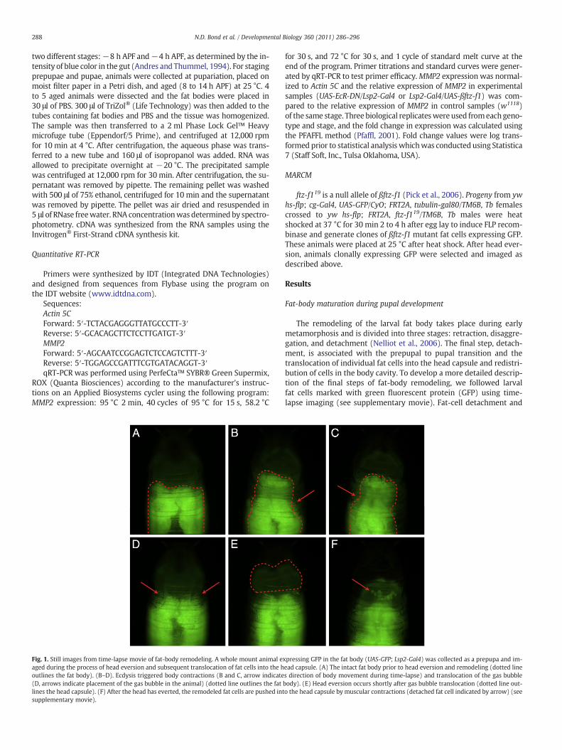
Fig. 1. Still images from time-lapse movie of fat-body remodeling. A whole mount animal eaged during the process of head eversion and subsequent translocation of fat cells into the houtlines the fat body). (B–D). Ecdysis triggered body contractions (B and C, arrow indicate(D, arrows indicate placement of the gas bubble in the animal) (dotted line outlines the fatlines the head capsule). (F) After the head has everted, the remodeled fat cells are pushed intsupplementary movie).
for 30 s, and 72 °C for 30 s, and 1 cycle of standard melt curve at theend of the program. Primer titrations and standard curves were gener-ated by qRT-PCR to test primer efficacy.MMP2 expression was normal-ized to Actin 5C and the relative expression of MMP2 in experimentalsamples (UAS-EcR-DN/Lsp2-Gal4 or Lsp2-Gal4/UAS-ßftz-f1) was com-pared to the relative expression of MMP2 in control samples (w1118)of the same stage. Three biological replicateswere used fromeach geno-type and stage, and the fold change in expression was calculated usingthe PFAFFL method (Pfaffl, 2001). Fold change values were log trans-formed prior to statistical analysis whichwas conducted using Statistica7 (Staff Soft, Inc., Tulsa Oklahoma, USA).
MARCM
ftz-f119 is a null allele of ßftz-f1 (Pick et al., 2006). Progeny from ywhs-flp; cg-Gal4, UAS-GFP/CyO; FRT2A, tubulin-gal80/TM6B, Tb femalescrossed to yw hs-flp; FRT2A, ftz-f119/TM6B, Tb males were heatshocked at 37 °C for 30 min 2 to 4 h after egg lay to induce FLP recom-binase and generate clones of ßftz-f1 mutant fat cells expressing GFP.These animals were placed at 25 °C after heat shock. After head ever-sion, animals clonally expressing GFP were selected and imaged asdescribed above.
Results
Fat-body maturation during pupal development
The remodeling of the larval fat body takes place during earlymetamorphosis and is divided into three stages: retraction, disaggre-gation, and detachment (Nelliot et al., 2006). The final step, detach-ment, is associated with the prepupal to pupal transition and thetranslocation of individual fat cells into the head capsule and redistri-bution of cells in the body cavity. To develop a more detailed descrip-tion of the final steps of fat-body remodeling, we followed larvalfat cells marked with green fluorescent protein (GFP) using time-lapse imaging (see supplementary movie). Fat-cell detachment and
xpressing GFP in the fat body (UAS-GFP; Lsp2-Gal4) was collected as a prepupa and im-ead capsule. (A) The intact fat body prior to head eversion and remodeling (dotted lines direction of body movement during time-lapse) and translocation of the gas bubblebody). (E) Head eversion occurs shortly after gas bubble translocation (dotted line out-o the head capsule by muscular contractions (detached fat cell indicated by arrow) (see

289N.D. Bond et al. / Developmental Biology 360 (2011) 286–296
translocation into the head capsule occurred during the prepupal topupal transition (Fig. 1E, F; Nelliot et al., 2006), a time period whichis also known as pupal ecdysis (Kim et al., 2006). This transitionwas characterized by small contractions of the body (Fig. 1B, C) pre-ceding the translocation of a gas bubble (Fig. 1D). Seconds after thegas bubble appeared in the anterior portion of the animal, strong ab-dominal contractions resulted in head eversion (Fig. 1E). This devel-opmental event marks the beginning of pupal development.Immediately after head eversion, small contractions pushed individu-al remodeled fat cells into the head capsule (Fig. 1F). Translocation ofthe fat cells into the head capsule was completed within 43 min ofhead eversion (+/−2.5 min at 23 °C).
Fig. 2. Fat-body disaggregation and detachment requires 20E signaling. (A). Fluorescent imaculum. A(1–6): Control animals, fat body is marked by GFP (UAS-GFP; Lsp2-Gal4). A(7–12):pression of a dominant-negative (DN) form of EcR (UAS-EcR-DN, UAS-GFP; Lsp2-Gal4). A(disrupted when 20E signaling is disrupted in the fat body. A(3–4) and A(9–10): In the disain the fat body (also see Fig. 2 B8). A(5–6) and A(11–12): In the detachment phase, the fatrupted in the fat body. Developmental time points marked in hours after pupariation. (B). Cfat body is marked by GFP (UAS-GFP; Lsp2-Gal4). B(6–9): Fat body from experimental animalnegative (DN) form of EcR (UAS-EcR-DN,UAS-GFP; Lsp2-Gal4). B(1): Fat body cells from a thirB(2–3) and B(6–7): In the retraction phase, the fat cells do not change shape. GFP accumulatesignaling is disrupted in the fat body. B(4) and B(8): In the disaggregation phase, fat-body cthe detachment phase the fat cells do not detach and become spherical when 20E signaling
The role of 20E signaling in fat-body remodeling
Blocking 20E signaling in the fat body results in clumps of non-dissociated cells in the pupa (Cherbas et al., 2003), thus 20E signal-ing is required for some aspect of fat-body remodeling. To under-stand the role of 20E signaling in specific stages of fat-bodyremodeling, a dominant-negative form of EcR, (EcRF645A) wasexpressed in larval fat body by expression of UAS-EcRF645A (hereinreferred to as UAS-EcR-DN) driven by Lsp2-Gal4. We did a detailedanalysis of fat-body remodeling by following the changes in grossfat-body morphology and fat-cell shape during the three stages ofremodeling (Fig. 2).
ges of whole mount animals expressing GFP in the fat body, dotted line marks the oper-Experimental animals, fat body is marked by GFP and 20E signaling is disrupted by ex-1–2) and A(7–8): The retraction of the fat body to the level of the operculum is notggregation phase, fat-body cells do not change shape when 20E signaling is disruptedcells do not separate and translocate into the head capsule when 20E signaling is dis-onfocal images of fat-body cells expressing GFP. B(1–5): Fat body from control animals,s, fat body is marked by GFP and 20E signaling is disrupted by expression of a dominant-d instar larva are attached, flattened and irregularly shaped. GFP expression is variable.s at the membrane in control animals while GFP expression remains variable when 20Eells do not round up when 20E signaling is disrupted in the fat body. B(5) and B(9): Inis disrupted in the fat body.

290 N.D. Bond et al. / Developmental Biology 360 (2011) 286–296
As described, fat body remodeling begins with the retraction of thefat body followed by disaggregation (Nelliot et al., 2006). We foundthat disruption of 20E signaling did not affect fat-body retraction(compare Fig. 2A(2) to 2A(8)), but did inhibit fat-body disaggregation(compare Fig. 2B(4) to 2B(8)). The fat cells expressing EcR-DN did notdisaggregate. They retained their flattened shape and did not round up(Fig. 2A(9–10), B(7–8)). Finally, at 12 h APF, when remodeled fat cellsbecome free-floating and are translocated into the head capsule(Fig. 2A(5–6), B(5)), we found that disruption of 20E signalingresulted in persistence of flattened, attached fat cells (Fig. 2B(9))that did not enter into the head capsule (Fig. 2A(11–12)). We con-clude that 20E signaling within the fat body is required for the disag-gregation and detachment phases of fat-body remodeling.
ßftz-f1, in concert with 20E signaling, is sufficient to induce fat-bodyremodeling
ßFTZ-F1 is a key transcription factor involved in the modulation ofthe transcriptional response to 20E signaling necessary to initiate the
Fig. 3. Expression of ßftz-f1 induces premature fat-body remodeling in the presence of 20A(1): Fat body from control pupa expressing GFP (UAS-GFP; Lsp2-Gal4). A(2–3): Fat body fr(UAS-GFP; Lsp2-Gal4/UAS-ßftz-f1). A(2): At 4 h APF, over expression of ßftz-f1 resulted in pßftz-f1 resulted premature fat-cell detachment (compare A(1) to A(3)). (B). Fluorescent imag(UAS-GFP; Lsp2-Gal4/UAS-ßftz-f1). B(1): In vivo fat body; fat cells are attached, flattened andits larval fat body morphology. B(3): Fat body explant cultured with 10−5 M 20E for 8 h undincubation with 20E. The red staining indicates that cells are not necrotic. (C). Schematic suto 20E. The process of fat-body remodeling in a wild-type animal is completed after the pupßftz-f1during the third-larval instar (in green) results in premature fat-body remodeling occ
prepupal to pupal transition (Broadus et al., 1999; Fortier et al., 2003;Yamada et al., 2000). The final steps of fat-body remodeling, detach-ment and fat cell translocation into the head capsule, also occur at thistime. We find that ßftz-f1 is expressed in the fat body just prior to theprepupal to pupal transition (data not shown). As a first step towardtesting the role of ßftz-f1 in fat-body remodeling, we expressed ßftz-f1prematurely in the larval fat body using the Lsp2-Gal4 driver. Ectopicßftz-f1 transcripts were readily detected in the larval fat body 8 h beforepupariation (data not shown) and fat-body remodeling occurred pre-maturely in the prepupa (Fig. 3B–C).While normal fat-bodydisaggrega-tion occurs at 6 to 8 h APF (Fig. 2A(3–4) and Nelliot et al., 2006),premature expression of ßftz-f1 in the larval fat body resulted in prema-ture disaggregation at 4 hAPF (Fig. 3A(2)). Thiswas followed by prema-ture fat-cell detachment by 6 h APF (Fig. 3A(3)), where normaldetachment occurs at 12 h APF (see Fig. 2A(5) and Nelliot et al., 2006).Thus, premature expression of ßftz-f1 in the fat body of the third-instarlarva induced premature fat-body remodeling in the prepupa.
The timing of premature remodeling suggests that ßftz-f1 mightmodify the transcriptional response of the fat body to the 20E pulse
E. (A). Fluorescent images of whole mount prepupae expressing GFP in the fat body.om experimental pupa, marked by GFP, prematurely expressing ßftz-f1 in the fat bodyremature disaggregation (compare A(1) to A(2)). A(3): At 6 h APF, overexpression ofes of wandering third-instar larvae fat bodies in which ßftz-f1 is prematurely expressedretain a polygonal shape. B(2): Fat body explant cultured in the absence of 20E retainsergoes fat-body remodeling. B(4): Fat body explant stained with SYTOX® after 8 hour
mmarizing fat-body remodeling in response to ßftz-f1 expression followed by exposureation pulse of 20E (endogenous expression of ßftz-f1 is shown in purple). Expression ofurring after the pupariation pulse of 20E.

291N.D. Bond et al. / Developmental Biology 360 (2011) 286–296
occurring at pupariation, thus setting in motion the prepupal to pupalresponse to 20E (Fig. 3C). To test this idea, we expressed UAS-ßftz-f1in the fat body while blocking 20E signaling by co-expression ofUAS-EcR-DN. Even in the presence of ectopic ßftz-f1, blocking 20E sig-naling in the fat body prevented fat-body remodeling (Fig. 4C). Headeversion occurred but fat cells were not translocated into the headcapsule (Fig. 4C). To further test the idea that ßFTZ-F1 works in con-cert with 20E signaling to program the final steps in the remodelingof the fat body, we used a fat-body ex vivo organ culture assay. Fat-body explants from control larvae and larvae in which ßftz-f1 is pre-maturely expressed in the larval fat body (Lsp2-Gal4; UAS-ßftz-f1)were co-cultured with and without 20E. In the absence of 20E, wild-type fat bodies did not exhibit the cell-shape changes associatedwith remodeling (data not shown). Likewise, cultured fat bodies ec-topically expressing ßftz-f1 did not remodel in the absence of 20E(Fig. 3B(2)). Addition of 20E in the presence of ßftz-f1 expressionhowever, was sufficient to induce fat-body remodeling and individualdetached cells were detected within 8 h (Fig. 3B(3)). The remodeledfat cells were viable (Fig. 3B(4)), and therefore it is unlikely that celldetachment in the ex vivo organ culture was due to degeneration ofthe tissue. These data taken together suggest that, in the fat body,ßftz-f1 is required to modulate the transcriptional response to 20Ein order to achieve fat-body remodeling at the prepupal to pupaltransition.
ßftz-f1 is necessary for fat-body remodeling
To determine whether ßftz-f1 is necessary for fat-body remodel-ing, we carried out a series of loss-of-function experiments. Becauseßftz-f1 null mutant animals die during embryonic development(Broadus et al., 1999; Ruaud et al., 2010; Yamada et al., 2000), wefirst chose to examine ßftz-f1 hypomorphs (ftz-f117/Df(3L)CatDH104),wherein a small percentage of the mutants complete the prepupalto pupal transition (Broadus et al., 1999). Unfortunately, the mutantphenotype of the ßftz-f1 hypomorph was extremely variable and
Fig. 4. Fat-body remodeling requires ßftz-f1 expression, 20E signaling, and Matrix metallop(animals imaged at post-head eversion stage, 12–14 h APF, dotted line marks operculum). (fat cells have entered the head capsule. (B) Fat-body remodeling is disrupted when EcR-DNand fat cells do not enter the head capsule. (C) Fat-body remodeling is also disrupted whenUAS-ßftz-f1). (D) Fat-body remodeling is disrupted in animal expressing dBlimp-1 in the fat bexpressing Timp specifically in the fat body (Lsp2-Gal4, UAS-GFP; UAS-Timp).
thus, it was impossible to determine whether ßftz-f1 is necessaryfor fat-body remodeling (data not shown). We attempted to knockdown ßftz-f1 expression by fat-body specific RNA interferenceusing the Lsp2-Gal4 driver and three different UAS-ßftz-f1-RNAilines. However, we determined by qRT-PCR analysis that this ap-proach failed to reduce levels of ßftz-f1 transcripts in the fat body(data not shown).
As an alternative approach, we took advantage of the known re-pressor of ßftz-f1, the Drosophila homolog of the mammalian B lym-phocyte-induced maturation protein-1 (dBlimp-1) (Agawa et al.,2007). We drove UAS-dBlimp-1 XA (herein referred to as UAS-dBlimp-1) in the fat body using the fat-cell driver, cg-Gal4, which isexpressed in the fat body throughout larval and pupal development.Misexpression of UAS-dBlimp-1 in the fat body resulted in lethalityat various stages of development, however, some animals proceededthrough the prepupal to pupal transition. In these animals, fat-cell de-tachment and the migration of fat cells into the head capsule did notoccur (Fig. 4D). Similar results were also achieved using the fat bodydriver Lsp2-Gal4 (data not shown).
As an additional strategy, we chose to useMosaic Analysis with a Re-pressible Cell Marker (MARCM) (Lee and Luo, 1999) to assess the loss ofßftz-f1 function in individual fat cells. Based on our previous results wepredicted that individual cells mutant for ßftz-f1would not undergo fat-body remodeling. Fat bodies from theMARCM progeny were examinedafter head eversion. A variable number of ßftz-f1 null mutant (ftz-f119)fat body cells were observed in each animal, but an average of 6 ßftz-f1 mutant fat cell clusters were identified per pupa. Some clusters con-tained a single ßftz-f1 mutant cell that was completely surrounded bynon-mutant cells (Fig. 5A, A″) while other clusters contained eithertwo or more mutant cells clustered together (Fig. 5B, B", C, C″, D, D″).The number of mutant cells per cluster was associated with distinctfat cell shapes. The majority (91%) of mutant cells residing in clustersof 3 or more cells did not undergo disaggregation and retained a morepolygonal shape (Fig. 5I). In contrast, 74% of the single mutant cellsthat were not surrounded by other mutant cells were spherical and
roteinase activity. (A–E) Fluorescent images of animals expressing GFP in the fat bodyA) Fat-body remodeling in the control pupa expressing UAS-GFP; Lsp2-Gal4. Remodeledis expressed in the fat body (UAS-GFP, UAS-EcR-DN; Lsp2-Gal4). Fat body remains intactßftz-f1 and EcR-DN are coexpressed in the fat body (UAS-GFP, UAS-EcR-DN; Lsp2-Gal4/ody (cg-Gal4, UAS-GFP; UAS-dBlimp-1). (E) Fat-body remodeling is disrupted in animals

Fig. 5. Mosaic analysis with a repressible cell marker. (A–D) Progeny from yw hs-flp; cg-Gal4, UAS-GFP/CyO; FRT2A, tubulin-gal80/TM6B, Tb females crossed to yw hs-flp; FRT2A, ftz-f119/TM6B, Tb males were heat shocked at 37 °C for 30 min 2 to 4 h after egg lay to induce FLP recombinase and generate clones of ßftz-f1 null mutant (ftz-f119) fat cells expressingGFP. Cells were imaged in vivo after head eversion using confocal microscopy. (A″–D″) GFP expressing clones are outlined in red for emphasis. (A, A″) Single ßftz-f1 mutant cell, cellis spherical and detached from adjacent cells. (B, B″) Two adjacent ßftz-f1 mutant cells, cells are disaggregated and spherical but attached to each other. (C, C″) Three adjacent ßftz-f1mutant cells, cells are attached and not disaggregated; two adjacent ßftz-f1mutant cells which are disaggregated, spherical and attached to each other and a single ßftz-f1mutantcell which is spherical and detached. (D, D″) Cluster of four ßftz-f1 mutant cells, all cells are attached and not disaggregated. (I) Percentage of disaggregation that occurs ßftz-f1mutant cells. ßftz-f1 mutant cells (GFP expressing cells) were categorized based on the number of adjacent mutant cells (number of mutant cells in cluster) and how many ofthose cells were disaggregated. Spherical cells were considered disaggregated and flat cells with polygonal shape were scored as not having undergone remodeling. (J) Diagramillustrating that fat body-remodeling is a tissue-autonomous process. ßftz-f1 mutant cells are depicted in green, wild-type cells in beige. Wild-type cells are capable of expressingMMP2 (proteinase shown in purple) which degrades ECMwhile the ßftz-f1mutant cells are not. MMP2 is present at the membrane of the wild-type cells and degrades ECM that thewild-type cell shares with the ßftz-f1 mutant cells. Thus, the ßftz-f1 mutant cell's ECM is remodeled by an adjacent wild-type cell's MMP2.
292 N.D. Bond et al. / Developmental Biology 360 (2011) 286–296
detached from the other fat cells (Fig. 5I). Due to the lack of completeremodeling in ßftz-f1 mutant MARCM cells, we conclude that ßftz-f1 isrequired for fat-body remodeling.
Misexpression of MMP2 is sufficient to induce fat-body remodeling
The process of fat-cell detachment presumably involves proteasesthat can cleave substrates present in the ECM that hold the cells to-gether. The detachment phase of fat-body remodeling occurs concur-rently with the expression ofMMP1 andMMP2 during the prepupal topupal transition (Nelliot et al., 2006; Page-McCaw et al., 2003). Previ-ous reports have shown that the MMPs are required for midgut andtracheal remodeling (Llano et al., 2002; Page-McCaw et al., 2003).Moreover, there is evidence that the expression of MMPs and their in-hibitor TIMP (tissue inhibitor of metalloproteases) might be regulatedby 20E signaling (Lee et al., 2003). Thus, these proteases are likelycandidates for involvement in fat-body remodeling and are possibletarget genes of the ßFTZ-F1-mediated 20E-signaling cascade. There-fore, we set out to test the role of MMPs in fat-body tissueremodeling.
We first tested whether Drosophila MMPs were sufficient to pro-mote fat-body remodeling by misexpression of MMP1 and MMP2 in
the fat body. UAS-MMP1 and UAS-MMP2 were individually expressedspecifically in fat body using the Lsp2-Gal4 driver (Cherbas et al.,2003). Misexpression of MMP2 resulted in lethality and prematurefat-body remodeling in mid-third instar larvae. Dissected fat cellsfrom third-instar larvae were free floating, spherical and resembledwild-type remodeled fat cells (Fig. 6C). We do not believe that thisis a generalized phenomenon of ectopic expression of metallopro-teases because, although misexpression of MMP1 in the fat bodyalso caused larval lethality, it did not induce fat-body remodeling.The fat cells maintained their associations with their neighboringcells and did not display cell shape changes associated fat-body remo-deling (Fig. 6B).
Misexpression of Timp in the fat body inhibits remodeling
TIMP inhibits the catalytic activity of MMPs by occupying the ac-tive site of the proteinase (Gomis-Rüth et al., 1997). There is oneTimp gene in the Drosophila genome (as compared to four in verte-brates) (Wei et al., 2003). To further explore the role of MMP2 infat-body remodeling, we ectopically expressed UAS-Timp in the fatbody using the Lsp2-Gal4 driver. If MMP2 activity is required for fat-body remodeling, we expected to observe a block in remodeling by
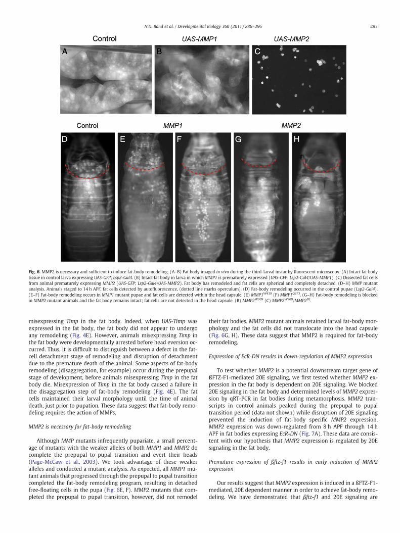
Fig. 6. MMP2 is necessary and sufficient to induce fat-body remodeling. (A–B) Fat body imaged in vivo during the third-larval instar by fluorescent microscopy. (A) Intact fat bodytissue in control larva expressing UAS-GFP; Lsp2-Gal4. (B) Intact fat body in larva in which MMP1 is prematurely expressed (UAS-GFP; Lsp2-Gal4/UAS-MMP1). (C) Dissected fat cellsfrom animal prematurely expressing MMP2 (UAS-GFP; Lsp2-Gal4/UAS-MMP2). Fat body has remodeled and fat cells are spherical and completely detached. (D–H) MMP mutantanalysis. Animals staged to 14 h APF, fat cells detected by autofluorescence, (dotted line marks operculum). (D) Fat-body remodeling occurred in the control pupae (Lsp2-Gal4).(E–F) Fat-body remodeling occurs in MMP1 mutant pupae and fat cells are detected within the head capsule. (E) MMP1W439 (F) MMP1Q273. (G–H) Fat-body remodeling is blockedin MMP2 mutant animals and the fat body remains intact; fat cells are not detected in the head capsule. (B) MMP2W309 (C) MMP2W309/MMP2Df.
293N.D. Bond et al. / Developmental Biology 360 (2011) 286–296
misexpressing Timp in the fat body. Indeed, when UAS-Timp wasexpressed in the fat body, the fat body did not appear to undergoany remodeling (Fig. 4E). However, animals misexpressing Timp inthe fat body were developmentally arrested before head eversion oc-curred. Thus, it is difficult to distinguish between a defect in the fat-cell detachment stage of remodeling and disruption of detachmentdue to the premature death of the animal. Some aspects of fat-bodyremodeling (disaggregation, for example) occur during the prepupalstage of development, before animals misexpressing Timp in the fatbody die. Misexpression of Timp in the fat body caused a failure inthe disaggregation step of fat-body remodeling (Fig. 4E). The fatcells maintained their larval morphology until the time of animaldeath, just prior to pupation. These data suggest that fat-body remo-deling requires the action of MMPs.
MMP2 is necessary for fat-body remodeling
Although MMP mutants infrequently pupariate, a small percent-age of mutants with the weaker alleles of both MMP1 and MMP2 docomplete the prepupal to pupal transition and evert their heads(Page-McCaw et al., 2003). We took advantage of these weakeralleles and conducted a mutant analysis. As expected, all MMP1 mu-tant animals that progressed through the prepupal to pupal transitioncompleted the fat-body remodeling program, resulting in detachedfree-floating cells in the pupa (Fig. 6E, F). MMP2 mutants that com-pleted the prepupal to pupal transition, however, did not remodel
their fat bodies. MMP2 mutant animals retained larval fat-body mor-phology and the fat cells did not translocate into the head capsule(Fig. 6G, H). These data suggest that MMP2 is required for fat-bodyremodeling.
Expression of EcR-DN results in down-regulation of MMP2 expression
To test whether MMP2 is a potential downstream target gene ofßFTZ-F1-mediated 20E signaling, we first tested whether MMP2 ex-pression in the fat body is dependent on 20E signaling. We blocked20E signaling in the fat body and determined levels of MMP2 expres-sion by qRT-PCR in fat bodies during metamorphosis. MMP2 tran-scripts in control animals peaked during the prepupal to pupaltransition period (data not shown) while disruption of 20E signalingprevented the induction of fat-body specific MMP2 expression.MMP2 expression was down-regulated from 8 h APF through 14 hAPF in fat bodies expressing EcR-DN (Fig. 7A). These data are consis-tent with our hypothesis that MMP2 expression is regulated by 20Esignaling in the fat body.
Premature expression of ßftz-f1 results in early induction of MMP2expression
Our results suggest thatMMP2 expression is induced in a ßFTZ-F1-mediated, 20E dependent manner in order to achieve fat-body remo-deling. We have demonstrated that ßftz-f1 and 20E signaling are
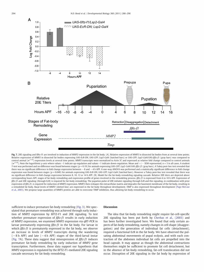
Fig. 7. 20E signaling and ßftz-f1 are involved in induction of MMP2 expression in the fat body. (A). Relative expression of MMP2 in dissected fat bodies from at several time points.Relative expression of MMP2 in dissected fat bodies expressing UAS-EcR-DN, UAS-GFP; Lsp2-Gal4 (hatched bars) or UAS-GFP; Lsp2-Gal4/UAS-ßftz-f1 (gray bars) was compared tocontrol animal (w1118) expression levels at several time points. MMP2 transcripts were normalized to Actin 5C and expressed as relative fold change compared to control animals(w1118). Note the logarithmic y-axis where values N1 indicate up-regulation and values b1 indicate down-regulation. Mean and +/− SEM represented, n=3 in all cases. A studentT-test was performed and no difference was found between stages (p=0.19) for animals expressing UAS-GFP; Lsp2-Gal4/UAS-ßftz-f1 (gray bars). A Tukey post-hoc test revealed thatthere was no significant difference in fold change expression between −8 and −4 h APF. A one-way ANOVA was performed and a statistically significant difference in fold changeexpression was found between stages (p=0.049) for animals expressing UAS-EcR-DN, UAS-GFP; Lsp2-Gal4 (hatched bars). However a Tukey post-hoc test revealed that there wasno significant difference in fold change expression between 8, 10, 12 or 14 h APF. (B). Model for the fat-body remodeling signaling cascade. Relative 20E titers are depicted abovecorresponding hours APF, stages of fat-body remodeling and expression profile of genes involved in the remodeling process. ßftz-f1 is expressed from 6 to 10 h APF. Expression ofßftz-f1 and 20E signaling (through EcR) is required for fat-body remodeling. The pupation pulse of 20E initiates signaling through EcR and this signaling, in combination with priorexpression of ßftz-f1, is required for induction ofMMP2 expression. MMP2 then reshapes the extracellular matrix and degrades the basement membrane of the fat body, resulting ina remodeled fat body. Basal levels of MMP2 (dotted line) are expressed in the fat body throughout development. TIMP is also expressed throughout development (Page-McCawet al., 2003). We propose large quantities of MMP2 protein are able to overcome TIMP inhibition, thus allowing fat-body remodeling to occur.
294 N.D. Bond et al. / Developmental Biology 360 (2011) 286–296
sufficient to induce premature fat-body remodeling (Fig. 3). We spec-ulated that premature remodeling was achieved through early induc-tion of MMP2 expression by ßFTZ-F1 and 20E signaling. To testwhether premature expression of ßftz-f1 results in early inductionof MMP2 expression, we examined MMP2 expression via qRT-PCR inlarvae prematurely expressing ßftz-f1 in the fat body. For larvae inwhich ßftz-f1 is prematurely expressed in the fat body, we observean increase in levels of MMP2 transcripts during the wandering(−8 h APF) and late (−4 h APF) stages of the third-larval instar(Fig. 7A). These data suggest that misexpression of ßftz-f1 inducespremature fat-body remodeling by early induction of MMP2 genetranscription. Furthermore, these data support our hypothesis thatMMP2 expression is regulated by the ßFTZ-F1-mediated 20E signalingcascade necessary for fat-body remodeling.
Discussion
The idea that fat-body remodeling might require fat-cell-specific20E signaling has been put forth by Cherbas et al., (2003) andhas been further investigated here. We found that only certain as-pects of fat-body remodeling, namely changes in cell shape (disaggre-gation) and the generation of individual fat cells (detachment),required a functional EcR in the fat body. We have observed the pul-sating abdominal movements of pupal ecdysis, and with each con-traction of the abdomen individual fat cells are propelled into thehead capsule. It may appear as though the abdominal contractionsthemselves might be sufficient to promote fat cell detachment, butin the absence of fat-body remodeling, fat-cell translocation did notoccur. Disruption of 20E signaling in the fat body by expression of

295N.D. Bond et al. / Developmental Biology 360 (2011) 286–296
EcR-DN did not affect the peristaltic abdominal contractions, but didprevent fat-body remodeling and translocation of the fat cells intothe head capsule. Thus, the abdominal movements do not provideshearing forces sufficient to detach cells from the fat-body tissuemass. The ecdysial abdominal contractions appear to be involved inthe translocation of the remodeled fat cells into the head capsuleand redistribution of the cells with the abdomen of the animal andnot the process of detachment itself.
We are intrigued by our observation that tissue-specific misex-pression of MMP2 in the larval fat body results in animal lethality inaddition to premature fat-body remodeling. Likewise, blocking 20Esignaling in the fat body (and thus down-regulatingMMP2 expressionin the pupa) results in animal death (Bond et al., 2010; Cherbas et al.,2003), as does blockingMMP2 function by misexpression of Timp. It isknown that MMPs are involved in cleaving signaling factors in theECM of mammalian cells (Sternlicht and Werb, 2001). It is possiblethat MMPs, in addition to remodeling the fat body, are involved incleavage of signaling molecules in Drosophila that have global devel-opmental effects necessary for normal animal development.
Due to the fact that ßftz-f1 null mutants do not survive embryo-genesis, we were not able to directly observe the effect of a ßftz-f1null mutation in the fat body at the prepupal to pupal transition.RNAi experiments were unsuccessful and hypomorphic mutantswere unreliable, so we employed the Mosaic Analysis with a Repress-ible Cell Marker (MARCM) technique (Lee and Luo, 2001) to examineßftz-f1 null mutant cells. Interestingly, the degree of fat-body remo-deling observed seemed to coincide with the number of adjacentßftz-f1 null mutant cells. ßftz-f1 null mutant cells that were found inclusters (3 or more adjacent mutant cells) did not undergo any remo-deling, while mutant cells found in clusters of two achieved disaggre-gation but not detachment, and single mutant cells (completelysurrounded by non-mutant cells) were disaggregated and detached.The clusters of 3 or more ßftz-f1 null mutant cells maintained theirlarval morphology, suggesting that, in these cells, ßFTZ-F1 is requiredfor fat-body remodeling. Moreover, these results suggest that the pro-cess of fat-body remodeling is not a cell autonomous process. Instead,perhaps fat-body remodeling is a tissue autonomous process in whichadjacent fat cells can participate in the remodeling of neighboringmutant cells. We have shown that the remodeling of the fat body re-quires expression ofMMP2, presumably to break down the ECM hold-ing the tissue together. Because MMP2 is attached to the membraneof cells by a GPI anchor (Page-McCaw et al., 2003), its localizationsupports our model for tissue-autonomous fat-body remodeling. Amembrane bound MMP2 would not only have the potential to re-model the cell that it is directly bound to but it could also potentiallyhave an effect on other cells in the immediate vicinity (Fig. 5J).
In this paper, we take advantage of the wealth of knowledge aboutthe 20E-signaling cascade, the expression patterns ofMMP2 and Timp,and our data presented here to propose a model for the ßFTZ-F1-me-diated, 20E-signaling cascade in the fat body (Fig. 7B). The cell-shapechanges and cell detachment phases of fat-body remodeling occur atthe pre-pupal to pupal transition. We propose that ßFTZ-F1 makesthe fat-body cells competent to respond to the 20E pulse released atthe prepupal to pupal transition resulting in an increase in MMP2 ex-pression, and that MMP2 functions to degrade fat body ECM in atissue-autonomous manner. Although TIMP is expressed throughoutdevelopment (Page-McCaw et al., 2003), we propose that the largequantities of MMP2 resulting from up-regulation of the gene areable to overcome TIMP inhibition, allowing fat-body remodeling tooccur. According to our results, up-regulation of MMP2 can beachieved by premature expression of ßftz-f1 in the fat body. Althoughit is highly suggestive, our results implicate a role for ßFTZ-F1 in theregulation of MMP2 expression. ßFTZ-F1 protein is indeed present atthe time of MMP2 expression (White et al., 1997; Yamada et al.,2000). It remains to be determined whether ßFTZ-F1 is capable of ac-tivating transcription of MMP2 by directly binding to the MMP2
promoter. In the mosquito, ßFTZ-F1 and the ecdysone receptor bindto the promoter of the vitellogenin (Vg) gene. ßFTZ-F1 also binds toFISC, a p160 coactivator of the ecdysone receptor. The binding ofßFTZ-F1 to FISC is required in order to achieve 20E-induction of Vgtranscription (Zhu et al., 2006). Further experiments are needed todetermine whether ßFTZ-F1 acts as a direct regulator of MMP2 tran-scription in Drosophila, or if the effect of ßFTZ-F1 onMMP2 expressionis indirect.
Determining the genetic cascade required for cell detachment inDrosophila fat-body remodeling might pave the way for developmentof a tractable model system for understanding mammalian processessuch as metastasis and wound healing. The larval fat body also pro-vides a good model system in which to analyze the genetic and mo-lecular control of programmed cell death. Unlike other larval tissuessuch as the salivary gland and midgut, the fat body is spared fromprogrammed cell death during metamorphosis. We are currently ex-amining the role of anti-cell death genes, such as diap1, in the regula-tion of cell death in the larval fat body. Understanding the molecularmechanisms responsible for sparing the larval fat body from apopto-sis may provide insight for potential ways to prevent cell death.
Supplementary materials related to this article can be found on-line at doi:10.1016/j.ydbio.2011.09.015.
Acknowledgments
The authors give great thanks to Andrea Page-McCaw for so gener-ously providing the MMP and TIMP reagents. We thank KarolZamora-Anderson for selecting the animals used in our mutant anal-ysis and Keiko Wright for annotating and editing the online time-lapse movie. We also wish to thank Marian Rice for help with micros-copy and Allen Gibbs for help with statistical analysis. This work wassupported by National Science Foundation (NSF) grant number IOS-0719591 awarded to DKH, University of Nevada, Las Vegas GREATScholarship awarded to NDB, NSF RUI grant number 0110238, andNSF MRI grant number DBI-0216242 and Howard Hughes Medical In-stitute (HHMI) grant number 52005134 awarded to CTW.
References
Agawa, Y., Sarhan, M., Kageyama, Y., Akagi, K., Takai, M., Hashiyama, K., Wada, T.,Handa, H., Iwamatsu, A., Hirose, S., Ueda, H., 2007. Drosophila blimp-1 is a transienttranscriptional repressor that controls timing of the ecdysone-induced develop-mental pathway. Mol. Cell. Biol. 27, 8739–8747.
Aguila, J.R., Suszko, J., Gibbs, A.G., Hoshizaki, D.K., 2007. The role of larval fat cells inadult Drosophila melanogaster. J. Exp. Biol. 210, 956–963.
Andres, A.J., Thummel, C.S., 1994. Methods for quantitative analysis of transcription inlarvae and prepupae. In: Goldstein, L.S.B., Fyrberg, E.A. (Eds.), Methods in Cell Biol-ogy, Volume 44— Drosophila melanogaster: Practical Uses in Cell and Molecular Bi-ology. Academic Press, New York, pp. 565–573.
Bainbridge, S.P., Bownes, M., 1981. Staging the metamorphosis of Drosophila melanoga-ster. J. Embryol. Exp. Morphol. 66, 57–80.
Bodenstein, D., 1950. The postembryonic development of Drosophila. In: Demerec, M.(Ed.), Biology of Drosophila. Cold Spring Harbor Laboratory Press, Cold SpringHarbor, pp. 275–375.
Bond, N.D., Hoshizaki, D.K., Gibbs, A.G., 2010. The role of 20-hydroxyecdysone signalingin Drosophila pupal metabolism. Comp. Biochem. Physiol. Part A Mol. Integr. Phy-siol. 157, 398–404.
Broadus, J., McCabe, J.R., Endrizzi, B., Thummel, C.S., Woodard, C.T., 1999. The Drosoph-ila ßFTZ-F1 orphan nuclear receptor provides competence for stage-specific re-sponses to the steroid hormone ecdysone. Mol. Cell. 3, 143–149.
Burtis, K.C., Thummel, C.S., Jones, C.W., Karim, F.D., Hogness, D.S., 1990. The Drosophila74EF early puff contains E74, a complex ecdysone-inducible gene that encodes twoets-related proteins. Cell 61, 85–99.
Butterworth, F.M., 1972. Adipose tissue of Drosophila melanogaster. V. Genetic and ex-perimental studies of an extrinsic influence on rate of cell death in larval fat-body.Dev. Biol. 28, 311–325.
Cherbas, L., Hu, X., Zhimulev, I., Belyaeva, E., Cherbas, P., 2003. EcR isoforms in Drosoph-ila: testing tissue-specific requirements by targeted blockade and rescue. Develop-ment 130, 271–284.
Costantino, B.F.B., Bricker, D.K., Alexandre, K., Shen, K., Merriam, J.R., Antoniewski, C.,Callender, J.L., Henrich, V.C., Presente, A., Andres, A.J., 2008. A novel ecdysone re-ceptor mediates steroid-regulated developmental events during the mid-third in-star of Drosophila. Plos Genet. e1000102.

296 N.D. Bond et al. / Developmental Biology 360 (2011) 286–296
DiBello, P.R., Withers, D.A., Bayer, C.A., Fristrom, J.W., Guild, G.M., 1991. The DrosophilaBroad-Complex encodes a family of related proteins containing zinc fingers. Genet-ics 129, 385–397.
Fortier, T.M., Vasa, P.P., Woodard, C.T., 2003. Orphan nuclear receptor ßFTZ-F1 is re-quired for muscle-driven morphogenetic events at the prepupal-pupal transitionin Drosophila melanogaster. Dev. Biol. 257, 153–165.
Gomis-Rüth, F.-X., Maskos, K., Betz, M., Bergner, A., Huber, R., Suzuki, K., Yoshida, N.,Nagase, H., Brew, K., Bourenkov, G.P., Bartunik, H., Bode, W., 1997. Mechanism ofinhibition of the human matrix metalloproteinase stromelysin-1 by TIMP-1. Na-ture 389, 77–81.
Hoshizaki, D.K., 2005. Fat-cell development. In: Gilbert, L.I., Iatrou, K., Gill, S.S. (Eds.),Comprehensive Molecular Insect Science. Elsevier, Oxford, pp. 315–345.
Jiang, C., Lamblin, A.-F.J., Steller, H., Thummel, C.S., 2000. A steroid-triggered transcrip-tional hierarchy controls salivary gland cell death during Drosophila metamorpho-sis. Mol. Cell. 5, 445–455.
Kageyama, Y., Masuda, S., Hirose, S., Ueda, H., 1997. Temporal regulation of the mid-prepupal gene FTZ-F1: DHR3 early late gene product is one of the plural positiveregulators. Genes Cells 2, 559–569.
Kim, Y.-J., Zitnan, D., Galizia, C.G., Cho, K.-H., Adams, M.E., 2006. A command chemicaltriggers an innate behavior by sequential activation of multiple peptidergic ensem-bles. Curr. Biol. 16, 1395–1407.
King-Jones, K., Thummel, C.S., 2005. Less steroids make bigger flies. Science 310, 630–631.Koelle, M.R., Talbot, W.S., Segraves, W.A., Bender, M.T., Cherbas, P., Hogness, D.S., 1991.
The Drosophila EcR gene encodes an ecdysone receptor, a new member of thesteroid-receptor superfamily. Cell 67, 59–77.
Lam, G.T., Jiang, C., Thummel, C.S., 1997. Coordination of larval and prepupal gene ex-pression by the DHR3 orphan receptor during Drosophila metamorphosis. Devel-opment 124, 1757–1769.
Lam, G., Hall, B.L., Bender, M., Thummel, C.S., 1999. DHR3 is required for the prepupal-pupal transition and differentiation of adult structures during Drosophila meta-morphosis. Dev. Biol. 212, 204–216.
Lavorgna, G., Karim, F.D., Thummel, C.S., Wu, C., 1993. Potential role for a FTZ-F1 steroidreceptor superfamily member in the control of Drosophila metamorphosis. Proc.Natl. Acad. Sci. U. S. A. 90, 3004–3008.
Lee, T., Luo, L., 1999. Mosaic analysis with a repressible cell marker for studies of genefunction in neuronal morphogenesis. Neuron 22, 451–461.
Lee, T., Luo, L., 2001. Mosaic analysis with a repressible cell marker (MARCM) for Dro-sophila neural development. Trends Neurosci. 24, 251–254.
Lee, C.-Y., Cooksey, B.A.K., Baehrecke, E.H., 2002. Steroid regulation of midgut cell deathduring Drosophila development. Dev. Biol. 250, 101–111.
Lee, C.-Y., Clough, E.A., Yellon, P., Teslovich, T.M., Stephan, D.A., Baehrecke, E.H., 2003.Genome-wide analyses of steroid- and radiation-triggered programmed celldeath in Drosophila. Curr. Biol. 13, 350–357.
Llano, E., Pendas, A.M., Aza-Blanc, P., Kornberg, T.B., Lopez-Otin, C., 2000. Dm1-MMP, amatrix metalloproteinase from Drosophilawith a potential role in extracellular ma-trix remodeling during neural development. J. Biol. Chem. 275, 35978–35985.
Llano, E., Adam, G., Pendas, A.M., Quesada, V., Sanchez, L.M., Santamaria, I., Noselli, S.,Lopez-Otin, C., 2002. Structural and enzymatic characterization of DrosophilaDm2-MMP, a membrane-bound matrix metalloproteinase with tissue-specific ex-pression. J. Biol. Chem. 277, 23321–23329.
Nelliot, A., Bond, N., Hoshizaki, D.K., 2006. Fat-body remodeling in Drosophila melano-gaster. Genesis 44, 396–400.
Oberlander, H., 1976. Hormonal control of growth and differentiation of insect tissuescultured in vitro. In Vitro Cell Dev. Biol. 12, 225–235.
Page-McCaw, A., 2008. Remodeling the model organism: Matrix metalloproteinasefunctions in invertebrates. Semin. Cell Dev. Biol. 19, 14–23.
Page-McCaw, A., Serano, J., Sante, J.M., Rubin, G.M., 2003. Drosophila matrix metallo-proteinases are required for tissue remodeling, but not embryonic development.Dev. Cell 4, 95–106.
Page-McCaw, A., Ewald, A.J., Werb, Z., 2007. Matrix metalloproteinases and the regula-tion of tissue remodeling. Nat. Rev. Mol. Cell Biol. 8, 221–233.
Pfaffl, M.W., 2001. A new mathematical model for relative quantification in real-timeRT-PCR. Nucleic Acids Res. 29, e45.
Pick, L., Anderson, W.R., Shulz, J., Woodard, C.T., 2006. The Ftz-F1 family: orphan nucle-ar receptors regulated by novel protein-protein interactions. In: Reshma Taneja, R.(Ed.), Advances in Developmental Biology Series. : Nuclear Receptors in Develop-ment, vol. 16. Elsevier, New York, pp. 255–296.
Rewitz, K.F., Yamanaka, N., O'Connor, M.B., 2010. Steroid hormone inactivation is re-quired during the juvenile-adult transition in Drosophila. Dev. Cell 19, 895–902.
Richards, G., 1981. The radioimmunoassay of ecdysteroid titers in Drosophila melano-gaster. Mol. Cell. Endocrinol. 21, 181–197.
Riddiford, L.M., 1996. Juvenile hormone: the status of its “status quo” action. Arch. In-sect Biochem. 32, 271–286.
Riddiford, L.M., Truman, J.W., 1993. Hormone receptors and the regulation of insectmetamorphosis. Am. Zool. 33, 340–347.
Robertson, C.W., 1936. The metamorphosis of Drosophila melanogaster, including an ac-curately timed account of the principal morphological changes. J. Morphol. 59,351–399.
Ruaud, A.-F., Lam, G., Thummel, C.S., 2010. The Drosophila nuclear receptors DHR3 andßFTZ-F1 control overlapping developmental responses in late embryos. Develop-ment 137, 123–131.
Schubiger, M., Wade, A.A., Carney, G.E., Truman, J.W., Bender, M., 1998. Drosophila EcR-B ecdysone receptor isoforms are required for larval molting and for neuron remo-deling during metamorphosis. Development 125, 2053–2062.
Segraves, W.A., Hogness, D.S., 1990. The E75 ecdysone-inducible gene responsible forthe 75B early puff in Drosophila encodes two new members of the steroid receptorsuperfamily. Genes Dev. 4, 204–219.
Sternlicht, M.D., Werb, Z., 2001. How matrix metalloproteinases regulate cell behavior.Annu. Rev. Cell Dev. Biol. 17, 463–516.
Thomas, H.E., Stunnenberg, H.G., Stewart, A.F., 1993. Heterodimerization of the Dro-sophila ecdysone receptor with retinoid-x receptor and ultraspiracle. Nature 362,471–475.
Thummel, C.S., 1995. From embryogenesis to metamorphosis — the regulationand function of Drosophila nuclear receptor superfamily members. Cell 83,871–877.
Thummel, C.S., 1996. Flies on steroids — Drosophila metamorphosis and the mecha-nisms of steroid hormone action. Trends Genet. 12, 306–310.
Wei, S., Xie, Z., Filenova, E., Brew, K., 2003. Drosophila TIMP is a potent inhibitor ofMMPs and TACE: similarities in structure and function to TIMP-3. Biochemistry42, 12200–12207.
White, K.P., Hurban, P., Watanabe, T., Hogness, D.S., 1997. Coordination of Drosoph-ila metamorphosis by two ecdysone-induced nuclear receptors. Science 276,114–117.
Woodard, C.T., Baehrecke, E.H., Thummel, C.S., 1994. A molecular mechanism for thestage specificity of the Drosophila prepupal genetic response to ecdysone. Cell 79,607–615.
Yamada, M., Murata, T., Hirose, S., Lavorgna, G., Suzuki, E., Ueda, H., 2000. Temporallyrestricted expression of transcription factor ßFTZ-F1: significance for embryogen-esis, molting and metamorphosis in Drosophila melanogaster. Development 127,5083–5092.
Yao, T.-P., Forman, B.M., Jiang, Z., Cherbas, L., Chen, J.-D., Mckeown, M., Cherbas, P.,Evans, R.M., 1993. Functional ecdysone receptor is the product of EcR and Ultra-spiracle genes. Nature 366, 476–479.
Yin, V.P., Thummel, C.S., 2005. Mechanisms of steroid-triggered programmed cell deathin Drosophila. Semin. Cell. Dev. Biol. 16, 237–243.
Zhu, J., Chen, L., Sun, G., Raikhel, A.S., 2006. The competence factor ßFtz-F1 potentiatesecdysone receptor activity via recruiting a p160/SRC coactivator. Mol. Cell. Biol. 26,9402–9412.