Sphingosine 1Phosphate Induces Myoblast Differentiation through Cx43 Protein Expression: A Role for...
15
Molecular Biology of the Cell Vol. 17, 4896 – 4910, November 2006 Sphingosine 1-Phosphate Induces Myoblast Differentiation through Cx43 Protein Expression: A Role for a Gap Junction-dependent and -independent Function □ D R. Squecco,* C. Sassoli, † F. Nuti, ‡ M. Martinesi, ‡ F. Chellini, † D. Nosi, † S. Zecchi-Orlandini, † F. Francini,* L. Formigli, † and E. Meacci ‡ Departments of ‡ Biochemical Sciences, † Anatomy, Histology, and Forensic Medicine, and *Physiological Sciences, University of Florence, Interuniversity Institute of Myology, Florence I-50134, Italy Submitted March 28, 2006; Accepted August 28, 2006 Monitoring Editor: Asma Nusrat Although sphingosine 1-phosphate (S1P) has been considered a potent regulator of skeletal muscle biology, acting as a physiological anti-mitogenic and prodifferentiating agent, its downstream effectors are poorly known. In the present study, we provide experimental evidence for a novel mechanism by which S1P regulates skeletal muscle differentiation through the regulation of gap junctional protein connexin (Cx) 43. Indeed, the treatment with S1P greatly enhanced Cx43 expression and gap junctional intercellular communication during the early phases of myoblast differentiation, whereas the down-regulation of Cx43 by transfection with short interfering RNA blocked myogenesis elicited by S1P. Moreover, calcium and p38 MAPK-dependent pathways were required for S1P-induced increase in Cx43 expression. Interestingly, enforced expression of mutated Cx43 130 –136 reduced gap junction communication and totally inhibited S1P-induced expression of the myogenic markers, myogenin, myosin heavy chain, caveolin-3, and myotube formation. Notably, in S1P-stimulated myoblasts, endogenous or wild-type Cx43 protein, but not the mutated form, coimmunoprecipitated and colocalized with F-actin and cortactin in a p38 MAPK-dependent manner. These data, together with the known role of actin remodeling in cell differentiation, strongly support the important contribution of gap junctional communication, Cx43 expression and Cx43/cytoskeleton interaction in skeletal myogenesis elicited by S1P. INTRODUCTION Skeletal muscle formation during development requires sev- eral cellular and molecular events that lead to the matura- tion of myoblasts into multinucleated myofibers. The onset of the terminal differentiation process is characterized by cell cycle withdrawal and myogenin synthesis followed by the expression of muscle-specific genes including sarcomeric proteins and creatine kinase (CK; Lassar et al., 1989). Adult skeletal muscle have the ability to regenerate after injury from quiescent mononucleated satellite cells which, lying between the sarcolemma and the basal lamina, use the same differentiation program as developing myoblasts (Hawke and Garry, 2001; Grounds et al., 2002). Although much re- mains to be learned on the inductive factors and molecular mechanisms involved in the myogenesis, it is well estab- lished that diverse intercellular signaling pathways may influence the regulation of myoblast differentiation during development and regeneration of skeletal muscle. One of them is mediated by gap junctions, specialized membrane regions composed of aggregate of intercellular channels con- necting directly the adjacent cells (Simon and Goodenough, 1998). Each intercellular channel is formed by the conjunc- tion of two hemichannels composed of six protein subunits belonging to the connexin family, whose connexin (Cx) 43 is the most widely expressed member (Sa `ez et al., 2003). Al- though absent in adult skeletal muscle fibers, Cx43 is present in the early stages of myogenesis, and Cx43-containing gap junctions are required for determining the correct cellular specification during myogenesis (Araya et al., 2003). Indeed, Cx43 is transiently expressed in myoblasts being down- regulated before their fusion into multinucleated myotubes (Gorbe et al., 2005). Moreover, the application of connexin channel blockers (Proulx et al., 1997a) as well as the induc- ible deletion of Cx43 protein (Araya et al., 2005) have been shown to dramatically affect myogenic differentiation, whereas the overexpression of Cx43 by rhabdomyosarcoma cells has been described as enhancing the differentiation capacity (Proulx et al., 1997b). In general, the effects of Cx43 expression have been attributed to the role of gap junctions in the establishment of organized pathways for the intercel- lular transfer of small metabolites and messenger molecules necessary for the coordination and guide of the interacting myoblasts to their final differentiation (Goldberg et al., 1999). However, evidence is accumulating that connexins may have additional functions independent of their gap channel– forming ability (Giepmans, 2004; Stout et al., 2004; Jiang and This article was published online ahead of print in MBC in Press (http://www.molbiolcell.org/cgi/doi/10.1091/mbc.E06 – 03– 0243) on September 6, 2006. □ D The online version of this article contains supplemental material at MBC Online (http://www.molbiolcell.org). Address correspondence to: Elisabetta Meacci (elisabetta.meacci@ unifi.it). Abbreviations used: S1P, sphingosine 1-phosphate; wtCx43, wild- type connexin 43; DNCx43, dominant negative connexin 43; PBS, phosphate buffer solution; FCS, fetal calf serum; DM, differentiation medium; HS, horse serum; MAPK, mitogen-activated protein ki- nase; ERK1/2, p44/42 MAPK; siRNA, short interfering RNA; GFP, green fluorescent protein; MHC, myosin heavy chain. 4896 © 2006 by The American Society for Cell Biology http://www.molbiolcell.org/content/suppl/2006/09/05/E06-03-0243.DC1.html Supplemental Material can be found at:
Transcript of Sphingosine 1Phosphate Induces Myoblast Differentiation through Cx43 Protein Expression: A Role for...
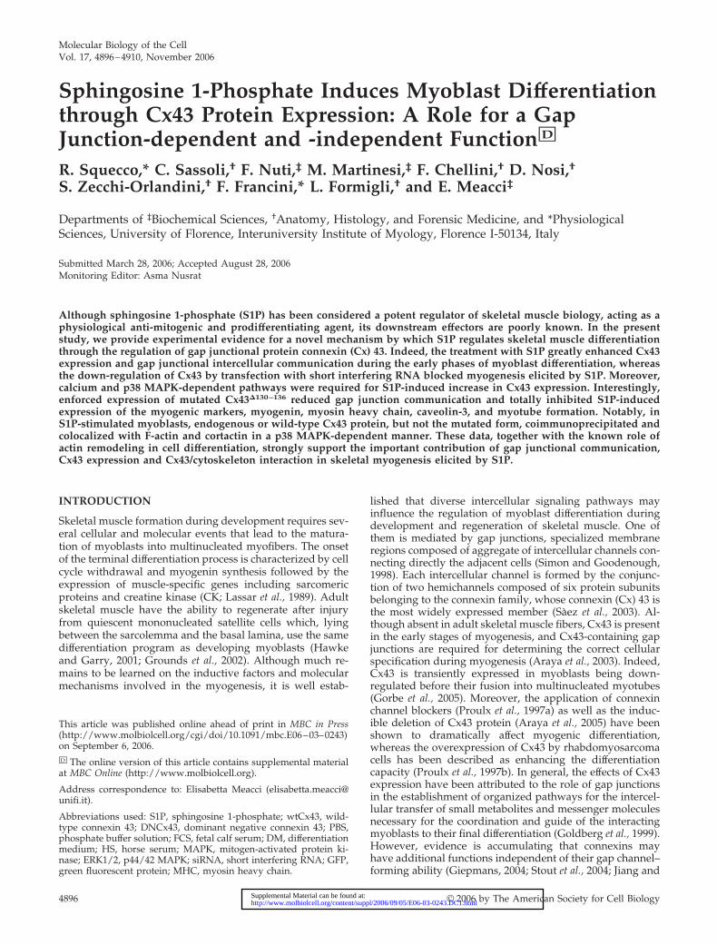
Molecular Biology of the CellVol. 17, 4896–4910, November 2006
Sphingosine 1-Phosphate Induces Myoblast Differentiationthrough Cx43 Protein Expression: A Role for a GapJunction-dependent and -independent Function□D
R. Squecco,* C. Sassoli,† F. Nuti,‡ M. Martinesi,‡ F. Chellini,† D. Nosi,†S. Zecchi-Orlandini,† F. Francini,* L. Formigli,† and E. Meacci‡
Departments of ‡Biochemical Sciences, †Anatomy, Histology, and Forensic Medicine, and *PhysiologicalSciences, University of Florence, Interuniversity Institute of Myology, Florence I-50134, Italy
Submitted March 28, 2006; Accepted August 28, 2006Monitoring Editor: Asma Nusrat
Although sphingosine 1-phosphate (S1P) has been considered a potent regulator of skeletal muscle biology, acting as aphysiological anti-mitogenic and prodifferentiating agent, its downstream effectors are poorly known. In the presentstudy, we provide experimental evidence for a novel mechanism by which S1P regulates skeletal muscle differentiationthrough the regulation of gap junctional protein connexin (Cx) 43. Indeed, the treatment with S1P greatly enhanced Cx43expression and gap junctional intercellular communication during the early phases of myoblast differentiation, whereasthe down-regulation of Cx43 by transfection with short interfering RNA blocked myogenesis elicited by S1P. Moreover,calcium and p38 MAPK-dependent pathways were required for S1P-induced increase in Cx43 expression. Interestingly,enforced expression of mutated Cx43�130–136 reduced gap junction communication and totally inhibited S1P-inducedexpression of the myogenic markers, myogenin, myosin heavy chain, caveolin-3, and myotube formation. Notably, inS1P-stimulated myoblasts, endogenous or wild-type Cx43 protein, but not the mutated form, coimmunoprecipitated andcolocalized with F-actin and cortactin in a p38 MAPK-dependent manner. These data, together with the known role ofactin remodeling in cell differentiation, strongly support the important contribution of gap junctional communication,Cx43 expression and Cx43/cytoskeleton interaction in skeletal myogenesis elicited by S1P.
INTRODUCTION
Skeletal muscle formation during development requires sev-eral cellular and molecular events that lead to the matura-tion of myoblasts into multinucleated myofibers. The onsetof the terminal differentiation process is characterized by cellcycle withdrawal and myogenin synthesis followed by theexpression of muscle-specific genes including sarcomericproteins and creatine kinase (CK; Lassar et al., 1989). Adultskeletal muscle have the ability to regenerate after injuryfrom quiescent mononucleated satellite cells which, lyingbetween the sarcolemma and the basal lamina, use the samedifferentiation program as developing myoblasts (Hawkeand Garry, 2001; Grounds et al., 2002). Although much re-mains to be learned on the inductive factors and molecularmechanisms involved in the myogenesis, it is well estab-
lished that diverse intercellular signaling pathways mayinfluence the regulation of myoblast differentiation duringdevelopment and regeneration of skeletal muscle. One ofthem is mediated by gap junctions, specialized membraneregions composed of aggregate of intercellular channels con-necting directly the adjacent cells (Simon and Goodenough,1998). Each intercellular channel is formed by the conjunc-tion of two hemichannels composed of six protein subunitsbelonging to the connexin family, whose connexin (Cx) 43 isthe most widely expressed member (Saez et al., 2003). Al-though absent in adult skeletal muscle fibers, Cx43 is presentin the early stages of myogenesis, and Cx43-containing gapjunctions are required for determining the correct cellularspecification during myogenesis (Araya et al., 2003). Indeed,Cx43 is transiently expressed in myoblasts being down-regulated before their fusion into multinucleated myotubes(Gorbe et al., 2005). Moreover, the application of connexinchannel blockers (Proulx et al., 1997a) as well as the induc-ible deletion of Cx43 protein (Araya et al., 2005) have beenshown to dramatically affect myogenic differentiation,whereas the overexpression of Cx43 by rhabdomyosarcomacells has been described as enhancing the differentiationcapacity (Proulx et al., 1997b). In general, the effects of Cx43expression have been attributed to the role of gap junctionsin the establishment of organized pathways for the intercel-lular transfer of small metabolites and messenger moleculesnecessary for the coordination and guide of the interactingmyoblasts to their final differentiation (Goldberg et al., 1999).However, evidence is accumulating that connexins mayhave additional functions independent of their gap channel–forming ability (Giepmans, 2004; Stout et al., 2004; Jiang and
This article was published online ahead of print in MBC in Press(http://www.molbiolcell.org/cgi/doi/10.1091/mbc.E06–03–0243)on September 6, 2006.□D The online version of this article contains supplemental materialat MBC Online (http://www.molbiolcell.org).
Address correspondence to: Elisabetta Meacci ([email protected]).
Abbreviations used: S1P, sphingosine 1-phosphate; wtCx43, wild-type connexin 43; DNCx43, dominant negative connexin 43; PBS,phosphate buffer solution; FCS, fetal calf serum; DM, differentiationmedium; HS, horse serum; MAPK, mitogen-activated protein ki-nase; ERK1/2, p44/42 MAPK; siRNA, short interfering RNA; GFP,green fluorescent protein; MHC, myosin heavy chain.
4896 © 2006 by The American Society for Cell Biology http://www.molbiolcell.org/content/suppl/2006/09/05/E06-03-0243.DC1.htmlSupplemental Material can be found at:

Gu, 2005). Indeed, several lines of evidences have shownthat Cx43 regulates cell growth and migration by mecha-nisms that do not require intercellular communication innormal and transformed neuronal and epithelial cells(Huang et al., 1998a, 1998b; Omori and Yamasaki, 1998;Moorby and Patel, 2001; Qin et al., 2002). Moreover, theexogenous expression of mutant nonfunctional connexinshas been reported to protect glial cells against injury andapoptotic cell death (Lin et al., 1998). So far, no evidence ofthe existence of a direct action of the Cx43 protein on theregulation of cell differentiation and skeletal muscle forma-tion has been reported.
Sphingosine 1-phosphate (S1P) is a bioactive lipid thatparticipates in the regulation of numerous cell processes,such as cell proliferation, differentiation, migration, and ap-optosis, and acts as intracellular mediator and ligand forspecific S1P receptors (Saba, 2004). We have recently shownthat S1P is a potent inducer of skeletal muscle differentiation(Donati et al., 2005), and its specific Edg5/S1P2 receptor isdown-regulated during myogenesis (Meacci et al., 2003).Notably, we have also demonstrated that the activity andprotein content of sphingosine kinase, the enzyme cata-lyzing the formation of S1P, is greatly enhanced in differen-tiating C2C12 myoblasts, and its silencing delays myoblastmaturation, thus implicating a physiological role of S1P inmyogenesis (Meacci, E., Nuti, F., Donati C., Cencetti, F.,Farnararo, M., and Bruni, P., unpublished results).
On the basis of the above reported observations, in thepresent study we investigated whether the regulation andassembly of Cx43 into gap junctions could represent a crit-ical event in C2C12 myoblast differentiation elicited by S1Pand whether Cx43 protein per se could contribute to thisprocess.
MATERIALS AND METHODS
Cell CulturesMurine C2C12 myoblasts (American Type Culture Collection, Manassas, VA)were routinely grown in Dulbecco’s modified Eagle’s medium (DMEM;Sigma, St. Louis, MO) supplemented with 10% fetal calf serum (FCS, Sigma).For proliferation experiments, cells were seeded in 96-well plates and utilizedwhen �50% confluent. For myogenic differentiation experiments, cells weregrown in 100-mm dishes or six-well plates until 95% confluent and inducedto differentiate by switching to differentiation medium (DM), DMEM contain-ing 2% horse serum (HS, Sigma) for different times (24, 48, and 72 h and 5 d)in the presence or absence of S1P (1 �M, 2 mM stock solution in dimethyl-sulfoxide, Calbiochem, La Jolla, CA).
Cell TreatmentsC2C12 cells were challenged with 5 �M of the specific p38 MAPK inhibitorSB239063 (Tocris Cookson, Bristol, United Kingdom) and 10 �M of theERK1/ERK2 pathway inhibitor PD98059 (Calbiochem) prepared in 0.05%DMSO, 30 min before agonist addition. The specific effect of the variousinhibitors was tested by Western blot analysis evaluating the phosphorylationstatus of p38 MAPK and ERK1/2. To investigate calcium-dependent events,C2C12 cells were incubated in the dark for 1 h before S1P challenge with 15�M of the Ca2� chelator 1,2-bis(2-aminophenoxy)ethane-N,N,N�,N�-tetraace-tic acid/acetoxymethyl ester (BAPTA/AM, Calbiochem) prepared in 100 mMTris/HCl, pH 7.0, and 0.1% Pluronic F-127 as previously described (Meacci etal., 2002b). The BAPTA/AM efficacy was tested by Western blot analysisexamining the inhibition of S1P-induced PKC� translocation to the membranefraction that, in C2C12 cells, occurs via a Ca2�-dependent mechanism (Meacciet al., personal communication).
Silencing of Cx43 by siRNATo silence the expression of Cx43, short interfering RNA duplexes (siRNA)were used (Santa Cruz Biotechnology, Santa Cruz, CA) corresponding tothree distinct regions of the DNA sequence of mouse Cx43 gene (NM_010288):5�CCCAACUGAACCUUAAGAA3�, 5�CCUCACCAAAUGA UUUCUA3�,and 5�CCUACCAGUUUCUUCAAGU3�. The sequences were evaluatedagainst the database using the NIH Blast program to test for specificity. A
nonspecific scrambled (SCR) siRNA was used as control. C2C12 cells growninto 60-mm dishes in DMEM supplemented with 10% FCS were transfectedwith the mixed combination of the above reported three RNA duplexes, usingLipofectamine 2000 reagent (1 mg/ml; Invitrogen, Carlsbad, CA) according tomanufacturer’s instructions. Briefly, Lipofectamine 2000 was incubated withCx43-siRNA (ratio 2:1) at room temperature for 20 min, and successively thelipid/RNA complexes were added with gentle agitation to C2C12 cells.Twenty-four hours after transfection, cells were shifted to DM in the presenceor absence of S1P for a further 48 h and used for the experiments. Longerincubation times had deleterious effects on C2C12 cell viability and survival,as evaluated by visual inspection and MTS-dye reduction assay (Promega,Madison, WI). To evaluate the specific knock-down of Cx43, cell lysates frommyoblasts transfected with Cx43- or SCR-siRNA were immunoblotted, andprotein was detected using specific anti-Cx43 antibodies.
Construction of GFP-wtCx43 and Mutated GFP-Cx43�130–136
Expression VectorsCx43 cDNA was obtained by reverse transcription of 1 �g of total RNAextracted from C2C12 myoblasts using TRIREAGENT (Sigma), accordingto the manufacturer’s protocol and amplified using SuperScriptOne-StepRT-PCR System (Invitrogen) and the following mouse gene-specific prim-ers designed in the coding region: forward primer 1: 5�ATGGGTGACTG-GAGCAGCG CCTTG3� and reverse primer 1: 5�GGCAGCTTGAT GTTC-AAGCCTG3�and). A cDNA fragment corresponding to mouse Cx43 with adeletion of amino acids 130–136 (Krutovskikh et al., 1998; Upham et al., 2003)was obtained by the amplification of two overlapped fragments: fragment Aamplified using forward primer 1 and the reverse primer 2 (5�TTCAATC-CCAAT CTGTTCAGGTGCAT3�) and fragment B amplified using forwardprimer 2 (5�AAGCAGATTGGGATTGAAGAACACGGC3�) and reverseprimer 1. Cx43 (wtCx43) or Cx43�130–136 (DNCx43) cDNAs were subclonedinto the mammalian expression vector pcDNA3.1/NT-GFP-TOPO using theTA cloning kit and following the manufacturer’s supplied protocol (Invitro-gen). The nucleotide sequences of all PCR products were confirmed byautomated DNA sequencing.
Stable Cell TransfectionsTo obtain cells stably overexpressing GFP-wtCx43 or GFP-DNCx43, myo-blasts were plated onto 60-mm dishes and transfected using Lipofectamine2000 reagent (1 mg/ml) mixed with pGFP-wtCx43, pGFP-DNCx43, or plas-mid alone. After 36 h, the cells were replated in the presence of G418 (300�g/ml; Invitrogen) and expanded under selective conditions. Selected bulkpopulation was used to avoid potential phenotypic changes due to selectionand propagation of clones from single individual cells. Ectopic Cx43 expres-sion levels were routinely monitored by Western blot analysis using specificanti-Cx43 or anti-GFP antibodies (Santa Cruz). Empty vector-transfected cellswere utilized as control. Because previous studies have shown that enforcedexpression of connexins can affect cell survival, MTS-dye reduction assay(Promega) in vector control and GFP-wtCx43– or GFP-DNCx43–transfectedcells was performed. The capability of reducing MTS dye after 24 h of serumdeprivation evaluated in GFP-wtCx43– or GFP-DNCx43–expressing myo-blasts was not significantly different from that in control cells transfected withempty vector alone (0.76, 0.81, and 0.79 arbitrary units, respectively; data aremedia � SEM; n � 3 for independent experiments performed in quadrupli-cate, with SEM always �15%).
Reverse Transcription and cDNA AmplificationTotal RNA (1 �g), isolated using TRIREAGENT (Sigma) from cells incubatedin the presence or the absence of S1P for 24, 48, and 72 h was added to 4 �lof 2.5 mM dNTP and 1 �l of 0.5 mg/ml random primers. Reverse transcrip-tion was performed at 42°C for 60 min using Superscript II reverse transcrip-tase (Invitrogen) as described in the manufacturer’s protocols. Differentamount of reverse transcription reaction were used for semiquantitative PCRin the presence of mouse Cx43 gene-specific primers designed in the codingregion: forward primer 1 (5�ATGGGTGACTGGAGC AGCGCCTTG3�) andthe reverse primer 2 (5�TTCAATCCCAATC TGTTCAGGTGCAT3�). Ampli-fied DNA was separated by electrophoresis onto 1.8% agarose gel, and exactsize was evaluated by comparison with PCR 100-base pair Low Ladder(Sigma). �-actin, was amplified using specific primers (forward: 5� TCAT-GTTTGAGACCTTCAACACCC3�; reverse: 5�GATGGAATTGAA TGTAGT-TTC3�; Invitrogen) and used as an internal reference control to normalizerelative levels of gene expression.
Lysate Preparation and Western Blot AnalysisNative, silenced, and stable overexpressing C2C12 myoblasts were incubatedin the presence or the absence of S1P and/or inhibitors, washed twice in coldPBS, scraped, and lysed for 30 min at 4°C in lysis buffer containing 50 mMTris, pH 7.5, 120 mM NaCl, 1 mM EDTA, 6 mM EGTA, 15 mM Na4P2O7, 20mM NaF, 1% Nonidet, and protease inhibitor cocktail (1.04 mM AEBSF, 0.08�M aprotinin, 0.02 mM leupeptin, 0.04 mM bestatin, 15 �M pepstatin A, and
Sphingosine 1-Phosphate Induces Myoblast Differentiation
Vol. 17, November 2006 4897

Figure 1. Effects of S1P on Cx43 expression and protein localization in C2C12 myoblasts. (A) RT-PCR analysis. Total RNA (1 �g), preparedfrom C2C12 myoblasts incubated in DM at the indicated times in the absence (�) or presence (�) of 1 �M S1P, was used for reversetranscription and semiquantitative PCR analysis. An ethidium bromide–stained 1.8% agarose gel showing a band corresponding to Cx43 ofthe expected size (390 base pairs) is reported. Quantitative analysis of Cx43 mRNA expression is shown in the graphic as relative
R. Squecco et al.
Molecular Biology of the Cell4898

14 �M E-64, Sigma) essentially as previously described (Kaliman et al., 1999).Lysates were centrifuged at 10,000 g for 10 min at 4°C, and proteinconcentration was measured using the Bradford microassay (Bio-Rad,Hercules, CA). For Western blot analysis, proteins from cell lysates weresubjected to SDS-PAGE. To immunodetect endogenous Cx43, specific mono-clonal anti-Cx43 antibodies (1:1000, Chemicon, Temecula, CA) directedagainst a synthetic peptide corresponding to position 252–270 of the mousesequence or polyclonal antibodies directed against C-terminal region of theprotein (C6219, Sigma) were used. Rabbit polyclonal anti-Cx39 antibodies(kindly provided by Drs. Willecke and von Maltzahn, Institut fur Genetik,Abteilung Molekulargenetik, Universitat Bonn, Germany) direct against theC-terminal region of rat protein were also used (von Maltzahn et al., 2004).Overexpression of recombinant GFP-wtCx43 or GFP-DNCx43 protein weretested using monoclonal anti-Cx43 or anti-GFP (1:1000; Santa Cruz). Formyogenic differentiation experiments, polyclonal anti-myogenin (clone F5D,Sigma), monoclonal anti-skeletal fast myosin heavy chain (MHC; clone MY-32, Sigma), monoclonal anti-caveolin-3 (cav-3; Transduction Laboratories,Lexington, KY), and monoclonal anti-�-actin (Cytoskeleton, Denver, CO)were used. Bound antibodies were detected by anti-rabbit and anti-goatimmunoglobulin G1 conjugated to horseradish peroxidase (Santa Cruz) andECL reagents (Amersham Pharmacia Biotech, Uppsala, Sweden).
Confocal ImmunofluorescenceC2C12 cells grown on glass coverslips were incubated with S1P (1 �M) for theindicated times, fixed in 0.5% buffered paraformaldehyde for 10 min at roomtemperature, permeabilized with cold acetone for 3 min, blocked with PBScontaining 0.5% bovine serum albumin (Sigma) and 3% glycerol, and immu-nostained (1 h at room temperature) with primary antibodies: monoclonalanti-Cx43 (1:250, Chemicon) and polyclonal anti-cortactin (1:50, Santa Cruz).After washing, cells were further incubated (1 h at room temperature) withAlexa 488- or Cy5-conjugated IgG (1:100, Molecular Probes, Eugene, OR),rinsed, and mounted with an antifade mounting medium (Biomeda Gelmount, Electron Microscopy Sciences, Foster City, CA). Negative controlswere carried out by replacing the primary antibody with nonimmune mouseserum. Counterstaining was performed with either tetramethyl rhodamine-isothiocyanate (TRITC)-labeled phalloidin (1:100; Sigma) to reveal F-actin orwith propidium iodide (PI, 1:30; Molecular Probes) to reveal nuclei. Cellswere then examined with a Bio-Rad MCR 1024 ES confocal laser scanningmicroscope (Bio-Rad) equipped with a Krypton/Argon laser (15 mW) forfluorescence measurements and with differential interference contrast optics.Fluorescence was collected by a Nikon PlanApo 60 oil immersion objective(Melville, NY). Series of optical sections (512 512 pixels) at intervals of 0.4�m were taken and superimposed as a single composite image. The laserpotency, photomultiplier, and pin-hole size were kept constant. The opticaldensity of Cx43 immunoreactivity was also measured to quantify Cx43 ex-pression in myoblasts during differentiation. In each experimental group, atleast 30 different cells were analyzed, and the mean optical density (�SEM)was calculated.
Lucifer Yellow Dye Transfer AnalysesTo reveal functional gap junctions, the gap junction-permeant dye Luciferyellow (20% in PBS; Molecular Probes) was microinjected into single cellsunder a phase-contrast microscope using a pressure injection system (Fem-tojet InjectMan NI2, Eppendorf, Hamburg, Germany) as previously de-scribed (Formigli et al., 2005a). The fluorescent coupling was viewed undera Nikon Diaphot 300 microscope equipped with fluorescence illuminationand FITC filters (excitation 488 nm, emission 512 nm) and photographedusing a Nikon digital camera (1 image per second). The specificity of dyetransfer was tested by pretreatment with heptanol (1 mM, Sigma), ablocker of gap junction coupling. The extent of gap junction intercellularcommunication was quantified by counting the number of fluorescent cellssurrounding each injected cell (number of dye-coupled cells/microinjec-tion). At least 20 independent microinjections were performed for eachsample (n � 3).
Electrophysiological MeasurementsElectrophysiological properties of both gap junctions and hemichannels wereinvestigated. Experiments aimed to study gap junction channels wereachieved in C2C12 cell pairs by dual whole cell patch-clamp, as previouslydescribed (Formigli et al., 2005a, 2005b). C2C12 myoblasts were used after 24 hof culture in DM in the presence or absence of S1P. Initially, the membranepotentials of cell 1 (V1) and 2 (V2) were clamped to the same value, V1 � V2.V1 was then changed to establish a transjunctional voltage Vj � V2 � V1. Cell1 was stepped using a bipolar pulse protocol. The pulses were 4.7 s long.Currents recorded from cell 1 represented the sum of two components: thetransjunctional current (Ij) and the membrane current of cell 1. Currentsrecorded from cell 2 corresponded to �Ij. The electrophysiological propertiesof connexin hemichannel, IhCh current, and GhCh conductance, and the IhCh-Vrelationship were studied in single cells using a single pulse protocol from�70 to 70 mV in 10-mV increments. The pulses were 4.7 s long. Holdingpotential was �60 mV. The amplitudes of IhCh were determined at thebeginning (IhCh,inst) and at the end of each pulse (IhCh,ss) to estimate theconductances GhCh,inst and GhCh,ss. The normalized GhCh,ss values were cal-culated from the ratios IhCh,ss/V, normalized to the maximal IhCh,ss at 70 mV,averaged, and plotted versus V. The normalized GhCh,ss-V plots were fitted bythe Boltzmann equation: GhCh,ss � (GhCh,max � GhCh,min)/(1 � e(A(V �V0)) � GhCh,min, where GhCh,max and GhCh,min are the maximal and minimalconductance at large positive and negative Vm, respectively. V0 correspondsto V at which GhCh,ss is half-maximally activated. In some experiments, weused the Tyrode’ solution or a bath solution as that previously reported by(Valiunas, 2002). In the presence of both solutions we observed an outwardK� current that appeared at positive potentials. This indicated that somevoltage-dependent K� channels were not activated by an holding potential of0 mV (Kondo et al., 2000). Therefore, to block K� channels and to improve theopen state of hemichannels (Valiunas, 2002), a bath solution containing TEAand low Ca2� concentration: 122.5 mM NaCl, 0.5 mM CaCl2, 20 TEA-OH, and10 mM HEPES were used.
Creatine Kinase AssayAfter 72 h of culture, cells were washed with PBS and homogenized in 20 mMTris-HCl buffer, containing 1 mM EDTA, pH 7.2. The 20,000 g supernatantwas used to measure the activity of muscle creatine kinase (CK), as previouslydescribed (Naro et al., 1999). CK-specific activity was calculated and ex-pressed as arbitrary units/mg.
ImmunoprecipitationStable overexpressing myoblasts were grown in DMEM supplemented with10% FCS to confluence and switched to DM. Native cells were incubated ingrowing medium and switched to DM in the presence or absence of 5 ıMSB239063, 30 min before S1P stimulation. After 24 h both cell preparationswere washed in PBS and harvested on ice in 200 �l of precipitation assay lysisbuffer (25 mM Tris-HCl, 100 mM NaCl, 10 mM EDTA, 50 mM NaF, 500 �MNa3VO4, 0.5% Triton X-100, and protease inhibitors (Sigma), and 2 mMphenylmethylsulfonyl fluoride, pH 7.2) as previously described (Singh et al.,2005). Lysates were cleared by centrifugation at 10,000 g for 10 min, and thecell supernatant was used. For immunoprecipitations, polyclonal anti-Cx43antibodies were incubated with cell lysates for 3 h followed by immunopre-cipitation with protein A-Sepharose beads for 1 h. The beads were washedextensively in PBS, and bound proteins were eluted in Laemmli sample bufferfollowed by separation on SDS-PAGE and immunodetected using anti-cor-tactin and anti-actin antibodies (Cytoskeleton) or anti-Cx43 as described inWestern Blot Analysis above.
Presentation of Data and Statistical AnalysisResults are expressed as mean � SEM. Statistical significance was determinedby Student’s t test, with a value of p � 0.05 considered significant. In RT-PCRand immunoblot experiments, densitometric analysis of the band intensities
Figure 1 (cont). percentage (means � SEM), obtained by calculat-ing the Cx43/�-actin ratios of band intensities and normalizing tocontrol (set as 100). The experiment was repeated three times withsimilar results. �-actin was used as an internal reference control. (B)Western analysis of Cx43 and Cx39 expression. Confluent C2C12myoblasts were processed as indicated above. The content of Cx43and Cx39 was analyzed in cell lysates by Western blot. Equallyloaded protein (30 �g) was checked by expression of the nonmuscle-specific � isoform of actin. A blot representative of at least threeindependent experiments with similar results is shown. Band inten-sity was calculated as indicated in Materials and Methods, normalizedand reported as relative percentage (means � SEM) respect tocontrol set as 100 (5 h in the absence of S1P). Quantitative analysisof Cx43 and Cx39 expression is shown in the graphic. (C) Confocalimmunofluorescence of Cx43 expression. Confocal fluorescence mi-crographs of cells grown in DM in the presence or absence of 1 �MS1P at the indicated times, fixed, and stained with the primaryantibodies for Cx43 to visualize the protein distribution (green) andwith PI (red) to show nuclei. Cx43 is poorly expressed in controlcells (5 h); its expression increases during differentiation and, inparticular, after S1P stimulation. In S1P-treated cells the stainingappears mainly localized in the perinuclear regions and at theappositional plasma membranes between adjacent myoblasts. Ex-pression of Cx43 was quantified and plotted by SCION IMAGE. Theimages are representative of at least three separate experiments withsimilar results (*p � 0.01, **p � 0.005, n � 4).
Sphingosine 1-Phosphate Induces Myoblast Differentiation
Vol. 17, November 2006 4899
was performed using Imaging and Analysis Software by Bio-Rad (Quantity-One), determined by calculating the Cx43/�-actin ratios as percentage ofcontrol (set at 100), and reported as means � SEM. Densitometric analysis ofthe intensity of the immunostaining for Cx43 was carried out on digitizedimages using NIH ImageJ software (NIH). In electrophysiological experi-
ments, statistical analysis of differences between the experimental groups wasperformed by one-way ANOVA and Newman-Keuls post-test (p � 0.05 wasconsidered significant). Calculations were made with Graph Pad Prism sta-tistical program (GraphPad Software, San Diego, CA). pClamp9 (Axon In-struments, Foster City, CA), SigmaPlot and SigmaStat (Jandel Scientific, San
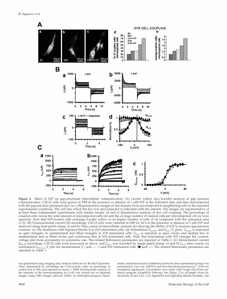
Figure 2. Effect of S1P on gap-junctional intercellular communication. (A) Lucifer yellow dye/transfer analysis of gap junctioncommunication. C2C12 cells were grown in DM in the presence or absence of 1 �M S1P at the indicated time and then microinjectedwith the gap junction–permeant dye. (a– c) Representative images of dye transfer from microinjected to neighboring cells in the reportedexperimental conditions. The cell into which the dye was microinjected is indicated with the asterisk. The images are representative ofat least three independent experiments with similar results. (d and e) Quantitative analysis of dye cell coupling. The percentage ofcoupled cells versus the total amount of microinjected cells (d) and the average number of stained cells per microinjected cell (e) werereported. Note that S1P-treated cells exchange Lucifer yellow to an higher number of cells (3– 4) compared with the untreated ones(1–2). (B) Transjunctional current (Ij) recordings. C2C12 cells were cultured in DM for 24 h in the presence or absence of 1 �M S1P andanalyzed using dual patch-clamp. (a and b) Time course of intercellular currents (Ij) showing the ability of S1P to increase gap junctioncurrents. (c) The treatment with heptanol blocks Ij in S1P-stimulated cells. (d) Normalized Gj,inst- and Gj,ss-Vj plots. Gj,inst is indicatedas open triangles in unstimulated and filled triangles in S1P-stimulated cells. Gj,ss is reported as open circles and dashed line inunstimulated and as filled circles and continuous line in S1P-stimulated cells. Note that stimulation with S1P changes the current-voltage plot from asymmetric to symmetric one. The related Boltzmann parameters are reported in Table 1. (C) Hemichannel current(IhCh) recordings. C2C12 cells were processed as above, and IhCh was recorded by single patch-clamp. (a and b) IhCh time course; (c)normalized GhCh,ss-V plot for unstimulated (� and – – –) and S1P-stimulated cells (� and —). The related Boltzmann parameters arereported in Table 1.
R. Squecco et al.
Molecular Biology of the Cell4900

Figure 3. Effect of inhibition of calcium increase, ERK1/2, and p38 MAPK on S1P-induced Cx43 and myogenin expression in C2C12myoblasts. (A) Confluent C2C12 cells were treated with 15 �M BAPTA/AM or 10 �M PD98059 or 5 �M SB239063 or each specific vehiclefor 30 min before incubation in DM in the presence (�) or absence (�) of 1 �M S1P for 48 h. The content of Cx43 and myogenin were analyzedon cell lysates by Western blot. Equally loaded protein (30 �g) was checked by expression of the nonmuscle specific �-actin. Band intensitywas determined by densitometry and relative percentage to control arbitrarily normalized to 100 is shown in the graphic. A blotrepresentative of at least three independent experiments is shown.
Table 1. Boltzmann parameters for gap junctions and hemichannels in C2C12 cells
Gap junctions
�S1P �S1P Hemichannels
Parameter Vj(�) Vj(�) Vj(�) Vj(�) �S1P �S1P
A (mV�1) 0.065 � 0.004 0.072 � 0.003a 0.064 � 0.004 0.063 � 0.004b 0.08 � 0.002 0.12 � 0.002c
V0 (mV) 62.10 � 4.42 �51.11 � 4.32a 62.03 � 5.02 �55.03 � 5.02 5.0 � 1.2 4.5 � 0.9Gmin 0.25 � 0.03 0.45 � 0.05a 0.34 � 0.04b 0.32 � 0.04b
Data were obtained from C2C12 cells incubated in DM for 24 h in the absence (�) or presence (�) of S1P. The parameters were obtained byfitting normalized Gj,ss and GhCh,ss vs. voltage data (20–24 for each experimental condition).Note that Boltzmann parameters at negative Vj, Vj(�), are significantly different with respect to those at positive Vj, Vj(�), in control but notin S1P-stimulated cells; a p � 0.05 for Vj(�) vs. Vj(�); b p � 0.05 for S1P-stimulated cell pairs vs. controls.For hemichannel analysis, the value A representing the GhCh,ss voltage sensitivity is significantly increased in S1P-stimulated cells withrespect to their control counterpart; c p � 0.05. Data are presented as mean � SEM.
Sphingosine 1-Phosphate Induces Myoblast Differentiation
Vol. 17, November 2006 4901

Rafael, CA) were used for mathematical and statistical analysis of electro-physiological data.
RESULTS
S1P Induces Cx43 Protein Expression and Gap JunctionalCommunication in Differentiating MyoblastsWe first ought to determine whether Cx43 could be a possibletarget of S1P-induced C2C12 myoblast differentiation by RT-PCR and Western blot analysis in differentiating cells. As re-ported in Figure 1, A and B, Cx43 was greatly up-regulated atthe mRNA and protein levels in differentiating cells and, par-ticularly, in those treated with S1P, indicating that the bioactivelipid acted as a potent inducer of Cx43 expression in C2C12
myoblasts. In particular, the expression of the gap junctionprotein gradually increased from 24 up to 72 h and then de-clined with the progression of myogenesis, being barely de-tected in cells at 5 d of differentiation and in fully differentiatedcells (unpublished data). Cx43 was found as a prevalent bandof �43 kDa in control cells and as multiple bands of 40–46 kDain S1P-treated myoblasts, suggesting that the bioactive lipidcould also affect Cx43 posttranslation modifications. Parallelexperiments were performed to verify whether S1P treatmentaffected the expression of other connexin such as Cx39, anisoform recently identified in differentiating myoblasts (vonMaltzahn et al., 2004, 2006; Belluardo et al., 2005). As shown inFigure 1B, Cx39 was weakly detectable in C2C12 myoblasts,and its expression was not affected by S1P, at least in the early
Figure 4. Effect of down-regulation and overexpression of wild-type and mutant Cx43. (A) Western analysis of Cx43 expression. C2C12myoblasts treated with scrambled (SCR) or Cx43-siRNA were incubated with DM in the presence (�) or absence (�) of 1 � S1P for 48 h.Proteins (30 �g) were separated on SDS-PAGE, and the content of Cx43 was analyzed using monoclonal anti-Cx43. Equally loaded proteinswere evaluated as reported in Figure 3. Band intensity was determined as reported in the legend of Figure 1 and shown in the graphic. Ablot representative of at least three independent experiments is shown. (B) Confocal microscopy of Cx43 expression. C2C12 cells treated withscrambled (SCR) or Cx43-siRNA were incubated in DM for 48 h in the presence of S1P. Cells were fixed and labeled for Cx43 to visualizethe intracellular localization of Cx43 (green) and counterstained with PI (red) to reveal nuclei. Note the marked reduction of the fluorescencesignal in Cx43-siRNA–treated myoblasts. Differential interference contrast images are shown to reveal the morphological features of SCR- orCx43-siRNA–treated cells. (C) Western analysis of recombinant Cx43 expression. C2C12 myoblasts stably transfected with GFP-wtCx43 orGFP-DNCx43 or vector alone were incubated in DM for 48 h in the absence or presence of 1 �M S1P and processed as above. Recombinantprotein and endogenous Cx43 were immunodetected by monoclonal anti-GFP and anti-Cx43 antibodies. Densitometric analysis determinedas reported in Materials and Methods is shown in the graphic. A blot representative of three independent experiments is shown. (D) Confocalmicroscopy of recombinant Cx43 expression. C2C12 myoblasts stably transfected with wtCx43 (GFP-wtCx43) or DNCx43 (GFP-DNCx43)were incubated in DM for 48 h and processed as above. Recombinant wtCx43 immunostaining (green) was mainly detected as a linearpunctate staining at the membrane interface in GFP-wtCx43 cells; in contrast, mutated Cx43 showed a predominant cytoplasmic localizationin GFP-DNCx43 cells. The images are representative of three separate experiments with similar results.
R. Squecco et al.
Molecular Biology of the Cell4902

hours of differentiation (24–48 h). However, the expressionlevel of Cx39 increased significantly in differentiating cellsstarting from 72 up to 120 h of incubation in DM, especially inthe presence of S1P (Figure 1).
Confocal immunofluorescence confirmed the temporalregulation of Cx43 protein expression detected by Westernblot analysis (Figure 1C). Confluent C2C12 myoblasts ex-pressed some Cx43 immunostaining, distributed as smallgreen fluorescent dots both in the cytoplasm—in the perinu-clear regions—and at the cell surface. However, after 24 and48 h of S1P stimulation, the expression of Cx43 appearedremarkably increased compared with the levels detected inuntreated cells. The immunofluorescent dots were abun-dantly found in the cytoplasm, likely within the proteinsynthesis pathway, as well as along regions of intimatecell-to-cell contacts, compatible with the formation of gapjunction plaques. Of interest, the quantification of the fluo-rescent signal indicated that cells treated with S1P for 5 hand left in DM in the absence of the bioactive lipid foradditional 20 h, showed higher levels of Cx43 than theuntreated cells, consistent with an effect of S1P on the pro-tein expression in the early hours of incubation (Figure 1C).
The increase of immunoreactive Cx43 at the appositionalplasma membranes between adjacent myoblasts after S1P
stimulation was consistent with the functional differences inthe gap junction permeability, as detected by Lucifer yellowdye-transfer assay (Figure 2A). The efficacy of dye spreadingin control was very low, with �20% of the cells showing dyecoupling with only one neighboring cell. After 24 and 48 h ofincubation in DM the extent of dye transfer slightly in-creased, with �30% of the cells showing 1–2 or 2–3 coupledcells, respectively. The long-term treatment with S1P signif-icantly increased the extent of gap junction communicationover that evaluated in controls, with �50 and 70% of thecells showing 1–2 and 3–4 coupled cells per injection, after24 and 48 h of stimulation, respectively.
To further investigate the role of S1P on intercellularcommunication, we characterized the biophysical propertiesof gap junctional current by dual whole cell voltage-clamp.Figure 2B shows transjunctional current traces (Ij) in repre-sentative cell pairs in control condition and after 24 h oftreatment. Interestingly, the amplitude of Ij and transjunc-tional conductance (Gj,ss) increased significantly at all thevoltage values in cells treated with S1P. The dependence ofthis current and conductance on junctional channels wasestablished by assessing its sensitivity to heptanol, a com-monly used gap junction channel blocker. As expected,heptanol (1 mM) was able to inhibit both Ij and Gj,ss within
Figure 5. Effect of down-regulation and overexpression of Cx43 on S1P-induced myogenic marker expression. (A) Western analysis ofmyogenic marker expression. Confluent C2C12 myoblasts were treated with scrambled (SCR) or Cx43-siRNA for 48 h in DM in the presence(�) or absence (�) of 1 �M S1P. Cell lysates (30 �g of protein) were separated by SDS-PAGE. Myogenin and MHC expression wereimmunodetected by specific antibodies. Equally loaded protein was checked by expression of the nonmuscle specific �-actin. Band intensitynormalized with respect to each specific control (set as 100) is reported in the graphic. A blot representative of three independent experimentswith similar results is shown. (B) Western analysis of myogenic marker expression. Confluent vector, GFP-wtCx43, or GFP-DNCx43myoblasts incubated with DM for the indicated period of time in the absence (�) or in the presence (�) of 1 �M S1P were processed asreported above. Myogenin, MHC, and cav-3 expression were immunodetected by specific antibodies. Band intensity was determined bydensitometry and relative percentage to control was arbitrarily normalized to 100 is shown in the graphic. A blot representative of at leastthree independent experiments with similar results is shown.
Sphingosine 1-Phosphate Induces Myoblast Differentiation
Vol. 17, November 2006 4903

3–5 min (Figure 2B), since Gj,ss at � 10 mV ranged from0.62 � 0.04 (n � 8) to 0.72 � 0.05 (n � 9) nS in control andstimulated cell pairs, respectively. Of interest, best-fit Bolt-zmann parameters showed that control cells had a slightasymmetrical voltage-dependent currents (Table 1). In keep-ing with our data that Cx39 was only weakly detectable inC2C12 myoblasts and in consideration that ambiguous dataexist on the expression of Cx45 in these cells (Araya et al.,2005; von Maltzahn et al., 2006), we suggested that theasymmetric Bolzmann parameters were predominantly dueto the inside-outside voltage dependence of Cx43 Gj previ-ously described (White et al., 1994). However, the plot be-came symmetric after treatment with S1P and showed aslower inactivation compared with control, suggesting thatS1P could affect the inside-outside voltage-dependence ofCx43-containing gap junctions, by reducing the fast gating,acting in the same manner as other chemicals (Bukauskas etal., 2001). Finally, we demonstrated that S1P affected thehemichannel conductance in myoblastic cells, because IhChas well as its voltage sensitivity were significantly increasedin S1P-treated cells compared with control, as indicated bythe A parameter (Figure 2C, Table 1).
Overall, all these data indicated that S1P promoted Cx43protein expression and increased gap junctional andhemichannel permeability during the early phases of myo-genesis.
Calcium Increase and p38 MAPK Activity Are Involved inS1P-induced Cx43 ExpressionBecause gap junctions are required for normal myogenesis(Araya et al., 2005), we next investigated whether knownmediators of the myogenic program and known targets ofS1P, such as Ca2� and p38 MAPK (Meacci et al., 2002a;Porter et al., 2002; Cabane et al., 2003; Donati et al., 2005),could also play a role in the regulation of Cx43 expression
induced by the bioactive lipid. To this end, we usedBAPTA/AM (15 �M), a calcium chelator capable of prevent-ing intracellular calcium increase, or SB239063 (5 �M), aspecific inhibitor of p38 MAPK, and focused our observa-tions on the time period where the effect of S1P on proteinexpression was predominant (48 h). As shown in Figure 3,depletion of Ca2� by BAPTA/AM as well as inhibition ofp38 MAPK activity by SB239063 prevented S1P-inducedCx43 up-regulation and strongly reduced expression ofmyogenin (Figure 3) and myosin heavy chain (MHC) andcaveolin-3 (cav-3; unpublished data). Notably, no significantchange was observed in the expression of Cx43 as well as ofthe other myogenic markers in myoblasts treated withPD98057 before S1P addition, suggesting that ERK1/2 activ-ity was not required for Cx43 expression and myogenesis.Moreover, BAPTA/AM or PD98057 was unable to affectbasal Cx43 and myogenin expression, whereas SB239063strongly reduced Cx43 expression below the control. Takentogether, these results indicated that S1P regulated the ex-pression of Cx43 through the activation of both Ca2�- andp38 MAPK-dependent pathways.
Cx43 Protein Expression Is Required for MyoblastDifferentiation Promoted by S1PTo investigate whether Cx43 expression represented an es-sential step in the myogenesis elicited by S1P, C2C12 myo-blasts were transfected with a mix of specific siRNA du-plexes directed against three distinct regions of mouse Cx43mRNA or with plasmids encoding either for dominant neg-ative mutant Cx43�130–136 or wild-type Cx43 fused to GFP(GFP-DNCx43, GFP-wtCx43) and examined for myogenicmarker expression after 48 h of incubation in DM. Prelimi-nary experiments were performed to verify the expressionlevel of Cx43 in silenced and stable overexpressing myo-blasts. As shown in Figure 4, A and B, Cx43 was significantly
Figure 6. Gap-junctional and hemichannel conduc-tances in overexpressing myoblasts. (A and B) Gap junc-tion, Gj,ss, and hemichannel, GhCh,ss, conductance. Elec-trophysiological parameters were measured in nativeC2C12 cells and in cells overexpressing GFP (Vector),GFP-DNCx43, and GFP-wtCx43. C2C12 cells cultured inDM in the presence (f) or absence of S1P (�) for 24 hwere analyzed by dual (A) or single (B) patch clamp.Transfection with vector alone does not modify Gj,ss andGhCh,ss, whereas transfection with GFP-DNCx43 or withGFP-wtCx43 constructs decrease and increase both Gj,ssand GhCh,ss (*p � 0.05), respectively. S1P stimulation en-hances the electrical coupling and hemichannel conduc-tance in all the cell preparations (§p � 0.05 and §§p �0.001). (C and D) Normalized Gj,ss-Vj plots. Note that S1Pchanges from asymmetrical to symmetrical the current-voltage plots in all the cell preparations, but not in GFP-DNCx43 cells. Open symbols are related to unstimulatedcells (C), whereas filled symbols refer to S1P-stimulatedcells (D). (Vector, �, Œ; GFP-wtCx43, �, f; GFP-DNCx43,�, F). The related Boltzmann parameters are shown inTable 2. (E and F) Normalized GhCh,ss-V plots. Note thatthe hemichannel voltage sensitivity is markedly increasedin GFP-wtCx43 cells compared with that of C2C12 andvector cells. Such increase is significantly enhanced byS1P stimulation (F, filled symbol) in all the cell prepara-tions, but not in GFP-DNCx43 cells. Symbols and super-imposed Boltzmann lines are as above. The related Bolt-zmann parameters are listed in Table 2.
R. Squecco et al.
Molecular Biology of the Cell4904

down-regulated in Cx43-siRNA-treated myoblasts both inunstimulated and stimulated cells. A band of �75 kDa,consistent with the predicted molecular weight of Cx43 pro-tein fused to GFP, was immunodetected in GFP-DNCx43–,as well as in GFP-wtCx43–overexpressing myoblasts, and itsexpression appeared not to be affected by S1P treatment(Figure 4C). Confocal microscopy, performed to reveal the insitu localization of recombinant Cx43, showed that bothGFP-wtCx43 and GFP-DNCx43 were present in the cyto-plasm as well as at the plasma membrane. However, thelocalization of the mutated protein at cell–cell contacts, wasless clear than that of wild-type Cx43, indicating some alter-ations in the organization of gap junctional plaques in GFP-DNCx43 myoblasts (Figure 4D).
Interestingly, the down-regulation of the endogenousCx43 protein or the enforced expression of mutated formdramatically reduced myogenic differentiation in controland S1P-stimulated cells, as judged by Western blot analysisfor the expression of the myogenic markers (Figure 5, A andB) and by CK assay (3.3 � 0.04 vs. 8.2 � 0.09 U/mg inS1P-treated GFP-DNCx43 cells vs. S1P-stimulated C2C12cells, p � 0.05, n � 3). In addition, both Cx43-siRNA-treatedor GFP-DNCx43 cells showed a delay in myoblast matura-tion, retaining spherical or star-shaped morphology at 48 hof differentiation in the presence or absence of S1P (Figure4B). By contrast, as shown in Figure 5B, enforced expressionof GFP-wtCx43 accelerated C2C12 myoblast differentiation.All these data indicated that Cx43 expression and plasmamembrane localization were required for skeletal muscledifferentiation.
To understand the mechanisms by which Cx43 proteincould regulate S1P-induced myogenesis, myoblasts overex-pressing GFP-wtCx43 and GFP-DNCx43 were analyzed forthe ability to form functional channels. Electrophysiologicalrecordings of myoblast pairs cultured in DM in the presenceor absence of S1P (Figure 6) indicated that the highest valuesof gap junction and hemichannel conductance were re-corded in cells transfected with the wild-type connexin,suggesting that the overexpressed recombinant protein as-sembled correctly into functional connexons. Both basal gapjunction and hemichannel conductance were reduced by�60% in GFP-DNCx43 myoblasts as compared with those ofvector and C2C12 cells (Figure 6). In particular, C2C12 myo-blasts overexpressing GFP-wtCx43 showed a symmetricalcurrent-voltage plot, whereas those overexpressing GFP-DNCx43 exhibited an asymmetrical relationship as in con-trol cells (Figure 6, Table 2). Similarly, the hemichannelvoltage sensitivity increased in GFP-wtCx43- and decreasedin GFP-DNCx43-expressing myoblasts (Figure 6, Table 2).Collectively, these data indicated a channel-dependent rolefor Cx43 in the regulation of skeletal myogenesis.
The treatment with S1P caused a significant elevation of boththe gap junction and hemichannel conductance in GFP-DNCx43 compared with vector cells (about twofold vs. 4–5-fold). Interestingly, the amplitude of Gjss and the Boltzmannparameters in S1P-treated GFP-DNCx43 cells did not differfrom that of unstimulated vector and control myoblasts. How-ever, it was of interest to note that 1) the residual gap junctionand hemichannel conductance in GFP-DNCx43 cells stimu-lated with S1P showed, differently from control, a more pro-
Table 2. Boltzmann parameters for gap junctions and hemichannels in overexpressing cells
Gap junctions
�S1P �S1P Hemichannels
Parameter Vj(�) Vj(�) Vj(�) Vj(�) �S1P �S1P
A (mV�1) 0.060 � 0.004 0.070 � 0.003 0.063 � 0.004 0.058 � 0.004ab 0.08 � 0.002 0.12 � 0.002c
V0 (mV) 61.10 � 4.42 �50.01 � 4.32a 61.03 � 5.02 �54.03 � 4.82a 5.01 � 0.8 4.61 � 0.91Gmin 0.25 � 0.03 0.46 � 0.05a 0.34 � 0.04b 0.33 � 0.04b
GFP-DNCx43 Hemichannels
�S1P �S1P �S1P �S1P
A (mV�1) 0.068 � 0.004 0.078 � 0.003a 0.066 � 0.004 0.075 � 0.004a 0.05 � 0.004d 0.06 � 0.004d
V0 (mV) 65.10 � 4.42 �48.11 � 4.32a 63.03 � 5.02 �50.03 � 5.10a 8.10 � 1.2d 7.03 � 0.89d
Gmin 0.24 � 0.03 0.52 � 0.05a 0.26 � 0.04 0.45 � 0.04
GFP-wtCx43 Hemichannels
�S1P �S1P �S1P �S1P
A (mV�1) 0.063 � 0.004 0.056 � 0.003 0.062 � 0.004 0.060 � 0.004 0.14 � 0.003e 0.15 � 0.003e
V0 (mV) 62.10 � 4.42 �61.11 � 4.32 62.03 � 5.02 �63.03 � 5.02 4.11 � 1.20 3.51 � 0.75Gmin 0.32 � 0.03 0.33 � 0.05 0.31 � 0.04 0.30 � 0.04
Data were obtained from vector, GFP-DNCx43 and GFP-wtCx43 cells incubated in DM for 24 h in the absence (�) or presence (�) of S1P.The parameters were obtained by fitting normalized Gj,ss and GhCh,ss vs. voltage data (15–20 recordings for each experimental condition). Gj,ssis asymmetrical in vector control and becomes symmetrical in S1P-treated cell pairs, a p � 0.05 for Vj(�) vs. Vj(�), and b p � 0.05 forS1P-stimulated vs. unstimulated cell pairs. In DN-Cx43 cells, Gj,ss remains asymmetrical after S1P stimulation, whereas in GFP-wtCx43 cells,Gj,ss is symmetrical in unstimulated and S1P-stimulated cells. As regards hemichannel parameters, in vector control cells, the value A,representing the GhCh,ss voltage sensitivity, is significantly increased in S1P-treated cells with respect to their control counterpart (c p � 0.05).In GFP-DNCx43 and GFP-wtCx43 cells, the voltage sensitivity are significantly decreased or increased, respectively, with respect to vectorcontrol cells (d p � 0.05, e p � 0.05). Data presented as mean � SEM.
Sphingosine 1-Phosphate Induces Myoblast Differentiation
Vol. 17, November 2006 4905
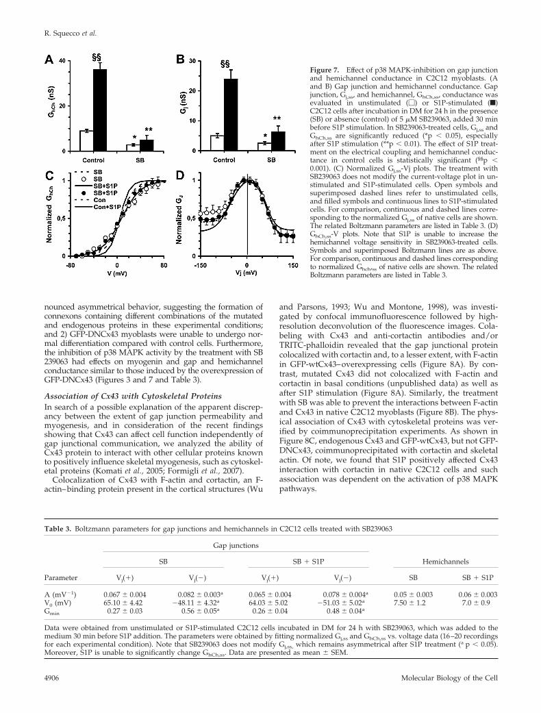
nounced asymmetrical behavior, suggesting the formation ofconnexons containing different combinations of the mutatedand endogenous proteins in these experimental conditions;and 2) GFP-DNCx43 myoblasts were unable to undergo nor-mal differentiation compared with control cells. Furthermore,the inhibition of p38 MAPK activity by the treatment with SB239063 had effects on myogenin and gap and hemichannelconductance similar to those induced by the overexpression ofGFP-DNCx43 (Figures 3 and 7 and Table 3).
Association of Cx43 with Cytoskeletal ProteinsIn search of a possible explanation of the apparent discrep-ancy between the extent of gap junction permeability andmyogenesis, and in consideration of the recent findingsshowing that Cx43 can affect cell function independently ofgap junctional communication, we analyzed the ability ofCx43 protein to interact with other cellular proteins knownto positively influence skeletal myogenesis, such as cytoskel-etal proteins (Komati et al., 2005; Formigli et al., 2007).
Colocalization of Cx43 with F-actin and cortactin, an F-actin–binding protein present in the cortical structures (Wu
and Parsons, 1993; Wu and Montone, 1998), was investi-gated by confocal immunofluorescence followed by high-resolution deconvolution of the fluorescence images. Cola-beling with Cx43 and anti-cortactin antibodies and/orTRITC-phalloidin revealed that the gap junctional proteincolocalized with cortactin and, to a lesser extent, with F-actinin GFP-wtCx43–overexpressing cells (Figure 8A). By con-trast, mutated Cx43 did not colocalized with F-actin andcortactin in basal conditions (unpublished data) as well asafter S1P stimulation (Figure 8A). Similarly, the treatmentwith SB was able to prevent the interactions between F-actinand Cx43 in native C2C12 myoblasts (Figure 8B). The phys-ical association of Cx43 with cytoskeletal proteins was ver-ified by coimmunoprecipitation experiments. As shown inFigure 8C, endogenous Cx43 and GFP-wtCx43, but not GFP-DNCx43, coimmunoprecipitated with cortactin and skeletalactin. Of note, we found that S1P positively affected Cx43interaction with cortactin in native C2C12 cells and suchassociation was dependent on the activation of p38 MAPKpathways.
Figure 7. Effect of p38 MAPK-inhibition on gap junctionand hemichannel conductance in C2C12 myoblasts. (Aand B) Gap junction and hemichannel conductance. Gapjunction, Gj,ss, and hemichannel, GhCh,ss, conductance wasevaluated in unstimulated (�) or S1P-stimulated (f)C2C12 cells after incubation in DM for 24 h in the presence(SB) or absence (control) of 5 �M SB239063, added 30 minbefore S1P stimulation. In SB239063-treated cells, Gj,ss andGhCh,ss are significantly reduced (*p � 0.05), especiallyafter S1P stimulation (**p � 0.01). The effect of S1P treat-ment on the electrical coupling and hemichannel conduc-tance in control cells is statistically significant (§§p �0.001). (C) Normalized Gj,ss-Vj plots. The treatment withSB239063 does not modify the current-voltage plot in un-stimulated and S1P-stimulated cells. Open symbols andsuperimposed dashed lines refer to unstimulated cells,and filled symbols and continuous lines to S1P-stimulatedcells. For comparison, continuous and dashed lines corre-sponding to the normalized Gj,ss of native cells are shown.The related Boltzmann parameters are listed in Table 3. (D)GhCh,ss-V plots. Note that S1P is unable to increase thehemichannel voltage sensitivity in SB239063-treated cells.Symbols and superimposed Boltzmann lines are as above.For comparison, continuous and dashed lines correspondingto normalized Ghch,ss of native cells are shown. The relatedBoltzmann parameters are listed in Table 3.
Table 3. Boltzmann parameters for gap junctions and hemichannels in C2C12 cells treated with SB239063
Gap junctions
SB SB � S1P Hemichannels
Parameter Vj(�) Vj(�) Vj(�) Vj(�) SB SB � S1P
A (mV�1) 0.067 � 0.004 0.082 � 0.003a 0.065 � 0.004 0.078 � 0.004a 0.05 � 0.003 0.06 � 0.003V0 (mV) 65.10 � 4.42 �48.11 � 4.32a 64.03 � 5.02 �51.03 � 5.02a 7.50 � 1.2 7.0 � 0.9Gmin 0.27 � 0.03 0.56 � 0.05a 0.26 � 0.04 0.48 � 0.04a
Data were obtained from unstimulated or S1P-stimulated C2C12 cells incubated in DM for 24 h with SB239063, which was added to themedium 30 min before S1P addition. The parameters were obtained by fitting normalized Gj,ss and GhCh,ss vs. voltage data (16–20 recordingsfor each experimental condition). Note that SB239063 does not modify Gj,ss, which remains asymmetrical after S1P treatment (a p � 0.05).Moreover, S1P is unable to significantly change GhCh,ss. Data are presented as mean � SEM.
R. Squecco et al.
Molecular Biology of the Cell4906

All these data, taken together, were consistent with theidea that C2C12 myoblast differentiation could depend onthe intercellular coupling as well as on an additional func-tion of Cx43 protein per se, likely involving its physicalinteraction with proteins of cytoskeleton.
DISCUSSION
In the present study we identified Cx43 as a key regulatoryprotein of C2C12 myoblast differentiation elicited by S1P and
proposed a novel mechanism by which Cx43 protein, in addi-tion to form functional channels, may regulates skeletal muscledifferentiation through other mechanisms requiring actin cy-toskeletal remodeling and actin–Cx43 protein interaction.
In line with previous reports (Proulx et al., 1997a; Con-stantin and Cronier, 2000; Gorbe et al., 2005), Cx43 proteinwas transiently up-regulated in differentiating C2C12 myo-blasts: Cx43 appeared early in proliferating cells, followedby a progressive increase in the cytoplasm and cell-mem-brane domains of adjacent myoblasts until fusion into myo-
Figure 8. Association between Cx43 and cytoskeletal proteins. (A) Intracellular localization of Cx43/actin complex in overexpressing C2C12myoblasts. C2C12 cells were transfected with plasmids containing GFP-wtCx43 (a and c–f) or GFP-DNCx43 (b and g–l) grown in DM for 48 h,fixed, and stained with specific anti-cortactin antibodies (blue). Counterstaining was performed with TRITC-phalloidin (red) to show actinfilaments. Fluorescent deconvolution microscopy analysis allowed the detection of F-actin (c and g) with Cx43 (green, d and h) and cortactin(e and i). (f–k) Merged images of F-actin, Cx43, and cortactin localization. White spots indicate colocalization of the three signals. Note thatCx43 colocalizes mainly with cortactin (cyan) and weakly with F-actin (yellow) in cells transfected with GFP-wtCx43, whereas theGFP-DNCx43 signal is found predominantly as green dots in the merged images, indicating the absence of colocalization with the cytoskeletalsignals. (B) Intracellular localization of Cx43/actin complex in C2C12 myoblasts. C2C12 cells incubated in DM in the absence (a–d) orpresence (e–h) of SB239063 (5 �M) 30 min before S1P addition were fixed and stained with specific anti-cortactin and anti-Cx43 antibodies.Counterstaining was performed with TRITC-phalloidin to show actin filaments. Fluorescent deconvolution microscopy analysis allowed thedetection of F-actin (red, a and e) with Cx43 (blue, b and f) and cortactin (green, c and g). (d and h) Merged images of F-actin, Cx43, andcortactin localization. White spots indicate colocalization of the three signals. (C) Interaction of Cx43 with actin and cortactin in overex-pressing C2C12 myoblasts. C2C12 cells were transfected with plasmids containing GFP-DNCx43 or GFP-wtCx43, incubated with DM for 48 h,and processed as described in Materials and Methods. Monoclonal anti-Cx43 or anti-GFP antibodies were incubated with cell lysates (200 �gof proteins) followed by immunoprecipitation (IP) with protein A-Sepharose beads. The bound proteins was eluted after extensive washingin Laemmli sample buffer followed by separation on SDS-PAGE and immunodetected using anti-cortactin or anti-actin or anti-Cx43antibodies as indicated. A blot representative of at least three independent experiments with similar results is shown. (D) Effect of p38MAPK-inhibition on Cx43 and cortactin interaction in S1P-stimulated C2C12 myoblasts. Confluent C2C12 cells were treated with 5 �MSB239063 or vehicle for 30 min before incubation in the presence (�) or absence (�) of 1 �M S1P and cultured for 48 h in DM. Cells wereprocessed as above, and cortactin and Cx43 were immunodetected in the immunoprecipitate by specific anti-cortactin or anti-Cx43 antibodies.A blot representative of three independent experiments with similar results is shown.
Sphingosine 1-Phosphate Induces Myoblast Differentiation
Vol. 17, November 2006 4907

tubes, where the expression decreased rapidly. Moreover,we demonstrated that S1P was a potent inducer of Cx43expression in these cells. This finding, together with ourprevious data showing that S1P is a stimulator of myogen-esis in C2C12 cells (Donati et al., 2005), provides the basis forconsidering Cx43 protein as an new intracellular target ofS1P action during myogenesis. Enhanced expression synthe-sis of Cx43 by S1P was accompanied by increased gapjunctional communication, as demonstrated by Lucifer yel-low dye transfer after microinjection and the evaluation ofgap junctional conductance using dual patch-clamp method.In particular, the transjunctional currents between undiffer-entiated myoblastic pairs exhibited an asymmetrical voltagedependence, indicating the involvement of heterotypic gapjunctional channels in C2C12 intercellular coupling as pre-viously reported (Beyer, 1990; Brink et al., 1997; Sakai et al.,2003; Yao et al., 2003). Interestingly, the transjunctional cur-rent showed a symmetrical behavior in S1P-stimulated cells,consistent with the prevalence of Cx43-homotypic channelsand the ability of the bioactive lipid to affect preferentiallythe expression of only one connexin isoform (i.e., Cx43). Inagreement, the expression of Cx39, weakly detectable atbasal level, was not affected by S1P in the early stages ofmyoblast differentiation. However, consistent with previousreports (von Maltzahn et al., 2004, 2006; Belluardo et al., 2005)the level of this protein increased in differentiated C2C12cells, especially upon S1P treatment. We also demonstratedthat S1P-dependent up-regulation of Cx43 was dependenton the intracellular p38 MAP kinase signaling and Ca2�
mobilization, in agreement with our previous data showinga clear correlation between Ca2� concentration and Cx43protein expression (Formigli et al., 2005a). On the otherhand, we showed that S1P likely affected the degradativeprocess of Cx43 in conditions where Ca2� had been depletedand p38 MAP kinase was inhibited. Despite the evidencesuggesting a role for ERK1/2 signaling pathway in the reg-ulation of Cx43 expression (Warn-Cramer et al., 1998; Hos-sain et al., 1999), we showed here that ERK1/2 inactivationby PD98057 treatment did not modify the synthesis of theprotein in C2C12 cells stimulated with S1P consistently withthe reported inability of ERK1/2 inhibition to affect S1P-induced myogenesis in C2C12 cells (Donati et al., 2005).
Gap junctional communications have been long thoughtto play an important role in the coordination of numerouscell functions, including maintenance of the cellular ho-meostasis and the regulation of cell growth, differentiation,and development. In particular, several studies have pro-posed that gap junctions are required for skeletal muscledevelopment and regeneration, because the blockade of theintercellular coupling with channel blockers, octanol and18beta-glycyrrhetinic acid, or the inducible deletion of Cx43in transgenic mice, inhibit the expression of myogenic mark-ers in differentiating myoblasts (Kalderon et al., 1977; Arayaet al., 2003). Based on these findings, it has been proposed apossible role for gap junctions in allowing the intercellularspread of second messengers and coordination of the cellfunctions in a network of cells (Simon and Goodenough,1998). To verify whether the intercellular communicationwas critical during myogenesis in C2C12 cells, we silencedCx43 expression, and prepared myoblasts expressing a dom-inant negative form of Cx43, which formed gap junctionchannels with reduced permeability. In both conditions wefound that cells expressing mutated Cx43 failed to enter themyogenic program elicited by the bioactive lipid, support-ing the idea that gap junction functionality was essential forthe promotion of myogenesis by S1P. However, myogenesisappeared not to be fully dependent on the extent of gap
junctional communication. In fact, it was found an almostcomplete inhibition of the expression of myogenic markersin GFP-DNCx43–expressing cells despite a 50% decrease inthe transjunctional conductance compared with that of con-trols. Such discrepancy was even greater in GFP-DNCx43myoblasts incubated with S1P for ,one d, in which the gapjunctional conductance was increased upon the basal levelsand similar in amplitude to that of native unstimulated cells,which, instead, underwent normal differentiation. We ex-plained the persistence of the cell-to-cell coupling in S1P-stimulated GFP-DNCx43 myoblasts by the ability of mu-tated connexin to form with the endogenous protein, up-regulated by the bioactive lipid, an ample range ofconnexons, whose function was dependent on the propor-tion of the endogenous and mutant form. It was likely thatthe different structure of connexons in S1P-treated GFP-DNCx43 cells compared with that of control cells (hetero-meric vs. homomeric), despite the similar residual conduc-tance, could explain the different capability to differentiateobserved in the two cell populations. We suggested thepossibility that Cx43 expression per se, in addition to itschannel forming ability, could influence skeletal myogenesisof C2C12 cells elicited by S1P. Consistent with this assump-tion, several lines of evidences have recently shown that theexpression of mutated Cx43 with no intrinsic channel activ-ity are as effective as the wild-type protein in the regulationof several biological processes, including cell growth andsurvival (Lin et al., 1998; Dang et al., 2003). In such a view, itis very likely that Cx43, similarly to other proteins localizedat the intercellular junctions, such as E-cadherin and �-cate-nin (Giepmans, 2004), may exert multiple functions withdifferent domains and play an important role as intermedi-ate protein in the transduction of signals from the membraneto nucleus.
Recent investigations have suggested an involvement foractin and actin-binding proteins in the regulation of myo-genesis, and several mechanisms have been proposed toexplain the effect of cytoskeleton on skeletal differentiation(Qu et al., 1997). Accumulating data have demonstrated adirect interaction of the Cx43 C-terminus, with cytoskeletalproteins displaying signal transduction activity, includingdrebrin (Butkevich et al., 2004), ZO-1 (zonula occludens 1(Toyofuku et al., 2001), and c-Src (Giepmans et al., 2003).Therefore, we analyzed the ability of the mutated and wild-type protein to interact with the cytoskeleton. Of note, en-dogenous connexin and recombinant wild-type Cx43, butnot DNCx43, were physically associated with skeletal actinas well as cortactin, strongly supporting the idea that theinteraction between Cx43 protein and cytoskeleton may beinvolved in the accomplishment of myogenesis in C2C12cells. We also showed that the interaction between the gapjunction protein and cortactin is regulated by S1P and isdependent on p38 MAPK activation, pointing to the phos-phorylation of Cx43 as an additional step in S1P regulationof gap junction protein function. The physical association ofCx43 with F-actin modulated by S1P is of particular interestin view of our recent observations, showing that actin re-modeling is crucial for myogenic process elicited by S1P inthe same cells (Formigli et al., 2005a; Formigli et al., 2007).Collectively, these data in combination with those reportedin the literature showing that the lack of a correct gap-junctional assembly of Cx43 on the cell surface hampersseveral cellular processes, including growth, proliferation,and differentiation (Moorby and Patel, 2001; Dang et al.,2003; Li et al., 2006), are consistent with the emerging ideathat Cx43 may also act as an adaptor protein and functionthrough gap-independent mechanisms.
R. Squecco et al.
Molecular Biology of the Cell4908

In conclusion, the results of the present study provide thefirst experimental evidence that up-regulation of Cx43 pro-tein and the subsequent increase in gap junction functional-ity are important mechanism by which S1P promotes myo-genesis in C2C12 myoblasts. Notably, our data, although notexcluding that the exchange of molecules through functionalgap junctions plays a dominant role in skeletal muscle differ-entiation, suggest that the interaction between Cx43 and cy-toskeletal proteins may represent a possible molecular mecha-nism by which Cx43 per se affects cellular differentiation.
ACKNOWLEDGMENTS
We thank Dr. J. von Maltzahn and K. Willecke from Institut fur Genetik,Abteilung Molekulargenetik, Universitat Bonn, Germany, for providing usantibodies against the C-terminus region of mouse Cx39. This article wassupported by grants from Ente Cassa di Risparmio di Pistoia e Pescia (toE.M.), from Ente Cassa di Risparmio di Firenze (to F.F. and S.Z.-O.), and fromthe University of Florence (ex 60%) (to E.M., L.F., S.Z.-O., and F.F.).
REFERENCES
Araya, R., Eckardt, D., Maxeiner, S., Kruger, O., Theis, M., Willecke, K., andSaez, J. C. (2005). Expression of connexins during differentiation and regen-eration of skeletal muscle: functional relevance of connexin43. J. Cell Sci. 8,27–37.
Araya, R., Eckardt, D., Riquelme, M. A., Willecke, K., and Saez, J. C. (2003).Presence and importance of connexin43 during myogenesis. Cell Commun.Adhes. 10, 451–456.
Belluardo, N., Trovato-Salinaro, A., Mudo, G., and Condorelli, D. F. (2005).Expression of the rat connexin 39 (rCx39) gene in myoblasts and myotubes indeveloping and regenerating skeletal muscles: an in situ hybridization study.Cell Tissue Res. 320, 299–310.
Beyer, E. C. (1990). Molecular cloning and developmental expression of twochick embryo gap junction proteins. J. Biol. Chem. 265, 14439–14443.
Brink, P. R., Cronin, K., Banach, K., Peterson, E., Westphale, E. M., Seul, K. H.,Ramanan, S. V., and Beyer, E. C. (1997). Evidence for heteromeric gap junctionchannels formed from rat connexin43 and human connexin37. Am. J. Physiol.273, C1386–C1396.
Butkevich, E., Hulsmann, S., Wenzel, D., Shiraro, T., Duden, R., and Majoul,I. (2004). Drebrin is a novel connexin-43 binding partner that links gapjunctions to the submembrane cytoskeleton. Curr. Biol. 14, 650–658.
Bukauskas, F. F., Bukauskiene, A., Bennett, M. V., and Verselis, V. K. (2001).Gating properties of gap junction channels assembled from connexin43 andconnexin43 fused with green fluorescent protein. Biophys. J. 81, 137–152.
Cabane, C., Englaro, W., Yeow, K., Ragno, M., and Derijard, B. (2003). Reg-ulation of C2C12 myogenic terminal differentiation by MKK3/p38alpha path-way. Am. J. Physiol. Cell Physiol. 284, C658–C666.
Constantin, B., and Cronier, L. (2000). Involvement of gap junctional commu-nication in myogenesis. Int. Rev. Cytol. 196, 1–65.
Dang, X., Doble, B. W., and Kardami, E. (2003). The carboxy-tail of con-nexin-43 localizes to the nucleus and inhibits cell growth. Mol. Cell Biochem.242, 35–38.
Donati, C., Meacci, E., Nuti, F., Becciolini, L., Farnararo, M., and Bruni, P.(2005). Sphingosine 1-phosphate regulates myogenic differentiation: a majorrole for S1P2 receptor. FASEB J. 19, 449–451.
Formigli, L., et al. (2005a). Morphofunctional integration between skeletalmyoblasts and adult cardiomyocytes in coculture is favored by direct cell-cellcontacts and relaxin treatment. Am. J. Physiol. Cell Physiol. 288, C795–C804.
Formigli, L., et al. (2005b). Sphingosine 1-phosphate induces cytoskeletalreorganization in C2C12 myoblasts: physiological relevance for stress fibres inthe modulation of ion current through stretch-activated channels. J. Cell Sci.118, 1161–1171.
Formigli, L., Meacci E., Sassoli C., Squecco, R., Nosi, D., Chellini F., Naro, F.,Francini, F., and Zecchi Orlandini, S. (2007). Actin cytoskeleton/stretch-acti-vated ion channels interaction regulates myogenic differentiation of skeletalmyoblasts. J. Cell Physiol. (in press).
Giepmans, B. N. (2004). Gap junctions and connexin-interacting proteins.Cardiovasc. Res. 62, 233–245.
Giepmans, B. N., Feiken, E., Gebbink, M. F., and Moolenaar, W. H. (2003).Association of connexin43 with a receptor protein tyrosine phosphatase. CellCommun. Adhes. 10, 201–205.
Goldberg, G. S., Lampe, P. D., and Nicholson, B. J. (1999). Selective transfer ofendogenous metabolites through gap junctions composed of different con-nexins. Nat. Cell Biol. 1, 457–459.
Gorbe, A., Becker, D. L., Dux, L., Stelkovics, E., Krenacs, L., Bagdi, E., andKrenacs, T. (2005). Transient upregulation of connexin43 gap junctions andsynchronized cell cycle control precede myoblast fusion in regenerating skel-etal muscle in vivo. Histochem. Cell Biol. 123, 573–583.
Grounds, M. D., White, J. D., Rosenthal, N., and Bogoyevitch, M. A. (2002).The role of stem cells in skeletal and cardiac muscle repair. J. Histochem.Cytochem. 50, 589–610.
Hawke, T. J., and Garry, D. J. (2001). Myogenic satellite cells: physiology tomolecular biology. J. Appl. Physiol. 91, 534–551.
Hossain, M. Z., Jagdale, A. B., and Boynton, A. L. (1999). Mitogen-activatedprotein kinase and phosphorylation of connexin43 are not sufficient for thedisruption of gap junctional communication by platelet-derived growth factorand tetradecanoylphorbol acetate. J. Cell Physiol. 179, 87–96.
Huang, G. Y., Cooper, E. S., Waldo, K., Kirby, M. L., Gilula, N. B., and Lo,C. W. (1998a). Gap junction-mediated cell-cell communication modulatesmouse neural crest migration. J. Cell Biol. 143, 1725–1734.
Huang, R. P., Fan, Y., Hossain, M. Z., Peng, A., Zeng, Z. L., and Boynton, A. L.(1998b). Reversion of the neoplastic phenotype of human glioblastoma cellsby connexin 43 (cx43). Cancer Res. 58, 5089–5096.
Kalderon, N., Epstein, M. L., and Gilula, N. B. (1977). Cell-to-cell communi-cation and myogenesis. J. Cell Biol. 75, 788–806.
Kaliman, P., Canicio, J., Testar, X., Palacin, M., and Zorzano, A. (1999).Insulin-like growth factor-II, phosphatidylinositol 3-kinase, nuclear factor-kappaB and inducible nitric-oxide synthase define a common myogenic sig-naling pathway. J. Biol. Chem. 274, 17437–17444.
Komati, H., Naro, F., Mebarek, S., De Arcangelis, V., Adamo, S., Lagarde, M.,Prigent, A. F., and Nemoz, G. (2005). Phospholipase D is involved in myo-genic differentiation through remodeling of actin cytoskeleton. Mol. Biol. Cell16, 1232–1244.
Kondo, R. P., Wang, S. Y., John, S. A., Weiss, J. N., and Goldhaber, J. I. (2000).Metabolic inhibition activates a non-selective current through connexinhemichannels in isolated ventricular myocytes. J. Mol. Cell. Cardiol. 32,1859–1872.
Krutovskikh, V. A., Yamasaki, H., Tsuda, H., and Asamoto, M. (1998). Inhi-bition of intrinsic gap junction intercellular communication and enhancementof tumorigenicity of the rat bladder carcinoma cell line BC31 by a dominant-negative connexin 43 mutant. Mol. Carcinog. 23, 254–261.
Jiang, J. X., and Gu, S. (2005). Gap junction- and hemichannel-independentactions of connexins. Biochim. Biophys. Acta 1711, 208–214.
Lassar, A. B., Buskin, J. N., Lockshon, D., Davis, R. L., Apone, S., Hauschka,S. D., and Weintraub, H. (1989). MyoD is a sequence-specific DNA bindingprotein requiring a region of myc homology to bind to the muscle creatinekinase enhancer. Cell 58, 823–831.
Li, Z., Zhou, Z., Saunders, M. M., and Donahue, H. J. (2006). Modulation ofconnexin43 alters expression of osteoblastic differentiation markers. Am. J.Physiol. Cell Physiol. 290, C1248–C1255.
Lin, J. H., Weigel, H., Cotrina, M. L., Liu, S., Bueno, E., Hansen, A. J., Hansen,T. W., Goldman, S., and Nedergaard, M. (1998). Gap junction-mediatedpropagation and amplification of cell injury. Nat. Neurosci. 1, 743–749.
Meacci, E., Becciolini, L., Nuti, F., Donati, C., Cencetti, F., Farnararo, M., andBruni, P. (2002a). A role for calcium in sphingosine 1-phosphate-inducedphospholipase D activity in C2C12 myoblasts. FEBS Lett. 521, 200–204.
Meacci, E., Cencetti, F., Formigli, L., Squecco, R., Donati, C., Tiribilli, B.,Quercioli, F., Zecchi Orlandini, S., Francini, F., Bruni, P. (2002b). Sphingosine1-phosphate evokes calcium signals in C2C12 myoblasts via Edg3 and Edg5receptors. Biochem. J. 362, 349–357.
Meacci, E., Cencetti, F., Donati, C., Nuti, F., Farnararo, M., Kohno, T., Igarashi,Y., and Bruni, P. (2003). Down-regulation of EDG5/S1P2 during myogenicdifferentiation results in the specific uncoupling of sphingosine 1-phosphatesignalling to phospholipase D. Biochim. Biophys. Acta 1633, 133–142.
Moorby, C., and Patel, M. (2001). Dual functions for connexins: Cx43 regulatesgrowth independently of gap junction formation. Exp. Cell Res. 271, 238–248.
Naro, F., Sette, C., Vicini, E., De Arcangelis, V., Grange, M., Conti, M.,Lagarde, M., Molinaro, M., Adamo, S., and Nemoz, G. (1999). Involvement oftype 4 cAMP-phosphodiesterase in the myogenic differentiation of L6 cells.Mol. Biol. Cell 10, 4355–4367.
Omori, Y., and Yamasaki, H. (1998). Mutated connexin43 proteins inhibit ratglioma cell growth suppression mediated by wild-type connexin43 in a dom-inant-negative manner. Int. J. Cancer 78, 446–453.
Sphingosine 1-Phosphate Induces Myoblast Differentiation
Vol. 17, November 2006 4909
Porter, G. A., Jr., Makuck, R. F., and Rivkees, S. A. (2002). Reduction inintracellular calcium levels inhibits myoblast differentiation. J. Biol. Chem.277, 28942–28947.
Proulx, A. M., Merrifield, P. A., and Naus, C. C. (1997a). Blocking gapjunctional intercellular communication in myoblasts inhibits myogenin andMRF4 expression. Dev. Genet. 20, 133–144.
Proulx, A. A., Lin, Z. X., and Naus, C. C. (1997b). Transfection of rhabdomyo-sarcoma cells with connexin43 induces myogenic differentiation. Cell GrowthDiffer. 8, 533–540.
Qin, H., Shao, Q., Curtis, H., Galipeau, J., Belliveau, D. J., Wang, T., Alaoui-Jamali, M. A., and Laird, D. W. (2002). Retroviral delivery of connexin genesto human breast tumor cells inhibits in vivo tumor growth by a mechanismthat is independent of significant gap junctional intercellular communication.J. Biol. Chem. 277, 29132–29138.
Qu, G., Yan, H., and Strauch, A. R. (1997). Actin isoform utilization duringdifferentiation and remodeling of BC3H1 myogenic cells. J. Cell Biochem. 67,514–527.
Saba, J. D. (2004). Lysophospholipids in development: miles apart and edgingin. J. Cell Biochem. 92, 967–992.
Saez, J. C., Berthoud, V. M., Branes, M. C., Martinez, A. D., and Beyer, E.(2003). Plasma membrane channels form by connexin: their regulation andfunction. Physiol. Rev. 83, 1359–1400.
Sakai, R., Elfgang, C., Vogel, R., Willecke, K., and Weingart, R. (2003). Theelectrical behaviour of rat connexin46 gap junction channels expressed intransfected HeLa cells. Pfluegers Arch. 446, 714–727.
Simon, A. M., and Goodenough, D. A. (1998). Diverse functions of vertebrategap junctions. Trends Cell Biol. 8, 477–483.
Singh, D., Solan, J. L., Taffet, S. M., Javier, R., and Lampe, P. D. (2005).Connexin 43 interacts with zona occludens-1 and -2 proteins in a cell cyclestage-specific manner. J. Biol. Chem. 280, 30416–30421.
Stout, C., Goodenough, D. A., and Paul, D. L. Connexins: functions withoutjunctions. (2004). Curr. Opin. Cell Biol. 16, 507–512.
Toyofuku, T., Akamatsu, Y., Zhang, H., Kuzuya, T., Tada, M., and Hori, M.(2001). c-Src regulates the interaction between connexin-43 and ZO-1 in car-diac myocytes. J. Biol. Chem. 276, 1780–1788.
Upham, B. L., Suzuki, J., Chen, G., Trosko, J. E., Wang, Y., McCabe, L. R.,Chang, C. C., Krutovskikh, V. A., and Yamasaki, H. (2003). Reduced gapjunctional intercellular communication and altered biological effects in mouseosteoblast and rat liver oval cell lines transfected with dominant-negativeconnexin 43. Mol. Carcinog. 37, 192–201.
Valiunas, V. (2002). Biophysical properties of connexin-45 gap junctionhemichannels studied in vertebrate cells. J. Gen. Physiol. 119, 147–164.
von Maltzahn, J., Euwens, C., Willecke, K., and Sohl, G. (2004). The novelmouse connexin39 gene is expressed in developing striated muscle fibers.J. Cell Sci. 117, 5381–5392.
von Maltzahn, J., Wulf, V., and Willecke, K. (2006). Spatiotemporal expressionof connexin 39 and -43 during myoblast differentiation in cultured cells and inthe mouse embryo. Cell Commun. Adhes. 13, 55–60.
Warn-Cramer, B. J., Trevor Cottrell, G., Burt, J. M., and Lau, A. F. (1998).Regulation of connexin-43 gap junctional intercellular communication bymitogen-activated protein kinase. J. Biol. Chem. 273, 9188–9196.
White, T. W., Bruzzone, R., Wolfram, S., Paul, D. L., and Goodenough, D. A.(1994). Selective interactions among the multiple connexin proteins expressedin the vertebrate lens: the second extracellular domain is a determinant ofcompatibility between connexins. J. Cell Biol. 125, 879–892.
Wu, H., and Montone, K. T. (1998). Cortactin localization in actin-containingadult and fetal tissues. J. Histochem. Cytochem. 46, 1189–1191.
Wu, H., and Parsons, J. T. (1993). Cortactin, an 80/85-kilodalton pp60src
substrate, is a filamentous actin-binding protein enriched in the cell cortex.J. Cell Biol. 120, 1417–1426.
Yao, J. A., Gutstein, D. E., Liu, F., Fishman, G. I., and Wit, A. L. (2003). Cellcoupling between ventricular myocyte pairs from connexin43-deficient mu-rine hearts. Circ. Res. 93, 736–743.
R. Squecco et al.
Molecular Biology of the Cell4910